Embed Size (px)
Citation preview

R
EPRODUCTIONRESEARCHHormonal profile and reproductive performance in lactationdeficient (OFA hr/hr) and normal (Sprague–Dawley) female rats
Susana R Valdez, Alicia B Penissi1, Ricardo P Deis and Graciela A Jahn
Laboratorio de Reproduccion y Lactancia, IMBECU, CRICYT-CONICET, Casilla de Correos 855, 5500 Mendoza,Argentina and 1Instituto de Histologıa y Embriologıa, Conicet-Universidad Nacional de Cuyo, Mendoza 5500,Argentina
Correspondence should be addressed to GA Jahn Larlac-Imbecu, CC 855, 5500 Mendoza, Argentina;Email: [email protected]
Abstract
Lactation deficiency may have important consequences on infant health, particularly in populations of low socioeconomic status.
The OFA hr/hr (OFA) strain of rats, derived from Sprague–Dawley (SD) rats, has deficient lactation and is a good model of
lactation failure. We examined the reproductive performance and hormonal profiles in OFA and SD strains to determine the
cause(s) of the lactation failure of the OFA strain. We measured hormonal (PRL, GH, gonadotropins, oxytocin, and progesterone)
levels by RIA in cycling, pregnant, and lactating rats and in response to suckling. Dopaminergic metabolism was assessed by
determination of mediobasal hypothalamic dopamine and dihydroxyphenylacetic acid (DOPAC) concentrations by HPLC and
tyrosine hydroxylase expression by immunocytochemistry and western blot. OFA rats have normal fertility but 50% of the litters
die of malnutrition on early lactation; only 6% of the mothers show normal lactation. The OFA rats showed lower circulating PRL
during lactation, increased hypothalamic dopamine and DOPAC, and impaired milk ejection with decreased PRL and oxytocin
response to suckling. Before parturition, PRL release and lactogenesis were normal, but dopaminergic metabolism was altered,
suggesting activation of the dopaminergic system in OFA but not in SD rats. The number of arcuate and periventricular neurons
expressing tyrosine hydroxylase was higher in SD rats, but hypothalamic expression of TH was higher in OFA rats at the end of
pregnancy and early lactation. These results suggest that the OFA rats have impaired PRL release linked with an augmented
dopaminergic tone which could be partially responsible for the lactational failure.
Reproduction (2007) 133 827–840
Introduction
Lactation insufficiency or failure is relatively commonamong women. In populations with sufficient economicresources, this may have minor consequences on thegrowth and health of the infant, because adequatesubstitute nourishment is available. However, in popu-lations of low socioeconomic status, inadequate lactationcan lead to severe health problems in the newborn,whereas a full lactation can assure the normal growth anddevelopment of the newborn even in situations of deficientavailability of nutrients for the general population.
Most of the ‘nude’ or ‘hairless’ strains of rats andmice, that became very useful experimental tools, are
hypoprolactinemic and fail to lactate. The OFA hr/hr
(OFA) strain of rats, originally developed in Lyon, France
and derived from Sprague–Dawley (SD) rats, is also
hairless and has deficient lactation but does not present
severe impairment of the immune system present in the
q 2007 Society for Reproduction and Fertility
ISSN 1470–1626 (paper) 1741–7899 (online)
‘nude’ strains. A related hypoprolactinemic strain withlactation failure, IPL Nu (Cohen et al. 1983, 1985,1986a, 1986b, 1988, Jordan et al. 1987) shows analtered dopaminergic system, which may be responsiblefor the hypoprolactinemia, since dopamine, originatingin the tuberoinfundibular dopaminergic (TIDA) andperiventricular hypophyseal dopaminergic (PHDA)neurons of the arcuate and periventricular hypo-thalamic nuclei (PeN), is the principal negativeregulator of Prolactin (PRL) release (Cohen et al.1985). It has been proposed that the diminished TIDAdopaminergic tone and TH expression and activityobserved at the end of pregnancy and during lactation(Demarest et al. 1983, Grattan & Averill 1990, Hoffmanet al. 1994, Arbogast & Voogt 1996, Fliestra & Voogt1997, Li et al. 1999, Andrews et al. 2001) are the resultsof decreased sensitivity to PRL negative feedback andthe adaptive response of the hypothalamus to allow forthe sustained hyperprolactinemia induced by suckling
DOI: 10.1530/REP-06-0032
Online version via www.reproduction-online.org
828 S R Valdez and others
(Andrews 2005). Alterations in the regulation of thehypothalamic dopaminergic system during the periodpreceding the initiation of lactation could well underliethe posterior lactation failure of these strains of rats.
The nature of the mutation of the OFA strain hasrecently been shown to be a large intragenic deletion ofthe desmoglein-4 gene (Dsg-4) encompassing nineexons (Bazzi et al. 2004, Kim et al. 2004, Meyer et al.2004), which codifies for a protein belonging to thedesmoglein family. The products of these genes,expressed in neural and neuroendocrine tissues, arecell adhesion molecules related to cadherins.
The main objective of the present paper is to elucidatethe possible factor(s) responsible for the lactationaldeficit of the OFA strain of rats by characterizing thereproductive performance of the OFA and SD strains. Wealso attempted to determine whether alterations in thedopaminergic TIDA and PHDA systems are responsiblefor the lactational deficit. With these aims, we performeda comparative study in OFA and SD rats of the hormonalprofile during proestrus, pregnancy, and lactation,mammary gland function, response to the sucklingstimulus and of the dopaminergic system throughmeasurements of dopamine (DA) and its metabolite,3,4-dihydroxyphenylacetic acid (DOPAC) and oftyrosine hydroxylase (TH, limiting enzyme for dopaminesynthesis) expression in hypothalamic areas.
Materials and Methods
Animals
Adult female and male SD and OFA (originally purchasedfrom Iffa Credo, France and denominated IFL Nu at thattime) rats bred in our laboratory were used. The rats wereused between 3 and 4 months of age, weighing 200–230 g (females) or 250–300 g (males). Animals were keptin a light- (lights on 0600–2000 h) and temperature(22–24 8C)-controlled room. Rat chow (Cargill, Cordoba,Argentina) and tap water were available ad libitum. Sincemost of the pups from the OFA rats died within the first3 days of lactation, they were fostered on the first day postpartum to SD mothers that had given birth within theprevious or subsequent 24 h. The foster mothers weregiven litters of six to eight pups and the fostered OFA pupsachieved normal growth rates. For the experiments,vaginal smears were taken daily from young (3- to4-month old) female rats and only those showing regular4-day cycles were used. The rats were made pregnant bycaging with a fertile male on the night of proestrus. Thepresence of sperm on the vaginal smear the followingmorning indicated day 0 of pregnancy. Two or three daysbefore delivery, the rats were caged individually. The dayand approximate hour of delivery and the size and weightof the litters were recorded. On day 1 of lactation, thenumber of pups in each litter was standardized to eight.Animal maintenance and handling were performed
Reproduction (2007) 133 827–840
according to the NIH guide for the Care and Use ofLaboratory Animals (NIH publication no. 86-23, revised1985 and 1991) and the UK requirements for ethics ofanimal experimentation (Animals Scientific Procedures,Act 1986). All the experimental procedures wereapproved by the Institutional Animal Ethics Committee.
To determine the pattern of hormonal secretion duringthe cycle, pregnancy and lactation, groups of eight to tenrats of each strain were bled (0.5–0.8 ml blood) by thetail vein under light ether anesthesia at 1800 or 1930 hon proestrus or at 1200 h on estrus, or on days 5 (at 1600,1900, and 2200 h), 10, 15, 19, 20 (all at 1200 h), and 21(1800 and 2200 h) of pregnancy and on days 2, 7, 14,and 21 of lactation (at 1600 h). Since this procedure mayproduce a mild stress, the blood samples were alwaystaken within 1 min of introducing the rat in the ether jar,thus, the hormonal values measured represent the basallevels. We also compared the results with hormonevalues obtained from decapitated rats in the differentphysiological states, and found no significantdifferences, in special of PRL, thus confirming that thevalues obtained represent those of unstressed animals.The blood was allowed to clot at room temperature andserum was separated by centrifugation and stored atK30 8C until used. Some rats were decapitated at 1800 hon day 21 of pregnancy and trunk blood and inguinalmammary glands were collected. The mammary glandswere washed in a cold saline solution and stored atK70 8C until they were analyzed.
In order to determine the hormonal and neurochemicalresponse to premature luteolysis at the end of pregnancy(Vermouth & Deis 1972, 1974), groups of rats wereinjected with 25 mg prostaglandin F2a (PGF2a) analog,cloprostenol (Estrumate, Schering-Plough Veterinaria SA,Argentina) in 0.2 ml saline at 0800 and 1200 h on day 19of pregnancy and decapitated 24 h after the secondinjection. Trunk blood was collected, allowed to clot atroom temperature and serum separated by centrifugationand stored frozen for hormone determinations. Brainswere rapidly removed and placed in a cold plate fordissection. The mediobasal hypothalami (MBH; collectedfrom bregma K2.12 to K4.52 mm, which includes thePeN, arcuate nucleus, and median eminence (ME)) wereremoved, frozen on dry ice, and stored at K80 8C untilprocessing for DA and DOPAC assays.
The expression of TH, determined by immunohisto-chemistry (IHC) in brain slices or western blot of MBHhomogenates was studied in groups of male and femalerats on estrus, on days 19 (G19), 21 (G21) of pregnancyat 1800 h or on day 2 of lactation (L2) at 1200 h. Theblood and inguinal mammary glands were obtainedfrom the pregnant and the lactating groups of female ratsfor serum hormone measurements and mammarygland histology.
To determine the response to suckling, groups of eightto ten mothers from each strain were isolated from thelitter at 0800 h on days 10 or 11 of lactation. The litters
www.reproduction-online.org

Reproduction in lactation deficient rats 829
were completed to eight pups when necessary withfoster pups of the same age, weighed, and reunited withtheir mothers at 1600 h. In order to check whether thefostering procedure altered the response to suckling, thesuckling experiments were also performed in SD rats thatnursed fostered OFA or SD pups, with no differences inthe responses compared with mothers nursing their ownlitters. After 15 or 30 min of vigorous suckling, themothers were bled from the tail vein under light etheranesthesia; the litters were weighed again and returnedto their mothers. To measure basal levels of hormones,other groups of rats were bled at 1600 h before replacingthe pups. Serum was separated and stored at K30 8C forRIA of PRL, growth hormone (GH), progesterone, andoxytocin. Other groups of six to eight SD or OFA ratswere decapitated at 1600 h after 8 h of separation or at1630 h after 30 min of suckling, trunk blood wascollected and serum obtained for RIA of hormones, thebrains were removed and the MBH dissected andprocessed as described below for the measurement ofDA and DOPAC by HPLC.
Hormone determinations
PRL, LH, FSH, and GH were measured by double antibodyRIA, using materials provided by Dr A F Parlow and theNHPP (National Hormone and Pituitary Program, Harbor-UCLA Medical Center, Torrance, CA, USA). The hormoneswere radioiodinated using the chloramine-T method andpurified by passage through Sephadex G75. The resultswere expressed in terms of rat PRL RP-3, LH RP-3, FSHRP-3, or rat GH RP-2 standard preparations. Assaysensitivity was 0.5 mg/l serum for PRL, FSH, and GH and0.2 mg/l serum for LH and the inter- and intra-assaycoefficients of variation were !10% for all hormones. Allthe sampleswere measured on the same assayby duplicate.
Oxytocin was measured by double antibody RIA usingan antibody and purified oxytocin generously provided byDr N Hagino and by Novartis-Argentina respectively. Thehormone was radioiodinated using the chloramine-Tmethod and purified by passage through Sephadex G50.The standard curve was prepared using the samepreparation of purified oxytocin used for radioiodination.To maximize sensitivity of the assay, the standards andserum samples were incubated 24 h at 4 8C with appro-priate dilution of the antibody, subsequently the labeledhormone (8–10!103 c.p.m.) was added and the tubesincubated overnight at 4 8C before addition of the secondantibody. Assay sensitivity was 8 ng/l serum and the intra-assay coefficients of variation were!10%. All the sampleswere measured on the same assay by duplicate.
Serum progesterone was measured using a RIAdeveloped in our laboratory (Bussmann & Deis 1979)with an antiserum raised in rabbits against progesterone-11-bovine serum albumin conjugate or using commer-cial kits (DSL-3400 double antibody RIA, from Diag-nostic Systems Laboratories, Webster, TX, USA). Assay
www.reproduction-online.org
sensitivity was !70 fmol/tube and the inter- and intra-assay coefficients of variation were !10%.
Mammary gland histology
Mammary tissue was fixed in buffered formaldehyde,dehydrated in ethanol, and embedded in paraffin wax.Sections of 3–5 mm were cut with a Reichert-Jung Hn 40microtome and stained with hematoxylin–eosin. Imageswere taken with a Zeiss Axioscop-2 light microscopefitted with a Sony CCD-IRIS/RGB videocamera under100 and 400! magnifications. For all the morphologicalanalyses, only the inguinal mammary glands were used.Sections were histologically evaluated for changes in theextent of lobulo-alveolar development and supportingadipose tissue and for the extent of ducto-lobularluminal secretions. The morphological state of thealveoli was determined by analyzing serial sectionsfrom three different animals per group.
Casein and lactose determinations
Mammary casein and lactose were measured in mam-mary gland samples of OFA and SD rats as previouslydescribed (Jahn & Deis 1991). Briefly, 200 mg mammarytissue were cut into small pieces and homogenized in2 ml of 50 mM sodium phosphate buffer, 150 mM NaCl,NaN3 0.1%, Triton X-100 0.1% (pH 7.6) with anUltraturrax homogenizer. The homogenate was centri-fuged at 600 g for 30 min and the supernatant used forb-casein determination by a homologous RIA (Bussmann& Deis 1985) and lactose concentration was assessed bythe method of Kuhn & Lowenstein (1967).
HPLC for DA and DOPAC in the MBH
In addition to the groups of late pregnant and lactating ratsdescribed above and to compare the dopaminergicmetabolism with them, groups of virgin rats in estrus(1200 h) were also killed and the MBH was dissected andprocessed for DA and DOPAC determination by HPLC.
The MBHs were homogenized in 0.2 ml of 0.2 Mperchloric acid, sonicated briefly in an ice bath to disruptthe tissues, and centrifuged at 12 000 r.p.m. for 30 min.DA and DOPAC were extracted on activated aluminaand eluted in 0.2 M perchloric acid. Following centri-fugation, the supernatants were separated and kept atK80 8C until the assay was performed. Protein contentin the remaining pellets was measured using themodified Coomassie blue method of Bradford.
The concentrations of DA and DOPAC in MBHhomogenates (extracts) were analyzed by reverse-phaseliquid chromatography using C18 columns and electro-chemical detection (LKB, Bromma, Sweden). The HPLCmobile phase consisted of 45 mM sodium phosphatedibasic buffer (pH 3.5) containing 0.43 mM sodium octyl
Reproduction (2007) 133 827–840

830 S R Valdez and others
sulphonate, 0.34 mM EDTA, and 2% acetonitrile. Theoxidation potential was set at 0.55 V. The results areexpressed as ng/mg protein for DA and DOPAC and alsothe DOPAC/DA ratio was calculated as an index ofdopaminergic activity.
TH IHC
For TH IHC, the rats were anesthetized at 1200 h with ani.p. injection of chloral hydrate and the brains were fixedby ex vivo intracardial perfusion with 0.9% salinefollowed by 4% paraformaldehyde in 0.01 M borate buffer(pH 7.4) as described by Ezquer & Seltzer (2003). Thebrains were serially sectioned (40 mm slides) with acryostat (Microm) beginning ~K1.80 mm posterior tobregma, corresponding to the PeN and through the entirerostrocaudal extent of the arcuate nucleus (K2.2 toK4.30 mm posterior to bregma, Paxinos & Watson 1986).
In order to detect the TH immunoreactive (C) cellbodies, their immediate processes in the arcuate nucleusand distal axons in the median eminence free-floatingsections were subjected to immunocytochemistry for THas described by Ezquer & Seltzer (2003) using mousemonoclonal antibody against TH (Semenenko et al.1986) diluted in the ratio of 1:50 in carrier solution andrevealed with avidin–biotin-peroxidase (ABC Kit, VectorCorporation) diluted to 1:100 in Tris buffer (TB, 0.1 MTris–HCl (pH 7.4)). In the control specificity experi-ments, the primary antibody was omitted in the course ofimmunostaining.
Immunostained sections were examined in a lightmicroscope with a Nikon Optiphot-2 microscopeattached with a Digital Net DN100 camera. Sectionsfrom each brain were classified according to locationwithin the MBH, based on planes comparable withplates 25–35 of the atlas of Paxinos & Watson (1986) forthe adult rat brain. One section from each of these planeswas selected, and the bluish-black TH (C) neurons werecounted in the PeN and the complete arcuate nucleus.A total of 11 sections per animal were obtained. The totalnumber of TH (C) cells within the arcuate nucleus or thePeN was calculated as the sum of TH (C) cells of the 11sections (corresponding to planes described above) fromeach rat. Computer generated photomicrographs of thedifferent plates were obtained with a Nikon Optiphotwith a Digital Net Camera DN100. Images wereassembled into figures for publication using AdobePhotoshop (version 7.0: Adobe System) with minimalalteration to the contrast and background. The regionsshown in the figures represent some of the areas fromwhich cell counts were obtained.
TH western blots
After decapitation, the brains were rapidly removed,placed on an ice-cold slicer (stainless steel brain slicer,
Reproduction (2007) 133 827–840
model RBM 4000C, ASI, Instruments, Inc., Warren, MI,USA), the MBH dissected, and protein homogenatesprepared as described by Ezquer & Seltzer (2003).
The samples were analyzed for the presence of THprotein after separation by 12% SDS-PAGE on minigelsin parallel with prestained protein molecular weightstandards as described by Laemmli (1951), loading 8 mgcytoplasmic proteins from each homogenate per lane.The samples were run, transferred to nitrocellulosemembranes (Hybond C Amershan Life Science), andblotted for TH immunoreactivity as described byEzquer & Seltzer (2003) using the primary antibodyanti-TH (Semenenko et al. 1986), at 1:500 dilution andrevealed with horseradish peroxidase-conjugated goatanti-mouse (DAKO Corporation, Carpinteria, CA, USA)and detection by chemiluminescence (ECL TM,Amersham Pharmacia Biotech). Multiple exposures ofdifferent times were made, to bring the exposureswithin the linear response range of the film (X-OMAT-AR, Kodak) and quantified by densitometry usingdigital image processing and the freeware programNIH Image 1.6/ppc (developed at the US NationalInstitutes of Health and available on the internet athttp://rsb.info.nih.gov/nih-image/). Samples from thetwo rat strains at the different reproductive states wererun and processed simultaneously. To correct forintergel variability, an MBH homogenate controlsample from an SD rat on day 19 of pregnancy wasrun in parallel with the samples in all the gels and theoptical densities from each sample were expressed asrelative to this control sample that was given anarbitrary value of 100. Three or four independentexperiments were made for each reproductive state. Toconfirm that the observed differences were not due tovariations in protein loading, duplicate gels werealways run and one of them was stained withCoomassie blue and the other used for transfer andblotting and the values normalized by densitometry ofthe Coomassie blue-stained gel.
Statistical analysis
Statistical analysis was performed using analysis ofvariance followed by the t-test for comparison betweenmeans (Snedecor & Cochran 1968). Differences betweenmeans were considered significant at the P!0.05 level.
Results
Comparative reproductive performances of OFA andSD rats
Since the OFA rats showed partial or total absence oflactation, for breeding purposes the newborn OFA pupswere fostered by SD mothers, who had delivered withinthe previous or subsequent 24 h. OFA hr/hr rats grewapproximately at the same rate and had puberty at the
www.reproduction-online.org
Reproduction in lactation deficient rats 831
same age as the other strain, showing mostly regular4-day vaginal cycles. When mated on the night ofproestrus at 3 months of age, OFA rats became pregnantand delivered normally between the afternoon of day 21(~1/4 of the rats) and the early morning of day 22 ofpregnancy. The SD rats gave birth somewhat later,between the early morning and the early afternoon ofday 22 of pregnancy. The OFA mothers showed a slightbut not significant increase in the number of stillbornpups, but when the OFA pups were left with theirmothers more than 90% died within 3 days afterparturition. When post partum OFA rats fostered SDlitters of eight pups, approximately half of the motherslost the entire litter before day 5 of lactation, while mostof the remaining mothers had partial lactation, losingbetween four and five pups on average on the first 2–4days of lactation, but they were able to nurse thesurviving ones until weaning. The surviving pups grew ata rate similar to that of SD pups nursed by their ownmothers. Very few OFA hr/hr rats (6%) showed normallactation and were able to nurse a litter of seven to eightpups until weaning.
www.reproduction-online.org
Hormonal profiles in male rats and cycling, pregnant,and lactating female rats
In order to determine the cause of the lactational deficitof the OFA hr/hr rats, we measured the serumconcentration profile of reproductive hormones relatedto pregnancy and mammary development duringpregnancy and lactation. Additionally, we measuredcirculating gonadotropin concentrations during proes-trus and estrus. We also included the values for male ratsof the two strains, representing hormonal concentrationsunaffected by the cyclic variations in ovarian hormonesseen in females.
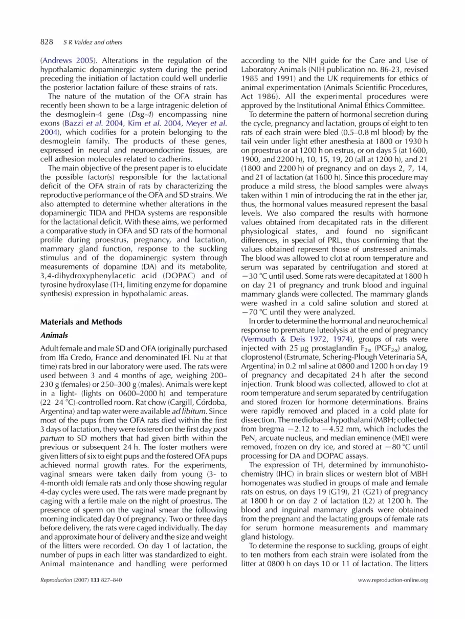
Figure 1A shows that there were no differences inbasal PRL levels on male rats. In the females duringproestrus, circulating PRL in the OFA rats was signi-ficantly lower at 1800 h and significantly higher at1930 h compared with the SD rats, suggesting a slightshift in the timing of the preovulatory hormone surge. Onestrus, SD rats had significantly higher values comparedwith the OFA strain. We measured hormone levels on theafternoon of day 5 of pregnancy as representative of thesemicircadian rhythm of PRL secretion observed during
Figure 1 Circulating hormones in male, virgin, pregnant, andlactating females of SD and OFA strains. A, PRL; B, GH; C,Progesterone. Hormones were measured by RIA. Unless statedotherwise, samples were taken at 1200 h in virgin, pregnant,and male rats, and at 1600 h during lactation. Values representthe meanGS.E.M. of groups of eight to ten rats. *P!0.05compared with SD rats of the same sex and reproductive state.
Reproduction (2007) 133 827–840

832 S R Valdez and others
early pregnancy (Jahn et al. 1986). There were nodifferences between the strains at 1600 and 1900 h;however, at 2200 h, PRL concentrations were decreasingin SD rats, while in the OFA strain they were stillelevated, suggesting that the PRL peak is prolonged inthis strain. There were no differences in the pattern ofserum PRL during the rest of pregnancy, but during earlylactation (days 2 and 7), the OFA rats had values thatwere !10% of the values in SD rats. On days 14 and 21,serum PRL values on the SD strain had decreased andwere similar to those of the OFA strain.
The pattern of circulating GH (Fig. 1B) was similarbetween the strains, with the exception of proestrus day,when the OFA rats showed values significantly higherthan the SD rats at 1800 h and significantly lower at1930 h, showing an inverse pattern with PRL.
There were significant differences in circulatingprogesterone between both strains in female rats(Fig. 1C), but not in males. On proestrus, there were nodifferences at 1800 h, while at 1930 h on proestrus andon estrus OFA rats showed significantly higher circulat-ing progesterone concentrations compared with SD rats.There were no differences on early pregnancy but at theend of pregnancy (day 21) and early lactation (day 2)circulating progesterone was lower in SD rats, suggestingthat, in this strain, luteolysis started somewhat earlierand that the activation of progesterone production by thecorpora lutea of the post partum ovulation startedsomewhat later compared with the OFA rats. Asmentioned above, the OFA rats delivered a few hoursearlier than the SD rats, in spite of an apparently later fallin circulating progesterone. This may indicate a reducedsensitivity to progesterone in the OFA rats. SD rats alsoshowed higher circulating progesterone on days 7 and14 of lactation, although only on the later day thedifference achieved statistical significance.
Circulating LH and FSH, determined in males andduring proestrus and estrus in females showed only slightdifferences in timing of the preovulatory surge, butachieved comparable levels that were capable ofinducing full ovulation (data not shown).
The differences in serum hormones observed in thetwo strains may reflect differences in pituitary function.We measured pituitary hormone content in males,females on estrus, and on late pregnancy (days 19and 21) and early lactation (day 2). There were nodifferences in pituitary weight between the two strains.PRL content was also similar, except on day 19 ofpregnancy SD 5.8G0.5 vs OFA 8.2G0.7 mg/mg freshtissue, P!0.025) and day 2 of lactation (SD 4.4G0.6 vsOFA 11.8G1.6 mg/mg, P!0.005), when OFA rats hadhigher pituitary PRL content than SD rats. There wereno differences on pituitary GH content in males andestrous female rats but on day 19 (SD 7.5G0.5 vs OFA13.2G1.9 mg/mg, P!0.025) and day 21 (SD 8.3G1.1 vsOFA 13.5G1.5 mg/mg, P!0.05) of pregnancy and day 2of lactation (SD 6.6G0.6 vs OFA 13.0G1.4 mg/mg,
Reproduction (2007) 133 827–840
P!0.005), the OFA rats had higher values than those ofthe SD rats. Pituitary LH values were between two- andsixfold higher in SD rats, with the smaller differenceobserved on day 2 of lactation due to a diminution in thelevels of the SD rats (not shown). The pattern of pituitaryFSH concentrations was similar to that observed in LH.Again, pituitary content of this hormone in the SD ratswas higher in males (threefold) and on late pregnancy(twofold), with no differences on estrus and day 2 oflactation, when the values in the SD rats were the lowest(not shown).
To determine whether there were any differences inMBH dopaminergic activity between the SD and theOFA virgin rats, we measured DA and DOPACconcentration in the MBH of rats on estrus day at1200 h. There were no significant differences in DA (SD117G7 and OFA 132G14 ng/mg protein, NS) orDOPAC (SD 126G5, OFA 110G4 ng/mg protein, NS)concentrations between the strains, nor in theDOPAC/DA ratio. However, these concentrations wereseveral times higher than those observed in late pregnantor lactating rats of both strains (see below and Fig. 6).
Effect of PGF2a on serum hormones and hypothalamicDA and DOPAC content
It is well known that the elevated circulating progesteroneobserved at the end of pregnancy produces a tonicinhibition of PRL release, and conversely, when its levelsdecay on day 21 of pregnancy or after removal of thecorpora lutea or induced luteolysis, a surge of PRL occurs(Vermouth & Deis 1972, Bussmann & Deis 1979, 1985,Jahn et al. 1986, 1993). We investigated whether theresponse to the progesterone decay 24 h after a luteolyticregimen of the PGF2a analog, cloprostenol, was different inOFA and SD rats. The treatment produced a decrease incirculating progesterone to!20% of control values (Fig. 2)and a surge of PRL that were similar in both strains. We alsoobserved a fall in circulating GH in SD but not in OFA rats.
In order to explore whether changes in DA trans-mission play a role in the release of PRL induced byprogesterone fall, we also measured DA metabolism onMBH in this experimental model, observing differentialresponses on the two strains (Fig. 2). DA concentrationswere similar in both strains, and were not modified byPGF2a treatment. DOPAC levels were unchanged afterPGF2a administration in SD rats while in the OFA rats thevalues increased significantly after PGF2a treatment,suggesting the establishment of the short negativefeedback loop by PRL. The DOPAC/DA ratios showedchanges similar to those shown for DOPAC.
Mammary gland function and lactogenesis
In order to establish the causes for the failure of lactationin OFA rats, we explored mammary histology and casein
www.reproduction-online.org

Figure 2 Circulating PRL, GH, progesterone (Pg), and MBH dopamine(DA) and DOPAC concentrations and DOPAC/DA ratio at 1200 h onday 20 of pregnancy in rats of SD and OFA strains, after vehicle orPGF2a treatment (25 mg/rat x 2, 24 h previously). Results are expressedas ng/ml for hormone values and ng/mg protein for DA and DOPAC.Values represent the meanGS.E.M. of groups of eight to ten rats.*P!0.05 compared with SD rats of the same group. †P!0.05compared with vehicle-treated groups of the same strain.
Reproduction in lactation deficient rats 833
and lactose contents at the end of pregnancy and earlypost partum in SD and OFA rats. Table 1 shows that inboth strains, casein values were similar on days 19 and20 of pregnancy and increased significantly on theafternoon of day 21, a time when lactogenesis is takingplace. Mammary lactose concentrations increase laterthan casein in normal lactogenesis (Deis et al. 1989), anddid not increase significantly before delivery but on days2 and 12 of lactation, both casein and lactose wereelevated (Table 1). The casein and lactose concentrations
Table 1 Mammary bcasein and lactose concentrations in SD and OFA hr/hr r(1200 h) of lactation and on day 20 of pregnancy (1200 h), 24 h after treatm
bcasein (mg/mg tissue)
SD OFA
Day 19 of pregnancy 5.92G1.14 5.10G0Day 21 of pregnancy 18.93G2.21* 9.40G0Day 2 of lactation 12.06G1.67* 8.44G1Day 12 of lactation 22.78G0.63* 26.00G4Day 20 of pregnancyC Saline 4.49G0.77 4.81G1C PGF2a 18.63G2.57† 18.42G0
Values represent the meanGS.E.M. of groups of eight to ten rats. *P!0.05 c
www.reproduction-online.org
in the OFA rats were similar to those of the SD rats, withthe exception of casein concentrations on day 21 ofpregnancy, when the SD rats had significantly highervalues (Table 1). These values indicate that lactogenesisat the end of pregnancy and milk synthesis duringestablished lactation seem to proceed normally in theOFA rats.
Advancement of luteolysis induced by PGF2a treat-ment triggered increases in casein and lactose 24 h laterlevels that were similar in the OFA and SD rats (Table 1).We also performed histological (stained with hematox-ylin–eosin) examination of the mammary glands on day19 and 21 of pregnancy and days 2, 7, 14, and 21 oflactation on the two strains. There were no differences inthe degree of mammary development among the strains,although the inguinal mammary glands of the OFA ratstended to be smaller. On day 19 of pregnancy, themammary glands of the OFA and SD rats showedcomparable development, with a slight distension ofsome alveoli, which was filled with material; on day 21,a greater distension of the alveoli was observed,indicating that lactogenesis had taken place normally;and on day 2 of lactation, the images were typical oflactation. There were no differences between the strains(results not shown). Taken together, these results indicatethat mammary function does not seem to be responsiblefor the deficit in lactation of the OFA rats.
Response to the suckling stimulus
OFA rats had diminished circulating PRL on days 2 and 7of lactation (Fig. 1A), suggesting an impaired sucklingreflex. To explore this hypothesis, we performed an acutesuckling experiment in mid-lactating rats (days 10–11post partum), consisting in measuring serum PRL, GH,progesterone, and oxytocin concentrations as well as theamount of milk ingested by the litter (estimated by theincrease in weight), after 15 or 30 min of suckling by alitter of eight pups to mothers that had been separatedfrom the litter for 8 h. As indicated in Materials andMethods, the OFA mothers nursed litters of foster SDpups, while the SD mothers nursed their own litters orfostered OFA or SD pups. The OFA mothers used were
ats on day 19 (1200 h) and day 21 (1800 h) of pregnancy, days 2 and 12ent with PGF2a.
Lactose (nmol/mg tissue)
SD OFA
.56 0.09G0.04 0.81G0.15
.81* 0.04G0.02 2.35G0.77
.06* 7.40G1.16* 8.93G0.24*
.21* 9.47G4.26* 19.41G4.91*
.10 0.06G0.2 0.23G0.09
.65† 5.41G1.03† 7.22G2.57†
ompared with day 19 of pregnancy. †P!0.05 compared with saline.
Reproduction (2007) 133 827–840

834 S R Valdez and others
those that had been able to nurse at least four pups ofnormal growth rate until the day of the experiment and, ifnecessary, the litters were completed to eight withadditional hungry foster pups for the experiment.
Figure 3A shows the pattern of hormone release and theincrease in litter weight after 15 and 30 min of suckling.While at 15 min of suckling, serum PRL had increased tosimilar levels in both groups, at 30 min the values of theOFA rats were significantly lower than those of the SDrats. Circulating oxytocin levels also showed differentpatterns. In both groups, hormone levels were increasedafter 15 min of suckling, but the OFA rats had significantlylower values than SD rats. After 30 min of suckling, theoxytocin levels in the SD rats remained elevated while inthe other strain there was a decrease, arriving to values notdifferent from time 0. The litters from the SD groupobtained significant amounts of milk, resulting in a litterweight increase of more than 7 g after 30 min of suckling.On the other hand, the litters suckling the OFA groupincreased !1 g/l, indicating a significant impairment inthe milk ejection reflex. Serum GH levels were notmodified by suckling in SD rats, but after 30 min ofsuckling they were significantly increased in the OFA ratswhen compared with the other strain. Interestingly,circulating progesterone, measured only at 0- and30-min suckling, increased in SD rats at 30 min but didnot change in the OFA strain, suggesting that the levels ofPRL achieved in the former strain were capable ofeliciting a response from the luteal tissue, while thelower levels of PRL observed in the OFA rats werenot sufficient to stimulate luteal progesterone release. We
Reproduction (2007) 133 827–840
also allowed some rats to suckle for longer periods (1, 2,and 4 h) to determine if the OFA rats were able to ejectmilk after more suckling time. While the pups suckling theSD rats had obtained approximately two thirds of theavailable milk during the first 30 min of suckling (7.8G0.9 g/l), with most of the rest at 2 h (3.8G1 g/l) andmarginal increments at later times, the litters sucklingOFA rats showed a modest positive increase in weightonly after 2 h of suckling (4.0G0.7 g/l) and no furtherincrease at 4 h. Thus, the total milk quantity that the OFArats were able to eject after 4 h of suckling was !50% ofthe values of the SD mothers. After 1 h of suckling,circulating PRL and oxytocin levels continued to besignificantly lower (~50%) in the OFA compared with theSD mothers (results not shown).
We also measured the MBH dopaminergic activity inSD and OFA rats after 30-min suckling. Figure 3B showsthat after 30-min suckling neither DA or DOPACconcentrations nor the DOPAC/DA ratio were differentfrom the values in non-suckled rats in both strains. Onthe other hand, the lactating OFA rats had significantlyhigher concentrations of DA and DOPAC compared withthe SD rats, and this may be at least partially responsiblefor the reduced suckling-induced PRL levels in the OFAcompared with the SD strain.
TH expression on hypothalamic areas in virgin (estrous)rats and on late pregnancy and early lactation
The diminished PRL secretion observed in the OFA ratsin different experimental situations, such as lactation or
Figure 3 A: Effect of 8-h separation and 15- or 30-minsuckling on pup weight increase, serum concentrationsof PRL, oxytocin, GH, and progesterone. B: Effect of 8-hseparation and 30-min suckling on MBH dopamine (DA),DOPAC and DOPAC/DA ratio of SD and OFA rats ondays 10–11 of lactation. Rats were separated from theirlitters at 0800 h, the litters were replaced at 1600 h andpups weighed and blood sampled from the mothers after15 and 30 min of vigorous suckling. See Materials andMethods for further details. Results are expressed asng/ml for hormone values and ng/mg protein for DA andDOPAC. Values represent the meanGS.E.M. of groups ofeight to ten rats. *P!0.05 compared with SD rats at thesame time. †P!0.05 compared with time 0.
www.reproduction-online.org

Reproduction in lactation deficient rats 835
proestrus at 18 h, along with the differential effect ofPGF2a on hypothalamic dopaminergic metabolism andthe different DA and DOPAC concentrations in MBH inlactation, may suggest alterations in the dopaminergicneurons of the arcuate nucleus, that are the mainregulatory system for pituitary PRL secretion. Toinvestigate changes in the dopaminergic system, wedetermined the localization and the number ofdopaminergic neurons in the periventricular andarcuate nuclei and the hypothalamic expression ofTH, on female rats of both strains on estrus, days 19 and21 of pregnancy and day 2 post partum and on malerats (for western blots only), using immunohistochem-istry and western blot respectively. Figure 4 shows thatthere were no differences in the localization of TH (C)neurons between the strains or among the differentgroups. In contrast, the number of TH (C) neurons wasvery variable. The SD rats had more TH (C) neurons inthe PeN and in the different regions of the arcuatenucleus compared with the other strain, in all the
www.reproduction-online.org
reproductive states studied, but the differences achievedstatistical significance only in virgin and 19-daypregnant rats in the PeN and in the medial arcuate. Inthe PeN, the number of TH (C) neurons decreasedsignificantly on day 21 of pregnancy in both strains,when compared with the virgin rats, while in thearcuate nucleus there were no significant differencesbetween the different reproductive states. When thevalues of the arcuate nucleus for all the reproductivestates were pooled, the difference between the OFA andthe SD rats was significant. The medial arcuate nucleushad more TH (C) neurons than the rostral or caudalpart of the nucleus, and accounted for the differencesobserved between the SD and the OFA strains (Fig. 4B).
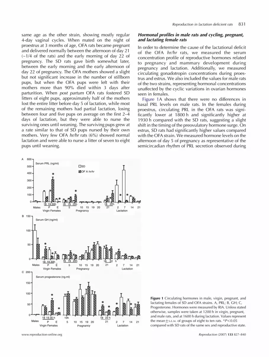
Western blot for TH of MBH protein extracts hadmaximal and similar values in virgin female rats of bothstrains (Fig. 5). The male OFA rats had slightly butsignificantly higher TH immunoreactivity comparedwith the SD rats. In SD rats, there were significantdecreases in TH expression on late pregnancy (days 19
Figure 4 Tyrosine hydroxylase immunoreactivity (THC)in neurons of the arcuate and periventricular nuclei ofestrous virgin (Virgins), 19- and 21-day pregnant (G19and G21) and 2-day lactating (L2) rats of SD and OFAstrains. A: Representative microphotographs of stainedarcuate and periventricular neurons from virgin andG21 rats of both strains. There were no differences inneuron size or morphology between the two strains.The sections shown correspond to plates 26 and 27and to plate 25 of the Paxinos–Watson atlas for arcuateand periventricular nuclei respectively. B: Totalnumber of TH (C) neurons in periventricular (A14),entire, rostral (1.8–2.1 mm posterior to bregma, pB),medial (2.1–3.6 mm), and caudal (3.6–4.3 mm, pB)arcuate (A12) nuclei of estrous virgin (Virgins), 19- and21-day pregnant (G19 and G21) and 2-day lactating(L2) rats of SD and OFA strains. See Materials andMethods for further details. Values represent themeansGS.E.M. of groups of three to four rats with theexception of the groups designated as total, whichrepresent the value for all the pooled groups, regardlessof the reproductive state. *P!0.05 compared with SDrats of the same sex and reproductive state. †P!0.05compared with virgin female rats of the same strain.
Reproduction (2007) 133 827–840
Figure 5 Western blot for tyrosine hydroxylase in MBH homogenates ofmale and female virgins in estrus (Virgins), 19- and 21-day pregnant(G19 and G21) and 2-day lactating (L2) rats of SD and OFA strains. A:Representative western blots with three samples for each strain in eachof the studied reproductive states. All the blots were exposed in thesame film. B: Densitometric quantification of the bands. See Materialsand Methods for further details. Values represent the meansGS.E.M. ofgroups of four to six rats. *P!0.05 compared with SD rats of the samesex and reproductive state. †P!0.05 compared with virgin female ratsof the same strain.
836 S R Valdez and others
and 21) and lactation, compared with virgin rats. Incontrast, in the OFA rats, TH expression only fellsignificantly on day 21 of pregnancy compared withvirgins of the same strain (Fig. 5B). Thus, during late
Reproduction (2007) 133 827–840
pregnancy and lactation the TH values were significantlyhigher in the OFA rats when compared with the SDstrain (Fig. 5).
Discussion
The present results show that OFA rats, when comparedwith their strain of origin, the SD strain, werehypoprolactinemic during the course of lactation, butdid not show impaired PRL secretion on other physio-logical states, such as pregnancy or the sexual cycle(Fig. 6). Thus, the surges in PRL secretion on theafternoons of proestrus and day 5 of pregnancy (whichwe took as representative of the diurnal PRL surgesduring early pregnancy) seemed to be even moreprolonged in the OFA rats, while on day 21 the patternof secretion was similar in both strains. PRL levels inmales, which are not subject to the cyclic ovarianhormone influences, only showed a non-significanttendency to be lower in the OFA rats. OFA rats showeda profound impairment in the PRL response to suckling,observed in the continuously lactating rats (Fig. 1) and inthe acute suckling experiment (Fig. 3). In addition, thislast experiment demonstrated partial blockade ofoxytocin secretion and a profound impairment in milkejection. In contrast, mammary gland development andlactogenesis proceeded normally, indicating that thereseems to be no deficit in mammary gland function. Thissuggests that the lactational failure of the OFA rats maybe exclusively neurogenic through an increased hypo-thalamic dopaminergic tone that may play a role in thereduced PRL response to suckling.
Thus, the decrease in MBH TH immunoreactivity(which includes PeN, arcuate nucleus and also the ME,that may contribute a significant proportion of themeasured protein in the western blots) and in DA andDOPAC concentrations in the MBH during latepregnancy and early post partum compared with thevalues in virgin rats was attenuated in the OFA rats incomparison with the SD strain (Fig. 6), suggesting anincreased dopaminergic tone in this strain. There arereports that show increases in dopaminergic activity inother brain areas in this (Estrella et al. 2002), and asimilar strain (Cohen et al. 1985), suggesting that thealtered dopaminergic tone is not exclusive of thehypothalamus. Since the diminished hypothalamic THexpression and activity may be essential for themaintenance of the full PRL response to suckling duringlactation (Andrews 2005), the increased MBH THimmunoreactivity and dopaminergic activity (DA andDOPAC concentrations) observed on late pregnancy andearly lactation in the OFA rats may indicate that thisadaptation is impaired, thus compromising suckling-induced PRL release (Fig. 6).
It has been well demonstrated that in normal rats, theresponsiveness of tuberoinfundibular DA neurons to PRLfeedback is markedly attenuated in late pregnancy and
www.reproduction-online.org

Figure 6 Relationship between the PRL levels,hypothalamic indices of dopaminergic activity (THexpression and DA and DOPAC concentrations), andthe number of TH (C) neurons in virgin, pregnant, andlactating rats of the OFA and SD strains. In SD rats, thedecrease in hypothalamic TH expression and DA andDOPAC concentrations observed on late pregnancyand lactation is inversely correlated with increasedcirculating PRL. In contrast, in the OFA rats, the lowcirculating PRL during lactation can be correlated withelevated TH expression and dopaminergic activity.*P!0.05 compared with SD rats of the same sex andreproductive state. †P!0.05 compared with virginfemale rats of the same strain.
Reproduction in lactation deficient rats 837
lactation (Arbogast & Voogt 1996, Andrews et al. 2001,Andrews 2005). This disruption of the normal PRLnegative feedback appears to be fundamental for thedevelopment of the lactational hyperprolactinemianecessary for optimal milk synthesis (Andrews 2005). Incontrast, the OFA rats seem to retain a higher capacity toactivate the DA system in response to elevated circulatingPRL at the end of pregnancy compared with the parentstrain. Thus, when PRL release was stimulated bytreatment with PGF2a, the OFA rats showed an increasein DA turnover that was not observed in the SD rats(Fig. 3).
However, in spite of the marked changes in MBHTH expression and DA concentrations, the number of TH(C) neurons in the arcuate nucleus remained constantin all the reproductive situations examined (Fig. 6).Surprisingly, the SD rats seemed to have more TH (C)neurons in the medial area of the arcuate nucleus andalso in the PeN than OFA rats (Fig. 4), although theirlocalization was similar. It is interesting to note thatcompared with the SD strain, Wistar rats also have lessTH (C) neurons in arcuate and PeN nuclei, but show acomparable decrease in MBH TH immunoreactivityduring late pregnancy and early post partum(unpublished results from our laboratory).
The decreased TH immunoreactivity and DA concen-trations observed during late pregnancy in SD rats whencompared with the virgin values correlates well with thepreviously reported reduction in TH gene expression onthese stages compared with early pregnancy (Arbogast &Voogt 1991, Fliestra & Voogt 1997, Andrews et al. 2001).This decrease precedes at least by 3 days the increase inPRL that occurs on day 21 after the progesterone fall,suggesting that other factors may be able to stimulate PRLsecretion at this stage, in the presence of a permissiveaction of the decreased dopaminergic tone. In contrast,although the OFA rats had diminished TH immuno-reactivity only on day 21, they showed a similar increasein PRL, suggesting that this decrease in the dopaminergic
www.reproduction-online.org
tone is sufficient to allow the prepartum PRL surge in thisstrain. There are many evidence showing that the effect ofPRL-releasing factors (PRFs), such as TRH (Samson et al.2003), are dependent on a decrease in the dopaminergictone that is not sufficient to stimulate PRL release per se,but that multiplies the release elicited by the PRF(Freeman et al. 2000, Ben-Jonathan & Hnasko 2001,Voogt et al. 2001). There are other neurotransmittersinvolved in the release of PRL at the end of pregnancy,such as norepinephrine (Jahn & Deis 1991), serotonin(Jahn & Deis 1987, Jahn et al. 1999), and opioid peptides(Soaje & Deis 1994, 2004, Soaje et al. 2004, Andrews &Grattan 2003) that participate in the stimulation of PRLrelease after the progesterone fall and that may beresponsible for the similar response observed in the twostrains, in spite of the elevated dopaminergic tone of theOFA rats at the end of pregnancy.
There are also a number of PRL-releasing factors, suchas TRH or cocaine- and amphetamine-regulated tran-script peptide, (CART) that have been implicated more orless directly in the PRL response to suckling (Freemanet al. 2000, Voogt et al. 2001, Samson et al. 2003). Wecannot exclude that alterations in one or more of thesesystems may also play a role in the impaired hormonalresponse to suckling.
While suckling-induced PRL secretion is impaired, itseems that GH secretion may be augmented in the OFArats, which had elevated GH levels after 30 min ofsuckling. Similarly, at the end of pregnancy, after PGF2a
treatment, circulating GH decreased in parallel with theincrease in PRL in the SD rats, but did not change in theOFA rats, indicating a differential regulation of GHsecretion in this strain. We have described (Jahn et al.1993) an inverse regulation of GH and PRL secretion atthe end of pregnancy in Wistar rats. We showed thatcirculating GH is elevated and PRL is very low during thelast days of pregnancy and when PRL release is inducedspontaneously or by progesterone fall, there is aconcomitant fall in GH secretion (Jahn et al. 1993).
Reproduction (2007) 133 827–840

838 S R Valdez and others
This pattern of secretion seems to be present also in theSD rats (see Figs 1 and 2). Although the pattern ofcirculating PRL and GH on the last days of pregnancy wassimilar in the OFA and SD rats, the differential GHresponse to PGF2a treatment and to suckling may be theproduct of the same hypothalamic alterations that areresponsible for the increased dopaminergic activity.Since GH has some lactogenic actions in the mammarygland in the absence of PRL (Caron et al. 1994), thehigher GH levels induced by suckling may have providedan additional stimulus for milk synthesis during lactation,providing some compensation for the reduced PRL.
There were important differences in pituitary hormonecontent between the two strains. The female SD rats hadconsistently lower PRL and GH contents, while the OFArats had higher values compared with the SD rats duringpregnancy and early lactation, suggesting that theimpairment in PRL release may produce accumulationof the hormone in the hypophysis.
It is interesting to note that although OFA rats at1800 h had lower preovulatory LH secretion, at 1930 hthe levels achieved were similar to those of the SD ratsand sufficient to insure a normal ovulation rate;furthermore, their overall fertility was similar to that ofthe SD rats.
In contrast to the relatively minor differences observedin the circulating gonadotropins, their pituitary contentsshowed marked variations between the two strains, inparticular in the male rats, where SD rats had valuesseveral times higher than those of the OFA strain. Thedifferences observed in the pituitary gonadotropincontent between the two strains, particularly in thefemales, may reflect a differential sensitivity to estrogenaction between both strains, since estrogen is animportant factor regulating synthesis and secretion ofgonadotropins. The significantly higher PRL valuesduring estrus in the SD compared with the OFA ratsmay also be indicative of a different sensitivity toestrogen. In this respect, it has been shown that SD ratshave differential sensitivity to estrogens or estrogenicsubstances in comparison with other strains, such as theWistar (el Abed et al. 1987, Garcia-Segura et al. 1992),that accounts for their heightened susceptibility tomammary cancer (Isaac 1986, el Abed et al. 1987).Our colony of OFA hr/hr rats shares the susceptibility tocarcinogens with the SD strain, with 70% of the ratsdeveloping tumors between 1 and 4 months afteradministration of the carcinogen DMBA (Jahn et al.2003, Ezquer et al. 2003). On the other hand, while inour colony of SD rats, 46% had spontaneous mammarytumors at 18 months of age, only 3 out of 50 OFA ratsshowed mammary tumors between 12 and 18 months ofage, of which only one was epithelial and two werefibrous and composed of connective tissue. Mostprobably, the altered PRL regulation of the OFA ratscontributed to this low incidence of mammary tumors.
Reproduction (2007) 133 827–840
The mutation affecting OFA hr/hr rats is a deletion of theDsg-4 gene, a member of the cadherin family of celladhesion proteins (Jahoda et al. 2004). This proteinparticipates in the assembly of desmosomal protein andhas calcium-binding sites (Jahoda et al. 2004). The productof this gene has been shown tobe present in the hair folliclebut its presence in other tissues has not yet beeninvestigated, although it may presumably be present inmost tissues that present tight junctions, the nervous tissueamong them. Besides disrupting hair follicle function andstructure, mutation of the Dsg-4 gene produces increasedcell proliferation in epithelial tissue (Jahoda et al. 2004). Itis interesting to note that mutations as different as those ofthe hairless and the Dsg genes give phenotypes that arequite similar, involving hairlessness, hypoprolactinemia,and lactational failure. The hairless gene encodes a proteinthat is expressed in hair follicles and in nervous tissue andits pattern of expression in developing brain tissue, as wellas somedescribed neural abnormalities, suggest that itmayhave a role in CNS development (Potter et al. 2002).Although at present, Dsg-4 has not been localized in thebrain and there are no reports on any relationship betweenthis protein and signaling pathways related to nervoustissue function, there are a number of reports in theliterature that show interactions between proteins belong-ing to cell adhesion families, such as cadherins andcatenins and neuron survival, synaptogenesis, and stabil-ization of synapses (Brose 1999, Tanaka et al. 2000,Ferreira & Paganoni 2002, Yu & Malenka 2003, Junghanset al. 2005). Theyare alsokey players in signalingpathwaysrelevant to neuronal functions and synaptic differentiationsuch as Wnt (Caricasole et al. 2005). Disruption ofmembers of the cadherin family (Gubkina et al. 2001,Rubinek et al. 2003) has also been linked to alterations inhypothalamic and pituitary regulation of hormonesecretion. Thus, the possible participation of Dsg proteinsin neuroendocrine and neuronal regulation and plasticityopens a new field of investigation that is being pursued atpresent in our laboratory.
In conclusion, we found that the OFA rats show aprofound impairment in the neuroendocrine response tosuckling that may account for the deficit in lactation andmay be caused by increased hypothalamic dopa-minergic tone at the end of pregnancy and duringlactation (Fig. 6). We also found subtle differencesbetween the OFA and the SD strains in the pattern ofhormone secretion in cycling and pregnant rats that didnot affect ovulation or fertility. The alterations in MBHdopaminergic metabolism described in the presentpaper may indicate that at least in the OFA strain, theDsg-4 deletion produces a subtle alteration in theregulation or function of hypothalamic dopaminergicneurons, perhaps mediated through abnormal cytoarch-itecture, that results in an elevated dopaminergic tone atthe end of pregnancy and during lactation that producehypoprolactinemia and lactational failure as aconsequence.
www.reproduction-online.org

Reproduction in lactation deficient rats 839
Acknowledgements
This work has been supported by grants PIP 0826 and 5795from CONICET (Consejo nacional de investigaciones cientı-ficas y tecnicas, Argentina) and PMT-PICT 06877 from theAgencia de Promocion Cientıfica y Tecnologica, Argentina.GAJ, AP, and RPD are Career Scientists from CONICET, SRVhas a fellowship from CONICET. The authors are grateful to DrClaudio Cuello for gifts of TH antibody, Dr N Hagino foroxytocin antibody, Prof. Norma B Carreno for her excellenttechnical assistance, and Drs Marta Soaje and Ruben Caron fortheir critical reading of the manuscript. The authors declare thatthere is no conflict of interest that would prejudice theimpartiality of this scientific work.
References
Andrews ZB 2005 Neuroendocrine regulation of prolactin secretionduring late pregnancy: easing the transition into lactation. Journal ofNeuroendocrinology 17 466–473.
Andrews ZB & Grattan DR 2003 Opioid receptor subtypes involved inthe regulation of prolactin secretion during pregnancy and lactation.Journal of Neuroendocrinology 15 227–236.
Andrews ZB, Kokay IC & Grattan DR 2001 Dissociation of prolactinsecretion from tuberoinfundibular dopamine activity in late pregnantrats. Endocrinology 142 2719–2724.
Arbogast LA & Voogt JL 1991 Mechanisms of tyrosine hydroxylaseregulation during pregnancy: evidence for protein dephosphory-lation during the prolactin surges. Endocrinology 129 2575–2582.
Arbogast LA & Voogt JL 1996 The responsiveness of tuberoinfundibulardopaminergic neurons to prolactin feedback is diminished betweenearly lactation and midlactation in the rat. Endocrinology 137 47–54.
Bazzi H, Kljuic A, Christiano AM & Panteleyev AA 2004 Intragenicdeletion in the Desmoglein 4 gene underlies the skin phenotype inthe Iffa Credo ‘hairless’ rat. Differentiation 72 450–464.
Ben-Jonathan N & Hnasko R 2001 Dopamine as a prolactin (PRL)inhibitor. Endocrine Reviews 22 724–763.
Brose N 1999 Brose N Synaptic cell adhesion proteins andsynaptogenesis in the mammalian central nervous system. Natur-wissenschaften 86 516–524.
Bussmann LE & Deis RP 1979 Studies concerning the hormonalinduction of lactogenesis by prostaglandin F2a in pregnant rats.Journal of Steroid Biochemistry 11 1485–1489.
Bussmann LE & Deis RP 1985 Hormonal regulation of casein synthesisat the end of pregnancy. Molecular and Cellular Endocrinology 3715–118.
Caricasole A, Bakker A, Copani A, Nicoletti F, Gaviraghi G &Terstappen GC 2005 Two sides of the same coin: wnt signaling inneurodegeneration and neuro-oncology. Bioscience Reports 25309–327.
Caron RW, Jahn GA & Deis RP 1994 Lactogenic actions of differentgrowth hormone preparations in pregnant and lactating rats.Journal of Endocrinology 142 535–545.
Cohen H, Guillaumot P, Sabbagh I & Bertrand J 1983 A newhypoprolactinemic rat strain. Prolactin, luteinizing hormone,follicle-stimulating hormone, testosterone and corticosterone levelsin males and effects of two anesthetics. Biology of Reproduction 28122–127.
Cohen H, Sabbagh I, Guillaumot P & Bertrand J 1985 Increaseddopaminergic inhibition of prolactin in the hypoprolactinaemic IPLnude rat. Journal of Endocrinology 107 325–329.
Cohen H, Sabbagh I, Abou-Samra AB & Bertrand J 1986a beta-Endorphin in genetically hypoprolactinemic rat: IPL nude rat. LifeSciences 38 217–224.
www.reproduction-online.org
Cohen H, Sabbagh I, Guillaumot P & Bertrand J 1986b Steroidregulation of gonadotropins in genetically hypoprolactinemicfemales (IPL nude rats). Journal of Steroid Biochemistry 24 431–435.
Cohen H, Sabbagh I, Guillaumot P, Bertrand J, Robertson M &Friesen HG 1988 Relationship between bioassay and radioimmuno-assay measurement of prolactin in the IPL rat, a hypoprolactinemicrat strain. Biology of Reproduction 38 84–90.
Deis RP, Carrizo DG & Jahn GA 1989 Suckling-induced prolactinrelease potentiates RU 486-induced lactogenesis in pregnant rats.Journal of Reproduction and Fertility 87 147–153.
Demarest KT, McKay DW, Riegle GD & Moore KE 1983 Biochemicalindices of tuberoinfundibular dopaminergic neuronal activity duringlactation: a lack of response to prolactin. Neuroendocrinology 36130–137.
el Abed A, Kerdelhue B, Castanier M & Scholler R 1987 Stimulation ofestradiol-17 beta secretion by 7,12-dimethylbenz(a)anthraceneduring mammary tumor induction in Sprague–Dawley rats.Journal of Steroid Biochemistry 26 733–738.
Estrella CR, Bregonzio C & Cabrera RJ 2002 Differencial responses incentral dopaminergic activity induced by apomorphine in IPL nuderat. Behavioral Brain Research 133 143–148.
Ezquer M, Caron RW, Deis RP & Jahn GA 2003 Expresion diferencialde factores de crecimiento y apoptoticos en tumores mamarios enratas hipoprolactinemicas vırgenes, madres o implantadas conestradiol en nucleo arcuato. XLVIII Reunion Cientıfica de laSociedad Argentina de Investigacion Clınica, Mar del Plata Abs.N8 208. Medicina 63 567.
EzquerM& Seltzer AM 2003 c-fos and tyrosine hydroxylase expressionafter an excitotoxic lesion on the nigrostriatal system: a study on theeffects of hypoxia used as a preconditioning stimulus. ExperimentalNeurology 183 523–531.
Ferreira A & Paganoni S 2002 The formation of synapses in the centralnervous system. Molecular Neurobiology 26 69–79.
Fliestra RJ & Voogt JL 1997 Lactogenic hormones of the placenta andpituitary inhibit suckling-induced prolactin (PRL) release but not theante-partum PRL surge. Proceedings of the Society of ExperimentalBiology & Medicine 214 258–264.
Freeman ME, Kanyicska B, Lerant A & Nagy G 2000 Prolactin:structure, function, and regulation of secretion. PhysiologicalReviews 80 1523–1631.
Garcia-Segura LM, Diolez-Bojda F, Lenoir V, Naftolin F & Kerdelhue B1992 Estrogen-like effects of the mammary carcinogen 7,12-dimethylbenz-(alpha)anthracene on hypothalamic neuronal mem-branes. Brain Research Bulletin 28 625–628.
Grattan DR & Averill RL 1990 Effect of ovarian steroids on a nocturnalsurge of prolactin secretion that precedes parturition in the rat.Endocrinology 126 1199–1205.
Gubkina O, Cremer H & Rougon G 2001 Mutation in the neural celladhesion molecule interferes with the differentiation of anteriorpituitary secretory cells. Neuroendocrinology 74 335–346.
Hoffman GE, Le WW, Abbud R, Lee WS & Smith MS 1994 Use of Fos-related antigens (FRAs) as markers of neuronal activity: FRA changesin dopamine neurons during proestrus, pregnancy and lactation.Brain Research 654 207–215.
Isaac JT 1986 Genetic control of resistance to chemically inducedmammary adenocarcinogenesis in the rat. Cancer Research 463958–3963.
Jahn G & Deis RP 1987 A possible dual regulation of prolactin releaseby the serotoninergic system in rats at pro-oestrus and during latepregnancy: role of ovarian hormones. Journal of Endocrinology 112367–374.
Jahn GA & Deis RP 1991 The involvement of the adrenergic system onthe release of prolactin and lactogenesis at the end of pregnancy inthe rat. Journal of Endocrinology 129 343–350.
Jahn GA, Alonso N & Deis RP 1986 Ovarian and feto-placental factorsand the regulation of prolactin release during pregnancy in the rat.Journal of Reproduction and Fertility 77 125–133.
Reproduction (2007) 133 827–840

840 S R Valdez and others
Jahn GA, Rastrilla AM & Deis RP 1993 Correlation of growth hormonesecretion during pregnancy with circulating prolactin in rats.Journal of Reproduction and Fertility 98 327–333.
Jahn GA, Kalia V, Hole D, Wilson CA & Deis RP 1999 Receptors andneurotransmitters involved in the dual modulation of prolactinrelease by the serotoninergic system in pregnant and lactating rats.Journal of Reproduction and Fertility 116 261–268.
JahnGA, EzquerM, Caron RW&Deis RP 2003 Expresion diferencial dereceptores hormonales en tumores mamarios en ratas hipoprolacti-nemicas vırgenes, madres o implantadas con estradiol en nucleoarcuato. XLVIII Reunion Cientıfica de la Sociedad Argentina deInvestigacion Clınica, Mar del Plata Abs. N8 207. Medicina 63 566.
Jahoda CA, Kljuic A, O’Shaughnessy R, Crossley N, Whitehouse CJ,Robinson M, Reynolds AJ, Demarchez M, Porter RM, Shapiro L &Christiano AM 2004 The lanceolate hair rat phenotype results from amissense mutation in a calcium coordinating site of the desmoglein4 gene. Genomics 83 747–756.
Jordan D, Sabbagh I, Guillaumot P, Veisseire M, Bertrand J & Cohen H1987 Thyroid-hypophyso-hypothalamic axis of the geneticallyhypoprolactinemic rats (IPL nude rats). Neuroendocrinology 46194–198.
Junghans D, Haas IG & Kemler R 2005 Mammalian cadherins andprotocadherins: about cell death, synapses and processing. CurrentOpinion in Cell Biology 17 446–452.
Kim H, Panteleyev AA, Jahoda CA, Ishii Y & Christiano AM 2004Genomic organization and analysis of the hairless gene in fourhypotrichotic rat strains. Mammalian Genome 15 975–981.
Kuhn NJ & Lowenstein JM 1967 Lactogenesis in the rat. Changes inmetabolic parameters at parturition. Biochemical Journal 105995–1002.
Laemmli UK 1951 Cleavage of structural proteins during the assemblyof the head of bacteriophage T4. Nature 277 680–685.
Li C, Chen P & Smith MS 1999 Neuropeptide Yand tuberoinfundibulardopamine activities are altered during lactation: role of prolactin.Endocrinology 140 118–123.
Meyer B, Bazzi H, Zidek V, Musilova A, Pravenec M, Kurtz TW,Nurnberg P & Christiano AM 2004 A spontaneous mutation in thedesmoglein 4 gene underlies hypotrichosis in a new lanceolate hairrat model. Differentiation 72 541–547.
Paxinos G & Watson C 1986 The rat brain in Stereotaxic Coordinates,2nd ed San Diego: Academic Press.
Potter GB, Zarach JM, Sisk JM & Thompson CC 2002 The thyroidhormone-regulated corepressor hairless associates with histonedeacetylases in neonatal rat brain. Molecular Endocrinology 162547–2560.
Reproduction (2007) 133 827–840
Rubinek T, Yu R, Hadani M, Barkai G, Nass D, Melmed S & Shimon I2003 The cell adhesion molecules N-cadherin and neural celladhesion molecule regulate human growth hormone: a novelmechanism for regulating pituitary hormone secretion. Journal ofClinical Endocrinology and Metabolism 88 3724–3730.
Samson WK, Taylor MM & Baker JR 2003 Prolactin-releasing peptides.Regulatory Peptides 114 1–5.
Semenenko FM, Cuello AC, Goldstein K, Lee Y & Sidebottom E 1986Monoclonal antibody against tyrosine hydroxylase: application inlight and electron microscopy. Journal of Histochemistry &Cytochemistry 34 817–821.
Snedecor GW & Cochran WG 1968 Statistical methods, 6 Ames: IowaState University Press.
Soaje M & Deis RP 1994 A modulatory role of endogenous opioids onprolactin secretion at the end of pregnancy. Journal of Endocrinology140 97–102.
Soaje M & Deis RP 2004 Involvement of opioid receptor subtypes inboth stimulatory and inhibitory effects of the opioid peptides onprolactin secretion during pregnancy. Cellular and MolecularNeurobiology 24 193–204.
Soaje M, Bregonzio C, Caron RW & Deis RP 2004 Neurotransmittersinvolved in the Opioid Regulation of Prolactin Secretion at the end ofPregnancy in Rats. Neuroendocrinology 80 11–20.
Tanaka H, Shan W, Phillips GR, Arndt K, Bozdagi O, Shapiro L,Huntley GW, Benson DL & Colman DR 2000 Molecularmodification of N-cadherin in response to synaptic activity. Neuron25 93–107.
Vermouth N & Deis RP 1972 Prolactin release induced byprostaglandin F2a in pregnant rats. Nature 238 248–250.
Vermouth N & Deis RP 1974 Prolactin release and lactogenesis afterovariectomy in pregnant rats: effect of ovarian hormones. Journal ofEndocrinology 63 13–20.
Voogt JL, Lee Y, Yang S & Arbogast L 2001 Regulation of prolactinsecretion during pregnancy and lactation. Progress in Brain Research133 173–185.
Yu X & Malenka RC 2003 Beta-catenin is critical for dendriticmorphogenesis. Nature Neuroscience 6 1169–1177.
Submitted 18 May 2006
First decision 14 June 2006
Revised manuscript received 20 December 2006
Accepted 15 January 2007
www.reproduction-online.org