Embed Size (px)
Citation preview
Osteoarthritis and Cartilage (2008) 16, 1267e1274
ª 2008 Osteoarthritis Research Society International. Published by Elsevier Ltd. All rights reserved.doi:10.1016/j.joca.2008.03.009
InternationalCartilageRepairSociety
Histone deacetylase inhibitors suppress interleukin-1b-induced nitricoxide and prostaglandin E2 production in human chondrocytesN. Chabane M.Sc.y, N. Zayed M.Sc.y, H. Afif Ph.D.y, L. Mfuna-Endam M.Sc.y,M. Benderdour Ph.D.z, C. Boileau Ph.D.y, J. Martel-Pelletier Ph.D.y,J.-P. Pelletier M.D.y, N. Duval M.D.x and H. Fahmi Ph.D.y*yOsteoarthritis Research Unit, Centre Hospitalier de l’Universite de Montreal (CHUM),Notre-Dame Hospital, Department of Medicine, University of Montreal, 1560 Sherbrooke Street East,Montreal, Quebec, Canada H2L 4M1zCentre de Recherche, Sacre-Coeur Hospital, 5400 Boul. Gouin Ouest, Montreal, Quebec,Canada H4J 1C5xCentre de Convalescence, Pavillon de Charmilles, 1487 Boul. des Laurentides, Montreal,Quebec, Canada H7M 2Y3
Summary
Objective: Overproduction of nitric oxide (NO) and prostaglandin E2 (PGE2) plays an important role in the pathogenesis of osteoarthritis (OA).In the present study, we determined the effect of trichostatin A (TSA) and butyric acid (BA), two histone deacetylase (HDAC) inhibitors, on NOand PGE2 synthesis, inducible NO synthase (iNOS) and cyclooxygenase (COX)-2 expression, and nuclear factor (NF)-kB DNA-binding ac-tivity, in interleukin-1b (IL-1)-stimulated human OA chondrocytes, and on IL-1-induced proteoglycan degradation in cartilage explants.
Methods: Chondrocytes were stimulated with IL-1 in the absence or presence of increasing concentrations of TSA or BA. The production ofNO and PGE2 was evaluated using Griess reagent and an enzyme immunoassay, respectively. The expression of iNOS and COX-2 proteinsand mRNAs was evaluated using Western blotting and real-time reverse transcriptase-polymerase chain reaction (RT-PCR), respectively.Proteoglycan degradation was measured with dimethymethylene blue assay. Electrophoretic mobility shift assay (EMSA) was utilized to an-alyze the DNA-binding activity of NF-kB.
Results: HDAC inhibition with TSA or BA resulted in a dose-dependent inhibition of IL-1-induced NO and PGE2 production. IL-17- and tumornecrosis factor-a (TNF-a)-induced NO and PGE2 production was also inhibited by TSA and BA. This inhibition correlated with the suppressionof iNOS and COX-2 protein and mRNA expression. TSA and BA also prevented IL-1-induced proteoglycan release from cartilage explants.Finally, we demonstrate that the DNA-binding activity of NF-kB, was induced by IL-1, but was not affected by treatment with HDAC inhibitors.
Conclusions: These data indicate that HDAC inhibitors suppressed IL-1-induced NO and PGE2 synthesis, iNOS and COX-2 expression, aswell as proteoglycan degradation. The suppressive effect of HDAC inhibitors is not due to impaired DNA-binding activity of NF-kB. Thesefindings also suggest that HDAC inhibitors may be of potential therapeutic value in the treatment of OA.ª 2008 Osteoarthritis Research Society International. Published by Elsevier Ltd. All rights reserved.
Key words: Chondrocytes, Nitric oxide, Inducible nitric oxide synthase, Prostaglandin E2, Cyclooxygenase-2, Histone deacetylases, Nuclearfactor-kB.
Abbreviations: BA butyric acid, COX-2 cyclooxygenase-2, HDAC histone deacetylase, IL interleukin, iNOS inducible nitric oxide synthase,NF-kB nuclear factor-kB, NO nitric oxide, OA osteoarthritis, PGE2 prostaglandin E2, TNF-a tumor necrosis factor-a, TSA trichostatin A.
Introduction
Osteoarthritis (OA) is the most common joint disorder anda leading cause of disability among the elderly population.It is characterized by progressive degenerative structuralchanges in articular cartilage, leading to loss of joint func-tion. It is also characterized by excessive production of sev-eral inflammatory mediators1e3. Among these mediators,the pro-inflammatory cytokine interleukin-1b (IL-1) plays
*Address correspondence and reprint requests to: Dr HassanFahmi, Ph.D., Osteoarthritis Research Unit, Notre-Dame Hospital,CR-CHUM, 1560 Sherbrooke East, Pavillon J. A. DeSeve, Y-2628,Montreal, Quebec, Canada H2L 4M1. Tel: 1-514-890-8000ext 25119; Fax: 1-514-412-7583; E-mail: [email protected]
Received 23 October 2007; revision accepted 9 March 2008.
1267
a pivotal role in the pathophysiology of OA. It induces a cas-cade of inflammatory and catabolic events in chondrocytesincluding the synthesis of prostaglandin E2 (PGE2) and ni-tric oxide (NO). IL-1 also alters chondrocyte anabolism bysuppressing the synthesis of proteoglycan and collagenand by enhancing the production of matrix metalloprotei-nases (MMPs)1e3.
NO is synthesized from L-arginine by a family of NO syn-thases (NOSs) of which three isoforms have been identi-fied. Neuronal NOS (nNOS) and endothelial NOS (eNOS)are constitutively expressed, while the inducible NOS(iNOS) is expressed following stimulation with a variety ofinflammatory agents such as endotoxins or cytokines4.NO promotes inflammation by enhancing the productionof inflammatory cytokines5 and PGE2
6 and by reducing
1268 N. Chabane et al.: HDAC inhibitors suppress IL-1b-induced NO and PGE2 production
the synthesis of endogenous IL-1 receptor antagonist (IL-1Ra)7. NO is also considered a potent catabolic agent in OAsince it inhibits collagen and proteoglycan synthesis8,9,stimulates the production and activation of MMPs10 and in-duces chondrocyte apoptosis11. Accordingly, the in vivo se-lective inhibition of iNOS in an experimental model of OAreduces the joint structural changes and the expression ofseveral inflammatory and catabolic factors, including IL-1and MMP-112.
The biosynthesis of PGE2 from arachidonic acid (AA) in-volves multiple enzymes including, cyclooxygenases(COXs). Two isoforms of COX have been identified: COX-1 is constitutively expressed in most tissues, whereasCOX-2 is induced by various stimuli such as endotoxins,growth factors and pro-inflammatory cytokines13. PGE2 isthe most abundant prostanoid in arthritic joint and one ofthe major catabolic mediators involved in cartilage resorp-tion. PGE2 elicits cartilage resorption by enhancing the ac-tivation and production of MMPs and the degradation ofcartilage matrix components14,15 and by promoting chon-drocyte apoptosis16. In addition PGE2 mediates pain re-sponses and potentiates the effects of other inflammatorymediators13.
Acetylation and deacetylation of nucleosomal histonesplay an important role in the regulation of gene expres-sion17,18. The histone acetylation status is controlled bythe opposing actions of two classes of enzymes: histoneacetyl transferases (HATs) and histone deacetylases(HDACs). Acetylation of histones loosens nucleosomalstructures, thereby promoting gene transcription. In con-trast, deacetylation of histones stabilizes nucleosomalstructures and represses gene transcription17,18. However,emerging evidence indicates that gene regulation by acety-lation/deacetylation is more dynamic and complex, and thatHATs can act as repressors and HDAC as activators oftranscription. Indeed, global analysis of gene expressionhas shown that inhibition of HDAC activity results both in in-duction and repression of gene expression19e24.
In recent years, significant interest has emerged in theinhibition of HDAC activity as a possible anti-cancer treat-ment. HDAC inhibitors induce growth arrest, differentiationand apoptosis of cancer cells in vitro and reduce thegrowth of experimental tumors in vivo25,26. Presently, sev-eral HDAC inhibitors are in clinical trials for the treatmentof solid and hematological tumors27,28. In addition to theiranti-cancer effects, recent studies have demonstrated thatHDAC inhibitors modulate inflammatory responses. Forinstance, HDAC inhibitors reduce the production of IL-1,tumor necrosis factor-a (TNF-a), and interferon-g (IFN-g)in lipopolysaccharide (LPS)-stimulated human peripheralblood mononuclear cells29,30. Likewise, HDAC inhibitorsprevent LPS-induced production of TNF-a, IL-6 and reac-tive oxygen species in neuroglia cultures, and primary mi-croglia31e33. HDAC inhibitors have also been reported tosuppress IL-12 production in dendritic cells and macro-phages34. However, it is currently unknown whetherHDAC inhibitors regulate inflammatory responses in artic-ular chondrocytes.
Since excessive production of the inflammatory media-tors NO and PGE2 plays an important role in the patho-genesis of OA, we assessed the effect of two HDACinhibitors, trichostatin A (TSA) and butyric acid (BA), onthe production of NO and PGE2 in primary cultured hu-man chondrocytes stimulated with IL-1. We additionallyanalyzed the expression of iNOS and COX-2 as well asthe binding activity of transcription factor nuclear factor-kB (NF-kB).
Materials and methods
REAGENTS
Recombinant human (rh) IL-1b was obtained from Genzyme (Cambridge,MA, USA), rhTNF-a and rhIL-17 were from R&D Systems (Minneapolis, MN,USA). TSA and BA were from SigmaeAldrich Canada (Oakville, ON, Canada).Dulbecco’s modified Eagle’s medium (DMEM), penicillin and streptomycin,fetal calf serum (FCS), and TRIzol� reagent were from Invitrogen (Burlington,ON, Canada). All other chemicals were purchased from either SigmaeAldrich Canada or Bio-Rad (Mississauga, ON, Canada).
SPECIMEN SELECTION AND CHONDROCYTE CULTURE
Human normal cartilage (from femoral condyles) was obtained at nec-ropsy, within 12 h of death, from donors with no history of arthritic disease(n¼ 7, mean� SD age: 54� 16 years). To ensure that only normal tissuewas used, cartilage specimens were thoroughly examined both macroscop-ically and microscopically. Only those with no alterations were further pro-cessed. Human OA cartilage samples from femoral condyles and tibialplateaus were obtained from OA patients undergoing total knee replacement(n¼ 47, mean�SD age: 66� 12 years). All OA patients were diagnosed ac-cording to the criteria developed by the American College of RheumatologyDiagnostic Subcommittee for OA35. At the time of surgery, the patients hadsymptomatic disease requiring medical treatment in the form of non-steroidalanti-inflammatory drugs (NSAIDs) or selective COX-2 inhibitors. Patientswho had received intra-articular injections of steroids were excluded. TheClinical Research Ethics Committee of Notre-Dame Hospital approved thestudy protocol and the use of human articular tissues.
Chondrocytes were released from cartilage by sequential enzymatic di-gestion as previously described36. In brief, this consisted of 2 mg/ml pronasefor 1 h followed by 1 mg/ml collagenase (type IV; SigmaeAldrich) for 6 h at37�C in DMEM and antibiotics (100 U/ml penicillin and 100 mg/ml streptomy-cin). The digested tissue was briefly centrifuged and the pellet was washed.The isolated chondrocytes were seeded at high density in tissue cultureflasks and cultured in DMEM supplemented with 10% heat-inactivatedFCS. At confluence, the chondrocytes were detached, seeded at high den-sity, and allowed to grow in DMEM, supplemented as above. The culture me-dium was changed every second day, and 24 h before the experiment thecells were incubated in fresh medium containing 0.5% FCS. Only first pas-saged chondrocytes were used.
NO AND PGE2 DETERMINATIONS
The nitrite levels, used as an indicator of NO production, were determinedusing the Griess assay as previously described36. The levels of PGE2 weredetermined using a PGE2 enzyme immunoassay from Cayman Chemical(Ann Arbor, MI, USA). The detection limit and sensitivity was 9 pg/ml. All as-says were performed in duplicate.
WESTERN BLOT ANALYSIS
Chondrocytes were lysed in ice-cold lysis buffer (50 mM TriseHCl, pH7.4, 150 mM NaCl, 2 mM EDTA, 1 mM phenylmethansulfonylfluorid(PMSF), 10 mg/ml each of aprotinin, leupeptin, and pepstatin, 1% NP-40,1 mM Na3VO4, and 1 mM NaF). Lysates were sonicated on ice and centri-fuged at 12,000 rpm for 15 min. The protein concentration of the supernatantwas determined using the bicinchoninic acid method (Pierce, Rockford, IL,USA). Twenty microgram of total cell lysate was subjected to SDS-poly-acrylamide gel electrophoresis and electrotransferred to a nitrocellulosemembrane (Bio-Rad). After blocking in 20 mM TriseHCl pH 7.5 containing150 mM NaCl, 0.1% Tween 20, and 5% (w/v) non-fat dry milk, blots wereincubated overnight at 4�C with the primary antibody and washed witha Tris buffer [Tris-buffered saline (TBS) pH 7.5, with 0.1% Tween 20]. Theblots were then incubated with horseradish peroxidase-conjugated second-ary antibody (Pierce), washed again, incubated with SuperSignal UltraChemiluminescent reagent (Pierce), and exposed to Kodak X-Omat film(Eastman Kodak Ltd, Rochester, NY, USA).
RNA EXTRACTION AND REVERSE
TRANSCRIPTASE-POLYMERASE CHAIN
REACTION (RT-PCR)
Total RNA from stimulated chondrocytes was isolated using the TRIzol�
reagent (Invitrogen) according to the manufacturer’s instructions. To removecontaminating DNA, isolated RNA was treated with RNase-free DNase I(Ambion, Austin, TX, USA). The RNA was quantitated using the RiboGreenRNA quantitation kit (Molecular Probes, Eugene, OR, USA), dissolved in di-ethylpyrocarbonate (DEPC)-treated-H2O and stored at �80�C until use. Onemicrogram of total RNA was reverse-transcribed using Moloney Murine Leu-kemia Virus reverse transcriptase (Fermentas, Burlington, ON, Canada) as
1269Osteoarthritis and Cartilage Vol. 16, No. 10
detailed in the manufacturer’s guidelines. One fiftieth of the reverse transcrip-tase reaction was analyzed by real-time PCR as described below. The fol-lowing primers were used: iNOS, sense 50-ACATTGATGAGAAGCTGTCCCAC-30 and antisense 50-CAAAGGCTGTGAGTCCTGCAC-30;COX-2, sense 50-TGTGTTGACATCCAGATCAC-30 and antisense 50-ACATCATGTTTGAGCCCTGG-30; and glyceraldehyde-3-phosphate dehydroge-nase (GAPDH), sense 50-CAGAACATCATCCCTGCCTCT-30 and antisense50-GCTTGACAAAGTGGTCGTTGAG-30.
REAL-TIME PCR
Real-time PCR analysis was performed in a total volume of 50 ml contain-ing template DNA, 200 nM of sense and antisense primers, 25 ml of SYBR�
Green master mix (QIAGEN, Mississauga, ON, Canada) and uracil-N-glyco-sylase (UNG, 0.5 Unit, Epicentre Technologies, Madison, WI, USA). After in-cubation at 50�C for 2 min (UNG reaction), and at 95�C for 10 min (UNGinactivation and activation of the AmpliTaq Gold enzyme), the mixtureswere subjected to 40 amplification cycles (15 s at 95�C for denaturationand 1 min for annealing and extension at 60�C). Incorporation of SYBR�
Green dye into PCR products was monitored in real-time using a GeneAmp5700 Sequence detection system (Applied Biosystems, Foster City, CA,USA) allowing determination of the threshold cycle (CT) at which exponentialamplification of PCR products begins. After PCR, dissociation curves weregenerated with one peak, indicating the specificity of the amplification. Athreshold cycle (CT value) was obtained from each amplification curve usingthe software provided by the manufacturer (Applied Biosystems).
Relative mRNA expression in chondrocytes was determined using theDDCT method, as detailed in the manufacturer’s guidelines (Applied Biosys-tems). A DCT value was first calculated by subtracting the CT value for thehousekeeping gene GAPDH from the CT value for each sample. A DDCT
value was then calculated by subtracting the DCT value of the control (unsti-mulated cells) from the DCT value of each treatment. Fold changes com-pared with the control were then determined by raising two to the �DDCT
power. Each PCR reaction generated only the expected specific ampliconas shown by the melting-temperature profiles of the final product and bygel electrophoresis of test PCR reactions. Each PCR was performed in trip-licate on two separate occasions for each independent experiment.
PROTEOGLYCAN RELEASE
Cartilage proteoglycan degradation was assessed by measuring sulfatedglycosaminoglycan (GAG) released into culture media using dimethyl meth-ylene blue (DMMB) with chondroitin sulfate as a standard37. Results are ex-pressed as mg of GAG released/mg cartilage.
NUCLEAR EXTRACT PREPARATION AND ELECTROPHORETIC
MOBILITY SHIFT ASSAY (EMSA)
Nuclear extracts were prepared as previously described38. Briefly, chon-drocytes were washed in ice-cold phosphate buffered saline (PBS) andgently scraped in ice-cold hypotonic buffer containing 10 mM hydroxyethylpi-perazine ethane sulfonic acid (HEPES)eKOH, pH 7.9, 10 mM KCl, 1.5 mMMgCl2, 0.5 mM dithiotheritol (DTT), 1 mM PMSF, 1 mM Na3VO4 and 10 mg/ml
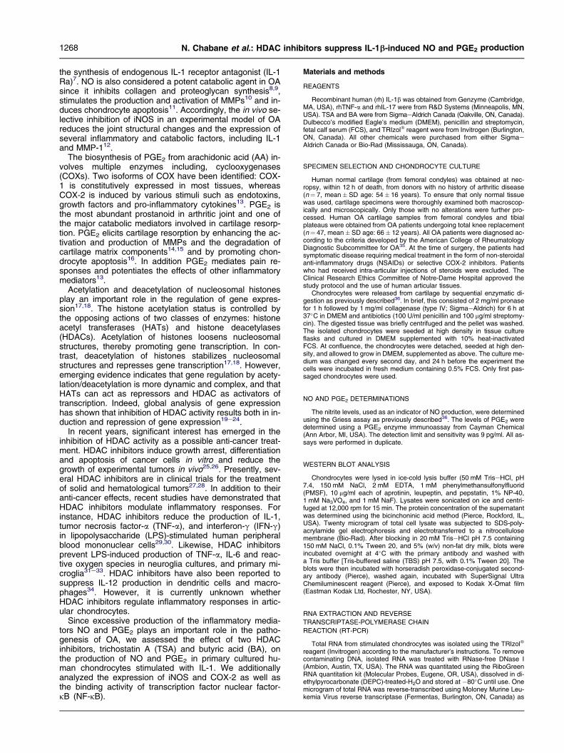
A
IL-1 - + + + + + + + + +TSA (ng/ml) - - 10 25 100 250 - - - -BA (mM) - - - - - - 0.5 1 5 10
ITB
*
* *
*
*
0
5
10
15
20
Nit
rite
(nm
ol/1
05 ce
lls)
Fig. 1. HDAC inhibitors TSA and BA prevent IL-1-induced NO and PGE100 pg/ml IL-1 in the absence or presence of increasing concentrations offor NO (A) and PGE2 (B) release. Results are expressed as percentage of
from four independent experiments. *P< 0.05 co
of aprotinin, leupeptin, and pepstatin. The cells were allowed to swell on iceand the nuclei were recovered by brief centrifugation. The pellets were resus-pended in high salt buffer containing 20 mM HEPES, pH 7.9, 420 mM NaCl,1.2 mM MgCl2, 0.5 mM DTT, 0.2 mM ethylenediaminetetraacetic acid (EDTA),25% glycerol, 0.5 mM PMSF, 1 mM Na3VO4 and 10 mg/ml of aprotinin, leupep-tin, and pepstatin, followed by incubation on ice for 20 min. The nuclear extractswere recovered by centrifugation and protein concentration was determined us-ing the Bradford method (Bio-Rad). A synthetic double-stranded oligonucleotidecontaining the kB consensus sequence 50-AGTTGAGGGGACTTTCCCAGGC-30 was end-labeled by T4 polynucleotide kinase in the presence of [g-32P]adenosine triphosphate (ATP). The mutant competitor oligonucleotide had thefollowing sequence with a 1 bp substitution (underlined): 50-AGTTGAGGC-GACTTTCCCAGGC-30. The binding buffer consisted of 10 mM TriseHCl, pH7.5, 50 mM NaCl, 0.5 mM DTT, 0.5 mM EDTA, 1 mM MgCl2, 4% glycerol and2.5 mg poly (dIedC). Binding reactions were conducted with 5 mg nuclear extractand 100,000 cpm 32[P]-labeled oligonucleotideprobeat 22�C for 20 min ina finalvolume of 10 ml. In supershift assays, the antibody to p65 (1 mg/reaction) was in-cubated with the reaction mixture for 1 h at 4�C before the addition of 32[P]-la-beled oligonucleotide. In cold competition assays, 50-fold molar excess ofcold wild-type or mutant oligonucleotide was used. Binding complexes were re-solved on non-denaturating 6% polyacrylamide gel electrophoresis in Trise-borate buffer system, after which the gels were fixed, dried, and subjected toautoradiography.
STATISTICAL ANALYSIS
Data are expressed as the mean� S.E.M. Statistical significance was as-sessed by the two-tailed Student’s t test. P values less than 0.05 were con-sidered statistically significant.
Results
TSA AND BA ATTENUATE IL-1-INDUCED NO AND PGE2
PRODUCTION IN HUMAN CHONDROCYTES
Chondrocytes were stimulated with 100 pg/ml IL-1 in theabsence or presence of increasing concentrations of twoHDAC inhibitors, TSA and BA, and the production of NOwas evaluated using Griess reagent. As shown in Fig. 1(A),treatment with either TSA or BA suppressed IL-1-inducedNO production in a dose-dependent manner. Similarly, theproduction of PGE2 was dose-dependently suppressed inthe presence of each HDAC inhibitor [Fig. 1(B)]. In anotherset of experiments, we found that TSA and BA also dose-dependently inhibited IL-1-induced NO and PGE2 productionin normal chondrocytes, (n = 3, data not shown). The ob-served inhibition was not a result of reduced cell viability asconfirmed by the methyl thiazolyl tetrazolium (MTT) assay(data not shown).
B
L-1 - + + + + + + + + +SA (ng/ml) - - 10 25 100 250 - - - -A (mM) - - - - - - 0.5 1 5 10
*
*
**
*
*
0
5
10
15
PG
E2
(ng/
105
cells
)
2 release from chondrocytes. Chondrocytes were stimulated withTSA or BA for 24 h. Conditioned media were collected and analyzedcontrol (i.e., cells treated with IL-1 alone) and are the mean� S.E.M.mpared with cells treated with IL-1 alone.

A
2.5
10
0
5
PG
E2
(ng/
105 c
ells
)
B
** ***0
5
10
Nit
rite
(nm
ol/1
05 cel
ls)
TNF-α - + + + - - -IL-17 - - - - + + +TSA - - + - - + -BA - - - + - - +
TNF-α - + + + - - -IL-17 - - - - + + +TSA - - + - - + -BA - - - + - - +
* * * **
Fig. 2. TSA and BA suppress TNF-a and IL-17-induced iNOS and COX-2 protein expression. Chondrocytes were treated with TNF-a (1 ng/ml)or IL-17 (100 ng/ml) in the absence or presence of TSA (250 ng/ml) or BA (10 mM) for 24 h. Culture media were collected and analyzed for theproduction of NO (A) and PGE2 (B). Results are expressed as mean� S.E.M. of three independent experiments. *P< 0.05 compared with cells
treated with TNF-a or IL-17 alone.
1270 N. Chabane et al.: HDAC inhibitors suppress IL-1b-induced NO and PGE2 production
TSA AND BA INHIBIT TNF-a AND IL-17-INDUCED NO AND PGE2
PRODUCTION
The pro-inflammatory cytokines TNF-a and IL-17 alsocontribute to the pathogenesis of OA and are potent in-ducers of NO and PGE2 production1e3. Therefore, we ex-amined whether HDAC inhibition could also attenuateTNF-a and IL-17-induced NO and PGE2 production. Asshown in Fig. 2, stimulation of chondrocytes with TNF-a orIL-17 dramatically increased the production of NO andPGE2. Interestingly, the induction of NO and PGE2 produc-tion by TNF-a or IL-17 was almost completely abolished af-ter treatment with TSA or BA. These data suggest that thesuppressive effect of HDAC inhibitors was not specific toIL-1, and that HDAC inhibitors might target common path-ways implicated in NO and PGE2 production.
TSA AND BA DECREASE IL-1-INDUCED iNOS AND COX-2
EXPRESSION IN CHONDROCYTES
To determine whether the inhibition of IL-1-induced NOand PGE2 production is due to reduced iNOS, and COX-2protein expression, the effects of HDAC inhibitors on the ex-pression of both proteins were analyzed by Western
A B
iNOS
β-actin
COX-2
COX-1
IL-1 - + + + + +TSA (ng/ml) 0 0 10 25 100 250
Fig. 3. TSA and BA decrease IL-1-induced iNOS and COX-2 protein exabsence or presence of increasing concentrations of TSA (A) or BA (BCOX-2 protein expression by Western blotting. In the lower panels the b
COX-1 antibodies. The blots are representative of similar
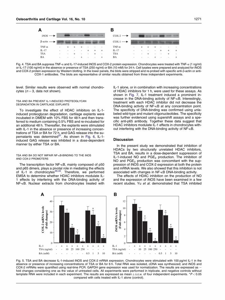
blotting. Under basal conditions, iNOS and COX-2 proteinswere undetectable and treatment with IL-1 resulted ina strong induction of both protein expressions (Fig. 3). Con-sistent with their effects on NO and PGE2 production,HDAC inhibitors prevented the induction of iNOS andCOX-2 protein expression by IL-1, in a concentration-de-pendent manner (Fig. 3). The levels of b-actin and COX-1were not influenced by IL-1 alone or in combination witheach HDAC inhibitors (Fig. 3). IL-1-induced iNOS andCOX-2 protein expression was also inhibited by TSA andBA in normal chondrocytes (n¼ 3, data not shown). As ex-pected, the induction of iNOS and COX-2 proteins by TNF-a or IL-17 was also suppressed by each HDAC inhibitor(Fig. 4).
Next, we used real-time PCR to determine whetherHDAC inhibitors modulate iNOS and COX-2 mRNAs’ induc-tion. The relative expression level of each gene mRNA wasplotted as fold changes over untreated control cells.GAPDH gene expression was used for normalization. Asexpected, IL-1 induced a marked increase of both iNOSand COX-2 mRNA levels (Fig. 5). Treatment with eitherTSA or BA dose-dependently suppressed the induction ofiNOS and COX-2 mRNA expression (Fig. 5), suggestingthat HDAC inhibitors exert their effects at the transcriptional
iNOS
β-actin
COX-2
COX-1
IL-1 - + + + + +BA (mM) 0 0 0.5 1 5 10
pression. Chondrocytes were stimulated with 100 pg/ml IL-1 in the) for 24 h. Cell lysates were prepared and analyzed for iNOS andlots were stripped and re-probed with specific anti-b-actin or anti-
results obtained from four independent experiments.
A B
iNOS COX-2
COX-1β-actin
TNF-α - + + + - - -IL-17 - - - - + + +TSA - - + - - + -BA - - - + - - +
TNF-α - + + + - - -IL-17 - - - - + + +TSA - - + - - + -BA - - - + - - +
Fig. 4. TSA and BA suppress TNF-a and IL-17-induced iNOS and COX-2 protein expression. Chondrocytes were treated with TNF-a (1 ng/ml)or IL-17 (100 ng/ml) in the absence or presence of TSA (250 ng/ml) or BA (10 mM) for 24 h. Cell lysates were prepared and analyzed for iNOSand COX-2 protein expression by Western blotting. In the lower panels, the blots were stripped and re-probed with specific anti-b-actin or anti-
COX-1 antibodies. The blots are representative of similar results obtained from three independent experiments.
1271Osteoarthritis and Cartilage Vol. 16, No. 10
level. Similar results were observed with normal chondro-cytes (n¼ 3, data not shown).
TSA AND BA PREVENT IL-1-INDUCED PROTEOGLYCAN
DEGRADATION IN CARTILAGE EXPLANTS
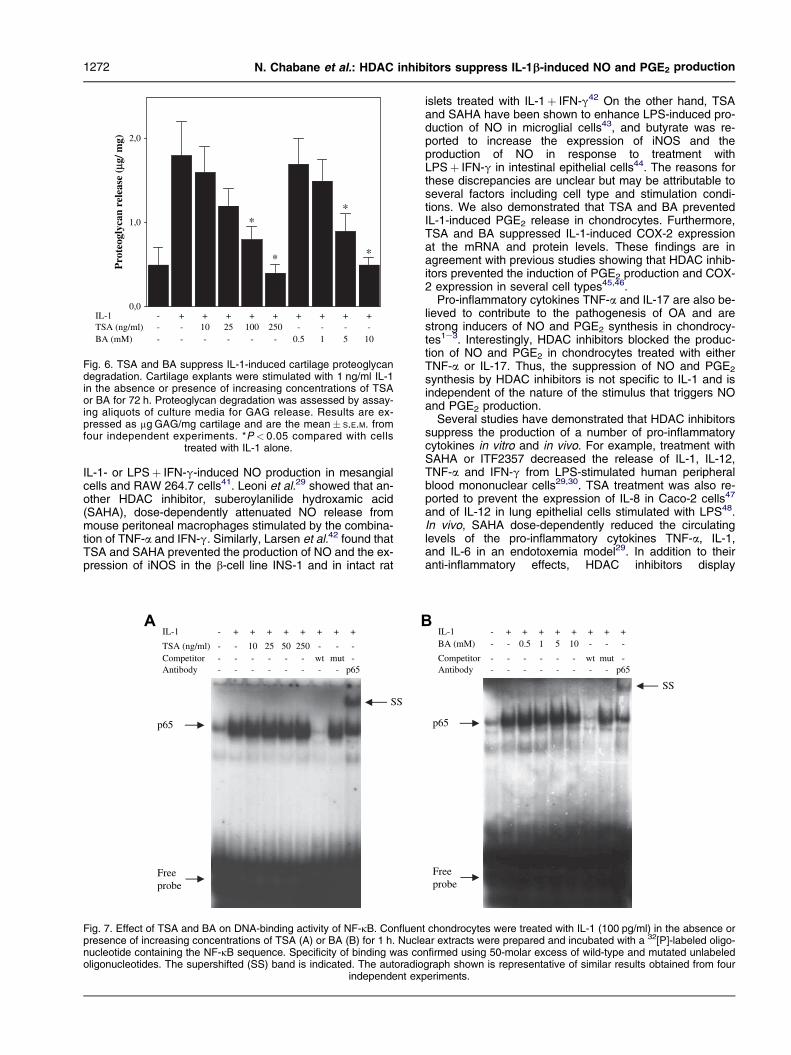
To investigate the effect of HDAC inhibitors on IL-1-induced proteoglycan degradation, cartilage explants wereincubated in DMEM with 10% FBS for 48 h and then trans-ferred to medium containing 0.5% FBS and re-incubated foran additional 48 h. Thereafter, the explants were stimulatedwith IL-1 in the absence or presence of increasing concen-trations of TSA or BA for 72 h, and GAG release into the su-pernatants was determined37. As shown in Fig. 6, IL-1-induced GAG release was inhibited in a dose-dependentmanner by either TSA or BA.
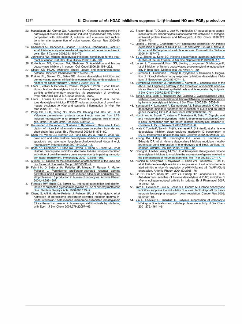
TSA AND BA DO NOT IMPAIR NF-kB-BINDING TO THE iNOS
AND COX-2 PROMOTERS
The transcription factor NF-kB, mainly composed of p50and p65 dimers, plays a pivotal role in mediating the effectsof IL-1 in chondrocytes39,40. Therefore, we performedEMSA to determine whether HDAC inhibitors modulate IL-1 effects by interfering with the DNA-binding activity ofNF-kB. Nuclear extracts from chondrocytes treated with
0
10
20
30
40
IT
B
IL-1 - + + + + + + + + +TSA (ng/ml) - - 10 25 100 250 - - - -
BA (mM) - - - - - - 0.5 1 5 10
CO
X-2
mR
NA
leve
ls(F
old
chan
ges)
A
*
*
*
*
**
Fig. 5. TSA and BA decrease IL-1-induced iNOS and COX-2 mRNA expabsence or presence of increasing concentrations of TSA or BA for 6 hCOX-2 mRNAs were quantified using real-time PCR. GAPDH gene exprefold changes considering one as the value of untreated cells. All expertemplate RNA were included in each experiment. The results are expre
compared with cells treated w
IL-1 alone, or in combination with increasing concentrationsof HDAC inhibitors for 1 h, were used for these assays. Asshown in Fig. 7, IL-1 treatment induced a prominent in-crease in the DNA-binding activity of NF-kB. Interestingly,treatment with each HDAC inhibitor did not decrease theDNA-binding activity of NF-kB at any concentration point.The specificity of DNA-binding was confirmed using unla-beled wild-type and mutant oligonucleotides. The specificitywas further evidenced using supershift assays and a spe-cific anti-p65 antibody. Together these data suggest thatHDAC inhibitors modulate IL-1 effects in chondrocytes with-out interfering with the DNA-binding activity of NF-kB.
Discussion
In the present study we demonstrated that inhibition ofHDACs by two structurally unrelated HDAC inhibitors,TSA and BA, results in a dose-dependent suppression ofIL-1-induced NO and PGE2 production. The inhibition ofNO and PGE2 production was concomitant with the sup-pression of iNOS and COX-2 expression at both the proteinand mRNA levels. We also showed that this inhibition is notassociated with changes in NF-kB DNA-binding activity.
The effects of HDAC inhibition on the production of NOand the expression of iNOS have been examined in a fewrecent studies. Yu et al. demonstrated that TSA inhibited
0
2
4
6
8
10
12
iNO
S m
RN
A le
vels
(Fol
d ch
ange
s)
B
*
*
*
*
*
*
L-1 - + + + + + + + + +SA (ng/ml) - - 10 25 100 250 - - - -
A (mM) - - - - - - 0.5 1 5 10
ression. Chondrocytes were stimulated with 100 pg/ml IL-1 in the. Total RNA was isolated, cDNA was synthesized; and iNOS andssion was used for normalization. The results are expressed as e
iments were performed in triplicate, and negative controls withoutssed as mean� S.E.M. of four independent experiments. *P< 0.05ith IL-1 alone (control).
0,0
1,0
2,0
Pro
teog
lyca
n re
leas
e (μ
g/ m
g)
*
**
*
IL-1 - + + + + + + + + +TSA (ng/ml) - - 10 25 100 250 - - - -BA (mM) - - - - - - 0.5 1 5 10
Fig. 6. TSA and BA suppress IL-1-induced cartilage proteoglycandegradation. Cartilage explants were stimulated with 1 ng/ml IL-1in the absence or presence of increasing concentrations of TSAor BA for 72 h. Proteoglycan degradation was assessed by assay-ing aliquots of culture media for GAG release. Results are ex-pressed as mg GAG/mg cartilage and are the mean� S.E.M. fromfour independent experiments. *P < 0.05 compared with cells
treated with IL-1 alone.
1272 N. Chabane et al.: HDAC inhibitors suppress IL-1b-induced NO and PGE2 production
IL-1- or LPSþ IFN-g-induced NO production in mesangialcells and RAW 264.7 cells41. Leoni et al.29 showed that an-other HDAC inhibitor, suberoylanilide hydroxamic acid(SAHA), dose-dependently attenuated NO release frommouse peritoneal macrophages stimulated by the combina-tion of TNF-a and IFN-g. Similarly, Larsen et al.42 found thatTSA and SAHA prevented the production of NO and the ex-pression of iNOS in the b-cell line INS-1 and in intact rat
BAIL-1 - + + + + + + + +
TSA (ng/ml) - - 10 25 50 250 - - -Competitor - - - - - - wt mut -Antibody - - - - - - - - p65
SS
p65
Freeprobe
Fig. 7. Effect of TSA and BA on DNA-binding activity of NF-kB. Confluenpresence of increasing concentrations of TSA (A) or BA (B) for 1 h. Nuclenucleotide containing the NF-kB sequence. Specificity of binding was cooligonucleotides. The supershifted (SS) band is indicated. The autoradio
independent exp
islets treated with IL-1þ IFN-g42 On the other hand, TSAand SAHA have been shown to enhance LPS-induced pro-duction of NO in microglial cells43, and butyrate was re-ported to increase the expression of iNOS and theproduction of NO in response to treatment withLPSþ IFN-g in intestinal epithelial cells44. The reasons forthese discrepancies are unclear but may be attributable toseveral factors including cell type and stimulation condi-tions. We also demonstrated that TSA and BA preventedIL-1-induced PGE2 release in chondrocytes. Furthermore,TSA and BA suppressed IL-1-induced COX-2 expressionat the mRNA and protein levels. These findings are inagreement with previous studies showing that HDAC inhib-itors prevented the induction of PGE2 production and COX-2 expression in several cell types45,46.
Pro-inflammatory cytokines TNF-a and IL-17 are also be-lieved to contribute to the pathogenesis of OA and arestrong inducers of NO and PGE2 synthesis in chondrocy-tes1e3. Interestingly, HDAC inhibitors blocked the produc-tion of NO and PGE2 in chondrocytes treated with eitherTNF-a or IL-17. Thus, the suppression of NO and PGE2
synthesis by HDAC inhibitors is not specific to IL-1 and isindependent of the nature of the stimulus that triggers NOand PGE2 production.
Several studies have demonstrated that HDAC inhibitorssuppress the production of a number of pro-inflammatorycytokines in vitro and in vivo. For example, treatment withSAHA or ITF2357 decreased the release of IL-1, IL-12,TNF-a and IFN-g from LPS-stimulated human peripheralblood mononuclear cells29,30. TSA treatment was also re-ported to prevent the expression of IL-8 in Caco-2 cells47
and of IL-12 in lung epithelial cells stimulated with LPS48.In vivo, SAHA dose-dependently reduced the circulatinglevels of the pro-inflammatory cytokines TNF-a, IL-1,and IL-6 in an endotoxemia model29. In addition to theiranti-inflammatory effects, HDAC inhibitors display
IL-1 - + + + + + + + +BA (mM) - - 0.5 5 10 - - -
Competitor - - - - - - wt mut -Antibody - - - - - - - - p65
SS
p65
Freeprobe
1
t chondrocytes were treated with IL-1 (100 pg/ml) in the absence orar extracts were prepared and incubated with a 32[P]-labeled oligo-nfirmed using 50-molar excess of wild-type and mutated unlabeledgraph shown is representative of similar results obtained from foureriments.

1273Osteoarthritis and Cartilage Vol. 16, No. 10
chondroprotective properties. Indeed, we demonstratedhere that treatment with TSA or BA prevents IL-1-inducedproteoglycan degradation in cartilage explants. Moreover,Young et al.49 showed that HDAC inhibitors blocked the in-duction of several enzymes responsible for cartilage degra-dation, including MMP-1, MMP-13, a disintegrin andmetalloproteinase domain with thrombospondin motifs(ADAMTS) -4, -5 and -9 and prevented cartilage degrada-tion in an explant assay49. Together these data suggestthat HDAC inhibitors may prevent cartilage destruction inarthritis. Indeed, HDAC inhibitors prevent cartilage damagein models of adjuvant-induced arthritis50 and autoantibody-mediated arthritis51. Protective effects of HDAC inhibitorson cartilage were also observed in collagen-induced arthri-tis models52.
The transcription factor NF-kB is important in the induction ofiNOS and COX-2 by pro-inflammatory cytokines and stimuli inchondrocytes, and the 50-flanking regions of both iNOS andCOX-2 genes contain binding sites for NF-kB39,40. In the pres-ent study, we demonstrated that IL-1 enhances the binding ac-tivity of NF-kB p65. Interestingly, treatment with TSA or butyratedid not affect the binding activity of NF-kB, suggesting thatHDAC inhibitors influence NF-kB-dependent gene expressiondown-stream of DNA-binding in chondrocytes. These resultsare in accordance with previous reports showing that HDAC in-hibitors did not affect the DNA-binding activity of NF-kB in IL-1-stimulated mesangial cells41, and Caco-2 cells47, as well as inLPS-stimulated N9 microglia cells43. In contrast, other groupshave reported that HDAC inhibitors reduced the DNA-bindingactivity of NF-kB in A549 cells53 and human colon cell lines54
treated with pro-inflammatory cytokines. Several reasons mayexplain this dichotomy including the differences in time expo-sure to HDAC inhibitors and the model used.
There are a number of potential mechanisms by whichHDAC inhibitors could inhibit IL-1-induced iNOS andCOX-2 expression. First, HDAC inhibitors may down-regu-late gene expression by altering local chromatin structuresecondary to increased histone acetylation. Secondly, thesuppressive effect of HDAC inhibitors could be mediatedby hyperacetylation of transcription factors or signaling mol-ecules that participate in IL-1-induced iNOS and COX-2 ex-pression. Finally, gene products induced by HDACinhibitors may also interfere with the signaling pathways in-volved in iNOS and COX-2 expression. Regardless of theexact mechanism by which HDAC inhibitors down-regulateIL-1-induced NO and PGE2 production, these results arevery interesting from a pharmacological point of view sinceinhibitors of PGE2 and NO production are a promising classof compounds with therapeutic potential for OA.
In conclusion, we have shown that HDAC inhibitors sup-press IL-1-induced NO and PGE2 production, iNOS andCOX-2 expression as well as proteoglycan degradation.The mechanism by which HDAC inhibitors attenuate IL-1-effects is independent of the DNA-binding activity of thetranscription factor NF-kB. These data also suggest thatHDAC inhibitors represent a promising new class of com-pounds in the treatment of OA.
Conflict of interest
The authors declare that there is no conflict of interest.
Acknowledgments
The authors thank Virginia Wallis for her assistance with themanuscript preparation. This work was supported by the
Canadian Institutes of Health Research (CIHR) GrantMOP-84282, and the Fonds de la Recherche du Centrede Recherche du Centre Hospitalier de l’Universite de Mon-treal (CHUM). HF is a Research Scholar of the Fonds deRecherche en Sante du Quebec (FRSQ).
References
1. Pelletier JP, Martel-Pelletier J, Abramson SB. Osteoarthritis, an inflam-matory disease: potential implication for the selection of new thera-peutic targets. Arthritis Rheum 2001;44:1237e47.
2. Goldring MB. The role of the chondrocyte in osteoarthritis. ArthritisRheum 2000;43:1916e26.
3. Goldring MB, Berenbaum F. The regulation of chondrocyte function byproinflammatory mediators: prostaglandins and nitric oxide. ClinOrthop Relat Res 2004;S37e46.
4. Scher JU, Pillinger MH, Abramson SB. Nitric oxide synthases and oste-oarthritis. Curr Rheumatol Rep 2007;9:9e15.
5. McInnes IB, Leung BP, Field M, Wei XQ, Huang FP, Sturrock RD, et al.Production of nitric oxide in the synovial membrane of rheumatoid andosteoarthritis patients. J Exp Med 1996;184:1519e24.
6. Salvemini D, Misko TP, Masferrer JL, Seibert K, Currie MG,Needleman P. Nitric oxide activates cyclooxygenase enzymes. ProcNatl Acad Sci U S A 1993;90:7240e4.
7. Pelletier JP, Mineau F, Ranger P, Tardif G, Martel-Pelletier J. The in-creased synthesis of inducible nitric oxide inhibits IL-1ra synthesisby human articular chondrocytes: possible role in osteoarthritic carti-lage degradation. Osteoarthritis Cartilage 1996;4:77e84.
8. Cao M, Westerhausen-Larson A, Niyibizi C, Kavalkovich K,Georgescu HI, Rizzo CF, et al. Nitric oxide inhibits the synthesis oftype-II collagen without altering Col2A1 mRNA abundance: prolyl hy-droxylase as a possible target. Biochem J 1997;324(Pt 1):305e10.
9. Taskiran D, Stefanovic-Racic M, Georgescu H, Evans C. Nitric oxidemediates suppression of cartilage proteoglycan synthesis by interleu-kin-1. Biochem Biophys Res Commun 1994;200:142e8.
10. Sasaki K, Hattori T, Fujisawa T, Takahashi K, Inoue H, Takigawa M. Ni-tric oxide mediates interleukin-1-induced gene expression of matrixmetalloproteinases and basic fibroblast growth factor in cultured rabbitarticular chondrocytes. J Biochem 1998;123:431e9.
11. Hashimoto S, Takahashi K, Amiel D, Coutts RD, Lotz M. Chondrocyteapoptosis and nitric oxide production during experimentally inducedosteoarthritis. Arthritis Rheum 1998;41:1266e74.
12. Pelletier JP, Jovanovic D, Fernandes JC, Manning P, Connor JR,Currie MG, et al. Reduced progression of experimental osteoarthritisin vivo by selective inhibition of inducible nitric oxide synthase. Arthri-tis Rheum 1998;41:1275e86.
13. Martel-Pelletier J, Pelletier JP, Fahmi H. Cyclooxygenase-2 and prosta-glandins in articular tissues. Semin Arthritis Rheum 2003;33:155e67.
14. Mehindate K, al-Daccak R, Dayer JM, Kennedy BP, Kris C, Borgeat P,et al. Superantigen-induced collagenase gene expression in humanIFN-gamma-treated fibroblast-like synoviocytes involves prostaglan-din E2. Evidence for a role of cyclooxygenase-2 and cytosolic phos-pholipase A2. J Immunol 1995;155:3570e7.
15. Hardy MM, Seibert K, Manning PT, Currie MG, Woerner BM,Edwards D, et al. Cyclooxygenase 2-dependent prostaglandin E2modulates cartilage proteoglycan degradation in human osteoarthritisexplants. Arthritis Rheum 2002;46:1789e803.
16. Miwa M, Saura R, Hirata S, Hayashi Y, Mizuno K, Itoh H. Induction of ap-optosis in bovine articular chondrocyte by prostaglandin E(2) throughcAMP-dependent pathway. Osteoarthritis Cartilage 2000;8:17e24.
17. Jenuwein T, Allis CD. Translating the histone code. Science 2001;293:1074e80.
18. Urnov FD. Chromatin remodeling as a guide to transcriptional regulatorynetworks in mammals. J Cell Biochem 2003;88:684e94.
19. Chang S, Pikaard CS. Transcript profiling in Arabidopsis reveals com-plex responses to global inhibition of DNA methylation and histone de-acetylation. J Biol Chem 2005;280:796e804.
20. Reid G, Metivier R, Lin CY, Denger S, Ibberson D, Ivacevic T, et al. Mul-tiple mechanisms induce transcriptional silencing of a subset ofgenes, including oestrogen receptor alpha, in response to deacety-lase inhibition by valproic acid and trichostatin A. Oncogene 2005;24:4894e907.
21. Bernstein BE, Tong JK, Schreiber SL. Genomewide studies of histonedeacetylase function in yeast. Proc Natl Acad Sci U S A 2000;97:13708e13.
22. Nawaz Z, Baniahmad C, Burris TP, Stillman DJ, O’Malley BW, Tsai MJ.The yeast SIN3 gene product negatively regulates the activity of thehuman progesterone receptor and positively regulates the activitiesof GAL4 and the HAP1 activator. Mol Gen Genet 1994;245:724e33.
1274 N. Chabane et al.: HDAC inhibitors suppress IL-1b-induced NO and PGE2 production
23. Mariadason JM, Corner GA, Augenlicht LH. Genetic reprogramming inpathways of colonic cell maturation induced by short chain fatty acids:comparison with trichostatin A, sulindac, and curcumin and implica-tions for chemoprevention of colon cancer. Cancer Res 2000;60:4561e72.
24. Chambers AE, Banerjee S, Chaplin T, Dunne J, Debernardi S, Joel SP,et al. Histone acetylation-mediated regulation of genes in leukaemiccells. Eur J Cancer 2003;39:1165e75.
25. Johnstone RW. Histone-deacetylase inhibitors: novel drugs for the treat-ment of cancer. Nat Rev Drug Discov 2002;1:287e99.
26. Kortenhorst MS, Carducci MA, Shabbeer S. Acetylation and histonedeacetylase inhibitors in cancer. Cell Oncol 2006;28:191e222.
27. Glaser KB. HDAC inhibitors: clinical update and mechanism-basedpotential. Biochem Pharmacol 2007;74:659e71.
28. Piekarz RL, Sackett DL, Bates SE. Histone deacetylase inhibitors anddemethylating agents: clinical development of histone deacetylase in-hibitors for cancer therapy. Cancer J 2007;13:30e9.
29. Leoni F, Zaliani A, Bertolini G, Porro G, Pagani P, Pozzi P, et al. The an-titumor histone deacetylase inhibitor suberoylanilide hydroxamic acidexhibits antiinflammatory properties via suppression of cytokines.Proc Natl Acad Sci U S A 2002;99:2995e3000.
30. Leoni F, Fossati G, Lewis EC, Lee JK, Porro G, Pagani P, et al. The his-tone deacetylase inhibitor ITF2357 reduces production of pro-inflam-matory cytokines in vitro and systemic inflammation in vivo. MolMed 2005;11:1e15.
31. Peng GS, Li G, Tzeng NS, Chen PS, Chuang DM, Hsu YD, et al.Valproate pretreatment protects dopaminergic neurons from LPS-induced neurotoxicity in rat primary midbrain cultures: role of micro-glia. Brain Res Mol Brain Res 2005;134:162e9.
32. Huuskonen J, Suuronen T, Nuutinen T, Kyrylenko S, Salminen A. Reg-ulation of microglial inflammatory response by sodium butyrate andshort-chain fatty acids. Br J Pharmacol 2004;141:874e80.
33. Chen PS, Wang CC, Bortner CD, Peng GS, Wu X, Pang H, et al. Val-proic acid and other histone deacetylase inhibitors induce microglialapoptosis and attenuate lipopolysaccharide-induced dopaminergicneurotoxicity. Neuroscience 2007;149:203e12.
34. Bode KA, Schroder K, Hume DA, Ravasi T, Heeg K, Sweet MJ, et al.Histone deacetylase inhibitors decrease toll-like receptor-mediatedactivation of proinflammatory gene expression by impairing transcrip-tion factor recruitment. Immunology 2007;122:596e606.
35. Altman RD. Criteria for the classification of osteoarthritis of the knee andhip. Scand J Rheumatol Suppl 1987;65:31e9.
36. Fahmi H, Di Battista JA, Pelletier JP, Mineau F, Ranger P, Martel-Pelletier J. Peroxisome proliferator-activated receptor gammaactivators inhibit interleukin-1beta-induced nitric oxide and matrix met-alloproteinase 13 production in human chondrocytes. Arthritis Rheum2001;44:595e607.
37. Farndale RW, Buttle DJ, Barrett AJ. Improved quantitation and discrim-ination of sulphated glycosaminoglycans by use of dimethylmethyleneblue. Biochim Biophys Acta 1986;883:173e7.
38. Cheng S, Afif H, Martel-Pelletier J, Pelletier JP, Li X, Farrajota K, et al.Activation of peroxisome proliferator-activated receptor gamma in-hibits interleukin-1beta-induced membrane-associated prostaglandinE2 synthase-1 expression in human synovial fibroblasts by interferingwith Egr-1. J Biol Chem 2004;279:22057e65.
39. Shalom-Barak T, Quach J, Lotz M. Interleukin-17-induced gene expres-sion in articular chondrocytes is associated with activation of mitogen-activated protein kinases and NF-kappaB. J Biol Chem 1998;273:27467e73.
40. Lianxu C, Hongti J, Changlong Y. NF-kappaBp65-specific siRNA inhibitsexpression of genes of COX-2, NOS-2 and MMP-9 in rat IL-1beta-in-duced and TNF-alpha-induced chondrocytes. Osteoarthritis Cartilage2006;14:367e76.
41. Yu Z, Zhang W, Kone BC. Histone deacetylases augment cytokine in-duction of the iNOS gene. J Am Soc Nephrol 2002;13:2009e17.
42. Larsen L, Tonnesen M, Ronn SG, Storling J, Jorgensen S, Mascagni P,et al. Inhibition of histone deacetylases prevents cytokine-induced tox-icity in beta cells. Diabetologia 2007;50:779e89.
43. Suuronen T, Huuskonen J, Pihlaja R, Kyrylenko S, Salminen A. Regula-tion of microglial inflammatory response by histone deacetylase inhib-itors. J Neurochem 2003;87:407e16.
44. Stempelj M, Kedinger M, Augenlicht L, Klampfer L. Essential role of theJAK/STAT1 signaling pathway in the expression of inducible nitric-ox-ide synthase in intestinal epithelial cells and its regulation by butyrate.J Biol Chem 2007;282:9797e804.
45. Tong X, Yin L, Joshi S, Rosenberg DW, Giardina C. Cyclooxygenase-2 reg-ulation in colon cancer cells: modulation of RNA polymerase II elongationby histone deacetylase inhibitors. J Biol Chem 2005;280:15503e9.
46. Yamaguchi K, Lantowski A, Dannenberg AJ, Subbaramaiah K. Histonedeacetylase inhibitors suppress the induction of c-Jun and its targetgenes including COX-2. J Biol Chem 2005;280:32569e77.
47. Hoshimoto A, Suzuki Y, Katsuno T, Nakajima H, Saito Y. Caprylic acidand medium-chain triglycerides inhibit IL-8 gene transcription in Caco-2 cells: comparison with the potent histone deacetylase inhibitor tri-chostatin A. Br J Pharmacol 2002;136:280e6.
48. Iwata K, Tomita K, Sano H, Fujii Y, Yamasaki A, Shimizu E, et al. a histonedeacetylase inhibitor, down-regulates interleukin-12 transcription inSV-40-transformed lung epithelial cells. Cell Immunol 2002;218:26e33.
49. Young DA, Lakey RL, Pennington CJ, Jones D, Kevorkian L,Edwards DR, et al. Histone deacetylase inhibitors modulate metallo-proteinase gene expression in chondrocytes and block cartilage re-sorption. Arthritis Res Ther 2005;7:R503e12.
50. Chung YL, Lee MY, Wang AJ, Yao LF. A therapeutic strategy uses histonedeacetylase inhibitors to modulate the expression of genes involved inthe pathogenesis of rheumatoid arthritis. Mol Ther 2003;8:707e17.
51. Nishida K, Komiyama T, Miyazawa S, Shen ZN, Furumatsu T, Doi H,et al. Histone deacetylase inhibitor suppression of autoantibody-medi-ated arthritis in mice via regulation of p16INK4a and p21(WAF1/Cip1)expression. Arthritis Rheum 2004;50:3365e76.
52. Lin HS, Hu CY, Chan HY, Liew YY, Huang HP, Lepescheux L, et al.Anti-rheumatic activities of histone deacetylase (HDAC) inhibitors invivo in collagen-induced arthritis in rodents. Br J Pharmacol 2007;150:862e72.
53. Imre G, Gekeler V, Leja A, Beckers T, Boehm M. Histone deacetylaseinhibitors suppress the inducibility of nuclear factor-kappaB by tumornecrosis factor-alpha receptor-1 down-regulation. Cancer Res 2006;66:5409e18.
54. Yin L, Laevsky G, Giardina C. Butyrate suppression of colonocyteNF-kappa B activation and cellular proteasome activity. J Biol Chem2001;276:44641e6.


![[Chondrocytes application in regenerative medicine]](https://img.dokumen.tips/doc/110x75/63498ce52cd4c1a3540d90c3/chondrocytes-application-in-regenerative-medicine.jpg)