Embed Size (px)
Citation preview
Chromosoma (1990) 99:281-288 C H R O M O S O M A © Springer-Verlag 1990
Heterogeneity and maintenance of centromere plasmid copy number in Saccharomyces cerevisiae Michael A. Resnick 1, James Westmoreland 1, and Kerry Bloom 2
1 Yeast Genetics/Molecular Biology Group, Cellular and Genetic Toxicology Branch, National Institute of Environmental Health Sciences, Research Triangle Park, NC 27709, USA 2 Biology Department, Wilson Hall, University of North Carolina, Chapel Hill, NC 27599, USA
Received February 27, 1990 / in revised from April 13, 1990 Accepted April 21, 1990 by J. Huberman
Abstract. We developed a novel approach to quantitate the heterogeneity of centromere number in yeast, and the cellular capacity for excess centromeres. Small circu- lar plasmids were constructed to contain the CUP1 me- tallothionein gene, ARS1 (autonomously replicating se- quence) and a conditionally functional centromcre (GAL1-GALIO promoter controlled centromere). The CUP1 gene provided a gene dosage marker, and there- fore a genetic determinant of plasmid copy number. Growth of cells on glucose is permissive for centromere function, while growth on galactose renders the centro- mere nonfunctional and the plasmids are segregated in an asymmetric fashion. We identified " l ines" of cells containing increased numbers of plasmids after transfor- mation. Cell lines containing as many as five to ten active centromeres are stably maintained in the absence of ge- netic selection. Thus haploid yeast cells can tolerate a 50% increase in their centromere number without affect- ing progression through the cell cycle. This system pro- vides the opportunity to address issues of specific cellular controls on centromere copy number.
Introduction
An essential component of the chromosome segregation- al apparatus in eukaryotic organisms is the centromere. The centromere has been characterized at the D N A se- quence level in the yeast Saccharomyces cerevisiae (Fitz- gerald-Hayes et al. 1982; Heiter et al. 1986). Strategies that involve centromere replacement, conditionally func- tional centromeres, and directed D N A mutagenesis have delineated the sequence and structural domains to ap- proximately 200 bp of D N A (reviewed in Resnick and Bloom 1987). Chromosomes with a wild-type centro- mere exhibit a frequency of malsegregation of approxi-
O/~lprint requests to : M. Resnick
mately 1 loss event per every l0 s cell divisions (Hartwell et al. 1982; Whit taker et al. 1988). Specific mutat ions in the centromere can result in partial to complete abol- ishment of orderly chromosomal segregation (McGrew et al. 1986; Panzeri et al. 1985). The addition o f a centro- mere to autonomously replicating plasmids reduces real- segregation rates from 0.8 to 0.02 errors per cell division for small circular plasmids and to less than 0.001 errors per cell division for very large circular or linear plasmids (greater than 100 kb, Heiter et al. 1985).
Malsegrcgation of the small circular molecules can result in plasmid loss or gain. To examine the extent of instability elegant genetic systems have been devel- oped based on colony color to detect the absence, or the presence of one and two copies of centromere plas- raids per cell (Heiter et al. 1985; Koshland et al. 1985). While it has been shown that two copies of centromere plasmids can be stably maintained in haploids in the absence of selection, it has not been possible to evaluate the normal limitations on centromere number in the ab- sence of selection or as a consequence of mutat ional change in the genome.
Several studies have demonstrated the feasibility of developing cells with many copies of ccntromere-con- taining plasmids, either by mutation, selection for high copy using a drug resistance marker (Bitoun and Zamir 1986; Tschumper and Carbon 1987), weakly comple- menting genetic markers (Chlebowicz-Sledziewska and Sledziewski 1985) or by complementat ion using centro- mere plasmids containing different genetic markers (Futcher and Carbon 1986). Because of the selection sys- tems used, it has not been possible to investigate the heterogeneity of centromere number in a cell populat ion or the consequences of altered copy number with respect to growth and meiotic development. In this report we describe the development of a plasmid system containing a centromere and the copper chelatin gene CUP1 (Karin et al. 1984) which allows these issues to be addressed without a requirement for direct selection of cells con- taining high copy number. We have been able rapidly to identify lines or populations of cells containing vat-
282
ious n u m b e r s o f p l a s m i d s a n d assess the deg ree o f he te r - o g e n e i t y o f c o p y n u m b e r in p o p u l a t i o n s o f cells. Cel l l ines c o n t a i n i n g b e t w e e n o n e a n d t en f u n c t i o n a l cop ies o f the p l a s m i d c a n be i s o l a t e d in the absence o f se lec t ion a n d the a v e r a g e p l a s m i d c o p y n u m b e r in these l ines re- m a i n s s table . T h e r e a p p e a r s to be a c o p y n u m b e r b a r r i e r a t f ive to t en ex t r a cop ie s o f c e n t r o m e r e s pe r h a p l o i d cell w i t h o u t a f f ec t i ng the g r o w t h o f the cell.
Materials and methods
Strains. The strains used were: ABDEI, M A Ta ade2 his7-2 leu2-3, 112 arg4-8 c u p l : : U R A 3 thrl-4 trpl-289 ura3-52, in which the CUP! gene has been deleted, provided by the laboratory of Dr. Seymour Fogel; BR1669-5~, MATer ade2-1 his4 arg4-8 CUPI s thrl-4 trpl-1, con- taining a single copy of CUP1 and derived from BR1669 (Whit- taker et al. 1988); MR104-1d, M A T ~ ade2-1 leu2-3, 112 arg4-8 CUPI : : URA3 thrl-4 trpl-1 ; MR104-4a, MATa ade2-1 leu2-3, 112 arg4-8 CUP1 : : URA3 thrl-4 trpl-1 ; MR104-10c, M A T ~ ade2-1 his4 arg4-8 CUPI : : URA3 thrl-4 trpl-I ; MR108-11c, MATa ade2-1 his4 arg4-8 CUP1 : : URA3 thrl-4 trpl-l . These congenic strains were derived from a cross of ABDE1 with BR1669-5~, followed by two backcrosses to BRl669-5~; MR108- 1 lc corresponded to three backcrosses. These were used for trans- formation and determination of copper resistance.
Media. Synthetic complete medium (SC) was as described by Whit- taker et al. (1988) except that Gibco Phytagar was replaced by Difco Bactoagar. Copper-containing agar medium (SC + CUP) was made by adding the appropriate dilution of a 50 mM stock solution of copper sulfate to SC following the cooling of the autoclaved medium to 65 ° C and shortly before pouring plates. Media lacking particular components such as tryptophan (i.e., SC-TRP) were also used in genetic analysis. To induce the GAL1-GALIO promoter, the glucose in SC or SC-TRP was replaced by 2% D-galactose.
Liquid replica plating. In order to assure uniformity in the transfer of cells from colonies to medium containing copper, liquid replica plating was used instead of the traditional replica plating involving imprints on velvet. Colonies were inoculated to liquid growth medi- um (200 lal) in 96 well microtiter dishes (the dishes were not tissue culture treated) and grown overnight to stationary phase at 30 ° C. The wells were sampled using a 48 pin replicator (West Coast Scien- tific) and spotted to the appropriate medium. Approximately 1.1 to 1.3 ~tl was delivered per spot. Plates were incubated for 2 days at 30 ° C. The resistance level of an isolate corresponds to the high- est copper concentration at which there is no clearly detectable reduction in growth relative to growth on a plate lacking copper. Higher concentrations lead to growth retardation as demonstrated in Figure 2.
Plasmid construction. Plasmids YCp5T and YCp5TAA were con- structed from the plasmid pBM150. Plasmid pBMI50 contains a 685 bp EcoRI-BamHI DNA fragment with the divergent GAL1- GALIO yeast promoters (Johnston and Davis 1984). The BamHI site is located 56 nucleotides downstream from the GALl transcrip- tion initiation site. A conditional centromere was constructed by isolating a 289 bp RsaI-AluI fragment containing CEN3 DNA (Bloom et al. 1984). The fragment was ligated to synthetic oligon- ucleotide linkers encoding the BamHI restriction recognition site in the presence of T4 D N A ligase. The BamHi linker-CEN3 DNA complex was subsequently tigated to XhoI oligonucleotide linkers in the presence of T4 DNA ligase.
The CEN3-BamHI linker cassette could then be shuttled into
substitution vectors at unique XhoI or Sall restriction sites without disrupting the tandem BamHI sites (Kenna et al. 1988). This frag- ment was introduced into pBM150 at the SalI site 275 bp down- stream from the GALl initation site. Ligation was followed by transformation into HB101 and the presence and orientation of the insert were confirmed by restriction digestion of a miniplasmid preparation from ampicillin-resistant transformants. CEN4 se- quences were removed by digesting the resulting plasmid with SmaI+HpaI , and blunt-end ligation of the resultant molecules. The resulting plasmid pBMBX289-C4 was digested with KpnI, and a 2.2 kb KpnI fragment containing the CUP1 gene from plasmid JW6 (Karin et al. 1984) was inserted. Finally, the TRPl-ars l 1.4 kb EcoRI fragment (Tschumper and Carbon 1980) was introduced into the unique EcoRI site adjacent to the GALIO initiation site. Plasmid replication does not require this arsl element. The result- ing plasmid YCp5T is shown in Fig. 1. The plasmid YCp5TAA was constructed by digesting YCp5T with BamHI to remove the wild-type CEN3 sequences and substitution with a 289 bp BamHI fragment containing a single point mutation in CEN3 with a C to A transversion at the central position of dyad symmetry in CDEIII (Saunders et al. 1988). The resulting plasmid YCp5TAA was used for transformation as indicated in the text.
Restriction endonucleases and T4 D N A ligase were used fol- lowing conditions recommended by suppliers (New England Bio- labs, Bethesda Research Laboratories or Boehringer Mannheim Biochemicals). Restriction enzyme linkers were purchased from P- L Biochemicals. Standard molecular cloning techniques were as described by Maniatis et al. (/982).
Transformation o f yeast. The lithium acetate (LiAc) method of Ito et al. (1983) was used for transforming yeast.
Preparation and restriction digestion o f yeast DNA for plasmid anal ysis. Transformed yeast strains were grown for 24-36 h at 32°C in 5 ml minimal medium with various amounts of copper supple- mented as indicated in the text. The cells were harvested by centrif- ugation, washed once with water, resuspended in lysis buffer (0.1 M Tris-HC1, pH 8.0, 50 mM EDTA, 1% SDS) and transferred to Eppendorf tubes. Cells were lysed with glass beads by shaking on an Eppendorf mixer for 15 min. The supernatant was trans- ferred to a new tube, extracted with phenol and 24:1 chloroform: isoamyl alcohol, ethanol precipitated and dissolved in 50 ~tl water.
Gel electrophoresis and hybridization analysis o f DNA. D N A sam- ples were analyzed on 1.0% agarose slab gels containing 0.09 M Tris-borate, pH 8.3 and 2.5 mM EDTA. Transfer of D N A from agarose gels to nitrocellulose paper, nick translation and hybridiza- tion analysis were performed as described by Bloom and Carbon (1982). Autoradiography was performed for 24-72 h at - 8 0 ° C with Kodak XAR-5 film (Eastman Kodak, Rochester, NY) and a DuPont Cronex Lightning-Plus intensifying screen (DuPont In- struments, Wilmington, Del). The film was preflashed to an A8oo of 0.065.
Copy number determination. The copy number of YCp5T was deter- mined by preparing genomic D N A from selected transformants. DNA was digested with EcoRI which linearizes the plasmid and generates a 5.1 kb genomic CEN3 fragment, and a 9.2 kb plasmid fragment. DNA was electrophoresed in a 1% agarose gel, trans- ferred to nictrocellulose and probed with a radiolabeled CEN3 fragment. The genomic CEN3 band was used as a single copy standard with which to compare the plasmid band. Dilutions of linearized YCp5T plasmid equivalent to 1, 2, 5, 20, 50, and 100 copies per cell were also electrophoresed and probed with CEN3. Autoradiographs were scanned using a Joyce Loebl Chromoscan 3. Peaks were analyzed by the built-in Rev 4.3 software (Resnick et al. 1987). The reproducibility of extraction was examined by isolating DNA from a single copy isolate that had been appor- tioned equally into three tubes. The copy number determinations revealed a range from 1-1.2 for the three samples.

283
A M P
Pst I
1.8
7 Pstl
Pstl
I~ 1.15
EcoR1
YCp5T 10.6 kb
Kpnl
GALl
f Bar
ARS1
N3
P"I 1.0 U R A 3
Kpnl
2.2
Fig. 1. The YCp5T plasmid containing a single copy of the CUP1 gene and CEN3
R e s u l t s
Plasmid detection system Jbr gene dosage
We developed a plasmid system (YCp5T, Fig. 1) to de- termine the cellular capacity for centromeres and the degree of copy number heterogeneity in the population. The plasmid contains an origin of replication (ARSI), a centromere (CEN3), and a reporter gene CUP1 (cop- per chelatin) that allows the in vivo monitoring of copy number within individual colonies based on resistance to exogenous levels of copper (Fig. 1). It has been dem- onstrated that a linear relationship exists between the number of copies of the CUP1 gene on chromosome VIII and the resistance of cells to the concentration of exogenous copper (Fogel et al. 1983). This strict relation- ship has been used in the detection and analysis of chro- mosomal aneuploidy (Whittaker et al. 1988). The plas- mid YCp5T, also contains the GALI-IO promoter (John- ston and Davis 1984)juxtaposed to the centromere as shown in Fig. 1. This feature allows the centromere to be conditionally inactivated, which in turn leads to plas- mid malsegregation and elevated copy number in the plasmid-bearing cells (Hill and Bloom t987; Snyder et al. 1988). Reactivation of the centromere in this plas- mid provides the opportunity to examine biological con- sequences in the distribution of high copy number cen- tromeres.
Copper sensitivity can be measured by liquid repli- caplating on plates containing increasing concentrations of copper (see Materials and methods) or, alternatively, by plating directly onto copper-containing medium and determining plating efficiency. Examples of both meth- ods are presented in Figs. 2 and 3, respectively. Since survival is based on growth capability of individual cells and the replica plating method is based on clonal growth, the copper concentrations at which significant
v
w [L Q.
0 c )
1000
500
4 0 0
300
200
1 5 0
1OO
50
20
10
5
O
c u p z : & & & /x + + /x & P l a s m i d * : - C C C - C A d
Cop ies * * : 0 I M H 0 I V V
Fig. 2. Growth of hapIoid cells containing various copies of the CUP1 gene on plasmids or in the genome. Cells were liquid replica plated (see Materials and methods) following growth to stationary phase in SC-TRP medium [except for the CUP1 deletion strain lacking a plasmid, in which case tryptophan was added to the medium]. All strains were tested at the same time on each of the copper-containing plates. [For presentation purposes, the plates were photographcd and groups of four strains were arranged as shown.] CUPI: A, strains with CUPI deleted from the genome; +, presence of a single copy of the CUP1 gcne. Plasmid*: - , no plasmid; C, presence of the YCp5T plasmid; A, mutated CEN plasmid YCp5TAA; J, YRp(CUPI)3 plasmid containing three cop- ies of the CUP1 gcne and lacking a centromere. Copies**: 0, none; 1, one; M, medium copy number (see Fig. 5); H, high copy number (5 to 10; see Fig. 5); V, greater than 10 20 copies, typical of YRp plasmids and YCp5TAA
differences are detectable in the two methods are not strictly comparable.
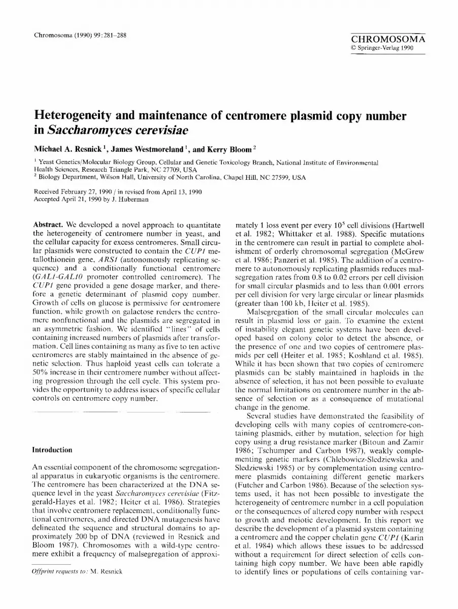
Strains with a single chromosomal copy of the copper gene grow at concentrations below 20 btM copper (CUP1 +, no plasmid, Fig. 2). Deletion of the CUP1 gene results in a considerable reduction in growth at concentrations above 2btM (CUP1A, no plasmid, Fig. 2). Correspondingly, there is less than 1% survival of CUP1 deletion cells on plates containing 10 btM cop- per compared with 100% survival for the strain contain- ing a single copy of CUP1 in the genome (open circles versus open triangles, Fig. 3). The ability to tolerate cop- per by increasing the number of copies of the CUP1 gene was examined by transforming the cells with a CUPl-containing plasmid (YRp(CUP1)3) lacking a cen- tromere, and containing three copies of the CUP1 gene (identical to JW6, Karin et al. 1984). The autonomously replicating plasmid (YRp) exhibits an elevated frequency of malsegregation, thereby generating a subpopulat ion of cells (TRP ÷) with high copy number. Cultures of cells containing this plasmid in the genomic deletion CUP1 strain, are greater than 300 times more resistant than when the plasmid is absent (far right column in Fig. 2, and closed triangles, Fig. 3). Thus there is a con- siderable range over which copper tolerance can be quantitated.
284
I00
"° 60" >
u~ 4O- T J f- i 20"
1 v
~. 10"
I--- 8'
6. "7
I
g
6 v
N
I
L t
~ C U P I t
~ C U P I
0 I00
\ \ , \ \ ,
x " \ .
\ . x,.
\
\ . \ . \
\ '\. \. \. \.
&CUP +lcopy
2 0 0 3 0 0 4 0 0
Copper (~M)
\ \
t
\ i ~ C U P
+ Y R p
I C U P I ) 5
\ '\ \
i
\ , \
\ \ \ ACUP ÷ \multi¢opy
\ \ \
5 0 0 6 0 0
Fig. 3. Survival of strains containing different copies of the CUPI gene on plasmids and having one (CUP1) or no copies (CUP1A) in the genome. No plasmid, CUP1A open circles; one genomic copy, CUP1 open triangles; single copy plasmid, CUP1A open squares; high copy plasmid isolate, CUP1A closed circles; high copy acentric plasmid, CUP1A closed triangles
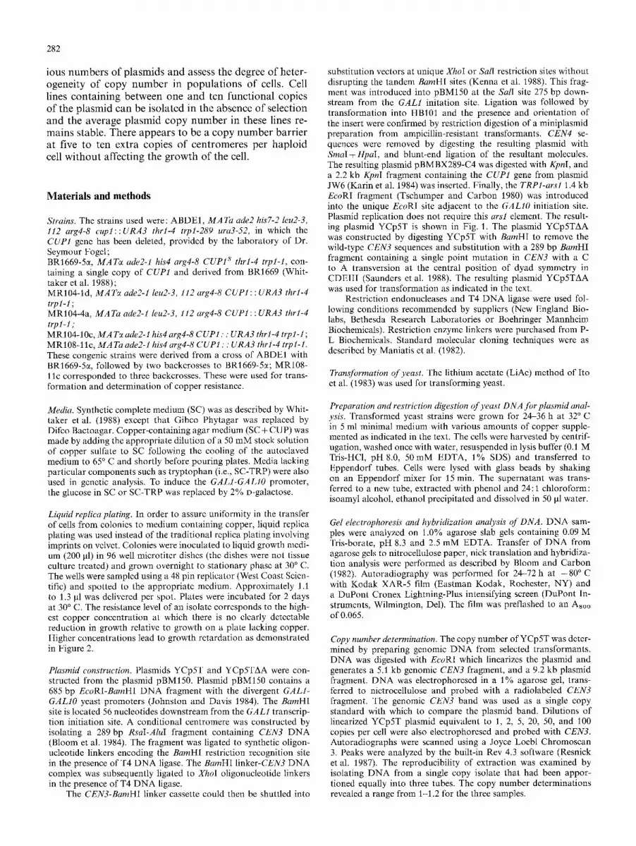
Table 1. Tetrad analysis and copper resistance of spore colonies from a strain presumed to contain a single copy of YCp5T
Strain a T R P + : t ~ asci
0+:4 1+:3 2+:2 3+:1 - 4+0 -
NPD b TET b PD
MR107 5 7 54 5 1 7 - (5/7) c (3/108) c,a (0/10) c (1/3) c (1/28)c
MR109 3 0 52 4 1 10 _ _ (0 /104) c (0 /8) ° (3 /3) ° (3 /40) °
" Strains MR107, MR104-4a/MR104-10c; MR109, MR104-1d/ MR108-11c b NPD, nonparental ditype; PD, parental ditype, TET, tetratype ° In parentheses are the spores containing multiple plasmids, based on copper resistance, divided by the total plasmid-containing spores. All spores that are TR P + are more resistant to copper (10-20 gM) than the trp spores (5 laM). The presumed higher copy number spores are resistant to 20 30 gM copper d From three different asci. These may have arisen during germina- tion
COM-trp a
b
c
d
M R 1 0 7 T e t r a d s
1 2 :5 4 5 6 7 8 9 lO 11 12
Transformation of cells lacking the CUP1 gene with centromere plasmid YCp5T results in increased copper resistance
Following transformation of the CUP1 deletion strains with YCp5T the T R P + isolates exhibited resistance to greater than 50 gM copper (lane 2 from the left, Fig. 2). A small percentage of the isolates exhibited significantly greater resistance to copper. Two of the isolates that were resistant to 50 gM copper and presumed to have a low plasmid copy number per cell, were mated to an appropriate haploid lacking the plasmid. The diploids were induced to sporulate to assess the segregation fideli- ty of the YCp5T plasmid in meiosis. As shown in Table 1, the plasmid generally segregates in a 2 + :2- fashion demonstrating that the level of resistance observed in the bulk of the transformants was due to the presence of a single plasmid bearing the CUP1 gene. Based on these results we can also conclude that presence of the GALI-IO promoter does not disrupt centromere func- tion under normal mitotic and meiotic growth condi- tions.
For one of the two strains examined, there were 7 among 79 tetrads that were 1 : 3 TRP + : t rp - . This class of segregants could represent two plasmids that segregat- ed to the same spore. Evidence to support this idea is provided by the increased resistance of the single plas-
2 5 0 ) J M o Copper b
Fig. 4. Tetrad analysis of strains containing the YCp5T plasmid presumed to be present as a single copy. Spore isolates from four spore tetrads that contain the plasmid exhibited good growth on 10 to 20 ~tM copper while those lacking the plasmid t rp- spores) exhibited reduced growth at 5 gM. For purposes of illustration only growth on a high copper concentration (250 gM) is presented. Only the plasmid-containing spore isolates yielded papillae at 250gM. Note that for tetrads 2 and 11 (1+:3 -) and 4 ( 3 + : 1 ) one of the spores appears to be more highly resistant, indicating the presence of an additional plasmid (see Table 1)
mid-containing spore in 5 of the 1 + : 3- tetrads (see Table 1 and spores 2c and 11 c in Fig. 4). Plasmid-bearing spores generated from 2 + : 2 - events were sensitive to 250 gM copper (Fig. 4), while plasmid-bearing spores from 1 + : 3- events were considerably more resistant to 250 gM copper. As shown below, the increased copper resistance reflects increased plasmid copy number. Thus with this system we can examine malsegregation events in' meiosis that lead to plasmid gain.
285
Isolation o f cell lines with increased copper resistance
Plasmid replication or segregation errors occur in about 2 % - 3 % of mitotic cell divisions (Fitzgerald-Hayes et al. 1982). In cells with a single replicated copy of the YCp5T plasmid segregational errors would be expected to give rise to daughter cells with either no plasmid or two plas- mids. Cells with two plasmids would in turn be expected to give rise to cells with three plasmids, and so on, pro- vided cells can tolerate increased extrachromosomal plasmids.
The segregation frequency of the YCp5T plasmid was examined in cells that had a single copy of the plas- mid during outgrowth of cells on nonselective medium. The frequency of malsegregation was comparable to that for other centromere plasmids (approximately 2% loss per generation). Based on this frequency of malsegrega- tion, cells with more than one copy of the plasmid should exist within the population (demonstrated by Larionov et al. 1989). The relation between the number of CUPI genes and copper resistance provides a convenient strate- gy to identify colonies (i.e., lines) with elevated plasmid copy number. Cells with a single plasmid were grown in liquid medium lacking tryptophan (selective growth for the plasmid) and subsequently plated to this medium. Individual colonies were then inoculated to microtiter wells containing selective medium (SC-Trp) and then liq- uid replica plated to solid medium containing increasing concentrations of copper. Most of the colonies (37/44) that arose after outgrowth from the single copy strain exhibited the copper sensitivity of the transformed par- ent. However, lines of cells that were more resistant (ca- pable of normal growth above 50 I~M copper, see Fig. 2, CUP1A, M and H) were also observed. The selection procedure was repeated with cells from lines exhibiting increased resistance to copper.
Following three successive rounds of screening for cell lines that exhibit higher copper resistance levels, a limit on the level of copper that could be tolerated with- out affecting growth was reached. An additional two rounds of screening did not yield lines with increased copper resistance. The maximum clonal resistance was less than that for cells containing a YRp plasmid and CUP1 deletion [CUPI deletion strains transformed with YRp(CUP1)3 , Figs. 2 and 3]. The maximum resistance was also lower than that observed for a CUP1 plasmid containing a functionally mutated centromere. The mu- tation in YCp5TAA was due to a C to A transversion in the central nucleotide of the conserved element i i i (Saunders et al. 1988). Thus, the copper tolerance that can be achieved is limited in cells with functional centro- mere plasmids. This level is significantly lower than that observed in congenic cells containing CUP1 plasmids that are acentric, or contain altered (nonfunctional) cen- tromeres.
CEN plasmid copy number and stability
Copper resistance is correlated with the number of chro- mosomal copies of C U P ! (Fogel et al. 1983). To deter-
7-
ID
E
6- 0 E o
"1o
E
8- 4- "¢, o
ID
g- 3
h
0
rl,.
t.u
0 510 ldo 15o 260 21o 3oo 31o Copper Concentration OuM)at 10% Survival
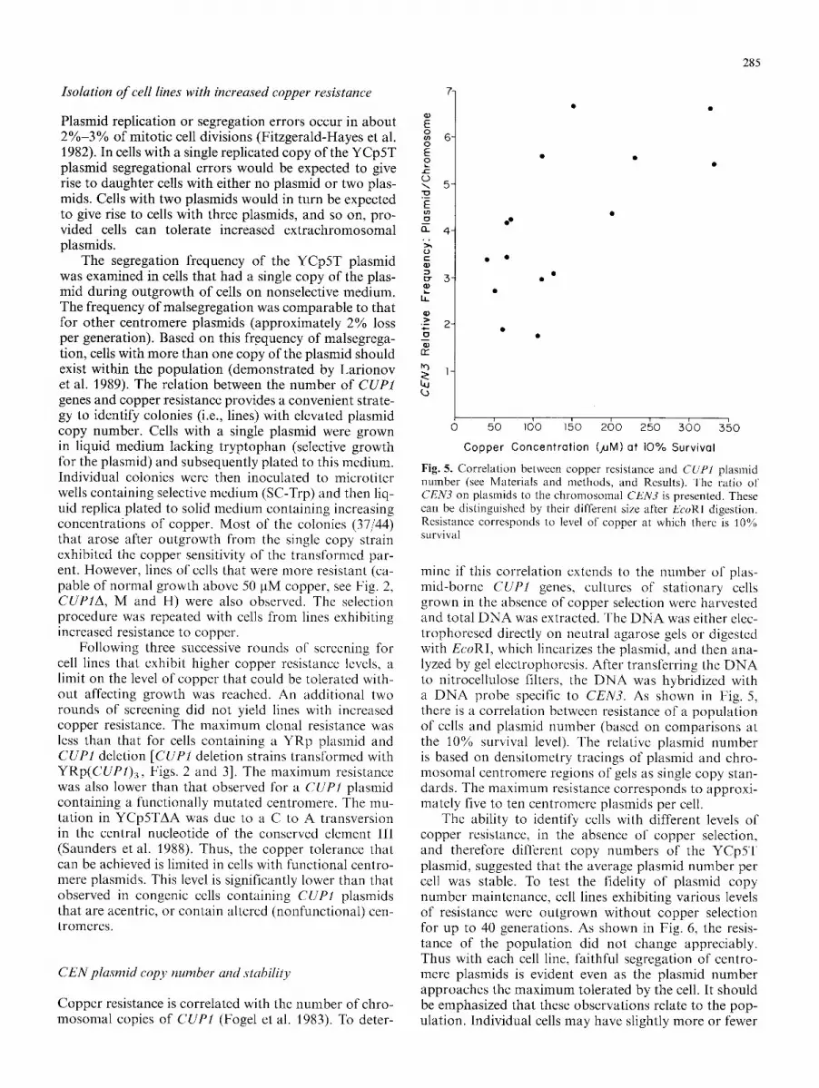
Fig. 5. Correlation between copper resistance and CUP1 plasmid nmnber (see Materials and methods, and Results). The ratio of CEN3 on plasmids to the chromosomal CEN3 is presented. These can be distinguished by their different size after EcoRl digestion. Resistance corresponds to level of copper at which there is 10% survival
mine if this correlation extends to the number of plas- mid-borne CUP1 genes, cultures of stationary cells grown in the absence of copper selection were harvested and total D N A was extracted. The D N A was either elec- trophoresed directly on neutral agarose gels or digested with EcoRI, which linearizes the plasmid, and then ana- lyzed by gel electrophoresis. After transferring the D N A to nitrocellulose filters, the DNA was hybridized with a D N A probe specific to CEN3. As shown in Fig. 5, there is a correlation between resistance of a population of cells and plasmid number (based on comparisons at the 10% survival level). The relative plasmid number is based on densitometry tracings of plasmid and chro- mosomal centromere regions of gels as single copy stan- dards. The maximum resistance corresponds to approxi- mately five to ten centromere plasmids per cell.
The ability to identify cells with different levels of copper resistance, in the absence of copper selection, and therefore different copy numbers of the YCp5T plasmid, suggested that the average plasmid number per cell was stable. To test the fidelity of plasmid copy number maintenance, cell lines exhibiting various levels of resistance were outgrown without copper selection for up to 40 generations. As shown in Fig. 6, the resis- tance of the population did not change appreciably. Thus with each cell line, faithful segregation of centro- mere plasmids is evident even as the plasmid number approaches the maximum tolerated by the cell. It should be emphasized that these observations relate to the pop- ulation. Individual cells may have slightly more or fewer

286
~o-~_
(I) ~D
10 -3- C
0~ c lo "4" O
la_ lO-5.
~0-6.
+47 \ '4 ~ generations ",.,',,. ",,,
+18 "',, \ generations'~
... '\ ; - ~.;
"..
. "% ~"%..~.,
"" +16 ''.....g enerotione
'~.~ "~..~.,,
• .,,. ",~,,, " ~ " ~ , ~. '~ • ~ ~'~-,
o ' 160 ' z6o ' 360 ' 460 ' 5dO
Copper (~uM) Fig. 6. Stability of copper resistance in cell lines. Clonal isolates exhibiting low (circles), medium (triangles) and high (squares) cop- per resistance were grown in SC-TRP. Survival on copper-contain- ing plates was determined before (open data points) and after 16, 18 and 47 generations (closed data points), respectively
plasmid molecules. For example, single colony isolates from the resistant line exhibited a variety of levels of copper resistance, although most of the sublines were highly copper resistant (Fig. 7).
We attempted to surpass the centromere barrier in cells harboring functional centromeres by conditionally inactivating the centromere with the GALI - IO promoter, thereby increasing the rate of malsegregation. As pre- viously shown (Hill and Bloom 1987), activating the GALI - IO promoter adjacent to C E N 3 results in loss of centromere function; plasmids exhibit biased patterns of segregation and accumulate to high copy number in less than 20% of the population. Returning cells to glu- cose medium results in repression of the promoter and reactivation of centromere function. This procedure was followed with cells containing the YCp5T plasmid; they were grown on galactose medium lacking tryptophan, and then plated to glucose medium lacking tryptophan. Colonies that arose after the centromere was returned to function were subsequently examined for levels of copper resistance. Considerable variation in resistance was obtained, but no colonies exhibited normal growth at levels above 300 laM copper (Fig. 7). Comparable re- sults were also obtained if lines of cells with high copper resistance were subjected to the same growth regime (middle panel, Fig. 7). Since some of the colonies exhib- ited low resistance, we conclude that the high level of copper resistance (prior to induction by galactose) was not due to chromosomal mutation(s) rendering cells more copper resistant. None of the colonies arising after galactose induction exhibited tolerance to the levels af- forded by YRp plasmids (greater than 500 gM copper). Thus the distribution of plasmid copy number can be dramatically altered in the population. Low copy isolates resistant to less than 200 ~tM copper (Fig. 2) when shifted to galactose give rise to many ( > 2 5 % ) colonies exhibiting resistance to 300 tIM copper (left panel, Fig. 7). High copy isolates give rise to nearly 50% resistant colonies (right panel, Fig. 7). Only through analysis of individual colonies by such an in vivo moni- toring system can the heterogeneity in the distribution of plasmid copy number be distinguished in plasmid- bearing cells.
Resistance of TRP + Sing le Colony I s o l a t e s to 3 0 0 ] J M Copper
Colonies isolated after growth of low copy plosmid strain in galactose
Colonies isolated after growth of high copy plasmid strain in galactose
Colonies isolated after growth of high copy plasmid strain in glucose
Fig. 7. Copper resistance of cell lines obtained after growth under conditions of CEN3 inactivation. A high copy and a low copy ¥Cp5T strain were grown in galactose medium (lacking tryptophan) to inactivate the centromere. Cells were plated to glucose medium (lacking tryptophan) and the resulting colonies were tested for copper resistance. Presented are the responses of the clonal isolates to 300 gM copper. The panel to the right corresponds to a high copy strain grown in glucose. Note the high proportion that were highly resistant (i.e., grew well on 300 gM copper) in comparison with the other two panels

287
Discussion
The integrity of chromosomal segregation during mitotic and meiotic cell divisions of the yeast S. cerevisiae is a consequence of coordinating the function of D N A - protein complexes at the centromere, of microtubule dy- namics within the mitotic spindle, and of cellular feed- back controls that moni tor the completion of cell cycle events (for reviews see Murray and Kirschner 1989; Har- twell and Weinert 1989). We have developed a genetic system that addresses limitations on the centromere component of the segregational apparatus and the heter- ogeneity in copy number distribution.
The system provides the opportuni ty for " c o u n t i n g " copies of a centromere plasmid using the CUP1 gene as a dosage marker. Starting with a populat ion of hap- loid cells containing only one copy of the plasmid, it is possible to identify a small number of single colony isolates that contain more than one copy. Through suc- cessive rounds of identification of colonies with higher capabilities for growth on copper medium, lines of cells can be developed that have different numbers of plas- raids per cell. Surprisingly, the cells can tolerate and stably maintain up to five to ten plasmids per cell with- out any apparent growth consequences. This corre- sponds to a 50% increase in the normal number of cen- tromeres in a cell. All the plasmid centromeres appear to be functional based on the observation that the CEN3 D N A of isolated nuclei is protected from nucleolytic attack (see Fig. 5 in Kenna et al. 1988, which includes cells with high copy C E N plasmids) as are chromosomal C E N D N A complexes (Saunders et al. 1988).
Previous reports have indicated that haploid yeast can tolerate an approximately 50% to 75% increase in centromere content (Futcher and Carbon 1986). The in- creases were accomplished either by selection and main- tenance of selection for high copy (Bitoun and Zamir 1986), mutat ions that allowed higher levels (Tschumper and Carbon 1987), or through the use of several plas- raids containing complementing markers (Chlebowicz- Slediewska and Sledzwiewski 1985; Futcher and Carbon 1986). The latter established that high copy number could be maintained; however, the requirement for the presence of all the complementing plasmids and the oc- casional malsegregation of" one or more of the plasmids meant that only about 10% of the cells had the entire complement and the population as a whole grew poorly. Because the present system does not require any selec- tion for high copy numbers of plasmids (i.e. genetically neutral), it was possible to establish a limit to the number of copies without selective pressure.
it has been possible to investigate the consequences of inactivating and subsequently activating the centro- mere in terms of plasmid distribution in the resulting population. Hill and Bloom (1987) demonstrated that centromere inactivation results in malsegregation and the development of a small number of cells with elevated copy number. While subsequent activation of the centro- mere resulted in a return to low plasmid number per cell, it was not possible to determine whether the initial and final plasmid distributions were the same. The
system which we have described has demonstrated that the activation/inactivation/activation cycle markedly changes the plasmid distribution in the cell population. Prior to inactivation most of the cells have one plasmid per cell. Following inactivation and activation, a large port ion of the cells have up to the natural limit o f ap- proximately five to ten plasmids (the populat ion is skewed toward the low number, Fig. 7). Approaches in- volving the physical determination of plasmid number do not discriminate such heterogeneity in single cells.
Several interesting questions can be addressed with a system that provides the opportuni ty to develop and maintain cell lines with different numbers of functioning centromere-containing plasmids. The system provides a means for obtaining more material for studies of centro- mere organization and function, and will allow a more accurate characterization of D N A damage and repair within the centromere (Resnick and Bloom 1987). The availability of cell lines with different copy numbers will provide the opportuni ty for studying gene dosage conse- quences of various isolated genes. The consequences of increased numbers of centromeres in meiosis and chro- mosomal segregation can also be investigated. Of partic- ular importance is the potential for eventually examining the relationship between expression of genes involved in the segregational apparatus and number of function- ing centromeres.
Acknowledgements. We gratefully appreciate the ABDEI yeast strain containing the CUPI deletion provided by Dr. Seymour Fogel's laboratory. This work was supported in part by Public tlealth Service Grant GM32238 from the National Institutes of I lealth and a Research Career Development Award Public Health Service Grant CA01175 awarded by the National Cancer Institute, Department of Health and lluman Services, to K.B.
References
Bitoun R, Zamir A (1986) Spontaneous amplification of yeast CEN ARS plasmids. Mol Cell Genet 204:98 102
Bloom KS, Carbon J (1982) Yeast centromere DNA is in a unique and highly ordered structure in chromosomes and small circular minichromosomes. Cell 29:305 317
Bloom KS, Amaya E, Carbon J, Clark L, Hill A, Yeh E (1984) Chromatin conformation of yeast centromeres. J Cell Biol 99:1559 1568
Chlebowicz-Sledziewska E, Sledziewski AZ (1985) Construction of multicopy yeast plasmids with regulated centromere ['unction. Gene 39:25 31
Fitzgerld-t tayes M, Clarke L, Carbon J (1982) Nucleotide sequence comparisons and functional analysis of yeast centromere DNAs. Cell 29:235 244
Fogel S, Welch JW, Cathala G, Karin M (1983) Gene amplification in yeast: CUP1 copy number regulates copper resistance. Curr Genet 7:347-355
Futcher B, Carbon J (1986) Toxic effects of excess cloned centro- mercs. Mol Cell Biol 6:2213 2222
Hartwell LH, Weinert TA (1989) Checkpoints: controls that ensure the order of cell cycle events. Science 246:629 634
Hartwell LH, Dutcher SK, Wood JS, Garrick B (1982) The fidelity of mitotic chromosome reproduction in S. cerevMae. Recent Adv Ycast Mol Biol 1:28 38
Heiter P, Mann C, Snyder M, Davis RW (1985) Mitotic stability of yeast chromosomes: a colony-color assay that measures non- disjunction and chromosome loss. Cell 40 : 381-392

288
Heiter P, Pridmore D, Hegemann JH, Thomas M, Davis RW, Philippsen P (1986) Functional selection and analysis of yeast centromeric DNA. Cell 42: 913-921
Hill A, Bloom KS (1987) Genetic manipulation of centromere func- tion. Mol Cell Biol 7:2397-2405
Ito H, Fukuda Y, Murata K, Kimura A (1983) Transformation of intact yeast cells treated with alkali cations. J Bacteriol 153:163-168
Johnston M, Davis RW (1984) Sequences that regulate the diver- gent GAL1-GALIO promoter in Saccharomyces cerevisiae. Mol Cell Biol 4:1440-1448
Karin M, Najarian R, Haslinger A, Valenzuela P, Welch J, Fogel S (1984) Primary structure and transcription of an amplification genetic locus: the CUP1 locus of yeast. Proc Natl Acad Sci USA 81 : 337 341
Kenna M, Amaya E, Bloom K (1988) Selective excision of the centromere chromatin complex for Saccharomyces cerevisiae. J Cell Biol 107:9-15
Koshland D, Kent JC, Hartwell LH (1985) Genetic analysis of the mitotic transmission of minichromosomes. Cell 40: 393- 403
Larionov VL, Kouprina NY, Strunnikov AV, Vlasov AV (1989) A direct selection procedure for isolating yeast mutants with an impaired segregation of artificial minichromosomes. Curr Genet 15:17 25
Maniatis T, Fritsch EF, Sambrook J (1982) Molecular cloning, a laboratory manual. Cold Spring Harbor Laboratory, Cold Spring Harbor, NY
McGrew JB, Diehl B, Fitzgerald-Hayes M (1986) Single base-pair mutations in centromere element III cause aberrant chromo-
some segregation in Saccharomyces cervisiae. Mol Cell Biol 6:530-538
Murray AW, Kirschner MW (1989) Dominoes and clocks: the union of two views of the cell cycle. Science 246:614-621
Panzeri L, Landonio L, Stotz A, Philippsen P (1985) Role of con- served sequence elements in yeast centromere DNA. EMBO J 4:1867-1874
Resnick MA, Bloom K (1987) Lessons learned from yeast: a molec- ular and genetic analysis of centromere function. In: Vig B, Sandberg A (eds) Aneuploidy, Part A: incidence and etiology. Alan R. Liss, New York, pp 395413
Resnick MA, Westmoreland J, Amaya E, Bloom KS (1987) UV- induced damage and repair in centromere DNA of yeast. Mol Gen Genet 210:16-22
Saunders M, Fitzgerald-Hayes M, Bloom K (1988) Chromatin structure of altered yeast centromeres. Proc Natl Acad Sci USA 85 : 175-179
Snyder M, Saplosky RJ, Davis RW (1988) Transcription interferes with elements important for chromosome maintenance in Sae- charomyces cerevisiae. Mol Cell Biol 8 : 2184-2194
Tschumper G, Carbon J (1980) Sequence of a yeast DNA fragment containing a chromosomal replicator and the TRP! gene. Gene 10:157-166
Tschumper G, Carbon J (1987) Saceharomyees eerevisiae mutants that tolerate centromere plasmids in high copy number. Proc Natl Acad Sci USA 84:7203 7207
Whittaker S, Rockmill BM, Blechel AE, Malone DH, Resnick MA, Fogel S (1988) The detection of mitotic and meiotic aneuploidy in yeast using a gene dosage selection system. Mol Gen Genet 215:10-18


