Embed Size (px)
Citation preview

Histone Deacetylase 1 Deficiency Impairs Differentiation andElectrophysiological Properties of Cardiomyocytes Derived frominduced Pluripotent Cells
Eneda Hoxha1, Erin Lambers1, Hehuang Xie, Ph.D.2, Alexandre De Andrade, Ph.D.2,Prasanna Krishnamurthy, Ph.D.1, John A. Wasserstrom, Ph.D.1, Veronica Ramirez1,Melissa Thal, Ph.D.1, Suresh K. Verma, Ph.D.1, Marcelo B. Soares, Ph.D.2, and Raj Kishore,Ph.D.1,#
1Feinberg Cardiovascular Research Institute, Feinberg School of Medicine, NorthwesternUniversity, 303 E Chicago Avenue, Chicago IL 606112 Children's Memorial Research Center, 2300 Children's Plaza, Chicago IL 60614
AbstractEpigenetic and chromatin modifications play particularly important roles in Embryonic andinduced Pluripotent Stem cells (ES and iPS cells) allowing for the cells to both differentiate anddedifferentiate back to a pluripotent state. We analyzed how the loss of a key chromatin modifyingenzyme, histone deacetylase 1(HDAC1), affects early and cardiovascular differentiation of bothES and iPS cells. We also investigated potential differences between these two cell types whendifferentiation is induced. Our data indicates an essential role for HDAC1 in deacetylatingregulatory regions of key pluripotency-associated genes during early differentiation. AlthoughHDAC1 functions primarily as a histone deacetylase, its loss also affects DNA methylation in ESand iPS cells both during pluripotency and differentiation. We show that HDAC1 plays a crucial,non-redundant role in cardiomyocyte differentiation and maturation. Our data also elucidatesimportant differences between ES and iPS cells, when levels of this enzyme are reduced, thataffect their ability to differentiate into functional cardiomyocytes. As varying levels of chromatinmodifying enzymes are likely to exist in patient derived iPS cells, understanding the molecularcircuitry of these enzymes in ES and iPS cells is critical for their potential use in cardiovasculartherapeutic applications.
#Address Correspondence to: Raj Kishore, PhD Feinberg Cardiovascular Research Institute Feinberg School of Medicine,Northwestern University 303 E Chicago Avenue, Chicago IL 60611 Telephone: 312-503-1651, Fax: 312-503-6060 [email protected].
Author Contribution:Eneda Hoxha: Conception and design, collection and/or assembly of data, data analysis and interpretation, manuscript writing.Erin M. Lambers: manuscript writingVeronica Ramirez: conception and designHehuang Xie: collection and/or assembly of dataAlexandre De Andrade: collection and/or assembly of dataMelissa A. Thal: conception and designSuresh K. Verma: conception and designPrasanna Krishnamurthy: conception and designMarcelo B. Soares: conception and designJohn A. Wasserstrom: collection and/or assembly of dataRaj Kishore: conception and design, final editing and approval of manuscript
DisclosuresThe authors have nothing to disclose.
NIH Public AccessAuthor ManuscriptStem Cells. Author manuscript; available in PMC 2013 November 01.
Published in final edited form as:Stem Cells. 2012 November ; 30(11): 2412–2422. doi:10.1002/stem.1209.
NIH
-PA Author Manuscript
NIH
-PA Author Manuscript
NIH
-PA Author Manuscript
KeywordsStem cells; induced Pluripotent Stem cells; Epigenetic regulation; Histone deacetylases; HDAC1;Cardiovascular differentiation
IntroductionThe ability to isolate Human Embryonic Stem cells (ES cells) from unused In VitroFertilization (IVF) embryos opened a door of opportunities and hopes for their manypotential uses in drug testing, use as models to help our understanding of various biologicalprocesses and most importantly their therapeutic potential in regenerative medicine.However, ethical, technical and regulatory issues as well as unavailability of autologoushuman ES cells for cell therapy applications limit the potential therapeutic utility of ES cellsfor cardiac repair in humans. Reprogramming of somatic cells into induced pluripotent stemcells (iPS cells) opened a new and exciting door of a cell type with the apparent plasticity ofembryonic stem cell and the added advantage of patient specificity [1,2]. Since then the focusof iPS cell biology has shifted towards understanding the epigenetic regulation and cellsignaling that underlie somatic cell dedifferentiation processes and the molecularmechanisms that are involved in the maintenance of the newly acquired pluripotentphenotype.
Induced Pluripotent Stem cells pose great potential for their use in clinical research andtherapy. However, despite their promises and the new advancements made in the field, somechallenges and unknowns still hinder the full realization of the clinical potential of thesecells. Critical gaps in our knowledge of iPS cell biology include incomplete epigenetic andmechanistic characterization of their reprogramming and directed differentiationprocesses [3,4,5,6]. Additionally, while it is widely accepted that one of the first steps thatallows these cells to differentiate is expression of pluripotency associated genes, such asOct4, Nanog and Sox2, how these genes are turned off as differentiation is induced is poorlyunderstood.
A cell's identity is defined by its epigenetic code, modifications of which directly influencegene expression or repression [8,9,10,11,12,13]. The epigenetic state of pluripotent cells isextremely complex as pluripotency needs to be tightly regulated and maintained duringcontinuous proliferation yet developmental genes should be accessible enough fordifferentiation to occur rapidly once the differentiation machinery in the cell hasstarted [11,12,13]. Histone Deacetylases (HDACs) have been identified as key players in bothreprograming of somatic cells into iPS cells as well as important enzymes duringdifferentiation of ES and iPS cells. However, most such studies rely on global inhibitors ofHDACs, which in fact have different roles and direct differentiation into different lineages.The epigenetic similarity between iPS and ES cells and their pluripotent potential has alsobeen recently questioned [11,14].
MethodsCell types and Cell culture
iPS cells were generated from NIH3T3 cells using cell extract mediated reprogramming.The efficiency of our technique in generating these cells as well as their pluripotent naturehas been previously analyzed and reported [15]. C57BL/6 murine ES cells were purchasedfrom ATCC (Cat.# SCRC-1002) and iPS cells were cultured in 15% FBS, 50uM Beta-Mercaptoethanol, 1mM nonessential amino acids and 100 U/ml Pen/Strep supplemented
Hoxha et al. Page 2
Stem Cells. Author manuscript; available in PMC 2013 November 01.
NIH
-PA Author Manuscript
NIH
-PA Author Manuscript
NIH
-PA Author Manuscript

Dulbeco's modified eagle medium (DMEM) in the presence of Leukemia Inhibitory factor(LIF; 10ng/mL).
Formation of Embryoid Bodies (EB)Differentiation of iPS and ES cells through embryoid body formation was performed usingstandard hanging drop method. Briefly, a single-cell suspension of each cell line at aconcentration of 2.5 × 10⁵ cell/mL in 20 mL of differentiating media (Iscove's ModifiedDulbecco's Medium (IDDM) supplemented with 15% FBS, 100 U/ml Pen/Strep, 200ug/mltransferrin, 0.5 mML-ascorbic acid and 4.5 × 10⁻⁴ M monothioglycerol) was deposited in20ul hanging drops in 100 × 100 mm square petri dishes. After 2 days of being cultured insuspension, the cells were plated onto 0.1% gelatin coated dishes for continueddifferentiation.
Real-Time Arrays and mRNA expressionExpression analysis of epigenetic modifying enzymes and factors was performed usingSABiosciences's RT2 Profiler™ PCR Array System according to manufacturer'sinstructions. Quantitative (Real-Time) RT-PCR was performed as described earlier [15] .Quantitative Real-Time PCR (Q-RTPCR or Q-PCR) was performed using gene specificlabeled probes and primers. Array data was verified with independent Q-PCR.
HDAC1 Knock DownThe lentiviral-hdac1 shRNA vectors were purchased from Sigma-Aldrich® and transductionwas performed according to manufacturer's instructions. Stably knocked-down clones weregenerated by puromycin selection for two weeks.
Immunofluorescence stainingProtein expression analysis through immunofluorescence staining was performed asdescribed earlier [15].
Ca++ StudiesCalcium studies were performed mostly as previously described [16]. Isolated EBs wereloaded with fluo-4AM (Invitrogen, 15 μmol/L, 20 minutes) and placed in an experimentalchamber on the stage of a confocal microscope. Media was re-circulated for the remainderof the experiment. Laser scanning of cells in beating loci was accomplished with anLSM510 laser scanning confocal microscope (Zeiss Instruments) and allowed measurementsof intracellular Ca2+ transients in individual myocytes during the experiment. Data werecollected during spontaneous beating and external stimulation (350ms). Image J was used tovisualize the beating profile.
Methylation and Pyrosequencing VerificationMethylation studies were performed as previously described [17]. PCR reactions werecarried out using the Hotstart Taq polymerase kit (Qiagen) in 25 μL total volume and with50 pm of forward primer and reverse primer. For each PCR reaction, 50 ng of the bisulfiteconverted DNA in 1 μL was used as a template. After 5 min of initial denaturation at 95°C,the cycling conditions of 44 cycles consisted of denaturation at 95°C for 15 s, annealing at65°C for 30 s (NKX2.5, T and GATA4,) and 60°C for 30 s (TBX5) and elongation at 72°Cfor 45 s. The PCR products were stored at 4°C until ready for pyrosequencing.Pyrosequencing was performed using the PyroMark MD Pyrosequencing System (Biotage)as described previously. In brief, the PCR product was bound onto streptavidin-SepharoseHP beads (GE Healthcare). Beads containing the immobilized PCR product were denaturedusing a 0.2M NaOH solution and neutralized. Pyrosequencing primer at a concentration of
Hoxha et al. Page 3
Stem Cells. Author manuscript; available in PMC 2013 November 01.
NIH
-PA Author Manuscript
NIH
-PA Author Manuscript
NIH
-PA Author Manuscript

0.3 μM was annealed to the purified single-stranded PCR product at 28°C. Methylationquantification was performed using the manufacturer-provided software. The primers usedin the PCR runs and pyrosequencing reactions are shown in supplementary Table 1.
Statistical analysisTwo-way ANOVA followed by a Bonferroni post-hoc test was used to analyze the data. Pvalues of < 0.05 were used to assign significance.
ResultsIn order to identify key chromatin modifying factors and enzymes in mES and iPS cells, weperformed quantitative Real Time PCR (Q-RT-PCR) array based analysis of the expressionpatterns of 172 chromatin modifying enzymes and factors in mES, iPS and NIH3T3 cells(Fig. 1A). ES and iPS cells had very similar expression profiles with less than 15% of genesshowing significant difference between these two cell types. Additionally, the few genes thatdid show significant difference only showed a 2-3 fold difference. When comparing the two-pluripotent cell types to differentiated cells, the majority of epigenetic enzymes and factors,that were differentially expressed, were up-regulated in pluripotent cells indicating moredynamic chromatin modifications in these cells (Fig. 1A). We assessed the expression levelsof several HDACs from all different HDAC classes. Only HDAC1 and to a lower extentHDAC2, were highly expressed in pluripotent cells. Independent real time PCR analysisverified this data (Fig.1B, Sup.Fig.1A-E). Based on this array data, we identified HDAC1 asone of the enzymes expressed at high levels in pluripotent cells. Independent quantitativeRT-PCR and immunoblots confirmed that HDAC1 is expressed at high levels in pluripotentcells and the expression levels significantly go down in somatic cells representative of thethree germ layers (Fig.1B, C).
This expression indicated a key role for HDAC1 in the pluripotency and differentiationplasticity of iPS cells. Additionally, it provided a model to study if slight differences at thechromatin levels in iPS cells would result in a biological deficiency of these cells todifferentiate. To elucidate the role of HDAC1 in pluripotent cell differentiation, particularlyinto cardiomyocyte differentiation, we created shRNA-mediated stable HDAC1-knock down(HDAC1-KD) cell lines in both ES and iPS cells (Fig. 1D). To test whether there was acompensatory elevated expression of other HDACs when HDAC1 is knocked down, weanalyzed RNA expression levels of HDAC2 (another class I HDAC) and HDAC5 (a class IIHDAC). In both cell types, we did not observe any significant increase in the expressionlevels of these HDACs (Sup.Fig. 2A).
Interestingly, loss of HDAC1 in either iPS or ES cells did not alter the expression levels oftwo pluripotency associated genes (Oct4 and Nanog) under basal undifferentiated and self-renewal conditions (Fig. 1E). Additionally, cell proliferation (Fig. 1F) and cell divisionparameters did not significantly change between wild type and HDAC1-KD cells, underbasal, undifferentiated conditions (Sup. Fig.2B-C). Even though iPS cells showed slightlydifferent cell numbers as proliferation progresses, this difference was not statisticallydifferent (fig. 1F).
HDAC1 has been widely studied due to its implication in many disorders and has beenshown to be important during development [18,19]. HDAC1 knockout mice are embryoniclethal at day 9 post fertilization. We next investigated a possible impact of HDAC1deficiency in cardiovascular cell lineage differentiation of ES and iPS cells.
The embryonic lethality in HDAC1 KO mice and the observed high expression of thisenzyme in iPS and ES cells indicated that HDAC1 potentially plays a role in the early stages
Hoxha et al. Page 4
Stem Cells. Author manuscript; available in PMC 2013 November 01.
NIH
-PA Author Manuscript
NIH
-PA Author Manuscript
NIH
-PA Author Manuscript
of differentiation. HDAC1 is a histone deacetylase and as such is involved in silencing geneexpression. Master regulators of pluripotency, such as OCT4, SOX2 and NANOG, maintainpluripotency by binding to activating regions of promoters of genes important inmaintaining pluripotency, autologous regulation, and associate with complexes which keepdevelopmental genes repressed [20,21,22]. Because lack of HDAC1 during earlydifferentiation could result in persistent high levels of these pluripotency master regulatorsthrough lack of deacetylation at the regulatory regions of these genes, we hypothesized thatHDAC1 could be involved in silencing pluripotency associated genes as the cells areinduced to differentiate. In order to test this hypothesis, we induced differentiation throughEmbryoid Body (EB) formation in both sets of pluripotent cells. As differentiationprogressed we analyzed changes in expression levels of pluripotency associated genes.
One of the first steps in the differentiation process is the silencing of pluripotency-associatedgenes. While levels of these genes vary slightly under self- renewal conditions (Fig.1E), weobserved that upon the induction of differentiation expression levels of Oct4, Sox2 andNanog dramatically decreased in wild type iPS and ES cells (Fig.2A-B). However, inHDAC1-KD cells, persistent high levels of these pluripotency-associated genes wereobserved (Fig. 2A-B). Since lack of HDAC1 could affect deacetylation of pluripotencyassociated genes, we checked acetylation levels at regulatory regions of these genes to testwhether the persistent high levels of expression were due to failure of these regulatoryregions of to get deacetylated by HDAC1. We analyzed the extent of acetylation of HistoneH3 at lysine 9 (H3AcK9) in day 6 differentiating EBs of wt and HDAC1-KD cells bychromatin immune-precipitation (ChIP). Because expression levels of pluripotency-associated genes dramatically decrease in wt cells after differentiation has been induced, wechose day 6 of differentiation so as to be able to directly compare acetylation levels at thesepromoters between wt and HDAC1-KD cells. At later days of differentiation, acetylationlevels of pluripotency associated genes in wt cells are undetectable. As expected, acetylationlevels of all 4 regulatory regions analyzed for Oct4 were very high in HDAC1-KD cells(Fig. 2C,D). Based on expression data (Fig.1A) acetylation levels of these genes would beexpected to stay high as differentiation progresses. Rather interestingly, acetylation of theseregions in the iPS-HDAC1-KD cells was lower than in mES-HDAC1-KD cells (Fig.2D).Acetylation levels of Nanog and Sox2 promoter regions were also higher in mES and iPSHDAC1-KD cells compared to wt cells (Fig.2E). Differences in acetylation levels betweenwt iPS and ES cells were insignificant. Acetylation levels at promoter regions of Nanog andSox2 follow the same pattern as with Oct4. However, iPS-HDAC1 KD cells show lowerlevels of acetylation at these promoters compared to ES-HDAC1 KD cells. This datasuggests that HDAC1 plays a crucial role in deacetylating regulatory regions ofpluripotency-associated genes during differentiation, resulting in their repression. We furtherassessed a direct physical association association of HDAC1 with OCT4 in both mES andiPS cells in their pluripotent, undifferentiated state (Sup. Fig.2F). HDAC1 is known toassociate with two complexes, the NuRD and the NODE complex [23,24]. This change islikely to occur through association with the NuRD complex, rather than with the NODEcomplex since key members of the latter would not be present during differentiation [23,24].The lack of HDAC1 leads to the deregulated suppression of pluripotency-associated genesthereby inhibiting mES and iPS cell differentiation.
Next we investigated whether this could lead to a higher differentiating potential of iPS cellseven when HDAC1 had been knocked down. In the first stages of differentiation weobserved EBs from cells in which HDAC1 had been knocked down failed to expand andgrow compared to their respective Wt counterparts (Sup. Fig. 2D). In order to bettervisualize differentiation within the EB, we stained for Alkaline Phosphatase, an enzymeexpressed in pluripotent cells. As an EB expands and differentiates cells in the periphery aremore differentiated than cells in the core of the EB. As EBs derived from ES and iPS cells
Hoxha et al. Page 5
Stem Cells. Author manuscript; available in PMC 2013 November 01.
NIH
-PA Author Manuscript
NIH
-PA Author Manuscript
NIH
-PA Author Manuscript
grew and differentiated they lost expression of Alkaline Phosphatase, an enzyme expressedin pluripotent cells. We observed higher expression, even in the periphery of ES cells whencompared to their respective Wt cells (Fig.2F). However, iPS cells in which HDAC1 hadbeen knocked down showed a pattern of Alkaline Phosphatase loss similar to theirrespective wt. This indicated a retention of limited differentiation ability in iPS cells inwhich HDAC1 had been knocked down compared to ES-HDAC1 KD cells.
Pluripotent ES and iPS cells are a promising source for potential therapeutic applications forregenerative medicine including cardiovascular repair and regeneration. In order to betterunderstand the differentiation ability of iPS HDAC1-KD cells and how that compared to ESHDAC1-KD cells, we looked at iPS-HDAC1 KD cells potential to differentiate specificallyinto fully functional cardiomyocytes. Lack of HDAC1 also reduced the expression of earlyendodermal and to some extent ectodermal markers (Sup.Fig. 2E), however we were mostinterested in the effect of HDAC1 on cardiovascular differentiation, partly due toinconsistencies in the current knowledge about the role of HDAC1 in cardiovasculardifferentiation. HDAC1-KO mice are embryonic lethal with defects in heart formation butcardiac specific HDAC1 knock-out mice (under myosine heavy chain promoter :- a latemarker of cardiac differentiation) do not present any overt cardiac phenotype althoughdouble HDAC1/HDAC2 cardiac specific KO mice did show arrhythmias, shortly after birth.Thus, we investigated the role of HDAC1 in the differentiation of iPS-HDAC1 KD cells intofully functional cardiomyocytes and how their differentiation compared to that of ES-HDAC1-KD cells.
We determined the comparative effects of HDAC1-silencing on cardiomyocytedifferentiation in both mES-HDAC1-KD and iPS-HDAC1-KD cells. As EBs differentiates,differentiated cardiomyocytes show spontaneous beating. Although wt mES and iPS cellsdifferentiated similarly and showed similar kinetics for spontaneous beating, their HDAC1-KD counterparts displayed either complete loss or significantly reduced and delayed beatingloci (Fig.3A). While about 30% of EBs generated from wt mES and iPS show loci ofspontaneous beating, none of the EBs generated from ES-HDAC1-KD showed anyspontaneous beating (Fig.3A, Sup. Videos1-2). However, some iPSHDAC1-KD cells didspontaneously beat, albeit the beating was delayed and significantly reduced when comparedto wt iPS cells (Sup. Videos 1-2). This data shows that some iPS cells, even under very low/absent HDAC1 levels are able to differentiate into spontaneously beating cardimyocytes.
We wanted to investigate whether iPS-HDAC1 KD cells, unlike their mES counterpart,retained the ability to differentiate into fully functional cardiomyocytes and expresscardiomyocyte specific markers. Expression of early mesodermal genes was significantlylower in both iPS and mES cells in which HDAC1 has been knocked down compared to therespective wild type (Fig. 3B). Expression levels of key cardiomyocyte markers wasconsistently lower in HDAC1-KD cells and the lack of expression of key markers was morepronounced in mES-HDAC1-KD cells (Fig 3C,D).
Expression of mature cardiomyocyte markers such as Cardiac Troponin T (CTT), and othercardiovascular specific proteins was also higher in iPS-HDAC1-KD cells than in mES-HDAC1-KD cells (Fig. 4A,B). Additionally, while mES-HDAC1-KD cells showed noexpression of mature cardiomyocyte proteins essential for spontaneous contractility such asCTT and α-Sarcomeric Actinin, few of the iPS-HDAC1-KD cells derived cardiomyocytesdid, (Fig.4C). In iPS-HDAC1-KD cells, fewer EBs developed spontaneously beating loci,thus global expression of cardiomyocyte specific proteins was lower than in iPS-HDAC1-KD derived EBs. However, expression of these proteins within beating loci in iPS-HDAC1-KD cell derived EBs is comparable to that of wt iPS cell derived EBs (Fig. 4C). This dataindicates that while ES cells lose their ability to differentiate into cardiomyocytes or other
Hoxha et al. Page 6
Stem Cells. Author manuscript; available in PMC 2013 November 01.
NIH
-PA Author Manuscript
NIH
-PA Author Manuscript
NIH
-PA Author Manuscript

cardiovascular lineages when HDAC1 is knocked down, iPS cells, to a certain extent, retainthe ability to differentiate under the same conditions and cope better with the drasticallyreduced levels of HDAC1 during differentiation.
In order to investigate whether the observed effect and difference in behavior between iPSand ES cells under reduced levels of HDAC1 was specific to our iPS cell type, we repeatedthese experiments in an independently generated iPS cell line, NC2. We tested whether NC2would also show restricted cardiovascular differentiation with the loss of HDAC1 comparedto mES-HDAC1-KD cells, which consistently show very little differentiation. Similarly tothe initial iPS cell line we analyzed, NC2 (iPS)-HDAC1-KD cells also showed delayed andreduced beating and repressed differentiation when compared to the Wt cells, and unlikemES-HDAC1-KD cells, NC2 (iPS)-HDAC1-KD cells show some beating anddifferentiation (Fig. 5A-D). This is more apparent at the protein level, where the few beatingloci within EBs derived from NC2 (iPS)-HDAC1-KD cells show robust expression of keycardiomyocyte proteins (Fig. 5E) as opposed to mES-HDAC1-KD cells which show none atthe protein level (Fig.4C).
Since we observed delayed and reduced cardiomyocyte differentiation in iPSHDAC1-KDcells, we investigated whether the few iPS-HDAC1-KD cells that showed some beatingwere physiologically competent and had the ability to become fully functionalcardiomyocytes. To test this we monitored calcium handling of the cells during beating inreal time. Fully mature cardiomyocytes possess the ability to beat in synchrony withadjacent cells and beat at the rate determined by the pacemakers. Thus, the beating colonieswere analyzed for these two crucial characteristics of mature cardiomyocytes: i) The abilityto beat in synchrony and ii) the ability to respond to external stimuli. All wt mES and iPScell derived beating cardiomyocytes analyzed had a synchronized intrinsic rate, respondedvery well to external stimuli (electric pulse at 350 ms) and recovered back to the initialintrinsic rate when the stimulus ceased (Fig.6A; Sup. Videos 3-4). Some iPS-HDAC1-KDderived beating EBs showed aberrant and non-synchronous calcium handling and did notrespond to external stimuli (Fig.6A, B; S. videos 5, 6). While all analyzed beating lociderived from wt iPS cells showed 100% synchronization and response to the externalstimulus, about 80% of beating loci derived from iPS-HDAC1-KD cells responded to theexternal stimulus and only 50% beat in synchrony (Fig.6B). We used electric stimulation toinduce beating in the ESHDAC1-KD derived EBs, however, after several exposures theseEBs did not show contraction. To investigate whether the reason for the poorsynchronization and calcium handling in iPS-HDAC1-KD derived EBs is due to aberrantexpression or localization of gap junction proteins, we analyzed the protein expression leveland pattern of Connexin-43 (CX-43) in beating loci derived from these cells. In the wt cells,CX-43 is both highly expressed and adequately organized in the periphery of the cells (Fig.6C-D). In iPS cells in which HDAC1 had been knocked down expression of CX-43 wassubstantially reduced and disorganized (Fig.6C-D). This data suggests that while some iPScells are able to overcome the need for HDAC1 in the first early stages of differentiation,HDAC1 is important for these cells to fully mature and maintain a cardiomyocytephenotype.
Clearly, iPS-HDAC1-KD cells coped better than mES-HDAC1-KD cells in terms ofcardiomyocyte differentiation. In an attempt to explain this differential characteristic of iPSand mES cells, we analyzed if iPS cells had an epigenetic memory which allowed them tocope better with the lack of HDAC1 during differentiation. Because our iPS cells used inthese experiments are derived from fibroblasts (of mesodermal origin), we tested promoterregions of 4 different mesodermal and cardiovascular genes to determine any differences inmethylation patterns between mES, iPS and their respective HDAC1 KD counterparts,before and during differentiation. In NIH3T3 cells, as expected, these promoters were highly
Hoxha et al. Page 7
Stem Cells. Author manuscript; available in PMC 2013 November 01.
NIH
-PA Author Manuscript
NIH
-PA Author Manuscript
NIH
-PA Author Manuscript

methylated (Fig.7A). However, the methylation pattern of these promoters in the fourpluripotent cell types prior to and during differentiation was interesting. Unlike a recentreport [14] we did not see any differences in the methylation pattern at any of thesepromoters when comparing wt iPS and mES cells (Fig.7B-D). Even during theundifferentiated state methylation levels of the promoters of all the genes analyzed werelower in HDAC1-KD cells compared to wt cells (Fig.7B-D). In the wt cells methylationlevels of these genes went down as the cells differentiated, whereas HDAC1-KD cellsmaintained similar levels of methylation with very little change between pluripotent anddifferentiated states. While there was no expression of these genes in the pluripotent state ofthese cells, the low methylation levels indicate a cross talk between the histone acetylationand DNA methylation at these promoters during early differentiation.
DiscussionIn summary, our data indicates that loss of HDAC1 in mES and iPS cells inhibits theirability to differentiate by suppressing the histone deacetylation of promoters of pluripotencyassociated genes, therefore resulting in their sustained expression and as a consequencerepressed lineage specific differentiation. While mES cells show no differentiation, iPS cellsshow some ability to differentiate even when HDAC1 has been knocked down. Otherreports have indicated a key role for HDACs in ES cell differentiation through globalinhibition of HDACs using Trichostatin A (TSA)[22,25]. Although these reports have greatlyextended the body of knowledge around histone deacetylases and differentiation, they werenot designed to recognize and determine crucial differences between different members ofthe HDAC family. ES cells have very dynamic chromatin maintenance and modificationmachinery. Treatment with TSA, which inhibits all class I and II HDACs, results in moreacetylated histones and thus a globally more active transcription state, which can result inboth inhibition and promotion of differentiation of ES cells, depending on the time of thetreatment. This may explain contradicting reports on the role of HDACs in inhibitingdifferentiation or in promoting differentiation, as both could be possible through differentHDACs or different complexes they associate with [18,19,22,25,26]. Our data using HDAC1-KD pluripotent cells suggests that HDAC1 is specifically important in the earlydifferentiation as it is required to deacetylate puripotency-associated genes whendifferentiation is induced. The prolonged expression of these pluripotent genes duringdifferentiation results in delayed/absent expression of early differentiation genes.
Lack of HDAC1 during differentiation results in reduced cardiovascular differentiation anddecreased or absent spontaneous contraction in differentiating ES and iPS cells. Inaccordance with recent reports, we saw a difference in differentiation ability between ESand iPS cells [14]. While mES-HDAC1-KD cells do not show any spontaneous beatingduring differentiation, iPS-HDAC1-KD cells do. Some of these cells show expression ofcardiovascular markers and some differentiation into cardiomoycytes. However those cellsthat do differentiate into cardiomyocytes show partial absence of synchrony and do notalways respond to external stimuli. This indicates a role for HDAC1 in the maturation to afully functional cardiomyocyte phenotype by regulating expression of Gap junction proteinConnexin-43 in iPS cells. Thus, even though these cells are able to differentiate whenHDAC1 had been knocked down, they are unable to maintain a functional cardiomyocytephenotype in its absence.
Recent reports have also shown differences in the methylation pattern of different generegions between iPS cells and mES cells [14]. When we compared the methylation pattern ofspecific cardiovascular promoters (short regions in CpG islands close to promoters) weobserved no difference between our iPS and mES cells. We did however see a difference in
Hoxha et al. Page 8
Stem Cells. Author manuscript; available in PMC 2013 November 01.
NIH
-PA Author Manuscript
NIH
-PA Author Manuscript
NIH
-PA Author Manuscript
the methylation of cardiomyocyte genes in HDAC1-KD cells both before and duringdifferentiation.
The process of repressing or expressing a gene involves histone modifications, DNAmethylation and expression of various transcription factors and enhancers, which not onlyact in synchrony but also interact and cross talk to each other. HDAC1 affects histoneacetylation and indirectly DNA methylation. Our observations indicate that analysis ofepigenetic molecular mechanisms important in maintaining pluripotency is crucial to ourunderstanding of what makes pluripotent cells pluripotent and what governs theirdifferentiation. This body of knowledge, in the future, could lead to the development of abetter translational strategy for the use of these powerful cells in regenerative medicine,including post-injury cardiovascular repair and regeneration.
ConclusionES and iPS cells carry great potential for therapeutic use. The epigenetic of these moleculeshowever is not fully understood. We showed that HDAC1, a key chromatin modifyingenzyme, is important in deacetylating pluripotency-associated genes during differentiation inboth these cell types. We also show that this molecule plays non-redundant role duringpluripotent cell-derived cardiomyocyte differentiation and maturation. Unlike ES-HDAC1-KD cells which do not show any cardiomyocyte differentiation ability, iPS-HDAC1 KDcells retain some ability to differentiate, albeit the derived cardiomyocytes are electro-physiologically incompetent. These data expand our knowledge of the chromatinmodifications involved in the differentiation of ES and iPS cells as well as elucidatedifferences in differentiation plasticity that are observed when changes at the epigeneticlevel exist.
Supplementary MaterialRefer to Web version on PubMed Central for supplementary material.
AcknowledgmentsSources of Funding
Work described in this manuscript was supported in part by National Institute of Health grants HL091983,HL105597, HL095874, HL053354 and HL108795 to R.K. and American Heart Association's pre-doctoralfellowship grant 11PRE7360065 to E.H.
References1. Takahashi K, Tanabe K, Ohnuki M, et al. Induction of pluripotent stem cells from adult human
fibroblasts by defined factors. Cell. 2007; 131(5):861–872. [PubMed: 18035408]
2. Takahashi K, Yamanaka S. Induction of pluripotent stem cells from mouse embryonic and adultfibroblast cultures by defined factors. Cell. 2006; 126(4):663–676. [PubMed: 16904174]
3. Rolletschek A, Wobus AM. Induced human pluripotent stem cells: promises and open questions.Biological Chemistry. 2009; 390(9):845–849. [PubMed: 19558327]
4. Stefanovic S, Abboud N, Desilets S, Nury D, Cowan C, Puceat M. Interplay of Oct4 with Sox2 andSox17: a molecular switch from stem cell pluripotency to specifying a cardiac fate. The Journal ofCell Biology. 2009; 186(5):665–673. [PubMed: 19736317]
5. Wang J, Rao S, Chu J, et al. A protein interaction network for pluripotency of embryonic stem cells.Nature. 2006; 444(7117):364–368. [PubMed: 17093407]
6. Xi S, Geiman TM, Briones V, Guang Tao Y, Xu H, Muegge K. Lsh participates in DNAmethylation and silencing of stem cell genes. Stem Cells. 2009; 27(11):2691–2702. [PubMed:19650037]
Hoxha et al. Page 9
Stem Cells. Author manuscript; available in PMC 2013 November 01.
NIH
-PA Author Manuscript
NIH
-PA Author Manuscript
NIH
-PA Author Manuscript
7. Chen X, Xu H, Yuan P, et al. Integration of external signaling pathways with the core transcriptionalnetwork in embryonic stem cells. Cell. Jun 13; 2008 133(6):1106–1117. [PubMed: 18555785]
8. Bernstein BE, Mikkelsen TS, Xie X, et al. A bivalent chromatin structure marks key developmentalgenes in embryonic stem cells. Cell. 2006; 125(2):315–326. [PubMed: 16630819]
9. Dovey OM, Foster CT, Cowley SM. Histone deacetylase 1 (HDAC1), but not HDAC2, controlsembryonic stem cell differentiation. Proceedings of the National Academy of Sciences of the UnitedStates of America. 2010; 107(18):8242–8247. [PubMed: 20404188]
10. Holliday R. Epigenetics: a historical overview. Epigenetics. 2006; 1(2):76–80. [PubMed:17998809]
11. Hu Q, Friedrich AM, Johnson LV, Clegg DO. Memory in induced pluripotent stem cells:reprogrammed human retinal-pigmented epithelial cells show tendency for spontaneousredifferentiation. Stem Cells. 2010; 28(11):1981–1991. [PubMed: 20882530]
12. Karantzali E, Schulz H, Hummel O, Hubner N, Hatzopoulos A, Kretsovali A. Histone deacetylaseinhibition accelerates the early events of stem cell differentiation: transcriptomic and epigeneticanalysis. Genome Biology. 2008; 9(4):R65. [PubMed: 18394158]
13. Lee TI, Jenner RG, Boyer LA, et al. Control of developmental regulators by Polycomb in humanembryonic stem cells. Cell. 2006; 125(2):301–313. [PubMed: 16630818]
14. Kim K, Doi A, Wen B, et al. Epigenetic memory in induced pluripotent stem cells. Nature. 2010;467(7313):285–290. [PubMed: 20644535]
15. Rajasingh J, Lambers E, Hamada H, et al. Cell-free embryonic stem cell extract-mediatedderivation of multipotent stem cells from NIH3T3 fibroblasts for functional and anatomicalischemic tissue repair. Circulation Research. 2008; 102(11):e107–117. [PubMed: 18483406]
16. Wasserstrom JA, Sharma R, Kapur S, et al. Multiple defects in intracellular calcium cycling inwhole failing rat heart. Circulation. 2009; 2(3):223–232. [PubMed: 19808344]
17. Xie H, Wang M, Bonaldo Mde F, et al. High-throughput sequence-based epigenomic analysis ofAlu repeats in human cerebellum. Nucleic Acids Research. 2009; 37(13):4331–4340. [PubMed:19458156]
18. Ma P, Schultz RM. Histone deacetylase 1 (HDAC1) regulates histone acetylation, development,and gene expression in preimplantation mouse embryos. Developmental Biology. 2008; 319(1):110–120. [PubMed: 18501342]
19. Montgomery RL, Davis CA, Potthoff MJ, et al. Histone deacetylases 1 and 2 redundantly regulatecardiac morphogenesis, growth, and contractility. Genes & Development. 2007; 21(14):1790–1802. [PubMed: 17639084]
20. Liang J, Wan M, Zhang Y, et al. Nanog and Oct4 associate with unique transcriptional repressioncomplexes in embryonic stem cells. Nature Cell Biology. 2008; 10(6):731–739.
21. Marmorstein R. Protein modules that manipulate histone tails for chromatin regulation. Naturereviews. Molecular Cell Biology. 2001; 2(6):422–432. [PubMed: 11389466]
22. Mikkelsen TS, Ku M, Jaffe DB, et al. Genome-wide maps of chromatin state in pluripotent andlineage-committed cells. Nature. 2007; 448(7153):553–560. [PubMed: 17603471]
23. Xue Y, Wong J, Moreno GT, Young MK, Cote J, Wang W. NURD, a novel complex with bothATP-dependent chromatin-remodeling and histone deacetylase activities. Molecular cell. Dec;1998 2(6):851–861. [PubMed: 9885572]
24. Liang J, Wan M, Zhang Y, et al. Nanog and Oct4 associate with unique transcriptional repressioncomplexes in embryonic stem cells. Nature cell biology. Jun; 2008 10(6):731–739.
25. Lee JH, Hart SR, Skalnik DG. Histone deacetylase activity is required for embryonic stem celldifferentiation. Genesis. 2004; 38(1):32–38. [PubMed: 14755802]
26. Lagger S, Meunier D, Mikula M, et al. Crucial function of histone deacetylase 1 for differentiationof teratomas in mice and humans. The EMBO Journal. 2010; 29(23):3992–4007. [PubMed:20967026]
Hoxha et al. Page 10
Stem Cells. Author manuscript; available in PMC 2013 November 01.
NIH
-PA Author Manuscript
NIH
-PA Author Manuscript
NIH
-PA Author Manuscript
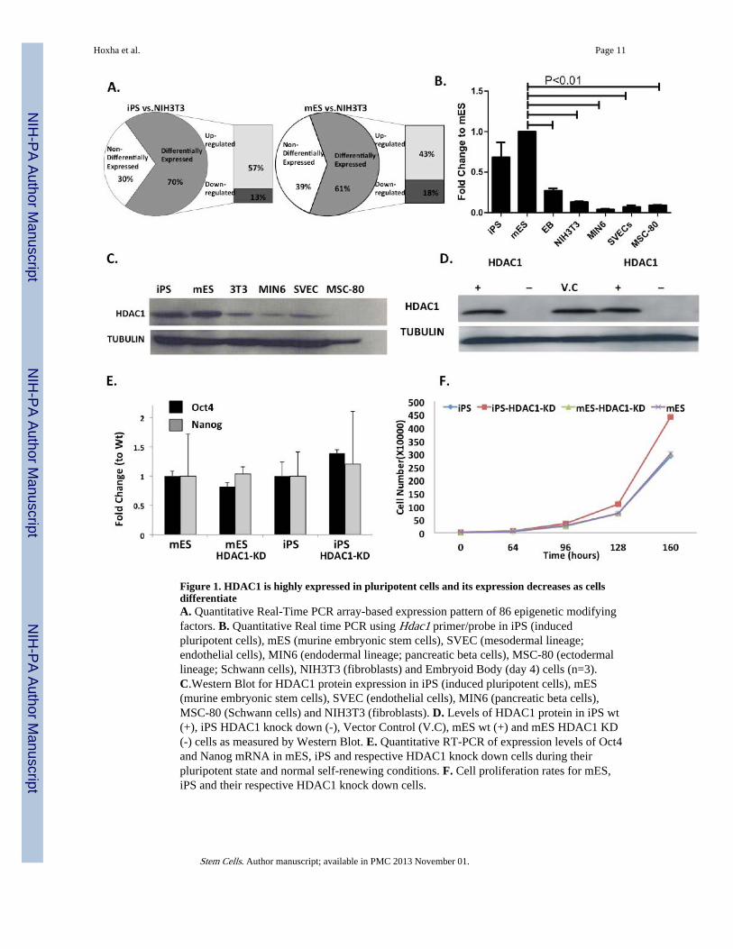
Figure 1. HDAC1 is highly expressed in pluripotent cells and its expression decreases as cellsdifferentiateA. Quantitative Real-Time PCR array-based expression pattern of 86 epigenetic modifyingfactors. B. Quantitative Real time PCR using Hdac1 primer/probe in iPS (inducedpluripotent cells), mES (murine embryonic stem cells), SVEC (mesodermal lineage;endothelial cells), MIN6 (endodermal lineage; pancreatic beta cells), MSC-80 (ectodermallineage; Schwann cells), NIH3T3 (fibroblasts) and Embryoid Body (day 4) cells (n=3).C.Western Blot for HDAC1 protein expression in iPS (induced pluripotent cells), mES(murine embryonic stem cells), SVEC (endothelial cells), MIN6 (pancreatic beta cells),MSC-80 (Schwann cells) and NIH3T3 (fibroblasts). D. Levels of HDAC1 protein in iPS wt(+), iPS HDAC1 knock down (-), Vector Control (V.C), mES wt (+) and mES HDAC1 KD(-) cells as measured by Western Blot. E. Quantitative RT-PCR of expression levels of Oct4and Nanog mRNA in mES, iPS and respective HDAC1 knock down cells during theirpluripotent state and normal self-renewing conditions. F. Cell proliferation rates for mES,iPS and their respective HDAC1 knock down cells.
Hoxha et al. Page 11
Stem Cells. Author manuscript; available in PMC 2013 November 01.
NIH
-PA Author Manuscript
NIH
-PA Author Manuscript
NIH
-PA Author Manuscript

Figure 2. HDAC1 is required for deacetylattion and turning off of pluripotency associated genesduring differentiationA. Quantitative RT-PCR showing Oct4 and Nanog mRNA levels in Wt and HDAC1-KDmES and iPS cells at 6-8 days of differentiation. B. Immunofluorescence staining for OCT4(green) in EBs derived from Wt mES, iPS and their respective HDAC1-KD cells. Image wastaken close to the periphery of the EB at day 6 of differentiation. Nuclie were counterstainedwith dapi (blue). C. Chromatin immunoprecipation of H3AcK9 levels at different regulatoryregions of Oct4 at day 6 of differentiation in Wt and HDAC1-KD mES cells. D. ChIP resultsof H3AcK9 levels at different regulatory regions of Oct4 at day 6 of differentiation in Wtand HDAC1-KD iPS cells. E. ChIP results of H3AcK9 levels at the promoter of Sox2 and
Hoxha et al. Page 12
Stem Cells. Author manuscript; available in PMC 2013 November 01.
NIH
-PA Author Manuscript
NIH
-PA Author Manuscript
NIH
-PA Author Manuscript

Nanog at day 6 of differentiation in Wt and HDAC1-KD mES and iPS cells. F. AlkalinePhosphatase staining of Wt iPS and mES cells (controls) and HDAC1-KD iPS and mEScells at day 8 of differentiation. Staining of pluripotent mES cells and NIH3T3 cells serve aspositive and negative staining controls, respectively. Images taken at 2.5X magnification.
Hoxha et al. Page 13
Stem Cells. Author manuscript; available in PMC 2013 November 01.
NIH
-PA Author Manuscript
NIH
-PA Author Manuscript
NIH
-PA Author Manuscript
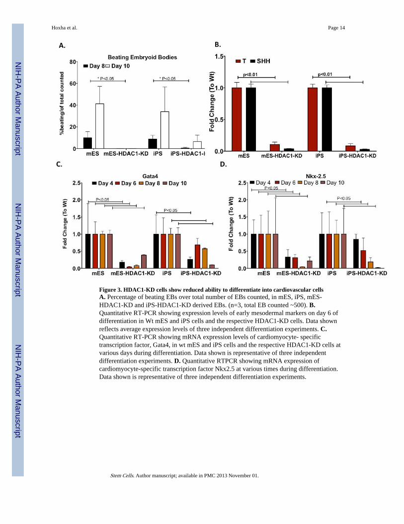
Figure 3. HDAC1-KD cells show reduced ability to differentiate into cardiovascular cellsA. Percentage of beating EBs over total number of EBs counted, in mES, iPS, mES-HDAC1-KD and iPS-HDAC1-KD derived EBs. (n=3, total EB counted ~500). B.Quantitative RT-PCR showing expression levels of early mesodermal markers on day 6 ofdifferentiation in Wt mES and iPS cells and the respective HDAC1-KD cells. Data shownreflects average expression levels of three independent differentiation experiments. C.Quantitative RT-PCR showing mRNA expression levels of cardiomyocyte- specifictranscription factor, Gata4, in wt mES and iPS cells and the respective HDAC1-KD cells atvarious days during differentiation. Data shown is representative of three independentdifferentiation experiments. D. Quantitative RTPCR showing mRNA expression ofcardiomyocyte-specific transcription factor Nkx2.5 at various times during differentiation.Data shown is representative of three independent differentiation experiments.
Hoxha et al. Page 14
Stem Cells. Author manuscript; available in PMC 2013 November 01.
NIH
-PA Author Manuscript
NIH
-PA Author Manuscript
NIH
-PA Author Manuscript
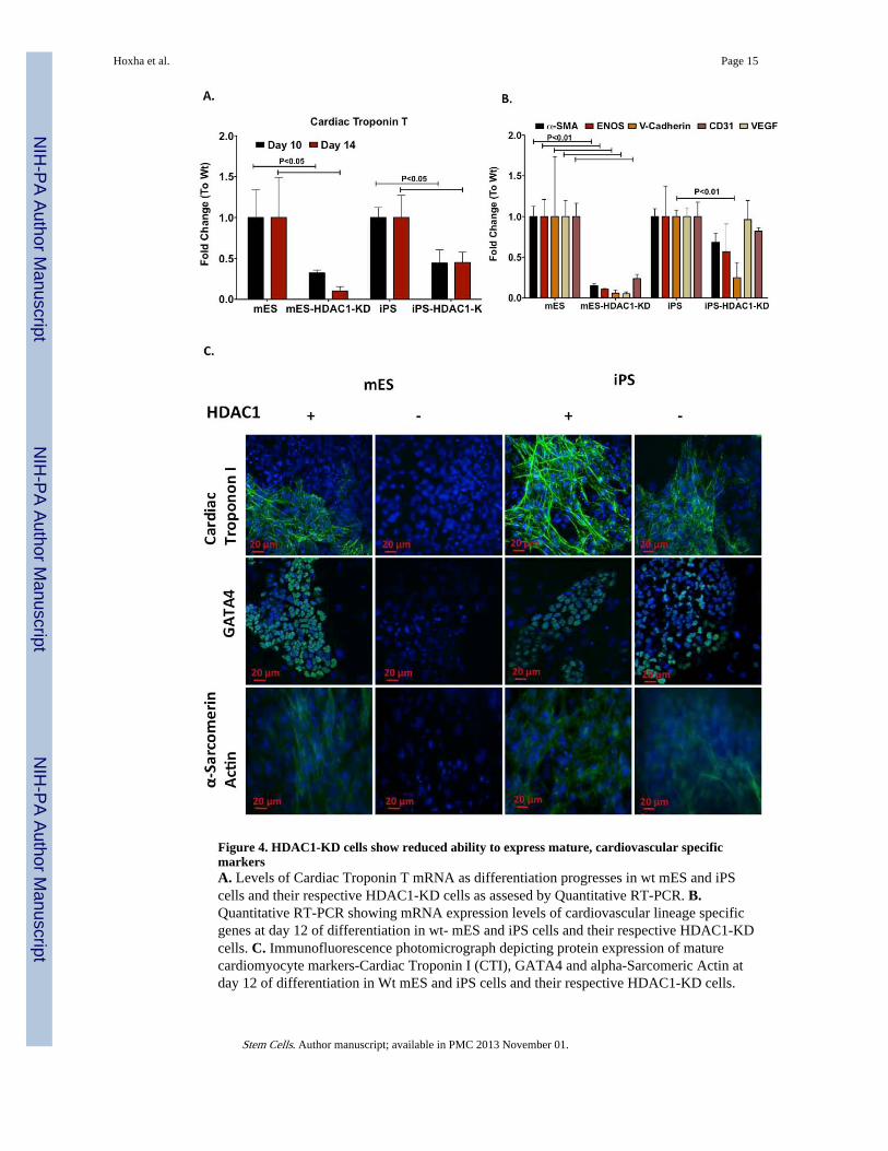
Figure 4. HDAC1-KD cells show reduced ability to express mature, cardiovascular specificmarkersA. Levels of Cardiac Troponin T mRNA as differentiation progresses in wt mES and iPScells and their respective HDAC1-KD cells as assesed by Quantitative RT-PCR. B.Quantitative RT-PCR showing mRNA expression levels of cardiovascular lineage specificgenes at day 12 of differentiation in wt- mES and iPS cells and their respective HDAC1-KDcells. C. Immunofluorescence photomicrograph depicting protein expression of maturecardiomyocyte markers-Cardiac Troponin I (CTI), GATA4 and alpha-Sarcomeric Actin atday 12 of differentiation in Wt mES and iPS cells and their respective HDAC1-KD cells.
Hoxha et al. Page 15
Stem Cells. Author manuscript; available in PMC 2013 November 01.
NIH
-PA Author Manuscript
NIH
-PA Author Manuscript
NIH
-PA Author Manuscript
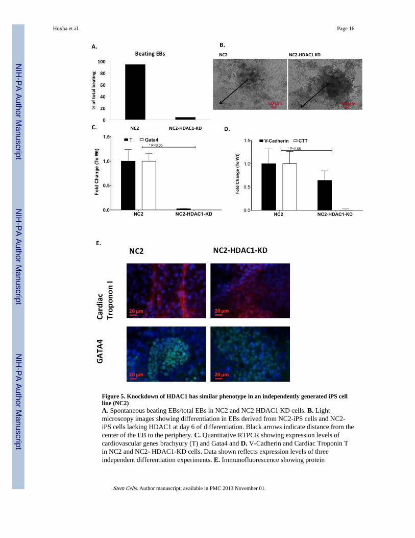
Figure 5. Knockdown of HDAC1 has similar phenotype in an independently generated iPS cellline (NC2)A. Spontaneous beating EBs/total EBs in NC2 and NC2 HDAC1 KD cells. B. Lightmicroscopy images showing differentiation in EBs derived from NC2-iPS cells and NC2-iPS cells lacking HDAC1 at day 6 of differentiation. Black arrows indicate distance from thecenter of the EB to the periphery. C. Quantitative RTPCR showing expression levels ofcardiovascular genes brachyury (T) and Gata4 and D. V-Cadherin and Cardiac Troponin Tin NC2 and NC2- HDAC1-KD cells. Data shown reflects expression levels of threeindependent differentiation experiments. E. Immunofluorescence showing protein
Hoxha et al. Page 16
Stem Cells. Author manuscript; available in PMC 2013 November 01.
NIH
-PA Author Manuscript
NIH
-PA Author Manuscript
NIH
-PA Author Manuscript

expression levels of Cardiac Troponin I (CTI) and Gata4 at day 12 of differentiation in WtNC2-iPS cells and their respective HDAC1-KD cells.
Hoxha et al. Page 17
Stem Cells. Author manuscript; available in PMC 2013 November 01.
NIH
-PA Author Manuscript
NIH
-PA Author Manuscript
NIH
-PA Author Manuscript
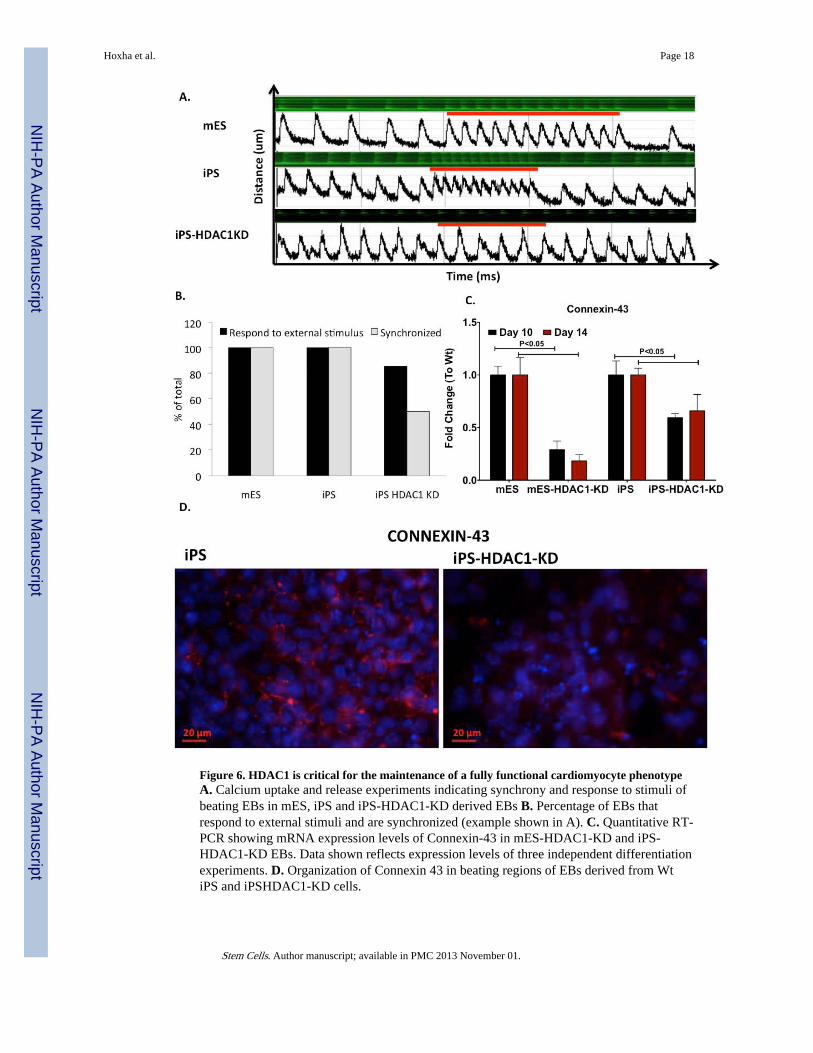
Figure 6. HDAC1 is critical for the maintenance of a fully functional cardiomyocyte phenotypeA. Calcium uptake and release experiments indicating synchrony and response to stimuli ofbeating EBs in mES, iPS and iPS-HDAC1-KD derived EBs B. Percentage of EBs thatrespond to external stimuli and are synchronized (example shown in A). C. Quantitative RT-PCR showing mRNA expression levels of Connexin-43 in mES-HDAC1-KD and iPS-HDAC1-KD EBs. Data shown reflects expression levels of three independent differentiationexperiments. D. Organization of Connexin 43 in beating regions of EBs derived from WtiPS and iPSHDAC1-KD cells.
Hoxha et al. Page 18
Stem Cells. Author manuscript; available in PMC 2013 November 01.
NIH
-PA Author Manuscript
NIH
-PA Author Manuscript
NIH
-PA Author Manuscript

Figure 7. HDAC1 affects DNA methylation levels of mesodermal/cardiac genesA. Methylation levels at the promoter regions of Nkx 2.5, Gata4 and Tbx5 in NIH3T3 cellsas assessed by pyrosequencing (n=3, 50 repeats each time). Methylation levels of B. Gata4,C. NKX2.5, and D. Tbx5 in mES, iPS and the respective HDAC1-KD cells in theirpluripotent state and at day 6 of differentiation.
Hoxha et al. Page 19
Stem Cells. Author manuscript; available in PMC 2013 November 01.
NIH
-PA Author Manuscript
NIH
-PA Author Manuscript
NIH
-PA Author Manuscript


