Embed Size (px)
Citation preview

H2AX phosphorylation marks gemcitabine-inducedstalled replication forks and their collapse uponS-phase checkpoint abrogation
Brett Ewald, Deepa Sampath,and William Plunkett
Department of Experimental Therapeutics, The University ofTexas M. D. Anderson Cancer Center, Houston, Texas and TheUniversity of Texas Graduate School of Biomedical Sciences,Houston, Texas
AbstractGemcitabine is a nucleoside analogue that is incorporatedinto replicating DNA, resulting in partial chain terminationand stalling of replication forks. The histone variant H2AXis phosphorylated on Ser139 (;-H2AX) and forms nuclearfoci at sites of DNA damage. Here, we characterize theconcentration- and time-dependent phosphorylation ofH2AX in response to gemcitabine-induced stalled replica-tion forks. The number of ;-H2AX foci increased with timeup to 2 to 6 h after exposure to gemcitabine, whereaslonger exposures did not cause greater phosphorylation orincrease cell death. The percentage of ;-H2AX–positivecells increased with concentrations of gemcitabine up to0.1 Mmol/L, and ;-H2AX was most evident in the S-phasefraction. Phosphorylation of ataxia-telangiectasia mutated(ATM) on Ser1981 was also associated with S-phase cellsand colocalized in the nucleus with phosphorylated H2AXfoci after gemcitabine exposure. Chemical inhibition ofATM, ATM- and Rad3-related, and DNA-dependent pro-tein kinase blocked H2AX phosphorylation. H2AX andATM phosphorylation were associated with inhibition ofDNA synthesis, S-phase accumulation, and activationof the S-phase checkpoint pathway (Chk1/Cdc25A/cyclin-dependent kinase 2). Exposure of previously gem-citabine-treated cultures to the Chk1 inhibitor 7-hydrox-ystaurosporine (UCN-01) caused a 10-fold increase inH2AX phosphorylation, which was displayed as an evenpan-nuclear staining. This increased phosphorylation was
not due to apoptosis-induced DNA fragmentation and wasassociated with the S-phase fraction and decreasedreproductive viability. Thus, H2AX becomes phosphory-lated and forms nuclear foci in response to gemcitabine-induced stalled replication forks, and this is greatlyincreased upon checkpoint abrogation. [Mol Cancer Ther2007;6(4):1239–48]
IntroductionNucleoside analogues are antimetabolites effective in thetreatment of a wide variety of solid tumors and hemato-logic malignancies (1). On cellular entry, these nucleicacid antagonists are phosphorylated to a triphosphate formand incorporated into DNA (2, 3). Incorporation causessteric hindrance of extending replication forks, leading tofork stalling, which induces S-phase checkpoint activation,a subsequent decrease in initiation of replication origins (4),and S-phase arrest (5, 6). These events are likely necessaryfor replication fork stabilization and prevention of irrevers-ible fork collapse (7). Nucleoside analogue incorporationinto DNA is critical for toxicity, and thus this class of agentsshows specificity for S-phase cells (2, 3, 8).The checkpoint kinase Chk1 is an important regulator
of the cell cycle machinery. On recognition of replicationstress by sensor molecules, Chk1 kinase is activated byphosphorylation on Ser317 and Ser345 and plays a key role inrelaying signals to effectors (9). In the presence of stalledreplication forks, Chk1 inhibits Cdc25A phosphatase fromdephosphorylating the Tyr15 residue of cyclin-dependentkinase 2 (Cdk2; ref. 10). Thus, in response to replicationstress, the inactive Tyr15-phosphorylated form of Cdk2 isunable to initiate replication origins (11), inducing cellcycle arrest. Direct pharmacologic inhibition, interferencewith Chk1 protein maturation, or siRNA knockdown ofChk1 induces checkpoint abrogation of cells blocked inS-G2 phase, resulting in greater than additive cell killing(6, 12–14). The Chk1 kinase inhibitor 7-hydroxystauro-sporine (UCN-01), which effectively abrogates the S and G2
checkpoints, is currently being investigated in phase I andII trials either alone or in combination with cytotoxic agents(15–17).The molecular mechanisms that sense aberrant DNA
structures at stalled replication forks and initiate cellularresponses are not well defined. Ataxia-telangiectasiamutated (ATM) has been identified as a sensor thatis a key signaling molecule for initiating cell cycle arrest,DNA repair, and apoptosis (18). Following DNA damage,nuclear ATM dimers disassociate into active monomers onautophosphorylation of Ser1981 and localize to sites of DNAdamage (19). As a central kinase in triggering cellularresponses, ATM can phosphorylate several substrates
Received 10/12/06; revised 1/26/07; accepted 2/21/07.
Grant support: Grants CA32839 and CA55164 and Cancer Center supportgrant CA16672 from the National Cancer Institute Department of Healthand Human Services.
The costs of publication of this article were defrayed in part by thepayment of page charges. This article must therefore be hereby markedadvertisement in accordance with 18 U.S.C. Section 1734 solely toindicate this fact.
Requests for reprints: William Plunkett, Department of ExperimentalTherapeutics, Unit 71, The University of Texas M. D. Anderson CancerCenter, 1515 Holcombe Boulevard, Houston, TX 77030.Phone: 713-792-3335; Fax: 713-794-4316.E-mail: [email protected]
Copyright C 2007 American Association for Cancer Research.
doi:10.1158/1535-7163.MCT-06-0633
1239
Mol Cancer Ther 2007;6(4). April 2007
on June 7, 2016. © 2007 American Association for Cancer Research. mct.aacrjournals.org Downloaded from
Published OnlineFirst April 3, 2007; DOI: 10.1158/1535-7163.MCT-06-0633

involved in the cell cycle checkpoints, including Chk1,Chk2, and p53. ATM has also been shown to colocalize atsites of ionizing radiation–induced DNA damage (20) andto phosphorylate the histone variant H2AX on Ser139 (21).Along with ATM, two other ATM-related kinases, ATM-and Rad3-related (ATR) and DNA-dependent proteinkinase, may also be capable of phosphorylating H2AX (22).Phosphorylated H2AX (g-H2AX) is a well-known marker
of DNA damage that forms nuclear foci containingthousands of molecules at DNA damage sites (23, 24).The function of g-H2AX has not been elucidated; how-ever, evidence suggests that these molecules may recruitcheckpoint proteins to sites of DNA damage and play arole in DNA repair (25). Depending on the nature of theDNA lesion, colocalization of g-H2AX can occur withother molecules, including 53BP1, Mre11, Rad50, andNbs1, thus further suggesting its involvement in DNArepair (20, 25). Recently, phosphorylation of H2AX has beenshown to be required for the repair of double-strand breaksusing sister chromatid recombination (26) and for DNAfragmentation during apoptosis (27, 28). Dephosphoryla-tion of H2AX by protein phosphatase 2A may be asso-ciated with efficient DNA repair (29). H2AX�/� micedisplay increases in chromosomal rearrangements andaccelerated tumor formation in the absence of p53, sug-gesting a role for H2AX in genomic stability (30–32).Although originally thought of as a molecular sensor fordouble-strand breaks, the involvement of g-H2AX inrecognizing other types of DNA damage and cellularstresses is becoming more evident (22, 33–35). WhetherH2AX is phosphorylated and forms nuclear foci at sitesof nucleoside analogue–induced stalled replication forkshas not been studied in detail (36).Although the mechanisms by which this drug class
induces cell killing have been well studied, the moleculesthat sense nucleoside analogue–induced stalled replicationforks are unknown. Proficient molecular recognition ofnucleoside analogue incorporation may lead to DNA repairand drug resistance, which could form the basis of less thanoptimal clinical response. Therefore, we investigated themolecular mechanisms by which cells sense nucleosideanalogue incorporation into DNA using gemcitabine as amodel drug. This study identifies H2AX phosphorylationas a pharmacodynamic indicator of gemcitabine-inducedstalled replication forks that correlates with cellularreproductive viability.
Materials andMethodsCell CultureML-1 and OCI-AML3, human adult myelogenous leu-
kemia cell lines, were gifts from Dr. Michael B. Kastan(St. Jude Children’s Research Hospital, Memphis, TN) andDr. Michael Andreeff (The University of Texas M. D.Anderson Cancer Center, Houston, TX), respectively. Bothcell lines were maintained in exponential growth phase inRPMI 1640 supplemented with 10% heat-inactivated fetalbovine serum (Invitrogen, Carlsbad, CA) at 37jC (5% CO2)
in a humidified atmosphere. Population doubling timeswere f18 to 22 h.
Chemicals and AntibodiesThe nucleoside analogues gemcitabine and troxacitabine
were kindly provided by Dr. L.W. Hertel (Lilly ResearchLaboratories, Indianapolis, IN) and Dr. Henriette Gourdeau(Biochem Pharma, Montreal, Canada), respectively. Thecytarabine used in the investigations was purchased fromSigma-Aldrich (St. Louis, MO). UCN-01 (NSC 638850) wasprovided by the Drug Synthesis and Chemistry Branch,Division of Cancer Treatment, National Cancer Institute(Bethesda, MD). Aliquots of UCN-01 were stored at aconcentration of 10 mmol/L in DMSO at�20jC and dilutedin serum-free medium immediately before each experiment.Z-VAD was purchased from MP Biomedicals (Solon, OH).All other chemicals were of reagent grade.Mouse monoclonal antibodies to Cdk2, phospho-H2AX
(Ser139), phospho-ATM (Ser1981), and rabbit polyclonalantibodies to Chk2 and phospho-H2AX (Ser139) werepurchased from Upstate Biotechnology (Charlottesville,VA). Rabbit polyclonal antibodies to phospho-Chk1(Ser317), phospho-Chk2 (Thr68), and phospho-Cdk2 (Tyr15)and rabbit monoclonal antibody to phospho-Chk1 (Ser345)were purchased from Cell Signaling Technology (Beverly,MA). Mouse monoclonal antibody to Chk1 was purchasedfrom Santa Cruz Biotechnology (Santa Cruz, CA). Mouseanti–poly(ADP-ribose) polymerase was purchased fromBD PharMingen International (San Diego, CA). Mousemonoclonal antibody to h-actin was purchased fromSigma-Aldrich. Alexa Fluor 488, Alexa Fluor 594, andAlexa Fluor 680 fluorescent secondary antibodies werepurchased from Molecular Probes (Eugene, OR). IRDye800 goat anti-rabbit immunoglobulin G antibody waspurchased from Rockland Immunochemicals (Gilbertsville,PA). FITC-conjugated goat anti-mouse immunoglobulin Gantibody was purchased from Jackson ImmunoResearchLaboratories, Inc. (West Grove, PA).
Immunoblotting AnalysisCell lysates were diluted with 4� SDS sample loading
buffer containing 200 mmol/L Tris-Cl (pH 6.8), 40%glycerol, 8% SDS, 0.1% bromphenol blue, and 10% h-mercaptoethanol. Lysates were then heated at 100jC for5 min. Aliquots of total cell protein (75 Ag) were loaded onto10% SDS-polyacrylamide gels. Proteins were electrophor-esed at a constant voltage (70–100 V) and transferred tonitrocellulose membranes (GE Osmonics Labstore, Minne-tonka, MN) for 3 h at 300 mA. Membranes were blocked for1 h with blocking buffer (Li-Cor Biosciences, Lincoln, NE)and then incubated overnight at 4jC with primary anti-bodies as indicated in the figure legends. Membranes werethen incubated with fluorescent-conjugated secondary anti-bodies (1:5,000 dilution) for 1 h. The blots were visualizedusing an Odyssey Infrared Imaging System (Li-Cor Bio-sciences) according to the manufacturer’s instructions.
Cell Cycle AnalysisCells were washed with ice-cold PBS and fixed in 70%
ethanol overnight at 4jC. Fixed cells were washed withPBS before incubation in 15 Ag/mL propidium iodide
g-H2AX Marks Stalled Replication Forks1240
Mol Cancer Ther 2007;6(4). April 2007
on June 7, 2016. © 2007 American Association for Cancer Research. mct.aacrjournals.org Downloaded from
Published OnlineFirst April 3, 2007; DOI: 10.1158/1535-7163.MCT-06-0633

(Sigma-Aldrich) and 2.5 Ag/mL RNase A (Roche, Indian-apolis, IN) for 30 min. At least 10,000 cells were evaluatedfor fluorescence using a Becton Dickinson FACSCaliburflow cytometer (San Jose, CA).
Confocal MicroscopyDrug-treated or ionizing radiation–exposed (3.17 Gy/
min) cultures were centrifuged onto slides, fixed in 4%paraformaldehyde, and permeabilized with 0.5% TritonX-100. After blocking with PBS containing 5% goat serum(Jackson ImmunoResearch Laboratories), cells were incu-bated with a primary antibody at a 500-fold dilution in3% goat serum in PBS for 2 h followed by three washeswith PBS containing 0.1% NP-40. Cells were incubated for1 h with fluorescent-conjugated secondary antibodies(1:100 dilution), followed by three washes with PBS. Cellnuclei were stained and slides were mounted with ProLongGold antifade reagent with 4¶,6-diamidino-2-phenylindole(Molecular Probes). Coverslips were sealed with nailpolish. Fluorescence was viewed with an Olympus Fluo-view FV500 confocal microscope system (Olympus, CenterValley, PA) using a 60� objective. The projections of singleslices were saved as TIF files.
Cytometric AnalysisCells were harvested at indicated times after ionizing
radiation (3.17 Gy/min) or drug treatment, washed twicewith ice-cold PBS, fixed in 4%paraformaldehyde for 10min atroom temperature, and postfixedwith 70% ethanol overnightat 4jC. The cells were washed in PBS and resuspended in5%goat serum/PBS (Jackson ImmunoResearch Laboratories)for 1 h at room temperature to suppress nonspecific antibodybinding. After centrifugation, the pellet was suspended in100 AL of 3% goat serum containing the primary antibody(1:500 dilution) and incubated for 2 h at room temperature.The cells were centrifuged and resuspended in 250 AL of 3%goat serum containing a fluorescent-conjugated secondaryantibody (1:100 dilution) for 1 h in the dark. The pellet wasthen washed twice and counterstained with propidiumiodide (15 Ag/mL; Sigma-Aldrich) containing 2.5 Ag/mLRNase A (Roche) for 30 min at 4jC. Fluorescence of atleast 10,000 cells was determined on a Becton DickinsonFACSCalibur flow cytometer.
DNASynthesis AssayExponentially growing cells were exposed to gemcitabine
for indicated times and incubated at 37jC. [3H]Thymidineat 5 ACi/mL (60.9 Ci/mmol; Moravek Biochemicals, Brea,CA) was added 30 min before the end of incubation. Cellswere harvested and diluted in 10-mL ice-cold PBS. Labeledcells were collected on glass fiber filters (Schleicher &Scheull, Riviera Beach, FL) using a Millipore vacuummanifold (Billerica, MA). The filters were washed with 5drops of 1% sodium phosphate to prevent nonspecificbinding before collection. Each sample was poured ontofilters and gradually pulled through using a vacuum.Filters were washed twice with 5-mL ice-cold 0.4 Nperchloric acid, rinsed with 70% ethanol and 100% ethanol,and allowed to dry in manifold. Radioactivity retained onthe filter was determined by a liquid scintillation counter(Packard, Ramsey, MN).
Colony Formation AssayExponentially growing cultures were exposed to drugs
for indicated times before being washed with PBS (37jC)and placed in fresh medium. Five hundred cells weredistributed in MethoCult H4230 methylcellulose medium(Stem Cell Technologies, Vancouver, Canada) with added1� Iscove’s modified Dulbecco’s medium (Invitrogen),NaHCO3, and 5% L-glutamine, and then placed at 37jCin a six-well plate for 10 days. Colonies of z50 cells werecounted under a dissecting microscope.
ResultsH2AX Phosphorylation at Sites of Gemcitabine-
Induced Stalled Replication ForksTo determine if there is an association between stalled
replication forks, the S-phase population, and H2AXphosphorylation, the action of gemcitabine on the rate ofDNA synthesis was investigated. Exposure of ML-1cultures to gemcitabine caused inhibition of DNA synthesisby 90% within 1 to 2 h in a concentration-dependentfashion, as measured by a decrease in [3H]thymidineincorporation (Fig. 1A). The decrease in DNA replica-tion was associated with an accumulation of cells inS phase within 24 h (Fig. 1B). Next, we investigated theimmunofluorescent detection of g-H2AX to determineif the gemcitabine-induced stalled replication forks re-sponsible for activation of the S-phase checkpoint alsoinitiated g-H2AX foci formation at sites of DNA incor-poration. Distinct phosphorylated H2AX foci were evi-dent in the nucleus of ML-1 cells by confocal microscopywithin 2 h of exposure to 10 nmol/L gemcitabine andwere similar to foci caused by ionizing radiation (Fig. 2A,top). Treatment with 100 nmol/L gemcitabine (2 h) causedan increased number of g-H2AX foci per cell, but fur-ther increases in foci formation were not observed whencultures were exposed to 20 Amol/L drug (Fig. 2A, top).Nuclear foci were also evident in another acute myeloge-nous leukemia cell line, OCI-AML3, after exposure to
Figure 1. Decreased DNA synthesis and increased S-phase arrestcaused by gemcitabine. A, effect of 10 nmol/L (.) or 100 nmol/L (o)gemcitabine on DNA synthesis, as measured by [3H]thymidine incorpora-tion. Points, mean of three independent experiments done in duplicate(n = 6); bars, SE. B, accumulation of cells in S phase after exposure to10 nmol/L gemcitabine for 24 h, as measured by DNA content (propidiumiodide). Cell populations representing sub-G1, G1, S, and G2 phases areindicated.
Molecular Cancer Therapeutics 1241
Mol Cancer Ther 2007;6(4). April 2007
on June 7, 2016. © 2007 American Association for Cancer Research. mct.aacrjournals.org Downloaded from
Published OnlineFirst April 3, 2007; DOI: 10.1158/1535-7163.MCT-06-0633

gemcitabine or other deoxycytidine nucleoside analogues,cytarabine (2) and troxacitabine (ref. 37; SupplementaryFig. S1A).1
Analysis of g-H2AX by flow cytometry confirmed thatH2AX phosphorylation was an early response to gemcita-bine treatment and was concentration dependent (Supple-mentary Fig. S1B). An increase above the basal level ofg-H2AX was detected in f25% of cells after treatment with10 nmol/L gemcitabine for 2 h and was most apparent inthe S-phase population (Fig. 2A, middle and bottom). The
cellular fraction at the G1-S border showed increasedH2AX phosphorylation in response to the lowest drugconcentration (Fig. 2A, bottom). Additionally, 55% to 60%of the total population exposed to 100 nmol/L gemcitabinehad detectable g-H2AX within 2 h, yet concentrations asgreat as 20 Amol/L failed to further enhance the percentageof cells with H2AX phosphorylation (Fig. 2B, left). Flowcytometric analysis of g-H2AX, concurrent with analysis ofcellular DNA content, showed that increases in H2AXphosphorylation were detectable in 90% of S-phase cellswithin 2 h of exposure to 0.1 to 20 Amol/L gemcitabine(Fig. 2B, right).To establish the time dependency of gemcitabine-
induced H2AX phosphorylation, cultures were exposed to10 nmol/L gemcitabine for 2 to 24 h and immunofluorescent
Figure 2. H2AX phosphorylation caused by gemcitabine-induced stalled replication forks. Exponentially growing cultures were incubated with 2.5 nmol/Lto 20 Amol/L gemcitabine for 2 to 24 h or with 5 Gy ionizing radiation, harvested, and subjected to fluorescent staining of g-H2AX. A, top, representativeconfocal microscopic images of g-H2AX foci (green ) and 4¶,6-diamidino-2-phenylindole (DAPI) nuclear staining (blue ) after exposure to gemcitabine for2 h or to 5 Gy ionizing radiation (IR ).Middle, cytometric analysis of g-H2AX and propidium iodide staining of DNA content. Percentages of g-H2AX–positivecells are shown for one experiment. Bottom, comparison of the total DNA content of the population (purple ) versus the g-H2AX–positivefraction (green ). B, percentage of total (left ) and S-phase (right ) cells g-H2AX positive within 2 h, as measured by flow cytometry. Columns, mean ofthree independent experiments (n = 3); bars, SE. PI, propidium iodide. C, H2AX phosphorylation in cultures exposed to 10 nmol/L gemcitabine for 2 to24 h (same analysis as in A). D, percentage of total (left ) and S-phase (right ) cells with detected g-H2AX after exposure to 10 nmol/L gemcitabine for 2 to24 h. Columns, mean of three independent experiments (n = 3); bars, SE.
1 Supplementary material for this article are available at Molecular CancerTherapeutics Online (http://mct.aacrjournals.org/).
g-H2AX Marks Stalled Replication Forks1242
Mol Cancer Ther 2007;6(4). April 2007
on June 7, 2016. © 2007 American Association for Cancer Research. mct.aacrjournals.org Downloaded from
Published OnlineFirst April 3, 2007; DOI: 10.1158/1535-7163.MCT-06-0633

g-H2AX was examined as previously described. The levelsof H2AX phosphorylation and nuclear foci formationincreased within 6 h after exposure to gemcitabine(Fig. 2C). However, proportions of the population that wereg-H2AX positive did not exceed 60% during extendedtreatments (Fig. 2D, left). This is similar to when cultureswere exposed to greater concentrations of gemcitabine(20 Amol/L) for only 2 h. H2AX phosphorylation was firstdetected within 2 h in the fraction of cells at the G1-S border(Fig. 2C, bottom). Further, 70% to 80% of the S-phasepopulation indicated an increase in H2AX phosphoryla-tion on exposure to 10 nmol/L gemcitabine within 4 h,which did not further increase with time (Fig. 2D, right).These results likely reflect the S-phase specificity ofgemcitabine and drug incorporation into DNA. In contrast,identification of g-H2AX in cultures exposed to ionizingradiation did not show cell cycle phase specificity (Fig. 2A,middle and bottom). H2AX phosphorylation was detectedby flow cytometry in f85% (5 Gy) and 95% (10 Gy) of thetotal population within 2 h of ionizing radiation treatment(Fig. 2A; data not shown). The coincident decrease of[3H]thymidine incorporation with the increase of g-H2AXin S-phase cells on gemcitabine exposure indicates thatphosphorylated H2AX may mark gemcitabine-inducedstalled replication forks.
ATM Is Phosphorylated and Regulates H2AX Phos-phorylation in Response to GemcitabineBecause H2AX phosphorylation is regulated predomi-
nately by ATM in response to ionizing radiation, weinvestigated if ATM plays a role in H2AX phosphoryla-tion after nucleoside analogue treatment. Flow cytometricanalysis provided evidence that ATM is phosphorylatedon Ser1981 within 2 h in response to 100 nmol/Lgemcitabine (Fig. 3A, top). Similar to H2AX phosphory-lation, ATM phosphorylation was predominantly associ-ated with the S-phase fraction when cultures wereexposed to gemcitabine, whereas ionizing radiationtreatment did not reveal cell cycle phase specificity(Fig. 3A). Phosphorylated ATM formed nuclear foci thatcolocalized with g-H2AX foci within 2 h of gemcitabineexposure, thus providing further evidence of H2AXregulation by ATM (Fig. 3B). Colocalization of g-H2AXand phosphorylated ATM foci was also evident inOCI-AML3 cultures exposed to cytarabine or troxacita-bine (data not shown). Inhibition of ATM, ATR, andDNA-dependent protein kinase before gemcitabine incu-bation with wortmannin or caffeine (38, 39) caused adecrease in g-H2AX phosphorylation within 6 h by 25%and 55%, respectively (Fig. 3C). When cultures werepreincubated with inhibitors in combination, g-H2AXwas undetectable within 2 h and inhibited by 75% at6 h (Fig. 3C). H2AX phosphorylation was not observedafter treatment with wortmannin and caffeine, alone or incombination (Fig. 3C). These investigations show thatATM is phosphorylated on Ser1981, forms nuclear foci atsites of gemcitabine-induced DNA damage, and, possiblyalong with other related kinases, plays a role in mediatingH2AX phosphorylation.
Figure 3. ATM is phosphorylated and regulates H2AX phosphorylationin response to gemcitabine-induced stalled replication forks. A, phosphor-ylation of Ser1981 on ATM versus DNA content (propidium iodide) afterexposure to 100 nmol/L gemcitabine (2 h) or 10 Gy ionizing radiation (1 h).Top, percentages of phospho-ATM (Ser1981)–positive cells (pSer1981 -ATM ) for one experiment. Bottom, comparison of the total DNA contentof the population (purple ) versus the phospho-ATM (Ser1981)–positivefraction (green ). B, colocalization of phospho-ATM (Ser1981) (green ) andg-H2AX (red) nuclear foci in response to gemcitabine (100 nmol/L, 2 h).C, cultures incubated with 10 nmol/L gemcitabine alone (.) or afterpreincubation with 10 Amol/L wortmannin (4) for 1 h, 5 mmol/L caffeine(!) for 2 h, or both inhibitors in combination (o). Percentage of cells withincreases in g-H2AX was determined by flow cytometry. Neithertreatment with wortmannin (n), caffeine (�), nor both inhibitors together(5) induced H2AX phosphorylation within 6 h when used in the absence ofgemcitabine. Points, mean of three independent experiments (n = 3);bars, SE.
Molecular Cancer Therapeutics 1243
Mol Cancer Ther 2007;6(4). April 2007
on June 7, 2016. © 2007 American Association for Cancer Research. mct.aacrjournals.org Downloaded from
Published OnlineFirst April 3, 2007; DOI: 10.1158/1535-7163.MCT-06-0633
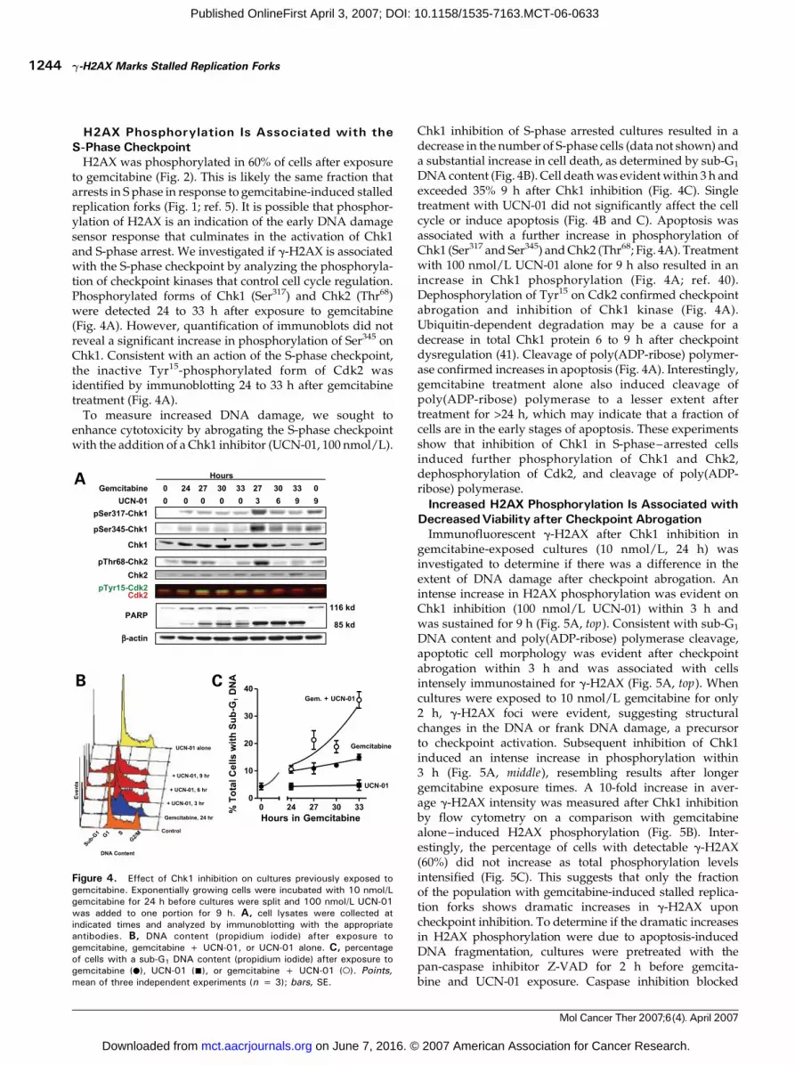
H2AX Phosphorylation Is Associated with theS-Phase CheckpointH2AX was phosphorylated in 60% of cells after exposure
to gemcitabine (Fig. 2). This is likely the same fraction thatarrests in S phase in response to gemcitabine-induced stalledreplication forks (Fig. 1; ref. 5). It is possible that phosphor-ylation of H2AX is an indication of the early DNA damagesensor response that culminates in the activation of Chk1and S-phase arrest. We investigated if g-H2AX is associatedwith the S-phase checkpoint by analyzing the phosphoryla-tion of checkpoint kinases that control cell cycle regulation.Phosphorylated forms of Chk1 (Ser317) and Chk2 (Thr68)were detected 24 to 33 h after exposure to gemcitabine(Fig. 4A). However, quantification of immunoblots did notreveal a significant increase in phosphorylation of Ser345 onChk1. Consistent with an action of the S-phase checkpoint,the inactive Tyr15-phosphorylated form of Cdk2 wasidentified by immunoblotting 24 to 33 h after gemcitabinetreatment (Fig. 4A).To measure increased DNA damage, we sought to
enhance cytotoxicity by abrogating the S-phase checkpointwith the addition of a Chk1 inhibitor (UCN-01, 100 nmol/L).
Chk1 inhibition of S-phase arrested cultures resulted in adecrease in the number of S-phase cells (data not shown) anda substantial increase in cell death, as determined by sub-G1
DNA content (Fig. 4B). Cell deathwas evidentwithin 3 h andexceeded 35% 9 h after Chk1 inhibition (Fig. 4C). Singletreatment with UCN-01 did not significantly affect the cellcycle or induce apoptosis (Fig. 4B and C). Apoptosis wasassociated with a further increase in phosphorylation ofChk1 (Ser317 and Ser345) andChk2 (Thr68; Fig. 4A). Treatmentwith 100 nmol/L UCN-01 alone for 9 h also resulted in anincrease in Chk1 phosphorylation (Fig. 4A; ref. 40).Dephosphorylation of Tyr15 on Cdk2 confirmed checkpointabrogation and inhibition of Chk1 kinase (Fig. 4A).Ubiquitin-dependent degradation may be a cause for adecrease in total Chk1 protein 6 to 9 h after checkpointdysregulation (41). Cleavage of poly(ADP-ribose) polymer-ase confirmed increases in apoptosis (Fig. 4A). Interestingly,gemcitabine treatment alone also induced cleavage ofpoly(ADP-ribose) polymerase to a lesser extent aftertreatment for >24 h, which may indicate that a fraction ofcells are in the early stages of apoptosis. These experimentsshow that inhibition of Chk1 in S-phase–arrested cellsinduced further phosphorylation of Chk1 and Chk2,dephosphorylation of Cdk2, and cleavage of poly(ADP-ribose) polymerase.
Increased H2AX Phosphorylation Is Associated withDecreasedViability after Checkpoint AbrogationImmunofluorescent g-H2AX after Chk1 inhibition in
gemcitabine-exposed cultures (10 nmol/L, 24 h) wasinvestigated to determine if there was a difference in theextent of DNA damage after checkpoint abrogation. Anintense increase in H2AX phosphorylation was evident onChk1 inhibition (100 nmol/L UCN-01) within 3 h andwas sustained for 9 h (Fig. 5A, top). Consistent with sub-G1
DNA content and poly(ADP-ribose) polymerase cleavage,apoptotic cell morphology was evident after checkpointabrogation within 3 h and was associated with cellsintensely immunostained for g-H2AX (Fig. 5A, top). Whencultures were exposed to 10 nmol/L gemcitabine for only2 h, g-H2AX foci were evident, suggesting structuralchanges in the DNA or frank DNA damage, a precursorto checkpoint activation. Subsequent inhibition of Chk1induced an intense increase in phosphorylation within3 h (Fig. 5A, middle), resembling results after longergemcitabine exposure times. A 10-fold increase in aver-age g-H2AX intensity was measured after Chk1 inhibitionby flow cytometry on a comparison with gemcitabinealone–induced H2AX phosphorylation (Fig. 5B). Inter-estingly, the percentage of cells with detectable g-H2AX(60%) did not increase as total phosphorylation levelsintensified (Fig. 5C). This suggests that only the fractionof the population with gemcitabine-induced stalled replica-tion forks shows dramatic increases in g-H2AX uponcheckpoint inhibition. To determine if the dramatic increasesin H2AX phosphorylation were due to apoptosis-inducedDNA fragmentation, cultures were pretreated with thepan-caspase inhibitor Z-VAD for 2 h before gemcita-bine and UCN-01 exposure. Caspase inhibition blocked
Figure 4. Effect of Chk1 inhibition on cultures previously exposed togemcitabine. Exponentially growing cells were incubated with 10 nmol/Lgemcitabine for 24 h before cultures were split and 100 nmol/L UCN-01was added to one portion for 9 h. A, cell lysates were collected atindicated times and analyzed by immunoblotting with the appropriateantibodies. B, DNA content (propidium iodide) after exposure togemcitabine, gemcitabine + UCN-01, or UCN-01 alone. C, percentageof cells with a sub-G1 DNA content (propidium iodide) after exposure togemcitabine (.), UCN-01 (n), or gemcitabine + UCN-01 (o). Points,mean of three independent experiments (n = 3); bars, SE.
g-H2AX Marks Stalled Replication Forks1244
Mol Cancer Ther 2007;6(4). April 2007
on June 7, 2016. © 2007 American Association for Cancer Research. mct.aacrjournals.org Downloaded from
Published OnlineFirst April 3, 2007; DOI: 10.1158/1535-7163.MCT-06-0633

apoptosis-associated nuclear blebbing but did not inhibitincreases in H2AX phosphorylation after checkpoint abro-gation (Fig. 5A, bottom). These results confirm that increasesin g-H2AX after checkpoint abrogation are not due toapoptotic DNA fragmentation.Clonogenic assays were done to determine if the clear
increase in H2AX phosphorylation was associated with adecrease in cellular reproductive viability. Gemcitabine(10 nmol/L, 21 h) alone killed 65% of cells as comparedwith untreated cultures (Fig. 5D). In contrast, a significantdecrease in viability (>80%) occurred when Chk1 wasinhibited in S-phase–arrested cultures for 6 h before cellplating (Fig. 5D). Chk1 inhibition alone did not significant-ly affect viability. A considerable decrease in reproductiveviability also occurred after Chk1 inhibition in culturespreviously exposed to gemcitabine for only 2 h (Fig. 5D),thus showing the value of an intact S-phase checkpoint forovercoming stalled replication forks. These experimentsestablish that nuclear foci formation of g-H2AX is associ-ated with gemcitabine-induced stalled replication forks,whereas a pronounced increase in H2AX phosphorylationis associated with apoptosis.
DiscussionThis study shows that H2AX is phosphorylated andforms distinct nuclear foci in response to nucleoside
analogue– induced stalled replication forks. g-H2AXincreases were most apparent in the S-phase fraction,which is likely due to the S-phase specificity of these drugs(Fig. 2). In particular, cells at the G1-S border were the firstfraction of the total population to be measured as g-H2AXpositive. This possibly reflects that gemcitabine incorpora-tion into DNA most rapidly occurs in cells initiating DNAsynthesis. The same S-phase fraction of cells also showedphosphorylation of ATM on Ser1981, a known regulator ofH2AX and other DNA damage sensors (Fig. 3A). Ongoinginvestigations indicate that g-H2AX colocalizes with DNAdamage repair proteins shortly after nucleoside analogueexposure.2 Inhibition of ATM and related kinases (ATR andDNA-dependent protein kinase) blocked H2AX phosphor-ylation (Fig. 3C), thus suggesting that this family of kinasesparticipates in regulating H2AX phosphorylation aftergemcitabine exposure.Although the details of mammalian replication fork
stabilization are poorly understood, the molecules associ-ated with the S-phase checkpoint likely participate in
2 B. Ewald, D. Sampath, and W. Plunkett. Colocalization of the Mre11-Rad50-Nbs1 complex, phosphorylated ATM, and g-H2AX may identify sitesof nucleoside analogue-induced stalled replication forks. AACR MeetingAbstracts 2007, Abstract #4037, unpublished.
Figure 5. Effect of checkpoint abro-gation on H2AX phosphorylation andcellular reproductive viability. Cellswere incubated with combinations of50 Amol/L Z-VAD, 10 nmol/L gemcita-bine, and 100 nmol/L UCN-01 beforecultures were harvested. A, represen-tative images taken of immunostainedg-H2AX (green ) and 4¶,6-diamidino-2-phenylindole (DAPI) nuclear staining(blue ) after exposure to combinationsof Z-VAD, gemcitabine, and UCN-01.B, cytometric quantitation of H2AXphosphorylation after exposure togemcitabine for 2 h F UCN-01 for 3to 9 h. C, percentage of cells that wereg-H2AX positive after treatment withgemcitabine (.) or gemcitabine +UCN-01 (o), determined by flowcytometry. Points, mean of three inde-pendent experiments (n = 3); bars,SE. D, percentage of cells that formedcolonies (z50 cells) within 10 d after10 nmol/L gemcitabine, 100 nmol/LUCN-01, or gemcitabine + UCN-01.Columns, mean of three independentexperiments (n = 7–9); bars, SE;*, P < 0.001; **, P = 0.006.
Molecular Cancer Therapeutics 1245
Mol Cancer Ther 2007;6(4). April 2007
on June 7, 2016. © 2007 American Association for Cancer Research. mct.aacrjournals.org Downloaded from
Published OnlineFirst April 3, 2007; DOI: 10.1158/1535-7163.MCT-06-0633

maintaining fork structure (7). Downstream of Chk1 kinase,inhibition of Cdk2 leads to a decrease in replicationorigin firing, further protecting DNA replication fidelity(11). Our studies show that H2AX phosphorylationwas associated with S-phase checkpoint activation inresponse to gemcitabine. It is unknown if H2AX partic-ipates in stabilization of stalled replication forks but itwould be an explanation for rapid phosphorylationand nuclear foci formation. Activation of Chk1 kinaseinduced an accumulation of the inactive phosphorylatedform of Cdk2, prompting S-phase arrest and suggestinginhibition of replication origin firing. Abrogation of theS-phase checkpoint by pharmacologic inhibition of Chk1led to a significant increase in DNA damage, as markedby H2AX phosphorylation (Fig. 5). Interestingly, Chk1inhibition caused an increase in H2AX phosphorylationand a decrease in reproductive viability when cultureswere previously exposed to gemcitabine for only 2 h. Thissuggests that the S-phase checkpoint may be activated inresponse to gemcitabine-induced DNA damage as earlyas 2 h. The increased DNA damage after checkpointabrogation is not due to apoptotic DNA fragmentation(Fig. 5A, bottom) and is likely caused by irreversiblereplication fork collapse, thus explaining a decrease inreproductive viability (Fig. 5D). An earlier study alsoshowed increased H2AX phosphorylation after exposureto UCN-01 of cells previously treated with the topoisomer-ase I inhibitor camptothecin (42). Together, these resultsconfirm the importance of an intact S-phase checkpoint forreplication fork stabilization and cellular recovery fromDNA-targeted therapeutics.Although UCN-01 is known to have inhibitory activity
against a number of kinases, the biological context ofthe present investigations implicates Chk1 as its targetkinase. For instance, the IC50 of Chk1 (5–30 nmol/L;refs. 43, 44) is one of the lowest inhibitory values thathave been reported. Two other kinases with low IC50sinclude protein kinase C (5 nmol/L; ref. 45) and phosphoi-nositide-dependent kinase 1 (33 nmol/L; ref. 46). However,when used alone, UCN-01 did not affect phosphorylationof Akt, a target of phosphoinositide-dependent kinase 1, inexponentially growing ML-1 cells (15), nor was H2AXphosphorylation induced (data not shown; ref. 42). Inaddition, inhibition of protein kinase C by the specificinhibitor bisindolylmaleimide (GFX) did not abrogate thegemcitabine-induced S-phase checkpoint (data not shown).Other potential UCN-01 targets that are involved in the cellcycle have significantly higher IC50s than concentrations usedin this study, including Cdk1 (1 Amol/L; ref. 47), Cdk2(>500 nmol/L; ref. 48), and Chk2 (1 Amol/L; ref. 43). Thus,the evidence indicates that the increased H2AX phosphory-lation during checkpoint abrogation after exposure to UCN-01 is due to the inhibition of Chk1.Nucleoside analogue exposure caused the formation
of g-H2AX nuclear foci at sites of stalled replication forksthat resembled those that accumulate in response toionizing radiation. Recently, Marti et al. (35) have reportedthat UV-C irradiation induces H2AX phosphorylation,
which is seen as a diffuse, even, and pan-nuclear stain-ing, but is not associated with double-strand breaks orapoptosis. They suggested that H2AX phosphorylation inresponse to UV-C may be associated with the excision of6-4 photoproducts. We found that a similar, distinct g-H2AX pan-nuclear staining occurs during checkpointabrogation, but did not occur due to stalled DNA repli-cation (Fig. 5A). In contrast to UV studies, the dramaticg-H2AX increases measured here were associated with adecrease in clonogenic survival but were not caused byapoptosis-induced DNA fragmentation. This links elevat-ed H2AX phosphorylation with a decreased capacity forcellular reproduction after checkpoint abrogation. Otherreports have made similar correlations with g-H2AX andapoptosis (27, 49). Therefore, intense pan-nuclear stainingof g-H2AX may signal that either DNA breaks areprevalent throughout the nucleus or DNA structure hasbeen significantly compromised. However, it may onlyinsinuate lethality under certain conditions. The phos-phorylation increases in this study were likely due toirreparable collapsed replication forks that were the causefor decreased clonogenic survival. Interestingly, thefraction of cells with measurable H2AX phosphorylationdid not increase upon checkpoint abrogation (Fig. 5C),suggesting that Chk1 inhibition specifically kills cells withan activated S-phase checkpoint. This occurred after only a2-h exposure to gemcitabine, indicating that such phar-macologic interaction with checkpoint function can gener-ate lethal damage.H2AX phosphorylation after gemcitabine exposure was
examined in this study. Other deoxycytidine nucleosideanalogues with slightly different mechanisms of action,1-h-D-arabinofuranosylcytosine and troxacitabine, inducedsimilar levels of H2AX phosphorylation. Recent reportshave shown that H2AX is phosphorylated on DNA poly-merase and ribonucleotide reductase inhibition by aphidi-colin and hydroxyurea, respectively (33, 50). Together, thoseinvestigations and this study suggest that H2AX is phos-phorylated in response to agents that inhibit DNA synthesis.Thus, g-H2AX may be useful for detection of pharmacody-namics of these drugs in the clinic. Measurement of H2AXphosphorylation by flow cytometry offers a novel techniquefor assessing nucleoside analogue DNA incorporation inprimary samples inwhich there is a low incidence of cells in Sphase (15). It may also offer insight into whether the drugsreach the tumor, are activated to their functional forms, andefficiently affect DNA synthesis.The present results showed that 20 Amol/L gemcitabine
exposure failed to increase H2AX phosphorylation abovethose achieved upon treatment with 100 nmol/L drug(Fig. 2). In addition, log-order apoptosis-associated in-creases in g-H2AX occurred on checkpoint dysregulationin cultures treated with gemcitabine for only 2 h (Fig. 5),suggesting that prolonged exposure to nucleoside ana-logues is unnecessary to kill the S-phase fraction by thiscombinational strategy. Together, these results indicatethat low-dose intermittent nucleoside analogue adminis-tration may be sufficient for S-phase checkpoint activation
g-H2AX Marks Stalled Replication Forks1246
Mol Cancer Ther 2007;6(4). April 2007
on June 7, 2016. © 2007 American Association for Cancer Research. mct.aacrjournals.org Downloaded from
Published OnlineFirst April 3, 2007; DOI: 10.1158/1535-7163.MCT-06-0633

and suggest a rationale for combinations with agents thatdysregulate such defense mechanisms. Future studiesaimed at a better understanding of the molecular mecha-nisms involved in recognizing nucleoside analogue–induced DNA damage may lead to identification of novelmolecular targets that cause nucleoside analogue drugresistance.
References
1. Sampath D, Rao VA, Plunkett W. Mechanisms of apoptosis inductionby nucleoside analogs. Oncogene 2003;22:9063–74.
2. Kufe DW, Major PP, Egan EM, Beardsley GP. Correlation of cytotoxicitywith incorporation of ara-C into DNA. J Biol Chem 1980;255:8997–900.
3. Huang P, Chubb S, Hertel LW, Grindey GB, Plunkett W. Action of 2¶,2¶-difluorodeoxycytidine on DNA synthesis. Cancer Res 1991;51:6110–7.
4. Zhang YW, Hunter T, Abraham RT. Turning the replication checkpointon and off. Cell Cycle 2006;5:125–8.
5. Shi Z, Azuma A, Sampath D, Li YX, Huang P, Plunkett W. S-Phase arrestby nucleoside analogues and abrogation of survival without cell cycleprogression by 7-hydroxystaurosporine. Cancer Res 2001;61:1065–72.
6. Sampath D, Shi Z, Plunkett W. Inhibition of cyclin-dependent kinase 2by the Chk1-25A pathway during the S-phase checkpoint activated byfludarabine: dysregulation by 7-hydroxystaurosporine. Mol Pharmacol2002;62:680–8.
7. Lopes M, Cotta-Ramusino C, Pellicioli A, et al. The DNA replicationcheckpoint response stabilizes stalled replication forks. Nature 2001;412:557–61.
8. Huang P, Chubb S, Plunkett W. Termination of DNA synthesis by 9-h-D-arabinofuranosyl-2-fluoroadenine. A mechanism for cytotoxicity. J BiolChem 1990;265:16617–25.
9. Chen Y, Sanchez Y. Chk1 in the DNA damage response: conservedroles from yeasts to mammals. DNA Repair (Amst) 2004;3:1025–32.
10. Busino L, Chiesa M, Draetta GF, Donzelli M. Cdc25A phosphatase:combinatorial phosphorylation, ubiquitylation and proteolysis. Oncogene2004;23:2050–6.
11. Mailand N, Diffley JF. CDKs promote DNA replication origin licensingin human cells by protecting Cdc6 from APC/C-dependent proteolysis. Cell2005;122:915–26.
12. Kohn EA, Ruth ND, Brown MK, Livingstone M, Eastman A.Abrogation of the S phase DNA damage checkpoint results in S phaseprogression or premature mitosis depending on the concentration of 7-hydroxystaurosporine and the kinetics of Cdc25C activation. J Biol Chem2002;277:26553–64.
13. Arlander SJ, Eapen AK, Vroman BT, McDonald RJ, Toft DO, KarnitzLM. Hsp90 inhibition depletes Chk1 and sensitizes tumor cells toreplication stress. J Biol Chem 2003;278:52572–7.
14. Shao RG, Cao CX, Pommier Y. Abrogation of Chk1-mediated S/G2
checkpoint by UCN-01 enhances ara-C-induced cytotoxicity in humancolon cancer cells. Acta Pharmacol Sin 2004;25:756–62.
15. Sampath D, Cortes J, Estrov Z, et al. Pharmacodynamics of cytarabinealone and in combination with 7-hydroxystaurosporine (UCN-01) in AMLblasts in vitro and during a clinical trial. Blood 2006;107:2517–24.
16. Kortmansky J, Shah MA, Kaubisch A, et al. Phase I trial of the cyclin-dependent kinase inhibitor and protein kinase C inhibitor 7-hydroxystaur-osporine in combination with Fluorouracil in patients with advanced solidtumors. J Clin Oncol 2005;23:1875–84.
17. Lara PN, Jr., Mack PC, Synold T, et al. The cyclin-dependent kinaseinhibitor UCN-01 plus cisplatin in advanced solid tumors: a Californiacancer consortium phase I pharmacokinetic and molecular correlative trial.Clin Cancer Res 2005;11:4444–50.
18. Zhou B-BS, Elledge SJ. The DNA damage response: puttingcheckpoints in perspective. Nature 2000;408:433–9.
19. Bakkenist CJ, Kastan MB. DNA damage activates ATM throughintermolecular autophosphorylation and dimer dissociation. Nature 2003;421:499–506.
20. Bekker-Jensen S, Lukas C, Kitagawa R, et al. Spatial organization ofthe mammalian genome surveillance machinery in response to DNA strandbreaks. J Cell Biol 2006;173:195–206.
21. Burma S, Chen BP, Murphy M, Kurimasa A, Chen DJ. ATM
phosphorylates histone H2AX in response to DNA double-strand breaks.J Biol Chem 2001;276:42462–7.
22. Fernandez-Capetillo O, Lee A, Nussenzweig M, Nussenzweig A. H2AX:the histone guardian of the genome. DNA Repair (Amst) 2004;3:959–67.
23. Rogakou EP, Boon C, Redon C, Bonner WM. Megabase chromatindomains involved in DNA double-strand breaks in vivo . J Cell Biol 1999;146:905–16.
24. Rogakou EP, Pilch DR, Orr AH, Ivanova VS, Bonner WM. DNA double-stranded breaks induce histone H2AX phosphorylation on serine 139.J Biol Chem 1998;273:5858–68.
25. Downey M, Durocher D. gH2AX as a checkpoint maintenance signal.Cell Cycle 2006;5:1376–81.
26. Xie A, Puget N, Shim I, et al. Control of sister chromatidrecombination by histone H2AX. Mol Cell 2004;16:1017–25.
27. Mukherjee B, Kessinger C, Kobayashi J, et al. DNA-PK phosphorylateshistone H2AX during apoptotic DNA fragmentation in mammalian cells.DNA Repair (Amst) 2006;5:575–90.
28. Lu C, Zhu F, Cho YY, et al. Cell apoptosis: requirement of H2AX inDNA ladder formation, but not for the activation of caspase-3. Mol Cell2006;23:121–32.
29. Chowdhury D, Keogh MC, Ishii H, Peterson CL, Buratowski S,Lieberman J. g-H2AX dephosphorylation by protein phosphatase 2Afacilitates DNA double-strand break repair. Mol Cell 2005;20:801–9.
30. Bassing CH, Suh H, Ferguson DO, et al. Histone H2AX: a dosage-dependent suppressor of oncogenic translocations and tumors. Cell 2003;114:359–70.
31. Celeste A, Petersen S, Romanienko PJ, et al. Genomic instability inmice lacking histone H2AX. Science 2002;296:922–7.
32. Bassing CH, Chua KF, Sekiguchi J, et al. Increased ionizing radiationsensitivity and genomic instability in the absence of histone H2AX. ProcNatl Acad Sci U S A 2002;99:8173–8.
33. Ward IM, Chen J. Histone H2AX is phosphorylated in an ATR-dependent manner in response to replicational stress. J Biol Chem 2001;276:47759–62.
34. Olive PL, Banath JP, Sinnott LT. Phosphorylated histone H2AX inspheroids, tumors, and tissues of mice exposed to etoposide and 3-amino-1,2,4-benzotriazine-1,3-dioxide. Cancer Res 2004;64:5363–9.
35. Marti TM, Hefner E, Feeney L, Natale V, Cleaver JE. H2AXphosphorylation within the G1 phase after UV irradiation depends onnucleotide excision repair and not DNA double-strand breaks. Proc NatlAcad Sci U S A 2006;103:9891–6.
36. Karnitz LM, Flatten KS, Wagner JM, et al. Gemcitabine-inducedactivation of checkpoint signaling pathways that affect tumor cell survival.Mol Pharmacol 2005;68:1636–44.
37. Grove KL, Cheng YC. Uptake and metabolism of the new anticancercompound h-L-(�)-dioxolane-cytidine in human prostate carcinoma DU-145 cells. Cancer Res 1996;56:4187–91.
38. Sarkaria J, Tibbetts R, Busby E, Kennedy A, Hill D, Abraham R.Inhibition of phosphoinositide 3-kinase related kinases by the radiosensitiz-ing agent wortmannin. Cancer Res 1998;58:4375–82.
39. Sarkaria JN, Busby EC, Tibbetts RS, et al. Inhibition of ATM and ATRkinase activities by the radiosensitizing agent, caffeine. Cancer Res 1999;59:4375–82.
40. Syljuasen RG, Sorensen CS, Hansen LT, et al. Inhibition of humanChk1 causes increased initiation of DNA replication, phosphorylation ofATR targets, and DNA breakage. Mol Cell Biol 2005;25:3553–62.
41. Zhang YW, Otterness DM, Chiang GG, et al. Genotoxic stress targetshuman Chk1 for degradation by the ubiquitin-proteasome pathway. MolCell 2005;19:607–18.
42. Furuta T, Hayward RL, Meng LH, et al. p21CDKN1A allows the repairof replication-mediated DNA double-strand breaks induced by topoisomer-ase I and is inactivated by the checkpoint kinase inhibitor 7-hydroxystaur-osporine. Oncogene 2006;25:2839–49.
43. Busby EC, Leistritz DF, Abraham RT, Karnitz LM, Sarkaria JN. Theradiosensitizing agent 7-hydroxystaurosporine (UCN-01) inhibits the DNAdamage checkpoint kinase hChk1. Cancer Res 2000;60:2108–12.
44. Jackson JR, Gilmartin A, Imburgia C, Winkler JD, Marshall LA,Roshak A. An indolocarbazole inhibitor of human checkpoint kinase (Chk1)abrogates cell cycle arrest caused by DNA damage. Cancer Res 2000;60:566–72.
45. Takahashi I, Saitoh Y, Yoshida M, et al. UCN-01 and UCN-02, new
Molecular Cancer Therapeutics 1247
Mol Cancer Ther 2007;6(4). April 2007
on June 7, 2016. © 2007 American Association for Cancer Research. mct.aacrjournals.org Downloaded from
Published OnlineFirst April 3, 2007; DOI: 10.1158/1535-7163.MCT-06-0633

selective inhibitors of protein kinase C. II. Purification, physico-chemicalproperties, structural determination and biological activities. J Antibiot(Tokyo) 1989;42:571–6.
46. Sato S, Fujita N, Tsuruo T. Interference with PDK1-Akt survivalsignaling pathway by UCN-01 (7-hydroxystaurosporine). Oncogene 2002;21:1727–38.
47. Wang Q, Worland PJ, Clark JL, Carlson BA, Sausville EA. Apoptosisin 7-hydroxystaurosporine-treated T lymphoblasts correlates with activa-tion of cyclin-dependent kinases 1 and 2. Cell Growth Differ 1995;6:927–36.
48. Akiyama T, Yoshida T, Tsujita T, et al. G1 phase accumulation
induced by UCN-01 is associated with dephosphorylation of Rb and CDK2proteins as well as induction of CDK inhibitor p21/Cip1/WAF1/Sdi1 inp53-mutated human epidermoid carcinoma A431 cells. Cancer Res 1997;57:1495–501.
49. Huang X, Halicka HD, Traganos F, Tanaka T, Kurose A, DarzynkiewiczZ. Cytometric assessment of DNA damage in relation to cell cycle phaseand apoptosis. Cell Prolif 2005;38:223–43.
50. Kurose A, Tanaka T, Huang X, Traganos F, Dai W, Darzynkiewicz Z.Effects of hydroxyurea and aphidicolin on phosphorylation of ataxia telan-giectasia mutated on Ser 1981 and histone H2AX on Ser 139 in relation tocell cycle phase and induction of apoptosis. Cytometry A 2006;69:212–21.
g-H2AX Marks Stalled Replication Forks1248
Mol Cancer Ther 2007;6(4). April 2007
on June 7, 2016. © 2007 American Association for Cancer Research. mct.aacrjournals.org Downloaded from
Published OnlineFirst April 3, 2007; DOI: 10.1158/1535-7163.MCT-06-0633

2007;6:1239-1248. Published OnlineFirst April 3, 2007.Mol Cancer Ther Brett Ewald, Deepa Sampath and William Plunkett abrogation
checkpointreplication forks and their collapse upon S-phase H2AX phosphorylation marks gemcitabine-induced stalled
Updated version
10.1158/1535-7163.MCT-06-0633doi:
Access the most recent version of this article at:
Material
Supplementary
http://mct.aacrjournals.org/content/suppl/2010/03/18/1535-7163.MCT-06-0633.DC1.html
Access the most recent supplemental material at:
Cited articles
http://mct.aacrjournals.org/content/6/4/1239.full.html#ref-list-1
This article cites 50 articles, 28 of which you can access for free at:
Citing articles
http://mct.aacrjournals.org/content/6/4/1239.full.html#related-urls
This article has been cited by 20 HighWire-hosted articles. Access the articles at:
E-mail alerts related to this article or journal.Sign up to receive free email-alerts
Subscriptions
Reprints and
To order reprints of this article or to subscribe to the journal, contact the AACR Publications
Permissions
To request permission to re-use all or part of this article, contact the AACR Publications
on June 7, 2016. © 2007 American Association for Cancer Research. mct.aacrjournals.org Downloaded from
Published OnlineFirst April 3, 2007; DOI: 10.1158/1535-7163.MCT-06-0633


![The Vital Role of Polymerase and REV1 in Mutagenic, but Not Correct, DNA Synthesis across Benzo[a]pyrene-dG and Recruitment of Polymerase by REV1 to Replication-stalled Site](https://img.dokumen.tips/doc/110x75/635cb39187785f44260649af/the-vital-role-of-polymerase-and-rev1-in-mutagenic-but-not-correct-dna-synthesis.jpg)


 using quartz tuning forks](https://img.dokumen.tips/doc/110x75/63411b1fd195fa2a1f0e7d62/characterization-of-the-magnetic-properties-of-fehtrz-2-trzbf-4-using-quartz.jpg)


