Embed Size (px)
Citation preview

GENERAL AND COMPARATIVE
ENDOCRINOLOGY
www.elsevier.com/locate/ygcen
General and Comparative Endocrinology 135 (2004) 1–16
Minireview
Evolutionary aspects of GnRHs, GnRH neuronal systemsand GnRH receptors in teleost fish
Christ�eele Lethimonier,a Thierry Madigou,a Jos�ee-Antonio Mu~nnoz-Cueto,b
Jean-Jacques Lareyre,c and Olivier Kaha,*
a Endocrinologie Mol�eeculaire de la Reproduction, UMR CNRS 6026, 35042 Rennes cedex, Franceb Departamento de Biologia, Facultad de Ciencias del Mar y Ambientales, Puerto Real, Cadiz, Spain
c INRA SCRIBE Universit�ee de Rennes 1, Campus de Beaulieu, Rennes, France
Accepted 14 October 2003
Abstract
Gonadotrophin-releasing hormone (GnRH) was originally believed to be released by a unique set of hypophysiotrophic neurons
to stimulate the release of gonadotrophins from the pituitary, therefore acting as a major initiator of the hormonal cascade con-
trolling the reproductive axis. However, it now appears that each vertebrate species expresses two or three GnRH forms in multiple
tissues and that GnRHs exert pleiotropic actions via several classes of receptors. This new vision of the GnRH systems arose
progressively from numerous comparative studies in all vertebrate classes, but fish in general, and teleosts in particular, have often
plaid a leading part in changing established concepts. To date fish still appear as attractive models to decipher the evolutionary
mechanisms that led to the diversification of GnRH functions. Not only do teleosts exhibit the highest variety of GnRH variants,
but recent data and whole genome analyses indicate that they may also possess multiple GnRH receptors. This paper intends to
summarize the current situation with special emphasis on interspecies comparisons which provide insights into the possible evo-
lutionary mechanisms leading to the diversification of GnRH functions.
� 2003 Elsevier Inc. All rights reserved.
Keywords: GnRH; GnRH receptors; Genome analysis; Hypothalamus; Pituitary; Reproduction; Teleost
1. Introduction
Since the pioneer studies of Breton and colleaguesshowing that a hypothalamic factor stimulates the re-
lease of pituitary gonadotropic hormone in carp (Breton
et al., 1971) and the characterization of the first fish
GnRH, salmon GnRH (Sherwood et al., 1983), research
on GnRH in teleost fish has focused increasing attention.
This is not only due to the important potential applica-
tions of GnRH in fish farming (Zohar and Mylonas,
2001), but also to the fact that teleost fish have turnedout to be of special interest to understand the mecha-
nisms underlying the evolution of GnRH genes in ver-
tebrates. In parallel, following the early work of Habibi
et al. (1987) on the binding properties of GnRH in the
* Corresponding author. Fax: +33-2-23-23-67-94.
E-mail address: [email protected] (O. Kah).
0016-6480/$ - see front matter � 2003 Elsevier Inc. All rights reserved.
doi:10.1016/j.ygcen.2003.10.007
goldfish pituitary and the cloning of the first fish GnRH
receptor (GnRH-R) in catfish (Tensen et al., 1997), a
growing number of studies are devoted to fish GnRH-R.These studies have largely contributed to the discovery
that there are several GnRH-R subtypes with differential
structural characteristics and expression sites.
This article intends to review recent data on the
evolution of GnRH forms and neuronal circuits and the
presence of multiple GnRH-R subtypes in teleosts with
special emphasis on evolutionary aspects.
2. Gonadotrophin-releasing hormones
2.1. Eight GnRH variants in teleost fish
The number of GnRH family members in vertebrates
has rapidly increased over the last decade, now reaching
a total of 14 variants, however unique GnRH forms
2 C. Lethimonier et al. / General and Comparative Endocrinology 135 (2004) 1–16
have also been found in prochordates (Adams et al.,2003) and in invertebrates (Iwakoshi et al., 2002). As
GnRH-like peptides have been found in cnidaria con-
sidered as the first animals with neurons and true syn-
apses (Anctil, 2000), it is possible that the origin of the
GnRH family goes back around 600 millions years.
Among vertebrates, teleost fish represent the group ex-
hibiting the highest number of GnRH variants. Fol-
lowing the identification of salmon GnRH (sGnRH;Sherwood et al., 1983), 7 other GnRH forms have been
purified in teleosts by high performance liquid chro-
matography (HPLC) and sequencing of GnRH-immu-
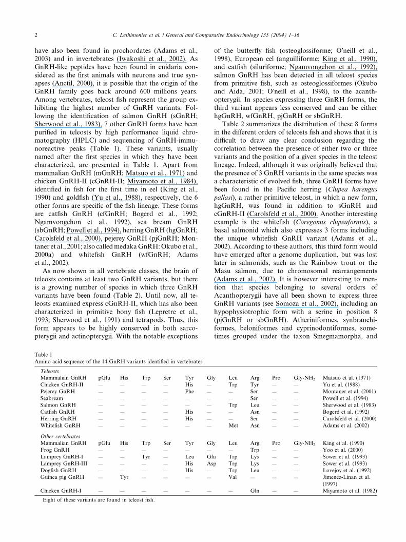
noreactive peaks (Table 1). These variants, usually
named after the first species in which they have been
characterized, are presented in Table 1. Apart from
mammalian GnRH (mGnRH; Matsuo et al., 1971) and
chicken GnRH-II (cGnRH-II; Miyamoto et al., 1984),identified in fish for the first time in eel (King et al.,
1990) and goldfish (Yu et al., 1988), respectively, the 6
other forms are specific of the fish lineage. These forms
are catfish GnRH (cfGnRH; Bogerd et al., 1992;
Ngamvongchon et al., 1992), sea bream GnRH
(sbGnRH; Powell et al., 1994), herringGnRH (hgGnRH;
Carolsfeld et al., 2000), pejerey GnRH (pjGnRH; Mon-
taner et al., 2001; also calledmedakaGnRH:Okubo et al.,2000a) and whitefish GnRH (wfGnRH; Adams
et al., 2002).
As now shown in all vertebrate classes, the brain of
teleosts contains at least two GnRH variants, but there
is a growing number of species in which three GnRH
variants have been found (Table 2). Until now, all te-
leosts examined express cGnRH-II, which has also been
characterized in primitive bony fish (Lepretre et al.,1993; Sherwood et al., 1991) and tetrapods. Thus, this
form appears to be highly conserved in both sarco-
pterygii and actinopterygii. With the notable exceptions
Table 1
Amino acid sequence of the 14 GnRH variants identified in vertebrates
Teleosts
Mammalian GnRH pGlu His Trp Ser Tyr Gl
Chicken GnRH-II — — — — His —
Pejerey GnRH — — — — Phe —
Seabream — — — — — —
Salmon GnRH — — — — — —
Catfish GnRH — — — — His —
Herring GnRH — — — — His —
Whitefish GnRH — — — — — —
Other vertebrates
Mammalian GnRH pGlu His Trp Ser Tyr Gl
Frog GnRH — — — — — —
Lamprey GnRH-I — — Tyr — Leu Gl
Lamprey GnRH-III — — — — His As
Dogfish GnRH — — — — His —
Guinea pig GnRH — Tyr — — — —
Chicken GnRH-I — — — — — —
Eight of these variants are found in teleost fish.
of the butterfly fish (osteoglossiforme; O�neill et al.,1998), European eel (anguilliforme; King et al., 1990),
and catfish (siluriforme; Ngamvongchon et al., 1992),
salmon GnRH has been detected in all teleost species
from primitive fish, such as osteoglossiformes (Okubo
and Aida, 2001; O�neill et al., 1998), to the acanth-
opterygii. In species expressing three GnRH forms, the
third variant appears less conserved and can be either
hgGnRH, wfGnRH, pjGnRH or sbGnRH.Table 2 summarizes the distribution of these 8 forms
in the different orders of teleosts fish and shows that it is
difficult to draw any clear conclusion regarding the
correlation between the presence of either two or three
variants and the position of a given species in the teleost
lineage. Indeed, although it was originally believed that
the presence of 3 GnRH variants in the same species was
a characteristic of evolved fish, three GnRH forms havebeen found in the Pacific herring (Clupea harengus
pallasi), a rather primitive teleost, in which a new form,
hgGnRH, was found in addition to sGnRH and
cGnRH-II (Carolsfeld et al., 2000). Another interesting
example is the whitefish (Coregonus clupeaformis), a
basal salmonid which also expresses 3 forms including
the unique whitefish GnRH variant (Adams et al.,
2002). According to these authors, this third form wouldhave emerged after a genome duplication, but was lost
later in salmonids, such as the Rainbow trout or the
Masu salmon, due to chromosomal rearrangements
(Adams et al., 2002). It is however interesting to men-
tion that species belonging to several orders of
Acanthopterygii have all been shown to express three
GnRH variants (see Somoza et al., 2002), including an
hypophysiotrophic form with a serine in position 8(pjGnRH or sbGnRH). Atheriniformes, synbranchi-
formes, beloniformes and cyprinodontiformes, some-
times grouped under the taxon Smegmamorpha, and
y Leu Arg Pro Gly-NH2 Matsuo et al. (1971)
Trp Tyr — — Yu et al. (1988)
— Ser — — Montaner et al. (2001)
— Ser — — Powell et al. (1994)
Trp Leu — — Sherwood et al. (1983)
— Asn — — Bogerd et al. (1992)
— Ser — — Carolsfeld et al. (2000)
Met Asn — — Adams et al. (2002)
y Leu Arg Pro Gly-NH2 King et al. (1990)
— Trp — — Yoo et al. (2000)
u Trp Lys — — Sower et al. (1993)
p Trp Lys — — Sower et al. (1993)
Trp Leu — — Lovejoy et al. (1992)
Val — — — Jimenez-Linan et al.
(1997)
— Gln — — Miyamoto et al. (1982)

Table 2
Distribution of GnRH variants in the brain of teleost fish
Tel POA MT Reference
Anguilliformes
Eel Anguilla anguilla m m cII Montero et al. (1994)
Pantodonidae
Butterfly fish Pantodon buchholzi m? m? cII? O�neill et al. (1998)Osteoglossiformes
Arowana Scleropages jardini s? s? cII? Okubo et al. (2001)
Clupeiformes
Herring Clupea harangus s? hg? cII? Carolsfeld et al. (2000)
Cypriniformes
Goldfish Carassius auratus s s cII Yu et al. (1988)
Zebrafish Danio rerio s s cII Powell et al. (1996)
Siluriformes
African catfish Clarias gariepinus cf cf cII Zandbergen et al. (1995)
Characiformes
Pacu Piaractus mesopotamicus s sb cII Powell et al. (1997)
Salmoniformes
Whitefish Coregonus clupeaformis s? wf? cII? Adams et al. (2000)
Rainbow trout Oncorhynchus mykiss s s cII Okuzawa et al. (1990)
Masu salmon Oncorhynchus masou s s cII Amano et al. (1991)
Atheriniformes
Pejerey Odonthestes bonariensis s pj cII Montaner et al. (2001)
Synbranchiformes
Swamp eel Synbranchus marmoratus s? pj? cII? Somoza et al. (2002)
Beloniformes
Medaka Oryzias latipes s pj cII Okubo et al. (2000a,b)
Cyprinodontiformes
Latyfish Xiphophorus maculatus s? pj? cII? Somoza et al. (2002)
Scorpaeniformes
Grass rockfish Sebastes rastrelliger s? sb? cII Powell et al. (1996)
Perciformes
African cichlid Haplochromis burtoni s sb cII White et al. (1995)
Seabream Sparus aurata s sb cII Gothilf et al. (1996)
Striped bass Morone saxatilis s sb cII Chow et al. (1998)
Sea bass Dicentrarchus labrax s sb cII Gonzalez-Martinez et al. (2001)
Red seabream Pagrus major s sb cII Senthilkumaran et al. (1999)
Tilapia Oreochromis mossambicus
Pleuronectiformes
Barfin flounder Verasper moseri s sb cII Amano et al. (2002)
Turbot Scophthalmus maximus s sb cII Andersson et al. (2001)
Tetraodontiformes
Torafugu Fugu rubripes s? sb? cII? Aparicio et al. (2002); this study
For clarity purpose, three main populations (olfactory bulbs+ telencephalon: Tel, preoptic area: POA and midbrain tegmentum: MT) have been
considered. Question marks refer to studies in which the distribution has not been formally established from neuroanatomical studies, but can be
predicted on the basis of comparison with other species and phylogenetical analysis.
C. Lethimonier et al. / General and Comparative Endocrinology 135 (2004) 1–16 3
Scorpaeniformes were found to have pjGnRH (Somoza
et al., 2002), whereas perciformes and pleuronectiformes
are until now characterized by the presence of sbGnRH.
To date, no GnRH has been purified in tetraodonti-formes, but a BLAST search performed on the torafugu
genome (http://www.fugu-sg.org/) indicates the presence
of sGnRH (SINFRUG00000081202; Scaffold 7),
sbGnRH (SINFRUP00000080578; Scaffold 137), and
cGnRH-II (SINFRUG00000062489; Scaffold 119).
2.2. Phylogenetic analysis of preproGnRH
Considering the number of GnRH variants in fish
and other vertebrates, one of the major issues concerns
the phylogenetic relationships between these different
variants. Because GnRHs are short peptides, it is thus
necessary to consider the sequence of their precursor,
the preproGnRH. All GnRHs are issued from a largeprecursor which includes a signal peptide (around 20–25
residues), the biologically active GnRH sequence, a
processing tripeptide (Gly–Lys–Arg) and the GnRH-
associated peptide (GAP; around 40–50 residues). The
cDNAs corresponding to the two or three GnRH vari-
ants have been cloned in a number of species allowing
sequence comparisons to be made. Such an analysis was
performed by Okubo et al. (2000a), who cloned twocDNAs corresponding to sGnRH and cGnRH-II in the
arowana (Scleropages jardini), belonging to the order

4 C. Lethimonier et al. / General and Comparative Endocrinology 135 (2004) 1–16
osteoglossiforme, one of the most primitive teleost or-ders. This analysis using the neighbor-joining method,
clearly shows three branches, two of which encompass-
ing sequences corresponding to a unique GnRH variant.
One includes all precursors of cGnRH-II, while another
contains all sequences corresponding to preprosGnRH.
The third branch includes sequences encoding eel
mGnRH, medaka pjGnRH, perciform sbGnRH, and
catfish cfGnRH (Okubo et al., 2001), all known forcorresponding to the main hypophysiotrophic form in
these species (see below). An interesting observation
resulting from such analyses is that each branch includes
species representative of basal, intermediate, and mod-
ern teleosts, which would indicate that these three
branches are ancient and evolved before the diversifi-
cation of teleosts. Unfortunately, the cDNA sequences
encoding wfGnRH and hgGnRH are not presentlyavailable.
When sequences encoding preproGnRHs from other
vertebrate classes are included in the analysis, there are
again three main branches (Fig. 1), as already shown
by White et al. (1998) who proposed to name them
type 1, 2, and 3. One contains hypophysiotrophic
variants, such as frog GnRH, guinea pig GnRH,
mammalian mGnRH in addition to a number of fishhypophysiotrophic variants (type 1). Another branch
clusters all cGnRH-II from mammals and fish (type 2),
and a third one includes only the fish preprosGnRH
(type 3). It is interesting to mention that lamprey
GnRH-I shows higher identity to prepro chicken
GnRH-II suggesting that they share a common origin.
As already stated by White et al. (1998), each of these
three branches correspond to forms with distinct ex-pression patterns and probably different biological
properties. Further evidence that type 1 and type 2 fish
GnRH genes are orthologs to human mGnRH and
cGnRH-II, respectively, was obtained from character-
ization of GnRH loci in the medaka and human ge-
nomes (Okubo et al., 2002). The fact that branches 1
and 2 contain sequences from both fish and land ver-
tebrates indicate that these branches are ancient andemerged before the divergence of these groups. Branch
3 includes only sGnRH fish sequences and this could
mean either that the corresponding gene has been lost
in land vertebrates or remains to be found. Alterna-
tively, it is possible that the gene duplication that gave
birth to this branch occurred within the fish lineage,
but in this case one would expect this branch to cluster
with one of the two other fish branches (White et al.,1998). Based on this analysis, it is surprising that no
type 1 GnRH sequence has been found in cyprinifor-
mes (ostariophysii) or some salmoniformes (protac-
anthopterygii). The fact that the whitefish, a basal
salmonid has three forms tends to indicate that indeed
some salmonids have lost one gene, or that it remains
to be found (Adams et al., 2002).
2.3. Organization of GnRH systems in the brain of teleost
fish
Another mean to find relationships between GnRH
variants is to look at their sites of expression as an in-
dication of their potential function. The assumption is
that ortholog genes will share common expression pat-
terns. A considerable amount of work has been devoted
to the identification and localization of GnRH-ex-pressing neurons in the brain of fish using immunohis-
tochemistry or, more recently, in situ hybridization.
Early studies in goldfish, using sGnRH antibodies, had
shown that GnRH neurons are distributed in the ante-
rior ventral brain from the olfactory nerves to the me-
diobasal hypothalamus (Kah et al., 1986). Small cell
clusters or isolated neurons were found in the olfactory
nerves and bulbs, ventral telencephalon, preoptic areaalong a tract of GnRH-immunoreactive fibers running
to the anterior pituitary. A small number of isolated
neurons were also found along these fibers in the me-
diobasal hypothalamus. An additional population of
large sGnRH-immunoreactive neurons was also identi-
fied in the synencephalon and immunoreactive fibers
were widely present in many brain regions including in
the spinal cord (Kah et al., 1986). Such a pattern oforganization was then found with minor variations in
many, if not all, teleost species studied, and three main
groups of GnRH neurons are usually recognized, al-
though this is a simplified view of a more complex sit-
uation: a clearly identified population of large GnRH
neurons in the tegmentum of the midbrain (MT), an
anterior telencephalic population including the terminal
nerve associated neurons (Tel), and a third populationin the caudal telencephalon-preoptic region (POA).
What has emerged from recent studies is that these
different populations express two or three GnRH vari-
ants depending on the species (Table 2 and Fig. 2).
2.3.1. Two GnRH variants
The presence of two GnRH variants in the brain of
a single teleost was first demonstrated in goldfish byYu et al. (1988) who, using HPLC and radio-
immunoassays on microdissected brain regions, showed
that the anterior brain contained more sGnRH and the
posterior brain more cGnRH-II. The first species in
which the differential distribution of two GnRH vari-
ants was demonstrated by means of immunohisto-
chemistry using specific antibodies to sGnRH and
cGnRH-II was the Masu salmon (Amano et al., 1991).It was found that neurons of the anterior ventral brain
(Tel + POA) were immunoreactive to sGnRH whereas
the large cell bodies of the synencephalon were positive
with antibodies against cGnRH-II (Fig. 2). This latter
cGnRH-II population was subsequently described in
many other teleost species (see Table 2) and other
vertebrate classes.

Fig. 1. Phylogenetic tree for preproGnRHs in vertebrates showing three main branches in GnRH evolution; one clustering all sGnRH from fish (3),
another including cGnRH-II from fish and mammals (2), and the third one with hypophysiotrophic forms GnRH forms from fish amphibian and
mammals (1). The tree was generated using clustalW based on the neighbor-joining method. The scale bar corresponds to estimated evolutionary
distance units. Sequence access numbers: African catfish (Clarias gariepinus): cf GnRH1 (X78049), cfGnRH2 (X78048), cGnRH-II (X78047);
arowana (Scleropages jardinii): sGnRH (AB047325), cGnRH-II (AB047326); cichlid (Haplochromis burtoni): sbGnRH (U31865), cGnRH-II
(L27435), sGnRH: (S63657); eel (Anguilla japonica) mGnRH (AB026989), cGnRHII (AB026990); frog (Rana dybowskii) GnRH (AF139911);
goldfish (Carassius auratus): sGnRH1 (AB017271), sGnRH2 (AB017272), cGnRH-II1 (U30386), cGnRH-II2 (U40567); guinea pig (Cavia porcellus)
gpGnRH (AF426176); human (Homo sapiens) mGnRH: (X01059), cGnRH-II: (AF036329); lamprey (Petromizon marinus) lGnRH (AF144479);
medaka (Oryzias latipes): sGnRH (AB041335), cGnRH-II (AB041334), mdGnRH (AB041334); musk shrew (Suncus murinus) cGnRH-II
(AF107315); rat (Rattus norvegicus) mGnRH (M15527); rhesus monkey (Macaca mulatta) cGnRH-II (AF097356); salmon (Oncorhynchus nerka)
sGnRH 1 (D31868), sGnRH 2 (D31869); sea bass (Diecentrarchus labrax): sbGnRH (AF224279), cGnRH-II (AF224281), sGnRH (AF224280);
seabream (Sparus aurata): sbGnRH, (U30320), sGnRH, (U30311), cGnRH-II, (U30325); tree shrew (Tupaia glis belangeri) mGnRH (U63326),
cGnRH-II (U63327); zebrafish (Danio rerio) sGnRH (AJ304429).
C. Lethimonier et al. / General and Comparative Endocrinology 135 (2004) 1–16 5
This pattern of organization is found in other species
expressing two GnRH variants, such as the African
catfish (Zandbergen et al., 1995) and the European eel
(Montero et al., 1994), although in these species anterior
neurons (Tel and POA), express cfGnRH and mGnRH,
respectively (Fig. 2). However, although the GnRH

Fig. 2. The main types of organization of the GnRH systems in teleosts. The eel illustrates the situation in which a type 1 GnRH is found together
with a type 2 GnRH (adapted from Montero et al., 1994). This situation is also found in catfish (Dubois et al., 2001). The goldfish reflects a unique
situation where a type 3 and a type 2 forms are found in neurons of the anteroventral brain (adapted from Kim et al., 1995). The Masu salmon is an
example where only a type 3 GnRH is expressed in neurons of the anterior ventral brain (adapted from Amano et al., 1991). The European sea bass is
an example of a species with three GnRH variants (types 1, 2, and 3). In this case, the distribution of sGnRH and sbGnRH neurons overlap in the
forebrain (adapted from Gonzalez-Martinez et al., 2002a). The relative abundance of pituitary GnRH fibers is indicated.
6 C. Lethimonier et al. / General and Comparative Endocrinology 135 (2004) 1–16
forms detected in Tel and POA neurons in catfish or eelbelong to branch 1 of the phylogenetical tree, the one
expressed in these cell populations in salmonids is found
in branch 3. If some salmonids have lost the type 1 gene,
it is then possible that a type 3 GnRH was recruited to
fulfill the hypophysiotropic roles. In the African catfish,
based on differences in temporal, spatial, and morpho-
logic appearance, two distinct cfGnRH populations
were identified in the ventral forebrain: a populationinnervating the pituitary (ventral forebrain system) and
a so-called terminal nerve (TN) population. DiI tracing
studies revealed that the TN population has no neuronal
connections with the pituitary (Dubois et al., 2001), as
already demonstrated by Oka and Matsushima (1993) in
the dwarf gourami.
A unique situation has been reported in the goldfish
(Fig. 2) known to express sGnRH and cGnRH-II (Yuet al., 1988). Indeed, both sGnRH and cGnRH-II-im-
munoreactive neurons are found in the anteroventral
brain (Kim et al., 1995), a result in agreement with
studies based on preproGnRH mRNA detection (Lin
and Peter, 1997). To date the goldfish remains the only
teleost species with cGnRH-II expression outside the
midbrain tegmentum.
2.3.2. Three GnRH variants
The first species in which three GnRH variants,
sGnRH, sbGnRH and cGnRH-II, were characterized is
the gilthead seabream (Powell et al., 1994). Subsequent
studies in other perciformes and pleuronectiformesshowed that these three forms have differential expres-
sion patterns with the newly discovered sbGnRH being
expressed mainly in the POA cells, sGnRH mostly in the
olfactory bulbs, and cGnRH-II in the midbrain teg-
mentum (Amano et al., 2002; Andersson et al., 2001;
Gonzalez-Martinez et al., 2001, 2002a; Gothilf et al.,
1996; Senthilkumaran et al., 1999; White et al., 1995).
The organization of three GnRH systems was studied indetails in the European sea bass by in situ hybridization
and immunohistochemistry with highly specific anti-
bodies against sea bass recombinant GAPs (Gonzalez-
Martinez et al., 2001, 2002a; Zmora et al., 2002). The
salmon GAP immunostaining was mostly detected in
terminal nerve neurons, but also in ventral telencephalic
and preoptic perikarya (Fig. 2). Salmon GAP-immu-
noreactive (ir) fibers were observed mainly in the fore-brain, although sGAP-ir projections were also evident in
the optic tectum, mesencephalic tegmentum, and ventral
rhombencephalon. The pituitary receives a small num-
ber of sGAP-ir fibers. The seabream GAP-ir cells were
mainly detected in the preoptic area. Nevertheless,
sbGAP-ir neurons were also found in olfactory bulbs,
ventral telencephalon, and ventrolateral hypothalamus.
The sbGAP-ir fibers were only observed in the ventralforebrain and massively innervate the pituitary gland
only. Finally, chicken-II GAP immunoreactivity was
only detected in large synencephalic cells, which are the
origin of a profuse innervation reaching the telenceph-

C. Lethimonier et al. / General and Comparative Endocrinology 135 (2004) 1–16 7
alon, preoptic area, hypothalamus, thalamus, pretec-tum, posterior tuberculum, mesencephalic tectum and
tegmentum, cerebellum, and rhombencephalon. How-
ever, no cIIGAP-ir fibers were detected in the hypoph-
ysis. These results showed the overlapping of sGAP- and
sbGAP-expressing cells in the forebrain of the sea bass,
and provided unambiguous information on the distri-
bution of projections of the three different GnRH forms
expressed in the brain of a single species (Gonzalez-Martinez et al., 2001, 2002a; Zmora et al., 2002).
2.4. Embryonic origins of the brain GnRH systems in
teleosts
If it seems clear that mesencephalic cGnRH-II neu-
rons differentiate early from a midbrain germinative
primordium and GnRH neurons of the terminal nervemigrate from the olfactory region, there is currently a
debate concerning the origin of the preoptic GnRH
system in species expressing three GnRH forms. Some
studies indicate that preoptic sbGnRH neurons also
originate from the olfactory region and migrate to their
final position (Gonzalez-Martinez et al., 2002b; White
and Fernald, 1998). This assumption is based on the fact
that sbGnRH neurons are first detectable in the olfac-tory region and then are found more caudally as
development proceeds. Based on the fact that sbGnRH-
expressing cells are first visible in their final position in
the POA, other groups claim that sbGnRH neurons
originate from the anteroventral preoptic area (Parhar,
2002). Given that sbGnRH is the ortholog of the genes
encoding the tetrapod GnRH forms expressed in hyp-
ophysiotrophic neurons, which are well known for dif-ferentiating from the olfactory region, it is very unlikely
that this second hypothesis is viable, unless it is shown
that proliferation markers are expressed in preoptic
GnRH neurons. Of particular interest are data obtained
in the African catfish showing that the cfGnRH TN and
the POA neurons, both originate from the olfactory
region, but at two different periods of development
(Dubois et al., 2001, 2002).In summary, based on the structural identities of
preproGnRHs and their sites of expression, there seems
to be three paralog genes in teleosts, but it appears that
some families have lost one of them (type 3 in eels and
catfish, type 1 in salmonids and cyprinids). However,
based on the very similar overall pattern of distribution
of the GnRH-expressing neurons, one can suggest that
another gene has been recruted to fulfill the roles of themissing form. In this respect, it is interesting to note that
in some perciformes sGnRH neurons send projections
to the pituitary (Gonzalez-Martinez et al., 2002a), in-
dicating that these neurons have retained the capacity to
fullfil hypophysiotropic functions. If it seems clear that
type 1 GnRH is involved in the regulation of pituitary
functions, the precise roles of the type 2 (see Millar, 2002
for review) and type 3 (see Oka, 2002 for review)GnRHs still remain highly enigmatic. Recent data in
tunicates indicate that 2 genes were already present in
protochordates, with characteristics similar to the type 1
and type 2 human genes (Adams et al., 2003). Finally,
GnRH peptides and mRNAs have been found in the
gonads where they may be involved in steroidogenesis,
reinititation of meiosis in females and germ cell prolif-
eration in males (see Pati and Habibi, 2002).
3. Teleost GnRH receptors
The first molecular characterization of a full-length
piscine GnRH-R was described in 1997 in African cat-
fish (Tensen et al., 1997). Since then, cDNA encoding
GnRH-R have been obtained in various teleost speciesincluding goldfish (two receptors; Illing et al., 1999),
rainbow trout (Madigou et al., 2000), African catfish
(two receptors; Bogerd et al., 2002), medaka (two
receptors; Okubo et al., 2001), striped bass (Mo-
rone saxatilis; Alok et al., 2000), amberjack (Seriola
dumerilii; GenBank AJ130876), African cichlid (two
receptors; Robison et al., 2001), European sea bass
(DLA419594), and Japanese eel (Okubo et al., 2000b).Information obtained about these receptors is summa-
rized in Table 3.
3.1. Piscine GnRH-R structure
Analysis of the primary amino acids sequence of fish
GnRH receptors indicates that they belong to the G
protein coupled receptors family (GPCRs). A recentphylogenetic analysis of the GPCRs shows that they
segregate into five subfamilies tentatively termed gluta-
mate, secretin, adhesion, frizzled/TAS2, and rhodopsin
subfamilies (Fredriksson et al., 2003). This latter sub-
family is itself subdivided into four subgroups (a; b; c; d)and many of the peptide hormone receptors, including
the GnRH-R, belong to the beta subgroup. Members of
the GPCRs show three main functional domains thatincludes an N-terminal extracellular domain (30–40 aa),
a large transmembrane domain (280–290 aa), and a
short C-terminal cytoplasmic domain (30–50 aa). The
transmembrane domain is constituted of seven highly
conserved transmembrane alpha helix (TM) that are
required to anchor the receptor to the cell membrane. In
contrast to their mammalian counterparts, fish GnRH-
R show an Asp residue in transmembrane domain 2 thatis conserved among members of the GPCRs family. This
Asp residue, that is converted to an Asn residue in
mammalian GnRH receptors, is required for piscine
receptor function (Blomenr€oohr et al., 2002). Intrigu-
ingly, opposite to other GPCRs, mammalian and fish
GnRH-R have a conserved Asp residue in the trans-
membrane helix 7, but conversion of this residue to an

Table
3
Pharm
acologicalcharacterization,tissuedistribution,andclassificationofknownpiscineGnRH-R
Reference
Receptor
abbreviation
GnRH-R
type
Ligand
selectivity
Tissue
distribution
Anguilliform
es
Eel
Okuboet
al.(2000a,b)
IIND
Pituitary
>brain>testis>eye>olfactory
epithelium
Cypriniform
es
Goldfish
Illinget
al.(1999)
GfA
IIGfA
*:cG
nRH-II>sG
nRH>mGnRH>sbGnRH
Brain
(butonly
GfA
isexpressed
in
ventraltelencephalon)>pituitary
(proxi-
malpars
distalis)>ovary
(interstitialcells
andtheca-granulosa
celllayers)¼liver
GfB
GfB:cG
nRH-II>sG
nRH>mGnRH>sbGnRH
Beloniform
es
Medaka
Okuboet
al.(2001)
GnRH-R
1I
GnRH-R
1:cG
n-
RHI>
¼sG
nRH¼mGnRH¼mdGnRH
ND
GnRH-R
2II
GnRH-R
2*:cG
n-
RHII>sG
nRH>mGnRH>mdGnRH
ND
Siluriform
es
Catfish
Tensenet
al.(1997)
cfGnRH-R
1II
cGnRH-II>cfGnRH
Pituitary
>brain>cerebellum>testis
Bogerdet
al.(2002)
cfGnRH-R
2II
cGnRH-II>cfGnRH¼mGnRH
Brain>ovary
>heart>
testis>cerebel-
lum>pituitary
Salm
oniform
es
Rainbow
trout
Madigouet
al.(2000)
rtGnRH-R
IIND
Brain>testis>ovary
>retina>pituitary
Perciform
es
Sea
bass
Gonzalez-Martinez
etal.
(2002c)
IND
Pituitary
>brain
Seriola
dumerilli
AJ130876
IND
ND
Striped
bass
Aloket
al.(2000)
stbGnRH-R
IcG
nRH-II>mGnRHa>
¼sG
nRH>sbGnRH
Pituitary
>brain>ovary
Astatotilapia
Robisonet
al.(2001)
GnRH-R
1II
cGnRH-II>sG
nRH¼
mGnRH>sbGnRH
Brain>testis>kidney
>
retina>muscle>pituitary
GnRH-R
2I
ND
ND
Asterisk(*)indicatesreceptorsubtypeshowingahigher
cGnRH-IIsensitivityin
thespeciesofinterest.GnRH-R
typerefers
tothephylogeneticalanalysisperform
edin
Fig.4.
8 C. Lethimonier et al. / General and Comparative Endocrinology 135 (2004) 1–16

C. Lethimonier et al. / General and Comparative Endocrinology 135 (2004) 1–16 9
Asn does not change receptor function in transfectedcells. The regions between the alpha helices form intra-
cellular or extracellular loops and have been involved in
signal transduction and ligand selectivity. For instance,
the motif DRXXXI/V located at the cytoplasmic end of
the third TM is involved in signal transduction and re-
ceptor activation. In addition, putative ligand contact
sites are evolutionary well conserved. The N-terminal
domain is less conserved, but contains glycosylationsites that may be required for GnRH-R expression. Fi-
nally, the most striking change in receptor structure
between piscine and mammalian receptors is the pres-
ence of a C-terminal cytoplasmic tail. This tail is re-
quired for piscine GnRH-R function (Blomenr€oohr et al.,2002) and has been involved in the desensitization of a
chimeric receptor by decreasing the rate of internaliza-
tion (Lin et al., 1998).
3.2. Two main GnRH-R types with subtypes
To determine the number of sequences encoding
GnRH receptors in teleosts, we have carried out an in
silico analysis on the whole genome of the tetraodonti-
forme Takifugu rubripes (Aparicio et al., 2002) and
partial zebrafish genome draft (Sanger Institute; ftp://ftp.sanger.ac.uk/pub/yyy). In the fugu, five different loci
showing open reading frames (termed GnRH-R1 to
GnRH-R5) encoding putative GnRH-R were identified
(Fig. 3). In the zebrafish, four open reading frames cor-
responding to putative GnRH-R were retrieved and
termed GnRH-R1 to GnRH-R4 (Fig. 3). Phylogenetical
analysis of these sequences and other known teleost
GnRH-R indicates the presence of two main types,termed type I and type II (Fig. 4). According to this
analysis, 3 of the putative fugu receptors (GnRH-R1,
GnRH-R2, and GnRH-R3) would belong to type I
whereas GnRH-R4 and GnRH-R5 are found with the
type II receptors. Two of the zebrafish sequences cluster
with type I receptors, while the other two are found
among type II. It is interesting to mention that the two
GnRH-R cloned in goldfish and catfish cluster in thesame branch (type II), and thus would appear as sub-
types of type II. In contrast, the two receptors identified
in medaka and African cichlid, two Acanthopterygii, are
found in the two main branches. In addition, a partial
sequence of a European eel GnRH-R (T. Madigou et al.,
unpublished) corresponding to a type I appears different
from that of the Japanese eel type II previously published
(Okubo et al., 2000b). Examination of the sequences oftype I and type II receptors revealed the presence of di-
vergent motifs, notably in transmembrane domains
(TM) 3 and 7 (Fig. 5). Indeed, all type I receptors have a
C/GAFVT motif in TM 3 whereas type II exhibit a
SAFIL type motif. All type I receptors have a DLE-
GKVSHSLTH like sequence at the beginning of TM7,
while type II have a VTPEYmotif at this site. In order to
see if those two types also exist in salmoniformes, wedecided to use primers specific to these motifs in order to
clone a type I GnRH-R in the rainbow trout, a species in
which a type II receptor was already characterized
(Madigou et al., 2000). Accordingly, a type I GnRH-R
sequence could be obtained (Fig. 4), whereas a sequence
corresponding to a type II receptor could be retrieved
using the same strategy in the European sea bass, in
which a type I receptor was already cloned(DLA419594). Thus, it appears that, within the teleost
lineage, two main types of GnRH-R could exist each of
which may include 2 or 3 subtypes as evidenced by the
fugu, the zebrafish, the goldfish or the catfish. At the
moment, examples of species exhibiting those two types
belong to the orders anguilliforme, cypriniforme, sal-
moniforme, beloniforme, perciforme, and tetraodonti-
forme showing a widespread distribution of these twoGnRH-R types. The presence of at least two GnRH-R
types, with possible subtypes, is not restricted to fish
since two GnRH-R have been characterized in mammals
(Millar, 2002) and three in bullfrog (Wang et al., 2001).
The existence of different types and subtypes of
GnRH-R in a single fish species raises the question of
whether these receptors have redundant or distinct
functions. To address this important issue, it is necessaryto combine the information on the gene structure and
regulation, ligand specificity and selectivity, and tissue/
cellular distribution of the different GnRH-R types and
subtypes.
3.3. Genes encoding distinct GnRH-R types show different
genomic organization
The genomic organization of GnRH-R genes has
been determined in medaka (Okubo et al., 2001, 2002),
eel (Okubo et al., 2000b), and in trout (Madigou et al.,
2000; Madigou et al., 2002). The genomic structure of
the fugu, zebrafish, and known GnRH-R genes was
compared with respect to their membership to each
clade (Fig. 6). The exon size and phase is well conserved
within each clade except for the first and last exon due tountranslated regions. Genes encoding Type I GnRH
receptors consist in 3 exons separated by two introns.
The 50 flanking region shows the presence of TATA and
CAAT boxes. This structure has been evolutionary
conserved in human type II GnRH receptor (Neill,
2002). The structure of the genes encoding piscine type
II GnRH-R is more complex. In trout, the use of an
alternative promoter and splicing leads to a gene struc-ture consisting in either three or four exons encoding a
receptor with a classic or shorter N-terminal end, re-
spectively (Madigou et al., 2002). In fugu, we also de-
termined that GnRH-R4 and GnRH-R5 genes were
composed of at least three exons similar to the trout and
eel genes. The medaka type II GnRH-R gene contains
four exons. Interestingly, in the second exon, there is an

Fig. 3. Several genes encoding putative GnRH receptors are present on the Fugu rubripes and zebrafish genomes. Using trout GnRH receptor
(GnRH-R) amino acids sequence as template an in silico search for orthologous genes has been carried out on the recently released F. rubripes
(Aparicio et al., 2002) and zebrafish genome draft using the links http://bahama.jgi-psf.org/fugu/bin/blast.fugu.cgi and http://www.ensembl.
org/Danio_rerio/. Five open reading frames present on the takifugu genome, numbered GnRH-R1 (scaffold 10498), GnRH-R2 (scaffold 1609),
GnRH-R3 (scaffold 4966), GnRH-R4 (scaffold 468), and GnRH-R5 (scaffold 4098), showed 58, 58, 61, 71, and 75% homology to trout GnRH-R,
respectively. Four open reading frame, numbered from GnRH-R1 to GnRH-R4 were identified on the zebrafish genome draft. Multiple amino acids
sequences alignment was carried out using the clustalW algorithm of the BioEdit shareware (Hall, 2002). Black bars indicate putative transmembrane
domains.
10 C. Lethimonier et al. / General and Comparative Endocrinology 135 (2004) 1–16
in frame ATG that corresponds to the putative trout
translation initiation codon ATG-2 found in alterna-
tively spliced messengers (Madigou et al., 2002). The
location of this ATG is also conserved in fugu GnRH-
R4 and GnRH-R5 genes as well as in the eel gene.
However, the use of an alternative splicing in medaka,eel, and fugu type II GnRH-R genes that could result in
a shorter GnRH-R remains to be demonstrated. Finally,
the analysis of GnRH-R transcripts in astatotilapia
suggests a more complex gene structure with five exons
(Robison et al., 2001). In conclusion, type I and type II
GnRH-R gene structures have striking differences in
exon number, exon size, and promoter components.
This observation strengthens the data obtained from the
phylogenetic data presented above which needs to be
completed by gene cluster analyses.
3.4. Ligand for GnRH receptors
The presence of different types and subtypes of
GnRH-R in a single fish species raises the question
whether the different GnRH-related peptides have sim-

Fig. 4. Phylogenic analysis of teleost GnRH-R. The phylogenic analysis
was carried out as described in Fig. 2. Only the region spreading from
TM5 to TM6 was considered in the study. Note that piscine GnRH-R
segregate (arrow) into two main phyla, termed type I and type II
GnRH-R. Sequences Accession number are eel (GnRH-R1: AB041327;
GnRH-R2: Madigou et al., unpublished), sea bass (GnRH-R1:
AJ419594; GnRH-R2: Lethimonier et al., unpublished), trout
(rtGnRH-R1: AJ272116; rtGnRH-R2: Lethimonier et al., unpub-
lished), catfish (GnRH-R1: X97497; GnRH-R2: AF329894), seriola
dumerilii GnRH-R (AJ130876), striped sea bass GnRH-R (AF218841),
Astatotilapia (AY028476), goldfish (gfGnRH-RA: AF121845; GnRH-
RB: AF121846), medaka (mdGnRH-R1: AB057677; mdGnRH-R2:
AB057676), and human GnRH-R2 (NM_057163).
Fig. 5. Type I and type II fish GnRH-Rs show different motifs in transmemb
an in silico analysis of the takifugu (fgGnRH-R) or zebrafish genome drafts.
sea bass (GnRH-R1: AJ419594), trout GnRH-R, catfish (GnRH-R1: X97
striped sea-bass GnRH-R (AF218841), Astatotilapia (AY028476), goldfish
GnRH-R2: AB057676).
C. Lethimonier et al. / General and Comparative Endocrinology 135 (2004) 1–16 11
ilar potency to activate these different GnRH-R. Apharmacological characterization of the GnRH-R types
was performed in different species including catfish
(Bogerd et al., 2002; Tensen et al., 1997), goldfish (Illing
et al., 1999), astatotilapia (Robison et al., 2001), striped
bass (Alok et al., 2000), and medaka (Okubo et al.,
2001). Binding of GnRH to GnRH-R results in the
activation of signal transduction mechanisms involving
G-proteins and membrane bound phospholipases (re-view Klausen et al., 2002). This leads to an increase in
cytosolic diacylglycerol (DAG) and inositol 1,4,5-
triphosphate (IP3) that, in turn, releases calcium from
intracellular stores. Moreover, GnRH-induced response
may also involves other signaling pathways leading to
production of cyclic adenosine monophosphate (cAMP)
and arachidonic acid (Bogerd et al., 2002; Pati and
Habibi, 2002).Only a few binding studies have been carried out on
fish GnRH-Rs (Bogerd et al., 2002; Robison et al., 2001)
and demonstrated that GnRH-Rs bind with different
affinities to different GnRH variants. Direct binding
studies (Bogerd et al., 2002) showed that cGnRH-II
binds to catfish type II GnRH-R with a much higher
affinity compared to other GnRH forms (Ka ¼ 2:18(cfGnRH-R1) to 4.3 nM (cfGnRH-R2) for cGnRH-IIversus Ka ¼ 1 (cfGnRH-R1) to 10 lM (cfGnRH-R2) for
cfGnRH). Competitive binding on type II astatotilapia
GnRH-R1 (Robison et al., 2001) showed similar results
since its binds cGnRH-II (EC50 ¼ 14:2 nM) with a much
rane domains 3 and 7. Asteriks (*) indicate sequences determined from
Eel (GnRH-R1: AB041327; GnRH-R2: Madigou et al., unpublished),
497; GnRH-R2: AF329894), seriola dumerilii GnRH-R (AJ130876),
(GfA: AF121845; GfB: AF121846), medaka (GnRH-R1: AB057677;

Fig. 6. Analysis of the structures of known GnRH-R genes. The in silico search for genes encoding putative fugu or zebrafish GnRH-R has been
carried out as described in Fig. 1 using tblastn algorithm and ORF finder. Our analysis indicates that genes encoding type I GnRH-R have a CAAT
and TATA box in the promoter region and are constituted of three exons interrupted by two introns. All exon/intron boundaries are conserved. The
size of exon 2 (205 nucleotides) is also conserved. Genes corresponding to type II GnRH-R are TATA-box less and constituted of at least three exons.
However, in trout, an alternative promoter usage and splicing has been described leading to an additive upstream exon (Madigou et al., 2002). This
leads to the use of another translation initiation codon (as shown with asterisks) that is conserved in all genes studied. Grey squares represent
transmembrane domains, black boxes: non-coding region; white boxes: coding region. Exon and intron sizes are indicated in bp.
12 C. Lethimonier et al. / General and Comparative Endocrinology 135 (2004) 1–16
higher affinity than sGnRH (EC50 ¼ 2:5lM) or
sbGnRH (EC50 ¼ 30lM). In addition, in all studies
carried out in transfected cells, both receptors are acti-
vated by different GnRH peptides. However, both re-
ceptors have a clear preference in terms of sensitivity for
cGnRH-II, followed by sGnRH and a third endogenousGnRH forms (species specific) when identified (Table 4).
Table 4
Biopotency of the different GnRH forms relative to mGnRH to stimulate IP
R types cGnRH-II sGnR
Medaka R1 Type 1 33 5.3
Medaka R2 Type II 1174 4.8
Goldfish GfA Type II 7000 45.6
Goldfish GfB Type II 838 51
Astatotilapia Type II 2000 2
Catfish GnRH-R1 Type II 1199 ND
Catfish GnRH-R2 Type II 6446 ND
ND: not determined.
Nevertheless, the different types of medaka GnRH-R
show different selectivity for GnRH variants (Okubo
et al., 2001). Medaka GnRH-R2 is particularly sensitive
to cGnRH-II whereas medaka GnRH-R1 shows no
clear preference for any of the GnRH tested. On the
other hand, catfish GnRH-Rs that belong to the samepiscine type II GnRH-R branch show no obvious
production in transfected cells
H Third GnRH form
0.023 (mdGnRH) Okubo et al. (2001)
0.316 (mdGnRH) Okubo et al. (2001)
0.3 (sbGnRH) Illing et al. (1999)
0.28 (sbGnRH) Illing et al. (1999)
0.1 (sbGnRH-R) Robison et al. (2001)
1.94 (cfGnRH) Bogerd et al. (2002)
6.8 (cfGnRH) Bogerd et al. (2002)

C. Lethimonier et al. / General and Comparative Endocrinology 135 (2004) 1–16 13
difference in ligand selectivity (Bogerd et al., 2002).However, in goldfish, GnRH-R subtypes GfA and GfB
have different ligand selectivity (Illing et al., 1999). In
conclusion, the pharmacological characterization of the
different GnRH-R types in a single fish species suggests
that they may have different ligand selectivity, but
whether two subtypes belonging to the same type share
similar ligand-induced potency remains unclear. The fish
lineage-specific gene duplication events may have led todifferent ligand selectivity for GnRH-R subtypes ob-
served in different species.
3.5. Tissue distribution of the GnRH receptors
Altogether GnRH-R genes are expressed widely, but
mostly in reproductive tissues (Table 3). However, dif-
ferences in tissue and/or cellular distribution have beendescribed for each GnRH-R types and subtypes identi-
fied in a single fish species. Sea bass and striped bass
GnRH-R genes belonging to type I are highly expressed
in the pituitary and to a lesser extent in brain and ovary.
In addition, pituitary GnRH-R type I gene expression
increases as sea bass and striped bass sexually mature
(Alok et al., 2000; Gonzalez-Martinez et al., 2002c). In
contrast, the trout GnRH-R (type II) is very poorlyexpressed in the pituitary, but was found in the brain
and the gonads (Madigou et al., 2000, 2002). The tissue
distribution of GnRH-R belonging to type II shows a
more complex picture. Certain fish type II receptors (i.e.,
goldfish GfA, trout rtGnRH-R, catfish cfGnRH-R2,
and astatotilapia GnRH-R1) have been found mainly
expressed in different brain regions (optic tectum, hy-
pothalamus, telencephalon, cerebellum) and moderatelyin pituitary, testis, and retina. In the goldfish, GfA was
shown to have a more widespread distribution than GfB
(Peter et al., 2003). However, although expressed in
brain, other subtypes belonging to this branch are
mostly expressed in pituitary as shown in catfish
(cfGnRH-R1; Bogerd et al., 2002), and eel (Okubo
et al., 2000b). In the goldfish, there is indication that
GfA and GfB could be expressed in gonadotrophs and,to a lesser extent in somatotrophs (Illing et al., 1999). In
the pituitary of tilapia, immunohistochemical studies
strongly suggest that two GnRH-R subtypes, belonging
to type II clade and termed 1A and 1B, show different
spatio-temporal expression patterns in LH containing or
prolactin producing cells, respectively (Parhar et al.,
2002). In addition, the use of a third antibody raised
against a type I piscine GnRH-R (striped bass) indicatesthat this GnRH-R type could be restricted to GH-im-
munoreactive cells.
Finally, there is need for more accurate information
on the expression of GnRH-R types and subtypes in
gonads. In goldfish ovary, GnRH-R (GfA) expression
appears to be restricted to intersticial and theca-granu-
losa cell layers (Illing et al., 1999).
In summary, there is now growing evidence that twodistinct GnRH-R types showing structural differences
and different tissue distribution are expressed in a single
fish species as observed in other vertebrate groups.
However, the evolutionary relationships between mam-
malian and fish GnRH-R remains unclear, mainly be-
cause of diverging sequences. One can expect that
comparison of large genomic regions harboring each
receptor type in fish and mammals will provide newinsights regarding evolution of the GnRH-R family.
In addition, a subsequent amplification of the GnRH-
R genes likely occurred in certain fish lineages. The re-
tention of distinct genes encoding GnRH-R strongly
suggests that they have gained non-redundant functions.
The functional characterization of fish GnRH-R will
require to address and combine different issues including
ligand selectivity, regulation, and spatio-temporal ex-pression patterns of each GnRH-R types and subtypes
within a single fish species. The use of technologies al-
lowing to switch off gene expression either in vivo or in
vitro would also bring new insights on type- and/or
subtype-specific function.
4. Conclusion
There is now evidence for a complex interplay be-
tween several GnRH-like peptides and different GnRH-
R types and subtypes within a single fish species but,
because of the long evolutionary history and the great
diversity of teleost fish, it is difficult at the present stage
to draw strait conclusions with regards to co-evolution
of GnRH peptides and GnRH receptors. Expansion ofgene families in euteleost fish is well documented, but
the mechanisms that generated these events are unclear.
One hypothesis is that an ancient genome duplication
occurred first during the evolution of ray-finned fishes
(Acanthopterygii) followed by subsequent fish lineage-
specific tetraploidization and gene loss events. However,
the importance of the ancestral whole genome duplica-
tion event in the abundance of duplicated genes in fishremains under debate (Taylor et al., 2001). Gene du-
plication is an important strength that triggers genome
and biological evolution. It is believed to provide op-
portunities for evolutionary novelties (Ohno, 1970). One
copy of the duplicated gene may accumulate degenera-
tive mutations leading to acquisition of new gene func-
tions. However, although fish genes seem to accumulate
substitutions at higher rate than mammalian genes(Robinson-Rechavi and Laudet, 2001), this process
known as neo-functionalization is predicted to be a rare
event (Lynch and Conery, 2000). Most of the duplicated
genes are predicted to be silenced or lost in mammals
and in fish (Bailey et al., 1978; Li, 1980; Lynch and
Conery, 2000). Another outcome of gene duplication
may explain retention and functional divergence of the

14 C. Lethimonier et al. / General and Comparative Endocrinology 135 (2004) 1–16
duplicated genes. Degenerative mutations can alter thespatial and/or temporal gene expression pattern and/or
regulation mechanisms of both gene copies so that they
are required to assume the biological function of the
single-copy ancestral gene. This sub-functionalization
process increases the likelihood of duplicated gene re-
tention, but also constitutes a resource for future de-
generative mutations that will lead ultimately to a novel
protein function. Although functional analyses remainto be carried out in fish to determine whether the dif-
ferent GnRH peptides on one hand and cognate recep-
tors on the other hand have redundant biological
function, the emergence of new GnRH systems showing
distinct spatial and temporal expression pattern suggests
that GnRH peptides may have pleiotropic function in
the reproductive physiology of the fish.
Acknowledgments
This work was supported by the European Union
Q5RS-2002-01801 PUBERTIMING.
References
Adams, B.A., Vickers, E.D., Warby, C., Park, M., Fischer, W.H.,
Grey Craig, A., Rivier, J.E., Sherwood, N.M., 2002. Three forms of
gonadotropin-releasing hormone, including a novel form, in a
basal salmonid, Coregonus clupeaformis. Biol. Reprod. 67, 232–
239.
Adams, B.A., Tello, J.A., Erchegyi, J., Warby, C., Hong, D.J.,
Akinsanya, K.O., Mackie, G.O., Vale, W., Rivier, J.E., Sherwood,
N.M., 2003. Six novel gonadotropin-releasing hormones are
encoded as triplets on each of two genes in the protochordate,
Ciona intestinalis. Endocrinology 144, 1907–1919.
Alok, D., Hassin, S., Sampath, K.R, Trant, J.M., Yu, K., Zohar, Y.,
2000. Characterization of a pituitary GnRH-receptor from a
perciform fish, Morone saxatilis: functional expression in a fish cell
line. Mol. Cell. Endocrinol. 168, 65–75.
Amano, M., Oka, Y., Aida, K., Okumoto, N., Kawashima, S.,
Hasegawa, Y., 1991. Immunocytochemical demonstration of
salmon GnRH and chicken GnRH-II in the brain of masu salmon,
Oncorhynchus masou. J. Comp. Neurol. 314, 587–597.
Amano, M., Oka, Y., Yamanome, T., Okuzawa, K., Yamamori, K.,
2002. Three GnRH systems in the brain and pituitary of a
pleuronectiform fish, the barfin flounder Verasper moseri. Cell
Tissue Res. 309, 323–329.
Anctil, M., 2000. Evidence for gonadotropin-releasing hormone-like
peptides in a cnidarian nervous system. Gen. Comp. Endocrinol.
119, 317–328.
Andersson, E., Fjelldal, P.G., Klenke, U., Vikingstad, E., Taranger,
G.L., Zohar, Y., Stefansson, S.O., 2001. Three forms of GnRH in
the brain and pituitary of the turbot, Scophthalmus maximus:
immunological characterization and seasonal variation. Comp.
Biochem. Physiol. B Biochem. Mol. Biol. 129, 551–558.
Aparicio, S., Chapman, J., Stupka, E., Putnam, N., Chia, J.M., Dehal,
P., Christoffels, A., Rash, S., Hoon, S., Smit, A., Gelpke, M.D.,
Roach, J., Oh, T., Ho, I.Y., Wong, M., Detter, C., Verhoef, F.,
Predki, P., Tay, A., Lucas, S., Richardson, P., Smith, S.F., Clark,
M.S., Edwards, Y.J., Doggett, N., Zharkikh, A., Tavtigian, S.V.,
Pruss, D., Barnstead, M., Evans, C., Baden, H., Powell, J.,
Glusman, G., Rowen, L., Hood, L., Tan, Y.H, Elgar, G., Hawkins,
T., Venkatesh, B., Rokhsar, D., Brenner, S., 2002. Whole-genome
shotgun assembly and analysis of the genome of Fugu rubripes.
Science 297, 1301–1310.
Bailey, G.S., Poulter, R.T., Stockwell, P.A., 1978. Gene duplication in
tetraploid fish: model for gene silencing at unlinked duplicated loci.
Proc. Natl. Acad. Sci. USA 75, 5575–5579.
Blomenr€oohr, M., Bogerd, J., Leurs, R., Goos, H., 2002. Differences in
structure–function relations between nonmammalian and mamma-
lian GnRH receptors: what we have learnt from the African catfish
GnRH receptor. Endocrinology 141, 87–93.
Bogerd, J., Li, K.W., Janssen-Dommerholt, C., Goos, H., 1992. Two
gonadotropin-releasing hormones from African catfish (Clarias
gariepinus). Biochem. Biophys. Res. Commun. 187, 127–134.
Bogerd, J., Diepenbroek, W.B., Hund, E., van, O.F, Teves, A.C.,
Leurs, R., Blomenr€oohr, M., 2002. Two gonadotropin-releasing
hormone receptors in the African catfish: no differences in ligand
selectivity, but differences in tissue distribution. Endocrinology 143,
4673–4682.
Breton, B., Jalabert, B., Billard, R., Weil, C., 1971. In vitro stimulation
of the release of pituitary gonadotropic hormone by a hypotha-
lamic factor in the carp Cyprinus carpio LCR. Acad. Sci. D 273,
2591–2594.
Carolsfeld, J., Powell, J.F., Park, M., Fischer, W.H., Craig, A.G.,
Chang, J.P., Rivier, J.E., Sherwood, N.M., 2000. Primary structure
and function of three gonadotropin-releasing hormones, including
a novel form, from an ancient teleost, herring. Endocrinology 141,
505–512.
Chow, M.M., Kight, K.E., Gothilf, Y., Alok, D., Stubblefield, J.,
Zohar, Y., 1998. Multiple GnRHs present in a teleost species are
encoded by separate genes: analysis of the sbGnRH and cGnRH-II
genes from the striped bass, Morone saxatilis. J. Mol. Endocrinol.
21, 277–289.
Dubois, E.A., Zandbergen, M.A., Peute, J., Bogerd, J., Goos, H.J.,
2001. Development of three distinct GnRH neuron populations
expressing two different GnRH forms in the brain of the African
catfish (Clarias gariepinus). J. Comp. Neurol. 437, 308–320.
Dubois, E.A., Zandbergen, M.A., Peute, J., Goos, H.J., 2002.
Evolutionary development of three gonadotropin-releasing hor-
mone (GnRH) systems in vertebrates. Brain Res. Bull. 57, 413–418.
Fredriksson, R., Lagerstr€oom, M.C., Lundin, L.-G., Schi€ooth, H.B.,
2003. The G-protein coupled receptors in the human genome form
five main families. Phylogenetic analysis, paralogon, groups, and
fingerprints. Mol. Pharmacol. 63, 1256–1272.
Gonzalez-Martinez, D., Madigou, T., Zmora, N., Anglade, I., Zanuy,
S., Zohar, Y., Elizur, A., Munoz-Cueto, J.A., Kah, O., 2001.
Differential expression of three different prepro-GnRH (gonado-
trophin-releasing hormone) messengers in the brain of the euro-
pean bass (Dicentrarchus labrax). J. Comp. Neurol. 429, 144–155.
Gonzalez-Martinez, D., Zmora, N., Mananos, E., Saligaut, D., Zanuy,
S., Zohar, Y., Elizur, A., Kah, O., Munoz-Cueto, J.A., 2002a.
Immunohistochemical localization of three different prepro-
GnRHs in the brain and pituitary of the European sea bass
(Dicentrarchus labrax) using antibodies to the corresponding
GnRH-associated peptides. J. Comp. Neurol. 446, 95–113.
Gonzalez-Martinez, D., Zmora, N., Zanuy, S., Sarasquete, C., Elizur,
A., Kah, O., Munoz-Cueto, J.A., 2002b. Developmental expression
of three different prepro-GnRH (gonadotrophin-releasing hor-
mone) messengers in the brain of the European sea bass (Dicen-
trarchus labrax). J. Chem. Neuroanat. 23, 255–267.
Gonzalez-Martinez, D., Madigou, T., Mananos, E., Zanuy, S., Kah,
O., Munoz-Cueto, J.A., 2002c. GnRH receptor mRNA expression
in the brain and pituitary of the sea bass (Dicentrarchus labrax).
Proceedings of the 21st conference of European and Comparative
Endocrinologists, Bonn (Germany), pp. 39–45.
Gothilf, Y., Munoz-Cueto, J.A., Sagrillo, C.A., Selmanoff, M., Chen,
T.T., Kah, O., Elizur, A., Zohar, Y., 1996. Three forms of

C. Lethimonier et al. / General and Comparative Endocrinology 135 (2004) 1–16 15
gonadotropin-releasing hormone in a perciform fish (Sparus
aurata): complementary deoxyribonucleic acid characterization
and brain localization. Biol. Reprod. 55, 636–645.
Habibi, H.R., Peter, R.E., Sokolowska, M., Rivier, J.E., Vale, W.W.,
1987. Characterization of gonadotropin-releasing hormone
(GnRH) binding to pituitary receptors in goldfish (Carassius
auratus). Biol. Reprod. 36, 844–853.
Hall, T.A., 2002. BioEdit: a user-friendly biological sequence align-
ment editor and analysis program for Windows 95/98/NT. Nucleic
Acid Symp. 41, 95–98.
Illing, N., Troskie, B.E., Nahorniak, C.S., Hapgood, J.P., Peter, R.E.,
Millar, R.P., 1999. Two gonadotropin-releasing hormone receptor
subtypes with distinct ligand selectivity and differential distribution
in brain and pituitary in the goldfish (Carassius auratus). Proc.
Natl. Acad. Sci. USA 96, 2526–2531.
Iwakoshi, E., Takuwa-Kuroda, K., Fujisawa, Y., Hisada, M., Ukena,
K., Tsutsui, K., Minakata, H., 2002. Isolation and characterization
of a GnRH-like peptide from Octopus vulgaris. Biochem. Biophys.
Res. Commun. 291, 1187–1193.
Jimenez-Linan, M., Rubin, B.S., King, J.C., 1997. Examination of
guinea pig luteinizing hormone-releasing hormone gene reveals a
unique decapeptide and existence of two transcripts in the brain.
Endocrinology 138, 4123–4130.
Kah, O., Breton, B., Dulka, J.G., Nunez-Rodriguez, J., Peter, R.E.,
Corrigan, A., Rivier, J.E., Vale, W.W., 1986. A reinvestigation of
the Gn-RH (gonadotrophin-releasing hormone) systems in the
goldfish brain using antibodies to salmon Gn-RH. Cell Tissue Res.
244, 327–337.
Kim, M.H., Oka, Y., Amano, M., Kobayashi, M., Okuzawa, K.,
Hasegawa, Y., Kawashima, S., Suzuki, Y., Aida, K., 1995.
Immunocytochemical localization of sGnRH and cGnRH-II in
the brain of goldfish, Carassius auratus. J. Comp. Neurol. 356, 72–
82.
King, J.A., Dufour, S., Fontaine, Y.A., Millar, R.P., 1990. Chro-
matographic and immunological evidence for mammalian GnRH
and chicken GnRH II in eel (Anguilla anguilla) brain and pituitary.
Peptides 11, 507–514.
Klausen, C., Chang, J.P., Habibi, H.R., 2002. Multiplicity of gona-
dotropin-releasing hormone signaling: a comparative perspective.
Prog. Brain Res. 141, 111–128.
Lepretre, E., Anglade, I., Williot, P., Vandesande, F., Tramu, G., Kah,
O., 1993. Comparative distribution of mammalian GnRH (gonad-
otrophin-releasing hormone) and chicken GnRH-II in the brain of
the immature Siberian sturgeon (Acipenser baeri). J. Comp. Neurol.
337, 568–583.
Li, W.H., 1980. Rate of gene silencing at duplicate loci: a theoretical
study and interpretation of data from tetraploid fishes. Genetics 95,
237–258.
Lin, X.W., Peter, R.E., 1997. Cloning and expression pattern of a
second [His5Trp7Tyr8]gonadotropin-releasing hormone (chicken
GnRH-H-II) mRNA in goldfish: evidence for two distinct genes.
Gen. Comp. Endocrinol. 107, 262–272.
Lin, X., Janovick, J.A., Brothers, S., Blomenrohr, M., Bogerd, J.,
Conn, P.M., 1998. Addition of catfish gonadotropin-releasing
hormone (GnRH) receptor intracellular carboxyl-terminal tail to
rat GnRH receptor alters receptor expression and regulation. Mol.
Endocrinol. 12, 161–171.
Lovejoy, D.A., Fischer, W.H., Ngamvongchon, S., Craig, A.G.,
Nahorniak, C.S., Peter, R.E., Rivier, J.E., Sherwood, N.M., 1992.
Distinct sequence of gonadotropin-releasing hormone (GnRH) in
dogfish brain provides insight into GnRH evolution. Proc. Natl.
Acad. Sci. USA 89, 6373–6377.
Lynch, M., Conery, J.S., 2000. The evolutionary fate and conse-
quences of duplicate genes. Science 290, 1151–1155.
Madigou, T., Mananos-Sanchez, E., Hulshof, S., Anglade, I., Zanuy,
S., Kah, O., 2000. Cloning, tissue distribution, and central
expression of the gonadotropin-releasing hormone receptor in the
rainbow trout (Oncorhynchus mykiss). Biol. Reprod. 63, 1857–
1866.
Madigou, T., Uzbekova, S., Lareyre, J.J., Kah, O., 2002. Two
messenger RNA isoforms of the gonadotrophin-releasing hormone
receptor, generated by alternative splicing and/or promoter usage,
are differentially expressed in rainbow trout gonads during game-
togenesis. Mol. Reprod. Dev. 63, 151–160.
Matsuo, H., Baba, Y., Nair, R.M., Arimura, A., Schally, A.V., 1971.
Structure of the porcine LH- and FSH-releasing hormone I. The
proposed amino acid sequence. Biochem. Biophys. Res. Commun.
43, 1334–1339.
Millar, R.P., 2002. GnRH II and type II GnRH receptors. Trends
Endocrinol. Metab. 14, 35–43.
Miyamoto, K., Hasegawa, Y., Minegishi, T., Nomura, M., Takahashi,
Y., Igarashi, M., Kangawa, K., Matsuo, H., 1982. Isolation and
characterization of chicken hypothalamic luteinizing hormone-
releasing hormone. Biochem. Biophys. Res. Commun. 107, 820–
827.
Miyamoto, K., Hasegawa, Y., Nomura, M., Igarashi, M., Kangawa,
K., Matsuo, H., 1984. Identification of the second gonadotropin-
releasing hormone in chicken hypothalamus: evidence that gona-
dotropin secretion is probably controlled by two distinct gonado-
tropin-releasing hormones in avian species. Proc. Natl. Acad. Sci.
USA 81, 3874–3878.
Montaner, A.D., Park, M.K., Fischer, W.H., Craig, A.G., Chang, J.P.,
Somoza, G.M., Rivier, J.E., Sherwood, N.M., 2001. Primary
structure of a novel gonadotropin-releasing hormone in the brain
of a teleost, Pejerrey. Endocrinology 142, 1453–1460.
Montero, M., Vidal, B., King, J.A., Tramu, G., Vandesande, F.,
Dufour, S., Kah, O., 1994. Immunocytochemical localization of
mammalian GnRH (gonadotropin-releasing hormone) and chicken
GnRH-II in the brain of the European silver eel (Anguilla anguilla
L.). J. Chem. Neuroanat. 7, 227–241.
Neill, J.D., 2002. GnRH and GnRH receptor genes in the human
genome. Endocrinology 143, 737–743.
Ngamvongchon, S., Sherwood, N.M., Warby, C.M., Rivier, J.E.,
1992. Gonadotropin-releasing hormone from thai catfish: chro-
matographic and physiological studies. Gen. Comp. Endocrinol.
87, 266–274.
Ohno, S., 1970. Evolution by Gene Duplication. Springer-Verlag,
Heidelberg, Germany.
Oka, Y., 2002. Physiology and release activity of GnRH neurons.
Prog. Brain Res. 141, 259–281.
Oka, Y., Matsushima, T., 1993. Gonadotropin-releasing hormone
(GnRH)-immunoreactive terminal nerve cells have intrinsic rhyth-
micity and project widely in the brain. J. Neurosci. 13, 2161–
2176.
Okubo, K., Amano, M., Yoshiura, Y., Suetake, H., Aida, K., 2000a. A
novel form of gonadotropin-releasing hormone in the medaka,
Oryzias latipes. Biochem. Biophys. Res. Commun. 276, 298–303.
Okubo, K., Suetake, H., Usami, T., Aida, K., 2000b. Molecular
cloning and tissue-specific expression of a gonadotropin-releasing
hormone receptor in the Japanese eel. Gen. Comp. Endocrinol.
119, 181–192.
Okubo, K., Aida, K., 2001. Gonadotropin-releasing hormones
(GnRHs) in a primitive teleost, the arowana: phylogenetic evidence
that three paralogous lineages of GnRH occurred prior to the
emergence of teleosts. Gen. Comp. Endocrinol. 124, 125–133.
Okubo, K., Nagata, S., Ko, R., Kataoka, H., Yoshiura, Y., Mitani,
H., Kondo, M., Naruse, K., Shima, A., Aida, K., 2001. Identifi-
cation and characterization of two distinct GnRH receptor
subtypes in a teleost, the medaka Oryzias latipes. Endocrinology
142, 4729–4739.
Okubo, K., Mitani, H., Naruse, K., Kondo, M., Shima, A., Tanaka,
M., Asakawa, S., Shimizu, N., Yoshiura, Y., Aida, K., 2002.
Structural characterization of GnRH loci in the medaka genome.
Gene 293, 181–189.

16 C. Lethimonier et al. / General and Comparative Endocrinology 135 (2004) 1–16
Okuzawa, K., Amano, M., Kobayashi, M., Aida, K., Hanyu, I.,
Hasegawa, Y., Miyamoto, K., 1990. Differences in salmon GnRH
and chicken GnRH-II contents in discrete brain areas of male and
female rainbow trout according to age and stage of maturity. Gen.
Comp. Endocrinol. 80, 116–126.
O�neill, D.F., Powell, J.F., Standen, E.M., Youson, J.H., Warby,
C.M., Sherwood, N.M., 1998. Gonadotropin-releasing hormone
(GnRH) in ancient teleosts, the bonytongue fishes: putative origin
of salmon GnRH. Gen. Comp. Endocrinol. 112, 415–425.
Parhar, I.S., 2002. Cell migration and evolutionary significance of
GnRH subtypes. Prog. Brain Res. 141, 3–17.
Parhar, I.S., Soga, T., Sakuma, Y., Millar, R.P., 2002. Spatio-temporal
expression of gonadotropin-releasing hormone receptor subtypes in
gonadotropes, somatotropes and lactotropes in the cichlid fish. J.
Neuroendocrinol. 14, 657–665.
Pati, D., Habibi, H.R., 2002. Involvement of protein kinase C and
arachidonic acid pathways in the gonadotropin-releasing hormone
regulation of oocyte meiosis and follicular steroidogenesis in the
goldfish ovary. Biol. Reprod. 66, 813–822.
Peter, R.E., Prasada Rao, P.D., Baby, S.M., Illing, N., Millar, R.P.,
2003. Differential brain distribution of gonadotropin-releasing
hormone receptors in the goldfish. Gen. Comp. Endocrinol. 132,
399–408.
Powell, J.F., Zohar, Y., Elizur, A., Park, M., Fischer, W.H., Craig,
A.G., Rivier, J.E., Lovejoy, D.A., Sherwood, N.M., 1994. Three
forms of gonadotropin-releasing hormone characterized from
brains of one species. Proc. Natl. Acad. Sci. USA 91, 12081–
12085.
Powell, J.F., Krueckl, S.L., Collins, P.M., Sherwood, N.M., 1996.
Molecular forms of GnRH in three model fishes: rockfish, medaka
and zebrafish. J. Endocrinol. 150, 17–23.
Powell, J.F., Standen, E.M., Carolsfeld, J., Borella, M.I., Gazola, R.,
Fischer, W.H., Park, M., Craig, A.G., Warby, C.M., Rivier, J.E.,
Val-Sella, M.V., Sherwood, N.M., 1997. Primary structure of three
forms of gonadotropin-releasing hormone (GnRH) from the pacu
brain. Regul. Pept. 68, 189–195.
Robinson-Rechavi, M., Laudet, V., 2001. Evolutionary rates of
duplicate genes in fish and mammals. Mol. Biol. Evol. 18, 681–683.
Robison, R.R., White, R.B., Illing, N., Troskie, B.E., Morley, M.,
Millar, R.P., Fernald, R.D., 2001. gonadotropin-releasing hor-
mone receptor in the teleost Haplochromis burtoni: structure,
location, and function. Endocrinology 142, 1737–1743.
Senthilkumaran, B., Okuzawa, K., Gen, K., Ookura, T., Kagawa, H.,
1999. Distribution and seasonal variations in levels of three native
GnRHs in the brain and pituitary of perciform fish. J. Neuroen-
docrinol. 11, 181–186.
Sherwood, N., Eiden, L., Brownstein, M., Spiess, J., Rivier, J., Vale,
W., 1983. Characterization of a teleost gonadotropin-releasing
hormone. Proc. Natl. Acad. Sci. USA 80, 2794–2798.
Sherwood, N.M., Doroshov, S., Lance, V., 1991. Gonadotropin-
releasing hormone (GnRH) in bony fish that are phylogenetically
ancient: reedfish (Calamoichthys calabaricus), sturgeon (Acipenser
transmontanus), and alligator gar (Lepisosteus spatula). Gen.
Comp. Endocrinol. 84, 44–57.
Somoza, G.M., Lescheid, D.W., Miranda, L.A., Lo Nostro, F.L.,
Magliulo-Cepriano, L., Montaner, A.D., Schreibman, M.P.,
Rivier, J.E., Sherwood, N.M., 2002. Expression of pejerrey
gonadotropin-releasing hormone in three orders of fish. Biol.
Reprod. 67, 1864–1871.
Sower, S.A., Chiang, Y.C., Lovas, S., Conlon, J.M., 1993. Primary
structure and biological activity of a third gonadotropin-releasing
hormone from lamprey brain. Endocrinology 132, 1125–1131.
Taylor, J.S., Van de Peer, Y., Meyer, A., 2001. Revisiting recent
challenges to the ancient fish-specific genome duplication hypoth-
esis. Curr. Biol. 11, R1005–R1008.
Tensen, C., Okuzawa, K., Blomenrohr, M., Rebers, F., Leurs, R.,
Bogerd, J., Schulz, R., Goos, H., 1997. Distinct efficacies for two
endogenous ligands on a single cognate gonadoliberin receptor.
Eur. J. Biochem. 243, 134–140.
Wang, L., Bogerd, J., Choi, H.S., Seong, J.Y., Soh, J.M., Chun, S.Y.,
Blomenrohr, M., Troskie, B.E., Millar, R.P., Yu, W.H., McCann,
S.M., Kwon, H.B., 2001. Three distinct types of GnRH receptor
characterized in the bullfrog. Proc. Natl. Acad. Sci. USA 98, 361–
366.
White, R.B., Fernald, R.D., 1998. Ontogeny of gonadotropin-releasing
hormone (GnRH) gene expression reveals a distinct origin for
GnRH-containing neurons in the midbrain. Gen. Comp. Endocri-
nol. 112, 322–329.
White, R.B., Eisen, J.A., Kasten, T.L., Fernald, R.D., 1998. Second
gene for gonadotropin-releasing hormone in humans. Proc. Natl.
Acad. Sci. USA 95, 305–309.
White, S.A., Kasten, T.L., Bond, C.T., Adelman, J.P., Fernald, R.D.,
1995. Three gonadotropin-releasing hormone genes in one organ-
ism suggest novel roles for an ancient peptide. Proc. Natl. Acad.
Sci. USA 92, 8363–8367.
Yoo, M.S., Kang, H.M., Choi, H.S., Kim, J.W., Troskie, B.E., Millar,
R.P., Kwon, H.B., 2000. Molecular cloning, distribution and
pharmacological characterization of a novel gonadotropin-releas-
ing hormone ([Trp8] GnRH) in frog brain. Mol. Cell. Endocrinol.
164, 197–204.
Yu, K.L., Sherwood, N.M., Peter, R.E., 1988. Differential distribution
of two molecular forms of gonadotropin-releasing hormone in
discrete brain areas of goldfish (Carassius auratus). Peptides 9, 625–
630.
Zandbergen, M.A., Kah, O., Bogerd, J., Peute, J., Goos, H.J., 1995.
Expression and distribution of two gonadotropin-releasing hor-
mones in the catfish brain. Neuroendocrinology 62, 571–578.
Zmora, N., Gonzalez-Martinez, D., Munoz-Cueto, J.A., Madigou, T.,
Mananos-Sanchez, E., Zanuy, S., Zohar, Y., Kah, O., Elizur, A.,
2002. The GnRH system in the European sea bass (Dicentrarchus
labrax). J. Endocrinol. 172, 105–116.
Zohar, Y., Mylonas, C.C., 2001. Endocrine manipulations of spawn-
ing in cultured fish: from hormones to genes. Aquaculture 197, 99–
136.


