Embed Size (px)
Citation preview
174A ANNUAL MEETING ABSTRACTS
Genitourinary767 Histopathologic Features of Bilateral Renal Cell Carcinomas: A Study of 24 CasesJ Abdelsayed, JY Ro, LD Truong, AG Ayala, SS Shen. The Methodist Hospital and Weill Medical College of Cornell University, Houston, TX.Background: The incidence of bilateral renal cell carcinoma (bRCC) has been reported to vary from 1.5% to 11%. Clear understanding of the clinicopathologic features of bRCCs including the distinction between synchronous and metachronous tumors has important implications in patients’ management and follow up. The purpose of this study is to summarize the clinicopathologic features of bRCCs and compare them with those of unilateral renal cell carcinomas (uRCCs).Design: Of the 1230 patients treated at our hospital for RCC from 1990 to 2006, 24 (2.0%) were found to have bRCCs. The clinicopathologic features of these patients were reviewed and compared with those of unilateral RCCs.Results: There were no significant differences in gender or age (59.7 vs. 60.3 years) between patients with bRCCs or uRCCs. Of the 24 bRCCs, 13 were synchronous and 11 were metachronous tumors (with an average of 22.6 months after 1st tumor). Three patients had Von Hippel Lindau (VHL) syndrome. Overall, 21 of 24 bRCCs (87.5%) had the same histology, while in 3 patients with metachronous tumors, the 1st tumor was a clear RCC, and the contralateral tumor was a papillary RCC. The incidence of clear cell, papillary, and chromophobe RCC was 54.1%, 41.6%, and 4.2% respectively for bilateral RCC, compared to 77.2%, 15.2%, and 5.6% for unilateral RCC. Six of 13 pts with synchronous tumors died within an average time of 22.6 month, contrasting with 2 of 11 patients with metachronous tumors died at an average time of 102.8 month.Conclusions: In this series, the incidence of bilateral RCC was 2.0%. VHL disease accounted for 14.2% of bRCC. The incidence of papillary RCC in patients with bRCC was much higher than in patients with uRCCs (41.6% vs. 15.0%). Our study also indicates that patients with synchronous tumors have worse survival than patients with metachronous tumors.
768 Unique Morphologic Characteristics of High Grade Urothelial Carcinoma with Fibroblast Growth Factor Receptor-3 (FGFR3) Gene MutationsHA Al-Ahmadie, O Lin, GV Iyer, A Heguy, A Gopalan, SW Fine, SK Tickoo, AJ Hanrahan, DF Bajorin, VE Reuter, DB Solit, MI Milowsky. Memorial Sloan-Kettering Cancer Center, New York, NY.Background: FGFR3 gene mutations in urothelial carcinoma (UC) demonstrate a predilection for low grade and low stage tumors. Mutations in high grade UC are less common, however, as a receptor tyrosine kinase; FGFR3 may represent a potential therapeutic target. We analyzed a large cohort of high grade UC for FGFR3 gene mutational status and histopathologic characteristics.Design: DNA extraction, whole genome amplification and Sanger Sequencing for FGFR3 gene mutations in exons 7, 10 and 15 was performed on frozen tumor and normal tissue samples from 137 cystectomy specimens with invasive or refractory high grade UC. All putative mutations were confirmed by a second PCR and sequencing reaction, in parallel with amplification and sequencing of matched normal tissue DNA. Detailed morphologic assessment of all cases was undertaken, including slides from corresponding transurethral resections when necessary.Results: FGFR3 gene mutations were detected in 16 of 137 (12%) cases (pTa, 1; pT1, 5; pT2, 4; pT3, 6). All were confirmed somatic missense mutations including S249C (9), R248C (3), G370C (2), S371C (1) and Y373C (1). Besides the invasive component, 15 of 16 FGFR3-mutated tumors (94%) displayed a distinct non invasive papillary component characterized by long, slender branching papillary formations, lined by polygonal cells with distinct cell borders and clear to eosinophilic cytoplasm. The nuclei were variable in size with vesicular chromatin and irregular “wrinkled” nuclear membrane attaining a “koilocytoid” appearance. In 10 cases (63%), a component of low grade morphology was also demonstrable.Conclusions: 1. We confirmed that FGFR3 gene mutations are present in a small but significant proportion of high grade UC. 2. FGFR3 gene mutations confer unique histopathologic features. 3. Identification of these histopathologic features may help to select patients for targeted therapy.
769 PAX8 (+)/p63 (+) Immunostaining Pattern in Renal Medullary Carcinoma (RMC): An Intermediate Phenotype between Urothelial Carcinoma of Upper Urinary Tract (UUC) and Collecting Ducts Carcinoma (CDC)R Albadine, L Schultz, A Billis, H Ellwood, DE Baydar, A Garvin, JI Epstein, P Argani, G Netto. Johns Hopkins University, Baltimore; UNICAMP State University, Campinas, Brazil; Hacettepe, Ankara, Turkey.Background: Renal Medullary carcinoma (RMC) is a rare highly aggressive tumor affecting young pts with sickle cell trait/disease. RMC displays variable morphology that may overlap with those of UUC and CDC. PAX8 is a lineage restricted transcription factor expressed in renal tubules. We here investigate the expression pattern of PAX8 in RMC and its utility, in combination with p63 in resolving the differential diagnosis of renal pelvis malignancies.Design: Archival tissues from 12 RMC, 21 CDCs and 34 invasive UUC were retrieved from participating institutional files. Standard immunohistochemistry for PAX8 (Protein tech group, Inc. IL) and p63 (NeoMarkers) were performed on routine and TMA sections using an automated Bond-Laica stainer. Positive extent of staining was categorized as focal (<25%), multifocal (25-75%) or diffuse (>75%). Intensity of PAX8 and p63 nuclear staining was assigned an incremental 0 to 3+ score.Results: PAX8: All 12 (100%) RMC and 21 (100%) CDC were positive for PAX8 (multifocal/diffuse;). PAX8 staining intensity was moderate to strong in 9/12 (75%) of
RMC and 19/21 (90%) of CDC cases. In contrast, 31/34 (91%) UUC were negative for PAX8. p63: p63 was positive in 7/12 (58%) RMC and in 3/21 (14%) CDC. Staining was focal in 6/7 RMC and strong in 4/7. Almost all (97%) UUC were p63 positive (moderate/strong and multifocal/diffuse in 80% of cases). The one p63 negative UUC was a microinvasive high grade tumor and was also negative for PAX8.Conclusions: We suggest a binary panel of PAX8 and p63 as an aid in the differential diagnosis of high grade renal sinus epithelial neoplasms. (PAX8+/p63+) profile supported the dx of RMC with a sensitivity of 58.3% and specificity of 89%. (PAX8+/p63-) profile supported the diagnosis of CDC with a sensitivity of 85.7% and a specificity of 89%. Finally (PAX8-/p63+) profile supported the diagnosis of UUC with a sensitivity of 88% and a specificity of 100%. The concomitant expression of p63 and PAX8 in RMC seen in our study further suggests an intermediate phenotype between renal tubular and a urothelial differentiation in RMC.
770 PAX-8 Expression in Urothelial Neoplasia – An Immunohistochemical Study of 236 CasesR Albadine, L Schultz, DA Fajardo, R Sharma, PB Illei, S Jadallah, JI Epstein, GJ Netto. Johns Hopkins University, Baltimore, MD.Background: PAX-8 is a transcription factor crucial for lineage commitment in thyroid, Mullerian duct and nephric development. For its role in ontogenesis and oncogenesis in the genitourinary tract, PAX-8 has recently gained great utility as a marker of renal and ovarian lineage. In the current study, we investigate PAX-8 expression in a large cohort of invasive and non-invasive urothelial neoplasms of upper and lower urinary tract, to further validate its utility in resolving the differential diagnosis of urothelial vs renal differentiation.Design: Tissue microarrays (TMA) were constructed from archival tissues of urothelial neoplasms retrieved from our institution (1985-2005). The cohort of 236 tumors included 200 bladder tumors: 6 Papillary Urothelial Neoplasm of Low Malignant Potential (PUNLMP), 43 non-invasive urothelial carcinoma (10 high grade) and 151 invasive urothelial carcinoma (UrCa). The cohort also included 36 urothelial carcinoma of the upper urinary tract (UUC), including 2 non-invasive tumors. Triplicate tumor samples were spotted from each case. Immunohistochemistry for PAX-8 was performed using standard protocol and appropriate controls. Tumors were evaluated for extent of nuclear staining, categorized as focal (<25%), multifocal (25-75%) or diffuse (>75%) and assigned an incremental 0 to 3+ intensity score. The UUC subset of the cohort was further evaluated for p63 immunostaining (NeoMarkers), using a similar approach.Results: Overall, 224/236 (95%) of urothelial neoplasms were negative for PAX-8. All 6 (100%) PUNLMP and 151 (100%) invasive UrCa of the bladder were negative for PAX-8. PAX-8 staining was encountered in 7/43 (16%) non-invasive bladder UrCa. In UUC, 3 (8%) invasive tumors were positive for PAX-8, all with weak/moderate intensity (1+/2+). Both cases of non-invasive UUC were negative for the PAX-8. p63 was positive in 33/36 (92%) UUC. The 3 negative cases included 2 non-invasive and 1 pT1 tumor and all were also negative for PAX-8.Conclusions: For all practical purposes, urothelial neoplasms of the bladder lack PAX-8 expression. All invasive UrCa of bladder origin were negative for PAX-8. Although rare PAX-8 positivity was encountered in non invasive tumors, their urothelial nature is evident by their architecture. In UUC, where the differential diagnosis includes RCC, PAX-8 was rarely expressed by invasive UUC (8%), the urothelial nature of the latter subset of cases can be resolved by their co-expression of p63.
771 mTOR Pathway Alterations in Chromophobe Renal Cell Carcinoma (RCC)R Albadine, L Schultz, J Hicks, AM Demarzo, P Argani, M Carducci, R Pili, GJ Netto. Johns Hopkins University, Baltimore.Background: Dysregulation of mTOR pathway has been demonstrated in several types of malignancies. mTOR pathway activation interacts with effectors of cell cycle progression and ultimately regulates protein translation and cell proliferation. Tumor hypoxia modulates mTOR pathway through HIF1α accumulation. Agents targeting mTOR are in various stages of clinical development. Here, we assess the status of several mTOR pathway components in Chormophobe RCC.Design: Standard immunohistochemical analysis was performed for PTEN, phos Akt, p27, c-MYC, 4-EBP1, phos S6, and HIF1α using tissue microarrays constructed from 33 primary Chormophobe RCC (60% pT1 and 40% pT2-3) treated at our hospital (2004-2006). Triplicate tumor samples and paired benign renal tissue controls were spotted in every case. Nuclear and or cytoplasmic expression was assessed for each marker as the percentage of positive cells (extent) and intensity of staining. A final H-score was calculated in each tumor as the product of intensity x extent, and was correlated with clinico-pathological parameters.Results: In our cohort, M:F ratio was 1.13 and mean age at diagnosis was 59.7 years. Mean tumor size was 4.7 cm. Three cases had multifocal disease. Mean length of follow-up was 28 months (range: 2-61). A 97% disease free survival rate was observed during follow up. Chromophobe RCC demonstrated PTEN lack of expression in 19 (57%) cases. We found significantly lower expression levels of p27 (p=0.0000) and higher expression of phos Akt, phos S6,and 4-EBP1 in Chromophobe RCC compared to benign controls (p=0.003, 0.004 and p=0.0000, respectively). Furthermore, both 4-EBP1 and c-MYC expression levels had a positive correlation with pTNM stage (p=0.01 and 0.03, respectively). Interestingly, multifocal tumors demonstrated a higher expression of PTEN, phos Akt, and HIF1α (p<0.04)Conclusions: We found the expression of several members of mTOR pathway to be significantly altered in Chromophobe RCC. The finding suggest a potential role for mTOR pathway in the oncogenesis of this histologic type of RCC. Analysis in a larger cohort is warranted.
ANNUAL MEETING ABSTRACTS 175A
772 The Association of Metabolic Syndrome and Renal Cell Carcinoma: Report on 42 CasesMP Alexander, LA Watts, J Gan. Boston University Medical Centre, Boston, MA.Background: The prevalence of Metabolic Syndrome (MS) in the United States is increasing, affecting almost a third of the US adult population. There has been great interest in research related to MSand its pro-inflammatory state. In the more recent past there have been associations drawn between MS and cancers such as ovary and prostate. The association between MS and renal cell carcinomas is so far unpublished. In this study we sought to examine the association between MS and the histological parameters of Renal Cell Carcinoma.Design: We retrospectively screened clinical information for 42 patients who underwent elective nephrectomy for renal cell carcinoma between January 2006 to December 2007. MS was defined using National Cholesterol Education Program ATP III guidelines. According to these guidelines, the diagnosis of metabolic syndrome is based on the presence of abnormalities of any 3 of the following criteria: abdominal obesity, serum triglyceride levels of 150mg/dl or greater or drug treatment for increased triglyceride level; serum HDL-C level less than 40mg/dl in men and less than 50mg/dL in women or drug treatment for low HDL-C level; BP of 130/85 mm Hg or greater or drug treatment for increased BP; and fasting plasma glucose level of 110mg/dl or greater or drug treatment for increased blood glucose level. Twenty one cases fulfilled the criterion for MS, and seventeen of the cases served as controls as they has one or less of the criterion for MS. In four cases there was inadequate clinical information. The two groups were compared for demography, tumor size, tumor morphology, Fuhrman nuclear grade and pathological staging.Results: MS was prevalent in 55% of RCC. The two groups were comparable with regards to age at presentation and gender distribution. African Americans with renal cell carcinoma had a higher prevalence of MS (66%). Wile comparing the two groups, those with MS as compared to those without MS had larger tumors (4.7cm vs 3.1 cm), higher Fuhrman nuclear grade (33% vs 0%) and more advanced AJCC pathological stage (2 and 3) at presentation (33% vs 12%).Conclusions: Although limited by the small sample size this study highlights a trend. In keeping with the epidemic spread of MS world wide, we find a high prevalence of MS in those with RCC. The significance of this is reflected in our study where in those with the MS had adverse pathology parameters. The biological basis of this could be ascribed to inflammation, reactive oxygen species activation and growth factor regulation.
773 Expression of CRP and COX-2 in Clear Cell Renal Cell Carcinoma: Correlation with Pathological Parameters in 110 PatientsS Ali, AN Young, TV Johnson, Q Yin-Goen, NA Johnson, W Harris, VA Master, AO Osunkoya. Emory University School of Medicine, Atlanta, GA.Background: Clear cell renal cell carcinoma (CCRCC) represents greater than 70% of all primary malignant renal neoplasms in adults. C-reactive protein (CRP) is an acute phase reactant that is produced in response to cytokines such as IL-6. It is known that increased plasma IL-6 levels induce increased hepatic and intratumoral production of CRP. Cyclooxygenase enzyme-2 (COX-2) is induced by various stimuli, including inflammation, various growth factors, and cytokines produced by tumor cells. Expression of these two markers has not been well studied in CCRCC. The objective of this study is to correlate the expression of CRP and COX-2 in CCRCC with pathologic parameters.Design: A search of the surgical pathology and expert consultation files at our institution was performed for nephrectomy specimens with CCRCC from 2007 to 2008. A representative section of the tumor in each case was obtained. Immunohistochemical stains for CRP and COX-2 were performed. Staining intensity was graded as 0, 1+, 2+ and 3+. The staining intensity was then correlated with pathologic stage and Fuhrman nuclear grade for each case.Results: A total of 110 cases were identified. There was a 2:1 male to female predominance, with 73 male patients (66%) and 37 female patients (34%). Mean patient age was 61 years (range 33-89 years). Pathologic stage was as follows: stage 1-66 patients (60%), stage 2 - 9 patients (8%), stage 3 - 30 patients (27%) and stage 4 - 5 patients (5%). Fuhrman nuclear grade was as follows: grade 1 - 4 patients (4%), grade 2 - 40 patients (40%), grade 3 - 53 patients (48%) and grade 4 - 12 patients (11%). Strong expression of CRP was associated with higher Fuhrman nuclear grade and pathologic stage, and the strength of correlation was statistically significant (p = 0.01 and p= 0.001) respectively. However, COX-2 expression did not show statistically significant correlation with both pathologic stage and Fuhrman nuclear grade (p = 0.1 and p = 0.15) respectively.Conclusions: There is a statistically significant increase in CRP expression in CCRCC with higher Fuhrman nuclear grade and pathologic stage. In our series, there was no statistically significant correlation between COX-2 expression and Fuhrman nuclear grade or pathologic stage. To our knowledge, this is the largest study correlating the expression of both CRP and COX-2 in tissue with pathologic parameters in patients with CCRCC, which could have significant prognostic and therapeutic implications.
774 Molecular Pathways Associated with ERG Rearranged PTEN Deleted Hormone Refractory Prostate CancerM Alshalalfa, A Bakkar, K Sircar, JA Squire, R Alhajj, TA Bismar. University of Calgary, Calgary, AB, Canada; University of Calgary and Calgary Laboratory Services, Calgary, AB, Canada; MD Anderson Cancer Center, Houston, TX; Queens University and Kingston Hospital, Kingston, ON, Canada.Background: ERG rearrangements and PTEN deletions have been proposed to signify specific molecular subtype of prostate cancer (PCA) associated with worse prognosis compared to tumors with none of those genetic aberrations. Here, we investigated gene expression differences between those two classes of tumors to identify potential pathways and genetic targets.Design: 6144 informative genes from 59 HRPCA samples were previously interrogated using the DASL platform. We used Singular Value Decomposition (SVD) which has recently been explored as an effective method for analyzing gene expression data to reanalyze those genes based on their ERG and PTEN status (assessed by FISH). SVD based approach is attractive because it provides a mathematical framework for processing and modeling genome-wide expression data and considers all genes to assess the significance of each individual gene, unlike other methods. Thus using SVD as a selection model makes our study more comprehensive and robust with less false discovery rate.Results: SVD identified a group of 16 differentially expressed genes between the two groups (n=11). Genes associated with cell adhesion and cell motility pathways (MCAM and ADAM9) as well as several tumor suppressor genes, like CHD5 and SPINT2, were among the most significant deregulated genes between the two groups with the first three showed to be down regulated in the ERG rearranged/PTEN deleted tumors. Other genes, related to apoptosis (SDCBP), chromatin remodeling (CHD5) as genes related to NF-kappaB were among the top discovered genes. Other identified genes (like Syntenin, POLD1, Sec61B, TCEAL4, HINT1) have poorly characterized role in cancer in general and PCA in particular.Conclusions: ERG rearranged/PTEN deleted tumors represent distinct subclass of PCA associated with downregulation of genes related to cell adhesion, cell motility as well as several tumor suppressor genes. As this represent a distinct subclass of tumors, investigating the genes identified here could lead to better understanding of the molecular mechanisms of this subclass of tumors. Further characterization and targeting of those discovered genes/pathways maybe beneficial in counteracting disease progression in this subclass of PCA.
775 Surgical Pathology of Non-Neoplastic Urachal Remnants in Pediatric PatientsA Alvarez, GP Paner. Loyola University Medical Center, Maywood, IL.Background: Most of the larger studies detailing the histoanatomic characteristics of urachus were performed in necropsy of newborns and infants. While details of its histoanatomy are fairly well-established, expectant findings in surgical pathology specimens of resected urachal remnants (URs) have not been described in details. No minimum histologic criteria for identification of URs exist, and surgical pathologists are sometimes confronted by the issue of adequately identifying a resected specimen.Design: From 1994 to 2009, 37 cases of resected URs were retrieved from patients < 20 years old from the institution’s surgical pathology files.Results: Patients ranged from 4 weeks to 20 years old (mean 2.9 years, median 1 year). There were 23 males and 14 females (M:F=1.6:1). Histopathologic examination revealed presence of lining epithelium in 22/37 (59%) cases. Only in 1 case was the urinary bladder too close that portion of bladder lumen was resected concomitantly with the UR. Almost all UR epithelium were pure urothelial (21/22, 95%) with varying thickness, except 1 which showed mixed intestinal lining in a 19 year old. URs were typically luminal or collapsed tubules with an inner loose connective tissue lining and outer muscular layer; only 2 cases had rudimentary appearance (epithelium without surrounding organized connective tissue and muscles). Elongated tubules were appreciated when cut perpendicularly. Occasionally, only streaks of loose connective tissue amidst denser fibroconnective tissue were seen indicating vestigial UR tracks. Inflammation was present in 19/37 (51%), predominantly acute including abscess formation in 9/19 (47%) and predominantly chronic inflammation in 10/19 (53%). Microcalcifications were seen in 7/37 (19%), usually at the vicinity of the epithelium and were often seen intraluminally. No urachal neoplasm was seen.Conclusions: Only more than half of surgically resected URs in pediatric patients can be confirmed histologically, if presence of epithelium is used as a minimum criterion. However, presence of collapsed tubules with an inner loose connective tissue lining and outer muscular layer and/or loose connective tissue tracks surrounded by denser fibroconnective tissues may indicate UR. Unlike in adults where URs are often encountered as incidental rudimentary structures within the bladder wall, more established histoanatomy and often extravesical is the case in pediatric URs. Further, the frequent urothelium and rare intestinal epithelium seen in URs of younger patients, suggests intestinal lining as metaplastic may had evolved with time.
776 XP11 Translocation Renal Cell Carcinoma (RCC): Extended Immunohistochemical (IHC) Profile Emphasizing Novel RCC MarkersP Argani, J Hicks, A De Marzo, R Albadine, P Illei, M Ladanyi, VE Reuter, G Netto. The Johns Hopkins Medical Institutions, Baltimore, MD; Memorial Sloan-Kettering Cancer Center, New York, NY.Background: Xp11 translocation RCC (Xp11 RCC) harbor various TFE3 gene fusions. Translocation associated RCC are known to underexpress epithelial IHC markers such as cytokeratin and EMA relative to usual adult type RCC; however, their profile in reference to other IHC markers which are differentially expressed in other subtypes of RCC has not been systematically assessed. Few therapeutic targets have been identified in these aggressive cancers.
176A ANNUAL MEETING ABSTRACTS Design: We created two tissue microarrays (TMA) containing five 1.4 mm cores from each of 21 Xp11 RCC (all confirmed by TFE3 IHC, 6 further confirmed by genetics), 7 clear cell RCC (CC RCC) and 6 papillary RCC (PRCC). These TMA were labeled for a panel of IHC markers.Results: In contrast to previously published data, Xp11 RCC frequently expressed renal transcription factors PAX8 (16/21 cases) and PAX2 (14/21 cases), while only 1 of 21 cases focally expressed MiTF and only 4 of 21 overexpressed p21. While experimental data suggests otherwise, Xp11 RCC did not express WT-1 (0/21 cases). While 24% of Xp11 RCC expressed HIF 1α (like CCRCC), unlike CCRCC CAIX expression was characteristically only focal (mean 6% cell labeling) in Xp11 RCC. Other markers preferentially expressed in CCRCC or PRCC yielded inconsistent results in Xp11 RCC; while 42% of Xp11 RCC expressed HIG2 (similar to CCRCC), 33% expressed claudin 7 and 38% expressed EpCAM (similar to PRCC). Xp11 RCC infrequently expressed Ksp-cadherin (3/21 cases) and c-kit (0/21 cases), markers frequently expressed in chromophobe RCC. Using an H-score which is the product of intensity and percentage labeling, Xp11 RCC expressed higher levels of phosphorylated S6, a measure of mTOR pathway activation (mean H score=88), than did CCRCC (mean H score=54) or PRCC (mean H score=44).Conclusions: In contrast to prior reports, Xp11 RCC usually express PAX2 and PAX8 but do not usually express MiTF. While they frequently express HIF 1α, they only focally express the downstream target CAIX. They inconsistently express markers associated with other RCC subtypes, further highlighting the lack of specificity of the latter markers. TFE3 and Cathepsin K remain the most sensitive and specific markers of these neoplasms. Elevated expression of phosphorylated S6 in Xp11 RCC suggests the mTOR pathway as an attractive potential therapeutic target for these neoplasms.
777 Intratumoral Fat and Concomitant Angiomyolipoma: A Potential Pitfall in Staging and Diagnosis of Renal Cell CarcinomaM Aron, H Aydin, L Sercia, C Magi-Galluzzi, M Zhou. Cleveland Clinic, Cleveland, OH.Background: The pathologic stage is one of the most important prognostic factors for renal cell carcinoma (RCC). If a tumor extends into perirenal fat or sinus, it is staged as pT3a. Intratumoral fat and angiomyolipomas (AML) occurring within renal cell neoplasms have been reported in literature, mainly in the context of radiologic differential diagnosis of AML. However, these lesions may be mistaken for invasion into perinephric or sinus fat or mis-diagnosed as sarcomatoid RCC if the concomitant AML is smooth muscle predominant. We report a series of 15 such cases to highlight the potential pitfall in staging and diagnosis associated with intratumoral fat and concomitant AML.Design: Nephrectomies between January1992 to September 2009 were reviewed for the following morphological features: intratumoral fat/AML, location of the fat/AML, number of foci, size of the largest focus, interphase with the tumor, presence of osseous metaplasia and chronic inflammation.Results: 13 renal cell neoplasms had intratumoral fat, including clear cell RCC (10), chromophobe RCC (1), papillary RCC (1), and oncocytoma (1). 7 cases (53.9%) had a single focus of fat while the remaining 6 (46.15%) showed 2 or more foci. The size of the largest focus of fat ranged from 0.1 to 1.8 mm. Six (46.2%) cases had fat within the center of the tumor while the remaining 7 (53.9%) cases had fat located peripherally, either near the capsule (3 cases), renal sinus (1 case) or both (3 cases). Chronic inflammation and osseous metaplasia were identified in 7 (53.9%) and 8 (61.5%) cases, respectively. 2 clear cell RCC cases had intratumoral AML foci, both of which were located at the periphery of the tumor. In one case, AML had a significant spindle cell component.Conclusions: Intratumoral fat and AML can be found in renal cell neoplasms. When present, they are often found at the periphery of RCC and can potentially be mistaken for invasion into perinephric or sinus fat. The presence of chronic inflammation may further raise the suspicion for desmoplastic response as the result of invasion by RCC. Smooth muscle predominant AML found within RCC may be mistaken for sarcomatoid differentiation. Pathologists should be aware of such staging and diagnostic pitfalls. Osseous metaplasia is seen in about 60% of intratumoral fat and helps recognize the lesion as metaplastic.
778 MiR-182 Is Increased in Prostate Cancer and Regulates Prostatic Zinc Transporter 1NC Arva, EA Khramtsova, A Vaishnav, O Bagasra, L Nonn, AK Balla. University of Illinois at Chicago, Chicago, IL; Claflin University, Orangeburg, SC.Background: Zn has a protective role against prostate cancer (PCa) development. The normal glandular epithelium accumulates Zn, whereas PCa cells do not. Human Zn transporter 1 (hZIP1) is the main protein involved in Zn accumulation in the prostate. hZIP1 mRNA and protein expression is down-regulated in PCa glands. African Americans, who have a disproportionally high risk of developing PCa, have decreased levels of hZIP1 mRNA compared to Caucasians. Since no mutations of hZIP1 gene have been described, our hypothesis is that downregulation of hZIP1 in African American men and in PCa may be due to upregulation of a miRNA (an important class of post-transcriptional regulators of gene expression).Design: The first step was to identify which miRNAs regulate hZIP1 expression. We selected six miRNAs that putatively target the 3’UTR of hZIP1 by using target-prediction software and published data sets of PCa miRNA profiles. Specific miRNA and hZIP1 mRNA levels were measured by qRT-PCR with Taqman™ assays on laser microdissected normal and PCa tissues from 10 patients, 5 Caucasian and 5 African American. Individual miRNA levels and their correlation with hZIP1 mRNA were compared between races and tissue types. Putative hZIP1-regulating miRNAs were further tested for their ability to decrease hZIP1 in vitro by transfection experiments. Additionally, we used immunohistochemistry in order to assess levels of hZIP1 protein in 80 PCa cases using a tissue microarray (TMA).Results: Of the six miRNAs examined (miR-96, miR-223, miR-346, miR-30c, miR-100 and miR-182), four were elevated in African Americans as compared to Caucasians.
MiR-182 levels were elevated in the PCa samples and had inverse correlation with hZIP1 mRNA in Caucasian patients (Spearman rho = -0.77, p=0.009). A similar inverse correlation between miR-182 and hZIP1 mRNA was observed in primary normal and PCa cell cultures. Overexpression of miR-182 in both normal and PCa cells decreased hZIP1 mRNA levels. TMA analysis showed lower hZIP1 protein expression in PCa tissues.Conclusions: Our data indicate that miR-182 targets hZIP1 and is higher in PCa, establishing the role for this microRNA in Zn transport. We are now using a larger TMA cohort in order to achieve sufficient statistical power for outcome and racial disparity analysis. This line of work may lead to the use of miR-182 and/or hZIP1 as a biomarker for identifying PCa patients with high risk of recurrence and individuals for zinc replacement therapy.
779 Oncocytic Tumors of the Kidney: Reappraisal of Histologic Spectrum and Clinical BehaviorH Aydin, B Lee, S Campbell, L Sercia, C Magi-Galluzzi, M Zhou. Cleveland Clinic, Cleveland.Background: Oncocytic tumors of the kidney (OT) represent a group of tumors derived from the intercalated cells of collecting ducts and include benign oncocytomas (Onco) and malignant chromophobe renal cell carcinoma (ChRCC). However, tumors with intermediate or overlapping histological features between Onco and ChRCC are not infrequent. Their pathologic features are not well studied and little is known about their clinical behavior.Design: Radical or partial nephrectomy specimens from1988 to 2004 containing Onco, ChRCC or OT were reviewed and categorized as follows: 1-typical oncocytoma (Onco), 2-oncocytoma with atypical features (Onco-atyp, if an Onco has diffuse nuclear atypia, perinuclear halo, or extension into perirenal fat), 3-oncocytoma with ChRCC (Onco-ChRCC, if an Onco has a ChRCC component < 25% of the tumor), 4-hybrid oncocytic tumor (HOT, if a tumor has both Onco and ChRCC each accounting for >25% of the tumor), 5-oncocytic tumor not otherwise specified (OTNOS, if a tumor has histological features intermediate between Onco and ChRCC), 6-eosinophilic ChRCC (ChRCC-e, if the eosinophilic component is >50% of the tumor) and 7-classical ChRCC (ChRCC). Clinical outcomes were categorized as no evidence of disease (NED), alive with stable disease (ASD), new disease in the remaining or contralateral kidney (NEW), and died of unknown or unrelated cause (DEAD).Results: 391 tumors were studied and included 171 (43.7%) Onco, 60 (15.3%) Onco-atyp, 9 (2.3%) Onco-ChRCC, 12 (3.1%) HOT, 33(8.4%) OTNOS, 33 (8.4%) ChRCC-e, and 73 (18.7%) ChRCC. None had skin adnexal tumors and 1 with HOT had spontaneous pneumothorax. Multifocal and bilateral disease was present in 60 (15.3%) and 51 (13.0%) of tumors respectively. Follow-up was obtained in 105/147 (71.4%) of tumors other than Onco and ChRCC for a mean length of 61.7 (range 1-212) months. 87 (82.6%) were NED, 4 (3.8%) were ASD (2 Onco-atyp and 2 ChRCC-e), 4 (3.8%) were NEW (1 Onco-atyp and 3 OTNOS), and 11 (10.5%) were DEAD. No patient developed metastasis or died of the disease.Conclusions: In addition to the typical Onco and ChRCC, the oncocytic tumors of the kidney show a wide spectrum of histologic features inbetween. The majority behave in a benign fashion while a minority (3.8%) develop new tumors in the remaining or contralateral kidney. No metastasis or death due to disease is seen. Therefore, they should be considered as benign or indolent tumors with potential to develop multifocal or bilateral disease but not metastasis.
780 Central Pathology Review Detects Significant Error in Archival Data on Histological Subtype, Grade and Stage for Renal Cell CarcinomaH Aydin, L Sercia, K Simmerman, B Lane, P Elson, R Baehner, B Rini, M Zhou. Cleveland Clinic, Cleveland; Genomic Health Institute, Redwood.Background: The classification and staging of renal cell carcinoma have undergone major changes recently. Therefore, simply plotting the archival pathology data for current and future studies may not be accurate. We re-evaluated 1280 renal cell carcinomas and established the role of central eview by a subspecilaist in providing up-to-date information for research.Design: 1280 nephrectomies performed in a single institution between 1985 and 2004 for stage < 3 clear cell RCC (CCRCC) or RCC not otherwise specified (RCCNOS) were collected. The diagnosis and histological typing were done by a genitourinary pathologist using 2004 WHO classification. Fuhrman nuclear grade (FNG), perinephric fat invasion (PNI), and renal sinus invasion (RSI) were also evaluated. Re-review data were compared to the archival one.Results: 110 (8.6%) cases, originally diagnosed as RCCNOS were not histologically subtyped. 101 (17.9%) were misclassified as CCRCC. The correct diagnoses for misclassified cases were papillary RCC (27, 31.0%), chromophobe RCC (24, 27.6%), oncocytoma (2, 2.3%), oncocytic tumor NOS (5, 5.7%), clear cell papillary RCC (9, 10.3%), leiomyomatous RCC (3, 3.4%), unclassified RCC (16, 18.4 %) and benign tumor (1, 1.1%). In evaluable cases, FNG was changed in 301/775 (38.8%) cases, with significant shift (from grade 1/2 to 3/4 and vice versa) in 180 (23.2%) cases. The over and under grading were 12.8 and 26 % respectively. Also noted were discrepancies in PNI 107/831 (12.9%) and RSI 112/841 (13.3%). Overall pathological stage was changed in 134/827 (16.2%) cases. Over and under staging were 14.4 and 1.8 % respectively.Conclusions: A significant proportion of cases were originally misclassified, graded and/or staged. Central re-review by a pathologist with specific expertise provides more reliable data for research and potential clinical applications in comparison to using the archival data as it is. Also, it enables identifying recently described histological subtypes that were not originally recorded.
ANNUAL MEETING ABSTRACTS 177A
781 Refining Fuhrman Nuclear Grading for Clear Cell Renal Cell CarcinomaH Aydin, P Elson, L Sercia, R Baehner, B Rini, M Zhou. Cleveland Clinic, Celeveland; Genomic Health Institution, Redwood.Background: The Fuhrman nuclear grading (FNG) system that utilizes nuclear size, shape and nucleolar prominence is the most widely used grading system for clear cell renal cell carcinoma (CCRCC) due to its well established prognostic significance. However, CCRCCs are heterogeneous and may have more than one grade within the same tumor. It is not clear how this grade heterogeneity impacts tumor behavior. Using FNG criteria, we explored if incorporation of grade heterogeneity may provide better clinical prognostication in comparison to the standard FNG.Design: 584 cases of clinically localized RCC were reviewed and graded using the standard FNG system. In addition, the highest grade and its estimated percentage as well as the predominant (quantitatively dominating) grade were recorded. The results were then correlated with recurrence free survival (RFS) and overall survival (OS).Results: All but 1 case (99.8%) had 3 different grades within the same tumor. Using standard FNG analysis, grade 1/2, 3 and 4 was seen in 65%, 26% and 9% cases respectively. In the highest grade analysis, grade 2, 3 and 4 was seen in 60%, 28% and 12% cases and by predominant grade analysis, grade 1, 2, 3/4 was seen in 20%, 70% and 10% cases. All the three analyses (standard FNG, highest grade, predominant grade) highly correlated with each other. The results of highest and standard FNG analyses were concordant in 93% of cases (541/584) and differed by 1 grade in minority (43) (7%) of the cases. All three analyses were significantly associated with both RFS and OS. Prognostically, the predominant grade was the best fit to RFS and standard FNG was the best fit to OS. A recursive partitioning algorithm demonstrated that for the highest grade analysis, presence of 60% or greater grade 2 component and 10 % or greater grade 4 component respectively, could further separate grade 2 and 4 tumors into prognostically different subgroups.Conclusions: Both the standard FNG system and incorporating the highest and predominant grades for grade heterogeneity significantly correlate with the clinical outcomes. However, our quantification of grade 2 and 4 components combined with the highest grade seems to provide additional prognostic value.
782 Characterization of ERG Rearrangements, PTEN and SPINK1 Expression in Hormone Refractory Prostate Cancer Associated with Lethal DiseaseA Bakkar, M Yoshimoto, S Liu, Q Duan, K Sircar, JA Squire, TA Bismar. University of Calgary and Calgary Laboratory Services, Calgary, AB, Canada; Queens University and Kingston General Hospital, Kingston, ON, Canada; Tom Baker Cancer Center, Calgary, AB, Canada; MD Anderson Cancer Center, Houston, TX.Background: Recent expression studies suggest that a molecular classification of prostate cancer (PCA) based on ERG rearrangement status is possible. Additional molecular makers of similar charesteristics include PTEN genomic deletions, SPINK1 expression and AR status. Hormone refractory prostate cancer (HRPCA) is considered an end stage of disease progression and is associated with cancer specific death in the vast majority of cases. Here, we investigate their association and relation to cancer specific death in a cohort of HRPCA.Design: Initial cohort of 59 HRPCA samples representing locally advanced tumors obtained by TURP was assembled onto TMA. ERG gene rearrangements (131 core), PTEN deletions (96 core) and AR copy number (74 core) were interrogated by FISH. SPINK1 (125 core) was evaluated by IHC.Results: ERG rearrangements and PTEN deletions were detected in 18/54 (33.3%) and 26/39 (66.6%) of the cases, with 13/18 (72%) of the ERG rearrangements occurring by deletions rather than insertion. SPINK1 overexpression was present in 4/51 (7.8%) of cases occurring exclusively in ERG non rearranged tumors. In this cohort, cases with hemizygous and homozygous PTEN deletions occurred simultaneously in (2/10) 20% and (7/16) 43.7% of patients. Increased AR copy number was identified in 5/25 (25%) of cases. Accounting for individual core status; PTEN deletions were significantly associated with each of ERG rearrangements (p=0.019), SPINK1 overexpression (p=0.026) and AR copy number (p=0.003). However, none of the SPINK1 overexpressing cores showed increased AR copy number (0/19) (p=0.061). Finally, only PTEN status (no deletions vs. hemizygous vs. homozygous deletions) was associated with death of disease (p=0.009).Conclusions: Significant interplay exists between PTEN deletions and each of ERG rearrangements, AR copy number and SPINK1 overexpression in HRPCA. No single marker is by itself able to correlate with cancer specific death which likely reflects the heterogeneous nature of HRPCA. However, complete loss of PTEN seems to play more significant role in advanced PCA as evidenced by its association with cancer specific death.
783 Differential MicroRNA Expression in Localised and Advanced Prostatic AdenocarcinomaC Barrett, P Smyth, A Murphy, B Loftus, O Sheils, JJ O’Leary. Trinity College Dublin, Dublin, Ireland; Adelaide & Meath Hospital, Dublin, Ireland.Background: MicroRNAs (miRNAs) are a group of small non-coding RNAs that negatively regulate gene expression by targeting mRNAs for cleavage or translational repression. Unique miRNA expression profiles have been able to subclassify various tumors. We hypothesise that distinctive miRNA expression profiles exist in localised (LC) and advanced (AC) prostate cancer, which may be important indicators of disease progression.Design: Using FFPE material, total RNA was extracted from Gleason grade 3 areas of 5 LC and 4 AC cases of PCa (Gleason score 6 and Gleason score 7 respectively) and 4 control cases of benign prostatic hyperplasia (BPH). Extractions were carried out with Ambion RecoverAll - Total Nucleic Acid Extraction Isolation Kit. Using TaqMan low density arrays [TLDAs], examining the profile of 380 miRNA molecules, TaqMan
RT-PCR was carried out in duplicate to examine global regulatory miRNA expression of the sample cohort. Analysis of relative RNA expression data was performed using the 2- ∆ ∆ Ct method. Statistical comparison between sample cohorts was performed using the t-test.Results: There was significant (p<0.05) differential expression of the following miRNAs:
DIFFERENTIAL EXPRESSION OF miRNAs miRNA p-value Status of miRNA miR-28-5p 0.041 Downregulatedlet-7c 0.027 DownregulatedmiR-93 0.018 UpregulatedmiR-15a 0.010 DownregulatedmiR-23b 0.002 DownregulatedLC v BPH
DIFFERENTIAL EXPRESSION OF miRNAs miRNA p-value Status of miRNA miR-19a 0.048 DownregulatedmiR-31 0.048 Downregulatedlet-7a 0.036 DownregulatedmiR-27b 0.034 DownregulatedmiR-10a 0.024 DownregulatedmiR-15a 0.020 DownregulatedmiR-1 0.017 DownregulatedmiR-25 0.004 DownregulatedmiR-23b 0.004 DownregulatedmiR-28-5p 0.002 DownregulatedAC v BPHThere was significant (p-value: 0.002) downregulation of miR-31 in cases of AC v LC.Conclusions: Overrepresentation of potential pathways affected by these upregulated/downregulated miRNAs include cell cycle, cell signalling, cell differentiation, apoptosis, DNA repair, hormone activity, inflammatory response and lipid metabolism. LC and AC have distinctive miRNA regulatory expression profiles relative to each other and relative to BPH. These up/down regulated miRNAs may have a role in the pathogenesis and progression of prostate cancer.
784 Differential Expression of Cell Cycle Regulated and Tumor Suppressor Gene Proteins in Localised and Advanced Prostatic AdenocarcinomaC Barrett, R Flavin, A Murphy, B Loftus, O Sheils, JJ O’Leary. Trinity College Dublin, Dublin, Ireland; Adelaide and Meath Hospital, Dublin, Ireland.Background: The spectrum of disease severity in prostate cancer is highly variable, ranging from indolent to aggressive, making prediction for individual patients difficult. Current methods used to assess the prognosis of prostate cancer at the time of diagnosis are limited. Many proteins have already been screened by immunohistochemistry in an attempt to find a reliable predictor of progressive disease. In this immunohistochemical study we evaluated and compared protein expression of a defined panel of cell cycle regulators and tumour suppressor genes in benign prostate tissue, localised and advanced prostate cancer with a view to identifying differences in protein expression that might signify a tumors propensity to clinically progress.Design: Prostate tissue microarrays comprising 54 cases of localised prostatic adenocarcinoma (LC), 59 cases of advanced prostatic adenocarcinoma (AC) and 61 cases of benign prostatic hyperplasia (BPH) were constructed using formalin fixed paraffin embedded archival material. Protein expression of a panel of cell cycle regulators and tumour suppressor genes; i.e., mdm2, topoisomerase II alpha, p16, Rb, cyclin d1, mib1 and p53 was detected by immunohistochemical staining of the sample cohort. The TMAs were scored semiquantitatively based on intensity (0-3) and percentage of tumor cells staining (1-4). Statistical comparison between sample cohorts was performed using the Chi square test and Fisher’s exact test.Results: There was significant (p<0.01) differential expression of cyclin D1, p16, topoisomerase II alpha and Mib-1 in cases of AC relative to cases of LC. Cyclin D1, p53, retinoblastoma, topoisomerase II alpha and Mib-1 showed significantly increased expression in AC versus BPH while p16 showed significantly increased expression in LC versus BPH.Conclusions: AC and LC have distinctive protein expression profiles relative to each other and relative to BPH, which may have a role in the pathogenesis and progression of prostate cancer. These expression profiles could be considered as new parameters that may be useful in discriminating patients at higher risk of disease progression.
785 FoxM1 Immunohistochemical Expression Profiles in Prostatic AdenocarcinomaTL Bartholow, AV Parwani. University of Pittsburgh School of Medicine, Pittsburgh, PA.Background: The transcription factor Forkhead Box M1, or FoxM1, has been shown to be involved in cell proliferation. Abnormal FoxM1 expression patterns have been tied to cervical cancer, breast cancer, lung cancer, and pancreatic cancer. In transgenic mouse models, its expression has been shown to accelerate tumor development and proliferation. In this study, we examined the immunostaining intensites of FoxM1 in prostatic adenocarcinoma.Design: 9 cases of normal donor prostates, 12 cases of benign prostatic hyperplasia (BPH), 12 cases of prostatic intraepithelial neoplasia (PIN), 49 cases of primary prostatic adenocarcinoma, and 13 cases of normal tissue adjacent to prostatic adenocarcinoma were used to construct tissue microarrays and immunostained for FoxM1. An automated imaging analysis system was used to quantitate intensity scores. Additional analysis was performed comparing prostatic adenocarcinoma specimens by grade and stage, along with a comparison of primary versus metastatic prostatic adenocarcinomas.
178A ANNUAL MEETING ABSTRACTS Results: PIN, BPH, and primary prostate adenocarcinoma groups had the highest absolute staining scores as compared to normal donor prostates and the normal tissue adjacent to adenocarcinoma. Significant differences were noted between PIN and normal tissue adjacent to prostatic adenocarcinoma (p = 0.004), BPH and normal tissue adjacent to prostatic adenocarcinoma (p = 0.008), and primary prostatic adenocarcinoma and normal tissue adjacent to prostatic adenocarcinoma (p = 0.047). Interestingly, no differences were seen when prostatic adenocarcinoma specimens were compared by grade or when metastatic prostatic adenocarcinoma was compared to primary prostatic adenocarcinoma. Stage 3 prostatic adenocarcinoma had a statistically significant increased staining intensity when compared to Stage 4 (p = 0.008).Conclusions: FoxM1 has critical functions in tumor progression but the mechanisms by which FoxM1 is involved in these processes are not clearly understood. To our knowledge, ours is one of the first studies that focuses on evaluating the FoxM1 expression profiles of a large number of cases of prostatic carcinoma . In this study, no consistent statistical differences were seen between normal donor tissue, BPH, and PIN in comparison to primary prostatic adenocarcinoma. An interesting finding from the current study was that there was a stastical difference noted in the staining intensities between select tumor stages. This warrants further investigation in future targeted studies and may have important implications in prostate neoplastic progression.
786 Immunohistochemical Expression of Radixin and Moesin in Prostatic AdenocarcinomaTL Bartholow, AV Parwani. University of Pittsburgh School of Medicine, Pittsburgh, PA.Background: Many members of the Protein 4.1 superfamily have been proposed to be involved in cell adhesion. Some are also believed to be involved in cell proliferation and growth, or in the regulation of these processes. While the expression levels of two members of this family, radixin and moesin, have been studied in many tumor types, to our knowledge they have not been investigated in prostate cancer.Design: Tissue microarrays were immunohistochemically stained for either radixin or moesin, with the staining intensities subsequently quantified and analyzed. There were 11 cases of normal donor prostates, 14 cases of benign prostatic hyperplasia (BPH), 23 cases of prostatic intraepithelial neoplasia (PIN), 87 cases of prostatic adenocarcinoma, and 24 cases of normal tissue adjacent to adenocarcinoma compared in the radixin-stained tissue microarrays. There were 11 cases of normal donor prostates, 12 cases of BPH, 23 cases of PIN, 88 cases of prostatic adenocarcinoma, and 25 cases of normal tissue adjacent to adenocarcinoma compared in the moesin-stained tissue microarrays.Results: Normal prostatic tissue, BPH, and PIN had higher absolute staining scores for radixin than prostatic adenocarcinoma and normal tissue adjacent to prostatic adenocarcinoma, with a significant difference observed between only PIN and prostatic adenocarcinoma (p = < 0.001) and PIN and normal tissue adjacent to prostatic adenocarcinoma (p = 0.001). In the moesin-stained specimens, prostatic adenocarcinoma, normal tissue adjacent to adenocarcinoma, PIN, and BPH all received absolute higher staining scores than normal donor tissue, but the differences were not significant. Stage 4 moesin-stained prostatic adenocarcinomas had a significantly reduced staining intensity compared to Stage 2 (p = 0.003).Conclusions: To our knowledge, these studies represent one of the first reports on the expression profiles of radixin and moesin, which have been shown to play important roles in cell adhesion. The current study has shown that there were stastically significant differences observed between PIN and prostatic adenocarcinoma in terms of radixin expression. Similar trends were noted in moesin expression profiles but the differences were not stastically significant. Overall, these findings have important implications for prostate cancer neoplastic progression. Additional larger studies with these markers and related cell adhesion markers such as the Ezrin–radixin–moesin-binding phosphoprotein 50 may further elucidate their potential roles in prostatic neoplasia progression.
787 Nephroblastomas in Postpubertal PatientsJH Barton, IA Sesterhenn, B Furusato. Armed Forces Institute of Pathology, Washington, DC; Center for Prostate Disease Research, Rockville, MD.Background: Nephroblastomas in postpubertal patients (defined as 13 years of age or older) are rare and usually the subject of individual case reports or small groups of patients. The purpose of this study is to report on the experience of the Armed Forces Institute of Pathology.Design: The Kidney Tumor Registry of the AFIP contains 97 patients diagnosed as nephroblastoma in postpubertal patients beginning in 1967. The clinical charts and slides were reviewed and the tumors were classified using the same categories as the pediatric age group, and the same definition for unfavorable histology.Results: Triphasic tumors were identified in 15 patients (8 males and 7 females) from the 2nd to 8th decades, and 10 died of disease. Blastemal tumors were found in 37 patients (23 males and 14 females), mostly in the 3rd decade. Ten died of disease and 4 were living with disease. Biphasic epithelial /blastemal tumors were seen in 33 patients (10 male and 17 females, with sex in the reminder unknown), mostly in the 2nd and 3rd decades. Only one death was observed in this group, occurring in a patient with unfavorable histology. Nine were living and well. Pure epithelial tumors, 5 with focal maturation, were found in 12 patients (5 males and 7 females), mostly in the 2nd and 3rd decades, and only one died of disease. Seventy-five percent of triphasic tumors were stage II or greater, 55% of blastemal tumors were stage II or greater, and 64% of biphasic tumors were stage II or greater. In contrast, eight of twelve pure epithelial tumors (including all with maturation) were stage I.Conclusions: In postpubertal patients, triphasic and blastemal nephroblastomas behaved aggressively, whereas biphasic did so only if unfavorable histology was present. Epithelial nephroblastomas had a good prognosis, particularly if there was evidence of maturation.
788 Pax8, HIG- 2, KSP Cadherin and CA-IX Expression in Papillary RCC, Collecting Ducts RCC and MTSCDE Baydar, L Schultz, PB Illei, R Albadine, R Sharma, S Abullazade, K Yildiz, GJ Netto. Johns Hopkins Univ, Baltimore; Hacettepe University, Ankara, Turkey.Background: Papillary renal cell carcinoma (PapRCC) displays variable morphology that may occasionally overlap with those of Mucinous Tubular & Spindle cell carcinoma (MTSC) in lower grade PapRCC and with those of Collecting Duct carcinoma (CDC) in high grade PapRCC. Here, we evaluated the expression of several novel renal markers in these three types of RCC including markers of renal differentiation [PAX8 and KSP-Cadherin (Kidney-specific Cadherin)] and markers of the hypoxia-induced pathway [HIG2 (hypoxia inducible gene 2) and CAIX (carbonic anhydrase IX)]. The findings were assessed for differences in expression pattern that could be of utility in the differential diagnosis.Design: Standard immunohistochemical analysis for Pax8 (Protein tech group, Inc., USA), KSP-Cadherin (Cell Marque, USA), HIG2 (Novocastra, UK), and CAIX (Novocastra, UK) was performed on 45 PapRCC, 8 MTSC and 6 CDC tumors using routine formalin fixed paraffin embedded routine sections and TMA slides. For each marker, extent of staining was categorized as negative (<5%), focal (5-25%), multifocal (25-75%) or diffuse (>75%).Results: AS illustrated in the table below, Pax8 and HIG2 were positive in all CDC and in the majority of PapRCC and MTSC. KSP-Cadherin showed similar expression in the three types of RCC (focal in 4/6 positive PapRCC, 1/1 MTSC and 1/1 CDC). CAIX was preferably expressed in MTSC (50%) with focal extent in all cases, while CAIX was positive in only 2/44 cases of PapRCC and focally in 1/6 CDC.
Immunoprofile of PapRCC,MTSC and CDCPAX8 KSP CAIX HIG2
PapRCC 43/44 (96%) 6/44 (14%) 2/44 (5%) 34/44 (77%)MTSC 7/8 (88%) 1/8 (12%) 4/8 (50%) 7/8 (88%)CDC 6/6 (100%) 1/6 (16%) 1/6 (16%) 6/6 (100%)p value p:NS p:NS p=0.001 p:NS
Conclusions: CAIX was significantly more frequently expressed in MTSC compared to CDC and PapRCC. For the remaining tested markers, PapRCC, MTSC and CDC demonstrated an overall comparable immunohistochemical profile.
789 Molecular Heterogeneity of Neuroendocrine Prostate CancerH Beltran, R Esgueva, D Pflueger, S Terry, B Moss, A Sboner, ST Tagawa, F Demichelis, MB Gerstein, D Nanus, M Rubin. Weill Cornell Medical College, New York, NY; Yale University, New Haven, CT.Background: Although fewer than 5% of men have pure small cell prostate cancer at diagnosis, neuroendocrine (NE) differentiation is a common feature of advanced disease and is a poor prognostic sign. Little is known about the underlying biology of these tumors. The goal of this study is to characterize the transcriptome in order to gain insight into pathogenesis and identify potential therapeutic targets.Design: We performed massively parallel paired-end RNA sequencing (RNASeq) from frozen tissue using the Illumina Genome Analyzer II and compared the transcription profiles between NE prostate cancer and hormone naive adenocarcinoma (adenoca). We used a bioinformatics approach to quantify gene expression at the exon level, evaluate alternative splicing events and point mutations, and utilized a novel computational tool (FusionSeq) to identify gene fusions.Results: Of the 7 NE prostate cancers evaluated, 3 were pure small cell and the others were mixed with adenocarcinoma. As expected, the transcriptome of the 5 evaluated differed significantly from that of hormone naive adenoca. A total of 65-125 million paired-end reads were obtained (52-61% mappable to reference genome). Using FusionSeq, we detected 5 putative fusion candidates with a DASPER ranking score >2.0, including TMPRSS2-ERG rearrangement in 1 of the cases. NE associated genes (CHGA, CGHB, S100, CALCA) were high, androgen regulated genes (KLK3, TMPRSS2, NKX3-1) and androgen receptor (AR) expression were low, and EZH2, MIB1, and SYP (synaptophysin) were differentially expressed in the NE tumors in comparison to the adenoca cases, with the exception of the TMPRSS2-ERG fusion positive case which showed a gene expression pattern similar to adenoca.Conclusions: A subset of NE prostate cancer molecularly resembles adenoca. Ongoing RNASeq analysis and pathway correlation will help verify and delineate the functional significance of our findings, better understand the transition from adenocarcinoma to small cell phenotype, and identify potential targetable lesions. It may also provide insight into the cell of origin of these tumors, which is currently debatable.
790 Venous Perfusion Optimizes the Fixation and Evaluation of the Renal Vasculature and Parenchyma in Nephrectomy SpecimensA Bhalodia, SM Bonsib. Louisiana State University Health Science Center, Shreveport, LA.Background: The importance of renal sinus fat and venous invasion in renal cell carcinoma (RCC) resulted in its inclusion in the 2002 TNM staging system. Retraction and collapse of the veins after surgery can make the gross recognition of the sinus vein (SV) involvement difficult. This study evaluates the practicality and effectiveness of a venous perfusion (VP) technique for examining nephrectomies for SV invasion in RCC.Design: The venous systems of 13 nephrectomies and 2 autopsy kidneys were examined. The 13 nephrectomies included 8 papillary, 5 clear cell, 1 renal medullary, and 1 pelvic urothelial carcinoma (2 cases contained 2 tumors). The renal arteries and veins were exposed, 60cc of formalin was injected into the renal artery and vein, followed by a perfusion of formalin under hydrostatic pressure into the main renal vein (MRV) for 4 -16 hrs. In 3 cases, prior to VP, a 1 cm slice of cortex was immersion fixed (IF) as an internal control for comparison with VP. Following VP, the nephrectomy specimens and autopsy kidneys were handled in a standard fashion.
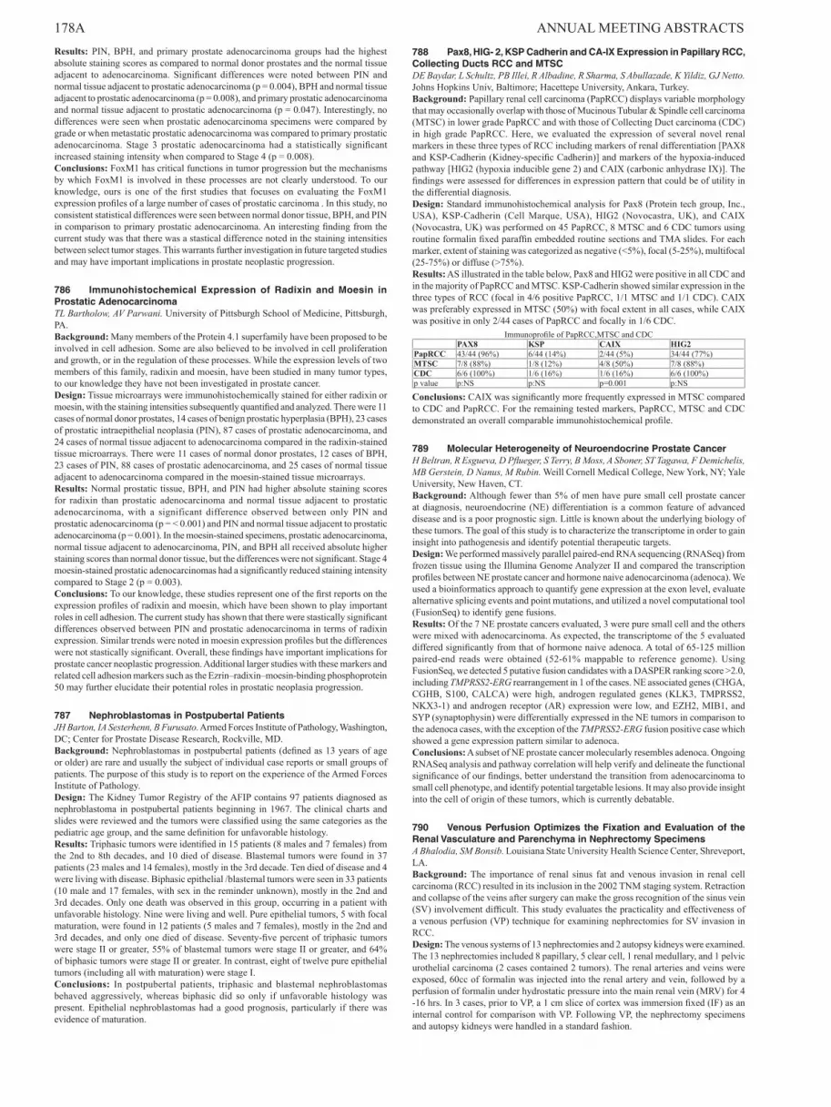
ANNUAL MEETING ABSTRACTS 179AResults: VP is easy and can be completed within 15 min. Grossly, the perfused veins from the MRV to its intralobular tributaries were fixed in an open position permitting visualization of internal surfaces and entering branch veins of 1-2 mm size.
The renal cortex appeared well fixed (pale). However, the renal pyramids were less well-fixed (reddish). Histologically, there was a marked difference between IF and VP fixed cortex. The tubules of IF cortex were eosinophilic and compressed, the interstitum scant, and veins often collapsed. The tubules of VP cortex were less eosinophilic, had open lumens, the interstitium expanded and the veins open compared to IF cortex.Conclusions: 1) Venous perfusion is a practical approach to specimen fixation that adds little additional handling time. 2) VP markedly enhances the direct inspection of the venous system for intravenous tumor. 3) VP results in markedly improved histology compared to IF. 4) VP facilitates gross recognition of important sinus vein staging features in nephrectomy specimens.
791 Should Pathologists Continue To Use the Current pT2 Substaging System for Reporting of Radical Prostatectomy Specimens?A Billis, L Meirelles, LLL Freitas, LA Magna, U Ferreira. School of Medicine, University of Campinas (Unicamp), Campinas, Brazil.Background: During the International Society of Urological Pathology (ISUP) consensus conference on handling and staging of radical prostatectomy specimens, 65.5% of the attendants answered that the current pT2 susbstaging system should not be used. Answering to another question, 63.4% favored to be reduced to two categories based on studies showing that pT2b does not exist. There was no consensus in regard to a minimum size for a second tumor to be considered for the whole case to be classified as pT2c as well as in regard to the definition of index tumor. We compared clinicopathologic findings and biochemical progression following surgery classifying pT2 patients into two categories.Design: The study was based on whole-mount consecutive surgical specimens from 142 patients with organ confined cancer. Using a semiquantitative method for evaluation of tumor extent, 10 positive points corresponds roughly to a 0.5ml tumor. We considered pT2a/pT2b substage (group 1) whenever a tumor presented >10 positive points on only one side and pT2c whenever presented >10 positive points on each of right and left side (group 2). The variables analyzed were: age, preoperative PSA, clinical stage, Gleason score on needle biopsy, and biochemical progression following surgery defined as PSA > 0.2ng/mL. The data were analyzed using the Mann-Whitney test, and the Kaplan-Meier product-limit analysis using the log-rank test for comparison between the groups. The P-values were two-sided at the significance level of <0.05.Results: Substage pT2a/pT2b corresponded to 84/142 (59.2%) patients and substage pT2c to 58/142 (40.8%) patients. There was no statistically significant difference between the groups in relation to: age (p=0.30), preoperative PSA (p=0.13), clinical stage (p=0.34), and Gleason score on needle biopsy (p=0.27). In 5 years of follow-up, 61% of patients pT2a/pT2b and 71% of patients pT2 were free of biochemical progression (log-rank, p=0.68).Conclusions: There was no significant difference for several clinicopathological variables and time of biochemical progression following surgery between patients with stage pT2a/pT2b and patients with stage pT3c. The results of this study favor to discontinue using the current pT2 substaging system for reporting of radical prostatectomy specimens.
792 Extraprostatic Extension in Radical Prostatectomy: Should Be Reported and Quantitated?A Billis, HA Fernandes, MM Padilha, AS Polidoro, MAM Bisson, AGE Duarte, RG Oliveira, F Rogerio, CAM Silva, LA Magna. School of Medicine, University of Campinas (Unicamp), Campinas, Brazil.Background: During the International Society of Urological Pathology (ISUP) consensus conference on handling and staging of radical prostatectomy (RP) specimens, 97.2% of the attendants answered that extraprostatic extension (EPE) should be reported and 93.6% that EPE should be quantitated. However there was no consensus to which subjective or quantitative method should be used. In this study we propose a simple method for EPE evaluation.Design: The study was based on 360 whole-mount consecutive surgical specimens. Each transversal section of the prostate was subdivided into 2 anterolateral and 2 posterolateral quadrants. Extraprostatic extension was stratified into 2 groups: present
in one quadrant (group 1) and in more than one quadrant (group 2). Each group was analyzed according to several clinicopathological variables: preoperative PSA, Gleason grade in the specimen, surgical positive margins, tumor extent, seminal vesicle invasion, and biochemical progression following RP defined as PSA > 0.2ng/mL. The data were analyzed using the Mann-Whitney test, Fisher’s exact test, Kaplan-Meier product-limit analysis using the log-rank test for comparison between the groups and the regression model of Cox.Results: EPE was found in 98/360 (27.2%) patients, 34/98 (34.7%) present in 1 quadrant (group 1) and 64/98 (65.3%) in more than 1 quadrant (group 2). In group 2 preoperative PSA was higher (p=0.02), more tumors were Gleason high-grade in the specimen (p<0.01), tumors were more extensive (p=0.04), and more tumors presented seminal vesicle invasion (p<0.01). There was no significant difference related to positive surgical margins (p=0.43). In 5 years of follow-up, 69% of patients without EPE and 57% of patients with EPE in group 1 were free of biochemical progression (log-rank, p=0.13); in group 2, 69% and 38%, respectively (log-rank, <0.01). In univariate Cox regression analysis, group 1 was not predictive of biochemical progression (p=0.131); group 2 was significantly predictive of biochemical progression (p<0.01).Conclusions: In whole-mount surgical specimens, EPE present in one quadrant vs. EPE present in more than one quadrant significantly discriminates patients according to several clinicopathological variables including biochemical progression following RP. This is an easy and valuable method for reporting and quantitating EPE.
793 Immunohistochemical Studies of Metastatic Germ Cell Tumors in the Retroperitoneal Dissection Specimens: A Sensitive and Specific PanelZ Bing, T Pasha, JE Tomaszewski. Hospital of the University of Pennsylvania, Philadelphia.Background: SALL4 and glypican 3 (GPC3) are two recently described germ cell tumor markers. In this study, we evaluated the usefulness of a panel of immunohistochemical markers (SALL4, GPC3, CD30 and D2-40) in the differential diagnosis of retroperitoneal dissection specimens for germ cell tumors.Design: Retroperitoneal lymph node dissection specimens were stained for SALL4, GPC3, CD30 and D2-40. The specimens included 17 cases of metastatic testicular germ cell tumors, 10 cases of metastatic melanoma, 14 cases of lymphoma, and 8 cases of metastatic carcinoma.Results: For SALL4, All of the seminoma (N=7), embryonal carcinoma (EC) (N= 8) and yolk sac tumor (YST) (N=4) showed strong nuclear staining (see table). Metastatic carcinoma and melanoma, and lymphoma were negative. For GPC3, only one out of 7 seminoma showed focal positivity. 4 out of 9 EC showed weak positivity. All 4 YST were positive. All of the melanoma were negative. 5 out of 8 cases of carcinoma were weakly positive. Only 1 out of 14 cases of lymphoma was weakly positive. The staining was cytoplasmic. For D2-40, all of the seminoma (N=7) were positive. The staining was strong and membranous. 4 out of 9 EC showed focal and weak cytoplasmic staining. Two cases of YST were negative. All of the melanoma and carcinoma were negative. Only 1 out of 14 cases of lymphoma was focally and weakly positive. For CD30, all 8 cases of EC were positive. All of the seminoma, YST, melanoma, and carcinomas were negative. 3 out of 14 cases of lymphoma were positive, one was strong and diffuse, and two were weak and focal. All of the positive lymphomas were subtyped as diffuse large cell lymphoma.
Immunohistochemical study of retroperitoneal dissection specimensSALL4 (%) (#positive/total)
GPC3(%)(#positive/total)
D2-40(%) (#positive/total)
CD30(%) (#positive/total)
Seminoma 100 (7/7) 14 (1/7) 100 (7/7) 0 (0/7)EC 100 (8/8) 44 (4/9) 44 (4/9) 100 (8/8)YST 100 (4/4) 100 (4/4) 0 (0/2) 0 (0/2)Melanoma 0 (0/10) 0 (0/9) 0 (0/10) 0 (0/10)Carcinoma 0 (0/7) 63 (5/8) 0 (0/6) 0 (0/8)Lymphoma 0 (0/14) 7 (1/14) 7 (1/14) 21 (3/14)
Conclusions: Immunohistochemical stains for SALL4, GPC3, D2-40 and CD30 are a sensitive and specific panel, which may facilitate in the differential diagnosis of metastatic germ cell tumors in retroperitoneal dissection specimens.
794 PTEN Genomic Deletions Is an Early Event Associated with ERG Gene Rearrangements in Prostate CancerTA Bismar, M Yoshimoto, RT Vollmer, Q Duan, M Firszt, J Corcos, JA Squire. University of Calgary and Calgary Laboratory Services, Calgary, AB, Canada; Queen’s University, Kingston, ON, Canada; VA and Duke University Medical Centers, Durham, NC; McGill University, Montreal, QC, Canada.Background: ERG gene rearrangements and PTEN genomic deletions are two of the most common genetic alteration in prostate cancer (PCA). The interaction and significance of those two genetic aberrations in relation to disease development and progression is still controversial.Design: We interrogated initial cohort of 220 men with localized PCA using fluorescence in-situ hybridization for ERG rearrangements and PTEN genomic deletions.Results: ERG rearrangements and PTEN deletions incidence in PCA was significantly higher than in HGPIN and benign prostate tissue (p< 0.0001). ERG rearrangements and PTEN deletions were detected in 41.9% and 42.6% of patients’ tumors, respectively. ERG rearrangements were never detected in benign prostate tissue; while PTEN aberrations were present at a basal level of 4.6%. PTEN hemizygous deletions showed higher frequency than homozygous deletions within each diagnostic category from benign prostate tissue to HGPIN and PCA (p=0.0001). Furthermore, in 29 cases where all three tissues were available, PTEN genomic aberration levels showed significant difference between PCA vs. benign (p=0.005) and HGPIN (p=0.02) reflective of accumulating genomic aberrations at early stages of disease progression. Within this cohort, 71.4% of homozygous and 44.2% of hemizygous PTEN deletions occurred simultaneously with ERG rearrangements. Stratified based on Gleason score (GS), hemizygous
180A ANNUAL MEETING ABSTRACTS PTEN deletions across various GS groups were observed at a higher frequency than homozygous deletions. However, PTEN homozygous deletions showed positive trends with higher GS, increasing in poorly differentiated PCA (GS 8-10) compared to moderately and well-differentiated tumors (GS 6 & 7).Conclusions: Our data confirm significant association between ERG gene rearrangement and PTEN genomic aberrations in PCA. Furthermore, our analysis provides further support for the observation that homozygous PTEN deletions can occur within subset of HGPIN lesions, and show accumulating genetic aberrations with disease progression, evidenced by higher detection in PCA vs. HGPIN and increased PTEN homozygous deletions in Gleason scores 8-10 vs. 6-7.
795 The Expression of Prostate Secretory Protein (PSP) and Hepatocyte Nuclear Factor-1 (HNF) in High Grade Prostate Intrepithelial Neoplasia (HGPIN) and Adenocarcinoma of the ProstateAM Blutreich, S Sasturkar, T He, M Nagar, L Chiriboga, R Hayes, J Melamed, J Ahn. New York University Medical Center, New York, NY; New York Universiry Medical Center, New York, NY.Background: Recent prostate cancer genome-wide association studies consistently report that common genetic variants in MSMB and HNF1B are associated with prostate cancer risk (Thomas G, 2008; Eeles RA, 2009), suggesting an important role of these genes in prostate cancer etiology and progression. However, it is unclear whether these genes are expressed in early stage prostate carcinogenesis. In order to further elucidate the role of these genes, we have investigated the expression of the protein Prostate Secretory Protein (PSP) which is encoded by MSMB and Hepatocyte Nuclear Factor (HNF) which is encoded by HNF1B, in both HGPIN and adenocarcinoma.Design: HGPIN tissue microarrays were constructed from radical prostatectomy specimens to include foci of HGPIN, adenocarcinoma and non-neoplastic peripheral zone tissue. Immunohistochemical studies using commercially available antibodies against HNF and PSP were performed on a Ventana Benchmark automated immunostainer. Tissue microarray stained slides were scored using an intensity/proportion score. The mean expression level for HNF and PSP in HGPIN, adenocarcinoma and non-neoplastic prostate tissue were evaluated and mean level differences were tested by t-test.Results: This study included 46 males, with a mean age of 61 (ranged 46-74) and mean Gleason score 6.6 (range 6-7), and tumor stages of pT2 and pT3. We found the mean expression levels of PSP94 were significantly lower in HGPIN (mean expression score = 4.8; P value = < 0.01) and carcinoma (mean expression score =5.3; P value = < 0.01) compared with adjacent non-neoplastic prostate tissue (mean score = 6.5).
Results for PSP and HNF marker expression
Marker HGPIN Prostate Adenocarcinoma Non-neoplastic Prostate Tissue Statisitcal Significance
PSP 4.8 5.3 6.5 P value <0.01HNF 6.9 7.4 7.1 Not significant
HNFIB expression was lower in HGPIN (mean score = 6.9); compared to non-neoplastic prostate tissue (mean score=7. 1), however was not statistically significant.Conclusions: We find that MSMB gene expression levels are lower in HGPIN and prostate cancer than non-neoplastic tissue. These findings suggest that the MSMB plays an early and continued role in prostate cancer progression and carcinogenesis.
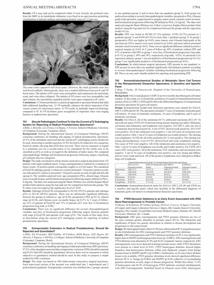
796 Intravenous Dissection, the Optimum Strategy for Staging of Renal Cell CarcinomaSM Bonsib, A Bhalodia. Louisiana State University Helath Science Center, Shreveport, LA.Background: The most common avenue of extra-renal extension for clear cell (CC) renal cell carcinoma (RCC) is via renal sinus veins (RSV). Opening a nephrectomy specimen through its venous system may permit gross demonstration of the earliest phase of this process.Design: Thirty-six radical nephrectomy specimens containing 38 RCCs (27 CC, 7 papillary, 2 chromophobe, 2 other) were examined for RSV involvement. Intravascular fixation was employed for 13/36 cases consisting of intraarterial and intravenous formalin injection, followed by intravenous formalin perfusion under hydrostatic pressure for 4-16 hrs. For all specimens, 2-3 probes were placed in RSV and the specimens bi-valved along the probes.Results: Intravenous perfusion permitted optimum visualization of RSV and intravenous tumor by fixing the veins in an open position.
Sinus intravenous extension was observed in 21/38 RCC; all 21 were CC.
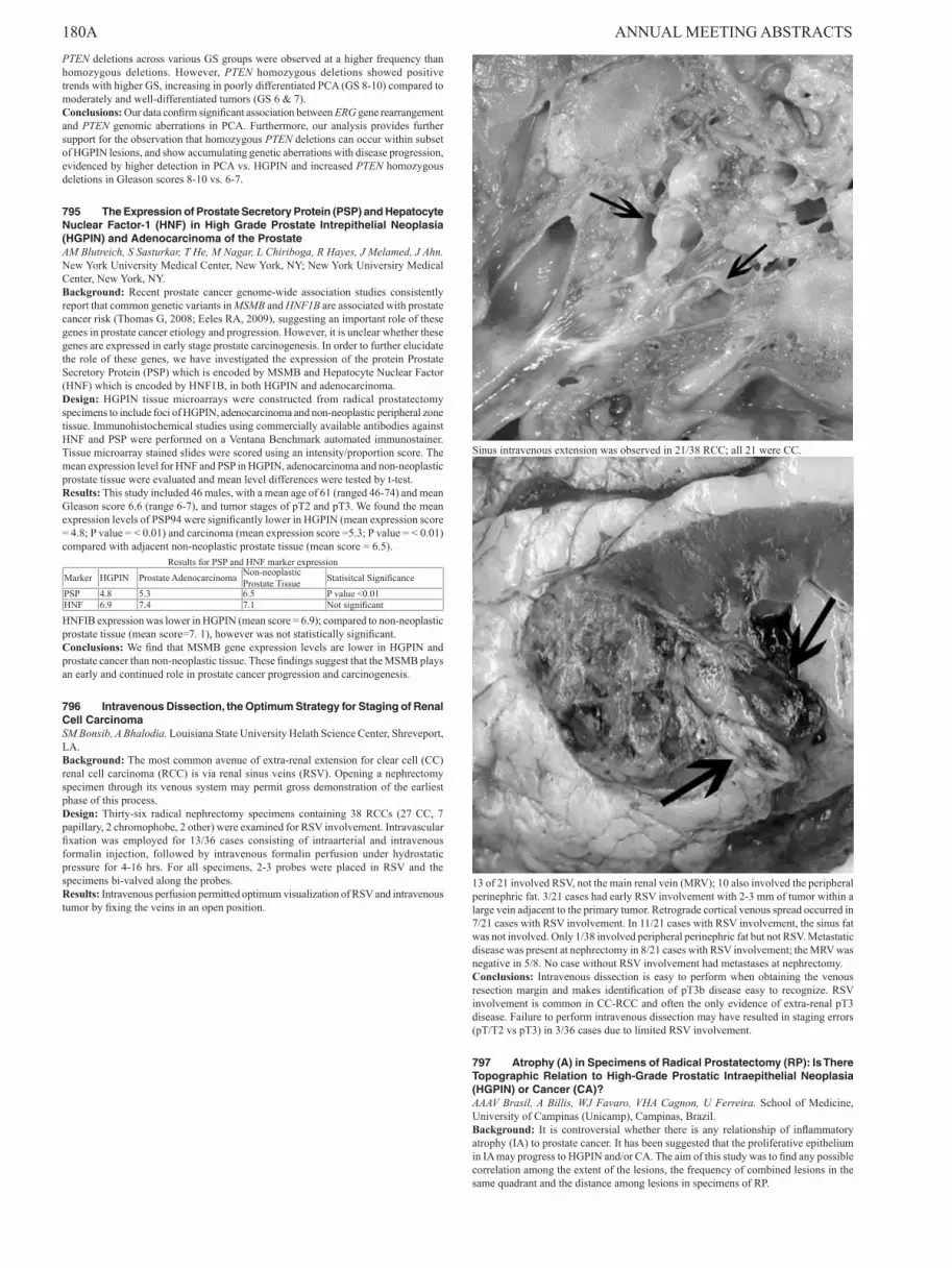
13 of 21 involved RSV, not the main renal vein (MRV); 10 also involved the peripheral perinephric fat. 3/21 cases had early RSV involvement with 2-3 mm of tumor within a large vein adjacent to the primary tumor. Retrograde cortical venous spread occurred in 7/21 cases with RSV involvement. In 11/21 cases with RSV involvement, the sinus fat was not involved. Only 1/38 involved peripheral perinephric fat but not RSV. Metastatic disease was present at nephrectomy in 8/21 cases with RSV involvement; the MRV was negative in 5/8. No case without RSV involvement had metastases at nephrectomy.Conclusions: Intravenous dissection is easy to perform when obtaining the venous resection margin and makes identification of pT3b disease easy to recognize. RSV involvement is common in CC-RCC and often the only evidence of extra-renal pT3 disease. Failure to perform intravenous dissection may have resulted in staging errors (pT/T2 vs pT3) in 3/36 cases due to limited RSV involvement.
797 Atrophy (A) in Specimens of Radical Prostatectomy (RP): Is There Topographic Relation to High-Grade Prostatic Intraepithelial Neoplasia (HGPIN) or Cancer (CA)?AAAV Brasil, A Billis, WJ Favaro, VHA Cagnon, U Ferreira. School of Medicine, University of Campinas (Unicamp), Campinas, Brazil.Background: It is controversial whether there is any relationship of inflammatory atrophy (IA) to prostate cancer. It has been suggested that the proliferative epithelium in IA may progress to HGPIN and/or CA. The aim of this study was to find any possible correlation among the extent of the lesions, the frequency of combined lesions in the same quadrant and the distance among lesions in specimens of RP.
ANNUAL MEETING ABSTRACTS 181ADesign: Partial and complete atrophy were considered. We studied the frequency of quadrants showing: only atrophy A, A+HGPIN, A+CA, or A+HGPIN+CA. Extent of atrophy, HGPIN and CA was evaluated by a semiquantitative point-count method previously described. Points were considered coincident whenever atrophy and HGPIN or CA were seen in a distance <5mm; non-coincident whenever in a distance >5mm. The means were compared using the Kruskal-Wallis and the Mann-Whitney tests. For the comparison of extent we used the Spearman correlation coefficient.Results: A total of 3186 quadrants from 100 whole-mount consecutive surgical specimens were examined. The mean (range) of quadrants showing only A, A+HGPIN, A+CA, and A+HGPIN+CA was 4.88 (0-24), 3.97 (0-14), 1.16 (0-7), and 0.65 (0-4) respectively (p<0.01); considering only foci of IA was 3.29 (0-21), 2.51 (0-11), 0.77 (0-6), and 0.44 (0-4), (p<0.01). No partial atrophy foci with chronic inflammation were seen. The mean (range) of coincident points was 1.12 (0-7) and non-coincident 12.05 (0-65) (p<0.01); considering only foci of IA was 0.81 (0-7) and 8.37 (0-60) (p<0.01). There was no significant correlation between extent of A (r=0.01, p=0.88) or IA (r=0.05, p=0.64) with extent of HGPIN. There was a significant negative correlation of extent of A (r=-0.23, p=0.02) or IA (r=-0.27, p=0.01) with extent of CA.Conclusions: In specimens of radical prostatectomy most of the quadrants show only atrophy (with or without inflammation). Extent of atrophy did not correlate with extent of HGPIN or carcinoma and the foci of A or IA are significantly located in a distance >5mm from HGPIN and/or CA comparing to foci in a distance <5mm . The results seem to favor a lack of topographical association among atrophy, HGPIN and carcinoma. A further finding in this study was absence of chronic inflammation in partial atrophy foci.
798 Cohort Design and Localization Is Critical for the Understanding of the Clinical Implications of Prostate Cancer with ERG RearrangementM Braun, VJ Scheble, T Wilbertz, AC Stiedl, K Petersen, D Schilling, R Kuefer, F Fend, G Kristiansen, MA Rubin, S Perner. University Hospital of Tuebingen, Tuebingen, Germany; University Hospital of Ulm, Ulm, Germany; University Hospital of Zuerich, Zuerich, Switzerland; Weill Cornell Medical College, New York, NY.Background: TMPRSS2-ERG gene fusions are the most frequent rearrangements in prostate cancer (PCa) with a great variability in its reported prevalence ranging from 15-78% depending on the study cohorts. The reason for this variability in different study cohorts is unknown. The aim of our study was to elucidate the differences in ERG rearrangement prevalence.Design: We assessed the frequency of ERG rearrangement by FISH in three clinical cohorts. The first cohort comprises the index tumor focus from the peripheral zone of 109 partially PSA-screened prostatectomy samples. The second cohort includes 105 PCa samples incidentally diagnosed by TUR-P, and the third cohort includes 71 PCa samples incidentally identified in the course of cystoprostatectomy. Within the cystoprostatectomy PCa cases, 9 cases had tumor foci within the transition zone.Results: Out of 109 prostatectomy samples, 49.5% harbored the ERG rearrangement. 29.5% of the PCa cases incidentally identified by TUR-P and 33.8% of the PCa cases incidentally detected in the course of cystoprostatectomy revealed ERG rearrangement. Of note, 32.2% of the PCa cases diagnosed by TUR-P and 23.8% of the PCa identified in cystoprostatectomy specimen showed interfocal heterogeneity with regard to the rearrangement status. Two of the 9 (22.2%) transition zone PCa foci revealed the ERG rearrangement. Overall, the prostatectomy cohort had a higher Gleason grade as compared to the incidentally diagnosed cohorts.Conclusions: We compared the ERG rearrangement frequency in incidentally detected cohorts and a prostatectomy PCa cohort. We could confirm that the ERG rearrangement appears in approximately half of the cases in the prostatectomy cohort but was observed significantly less frequently in incidentally diagnosed PCa cohorts. Our observations support the hypothesis that the ERG rearrangements may be more frequent in peripheral zone tumors than transition zone tumors, a finding consistent with the potentially more aggressive nature of TMPRSS2-ERG fusion PCa cases.
799 The Value of Mandatory Second Opinion Pathology Review in the Interpretation of Prostate Needle Biopsies Prior to Radical ProstatectomyF Brimo, L Schultz, JI Epstein. The Johns Hopkins Hospital, Baltimore.Background: We determined the value of mandatory second opinion pathology review in the interpretation of prostate needle biopsies prior to radical prostatectomy (RP).Design: The original and reviewed needle biopsy pathology reports for all cases referred to our institution for RP over a 1 year period were compared. A major discrepancy in GS was defined as a change that places the patient in a different risk stratification category (GS of 6, 7, and 8-10). A significant difference in the reporting of the highest percentage of cancer in a single core was defined as a difference >30% between the two reports.Results: 855 cases were included. All had an original diagnosis of prostatic adenocarcinoma (PCa). Of these, 844/855 (98.8%) were diagnosed as carcinoma upon review. Nine (1%) cases were diagnosed as atypical and 2 (0.2%) as benign upon review. A major discrepancy in GS between the original and the reviewed reports was present in 124 (14.7%) of cases; of those the majority 67 (54%) were downgraded upon review. 80 cases (64.5%) of the disagreements were between GS 6 and 7. Of the 777 cases in which the number of positive cores was reported in both reports, 71 (9.1%) had discrepancies upon review; the difference was one core in 53/71 cases (75%). The number of positive cores was reported to be higher in 45/71 (63.4%) after review. A significant difference in the highest percentage of cancer in a single core was present in 76/844 (9%) evaluable cases; in all of these cases, the extent of cancer was underestimated in the original report. In 60/76 (78.9%) cases, cancer was reported to involve the core discontinuously on review. Perineural invasion was reported to be present in 138/844 (16.3%) cases after revision, from which this information was lacking in 37 of the original reports. Finally, in 4 cases, presence of extra-prostatic extension was noted on the needle biopsy only upon review.
Conclusions: Compared to a smaller study done over 10 years ago at our institution, the percent of cancers that could not be confirmed was identical at 1.2%. There was a greater concordance in terms of grade, as compared to another earlier study from our institution. This study is the first to analyze concordance upon review for number of positive cores and maximum percent positive in a core with a 9% discrepancy in both. Mandatory second opinion on prostate needle biopsies can, in a minority of cases, result in significantly different reports that may affect therapy.
800 Tumor Grade at Margins of Resection in Radical Prostatectomy Specimens Is an Independent Predictor of PrognosisF Brimo, JI Epstein. The Johns Hopkins Hospital, Baltimore.Background: We sought to assess whether reporting the grade of cancer at the site of positive margins in a radical prostatectomy specimen was independently prognostic.Design: We restricted our study to 108 cases (1995-2008) of Gleason score 7 with non-focal extra-prostatic extension (pT3a) and positive surgical margins. Cases with tertiary pattern 5, neoadjuvant therapy, or positive margins as a result of intraprostatic incision were excluded. When more than one section showed a positive margin, the overall length of positive margin was obtained by adding the lengths of all areas of cancer in contact with ink regardless of their location. When two consecutive slides going from apex to base showed positive margins, the length of positive margin in these two slides was calculated as 3 mm (1.5 mm being half of the thickness of each tissue section on a slide) provided that none of these slides showed a positive margin on the one side larger than 3mm. Cancer present at a margin was assigned a Gleason score independent from the cancer score of the entire case. In slides where cancer at the margin showed obscuring cautery artifact, the Gleason score at the margin was assigned based on the grade of uncauterized cancer in direct continuity with the cauterized cancer.Results: The overall Gleason score was 3+4 in 73 (67%) and 4+3 in 35 (33%) cases. The median length of positive margin was 3.0 mm (0.5-10 mm). Gleason score at the margin was 3+3, 3+4, 4+3, and 4+4 in 40 (37%), 41 (38%), 16 (14.8%) and 11 (10.2%) cases respectively. 45 (42%) patients remained free of disease post-radical prostatectomy (median follow-up=6 years, range 3-13 years). Univariate and multivariate analysis showed no correlation between biochemical recurrence and both pre-operative serum PSA levels (p=0.7) and overall Gleason score (p=0.5). A strong association was noted between biochemical recurrence and Gleason score at the margin (p=0.007) with length of cancer at the margin also predictive (p=0.015) in multivariate analysis. Using the median length of positive margin (3mm) as a cutoff, the association with biochemical recurrence was significantly different between the two groups (p=0.004) on Kaplan-Meier analysis.Conclusions: The current study is the first to prove that the grade of cancer at the site of a positive margin influences outcome. We were able to stratify grade into 3 categories: 3+3; 3+4; and 4+3 or higher (4+3 and 4+4 at the positive margin carried equal prognostic information).
801 Well-Differentiated Papillary Mesothelioma (WDPM) of the Tunica Testis: Expanding the Histological SpectrumF Brimo, PB Illei, JI Epstein. The Johns Hopkins Hospital, Baltimore.Background: While the histology of the originally described WDPM consisted of papillae lined by a single layer of bland cuboidal cells, more complex architectural patterns of growth have rarely been described. We evaluate whether there are prognostic or IHC differences between these 2 different patterns, and compare them to overt diffuse malignant mesothelioma (DMM).Design: IHC for GLUT-1, p53, and Ki-67 was performed in 8 cases of WDPM of the testicular tunica and compared to the immunoprofile of DMM reported in the literature as well as to 28 DMM cases taken from a recently constructed TMA.Results: Patients’ ages were 34-75 years (mean 55.6). In 7/8, hydrocele was the initial manifestation of disease; only 1 patient presented with a scrotal mass. 5 cases presented as a solitary papillary mass, multiple separate nodules in 2 cases, and in 1 case a plaque-like lesion on the tunica vaginalis. All but 1 case had in areas a papillary/tubulopapillary architecture, and the fibrovascular papillary cores were lined by a single layer of bland cuboidal cells. In 5 cases, the papillary projections also contained anastomosing cords of tumor cells that formed complex cribriform structures, which in some areas had a back-to-back arrangement with minimal intervening stroma displaying a condensed or pseudo-solid appearance. In one case, a solid nested growth pattern was noted where tumor cells partially replaced the submesothelial stroma. Despite the complex architectural patterns, none of the cases showed evidence of invasion or elicited a surrounding stromal reaction. Mitotic figures were present in 7 cases (mean 2.6/50 HPF). All cases lacked pleomorphism, atypical mitotic figures or necrosis. All cases showed ≤1% of positive cells for Ki-67 and p53 and only 1 case was focally positive for GLUT-1. In contrast, 50% (n=14) of cases of DMM were positive for GLUT-1 with the majority showing less than 25% of cells’ positivity. 4 patients underwent radical orchiectomy and 4 only hydrocelectomy. 3 did not show evidence of disease recurrence or progression at 1.5 years (2 patients) and 3.5 years (1 patient) post-surgery. In one the 8 cases, the patient was alive (disease status unknown) at 3 years and another patient died of unknown causes 47 years following diagnosis. The three remaining cases were recent.Conclusions: Our findings support that those lesions with only papillary or tubulopapillary formation and those with more complex growth represent the same entity and should therefore be labeled as WDPM rather than as DMM.
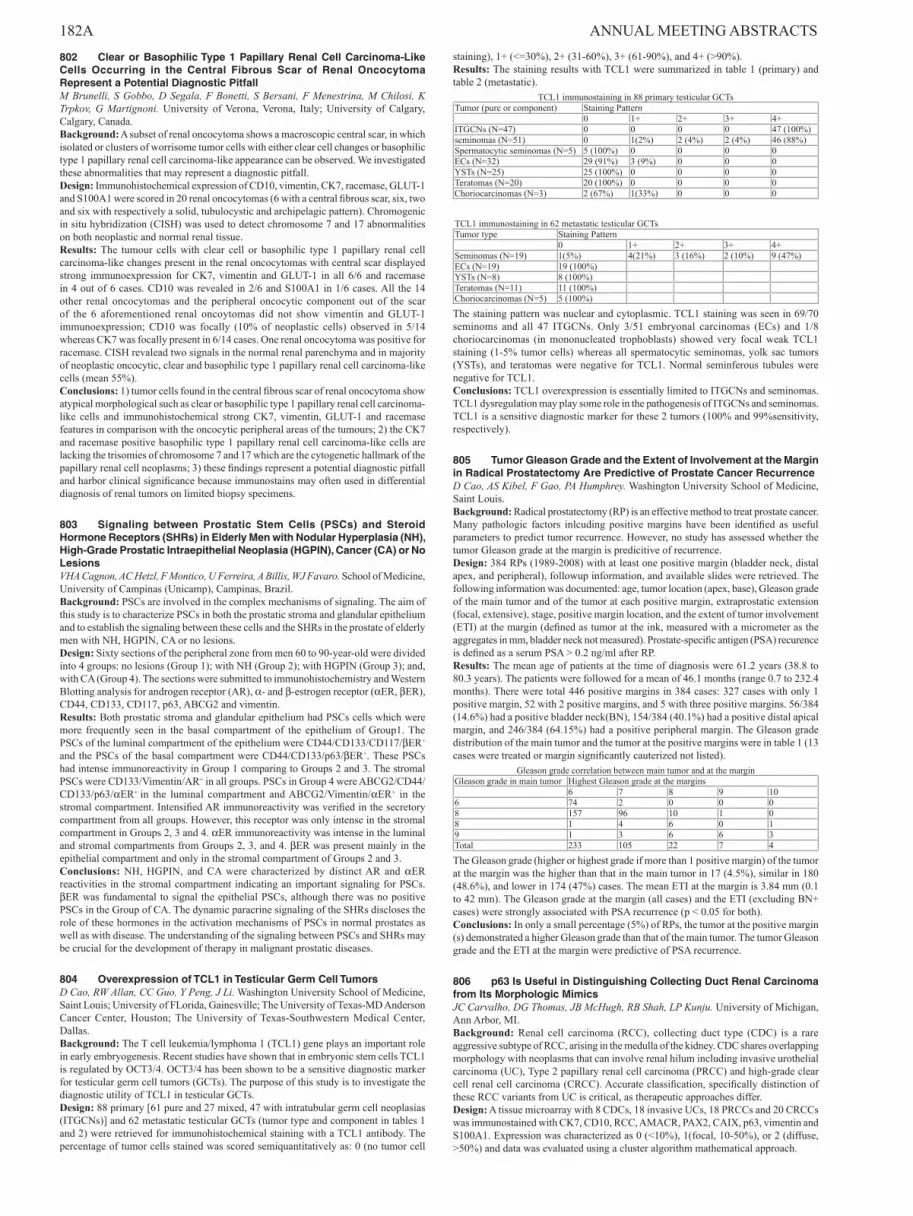
182A ANNUAL MEETING ABSTRACTS 802 Clear or Basophilic Type 1 Papillary Renal Cell Carcinoma-Like Cells Occurring in the Central Fibrous Scar of Renal Oncocytoma Represent a Potential Diagnostic PitfallM Brunelli, S Gobbo, D Segala, F Bonetti, S Bersani, F Menestrina, M Chilosi, K Trpkov, G Martignoni. University of Verona, Verona, Italy; University of Calgary, Calgary, Canada.Background: A subset of renal oncocytoma shows a macroscopic central scar, in which isolated or clusters of worrisome tumor cells with either clear cell changes or basophilic type 1 papillary renal cell carcinoma-like appearance can be observed. We investigated these abnormalities that may represent a diagnostic pitfall.Design: Immunohistochemical expression of CD10, vimentin, CK7, racemase, GLUT-1 and S100A1 were scored in 20 renal oncocytomas (6 with a central fibrous scar, six, two and six with respectively a solid, tubulocystic and archipelagic pattern). Chromogenic in situ hybridization (CISH) was used to detect chromosome 7 and 17 abnormalities on both neoplastic and normal renal tissue.Results: The tumour cells with clear cell or basophilic type 1 papillary renal cell carcinoma-like changes present in the renal oncocytomas with central scar displayed strong immunoexpression for CK7, vimentin and GLUT-1 in all 6/6 and racemase in 4 out of 6 cases. CD10 was revealed in 2/6 and S100A1 in 1/6 cases. All the 14 other renal oncocytomas and the peripheral oncocytic component out of the scar of the 6 aforementioned renal oncoytomas did not show vimentin and GLUT-1 immunoexpression; CD10 was focally (10% of neoplastic cells) observed in 5/14 whereas CK7 was focally present in 6/14 cases. One renal oncocytoma was positive for racemase. CISH revalead two signals in the normal renal parenchyma and in majority of neoplastic oncocytic, clear and basophilic type 1 papillary renal cell carcinoma-like cells (mean 55%).Conclusions: 1) tumor cells found in the central fibrous scar of renal oncocytoma show atypical morphological such as clear or basophilic type 1 papillary renal cell carcinoma-like cells and immunohistochemical strong CK7, vimentin, GLUT-1 and racemase features in comparison with the oncocytic peripheral areas of the tumours; 2) the CK7 and racemase positive basophilic type 1 papillary renal cell carcinoma-like cells are lacking the trisomies of chromosome 7 and 17 which are the cytogenetic hallmark of the papillary renal cell neoplasms; 3) these findings represent a potential diagnostic pitfall and harbor clinical significance because immunostains may often used in differential diagnosis of renal tumors on limited biopsy specimens.
803 Signaling between Prostatic Stem Cells (PSCs) and Steroid Hormone Receptors (SHRs) in Elderly Men with Nodular Hyperplasia (NH), High-Grade Prostatic Intraepithelial Neoplasia (HGPIN), Cancer (CA) or No LesionsVHA Cagnon, AC Hetzl, F Montico, U Ferreira, A Billis, WJ Favaro. School of Medicine, University of Campinas (Unicamp), Campinas, Brazil.Background: PSCs are involved in the complex mechanisms of signaling. The aim of this study is to characterize PSCs in both the prostatic stroma and glandular epithelium and to establish the signaling between these cells and the SHRs in the prostate of elderly men with NH, HGPIN, CA or no lesions.Design: Sixty sections of the peripheral zone from men 60 to 90-year-old were divided into 4 groups: no lesions (Group 1); with NH (Group 2); with HGPIN (Group 3); and, with CA (Group 4). The sections were submitted to immunohistochemistry and Western Blotting analysis for androgen receptor (AR), α- and β-estrogen receptor (αER, βER), CD44, CD133, CD117, p63, ABCG2 and vimentin.Results: Both prostatic stroma and glandular epithelium had PSCs cells which were more frequently seen in the basal compartment of the epithelium of Group1. The PSCs of the luminal compartment of the epithelium were CD44/CD133/CD117/βER+ and the PSCs of the basal compartment were CD44/CD133/p63/βER+. These PSCs had intense immunoreactivity in Group 1 comparing to Groups 2 and 3. The stromal PSCs were CD133/Vimentin/AR+ in all groups. PSCs in Group 4 were ABCG2/CD44/CD133/p63/αER+ in the luminal compartment and ABCG2/Vimentin/αER+ in the stromal compartment. Intensified AR immunoreactivity was verified in the secretory compartment from all groups. However, this receptor was only intense in the stromal compartment in Groups 2, 3 and 4. αER immunoreactivity was intense in the luminal and stromal compartments from Groups 2, 3, and 4. βER was present mainly in the epithelial compartment and only in the stromal compartment of Groups 2 and 3.Conclusions: NH, HGPIN, and CA were characterized by distinct AR and αER reactivities in the stromal compartment indicating an important signaling for PSCs. βER was fundamental to signal the epithelial PSCs, although there was no positive PSCs in the Group of CA. The dynamic paracrine signaling of the SHRs discloses the role of these hormones in the activation mechanisms of PSCs in normal prostates as well as with disease. The understanding of the signaling between PSCs and SHRs may be crucial for the development of therapy in malignant prostatic diseases.
804 Overexpression of TCL1 in Testicular Germ Cell TumorsD Cao, RW Allan, CC Guo, Y Peng, J Li. Washington University School of Medicine, Saint Louis; University of FLorida, Gainesville; The University of Texas-MD Anderson Cancer Center, Houston; The University of Texas-Southwestern Medical Center, Dallas.Background: The T cell leukemia/lymphoma 1 (TCL1) gene plays an important role in early embryogenesis. Recent studies have shown that in embryonic stem cells TCL1 is regulated by OCT3/4. OCT3/4 has been shown to be a sensitive diagnostic marker for testicular germ cell tumors (GCTs). The purpose of this study is to investigate the diagnostic utility of TCL1 in testicular GCTs.Design: 88 primary [61 pure and 27 mixed, 47 with intratubular germ cell neoplasias (ITGCNs)] and 62 metastatic testicular GCTs (tumor type and component in tables 1 and 2) were retrieved for immunohistochemical staining with a TCL1 antibody. The percentage of tumor cells stained was scored semiquantitatively as: 0 (no tumor cell
staining), 1+ (<=30%), 2+ (31-60%), 3+ (61-90%), and 4+ (>90%).Results: The staining results with TCL1 were summarized in table 1 (primary) and table 2 (metastatic).
TCL1 immunostaining in 88 primary testicular GCTsTumor (pure or component) Staining Pattern
0 1+ 2+ 3+ 4+ITGCNs (N=47) 0 0 0 0 47 (100%)seminomas (N=51) 0 1(2%) 2 (4%) 2 (4%) 46 (88%)Spermatocytic seminomas (N=5) 5 (100%) 0 0 0 0ECs (N=32) 29 (91%) 3 (9%) 0 0 0YSTs (N=25) 25 (100%) 0 0 0 0Teratomas (N=20) 20 (100%) 0 0 0 0Choriocarcinomas (N=3) 2 (67%) 1(33%) 0 0 0
TCL1 immunostaining in 62 metastatic testicular GCTsTumor type Staining Pattern
0 1+ 2+ 3+ 4+Seminomas (N=19) 1(5%) 4(21%) 3 (16%) 2 (10%) 9 (47%)ECs (N=19) 19 (100%)YSTs (N=8) 8 (100%)Teratomas (N=11) 11 (100%)Choriocarcinomas (N=5) 5 (100%)
The staining pattern was nuclear and cytoplasmic. TCL1 staining was seen in 69/70 seminoms and all 47 ITGCNs. Only 3/51 embryonal carcinomas (ECs) and 1/8 choriocarcinomas (in mononucleated trophoblasts) showed very focal weak TCL1 staining (1-5% tumor cells) whereas all spermatocytic seminomas, yolk sac tumors (YSTs), and teratomas were negative for TCL1. Normal seminferous tubules were negative for TCL1.Conclusions: TCL1 overexpression is essentially limited to ITGCNs and seminomas. TCL1 dysregulation may play some role in the pathogenesis of ITGCNs and seminomas. TCL1 is a sensitive diagnostic marker for these 2 tumors (100% and 99%sensitivity, respectively).
805 Tumor Gleason Grade and the Extent of Involvement at the Margin in Radical Prostatectomy Are Predictive of Prostate Cancer RecurrenceD Cao, AS Kibel, F Gao, PA Humphrey. Washington University School of Medicine, Saint Louis.Background: Radical prostatectomy (RP) is an effective method to treat prostate cancer. Many pathologic factors inlcuding positive margins have been identified as useful parameters to predict tumor recurrence. However, no study has assessed whether the tumor Gleason grade at the margin is predicitive of recurrence.Design: 384 RPs (1989-2008) with at least one positive margin (bladder neck, distal apex, and peripheral), followup information, and available slides were retrieved. The following information was documented: age, tumor location (apex, base), Gleason grade of the main tumor and of the tumor at each positive margin, extraprostatic extension (focal, extensive), stage, positive margin location, and the extent of tumor involvement (ETI) at the margin (defined as tumor at the ink, measured with a micrometer as the aggregates in mm, bladder neck not measured). Prostate-specific antigen (PSA) recurence is defined as a serum PSA > 0.2 ng/ml after RP.Results: The mean age of patients at the time of diagnosis were 61.2 years (38.8 to 80.3 years). The patients were followed for a mean of 46.1 months (range 0.7 to 232.4 months). There were total 446 positive margins in 384 cases: 327 cases with only 1 positive margin, 52 with 2 positive margins, and 5 with three positive margins. 56/384 (14.6%) had a positive bladder neck(BN), 154/384 (40.1%) had a positive distal apical margin, and 246/384 (64.15%) had a positive peripheral margin. The Gleason grade distribution of the main tumor and the tumor at the positive margins were in table 1 (13 cases were treated or margin significantly cauterized not listed).
Gleason grade correlation between main tumor and at the marginGleason grade in main tumor Highest Gleason grade at the margins
6 7 8 9 106 74 2 0 0 08 157 96 10 1 08 1 4 6 0 19 1 3 6 6 3Total 233 105 22 7 4
The Gleason grade (higher or highest grade if more than 1 positive margin) of the tumor at the margin was the higher than that in the main tumor in 17 (4.5%), similar in 180 (48.6%), and lower in 174 (47%) cases. The mean ETI at the margin is 3.84 mm (0.1 to 42 mm). The Gleason grade at the margin (all cases) and the ETI (excluding BN+ cases) were strongly associated with PSA recurrence (p < 0.05 for both).Conclusions: In only a small percentage (5%) of RPs, the tumor at the positive margin (s) demonstrated a higher Gleason grade than that of the main tumor. The tumor Gleason grade and the ETI at the margin were predictive of PSA recurrence.
806 p63 Is Useful in Distinguishing Collecting Duct Renal Carcinoma from Its Morphologic MimicsJC Carvalho, DG Thomas, JB McHugh, RB Shah, LP Kunju. University of Michigan, Ann Arbor, MI.Background: Renal cell carcinoma (RCC), collecting duct type (CDC) is a rare aggressive subtype of RCC, arising in the medulla of the kidney. CDC shares overlapping morphology with neoplasms that can involve renal hilum including invasive urothelial carcinoma (UC), Type 2 papillary renal cell carcinoma (PRCC) and high-grade clear cell renal cell carcinoma (CRCC). Accurate classification, specifically distinction of these RCC variants from UC is critical, as therapeutic approaches differ.Design: A tissue microarray with 8 CDCs, 18 invasive UCs, 18 PRCCs and 20 CRCCs was immunostained with CK7, CD10, RCC, AMACR, PAX2, CAIX, p63, vimentin and S100A1. Expression was characterized as 0 (<10%), 1(focal, 10-50%), or 2 (diffuse, >50%) and data was evaluated using a cluster algorithm mathematical approach.
ANNUAL MEETING ABSTRACTS 183AResults: Cluster analysis sorts the tumors into four groups. A majority (63%) of CDC form a distinct group with diffuse strong CK7 expression, variable PAX2 and lack of reactivity with other markers. Almost all (94%) UC cluster together with strong diffuse reactivity for p63 and CK7 and absent to minimal staining with renal markers (CD10, RCC, PAX2, AMACR, and CAIX) and vimentin. The remaining two groups are predominantly a mixture of PRCC and CRCC that differ based on expression of renal markers. Both these clusters lack p63, have minimal to absent CK 7 expression and strongly express vimentin. The remaining CDC cases (27%) segregate with the two clusters of PRCC and CRCC due to lack of CK7 expression. Overall, 63% of CDC expressed PAX2. Staining results are summarized in the table. S100A1 had minimal utility and was excluded from analysis.
Antibody Percentages of Four Tumor TypesCollecting Duct RCC Urothelial Carcinoma Type 2 Papillary RCC Clear Cell RCC
P63 0 100 0 0PAX2 63 6 56 70CA IX 13 24 50 95CK7 63 100 28 20Vimentin 25 11 94 90RCC 13 0 50 67CD10 0 50 67 95AMACR 25 39 94 30
Conclusions: p63 is most useful in the distinction of morphologic mimics of CDC. Invasive UC and CDC have overlapping immunohistochemical profiles but can be differentiated by intense p63 expression in UC. PAX2 is rarely positive (6%) in UC; is variably expressed in CDC, PRCC and CRCC and may be helpful in separating UC from all other RCCs when positive. No unique immunohistochemical marker or panel of markers can differentiate PRCC from CRCC.
807 An In-Depth Study of Extraprostatic Extension and Margin Status in Radical ProstatectomiesS Chan, F Garcia, M Moussa, J Chin, M Gabril. LHSC/UWO, London, ON, Canada.Background: In radical prostatectomies for prostate cancer (PCa), a positive margin is defined as tumor in contact with the inked resection margin. Extraprostatic extension (EPE) signifies tumor extending outside the normal confines of the prostate. In this study, we aim to determine the prognostic significance of EPE and positive margins in combination and independently, EPE focality and EPE subtypes.Design: 148 radical prostatectomies (without neoadjuvant therapy) performed by a single surgeon at our institution (1993-2001) were identified as having EPE/capsular invasion or positive margins. All slides were reviewed and pathological features (Gleason score, PCa volume, seminal vesicle & lymphovascular invasion, lymph node metastasis, margin status and EPE including focality (focal=1 slide, non-focal≥2 slides) and subtype (abut/admixed with fat, level of fat, neurovascular bundle involvement, beyond confines, bulging nodule)) were recorded. Clinical data (age, PSA, biochemical failure, metastasis and survival) was also collected. Cases were subdivided into 4 groups: 1.+EPE only 2.+Margin only 3.+EPE&Margin 4.–EPE&Margin (control).Results:
Multivariate Statistical Analysis of 4 Groups is Summarized in Table 1+EPE only (n=47)
+Margin Only (n=23)
+EPE&Margin (n=38)
Control (n=40) P Value
%Biochemical Failure (3-160 mo) 42 28 58 13 <.001
%Metastasis 19 0 24 0 <.001%Survival (1-6 yrs) 91 100 85 100 .027Biochemical failure (P<.252), metastasis (P<.790), survival (P<.484) rates between +EPE only and +EPE&Margin groups are statistically insignificantEPE Focality: Non-focal EPE is present in 89% of cases with metastasis and in 89% of cases with death due to PCa. Subtypes: 94% of cases with metastasis and 89% of cases with death have tumor abutting/admixed with fat. 72% of cases with metastasis and 78% of cases with death have neurovascular bundle involvement.Conclusions: Our data indicates that the presence of EPE regardless of margin status plays an important role in predicting metastasis and death due to PCa, whereas, positive margins in the absence of EPE does not. Clinically, this may suggest that EPE should be given greater importance than positive margins when determining prognosis and treatment. We also find that non-focal EPE is far more common in cases of metastasis and death than focal EPE. Additionally, of the EPE subtypes, tumor abutting/admixed with fat is the most consistent predictor of metastasis and death.
808 Immunohistochemistical Profile of Sarcomatoid Renal Cell CarcinomaA Chang, E Montgomery, JI Epstein. The Johns Hopkins Hospital, Baltimore.Background: Sarcomatoid renal cell carcinoma (RCC) has a poor prognosis. Its diagnosis as a primary renal tumor may be difficult, especially when morphologic evidence of epithelial differentiation is limited or entirely absent. Only limited immunohistochemical (IHC) studies using antibodies that more specifically label RCC have addressed sarcomatoid RCC.Design: TMAs were made from 46 primary sarcomatoid RCCS (including 18 cases with a carcinomatous component), including 3 lymph node and 1 intestinal metastases. TMAs included sarcomatoid and carcinoma areas, if present. IHC for PAX8, PAX2, RCC, and CD10 was performed.Results: In areas of sarcomatoid morphology, PAX8 was the most sensitive marker.
PAX8 PAX2 RCC CD10(+) staining in sarcomatoid areas
36/46 (78%), 10 focal
25/46 (54%), 19 focal
4/46 (6%), 2 focal
33/46 (72%), 8 focal
Of 18 cases with epithelial components, 10 (55%) were clear cell, 2 (11%) were chromophobe, 2 cases (11%) were papillary, and 4 (22%) unclassified. Histology of the non-sarcomatoid areas did not correlate with staining characteristics. The IHC of cases with both carcinomatous and sarcomatoid areas is shown in the table below.
PAX8 PAX2 RCC CD(+) staining in carcinomatous areas 14/18 (78%) 10/18 (56%) 9/18 (50%) 14/18 (78%)retention of staining in sarcomatous areas 12/14 (86%) 9/10 (90%) 3/9 (33%) 11/14 (79%)
All 4 primaries and their metastases were positive for PAX2. The primaries associated with the intestinal and 2 of 3 cases lymph node metastases were positive for PAX8 and CD10, which were retained within the metastatic sites. All 4 primaries and their metastases were negative for RCC.Conclusions: PAX8 was the most sensitive marker for sarcomatoid RCC. CD10 was a fairly sensitive marker of sarcomatoid RCC, however, is expressed in a variety of non-renal tissues, while PAX 8, a nephric-lineage transcription factor, is more specific for RCC. PAX2, while being closely related to PAX8, was less sensitive than both PAX8 and CD10, and PAX2 showed more background non-specific staining. RCC was the least sensitive marker examined. Concerning metastatic lesions, while our sample size was small, staining for PAX8, PAX2, and CD10, if present in the primary renal lesion, was retained in the metastatic site. We are in the process of labeling a TMA composed of various sarcomas for PAX8 to assess its specificity for sarcomatoid RCC when presented with a malignant spindle cell tumor and no known primary.
809 Smooth Muscle Component of Renal Angiomyolipoma Is Polysomic for Chromosome 7O Chang, S Madugula, L Deane, E Wiley, G Chappell, V Lindgren, S Setty. University of Illinois at Chicago, Chicago, IL.Background: Renal angiomyolipoma (AML) is a rare neoplasm in the PEComa family with a triphasic cell population composed of smooth muscle (SM), fat (F), and vessels (V) in varying proportions. Larger tumors carry the potential for rapid and massive hemorrhage, and current management relies on radiographic tumor size (≥4cm necessitating intervention). The cell of origin and factors influencing progression of tumor growth remain unclear. We investigated molecular cytogenetic and immunohistochemical (IHC) properties of renal AML to gain insight on the development, and potential prognostic features of this neoplasm.Design: Formalin-fixed, paraffin-embedded tumors resected from 11 subjects (10 female, 1 male with tuberous sclerosis) between 2004-2009, were obtained from UICMC archives. A tissue microarray consisting of 2mm cores was constructed by sampling the three histologic elements and IHC for HMB45 (Dako)and EGFR (Ventana) was performed. Scoring was based on percentage and intensity of reactivity, ranging from 0-3+. 4 µm sections were processed for dual-color interphase fluorescence in situ hybridization (FISH) using a centromeric probe for chromosome 7 and an EGFR locus specific probe (7p12, Abbott Molecular). Regions of SM, F, and V were scored for EGFR copy-number (n=40 cells per tumor). Protein expression and copy-number were correlated with tumor size (<4cm versus ≥4cm).Results: Polysomy of chromosome 7 was identified in 10 of 11 cases, confined to the SM cell population. Copy-number of the EGFR gene was significantly higher for SM versus F (p<0.001) and V (p<0.001) of the tumors. Tumors ≥4cm exhibited higher copy-number compared to those <4cm (p=0.010). EGFR gene amplification was absent in all tumors. 10 of 11 cases (91%) were positive for EGFR IHC without a difference in expression between tumors <4cm and those ≥4cm. While all 11 tumors had at least focal positivity for HMB45, stronger staining was seen in the SM component of tumors ≥4cm (p=0.037).Conclusions: Our data suggests that polysomy of chromosome 7 characterizes the smooth muscle component of renal AML and this abnormality is not present in the tumor fat and vessel components. This may suggest divergence of the smooth muscle tumor clone from the vessel component during development. The high EGFR copy-number and stronger HMB45 expression in larger tumors, also favors tumor progression and supports the possibility of targeted therapy.
810 Expression of TFE3 Protein in Adult Renal Cell CarcinomaJR Chapman-Fredricks, M Cioffi-Lavina, C Reyes, J Goldberg, C Gomez-Fernandez, M Jorda. University of Miami Jackson Memorial Hospital and Sylvester Comprehensive Cancer Center, Miami, FL.Background: Xp11 translocations are thought to be present in more than a third of renal cell carcinomas (RCC) in children and adolescents. The true incidence of this abnormality however, in adult RCC is unknown. Furthermore, adult RCC could harbor several different translocations involving chromosome Xp11, all resulting in fusions of the TFE3 gene. These gene fusions lead to overexpression of TFE3 protein detectable by immunohistochemistry. Because of their aberrant biology and poor response to conventional modes of therapy, it is important to identify these neoplasms more objectively as they may be under-recognized in the adult population because of their nonspecific morphology. The aim of this study was to identify the incidence of TFE3 protein expression in adult RCC and explore its potential correlation with histomorphology.Design: We reviewed 90 consecutive nephrectomy samples from patients with adult renal cell carcinoma including 76 (84%) conventional, 10 (11%) papillary (6 type 1, 4 type 2), and 4 (4%) chromophobe types. Immunohistochemical analysis for TFE3 (Santa Cruz, SZ-5958, dilution 1:500) was performed in all cases using the L-SAB method.Results: Eleven of the ninety tumors (12%) showed strong nuclear positivity for TFE3. Ten of these cases were conventional renal cell carcinoma and the single remaining case was a type 2 papillary carcinoma. The average size of TFE3-positive tumors was 6.0 cm. Patients were 6 males and 5 females with an average age of 58 years (range 45 to 66 years).Conclusions: TFE3 protein over expression is detectable in about 12% of adult RCCs. The incidence of TFE3-positivity is similar in conventional (13%) and papillary (10%) types. TFE3 over expression could be assessed by IHC in tumor samples of patients with histologically typical RCC.
184A ANNUAL MEETING ABSTRACTS 811 P16 Expression in Squamous Cell Carcinomas of the Urinary Bladder Is Not Related to Infection with High Risk Human Papilloma Virus 16 and 18JR Chapman-Fredricks, M Cioffi-Lavina, C Gomez-Fernandez, P Ganjei-Azar, M Jorda. University of Miami Jackson Memorial Hospital and Sylvester Comprehensive Cancer Center, Miami, FL.Background: p16 INK4a (p16) is a tumor suppressor gene located on chromosome 9p21. The translational product of this gene can be over expressed as a result of infection by high-risk human papillomavirus (HPV). This increased expression can be detected by immunohistochemical analysis and thus p16 has been used as a surrogate marker for the presence of HPV infection in gynecologic malignancies. Over expression of this protein has also been detected in non-gynecologic neoplasms, such as head and neck and bladder carcinomas. A subset of head and neck carcinomas are largely associated with HPV 16 infection. The aim of this study is to determine whether the increased expression of p16 protein in SCC of the urinary bladder is due to the presence of HPV 16 and 18.Design: We reviewed 14 cases of primary squamous cell carcinoma (SCC) of the urinary bladder (8 arising in males and 6 in females) which in a prior study proved to be positive for p16 (DAKO M7247, clone 484, dilution of 1:50) by immunohistochemical analysis. In situ hybridization for high risk HPV types 16 and 18 was performed in all cases. Two SCCs of the uterine cervix positive for high risk HPV types 16 and 18 by in situ hybridization were used as positive control.Results: In situ hybridization for the detection of high risk HPV 16 and 18 was negative in all cases.Conclusions: The over expression of p16 observed in SCC of the urinary bladder is probably not related to the presence of high risk HPV 16 and 18.
812 Atypical Glands Suspicious for Carcinoma Followed by the Diagnosis of Carcinoma on Prostate Needle Biopsy: Findings at Radical Prostatectomy (RP)YB Chen, PM Pierorazio, JI Epstein. The Johns Hopkins Hospital, Baltimore.Background: Very limited data is available on findings at RP following the diagnosis of prostate cancer after an initial biopsy showing atypical glands.Design: We identified 169 men who underwent RP from 1993 to 2008 in our hospital with initial needle biopsy diagnosis of atypical glands suspicious for cancer followed by cancer diagnosed on repeat biopsy. We compared their findings on cancer core biopsies and RPs to 15810 men without an initial atypical diagnosis who underwent RP during the same period of time in our hospital. All RP specimens were serially sectioned and totally submitted.Results: Mean age at time of atypical diagnosis was 58 years (43-73). The median time elapsed between the atypical biopsy and the repeat biopsy showing cancer was 6 months (range 0.7-93). About 75% of the needle cores with cancer were from the same side as the cores with atypical findings. An initial atypical diagnosis significantly correlated with a non-palpable disease, biopsy Gleason score (GS) of 6, and lower tumor volume (maximum percentage of involvement) on needle cores. Compared to RP without prior atypical findings, RPs with an initial atypical diagnosis showed significantly lower GS (p<0.0001) and pathologic stage (p=0.001); 127/169 (75%) were GS 6 and 140/169 (83%) were organ-confined. Only 2/169 (1%) cases showed seminal vesicle involvement and none of the cases had lymph node metastases. However, no difference in surgical margin status was identified. Neither the time lapse between initial atypical diagnosis and repeat biopsy with cancer, nor the number of cores with atypical findings correlated with GS and stage at RP. The presence of atypical findings on biopsy was an independent predictor of organ confined disease at RP in addition to the known parameters such as clinical stage and GS on needle biopsy. Yet when the tumor volume on needle biopsy was included in the multivariate analysis, an atypical diagnosis lost its independent prediction of organ-confined disease.Conclusions: Prostate cancer diagnosed on needle biopsy following an atypical diagnosis demonstrates a significantly lower GS and pathologic stage at RP than men who are diagnosed with cancer on their first biopsy. Correlating with lower tumor volume on repeat biopsy, the presence of atypical glands is a predictor for organ confined disease at RP. However, a few cases with high GS and aggressive behavior do exist in this group, emphasizing the importance of follow-up biopsy within 3-6 months.
813 Atypical Renal Cysts: A Clinicopathologic Study of 33 CasesYB Chen, JI Epstein. The Johns Hopkins Hospital, Baltimore.Background: Renal cysts with more complex architectural patterns than a simple flat lining are infrequently encountered and have typically been reported in association with renal cell carcinoma (RCC), end-stage renal disease (ESRD), autosomal dominant polycystic kidney disease (ADPKD), and von Hippel-Lindau syndrome (VHL). The morphology and prognosis of these cystic lesion, especially outside of these settings have not been well characterized.Design: We identified 33 cases (15 sent in consultation) with atypical renal cysts (1993-2009).Results: All Patients: There were 24 males and 9 females ranging in age from 34 to 85 years old (median 56). Concurrent renal diseases included RCC 12/33 (36%) (7 clear cell, 5 papillary), ADPKD 4/33 (12%), ESRD other than due to ADPKD 6/33 (18%), calculi 3/33 (9%), and oncocytoma 1/33 (1 patient with RCC had VHL; 2 RCCs developed in patients with ESRD). The other 9 (27%) patients had no known renal abnormalities besides the cysts. Because RCC, ADPKD and ESRD significantly impacts prognosis and these diseases have been reported to be associated with atypical cysts, we focused on the remaining 13 (39%) patients. Cases without RCC, ADPKD, ESRD: There were 9 males and 4 females, ages 34-80. On imaging, the cysts ranged from 0.8 to 6.5 cm, Bosniak grade 2-3; at least 5 appeared to be multiloculated complex cysts. 5 patients had additional smaller cysts in the ipsilateral or contralateral kidney. The epithelial lining in atypical cysts either contained: 1) areas of papillary hyperplasia with single or multiple layers of cuboidal cells with either hobnail or clear cell features; or 2) layers
of stratified cells with eosinophilic or clear cytoplasm. No mitoses or prominent atypia were present. Proteinaceous material and in rare cases hemorrhage were seen within the cyst lumen. In contrast to multiloculated cystic RCC, there were no mural nodules of clear cells. The histologic features of atypical cysts in cases associated with RCC, ADPKD and ESRD were similar to that observed in isolated atypical cysts. Treatment included cyst resection (n=3), partial nephrectomy (n=4) and nephrectomy (n=6). Among 8 patients with follow-up information (mean 46 months, range 3-113), 1 died of metastatic lung cancer, yet the other 7 patients were well with no additional renal tumor or cyst development.Conclusions: Atypical renal cysts clinically often present as complex cysts suspicous for cystic RCC. Atypical cysts as isolated findings appear to have a low-risk of recurrence or malignant transformation. It is critical to distinguish them from multiloculated cystic RCC.
814 Novel Transcript Fusion SLC45A3-ELK4 Variants in Prostate CancerH Cheng, SC Aisner, FD Coffman, V Fitzhugh, AM Chinnaiyan, S Cohen. UMDNJ-NJMS, Newark, NJ; University of Michigan, Ann Arbor, MI.Background: Chromosomal rearrangements and trans-splicing events that result in high level expression of erythroblast transformation–specific (ETS) gene family members are common events in human prostate cancer. Most recently, a novel ETS family fusion transcript, SLC45A3-ELK4, is identified in a subset of prostate cancer and multiple SLC45A3-ELK4 mRNA variants have been reported in both benign prostate tissue and prostate cancer. The objective of this study is to characterize SLC45A3-ELK4 mRNA variants and their expression pattern in prostate cancer.Design: The prostate and non-prostate cancer cell lines were obtained from ATCC, with the prostate cell lines treated with or without androgen after starvation. 166 human prostate specimens were evaluated with hematoxylin and eosin (H&E) stain for cancer extent and tumor grade. RNA was isolated using TRIzol Reagent. Reverse transcription-PCR (RT-PCR) was performed using forward primers to SLC45A3 exons and a reverse primer to ELK4 exon 2. PCR products were separated and visualized on the ethidium bromide stained agarose gels and the DNA fragments corresponding to the expected sizes of fusion transcripts were sequenced.Results: At the transcript level, we identified two novel transcript fusion SLC45A3-ELK4 variants in addition to the previously published ones. One of these variants was highly expressed in the prostate cell line VCaP and its expression level was further induced after androgen treatment. Furthermore, this variant was also detected in several human prostate cancer samples. The evaluation of the expression pattern of these SLC45A3-ELK4 variants in additional human prostate cancer samples are in process.Conclusions: Based on the current data, we hypothesize that: 1) high expression level of ELK4 induced by androgen regulated elements plays an important role in prostate cancer development; and 2) multiple SLC45A3-ELK4 variants in the prostate cancer samples might result from trans-splicing events.
815 Topoisomerase II alpha Protein Expression Is Predictor of Outcome in Gleason Score ≥ 7 Prostate Cancer Patients Treated Surgically: An Immunohistochemical Comparison to MIB1JC Cheville, CM Ida, RJ Karnes, A Nair, G Vasmatzis. Mayo Clinic, Rochester, MN.Background: Tumor proliferative status is often used as an indicator of biological aggressiveness. MIB1 is the prototypical immunohistochemical (IHC) proliferative marker in diagnostic histopathology. Topoisomerase II alpha (TOP2A), an enzyme involved in DNA synthetic period, is an alternative IHC proliferative marker. Recently, its gene (TOP2A) has been described as the strongest predictor of outcome in a prognostic model for high Gleason Score (GS) prostate cancer (GS ≥ 7). This model also included the TMPRSS2-ERG fusion, which results in ERG gene overexpression. We performed an IHC study to evaluate the prognostic value of TOP2A in comparison to MIB1 among GS ≥ 7 prostate cancer in the context of ERG status.Design: GS ≥ 7 prostate cancer patients that developed systemic progression or died of prostate cancer within 5 years following surgery (n=140, cases) and patients free of disease within at least 7 years of follow-up (n=117, controls) were selected. A computerized score combining GS, margin status and preoperative serum PSA, was used to match cases and controls. ERG mRNA levels were measured by qRT-PCR and defined as high (ERG+) or low (ERG-) gene expression. IHC studies directed against TOP2A protein and Ki-67 antigen (MIB1 clone) were performed. IHC staining was quantified using IHC Score Software (Bacus Laboratories, Inc.) to obtain TOP2A and MIB1 labeling indices (LIs).Results: LIs mean ± SD for cases and controls were 3.45±5.18 (range,0.02-30.74) and 0.92±1.34 (range,0.01-7.09) for TOP2A, and 7.99±8.86 (range,0.11-43.58) and 3.25±3.33 (range,0.04-18.05) for MIB1, respectively (p⟨0.001). TOP2A was a better predictor of outcome than MIB1 (AUC 0.73 for TOP2A vs.0.70 for MIB1), especially among ERG- prostate cancer (AUC 0.77 for TOP2A vs.AUC 0.72 for MIB1).
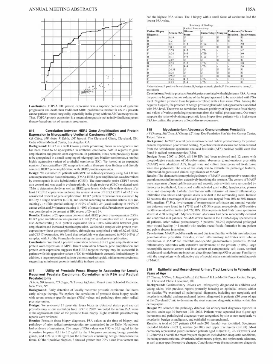
ANNUAL MEETING ABSTRACTS 185A
Conclusions: TOP2A IHC protein expression was a superior predictor of systemic progression and death than traditional MIB1 proliferative marker in GS ≥ 7 prostate cancer patients treated surgically, especially in the group without ERG overexpression. Thus, TOP2A protein expression is a potential prognostic tool to individualize adjuvant therapy based on risk of systemic progression.
816 Correlation between HER2 Gene Amplification and Protein Expression in Micropapillary Urothelial Carcinoma (MPC)CB Ching, MB Amin, R Tubbs, DE Hansel. The Cleveland Clinic, Cleveland, OH; Cedars-Sinai Medical Center, Los Angeles, CA.Background: HER2 is a well known growth promoting factor in oncogenesis and has been found to be up-regulated in urothelial carcinoma, both in regards to gene amplification and protein over-expression. In particular, it has been previously found to be upregulated in a small sampling of micropapillary bladder carcinomas, a rare but highly aggressive variant of urothelial carcinoma (UC). We looked at an expanded number of micropapillary UC samples to confirm these previous findings and directly compare HER2 gene amplification with HER2 protein expression.Design: We evaluated 20 patients with MPC on radical cystectomy using 3-4 1.0 mm cores represented on tissue microarray (TMA). HER2 gene amplification was determined by chromogenic in situ hybridization (CISH). The centromeric probe CEP17 served as a control and was used to evaluate ploidy. A single reviewer (CBC) evaluated each TMA to determine ploidy as well as HER2 gene levels. Only cells with evidence of at least 2 CEP17 copies were included for review. A ratio of HER2:CEP17 of >2.2 was considered evident of gene amplification. HER2 protein expression was evaluated by IHC by a single reviewer (DEH), and scored according to standard criteria as 0 (no staining), 1+ (faint partial staining in >10% of cells), 2+ (weak staining in >10% of cancer cells), and 3+ (intense staining in >10% of cancer cells). Protein over-expression was considered to be present at IHC scores of 2+ and 3+.Results: Thirteen of 20 specimens demonstrated HER2 protein over-expression (65%). HER2 gene amplification was present in 11/20 (55%) of samples with all 11 samples also demonstrating 2-3+ protein expression for a 100% correlation between gene amplification and increased protein expression. We found 2 samples with protein over-expression without gene amplification, although one sample had a ratio of 3:2 of HER2 and CEP17 expression. We found that polyploidy was demonstrated in 9/20 (45%) of samples, with 5 of the 9 samples demonstrating increased gene amplification.Conclusions: We found a positive correlation between HER2 gene amplification and protein over-expression in MPC. Direct correlation between gene amplification and protein over-expression suggests that HER2-targeted therapy may be successful in patients with this aggressive variant of UC for which there is currently limited therapy. In addition, a large proportion of patients demonstrated polypoidy within tumor specimens, suggesting an inherent genomic instability in these patients.
817 Utility of Prostatic Fossa Biopsy in Assessing for Locally Recurrent Prostate Carcinoma: Correlation with PSA and Radical ProstatectomyJ Chow, DB Samadi, PD Unger, HJ Lavery, GQ Xiao. Mount Sinai School of Medicine, New York, NY.Background: Early detection of locally recurrent prostatic carcinoma facilitates early salvage therapy. We explore the correlation of prostatic fossa biopsy results with serum prostate-specific antigen (PSA) values and pathology from prior radical prostatectomies.Design: We reviewed 15 prostatic fossa biopsies obtained status post radical prostatectomy at our institution from 1997 to 2009. We noted the serum PSA value at the approximate time of the prostatic fossa biopsy. Eight available prostatectomy reports were reviewed.Results: Prostatic fossa biopsy diagnoses, PSA values at the time of biopsy, and pathology of prior radical prostatectomies are summarized in the Table. No patients had evidence of metastases. The range of PSA values was 8.85 to 30.1 ng/ml for the 4 positive biopsies, 0.11 to 3.2 ng/dl for the 7 biopsies containing benign prostatic glands, and 0.24 to 3.78 ng/ml for the 4 biopsies containing benign fibroconnective tissue. Of the 4 positive biopsies, 3 showed greater than 70% tissue involvement and
had the highest PSA values. The 1 biopsy with a small focus of carcinoma had the lowest PSA value.
Summary of FindingsRadical Prostatectomy
Patient Biopsy Diagnosis Serum PSA Gleason
Score Tumor Stage Margins Perineural Invasion
% Tumor Involvement
B 0.19 U U U U UB 1.2 U U U U UB 0.4 3+4 pT2c - + 50P 30.1 U U U U UF 3.78 4+3 pT3a - + 42F 0.24 4+4 pT3b - + 60B 0.11 3+4 pT2c + + 55F 2.07 U U U U UB 0.53 3+3 pT2a - + 20B 3.2 4+4 pT2a - - 20P 24.31 U U U U UB 0.38 U U U U UP 19.58 U U U U UF 0.45 4+3 pT3a - + 15P 8.85 3+4 pT2c - - UAbbreviations: P, positive for carcinoma; B, benign prostatic glands; F, fibroconnective tissue; U, unavailableConclusions: Positive prostatic fossa biopsies correlated with a high serum PSA. Among the positive biopsies, tumor volume of the biopsy appeared to be associated with PSA level. Negative prostatic fossa biopsies correlated with a low serum PSA. Among the negative biopsies, the presence of benign prostatic glands did not appear to be associated with PSA level. There was no correlation between positivity of the prostatic fossa biopsy and values of various pathologic parameters from the radical prostatectomy. Our study supports the value of obtaining a prostatic fossa biopsy from patients with a high serum PSA to confirm the presence of local disease recurrence.
818 Mycobacterium Abscessus Granulomatous ProstatitisAY Chuang, MH Tsou, SJ Chang, LY Yang. Koo Fundation Sun Yat-Sen Cancer Center, Taipei, Taiwan.Background: In 2007, several patients who received radical prostatectomy for prostate cancers experienced poor wound healing. Mycobacterium abscessus had been cultured from the debridement specimens and acid fast stain (AFS)-positive bacilli were also found in radical prostatectomies (RPs).Design: From 2007 to 2009, all 180 RPs had been reviewed and 12 cases with morphologies suspicious of Mycobacterium abscessus granulomatous prostatitis (MAGP) were identified. AFS, fungal stain and culture from preserved fresh tissue were also performed. The aim of this study was to evaluate the pathologic features, differential diagnosis and clinical significance of MAGP.Results: The characteristic morphologic feature of MAGP was suppurative necrotizing granulomatous inflammation extensively involving the prostate. The centers of MAGP were large areas of neutrophilic abscess and necrotic debris which were surrounded by histiocytes (epithelioid, foamy, and multinucleated giant cells), lymphocytes, plasma cells, and eosinophils. Lobular distribution with extension of mixed inflammatory infiltration into dilated and ruptured ducts is evident in the surrounding areas. In these 12 patients, the percentage of involved prostate area ranged from 10% to 80% (mean: 39%, median: 37.5%). Involvement of extraprostatic soft tissue and seminal vesicle/vas deferens were found in 9 (75%) and 4 (33.3%) cases, respectively. AFS-positive bacilli were identified in five (41.7%) RPs. Eleven patients had fresh tissue specimens stored at -150 centigrade. Mycobacterium abscessus had been successfully cultured and confirmed in 8 patients. No MAGP was found in the TRUS-biopsy specimens in all patients. After radical prostatectomy, 8 patients (66.7%) experienced prolonged poor wound healing (> 1 month) with urethra-rectal fistula formation in one patient and pelvic abscess in another.Conclusions: MAGP could be easily missed due to unfamiliar with this rare infectious granulomatous prostatitis. Besides, mixed inflammatory infiltrates with lobular distribution in MAGP can resemble non-specific granulomatous prostatitis. Mixed inflammatory infiltrates with extensive involvement of the prostate (>10%), large neutrophilic necrotic centers and involvement of extraprostatic soft tissue, seminal vesicles and vas deferens are important clues for performing AFS or culture. Familiarity with the morphology with adjunctive use of special stains can minimize misdiagnosis of MAGP.
819 Epithelial and Mesenchymal Urinary Tract Lesions in Patients ≤30 Years of AgeST Chuang, M Zhou, C Magi-Galluzzi, DE Hansel. H Lee Moffitt Cancer Center, Tampa, FL; Cleveland Clinic, Cleveland, OH.Background: Genitourinary lesions are infrequently diagnosed in children and young adults, with previous reports primarily focusing on epithelial lesions within the bladder. We examined all pathological diagnoses, including non-neoplastic and neoplastic epithelial and mesenchymal lesions, diagnosed in patients ≤30 years of age at the Cleveland Clinic to determine the most common diagnostic entities within this demographic.Design: We searched the pathology database for urinary tract diagnoses reported in patients under age 30 between 1981-2008. Patients were separated into 5-year age increments and pathological diagnoses were categorized by site as non-neoplastic vs neoplastic, benign vs malignant, and epithelial vs mesenchymal.Results: A total of 507 patients (304 male:203 female) was identified. Specimens included bladder (n=211), urethra (n=108) and upper tract/ureter (n=188). Most commonly represented groups included patients aged 0-5(n=118), 26-30(n=107), and 21-25(n=95). Overall, the most frequent diagnoses included non-neoplastic conditions, including ureteral stricture, diverticula, inflammatory polyps, and nephrogenic adenoma, as well as non-specific reactive changes. Condylomas were the most common diagnosis
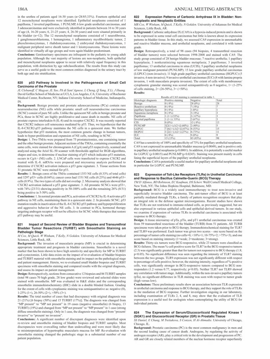
186A ANNUAL MEETING ABSTRACTS in the urethra of patients aged 16-30 years (n=28/85;33%). Fourteen epithelial and 12 mesenchymal neoplasms were identified. Epithelial neoplasms consisted of 1 papilloma, 3 inverted papillomas, 1 PUNLMP, 6 low grade urothelial carcinomas, and 3 adenocarcinomas and were exclusively identified in patients between 16 to 30 years of age (4, 16-20 years; 6, 21-25 years; 4, 26-30 years) and were situated primarily in the bladder (n=12). The 12 mesenchymal neoplasms consisted of 1 neurofibroma, 1 ganglioneuroblastoma, 1 hemangioma, 1 inflammatory myofibroblastic tumor, 2 pheochromocytomas, 1 rhabdomyosarcoma, 2 embryonal rhabdomyosarcomas, 1 malignant peripheral nerve sheath tumor and 1 leiomyosarcoma. These lesions were identified in virtually all age groups and were again bladder-predominant.Conclusions: Genitourinary specimens are infrequent in the pediatric to young adult population. Although the vast majority of lesions are non-neoplastic, both epithelial and mesenchymal neoplasms appear to occur with relatively equal frequency in this population, with distinction by age subclassification. The results from this study can serve as a useful guide to the most common entities diagnosed in the urinary tract by both age and site stratification.
820 p53 Pathway Is Involved in the Pathogenesis of Small Cell Carcinoma of the ProstateJL Clebanoff, C Magyar, JL Yao, PA di Sant’Agnese, L Cheng, Q Yang, X Li, J Huang. David Geffen School of Medicine at UCLA, Los Angeles, CA; University of Rochester Medical Center, Rochester, NY; Indiana University School of Medicine, Indianapolis, IN.Background: Benign prostate and prostate adenocarcinoma (PCa) contain rare neuroendocrine (NE) cells while prostatic small cell neuroendocrine carcinomas (SCNC) consist of pure NE cells. Unlike the quiescent NE cells in benign prostate and PCa, those in SCNC are highly proliferative and cause death in months. NE cells of prostate express interleukin-8 (IL-8) and its receptor CXCR2. It was recently reported that CXCR2 induces cell senescence mediated by p53. Thus, we hypothesize that the IL-8-CXCR2-p53 pathway maintains the NE cells in a quiescent state. We further hypothesize that p53 mutation, the most common genetic change in human tumors, leads to hyper-proliferation and expansion of NE cells, resulting in SCNC.Design: 1. Two TMAs were prepared from 75 prostatectomies, one containing tumor and the other benign prostate. Adjacent sections of the TMAs, containing essentially the same cells, were stained for chromogranin A (CgA) and p53 respectively, scanned and analyzed using the Ariol SL-50 scanner with thresholds applied for RGB algorithms, shapes and size, to determine if nuclear staining of p53 which indicates p53 mutation, occurs in CgA+ (NE) cells. 2. LNCaP cells were transfected to express CXCR2 and treated with IL-8. mRNAs were prepared and microarray analysis performed to determine if CXCR2 activation induces a p53 gene signature. 3. Tissue sections from 31 prostatic SCNC were stained for p53.Results: 1. Benign cores of the TMAs contained 1353 NE cells (0.33% of total cells) and 3297 p53+ cells (0.84%); cancer cores had 1331 NE cells (0.23%) and 4646 p53+ cells (0.9%). The two types of cells did not overlap. 2. Microarray analysis showed that CXCR2 activation induced a p53 gene signature. 3. All prostatic SCNCs were p53+, with 74% (23/31) showing positivity in 50-100% cells and the remaining 26% (8/31) being positive in 5-50% cells.Conclusions: 1. In normal prostate and PCa, activation of CXCR2 by IL-8 activates p53 pathway in NE cells, maintaining them in a quiescent state. 2. In prostatic SCNC, p53 mutation results in inactivation of the IL-8-CXCR2-p53 pathway and hyperproliferation and aggressive behavior of the NE cells. 3. In contrast to PCa, hormonal therapy targeting androgen receptor will not be effective for SCNC while therapies that restore p53 pathway may be useful.
821 Impact of Second Review of Bladder Biopsies and Transurethral Bladder Tumor Resections (TURBT) with Smoothelin Staining on Pathologic StageM Cox, M Quick, W Winham, T Kelly, N Gokden. University of Arkansas for Medical Sciences, Little Rock, AR.Background: The invasion of muscularis propria (MP) is crucial in determining appropriate treatment and prognosis in bladder carcinoma. Smoothelin is a novel marker that has been shown to have high sensitivity and specificity for MP in TURBT and cystectomies. Little data exists on the impact of re-evaluation of bladder biopsies and TURBT material with smoothelin staining and its impact on the pathological stage and patient management. Herein, we re-evaluated small bladder biopsies and TURBT specimens with smoothelin staining and compared results with the original diagnosis, and assess its impact on patient management.Design: Retrospectively, sections from consecutive 12 biopsies and 86 TURBT samples (total 98 cases-70 high grade, 28 low grade) were reviewed and selected slides were stained with smoothelin. MP was evaluated in H&E slides and the corresponding smoothelin immunohistochemistry (IHC) slide in a double blinded fashion. Grading for the extent of cells with cytoplasmic staining was semiquantitative as: negative (0), 1-25% (1+), 26-50% (2+), >50% (3+).Results: The total number of cases that had discrepancy with original diagnosis was 21 (21%) [6 biopsy (50%) and 15 TURBT (17%)]. The diagnosis was changed from “MP present” to “MP not present” in 19 cases (negative smoothelin) (6 biopsy, 13 TURBT) (19%) and changed from “MP not present” to “MP present” in 1 case (strong diffuse smoothelin staining). Only in 1 case, the diagnosis was changed from “present/ invasive” to “present/ no invasion”.Conclusions: A significant number of discrepant diagnoses were identified upon re-review and smoothelin staining of bladder biopsies and TURBT samples. Most discrepancies were overcalling rather than undercalling and were most likely due to misinterpretation of hypertrophic muscularis mucosa for MP. Re-evaluation with smoothelin staining changed the pathologic stage in a substantial number of our patient population.
822 Expression Patterns of Carbonic Anhydrase IX in Bladder: Non-Neoplastic and Neoplastic EntitiesMR Cox, W Winham, M Quick, T Kelly, N Gokden. University of Arkansas for Medical Sciences, Little Rock, AR.Background: Carbonic anhydrase IX (CA9) is a hypoxia-induced protein and is shown to be expressed in some renal cell carcinomas but little is known about its expression patterns in bladder tissue. In this study, we assessed CA9 expression in unremarkable and reactive bladder mucosa, and urothelial neoplasms, and correlated it with tumor grade.Design: Retrospectively, a total of 98 cases (94 biopsies, 4 transurethral resection of bladder tumors) were selected between 1998-2008 and stained with CA9. The study group consisted of 28 benign bladder mucosae; 7 reactive urothelia; 1 papillary hyperplasia; 3 nonkeratinizing squamous metaplasia; 3 papillomas; 3 inverted papillomas; 13 urothelial carcinoma in situs (UCIS); 5 papillary urothelial neoplasms of low malignant potential (PUNLMP); 15 low grade papillary urothelial carcinomas (LGPUC) (non-invasive); 11 high grade papillary urothelial carcinomas (HGPUC) (5 invasive, 6 non-invasive); 9 invasive urothelial carcinomas (IUC) (8 with lamina propria invasion, 1 with muscularis propria invasion). The extent of cells with membranous/cytoplasmic granular staining was scored semiquantitively as 0 negative, 1+ (1-25% of cells staining, 2+ (26-50%), 3+ (>50%).Results:
Results of CA9 staining is summarized in table 1.Histologic diagnosis Positive staining Negative TotalBenign 0 ( 0%) 28 (0%) 28Reactive urothelium 0 ( 0%) 7 ( 0%) 7papillary hyperplasia 0 ( 0%) 1 (0%) 1Squamous metaplasia 0 ( 0%) 3 ( 0%) 3Papilloma 1 (1+) (33%) 2 ( 67%) 3Inverted papilloma 1 (1+) ( 33%) 2 (67%) 3CIS 0 (0%) 13 ( 0%) 13PUNLMP 3[2(1+);1(2+)] (60%) 2 (40%) 5LGPUC 7 [6(1+);1(2+)] (47%) 8 (53%) 15HGPUC 3 [2(1+);1(2+) (27%) 8 (73%) 11IUC 0 ( 0%) 9 (0%) 9Total 15 (15%) 83 (85%) 98
CA9 has a sensitivity of 100% and specificity of 73% for papillary urothelial neoplasms. CA9 is not expressed in unremarkable bladder mucosa (p 0.0049), and is positive only in papillary urothelial neoplasms (p 0.0001). In addition, it is potentially a useful marker for LGPUC (p 0.0015) and PUNLMP (p 0.0245). Staining was present mainly in cells lining the superficial layers of the papillary urothelial neoplasms.Conclusions: CA9 is potentially a useful marker for papillary urothelial neoplasms and in particular for LGPUC and PUNLMP.
823 Expression of Toll-Like Receptors (TLRs) in Urothelial Carcinoma and Response to Bacillus Calmette-Guerin (BCG) TherapyTM D’Alfonso, BD Robinson, EC Kaufman, DS Scherr. Weill Cornell Medical College, New York, NY; The Johns Hopkins Hospital, Baltimore, MD.Background: BCG is a widely used immunotherapy to treat non-invasive and superficially invasive bladder carcinoma. The anti-tumor effect of BCG is at least partially mediated through TLRs, a family of pattern recognition receptors that play an integral role in the defense against microorganisms. Recent studies have shown that TLRs are not restricted to immune-related cells, as previously suggested, but are also expressed in various epithelial cells and epithelial-derived tumors. In our study, we examine if expression of various TLRs in urothelial carcinoma is associated with response to BCG therapy.Design: A tissue microarray of pTa, pTis, and pT1 urothelial carcinomas was created from 59 transurethral resections of the bladder (TURB) from 51 patients. All TURB specimens were taken prior to BCG therapy. Immunohistochemical staining for TLR7 and TLR9 was performed. Each tumor was given two scores – one score based on the percentage of tumor cells staining (no cells=0; <10%=1; 10-75%=2; >75%=3), and one score based on staining intensity (1=weak; 2=moderate; 3=strong).Results: Thirty-six tumors were BCG-responsive, while 23 tumors were classified as BCG-failures. The mean %-cell positive score for TLR7 in the BCG-responsive tumors (2.3) was significantly greater than that for tumors not responsive to BCG therapy (1.8; p<0.05). No significant difference was seen regarding the intensity of TLR7 staining between the two groups. TLR9 expression was not significantly different with respect to percentage of cells positive; however, the staining intensity, regardless of %-positive cells, was significantly stronger in BCG-responsive tumors compared to BCG non-responders (1.2 versus 0.77, respectively; p<0.05). Neither TLR7 nor TLR9 showed any correlation with tumor stage. Additionally, within the non-invasive papillary tumors (pTa), no significant difference in TLR staining was seen with respect to tumor grade (high versus low).Conclusions: These preliminary results show an association between TLR expression in urothelial carcinoma and response to BCG therapy, and they support the role of TLRs in the mediation of BCG response. Further investigation ongoing in our laboratory, including examination of TLRs 2, 4, and 8, may show that the evaluation of TLR expression is a useful tool for urologists when contemplating the utility of BCG for individual patients.
824 The Expression of Serum/Glucocorticoid Regulated Kinase 1 (SGK1) and Glucocorticoid Receptor (GR) in Prostatic TissueSS Daniel, E Chung, M Tretiakova, S Conzen, H Al-Ahmadie. University of Chicago, Chicago, IL.Background: Prostatic carcinoma (PC) is the most common malignancy in men and the second leading cause of cancer death. Androgens, by regulating the activity of androgen receptor (AR), play a critical role in the development and progression of PC. AR and GR are closely related members of the nuclear hormone receptor superfamily
ANNUAL MEETING ABSTRACTS 187Awhose role in PC has not been well understood. SGK1, a Ser/Thr protein kinase, was recently identified as an AR-induced gene and is also required for GR-mediated cell survival signaling, but its exact role in PCs has not been explored. We analyzed the expression of AR, SGK1 and GR by immunohistochemistry (IHC) in a cohort of PC patients treated by radical prostatectomy (RP).Design: Tissue microarrays containing 141 RP specimens with varying Gleason grades were stained by IHC for AR, SGK1 and GR. The stains were assessed semi-quantitatively in both PC and adjacent nonneoplastic prostatic tissue (NP). Any staining was recorded. Intensity was graded on a scale from 0-3 representing negative, weak, moderate and strong, respectively.Results: Results are summarized in Table 1. All 3 markers exhibited nuclear localization, with SGK1 additionally showing cytoplasmic expression. AR and SGK1 were diffusely expressed in all NP and PC with no significance difference in expression according to Gleason grade. In contrast, GR was detected in 33% of PC and in 65% of NP. In NP, SGK1 and GR were expressed in nuclei of acinar as well as basal cells in varying proportions. Interestingly, SGK1 was uniformly more strongly staining PC compared to NP.
Table 1. Percentage of IHC expression of SGK1, AR, and GR in prostatic tissueSGK1 (%) AR (%) GR (%)
PC (n =132) 132 (100) 132 (100) 44 (33)NP (n=138) 138 (100) 138 (100) 90 (65)
Conclusions: Increased SGK1 expression in PC compared to NP suggests a potential role in the development and progression of PC. SGK1 expression is not related to the level of differentiation of PC as determined by Gleason grade. The strong correlation between SGK1 and AR expression supports the hypothesis that AR-mediated induction of SGK1 expression may contribute to the development and progression of PC and offers a potential novel pathway for targeted therapy. Unlike AR and SGK1, GR was not expressed ubiquitously. Therefore, GR may play a role favoring growth in a specific subset of PC.
825 Does the Size Matter? – Prostate Weight Does Not Predict PSA Recurrence after Radical ProstatectomyDD Davidson, MO Koch, H Lin, L Cheng. Indiana University School of Medicine, Indianapolis, IN; Yale University, New Haven, CT.Background: Previous studies suggest that low prostate weight is a significant negative prognostic guide for prostate cancer assessment. If this observation were sustained by independent clinicopathologic studies, then prostate size would contribute a valuable non-invasive parameter for preoperative patient staging.Design: 431 men who underwent radical retropubic prostatectomy between 1990 and 1998 were analyzed for association between prostate weight and various clinical or pathological parameters. These included age, preoperative PSA level, PSA recurrence, pathological stage, Gleason grade, extraprostatic extension, positive surgical margins, tumor volume, associated high grade PIN, perineural invasion and lymph node metastasis. Potential associations between evaluated parameters and clinical outcome were probed by Cox regression analysis.Results: A significant positive correlation was found between prostate weight and increasing patient age or increasing preoperative PSA level. There was no significant independent association between prostate weight and any of the other variables examined, including PSA recurrence. Preoperative PSA and Gleason biopsy grade were significant independent predictors of pathologic stage, Gleason prostatectomy grade, positive surgical margins and PSA recurrence.Conclusions: Although increasing prostate weight correlates with both increased patient age and higher preoperative PSA level, it does not independently predict favorable postoperative clinical outcome.
826 Soft Tissue Neoplasms of the Male External Genitalia: An Institutional Review of 110 CasesBC Dickson, DJC Howarth, KPH Pritzker, RA Kandel. Mount Sinai Hospital; University of Toronto, Toronto, ON, Canada.Background: Soft tissue neoplasms involving the female external genitalia have been well-characterized in the literature; however, comparatively less is known about those occurring in adult male counterparts. The purpose of this study was to examine both benign and malignant soft tissue neoplasm affecting the external male genitalia.Design: We performed an institutional review (1999-2009) to identify all cases of soft tissue neoplasms affecting the penis, perineum, scrotum and spermatic cord.Results: One hundred and ten soft tissue neoplasms affecting the male external genitalia were identified in the following locations: groin (57), paratesticular region (6), penis (2), perineum (3), pubic region (5), scrotum (10) and spermatic cord (27). The most commonly encountered neoplasms included liposarcoma (23), lipoma (11), pleomorphic undifferentiated sarcoma (11), leiomyosarcoma (9), dermatofibrosarcoma protuberans (8), benign genital stromal tumors [angiomyofibroblastoma and cellular angiofibroma] (7), nodular fasciitis (4), fibrosarcoma (3), synovial sarcoma (3), solitary fibrous tumour (2), angiosarcoma (2), hemangioma (2), low-grade fibromyxoid sarcoma (2) and rhabdomyosarcoma (2). In addition, single numbers of other tumors were also identified. In 11 cases malignant tumors could not be further classified due to the limited size of the biopsy or treatment effect.Conclusions: Aside from case reports and case series, few reviews exist that characterize the incidence of soft tissue neoplasms involving the male external genitalia. Acknowledging the likelihood of a referral bias for a tertiary care center, we found that both malignant and benign fatty tumors were most commonly observed at this location. Given the substantive number of undifferentiated sarcomas also encountered, it is possible that some of these cases may, in fact, represent dedifferentiated liposarcoma. Tumors within the family of benign genital stromal tumors were noted to have a morphologic and immunohistochemical profile relatively similar to those occurring
in females. An awareness for the nature of soft tissue tumors occurring in these locations, along with their potential mimics, is of diagnostic relevance to pathologists and clinicians alike.
827 Automated Image Analysis of Microvascular Density in Clear Cell Renal Cell Carcinoma and Its Prognostic UtilityW Dubinski, V Iakovlev, M Gabril, Y Youssef, K Kovacs, S Metias, M Mankaruous, GM Yousef. St. Michael’s Hospital, Toronto, ON, Canada; London Health Sciences Center, London, ON, Canada.Background: Tumor microvascular density (MVD) has been shown to correlate with the aggressiveness of several cancers and adjuvant anti-angiogenic therapy is currently being used. The significance of tumor vascularity in clear cell renal cell carcinoma (ccRCC) has been debated, with various publications showing contradictory results. Previous publications have been limited by manual quantification of MVD within only a small area of tumor. To overcome this, we employed a standardized image analysis approach, and normalized the data by assessing the cellular density of the tumor; we then assessed the vascularity of ccRCC and compared it with clinical outcome data.Design: Clinico-pathologic data was collected for 50 cases of ccRCC. Each tumor and adjacent normal kidney underwent immunostaining for CD34 and whole slide scanning using an Aperio CS Scanner (Vista, CA) at 20x magnification. Then, computer image analysis was used to assess MVD and nuclear density within tumoral, and normal cortical and medullary tissue. We generated sets of normalized and raw non-normalized vascular data and tested these for correlations with clinical prognostic parameters and survival data.Results: Tumors with increased MVD showed a statistically significant association with higher tumor stage compared to tumors with lower vascularity (p=0.017). There was a trend toward increased MVD in higher versus lower grade tumors. Increased MVD showed a statistically significant correlation with poor prognosis and a decrease in progression free survival versus those tumors with low MVD (p=0.016). In the univariate analysis, using MVD as a categorical variable, there was a hazard ratio of 9.14 (95% CI, 1.02-82.00). This was not statistically significant when used as a continuous variable. In the multivariate analysis, patients with highly vascular tumors had an increased hazard ratio of 4.82 compared to those with lower CD34 expression (95% CI, 0.51-45.6; p=0.17). There was a positive correlation between MVD and tumor size (rs=0.456; p=0.001).Conclusions: The latest developments in digitized histology can be used for objective standardized assessment of prognostic markers. Our standardized assessment revealed that increased MVD in ccRCC was associated with higher tumor stage and size and a decreased disease free survival. These results may prove useful in the development of new prognostic tests and anti-cancer strategies.
828 Micropapillary Urothelial Carcinoma of the Urinary Bladder: A Clinicopathologic Analysis of 24 CasesN Edgerton, SJ Sirintrapun, MJ Munoz, AO Osunkoya. Emory University School of Medicine, Atlanta, GA.Background: Micropapillary urothelial carcinoma (MUCa) of the urinary bladder is relatively rare. We report one of the largest series to date of MUCa with follow up information.Design: The surgical pathology and expert consultation files at our institution were searched for MUCa of the bladder diagnosed in transurethral resection of bladder tumor (TURBT) and radical cystectomy/cystoprostatectomy specimens from 2002 to 2009. Only patients with available follow up information were selected. Tumor volumes were stratified based on percentage of the micropapillary component (MC) <50%, >50% and 100%. Other pathologic parameters were obtained on all patients.Results: 24 cases of MUCa with available follow up data were identified. The mean patient age was 71 years (range 55-86 years), with a male to female ratio of 3:1. 6 cases (6/24; 25%) were composed entirely of MC; 12 cases (12/24. 50%) showed >50% MC; and 6 cases (6/24; 25%) showed <50% MC. 7 cases (7/24; 29%) were also composed of variable amounts of nested, clear cell, signet ring cell pattern and adenocarcinoma. Of the 14 patients that opted for radical cystectomy/cystoprostatectomy as monotherapy, the majority (10/14; 71%) had pT3 or more advanced staged disease. 4 patients (4/24; 17%) had neoadjuvant chemotherapy prior to radical cystectomy/cystoprostatectomy and showed minimal residual disease with urothelial carcinoma in situ. 3 patients (3/24; 13%) had local extension into surrounding organs (prostate, cervix, ovary) pT4 showing 100% MC. 19 cases (19/24; 79%) had angiolymphatic invasion. Of the 16 cases in which nodal status was known, 9 (9/16; 56%) were positive; 5 cases (5/9; 55%) showing metastases composed entirely of MUCa. Mean duration of follow up of all patients was 16 months (range 1-59 months). 11 patients (11/24; 46%) died of disease. The tumors in most patients that died of disease 9/11(81%) were composed of 100% MUCa or >50% MUCa. Mean survival time was 14 months.Conclusions: Our study confirms that MUCa is typically aggressive and presents with advanced stage disease in most cases. MUCa may also be associated with other aggressive variants of urothelial carcinoma. We recommend reporting the percentage of MC in these tumors in view of the direct relationship to adverse clinical outcome. It is also critical to make an accurate diagnosis of this entity especially in relatively small biopsies or TURBT specimens, because urologists may proceed to cystectomy/cystoprostatectomy based on pT1 disease.
188A ANNUAL MEETING ABSTRACTS 829 Digital Slides for Standardization of Gleason Grading by International ExpertsL Egevad, F Algaba, DM Berney, L Boccon-Gibod, E Comperat, AJ Evans, R Grobholz, G Kristiansen, C Langner, G Lockwood, A Lopez-Beltran, R Montironi, P Oliveira, M Schwenkglenks, B Vainer, M Varma, V Verger, P Camparo. Karolinska Inst, Stockholm, Sweden; Fundacio Puigvert-Univ, Barcelona, Spain; St Barths Hospital, London, United Kingdom; Armand Trousseau, Paris, France; Pitié-Salpetrière, Paris, France; Univ Toronto, Toronto, Canada; Saarland Univ Hospital, Homburg, Germany; Univ Hospital, Zurich, Switzerland; Medical Univ, Graz, Austria; CPAC, Toronto, Canada; Cordoba Univ, Cordoba, Spain; Polytechnic Univ Marche Region, Ancona, Italy; Hospital da Luz, Lisboa, Portugal; Univ Hospital, Basel, Switzerland; Rigshospitalet, Copenhagen, Denmark; Univ Hospital, Cardiff, United Kingdom; CCITI, Dijon, France; Hopital Foch, Paris, France.Background: Our aims were to analyze reporting of GP 3 and 4 when using the ISUP 2005 revision of Gleason grading, to identify interpretation difficulties, and to collect consensus cases for standardization.Design: A set of 25 NBX cores diagnosed as GS 6-7 cancer were scanned. Only GS 6 cases that were borderline to GS 7 were included. 15 uropathology experts graded the digital slides and encircled any GP 4 and 5 in the slide reader. Grading difficulty was scored as 1-3. GP 4 components were classified as Type 1 (cribriform), 2 (fused) or 3 (poorly formed glands). After individual review, the experts met to anlyze diagnostic difficulties and agree on a set of consensus cases.Results: A GS 5-6, 7 (3+4), 7 (4+3), 8-9 was given in 29%, 41%, 19% and 10% (mean GS 6.84, range 6.44-7.36). In 15 cases, at least 67% of observers agreed on GS groups. Mean weighted kappa of interobserver reproducibility for GS and GS groups was 0.346 and 0.429. A difficulty score of 1, 2 and 3 was given in 58%, 32% and 10%. Means in consensus and non-consensus cases were 1.44 and 1.66 (p = 0.003). When a GP 4 was reported, Types 1, 2 and 3 were seen in 28%, 86% and 67%. Types 2 and 3 were found together in 41% and all 3 co-existed in 16% (11% and 23% in consensus and non-consensus cases, p = 0.03). Average estimated and calculated %GG4/5 were 29% and 16%. Areas of GP 4 and 5 were displayed as heat maps with overlaying regions. The maps were helpful for identifying contentious areas. A key problem was to agree on minimal criteria for small foci of GP 4.Conclusions: There is still considerable disagreement among experts on how to report borderline GS 6-7 cases. The detection threshold for minimal foci of GP 4 in NBX needs to be better defined.
830 CK7 and Claudin 7 Are Superior to Other Markers in Distinguishing Oncocytoma from Chromophobe Renal Cell Carcinoma – Automatic Imaging Analysis and Hierarchical Clustering AnalysisLJ Eisengart, SM Rohan, V Parimi, JJ Wei, XJ Yang. Northwestern University, Chicago, IL.Background: Distinguishing oncocytoma (ONC) from chromophobe renal cell carcinoma (CHR) can be a diagnostic challenge. The utility of immunohistochemical (IHC) stains in differentiating these tumor subtypes has been an active area of investigation. However, to date, a specific marker or panel of markers that would be useful in this regard has remained elusive. We assessed the diagnostic utility of a panel of IHC markers comparing ONC and CHR using a digital imaging system and hierarchical clustering analysis.Design: Tissue microarray (TMA) sections composed of 61 CHR, 34 ONC, and 12 normal kidneys were evaluated with a panel of markers previously reported to help distinguish ONC from CHR: CK7, claudin 7, vimentin, cyclin-D1, and H2AF. An additional marker identified by microarray analysis, SYNPR3, was also evaluated. Slides were scanned and staining intensity was determined by the ChromoVision Automatic Cellular Imaging System (ACIS II). Hierarchical clustering analysis was performed using MayDay microarray data analysis software.Results: Clustering analysis using the full panel of 6 markers did not separate ONC from CHR. However, using a 2 marker panel of CK7 and Claudin 7, clustering analysis distinguished CHR, ONC, and normal kidney. The dendrogram showed 4 clusters: ONC, normal kidney, and two discrete clusters of CHR. The majority of ONC (29 of 34) clustered together. Most normal kidney samples (9 of 12) clustered together. The majority of CHR clustered together on one branch (44 of 61), while a separate group of 8 cases clustered together on a different branch which was more closely related, by branch node, to the ONC. Histologically, these 8 cases were classic CHR. Further, the difference in mean staining intensity of ONC and CHR was statistically significant for both CK7 (p<0.001) and Claudin 7 (p<0.001).Conclusions: Digital imaging and hierarchical clustering analysis provides a more objective measure of an immunomarker in distinguishing oncocytoma from chromophobe RCC. We found that a panel of CK7 and Claudin 7 established a staining signature that can distinguish oncocytoma from chromophobe RCC by hierarchical clustering. This was superior to an extended 6 marker panel, in which the staining patterns of the additional 4 markers did not generate sufficient distinguishing power. This method is promising as an aid in establishing accurate diagnoses, although further study is needed to evaluate the clinical utility of these findings.
831 Histopathology and Response of Bladder Cancer to Neoadjuvant TherapyLJ Eisengart, SM Rohan, G MacVicar, XJ Yang. Northwestern University, Chicago, IL.Background: Radical cystectomy is the mainstream treatment for muscle-invasive urothelial carcinoma. Several large randomized trials have suggested a survival benefit for neoadjuvant therapy followed by cystectomy as compared to cystectomy alone. As neoadjuvant therapy becomes more common, it will be important to document response of different carcinoma variants to these therapies and to recognize treatment associated
histologic changes. We evaluated the histology of post-neoadjuvant cystectomy specimens as compared to the diagnostic biopsy.Design: Slides from the diagnostic biopsies and post-chemotherapy cystectomy specimens from 14 patients were reviewed. Tumor grade, histologic subtype, presence of divergent differentiation, depth of invasion, and lymphovascular invasion (LVI) were recorded for both biopsy and cystectomy specimens. Percent tumor necrosis, presence of foreign body giant cells, and TNM stage at cystectomy were also recorded.Results: All biopsies showed high grade urothelial carcinoma with muscularis propria invasion. At cystectomy, 6 of 14 patients (43%) showed a good histologic response to chemotherapy: 3 (21%) showed no residual tumor, 1 contained only non-invasive papillary carcinoma, and 2 had a single focus of microscopic tumor in the superficial muscularis propria. Five patients showed some response to chemotherapy, with 10-50% tumor necrosis. Three patients showed no response to chemotherapy. Those with some or no response had pT3 or pT4 disease. 6 cases had LVI on biopsy, 4 of which had none at cystectomy. Eight patients had no LVI on biopsy but 3 of those did at cystectomy. Patients with lymph node metastases were evenly distributed across groups with residual tumor. Only 1 of 6 with good response was of a specific subtype (micropapillary), whereas 4 of 5 with some response and 3 of 3 with no response showed divergent differentiation or belonged to a specific histologic variant (plasmacytoid, micropapillary, small cell neuroendocrine carcinoma) (p=0.026, two-tailed Fisher’s exact test).Conclusions: In this series, neoadjuvant therapy resulted in either no or microscopic residual invasive carcinoma in 43% of the cystectomy specimens. LVI was eradicated in 4 of 6 cases. Our results suggest that neoadjuvant therapy might be less effective for histologic variants of urothelial carcinoma and in cases with divergent differentiation. Larger studies will be valuable to substantiate these findings and to design more effective neoadjuvant therapy protocols.
832 Hybrid Oncoctyic Tumors of the Kidney in Patients with Birt-Hogg-Dubé (BHD) SyndromeLJ Eisengart, M Tretiakova, SM Rohan, J Zhang, M Zhou, C Luan, BT Teh, XJ Yang. Northwestern University, Chicago, IL; University of Chicago, Chicago, IL; Mayo Clinic, Rochester, MN; Cleveland Clinic, Cleveland, OH; Van Andel Research Institute, Grand Rapids, MI.Background: BHD is a rare autosomal dominant disorder characterized by cutaneous fibrofolliculomas, renal tumors and pulmonary cysts. Oncocytic hybrid tumors represent 50% of BHD associated renal tumors. Studies characterizing these hybrid tumors are limited; expression microarray, immunohistochemistry (IHC) and electron microscopy (EM) have never been reported to our knowledge.Design: We studied 12 hybrid tumors from 8 patients with BHD syndrome. Gene expression microarrays were performed on 4 cases and compared to our established database of kidney neoplasms. Histopathologic and IHC features (CA9, vimentin, c-Kit and CK7) were evaluated. EM was performed on one case.Results: Histologically, hybrid tumors showed prominent cell membranes and dense eosinophilic cytoplasm, similar to both chromophobe RCC and oncocytoma. Hybrid tumors contained scattered clear epithelial cells of low Fuhrman grade intermingled with oncocytic cells, composing from 5 to 25% of cells. By molecular clustering analysis, hybrid tumors were located between the cluster of 7 chromophobe renal cell carcinomas (RCCs) and the cluster of 7 oncocytomas. Comparative genomic microarray analysis showed minimal chromosomal losses in hybrid tumors similar to oncocytomas; chromophobe RCCs show numerous chromosomal losses. Hybrid tumors were positive for C-kit and showed patchy Ck7 positivity, different from the diffuse CK7 positivity in chromophobe RCCs or CK7 negativity in oncocyotmas. The intermingled clear cells were negative for clear cell RCC markers such as vimentin or CA9. By EM, hybrid tumor cells contained small membrane-bound microvesicles (similar to chromophobe RCC) and abundant pleomorphic mitochondria (typical of oncocytoma). Some tumor cells contained cytoplasmic lipid vacuoles as typically seen in clear cell RCC.Conclusions: In this study, the molecular signature of BHD associated hybrid oncocytic tumors has been defined. We also report here the morphological and immunohistochemical characteristics of these tumors. Recognition of hybrid tumors is important, as they suggest the presence of BHD syndrome. These hybrid tumors should be considered a distinct subtype based on their morphologic, molecular genetic and immunohistochemical characteristics.
833 Increasing Levels of a Mitochondrial Protein Marker Accompany Prostatic CarcinogenesisCL Ellis, TC Cornish, C Koh, JL Hicks, B Gurel, AM De Marzo. The Johns Hopkins University School of Medicine, Baltimore, MD.Background: Mitochondrial DNA (mtDNA) copy number changes are commonly found in human cancers. In prostatic adenocarcinoma (CaP), studies evaluating mtDNA content have reported mixed results. The purpose of this study was use immunohistochemistry (IHC) and image analysis to determine whether increased mitochondria accompany prostatic carcinogenesis.Design: Using a commercial antibody that recognizes mitochondria by IHC (MTC02, Abcam), we examined tissue microarrays (TMAs) from radical prostatectomies containing normal prostate tissue (253 cores from 59 patients), atrophy (129 cores from 28 patients), PIN (130 cores from 38 patients) and CaP (75 cores from 38 patients) using the TMAJ/FrIDA image analysis program. Since MYC has been shown to stimulate mitochondrial biogenesis, we correlated the expression levels with MYC protein from the same TMAs. The Gleason score ranged from 5-9 and pathological stages ranged from organ confined (OC) (T2N0Mx) to locally metastatic disease (T3BN1Mx).Results: Image analysis revealed an increase in the intensity of staining (MitoInt) in focal atrophy (121.9; p= 0.0001), PIN (123.5, p=0.0001) and CaP (118.6, p=0.001) when compared to matched normal (107.7). Overall mitochondrial area per total nuclear area (MitoRatio) showed a stepwise increase in the median values going from
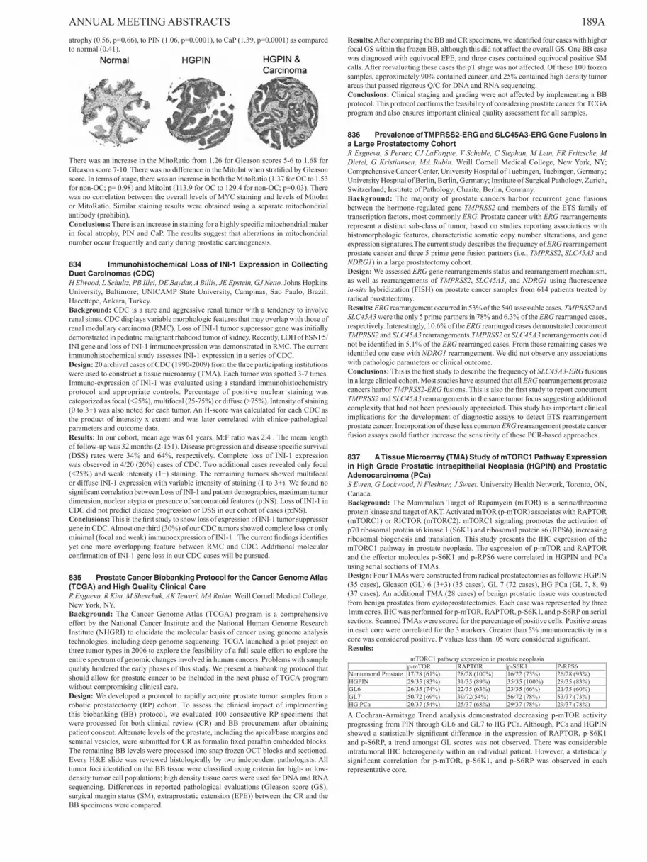
ANNUAL MEETING ABSTRACTS 189Aatrophy (0.56, p=0.66), to PIN (1.06, p=0.0001), to CaP (1.39, p=0.0001) as compared to normal (0.41).
There was an increase in the MitoRatio from 1.26 for Gleason scores 5-6 to 1.68 for Gleason score 7-10. There was no difference in the MitoInt when stratified by Gleason score. In terms of stage, there was an increase in both the MitoRatio (1.37 for OC to 1.53 for non-OC; p= 0.98) and MitoInt (113.9 for OC to 129.4 for non-OC; p=0.03). There was no correlation between the overall levels of MYC staining and levels of MitoInt or MitoRatio. Similar staining results were obtained using a separate mitochondrial antibody (prohibin).Conclusions: There is an increase in staining for a highly specific mitochondrial maker in focal atrophy, PIN and CaP. The results suggest that alterations in mitochondrial number occur frequently and early during prostatic carcinogenesis.
834 Immunohistochemical Loss of INI-1 Expression in Collecting Duct Carcinomas (CDC)H Elwood, L Schultz, PB Illei, DE Baydar, A Billis, JE Epstein, GJ Netto. Johns Hopkins University, Baltimore; UNICAMP State University, Campinas, Sao Paulo, Brazil; Hacettepe, Ankara, Turkey.Background: CDC is a rare and aggressive renal tumor with a tendency to involve renal sinus. CDC displays variable morphologic features that may overlap with those of renal medullary carcinoma (RMC). Loss of INI-1 tumor suppressor gene was initially demonstrated in pediatric malignant rhabdoid tumor of kidney. Recently, LOH of hSNF5/INI gene and loss of INI-1 immunoexpression was demonstrated in RMC. The current immunohistochemical study assesses INI-1 expression in a series of CDC.Design: 20 archival cases of CDC (1990-2009) from the three participating institutions were used to construct a tissue microarray (TMA). Each tumor was spotted 3-7 times. Immuno-expression of INI-1 was evaluated using a standard immunohistochemistry protocol and appropriate controls. Percentage of positive nuclear staining was categorized as focal (<25%), multifocal (25-75%) or diffuse (>75%). Intensity of staining (0 to 3+) was also noted for each tumor. An H-score was calculated for each CDC as the product of intensity x extent and was later correlated with clinico-pathological parameters and outcome data.Results: In our cohort, mean age was 61 years, M:F ratio was 2.4 . The mean length of follow-up was 32 months (2-151). Disease progression and disease specific survival (DSS) rates were 34% and 64%, respectively. Complete loss of INI-1 expression was observed in 4/20 (20%) cases of CDC. Two additional cases revealed only focal (<25%) and weak intensity (1+) staining. The remaining tumors showed multifocal or diffuse INI-1 expression with variable intensity of staining (1 to 3+). We found no significant correlation between Loss of INI-1 and patient demographics, maximum tumor dimension, nuclear atypia or presence of sarcomatoid features (p:NS). Loss of INI-1 in CDC did not predict disease progression or DSS in our cohort of cases (p:NS).Conclusions: This is the first study to show loss of expression of INI-1 tumor suppressor gene in CDC. Almost one third (30%) of our CDC tumors showed complete loss or only minimal (focal and weak) immunoexpression of INI-1 . The current findings identifies yet one more overlapping feature between RMC and CDC. Additional molecular confirmation of INI-1 gene loss in our CDC cases will be pursued.
835 Prostate Cancer Biobanking Protocol for the Cancer Genome Atlas (TCGA) and High Quality Clinical CareR Esgueva, R Kim, M Shevchuk, AK Tewari, MA Rubin. Weill Cornell Medical College, New York, NY.Background: The Cancer Genome Atlas (TCGA) program is a comprehensive effort by the National Cancer Institute and the National Human Genome Research Institute (NHGRI) to elucidate the molecular basis of cancer using genome analysis technologies, including deep genome sequencing. TCGA launched a pilot project on three tumor types in 2006 to explore the feasibility of a full-scale effort to explore the entire spectrum of genomic changes involved in human cancers. Problems with sample quality hindered the early phases of this study. We present a biobanking protocol that should allow for prostate cancer to be included in the next phase of TGCA program without compromising clinical care.Design: We developed a protocol to rapidly acquire prostate tumor samples from a robotic prostatectomy (RP) cohort. To assess the clinical impact of implementing this biobanking (BB) protocol, we evaluated 100 consecutive RP specimens that were processed for both clinical review (CR) and BB procurement after obtaining patient consent. Alternate levels of the prostate, including the apical/base margins and seminal vesicles, were submitted for CR as formalin fixed paraffin embedded blocks. The remaining BB levels were processed into snap frozen OCT blocks and sectioned. Every H&E slide was reviewed histologically by two independent pathologists. All tumor foci identified on the BB tissue were classified using criteria for high- or low-density tumor cell populations; high density tissue cores were used for DNA and RNA sequencing. Differences in reported pathological evaluations (Gleason score (GS), surgical margin status (SM), extraprostatic extension (EPE)) between the CR and the BB specimens were compared.
Results: After comparing the BB and CR specimens, we identified four cases with higher focal GS within the frozen BB, although this did not affect the overall GS. One BB case was diagnosed with equivocal EPE, and three cases contained equivocal positive SM calls. After reevaluating these cases the pT stage was not affected. Of these 100 frozen samples, approximately 90% contained cancer, and 25% contained high density tumor areas that passed rigorous Q/C for DNA and RNA sequencing.Conclusions: Clinical staging and grading were not affected by implementing a BB protocol. This protocol confirms the feasibility of considering prostate cancer for TCGA program and also ensures important clinical quality assessment for all samples.
836 Prevalence of TMPRSS2-ERG and SLC45A3-ERG Gene Fusions in a Large Prostatectomy CohortR Esgueva, S Perner, CJ LaFargue, V Scheble, C Stephan, M Lein, FR Fritzsche, M Dietel, G Kristiansen, MA Rubin. Weill Cornell Medical College, New York, NY; Comprehensive Cancer Center, University Hospital of Tuebingen, Tuebingen, Germany; University Hospital of Berlin, Berlin, Germany; Institute of Surgical Pathology, Zurich, Switzerland; Institute of Pathology, Charite, Berlin, Germany.Background: The majority of prostate cancers harbor recurrent gene fusions between the hormone-regulated gene TMPRSS2 and members of the ETS family of transcription factors, most commonly ERG. Prostate cancer with ERG rearrangements represent a distinct sub-class of tumor, based on studies reporting associations with histomorphologic features, characteristic somatic copy number alterations, and gene expression signatures.The current study describes the frequency of ERG rearrangement prostate cancer and three 5 prime gene fusion partners (i.e., TMPRSS2, SLC45A3 and NDRG1) in a large prostatectomy cohort.Design: We assessed ERG gene rearrangements status and rearrangement mechanism, as well as rearrangements of TMPRSS2, SLC45A3, and NDRG1 using fluorescence in-situ hybridization (FISH) on prostate cancer samples from 614 patients treated by radical prostatectomy.Results: ERG rearrangement occurred in 53% of the 540 assessable cases. TMPRSS2 and SLC45A3 were the only 5 prime partners in 78% and 6.3% of the ERG rearranged cases, respectively. Interestingly, 10.6% of the ERG rearranged cases demonstrated concurrent TMPRSS2 and SLC45A3 rearrangements.TMPRSS2 or SLC45A3 rearrangements could not be identified in 5.1% of the ERG rearranged cases. From these remaining cases we identified one case with NDRG1 rearrangement. We did not observe any associations with pathologic parameters or clinical outcome.Conclusions: This is the first study to describe the frequency of SLC45A3-ERG fusions in a large clinical cohort. Most studies have assumed that all ERG rearrangement prostate cancers harbor TMPRSS2-ERG fusions. This is also the first study to report concurrent TMPRSS2 and SLC45A3 rearrangements in the same tumor focus suggesting additional complexity that had not been previously appreciated. This study has important clinical implications for the development of diagnostic assays to detect ETS rearrangement prostate cancer. Incorporation of these less common ERG rearrangement prostate cancer fusion assays could further increase the sensitivity of these PCR-based approaches.
837 A Tissue Microarray (TMA) Study of mTORC1 Pathway Expression in High Grade Prostatic Intraepithelial Neoplasia (HGPIN) and Prostatic Adenocarcinoma (PCa)S Evren, G Lockwood, N Fleshner, J Sweet. University Health Network, Toronto, ON, Canada.Background: The Mammalian Target of Rapamycin (mTOR) is a serine/threonine protein kinase and target of AKT. Activated mTOR (p-mTOR) associates with RAPTOR (mTORC1) or RICTOR (mTORC2). mTORC1 signaling promotes the activation of p70 ribosomal protein s6 kinase 1 (S6K1) and ribosomal protein s6 (RPS6), increasing ribosomal biogenesis and translation. This study presents the IHC expression of the mTORC1 pathway in prostate neoplasia. The expression of p-mTOR and RAPTOR and the effector molecules p-S6K1 and p-RPS6 were correlated in HGPIN and PCa using serial sections of TMAs.Design: Four TMAs were constructed from radical prostatectomies as follows: HGPIN (35 cases), Gleason (GL) 6 (3+3) (35 cases), GL 7 (72 cases), HG PCa (GL 7, 8, 9) (37 cases). An additional TMA (28 cases) of benign prostatic tissue was constructed from benign prostates from cystoprostatectomies. Each case was represented by three 1mm cores. IHC was performed for p-mTOR, RAPTOR, p-S6K1, and p-S6RP on serial sections. Scanned TMAs were scored for the percentage of positive cells. Positive areas in each core were correlated for the 3 markers. Greater than 5% immunoreactivity in a core was considered positive. P values less than .05 were considered significant.Results:
mTORC1 pathway expression in prostate neoplasiap-mTOR RAPTOR p-S6K1 P-RPS6
Nontumoral Prostate 17/28 (61%) 28/28 (100%) 16/22 (73%) 26/28 (93%)HGPIN 29/35 (83%) 31/35 (89%) 35/35 (100%) 29/35 (83%)GL6 26/35 (74%) 22/35 (63%) 23/35 (66%) 21/35 (60%)GL7 50/72 (69%) 39/72(54%) 56/72 (78%) 53/37 (73%)HG PCa 20/37 (54%) 25/37 (68%) 29/37 (78%) 29/37 (78%)
A Cochran-Armitage Trend analysis demonstrated decreasing p-mTOR activity progressing from PIN through GL6 and GL7 to HG PCa. Although, PCa and HGPIN showed a statistically significant difference in the expression of RAPTOR, p-S6K1 and p-S6RP, a trend amongst GL scores was not observed. There was considerable intratumoral IHC heterogeneity within an individual patient. However, a statistically significant correlation for p-mTOR, p-S6K1, and p-S6RP was observed in each representative core.
190A ANNUAL MEETING ABSTRACTS Conclusions: p-mTOR, RAPTOR, p-S6K1, and p-S6RP were highly expressed in HGPIN and may provide novel targets for its treatment. A decreasing trend in p-mTOR IHC was observed progressing from HGPIN to HG PCa. The extent of mTOR expression in an individual patient would determine the effective use of mTOR inhibitors as a potential therapeutic strategy.
838 Correlation between Fuhrman Grading in Percutaneous Needle Biopsy and Surgical Resection Specimens in Clear Cell Renal Carcinoma ≤ 4 cm in SizeL Fahim, A Finelli, P Ryan, M Jewett, J Kachura, AJ Evans. University Health Network, Toronto, Canada; Mount Sinai Hospital, Toronto, Canada.Background: Renal cell carcinoma (RCC) is the most common primary malignant tumor of kidney in adults, the vast majority of which (< 80%) are of conventional or clear cell (CC-RCC) type. The Fuhrman grade (FG), a 4-tier grading scheme based on nuclear features, is the reference histoprognostic grading system most widely used for CC-RCC.) The prognostic value of FG for patients with RCC has been well-characterized. Recently, percutaneous needle biopsy has gained popularity for establishing the diagnosis of incidentally detected small renal masses (SRM) < 4 cm in size. Since it is possible that this procedure may under-represent the true FG as a result of limited sampling, we compared FG between paired biopsy and subsequent resection specimens in a consecutive series of SRM CC-RCC cases.Design: We identified 30 patients with SRM who underwent percutaneous biopsy showing CC-RCC between 2004 and 2009. All patients subsequently underwent partial or radical nephrectomy. The original slides from the biopsies and resection specimens were reviewed to determine the correlation between biopsy and final FG. If multiple FG were present in the same specimen, the highest FG was considered to be the overall FG.Results: There were 11 female and 19 male patients. Median tumor size was 2.5 cm (range 1.4 - 4) and the median age at presentation was 58 years (range 32 - 78). The average time between biopsy and surgery was 3.7 months. There was exact agreement between biopsy and resection specimen FG in 29 of 30 cases (96%). Upon collapsing the FG system into either low grade (FG 1 and 2) or high grade (FG 3 and 4), we found 100% agreement between biopsy and resection FG. No cases showing heterogeneity with respect to low- and high-grade areas were identified in this SRM CC-RCC series.Conclusions: Percutaneous renal tumor biopsies are safe, cost-effective and most often conclusive for histological diagnosis of SRM. Our experience indicates that percutaneous biopsy will provide highly accurate FG information for CC-RCC < 4 cm in size. This information may be critical in terms of choosing the optimal treatment for SRM CC-RCC that are incidentally detected, particularly in elderly or infirm patients where it may be desirable to avoid or delay surgical intervention.
839 Immunohistochemical Expression of Secretory Clusterin (sCLU) in Benign and Malignant Prostate in the Context of Hormonal TherapyL Fahim, N Cockburn, A Joshua, P Warde, J Sweet. University Health Network, Toronto, Canada.Background: Clusterin (apolipoprotein J) is an androgen-repressed oncogenic molecule involved in cell cycle regulation, cell survival and DNA repair. In prostate cancer (PCa), secretory Clusterin (sCLU) is linked to androgen resistance and post therapy cell survival. Anti-sense oligonuleotide repression of CLU gene has been shown to inhibit prostate cancer progression in phase-1 clinical trials. We investigated the immunohistochemical (IHC) expression of sCLU in benign prostate epithelium, surgically treated PCa with no prior therapy (SRx), hormone responsive PCa (HRsv) and hormone resistant PCa (HR).Design: Prostate tissue from 10 cystoprostatectomies with no cancer, 25 (HR) cases from TURPS, 24 (HRsv) cases from radical prostatectomies (RP) and 72 SRx cases including, 35 Gleason score (GS) 6 and 37 high grade (HG) (GS 8,9,10) cases were used to construct 5 TMAs. Each case was represented by 3, 1mm cores. TMAs were stained with H&E and IHC was performed for sCLU. Epithelial expression of sCLU was scored as negative(0), weak(+1) or strong(+2) in each core. The summed scores from a case formed the composite score for that case and a mean score for each array was calculated.Results: sCLU was not observed in benign prostate epithelium. sCLU was differentially expressed in SRx, HR and HRsv. sCLU was expressed in SRx PCa (GS 6 and HG) and a statistically significant increase in sCLU was observed in HG PCa compared to GS 6 PCa (p<0.0001). HRsv PCa showed significantly less expression of sCLU compared to HR PCa. HR PCa highly expressed sCLU at a level comparable to that of HG PC with no statitistical difference between these 2 groups.
IHC expression of sCLU in different study groupsstudy groups NO of patients mean score of sCLU staining in each groupBenign prostate 10 0Gleason 6 35 1.1HG 37 3.7HRsv 24 2.0HR 25 3.72
Conclusions: sCLU expression was stronger in HG compared to GS 6, suggesting a positive correlation between sCLU and GS. Increased expression of sCLU may impart a cell survival advantage and may influence the aggressive nature of a specific PCa. Increased expression of sCLU in HR compared to HRsv may be linked to androgen resistance, implicating sCLU as a putative prosurvival factor. These findings are clinically relevant with respect to prognostication and the development of novel therapeutic strategies to inhibit sCLU.
840 Identification of Gleason Pattern 5 on Prostatic Needle Core Biopsy: Frequency of Underdiagnosis and Morphologic AnalysisDA Fajardo, TK Lee, JS Miller, H Miyatmoto, JI Epstein. Johns Hopkins Medical Institution, Baltimore, MD.Background: It has been our anectdotal experience that pathologists underdiagnose Gleason pattern 5 prostate cancer, possibly because of hesitance to assign a grade which has such adverse prognostic implications. The current study assesses the accuracy of grading of Gleason pattern 5 and potential factors contributing to undergrading.Design: From the consult service of one of the authors, we identified 59 consecutive needle biopsy cases comprising 138 parts that upon review were graded as having Gleason pattern 5. All cases were reported as the final diagnosis by the outside pathologist. They were sent for 2nd opinion by clinicians or patients and not because the pathologist was seeking a 2nd opinion.Results: Upon review, the final Gleason score was increased in 101/138 (73.2%), decreased 5/137 (3.6%), and remained unchanged in 32/138 (23.2%). Gleason pattern 5 was correctly identified by the outside pathologist in 71/138 parts (51.4%). The architectural patterns of pattern 5 were: single cells (n=104, 75.3%), solid (n=69, 50%), cords (n=62, 44.9%), and comedonecrosis (n=3, 2.2%). In the 1st column of the table is the total number of cases with each Gleason score. The 2nd column is the number of cases undergraded for each Gleason score. The next 4 columns show the number of undergraded cases for each pattern. For example, with Gleason 4+5=9, of the 18 cases primarily composed of single cells, 14 were undergraded.
Undergrading of Pattern 5: By Morphology
Grade Total Cases Underdiagnosis Mostly Single Cells
Mostly Solid
Mostly Cords
Mostly Comedo
3+5=8 10/138 (7.2%) 8/10 (80%) 3/4 (75%) 2/3 (67%) 3/3 (100%)4+5=9 56/138 (40.6%) 43/56 (76.8%) 14/18 (78%) 17/23 (74%) 10/13 (77%) 2/2 (100%)Primary 5 72/138 (52.2%) 15/72 (20.8%) 9/30 (30%) 2/30 (7%) 4/15 (27%)
Conclusions: Pattern 5 was missed in 48.6% of the prostate needle core biopsies. It is most often missed when it is not the primary pattern. The most commonly present Gleason pattern 5 architectural type was single cells and the least common was comedonecrosis. None of the architectural patterns appear to be incorrectly identified more than another. Most accurate grading was when the primary pattern was 5 and composed mostly of solid sheets. Due to the important prognostic and therapeutic implications of Gleason pattern 5, pathologists must be attuned to its varied patterns and that it often may represent a secondary component of the carcinoma.
841 Isolated Single Core High Grade Prostatic Adenocarcinoma Is Associated with Increased Likelihood of Extraprostatic Extension, Seminal Vesicle Invasion, Positive Surgical Margins and Lymphovascular Invasion at Radical ProstatectomyDA Fajardo, AW Partin, M Eisenberger, TL DeWeese, GJ Netto. Johns Hopkins University, Baltimore.Background: High grade PCa can rarely be diagnosed as a single focus involving a single biopsy core. It is of clinical interest to determine whether such limited high grade prostatic adenocarcinoma biopsy involvement carry the same aggressive implications in regard to risk of extraprostatic extension (EPE), seminal vesicle invasion (SVI) and likelihood of positive margin status (MG+) on radical prostatectomy (RRP).Design: 92 PCa patients diagnosed with a standard ultrasound guided prostate needle biopsy procedure revealing only a single positive core and undergoing subsequent prostatectomy were retrieved from our surgical pathology archives (1999-2008). Cases were of four Gleason score (GS) groups: 3+3=6 (group1), 3+4=7 (group2), 4+3=7 (group3) and > 8 (group4). Bx data on pt age, pre-biopsy PSA (prePSA), GS, percentage of core involvement (Pos%), and total number of cores performed per case were collected. In addition, prostatectomy data on GS, extent of RRP involvement by PCa (RRPext), EPE, margin status, SVI, lymphovascular invasion (LVI), and lymph node metastasis (LN) were gathered on all cases.Results: No statistically significant difference was observed among the four biopsy groups in: Total number of cores obtained, prePSA or Pos% (p:NS). A significantly higher rate of EPE was observed in group 4 compared to groups 1-3 (50.0%, 4.3%, 8% and 30% respectively; p<0.0006). Likewise, a significantly higher incidence of positive MG status (p<0.03), SVI (p<0.003), and LVI (p<0.008) was also observed in group 4 compared to the remaining groups. Furthermore, when cases in group 4 were stratified, according to their %Pos involvement of their single positive core using 50% as a cutoff, the incidence of EPE was higher in those with PCa involving > 50% of one core compared to cases with cancer involving < 50% (p<0.0001). The latter cases in turn had a higher incidence of EPE than the groups of cases with <GS7.Conclusions: Isolated single biopsy core GS >8 prostatic adenocarcinoma is associated with a higher incidence of adverse RRP findings (EPE, SVI, LVI, and positive margins) when compared to cases with a single biopsy core containing PCa with GS<7. Cases with GS>8 involving less than 50% of a single core are still more likely to have EPE on RRP compared to those with a single GS <7 core cases.
842 Stage Migration in Prostate Cancer at Radical Prostatectomy during the PSA Era Is Associated with Relatively Constant Gleason ScoreSM Falzarano, M Zhou, AV Hernandez, EA Klein, C Magi-Galluzzi. Cleveland Clinic, Cleveland, OH; Lerner Research Institute, Clevaland, OH.Background: Prostate specific antigen (PSA) screening has been associated with a sharp increase in prostate cancer (PCA) detection after its introduction in the late 1980s. Increase PCA detection has been associated with decreased incidence of high-grade, high-risk PCA with a stage migration toward organ confined (OC) disease.Design: 471 patients who underwent radical prostatectomy (RP) for clinically localized PCA between 1987 and 2004 were included in the study. Cases were selected using data from patients with (n=125) and without (n=375) clinical recurrence after RP. All surgical specimens were reviewed blindly by a pathologist, staged and graded
ANNUAL MEETING ABSTRACTS 191Aaccording to the 2005-ISUP consensus conference on Gleason grading. Gleason score (GS) and pathologic stage (T) distribution was examined in the early (1987-1998) vs. late (1999-2004) PSA-era.Results: Patients mean age was 62.9 and 60.9 years in early-PSA-era and late-PSA-era, respectively (p=0.001). Mean pre-operative PSA (iPSA) was 12.14 and 6.96 ng/ml in early-PSA-era and late-PSA-era, respectively (p<0.0001). Compared with the original report 19.7% of cases were upgraded, 2.1% downgraded and 2.5% upstaged from T2 to T3. GS and stage distribution among PSA-eras is reported in Table 1.
Table 1PSA eraearly (n=228) late (n=243) p value
GS≤6 (17%) 37 44 * p=0.2; ** p=05GS7 (46%) 97 118GS≥8 (37%) 94 81T2 (38%) 63 117 * p<0.00003; ** p=0.003T3 (53%) 141 107T2+ (9%) 24 19T2+: T2 with margins positiveThere was no difference in the numbers of cases in the GS categories between PSA periods neither by univariable (Chi-Square*), or by multivariable logistic regression analysis while adjusting for iPSA and age (**). A significant difference was found in the numbers of cases in T stages categories between the two PSA-eras (* p<0.00003); such difference was still significant after adjustment for iPSA and age (** p=0.003) (Table 1).Conclusions: PCA screening with lower PSA cutoffs has lead to a stage migration with a significant increase in the percentage of OC PCA in RP specimens, whereas the GS distribution and the percentage of high-grade PCA has remained relatively constant overtime. This finding challenges the hypothesis of tumor progression.
843 ERG Rearrangement Is Present in a Subset of Transition Zone Prostatic TumorsSM Falzarano, M Navas, K Simmerman, MA Rubin, EA Klein, M Zhou, C Magi-Galluzzi. Cleveland Clinic, Cleveland, OH; Summa Health System, Akron, OH; Cornell University, New York, NY; University of Siena, Siena, Italy.Background: Most prostate cancers (PCA) exhibit a unique chromosomal aberration in chromosome 21. In approximately 90% of cases, the rearrangement is characterized by the fusion of the protein TMPRSS2 with the oncogene ERG. A recent study has shown that the TMPRSS2-ERG gene fusion is lacking in PCA arising from the transition zone (TZ) of the prostate.Design: A tissue microarray of TZ-predominant PCA from 62 patients who underwent radical prostatectomies (RP) between 2004 and 2009 was constructed. Two separate tumor nodules, one in the TZ (n=62) and, when available, one in the peripheral zone (PZ) (n=35) were selected for gene fusion analysis. TMPRSS2-ERG fusion status was determined using a multicolor interphase fluorescence in situ hybridization assay for ERG break-apart. A nucleus with an ERG break-apart (indicative of a TMPRSS2-ERG fusion) shows split apart of one juxtaposed red-green signal pair resulting in a single red and green signal. The telomeric green signal may be lost due to deletion resulting in one yellow and one red signal in a nucleus with rearrangement through deletion.Results: The patients median age was 59 years (range 42-81). PCA Gleason score (GS) was 6 (34%), 7 (55%); 7 with pattern 5 (5%) and 9 (6%). The mean tumor volume was 294 mm2 (>2.0cc). Fifty-nine/62 cases were informative. TMPRSS2-ERG gene fusion was present in 11.9% (7/59) of TZ tumors, and in 34.3% (12/35) of PZ tumors. Gene fusion through deletion occurred in 4/7 TZ (57.1%) and 6/12 PZ (50%) cases. In three patients, the TMPRSS2-ERG gene fusion was present in both TZ and PZ tumors: in one case the rearrangement was associated with a deletion in both tumors; in the other two cases, a deletion was present either in the TZ or in the PZ tumor.Conclusions: PCA arising from TZ is biologically and genetically different from PZ tumors. Although ERG rearrangement is prevalent in PZ tumors, is present also in a subset of TZ prostate cancer (11.9%). The lower frequency of gene fusion in TZ PCA may suggest a different carcinogenic pathway. Further studies are needed to address whether such chromosomal aberration difference may account for the clinical and biological differences between TZ and PZ tumors.
844 Renal Cell Carcinoma with Hybrid Features of Conventional and Chromophil Cytomorphology: A Fluorescent In Situ Hybridization StudyH Faraji, EC Belanger, SJ Robertson, BN Nguyen, KT Mai. University of Ottawa, Ottawa General Hospital, Ottawa, ON, Canada.Background: We performed fluorescent in situ hybridization (FISH) to investigate the numeric change of chromosomes 7, 17 and Y, as well as loss of chromosome 3p in chromophil (papillary) renal cell carcinomas (PRCC) with extensive clear cell changes (CCC).Design: Consecutive cases of renal cell carcinoma (RCC) over a 12-year period were reviewed to identify PRCC with extensive CCC. Immunostaining for cytokeratin 7 (CK7) and AMACR were performed and FISH for chromosomes 7, 17, Y and 3p was done.Results: Of the total of 521 renal cell carcinomas (RCC) retrieved, there were 49 RCC having chromophil or papillary features that could be grouped into: a) group 1 (12 cases) - typical clear cell RCC (CRCC) with focal areas of papillary formation; b) group 2 (28 cases) - focal typical PRCC showing a papillary architecture with extensive CCC; c) group 3 (9 cases) - RCC with an admixture of eosinophilic/clear cytoplasm and a solid/papillary architecture. Group 1 showed negative immunoreactivity for CK7 and AMACR and absence of numeric chromosomal gains or losses of chromosomes 7/17 and Y. Groups 2 and 3 showed variable reactivity for CK7 and AMACR. The tumors from group 2 and 5 tumors from group 3 showed trisomies for chromosomes 7 and/or 17 with or without loss of chromosome Y. Loss of 3p was observed in groups 1 and 3, but not in group 2.
Conclusions: PRCC may show phenotypical CCC mimicking CRCC. In a small number of cases with mixed histopathological features, FISH is helpful in sub-typing RCC.
845 Fluorescent In Situ Hybridization of Clear Cell Renal Cell Carcinoma with Diffuse Cytokeratin 7 ImmunoreactivityH Faraji, EC Belanger, SJ Robertson, BN Nguyen, KT Mai. University of Ottawa, Ottawa General Hospital, Ottawa, Canada.Background: We studied the numeric gain or loss of chromosomes 7, 17, Y and 3p in clear cell renal cell carcinoma (CRCC) with diffuse immunoreactivity for cytokeratin 7 (CK7), which has been recently described.Design: Thirty diffusely CK7-immunoreactive and predominantly solid CRCC measuring less than 2 cm (small) in diameter and 3 diffusely CK7-immunoreactive and predominantly solid CRCC measuring from 3 to 6 cm (large) were submitted for fluorescent in situ hybridization (FISH).Results: Architecturally, the carcinoma consisted of small cysts and solid cell nests with tubules/acini with or without a papillary architecture. The nuclei were frequently and characteristically arranged in a row in an apical location of the tubular/cystic/papillary structures. None of the tumors were associated with distant metastasis or local recurrence. FISH demonstrated no numeric change of chromosomes 7/17 or Y. Loss of 3p25/3p14 was observed in focal areas in 12 small CK7-immunoreactive CRCC and in all 3 large CK7-immunoreactive CRCC. The areas in both small and large CK7-immunoreactive CRCC often displayed weak or negative CK7 immunoreactivity.Conclusions: CRCC characterized by diffuse CK7 positivity represent a subtype of CRCC with characteristic cytohistopathological morphology distinct from conventional CRCC, with an apparently favorable clinical outcome. There was loss of chromosome 3p in focal areas in many tumors. It remains to be investigated if these neoplasms represent an early stage of development of some CRCC.
846 Intracystic Oncocytic Papillary Renal Neoplasms: A Variant of Epithelial Papillary Neoplasms Distinct from Papillary Renal Cell CarcinomaH Faraji, KT Mai. University of Ottawa, Ottawa General Hospital, Ottawa, Canada.Background: We described 6 intracystic oncocytic papillary renal cell carcinoma (IOPRCC) that were histopathologically and cytogenetically distinct from the common chromophil (papillary) renal cell carcinoma (PRCC).Design: IOPRCC were identified the Anatomical files at our institution and were further characterized with immunostaining for AMACR, CK7 and fluorescent in situ hybridization (FISH) for chromosomes 7, 17, Y and 3p (loci 3p14 and 3p25).Results: Of the total of 659 renal cell carcinoma (RCC), there were 6 IOPRCC, measuring 1.5 to 6 cm (2.4±1.1) occurring in patients with male: female ratio of 5:1 and ages of 46-73 years (57± 8). All tumors were not associated with distant metastasis or local recurrence in a period of follow-up from 1 to 12 years. The IOPRCC consisted of oncocytic /eosinophilic cells forming characteristic papillae sparsely arranged and non-papillary architecture within the unilocular cysts. In 4 tumors, the neoplastic cells accounted for 30-50% of the cystic volume. The remaining two tumors displayed similar features in focal areas but occupied almost the entire cysts. The tumors displayed extensive foamy cytoplasm with or without focal clear cell changes in 4 cases. Capsular invasion was present in one case. Immunostaning was negative for progesterone receptor, cytokeratin 7, CD117 and carbonic anhydrase IX and positive for alpha-methylacyl-CoA racemase, vimentin, CD10 and RCC. FISH revealed no loss of chromosome 3p in all tumors and Y in all male patients and no trisomies of 7/17 in five tumors. One tumor (6 cm in diameter) showed trisomies 7/17 in focal areas.Conclusions: IOPRCC is a distinct variant of RCC due to the favourable clinical behaviour, the intracystic proliferation of sparsely arranged oncocytic papillae and the absence of chromosomal numeric changes of PRCC.
847 Value of Early Repeat Needle Biopsy in Assessing Patients with Low Risk Features on Initial Diagnostic Biopsy for Prostate CancerSW Fine, MZ Spira, HA Al-Ahmadie, A Gopalan, SK Tickoo, VE Reuter. MSKCC, NY, NY.Background: Active surveillance (AS) is increasingly utilized in patients with low risk prostate cancer. Low risk pathologic features on needle biopsy (NB) include: Gleason score (GS) < 6, < 2 positive cores, and < 50% cancer involvement on any core. The impact of early repeat extended NB for patients being considered for AS has not been well studied.Design: Since 1999, urologists at our institution have been performing early extended (> 12 core) NB on patients with initial diagnostic NB meeting criteria for low risk disease. We retrospectively studied 51 such cases among a well-characterized cohort of 285 patients who underwent both early extended (> 12 core) NB and subsequent radical prostatectomy to determine whether repeat NB can better stratify patients who might be eligible for AS.Results: Repeat NB findings: Group 1: 10/51 (20%) showed low risk findings; Group 2: 41/51 (80%) exceeded low risk criteria. In Group 1, all patients had GS 3+3=6, one (7/10) or two (3/10) positive cores, and 1-20% core involvement by cancer. Overall, 28/41 (68%) patients in Group 2 had GS > 6 (3+4=7: 23 patients; 4+3=7: 5 patients) and 80% had > 2 positive cores. Stratification of Group 2 revealed that 17 (42%), 14 (34%), and 10 (24%) patients exceeded one, two, or three low risk criteria, respectively. - Patients exceeding 1 criterion could be sub-stratified into those with: > 2 cores positive (range: 3-7) [n=9] GS > 6 (3+4=7: 6 patients; 4+3=7: 1 patient) [n=7] > 50% involvement of any core [n=1] - Patients exceeding 2 criteria showed either: GS > 6 (3+4=7: 9 patients; 4+3=7: 2 patients) and > 2 cores positive (range 3-9) [n=11] > 2 cores positive (range 3-10) and > 50% involvement of any core [n=3] - Patients exceeding all 3 low risk criteria [n=10] showed: GS > 6 (3+4=7: 8 patients; 4+3=7: 2 patients) > 2 cores positive (range 3-10) > 50% involvement of any core Pathologic stage at radical
192A ANNUAL MEETING ABSTRACTS prostatectomy: Group 1: all 10 patients had organ-confined disease (pT2) Group 2: 11/41 (27%) patients had non-organ-confined disease (10 pT3a; 1 pT3b)Conclusions: In this series, early repeat extended NB alone would have excluded up to 80% of patients initially diagnosed with low risk prostate cancer from eligibility for AS. Almost 70% of all repeat biopsies revealed GS > 6, 80% showed > 2 cores positive for cancer, and 20% exceeded all three low risk criteria. These findings strongly suggest that early repeat extended (> 12 core) NB in patients considered for AS may provide better discrimination of truly low risk patients.
848 Androgen Receptors Are Differentially Expressed in Gleason Patterns of Prostate Cancer and Down-Regulated in Matched Lymph Node MetastasesA Fleischmann, C Rocha, S Schobinger, R Seiler, GN Thalmann. University of Bern, Bern, Switzerland.Background: Androgen receptors (AR) play a central role in prostate cancer. However, heterogeneous distribution with AR negative areas is a constant finding. AR expression profile in the different Gleason patterns (GP) of primary tumors and nodal metastases is unknown and, furthermore, the prognostic potential of AR is controversial. More information about AR distribution is needed to optimize evaluation methods and to better understand the role of AR in development and progression of prostate cancer.Design: A tissue microarray (TMA) was constructed from 119 hormone-naïve nodal positive, surgically treated prostate cancers containing tissues from all GP present in every primary tumor and the matched metastases. AR expression score was determined immunohistochemically (percentage x staining intensity (1-3)) and correlated with various tumor features and biochemical recurrence-free, disease specific and overall survival.Results: ARs were up-regulated in primary tumors compared to normal glands and significantly different expressed in the GP (AR scores: GP3=134.8, GP4=157.7, GP5=123.5; p=0.016). A similar expression profile was observed in metastases, however, on significantly lower level (p<0.001). AR expression was not associated with any quantitative (age, prostate cancer volume, number and total size of metastases) or categorical (tumor stage, Gleason score of the primary tumor and metastases) tumor characteristics or with survival.Conclusions: ARs are differentially expressed in GP what should be considered in prognostic models which include AR. In nodal metastases, ARs are significantly down-regulated suggesting decreased dependence on androgens already under hormone-naïve conditions. The extremely low AR levels in metastatic GP5 might play a role in the predominance of this tissue component in hormone-refractory metastases. AR expression is no prognosticator in nodal positive disease.
849 Extensive Multifocal Prostatic Adenocarcinomas (Greater Than 20) in Radical Prostatectomy Specimens of Young MenB Furusato, P Parker, T Nydam, K Rice, S Srivastava, S Brassell, DG McLeod, IA Sesterhenn. Armed Forces Institute of Pathology, Washington, DC; Walter Reed Army Medical Center, Washington, DC; Uniformed Services University of the Health Sciences, Bethesda, MD.Background: Although the majority of prostatectomy specimens contain 3 to 4 tumors on average, it is rare to encounter greater than 20 tumors in prostatectomy specimens. The aim of this study is to characterize patients in the latter group.Design: Radical prostatectomy specimens of 1400 consecutive patients obtained from a single institution between 1993 and 2008 were analyzed retrospectively. For each specimen, the entire prostate was sectioned and embedded as whole-mounts. Each tumor was circled and characterized separately. The presence of prostatic intraepithelial neoplasia, atypical glands, atrophy etc. were recorded. Biochemical recurrence was defined as two consecutive values of serum PSA levels ≥ 0.2 ng/mL.Results: Among the 1400 consecutive radical prostatectomy specimens, 5 patients (0.36%) had more than 20 tumors. The median age was 42 years, and the median time of follow-up was 52 months. In 4 patients all tumors were well differentiated corresponding to a Gleason score of 6 (3+3). In one case, one of the tumors had a Gleason score of 7 (3+4, well and poorly differentiated). A number of tumors clustered in a specific region suggesting confluence to a single tumor and were measured as a single tumor. The median total tumor volume was 1.2 cc; all tumors were organ confined, surgical margin negative. Most of the tumors were located in the peripheral zone of the mid and apical thirds and were associated with prostatic intraepithelial neoplasia. In all of the patients, atypical glands were also seen. The median preoperative PSA was 2.8 ng/ml. To date, none of the patients has exhibited PSA recurrence.Conclusions: The large number of individual microscopic tumors and the focal confluence may indicate an early stage in the development of carcinoma and could explain the heterogeneity of a large tumor. The tumor with 40% poorly differentiated component did not cause PSA recurrence at this point.
850 Assessment of Circulating Tumor Cells (CTCs) in Low Tumor Volume Prostate Cancer PatientsB Furusato, A Ali, PO Ts’o, ZP Lum, S Srivastava, S Brassell, DG McLeod, IA Sesterhenn. Armed Forces Institute of Pathology, Washington, DC; Uniformed Services University of the Health Sciences, Bethesda, MD; Walter Reed Army Medical Center, Washington, DC; CCC Diagnostics, LLC., Baltimore, MD.Background: The objective of this study was to assess the incidence of circulating tumor cells (CTCs) in low tumor volume (less than 0.5 cc) prostate cancer patients after radical prostatectomy (RP).Design: Between 2000 and 2004, blood samples were collected in order to assess the CTCs from 64 consecutive radical prostatectomy patients. The prostatectomy specimens were processed by whole mounting and close step sectioning at 2.2 mm intervals. Clinicopathologic data were obtained, including patient age, race, clinical stage, biopsy
grade, preoperative PSA, prostatectomy data (specimen weight, tumor volume, grade, stage, and surgical margin status), and follow-up PSA data. These parameters were then compared with the CTCs status.Results: Nine of 64 RP patients had low volume prostate cancer. Among these, 7 patients had detectable levels of CTCs. In 2 of the 7 patients with positive CTCs, PSA elevation was observed. These 2 cases had a Gleason score of 3+3 (well differentiated tumor), organconfined disease and negative surgical margin for tumors.Conclusions: Isolation and detection of circulating epithelial cells is possible in low volume prostate cancer patients. In the setting of low volume prostate cancer, CTCs may be associated with measurable PSA levels. However, only a one time event of detecting CTCs did not predict PSA failure. The clinical significance of measuring CTCs has yet to be defined.
851 Tau Protein Expression in Recurrent and Non-Recurrent Prostatic AdenocarcinomaMP Gailey, G Lee, BR De Young. University of Iowa Hospitals and Clinics, Carver College of Medicine, Iowa City, IA.Background: Tau is a microtubule-associated protein whose principle function is in stabilizing and promoting microtubule (MT) assembly. Previous studies have investigated the relationship between tau protein expression and drug resistance in certain forms of carcinoma. These studies suggest that tau protein competes with antimotitic drugs for binding sites on MTs and thus renders resistance. However, another study has shown that in prostatic adenocarcinoma tau does not bind MTs, suggesting another mechanism of resistance. No studies to date that have examined the relationship of tau protein expression and outcome in prostatic adenocarcinoma.Design: We matched 26 patients with recurrent prostatic adenocarcinoma and 25 patients, who were free of recurrent prostatic adenocarcinoma after 10 years of follow-up, according to Gleason score and margin status at prostatectomy. Utilizing standard technique, immunohistochemical staining of formalin fixed paraffin embedded tissue from each patient’s prostatectomy specimen was performed with an antibody specific for tau-13 (B11E8, 1:12,500 dilution). Tau protein expression was recorded as the percentage of tumor cells that stained positive.Results: In patients with recurrent prostatic adenocarcinoma, a slightly higher percentage of tumor cells (52%) showed tau expression when compared to those without recurrence (46%). However, no statistical difference between cases of recurrent versus non-recurrent prostatic adenocarcinoma was identified (p=0.61). Moreover, expression of tau was variable, independent of Gleason score at prostatectomy (R2=0.02 and 0.003 for non-current and recurrent cases, respectively). Tau expression also varied within the same gland amongst benign acini, acini involved with high grade prostatic intraepithelial neoplasia and adenocarcinoma.Conclusions: These findings demonstrate that while cases of recurrent prostatic adenocarcinoma show slightly higher expression of tau protein expression, no statistical difference in expression was identified. Furthermore, Gleason score does not correlate with tau expression and there is variable staining for tau between both benign and malignant acini within the same gland. These results support the hypothesis that tau is a multi-functional protein that does not influence outcome prostatic adenocarcinoma.
852 Cell Surface Peptidases CD13 and CD10 in Renal Cell NeoplasmsS Gobbo, M Brunelli, D Segala, E Munari, M Chilosi, F Menestrina, G Martignoni. University of Verona, Verona, Italy.Background: Renal cell tumors originating from the proximal tubule, clear cell renal cell carcinoma and papillary renal cell carcinoma, show a worse behaviour than chromophobe renal cell carcinoma and renal oncocytoma which display differentiation toward distal nephron. CD10 has been described to be a specific marker for clear cell and papillary renal cell carcinoma even if its immunoexpression has been observed also in a consistent portion of chromophobe renal cell carcinomas and in some renal oncocytomas. CD13 is another surface peptidase and recently a monoclonal antibodies have been reported to be applicable to human paraffin-embedded specimens. The current study compares CD13 and CD10 protein expression by immunohistochemical and western blot analyses in a large series of renal cell tumors and evaluates their capability to distinguish “proximal renal cell tumors” from “distal renal cell tumors”.Design: We analyzed the immunoexpression of CD10 and CD13 in 166 renal cell neoplasms including 79 clear cell renal cell carcinomas, 34 papillary renal cell carcinomas, 25 chromophobe renal cell carcinomas and 24 renal oncocytomas. We performed western blot analysis to confirm the expression of these proteins.Results: Both CD13 and CD10 immunoreactions showed a membranous staining pattern. CD10 was observed in 94% of clear cell renal cell carcinomas, in 43% of papillary renal cell carcinomas, in 40% of chromophobe renal cell carcinomas and in 15% of renal oncocytomas. CD13 was expressed in 78% of clear cell renal cell carcinomas, and in 76% papillary renal cell carcinomas whereas neither chromophobe renal cell carcinomas nor renal oncocytomas showed any immunoreaction for CD13. Western blot analysis confirmed the presence of CD10 in chromophobe renal cell carcinoma and in renal oncocytoma and the protein expression of CD13 in clear cell renal cell carcinoma and papillary renal cell carcinoma.Conclusions: We concluded that CD13 is more sensitive and specific than CD10 to distinguish “proximal renal cell tumors”, clear cell and papillary renal cell carcinomas, from “distal renal cell tumors” such as chromophobe renal cell carcinomas and renal oncocytomas.
ANNUAL MEETING ABSTRACTS 193A
853 Carcinoid Tumours of the Kidney: A Clinico-Pathologic and 11q13 FISH AnalysisS Gobbo, M Brunelli, D Segala, I Alvarado-Cabrero, O Hes, G Pelosi, S Bersani, M Chilosi, F Menestrina, G Martignoni. University of Verona, Verona, Italy; Hospital de Oncología, Mexico, Mexico; Charles University Hospital, Pilsen, Czech Republic; European Institute of Oncology, Milan, Italy.Background: Renal carcinoid tumours rarely occur and a few small series have been reported with emphasis on clinico-pathological features. Little is known among cytogenetic abnormalities. Due to the high incidence of 11q13 riarrangements in neuroendocrine tumors arising in other human organs, we sought to assess 11q13 status in a serie of renal carcinoid tumours.Design: Clinico-pathologic information on seven cases were recruited. Immunohistochemical and FISH analyses, using 11q13 break-apart locus specific probe, were performed.Results: Patient age ranged from 35 to 74 years (average 56 y) (M:F; 5:2). Specimens consisted of radical nephrectomies with associated lymph node dissection in two cases. Five tumors were present in the left and 2 in the right kidney. Unifocal tumours ranged in size from 2.6 to 12 cm (average 5 cm). One tumour had history of a horseshoe kidney. Variety of patterns including tightly packed cords and trabeculae (80%) with minimal stroma, trabecular growth with prominent stroma, focal solid nests (15%), focal glandlike lumina (5%) were present. Calcifications were present in 3/7 cases. Mitoses measured 0-12 (mean 4) per 10 high-power fields. Necrosis was present in one case. Perirenal fat, renal sinus and renal vein invasions were documented in two cases whereas lymph-nodal metastases occurred in one. Hepatic and bone-marrow metastases were observed in two distinct cases. Carcinoid tumours were positive for synaptophysin (6/7) and CK8-18 (7/7) whereas chromogranin was expressed in 4 out of 7 cases. CK7 and CK20 were focally positive in 1 case; TTF-1 and DOG-1 were negative in all cases examined. Follow-up was available on 5 patients and ranged from 6 months to 4 years. One patient died of disease at 8 months after surgery and 1 patient died without disease at 4 years after surgery. Three were alive and well after 2 years. 11q13 riarrangements were observed in 3/7 cases (one breaked signals, one loss of q arm, one with gains of signals).Conclusions: 1) synaptophysin represents the best immunohistochemical marker to document the endocrine nature of renal carcinoid tumours; 2) a few renal carcinoid tumours harbour 11q13 riarrangements in contrast with the neuroendocrine tumours arising from other organs.
854 TMPRSS2-ERG Rearrangement in Transition Zone (TZ) Versus Anterior Peripheral Zone (PZ) Prostate Cancer: Relative Incidence and Correlation with ‘TZ-LOOK’ HistologyA Gopalan, C Savage, A Maschino, MA Leversha, J McGuire, HA Al-Ahmadie, SK Tickoo, WL Gerald, VE Reuter, SW Fine. MSKCC, NY, NY.Background: TMPRSS2-ERG gene rearrangement has been shown to be a common genetic event occurring in about 30-70% of prostate cancer (PC). Zonal differences in the incidence have not been well studied with the only published study to date showing that rearrangement was completely lacking in transition zone PC. We have previously studied a large cohort of whole-mounted, entirely submitted radical prostatectomy specimens which contained dominant anterior tumors. These were defined using strict anatomic considerations, including relationships between glandular zones and the anterior fibromuscular stroma and the variability of anterior prostatic anatomy from apex through base. It has recently been shown that the ‘clear cell’ histology attributed to TZ tumors by McNeal (TZ-LOOK) may be seen in cancers arising from either TZ or PZ, although it is more frequent in TZ-PC. We studied a well-characterized cohort of anterior PZ and TZ cancers for TMPRSS2-ERG rearrangement status and correlated it with TZ-LOOK histology.Design: We analyzed TMPRSS2-ERG gene rearrangement status using FISH in 61 TZ and 75 anterior PZ cancers, arrayed on two tissue microarrays as triplicate cores. FISH break-apart probe used BAC clones against 3’ ERG and 3’ and 5’ TMPRSS2. H&E sections were used to assess the presence of the TZ-LOOK in these samples.Results: FISH results were available for 58 TZ and 71 anterior PZ samples. The overall incidence of rearrangement (17%) was lower than that previously reported in radical prostatectomy-based series. Significantly, fewer TZ cancers were found to have a rearrangement (4/58; 6.9%) than anterior PZ cancers (18/71; 25%) [p=0.001]. Deletion was the sole mechanism of rearrangement in all TZ cancers. TZ-LOOK histology was present in 10/75 (13%) anterior PZ tumors and 22/61 (36%) TZ tumors. Five percent of PC with and 17% without TZ-LOOK histology had rearrangement, regardless of zone of origin.Conclusions: TMPRSS2-ERG rearrangement occurs in prostate cancers arising from both transition zone and peripheral zone, with all transition zone cancers rearranged through deletion. The incidence of rearrangement is significantly lower in transition zone tumors. Although infrequent, TMPRSS2-ERG rearrangement may be present in tumors displaying TZ-LOOK histology irrespective of zonal origin.
855 PTEN Protein Expression in Transition Zone Prostate Cancer: Loss of Expression and Correlation with TMPRSS2-ERG Rearrangement StatusA Gopalan, C Savage, A Maschino, MA Leversha, J McGuire, HA Al-Ahmadie, SK Tickoo, WL Gerald, VE Reuter, SW Fine. MSKCC, NY, NY.Background: It has been postulated that transition zone prostate cancers (TZ-PC) are distinct from peripheral zone prostate cancers and have more favorable pathologic features and clinical behavior. Molecular differences may contribute to this phenomenon. Loss of PTEN at the genomic and protein levels has been reported to occur in about 30-70% of localized PC and it has been shown that this loss is significantly associated with TMPRSS2-ERG gene rearrangement in human PC. Furthermore, data in mouse models has shown that ERG cooperates with PTEN haploinsufficiency to promote PC
progression. PTEN expression and correlation with TMPRSS2-ERG gene rearrangement in TZ-PC has not been previously studied.Design: 61 TZ-PC arrayed on a TMA in triplicate were evaluated for PTEN expression by immunohistochemistry (mAb clone 6H2.1, 1:50; DAKO). These tumors were also evaluated by interphase FISH for TMPRSS2-ERG gene rearrangement status, using a FISH break-apart probe with BAC clones against 3’ ERG and 3’ and 5’ TMPRSS2.Results: 17 of 61 (28%) TZ-PC showed complete loss of PTEN protein expression. 16/17 lacked TMPRSS2-ERG gene rearrangement. Only four TZ tumors demonstrated TMPRSS2-ERG rearrangement and 3 of 4 retained at least some PTEN expression. Of the 17 PC with PTEN loss, 15 (88%) and 2 (12%) had Gleason scores of 6 and 7, respectively. No case was associated with a high Gleason score (8-10).Conclusions: Approximately one quarter of transition zone prostate cancers have loss of PTEN expression at the protein level. Transition zone tumors with PTEN loss were rarely associated with TMPRSS2-ERG gene rearrangement. Correlation of these PTEN and TMPRSS2-ERG findings with well-annotated clinical data may provide further insight into the molecular differences that characterize transition zone prostate cancer.
856 TMPRSS2-ERG Gene Rearrangement in Prostate Cancers in the African American PopulationA Gopalan, JM Satagopan, MA Leversha, HA Al-Ahmadie, SW Fine, SK Tickoo, JA Eastham, PT Scardino, M Koscuiszka, P Lee, I Osman, WL Gerald, VE Reuter. Memorial Sloan-Kettering Cancer Center, New York, NY; New York University, New York, NY.Background: Prior studies have demonstrated striking differences in the incidence and mortality rates from prostate cancer in African American (AA) men as compared to Caucasians, which are thought to be due to a combination of socioeconomic, cultural and poorly understood biological differences. Prior data from our institution suggested a low frequency of prostate tumors with high expression of ERG in AA versus white men. We also found that rearrangement was associated with lower grade cancers in a predominantly Caucasian cohort, which suggests that the frequency of cancers harboring the rearrangement and race-associated outcome might be related. Given the paucity of data in the literature about TMPRSS2-ERG gene rearrangement status in the AA population, we aimed to determine if there were distinct racial differences in the frequency of rearrangement-associated cancer.Design: We evaluated TMPRSS2-ERG rearrangement status in 72 prostate cancers from AA men and a set of 43 Caucasian men by interphase FISH using a 3 color breakapart probe containing BAC clones against 3’ERG, 3’ and 5’ TMPRSS2.Results: 6 of 64 evaluable tumors in AA (9%), and 11 of 38 evaluable tumors in Caucasians (29%) were positive for rearrangement (p=0.014). All rearrangement in AA was through interstitial deletion. In Caucasians, roughly two-thirds were rearrangement by deletion and the rest by translocation. There was no significant difference in Gleason score between AA and Caucasian men with the rearrangement. None of the AA patients with rearrangement had disease recurrence. Logistic regression analysis suggested that AA patients have a reduced probability of having disease recurrence in the presence of a rearrangement.Conclusions: African American men have a significantly lower incidence of TMPRSS2-ERG gene rearranged prostate cancer. Rearrangement in this population occurs exclusively through interstitial deletion. Although the sample set is small, our data suggests that rearrangement is associated with a low probability of disease recurrence in African Americans. This preliminary data suggests that distinct molecular differences may play a role in the biological disparity seen in cancers in these ethnic populations.
857 The Relationship of TMPRSS2-ERG Gene Fusion between Primary and Metastatic Prostate CancersCC Guo, Y Wang, L Xiao, I Prokhorova, P Troncoso, BA Czerniak. University of Texas MD Anderson Cancer Center, Houston, TX.Background: Most prostate cancers carry a chromosomal rearrangement leading to the fusion of TMPRSS2 and ERG genes. The TMPRSS2-ERG gene fusion has been found in both primary and metastatic prostatic cancers. However, the relationship of the TMPRSS2-ERG gene fusion between primary and metastatic prostate cancers remains unclear.Design: We selected 23 radical prostatectomy specimens (RPS) and corresponding lymph node metastases. Histologic slides were reviewed for pathologic analysis. TMPRSS2-ERG gene fusions in all primary tumor foci of the RPS and metastases were evaluated by fluorescence in situ hybridization (FISH) using ERG break-apart probes.Results: The average age of the patients was 64.4 years (range, 44-82 years). In the RPS, the median Gleason score of the tumor was 7 (4+3) (range, 7-9); the tumors were unifocal in 10 cases and multifocal in 13 cases, including 2 foci (n=10) and 3 foci (n=3). In the multifocal prostate cancers, the index or largest tumor foci had a mean volume of 3.8 cm3, while the secondary tumor foci had a mean volume of 0.3 cm3 with a median Gleason score of 6 (3+3). In the RPS, rearrangement of the ERG gene was present in 15 cases and the arrangement was associated with deletion of the 5’ ERG gene in 9 cases. In the metastases, rearrangement of the ERG gene was present in 12 cases and the rearrangement was associated with a deletion of the 5’ ERG gene in 7 cases. In unifocal prostate cancers, rearrangement of the ERG gene was present in 7 cases, and there was concordance of the ERG gene rearrangement status between the primary tumor and the metastasis in 9 of 10 cases. In multifocal prostate cancers, rearrangement of the ERG gene was present in 8 cases, including in the index tumor focus only (n=4), a secondary tumor focus only (n=2), or both (n=2). All 13 cases of multifocal prostate cancers showed concordance of the ERG gene rearrangement status between the metastasis and the primary index tumor focus.Conclusions: Our study demonstrates concordance of TMPRSS2-ERG gene fusion status between primary and metastatic prostate cancers in the majority of unifocal prostate cancers. Although there is discordance of TMPRSS2-ERG gene fusion status among
194A ANNUAL MEETING ABSTRACTS different primary tumor foci in multifocal prostate cancers, there is concordance of TMPRSS2-ERG gene fusion status between the index tumor focus and the metastasis, suggesting that the metastasis is likely to originate from the index tumor focus.
858 Cytogenetic Analysis Demonstrates Relatively Conserved Karyotypes When Taken from Multiple Areas of Individual Renal Cell CarcinomasM Gupta, K Gill, P Dal Cin, MS Hirsch. Brigham and Womens Hospital, Boston, MA.Background: Renal cell carcinomas (RCCs) are a group of heterogeneous tumors with differences in gross, histologic and genetic features. Even within a single tumor, variable morphology and Fuhrman nuclear grade (FNG) can be seen. Although clinical stage is the strongest predictor of prognosis, RCC subtype and cytogenetic abnormalities also play a role in predicting clinical outcome. The aim of this study was to analyze if morphological (gross and microscopic) variation in RCCs reflect heterogeneity at the cytogenetic level.Design: 57 consecutive, unselected RCCs were evaluated grossly at the time of surgery. From each tumor, 2 to 4 samples were collected for cytogenetic analysis (G Banding), and corresponding sections were submitted for routine histologic examination from the same areas. After processing was complete, cases were excluded if tumor cells did not grow in culture, and if only 1 karyotype or normal karyotypes were obtained. Comparisons were then made between histologic findings and karyotypic abnormalities.Results: 23 cases with 2 or more corresponding morphologic and cytogenetic findings were available for final analysis. The majority of cases (18; 78%) were classified as clear cell (CC) RCC, and the remaining 5 cases were classified as papillary (4) and chromophobe (1) RCC. Two CC RCCs, 1 papillary RCC and the 1 chromophobe RCC demonstrated sarcomatoid differentiation. 14 cases (61%) had identical karyotypes and 9 cases (39%) had similar “base karyotypes” with a diagnostic finding consistent with a particular RCC subtype, but also showed additional losses, gains, or translocations in at least one of the other samples collected from a different area of the tumor. Of the latter 9 cases, the distribution of T stage was T1 (1), T2 (3), T3 (4) and T4 (1). Most of these cases contained cells with FNG III nuclei, and only 1 was associated with sarcomatoid differentiation. None of the cases demonstrated unique karyotypes from 2 or more cultures.Conclusions: Although a heterogeneous group of tumors, the cytogenetic findings within a single RCC are relatively conserved, most (61%) with identical karyotypes. Those that were not identical still had a similar underlying diagnostic findings classic for a particular subtype of RCC, but also showed cytogenetic heterogeneity in the form of additional gains, losses or translocations. Further evaluation is necessary to determine if the latter findings, which were frequently associated with high grade and stage, have any independent prognostic value.
859 Chronic Inflammation in Benign Prostate Tissue Is Related to the Presence of High Grade Prostate CancerB Gurel, MS Lucia, EA Platz, AM De Marzo, for the Biology of PCPT Investigators. Johns Hopkins University, Baltimore, MD; University of Colorado School of Medicine, Denver, CO; Johns Hopkins Bloomberg School of Public Health, Baltimore, MD.Background: Chronic inflammation may be crucial for the etiology of prostatic adenocarcinoma (CaP). If inflammation is part of the pathogenesis of this disease then it might be expected that prostates with adenocarcinoma would harbor more inflammation than prostates without adenocarcinoma.Design: Cases (biopsy detected and centrally reviewed) and age frequency-matched controls, defined as negative for adenocarcinoma on an end-of-study biopsy, were selected using a nested case-control design from among participants in placebo arm of the Prostate Cancer Prevention Trial (PCPT). The PCPT (n= ∼18,000 men) determined whether finasteride, a 5-alpha reductase inhibitor, could reduce the period prevalence of adenocarcinoma over 7 years. All men were screened annually by PSA and DRE, and all men not diagnosed with adenocarcinoma during the study were offered an end-of-study biopsy. Cases consisted of 191 men, and controls were from 209 men. Microscope slides (1- 5 core biopsies for each man) were digitized and evaluated online after masking of adenocarcinoma and random benign areas to ensure blinding of the pathologist to case/control status. Inflammation was assessed using a modified National Institutes of Health (NIH) grading system and data were analyzed using logistic regression.Results: Men who had at least one biopsy core positive for chronic inflammation (CI) (majority was chronic) had 1.79 (95% CI 1.06-3.04) times the risk of prostate cancer compared with no cores positive. The association was stronger for higher-grade (Gleason score 7-10; OR=2.41, 95% CI 1.17-4.95) disease than for lower-grade (Gleason score 6; OR=1.45, 95% CI 0.79-2.68) disease. Risk of prostate cancer (p-trend=0.05) and higher-grade disease (p-trend=0.02) increased with increasing percent of cores positive.Conclusions: The presence of any CI in benign prostate tissue was positively and statistically significantly associated with adenocarcinoma, especially higher grade lesions (Gleason score 7-10). Additional studies to further examine a potential role for inflammation as a cofactor in overall and high-grade prostate cancer should help determine whether chronic inflammation present in benign tissue is a reaction to the presence of cancer, or, is related to the pathogenesis of prostate cancer.
860 Topoisomerase 2 Beta Is Expressed in Primary and Metastatic Prostate AdenocarcinomaMC Haffner, L Schultz, AM De Marzo, WG Nelson, S Yegnasubramanian, GJ Netto. Johns Hopkins University, Baltimore, MD.Background: Recent evidence suggests that Topoisomerase 2 beta (TOP2β) is involved in androgen receptor (AR) signaling and is required for efficient AR-mediated transcriptional initiation. TOP2β could therefore represent a novel target to modulate androgen signaling in prostate cancer (PCa). Thus far however, no data exist on the
expression pattern of TOP2β in different stages of PCa progression.Design: Tissue microarrays (TMA) were constructed from archival formalin fixed paraffin embedded tissue from 31 primary PCa (2000-2001) and unrelated 66 metastatic tumors treated in our institution (1992-2001). The mean representation for each tumor was 2.4 spots. Standard immunohistochemistry was performed using TOP2β (Bethyl Laboratories, Montgomery, TX) specific atibody. Specificity was confirmed by siRNA mediated knockdown experiments by immunohistochemical staining and western blotting. Each TMA spot was individually assigned a Gleason score. Nuclear and cytoplasmic expression was assigned a extent (percentage of positive cells) for each category of intensity level (0 to 3+). A final H-score was obtained for each spot as the sum of intensity x extent products and was later correlated with pathologic characteristics.Results: All 97 cases (100%) expressed TOP2β. We observed positive staining for TOP2β in 68/72 spots (94%) of primary PCa and 149/154 (97%) metastatic PCa. Staining was predominantly localized to the nucleus with moderate to strong intensity. A weak to moderate coexistent cytoplasmic staining was seen in the majority of cases. We observed a significant negative correlation between Gleason sum and TOP2β expression scores (r = - 0.332, P = 0.004) indicating that lower Gleason grade tumors (Gleason sum <7) show higher TOP2β expression.Conclusions: Our study is the first to document TOP2β expression in primary and metastatic PCa and therefore validates TOP2β as a potential drug target in prostate cancer. Furthermore we show an inverse relationship between TOP2β expression and Gleason score suggesting that high levels of TOP2β are associated with a more differentiated phenotype.
861 Multilocular Cystic Renal Cell Carcinoma Is a Subtype of Clear Cell Renal Cell CarcinomaSK Halat, JN Eble, DJ Grignon, A Lopez-Beltran, R Montironi, PH Tan, M Wang, S Zhang, GT MacLennan, L Cheng. Indiana University, Indianapolis; Cordoba University, Cordoba, Spain; Polytechnic University of the Marche Region (Ancona), Ancona, Italy; Singapore General Hospital, Singapore, Singapore; Case Western Reserve University, Cleveland.Background: Multilocular cystic renal cell carcinoma (MCRCC) is an uncommon low grade renal cell carcinoma with unique morphologic features. Its immunohistochemical and cytogenetic characteristics have not been fully investigated. Its relationship to typical clear cell renal cell carcinoma is uncertain. Chromosome 3p deletion is considered one of the hallmark genetic alterations of clear cell renal cell carcinoma. Our study is aimed at further characterizing the cytogenetic and immunohistochemical features of MCRCC.Design: We evaluated 19 cases of MCRCC diagnosed by strict morphologic criteria using the 2004 WHO classification system. The control group consisted of 19 low grade (Fuhrman grade 1-2) clear cell renal cell carcinomas (CCRCC). Chromosome 3p deletion status was determined by dual color interphase fluorescence in situ hybridization (FISH) analysis. For each tumor, 100 to 150 nonoverlapping nuclei were scored for signals from a-satellite DNA probes for chromosome 3 (CEP3; Spectrum Orange, Vysis, Downers Grove, IL) and subtelomeric probe for 3p25 (3pTel25; Spectrum Green). The ratio of 3p/CEP3 signals was determined. The cutoff value for 3p deletion was defined as a 3p/CEP3 ratio of <0.7.Results: Chromosome 3p deletion was identified in 17 out of 19 (89%) of CCRCC cases and 14 out of 19 (74%) of MCRCC cases, respectively. There was no difference in the status of chromosome 3p deletion between CCRCC and MCRCC (P value =0.40). Both CCRCC and MCRCC groups showed uniform immunoreactivity for EMA (100%), Cam 5.2 (100%), and CA IX (100%).Conclusions: These results support the concept that multilocular cystic renal cell carcinoma is a subtype of clear cell renal cell carcinoma.
862 Ectopic Prostatic Tissue: Histogenesis and Histopathologic CharacteristicsSK Halat, JN Eble, DJ Grignon, S Lacy, R Montironi, GT MacLennan, A Lopez-Beltran, PH Tan, LA Baldridge, L Cheng. Indiana University, Indianapolis; Polytechnic University of the Marche Region (Ancona), Ancona, Italy; Case Western Reserve University, Cleveland; Crodoba University, Cordoba, Spain; Singapore General Hospital, Singapore, Singapore.Background: Ectopic prostatic tissue has been described in various organs, including bladder, seminal vesicles, epididymis, anal canal, uterus and cervix. Various theories exist regarding its origin, including persistent embryonic structures, misplaced tissue, or metaplasia from one tissue type to another in response to irritating stimuli. The present study was conducted to evaluate its histological and immunohistochemical characteristics in order to better define its origin and nature.Design: We studied 21 cases found in our surgical pathology archives from 1987 to 2008. All patients were men, ranging in age from 30 to 84 years old (mean age 46). Tissue sections were immunostained using antibodies against PSA, PSAP, P501S, 34βE12, p63, AMACR, CK18, and CD10.Results: In 86% (18/21) of the cases, the ectopic prostate tissue was located in the bladder; in the remaining cases it was located in the urethra. In no case was the ectopic prostate tissue contiguous with the prostate gland. In 66% of the cases (14/21), no significant inflammatory or reactive/reparative changes were identified in the adjacent tissue. Immunohistochemical stains for PSA, PSAP, and P501S were positive in the glandular epithelial cells of all cases. Stains for 34βE12 and p63 confirmed the presence of basal cells in all cases. There was no overexpression of AMACR in any of the cases. There was cytoplasmic luminal staining for CD10 and cytoplasmic staining for CK18 in acinar cells in 20 cases. In cases in which follow-up data were available, no patient was found to have residual or recurrent ectopic prostatic tissue, and no patient developed prostatic adenocarcinoma.Conclusions: Ectopic prostate tissue is occasionally encountered in the lower urinary tract, most commonly in the bladder and urethra of males. Foci of ectopic prostate
ANNUAL MEETING ABSTRACTS 195Atissue exhibit all the characteristics of benign prostatic glands, including the presence of secretory and basal cell layers and immunohistochemical profiles indistinguishable from that of normal benign prostate tissue. The lack of inflammation in the majority of these lesions suggests that these lesions are likely persistent embryonic structures, rather than reactive metaplastic processes.
863 Interobserver Variability in Determining Carcinoma Extent Based on Percentage in Radical Prostatectomy (RP) SpecimensO Hameed, VL Dailey, HA Al-Ahmadie, MB Amin, TA Bismar, L Cheng, DE Hansel, PA Humphrey, JK McKenney, GJ Netto, GP Paner, J Ro, RB Shah, S Shen, K Trpkov, M Zhou. University of Alabama at Birmingham, Birmingham; Memorial Sloan-Kettering Cancer Center, New York; Cedars-Sinai Hospital, Los Angeles; University of Calgary, Calgary, Canada; Indiana University, Indianapolis; Cleveland Clinic, Cleveland; Washington University, St. Louis; Stanford School of Medicine, Stanford; Johns Hopkins University, Baltimore; Loyola University, Maywood; The Methodist Hospital, Houston; University of Michigan, Ann Arbor.Background: The International Society of Urological Pathology recommends that some measurement of tumor extent be provided when reporting carcinoma in RP specimens. Percentage of tumor involvement by visual estimation is one such assessment that has been found to correlate well with outcome. However, there are no systematic studies that adequately evaluate the interobserver variability in reporting tumor extent based on such percentages.Design: Replicate slides (n=108) of 10 partially-embedded RP specimens (9-15 slides/case) were sent to 14 genitourinary pathologists. Participants estimated the percentage of prostatic tissue involved by carcinoma in each section and used the average to derive the total percentage of prostate involved by tumor. These percentages, as well as another set derived from image analysis of the same slides, were then broken down into 4 categories (≤5%, 6% to 20%, 21% to 50% and > 50%) which have been found to correlate well with outcome (Urol. 2008;180:571). Data were then used to evaluate agreement.Results: All 14 (100%) participants agreed on the extent category in 5 cases, 13 (93%) agreed in 2 cases, and 12 (86%), 11 (79%) and 10 (71%) agreed on each of the remaining 3 cases, respectively. Disagreements were more frequent at both ends of the spectrum; nevertheless, the overall agreement rate was 86% and the overall kappa was 0.82. There was also excellent agreement between the participants’ interpretations and those derived from image analysis with 3 (21%), 5 (36%), and 6 (43%) participants achieving 80%, 90%, and 100% agreement rates, respectively, and pair-wise kappa values ranging from 0.73 to 1.0.Conclusions: Percentage of prostate gland involvement by carcinoma can be converted into reproducible and accurate extent categories. Additional data to follow.
864 Characterization of ERG Gene Aberrations in Atypical Cribriform Lesions of the ProstateB Han, K Suleman, AM Chinnaiyan, M Zhou, RB Shah. University of Michigan, Ann Arbor, MI; Cleveland Clinic, Cleveland, OH.Background: Atypical cribriform lesion (ACL) of prostate consists of cribriform glands filled with cytologically malignant cells with partial/complete basal cell lining. It may represent Cribriform High Grade Prostatic Intraepithelial Neoplasia (C-HGPIN) or Intraductal Carcinoma of the Prostate (IDC-P). The distinction between these two lesions on biopsy is critical as the latter is associated with high-grade invasive prostate cancer (PCa). The recognition of IDC-P relies on marked nuclear atypia (nuclear size 6x normal or larger) and/or comedonecrosis (Guo & Epstein, 2006).Design: From radical prostatectomy specimens, 16 cases of ACL not associated with cancer (ACL-nonPCa, distance > 3 mm from the closest cancer), presumed to be isolated C-HGPIN, and 45 cases of cancer-associated ACL (ACL-PCa, distance < 3 mm from the closest cancer) were selected for assessment of ERG rearrangement by FISH. ACL-PCa were further divided into IDC-P (n=21) and ACL-PCa not fulfilling the criteria of IDC-P (n=24).Results: ERG gene rearrangement was invariably absent (0/16, 0%) in ACL-nonPCa, whereas present in 75% (34/45) of ACL-PCa, of which 65% (22/34) were through deletion and 35% (12/34) through insertion. Interestingly, 16% (7/34) of the ACL-PCa showed duplication of ERG rearrangement in combination with deletion of 5’-ERG (Edel 2+). Of note, 100% (34/34) of the ACL-PCa showed concordance of ERG rearrangement with adjacent invasive PCa. There was no difference between IDC-P and ACL-PCa not fulfilling the IDC-P criteria regarding the prevalence of ERG gene aberration (79% vs.74%) and Edel2+ (21% vs. 15%).Conclusions: ERG gene rearrangement was exclusively observed in ACL-PCa. There is no difference in ERG aberration between two classess of ACL-PCa: IDC-P and those which are not fulfilling IDC-P criteria, suggesting that the latter lesion may still represent intraductal spread of invasive cancer, instead of precancerous lesion. Understanding ERG aberration in ACL has important biological and diagnostic implications.
865 Correlation between Prostate Cancer Volume Determined by Three-Dimensional Computer-Aided Reconstruction of Radical Prostatectomy Specimens and Preoperative PCA3 ScoreCW Hann von Weyhern, D Schilling, J Hennenlotter, AE Pelzer, A Stenzl, F Fend. University of Tuebingen, Tuebingen, Germany.Background: DRE (digital-rectal examination) and serum PSA have traditionally been used to aid in biopsy-based diagnosis and decision making for prostate carcinoma. Serum PSA is a widely used marker, but is significantly prone to bias. Recently, a non-invasive test for prostate cancer gene 3 (PCA3) has been developed using voided urine samples. PCA3 is a prostate specific non-coding RNA which is highly overexpressed in more than 95% of primary prostate tumors, with a median 66-fold up-regulation compared with adjacent non-cancer prostate tissues. In this study, we correlated PCA3-score, PSA and overall Gleason score with the tumor volume, tumor location and prostate volume.
Design: 55 patients with biopsy-proven cancer were included in this study between April and September 2008. Initial PCA3 score in voided urine post-DRE and serum PSA were quantified pre-operatively. Radical prostatectomy including regional lymph node dissection was performed. Prostatectomy specimens were whole-mounted and sectioned. Cancer areas were marked on the slides and computer-aided 3D-reconstruction along the urethra was performed.Results: Mean total serum PSA was 7.6ng/ml (range 3.5-17ng/ml), mean PCA3-score was 48 (range 5-233), mean tumor volume was 1.4ccm (range 0.3-9.9ccm), and the mean prostate specimen volume 28ccm (range 13-95ccm). Using the colliculus spermaticus as topographic reference, tumors were divided in groups according to their distance from the prostatic part of the urethra, using 8, 16 and 32 mm radius as cutoff points. Statistical correlation of PCA3-score with location and tumor volume revealed no significant correlation with overall tumor volume but slight tendency of association (p<0.7) with periurethal location. No correlation was observed between overall Gleason score and more aggressive behaviour (Gleason 7a vs. 7b).Conclusions: PCA3 is a new diagnostic preoperative tool for diagnosis of prostate cancer which is independent of tumor volume, but shows a trend to higher scores for periurethral tumor localization. No correlation was observed regarding overall Gleason-score. Thus, PCA3-Score is a helpful tool in diagnosis of prostate cancer independent of features like tumor volume at diagnosis or grading. PCA3-score was not dependent of prostate volume or other random bias in contrast to PSA.
866 CD44 Promoter Methylation and Prostate Cancer Progression – Analysis of Microdissected SamplesJC Hanson, O Akhtar, LG Adams, FC Eberle, WM Linehan, PA Pinto, DC Edelman, MR Emmert-Buck, J Rodriguez-Canales. National Cancer Institute, Bethesda.Background: CD44 gene promoter methylation is thought to play a role in prostate cancer progression. Previous studies identified CD44 methylation using a PCR-based technique; however, no quantitative measurements were performed. The goal of the present study was to quantify CD44 promoter methylation using pyrosequencing, and to correlate methylation status with prostate cancer progression using laser capture microdissection (LCM) of tumor cells exhibiting specific Gleason patterns.Design: 9 radical prostatectomies with cancer were analyzed. LCM epithelial samples included normal, prostatic intraepithelial neoplasia (HGPIN), and acinar prostate carcinoma with Gleason patterns ranging from 2 to 5. In one case, tumor epithelium was sampled at the apex and the base of the prostate from a single tumor focus. 2 prostate cancer cell lines (LNCaP and PC3) and 3 benign prostate cell lines (RWPE1, RWPE2 and PZ) were included as controls. DNA was extracted from the LCM samples and pyrosequencing analysis of 4 CpG in the CD44 promoter region was performed on all DNA samples. Immunostaining (IHC) for CD44 was done on the same cases used for LCM.Results: In the cancer cell lines, LNCaP showed 95% methylation while PC3 showed 7% methylation. The 3 benign cell lines showed <8% methylation. In the tissue specimens, all normal and HGPIN samples showed <10% methylation. 5 of 9 tumor samples showed methylation >10% (15 to 53% average) and 4 of 9 tumor samples showed <10% methylation. No correlation was found between Gleason pattern and amount of methylation. In one case, LCM samples from a single tumor focus were analyzed at the apical region (9% methylation) and the base (50% methylation) of the same tumor focus. CD44 IHC expression was found only in cells of the basal cell layer, while luminal cells from normal, HGPIN and tumor were negative.Conclusions: CD44 methylation >10% were found only in tumor epithelium (5/9 cases and 1/2 cancer cell lines), but did not correlate with Gleason pattern. CD44 methylation differences in the one tumor studied at both the apex and base of the gland suggests a regional variation of CD44 methylation that should be investigated further in a larger set of cases. CD44 IHC expression showed no difference between tumor and normal luminal cells and was not correlated with methylation percentages. These results suggest that CD44 promoter methylation may have a role in prostate carcinogenesis but it is not correlated with cancer progression per se.
867 Neuroendocrine Differentiation in High Gleason Score Prostatic Adenocarcinoma and Response to Radiation TreatmentS Hayek, D Krauss, A Martinez, L Kestin, S Zadora, M Amin. William Beaumont Hospital, Royal Oak, MI.Background: Radiation treatment is often the treatment of choice for patients with high Gleason score prostate carcinoma (PCa). There is a concern among the treating physicians about presence of small cell carcinoma/neuroendocrine (NE) differentiation, and possible lack of response to standard modalities of treatment. The concern for a pathologist is that high grade prostate carcinoma often has areas that may mimic or actually possess neuroendocrine components. The aim of our study was to examine for presence of NE component in high Gleason score PCa, and correlate with clinical outcome in patients treated primarily with radiation therapy.Design: We studied a cohort of 95 PCa patients with Gleason scores 8-10, who received radiation treatment (external beam and/or high boost brachytherapy) as their primary treatment. The diagnostic H&E stained needle biopsies were reviewed and traditional clinical and pathologic prognostic parameters studied. Immunostaining with chromogranin, synaptophysin and CD56 was performed on representative sections. Staining results were graded for intensity (1-4+) and extent (0 staining; < 1% cells positive; 1-10% positive and: >10% cells positive). The examined parameters were correlated for clinical outcome.Results: The mean age of patients was 74 years. The mean pre-radiotherapy PSA value was 19.77. 25% patients were stage I, 64% - stage II and 11% - stage III. The mean follow-up time was 6 years (range 0.3 - 18.9 years). Univariate analysis showed maximum tumor length in a core and the highest percent of tumor involvement in any core in a patient to be statistically significant for biochemical failure, overall survival, cause specific survival, distant metastasis and local recurrence. Presence of any staining

196A ANNUAL MEETING ABSTRACTS with antibodies to chromogranin, synaptophysin and CD56 was noted in 44%, 47% and 27% cases respectively. Staining results with these NE markers showed some trends but was not statistically significant for clinical outcome.Conclusions: We found neuroendocrine differentiation in approx 50% of high Gleason score prostate cancer. The role of NE differentiation reported in literature with regards to outcome is controversial. In this cohort of patients that were treated primarily with radiation treatment, NE differentiation does not appear to be statistically signifcant as regards clinical outcome.
868 MUC1 Immunohistochemical Staining of Ductal, Cribriform and Noncribriform Gleason Grade 4 Acinar Adenocarcinomas of the Prostate and Its Comparison in pT2 Versus pT3M Herawi, JI Epstein. Wayne State University, Karmanos Cancer Institute & Harper Hospital, Detroit, MI; Johns Hopkins Medical Institution, Baltimore, MD.Background: Overexpression of MUC1, a membrane-bound high MW glycoprotein, has been suggested to be associated with higher grade and stage prostate cancer. We studied the immunohistochemical expression of MUC1 in cribriform and noncribriform acinar Gleason grade 4 and in ductal prostatic adenocarcinoma (PCa) versus benign prostatic glands. In addition, MUC1 expression was compared in pT2 versus pT3 cancers where staging information was available.Design: Two paraffin Tissue Micro-Arrays (TMA) were used to evaluate 742 tissue samples (0.6-mm-diameter): 106 cribriform acinar and 134 noncribriform acinar Gleason Grade 4 PCa, 139 ductal PCa, and 202 benign prostate tissues. Among included control tissues were 20 benign seminal vesicles. Immunohistochemistry was performed using a monoclonal antibody against MUC1 (Novocastra, clone Ma695). Moderate to strong focal and patchy or diffuse staining was considered positive. Tumors with known pathologic stage were grouped accordingly and MUC1 staining was compared in pT2 versus pT3 groups.Results: 64% of noncribriform acinar, 51% of ductal and 41% of cribriform PCa stained positive for MUC1 compared to 40% of benign prostatic glands. Luminal/apical staining pattern in benign was in contrast to often cytoplasmic/luminal staining in cancerous glands. Among all tumors, 51% of pT2 showed positivity compared to 56% of pT3 cancers. Among the ductal PCa, 47% of pT2 were MUC1 positive versus 61% of pT3 cancers.Conclusions: 1) MUC1 staining was seen in 52% of all PCa compared to 40% of benign prostatic glands. 2) While 64% of noncribriform acinar Gleason grade 4 PCa and 51% of ductal PCa showed positive staining for MUC1, only 41% of cribriform Gleason grade 4 PCa were positive. 3) There was no significant difference in MUC1 staining between pT2 (51%) and pT3 (56%) prostatic adenocarcinomas. 4) Staining pattern in cancerous glands was often cytoplasmic/luminal compared to luminal/apical staining in benign glands. 5) In contrast to 40% of benign prostatic tissue, all samples of benign seminal vesicle tissues (100%) were positive for MUC1.
869 Sporadic Hybrid Oncocytic/Chromophobe Tumor of the Kidney: A Clinicopathologic, Histomorphologic, Immunohistochemical, Ultrastructural and Molecular Cytogenetic Study of 14 CasesO Hes, FB Petersson, Z Gatalica, R Sima, S Bulimbasic, DM Perez Montiel, N Kuroda, I Alvarado Cabrero, M Michal. Charles University Hospital Plzen, Plzen, Czech Republic; National University Health System Hospital, Singapore, Singapore; Creighton University, Omaha; University Hospital Dubrava, Zagreb, Croatia; INCAN, Mexico City, Mexico; Red Cross Hospital, Kochi, Japan; Centro Medico, Mexico City, Mexico.Background: Hybrid oncocytic/chromophobe tumors (HOCT) of the kidney have been described in patients with Birt-Hogg-Dubé syndrome (BHD) and/or in association with renal oncocytosis. We have studied HOCT occurring in patients without any clinical evidence of BHD or renal oncocytosis.Design: 14 cases of HOCT were identified out of 398 previously diagnosed renal oncocytomas and 351 chromophobe carcinomas. Immunohistochemical, ultrastructural and molecular genetic studies analyzing numerical chromosomal changes, losses of heterozygosity (LOH) and mutational status of folliculin (FLCN) gene were performed.Results: HOCT were identified in 9 men and 5 women (age range 40 to 79 years). The size of tumors ranged from 2 to 11cm. All tumors displayed a solid-alveolar architecture and were composed of cells with abundant eosinophilic granular oncocytic cytoplasm with perinuclear halos. Occasional binucleated neoplastic cells were seen but irregular, wrinkled (raisinoid) nuclei were absent. Tumors were positive for CK7 (12/14), AE1-AE3 (14/14), antimitochondrial antigen (14/14), E Cadherin (11/13), parvalbumin (12/14), EMA (14/14). Tumors were generally negative for racemase, CK20, CD10 and carboanhydrase IX. Multiple mitochondrias and occasional microvesicle were found in the cytolasm. Tumors showed both multiple monosomies and polysomies in analyzed chromosomes. Monosomy of chromosome 20 was seen in 7/14 cases. Monosomy of chromosome 6 and 9 were present in 4/14 cases, respectively. Polysomy of chromosome 10, 21 and 22 was found in 4/14 cases, respectively. No pathogenic mutations were found in the VHL, c-kit, PDGFR and FLCN genes.Conclusions: HOCT of the kidney exist outside the setting of BHD and renal oncocytosis. HOCT constitute a morphologically distinctive group of tumors characterized by multiple numerical aberrations of chromosomes 1,2,6,9,10,13,17,21,22. The tumors seem to behave indolently, as no signs of malignancy were documented in our series. Study supported by IGA 9722-4
870 Clear Cell Cysts in Acquired Cystic Disease Represent an Early Lesion of Clear Cell CarcinomaM Hosseini, T Antic, JB Taxy, A Chang. University of Chicago, Chicago, IL.Background: Acquired cystic disease (ACD) is common in patients with end-stage renal disease who are on dialysis. The cysts are often lined by clear cells. Given the significant
increased risk of renal cell carcinoma (RCC) in these patients, these cysts may represent precursor lesions to the development of RCC. This study is conducted to determine the incidence and significance of clear cell lined cysts in the setting of ACD.Design: A search of pathology archives from 2007-2009 identified 17 cases of native nephrectomies with end-stage renal disease and ACD. The specimens were carefully evaluated by light microscopy with an emphasis on the cysts and cystic lesions. Standard immunohistochemistry for carbonic anhydrase IX (CAIX, NCL-L-CAIX, Leica) was performed on cysts lined by epithelial cells with clear, foamy or eosinophilic cytoplasm.Results: All 17 ACD cases contained multiple cysts (ranging from 0.1 to 4 cm in diameter). 7 cases (41%) showed strong (3+) membranous CAIX staining of the clear cell cysts (1-5 cysts per case). Adjacent cysts lined by epithelial cells with eosinophilic or foamy cytoplasm did not stain for CAIX. Of the 7 cases with CAIX positive clear cell cysts, 4 had at least two foci of RCC ranging in measurement from 0.2 to 4 cm. Three of 4 RCC contained a clear cell component, of which two were predominantly cystic. All had a Fuhrman grade of 1 or 2, and showed strong CAIX membranous staining. The fourth case had multiple microscopic foci (0.5 cm) of papillary RCC, which were not identified grossly. 6 additional cases containing cysts lined by epithelial cells with foamy cytoplasm were negative for CAIX staining. Of these 6 CAIX negative cases, 4 had papillary RCC and the other 2 had no carcinoma. 4 additional ACD cases had cysts lined with variable tufts of eosinophilic cuboidal epithelial cells, which did not stain with CAIX. Three of the cases with eosinophilic cuboidal epithelial lining cells had no cancer while one had a papillary RCC.Conclusions: Clear cell cysts while few in number are common in ACD nephrectomy specimens. These cysts were present in all cases with clear cell carcinoma and a few cases without an obvious mass. These data support the idea that clear cell cysts represent the earliest neoplastic lesion of clear cell carcinomas. In ACD nephrectomy specimens with papillary RCC, only epithelial cells with eosinophilic or foamy cytoplasm but not clear cytoplasm were identified, suggesting a distinct line of tumorigenesis for papillary RCC.
871 ERG Protein Expression in Prostatic Adenocarcinoma, Prostatic Intraepithelial Neoplasm and Benign Prostatic TissueCC Huang, W Wang, M Baird, G He, FK Racke. The Ohio State University Medical Center, Columbus, OH; Baltimore VA Medical Center, Baltimore, MD.Background: The ETS family genes involve in various biological processes, such as cellular proliferation, differentiation and transformation. The TMPRSS2-ERG gene fusion is observed in approximately 50% of prostatic cancers. The ERG protein expression in human prostatic cancers has not been well evaluated.Design: 54 cases of prostatectomies were selected for this study. In each case, benign prostatic tissue (BPT), high grade prostatic intraepithelial neoplasm (PIN), and two sites of prostatic adenocarcinoma (PCa) were selected to make tissue microarray (TMA). The ERG protein expression was evaluated by IHC using polyclonal anti-human ERG. The nuclear ERG protein stain was scored as negative, weakly positive, moderately positive and strongly positive. Statistical analysis was performed using Kruskal-Wallis H test and Nemenyi test.Results: The ERG protein expression in PCa, PIN, and BPT is listed in Table 1. Most BPT was negative for ERG stain (94.3%) and only 5.7% of BPT was weakly stained with ERG. ERG was positive in 24.3% of PIN. ERG was stained in 67.3% and 57.1% of PCa with Gleason grade 3 and Gleason grade 4, respectively. Kruskal-Wallis H test showed a significant difference between four types of lesion (p<0.0001). Nemenyi test demonstrated a statistical difference between PCa and PIN (p<0.0001 and p<0.05 for Gleason grade 3 and Gleason grade 4, respectively), and between PCa and BPT (p<0.0001). There was no significant difference either between Gleason grade 3 and Gleason grade 4 of PCa (p=0.626), or between PIN and BPT (p=0.542). Thirteen cases (24%) were negative for ERG stain in both Gleason grade 3 and Gleason grade 4 of PCa. Four cases (7%) were identified in which only one of PCa foci was ERG positive.
Table 1. ERG Protein Expression in PCa, PIN and BPTNegative Weak Positive Moderate Positive Strong Positive
BPT 50/53(94.3%) 3/53(5.7%) 0/53(0.0%) 0/53(0.0%)PIN 28/37(75.7%) 7/37(18.9%) 1/37(2.7%) 1/37(2.7%)PCa with Gleason grade 3 17/52(32.7%) 6/52(11.5%) 10/52(19.3%) 19/52(36.5%)PCa with Gleason grade 4 15/35(42.9%) 6/35(17.1%) 6/35(17.1%) 8/35(22.9%)
Conclusions: The ERG protein is overexpressed in PCa. The high ERG stain positivity in PCa suggests its potential diagnostic values in prostate cancer. Correlations of ERG protein expression with TMPRSS2-ERG gene fusion, and with clinical pathological findings need to be investigated.
872 IMP-3 Is an Independent Prognostic Marker in Clear Cell Renal Cell CarcinomaJ Huang, P Elson, H Aydin, L Sercia, K Simmerman, B Rini, M Zhou. Cleveland Clinic, Cleveland, OH.Background: IMP-3, a member of the insulin-like growth factor II mRNA binding protein family, was recently implicated as promising prognostic marker for several human cancers including renal cell carcinoma. We validated the prognostic significance of IMP-3 with an independent cohort of patients (pts) with clear cell renal cell carcinoma (CCRCC).Design: A tissue microarray including 582 clinically localized CCRCC treated surgically between 1988 and 2003 was immunostained with an anti-IMP3 antibody. The staining intensity (0/negative, 1+/weak, 2+/moderate or 3+/strong) and % of positive cells were correlated with other pathologic parameters, including T-stage, Fuhrman nuclear grade, presence of sarcomatoid differentiation and tumor necrosis, and clinical outcome data, including recurrence-free survival (RFS) and overall survival (OS).Results: IMP-3 was positive in 56 pts (9.6%), including 14 (2.4%) 1+, 22 (3.8%) 2+, and 20 (3.4%) 3+. The mean % of tumor cells positive staining for staining was 30%
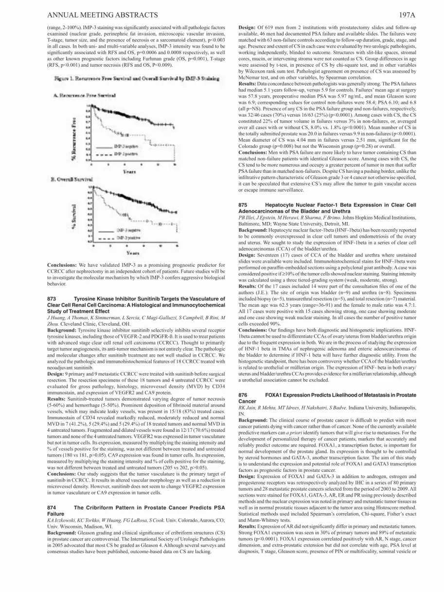
ANNUAL MEETING ABSTRACTS 197A(range, 2-100%). IMP-3 staining was significantly associated with all pathologic factors examined (nuclear grade, perinephric fat invasion, microscopic vascular invasion, T-stage, tumor size, and the presence of necrosis or a sarcomatoid element), p<0.003 in all cases. In both uni- and multi-variable analyses, IMP-3 intensity was found to be significantly associated with RFS and OS, p=0.0006 and 0.0008 respectively, as well as other known prognostic factors including Furhman grade (OS, p=0.001), T-stage (RFS, p<0.001) and tumor necrosis (RFS and OS, P<0.009).
Conclusions: We have validated IMP-3 as a promising prognostic predictor for CCRCC after nephrectomy in an independent cohort of patients. Future studies will be to investigate the molecular mechanism by which IMP-3 confers aggressive biological behavior.
873 Tyrosine Kinase Inhibitor Sunitinib Targets the Vasculature of Clear Cell Renal Cell Carcinoma: A Histological and Immunocytochemical Study of Treatment EffectJ Huang, A Thomas, K Simmerman, L Sercia, C Magi-Galluzzi, S Campbell, B Rini, M Zhou. Cleveland Clinic, Cleveland, OH.Background: Tyrosine kinase inhibitor sunitinib selectively inhibits several receptor tyrosine kinases, including those of VEGFR-2 and PDGFR-ß. It is used to treat patients with advanced stage clear cell renal cell carcinoma (CCRCC). Thought to primarily target tumor angiogenesis, its anti-tumor mechanism is not entirely clear. The pathologic and molecular changes after sunitinib treatment are not well studied in CCRCC. We analyzed the pathologic and immunohistochemical features of 18 CCRCC treated with neoadjuvant sunitinib.Design: 9 primary and 9 metastatic CCRCC were treated with sunitinib before surgical resection. The resection specimens of these 18 tumors and 4 untreated CCRCC were evaluated for gross pathology, histology, microvessel density (MVD) by CD34 immunostain, and expression of VEGFR2 and CA9 protein.Results: Sunitinib-treated tumors demonstrated varying degree of tumor necrosis (5-60%) and hemorrhage (5-50%). Prominent deposition of fibrinoid material around vessels, which may indicate leaky vessels, was present in 15/18 (83%) treated cases. Immunostain of CD34 revealed markedly reduced, moderately reduced and normal MVD in 7 (41.2%), 5 (29.4%) and 5 (29.4%) of 18 treated tumors and normal MVD in 4 untreated tumors. Fragmented and dilated vessels were found in 12/17 (70.6%) treated tumors and none of the 4 untreated tumors. VEGFR2 was expressed in tumor vasculature but not in tumor cells. Its expression, measured by multiplying the staining intensity and % of vessels positive for the staining, was not different between treated and untreated tumors (180 vs 181, p>0.05). CA9 expression was found in tumor cells. Its expression, measured by multiplying the staining intensity and % of cells positive for the staining, was not different between treated and untreated tumors (205 vs 202, p>0.05).Conclusions: Our study suggests that the tumor vasculature is the primary target of sunitinib in CCRCC. It results in altered vascular morphology as well as a reduction in microvessel density. However, sunitinib does not seem to change VEGFR2 expression in tumor vasculature or CA9 expression in tumor cells.
874 The Cribriform Pattern in Prostate Cancer Predicts PSA FailureKA Iczkowski, KC Torkko, W Huang, FG LaRosa, S Cook. Univ. Colorado, Aurora, CO; Univ. Wisconsin, Madison, WI.Background: Gleason grading and clinical significance of cribriform structures (CS) in prostate cancer are controversial. The International Society of Urologic Pathologists in 2005 advocated that most CS be graded as Gleason 4. Although several surveys and consensus studies have been published, outcome-based data on CS are lacking.
Design: Of 619 men from 2 institutions with prostatectomy slides and follow-up available, 46 men had documented PSA failure and available slides. The failures were matched with 63 non-failure controls according to follow-up duration, grade, stage, and age. Presence and extent of CS in each case were evaluated by two urologic pathologists, working independently, blinded to outcome. Structures with slit-like spaces, stromal cores, mucin, or intervening stroma were not counted as CS. Group differences in age were assessed by t-test, in presence of CS by chi-square test, and in other variables by Wilcoxon rank sum test. Pathologist agreement on presence of CS was assessed by McNemar test, and on other variables, by Spearman correlation.Results: Data concordance between pathologists was generally strong. The PSA failures had median 5.1 years follow-up, versus 5.9 for controls. Failures’ mean age at surgery was 57.8 years, preoperative median PSA was 5.97 ng/mL, and mean Gleason score was 6.9; corresponding values for control non-failures were 58.4; PSA 6.10; and 6.8 (all p=NS). Presence of any CS in the PSA failure group and non-failures, respectively, was 32/46 cases (70%) versus 16/63 (25%) (p<0.0001). Among cases with CS, the CS constituted 22% of tumor volume in failures versus 3% in non-failures, or, averaged over all cases with or without CS, 8.0% vs. 1.8% (p<0.0001). Mean number of CS in the totally submitted prostate was 20.0 in failures versus 9.9 in non-failures (p<0.0001). Mean diameter of CS was 4.04 mm in failures versus 2.51 mm, significant for the Colorado group (p=0.008) but not the Wisconsin group (p=0.28) or overall.Conclusions: Men with PSA failure are more likely to have tumor containing CS than matched non-failure patients with identical Gleason score. Among cases with CS, the CS tend to be more numerous and occupy a greater percent of tumor in men that suffer PSA failure than in matched non-failures. Despite CS having a pushing border, unlike the infiltrative pattern characteristic of Gleason grade 3 or 4 cancer not otherwise specified, it can be speculated that extensive CS’s may allow the tumor to gain vascular access or escape immune surveillance.
875 Hepatocyte Nuclear Factor-1 Beta Expression in Clear Cell Adenocarcinomas of the Bladder and UrethraPB Illei, J Epstein, M Herawi, R Sharma, F Brimo. Johns Hopkins Medical Institutions, Baltimore, MD; Wayne State University, Detroit, MI.Background: Hepatocyte nuclear factor-1beta (HNF-1beta) has been recently reported to be commonly overexpressed in clear cell tumors and endometriosis of the ovary and uterus. We sought to study the expression of HNF-1beta in a series of clear cell adenocarcinomas (CCA) of the bladder/urethra.Design: Seventeen (17) cases of CCA of the bladder and urethra where unstained slides were available were included. Immunohistochemical stains for HNF-1beta were performed on paraffin-embedded sections using a polyclonal goat antibody. A case was considered positive if ≥10% of the tumor cells showed nuclear staining. Staining intensity was calculated using a three tiered-grading system (weak, moderate, strong).Results: Of the 17 cases included 14 were part of the consultation files of one of the authors (J.E.). The site of origin was bladder (n=9) and urethra (n=8). Specimens included biopsy (n=5), transurethral resection (n=5), and total resection (n=7) material. The mean age was 62.5 years (range=36-91) and the female to male ratio was 4.7:1. All 17 cases were positive with 15 cases showing strong, one case showing moderate and one case showing weak nuclear staining. In all cases the number of positive tumor cells exceeded 90%.Conclusions: Our findings have both diagnostic and histogenetic implications. HNF-1beta cannot be used to differentiate CCAs of ovary/uterus from bladder/urethra origin due to the frequent expression in both. We are in the process of studying the expression of HNF-1 beta in TMAs of nephrogenic adenoma and enteric adenocarcinomas of the bladder to determine if HNF-1 beta will have further diagnostic utility. From the histogenetic standpoint, there has been controversy whether CCA of the bladder/urethra is related to urothelial or müllerian origin. The expression of HNF- beta in both ovary/uterus and bladder/urethra CCAs provides evidence for a müllerian relationship, although a urothelial association cannot be excluded.
876 FOXA1 Expression Predicts Likelihood of Metastasis in Prostate CancerRK Jain, R Mehta, MT Idrees, H Nakshatri, S Badve. Indiana University, Indianapolis, IN.Background: The clinical course of prostate cancer is difficult to predict with most cancer patients dying with cancer rather than of cancer. None of the currently available predictive markers can a priori identify tumors that will give rise to metastases. For the development of personalized therapy of cancer patients, markers that accurately and reliably predict outcome are required. FOXA1, a transcription factor, is important for normal development of the prostate gland. Its expression is thought to be controlled by steroid hormones and GATA-3, another transcription factor. The aim of this study is to understand the expression and potential role of FOXA1 and GATA3 transcription factors as prognostic factors in prostate cancer.Design: Expression of FOXA1 and GATA-3 in addition to androgen, estrogen and progesterone receptors was retrospectively analyzed by IHC in a series of 80 primary tumors and 28 metastatic prostate cancers selected from the period of 2003 to 2009. All sections were stained for FOXA1, GATA-3, AR, ER and PR using previously described methods and the nuclear expression was noted in primary and metastatic tumor tissues as well as in normal prostatic tissues adjacent to the tumor area using Histoscore method. Statistical methods used included Spearman’s correlation, Chi-square, Fisher’s exact and Mann-Whitney tests.Results: Expression of AR did not significantly differ in primary and metastatic tumors. Strong FOXA1 expression was seen in 30% of primary tumors and 89% of metastatic tumors (p<0.0001). FOXA1 expression correlated positively with AR, N stage, cancer dimension, and extra-prostatic extension but did not correlate with age, PSA level at diagnosis, T stage, Gleason score, presence of PIN or multifocality, seminal vesicle or
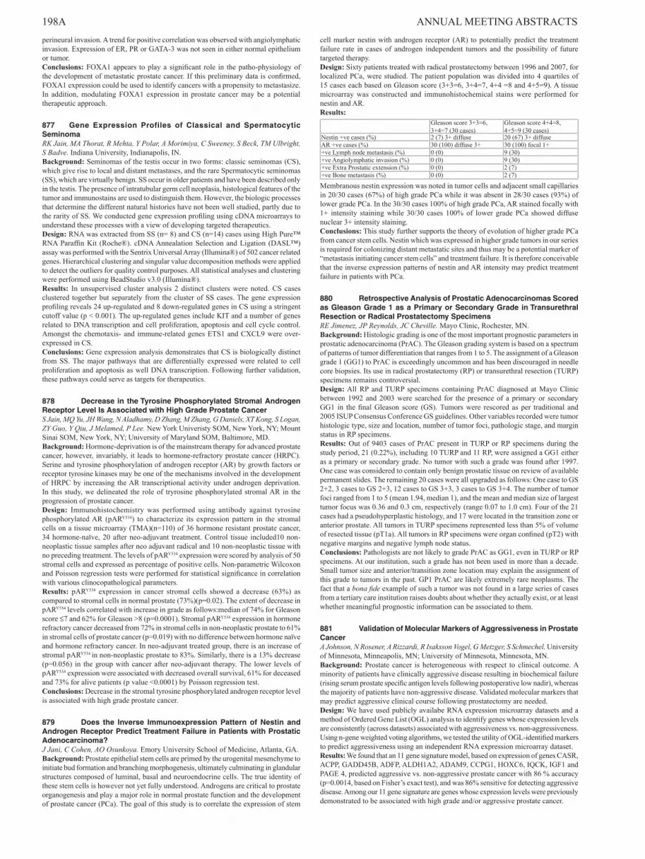
198A ANNUAL MEETING ABSTRACTS perineural invasion. A trend for positive correlation was observed with angiolymphatic invasion. Expression of ER, PR or GATA-3 was not seen in either normal epithelium or tumor.Conclusions: FOXA1 appears to play a significant role in the patho-physiology of the development of metastatic prostate cancer. If this preliminary data is confirmed, FOXA1 expression could be used to identify cancers with a propensity to metastasize. In addition, modulating FOXA1 expression in prostate cancer may be a potential therapeutic approach.
877 Gene Expression Profiles of Classical and Spermatocytic SeminomaRK Jain, MA Thorat, R Mehta, Y Polar, A Morimiya, C Sweeney, S Beck, TM Ulbright, S Badve. Indiana University, Indianapolis, IN.Background: Seminomas of the testis occur in two forms: classic seminomas (CS), which give rise to local and distant metastases, and the rare Spermatocytic seminomas (SS), which are virtually benign. SS occur in older patients and have been described only in the testis. The presence of intratubular germ cell neoplasia, histological features of the tumor and immunostains are used to distinguish them. However, the biologic processes that determine the different natural histories have not been well studied, partly due to the rarity of SS. We conducted gene expression profiling using cDNA microarrays to understand these processes with a view of developing targeted therapeutics.Design: RNA was extracted from SS (n= 8) and CS (n=14) cases using High Pure™ RNA Paraffin Kit (Roche®). cDNA Annealation Selection and Ligation (DASL™) assay was performed with the Sentrix Universal Array (Illumina®) of 502 cancer related genes. Hierarchical clustering and singular value decomposition methods were applied to detect the outliers for quality control purposes. All statistical analyses and clustering were performed using BeadStudio v3.0 (Illumina®).Results: In unsupervised cluster analysis 2 distinct clusters were noted. CS cases clustered together but separately from the cluster of SS cases. The gene expression profiling reveals 24 up-regulated and 8 down-regulated genes in CS using a stringent cutoff value (p < 0.001). The up-regulated genes include KIT and a number of genes related to DNA transcription and cell proliferation, apoptosis and cell cycle control. Amongst the chemotaxis- and immune-related genes ETS1 and CXCL9 were over-expressed in CS.Conclusions: Gene expression analysis demonstrates that CS is biologically distinct from SS. The major pathways that are differentially expressed were related to cell proliferation and apoptosis as well DNA transcription. Following further validation, these pathways could serve as targets for therapeutics.
878 Decrease in the Tyrosine Phosphorylated Stromal Androgen Receptor Level Is Associated with High Grade Prostate CancerS Jain, MQ Yu, JH Wang, N Aladhamy, D Zhang, M Zhang, G Daniels, XT Kong, S Logan, ZY Guo, Y Qiu, J Melamed, P Lee. New York Univeristy SOM, New York, NY; Mount Sinai SOM, New York, NY; University of Maryland SOM, Baltimore, MD.Background: Hormone-deprivation is of the mainstream therapy for advanced prostate cancer, however, invariably, it leads to hormone-refractory prostate cancer (HRPC). Serine and tyrosine phosphorylation of androgen receptor (AR) by growth factors or receptor tyrosine kinases may be one of the mechanisms involved in the development of HRPC by increasing the AR transcriptional activity under androgen deprivation. In this study, we delineated the role of tryrosine phosphorylated stromal AR in the progression of prostate cancer.Design: Immunohistochemistry was performed using antibody against tyrosine phosphorylated AR (pARY534) to characterize its expression pattern in the stromal cells on a tissue microarray (TMA)(n=110) of 36 hormone resistant prostate cancer, 34 hormone-naïve, 20 after neo-adjuvant treatment. Control tissue included10 non-neoplastic tissue samples after neo adjuvant radical and 10 non-neoplastic tissue with no preceding treatment. The levels of pARY534 expression were scored by analysis of 50 stromal cells and expressed as percentage of positive cells. Non-parametric Wilcoxon and Poisson regression tests were performed for statistical significance in correlation with various clinocopathological parameters.Results: pARY534 expression in cancer stromal cells showed a decrease (63%) as compared to stromal cells in normal prostate (73%)(p=0.02). The extent of decrease in pARY534 levels correlated with increase in grade as follows:median of 74% for Gleason score ≤7 and 62% for Gleason >8 (p=0.0001). Stromal pARY534 expression in hormone refractory cancer decreased from 72% in stromal cells in non-neoplastic prostate to 61% in stromal cells of prostate cancer (p=0.019) with no difference between hormone naïve and hormone refractory cancer. In neo-adjuvant treated group, there is an increase of stromal pARY534 in non-neoplastic prostate to 83%. Similarly, there is a 13% decrease (p=0.056) in the group with cancer after neo-adjuvant therapy. The lower levels of pARY534 expression were associated with decreased overall survival, 61% for deceased and 73% for alive patients (p value <0.0001) by Poisson regression test.Conclusions: Decrease in the stromal tyrosine phosphorylated androgen receptor level is associated with high grade prostate cancer.
879 Does the Inverse Immunoexpression Pattern of Nestin and Androgen Receptor Predict Treatment Failure in Patients with Prostatic Adenocarcinoma?J Jani, C Cohen, AO Osunkoya. Emory University School of Medicine, Atlanta, GA.Background: Prostate epithelial stem cells are primed by the urogenital mesenchyme to initiate bud formation and branching morphogenesis, ultimately culminating in glandular structures composed of luminal, basal and neuroendocrine cells. The true identity of these stem cells is however not yet fully understood. Androgens are critical to prostate organogenesis and play a major role in normal prostate function and the development of prostate cancer (PCa). The goal of this study is to correlate the expression of stem
cell marker nestin with androgen receptor (AR) to potentially predict the treatment failure rate in cases of androgen independent tumors and the possibility of future targeted therapy.Design: Sixty patients treated with radical prostatectomy between 1996 and 2007, for localized PCa, were studied. The patient population was divided into 4 quartiles of 15 cases each based on Gleason score (3+3=6, 3+4=7, 4+4 =8 and 4+5=9). A tissue microarray was constructed and immunohistochemical stains were performed for nestin and AR.Results:
Gleason score 3+3=6, 3+4=7 (30 cases)
Gleason score 4+4=8, 4+5=9 (30 cases)
Nestin +ve cases (%) 2 (7) 3+ diffuse 20 (67) 3+ diffuseAR +ve cases (%) 30 (100) diffuse 3+ 30 (100) focal 1++ve Lymph node metastasis (%) 0 (0) 9 (30)+ve Angiolymphatic invasion (%) 0 (0) 9 (30)+ve Extra Prostatic extension (%) 0 (0) 2 (7)+ve Bone metastasis (%) 0 (0) 2 (7)
Membranous nestin expression was noted in tumor cells and adjacent small capillaries in 20/30 cases (67%) of high grade PCa while it was absent in 28/30 cases (93%) of lower grade PCa. In the 30/30 cases 100% of high grade PCa, AR stained focally with 1+ intensity staining while 30/30 cases 100% of lower grade PCa showed diffuse nuclear 3+ intensity staining.Conclusions: This study further supports the theory of evolution of higher grade PCa from cancer stem cells. Nestin which was expressed in higher grade tumors in our series is required for colonizing distant metastatic sites and thus may be a potential marker of “metastasis initiating cancer stem cells” and treatment failure. It is therefore conceivable that the inverse expression patterns of nestin and AR intensity may predict treatment failure in patients with PCa.
880 Retrospective Analysis of Prostatic Adenocarcinomas Scored as Gleason Grade 1 as a Primary or Secondary Grade in Transurethral Resection or Radical Prostatectomy SpecimensRE Jimenez, JP Reynolds, JC Cheville. Mayo Clinic, Rochester, MN.Background: Histologic grading is one of the most important prognostic parameters in prostatic adenocarcinoma (PrAC). The Gleason grading system is based on a spectrum of patterns of tumor differentiation that ranges from 1 to 5. The assignment of a Gleason grade 1 (GG1) to PrAC is exceedingly uncommon and has been discouraged in needle core biopsies. Its use in radical prostatectomy (RP) or transurethral resection (TURP) specimens remains controversial.Design: All RP and TURP specimens containing PrAC diagnosed at Mayo Clinic between 1992 and 2003 were searched for the presence of a primary or secondary GG1 in the final Gleason score (GS). Tumors were rescored as per traditional and 2005 ISUP Consensus Conference GS guidelines. Other variables recorded were tumor histologic type, size and location, number of tumor foci, pathologic stage, and margin status in RP specimens.Results: Out of 9403 cases of PrAC present in TURP or RP specimens during the study period, 21 (0.22%), including 10 TURP and 11 RP, were assigned a GG1 either as a primary or secondary grade. No tumor with such a grade was found after 1997. One case was considered to contain only benign prostatic tissue on review of available permanent slides. The remaining 20 cases were all upgraded as follows: One case to GS 2+2, 3 cases to GS 2+3, 12 cases to GS 3+3, 3 cases to GS 3+4. The number of tumor foci ranged from 1 to 5 (mean 1.94, median 1), and the mean and median size of largest tumor focus was 0.36 and 0.3 cm, respectively (range 0.07 to 1.0 cm). Four of the 21 cases had a pseudohyperplastic histology, and 17 were located in the transition zone or anterior prostate. All tumors in TURP specimens represented less than 5% of volume of resected tissue (pT1a). All tumors in RP specimens were organ confined (pT2) with negative margins and negative lymph node status.Conclusions: Pathologists are not likely to grade PrAC as GG1, even in TURP or RP specimens. At our institution, such a grade has not been used in more than a decade. Small tumor size and anterior/transition zone location may explain the assignment of this grade to tumors in the past. GP1 PrAC are likely extremely rare neoplasms. The fact that a bona fide example of such a tumor was not found in a large series of cases from a tertiary care institution raises doubts about whether they actually exist, or at least whether meaningful prognostic information can be associated to them.
881 Validation of Molecular Markers of Aggressiveness in Prostate CancerA Johnson, N Rosener, A Rizzardi, R Isaksson Vogel, G Metzger, S Schmechel. University of Minnesota, Minneapolis, MN; University of Minnesota, Minnesota, MN.Background: Prostate cancer is heterogeneous with respect to clinical outcome. A minority of patients have clinically aggressive disease resulting in biochemical failure (rising serum prostate specific antigen levels following postoperative low nadir), whereas the majority of patients have non-aggressive disease. Validated molecular markers that may predict aggressive clinical course following prostatectomy are needed.Design: We have used publicly availabe RNA expression microarray datasets and a method of Ordered Gene List (OGL) analysis to identify genes whose expression levels are consistently (across datasets) associated with aggressiveness vs. non-aggressiveness. Using n-gene weighted voting algorithms, we tested the utility of OGL-identified markers to predict aggressiveness using an independent RNA expression microarray dataset.Results: We found that an 11 gene signature model, based on expression of genes CASR, ACPP, GADD45B, ADFP, ALDH1A2, ADAM9, CCPG1, HOXC6, IQCK, IGF1 and PAGE 4, predicted aggressive vs. non-aggressive prostate cancer with 86 % accuracy (p=0.0014, based on Fisher’s exact test), and was 86% sensitive for detecting aggressive disease. Among our 11 gene signature are genes whose expression levels were previously demonstrated to be associated with high grade and/or aggressive prostate cancer.
ANNUAL MEETING ABSTRACTS 199AConclusions: Our findings further establish that gene signatures may be predictive of aggressive clinical course following prostatectomy. Further studies of the correlation between expression levels of these 11 genes and clinical outcome are required to establish whether tests based on expression of these genes may be clinically useful in identifying aggressive prostate cancer.
882 The Expression of the Androgen Receptor Coactivator P44 in Proliferative Inflammatory AtrophyOE Kabiawu, J Melamed, M Yu, J Wang, S Jain, N Aladhamy, Z Wang, P Lee. New York University School of Medicine, New York, NY.Background: Proliferative inflammatory atrophy (PIA) in prostate consists of atrophic glandular foci with proliferative glandular epithelium and associated inflammation. It has been hypothesized that PIA may be linked to prostate carcinogenesis. Importantly, PIA lesions share a number of molecular and genetic changes with high grade prostatic intraepithelial neoplasia and carcinoma, including chromosome 8 abnormalities, p53 mutations, an increased level of Bcl-2 protein and down-regulation of p27. A functional androgen-signaling pathway is essential for the development and progression of prostate cancer. Various AR cofactors, including p44, have been shown to regulate androgen receptor (AR) activation. We have previously shown that nuclear p44 maintains prostatic epithelial cells in a growth-arrest state, conversely, the cytoplasmic localization of p44 results in epithelial cell proliferation. In this study, we examined the expression of AR and its coactivator p44 in PIA.Design: Cases (n = 44) containing PIA, benign and carcinoma foci were identified from a radical prostatectomy series. Immunohistochemistry using AR and p44 antibodies were performed on paraffin embedded tissue sections. Areas satisfying criteria for PIA [Working group classification of focal prostate atrophy lesions; De Marzo et al 2006] were evaluated together with carcinoma and non-neoplastic foci. Immunoreactivity for p44 and AR was scored separately using nuclear and cytoplasmic proportion-intensity score. The Applied Wilcoxon and Shapiro-Wilk Normality tests were performed for statistical significance.Results: The mean value for the PIA nuclei score = 4.625, and the cytoplasmic score = 4.875. p44 expression in PIA showed an increase in nuclei staining as compared with carcinoma foci (p= 0.02), and a decrease from benign prostate tissue (p < 0.01). Also, p44 cytoplasmic expression was decreased in PIA as compared with the baseline carcinoma foci control (p < 0.01), and increased compared with benign prostate tissue (p < 0.01). The trend in staining shows a progressive increase in nuclear staining and decrease in cytoplasmic staining in benign prostate (p < 0.01), PIA and carcinoma respectively (p < 0.01). Androgen receptor, on the other hand, is expressed strongly and uniformly in all three categories.Conclusions: The above findings of nuclear and cytoplasmic expression of p44 at an intermediate level between benign and carcinoma provides additional support for PIA as a putative premalignant lesion.
883 Double Immunohistochemistry for CK20/ Ki67 in the Diagnosis of Neoplastic and Non-Neoplastic BladderSN Kamat, M Acquafondata, S Bastacky, AV Parwani. University of Pittsburgh, Pittsburgh, PA.Background: Distinguishing neoplasia from reactive atypia in bladder is difficult on histopathology alone. Previous studies have demonstrated CK20 and Ki67 as objective markers for assessing neoplasia/dysplasia in bladder. We describe the utility of CK20/ Ki67 double IHC (DIHC) as a tool for differentiating benign, dysplastic, carcinoma in situ (CIS) and invasive lesions in bladder.Design: 100 cases (62 biopsies (Bx), 14 transurethral resection of tumor, and 24 radical cystectomies) were retrieved from our surgical pathology files which included 28 benign/reactive, 12 dysplasia/CIS, and 60 urothelial carcinomas (UC). H&E and CK20/Ki67 DIHC slides were reviewed. CK20 cytoplasmic stain was evaluated as 0 (no staining), 1+ (patchy, weak intensity), 2+ (<50%, moderate intensity) and 3+ (>50% strong intensity). Location of the staining in upper 1/3rd-umbrella cells, upper 2/3rd or full-thickness urothelium (including the basal layer) was documented. Ki67 was scored as 0 (no staining), 1+ (<15% tumor), 2+ (25-50%), 3+ (50-75%) and 4+ (>75%).Results: Benign/reactive, 27/28(96%) cases were 2-3+ CK20 in the umbrella cells; 9/28 (32%) cases showed 2+ Ki67 in the basal epithelium. 5/5 (100%) cases of dysplasia showed 2-3+ CK20 in upper 2/3 urothelium sparing the basal layer and 1+ Ki67. 7/7 (100%) cases of CIS (Bx) had 3+ full-thickness (focal or diffuse) CK20 staining and 2+ Ki67. 9/9(100%) cases of bladder resections for invasive carcinoma with CIS showed 3+ full thickness CK20 with 1+ and 2+ Ki67 in 6 and 3 of these 9 cases respectively. In UC, CK20 was 3+ full thickness positive in 41/60 (68%) cases (4 squamous cell, 7 invasive low-grade and 30 invasive high-grade); CK20 was negative in 19/60 (32%) cases (4 poorly differentiated, 4 small cell/ neuroendocrine, 8 sarcomatoid, 3 squamous cell), Ki67 varied from 1+ to 4+ in all high grade UC and 0 to 1+ in invasive low grade UC.Conclusions: Our study demonstrates that CK20/Ki67 DIHC may be a useful diagnostic tool in the assessment of bladder lesions. Benign/reactive cases consistently showed CK20+ in umbrella cells with majority Ki67 negative. Dysplastic lesions were CK20 positive in upper 2/3rd of urothelium with a low Ki 67 index. CIS and UC demonstrated full thickness CK20/Ki67 dual staining. CK20/Ki67 further aided in subclassification, negative CK20 in neuroendocrine/ small cell and poorly differentiated carcinomas and Ki67 correlated with the histomorphological diagnosis, very high Ki67 in small cell/ neuroendocrine carcinomas while lower index in squamoid differentiation.
884 Expression of Caveolin-1 (CAV1), PAX2 and Survivin (SURV) in Clear Cell Renal Cell Carcinoma (CRCC): No Evidence for Prognostic SignificanceD Kankaya, S Kiremitci, O Tulunay, S Baltaci. Medical School of Ankara University, Ankara, Turkey.Background: CRCC is the most common and aggressive subtype of RCC involving the adult kidney. Molecular markers have the potential to predict reliably, the biological behavior of these tumors and could serve as therapeutic targets.Design: Immunohistochemistry was performed on 100 cases of CRCC using antibodies against CAV-1, PAX2, and SURV. Tumors were grouped by nuclear grade as low-grade (LNG; NG1,2) or high-grade (HNG; NG3,4), and by pathological stage as localized (LC; pT1,2), or locally invasive (IS; pT3,4). Clinically the tumors were staged as early stage (ES; cTI,II), or late stage (LS; cTIII,IV). Lymphovascular, renal vein, perirenal, regional lymph node invasions (LVI, RVI, PRI, RLN, respectively), and Mx were noted. Marker status, and expression patterns (cyt/nuc) of markers, established predictors of prognosis were considered when developing a prognostic model for disease-specific survival (DSS).Results: Median diameter of the tumors was 7 cm (range=1,5-20 cm). HNG tumors were larger. Local invasiveness correlated with larger size (p=0.003), HNG (p=0.006). Mx correlated with HNG (p=0.001), and older age (>57 yrs, p=0.045). Median follow-up was 24 months (range=1-144 months). While NG (p=0.001), LVI (p=0.042), RVI (p=0.001), PRI (p=0.012), pT (p=0.006), RLN (p=0.001), Mx (p=0.001), cT (p=0.001) correlated with DSS, only cT (p=0.010) was found to be an independent predictor of survival. Loss of SURVCyt correlated with a low rate of HNG (p=0.018), and tumor invasiveness (p=0.054). SURVNuc and PAX2 over-expressions were correlated (p=0.040, CC:0.206). CAV1 and PAX2 did not correlate with clinicopathological factors, and CAV1, PAX2, and SURV (SURVCyt,, SURVNuc), did not correlate with DSS.Conclusions: Novel molecular proteins in CRCC research; CAV1, PAX2, and SURV, either alone or in a combination, does not seem to identify a group of patients with CRCC at high risk for Mx and decreased DSS. Although DSS is not dependent on SURV, the correlation of SURV with HNG, and tumor invasiveness, may designate SURV as a potential marker for tumor aggressiveness.
885 Impact of Epidermal Growth Factor Receptor (EGFR), Gelsolin, NF-kB, and p53 Expression on Clinicopathological Parameters in Clear Cell Renal Cell Carcinoma (CRCC)D Kankaya, S Kiremitci, O Tulunay, S Baltaci. Medical School of Ankara University, Ankara, Turkey.Background: CRCC has an unpredictable behavior and tendency for recurrence and metastasis (Mx). Novel markers are necessary to develop a prognostic profiling system for disease specific survival (DSS), and to design therapeutic regimens in CRCC.Design: Immunohistochemistry was performed on 100 cases of CRCC using antibodies against EGFR, Gelsolin, NF-kB, and p53. Tumors were grouped by nuclear grade as low NG (LNG; NG1,2) or high NG (HNG; NG3,4), and by pathological stage as localized (LC; pT1,2), or locally invasive (IS; pT3,4). Clinical disease was grouped by clinical stage as early stage (ES; cTI,II), or late stage (LS; cTIII,IV). Evaluation was based on cytoplasmic (EGFRCyt), and membranous (EGFRMem) expression for EGFR; cytoplasmic (NF-kBCyt), and nuclear (NF-kBNuc) expression for NF-kB.Results: Mx correlated with HNG (p=0.001), and older age (>57 yrs, p=0.045). While NG (p=0.001), LVI (p=0.042), RVI (p=0.001), PRI (p=0.012), pT (p=0.006), RLN (p=0.001), Mx (p=0.001), cT (p=0.001) correlated with DSS, only cT was an independent predictor of DSS (p=0.010) . EGFRCyt correlated with HNG (p=0.001), LVI (p=0.028), RLN involvement (p=0.027), Mx (p=0.001), LS (p=0.003), and was a predictor for DSS (p=0.012). EGFRMem correlated with IS tumor (p=0.021), PRI (p=0.009). Gelsolin expression correlated with HNG (p=0.001), Mx (p=0.003), LS (p=0.008), DSS (p=0.006). NF-kBCyt expression correlated with HNG (p=0.002), PRI (p=0.010), IS tumor (p=0.020), LS (p=0.003). NF-kBNuc did not predict the prognosis of CRCC. p53 expression correlated with HNG (p=0.045), LVI (p=0.05), Mx (p=0.001), LS (p=0.028). Gelsolin correlated with EGFRCyt (p=0.005), p53 (p=0.045), and NF-κBCyt (p=0.028). p53 correlated with EGFRCyt. EGFRMem correlated with NF-κBCyt(p=0.044).Conclusions: Expressions of Gelsolin, NF-κBCyt, and p53 associated with aggressive behavior of CRCC. EGFR and Gelsolin demonstrated to be an unfavourable prognostic factor in CRCC. Prognostic value of EGFR differed significantly with expression pattern. EGFRCyt expression associated with with short survival, while EGFRMem is not correlated with DSS. This study demonstrated a staining profile as a classifier of the high-risk group of patients with CRCC; overexpressions of EGFRCyt, Gelsolin, NF-kBCyt, and p53. EGFR seems an attractive target for therapeutic manipulation in patients with advanced CRCC.
886 Diffuse and Strong Expression of PAX2 and PAX8 in the Cystic Epithelium of Angiomyolipoma with Epithelial Cysts (AMLEC) and Mixed Epithelial Stromal Tumor (MEST) Supports Distal Tubular DifferentiationM Karafin, A Parwani, G Netto, P Illei, J Epstein, P Argani. Johns Hopkins Medical Institutions, Baltimore, MD; University of PittsBurgh Medical Center, Pittsburgh, PA.Background: PAX2 and PAX8 are tissue specific transcription factors expressed in the renal and Müllerian systems. Their expression has not been examined in the recently described cystic renal lesions AMLEC and MEST. The nature of the epithelium in these lesions is not clear, postulated by some to represent entrapped renal tubular epithelium. Others disagree, based largely upon extrarenal localization in some reported cases.Design: PAX2 and PAX8 immunohistochemistry was performed on representative sections of cases of AMLEC (7) and MEST (8). The relative percentage and intensity (none, weak, moderate, strong) of nuclear labeling were evaluated in both the benign adjacent renal tubules and the lesion’s epithelial cysts.
200A ANNUAL MEETING ABSTRACTS Results: In the adjacent kidney, distal convoluted tubules (DCT) labeled strongly for PAX2 and PAX8, while proximal convoluted tubules labeled minimally. The cystic epithelium of 7 of 7 AMLEC, including 4 which extended beyond the renal capsule into the perirenal fat, demonstrated strong diffuse labeling for both PAX2 and PAX8. In the course of this review, we uncovered a mimic of entirely extrarenal AMLEC: extrarenal angiomyolipoma (AML) involved by endosalpingiosis. The epithelium of the latter labeled for estrogen receptor (ER) (unlike that of AMLEC) in addition to PAX2 and PAX8. PAX2 and PAX8 diffusely and strongly labeled the epithelial component of 8 of 8 MESTs, including all architectural (phyllodes-like, large cysts, small cysts, clustered microcysts) and cytologic (hobnail, flat, cuboidal, columnar, apocrine, ciliated, and clear cell) epithelial variants present. Cyst epithelial labeling intensity was similar to that of renal DCT in all cases.Conclusions: The diffuse labeling for PAX2/PAX8 in the epithelial cysts of AMLEC, taken together with their consistent negativity for ER and HMB45, supports the hypothesis that this epithelium represents entrapped, cystically dilated DCT. Renal AMLEC commonly herniate beyond the renal capsule: previously reported cases of entirely extrarenal AMLEC may represent extrarenal AML involved by endosalpingiosis. The diffuse labeling for PAX2/PAX8 in MEST epithelium, coupled with its usual ER negativity, suggests that the epithelium of MEST is derived from the entrapped DCT which may undergo cytologic changes as it grows along with the stromal component.
887 Should Saved Interval Sections of Prostate Needle Biopsy (PNBx) Specimens Be Examined Microscopically?EH Kerr, VL Dailey, WE Grizzle, O Hameed. University of Alabama at Birmingham, Birmingham, AL.Background: In addition to using multiple histologic levels to evaluate PNBx material, many pathologists cut and save interval unstained sections for potential analysis by immunohistochemistry (IHC); these are usually discarded if not utilized. It is unclear, however, whether additionally evaluating these saved interval sections has an impact on the final diagnosis especially in contemporary twelve-part PNBx specimens.Design: A retrospective search of twelve-part PNBx specimens over a consecutive seven month period (02-09/08) was performed to identify cases where a diagnosis of prostatic adenocarcinoma (PC) was not made on any specimen part and saved interval sections were available. These sections were retrieved, stained with hematoxylin and eosin, and histologically evaluated independent from the original sections to identify any possible foci of atypia. Identified foci were further characterized by a genitourinary pathologist and, if PC was contemplated, by IHC. Subsequent to that, the original surgical pathology slides were then reviewed to determine whether the atypical foci were present and, if so, compare their linear extent and the number of atypical glands seen to those in the interval sections.Results: Out of 149 consecutive PNBx cases, 36 met the inclusion criteria and had unstained sections available for at least one specimen part. These cases were originally diagnosed as benign (n=28), high grade prostatic intraepithelial neoplasia (n=5), or focal glandular atypia (n=3). Two to 75 slides were re-evaluated per case (mean=34.9) with 2 to 10 slides (mean=3.1) re-evaluated per specimen part. Overall, examination of 1258 newly-stained slides for 404 uniquely designated specimen parts revealed atypical glandular foci (1-16 glands; 0.03-0.47 mm in length) suspicious for PC in 4 (1%) cases. In 2 of these, PC was confirmed by IHC. Although atypical foci were present in at least a single original section, only one was originally reported as such. It was, however, better visualized in the saved interval sections due to the presence of more glands and greater linear extent.Conclusions: Although examination of newly-stained saved interval sections of PNBx specimens can identify atypical, potentially carcinomatous, foci, such foci are not new as they are usually present in the original sections. These findings support the practice of examining only 3 levels of PNBx specimens and, in the absence of atypia, discarding unused saved interval sections.
888 IgG4-Associated Inflammatory Pseudotumor of Ureter; a Report of Three CasesSA Kim, SR Lee, J Huh, SS Shen, JY Ro. University of Ulsan College of Medicine, Asan Medical Center, Seoul, Korea; The Methodist Hospital, Weill Medical College of Cornell University, Houston, TX.Background: Inflammatory pseudotumors (IPT) are a generic term for a wide range of lesions characterized by proliferation of fibro-/myofibroblasts in the background of variable chronic inflammatory cell infiltration. Recently, IPT with abundant IgG4-positive plasma cells is considered to be a unique entity belonging to systemic IgG4-related sclerosing disease, and distinguished from other lesions in IPT category such as inflammatory myofibroblastic tumor (IMT) and fibrohistiocytic type IPT. IPT has been rarely reported in the ureter, and to the best knowledge, no ureter IPT in relation to IgG4-positive plasma cells has been described.Design: We describe herein three cases of ureteral lymphoplasmacytic type (IgG4-associated) IPT with emphasis on their histologic and immunohistochemical features.Results: Three patients, 45 and 47-year-old men and an 84-year-old woman presented with flank pain. Abdominal computed tomography revealed ureteral narrowing by a mass-effect and hydronephrosis in all cases (left, left and right ureter). Microscopic exam of the resected ureters revealed suburothelial mass-like lesions consisted of spindle-shaped fibro-/myofibroblasts without atypia, and abundant plasma cells. Eosinophils and dendritic or spindle-shaped histiocytes were scattered throughout. The ureteral lesion from the 47-year-old man extended to the periureteral adipose tissue and showed obliterative phlebitis. The lesion of the 84-year-old woman was accompanied with transitional cell carcinoma in situ in the overlying urothelium. On immunohistochemical studies, anaplastic lymphoma kinase (ALK) was negative and only a few fibroblasts/myofibroblasts were positive for smooth muscle actin (SMA). More than 50 plasma cells per one high power field were immunoreactive for IgG4 in
all three cases, a diagnostic feature of IgG4-associated IPT. All three cases were treated with ureterectomy and alive and well with no evidence of recurrence.Conclusions: Numerous IgG4-positive plasma cells, obliterative phlebitis, scattered distribution of eosinophils and dendritic histiocytes in IPT of our three cases were consistent with lymphoplasmacytic type IPT. These IgG4-related IPTs suggest that it would be interesting to review plasma cell-rich IPTs in other organs with regard to IgG4 and search for the features of systemic sclerosing disease.
889 Adenocarcinoma of the Urinary Bladder: Value of Cell Cycle BiomarkersE King, R Youssef, W Kabbani, A Mosbah, H Abol-Enein, M Ghoniem, Y Lotan, P Kapur. UT Southwestern Medical Center, Dallas, TX; Urology & Nephrology Center Mansoura University, Mansoura, Egypt.Background: Primary adenocarcinomas of the urinary bladder are uncommon, accounting for 0.5 to 2% of all bladder malignancies. Currently, molecular pathways for adenocarcinoma of the bladder are not well defined. We assessed the association between biological markers and clinico-pathologic characteristics of bladder adenocarcinoma.Design: Immunohistochemical staining for p53, p21, p27, and cyclin E were performed on sections of a tissue microarray construct of bladder adenocarcinoma specimens from 21 patients who underwent radical cystectomy between 1997 and 2003. Altered immunohistochemical expression was defined as p53 ≥10%, p21 <10%, p27 <30%, and cyclin E <30% nuclear reactivity.Results: The 21 patients included 16 males and 5 females, with mean age of 49±8 y (range 35-72 years). Of these patients, 14 (67 %) had history of schistosomiasis, 12 (57 %) were high grade, 18 (86%) were ≥ pT2, and 6 (27 %) had positive lymph nodes. Recurrence occurred in 6 (29%) patients during 4.8±1.3 year follow up. Each biomarker was altered in at least 60% of cases. P27 and p21 were associated with grade, pathological T stage, and lymph node involvement (P < 0.05). The incremental number of altered biomarkers was associated with lymph node involvement as well as lymphovascular invasion (P < 0.05).Conclusions: Assessment of cell cycle regulatory markers in adenocarcioma of the bladder may improve our knowledge about the biological behavior of this rare but dangerous disease. P27 and p21 are the most promising markers. Their combination can predict higher stage and grade as well as lymph node involvement. Combination of biomarkers can improve prediction of prognosis and may identify patients who will benefit from multimodal therapy.
890 Expression Pattern of Epidermal Growth Factor Receptor (EGFR) in Clear Cell Renal Cell Carcinoma (CRCC): Association with Clinicopathological Features and Clinical OutcomeS Kiremitci, D Kankaya, O Tulunay, S Baltaci. Medical School of Ankara University, Ankara, Turkey.Background: CRCC is the most common malignancy involving the adult kidney, and generally presents as a metastatic disease. Molecular markers to identify patients at risk for poor disease-specific survival (DSS) would benefit in such situations where additional therapeutic modalities are imperative.Design: Immunohistochemistry was performed on 100 cases of CRCC using antibody against EGFR. Tumors were grouped by nuclear grade (NG) as low NG (LNG; NG1,2) or high NG (HNG; NG3,4), and by pathological stage as localized (LC; pT1,2), or locally invasive (IS; pT3,4). Clinical stage was grouped as early stage (ES; cTI,II), or late stage (LS; cTIII,IV). Lymphovascular, renal vein, perirenal, and regional lymph node invasions (LVI, RVI, PRI, RLN, respectively), and metastasis (Mx) were noted. Evaluation of EGFR is based on the pattern of staining; cytoplasmic (EGFRCyt), membranous (EGFRMem), and cytoplasmic and/or membranous (EGFRGroup). EGFR analysis was based on intensity and distribution of EGFR.Results: HNG tumors were larger (>7 cm). Local invasiveness correlated with larger size (p=0.003), HNG (p=0.006). Mx correlated with HNG (p=0.001), and older age (>57 yrs, p=0.045). Median follow-up was 24 months (range=1-144 months). While NG (p=0.001), LVI (p=0.042), RVI (p=0.001), PRI (p=0.012), pT (p=0.006), RLN (p=0.001), Mx (p=0.001), cT (p=0.001) correlated with DSS, only cT (p=0.010) was an independent predictor of survival. EGFRCyt overexpression correlated with HNG (p=0.001), LVI (p=0.028), RLN involvement (p=0.027), Mx (p=0.001), LS (p=0.003), and DSS (p=0.009), and was a predictor for DSS (p=0.012). EGFRMem overexpression correlated with IS tumor (p=0.021), PRI (p=0.009), but not DSS. Cytoplasmic dominant EGFRGroup overexpression correlated with multifocal tumor (p=0.018), HNG (p=0.001), IS tumor (p=0.047), LVI (p= 0.035), RLN involvement (p=0.009), Mx (p=0.022), and DSS (p=0.012).Conclusions: This study demonstrates the importance of the assessment methods of EGFR immunostaining, and explain the conflicting results in the literature. We demonstrate that, EGFR overexpression is an important prognostic factor in CRCC which is modified by the location of expression in tumor cells. EGFRCyt expression; either alone or as cytoplasmic dominat EGFRGroup, demonstrated powerful potential for the prediction of poor DSS. EGFRMem expression does not worsen clinical outcome and is associated with better prognosis.
891 Carbonic Anhydrase 9 (CA9) and Caspase-9 (CASP-9) Expression in Clear Cell Renal Cell Carcinoma (CRCC): Diagnostic, Prognostic and Therapeutic ImplicationsS Kiremitci, D Kankaya, O Tulunay, S Baltaci. Medical School of Ankara University, Ankara, Turkey.Background: Although clinicopathological parameters have proven their prognostic significance in primary CRCC, there is an utmost need of biological predictors for individual tumor behavior with respect to response to survival rate, progression to metastasis, and of biological candidates for targeted therapies.
ANNUAL MEETING ABSTRACTS 201ADesign: Immunohistochemistry (IHC) was performed on 100 cases of CRCC using antibodies against CASP-9 and CA9. Tumors were grouped by nuclear grade as low-grade (LNG; NG1,2) or high-grade (HNG; NG3,4), and by pathological stage as localized (LC; pT1,2), or locally invasive (IS; pT3,4). Clinically the tumors were staged as early stage (ES; cTI,II), or late stage (LS; cTIII,IV). Lymphovascular, renal vein, perirenal, regional lymph node invasions (LVI, RVI, PRI, RLN, respectively), and metastasis (Mx) were noted. IHC analysis was based on intensity and/or distribution of the markers. Prognostic factors and expression patterns (cyt/nuc) of markers were considered when developing a prognostic model for disease-specific survival (DSS).Results: HNG tumors were larger (> 7 cm). Local invasiveness correlated with larger size (p=0.003), HNG (p=0.006). Mx correlated with HNG (p=0.001), and older age (>57 yrs, p=0.045). While NG (p=0.001), LVI (p=0.042), RVI (p=0.001), PRI (p=0.012), pT (p=0.006), RLN (p=0.001), Mx (p=0.001), cT (p=0.001) correlated with DSS, only cT (p=0.010) was found to be an independent predictor of survival. Low expression of CA9 correlated with large tumor size (p=0.002), HNG (p=0.001), LVI (p=0.012), PRI (p=0.015), IS tumor (p=0.045), RLN invasion (p=0.007), Mx (p=0.013), and LS (p=0.003). More patients with low CA9 died of the disease (p=0.001) within shorter time (p=0.004). Low CA9 expression reflected increased aggressiveness for CRCC (OR 3.06, 95% CI 1.35 to 6.91, p=0.007). Loss of CASP-9Nuc correlated with large tumor size (p=0.026), HNG (p=0.001), LVI (p=0.018), Mx (p=0.047), but not with DSS. CASP-9Nuc and CA9 expressions correlated significantly (p=0.017, CC:0.239).Conclusions: Co-expression of CA9, and CASP-9 reflects significant changes in tumor biology which correlates with survival in CRCC. A combination of CA9 and CASP-9 identify a group of patients with CRCC at high risk for Mx and poor survival. CA9 is a predictor of survival in CRCC and could be a potential tumor-associated antigen and a target for a novel therapeutic target for patients with CRCC.
892 Prostate-Specific Antigen, Prostate-Specific Acid Phosphatase, and Hormone Receptor Expression in Male and Female Breast CarcinomaTS Kraus, C Cohen, MT Siddiqui. Emory University, Atlanta, GA.Background: Prostate specific antigen (PSA) and prostate-specific acid phosphatase (PSAP) are expressed in benign and malignant prostatic tissue. Immunohistochemical staining for these markers can be used to confirm the prostatic origin of metastatic carcinoma. PSA expression has been reported in male and female breast carcinoma and in gynecomastia, raising concerns about the utility of PSA for differentiating prostate carcinoma metastasis to the male breast from primary breast carcinoma. This study examined the frequency of PSA, PSAP, and hormone receptor expression in male breast carcinoma (MBC), female breast carcinoma (FBC), and gynecomastia.Design: Immunohistochemical staining for PSA, PSAP, AR, ER, and PR was performed on tissue microarrays representing six cases of gynecomastia, thirty MBC, and fifty-six FBC. PSA and PSAP were considered positive if any cytoplasmic staining was observed. Positive reactions for AR, ER, and PR were defined by nuclear staining observed in at least 10% of tumor cells.Results:
MBC FBC Gynecomastia P-value (MBC vs FBC)PSA 1/30 (3.3%) 2/54 (3.7%) 0/5 (0%) 1PSAP 0/29 (0%) 0/54 (0%) 0/5 (0%) 1AR 20/27 (74.1%) 38/56 (67.9%) 4/5 (80%) 0.62ER 23/37 (85.2%) 37/54 (68.5%) 6/6 (100%) 0.18PR 14/27 (51.9%) 27/57 (47.4%) 5/5 (100%) 0.82
PSA was positive in two of fifty-six FBC (3.7%), focally positive in one of thirty MBC (3.3%), and negative in the five examined cases of gynecomastia.Conclusions: PSA and PSAP are useful markers to distinguish primary breast carcinoma from prostate carcinoma metastatic to the male breast. The incidence of PSA expression in our population was too low to draw significant conclusions about an association between PSA expression and hormone receptor status in breast lesions.
893 Pre-Operative Predictors of Adverse Prostatectomy Outcome. Correlation of 389 12-Core Needle Biopsy Results with Subsequent Prostatectomy DataON Kryvenko, M Diaz-Insua, M Ramineni, M Menon, NS Gupta. Henry Ford Hospital, Detroit, MI.Background: The aim of our study is to determine if pre-operative PSA (PSA) and pathologic variables on 12 core transrectal needle biopsy of prostate (NBX) are useful in predicting adverse outcomes on subsequent radical prostatectomy (RP).Design: 389 NBX with their corresponding RP specimens collected between 2004 and 2008 at our institution were analyzed. RP outcomes such as extraprostatic extension (EPE), seminal vesicle invasion (SVI), positive surgical margin (PSM) and positive lymph nodes (PLN) were studied with respect to PSA and the following features on NBX: location of positive cores (apex, mid, base), number of positive cores (NPC), percentage of each core involved (%PC), perineural invasion (PNI), highest Gleason score on biopsy (HGS) and distribution of positive cores (DPC). Continuous variables were categorized by quartiles or by the median when their association with a categorical variable was thought. Statistical methods used were Chi-square statistics for contingency tables, Mantel-Haenszel Chi-square, Fisher Exact test and Pearson correlation coefficient.Results: (1) The majority of the variable in which a gradient could be coded show a statistically significant trend (p<0.0001) in predicting EPE and SVI. (2) For PSM, this trend was significant for NPC, %PC, and PNI. (3) In the case of contiguity of involvement, the smallest likelihood for adverse outcome is for isolated positive cores, while the largest is for contiguous core regions. (4) Preoperative PSA >10 ng/dl significantly increases SVI and PLN (p<0.0001). (5) Absence of positive cores in the base or the mid region determines a negative lymph node status, positive cores in the apex do not increase the likelihood of PLN (not statistically significant). (6) RP tumor volume is directly proportional to the NPC, maximal core percentage, and average core
percentage. (7) There is no particular region where location of positive cores increases the likelihood of EPE or PSM. The contiguity of positive cores has no bearing in whether EPE appears isolated in a given region or in more than one region.Conclusions: The NPC, HGS, contiguity of the positive cores, highest percentage of tumor in a core, and PNI are strong predicting factor for EPE, SVI, PSM and PLN. Preoperative PSA >10 ng/dl independently and significantly increases pT3 stage. The likelihood of EPE and PSM in a particular location cannot be predicted on analyzed preoperative data.
894 Variant Histologic Differentiation in Urothelial Carcinoma Is Frequently Under-Recognized and Documented in Community PracticeLP Kunju, RB Shah. University of Michigan, Ann Arbor, MI.Background: Urothelial carcinomas (UCs) have a peculiar capacity for “divergent” or “mixed histology” differentiation comprising of several unusual histologic variants, most of which have been recognized in the WHO 2004 classification. The recognition and documentation of these variants in the pathology report is critical as they have diagnostic, therapeutic and prognostic implications. Our aim was to assess the awareness and reporting practices of variant histology of UCs amongst community pathologists.Design: All transurethral bladder tumor resections (TURBT) performed at outside institutions and reviewed at our institution prior to instituting therapy, were retrieved from the pathology database. UCs demonstrating divergent histologic differentiation diagnosed at our institution were selected and were compared with pathology reports sent by the referring community pathologists, to assess the differences in documentation of divergent histology. Cases with pure squamous cell carcinoma, adenocarcinoma and sarcoma were excluded. Mixed histologic differentiation was quantitated as focal (10-50%) and extensive (>50%).Results: Of 589 TUBRT, 115 (19.5%) showed UCs with divergent differentiation. Majority (88%) cases showed a single pattern while 12% showed multiple patterns of divergent differentiation. Overall, 55% cases showed extensive divergent differentiation. Squamous differentiation (32%) was most commonly seen followed by small cell (14%), glandular (13%), micropapillary (10%), nested (8%), sarcomatoid (6%), lymphoepithelial (3%) and plasmacytoid (2%) differentiation. Divergent differentiation was not documented by the contributing community pathologist in 44% (51) cases. Of these 51 cases, divergent histologic differentiation was focal in 57% and extensive in 43% cases. The histologic variants most likely to be under-recognized included lymphoepithelial and plasmacytoid types (100%), followed by micropapillary (86%), nested (75%) and small cell (42%) differentiation. Overall, squamous, sarcomatoid and glandular differentiation had good reporting correlation (≥75%).Conclusions: Our results suggest that histologic variants of UC are often under-reported and/or under-recognized in community practice, specifically lymphoepithelial, plasmacytoid, nested, micropapillary and small cell types. Increased awareness for recognition and documentation of these histologic variants is needed in order to avoid critical diagnostic misinterpretations.
895 Chromophobe Renal Cell Carcinoma with Sarcomatoid Differentiation: A Clinicopathologic Study of 12 CasesSR Lauer, M Zhou, AO Osunkoya. Emory University School of Medicine, Atlanta, GA; The Cleveland Clinic Foundation, Cleveland, OH.Background: Chromophobe renal cell carcinoma (CHRCC) typically has a better prognosis than clear cell renal cell carcinoma (CCRCC) and papillary renal cell carcinoma (PRCC). It is well established that patients diagnosed with CCRCC or PRCC with sarcomatoid differentiation have a poorer prognosis than patients whose tumors do not show sarcomatoid differentiation. However, CHRCC with sarcomatoid differentiation is relatively rare and has not been well studied.Design: The surgical pathology and expert consultation files of two major academic institutions were searched for radical nephrectomy specimens with CHRCC with sarcomatoid differentiation from 2003 to 2009. Clinicopathologic parameters and follow up information were analyzed.Results: 12 patients with a diagnosis of CHRCC with sarcomatoid differentiation were identified, including 7 females (58%) and 5 males (42%). The mean patient age was 62 years (range 40-82 years). There was a 2:1 left sided predominance, left (8 patients) and right (4 patients). The mean tumor size was 14.2 cm (range 9.5-23.0 cm), and the mean percentage sarcomatoid differentiation was 70% (range 40-99%). The non-sarcomatoid component in all cases demonstrated classic features of CHRCC. All tumors exhibited moderate to extensive areas of necrosis. 9 patients (75%) had pT3 disease and 3 patients (25%) had pT4 disease. 4 patients (33%) had positive surgical margins. 5 patients (42%) had pathologic and/or radiologic evidence of multiple or isolated metastatic disease. The common metastatic sites were lungs (4 of 5 patients, 80%), lymph nodes (3 of 5 patients, 60%); other sites included mediastinum, adrenal gland, omentum and orbit. Follow up information was available in 11 patients. Mean follow up time was 12 weeks (range 2-32 weeks). 8 of 11 patients (70%) died of disease, 7 of which were within 6 months (mean survival time of 13 weeks), 2 patients were alive with disease, and only 1 patient was alive with no evidence of disease.Conclusions: CHRCC with sarcomatoid differentiation should be considered in the differential diagnosis of sarcomatoid tumors on renal biopsy. Most patients present at high stage and have a mean survival of 13 weeks. It is thus critical to make the distinction between CHRCC and oncocytoma on biopsies of large renal tumors. This study is one of the largest series to date specifically examining the clinicopathologic features of sarcomatoid CHRCC and confirms that these tumors behave more aggressively than conventional CCRCC and PRCC.
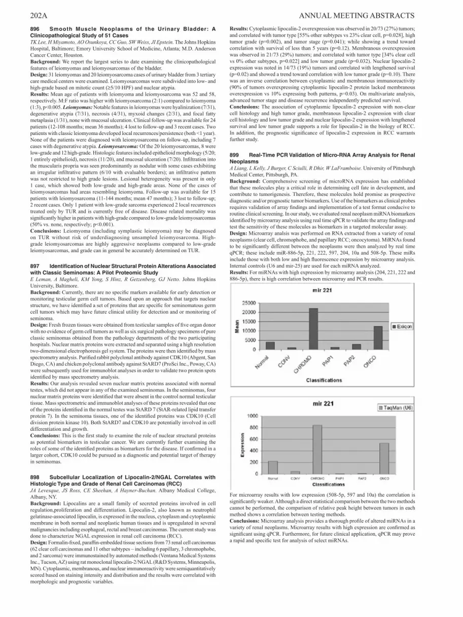
202A ANNUAL MEETING ABSTRACTS 896 Smooth Muscle Neoplasms of the Urinary Bladder: A Clinicopathological Study of 51 CasesTK Lee, H Miyamoto, AO Osunkoya, CC Guo, SW Weiss, JI Epstein. The Johns Hopkins Hospital, Baltimore; Emory University School of Medicine, Atlanta; M.D. Anderson Cancer Center, Houston.Background: We report the largest series to date examining the clinicopathological features of leiomyomas and leiomyosarcomas of the bladder.Design: 31 leiomyomas and 20 leiomyosarcoma cases of urinary bladder from 3 tertiary care medical centers were examined. Leiomyosarcomas were subdivided into low- and high-grade based on mitotic count (≥5/10 HPF) and nuclear atypia.Results: Mean age of patients with leiomyoma and leiomyosarcoma was 52 and 58, respectively. M:F ratio was higher with leiomyosarcoma (2:1) compared to leiomyoma (1:3), p<0.005. Leiomyomas: Notable features in leiomyomas were hyalinization (7/31), degenerative atypia (7/31), necrosis (4/31), myxoid changes (2/31), and focal fatty metaplasia (1/31), none with mucosal ulceration. Clinical follow-up was available for 24 patients (12-108 months; mean 36 months); 4 lost to follow-up and 3 recent cases. Two patients with classic leiomyoma developed local recurrences/persistence (both <1 year). None of the patients were diagnosed with leiomyosarcoma on follow-up, including 7 cases with degenerative atypia. Leiomyosarcoma: Of the 20 leiomyosarcomas, 8 were low-grade and 12 high-grade. Histologic features included epithelioid morphology (5/20; 1 entirely epithelioid), necrosis (11/20), and mucosal ulceration (7/20). Infiltration into the muscularis propria was seen predominantly as nodular with some cases exhibiting an irregular infiltrative pattern (6/10 with evaluable borders); an infiltrative pattern was not restricted to high grade lesions. Lesional heterogeneity was present in only 1 case, which showed both low-grade and high-grade areas. None of the cases of leiomyosarcomas had areas resembling leiomyoma. Follow-up was available for 15 patients with leiomyosarcoma (11-144 months; mean 47 months); 3 lost to follow-up; 2 recent cases. Only 1 patient with low-grade sarcoma experienced 2 local recurrences treated only by TUR and is currently free of disease. Disease related mortality was significantly higher in patients with high-grade compared to low-grade leiomyosarcomas (50% vs. none, respectively; p<0.001).Conclusions: Leiomyoma (including symplastic leiomyoma) may be diagnosed on TUR without risk of underdiagnosing unsampled leiomyosarcoma. High-grade leiomyosarcomas are highly aggressive neoplasms compared to low-grade leiomyosarcomas, and grade can in general be accurately determined on TUR.
897 Identification of Nuclear Structural Protein Alterations Associated with Classic Seminomas: A Pilot Proteomic StudyE Leman, A Magheli, KM Yong, S Hinz, R Getzenberg, GJ Netto. Johns Hopkins University, Baltimore.Background: Currently, there are no specific markers available for early detection or monitoring testicular germ cell tumors. Based upon an approach that targets nuclear structure, we have identified a set of proteins that are specific for seminomatous germ cell tumors which may have future clinical utility for detection and or monitoring of seminoma.Design: Fresh frozen tissues were obtained from testicular samples of five organ donor with no evidence of germ cell tumors as well as six surgical pathology specimens of pure classic seminomas obtained from the pathology departments of the two participating hospitals. Nuclear matrix proteins were extracted and separated using a high resolution two-dimensional electrophoresis gel system. The proteins were then identified by mass spectrometry analysis. Purified rabbit polyclonal antibody against CDK10 (Abgent, San Diego, CA) and chicken polyclonal antibody against StARD7 (ProSci Inc., Poway, CA) were subsequently used for immunoblot analyses in order to validate two protein spots identified by mass spectrometry analysis.Results: Our analysis revealed seven nuclear matrix proteins associated with normal testes, which did not appear in any of the examined seminomas. In the seminomas, four nuclear matrix proteins were identified that were absent in the control normal testicular tissue. Mass spectrometric and immunoblot analyses of these proteins revealed that one of the proteins identified in the normal testes was StARD 7 (StAR-related lipid transfer protein 7). In the seminoma tissues, one of the identified proteins was CDK10 (Cell division protein kinase 10). Both StARD7 and CDK10 are potentially involved in cell differentiation and growth.Conclusions: This is the first study to examine the role of nuclear structural proteins as potential biomarkers in testicular cancer. We are currently further examining the roles of some of the identified proteins as biomarkers for the disease. If confirmed in a larger cohort, CDK10 could be pursued as a diagnostic and potential target of therapy in seminomas.
898 Subcellular Localization of Lipocalin-2/NGAL Correlates with Histologic Type and Grade of Renal Cell Carcinomas (RCC)JA Levesque, JS Ross, CE Sheehan, A Hayner-Buchan. Albany Medical College, Albany, NY.Background: Lipocalins are a small family of secreted proteins involved in cell regulation,proliferation and differentiation. Lipocalin-2, also known as neutrophil gelatinase-associated lipocalin, is expressed in the nucleus, cytoplasm and cytoplasmic membrane in both normal and neoplastic human tissues and is upregulated in several malignancies including esophageal, rectal and breast carcinomas. The current study was done to characterize NGAL expression in renal cell carcinoma (RCC).Design: Formalin-fixed, paraffin-embedded tissue sections from 73 renal cell carcinomas (62 clear cell carcinomas and 11 other subtypes – including 6 papillary, 3 chromophobe, and 2 sarcoma) were immunostained by automated methods (Ventana Medical Systems Inc., Tucson, AZ) using rat monoclonal lipocalin-2/NGAL (R&D Systems, Minneapolis, MN). Cytoplasmic, membranous, and nuclear immunoreactivity were semiquantitatively scored based on staining intensity and distribution and the results were correlated with morphologic and prognostic variables.
Results: Cytoplasmic lipocalin-2 overexpression was observed in 20/73 (27%) tumors; and correlated with tumor type [55% other subtypes vs 23% clear cell, p=0.028], high tumor grade (p=0.002), and tumor stage (p=0.041); while showing a trend toward correlation with survival of less than 5 years (p=0.12). Membranous overexpression was observed in 21/73 (29%) tumors; and correlated with tumor type [34% clear cell vs 0% other subtypes, p=0.022] and low tumor grade (p=0.032). Nuclear lipocalin-2 expression was noted in 14/73 (19%) tumors and correlated with lengthened survival (p=0.02) and showed a trend toward correlation with low tumor grade (p=0.10). There was an inverse correlation between cytoplasmic and membranous immunoreactivity (90% of tumors overexpressing cytoplasmic lipocalin-2 protein lacked membranous overexpression vs 10% expressing both patterns, p=0.03). On multivariate analysis, advanced tumor stage and disease recurrence independently predicted survival.Conclusions: The association of cytoplasmic lipocalin-2 expression with non-clear cell histology and high tumor grade, membranous lipocalin-2 expression with clear cell histology and low tumor grade and nuclear lipocalin-2 expression with lengthened survival and low tumor grade supports a role for lipocalin-2 in the biology of RCC.In addition, the prognostic significance of lipocalin-2 expression in RCC warrants further study.
899 Real-Time PCR Validation of Micro-RNA Array Analysis for Renal NeoplasmsA Liang, L Kelly, J Burger, C Sciulli, R Dhir, W LaFramboise. University of Pittsburgh Medical Center, Pittsburgh, PA.Background: Comprehensive screening of microRNA expression has established that these molecules play a critical role in determining cell fate in development, and contribute to tumorigenesis. Therefore, these molecules hold promise as prospective diagnostic and/or prognostic tumor biomarkers. Use of the biomarkers as clinical probes requires validation of array findings and implementation of a test format conducive to routine clinical screening. In our study, we evaluated renal neoplasm miRNA biomarkers identified by microarray analysis using real time qPCR to validate the array findings and test the sensitivity of these molecules as biomarkers in a targeted molecular assay.Design: Microarray analsis was performed on RNA extracted from a variety of renal neoplasms (clear cell, chromophobe, and papillary RCC; oncocytoma). MiRNAs found to be significantly different between the neoplasms were then analyzed by real time qPCR; these include miR-886-5p, 221, 222, 597, 204, 10a and 508-5p. These miRs include those with both low and high fluorescence expression by microarray analysis. Internal controls (U6 and mir-25) are used for each miRNA analyzed.Results: For miRNAs with high expression by microarray analysis (204, 221, 222 and 886-5p), there is high correlation between microarray and PCR results.
For microarray results with low expression (508-5p, 597 and 10a) the correlation is significantly weaker. Although a direct statistical comparison between the two methods cannot be performed, the comparison of relative peak height between tumors in each method shows a correlation between testing methods.Conclusions: Microarray analysis provides a thorough profile of altered miRNAs in a variety of renal neoplasms. Microarray results with high expression are confirmed as significant using qPCR. Furthermore, for future clinical application, qPCR may prove a rapid and specific test for analysis of select miRNAs.
ANNUAL MEETING ABSTRACTS 203A
900 MDM2 Hyperploidy and Monoploidy in Urothelial Carcinoma May Have Diagnostic and Prognostic SignificanceAF Liang, R Dhir, S Bastacky, A Perepletchikov, AV Parwani. University of Pittsburgh Medical Center, Pittsburgh, PA.Background: The protein MDM2 is a negative regulator of the tumor suppressor p53, and appears to exert effects on RB and E2f as well. Overexpression of MDM2 has been implicated in many human tumors, including epithelial and mesenchymal neoplasms of various organ systems. Additionally, MDM2 is regarded as a marker of prognosis, with increased expression being related to higher stage and propensity for metastases. In the current study, the expression of MDM2 as it relates to different grades and stages of urothelial carcinoma is analyzed.Design: A series of urothelial neoplasms is selected, which include one inverted papilloma, one papilloma, six low grade (LG) urothelial carcinomas (UC), one LG UC with invasion, one LG UC with focal high grade (HG) component, three LG UC with focal HG and microinvasion, two HG UC with microinvasion, seven HG UC with frank invasion, and five HG UC without invasion, totalling 27 cases. Fluorescent in-situ hybridization (FISH) for the MDM2 gene was performed on unstained blanks. Immunohistochemistry (IHC) for p53 was performed on the same tissue section as FISH. A minimum of 60 cells were counted for FISH, and the IHC results were scored from 0 to 3, with 3 representing most intensity and quantity of staining.Results: Of the 25 cases with urothelial carcinoma, four (16%) have greater than 20% hyperploidy for MDM2 (the cut-off for significance). These cases include one LG UC with focal HG and microinvasion, one HG UC without invasion, and two HG UC with invasion. Of the same cases, four (16%) have greater than 50% monosomy for MDM2 (the cut-off for significance). These cases include two LG UC, one HG UC with microinvasion, and one HG UC with invasion. IHC results with p53 do not necessarily correspond with MDM2 results, as strong staining was seen in cases of UC with hyperploidy, and absent/weak staining was seen in cases of hypoploidy.Conclusions: The MDM2 results seen in these cases of urothelial carcinoma support previous observations in other organ systems that hyperploidy is seen in more advanced neoplasms with higher stage. The observation of hypoploidy in UC is novel, and the relationship between tumorigenesis and hypoploidy is still being evaluated. The lack of significant correlation between p53 and MDM2 indicates the likelihood of other factors affecting p53 expression in UC neoplasia.
901 E-Cadherin Expression in Plasmacytoid, Signet Ring Cell and Micropapillary Variants of Urothelial Carcinoma: Comparison with Usual Type High Grade Urothelial CarcinomaMG Lim, NV Adsay, DJ Grignon, AO Osunkoya. Emory University School of Medicine, Atlanta, GA; Indiana University School of Medicine, Indianapolis, IN.Background: Loss of E-cadherin expression has been linked to the invasive phenotypes of a variety of neoplasms, including lobular breast cancer. The expression of E-cadherin in variants of urothelial carcinoma (UCa) relative to usual type UCa, maximum depth of invasion and angiolymphatic invasion has not been well characterized.Design: 8 cases of micropapillary UCa, 4 cases of plasmacytoid UCa, 2 cases of UCa with signet ring cell features and 2 cases of UCa with mixed plasmacytoid and signet ring cell features, all obtained from cystectomy/cystoprostatectomy cases were identified. 9 cases of usual type invasive and non-invasive high grade UCa were also included in the study. Immunohistochemical stains for E-cadherin was performed on all cases. Pathologic parameters including depth of invasion and presence of angiolymphatic invasion were documented.Results: Maximum depth of invasion: Micropapillary UCa: extravesicular extension 3 of 8 cases; muscularis propria invasion 4 of 8 cases; lamina propria invasion 1 of 8 cases. Plasmacytoid UCa: extravesicular extension 2 of 4 cases; muscularis propria invasion and lamina propria invasion 1 of 4 cases each. UCa with signet ring cell features: extravesicular extension and muscularis propria invasion 1 of 2 cases each. UCa with mixed plasmacytoid and signet ring cell features: muscularis propria invasion and lamina propria invasion 1 of 2 cases each. Usual type high grade UCa: extravesicular extension 6 of 9 cases; non-invasive 3 of 9 cases. Angiolymphatic invasion: Micropapillary UCa 8 of 8 cases; plasmacytoid UCa 2 of 4 cases; UCa with signet ring cell features 1 of 2 cases; UCa with mixed plasmacytoid and signet ring cell features 1 of 2 cases. Usual type high grade UCa 6 of 9 cases. E-cadherin expression: All 8 cases of micropapillary UCa were positive for E-cadherin in the micropapillary component and adjacent usual type UCa. The 4 cases of plasmacytoid UCa, 2 cases of UCa with signet ring cell features and 2 cases of UCa with mixed plasmacytoid and signet ring cell features were all negative for E-cadherin. All 9 additional cases of usual type high grade UCa were diffusely positive for E-cadherin.Conclusions: E-cadherin is diffusely positive in usual type UCa and micropapillary UCa irrespective of pathologic stage and angiolymphatic invasion. Loss of E-cadherin expression may be a marker of plasmacytoid and signet ring cell differentiation in UCa.
902 Frequency of Somatic Mutations in High Grade Urothelial CarcinomasO Lin, D Solit, H Al-Ahmadie, A Heguy, N Ishill, VE Reuter, M Millowsky. Memorial Sloan-Kettering Cancer Center, New York, NY.Background: Urothelial carcinoma is a common malignancy with a variable biology and natural history. The majority of patients are initially diagnosed with superficial cancer, however, 20-40% of patients either present with more advanced disease or progress after therapy for early stage disease. Genetic mutations are frequent in urothelial carcinomas and may predict the likelihood of response to targeted therapies. Prior studies have shown that the most common somatic mutations in urothelial carcinomas, independent of grade, are FGFR3, RB1, PIK3CA, CDKN2A and HRAS. In this study, we characterized the frequency of these selected mutations in high grade bladder urothelial carcinomas, which are the ones that might benefit from targeted therapies against these mutations.
Design: Frozen material from 137 cystectomy specimens were retrieved from our institution tumor bank for analysis. All cases represented cases of high grade urothelial carcinoma, superficial and invasive. Only cases with matched pair of tumor and normal tissue were included in this study. DNA was extracted from these specimens and submitted to Sanger Sequencing for FGFR3, RB1, PIK3CA,CDKN2A and HRAS and BRAF. PCR reactions were carried out with 10 ng of whole genome amplified DNA. All putative mutations were confirmed by a second PCR and sequencing reaction, in parallel with amplification and sequencing of matched normal tissue DNA. All traces for mutation calls were manually reviewed.Results: The observed frequency for each mutation appear in the table below. The majority of patients (60%) did not demonstrate the presence of any of the mutations analyzed. Forty-six (34%) had only one mutation, and nine (7%) had two mutations among the ones evaluated. Among the nine patients with two mutations, three had both BRAF and TP53, two had PIK3CA and TP53, two had both FGFR3 and PIK3CA, one had TP53 and CDKN2A, and one had FGFR3 and BRAF mutations.
Frequency of somatic mutations in high grade urothelial carcinomaMutation Number of Cases Percentage (%)TP53 19 14FGFR3 16 12PIK3CA 17 12BRAF 6 4CDKN2A 3 2RB 3 2
Conclusions: In this cohort, the most common mutations were in TP53, PIK3CA and FGFR3 genes. BRAF mutations can be present in high grade urothelial carcinomas. BRAF and PIK3CA mutations appear mutually exclusive, while TP53 can be present with other somatic mutations. HRAS mutation was not identified in high grade urothelial carcinomas in our series.
903 Correlation of Selected Somatic Mutations and Pathologic Stage in High Grade Urothelial CarcinomasO Lin, D Solit, H Al-Ahmadie, A Heguy, VE Reuter, NM Ishill, M Milowsky. Memorial Sloan-Kettering Cancer Center, New York, NY.Background: Urothelial carcinoma is a common malignancy with a variable biology and natural history. The majority of patients are initially diagnosed with superficial cancer, however, 20-40% of patients either present with more advanced disease or progress after therapy for early stage disease. Genetic mutations are frequent in urothelial carcinomas and maybe associated with clinical outcome. Prior studies have shown that the most common somatic mutations in urothelial carcinomas, independent of grade, are FGFR3, RB1, PIK3CA, CDKN2A and HRAS. This study analyzes the correlation of the most common somatic mutations in urothelial carcinoma with pathologic stage.Design: Frozen material from 137 cystectomy specimens with superficial and invasive urothelial carcinomas were retrieved from our institution tumor bank for analysis. Only cases with matched pair of tumor and normal tissue were included in this study. DNA was extracted from these specimens and submitted to Sanger Sequencing for FGFR3, RB1, PIK3CA, CDKN2A and HRAS and BRAF. PCR reactions were carried out with 10 ng of whole genome amplified DNA. All putative mutations were confirmed by a second PCR and sequencing reaction, in parallel with amplification and sequencing of matched normal tissue DNA. All traces for mutation calls were manually reviewed. Statistical analysis is performed using Fisher’s exact test.Results: The correlation of somatic mutations in urothelial carcinomas and pathologic stage is listed below. Interestingly, four percent of those patients with invasive disease at diagnosis had an FGFR3 mutation, while 18% of those who presented with superficial disease had an FGFR3 mutation.
Correlation of somatic mutations with pathology stageMutation Stage 0-1 (n=25) Stage 2 (n=29) Stage 3 (n=74) Stage 4 (n=6) PValueFGFR3 6 (24%) 5 (17%) 4 (5%) 0 (0%) 0.04BRAF 1 (4%) 0 (0%) 4 (5%) 1 (17%) 0.25PIK3CA 3 (12%) 5 (17%) 9 (12%) 0 (0%) 0.81TP53 3 (12%) 3 (10%) 11 (15%) 2 (33%) 0.48RB 0 (0%) 0 (0%) 2 (3%) 1 (17%) 0.19CDKN2A 0 (0%) 0 (0%) 3 (4%) 0 (0%) 0.63Path Stage (stage 0-1: Tis, Ta, T1)Conclusions: FGFR3 mutation status appears to be associated with a lower pathologic stage in high grade urothelial carcinomas. The other somatic mutations show no significant correlation with pathology stage in the same cohort.
904 Clinical Implications of Selected Somatic Mutations in the Recurrence of Invasive Urothelial CarcinomasO Lin, D Solit, H Al-Ahmadie, A Heguy, VE Reuter, N Ishill, M Milowsky. Memorial Sloan-Kettering Cancer Center, New York.Background: Urothelial carcinoma is a common malignancy with a variable biology and natural history. The majority of patients are initially diagnosed with superficial cancer, however, 20-40% of patients either present with more advanced disease or progress after therapy for early stage disease. Genetic mutations are frequent in urothelial carcinomas and maybe associated with clinical outcome. Prior studies have shown that the most common somatic mutations in urothelial carcinomas, independent of grade, are FGFR3, RB1, PIK3CA, CDKN2A and HRAS. This study analyzes the correlation of somatic mutations and recurrence interval in invasive urothelial carcinomas.Design: Frozen material from 56 cystectomy specimens with invasive urothelial carcinomas were retrieved from our institution tumor bank for analysis. Only cases with matched pair of tumor and normal tissue were included in this study. DNA was extracted from these specimens and submitted to Sanger Sequencing for FGFR3, RB1, PIK3CA,CDKN2A and HRAS and BRAF. PCR reactions were carried out with 10 ng of whole genome amplified DNA. All putative mutations were confirmed by a second PCR and sequencing reaction, in parallel with amplification and sequencing of matched normal tissue DNA. All traces for mutation calls were manually reviewed.

204A ANNUAL MEETING ABSTRACTS Results: Result: Of the 56 patients analyzed there were 32 recurrences with a median time to recurrence of 18 months. The table below summarizes the findings.Mutation N Recurrence Median Time to Recurrence (95% CI) PValueFGFR3Mutated 2 1 10 0.85Wild Type 54 31 18 (13 - 35)BRAFMutated 3 2 16 (10 - 16) 0.45Wild Type 53 30 20 (13 - 38)PIK3CAMutated 5 2 Not reached 0.44Wild Type 51 30 18 (13 - 35)TP 53Mutated 8 3 Not reached 0.28Wild Type 48 39 17 (13 - 35)RBMutated 2 2 17 (17 - 18) 0.59Wild Type 54 30 20 (13 - 36)CDKN2AMutated 2 2 8 (7 - 9) 0.0005Wild Type 54 30 20 (14 - 36)
Conclusions: CDKN2A mutations appear associated with shorter recurrence interval in patients with invasive urothelial carcinomas. The other mutations analyzed do not appear to correlate with earlier recurrence.
905 Intensity of Smoothelin Immunohistochemical Staining in Bladder Muscularis Mucosae Is Strongly Dependent on Antigen Retrieval – Positive Staining Is Not a Reliable Marker for Muscularis PropriaC Lindh, R Nilsson, MA Lindstrom, L Lundin, G Elmberger. Karolinska University Hospital, Stockholm, Sweden.Background: Distinguishing bladder muscularis propria (MP) from muscularis mucosae (MM) is crucial in bladder cancer staging. Immunohistochemical staining for Smoothelin is reportedly a robust marker in staging bladder cancer since it strongly stains MP muscle but not MM muscle. While performing validation studies of the smoothelin antibody in our laboratory we noted that staining intensity varied with pretreatment protocol. This study was conducted to investigate the variation of Smoothelin staining with different IHC pretreatment protocols.Design: Smoothelin expression in MP and MM was evaluated by IHC in 18 cystectomy specimens. For each specimen three different IHC pretreatment protocols were used: Protocol 1 (enzyme dilutant), Protocol 2 (HIER in acidic buffer (pH 6)) and Protocol 3 (HIER in alkaline buffer (pH 9)) before immunostaining with Smoothelin (R4A, 1:100 dilution). Smoothelin IHC staining intensity in MM and MP was then graded as absent (0), weak (1+), moderate (2+) or strong (3+).Results: Optimal Smoothelin expression in the MP was achieved using pretreatment protocol 3 (moderate-strong staining in 18/18 (100%) of the cases) but in this group there was also a moderate-strong staining of the MM in 11/18 (61%) of the cases. Protocol 1 and 2 pretreatment resulted in generally weaker staining for both the MP and MM.Intensity MM (%) MP (%)Protocol 1 (enzyme dilutant)Absent 0 17/18 (94) 14/18 (78)Weak 1+ 1/18 (6) 4/18 (22Moderate 2+ 0/18 (0) 0/18 (0)Strong 3+ 0/18 (0) 0/18 (0)Protocol 2 (HIER in acidic buffer)Absent 0 10/18 (56) 1/18 (5,5)Weak 1+ 7/18 (39) 10/18 (56)Moderate 2+ 1/18 (5) 6/18 (33)Strong 3+ 0/18 (0) 1/18 (5,5)Protocol 3 (HIER in alkaline buffer)Absent 0 1/18 (6) 0/18 (0)Weak 1+ 6/18 (33) 0/18 (0)Moderate 2+ 7/18 (39) 1/18 (6)Strong 3+ 4/18 (22) 17/18 (94)
Conclusions: This study shows that the immunohistochemical detection of Smoothelin is largely dependent on antigen epitope retrieval techniques. Pretreatment protocol 3 resulted in optimal Smoothelin staining of the bladder MP, but this protocol also had a significant number of cases with staining of the MM. This staining overlap would make it difficult to use Smoothelin as a reliable marker for distinguishing between MM and MP in the bladder. Thus, in our laboratory, Smoothelin cannot be confidently used in assessing MP invasion in bladder cancer.
906 Does Perineural Invasion on Prostate Biopsy Predict Adverse Prostatectomy Outcomes?S Loeb, JI Epstein, EB Humphreys, PC Walsh. The Johns Hopkins Hospital, Baltimore.Background: There is conflicting evidence on the prognostic significance of perineural invasion in prostate needle biopsy specimens. Our objective was to determine the relationship between biopsy perineural invasion and radical prostatectomy outcomes in a contemporary radical prostatectomy series.Design: From 2002 to 2007, 1256 men underwent radical prostatectomy by a single surgeon. Multivariable logistic regression and Cox proportional hazards models were used to examine the relationship between perineural invasion with pathological tumor features and biochemical progression, respectively, after adjustment for PSA, clinical stage, and biopsy Gleason score. Additional Cox models were performed to examine the relationship between nerve-sparing and biochemical progression among men with perineural invasion.Results: Perineural invasion was found in 188 (15%) patients, and was significantly associated with aggressive pathology and biochemical progression in univariate analysis. On multivariate analysis, PNI was significantly associated with extra-prostatic extension,
and seminal vesicle invasion (p<0.001), yet not lymph node metastases (p=0.059) and not positive margins (p=0.13). Biochemical progression occurred in 10.5% of patients with PNI, compared to 3.5% of those without PNI (unadjusted HR 3.12, 1.77-5.52, p<0.001). However, PNI was not a significant independent predictor (p=0.14) of biochemical progression on multivariate analysis when PSA (p<0.001) and biopsy Gleason score (p<0.001) were factored in. Clinical stage was also not a significant independent predictor. Finally, nerve-sparing did not adversely affect biochemical progression even among men with PNI.Conclusions: PNI is an independent risk factor for aggressive pathology features, and a nonindependent risk factor for biochemical progression after radical prostatectomy. Our findings support the routine reporting of PNI in biopsy pathology reports.
907 Clear Cell Papillary Renal Cell Carcinoma. A Histological Study of 12 Cases in Patients under 40 Years of AgeJI Lopez, I Anton, JM Onate, H Garcia-Munoz. Hospital de Cruces, University of the Basque Country (UPV/EHU), Barakaldo, Bizkaia, Spain; POVISA, Vigo, Pontevedra, Spain; Hospital del Rio Hortega, Valladolid, Spain; Hospital 12 de Octubre, Complutensis University, Madrid, Spain.Background: Clear cell papillary renal cell carcinoma (CCPRCC) is a recently described but not yet well defined entity within the histological spectrum of renal tumours. Its real incidence is unknown because some cases may have been included within other well known histological subtypes of renal cancer. Only single cases have been reported so far in young patients. Our aim in this study was to delineate the light and immunohistochemical profile of CCPRCC in young patients.Design: A total of 130 renal cell carcinomas in young patients (<40 year old) were collected in a multicenter study in Spain. Among them, 12 (9.2%) cases fulfilled the histological criteria of CCPRCC (Gobbo et al., AJSP 32: 1239, 2008). An immunohistochemical study using CD10, EMA, CK7, CK20, CD15, CD117, AMACR, Cam 5.2, 34betaE12, e-cadherin, Melan-A and TFE-3 was performed and the results compared with those obtained in the whole series.Results: Males predominated in the series (10M/2F) with an average age of 33.6 years (range, 23-39). Mean tumour diameter was 6.3 cm. Overall, 83.3% were organ confined tumours. Low nuclear grade (Fuhrman) predominated in the series (G1, 5 cases; G2, 6 cases; G3, 1 case). Concurrent metanephric adenoma was found in one case. No significant differences were observed in terms of age, sex, tumour diameter and tumour staging when compared CCPRCCs with the rest. Immunostaining with CD10 (9 cases, 75%), EMA (9 cases, 75%), Cam 5.2 (9 cases, 75%) and AMACR (8 cases, 66.6%) was the most commonly found pattern in these tumours. CK7 and e-cadherin were less commonly found, being positive in 5 (41.6%) and 3 (25%) cases, respectively. AMACR distinguished CCPRCCs from conventional clear cell renal cell carcinomas [χ2, p=.000, Relative Risk=6.07, CI (95%)=1.98-18.64]. CD117, CK20, 34βE12, Melan-A and TFE-3 were negative in all cases.Conclusions: CCPRCC is a quite common renal tumour subtype in patients under the age of 40. A high percentage of these tumours show low grade features and organ confined disease at diagnosis. Distinct light and immunohistochemical features allow the recognition of CCPRCC in routine practice.
908 ERG Gene Rearrangement Is Common in Prostatic Small Cell CarcinomasTL Lotan, W Wang, NS Gupta, A Toubaji, MC Haffner, AK Meeker, JI Epstein, GJ Netto. Johns Hopkins Hospitals, Baltimore, MD; Henry Ford Hospital, Detroit, MI.Background: Small cell carcinoma (SCC) of the prostate is a rare subtype with an aggressive clinical course which often occurs with an associated acinar prostatic adenocarcinoma (ACa) component. Despite the frequent occurrence of TMPRSS2-ERG gene rearrangements in ACa, the incidence of these rearrangements in prostatic SCC is unclear. We examined the occurrence of ERG gene rearrangements in proven prostatic SCC cases and their associated ACa component.Design: FISH using a break-apart probe for 5’ and 3’ ERG was performed on a TMA constructed from 29 cases of prostatic SCC received in consultation. Of 22 evaluable cases, 15 (68%) were transurethral resections of the prostate (TURP), 4 (18%) were bladder biopsies, 2 (9%) were prostate needle biopsies and 1 (4%) was a radical prostatectomy. The average patient age was 73 years. In 82% (18/22) of evaluable cases, a concurrent or prior history of prostatic ACa was established, while 9% (2/22) of cases were diagnosed by positive PSA immunostaining and 9% (2/22) of cases had a documented negative cystoscopy. 27% (6/22) of cases had a concurrent ACa component present on the TMA for analysis. Cases were scored for presence of ERG gene rearrangement through deletion or translocation at the ERG locus. Each case was spotted in quadruplicate and at least 50 cells were scored.Results: 45% (10/22) of prostatic SCC cases had ERG gene rearrangements, with 80% (8/10) showing the deletion and 20% (2/10) showing the translocation. In 83% (5/6) of SCC cases where a concurrent conventional prostatic ACa component was available for analysis, there was concordance for presence/absence of ERG gene rearrangement between the different histologic subtypes. 50% (3/6) of cases showed concurrent ERG deletion in both subtypes, and 17% (1/6) of cases showed an ERG translocation in the SCC component and an ERG deletion in the ACa component. 33% (2/6) showed no ERG rearrangement in either component.Conclusions: The presence of ERG gene rearrangements in nearly half of prostatic SCC cases is a similar rate of rearrangement to that found in prostatic ACa. Further, the high concordance rate of ERG rearrangement between the SCC and ACa components in a given patient supports a common origin for these two subtypes of prostate cancer. Finally, as ERG rearrangements have not been documented to date in non-prostatic neoplasms, the presence of ERG rearrangement in a small cell carcinoma of unknown primary is highly suggestive of prostatic origin.
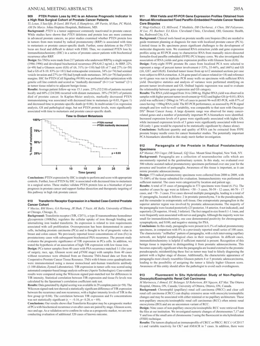
ANNUAL MEETING ABSTRACTS 205A
909 PTEN Protein Loss by IHC Is an Adverse Prognostic Indicator in a High Risk Surgical Cohort of Prostate Cancer PatientsTL Lotan, S Sutcliffe, B Gurel, BH Park, E Humphries, AW Partin, M Han, PC Walsh, AM De Marzo. Johns Hopkins Hospitals, Baltimore, MD.Background: PTEN is a tumor suppressor commonly inactivated in prostate cancer. While studies have shown that PTEN deletions and protein loss are more common in advanced prostate cancers, no prior studies examined whether PTEN protein loss in tumors from men treated by radical prostatectomy (RRP) is associated with time to metastasis or prostate cancer-specific death. Further, some deletions at the PTEN locus are focal and difficult to detect with FISH. Thus, we examined PTEN loss by immunohistochemistry (IHC) in a series of prostate cancer patients with biochemical recurrence after RRP.Design: Six TMAs were made from 217 patients who underwent RRP by a single surgeon (1986-1996) and developed biochemical recurrence (PSA≥0.2 ng/mL). At RRP, 22% (n=49) had a Gleason score (GS) of ≤6, 51% (n=110) had GS of 7 and 27% (n=58) had a GS of 8-10. 83% (n=181) had extracapsular extension, 34% (n=74) had seminal vesicle invasion and 27% (n=58) had lymph node metastases. 36% (n=78) had positive margins. IHC for PTEN (Cell Signaling #9188) was performed after optimization with genetic cell line controls and scored as “markedly decreased” (0-1+) or “normal” (2+) in tumor tissue relative to adjacent benign tissue.Results: Average patient follow-up was 15.1 years. 25% (52/210) of patients recurred locally and 60% (124/208) recurred with distant metastases. 38% (79/207) of patients died of prostate cancer. In Kaplan-Meier analysis, markedly decreased PTEN immunostaining was associated with decreased time to metastasis (p=0.03, Figure 1) and decreased time to prostate-specific death (p=0.06). In multivariate Cox regression analysis, GS and pathological stage, but not PTEN protein levels, were significantly associated with time to metastasis and prostate cancer-specific death.
Conclusions: PTEN expression by IHC is simple to perform and score with appropriate controls. Further, loss of PTEN by IHC is associated with decreased time to metastasis in a surgical series. These studies validate PTEN protein loss as a biomarker of poor prognosis in prostate cancer and support further dissection and therapeutic targeting of this pathway in high risk prostate cancer.
910 Transferrin Receptor Expression in a Nested Case-Control Prostate Cancer CohortV Macias, BM Henry, GA Hartung, JR Shah, T Nazir, AK Balla. University of Illinois at Chicago, Chicago, IL.Background: Transferrin receptor (TfR, CD71), a type II transmembrane homodimer glycoprotein (180kDa), regulates the cellular uptake of iron through binding and internalizing iron loaded transferrin. Its expression is related to iron requirements associated with cell proliferation. Overexpression has been demonstrated in cancer cells, including prostate carcinoma (PCa) and is thought to be of prognostic value in breast and colon cancer. We previously reported lower concentrations of iron (Fe) in prostatectomy cases with subsequent biochemical PSA recurrence. The present study evaluates the prognostic significance of TfR expression in PCa cells. In addition, we tested the hypothesis of an association of high TfR expression with low tissue iron.Design: PCa tumor samples from 40 cases with PSA recurrence matched 1:1 (for year of surgery, race, age, Gleason score and pTNM stage) with samples from 40 cases without recurrence were obtained from an Outcome TMA-based data set from the Cooperative Prostate Cancer Tissue Resource. TMA with 0.6mm cores quadruplicates were immunostained using a mouse monoclonal anti-human transferrin antibody (1:100 dilution; Zymed Laboratories). TfR expression in tumor cells was scored using automated computer-based image analysis software (Aperio Technologies). Case-control results were compared using the Wilcoxon signed pair-matched test for differences in TfR intensity. Statistical correlation between TfR expression and tissue Fe levels was calculated by the Spearman’s correlation coefficient rank test.Results: Data generated by digital scoring was available in 29 complete pairs (n=58). The Wilcoxon signed rank test showed a statistically significant difference of TfR expression between the recurrence and non recurrence, with a higher intensity levels of TfR in the first group (p=0.04). The correlation between TfR expression and Fe concentrations was not statistically significant (rs = - 0.16, p= 0.20, n = 69).Conclusions: Our results shows that Transferrin Receptor may be a prognostic marker of PCa with biochemical recurrence even after cases are paired by stage, Gleason score, race and age. As a validation set to confirm its value as a prognostic marker, we are now conducting evaluation of additional 320 cases of known outcome.
911 RNA Yields and RT-PCR Gene Expression Profiles Obtained from Manual-Microdissected Fixed Paraffin-Embedded Prostate Cancer Needle Core BiopsiesC Magi-Galluzzi, CL Millward, T Maddala, DB Cherbavaz, A Chen, SM Falzarano, M Lee, FL Baehner, EA Klein. Cleveland Clinic, Cleveland, OH; Genomic Health, Inc, Redwood City, CA.Background: Clinical tools based on prostate needle core biopsies (Bx) are needed to guide treatment planning at diagnosis for men with localized prostate cancer (PCA). Limited tissue in Bx specimens poses significant challenges to the development of molecular diagnostic tests. We examined RNA extraction yields and gene expression profiles using an RT-PCR assay to characterize RNA from manually micro-dissected formalin-fixed paraffin embedded (FFPE) PCA biopsy cores. We also investigated the association of RNA yields and gene expression profiles with Gleason Score (GS).Design: Forty-eight FFPE prostate Bx cases from localized PCA were selected to represent a range of percent tumor involvement (<33%, 33-66%, and ≥66%) and GS (≤6, GS7, and GS≥8). Enriched tumor tissue from up to12 unstained sections per case were subject to RNA extraction. A 24-gene panel of cancer-related (n=18) and reference (n=6) genes was run in triplicate PCR assay wells on specimens with sufficient RNA yield. Descriptive statistics and analysis of variance were performed stratifying on percent tumor involvement and GS. Ordinal logistic regression was used to evaluate the relationship between gene expression and GS category.Results: The RNA yield ranged from 16 to 2406 ng. Higher RNA yield was observed in samples with higher percent tumor involvement (p=0.02) and higher GS (p=0.01). RNA yield was sufficient (>200ng) in 74% of cases to permit 96-well RT-PCR, with 87% of cases having >100ng RNA yield. The RT-PCR performance, as assessed by PCR signal strength and low well-to-well variability, was comparable to that seen with Oncotype DX® Breast Cancer Assay. A large dynamic range was observed for many cancer-related genes and a number of potentially important PCA biomarkers were identified. Increased expression levels of 6 genes were significantly associated with higher GS, while increased expression levels of 3 genes were significantly associated with lower GS (only 1 gene would be expected to be statistically significant by chance alone).Conclusions: Sufficient quantity and quality of RNA can be extracted from FFPE prostate biopsy needle cores for cancer biomarker studies. The potentially important PCA biomarkers identified in this study merit further investigation.
912 Paraganglia of the Prostate in Radical Prostatectomy SpecimensKP Maniar, PD Unger, DB Samadi, GQ Xiao. Mount Sinai Hospital, New York, NY.Background: Paraganglia are a collection of neuroendocrine cells which are uncommonly reported in the genitourinary system. In this study, we evaluated over 500 robotic assisted radical prostatectomy specimens performed over one year for the presence and location of paraganglia. Awareness of this tissue is important, as it can mimic prostatic adenocarcinoma.Design: 573 radical prostatectomy specimens were collected from 2008 to 2009, with 75-100% of the tissue submitted for evaluation. Immunochemistry was performed on sufficient material. The cases were categorized by location and patient age.Results: A total of 35 cases of paraganglia in 573 specimens were found (6.1%). The number of cases by age were as follows: <50 - 3 cases, 50-59 - 12 cases, 60-70 - 17 cases, and >70 - 3 cases. Five cases showed multiple paraganglia, leading to a total of 41 paraganglia, located as follows: 2 in prostatic parenchyma, 5 in the pseudocapsule, and the remainder in extraprostatic soft tissue. One extraprostatic paraganglion in the superior anterior region was involved by prostatic adenocarcinoma. The majority of the paraganglia were located posteriorly (23 posterior, 14 anterior, 4 not indicated), and superiorly (28 superior, 10 mid, 3 inferior). The foci ranged in size from 0.1-1.1mm, and were frequently seen associated with nerves and ganglia. Although the majority were too small for immunohistochemistry, one case demonstrated positivity for chromogranin, synaptophysin, and S100, and negative staining for PSA.Conclusions: In this study, paraganglia were present in 6.1% of radical prostatectomy specimens, in comparison with 8% in a previously reported small series of 100 cases. The characteristic “zellballen” pattern of paraganglia, with a rich intervening capillary network, are helpful morphological clues in their recognition. In difficult cases, immunohistochemistry is helpful if sufficient material is present. Recognition of this benign tissue is important in distinguishing it from prostatic adenocarcinoma. This becomes especially significant when the paraganglia are located external to the prostatic pseudocapsule; misidentifying these foci as tumor may lead to falsely diagnosing the patient with a higher stage of disease. Additionally, the characteristic appearance of paraganglia most closely resembles Gleason pattern 4 or 5 prostatic adenocarcinoma, leading to the possibility of assigning the tumor a falsely higher Gleason score. Awareness of this entity should allow the pathologist to avoid such overdiagnosis.
913 Fluorescent In Situ Hybridization Study of Non-Papillary Oncocytic/Eosinophilic Renal Cell CarcinomaM Marinescu, I Ahmed, EC Belanger, SJ Robertson, BN Nguyen, KT Mai. The Ottawa Hospital, Ottawa, ON, Canada; University of Ottawa, Ottawa, ON, Canada.Background: Chromophil (papillary) renal cell carcinoma (PRCC) and clear cell renal cell carcinoma (CRCC) can display extensive areas with oncocytic/eosinophilic changes and may be associated with either minimal or no papillary architecture. These non-papillary oncocytic/eosinophilic renal cell carcinomas (RCC) often mimic renal oncocytoma (RO) and are an uncommon variant of RCC.Design: Nine cases of non-papillary oncocytic/eosinophilic RCC were retrieved from the files at our institution. We investigated numeric changes of chromosomes 7,17 and Y and loss of the small arm of chromosome 3 using the fluorescent in situ hybridization (FISH) technique.Results: The tumors displayed an immunoprofile of CRCC or PRCC: RCC (+)/CD117 (-) and variable reactivity for CK7 and AMACR in 7 cases. In addition, there were
206A ANNUAL MEETING ABSTRACTS 2 cases which were previously reported as malignant renal oncocytoma (RO) with an immunoprofile of RCC(-)/CD117(+), marked cytologic atypia and lymph node metastasis. FISH studies showed trisomies 7/17 with or without loss of Y in five tumors. Loss of loci 3p25 and 3p14 were identified in another 2 cases. The remaining 2 tumors with features of RO showed no evidence of numeric changes for chromosomes 7, 17 and Y or loss of loci 3p25 or 3p14.Conclusions: In this uncommon variant of RCC, FISH for chromosomes 7, 17, Y and 3p lend more support to the role of immunostaining in distinguishing RO and chromophobe RCC from the non-chromophobe RCC. FISH for chromosomes 7, 17, Y and loci 3p25 and 3p14, and not immunostaining for AMCR or CK7, is helpful in distinguishing CRCC from PRCC.
914 Stem Cell Marker ALDH1 Expression Correlates with Unfavorable Prognosis in Prostate CancerA Matoso, R Tavares, L Noble, Y Zhang, R Jacob, J Renzulli, G Pareek, G Haleblian, MB Resnick, LJ Wang. Brown University and Rhode Island Hospital, Providence, RI.Background: Aldehyde dehydrogenase (ALDH) 1 is an enzyme that is ubiquitously expressed and catalyzes the oxidation of aldehydes to their carboxylic forms. ALDH1 activity has been used to identify stem-like subset of cells in human and murine hematopoietic and solid tumors. Recent studies demonstrated that increased ALDH1 expression correlates with poor clinical outcome in breast cancer but favorable prognosis in ovarian cancer. We hypothesized that the level of ALDH1 expression may correlate with the clinical outcome of patients with prostate cancer.Design: Immunohistochemical staining of ALDH1 expression was analyzed in 175 radical prostatectomy specimens using tissue microarrays (clone 44/ALDH; 1:400 dilution; BD Biosciences). Results were scored using a quantitative system that assigned 1-4 points for extent and for intensity of staining. A total score of 5 or higher was considered strong and 4 or lower was considered weak. The associations between the expression of ALDH1, pathological stage, Gleason score and overall survival time were analyzed.Results: Focal expression of ALDH1 was noticed in the cytoplasm of non-tumoral prostatic acini in 5% of the cases studied. Weak staining was also noted in stromal cells and blood vessel walls. Elevated ALDH1 expression was found in 39.6% of all tumor samples and correlated with Gleason score, 26.5% positive in Gleason ≤6, and 55.3% positive in Gleason ≥7 (p<0.001)). Strong ALDH1 expression also correlated with higher pathological stage (pT2c or higher; 46% vs. 28%; P=0.04). As expected, Gleason score and stage inversely correlated with overall survival. Patients with Gleason score ≥7 and strong ALDH1 expression had a shorter overall survival time than patients with Gleason score ≥7 and weak ALDH1 expression (P=0.03). Although not statistically significant, patients with Gleason score ≤ 6 and strong ALDH1 expression also had a tendency towards shorter survival time than patients with Gleason score ≤ 6 and weak ALDH1 expression.Conclusions: This is the first study to describe ALDH1 expression and to determine its prognostic significance in prostate cancer. Our results demonstrate that ALDH1 expression correlates with high Gleason score, higher stage at diagnosis and shorter overall survival time.
915 Overexpression of Cancer Stem Cell Markers in Renal Cell CarcinomaA Matta, S El Jamal, HH Zhang, N Gokden, CY Fan. University of Arkansas for Medical Sciences, Little Rock, AR; John L. McClellan Memorial Veterans Hospital and UAMS, Little Rock, AR.Background: SALL4 and BMI-1 are putative oncogenes that modulate stem cell pluripotency and play a role in oncogenesis of a wide variety of human malignant tumors. Aldehyde dehydrogenase (ALDH) and CD44 have been implicated in multiple biological and biochemical pathways and has been used to identify potential cancer stem cells. In this study, we analyze gene expression levels of these 4 cancer stem cell markers in clear cell renal cell carcinoma (CC-RCC) as compared to matched normal renal parenchyma.Design: Total RNA was extracted from 14 fresh CC-RCC samples and their matched benign renal parenchyma. Gene expression levels for SALL4, BMI-1, ALDH1A1 and CD44 were analyzed by real-time RT-PCR Quantitation with the ABI Fast 7500 Real-time PCR System using Taqman 18S RNA as an internal control for normalization.Results: The mRNA expression levels are significantly higher in CC-RCC than those seen in adjacent normal parenchyma in 8 of 14 cases (57%) for the SALL4 gene, 12 of 14 cases (85%) for the BMI-1 gene, 12 of 14 cases (85%) for the ALDH1A1 gene and 11 of 14 cases (78%) for the CD44 gene. Overall, the mRNA expression levels in CC-RCC is about 6 folds for the SALL4 gene, 5 folds for the BMI-1 gene and 3 folds for the ALDH1A1 gene and 4 folds for the CD44 gene, as much as those seen in normal renal tissues. Mean relative mRNA levels for SALL4, BMI-1, ALDH1A1 and CD44 in all 14 tumor cases are 1.2, 4.8, 5.8 and 16.3 respectively as compared to a mean of 0.1, 0.9, 1.7 and 4.8 for normal renal parenchyma.Conclusions: Significant upregulation of cancer stem cell markers occurs commonly in CC-RCC as compared to matched normal renal parenchyma. These results support a stem cell carcinogenesis theory for clear cell RCC in which the driven force for initiation and progression of a cancer is derived from a small population of cancer stem cells. Therefore, anticancer therapy should aim at eradicating this small population of cancer stem cells.
916 Identification and Characterization of Viable Bone Metastases from Rapid Autopsies of Androgen Independent Prostate Cancer PatientsR Mehra, Bo Han, C Kumar-Sinha, X Jing, X Cao, J Granger, S Shanker, DC Smith, RB Shah, AM Chinnaiyan, KJ Pienta. University of Michigan, Ann Arbor, MI.Background: Bone is the most common metastatic site for prostate cancer, and osseous metastases are the leading cause of morbidity of this disease. In fact, recent careful autopsy studies prove that 100% of the men who die of prostate cancer have bone involvement. Understanding the biology of prostate cancer and its evolution to an incurable androgen independent phenotype thus warrants an investigation into the genetic and cellular alterations that lead to the seeding and proliferation of these tumor foci in bone, as well as the microenvironment in which these metastases arise.Design: Between September 1996 to present, 51 rapid autopsies were performed on men who died of advanced androgen independent metastatic prostate cancer at the University of Michigan. As a part of a new protocol, multiple osseous sites including the vertebrae, femurs, and humeri of all patients were longitudinally sectioned and examined for presence of grossly visible metastases. Examination of bone metastases was also guided by the patient’s bone scan. Bone marrow tissues harboring either spongy beefy red normal marrow or grossly evident tumor and foci suspicious for tumor were procured for phenotypic assessment and RNA extraction.Results: We employed a novel technique using gross examination and bone scan guided dissections to procure viable metastatic tumor tissues from involved bones in 13 recent warm autopsies. The presence of viable bone metastasis was confirmed by evaluation of hematoxylin and eosin stained sections of frozen material. The bone metastatic tissues were used for isolating RNA and DNA. Real-time PCR based expression profiling of the most common prostate cancer specific genes, such as PSA, PCA3, ERG, TMPRSS2-ERG, and AMACR was carried out. As expected, PSA and PCA3 show very high levels of expression in all the samples examined, while the expression of AMACR, ERG, and TMPRSS2-ERG showed variable expression across the sample set. Ongoing studies include microarray based comparison of osseous metastatic tissues with non-osseous metastatic tissues within an individual patient as well as in the entire cohort.Conclusions: Taken together, we describe a versatile and practical approach to describe the phenotypic and molecular/biochemical characteristics of osseous metastasis in men who died of androgen independent prostate cancer.
917 The Unrecognized Morphology of Renal Tumors in SDH Syndromes. Immunohistochemical and Genetic ChangesMJ Merino, ER Parrilla-Castellar, M Linehan. NCI, Bethesda, MD.Background: More than 35,710 new cases of renal cell carcinoma (RCC) are diagnosed in the US, with about 12,480 deaths per year.Recently our understanding of renal cancer predisposing syndromes has improved, and genes associated with specific tumors have been discovered. Mutations in the genes that encode mitochondrial succinate dehydrogenase can cause the paraganglioma syndromes with development of paraganglioma and pheochromocytoma. The purpose of this study is to provide the first morphological, immunohistochemical and genetic description of the renal tumors found in these syndromes.Design: Five patients presented with renal tumors.Two had prior history of familial SDH deficiency and one patient presented with metastatic RCC to bone. Microdissected DNA was evaluated for LOH in chromosomes 1p35 and 11q23.Results: Patients ranged in age from 19-68 years. Four patients were females and 1 was male. Four tumors involved the left kidney and 1 the right. Tumor size varied from 6-2.4 cm. Grossly the lesions had a brown color and were well circumscribed. Histologically the solid masses were composed of uniform cells with clear and light granular eosinophilic cytoplasm. Many of the cells had a signet ring cell appearance and the vacuole seemed to be filled by amorphous pink material. This material could be seen filling small cystic dilated spaces lined by eosinophilic cells resembling oncocytes. The nucleus of the cells was large and with irregular chromatin distribution. A smaller cell with granular cytoplasm and dark nucleous was present and in some cases large darkly pigmented cells were identified. One tumor showed spindle cell differentiation and areas of nuclear pleomorphism and necrosis; but nodules of cells similar to the ones described above were present. The bone metastasis was composed predominantly of eosinophilic spindle cells. IHC was performed for CK7, CK20, CAM5.2, CD10, CD56, CKIT, Synaptophysin, Chromogranin, grimelius, melanin, PAS and colloidal iron stains.Conclusions: Renal tumors associated with SDH have a unique morphologic appearance. Recognition of this group of tumors is important both, clinically and pathologically, because they are frequently misdiagnosed as oncocytomas and clear cell RCC. Proper diagnosis of these tumors, will allow for identification of specific genes that will allow elucidation of mechanisms responsible for the development of tumors, as well as to establish appropriate therapies and the genetic counseling of the families,
918 Smooth Muscle Rich, Tubulo-Cystic Papillary Renal Cell Carcinoma. A New Renal Tumor? Immunohistochemical and Genetic CharacteristicsMJ Merino, L Jinping, K Killian, M Linehan. NCI, Bethesda, MD.Background: While the majority of renal tumors occur sporadically, the true incidence of hereditary tumors is not exactly known. It is possible that the incidence is higher than the predicted 5%, since it is only now that considerable insight into the genetic basis of renal cancer is available. Proliferation of smooth muscle cells and lymphatic vessels has been described in the kidney as lymphangioleiomyomatosis and angiomyoleiomas that occur in the Tuberous sclerosis complex. We studied six unusual cases of smooth muscle rich renal cell carcinoma and describe their morphological, immunohistochemical and genetic changes.Design: Six patients with multifocal and bilateral renal tumors, were evaluated for protocol acceptance. Tumors were morphologically and immunohistochemically evaluated. Tissue was microdissected and DNA obtained for CGH and LOH analysis of multiple genes.
ANNUAL MEETING ABSTRACTS 207AResults: Patients ranged in age from 35-79 years. Five patients were males and 1 was female. Multiple tumors were removed from each patient varying in size from 4-0.8 cm. Grossly they had a tan to light yellow color. Histologically, there were composed of solid and diffuse clusters of clear tubules surrounded by smaller tubules with less cytoplasm and occluded lumen, that gave the appearance of mantles. The tubules had a centrally located indolent nucleous. These nests were surrounded by muscle and collagenous fibers. Cysts and malformed papillae with abundant muscle in the stall were also present. In areas the muscle and fibrous proliferations were prominent. IHC was performed for CK7, CK20, CAM5.2, EMA, CD10, CD56, SMA, Collagen IV, Calponin, Vimentin, S-100 and HMB45. CGH analysis did not reveal significant changes in the VHL or BHD genes.Conclusions: We report an unusual group of renal cell tumors with prominent proliferation of smooth muscle and tubular, cystic and papillary elements. The tumors are low grade and stain strongly with CK7 and SMA. It is possible that these tumors represent new entities or unsuspected variants on known syndromes. Recognition of these neoplasms will allow for identification of new and specific genes, that will lead to the development of specific molecular targets.
919 Primary Leiomyosarcoma of the Kidney: A Clinicopathologic Study of 27 CasesJS Miller, M Zhou, F Brimo, CC Guo, JI Epstein. Johns Hopkins Hospital, Baltimore; Cleveland Clinic, Cleveland; MD Anderson Cancer Center, Houston.Background: Primary leiomyosarcoma of the kidney is a rare entity that has not been well-characterized.Design: We retrieved 27 cases of primary renal leiomyosarcomas diagnosed at three institutions between 1986 and 2009. Any evidence of a carcinomatous component, either histologically or immunohistochemically, indicated the diagnosis of the more-common sarcomatoid carcinoma (carcinosarcoma) and led to the case’s exclusion from the study. Cases with leiomyosarcomas secondarily involving the kidney (e.g. retroperitoneal leiomyosarcomas) were also excluded based on radiologic, operative, and/or pathologic findings.Results: Mean patient age at diagnosis was 58.5 years (range 22 to 85) and 59% were female. Mean tumor size was 13.4 cm (range 4 to 26) and 59% of the tumors were identified in the right kidney. Detailed histologic examination was possible for 24 of the cases. Average mitotic count per ten high power fields was 11.1 (range 0 to 50) and the average extent of necrosis was 21% (range 0 to 60). Cellular pleomorphism was classified as either focal (n=13) or extensive (n=11) and graded as mild (n=3), moderate (n=7), or severe (n=14). Tumors were either grade 2 (n=12) or grade 3 (n=12) using the French Federation of Cancer Centers System. Direct extension beyond the kidney capsule was identified in 55% of the cases, and lymphovascular invasion was identified in 26%. Clinical follow-up information was available for 20 of the cases, and patients were followed for an average of 2.8 years (range 0.25 to 9). Distant metastases were identified in 90% of the patients, and 75% eventually died from their tumor burden. No correlation was identified between histologic features and clinical progression; virtually all of the patients in this study had a poor clinical outcome regardless of morphologic features.Conclusions: Primary renal leiomyosarcomas have a grim prognosis regardless of the underlying histology.
920 Extraprostatic Extension of Prostatic Adenocarcinoma on Needle Core Biopsy: Report of 72 Cases with Clinical Follow-UpJS Miller, Y Chen, BD Robinson, F Brimo, JI Epstein. Johns Hopkins Hospital, Baltimore.Background: Extraprostatic extension (EPE) of prostatic adenocarcinoma is rarely found on needle core biopsy and has not been formally addressed in the literature.Design: We retrieved 99 cases of prostatic adenocarcinoma with EPE at initial diagnosis on biopsy from the consult files of one of the authors between 1997 and 2009. The 72 cases that had available clinical follow-up data formed the basis of this study.Results: Mean patient age was 64 years (range 48 to 87), median serum prostatic specific antigen (PSA) was 7.8 ng/mL (mean 64.8, range 0.3 to 1505), and 60 of the patients (83%) had an abnormal digital rectal examination (DRE). The average number of malignant cores was 7.7 (range 1 to 23); average percent of carcinoma in each core was 69.6%; average percent of carcinoma in the core(s) demonstrating EPE was 76.8%. The average Gleason score in the core(s) demonstrating EPE was 8; average highest Gleason score per case was 8.4. Perineural invasion was demonstrated in 54 cases (75%). Ten out of 11 cases treated surgically demonstrated EPE on the radical prostatectomy; additionally, 6 had positive resection margins, 5 showed invasion into the seminal vesicles and 1 demonstrated lymph node metastasis. The Gleason scores in 9 of the prostatectomies did not differ from the highest grade found in the associated biopsies (score 9 [n=3], score 8 [n=2], score 7 [n=4]); in one case it increased (from score 6 to 8) and in one case it decreased (from score 9 to 8). Patients were followed for an average of 2.9 years (median 2, range 0.1 to 9) with metastases identified in 29 (40%); 10 (14%) died from the disease.Conclusions: EPE on needle core biopsy of the prostate is strongly associated with extensive, high-grade prostatic adenocarcinoma, such that its usefulness as an isolated prognostic factor is relatively limited.
921 Pitfalls in the Use of Smoothelin To Identify Muscularis Propria Invasion by Urothelial CarcinomaH Miyamoto, RB Sharma, PB Illei, JI Epstein. The Johns Hopkins Hospital, Baltimore.Background: Studies predominantly performed on cystectomy specimens have shown that antibodies against smoothelin can distinguish between muscularis mucosae (MM)
(negative or weak stain) and muscularis propria (MP) (strong stain). However, studies on diagnostically difficult (TUR) specimens have not been performed.Design: IHC for smoothelin was performed in 2 groups.Results: The 1st cohort was 34 TUR cases where outside pathologists questioned the presence of MP invasion. Upon expert review of the H&E slides, there was no MP invasion in 18 cases. Smoothelin in MM was negative in 8/18 (44%); weakly positive (1+) in 5/18 (28%); moderately positive (2+) in 4/18 (22%); and moderately/strongly (2-3+) positive in 1/18 (6%). Smoothelin in uninvolved MP present in 8 cases was: 2+ in 2/8 (25%) and 3+ in 6/8 (75%). Smoothelin expression in MM was weaker than in MP 7/8 (88%) cases where both were present. Of 16 tumors with MP invasion, smoothelin in involved MP was: 1+ in 1/16 (6%), 2+ in 3/16 (19%), 2-3+ in 9/16 (56%), and 3+ in 3/16 (19%). Smoothelin expression in concurrent uninvolved MP was similar. The 2nd cohort was 16 cases of urothelial cancer involving muscle bundles that upon expert review were indeterminate for MM or MP, where a subsequent procedure proved their stage (T1 in 7 cases and T2 in 9 cases). Among 7 eventually proven T1 tumors, muscle bundles (presumably MM) involved by tumor: smoothelin was 0 in 3/7 (43%), 1+ in 2/7 (29%), 2+ in 1/7 (14%), and 2-3+ in 1/7 (14%). Of 9 eventually proven T2 tumors, muscle bundles (presumably MP) involved by tumor: smoothelin was 0 in 2/9 (22%), 1+ in 4/9 (44%), 1-2+ in 1/9 (11%), 2+ in 1/9 (11%), and 2-3+ in 1/9 (11%).Conclusions: Using diagnostically difficult TUR specimens where it was difficult to distinguish MM from MP invasion, we have confirmed the relatively distinct staining pattern of smoothelin immunohistochemistry. Nonetheless, approximately 25% of cases in the current study showed overlap in the staining pattern between MM and MP. Smoothelin staining in these cases included moderate to strong positivity in MM or weak to moderate smoothelin in MP. Typically, although there were some exceptions, smoothelin immunoreactivity was stronger in MP compared to that seen in MM or smooth muscle in blood vessels at the level of the MM within the same case. However, interpretation of smoothelin in thin muscle bundles is especially problematic, when concurrent blood vessels at the level of the MM and/or MP, as internal intensity controls, are absent in the same specimen.
922 Nodular Periorchitis: A Morphological and Immunohistochemical Study of 13 CasesH Miyamoto, E Montgomery, JI Epstein. The Johns Hopkins Hospital, Baltimore.Background: Nodular periorchitis is a rare intrascrotal mass that has been thought to represent a reactive process arising mostly in the tunica vaginalis and less commonly in the epididymis, spermatic cord, or tunica albuginea. Numerous synonyms have been proposed for this entity. These include “fibrous pseudotumor”, “nodular and diffuse fibrous proliferation”, “chronic proliferative periorchitis”, and “inflammatory pseudotumor”, among others. Its histology has been mainly described in case reports and there are no series on its immunohistochemical features.Design: A database search (2000-2008) from the consult files of one of the authors, as well as archival files of Surgical Pathology at our institution, identified 13 cases in which paraffin blocks or unstained slides were available for further studies. CD34 and calretinin/cytokeratin results were recorded for the lesional spindle cells not blood vessels and mesothelium, respectively.Results: Patients’ ages ranged from 19 to 75 years old (mean 41.9 years). All men presented with scrotal mass, with 6 (46%) cases also having a clinical history of hydrocele. Six men were treated by orchiectomy, while the remaining 7 men underwent excisional biopsy. Histologically, the lesions could be subdivided into 3 types: 1) “plaque-like”, consisting of dense fibrous tissue with clefts without significant inflammation identical to a pleural plaque (5 cases); 2) “inflammatory sclerotic” with dense fibrous tissue containing lymphocytes (diffusely or aggregates or germinal centers), plasma cells, and an increased capillary network (6 cases); and 3) “myofibroblastic” (2 cases) consisting of reactive appearing tissue-culture-like cells with numerous capillaries and sparse chronic inflammation. All cases were negative for beta-catenin and ALK-1. Ki-67 showed <1% positivity in all but 2 cases, which had 5% positivity. Results for other stains are listed in table.
Actin Desmin Cytokeratin Calretinin CD34Plaque-like 4/5 2/5 (1 focal) 2/5 (2 focal) 1/5 (1 focal) 2/5 (1 focal)Inflammatory/Sclerotic 5/6 (4 focal) 0/6 3/6 (1 focal) 3/6 (3 focal) 5/6 (2 focal)Myofibroblastic 2/2 2/2 (1 focal) 2/2 2/2 (1 focal) 0/2
Conclusions: There are 3 distinct histologic patterns seen in nodular periorchitis, although their immunohistochemical profile has overlapping features. Because of negative stains for ALK-1 and beta-catenin, nodular periorchitis appears distinct from inflammatory myofibroblastic tumor and fibromatosis, respectively, seen in other organs.
923 Transurethral Resection Specimens of the Bladder (TURB): Outcome of Invasive Urothelial Cancer Involving Muscle Bundles Indeterminate between Muscularis Mucosae and Muscularis PropriaH Miyamoto, JI Epstein. The Johns Hopkins Hospital, Baltimore.Background: It may be difficult to diagnose muscularis propria on TURB as thin muscle fibers on TURB may represent either muscularis propria destroyed or splayed by urothelial carcinoma or muscularis mucosae, which may be hyperplastic.Design: 95 invasive bladder cancers seen at our instituion (1986-2008) with follow-up (mean 25.4 months) where the initial TUR pathologic stage was ambiguous (T1 vs. T2) were analyzed (73 men; 22 women; mean age 69.4 years).Results: Subsequent restaging TURB or definitive therapeutic procedures performed ≤3 months after the original TURB done in 58 cases revealed 22 (37.9%) patients with non-muscle invasive disease and 32 (55.2%) patients with ≥pT2 disease. Staging in 4 cases remained ambiguous. 37 cases eventually developed ≥pT2 disease in 2/22 (9.1%) cases with non-muscle invasive disease on initial restaging TURB, 2/4 (50.0%) of cases with uncertain stage disease, and 14/37 (37.8%) cases with no restaging TURB. Patients with a final stage of non-muscle invasive disease had a lower risk of progression (T4
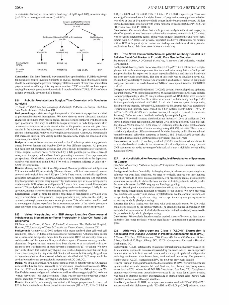
208A ANNUAL MEETING ABSTRACTS or metastatic disease) vs. those with a final stage of ≥pT2 (p=0.003), uncertain stage (p=0.012), or no stage confirmation (p=0.043).
Conclusions: This is the first study to evaluate follow-up when initial TURB is equivocal for muscularis propria invasion. Similar to an atypical prostate needle biopsy, urologists should be encouraged to perform restaging TURBs in cases of equivocal muscularis propria invasion. Although this may seem intuitive, 37/95 cases did not have repeat staging/therapeutic procedures done within 3 months of initial TURB; 37.8% of these patients eventually developed ≥T2 disease.
924 Robotic Prostatectomy Surgical Time Correlates with Specimen AutolysisAP Modi, AP Patel, GN Box, DS Sharp, A Shabsigh, R Abaza, DL Zynger. The Ohio State Medical Center, Columbus, OH.Background: Appropriate pathologic interpretation of prostatectomy specimens is vital to postoperative patient management. We have observed more substantial autolytic changes in specimens from robotic radical prostatectomies compared with those from open procedures. This may be related to longer exposure to body temperature after devascularization prior to specimen extraction as the prostate in a robotic procedure remains in the abdomen after being devascularized while in an open prostatectomy the prostate is immediately removed following devascularization. As such, we hypothesized that increased surgical time during robotic prostatectomy might be associated with more autolytic artifact.Design: A retrospective review was undertaken for 85 robotic prostatectomy patients treated between January and October 2009 by four different surgeons. All prostates had been sent for immediate grossing and whole mount processing after extraction. These original sections were re-reviewed by a GU pathologist to assess percent autolysis at the apex, mid and base levels generating a measure of average autolysis per specimen. Multivariate regression analysis using total autolysis as the dependent variable was performed using SPSS 17.0 with a Bonferroni-adjusted p value of < 0.004 for significance.Results: Mean age of the patient was 60 years old. Mean surgical time and autolysis was 229 minutes and 4.8%, respectively. The correlation coefficient between total percent autolysis and surgical time was 0.603 (p <0.001). There was no statistically significant correlation between autolysis and PSA, Gleason score, % tumor, prostate size, maximum length of prostate, surgeon, margin positivity, node status, age, or use of preoperative medication for BPH. Above a cutoff point of 4.5 hours, there was a mean 8.8% autolysis versus 2.7% autolysis below 4.5 hours using the paired sample t-test (p <0.001). In one specimen, margin status was indeterminate due to autolysis artifact.Conclusions: Length of time for robotic procedures is significantly correlated with degree of autolysis in the prostate. Longer procedures may influence the ability to evaluate pathologic parameters such as margin status. This information could be used to encourage urologists to perform the prostatectomy portion of the robotic procedure after the lymph node dissection to decrease the warm ischemia time of the prostate.
925 Virtual Karyotyping with SNP Arrays Identifies Chromosomal Imbalances as Biomarkers for Tumor Progression in Clear Cell Renal Cell CarcinomaFA Monzon, K Alvarez, K Sircar, P Tamboli, E Jonasch. The Methodist Hospital, Houston, TX; University of Texas MD Anderson Cancer Center, Houston, TX.Background: As many as 20-30% patients with organ confined clear cell renal cell carcinoma (ccRCC) will develop metastases after nephrectomy. Antiangiogenic agents are a successful therapeutic modalities for metastatic RCC but currently there are no biomarkers of therapeutic efficacy with these agents. Chromosome copy number alterations frequent in renal tumors have been shown to be associated with poor prognosis (9p/14q deletions) or more favorable outcomes (5q31-ter gains). We have previously shown that virtual karyotyping is a reliable diagnostic tool that can help reduce uncertainty in the diagnosis of difficult renal tumors. In this study, the goal was to determine whether chromosomal imbalances identified with SNP arrays could be used as biomarkers for progression in metastatic ccRCC (mRCC).Design: We obtained archival FFPE tumor samples from 56 patients with mRCC treated with sorafenib (after tumor removal) or bevacizumab (neoadjuvant treatment). DNA from the FFPE blocks was analyzed with Affymetrix 250K Nsp SNP microarrays. We identified the presence of genomic imbalances and loss of heterozygosity (LOH) to obtain “virtual karyotypes”. We then determined if there was any association between genetic lesions identified by virtual karyotyping and tumor outcome measures.Results: Gain of 5q was strongly associated with longer progression free survival PFS in both sorafenib and bevacizumab treated cohorts (HR = 0.25, 95% CI 0.08 to
0.81, P = 0.021 and HR = 0.82 95% CI 0.65- 1, P < 0.0001 respectively). There was a nonsignificant trend toward a higher hazard of progression among patients who had loss of 9p or loss of 14q in the sorafenib cohort. In the bevacizumab cohort, 14q loss showed a significant association with worse response to treatment (CR or PR vs SD or PD, Fisher exact test, P = 0.0473).Conclusions: Our results show that whole genome analysis with virtual karyotypes identifies genetic lesions that are associated with outcomes in metastatic RCC treated with novel anti-angiogenic agents. These results suggest that genomic analysis of renal tumors with SNP arrays can provide important predictive information for patients with mRCC. A larger study to confirm our findings are studies to identify potential mechanisms that explain these associations are underway.
926 The Novel Immunohistochemical p75/p63 Antibody Cocktail Is a Reliable Basal Cell Marker in Prostatic Core Needle BiopsiesSM Moran, O O’Brien, F O’Connell, D McCrae, TJ Browne. Cork University Hospital, Cork, Ireland.Background: Nerve growth Factor receptor (NGFR/p75NTR ) is a cell surface receptor glycoprotein with tumour suppressor functions and roles in regulation of cell growth and proliferation. Its expression in breast myoepithelial cells and prostate basal cells has been previously established. The aim of this study was to develop a novel p75/p63 antibody cocktail (p75 cocktail), to evaluate it as a basal cell marker in benign and malignant prostatic core needle biopsies (CNB) and to compare this to the p63/34BE12 cocktail.Design: A novel immunohistochemical (IHC) p75 cocktail was developed and optimized in our laboratory. With institutional approval 56 sequential prostatic CNB were selected from surgical pathology files (28 benign, 28 malignant). All H&E slides were reviewed and diagnosis confirmed. Paraffin sections were stained with the newly developed p75/P63 and previously validated p63/ 34BE12 cocktails. A scoring system incorporating distribution and intensity in basal cells, luminal cells and stromal cells was established. Distribution and intensity were graded on 4 tier systems (Distribution: 1<5%, 2= 5-25%, 3=25-50%, 4>50%, cells positive. Intensity: 0=negative, 1=weak, 2=moderate, 3=strong). Each case was scored independently by two pathologists.Results: P75 cocktail staining distribution and intensity: 100% of malignant CNB showed absent basal cell staining. All benign CNB showed positivity with excellent distribution (92.3% score 4/4, 7.7% score 3/4). Staining intensity was at least moderate (11.5%) with the majority of cases showing strong (88.5%) intensity. There was no statisitically significant difference observed for either intensity or distribution in basal, luminal or stromal cells when compared to the p63/34BE12 cocktail. p75 cocktail also highlighted nerves aiding identification of perineural invasion (PNI).Conclusions: A novel IHC antibody cocktail p75/p63 was developed and shown to be a reliable basal cell marker in the evaluation of both malignant and benign prostate CNB specimens. An added advantage of this cocktail is that it highlights nerves aiding evaluation of PNI.
927 A Novel Method for Processing Radical Prostatectomy Specimens for CancerPSS Naik, JP Sweeney, N Khan, E Rogers, JF Fitzgibbon. Mercy University Hospital, Cork, Ireland.Background: In these financially challenging times, it behoves us as pathologists to influence our own fiscal decisions. We need to critically analyze our time honored published methods of gross prostate pathology. For processing radical prostatectomy specimens, we decided to utilize the least number of histologic blocks which yielded the most information for TNM staging and histologic grading.Design: We adopted a novel capsular dissection akin to the widely accepted method of processing encapsulated follicular neoplasms of the thyroid. We have processed two hundred and seventy-nine radical prostatectomies by the capsular method and prospectively analyzed grade and stage on ten specimens by comparing capsular processing to whole gland processing.Results: The TNM staging was the same with both methods except for T2b which could not be assessed by the capsular method. The grading remained unchanged in both methods. The mean number of blocks by the capsular method was twenty compared to forty-two blocks by whole gland processing.Conclusions: We conclude that the capsular method is cost-effective and less labour-intensive than other methods without significantly compromising either stage or grade.
928 Aldehyde Dehydrogenase Class 1 (ALDH1) Expression Is Associated with Disease Outcome in Prostatic Adenocarcinomas (PAC)T Nazeer, KD Linos, BVS Kallakury, HAG Fisher, BM Mian, CE Sheehan, JS Ross. Albany Medical College, Albany, NY, 12208; Georgetown University Hospital, Washington, DC.Background: ALDH1 catalyzes the oxidation of intracellular aldehydes involved in cell differentiation, response to oxidative stress and drug resistance. ALDH1 is also expressed in adult stem cells in both normal tissues and a variety of epithelial malignancies including carcinomas of the breast, lung, head and neck and ovary. The prognostic significance of ALDH1 expression in PAC has not been previously studied.Design: Formalin-fixed, paraffin embedded sections from 154 PAC were immunostained by automated methods (Ventana Medical Systems, Inc., Tucson, AZ) using mouse monoclonal ALDH1 (clone 44/ALDH; BD Biosciences, San Jose, CA). Cytoplasmic immunoreactivity was semi quantitatively assessed in the tumor for all cases. Scoring was based on staining intensity and percentage of stained tumor cells. Results were correlated with clinicopathologic variables.Results: Cytoplasmic ALDH1 over-expression was observed in 81/154 (53%) of PAC and correlated with high tumor grade [65% HG vs 43% LG, p=0.007], advanced stage
ANNUAL MEETING ABSTRACTS 209A[66% advanced vs 44% early, p=0.006], and biochemical disease recurrence [62% recur vs 45% non-recur, p=0.049]. On multivariate analysis, high tumor grade and advanced tumor stage independently predicted overall survival.Conclusions: Over-expression of ALDH1 in PAC is associated with adverse prognostic factors and predicts disease relapse after primary therapy. Further study of this putative cancer stem cell marker in PAC appears warranted.
929 Radical Retropubic Prostatectomy for Prostate Cancer with Microscopic Bladder Neck Involvement: Survival and Prognostic ImplicationsG Nesi, D Villari, A Della Melina, M Ceroti, D Palli, G Nicita. University of Florence, Florence, Italy; Cancer Research and Prevention Institute, Florence, Italy.Background: We report the oncological outcome of 106 patients who had locally advanced prostate cancer with microscopic bladder neck invasion, identified in a series of 1129 patients surgically treated with retropubic radical prostatectomy over a 12-year period.Design: All specimens were reviewed. Microscopic bladder neck invasion was defined as the presence of neoplastic cells within the smooth muscle bundles of the bladder neck, with no accompanying prostatic glandular tissue on the corresponding slide. Survival was analysed for different subgroups in relation to several variables.Results: The follow-up (median 7.2 years, mean 6.68, range 0.3–14) was available for 106 patients with microscopic bladder neck invasion. Seminal vesicle invasion was present in 69.8% of the cases, lymph node involvement in 29.2%, apex infiltration in 31.8%, and positive surgical margins in 23.6%. Biochemical progression occurred in 61 (57.5%) patients, and 25 of them died from cancer. The mean (SD) biochemical progression-free survival was 0.68 (0.05), 0.59 (0.05), 0.40 (0.05) and 0.38 (0.05) at 1, 2, 5 and 10 years, respectively. Age, Gleason score and lymph node invasion were independent prognostic factors on multivariate analysis. Overall and cancer-specific survival rates were 0.75 (0.04) and 0.80 (0.04) at 5 years and 0.57 (0.04) and 0.75 (0.04) at 10 years, respectively. Univariate analysis showed that seminal vesicle invasion, lymph node involvement and surgical Gleason score ≥8 significantly increased the risk of death. On multivariate analysis only the surgical Gleason score had an independent prognostic role with regard to overall survival (p=0.01; odds ratio 2.82, 95% confidence interval 1.2–6.4) and cancer-specific survival (p<0.001; 8.6, 2.5–28.8).Conclusions: In this series, overall and cancer-specific survival rates were comparable to those reported for surgically treated cT3 prostate cancers. The lack of need for external urinary diversion during the entire follow-up significantly contributed to the patients’ quality of life.
930 Loss of Heterozygosis on Interferon-Alpha Locus Is a Prognostic Indicator of BCG Response in Non-Muscle Invasive Bladder CancerG Nesi, T Cai, M Dal Canto, N Mondaini, M Piazzini, R Bartoletti. University of Florence, Florence, Italy.Background: Non-muscle invasive bladder carcinoma (NMIBC), treated by resection and intravesical administration of Bacillus Calmette Guerin (BCG), yields a remission rate that approaches 70%. We examined whether loss of heterozygosis (LOH) on interferon-alpha (IFN-α) locus can serve to predict response.Design: A total of 117 consecutive subjects (77 patients affected with NMIBC and 40 controls) were selected. LOH on IFN-α locus (Chromosome 9) was assessed on blood and urine samples before transurethral resection (TUR). All patients underwent TUR and subsequent 6 weekly BCG instillations. Patients affected with NMIBC were assigned to group A (with LOH on IFN-α locus) and group B (without LOH on IFN-α locus). The main outcome parameters were time to first recurrence and recurrence rate. The relative data were compared with follow-up information.Results: Out of the 77 patients with NMIBC, 39 (50.6%) showed at least one alteration on IFN-α locus (group A), with 38 (49.4%) showing no alteration (group B). Only 1 out of 40 controls exhibited LOH on IFN-α locus. At the end of follow-up, 13 patients in group A and 27 in group B were alive without recurrence. A significant difference between LOH on IFN-α and status at follow-up was found (p=0.003; dF01; LR=11.252). Kaplan-Meier analysis demonstrated a significant difference in terms of recurrence probability (response to BCG) and LOH on IFN-α (p=0.0001). At multivariate analysis, LOH on IFN-α locus (p=0.002) (LOH, HR 4.09 2.59–6.28, 95% CI), grade (p=0.03) (grade 3, HR 3.31 1.38–3.35, 95% CI) and the number of lesions (p=0.03) (≥3 lesions, HR 2.31 1.38–3.25, 95% CI) were identified as independent predictors of BCG response.Conclusions: This study highlights the predictive value of LOH analysis on IFN-α in patients affected with NMIBC and treated by intravesical administration of BCG.
931 Immunohistochemical Evidence of Successful inhibition of S6 Kinase Activity by Rapamycin Therapy in Advanced Localized Prostate CancerGJ Netto, A Armstrong, L Schultz, R Albadine, AM Demarzo, R Shah, H Fedor, J Hicks, M Carducci. Johns Hopkins University, Duke University and University of Michigan.Background: mTOR pathway activation due to PTEN loss has been associated with adverse pathologic features in men with prostatic carcinoma (PCa). However, the benefit of mTOR inhibition in prostate cancer remains unclear. We aimed to study the inhibition of S6 kinase activity in radical prostatectomy (RRP) following Rapamycin therapy.Design: We conducted a two-arm open label multicenter prospective clinical trial of Rapamycin Rx in men with localized intermediate to high-risk PCa undergoing RRP. S6 kinase activity was assessed by measuring phosphorylation levels of S6 protein (serine 240/244 ; Cell Signaling) using standard immunohistochemistry. Semi-quantitative scoring was performed (H-score) measuring the percentage of positive cells (0-100%) multiplied by the intensity of staining (0-3+). We similarly assessed expression levels
of 4EBP-1, phosphorylated Akt (Serine 473; Cell Signaling), p27 and PTEN (Cell Signaling). Tumor cell proliferation index was assessed using Ki67 while rate of apoptosis was evaluated using anti-cleaved caspase 3.Results: 32 treated subjects and 10 non treated control PCa pts were accrued from 3 centers. No significant difference in rate of serious adverse events was detected between treated and control groups. Five (50%) of treated men achieved >60% tumor S6 kinase inhibition as assessed by significant reduction of phos S6 immunohistochemical levels in RRP PCa compared to pretreatment biopsy levels (p=0.04). The latter was further validated by lack of significant reduction in phos S6 in RRP compared to pretreatment biopsy in the non treated controls (p=0.67). Treated PCa also demonstrated significant reduction in cytoplasmic p27 immunostaining with shift toward nuclear localization compared to non-treated controls. No significant effect on Akt activity was detected in treated PCa tumors. There was no change in 4EBP-1 activity, Ki-67, or caspase-3 cleavage.Conclusions: This is the first study to show pharmacodynamic evidence of mTOR inhibition in localized PCa following Rapamycin treatment. Rapamycin successfully and safely inhibited PCa S6 kinase activity leading to decreased phos S6 in PCa. No effect on Akt activation or change in proliferation or apoptosis was observed in our cohort. Additional studies in larger cohort and further analysis to define the mechanisms of mTOR pathway modulation in PCa are needed.
932 mTOR Pathway in Papillary Renal Cell Carcinoma (PapRCC)GJ Netto, R Albadine, J Hicks, AM DeMarzo, VE Reuter, M Carducci, P Argani, R Pili, L Schultz. Johns Hopkins University and Memorial Sloan Kettering Cancer Ctr.Background: Dysregulation of mTOR pathway has been demonstrated in several types of solid malignancies. mTOR pathway activation interacts with effectors of cell cycle progression and ultimately regulates protein translation and cell proliferation. Hypoxia modulates mTOR pathway through HIF1α accumulation. Agents targeting mTOR are in various stages of clinical development. We assessed the status of mTOR pathway components in PapRCC and their potential prognostic role.Design: Standard immunohistochemical analysis was performed for PTEN, phos Akt, p27, c-MYC, 4-EBP1, phosS6, and HIF1 α using tissue microarrays constructed from 54 primary PapRCC treated at our hospital (2004-2006). Nuclear and or cytoplasmic expression was assessed for each marker as the percentage of positive cells (extent) and intensity of staining (0 to 3+). A final H-score was calculated in each tumor as the product of intensity X extent, and was correlated with clinicopathological parameters and outcome on univariate and multivariate analyses.Results: Median and mean follow up in our cohort was 36 months (range: 2-68). The overall progression rate and disease specific survival rates were 17% and 92%, respectively. Comparing biomarkers expression in paired benign and PapRCC tissue, we found significantly higher expression of phos S6, 4-EBP1 and HIF1 α in PapRCC (p<0.005). In contrast, p27 and phos Akt expression was lower in PapRCC (p<0.03). Loss of PTEN was seen in 44% of PapRCC and was significantly correlated with tumor size (p=0.02) while loss of p27 correlated with Fuhrman grade (p=0.04). Higher 4-EBP1 expression significantly correlated with both pTNM stage (p=0.01) and Fuhrman grade (p=0.02). Higher expression of HIF1α also correlated with pTNM stage (p=0.02) and tumor size (p=0.001). On univariate analysis, only Phos S6 expression levels correlated with outcome. Phos S6 was a significant predictor of disease progression (p=0.03). We also found Phos S6 to be an independent predictor of disease progression on multivariate analysis adjusting for Fuhrman grade, pTNM stage and patient gender (p=0.05).Conclusions: We found the expression of several members of mTOR pathway to be significantly correlated with clinicopathologic parameters in PapRCC including tumor size, pTNM stage and Fuhrman grade. High expression status of phosS6 was an independent predictor of progression in our cohort of PapRCC. If confirmed, our results support consideration of therapeutically targeting mTOR pathway in PapRCC.
933 The Role of Suspicious UroVysion Cases as a Monitor for Bladder CancerS Nishtar, A Khan, PJ Ringer, L Toomer, MN Qureshi. QDx Pathology Services, Cranford, NJ.Background: UroVysion, (Abbott Molecular, Des Plaines, IL), is a fluorescence in situ hybridization (FISH) assay which detects chromosomal abnormalities commonly associated with bladder cancer including aneuploidy of chromosomes 3, 7, 17 and loss of both copies of 9p21 loci. A positive FISH result is defined as a gain of two or more of chromosomes 3, 7 or 17 in four (4) or more cells or isolated loss of both copies of 9p21 in twelve (12) or more cells. These chromosomal abnormalities have a high predictive index for the presence or development of bladder cancer in patients with a known history of bladder cancer and hematuria of unknown cause. In our institute, we recognize aneuploidy in 2-3 cells (less than four cells) as a “suspicious category”, which does not fit the standard criteria for a positive FISH test.Design: We monitored the follow-up outcome of 30 patients that were suspicious for FISH. 15 patients out of 30 (50%) had an initial diagnosis of hematuria, 15 out of 30 (50%) had a diagnosis of bladder cancer. 6 out of 15 (40%) patients with a history of bladder cancer had a follow up biopsy done within a period of four to eight weeks.Results: 3 out of 6 (50%) patients had a positive bladder biopsy for cancer whereas 3 out of 6 (50%) patients had a negative outcome. Of the remaining nine (9) patients with bladder cancer and fifteen (15) with a history of hematuria, concurrent or short-term (4-8 weeks) follow-up with urine cytologies showed atypia in 20 out of 24 (83.3%) cases. All others had negative urine cytology results.Conclusions: Based on our preliminary data, suspicious FISH cases warrant further follow-up and careful monitoring for the primary detection or recurrence of bladder cancer. Longer term follow- up will be provided.
210A ANNUAL MEETING ABSTRACTS 934 Utilization of FISH To Distinguish Urothelial Carcinoma with Nested Variant Growth Pattern from von Brunn’s NestsTN Oberg, BR Kipp, MB Campion, JS Voss, RE Jimenez, TJ Sebo, JC Cheville, KC Halling, M Zhou, J Zhang. Mayo Clinic, Rochester, MN; Cleveland Clinic, Cleveland, OH.Background: Urothelial carcinoma (UC) with a nested variant (NV) growth pattern is a rare form of UC with an aggressive clinical behavior. The bland morphology of NVUC characterized by large closely packed nests of urothelial cells can make the diagnosis difficult to distinguish from benign von Brunn’s nests (VBN). Fluorescence in situ hybridization (FISH) studies have demonstrated that the UroVysion™ probe set (Abbott Molecular Inc., Des Plaines, IL) can detect chromosomal abnormalities in paraffin embedded specimens, not only in typical UC but also rarer histological variants of bladder cancer. The goal of this study was to determine whether FISH could distinguish NVUC from VBN on paraffin-embedded biopsy specimens.Design: Paraffin-embedded bladder specimens (N=31) were reviewed and identified by pathologists to either contain UC with NV (n=23) or VBN (n=8). Utilizing centromere enumeration probes 3, 7, and 17 and the locus specific 9p21 probe (UroVysion), a cytotechnologist enumerated signal patterns of 50 tumor cells in NVUC or VBN areas identified by a uropathologist. Polysomy was defined as ≥3 signals for ≥2 probes and loss of both 9p21 signals defined homozygous 9p21 loss. Cell counts for each type of FISH abnormality in NVUC and VBN were compared using the Wilcoxon rank sum test.Results: The mean number of polysomy cells present in NVUC cases was 5 cells (median 2, range 0-30) and the mean number of cells with homozygous 9p21 loss was 21 cells (median 14, range 0-46). VBN cases demonstrated a mean of 0.4 polysomic cells (median 0, range, 0-1) and 11 cells with homozygous 9p21 loss (median 6, range 2-49). A statistically significant difference was found between NVUC and VBN for the number of polysomy cells present (p=0.043) but not for the number of cells present with only homozygous 9p21 loss (p=0.109). Twelve of the 23 (52%) NVUC cases and no VBN cases demonstrated ≥2 polysomic cells. The remaining 11 (48%) NVUC cases and one of the eight (13%) VBN cases had ≥10 cells with homozygous 9p21 loss.Conclusions: The data from this study suggest that FISH may help distinguish NVUC from VBN on paraffin-embedded biopsy specimens.
935 Urothelial Carcinoma with Prostatic Stromal Invasion: Does Extent of Stromal Invasion Significantly Impact Patient Outcome?IV Oliva, SL Smith, AO Osunkoya. Emory University School of Medicine, Atlanta, GA.Background: Urothelial carcinoma (UCa) of the bladder with prostatic stromal invasion is included in stage pT4a of the TNM classification. Despite being a strong indicator of poor prognosis, there have been few large studies investigating the impact of extent of prostatic stromal invasion on patient outcome.Design: A search of the surgical pathology and expert consultation files at our institution was made for cystoprostatectomy specimens diagnosed as UCa with prostatic stromal invasion from 2002 to 2009. Cases were further stratified as follows: group 1 – focal prostatic stromal invasion and group 2 – extensive prostatic stromal invasion. Only cases with available follow up information were selected.Results: 35 cases of Uca with prostatic stromal invasion and follow up information were identified. Mean patient age was 70 years (range 44-88 years). Of these 35 patients, 15 (43%) had focal prostatic stromal invasion and 20 (57%) had extensive prostatic stromal invasion. Angiolymphatic invasion was identified in 93% of group 1 cases and 79% of group 2 cases. Positive margins were identified in 50% of group 1 cases and 45% of group 2 cases. Incidence of nodal metastasis was 64% for group 1 and 60% for group 2. 4 of 15 cases (27%) in group 1 and 6 of 20 cases (30%) in group 2 had various histologic variants identified. In group 1: 2 cases of UCa with micropapillary features and UCa with focal squamous differentiation. In group 2: 3 cases of UCA with focal squamous differentiation, 2 cases of UCa with focal sarcomatoid differentiation and 1 case of UCa with focal micropapillary features. One and three-year overall survival for group 1 was 53% and 13%, respectively. One and three-year overall survival for group 2 was 40% and 15%, respectively. Mean survival was 17.4 and 16.3 months for group 1 and 2, respectively. Overall survival curves did not show a statistically significant difference (p = 0.61) between the two groups.Conclusions: Though one-year survival rates and mean survival appeared to be slightly better for patients with focal vs extensive prostatic stromal invasion, our study suggests that extent of prostatic stromal invasion by UCa of the bladder as an independent factor, does not impact overall patient survival. Other well known prognostic factors including margin status, aggressive components of UCa, angiolymphatic invasion and distant metastasis play a more critical role in predicting patient outcome between the two groups.
936 XMRV and Prostatic Adenocarcinoma: Is There Really a Link?AO Osunkoya, RS Arnold, NA Johnson, NV Makarova, S Suppiah, TA Scott, S Bhosle, E Hunter, H Ly, RJ Molinaro, JL Blackwell, D Liotta, FF Marshall, JA Petros. Emory University School of Medicine, Atlanta, GA.Background: The recent discovery of Xenotropic Murine Retrovirus (XMRV) in patients with prostate cancer (PCa) is notable because it leads to the intriguing hypothesis that XMRV is a new cancer causing virus. There has been considerable difficulty in replicating the initial reports of this virus in PCa, leading some to question the validity of prior studies.Design: We preformed PCR of XMRV-specific sequences on banked frozen prostate tissue derived from patients that had radical prostatectomy as monotherapy for PCa at our institution. 420 DNA samples from prostatectomy specimens were analyzed for the R462Q RNase L variant of RNASEL, the familial PCa gene thought to predispose to XMRV infection and thus PCa. In a subset of these patients, confirmatory FISH analysis was performed on adjacent sections to directly visualize the presence or absence as well as cellular localization of XMRV nucleotide sequences. Immunohistochemical
stains (IHC) for XMRV protein was also performed. A fluorescent cell infection assay was developed to test selected patients’ serum for the presence of antibodies capable of neutralizing XMRV infection. A tissue microarray constructed from 104 PCa patients unrelated to the initial group was also stained for the XMRV protein.Results: 187 (45 %) patients were found to be homozygous wild type (RR), 185 (44 %) patients were heterozygous (RQ), and 48 (11%) patients were homozygous mutant (QQ) at the critical amino acid of the HPC1 gene RNASEL. We then selected a small cohort of QQ patients to analyze by multiple methods, ultimately resulting in 6 patients with definitive results in all 4 assays (PCR, FISH, serology and IHC). There was complete concordance of all 4 methods of detecting infection in all 6 patients with 4 being positive for XMRV viral infection and 2 being negative. PCa specimens found to be positive for XMRV DNA sequences by PCR and confirmed by FISH of adjacent sections, also had high titers of neutralizing antibodies in their serum and XMRV proteins in their malignant glandular epithelium by IHC. 5 of the 104 unrelated patients with PCa stained positive for XMRV protein.Conclusions: XMRV viral infection though relatively uncommon, is confirmed to exist in clinical specimens of patients with PCa. The XMRV virus infects and replicates in prostatic tissue. A strong correlation exists between DNA, IHC, FISH positive patients and high titers of neutralizing antibodies detectable in serum.
937 Prognostic Significance of the 2004 WHO/ISUP Classification for Prediction of Recurrence, Progression and Cancer-Specific Mortality of Non-Muscle-Invasive Urothelial Tumors of the Urinary Bladder: A Clinicopathologic Study of 1515 CasesCC Pan, HJ Yu, CH Sun. Taipei Veterans General Hospital, Taipei, Taiwan.Background: In order to verify prognostic significance of the 2004 World Health Organization (WHO)/International Society of Urological Pathology (ISUP) grading system, we retrospectively studied 1,515 patients who underwent transurethral resection of primary non–muscle-invasive urothelial tumors (pTa: 1,006 patients; pT1: 509 patients) confined to the bladder.Design: All cases were reviewed and classified according to the 2004 WHO/ISUP systems as 212 cases of papillary urothelial neoplasm of low malignant potential (PUNLMP), 706 cases of low-grade papillary urothelial carcinoma (LGPUC) and 597 cases of high-grade papillary urothelial carcinoma (HGPUC). Cumulative incidences of recurrence, progression and cancer-specific mortality were compared using univariate and stepwise Cox regression models.Results: PUNLMP showed the statistically significantly lowest recurrence cumulative incidence compared with the other two tumor types. There were significant differences and trends for higher cumulative incidences progression and cancer-specific mortality in the following order: PUNLMP, LGPUC, pTa HGPUC and pT1 HGPUC. No differences of progression and cancer-specific mortality cumulative incidence were found between pTa and pT1 LGPUC.Conclusions: A satisfactory prognostic model and nomogram can be developed incorporating 2004 WHO/ISUP grade and stage to predict prognosis of patients with non–muscle-invasive urothelial tumors of the bladder.
938 Non-Glandular Carcinomas of the UrachusGP Paner, TJ Sebo, RE Jimenez. Loyola University Medical Center, Maywood, IL; Mayo Clinic, Rochester, MN.Background: The vast majority of urachal carcinomas (UCs) are adenocarcinoma (AdenoCa), to which the clinicopathological characteristics and proposed sets of diagnostic criteria for UC are largely ascribed. The 2002 WHO classification of bladder cancer recognizes rarer UC histologic types, however, these are only sporadically reported and available clinicopathological data is very limited.Design: 8 UCs that are purely or predominantly non-glandular (i.e. non-AdenoCa) are herein described.Results: Patients included 6 males and 2 females from 45 to 72 years old (mean 61 years). All tumors were situated in the bladder dome and mass epicenter was mainly bladder intramural (4/8), extravesical (1/8), or both (3/8). Tumor size available in 5 ranged from 2.5cm to 12cm (mean, 5.9cm). Histologically, 5/8 were urothelial carcinomas (UroCas; 2 pure, 2 predominant, 1 admixed with other histologies), 2/8 were small cell carcinomas (SCCs), and 1/8 was mixed UroCa and SCC. The 3 non-pure UroCa include: 1 with focal glandular differentiation; 1 with focal signet ring cell feature; and 1 admixed with sarcomatoid carcinoma, rhabdoid, and signet ring cell differentiations. Urachal remnants were identified in 3 cases; all with urothelium including 1 admixed with benign glandular lining. In 2 tumors, urachal in-situ and/or papillary UroCa were identified. In 3 other tumors, discrete intratumoral papillary structures were identified. Tapered invasive and/or focal in-situ UroCa were present in bladder dome surface in 3 tumors. Tumors were Sheldon stage III3A (3), IVA (3), and IVB (2) or Mayo stage II (3), III (3), and IV (2) at presentation. 6 tumors had metastasis, mostly to the lymph nodes. Follow-up in all 8 cases (range 0.5 - 60 months) showed 1-year and 2-year overall survival of 50% and 12.5%, respectively; 1 case had lumbar metastasis at 23 months. Only 1 case with stage IIIA (Mayo stage II) tumor was alive and disease free at 60 months interval.Conclusions: 1) Non-glandular UCs vary in histology and are mainly UroCa and SCC. 2) Urachal UroCa is often admixed with other histology, including a subset which shows glandular differentiation. 3) Helpful features in diagnosis of primary non-glandular UCs include dome location, predominantly intramural and/or extravesical location with absent or minimal bladder luminal surface involvement, merging with urachal remnants, and for UroCa, identification of in-situ and/or papillary UroCa within the urachus. 4) Non-glandular UC presents with high tumor stage and suggests having a very poor prognosis, although number of cases in this study is low.
ANNUAL MEETING ABSTRACTS 211A
939 Validation of Pathologic Substaging of “Deeply Invasive/Locally Aggressive” Urinary Bladder Cancers: A Data from the National Cancer Data Base (NCDB)GP Paner, K Pollitt, A Stewart, B Bochner, J McKiernan, Members of AJCC GU Task Force, SS Chang, MB Amin. Loyola University Medical Center, Maywood, IL; American College of Surgeons, Chicago, IL; Cedars-Sinai Medical Center, Los Angeles, CA.Background: Urinary bladder cancer is the 4th leading cancer type and the 8th leading cause of cancer death in men in the United States (ACS, 2009). From the therapeutic standpoint, non-metastatic bladder cancers are grouped into “superficial” (pTa, pTis, pT1) and “deeply invasive/locally aggressive” (pT2, pT3, pT4) tumors, the latter group usually being candidates for more aggressive management. According to the 2002 AJCC staging, pT2 bladder cancers are further subdivided based on the depth of muscularis propria invasion into less than (pT2a) or more than half (pT2b) depth of invasion. Perivesical fat extension is further subdivided into microscopic (pT3a) or gross (pT3b) involvement. The purpose of this study is to validate the stratification capability and predictive value of pT2 and pT3 substaging in non-metastatic bladder cancers using materials from the NCDB.Design: Cases are selected from NCDB bladder cancer data base entered 1998-1999 under primary site C67.0-C67.9 and limited to surgical 30-80 codes. Only pT2 and pT3 tumors are included with no documented nodal involvement and/or metastasis (pN0, pM0 or cM0).Results: A total of 1,326 deeply invasive/locally aggressive bladder cancers fit our case criteria, including 323 (24%), 403 (30%), 376 (28%) and 224 (17%) pT2a, pT2b, pT3a and pT3b, respectively. In terms of survival, stratification is achieved across 4 substaging categories. The mean survival estimate for pT2a and pT2b is 48.7 (95% CI: 46.6 and 50.8) and 43.8 (95% CI: 41.6 and 45.9) months, respectively (p=0.021). At least 50% of both pT2 cohorts were alive at 60 months. The mean survival estimate for pT3a and pT3b is 35.1 (95% CI: 32.7 and37.4) and 31.5 (95% CI: 28.3 and 34.6) months, respectively (p=0.067).Conclusions: This NCDB experience in bladder cancers validates the importance of pathologic substaging of muscle invasive bladder cancers. Trend and stratification is observed in the outcome of pT3a versus pT3b tumors, although this is not significant at the 0.05 cut-off. This NCDB data in bladder cancers has important implications in setting stage definitions to increase the prognostic accuracy of the future editions of the TNM staging.
940 Adult-Onset Xp11.2 Translocations/TFE3 Gene Fusions Renal Cell Carcinoma: Clinico-Pathological Features of 69 CasesA Panizo, I de Torres, JJ Sola, J Pardo. Clinica Universidad de Navarra, Pamplona, Spain; Hospital Vall de Hebron, Barcelona, Spain.Background: The recently recognized renal cell carcinomas (RCC) associated with Xp11.2 translocations (TFE3 gene fusions) are rare tumors predominantly described in children, and only rare adult cases have been reported. The aim of our study was to evaluate the clinical and pathological features of 69 Xp11.2 translocations RCC in adult patients.Design: We identified 69 TFE3 RCC in adults (over the age of 20 yrs.). All cases were confirmed by TFE3 IHC, and two cases were also confirmed genetically. Morphological, IHC, and flow-cytometry (FC) features, clinical data and follow-up were assessed.Results: The patients were 46 males and 23 females (mean age 54 yrs.; range 20-77 yrs.). Mean tumor size was 7.4 cm. 38 tumors were in the right kidney and 31 in the left. TNM stage at diagnosis was pT1 (n= 21), pT2 (n= 9), pT3 (n= 36), pT4 (n= 3), pN+ (n=12), and pM+ (n= 23). Five histological patterns were found: solid/tubular, alveolar, pseudopapillary, true papillary, and multicystic. Tumors were composed of clear (40%), eosinophilic (15%) or mixed cells (45%). In some cases we found tumor spindle cells, tumor giant multinucleated cells, and bone metaplasia. The Fuhrman grade was: 2 (16 %), 3 (67%), and 4 (17%). IHC study showed that all cases strongly expressed TFE3, CD10, and P504s. Variable positivity for pan-CK, vimentin, EMA, and RCC antigen was also noted. Three cases were positive for Melan-A/MART1. None of the tumors expressed CK7, HMW CK, and CD117. From 11 tumors studied by FC, 6 were diploid, 3 aneuploid, and 2 multiploid. Two cases contained ASPL/TFE3 gene fusion. Follow-up was available for 57 cases (median 21.9 months; range 1 to 208 months): 13 patients were alive without disease, 24 patients were alive with metastasis, and 20 died of the disease. Locations of distant metastasis were: liver (n= 17), lung (n= 30), bone (n= 20), retroperitoneum (n=23), mediastinum (n=18), central nervous system (n= 6), and others (n= 5).Conclusions: Xp11.2 translocations RCC occur in adults, and males were affected more frequently. Tumors exhibited heterogeneous morphology rendering this characteristic unreliable for identification. We recommend evaluating the TFE3 IHC expression in adult renal tumors with multiple morphologies or unclassified RCC. The IHC profile (TFE3+, CD10+, P504s+, and CK7-) is helpful for the diagnosis. Overall, Xp11.2 translocations RCC were frequently associated with an advanced stage, distant metastasis at presentation and an aggressive clinical course.
941 Utility of Triple Combination Immunohistochemical (IHC) Stain “Uro3” (CK20/CD44/p53) in the Distinction of Urothelial Carcinoma In Situ (CIS) from Its MimicsRS Parakh, DJ Luthringer, JK McKenney, DE Hansel, DE Westfall, M Vankalakunti, R Alsabeh, MB Amin. Cedars- Sinai Medical Center, Los Angeles, CA; Stanford School of Medicine, Stanford, CA; Cleveland Clinic, Cleveland, OH.Background: CIS is a prognostically and therapeutically significant lesion with considerable morphologic overlap with reactive conditions; the diagnostic difficulty is compounded in the setting of prior therapy. The utility of CK20, CD44 and p53 IHC has been reported, however its value in post-treatment scenario is not fully established.Herein we evaluate the use of URO3, a triple antibody cocktail (CK20/p53/CD44), as a tool for detecting expression of all three markers in a single bladder biopsy section.
Design: Sixty-five bladder biopsies [14 CIS; 17 benign/reactive urothelium; 9 post-radiation with reactive atypia(8) or CIS(1); 11 cases of normal/reactive urothelium post BCG treatment; 11 CIS post-BCG treatment, 3 CIS post-mitomycin/systemic therapy showing reactive(1), “highly atypical but not diagnostic”(1) and CIS(1)] were evaluated. The URO3 cocktail consisted of brown nuclear p53 (clone DO7), brown membranous CD44 (clone MRQ-13) and red cytoplasmic and membranous CK20 (clone Ks20.8). Three staining patterns were evaluated: “URO3 -malignant” (full thickness CK20+/-, full thickness p53+/- and CD44-), “URO3-reactive” (full thickness CK20 and p53- and CD44+ basal to full thickness) and “indeterminate URO3” (CK20 and p53+/- not full thickness and/or CD44+).Results: CIS without history of prior treatment showed URO3-malignant in 93% and URO3-reactive in 7%. CIS with history of prior treatment showed URO3-malignant in 77%, URO3-reactive in 15% and indeterminate-URO3 in 8%. Benign or reactive urothelium showed URO3- reactive pattern in 100%. Benign or reactive atypia after treatment showed URO3-reactive in 86% and indeterminate-URO3 in 14%; no case showed URO3-malignant pattern.Conclusions: 1) Our data suggests diagnostic utility of the URO3 cocktail (CK20, p53, and CD44) in the distinction of CIS from reactive/regenerative urothelium in the pre- and post- treatment settings. 2) Simultaneous evaluation of all three markers in a single slide (URO3) is advantageous, as the overall immunoprofile is more useful than for any of individual marker; multiplexing is further valuable in small biopsy specimens where tissue may be lost in subsequent levels for IHC.
942 Spectrum of Cystic Changes in Translocation Associated Renal Cell Carcinoma (RCC): A Report of 5 Cases with Cystic Change and Special Emphasis on Tumors with Multilocular AppearanceRS Parakh, J Brown, S Suzigan, G Martignoni, DE Westfall, M Vankalakunti, AV Parwani, S Bastacky, MB Amin. Cedars- Sinai Medical Center, Los Angeles, CA; Larpac (Sa˜o Jose´ do Rio Preto), Sa˜o Paulo, Brazil; Universita’ di Verona, Verona, Italy; University of Pittsburgh, Pittsburgh, PA.Background: A distinct subset of RCCs, more commonly occurring in young patients, have various translocations involving chr. Xp11.2 resulting in gene fusions that involve TFE3 transcription factor. These tumors exhibit a variable proportion of clear and eosinophilic cells with solid and papillary architecture, often with abundant voluminous cytoplasm and calcifications. The presence of prominent cystic change has been reported as case reports in two cases.Design: The clinicopathologic features of five unique cases with variable, often prominent cystic change and TFE3 positivity are presented.Results: The mean age was 27 years (range 17-54); all were women. Mean tumor size was 5.4 cm (range 1.3-11.2); macroscopically, two were multilocular, one predominantly cystic, one unilocular with mural nodules and one was a solid and cystic tumor. Microscopically, the cystic component varied from 40% to almost 100%; non-cystic patterns included nested, alveolar, papillary, tubular and tubulocystic. The cytoplasm was clear to eosinophilic with variations of both cell types within the same case. All tumors were Fuhrman grade 3; four were organ confined and one was adherent to the liver. All tumors showed strong positivity for TFE3, racemase and PAX2; MiTF was positive in all (focal in two). Melan A (1/5), Cathepsin K (2/4) and AE1/AE3 (2/5) were other positive markers. Follow up of 14.4 months (range 4-27) showed no evidence of disease progression. Two patients were pregnant at the time of diagnosis, one delivered a stillborn infant with Turner’s syndrome and the other pregnancy was uneventful. One patient had a sibling with clear cell RCC and the mother of one patient died of “RCC” (histology unconfirmed).Conclusions: 1) Recognition of prominent cystic change including multiloculated appearance resembling multiloculated cystic RCC is an important morphologic surrogate to recognize translocation associated RCC. 2) The strong predilection in women, with further proclivity for those in pregnancy, is unique from other translocation associated carcinomas. 3) Accurate histologic subtyping of translocation associated carcinomas is important as preliminary evidence suggests a role for emerging targeted therapy.
943 Sensitivity and Specificity of Novel and Contemporarily Used Immunohistochemical (IHC) Markers in the Histologic Typing of Germ Cell Tumors (GCT) of Testis: Implications for Creating a Best Practices Panel ApproachRS Parakh, M Venturina, B Balzer, AM Gown, DE Westfall, M Vankalakunti, MB Amin. Cedars- Sinai Medical Center, Los Angeles, CA; Phenopath Laboratories, Seattle, WA.Background: The distinction between different GCT components has important prognostic and therapeutic ramifications and IHC may play a vital adjunctive role. Recently, several novel markers have been described and studied individually in the spectrum of GCTs. A comprehensive study evaluating the entire spectrum of GCTs with novel and contemporary markers has not been undertaken based on the growing armamentarium of IHC antibodies.Design: IHC for AE1/AE3, alpha- fetoprotein (AFP), beta-human chorionic gonadotropin (HCG), CD30, C-kit, OCT3/4, Podoplanin, Glypican-3 and OSCAR was performed in 80 primary testicular GCTs (51 mixed GCT and 29 pure GCTs of varying histology). GCT components included 50 intratubular germ cell neoplasia (ITGCN), 37 classic seminoma (CS), 3 spermatocytic seminoma (SS), 42 embryonal carcinoma (EC), 1 pediatric and 40 postpubertal yolk sac tumor (YST), 1 pediatric and 28 postpubertal mature and 28 immature teratoma, and 9 choriocarcinoma (CC).Results: CS were positive for podoplanin, C-kit and OCT3/4 with sensitivity and specificity of (100%,65%),(100%,81%),(95%,64%) respectively. EC stained with AE1/AE3,OSCAR, OCT3/4 and CD30 with sensitivity and specificity of (100%,16%),(100%,16%),(100%,68%),(97%,96%). YST stained with AE1/AE3,OSCAR, Glypican-3 and AFP with sensitivity and specificity of (100%,16%),(100%,16%),(87%,91%),(77%,100%). SS stained with C-Kit only with a sensitivity and specificity of 100%,73%.
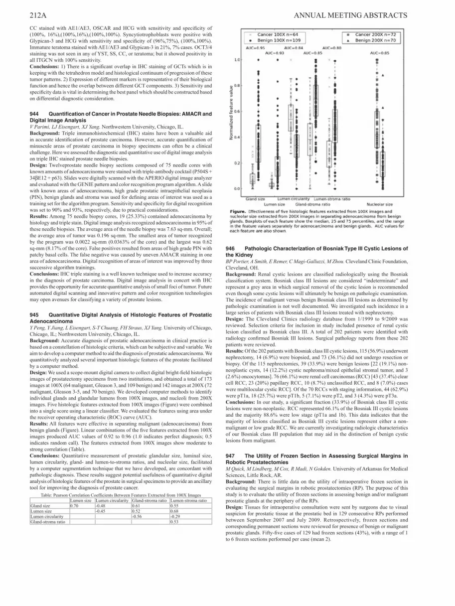
212A ANNUAL MEETING ABSTRACTS CC stained with AE1/AE3, OSCAR and HCG with sensitivity and specificity of (100%, 16%),(100%,16%),(100%,100%). Syncytiotrophoblasts were positive with Glypican-3 and HCG with sensitivity and specificity of (96%,75%), (100%,100%). Immature teratoma stained with AE1/AE3 and Glypican-3 in 21%, 7% cases. OCT3/4 staining was not seen in any of YST, SS, CC, or teratoma; but it showed positivity in all ITGCN with 100% sensitivity.Conclusions: 1) There is a significant overlap in IHC staining of GCTs which is in keeping with the tetrahedron model and histological continuum of progression of these tumor patterns. 2) Expression of different markers is representative of their biological function and hence the overlap between different GCT components. 3) Sensitivity and specificity data is vital in determining the best panel which should be constructed based on differential diagnostic consideration.
944 Quantification of Cancer in Prostate Needle Biopsies: AMACR and Digital Image AnalysisV Parimi, LJ Eisengart, XJ Yang. Northwestern University, Chicago, IL.Background: Triple immunohistochemical (IHC) stains have been a valuable aid in accurate identification of prostate carcinoma. However, accurate quantification of minuscule areas of prostate carcinoma in biopsy specimens can often be a clinical challenge. Here we assessed the diagnostic and quantitative use of digital image analysis on triple IHC stained prostate needle biopsies.Design: Twelveprostate needle biopsy sections composed of 75 needle cores with known amounts of adenocarcinoma were stained with triple-antibody cocktail (P504S + 34βE12 + p63). Slides were digitally scanned with the APERIO digital image analyzer and evaluated with the GENIE pattern and color recognition program algorithm. A slide with known areas of adenocarcinoma, high grade prostatic intraepithelial neoplasia (PIN), benign glands and stroma was used for defining areas of interest was used as a training set for the algorithm program. Sensitivity and specificity for digital recognition was set to 90% and 93%, respectively, due to practical considerations.Results: Among 75 needle biopsy cores, 19 (25.33%) contained adenocarcinoma by histology and triple stain. Digital image analysis recognized adenocarcinoma in 95% of these needle biopsies. The average area of the needle biopsy was 7.63 sq-mm. Overall; the average area of tumor was 0.196 sq-mm. The smallest area of tumor recognized by the program was 0.0022 sq-mm (0.0363% of the core) and the largest was 0.62 sq-mm (8.17% of the core). False positives resulted from areas of high grade PIN with patchy basal cells. The false negative was caused by uneven AMACR staining in one area of adenocarcinoma. Digital recognition of areas of interest was improved by three successive algorithm trainings.Conclusions: IHC triple staining is a well known technique used to increase accuracy in the diagnosis of prostate carcinoma. Digital image analysis in concert with IHC provides the opportunity for accurate quantitative analysis of small foci of tumor. Future automated digital scanning and innovative pattern and color recognition technologies may open avenues for classifying a variety of prostate lesions.
945 Quantitative Digital Analysis of Histologic Features of Prostatic AdenocarcinomaY Peng, Y Jiang, L Eisengart, S-T Chuang, FH Straus, XJ Yang. University of Chicago, Chicago, IL; Northwestern University, Chicago, IL.Background: Accurate diagnosis of prostatic adenocarcinoma in clinical practice is based on a constellation of histologic criteria, which can be subjective and variable. We aim to develop a computer method to aid the diagnosis of prostatic adenocarcinoma. We quantitatively analyzed several important histologic features of the prostate facilitated by a computer method.Design: We used a scope-mount digital camera to collect digital bright-field histologic images of prostatectomy specimens from two institutions, and obtained a total of 173 images at 100X (64 malignant, Gleason 3, and 109 benign) and 142 images at 200X (72 malignant, Gleason 3-5, and 70 benign). We developed computer methods to identify individual glands and glandular lumens from 100X images, and nucleoli from 200X images. Five histologic features extracted from 100X images (Figure) were combined into a single score using a linear classifier. We evaluated the features using area under the receiver operating characteristic (ROC) curve (AUC).Results: All features were effective in separating malignant (adenocarcinoma) from benign glands (Figure). Linear combinations of the five features extracted from 100X images produced AUC values of 0.92 to 0.96 (1.0 indicates perfect diagnosis; 0.5 indicates random call). The features extracted from 100X images show moderate to strong correlation (Table).Conclusions: Quantitative measurement of prostatic glandular size, luminal size, lumen circularity, gland- and lumen-to-stroma ratios, and nucleolar size, facilitated by a computer segmentation technique that we have developed, are concordant with pathologic diagnosis. These results suggest potential usefulness of quantitative digital analysis of histologic features of the prostate in surgical specimens to provide an ancillary tool for improving the diagnosis of prostate cancer.
Table: Pearson Correlation Coefficients Between Features Extracted from 100X ImagesLumen size Lumen circularity Gland-stroma ratio Lumen-stroma ratio
Gland size 0.70 -0.48 0.61 0.55Lumen size -0.45 0.52 0.68Lumen circularity -0.56 -0.29Gland-stroma ratio 0.53
946 Pathologic Characterization of Bosniak Type III Cystic Lesions of the KidneyBP Portier, A Smith, E Remer, C Magi-Galluzzi, M Zhou. Cleveland Clinic Foundation, Cleveland, OH.Background: Renal cystic lesions are classified radiologically using the Bosniak classification system. Bosniak class III lesions are considered “indeterminate” and represent a grey area in which surgical removal of the cystic lesion is recommended even though some cystic lesions will ultimately be benign on pathologic examination. The incidence of malignant versus benign Bosniak class III lesions as determined by pathologic examination is not well documented. We investigated such incidence in a large series of patients with Bosniak class III lesions treated with nephrectomy.Design: The Cleveland Clinics radiology database from 1/1999 to 9/2009 was reviewed. Selection criteria for inclusion in study included presence of renal cystic lesion classified as Bosniak class III. A total of 202 patients were identified with radiology confirmed Bosniak III lesions. Surgical pathology reports from these 202 patients were reviewed.Results: Of the 202 patients with Bosniak class III cystic lesions, 115 (56.9%) underwent nephrectomy, 14 (6.9%) were biopsied, and 73 (36.1%) did not undergo resection or biopsy. Of the 115 nephrectomies, 39 (33.9%) were benign lesions [22 (19.1%) non-neoplastic cysts, 14 (12.2%) cystic nephroma/mixed epithelial stromal tumor, and 3 (2.6%) oncocytomas]. 76 (66.1%) were renal cell carcinomas (RCC) [43 (37.4%) clear cell RCC, 23 (20%) papillary RCC, 10 (8.7%) unclassified RCC, and 8 (7.0%) cases were multilocular cystic RCC]. Of the 70 RCCs with staging information, 44 (62.9%) were pT1a, 18 (25.7%) were pT1b, 5 (7.1%) were pT2, and 3 (4.3%) were pT3a.Conclusions: In our study, a significant fraction (33.9%) of Bosniak class III cystic lesions were non-neoplastic. RCC represented 66.1% of the Bosniak III cystic lesions and the majority 88.6% were low stage (pT1a and 1b). This data indicates that the majority of lesions classified as Bosniak III cystic lesions represent either a non-malignant or low grade RCC. We are currently investigating radiologic characteristics of our Bosniak class III population that may aid in the distinction of benign cystic lesions from malignant.
947 The Utility of Frozen Section in Assessing Surgical Margins in Robotic ProstatectomiesM Quick, M Lindberg, M Cox, R Madi, N Gokden. University of Arkansas for Medical Sciences, Little Rock, AR.Background: There is little data on the utility of intraoperative frozen section in evaluating the surgical margins in robotic prostatectomies (RP). The purpose of this study is to evaluate the utility of frozen sections in assessing benign and/or malignant prostatic glands at the periphery of the RPs.Design: Tissues for intraoperative consultation were sent by surgeons due to visual suspicion for prostatic tissue at the prostatic bed in 129 consecutive RPs performed between September 2007 and July 2009. Retrospectively, frozen sections and corresponding permanent sections were reviewed for presence of benign or malignant prostatic glands. Fifty-five cases of 129 had frozen sections (43%), with a range of 1 to 6 frozen sections performed per case (mean 2).
ANNUAL MEETING ABSTRACTS 213AResults:
Results of the margins and the diagnoses in frozen and permanent sectionsFeatures Frozen PermanentFibromuscular/Fibroconnective tissue 44 46Prostatic stroma 29 28benign prostatic glands 31 29Prostate cancer 5 6Frozen negative/permanent positive 1Frozen positive/permanent negative 0# cases with any discrepancies 12
The negative predictive value of benign prostatic glands by frozen was 99%. The positive predictive value for malignant prostatic glands by frozen was 100%. The positive predictive value of malignant prostatic tissue by intraoperative visual assessment was 9%. The negative predictive value of negative tissue margins by intraoperative visual assessment was 71.6%.Conclusions: Frozen section has both high positive and negative predictive values in assessing benign / malignant prostatic glands. On the other hand, intraoperative visual assessment by surgeon has low predictive value for prostatic glands in robotic prostatectomies. Our data supports that frozen section is a reliable method to evaluate margins for benign and malignant glands in RP, and should be used liberally regardless of the experience of the surgeon due to its higher predictive values.
948 Various Morphometric Measurements of Cancer in Needle Prostatic Biopsies: Which Is Predictive of Pathologic Stage and PSA Failure Post-Surgery?MM Quintal, A Billis, L Meirelles, LLL Freitas, LA Magna, U Ferreira. School of Medicine, University of Campinas (Unicamp), Campinas, Brazil.Background: Cancer extent in needle biopsies (NB) can be measured in a number of ways that have been found to correlate with several clinicopathological findings. It is controversial, however, which one has the highest predictive value. The aim of this study is to investigate which method used to measure tumor is most predictive of stage >pT2 and PSA progression post-surgery.Design: The study was based on 168 NB with the correspondent radical prostatectomies. All NB were extended with a mean of 11 cores (range 7-20). Tumor extent was evaluated as: number and percentage of positive cores; total length of cancer in mm and the percentage of cancer in all cores; and, the greatest length and the greatest percentage of cancer in a single core. To relate the variables to the outcome of stage >pT2 (stage pT2+ was excluded) we used logistic regression; to PSA progression the Cox model; and, to time of biochemical failure the Kaplan-Meier curves. Two-sided P-values of <0.05 were considered.Results: All methods of measurement significantly predicted stage >pT2. Percentage of cancer in all cores had the strongest prediction. On multivariate analysis, this method had a gain in prediction when associated either to preoperative PSA, Gleason grade in the biopsy or both. With the exception of the greatest length and the greatest percentage of cancer in a single core, the other methods significantly were associated with PSA biochemical failure. Percentage of cancer in all cores had again the strongest prediction but no gain was found when associated to preoperative PSA, Gleason grade in the biopsy or both. Time of PSA progression was significantly related to percentage of positive cores, greatest length of cancer in a single core and length and percentage of extent of cancer in all cores. The latter method had the strongest relation.Conclusions: All methods for measurement of cancer extent in needle biopsies had positive predictive value for stage >pT2 but not all of them predicted PSA progression. Percentage of linear extent in mm of cancer in all cores had the strongest positive predictive value for stage >pT2 and PSA progression; in association with preopeative PSA and Gleason grade may improve the predictive value of stage >pT2 of the available presurgical nomograms.
949 Accuracy, Clinical Impact and Limitations in Interpretation of CT Guided Needle Biopsy of Renal MassesM Ramineni, ON Kryvenko, NS Gupta. Henry Ford Hospital, Detroit, MI.Background: With the advent of sophisticated imaging techniques, the detection rates of indeterminate masses in the kidney have increased in numbers. Pre-operative characterization of the tumor plays in important role in deciding between conservative management with minimally invasive procedures versus radical surgery. We evaluated the accuracy, impact and limitations of CT guided biopsy of renal masses.Design: We retrospectively reviewed 82 biopsies performed at our hospital from 2005 to 2008 for indeterminate kidney lesions. Relevant data such as size of the tumor, location, and radiologic description, number of cores, pathologic diagnosis, and necessity for immunostains were recorded on the biopsies. All patients were followed-up and data on the type of treatment received, nephrectomy diagnosis, and location of the tumor, stage, Fuhrman grade, and metastasis were recorded.Results: Biopsies were interpreted as clear cell renal cell carcinoma 13 (16%), papillary renal cell carcinoma 7 (9%), renal cell carcinoma, not further classified 14 (17%), oncocytic neoplasm (ON) 13 (16%) subdivided as favor oncocytomas 10 (12%) and inconclusive for chromophobe renal cell carcinoma/oncocytoma 3 (4%), renal cell carcinoma favor mucinous tubular and spindle cell tumor 1 (1%), malignant/carcinoma 7 (9%), metastatic carcinoma 2 (3%), lymphoma/leukemia 3 (4%), suspicious for malignancy 3 (4%), angiomyolipoma 2 (3%), benign non-neoplastic diagnosis including inflammation, fibrosis, hemorrhage and necrosis 17 (21%). 18 (22%) of these patients underwent nephrectomy, 5 (6%) received cryoablation, 2 (3%) received radio-frequency ablation, 17 (21%) were benign and required no therapy. All cases on nephrectomy showed renal cell carcinomas (RCC). All 11 cases (100%) diagnosed as RCC showed RCC on subsequent nephrectomy. Biopsy results on 5 (6%) cases were false negative. 2 were multilocular cystic RCC, 1 papillary RCC, 1 clear cell RCC and 1 sarcomatoid RCC. 1 case with repeat biopsy showed clear cell RCC. 2 cases classified as ON on biopsy showed RCC on nephrectomy.
Conclusions: CT guided biopsy for renal mass is reliable and accurate. It can impact clinical management of a renal mass and can help in avoiding nephrectomy. We did not observe any false positive results. Low rate of false negative results were observed but when correlated with high suspicion for malignancy on clinical and radiologic data, those patients received repeat biopsies and nephrectomies for management of the renal masses.
950 Primary Ewing Sarcoma (ES)/Primitive Neuroectodermal (PNET) of the Kidney in Adults – Is Molecular Confirmation Necessary for Treatment?P Rao, JM Meis. MD Anderson Cancer Center, Houston, TX.Background: ES is a rare primary renal neoplasm that is included in the differential diagnosis of other round cell neoplasms of the kidney. Experience with clinical outcome of these tumors is limited.Design: We retrieved 13 cases from our archives using the search terms “Ewing sarcoma”, “neuroectodermal tumor” & “kidney” All available slides were reviewed and patients (pts) were divided into 3 groups (Grp).Results: Grp 1: Cases with molecular confirmation of EWSR1 gene rearrangement (n=6). Mean age at presentation was 38.1 yrs, range 21-56 yrs. Tumor sizes were known in 4 pts; mean 12 cm. Mean follow up (FU) in this group was 12.2 mos, range 4-24 mos. 4 pts were DOD, mean interval to death, 7.7 mos. 1 pt was AWD at 18 mos & 1 pt was lost to FU at 5 months. EWSR1 gene rearrangement was confirmed by FISH in 5 cases &PCR in 1 case. Grp 2: Cases with morphologic features &immunohistochemical evidence of ES but were negative for EWSR1 gene rearrangement by FISH (n=2). Both cases had typical morphologic features of ES. Both pts were female (31 & 32 yrs)& had 12 cm renal masses. 1 patient died of disease 23 mos after diagnosis while the other showed NED at 86 mos. Grp 3: Cases with morphologic &immunohistochemical support for the diagnosis of ES but without molecular confirmation due to current lack of material (n=5). Mean age for this grp was 39 yrs, range 23-52 yrs. FU information was available in 5/6 pts. 3 pts were DOD (mean interval to death 15.6 mos) & 2 pts were AWD at 68 & 3 mos. All pts presented with high stage disease (T3 or T4). The most common site of metastasis was lung (9/12) followed by bone (3/12). Microscopically, tumors were composed of primitive round cells arranged in sheets & lobules. Occasionally tumor cells displayed a more spindled morphology & a prominent pseudopapillary pattern. Mitotic activity &necrosis were prominent. 11/13 cases were positive for CD99. Of the 2 cases that were negative, 1 had a FISH confirmed EWSR1 gene rearrangement.Conclusions: 1) ES is a rare primary renal neoplasm with a distinct predilection for adults. 2) CD99 is positive in virtually all cases of ES. 3) Molecular testing is a useful adjunct to confirm the diagnosis of ES. However, cases with typical morphologic features in the absence of molecular confirmation are often included in the same group for therapeutic purposes. 4) The majority of cases of renal ES both with & without molecular confirmation seem to have a poor prognosis, unlike those of ES of bone and soft tissue in which prognosis has steadily improved over time.
951 Immunohistochemical (IHC) Study of Wilms’ Tumor Gene 1 (WT1) in Angiomyolipoma (AML) of the Kidney: Dual Role in Tumorigenesis?R Rawson, DE Westfall, MB Amin, RS Parakh, BL Balzer. Cedars-Sinai Medical Center, Los Angeles, CA.Background: AML has a unique immunophenotype with co-expression of melanocytic markers such as HMB-45 and Melan-A as well as muscle-specific actin. To fully understand the scope of WT1 expression, which has been recently reported in melanocytic tumors as well as in uterine smooth muscle neoplasms, we studied its expression in AML, which has not been evaluated to date. The role of WT1 in tumorigenesis is controversial, as it may act as either a transcriptional activator at the carboxyl (C) terminus or a repressor at the amino (N) terminus based on splices in its transcript. It is hypothesized that WT1 may influence the mesenchymal-epithelial state of cells, thus it may prove helpful in understanding the histogensis and phenotypic characteristics of this triphasic tumor. Monophasic variants of AML, such as epithelioid and leiomyomatous types, may compound the diagnostic dilemma and WT1 may prove useful in this scenario.Design: We tested 58 renal AMLs (52 classic histology, 2 epithelioid variants and 4 leiomyomatous variants) with WT1 (N-terminal amino acids, Mouse Monoclonal Clone: 6F-H2, Dako) and WT1 carboxyl (C-terminal amino acids, Rabbit Polyclonal Clone: C-19, Santa Cruz Biotechnology) immunohistochemistry.Results: WT1 was positive in five of 58 cases (4 leiomyomatous AML, 1 epithelioid AML). WT1 carboxyl was positive in six of 58 cases (3 leiomyomatous AML, 1 epithelioid AML, 2 classic AML). Three cases, all leiomyomatous AML, were positive for both WT1 and WT1 carboxyl. When WT1 and WT1 carboxyl positivity was observed, it was seen most frequently in spindled areas, rarely in epithelioid areas and never in adipocytic areas.Conclusions: 1) The expression of WT1 in the leiomyomatous variant of AML is intriguing, as melanoma-associated markers are more commonly associated with the epithelioid morphology of AML and pure epithelioid AML. 2) WT1 may have utility in diagnosing rare monomorphic variants of AML. 3) WT1 positivity in AML expands the list of tumors that express this marker. 4) WT1 expression was positive for both WT1 N terminus and C terminus antibodies in AML, which highlights the pro- and anti-differentiation functions of this gene as reported in the literature. This further supports that WT1 may be involved in maintaining the mesenchymal-epithelial balance in cells and its activation or suppression may aid in understanding the triphasic morphology unique to this tumor.
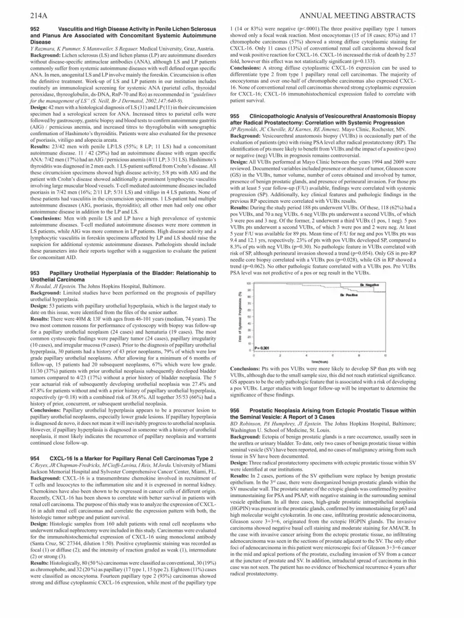
214A ANNUAL MEETING ABSTRACTS 952 Vasculitis and High Disease Activity in Penile Lichen Sclerosus and Planus Are Associated with Concomitant Systemic Autoimmune DiseaseY Razmara, K Pummer, S Mannweiler, S Regauer. Medical University, Graz, Austria.Background: Lichen sclerosus (LS) and lichen planus (LP) are autoimmune disorders without disease-specific antinuclear antibodies (ANA), although LS and LP patients commonly suffer from systemic autoimmune diseases with well defined organ specific ANA. In men, anogenital LS and LP involve mainly the foreskin. Circumcision is often the definitive treatment. Work-up of LS and LP patients in our institution includes routinely an immunological screening for systemic ANA (parietal cells, thyroidal peroxidase, thyreoglobulin, ds-DNA, RnP-70 and Ro) as recommended in “guidelines for the management of LS” (S. Neill, Br J Dermatol, 2002,147:640-9). Design: 42 men with a histological diagnosis of LS (31) and LP (11) in their circumcision specimen had a serological screen for ANA. Increased titres to parietal cells were followed by gastroscopy, gastric biopsy and blood tests to confirm autoimmune gastritis (AIG) / pernicious anemia, and increased titres to thyroglobulin with sonographic confirmation of Hashimoto’s thyroiditis. Patients were also evaluated for the presence of psoriasis, vitiligo and alopecia areata.Results: 23/42 men with penile LP/LS (55%; 8 LP; 11 LS) had a concomitant autoimmune disease. 11 / 42 (29%) had an autoimmune disease with organ specific ANA: 7/42 men (17%) had an AIG / pernicious anemia (4/11 LP; 3 /31 LS). Hashimoto’s thyroiditis was diagnosed in 2 men each. 1 LS-patient suffered from Crohn’s disease. All these circumcision specimens showed high disease activity; 5/8 pts with AIG and the patient with Crohn’s disease showed additionally a prominent lymphocytic vasculitis involving large muscular blood vessels. T-cell mediated autoimmune diseases included psoriasis in 7/42 men (16%; 2/11 LP; 5/31 LS) and vitiligo in 4 LS patients. None of these patients had vasculitis in the circumcision specimens. 1 LS-patient had multiple autoimmune diseases (AIG, psoriasis, thyroiditis); all other men had only one other autoimmune disease in addition to the LP and LS.Conclusions: Men with penile LS and LP have a high prevalence of systemic autoimmune diseases. T-cell mediated autoimmune diseases were more common in LS patients, while AIG was more common in LP patients. High disease activity and a lymphocytic vasculitis in foreskin specimens affected by LP and LS should raise the suspicion for additional systemic autoimmune diseases. Pathologists should include these parameters into their reports together with a suggestion to evaluate the patient for concomitant AID.
953 Papillary Urothelial Hyperplasia of the Bladder: Relationship to Urothelial CarcinomaN Readal, JI Epstein. The Johns Hopkins Hospital, Baltimore.Background: Limited studies have been performed on the prognosis of papillary urothelial hyperplasia.Design: 53 patients with papillary urothelial hyperplasia, which is the largest study to date on this issue, were identified from the files of the senior author.Results: There were 40M & 13F with ages from 46-101 years (median, 74 years). The two most common reasons for performance of cystoscopy with biopsy was follow-up for a papillary urothelial neoplasm (24 cases) and hematuria (19 cases). The most common cystoscopic findings were papillary tumor (24 cases), papillary irregularity (10 cases), and irregular mucosa (9 cases). Prior to the diagnosis of papillary urothelial hyperplasia, 30 patients had a history of 43 prior neoplasms, 79% of which were low grade papillary urothelial neoplasms. After allowing for a minimum of 6 months of follow-up, 15 patients had 20 subsequent neoplasms, 67% which were low grade. 11/30 (37%) patients with prior urothelial neoplasia subsequently developed bladder tumors compared to 4/23 (17%) without a prior history of bladder neoplasia. The 5 year actuarial risk of subsequently developing urothelial neoplasia was 27.4% and 47.8% for patients without and with a prior history of papillary urothelial hyperplasia, respectively (p=0.18) with a combined risk of 38.6%. All together 35/53 (66%) had a history of prior, concurrent, or subsequent urothelial neoplasia.Conclusions: Papillary urothelial hyperplasia appears to be a precursor lesion to papillary urothelial neoplasms, especially lower grade lesions. If papillary hyperplasia is diagnosed de novo, it does not mean it will inevitably progress to urothelial neoplasia. However, if papillary hyperplasia is diagnosed in someone with a history of urothelial neoplasia, it most likely indicates the recurrence of papillary neoplasia and warrants continued close follow-up.
954 CXCL-16 Is a Marker for Papillary Renal Cell Carcinomas Type 2C Reyes, JR Chapman-Fredricks, M Cioffi-Lavina, I Reis, M Jorda. University of Miami Jackson Memorial Hospital and Sylvester Comprehensive Cancer Center, Miami, FL.Background: CXCL-16 is a transmembrane chemokine involved in recruitment of T cells and leucocytes to the inflammation site and it is expressed in normal kidney. Chemokines have also been shown to be expressed in cancer cells of different origin. Recently, CXCL-16 has been shown to correlate with better survival in patients with renal cell carcinoma. The purpose of this study was to analyze the expression of CXCL-16 in adult renal cell carcinomas and correlate the expression pattern with both, the histologic tumor subtype and patient survival.Design: Histologic samples from 160 adult patients with renal cell neoplasms who underwent radical nephrectomy were included in this study. Carcinomas were evaluated for the immunohistochemichal expression of CXCL-16 using monoclonal antibody (Santa Cruz, SC 27344, dilution 1:50). Positive cytoplasmic staining was recorded as focal (1) or diffuse (2); and the intensity of reaction graded as weak (1), intermediate (2) or strong (3).Results: Histologically, 80 (50 %) carcinomas were classified as conventional, 30 (19%) as chromophobe, and 32 (20 %) as papillary (17 type 1, 15 type 2). Eighteen (11%) cases were classified as oncocytoma. Fourteen papillary type 2 (93%) carcinomas showed strong and diffuse cytoplasmic CXCL-16 expression, while most of the papillary type
1 (14 or 83%) were negative (p<.0001).The three positive papillary type 1 tumors showed only a focal weak reaction. Most oncocytomas (15 of 18 cases; 83%) and 17 chromophobe carcinomas (57%) showed a strong diffuse cytoplasmic staining for CXCL-16. Only 11 cases (13%) of conventional renal cell carcinoma showed focal and weak positive reaction for CXCL-16. CXCL-16 increased the risk of death by 2.57 fold, however this effect was not statistically significant (p=0.133).Conclusions: A strong diffuse cytoplasmic CXCL-16 expression can be used to differentiate type 2 from type 1 papillary renal cell carcinomas. The majority of oncocytomas and over one-half of chromophobe carcinomas also expressed CXCL-16. None of conventional renal cell carcinomas showed strong cytoplasmic expression for CXCL-16; CXCL-16 immunohistochemical expression failed to correlate with patient survival.
955 Clinicopathologic Analysis of Vesicourethral Anastomosis Biopsy after Radical Prostatectomy: Correlation with Systemic ProgressionJP Reynolds, JC Cheville, RJ Karnes, RE Jimenez. Mayo Clinic, Rochester, MN.Background: Vesicourethral anastomosis biopsy (VUBx) is occasionally part of the evaluation of patients (pts) with rising PSA level after radical prostatectomy (RP). The identification of pts more likely to benefit from VUBx and the impact of a positive (pos) or negative (neg) VUBx in prognosis remains controversial.Design: All VUBx performed at Mayo Clinic between the years 1994 and 2009 were reviewed. Documented variables included presence or absence of tumor, Gleason score (GS) in the VUBx, tumor volume, number of cores obtained and involved by tumor, presence of benign prostatic glands, and presence of perineural invasion. For those pts with at least 5 year follow-up (F/U) available, findings were correlated with systemic progression (SP). Additionally, key clinical features and pathologic findings in the previous RP specimen were correlated with VUBx results.Results: During the study period 188 pts underwent VUBx. Of these, 118 (62%) had a pos VUBx, and 70 a neg VUBx. 6 neg VUBx pts underwent a second VUBx, of which 3 were pos and 3 neg. Of the former, 2 underwent a third VUBx (1 pos, 1 neg). 5 pos VUBx pts underwent a second VUBx, of which 3 were pos and 2 were neg. At least 5 year F/U was available for 89 pts. Mean time of F/U for neg and pos VUBx pts was 9.4 and 12.1 yrs, respectively. 23% of pts with pos VUBx developed SP, compared to 8.3% of pts with neg VUBx (p=0.30). No pathologic feature in VUBx correlated with risk of SP, although perineural invasion showed a trend (p=0.054). Only GS in pre-RP needle core biopsy correlated with a VUBx pos (p=0.028), while GS in RP showed a trend (p=0.062). No other pathologic feature correlated with a VUBx pos. Pre VUBx PSA level was not predictive of a pos or neg result in the VUBx.
Conclusions: Pts with pos VUBx were more likely to develop SP than pts with neg VUBx, although due to the small sample size, this did not reach statistical significance. GS appears to be the only pathologic feature that is associated with a risk of developing a pos VUBx. Larger studies with longer follow-up will be important to determine the significance of these findings.
956 Prostatic Neoplasia Arising from Ectopic Prostatic Tissue within the Seminal Vesicle: A Report of 3 CasesBD Robinson, PA Humphrey, JI Epstein. The Johns Hopkins Hospital, Baltimore; Washington U. School of Medicine, St. Louis.Background: Ectopia of benign prostatic glands is a rare occurrence, usually seen in the urethra or urinary bladder. To date, only two cases of benign prostatic tissue within seminal vesicle (SV) have been reported, and no cases of malignancy arising from such tissue in SV have been documented.Design: Three radical prostatectomy specimens with ectopic prostatic tissue within SV were identified at our institutions.Results: In 2 cases, portions of the SV epithelium were replace by benign prostatic epithelium. In the 3rd case, there were disorganized benign prostatic glands within the SV muscular wall. The prostatic nature of the ectopic glands was confirmed by positive immunostaining for PSA and PSAP, with negative staining in the surrounding seminal vesicle epithelium. In all three cases, high-grade prostatic intraepithelial neoplasia (HGPIN) was present in the prostatic glands, confirmed by immunostaining for p63 and high molecular weight cytokeratin. In one case, infiltrating prostatic adenocarcinoma, Gleason score 3+3=6, originated from the ectopic HGPIN glands. The invasive carcinoma showed negative basal cell staining and moderate staining for AMACR. In the case with invasive cancer arising from the ectopic prostatic tissue, no infiltrating adenocarcinoma was seen in the sections of prostate adjacent to the SV. The only other foci of adenocarcinoma in this patient were microscopic foci of Gleason 3+3=6 cancer in the mid and apical portions of the prostate, excluding invasion of SV from a cancer at the juncture of prostate and SV. In addition, intraductal spread of carcinoma in this case was not seen. The patient has no evidence of biochemical recurrence 4 years after radical prostatectomy.

ANNUAL MEETING ABSTRACTS 215AConclusions: Recognition of the potential for prostatic neoplasia to arise from ectopic prostatic tissue is important for proper pathologic staging, as these tumors may behave similar to comparable T2 tumors as opposed to T3b cancers. These findings may be more common than what is reported, as 2 of the cases are recent. Also, HGPIN involving the SV can be easily overlooked, with one of the cases missed at a referring institution. Neoplasia arising in the SV is rare consisting of predominantly case reports of primary adenocarcinoma, sarcoma, and mixed epithelial-stromal tumors. This series expands on the neoplastic processes that can arise in the SV.
957 Radical Prostatectomy (RP) Findings in Cases with Only Intraductal Carcinoma of the Prostate (IDC-P) on Needle BiopsyBD Robinson, JI Epstein. The Johns Hopkins Hospital, Baltimore.Background: When IDC-P is present on biopsy, it is usually seen with infiltrating acinar adenocarcinoma. In 2006, we reported 27 cases with IDC-P only on biopsy; however, only 6 cases had available RP findings.Design: 82 men with IDC-P only on prostate biopsy were identified from the consult files of one of the authors. Follow-up information was available in 66 cases. 20 men were treated with RP, 17 radiation therapy (RT), 8 hormone therapy (HT), 13 RT and HT, 6 active surveillance, and 2 rebiopsy. An attempt was made to retrieve the slides of all 20 RP cases.Results: Of the 20 RP cases, 5 showed extraprostatic extension, 3 seminal vesicle invasion, 10 were organ-confined, and 2 showed extensive IDC-P only without identifiable invasive cancer. Of the 18 cases with invasive cancer, the average Gleason score (GS) was 7.8. 1 patient developed bone metastases 3 years post-RP, and 3 others were post-RP PSA failures. 13 RPs were available for our review. 9 showed extensive IDC-P (including one case of IDC-P only), defined as >10% of the tumor volume being intraductal; 3 focal IDC-P; and 1 no IDC-P. All cases with invasive carcinoma were acinar, although 3 cases were classified as ductal by referring pathologists. We concurred with the outside GS in 5/13 cases (5 undergraded, 3 overgraded). In the 3 cases that we gave lower GS, the outside institution graded cribiform IDC-P with and without necrosis as Gleason pattern 5 or 4, respectively.Case Outside GS JHH GS Extent IDC pStage RP Sampling1 IDC only N/A N/A Total2 IDC only IDC only Extensive Total3 IDC only 4+5=9 Extensive 2 Partial4 3+3=6 N/A N/A 2 Partial5 3+3=6 N/A N/A 2 Partial6 4+4=8 N/A N/A 2 Total7 5+3=8 N/A N/A 2 Partial8 3+3=6 3+4=7 Focal 2 Partial9 3+4=7 3+4=7 Extensive 2 Partial10 3+5=8 5+4=9 Focal 2 Partial11 4+4=8 3+4=7 Extensive 2 Partial12 4+5=9 3+4=7 None 2 Partial13 4+5=9 N/A N/A 3a Partial14 3+5=8 4+5=9 Extensive 3a Partial15 4+3=7 4+3=7 Extensive 3a Partial16 4+4=8 3+4=7 Extensive 3a Total17 4+4=8 4+4=8 Extensive 3a Total18 4+4=8 N/A N/A 3b Total19 4+4=8 4+4=8 Extensive 3b Total20 5+5=10 5+5=10 None 3b Partial
Conclusions: Our study, the largest to date with RP findings following IDC-P only on needle biopsy, confirms that aggressive therapy is appropriate for patients whose biopsies show only IDC-P. It is likely that the pathological findings are even worse than we report herein, as most RPs were only partially sampled. Most cases likely represent intraductal spread of high grade cancer, but some cases represent in situ acinar adenocarcinoma.
958 Diagnostic Accuracy of Ureteroscopic Biopsies from Upper Urinary Tract Carcinomas: Does Size Really Matter?CP Rojas, S Rodriguez, V Bird, I Reis, W Zhao, J Santos, R Leveillee, C Gomez-Fernandez, M Jorda. University of Miami, Jackson Memorial Hospital and Sylvester Comprehensive Cancer Center, Miami; University of Miami, Jackson Memorial Hospital and Sylvester Comprehensive Cancer Center, Miami.Background: Urothelial carcinomas from the upper urinary tract system represent 7-10% of all kidney malignancies. With the advent of new uteroscopic equipment, small tissue samples are available representing a diagnostic challenge. Preciseness in diagnosis is essential for treatment decision. This study correlated tumor grade and pT stage between biopsy and resected specimens and examined if sample volume impacts accuracy of diagnosis.Design: We reviewed histologic slides from ureteroscopic biopsy samples and corresponding nephroureterectomy specimens from 33 patients with urothelial carcinoma of upper urinary tract. Two urologic pathologists independently evaluated the following disease characteristics: histologic grade (ISUP/WHO 2004), pleomorphism, mitosis, lymphovascular invasion (LVI), and pT stage. We reported the number, percentage of concordant pairs and corresponding 95% confidence interval (95%CI) for the true proportion of concordant pairs between biopsy and resected specimens. A repeated measures model analysis was used to examine whether agreement on a particular disease characteristic was related to specimen size. The outcome for our analysis was log-transformed data on specimen volume ([/6]×a×b×c, where a, b, and c are orthogonal measurements in mm).Results: The majority of patients were males (21, 63.6%). Mean age was 73 years and 79% of patients were 65 years or older. Good agreement was observed for grade (concordance 87.9%, 95%CI: 71.8 to 96.6%), pleomorphism (75.8%, 95%CI: 57.5 to 88.9%), mitosis (84.8%, 95%CI: 68.1 to 94.9%), and LVI (87.9%, 95%CI: 71.8 to 96.6%), but agreement was lower for pT stage (45.5%, 95%CI: 28.1 to 63.6%). Biopsy volume yield a geometric mean of 0.022 mm3, range 0.001 to 4.514 mm3; tumor volume
yield geometric mean of 1.351 mm3, range 0.002 to 65.0 mm3 (p<0.05). The volume of biopsy was not related to agreement for any of the disease characteristics (p>0.05).Conclusions: 1- Histologic grade assigned on the biopsy sample accurately predicts histologic grade in the resected specimen (87.9%), even when biopsy volume was small. 2- Ureteroscopic biopsies provided sufficient information for clinical decision-making. 3- The agreement between biopsy and resected specimen was lower for the characteristic of pT stage.
959 Activating Transcription Factor 3 (ATF3) Protein Expression in Prostatic Adenocarcinomas (PAC)JS Ross, C Yan, GM Sheehan, CE Sheehan, T Nazeer, HAG Fisher, BM Mian, BVS Kallakury. Albany Medical College, Albany, NY; Georgetown University Hospital, Washington, DC.Background: Human transcription factor ATF3 is involved in gene expression by its binding to the cAMP response element. ATF3 has been shown to be upregulated as an intracellular response to various stresses including DNA damage. ATF3 expression has been associated with mouse esophageal and mammary tumorigenesis, but there are no reports of comprehensive analysis in human cancers. In this study, we evaluate, for the first time, the prognostic significance of ATF3 expression in human PAC.Design: Formalin-fixed paraffin-embedded tissue sections from 149 PAC were immunostained by an automated method (Ventana Medical Systems, Tucson, AZ) using polyclonal rabbit anti-human ATF3 antibody (clone C-19; Santa Cruz, Santa Cruz, CA). Cytoplasmic and nuclear immunoreactivity was semi-quantitatively scored based on intensity and percentage of positive cells in both the tumor and adjacent benign epithelium in each case. Scoring was based on staining intensity and percentage of positive cells. A multiplicative index was calculated and cases were assessed as tumor=benign baseline (T=B), tumor>benign (T>B) and tumor<benign (T<B). Results were correlated with clinicopathologic variables.Results: Variable cytoplasmic and nuclear ATF3 immunoreactivity was noted in the tumor and adjacent benign basal cells and secretory epithelium in all cases. Cytoplasmic ATF3 protein was expressed as follows [T=B 58%, T>B 39%, T<B 3%] and correlated with tumor grade [T=B 48% HG vs 65% LG, T>B 52% HG vs 30% LG, T<B 0% HG vs 5% LG, p=0.009]; tumor stage [T=B 38% HG vs 73% LG, T>B 59% HG vs 25% LG, T<B 3% HG vs 2% LG, p<0.0001] and biochemical disease recurrence [T=B 44% recur vs 67% non-recur, T>B 53% recur vs 31% non-recur, T<B 3% recur vs 2% non-recur, p=0.021]. Nuclear ATF3 protein was expressed as follows [T=B 64%, T>B 25%, T<B 11%] and correlated with tumor grade [T=B 57% HG vs 69% LG, T>B 40% HG vs 15% LG, T<B 3% HG vs 16% LG, p=0.001]. On multivariate analysis, high tumor grade (p=0.023) and T>B cytoplasmic ATF3 protein expression (p=0.016) independently predicted biochemical disease recurrence.Conclusions: ATF3 over-expression is associated with high grade, advanced stage PAC and independently predicts biochemical disease recurrence. Further study of ATF3 expression as both a prognostic factor and potential target of therapy for PAC appears warranted.
960 High Intensity Focused Ultrasound (HIFU) in Prostatic Adenocarcinoma – Pathologic Features in Tumor and Non-Tumor Tissue in 45 Treated CasesP Ryan, L Fahim, A Finelli, N Fleshner, M Robinette, S Radomski, T van der Kwast, A Evans. Mount Sinai Hospital, Toronto, Canada; University Health Network, Toronto, Canada.Background: High Intensity Focused Ultrasound (HIFU) is one of several alternative management options being offered to patients with prostate-confined low risk prostatic adenocarcinoma. Little data exists on the effects of treatment on tumor and non-tumorous prostate tissue. This study summarizes the pathologic features in biopsies of HIFU-treated prostatic adenocarcinoma.Design: Pretreatment needle biopsies from 45 patients with prostate cancer who received HIFU were identified retrospectively. 30 patients also had post-HIFU biopsy either due to rising PSA interpreted clinically as biochemical failure (n = 22) or as per surgeon practice pattern (n = 8). The Gleason score, location, and extent of tumor were assessed along with (in post-HIFU biopsies) the presence of coagulative necrosis and stromal fibrosis.Results: Pretreatment Gleason score was 6 or 7 in all cases, with mean tissue involvement of 7.5% and bilateral disease present in 18 cases. 23 patients experienced biochemical failure at a mean follow-up of 13.2 months with one patient commenced on anti-androgen therapy without further biopsy and 22 having repeat needle biopsy - 17 contained adenocarcinoma, 4 had higher Gleason score, 5 had newly diagnosed contralateral carcinoma, and 4 had increased percentage tissue involvement. Of 22 cases without evidence of treatment failure at 12.2 months mean follow-up, 8 had repeat biopsy with 2 containing carcinoma similar to the pre-treatment biopsy. In non-tumor prostate tissue coagulative necrosis was seen in 4 of 30 post-treatment cases (13.3%, mean interval since HIFU 8.5 months), stromal fibrosis in 17 cases (56.7%, 15.3 months), and in 5 cases viable tumour as well as treatment effects were found in the same core.Conclusions: Notwithstanding that many patients successfully treated on the basis of outside biopsy reports would not be detected in this review the positive biopsy rate is low in cases with stable or undetectable post-HIFU PSA, compared with cases of biochemical failure where >75% have viable tumor on follow-up biopsy. Coagulative necrosis is present in a minority of biopsies and fibrosis may be seen in more than 50% of cases, features whose detection may be influenced by the interval since HIFU. However, in our experience HIFU does not affect ability to apply the modified Gleason scoring system to viable post-treatment tumour.
216A ANNUAL MEETING ABSTRACTS 961 Molecular, Immunohistochemical, and Ultrastructural Characterization of Clear Cell Papillary Renal Cell CarcinomaCJ Sailey, M Dvorakova, JC Papadimitriou, JF Borin, T Ali, AV Parwani. University of Maryland Med Ctr, Baltimore, MD; University of Pittsburgh Med Ctr, Pittsburgh, PA.Background: Clear cell papillary renal cell carcinoma (CCPRCC), with features of both clear cell (CCC) and papillary (PRC) renal cell carcinomas, is a relatively new entity. CCPRCC has been documented in end stage renal disease (ESRD) kidneys as well as occurring sporadically. Our study characterized the largest known series of CCPRCCs from ESRD and normally functioning kidneys using molecular methods, immunohistochemistry (IHC), and electron microscopy (EM).Design: Formalin fixed, paraffin embedded tissue of 19 CCPRCCs from 11 ESRD (age 46-74 yr) and 6 sporadic (age 33-79 yr) cases were selected. 11 tumors were subjected to IHC and EM; 10 were analyzed for loss of heterozygosity (LOH) of the VHL gene at 3p and by FISH for abnormalities of chromosomes 7 and 17.Results: The ESRD tumors ranged in size from 1.1 to 6.3 cm (mean 3.3 cm) with a pathologic stage up to pT1b and displayed relatively low nuclear grade (Fuhrman 1-2). The sporadic tumors ranged from 1.5 to 10.2 cm (mean 5.8 cm) with a higher stage (up to pT2) and higher nuclear grade (Fuhrman 2-3). Morphologically, all of the tumors displayed cystic and or solid architecture with simple branching papillae. Polygonal to columnar cells with abundant clear cytoplasm, delicate membranes and small ovoid basal nuclei lined the papillae. IHC revealed that the tumors were positive for E-cadherin, CAM5.2, CK7, Vimentin (100%), EMA, CK903 (91%), and CD10 (54%); negative for RCC, AMACR, and p63. On EM, the tumor cells showed short microvilli with occasional reverse directionality. Vacuoles of lipids/polysaccharides were apparent. There was prominent rER, and scant mitochondria. The nuclei had regular membranes & clumped chromatin. The basal lamina was regular with distinct collagen. One sporadic case showed VHL LOH; another showed trisomies of both chromosomes 7 and 17. Monosomy 7 was detected in 50% (4 ESRD / 1 sporadic) and monosomy 17 in 20% (1 ESRD / 1 sporadic) of cases.Conclusions: CCPRCC is a distinct entity with morphologic and IHC overlap between CCC and PRC. Molecular heterogeneity of these tumors and the absence of abnormalities specific for CCC or PRC suggest a separate molecular pathway. Compared to sporadic tumors, the ESRD cases had a lower stage and nuclear grade. Immunophenotypic, genetic, and ultrastructural profiling of this entity can aid in diagnosis.
962 Seminal Vesicle Sampling in Radical Prostatectomy SpecimensH Samaratunga, J Perry-Keene, D Samaratunga, M Adamson, J Yaxley, B Delahunt. Aquesta Pathology, Brisbane, Queensland, Australia; Royal Brisbane Hospital, Brisbane, Queensland, Australia; Wellington School of Medicine and Health Sciences, University of Otago, Wellington, Otago, New Zealand.Background: Seminal vesicle (SV) invasion by prostatic adenocarcinoma is a poor prognostic indicator. There are no guidelines on SV sampling in radical prostatectomy specimens.Design: Seminal vesicles of 498 consecutive radical prostatectomy specimens were serially sectioned and blocked entirely, grouped as proximal, mid and distal one-third blocks. Carcinomatous infiltration of the muscular wall of each of these and ejaculatory duct/ intraprostatic SV were recorded.Results: Extraprostatic SV involvement was present in 37 (7.42%) cases (pT3b). The mean age and serum PSA of these patients were 64.1 years (range 52-71) and 10.1 ng /ml (range 3.7- 29) respectively. Eight patients had a palpable nodule or induration on digital rectal examination. Extraprostatic extension and intraprostatic SV/ ejaculatory duct involvement were respectively present in all 37 (100%) and 35 (97%) of these cases. All cases with intraprostatic SV/ejaculatory duct involvement had extraprostatic SV involvement. The mean tumor volume was 5.79 cc (range 1.0-12.1 cc). The tumours had a Gleason score of 4+3 in 4, 4+3 with tertiary pattern 5 in 7, 8 in 2 and 5+4/ 4+5 in 24 cases. All SV positive cases had involvement of the proximal one third (8 right SV, 11 left SV and 18 both SV) 25 of which (67 %) had only proximal SV involvement. For the remainder, 6 had mid one third and 6 had mid and distal one third involvement in addition to proximal SV involvement.Conclusions: In this study, mid or distal SV carcinomatous involvement was not found in the absence of proximal seminal vesicle involvement. Blocking the proximal part of the seminal vesicles should be sufficient to identify all cases of carcinomatous involvement of SV. In cases with intraprostatic SV/ ejaculatory duct involvement, thorough examination of the extraprostatic SV is warranted if the initial examination does not demonstrate carcinoma.
963 Prostate Specific Membrane Antigen (PSMA) Expression in Bladder Cancer Neovasculature and Tumor SubtypesMK Samplaski, W Heston, C Magi-Galluzzi, DE Hansel. The Cleveland Clinic, Cleveland, OH.Background: PSMA is a transmembrane receptor expressed on prostate cancer cells that correlates with a more aggressive phenotype. Recent studies have demonstrated PSMA expression in numerous other tissue types, as well as tumor neovasculature. We sought to determine the extent of PSMA expression in both bladder cancer vasculature and bladder cancer cells of various subtypes.Design: Immunohistochemical analysis (IHC) of PSMA was performed using tissue microarrays constructed from 97 urothelial carcinomas (UCCs), 37 squamous cell carcinomas, 17 adenocarcinomas and 17 small cell carcinomas of the bladder. We used a PSMA monoclonal antibody obtained from Dako (clone 3E6, dilution 1:100), which recognizes the epitope present in the 57-134 amino acid region of the extracellular portion of the PSMA molecule. Intensity of IHC staining was scored as 0 (no expression) to 3+ (strong expression), with 2-3+ IHC considered a positive result.Results: All bladder cancers examined demonstrated robust expression of PSMA in the tumor vasculature, suggesting a potential role for PSMA in mediating new vessel
ingrowth and providing a valuable internal control. Most commonly, PSMA expression was identified in small cell carcinoma (3/17; 18%), with co-expression of PSMA in admixed forms of bladder cancer, including glandular elements (1/3) and urothelial elements (1/3). PSMA expression was rare in instances of pure UCC (3/97; 3%) and pure adenocarcinoma (2/17; 12%). No case of pure squamous cell carcinoma of the bladder was positive for PSMA within the tumor cells.Conclusions: PSMA is commonly expressed in bladder cancer neovasculature and may play a potential active role in new vessel ingrowth. Additionally, PSMA may be occasionally expressed in bladder cancer subtypes, including small cell carcinoma of the bladder. These findings warrant caution in the diagnostic use of PSMA to determine potential prostate origin of invasive carcinoma in this anatomic site.
964 Evaluation of Brachyury Expression in Germ Cell Tumors (GCT) and Clear Cell Renal Cell Carcinomas (CC-RCC): A Tissue Microarray Study of 295 CasesAR Sangoi, JP Higgins, JD Brooks, JK McKenney. Stanford University, Stanford, CA; El Camino Hospital, Mountain View, CA.Background: Brachyury is recognized as a specific marker for notochord derived lesions such as chordoma and has become a defining immunohistochemical feature. Within the spectrum of genitourinary neoplasia, metastatic GCTs and CC-RCC may be close morphologic mimics of chordoma. Expression of brachyury has been reported in a few GCTs, which have included mostly seminomas; however, a thorough characterization of staining by GCT subtype within a large series has not been reported. Additionally, brachyury expression in RCC has not been well studied.Design: Immunohistochemical expression of brachyury (1:50, Santa Cruz, CA, polyclonal) was evaluated in 111 GCTs and 184 metastatic RCC using tissue microarray technology (in duplicate) with standard avidin-biotin technique. Additionally, 10 whole-sections of seminomas were also stained with brachyury. Classic chordoma was used as control tissue, and only nuclear reactivity was scored as positive.Results: No nuclear brachyury expression was identified in any of the 111 GCT [including choriocarcinoma (1), embryonal carcinoma (20), intratubular germ cell neoplasia unclassified (2), seminoma (64), spermatocytic seminoma (1), teratoma (5), and yolk sac tumor (8) or in any of the 10 whole-section seminomas. All 184 metastatic RCC were also non-reactive for brachyury with strong external controls.Conclusions: This study confirms the specificity of brachyury for chordoma in the differential diagnostic distinction from metastatic genitourinary mimics, GCT and CC-RCC.
965 Interobserver Reproducibility (IOR) in the Diagnosis of Invasive Micropapillary Carcinoma (MPC) of the Genitourinary Tract among Expert Urologic PathologistsAR Sangoi, AH Beck, MB Amin, L Cheng, JI Epstein, DE Hansel, KA Iczkowski, JY Ro, A Lopez-Beltran, E Oliva, GP Paner, VE Reuter, RB Shah, SS Shen, P Tamboli, JK McKenney. Stanford, Stanford, CA; Cedars-Sinai, Los Angeles, CA; U Indiana, Indianapolis, IN; Johns Hopkins, Baltimore, MD; Cleveland Clinic, Cleveland, OH; U Colorado, Aurora, CO; Methodist, Houston, TX; Unit Anatomic Pathology, Cordoba, Spain; Massachusetts General Hospital, Boston, MA; Northwestern, Chicago, IL; Memorial Sloan Kettering, New York, NY; U Michigan, Ann Arbor, MI; UT MD Anderson, Houston, TX.Background: IOR for the diagnosis of MPC has not been studied, despite the reported clinical relevance.Design: Digital images from 30 urothelial carcinomas (UC) were given to 14 GU subspecialists with request to indicate cases qualifying as MPC. The 30 pre-selected cases included both (presumably) classic MPC (n=10) and UC with retraction and variably sized nests (non-classic/potential MPC)(n=20). IOR was evaluated by kappa analysis. To assess putative criteria applicable in difficult cases, tumor nest size, nest anastomosis, extensive retraction, multiple nests in same lacunar space, peripheral nuclei, rings, elongate processes, and columnar cells were recorded.Results: Moderate agreement (kappa:0.54) was reached for the 30 cases. Cases diagnosed as MPC ranged from 9/30 to 20/30 (median:13/30). Of the 10 proposed classic MPC, all pathologists classified at least 8/10 as MPC [sensitivity of 130/140 (93%)]. Of the 20 other cases, MPC diagnoses varied with 6 pathologists classifying no more than 2/20 as MPC; 5 classifying between 4-7/20 as MPC; and 3 classifying from 9-11/20 as MPC. Cluster analysis revealed a distinct group of 6 pathologists with high diagnostic agreement (kappa:0.79) and high sensitivity (0.95) and specificity (0.95) for classic MPC. Multiple nests within the same lacunar space had the most significant association with an MPC diagnosed by this group.Conclusions: Pathologists demonstrate high levels of sensitivity for identifying classic MPCs, but there is considerable variability in the diagnosis of other UC with retraction. Evaluation of features that potentially correlate with aggressive behavior is warranted to refine diagnostic criteria for MPC.
966 Exploring the Specificity of Putative Renal Cell Carcinoma (RCC) Markers in Non-Renal Tissues and Neoplasms from Various Organ Systems: A Tissue Microarray (TMA) Study of 501 CasesAR Sangoi, RB West, JV Bonventre, KD Montgomery, JK McKenney. Stanford, Stanford, CA; El Camino Hospital, Mountain View, CA; Brigham and Women’s Hospital, Boston, MA.Background: PAX2, PAX8, and hKIM-1 are documented to be sensitive immunohistochemical markers for RCC, but reactivity in non-renal tissues and neoplasms is not thoroughly studied.Design: Semiquantitative (0-2) staining intensity and extent for PAX2 (nuclear), PAX8 (nuclear), and hKIM-1 (membranous) was evaluated in 501 tissues and neoplasms (TMA method) from 14 non-renal organ systems: breast (27), endocrine (80), ENT (38), female
ANNUAL MEETING ABSTRACTS 217Areproductive (75), GI (47), hematolymphoid (14), liver (23), male reproductive (59), CNS (20), pancreas (18), respiratory (29), skin (36), soft tissue (15), and urinary (20). Uninterpretable or missing cores were excluded.Results: Immunoreactivity for PAX2, PAX8, and hKIM-1 was seen in 25/494 (5%), 45/495 (9%), and 10/480 (2%) of total cases evaluated, respectively. PAX2 reactivity was identified in breast lobular carcinoma (2/7) and metaplastic carcinoma (1/2); thyroid follicular adenoma (1/2); uterine cervical adenocarcinoma (1/5) and carcinosarcoma (1/1); ovarian carcinosarcoma (2/2); uterine serous carcinoma (1/2); peritoneal carcinosarcoma (4/4); lymphoma (5/9); testicular yolk sac tumor (4/5); and skin merkel cell carcinoma (3/3). PAX8 reactivity was identified in breast lobular carcinoma (1/7) and metaplastic carcinoma (1/2); thyroid follicular carcinoma (1/1), medullary thyroid carcinoma (1/3), papillary thyroid carcinoma (1/3); uterine cervical adenocarcinoma (4/5), squamous cell carcinoma (1/5), carcinosarcoma (1/1); ovarian clear cell carcinoma (1/4), carcinosarcoma (2/2), serous borderline tumor (1/2), serous carcinoma (3/3); uterine endometrioid carcinoma (4/9), serous carcinoma (2/2); peritoneal carcinosarcoma (4/4); lymphoma (4/9); thymoma (2/4); testicular yolk sac tumor (1/5); CNS medulloblastoma (1/2) and medullomyoblastoma (1/1); pancreatic islet cell tumor (2/2); skin merkel cell carcinoma (3/3); and soft tissue MFH (1/1), synovial sarcoma (1/1), clear cell sarcoma (1/1). hKIM-1 reactivity was identified in ovarian cell carcinoma (4/4) and yolk sac tumor (1/1); uterine serous carcinoma (1/2); testicular yolk sac tumor (1/5); lung adenocarcinoma (1/8); and bladder urothelial carcinoma (2/15). All other tissues/neoplasms were negative.Conclusions: h-KIM-1 stained only 2% of non-renal tissues/neoplasms and appears to be a more specific renal marker compared to PAX2 and PAX8.
967 The Expression of Vitamin D Associated Markers in High Grade Prostatic Intraepithelial Neoplasia (HGPIN)S Sasturkar, A Blutreich, TW He, M Nagar, J Small, L Chiriboga, R Hayes, J Melamed, J Ahn. NYU School of Medicine, New York, NY.Background: There has been increasing evidence that vitamin D may participate in early prostate cancer carcinogenesis. We have examined tissue protein expression of vitamin D activating (CYP27A1) and deactivating (CYP24) enzymes of 1,25-dihydroxy vitamin D, an active vitamin D metabolite and the vitamin D receptor (VDR) in high grade prostate intraepithelial neoplasia (HGPIN) compared to adjacent normal prostate tissue.Design: HGPIN tissue microarrays were constructed from radical prostatectomy specimens (n=46 cases) to include foci of HGPIN (4 cores/case) and of non-neoplastic peripheral zone. Immunohistochemical studies using commercially available antibodies against CYP24 (Goat Polyclonal C-18; Santa Cruz), CYP-27A1(Goat Polyclonal P-17; Santa Cruz) and VDR (Rabbit Polyclonal C-20; Santa Cruz) were performed on an automated immunostainer (Benchmark; Ventana Medical Systems, Tucson, AZ). Tissue microarray stained slides were scored manually using an intensity + proportion score (range: 0-8). The mean expression level difference between HGPIN and non-neoplastic prostate tissue was tested by t-test.Results: The cohort studied included 46 male, mean age 61 (range 46-74), mean Gleason score 6.6 (range 6-7), tumor stage pT2-pT3. The expression of these markers was as follows:
Table 1 showing Marker ExpressionMarker HGPIN Non neoplastic Prostate tissue significance by t-testCYP24 6.9 7.0 NoneCYP-27A1 5.7 6.1 P < 0.05VDR 6.1 6.4 P < 0.05
We found that gene expression levels in CYP27A1 and VDR in HGPIN are significantly lower than non-neoplastic prostate tissue.Conclusions: These results suggest that a decrease in the vitamin D activating enzyme, CYP-27A1 and the Vitamin D nuclear receptor, VDR may play a role in the decreased Vitamin D metabolism in early prostate cancer development.
968 Utility of Qdot FISH Probes for Detecting Gene Rearrangements in Prostate CancerU Sathyanarayana, C Lafargue, D Nagy, J Riley, M Bachrach, R Esgueva, A Santa-Cruz, N Palanisamy, R Nagle, M Rubin, F Demichelis, P Miller, G Pestano. Ventana, a Member of the Roche Group, Tucson; Weill Cornell Medical College, NYC; University of Michigan Medical School, Ann Arbor; Arizona Cancer Center, Tucson.Background: Prostate cancer is one of the most prevalent malignancies affecting men worldwide. While men with distal spread of prostate cancer have a 5 year survival of 32%, for those with organ-confined disease it is 100%. It is thus critical to correctly diagnose and treat prostate cancer. Rearrangements in the ETS gene in prostate cancer has both therapeutic and diagnostic implications. Gene fusions identified thus far are characterized by 5’ regulatory elements (example TMPRSS2), fused to transcription factors, including ERG, ETV1, 4 and 5.Design: In this report, we describe the development of automated in situ hybridization sssays including novel repeat-depleted DNA probes, and Qdot bioconjugates. In an initial study, the new ERG-specific probes were evaluated for concordance with BAC probes developed using traditional methods and currently cited in the literature. Two prostate cancer cohorts with a total of 107 clinical cases represented on tissue microarrays were evaluated using ERG break-apart probes. Both cohorts were previously characterized for ERG rearrangement status using BAC clones RP11-24A11 (3’ ERG) and RP11-372O17 (5’ ERG) labeled with organic dyes. Slides were independently evaluated by three individual readers.Results: We have shown sensitive and specific detection of gene rearrangements with these reagents in xenografts, prostate needle biopsies, and in radical prostatectomies. Specifically, there were 103 cases and 175 foci that were evaluable by both the newly developed ERG break-apart probes and the traditionally developed BAC probes. The various genomic events assessed were: no rearrangement, translocation through
insertion, and translocation through deletion. Overall, there was 100% concordance between evaluations using either probe set.Conclusions: In summary, these reagents provide new technology for detecting diagnostic markers in situ. Qdots provide greater signal intensity and stability for fluorescence detection, while repeat-depleted probes increase sensitivity of the assay for the target genes. Studies are in progress to validate these reagents in selected cancer and biopsy cohorts. This novel assay is also being developed to allow for the detection of multiple translocation events in single cells in prostate cancer.
969 ERG Rearrangement as a Marker To Differentiate between Small Cell Lung Cancer and Small Cell Prostate CancerVJ Scheble, M Braun, T Wilbertz, AC Stiedl, K Petersen, D Schilling, G Seitz, F Fend, G Kristiansen, S Perner. University Hospital Tuebingen, Tuebingen, Germany; Klinikum of the Sozialstiftung Bamberg, Bamberg, Germany; University Hospital Zurich, Zurich, Switzerland.Background: Small cell prostate cancer is a rare but aggressive disease. Currently, its histogenetic origin is unclear and its distinction from metastatic small cell lung cancer is challenging. The aim of our study was to determine whether the ERG rearrangement commonly observed in acinar prostate cancer can distinguish small cell prostate cancer from small cell lung cancer samples.Design: We assessed 15 small cell prostate cancers and 22 small cell lung cancers for ERG rearrangement using FISH. Commonly used and novel immunohistochemical markers (i.e. AR, CANT1, GOLPH2, PSA, PSMA, CD56, EMA, TTF1, Chromogranin A, Synaptophysin and Ki-67) were further studied.Results: ERG rearrangement occurs in 86% small cell prostate cancers but in none of the small cell lung cancers. The prostate targeting markers AR, CANT1, PSA, and PSMA were positive in a minority of small cell prostate cancer samples but none of the small cell lung cancer samples whereas GOLPH2 was positive in the majority of both entities. CD56, EMA, and TTF1 were negative in most small cell prostate cancer samples and positive in the majority of small cell lung cancer samples. Thus, ERG rearrangement is the best marker to differentiate between both tumours (p<0.0001).Conclusions: The ERG rearrangement is commonly observed in small cell prostate cancer supporting the hypothesis that ERG rearrangement occurs in aggressive prostate cancers. Furthermore, the ERG rearrangement is the most significant marker to differentiate between small cell prostate cancer and small cell lung cancer. Moreover, our data suggest that small cell prostate cancer is not a tumour entity on its own but a dedifferentiated variant of common acinar prostate cancer.
970 ERG Rearrangement Is Specific to Prostate Cancer and Does Not Occur in Any Other Common Epithelial TumourVJ Scheble, M Braun, C Ruiz, T Wilbertz, AC Stiedl, K Petersen, M Reischl, F Fend, G Kristiansen, L Bubendorf, M Rubin, S Perner. University Hospital of Tuebingen, Tuebingen, Germany; University Hospital Basel, Basel, Switzerland; University Hospital Zurich, Zurich, Switzerland; Weill Cornell Medical College, New York, NY; Research Center Karlsruhe, Karlsruhe, Germany.Background: The ERG rearrangement, a genetic alteration that is known from hematological diseases and Ewing’s sarcoma, has recently been described in prostate cancer. With a frequency of 40 up to 78% reported in prostate resection material, it is the most common gene rearrangement in prostate cancer. The ERG rearrangement in prostate cancer might be an adjunct diagnostic marker in specific settings. The aim of this study was to assess, if the ERG rearrangement occurs in other common epithelial and non-epithelial tumors.Design: We assessed 3033 tumor samples for their ERG rearrangement status using an ERG break-apart FISH assay. There were 2942 samples from common epithelial and non-epithelial tumors. We also evaluated 91 prostate cancer samples.Results: Seventy-seven percent (2325) of the 3033 cases were assessable by FISH. We found ERG rearrangement in 24 of 63 (38%) prostate adenocarcinoma samples. Of note, none of the epithelial and non-epithelial tumors assessed revealed an ERG rearrangement. This included lung, colon, kidney, breast, endocrine, and hematopoietic malignancies among others.Conclusions: We were able to confirm ERG rearrangement in approximately 40% of the prostate cancers tested. This is largest survey of non-prostate cancer tumors for the presence of ERG rearrangements. Although Ewing’s sarcoma (EWS-ERG) and AML (FUS-ERG) have known rearrangements involving ERG, the current finding supports the hypothesis that ERG rearrangement is a highly prostate cancer specific alteration. This study does not exclude rearrangements of other ETS transcription factors in these other tumor types.
971 Phosphorilated S6 Expression Status Predicts Progression and Disease Specific Survival in Urothelial Carcinoma Following CystectomyL Schultz, R Albadine, J Hicks, AM De Marzo, M Schoenberg, GJ Netto. Johns Hopkins University, Baltimore, MD.Background: Bladder urothelial carcinoma (UrCa) continues to carry an unacceptably high rates of mortality and morbidity with 10-years survival rate of 40-50%. Identifying novel molecular prognostic factors and targets of therapy is therefore crucial. mTOR pathway plays a pivotal role in establishing cell shape, polarity, migration and proliferation.Design: Tissue microarrays (TMA) were constructed from 144 archival cystectomies (14 pTis/pTa, 16 pT1, and 111 pT2-4 cases) performed at our hospital (1994-2002). They included 121 invasive UrCa and 12 UrCa with divergent aggressive differentiation (sarcomatoid, squamous and undifferentiated). Triplicate tumor and paired benign urothelium were spotted from each specimen. TMA spots were categorized as to presence of invasion. Standard immunohistochemistry analysis for mTOR pathway
218A ANNUAL MEETING ABSTRACTS members PTEN, c-MYC, p27, phos Akt, phos S6, and 4-EBP1 was performed. Markers were evaluated for pattern of staining, percent of positive cells and intensity (0 to 3+ score). A final H-score was generated for each marker as a product of intensity x extent of immunoexpression.Results: Mean patient age at cystectomy was 68 y; M:F ratio was 4:1 and mean length of follow-up was 82.6 months (1-182). Forty-nine (36%) pts received intravesical BCG/mitomycin and 13 (9%) received neoadjuvant radio/chemotherapy. In our cohort, disease progression rate was 42% (25% local recurrence and 17% distant metastases). Overall survival (OS) and disease specific survival (DSS) rates were 60% and 68%, respectively. PTEN showed loss of expression in 35% of UrCa. On univariate analysis, all markers showed lower expression in invasive UrCa compared to benign urothelium with the exception of 4-EBP1 (higher in UrCa). PTEN, p27, phos Akt, phos S6 and 4-EBP1 expression correlated with pTNM (p< 0.03). C-MYC, p27, and phos Akt expression significantly correlated with divergent aggressive differentiation and presence of invasion in analyzed TMA spot. Phos S6 was predictive of OS (p=0.01), DSS (p=0.008) and progression (p=0.05) with higher expression levels predicting favorable outcome. On multivariate analysis phos S6 remained an independent predictor of DSS (p=0.006) after adjusting for clinical stage, pTNM, invasive TMA spot histology and divergent aggressive differentiation.Conclusions: We found an overall downregulation of mTOR pathway members in our cohort of bladder cancers. Phos S6 was an independent predictor of DSS on univariate and multivariate analysis.
972 HIF 1 alpha and Phos S6 Are Independent Predictors of Survival in Clear Cell Renal Cell Carcinoma (ccRCC)L Schultz, R Albadine, J Hicks, AM DeMarzo, VE Reuter, M Carducci, R Pili, P Argani, GJ Netto. Jonhs Hopkins Universityand Memorial Sloan Kettering Cancer Ctr.Background: Dysregulation of mTOR pathway has been demonstrated in several types of malignancies. mTOR pathway interacts with effectors of cell cycle progression and ultimately regulates protein translation and cell proliferation. Hypoxia modulates mTOR pathway through accumulation of HIF1α. Agents targeting mTOR are in various stages of clinical development. Here we assess the expression of members of mTOR pathway in ccRCC and their potential prognostic role.Design: Standard immunohistochemical analysis was performed for PTEN, phos Akt, p27, c-MYC, 4-EBP1, phos S6, and HIF1α using TMAs constructed from 176 pts treated at our hospital (1982-2005). The tumors included 135 primary ccRCC (53.5% pT1, 46.5% pT2-3) and 41 unrelated metastatic lesions . Nuclear and or cytoplasmic expression was assessed for each marker as the percentage of positive cells (extent) and intensity of staining (0 to 3+). A final H-score was calculated in each tumor as the product of intensity x extent. Marker H-score was correlated with clinico-pathological parameters, disease progression and disease specific survival (DSS) on univariate analysis. Immunoexpression of each marker was also correlated with outcome on multivariate analysis adjusting for established prognostic parameters.Results: Disease progression and DSS rates in our cohort were 65% and 49%, respectively. Mean length of follow-up was 55 months (2-231). Primary ccRCC: On univariate analysis, all tested markers significantly correlated with pTNM stage (p<0.03). p27, phosS6, 4-EBP-1 and HIFα expression was significantly higher in tumors compared to benign controls (p=0.0000). These markers were also predictors of disease progression (p<0.009) and DSS (p=0.001;p=0.001; p=0.01and p=0.0008 respectively). Additionally, loss of p27 correlated with tumor size (p=0.000) and Fuhrman grade (p=0.03). On multivariate analysis, both elevated phos S6 and HIF1α expression were independent predictors of DSS (p= 0.000 and 0.001, respectively). Metastatic ccRCC: A statistically significantly higher expression of c-myc, phos S6, phos Akt and 4-EBP1 (p<0.01;) was detected in metastatic tumors compared to primary ccRCC.Conclusions: We found the expression of several members of the mTOR pathway to be significantly correlated with pTNM stage, disease progression and DSS in ccRCC further lending support to therapeutically targeting mTOR pathway in these tumors. HIF1α and phosS6 independently predicted DSS in our cohort.
973 Loss of PTEN Expression Is an Independent Predictor of Recurrence in Prostate AdenocarcinomaL Schultz, E Platz, J Hicks, A Toubaji, WB Isaacs, A Meeker, AM Demarzo, GJ Netto. Johns Hopkins University, Baltimore.Background: Prostate adenocarcinoma (PCa) shows genetic loss or mutation of at least one PTEN allele in approximately 30–70% of cases. PTEN loss is thought to play an early critical role in prostate oncogenesis possibly in cooperation with ERG oncogene activation. In the current study, we investigated the role of loss of PTEN expression in predicting disease recurrence in a well characterized nested case-control cohort of PCa which we have previously assessed for TMPRSS2-ERG fusion status.Design: The cohort consisted of 8 TMAs containing paired tumor and normal tissues from radical prostatectomies (RRP) performed at our hospital between 1993 and 2001. They included 209 cases (recurred on follow up) and 209 controls (without PCa recurrence) that were tightly matched for Gleason grade, pTNM stage, ethnicity and patient age. Standard immunohistochemistry for PTEN (Cell Signalling; 1:50 dilution) was performed. PTEN expression was assessed as the percentage of cells staining (extent) and assigned an incremental (0 to 3+) intensity score. Additionally, an “H-score” was calculated in each tumor as the product of intensity x extent.Results: Mean length of follow-up was 2.6 and 6.2 years in the case and control groups respectively. PTEN expression decreased with patient age (p=0.0002) but did not differ by year of RRP (p=0.8). Overall, PTEN extent of expression was significantly lower in patients with PCa recurrence compared to controls (p=0.02). Furthermore, the control group demonstrated a significantly higher intensity (2+ or 3+) of PTEN staining (p=0.05). Lack of PTEN expression, as well as presence of low PTEN expression (extent <10%), was more frequently encountered in recurrent PCa compared to controls (OR=0.32;
p<0.0001). However, the protective effect of PTEN expression was non-linear based on H-score levels.Conclusions: Lack of PTEN expression in PCa predicted higher likelihood of disease recurrence in our matched case-control cohort. A PTEN immunoexpression extent higher than 10% seems to exert a positive, albeit non-linear, effect on PCa outcome.
974 Nodal Positive Urothelial Cancer of the Bladder: High CD10 Expression in Primary Tumor Independently Predicts Favorable Survival in Surgically Treated PatientsR Seiler, M von Gunten, GN Thalmann, A Fleischmann. University Bern, Bern, Switzerland.Background: CD10 is an ectopeptidase bound to the cell surface and predicts survival in different cancers. The prognostic relevance in nodal positive urothelial cancer of the bladder has not been determined.Design: Hundred and fifty patients with urothelial bladder cancer underwent cystectomy and standardized extended pelvic lymphadenectomy in curative intent and had positive lymph nodes upon histological examination. CD10 expression was evaluated in tissue microarrays (TMA) constructed from normal urothelium (NU), primary tumor (PT) and matched lymph node metastases (MET) of each patient. CD10 expression score, defined as the product of percentage of positive cells and intensity of staining (1-3), was correlated with different tumor features (primary tumor stage and extracapsular extension of lymph node metastases) and outcome (disease-specific (DSS), overall (OS) and recurrence-free (RFS)survival).Results: Median CD10 expression was successively lost from NU over PT to MET (scores: 27.5, 15, 7.5; p<0.05). CD10 expression neither in PT nor in MET was correlated with any histopathological tumor feature. In multivariate analysis, all tested parameters (primary tumor stage, extracapsular extension, CD10 expression in PT and MET) were independent risk factors for at least one endpoint. High CD10 expression was a favorable risk factor (DSS: p<0.02; hazard ratio: 0.2; RFS: p<0.03, hazard ratio: 0.6), whereas high CD10 expression in MET predicted early tumor associated death (DSS: p<0.05, hazard ratio: 1.9).Conclusions: CD10 expression is successively lost from NU over PT to MET suggesting a role in tumor progression. The independent prognostic information of CD10 in nodal positive urothelial bladder cancer might help to schedule adjuvant therapies. Finally, this prognostic significance might guide therapeutic decisions in early invasive bladder cancer.
975 Nodal Positive Urothelial Cancer of the Bladder: Extracapsular Extension of Lymph Node Metastases but Not Nodal Tumor Burden Is an Independent Adverse Risk FactorR Seiler, M von Gunten, GN Thalmann, A Fleischmann. University Bern, Bern, Switzerland.Background: Nodal positive bladder cancer is a heterogeneous disease with a chance of cure in a subset of surgical treated patients. Identification of risk factors is highly warranted to better predict the individual clinical course. Recently the total diameter of all metastases has been presented as new prognostic factor.Design: A cohort (n=162) of nodal positive bladder cancer patients (preoperative stage N0M0) was treated by cystectomy and standardized extended bilateral lymphadenectomy in curative intent. The prognostic impact of different tumor characteristics (diameter of all metastases, tumor and lymph node (LN) stage, number of positive nodes and extracapsular extension of LN metastases) were evaluated for recurrence-free (RFS), disease-specific (DSS) and overall (OS) survival, respectively. For independent risk factors recurrence patterns were studied.Results: In all, 4494 LN (median 27 per patient, range: 10-56) were evaluated and 866 LN metastases (median 3 per patient, range: 1-46) were detected. The 5-year overall survival of the cohort was 33%. In univariate analysis all endpoints (RFS, DSS and OS) were significantly stratified by tumor stage (p<0.005), extracapsular extension (p<0.001), total diameter of metastases (p<0.03) and LN stage (pN1 vs. pN2; p<0.03). In multivariate analysis extracapsular extension was the strongest independent risk factor (p<0.03; hazard ratios: RFS: 1.8, DSS: 2.0, OS: 2.1), while total diameter of all metastases missed to add independent prognostic information (p>0.5). Advanced tumor stage (pT3/4 vs pT1/2) was a significant (p<0.005) predictor for distant recurrence, whereas extracapsular extension did not predict a specific recurrence pattern.Conclusions: The most important independent risk factor in nodal positive bladder cancer is the qualitative feature ¨extracapsular extension of LN metastases¨, while the nodal tumor burden does not add independent prognostic information. This suggests important biological differences between subsets of nodal positive bladder cancers that are not adequately represented in the current TNM classification.
976 Expression Patterns of Markers for Targeted Therapies in Renal Cell CarcinomaSG Sharma, N Gokden. University of Arkansas for Medical Sciences, Little Rock, AR.Background: Renal cell carcinoma (RCC) is the classical example of the tumor with progress in the translational research and patient management. Many aspects of biology are focused for targeted therapy, such as anti-angiogenesis, anti-lymphangiogenesis, anti-COX-2, anti-EGFR and anti-protein kinase therapy. Anti-angiogenic therapy is the category 1 treatment now for relapse patients or stage IV patients with surgically unresectable tumors. The role of lymphangiogenesis and anti-EGFR and anti-cox-2 therapy is not clear in RCC. Anti protein-kinase therapy is still under clinical trials. The present study was designed to predict the possible targets for specific drug therapy.Design: Retrospectively 33 cases were included in the study out of which 26 were clear cell RCC (cRCC) and 7 were papillary RCC (pRCC). To assess angiogenesis anti-VEGF A [VEGF-A] was used and the receptors assessed were Flt-1[for VEGF-1 using H-225],
ANNUAL MEETING ABSTRACTS 219AFlk-1[receptor for VEGF-2 using A-3]. The lymphangiogenesis was assessed using the antibody against VEGF-D [using H-144] and the receptor Flt-4 [antibody C-20]. The status of EGFR receptors was assessed using the Her-2, TYR-192H and TYR-1110. The involvement of protein kinase and Cox-2 was confirmed by P-TYR and Cox-2 staining. The staining was graded according to the number of positive cells and was reported as +, <10% of cells stained, ++, 10-50% positive and +++, >50% positive.Results: VEGF D and Flt-4 are expressed 3+ in all grades of cRCC and in pRCC. VEGF A is 3+ expressed in higher grade cRCC while the receptor Flk1 is expressed in all grades of cRCC as well as pRCC. Flt-1 is not expressed widely in cRCC while it is 3+ expressed in pRCC. Anti-EGFR staining using antibody TYR-1092h showed a 3+ staining for all the grades of cRCC and the pRCC. Her-2 is not expressed in cRCC and pRCC. Cox-2 and P-TYR showed 1+ expression in cRCC and pRCC.Conclusions: Lymphangiogenesis can be an important target for drug therapy using anti VEGF-D and anti-Flt-4. For targeting angiogenesis Flk1 is an important target for both cRCC and pRCC while VEGF-A can be targeted in higher grade cRCC. Flt 1 can be an important target in pRCC. EGFR receptor TYR (1092h) can be targeted in both cRCC and pRCC. Evaluation and further characterization of the expression patterns of these targets on tissue sections can have a potential impact on designing the specific targeted therapy.
977 Clinicopathologic Features and Types of Surgical Resection of Renal Cell Carcinoma: Experience from a Single Tertiary Hospital in 18 YearsSS Shen, LD Truong, JY Ro, RE Link, SP Lerner, AG Ayala. The Methodist Hospital and Weill Medical College of Cornell University, Houston, TX; Baylor College of Medicine, Houston, TX.Background: The incidence of renal cancer has been rising over the last two decades with increased extirpative surgery due to widespread using of imaging modalities. In this study, we reviewed and compared the clinicopathologic features and mode of surgical resections of renal neoplasms at a single hospital over a period of 18 years.Design: We reviewed the clinicopathologic features of 1280 renal neoplasms operated at Our hospital from 1990 to 2007. Renal urothelial carcinomas were excluded from the study. Tumor histologic subtypes were classified based on 2004 WHO classification system and staged based on 2002 AJCC staging manual. The cases were divided into three periods: 1990-1998, 1999-2003, and 2004-2007.Results: Of the 1280 patients, 768 were male and 512 were female with an average age of 60.6 years. 121 (9.5%) and 1159 (91.5%) were benign and malignant (renal cell carcinoma, RCC) neoplasms, respectively. The benign neoplasms accounted for 4.7%, 8.6%, and 14.1% of all renal neoplasm in periods 90-98, 99-03, and 04-07, respectively. Among the RCCs, there was a lower percentage clear cell RCC, and higher percentage of papillary and chromophobe RCC from 1990 to 2007 (see Table).
Histologic Subtypes of RCC and Period of Time
Years (# Cases) Clear Cell RCC
Papillary RCC
Chromophobe RCC
Collecting Duct Carcinoma
Unclassified RCC
90-98 (N=362) 304 (84.0%) 44 (12.2%) 10 (2.8%) 2 (0.6%) 2 (0.6%)99-03 (n=400) 302 (76.8%) 58 (14.5%) 24 (6.0%) 2 (0.5%) 9 (2.3%)04-07 (397) 283 (71.3%) 78 (19.7%) 31 (7.8%) 1 (0.3%) 4 (1.0%)
The average sizes of RCCs were 6.2, 5.7 and 5.3 cm, and local advanced RCCs (T3/T4) accounted for 34.3%, 28%, and 26.7% of all RCCs in the periods of 90-98, 99-03, and 04-07, respectively (p<0.05). For the surgical resections of RCCs, partial nephrectomies were performed more frequently in recent years from 12.2%, to 20.8% and 28.5% in periods 90-98, 99-03, and 04-07, respectively.Conclusions: Compared to the earlier period, in the recent years, there were increasing numbers of benign renal neoplasms treated by surgical resections; there were higher percentage of papillary and chromophobe RCCs; more small tumors and lower staged RCCs were treated by surgical resections; and partial nephrectomy has became more frequently used surgical modality.
978 Assessment of TMPRSS2:ERG (T:E) Gene Fusion in Prostatic Adenocarcinomas with Mixed Acinar (PCA-A) and Ductal (PCA-D) Components: Comparison of ERG Break-Apart and T:E Fusion ProbesC Sherer, K Cieply, A Liang, A Perepletchikov, A Parwani, R Dhir, T McHale, J Salvatore, L Niemeier, S Bastacky. University of Pittsburgh, Pittsburgh, PA.Background: T:E fusion is present in many PCAs. Most studies have used an ERG break-apart probe to assess ERG disruption as a surrogate marker of T:E fusion. A T:E fusion probe assay is now available to directly assesses T:E fusion. The aim of this study was to compare both methods in pts with PCA (A+D).Design: Formalin-fixed, paraffin-embedded sections from 19 PCA pts and 9 organ donor prostates (ODP) were obtained from the tissue bank of the University of Pittsburgh. Dxs were confirmed prior to ERG and T:E FISH. All pts had PCA (A+ D) on initial bxs (n=5) or resections (n=14), and some had benign prostate tissue (BPT). PCA-A, PCA-D, and BPT were targeted. Dual-color FISH using either a break-apart probe for ERG disruption or separate probes assessing T:E fusion were compared. Cases were positive if > 20% of cells had ERG disruption or T:E fusion, with thresholds based on ODPs.Results:
Table 1: Comparison of ERG disruption and T:E fusion assays vs PCA and PCA subtypesPCA (A or D) PCA-A PCA-D PCA (A+D) BPT
ERG (+/total cases (%)) 14/19 (74%) 9/16 (56%) 14/19 (74%) 9/16 (56%) 3/10 (30%)*TE (+/total cases (%)) 15/19 (79%) 11/17 (65%) 9/15 (60%) 7/11 (64%) 0/18 (0%)p-value > 0.05 > 0.05 > 0.05 > 0.05 > 0.05* 1 borderline caseMean/SD/range % + cells was: ERG+ (PCA-A(41/31/9-97; PCA-D(55/35/9-100); BPT(17/8/8-31) and T:E+ (PCA-A(28/15/11-55; PCA-D(30/17/7-61); BPT(11/4/6-19).
Table 2: Comparison of ERG disruption and T:E fusion assays vs Gleason scoreGleason score ERG+ T:E+ p-value3+4=7 4/6 (67%) 4/6 (65%) p>0.054+3=7 6/8 (75%) 8/8 (100%)* p>0.054+4=8 3/4 (75%) 3/4 (75%) p>0.054+5=9 1/1 (100%) 1/1 (100%) p>0.05* 2 borderline casesConclusions: ERG break-apart and T:E fusion probes were equally sensitive for detecting pts with PCA (A or D) (79% and 74%), independent of Gleason score. The mean % of positive cells was higher for the ERG break-apart probe, suggesting that not all ERG disruptions lead to T:E fusion. Low-level ERG disruption / T:E fusion were present in BPT, most below threshhold levels.
979 Unclassified Renal Cell Carcinoma: Clinico-Pathologic and Immunohistochemical AnalysisA Shukla, J Carvalho, RB Shah, LP Kunju. University of Michigan, Ann Arbor, MI.Background: Renal cell carcinoma, unclassified type (URCC) do not fit readily into any of the clearly described subtypes of RCC. Studies have shown that URCC are usually aggressive carcinomas.Our aims were to assess the spectrum of morphologic and immunohistochemical features of URCC, assess whether a subset of URCC may potentially be re-classified into known subtypes with judicious use of IHC and to identify any adult Xp11.2 translocation RCC.Design: 30 URCC of 1245 RCC diagnosed between 1998 and 2008 at our institution were retrospectively evaluated. Multiple clinico-pathologic parameters were assessed including age, tumor size, Fuhrman nuclear grade (FNG), pathologic stage, and incidence of documented metastasis. After detailed morphologic assessment, a tissue microarray was constructed and stained with CK cocktail, CK7, AMACR, CD 10, PAX2 CAIX and TFE 3. Expression was characterized as 1 (<10%), 2(weak, 10-50%), or 3 (strong, >50%).Results: URCC accounted for 2.4% of all RCC, with a mean age at diagnosis of 52 Y (16-83Y) and a slight male predominance (19M:12F). The average tumor size was 9.9cm (1.7-20cm). Majority (18/30, 60%) tumors were FNG 4, while 8 (27%) and 4 (13 %) cases were FNG 3 and 2 respectively. 9 (30%) tumors showed clear cells and papillary architecture, 6 (20%) showed clear cell and chromophobe RCC pattern, 5 were high-grade tumors with solid areas and focal acinar differentiation, 4 had sarcomatoid features mixed with variable clear, hobnail and rhabdoid cells, 3 had tubulocystic pattern with low-grade nuclei and/or oncocytoma like areas and 3 were extensively necrotic with rare papillary areas. 2 (7%) tumors were pT1a, 4 (13%) pT1b, 9 (30%) pT2, 6 (20%) pT3a, and 7 (23%) pT3b, 2 (7%) pT4. Metastasis was documented histologically in 40% cases. UCRCC showed a variable immunoprofile with 72%, 57%, 55% and 37% tumors positive with CD 10, CK cocktail, CA IX and AMACR respectively. Few cases expressed CK 7 and/or PAX2 (17%). No tumors strongly expressed TFE3. Overall, 4 (13%) cases could potentially be re-classified into papillary or clear cell type RCC.Conclusions: URCC are a heterogenous group of tumors; majority are aggressive, presenting with high grade/stage and subsequently develop metastasis. However, a small group of URCC present with low grade and low-stage. A small subset (13%) could potentially be re-classified into known subtypes. Additional confirmatory molecular studies are ongoing. No adult Xp11 RCC were identified in our cohort. Strong CAIX immunoreactivity in a subset of cases may be of therapeutic significance.
980 Primary Small Cell Carcinoma of the Renal Pelvis: A Report of 11 CasesQ Si, M Stanton, P Tamboli, BA Czerniak, CC Guo. University of Texas MD Anderson Cancer Center, Houston, TX.Background: Most small cell carcinomas (SCC) of the genitourinary tract arise from the urinary bladder and the prostate. Primary SCC of the renal pelvis is distinctively rare. Here we reported 11 cases of primary SCC arising in the renal pelvis.Design: We searched our pathology file and identified 15 patients with SCC of the kidney. Four patients were excluded from the study, because they had a previous history of lung SCC and their renal SCC was considered a metastasis. The specimens included radical nephrectomy (n=10) and renal biopsy (n=1). Histologic slides were reviewed and clinical data was collected from the patients’ medical records.Results: The mean age of 11 patients at diagnosis was 57 years (range, 22-75 years). Seven patients were women and 4 were men. The most common presenting symptoms were flank pain (n=4) and hematuria (n=4). All the tumors were located in the renal pelvis with a mean size of 8.2 cm (range, 4.7-14.0 cm). In 8 cases, the tumors showed typical microscopic features of pure SCC, while in 3 cases they were associated with a high-grade urothelial carcinoma of the renal pelvis. The tumor cells were positive for cytokeratin (9/9), chromogranin (5/8) and synaptophysin (5/8). Lymphovascular invasion was present in 10 cases. Lymph node metastases were identified in all patients who underwent lymph node dissection (4/4). In 10 cases, extensive involvement of perinephric adipose tissue was present, and in 1 case only, the tumor was confined to the kidney. Nine patients died of disease at a mean of 13 months (range, 4-18 months), and 1 patient did not have follow-up information available. Interestingly, the patient with tumor confined to the kidney was alive without evidence of disease at 135 months after radical nephrectomy.Conclusions: Primary SCC of the renal pelvis is a highly aggressive disease. Most patients present at an advanced stage with widespread metastases. Despite multimodal therapy, patients with an extensive disease have a dismal prognosis. Although the origin of SCC in the renal pelvis is unclear, the association with urothelial carcinoma in some cases suggests that it may evolve from the preexisting urothelial carcinoma.
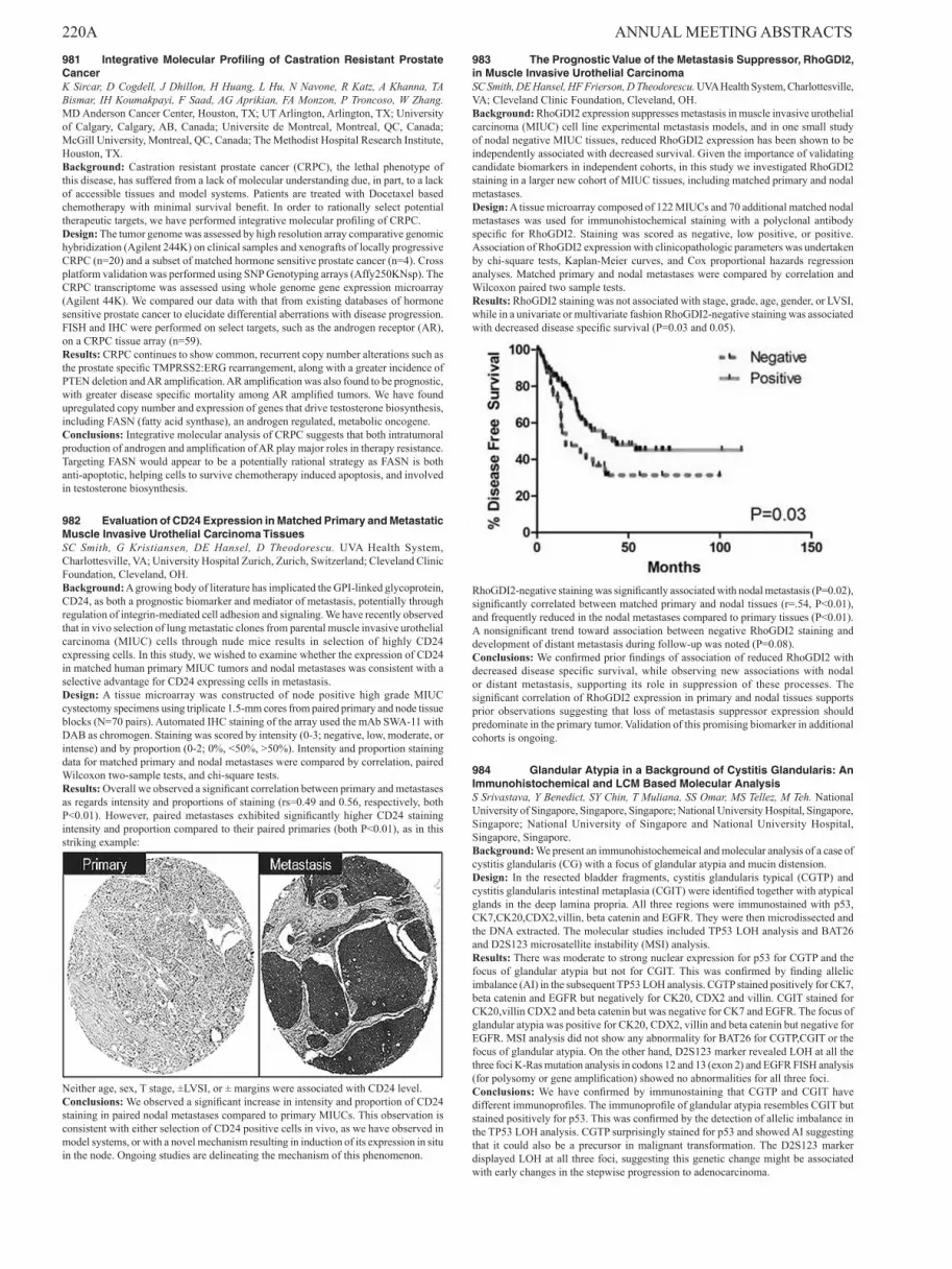
220A ANNUAL MEETING ABSTRACTS 981 Integrative Molecular Profiling of Castration Resistant Prostate CancerK Sircar, D Cogdell, J Dhillon, H Huang, L Hu, N Navone, R Katz, A Khanna, TA Bismar, IH Koumakpayi, F Saad, AG Aprikian, FA Monzon, P Troncoso, W Zhang. MD Anderson Cancer Center, Houston, TX; UT Arlington, Arlington, TX; University of Calgary, Calgary, AB, Canada; Universite de Montreal, Montreal, QC, Canada; McGill University, Montreal, QC, Canada; The Methodist Hospital Research Institute, Houston, TX.Background: Castration resistant prostate cancer (CRPC), the lethal phenotype of this disease, has suffered from a lack of molecular understanding due, in part, to a lack of accessible tissues and model systems. Patients are treated with Docetaxel based chemotherapy with minimal survival benefit. In order to rationally select potential therapeutic targets, we have performed integrative molecular profiling of CRPC.Design: The tumor genome was assessed by high resolution array comparative genomic hybridization (Agilent 244K) on clinical samples and xenografts of locally progressive CRPC (n=20) and a subset of matched hormone sensitive prostate cancer (n=4). Cross platform validation was performed using SNP Genotyping arrays (Affy250KNsp). The CRPC transcriptome was assessed using whole genome gene expression microarray (Agilent 44K). We compared our data with that from existing databases of hormone sensitive prostate cancer to elucidate differential aberrations with disease progression. FISH and IHC were performed on select targets, such as the androgen receptor (AR), on a CRPC tissue array (n=59).Results: CRPC continues to show common, recurrent copy number alterations such as the prostate specific TMPRSS2:ERG rearrangement, along with a greater incidence of PTEN deletion and AR amplification. AR amplification was also found to be prognostic, with greater disease specific mortality among AR amplified tumors. We have found upregulated copy number and expression of genes that drive testosterone biosynthesis, including FASN (fatty acid synthase), an androgen regulated, metabolic oncogene.Conclusions: Integrative molecular analysis of CRPC suggests that both intratumoral production of androgen and amplification of AR play major roles in therapy resistance. Targeting FASN would appear to be a potentially rational strategy as FASN is both anti-apoptotic, helping cells to survive chemotherapy induced apoptosis, and involved in testosterone biosynthesis.
982 Evaluation of CD24 Expression in Matched Primary and Metastatic Muscle Invasive Urothelial Carcinoma TissuesSC Smith, G Kristiansen, DE Hansel, D Theodorescu. UVA Health System, Charlottesville, VA; University Hospital Zurich, Zurich, Switzerland; Cleveland Clinic Foundation, Cleveland, OH.Background: A growing body of literature has implicated the GPI-linked glycoprotein, CD24, as both a prognostic biomarker and mediator of metastasis, potentially through regulation of integrin-mediated cell adhesion and signaling. We have recently observed that in vivo selection of lung metastatic clones from parental muscle invasive urothelial carcinoma (MIUC) cells through nude mice results in selection of highly CD24 expressing cells. In this study, we wished to examine whether the expression of CD24 in matched human primary MIUC tumors and nodal metastases was consistent with a selective advantage for CD24 expressing cells in metastasis.Design: A tissue microarray was constructed of node positive high grade MIUC cystectomy specimens using triplicate 1.5-mm cores from paired primary and node tissue blocks (N=70 pairs). Automated IHC staining of the array used the mAb SWA-11 with DAB as chromogen. Staining was scored by intensity (0-3; negative, low, moderate, or intense) and by proportion (0-2; 0%, <50%, >50%). Intensity and proportion staining data for matched primary and nodal metastases were compared by correlation, paired Wilcoxon two-sample tests, and chi-square tests.Results: Overall we observed a significant correlation between primary and metastases as regards intensity and proportions of staining (rs=0.49 and 0.56, respectively, both P<0.01). However, paired metastases exhibited significantly higher CD24 staining intensity and proportion compared to their paired primaries (both P<0.01), as in this striking example:
Neither age, sex, T stage, ±LVSI, or ± margins were associated with CD24 level.Conclusions: We observed a significant increase in intensity and proportion of CD24 staining in paired nodal metastases compared to primary MIUCs. This observation is consistent with either selection of CD24 positive cells in vivo, as we have observed in model systems, or with a novel mechanism resulting in induction of its expression in situ in the node. Ongoing studies are delineating the mechanism of this phenomenon.
983 The Prognostic Value of the Metastasis Suppressor, RhoGDI2, in Muscle Invasive Urothelial CarcinomaSC Smith, DE Hansel, HF Frierson, D Theodorescu. UVA Health System, Charlottesville, VA; Cleveland Clinic Foundation, Cleveland, OH.Background: RhoGDI2 expression suppresses metastasis in muscle invasive urothelial carcinoma (MIUC) cell line experimental metastasis models, and in one small study of nodal negative MIUC tissues, reduced RhoGDI2 expression has been shown to be independently associated with decreased survival. Given the importance of validating candidate biomarkers in independent cohorts, in this study we investigated RhoGDI2 staining in a larger new cohort of MIUC tissues, including matched primary and nodal metastases.Design: A tissue microarray composed of 122 MIUCs and 70 additional matched nodal metastases was used for immunohistochemical staining with a polyclonal antibody specific for RhoGDI2. Staining was scored as negative, low positive, or positive. Association of RhoGDI2 expression with clinicopathologic parameters was undertaken by chi-square tests, Kaplan-Meier curves, and Cox proportional hazards regression analyses. Matched primary and nodal metastases were compared by correlation and Wilcoxon paired two sample tests.Results: RhoGDI2 staining was not associated with stage, grade, age, gender, or LVSI, while in a univariate or multivariate fashion RhoGDI2-negative staining was associated with decreased disease specific survival (P=0.03 and 0.05).
RhoGDI2-negative staining was significantly associated with nodal metastasis (P=0.02), significantly correlated between matched primary and nodal tissues (r=.54, P<0.01), and frequently reduced in the nodal metastases compared to primary tissues (P<0.01). A nonsignificant trend toward association between negative RhoGDI2 staining and development of distant metastasis during follow-up was noted (P=0.08).Conclusions: We confirmed prior findings of association of reduced RhoGDI2 with decreased disease specific survival, while observing new associations with nodal or distant metastasis, supporting its role in suppression of these processes. The significant correlation of RhoGDI2 expression in primary and nodal tissues supports prior observations suggesting that loss of metastasis suppressor expression should predominate in the primary tumor. Validation of this promising biomarker in additional cohorts is ongoing.
984 Glandular Atypia in a Background of Cystitis Glandularis: An Immunohistochemical and LCM Based Molecular AnalysisS Srivastava, Y Benedict, SY Chin, T Muliana, SS Omar, MS Tellez, M Teh. National University of Singapore, Singapore, Singapore; National University Hospital, Singapore, Singapore; National University of Singapore and National University Hospital, Singapore, Singapore.Background: We present an immunohistochemeical and molecular analysis of a case of cystitis glandularis (CG) with a focus of glandular atypia and mucin distension.Design: In the resected bladder fragments, cystitis glandularis typical (CGTP) and cystitis glandularis intestinal metaplasia (CGIT) were identified together with atypical glands in the deep lamina propria. All three regions were immunostained with p53, CK7,CK20,CDX2,villin, beta catenin and EGFR. They were then microdissected and the DNA extracted. The molecular studies included TP53 LOH analysis and BAT26 and D2S123 microsatellite instability (MSI) analysis.Results: There was moderate to strong nuclear expression for p53 for CGTP and the focus of glandular atypia but not for CGIT. This was confirmed by finding allelic imbalance (AI) in the subsequent TP53 LOH analysis. CGTP stained positively for CK7, beta catenin and EGFR but negatively for CK20, CDX2 and villin. CGIT stained for CK20,villin CDX2 and beta catenin but was negative for CK7 and EGFR. The focus of glandular atypia was positive for CK20, CDX2, villin and beta catenin but negative for EGFR. MSI analysis did not show any abnormality for BAT26 for CGTP,CGIT or the focus of glandular atypia. On the other hand, D2S123 marker revealed LOH at all the three foci K-Ras mutation analysis in codons 12 and 13 (exon 2) and EGFR FISH analysis (for polysomy or gene amplification) showed no abnormalities for all three foci.Conclusions: We have confirmed by immunostaining that CGTP and CGIT have different immunoprofiles. The immunoprofile of glandular atypia resembles CGIT but stained positively for p53. This was confirmed by the detection of allelic imbalance in the TP53 LOH analysis. CGTP surprisingly stained for p53 and showed AI suggesting that it could also be a precursor in malignant transformation. The D2S123 marker displayed LOH at all three foci, suggesting this genetic change might be associated with early changes in the stepwise progression to adenocarcinoma.
ANNUAL MEETING ABSTRACTS 221A
985 The RB/p16 Pathway but Not p53 Is Disrupted by High Risk HPV in Penile Squamous Cell CarcinomaE Stankiewicz, DM Prowse, J Cuzick, L Ambroisine, S Kudahetti, N Watkin, C Corbishley, DM Berney. Queen Mary, University of London, London, United Kingdom; St George’s Hospital, London, United Kingdom.Background: Several risk factors have been identified for penile cancer, but its pathogenesis is not well understood. Human papillomavirus (HPV) may be involved in the carcinogenesis of penile squamous cell carcinoma (PSCC), yet few studies have compared the cell cycle protein expression in HPV positive and negative cancers of this type. We hypothesized that the types of human papillomavirus infection in different histological types of PSCC and would impact on the expression of key cell cycle proteins: p53, p21, p16INK4A and RB.Design: We collected 148 samples of PSCC including 97 usual type SCCs, 17 basaloid, and 15 verrucous carcinomas, from patients treated at St George’s Hospital between years: 2000-2007 and prepared tissue microarrays. Samples were analysed for RB, p16INK4A, p53 and p21 protein expression by immunohistochemistry. 102 cases were typed for HPV by a broad-spectrum HPV PCR method using SPF10 primers which amplify a 65-bp fragment of the L1 open reading frame and HPV genotypes identified by the INNO-LiPA line probe assay.Results: HPV DNA was present in 56% of tumours with HPV16 being the most prevalent type. HPV16 was the most common type detected in46/111 cases. Marked differences were observed in HPV prevalence between tumour types with HPV16 detected in 10 cases out of 13 where HPV was detected. 9 other types of HPV were detected including types 11, 31 and 33, but no HPV18. RB protein negatively (p<0.0001) and p16INK4A and p21 positively correlated (p<0.0001) with high-risk HPV infection. There was a significant difference in RB and p16INK4A expression between usual type, basaloid and verrucous tumours(p<0.0001). No correlation was found between HPV or tumour type with p53 protein expression.Conclusions: HPV infection is present in over half of penile cancers and appears to be responsible for RB pathway disruption in those tumours. However, no link between high-risk HPV types and p53 immunodetection was found. Different histological types of PSSC express differnt HPV DNA subtyoes, confirming possible separate aetiologies for those tumours. We confirm that usual type PSCC seems to have at least two different aetiologies: one related to HPV and one unrelated. We suggest that use of the bivalent HPV16/18 prophylactic vaccine in men could reduce occurrence of penile SCC by only 45%.
986 TMPRSS2-ERG Gene Fusion Status in Salvage Radical Prostatectomy Specimens Following RadiotherapyM Stanton, CC Guo, X Li, Y Wang, L Andino, IN Prokhorova, LL Pisters, P Troncoso. The University of Texas MD Anderson Cancer Center, Houston, TX.Background: A common chromosomal rearrangement leading to the fusion of the TMPRSS2 and ERG genes has been identified in a majority of untreated prostate cancer cases. Information regarding TMPRSS2-ERG gene fusion in recurrent prostate cancer following radiotherapy is limited.Design: We reviewed 50 radical prostatectomy specimens (RPS) from patients who underwent salvage radical prostatectomy after radiotherapy between 1993-2008. Twenty-five cases have been analyzed to date for TMPRSS2-ERG gene fusion by fluorescence in situ hybridization using ERG break-apart probes.Results: Median overall patient age was 68 years (range 45-75 years). Median interval between radiation therapy and surgery was 4 years (range 1-13 years). Median pre-salvage serum PSA was 3.40 ng/mL (range <0.1-39.20 ng/mL). The Gleason score was ≤7 (4+3) in 17 and ≥8 in 24 cases. Therapy effect precluded grading in 9 cases. The pathologic stage was pT2: 23; pT3a: 7 and pT3b: 20. Eight patients had lymph node metastases. The tumors were unifocal in 33/50 cases (66%) and multifocal in 17/50 cases (34%). Median total tumor volume was 1.27 cc (range 0.05-12.96 cc). Of the 25 cases analyzed thus far (17 unifocal tumors, including 2 with corresponding nodal metastases, and 8 multifocal tumors), 10 (40%) were positive for the ERG gene rearrangement. In 9 of the cases, the rearrangement was associated with deletion of the 5’ ERG gene. In the 8 multifocal tumors, the dominant focus was analyzed in 5 cases, and all foci were analyzed in 3 cases (2 foci in 2 cases; 3 foci in 1 case). The two primary tumors with lymph node metastases were positive, and there was concordance of the ERG gene fusion status between the primary tumors and the nodal metastases. ERG gene rearrangement was not detected in any of the foci studied in 3 multifocal tumors.Conclusions: Recurrent prostate cancer following radiotherapy exhibits TMPRSS2-ERG gene rearrangement in the range observed in untreated tumors.
987 Urothelial Tumors in Patients under the Age of 30 Years: A Report of 58 CasesML Stanton, L Xiao, BA Czerniak, CC Guo. University of Texas MD Anderson Cacner Center, Houston, TX.Background: Urothelial tumors occur mainly in patients between the ages of 50 to 80 years, and they are distinctively rare in young patients under the age of 30 years. Here we studied the clinicopathologic features of a large series of young patients with urothelial tumors.Design: We retrospectively searched our pathology file and identified 58 patients under the age of 30 years with an urothelial tumor. Histologic slides were reviewed for pathologic analysis, and clinical information was collected from medical records.Results: The mean age of patients was 23 years (range, 5-30 years). Forty patients were male and 18 were female. Clinical history was available for 32 patients, and 16 patients had a history of smoking tobacco. The most common presenting symptom was hematuria (n=26), followed by abdominal pain (n=8) and urinary tract infections (n=6). The tumors were found in the urinary bladder (n=53), renal pelvis (n=5) and ureter (n=1). The tumors were divided into non-invasive (n=45) and invasive (n=13) groups. The non-invasive tumors included papillary urothelial neoplasm of low malignant potential
(PUNLMP) (n=7), low- grade papillary urothelial carcinoma (n=33) and high-grade papillary urothelial carcinoma (n=5). The invasive tumors were all high-grade urothelial carcinoma (n=13), and some showed focal small cell carcinoma (n=2) and sarcomatoid features (n=1). The invasive tumors invaded lamina propria (n=4), muscularis propria (n=4), and perivesical adipose tissue (n=1) in the bladder; the invasive tumors all invaded renal parenchyma in the renal pelvis (n=4). Follow-up information was available for 39 patients (mean 55 months, range 3-250 months): 2 patients with PUNLMP had no evidence of disease (NED); in 25 patients with low-grade papillary urothelial carcinoma, 23 had NED, 1 was alive with disease, and 1 subsequently developed invasive tumor and died at 160 months; 1 patient with non-invasive high grade papillary urothelial carcinoma had NED; in 11 patient with invasive high grade urothelial carcinoma, 7 died at 45 months (range, 15-102 months), 2 had NED, and 2 were alive with disease.Conclusions: The majority of urothelial tumors in patients under the age of 30 years are non-invasive low-grade papillary urothelial carcinoma and PUNLMP, which are generally associated with an excellent prognosis. However, a small number of young patients can develop a high-grade invasive urothelial carcinoma at an advanced stage, and their clinical outcome is poor despite therapy.
988 Claudin-7 and Cytokeratin-7 are Immunohistochemical Markers for the Differential Diagnosis of Chromophobe Renal Cell Carcinoma and Renal OncocytomaW-J Sung, M-J Kim. Yeungnam University Hospital, Daegu, Republic of Korea.Background: The differential diagnosis of chromophobe renal carcinoma and renal oncocytoma is frequently difficult by H&E stained sections based on histologic features only. Both tumors often have overlapping morphological features, characterized by neoplastic cells with granular eosinophilic cytoplasm. Distinction of these two lesions is important because of differences in clinical behavior and prognosis. Chromophobe renal cell carcinoma is a malignant renal tumor with metastatic potential and renal oncocytoma, in contrast, is a benign lesion. Several immunohistochemical markers have been studied to distinghish chromophobe renal cell carcinoma from renal oncocytoma. However, the results have been still challenging. Claudin-7 has recently been suggested to be a distal nephron marker. In this study, we examined the immuohistochemical expression of claudin-7 and cytokeratin-7 in these two tumors.Design: 11 cases of chromophobe renal cell carcinomas and 5 renal oncocytomas were obtained at Yeungnam University Hospital from Jan. 2005 to July 2009. 15 clear cell renal cell carcinomas were included. Immunohistochemistry using Cytokeratin 7 (DakoCytomation, Denmark) and claudin-7 (Invitrogen, Camarillo, CA, USA) was done on tissue microarray slide by BenchMark XT IHC/ISH Staining Module (Ventana Medical System, Tucson, AZ, USA). A positive reaction was definded as strong and diffuse membranous staining on tumor cells. Hale’s colloidal iron staining was also performed.Results: Claudin-7 protein was overexpressed in a diffuse, strong membranous pattern in 10 of 11 chromophobe renal cell carcinoma and not diffusely expressed in renal oncocytoma (p < 0.001). Claudin-7 in renal oncocytoma was focally expressed in 2 of 5 cases and compeletely not expressed in 3 of 5 cases. Cytokeratin-7 was expressed in 10 of 11 chromophobe renal cell carcinoma, coupled with its negative reactivity for renal concocytoma (p < 0.001) based on our data. 11 of 11 chromophobe renal cell carcinoma and 1 of 5 renal oncocytoma were positive for Hale’s colloidal iron stain (p < 0.001). Any of clear cell renal cell carcinomas revealed no positive reaction, for claudin-7, cyokeratin-7 and Hale’s colloidal iron stain. Claudin-7 was focally stained in 4 cases including one case of cytoplasmic staining, and negative in 8 of 15 clear cell renal cell carcinoma.Conclusions: Claudin-7 and cytokeratin-7 can be used as immunohistochemical biomarkers in differential diagnosis of chromophobe renal cell carcinoma and renal oncocytoma.
989 Testing Mutual Exclusivity of ETS Rearranged Prostate CancerMA Svensson, CJ LaFargue, D Pflueger, N Kitabayashi, AK Tewari, F Demichelis, MA Rubin. Weill Cornell Medical College, New York, NY; University Hospital of Orebro, Orebro, Sweden.Background: Prostate cancer is a clinically heterogeneous and multifocal disease. More than 80% of patients with prostate cancer harbor multiple geographically discrete cancer foci at the time of diagnosis. Emerging data suggest that these foci are molecularly distinct consistent with the hypothesis that they arise as independent clones. One of the strongest arguments is the heterogeneity observed in the status of ETS rearrangements between discrete tumor foci. The clonal evolution of individual prostate cancer foci based on recent studies demonstrates intra-tumoral homogeneity but inter-tumoral heterogeneity. The issue of multifocality and interfocal heterogeneity is important and has not been fully elucidated due to lack of the systematic evaluation of ETS rearrangements in multiple tumor sites.Design: The current study investigates the frequency of multiple ETS gene rearrangements within the same focus and between different cancer foci. Fluorescence in-situ hybridization (FISH) assays were designed to detect the four most common recurrent ETS gene rearrangements in a cohort of 88 men with localized prostate cancer.Results: We found ERG, ETV1, and ETV5 rearrangements in 51% (44/86), 6% (5/85), and 1% (1/86), respectively. None of the cases demonstrated ETV4 rearrangements. Mutual exclusiveness of ETS rearrangements was observed in the vast majority of cases, however in six cases we discovered multiple ETS or 5 prime fusion partner rearrangements within the same tumor focus.Conclusions: Our results demonstrate an additional level of complexity concerning the distribution of gene rearrangements in prostate cancer. This is exemplified by our observation that multiple rearrangements can exist within the prostate and can also occur within the same tumor focus. In conclusion, we provide further evidence for prostate cancer tumor heterogeneity with the identification of multiple concurrent ETS rearrangements.
222A ANNUAL MEETING ABSTRACTS 990 CEACAM1 Expression Distinguishes Indolent from Lethal Prostate CancerMA Svensson, D Tilki, S Shariat, BB Singer, O Reich, CG Stief, O Andren, SO Andersson, JE Johansson, F Demichelis, S Rafii, S Ergun, MA Rubin. Weill Cornell Medical College, New York, NY; University Hospital of Orebro, Orebro, Sweden; University Hospital of Grosshadern, Munich, Germany; Memorial Sloan Kettering Cancer Center, New York, NY; University Hospital of Essen, Essen, Germany.Background: Early detection of prostate cancer (PCa) has increased with the use of prostate specific antigen (PSA) screening. Many of these tumors will never progress to lethal disease, but without a good prognostic tool the number of over treated patients increases. Therefore, it is important to assess new markers which might aid in the prognosis of PCa. Previous studies have shown human carcinoembryonic antigen-related cell adhesion molecule 1 (CEACAM1) to be down regulated in several carcinomas, including PCa. It has been reported as independent predictor of disease specific outcome in malignant melanoma and adenocarcinoma of the lung.Design: In the current study, we interrogate a population-based Watchful Waiting cohort (n=220) consisting of incidental PCa cases, with respect to CEACAM1 expression using immunohistochemistry (IHC). Equal proportions of men who died of prostate cancer or developed metastasis and men who lived a minimum of 10 years without clinical recurrence are included in this study. The follow up time is now up to 30 years.Results: PCa tumors were classified as CEACAM1 positive or negative. Cases with CEACAM1 negative tumors had a significantly (p=0.0013) poorer overall survival compared to CEACAM1 positive tumors. Multivariate analysis shows that CEACAM1 status, next to age and Gleason score, as a significant independent prognostic factor for survival (HR 2.709; 95% CI 1.069-6.864); p=0.0357).Conclusions: Expression of CEACAM1 was an independent prognostic factor in a population-based cohort of men with localized prostate cancer followed by watchful waiting therapy. Evaluating tumors for CEACAM1 may help stratify patients with PCa into low-risk and high-risk groups, which may be used to guide clinical decision making.
991 Expression of PAX8 in Normal and Neoplastic Tissues: A Comprehensive IHC AnalysisD Tacha, L Cheng, D Zhou, RL Henshall-Powell. Biocare Medical, Concord, CA; Indiana Univ School of Med, Indianapolis, IN.Background: PAX genes are a family of cell-lineage transcription factors that play a role during organogenesis and are regulatory proteins expressed embryonic or neoplastic cells of the same lineage. These proteins are required for cell growth and differentiation in embryonic tissues and in adult tissues in specific cell-lineage neoplastic tissues. Other transcription factors in this family include TTF-1 and CDX2. PAX8 is a nephric-lineage transcription factor and can be expressed in kidney, thyroid and ovarian cancers. Little is known about the specificity of PAX8 in various neoplastic tumors. This study examines the IHC expression in multiple normal and neoplastic tissues.Design: Tissue microarrays (TMA) of various normal and neoplastic tissues were constructed from archival tissues. The patient information containing age, sex, diagnosis, stage, grade and IHC staining was analyzed. TMAs were deparaffinized in the usual way, and antigen retrieval was performed using a pressure cooker. TMA tissue cores were incubated with PAX8 for 30 minutes, followed by visualization (DAB) using a biotin-free detection system.Results: PAX expression was positive in renal cell carcinoma (90%), ovarian cancers (82%), thyroid cancers (90%) and endometrial carcinomas (81%). In lung cancer, PAX8 staining was found in only 1 case (1+ staining). The majority of other cancers were 100% negative (Table 1). PAX 8 was also seen in normal tonsil and lymph node. Expression appeared to be confined to B-cells and/or cells of B-cell lineage.
Table 1Diagnosis Total Cases Positive % PositiveRenal Cell Carcinoma 122 110 90%Ovarian Cancer 244 201 82%Endometrial Cancer 134 108 81%Thyroid Tumors 10 9 90%Neuroendocrine Tumors 15 1 7%Cervical Cancer 73 7 4%Pancreatic Cancer 23 3 13%Bladder Cancer 75 6 8%Lung Cancer 100 1 1%Colon Cancer 100 0 0%Breast Cancer 100 0 0%Prostate Cancer 35 0 0%Liver Cancer 36 0 0%Melanoma 20 0 0%Seminoma 18 0 0%Stomach Cancer 6 0 0%Esophageal Cancer 6 0 0%Soft Tissue Tumor 21 2 5%FDA Normal Tissues 33 *1 3%*Normal ProstateConclusions: PAX8 is a very specific and sensitive marker for renal cell and ovarian carcinomas, and may prove to be equally sensitive for thyroid and endometrial cancers. In this study, PAX8 specificity has been evaluated in over 1000 cases; 21 different cancers and their subtypes, and 33 different normal tissues.
992 Overexpression of AKT in Prostate Cancers and Association with an Agressive Tumor TypeH Takahashi, T Yamamoto, M Furusato, S Egawa, H Hano. The Jikei University School of Medicine, Tokyo, Japan.Background: AKT is a component of the PI-3 kinase pathway and is activated by phosphorylation. It is a key regulator of many signal transduction pathways and
overexpression has been noted in many types of cancer. Little is known about this phenomenon and clinical correlation in prostate cancer.Design: A total of 101 cancer foci and 38 HGPIN were selected from our prostatectomy series. The cancer included 53 clinically-evident tumors (index tumor: IT), and 48 microscopic tumors (MT, the largest diameter <2mm). Immunohistochemistry was performed with anti-AKT (phosphorylation form specific) and expression was evaluated. Inensity and distribution were evaluated as grade 0 (none), 1 (weak/<10% of the tumor), 2 (intermediate/10-49%), 3 (strong/>50%), 4 (very strong/>50%). Grades 2-4 were designated as expression+. Grade 4 was defined as overexpression+. The data were compared with stage (pT), Gleason score, and PSA-free survival.Results: Frequency of expression+ was significantly higher in IT than in MT (51% vs. 9.1%: p<0.05). Nine cases showed overexpression in IT and none in MT/HGPIN.
Tabel 2 Immunohistochemistry of AKT0 1 2 3 4
IT (n=53) 10 (18.8%) 15 (28.3%) 8 (15.1%) 11 (20.8%) 9 (17.0%)MT (n=48) 34 (70.8%) 10 (20.8%) 3 (6.3%) 1 (2.1%) 0HGPIN (n=38) 38 (100%) 0 0 0 0IT vs. MT, p<0.05; IT vs. HGPIN, p<0.05Nine cases with overexpression+ showed significantly higher stage and grade (3 cases: pT2c, 4: pT3a, 2: pT3b; 2: 3+4=7, 5: 4+3=7, 2: 4+5=9) than cases with overexpression- (10 cases : pT2a-b, 24: pT2c, 5: pT3a, 5: pT3b; 11:3+3=6, 25: 3+4=7, 4: 4+3=7, 2: 4+4=8, 2: 4+5=9).
Table 2 pT and GS in overexpression+ and - tumorspT T2a-b T2c T3a T3bOverexpression+ 0 (0%) 3 (33.3%) 4 (44.5%) 2 (22.2%)Overexpression- 10 (22.7%) 24 (54.5%) 5 (11.4%) 5 (11.4%)GS 3+3=6 3+4=7 4+3=7 8-10Overexpression+ 0 (0%) 2 (22.2%) 5 (55.6%) 2 (22.2%)overexpression- 11 (25.0%) 25 (26.9%) 4 (9.1%) 4(9.1%)overexpression+ vs. - in pT and GS, p<0.05In the follow-up (36 months), 4/8 cases with overexpression+ showed biochemical failure (0 month-24 months).Conclusions: Overexpression of phosphorylated AKT is suggested to play an important role in the tumor progression and indicating an aggressive tumor type. Reversely, it is not significant in the pre-clinical prostate cancer and HGPIN.
993 Hypoxia-Inducible Gene 2 (HIG2) Protein – Novel Hypoxia Marker for Primary and Metastatic Renal Cell CarcinomaM Tretiakova, M Hall, T Smertenko, E Hyjek, H Al-Ahmadie. University of Chicago, Chicago; Leica, Newcastle, United Kingdom; Memorial Sloan-Kettering Cancer Center, New York.Background: Tumor viability and progression is partly dependant on its response and adaptation to hypoxic conditions resulting from tumor growth. Clear cell renal cell carcinoma (CCRCC) is characterized by rich vascularity induced via hypoxia-inducible factor/mTOR pathways. Successes in treating CCRCC are largely attributed to molecular targeting of factors in these pathways, which is also being tried in other subtypes of RCC. HIG2 is a recently identified hypoxia-inducible growth factor that has not been well studied in renal tumors, and may be an option for targeted therapy in these tumors. Here we examined HIG2 expression in a large set of primary and metastatic RCC using a novel monoclonal antibody.Design: Tissue microarray sections of 155 primary and 57 metastatic CCRCC (including 18 matched pairs), 76 papillary (PRCC) and 53 chromophobe (ChRCC) renal carcinomas, 24 oncocytomas (ONC) and 41 normal tissues from different organs were stained with HIG2 antibody (clone HX34Y, Leica). Reactivity with >5% of cells was considered positive. IHC intensity was scored as weak (1+), moderate (2+) or strong (3+).Results: Detailed HIG2 expression is shown in Table 1. In primary CCRCC and PRCC 92% and 67% of cases expressed HIG2 with mean intensity of 2.14 and 1.49, respectively (p<0.01). In ChRCC and ONC, HIG2 was expressed in only 8% and 4% of cases with mean intensity of 0.11 and 0.08, respectively. In metastatic CCRCC, HIG2 was expressed in 91% of cases with mean intensity of 2.05. In 18 cases of matched primary and metastatic CCRCC, HIG2 was expressed in both sites in 14 cases with similar pattern and intensity, negative in both sites in 3 cases and positive in the metastasis only in 1 case.
HIG2 expression in renal tumorsType Negative 1+ 2+ 3+CCRCC primary (n=155) 12 (8%) 30 (19%) 44 (28%) 69 (45%)CCRCC met (n=57) 5 (9%) 11 (19%) 16 (28%) 25 (44%)PRCC (n=76) 25 (33%) 10 (13%) 20 (26%) 21 (28%)ChRCC (n=53) 49 (92%) 2 (4%) 2 (4%) 0ONC (n=24) 23 (96%) 0 1 (4%) 0
Conclusions: In renal tumors, HIG2 expression is the highest in primary and metastatic CCRCC, followed by PRCC. HIG2 expression in ChRCC and ONC is negligibly low. Preferential HIG2 expression in both primary and metastatic CCRCC may help to understand further mechanisms in the development and progression of this tumor and could potentially serve as a molecule for targeted therapy in patients with advanced and metastatic CCRCC.
994 Positive Margins in pT2 Prostate Cancer in Open, Laparoscopic and Robot-Assisted Radical Prostatectomy SpecimensP Troncoso, S Matin, RJ Babaian, CA Pettaway, LL Pisters, I Prokhorova, JW Davis. The University of Texas MD Anderson Cancer Center, Houston, TX.Background: Open radical retropubic prostatectomy (ORP) is the gold standard surgical treatment for clinically localized prostate cancer. While a small number of centers introduced the laparoscopic technique (LRP), a much larger number have moved on to robotic-assisted technique (RARP). Comparison of the positive margin (+MOR)
ANNUAL MEETING ABSTRACTS 223Arates in pT2 cases is a useful surrogate for oncologic safety when new technologies are introduced into clinical practice.Design: We selected from our contemporary series patients with pT2, NX/N0 disease and a +MOR, treated by either open radical prostatectomy (2002-07), LRP (2002-06) or RARP (2006-09). Patients who received neoadjuvant treatment were excluded. These three groups were compared for the incidence, extent, and location of the observed +MOR.Results: There were 152 pT2 cases with +MOR, including 95 ORP (95/1162, 8%), 22 LRP (22/193, 11%) and 35 RARP (35/588, 6%) The majority of the patients were white (76%) and the clinical stage was T1c in 64% of the patients. The median PSA was 5.30 ng/ml. The median Gleason score was 7(3+4). Tumor was multifocal in 91% of the cases with a median number of three cancer foci per specimen. The median total tumor volume was 1.58 cc. A +MOR involved a single location in 87% of the cases (83% ORP; 91% LRP; 97% RARP). The most common locations for a single +MOR were the apex (44% ORP; 80% LRP; 44% RARP), posterolateral surface (27% ORP; 10% LRP; 26% RARP) and the bladder neck (15% ORP; 10% LRP ; 21% RARP). In cases with more than one +MOR location, the apex was included in 68% (13/19) of the cases. The extent of the +MOR was <1.0 mm in over one third of the cases (34% ORP; 32% LRP; 46% RARP). The mean +MOR extent for cases with a +MOR ≥1 was 5.19 mm (5.04 ORP; 6.97 LRP; 4.32 RARP). The dominant tumor focus involved the MOR in cases with single or multiple +MOR locations in 81% of the cases, including dominant foci of transition zone origin in 38% of the cases.Conclusions: The data in this study suggest that the introduction of the novel laparoscopic technique resulted in an increase in pT2 positive margin cases relative to open surgery at a high volume center, whereas with the robotic technique, the trend was favorable. The location of positive margins was identical with open and robotic techniques.
995 Validation of Preoperative Predictive Models for Adverse Radical Prostatectomy OutcomesK Trpkov, J Zhang, P Faris, A Yilmaz. Calgary Laboratory Services and University of Calgary, Calgary, Canada.Background: We previously developed preoperative predictive models for adverse radical prostatectomy (RP) outcomes, such as: stage pT3 prostate cancer, positive seminal vesicles (SV) or lymph nodes (LN), positive margins and tumor (TU) volume ≥10% gland volume (Lab Invest 2006;86:136A (Suppl 1).Design: Predictive models were developed on a cohort comprising 1174 RP with matched 10-core biopsies (Cohort 1), performed in our institution from 07/2000 to 04/2005. We performed univariate and multivariate logistic regression analysis on Cohort 1 to establish the best predictors for adverse RP outcomes. The following biopsy variables were evaluated: total percent cancer (TPC), cancer length in mm, number of positive cores, Gleason Score (GS), perineural invasion, in addition to prostate specific antigen (PSA) and PSA density (PSAD). Validation of the predictive models was done on a separate cohort of 971 RP with matched biopsies (Cohort 2), accrued in our institution from 05/2005 to 12/2008.Results: Multivariate models for the adverse RP outcome performed in the validation Cohort 2 as follows:RP Outcome OR (P) OR (P) OR (P)Stage pT3 GS≥8 12.9 (<0.001) TPC>30% 5 (<0.001) PSAD>0.3 2.1 (<0.01)Positive SV or LN GS≥8 17.1 (<0.001) TPC>30% 6.5 (0.004) PSAD>0.3 2.4 (0.013)Positive margins
>3 positive cores 2.6 (<0.001) Neural
invasion 1.5 (0.011) GS 7 1.5 (0.023)TU vol> 10% gl vol TPC>30% 10.8 (<0.001) PSAD >0.3 4.4 (<0.001) >3 positive
cores 4.2 (<0.001)
Odds Ratio (OR) calculated vs. referenceComparison of the areas under the curves (AUC) for the initial Cohort 1 and the validation Cohort 2 are shown in the following table:
Stage pT3 Positive SV or LN Positive margins TU vol>10% gl volCohort 1 AUC (CI) 0.80 (0.76-0.84) 0.82 (0.76-0.87) 0.65 (0.61-0.68) 0.77 (0.75-0.80)Cohort 2 AUC (CI) 0.78 (0.72-0.81) 0.81 (0.76-0.86) 0.67 (0.63-0.70) 0.79 (0.76-0.82)
AUC, area under the curve; CI, confidence intervalsConclusions: We validated the previously developed predictive models for adverse RP outcomes on an independent cohort from our institution. The models performed similarly in both cohorts. Validation of the models on cohorts from other institutions may further test their performance and substantiate their utility.
996 Immunohistochemical Ratio of α-3 and α-1 Subunits of Sodium Pump Correlates with Prostate Cancer Biochemical Recurrence: A Tissue Microarray StudyD Trudel, A Roussel-Jobin, B Tetu, Y Fradet, L Lacombe, H Hovington, M Njoya, T Mijatovic, R Kiss. Centre Hospitalier Universitaire de Québec, Québec, Canada; Université Libre de Bruxelles, Bruxelles, Belgium.Background: Sodium pump (Na+/K+ ATPase) is made of α and β-subunits. Expression of the α subunit is altered in many cancer types. Considering that Na+/K+ ATPase is the target of cardiotonic steroids such as digoxin, we hypothesized that the finding of an association between the expression of α-1 (SUα1) and α-3 (SUα3) subunits and prostate cancer (PCA) prognosis might have important therapeutic implications.Design: Men included in the cohort were diagnosed with a pT3N0-2M0 PCA treated by radical prostatectomy. A tissue microarray was built from representative areas of prostate tumors and the percentage of cancer cells expressing SUα1 and SUα3 by immunohistochemistry was evaluated. Frequency tables with Pearson’s correlations were built to evaluate the association between the expression levels of SUα1, SUα3, SUα3:SUα1 ratio and known PCA prognostic factors. Wald test from Cox regression
model was used to measure the association between SUα1, SUα3, SUα3:SUα1 ratio and time to biochemical recurrence and death of any causes.Results: Mean follow-up time of the 74 men included in the cohort was 13 years. Thirty-two biochemical recurrences and 8 deaths were observed. SUα1 expression level was inversely associated with Gleason score on biopsy (p = 0.0019). SUα3 and SUα3:SUα1 ratio were not associated with any known PCA prognostic factor. While no association with recurrence or death was found with SUα1 and SUα3 expression levels, the SUα3:SUα1 ratio was significantly associated with PCA biochemical recurrence (HR = 1.68, 95%CI [1.03; 2.74], Cox regression adjusted for initial serum prostate specific antigen level).Conclusions: This study shows that, although SUα1 expression level is inversely associated with PCA Gleason score, SUα3:SUα1 ratio correlates with PCA biochemical recurrence. The ratio of sodium pump subunits expression appears as an interesting potential PCA marker that may lead to the development of new targeted therapies.
997 Renal Cell Carcinoma (RCC) in Hemodialysed (HD) Patients: The Duration of Hemodialysis May Influence the Pathological Type and PrognosisT Tsuzuki, N Sassa, M Fukatsu, T Fujita, S Katsuno, T Nishikimi, R Hattori, M Gotoh. Nagoya Daini Red Cross Hopital, Nagoya, Japan; Nagoya University, Nagoya, Japan; Komaki Municipal Hospital, Komaki, Japan; Chyukyo Hospital, Nagoya, Japan; Okazaki Municipal Hospital, Okazaki, Japan.Background: It is well established that RCC is one of the most important complications in hemodialysis patients. However, the influence of the HD duration on RCC has not been determined. Therefore, this study determined if the HD duration relates to the pathological type and prognosis.Design: This study consisted of 69 patients on HD (73 kidneys) who underwent a radical nephrectomy for RCCs from January 1991 to December 2008. We classified the patients into three groups based on the duration of HD (Group 1: less than 10 years; Group 2: from 11 to 20 years; Group 3: over 21 years). All cases were reviewed without clinical information, and the pathological type and parameters were recorded.Results: The patients who received HD for more than 10 years showed a distinctive distribution of the pathological type (p=0.01) and pT stage (p=0.003), as listed in Table 1.
Pathological subtype and pT stage according to the HD duration.Total Group 1 Group 2 Group 3
Clear cell RCC 32 18 8 6Papillary RCC 11 3 3 4Sarcomatoid RCC 8 0 2 6ACDK-associated RCC 17 2 11 5Others 5 1 1 2pT1a 42 18 14 10pT1b 9 4 5 0pT2 5 1 3 1pT3a 11 1 3 7pT3b 3 0 0 3Unknown 3 0 1 2
The disease free survival (DFS) among the three groups was statistically different (p<0.05). The incidence of RCCs with sarcomatoid component was detected after 10 years of HD. The DFS of RCCs with sarcomatoid component was 55.9% and 37.3% at 5 years and 10 years, respectively.Conclusions: The HD duration influenced the pathological type and pT stage of RCCs in HD patients. RCCs with sarcomatoid component, which led to a poor outcome, occurred after more than 10 years of HD. Therefore, it is necessary that patients who receive HD, for an extended period of time, especially more than 10 years, undergo frequent and careful medical examinations.
998 Clinicopathological Study of Urothelial Carcinoma (UC) of the Renal Pelvis (RP): A Proposal for New pT3 CriteriaT Tsuzuki, N Sassa, M Fukatsu, T Fujita, A Katsuno, T Nishikimi, R Hattori, M Gotoh. Nagoya Daini Red Cross Hopital, Nagoya, Japan; Nagoya University, Nagoya, Japan; Komaki, Komaki Municipal Hospital, Japan; Chyukyo Hospital, Nagoya, Japan; Okazaki Municipal Hospital, Okazaki, Japan.Background: UC of the RP is thought to have a better prognosis than UC of the ureter. UC of the RP is characterized as stage pT3 when the tumor invades beyond the muscularis into the peripelvic fat or renal parenchyma. However, both general pathologists and GU pathologists often have difficulty differentiating parenchymal invasion from ductal spreading in the collecting ducts. Therefore, some cases with ductal spreading but without parenchymal invasion could be misclassified as “pT3”, which may indicate a better prognosis for UC of the RP.Design: This study consisted of patients who underwent radical nephroureterectomy for UC of the RP. All cases were reviewed without clinical information by one pathologist (T.T.). We classified pT3 cases into the following four groups based on the extent of UC: pT3a, UC extended into the renal medulla; pT3b, UC extended into the renal cortex; pT3b, UC with peripelvic fat invasion; pT3d, pT3a or pT3b cases with UC peripelvic fat invasion. The pT stage and other parameters (tumor necrosis (TN), lymph-vascular invasion (LVI), nuclear grade (1973 WHO) (NG), and presence of CIS) were recorded.Results: This study included 246 patients (177 males, 69 females; age range 33-94 years, mean 66.7 years). The pT stage and overall 5-year survival (OS) are listed.
224A ANNUAL MEETING ABSTRACTS
pT stage and 5-year OSNumber % OS
pTa and/or pTis 88 35.6 98.4pT1 24 9.7 84.0pT2 12 4.9 81.8pT3a 52 21.1 89.0pT3b 17 6.9 42.4pT3c 5 2.0 0pT3d 30 12.1 13.6pT4 19 7.7 0
There was a statistically significant difference between pT3a and the other pT3 subtypes (pT3b, pT3c, and pT3d) (p<0.001). However, there were no statistically significant differences between pT3b, pT3c, pT3d, and pT4. In a univariate analysis, the presented pT classification, TN, LVI, and NG were statistically significant (p<0.001). In a multivariate analysis, the presented classification (p=0.0001, HR: 1.459), TN (p=0.012, HR: 2.586) and LVI (p=0.038, HR: 2.677) were statistically significant.Conclusions: Our results suggest pT3b, pT3c, and pT3d should be classified as true pT3 and that pT3a should be classified as pT1 or pT2. We believe this proposed classification is simple and easy to apply, even for general pathologists. It is important to determine the appropriate pT stage in order to predict the prognosis of patients.
999 Hedgehog Pathway Activation Is Associated with VEGF Induction in Prostate Cancer MicroenvironmentV Tzelepi, M Karlou, A Hoang, C Logothetis, P Troncoso, E Efstathiou. M.D. Anderson Cancer Center, Houston.Background: Investigators have speculated that prostate cancer progression usurps normal prostate and bone development pathways. Hedgehog (HH) signaling is a stromal-epithelial interacting network central to prostate development and likely implicated in the aggressiveness of prostate cancer (PCa).Design: A tissue microarray was constructed from prostatectomy specimens from 141 untreated PCa patients (49 with Gleason score ≤6 and 92 ≥7). Non-neoplastic peripheral (PZ) and transition (TZ) zone were also included. Expression of Sonic HH ligand (SHH), Smoothened (SMO), Gli2 and VEGF were evaluated by immunohistochemistry in both epithelium and stroma (cancer associated fibroblasts).Results: A parallel activation of HH pathway in epithelial and stromal cells, as determined by the correlation of nuclear Gli2 expression (p<0.001 by Pearson’s), was noted in PCa and adjacent PZ. Additionally, Gli2 expression was higher in the epithelium and stroma of PZ compared to TZ (p=0.005, p=0.018, respectively). Upstream components SHH, SMO and angiogenic factor VEGF were higher in malignant compared to non-tumor epithelium (p<0.001), whereas expression in the stroma was high in both PCa and PZ. Epithelial VEGF was highly correlated with SHH and SMO expression in the epithelium (p<0.001 and p=0.001, respectively) and stroma (p=0.01, p=0.046, respectively). Moreover, stromal VEGF expression was correlated with stromal SHH, SMO and Gli2 expression (p<0.001, p=0.007, p=0.039, respectively). Finally a parallel downregulation of SHH and VEGF expression was noted in stromal cells of high grade compared to low grade tumors (p=0.004, p<0.001, respectively).Conclusions: Our findings demonstrate that HH signaling is increased in human PCA microenvironment and adjacent non-tumor areas. These findings are consistent with the view that HH signaling broadly remodels the PCa microenvironment. Increased expression of Gli2 in PZ microenvironment may be related with the susceptibility of this zone to PCa development. The observed co-expression of HH signaling and VEGF suggests that the link between them reported in development may also be implicated in PCa progression. These data suggest that stromal SHH and VEGF expression is implicated in disease aggressiveness, consistent with the view that VEGF and SHH induction is paracrine-mediated early in PCa but autocrine in more advanced settings. These findings, if confirmed, will form the basis for the optimum timing of SHH inhibition and development of combinatorial microenvironment targeting therapies.
1000 Comparison of Immunohistochemical Profile of Prostate Cancer Tumorgrafts and Their Donor TumorV Tzelepi, S Maity, J Yang, E Efstathiou, A Aparicio, C Logothetis, P Troncoso, N Navone. M.D. Anderson Cancer Center, Houston.Background: The study of prostatic cancer (PCa) is limited by the lack of models that reflect its complexity. Tumorgrafts (TGs) intent was to bridge the gap between existing models and human PCa. We compare the immunohistochemical profile of five new TGs and their donor tumors (DT) in order to determine whether this profile will inform on the optimum use of TGs.Design: Samples from primary (TG1,4,5) and metastatic (TG2,3) PCa specimens were implanted subcutaneously in SCID mice. A tissue microarray was constructed containing cores from different passages. The TGs were compared to their DT in terms of histology and immunohistochemical expression of basic markers [AR, PSA, AE1/AE3, chromogranin (ChrA), synaptophysin (syn), ki67]. In order to test the presence of concordance in signaling pathways, expression of components of pathways implicated in PCa progression (PTEN, pAKT, bcl2, CD31, p53) was assessed in TG4 and its DT.Results: Five TGs and 10 sublines were developed: TG1,2: Adenocarcinoma with neuroendocrine (NE) differentiation (positive for AR, PSA, AE1/AE3 and syn, ki67=10%) TG3A,3B,3C,5A: Adenocarcinoma (positive for AR, PSA and AE1/AE3, ki67<10%) TG4A,4B,4C: Large cell NE Carcinoma (LCNEC) (positive for AE1/AE3, syn and ChrA, ki67=95%) TG4D,4E,4F,5B,5C,5D: Small Cell Carcinoma (SCC) (positive for syn and ChrA, dot-like expression of AE1/AE3, ki67=95%). The histology and the expression of basic markers of the TGs matched their DT. Additionally, proliferation rate, p53 expression and vascularity seem to be in concordance. However, bcl2 was downregulated and p-AKT was upregulated in some TGs compared to the DT.
Expression profile of TG4 and its DTp53 PTEN* pAKT* bcl2* ki67* CD31**
DT (mixed) Adenocarcinoma + 20 20 30 30 13SCC + 0 80 60 95 21LCNEC + 0 20 100 95 10
TG4D SCC + 10 91 0 95 18TG4E SCC + 1 89 2 95TG4F SCC + 1 72 0 95TG4A LCNEC + 11 93 49 95 12TG4B LCNEC + 8 100 72 95TG4C LCNEC + 4 100 1 95*%positive cells-mean, ** /high power fieldConclusions: Our data show that TGs do not share all signaling pathways with their DT. These data support the hypothesis that the optimum use of TGs will require understanding of their molecular features. Furthermore, they suggest that a panel of TGs will be needed to model the complexity of PCa. Future studies will determine the effect of the murine background on TG expression profile in order to gain insight into PCa progression and survival pathways.
1001 Expression of Stem Cell Markers EZH2 and Sox2 in Prostate CarcinomasAV Ugolkov, LJ Eisengart, XJ Yang. Northwestern University, Chicago, IL.Background: A small population of tumor-initiating cells or cancer stem cells has been suggested to contribute to carcinogenesis, tumor relapse and chemoresistance. In prostate cancer, CD44+ and CD133+ cancer cells have been shown to have stem cell properties. To identify potential prostate stem cells, we conducted immunohistochemical (IHC) analysis of stem cell marker expression in benign prostate and prostatic adenocarcinoma.Design: Immunohistochemistry was performed on a formalin-fixed, paraffin embedded tissue microarray (TMA) composed of high grade PIN, Gleason 3, 4 and 5 cancers, and benign prostate. Stains for CD44 and CD133, as well as embryonic stem cell markers Oct4, EZH2 and Sox2 were done. Positive staining was defined as 1+(<10%), 2+(10-50%), 3+(>50%). Statistical analysis was done with the Chi-Square test.Results: We found CD44 staining in 97% and 72% of benign and malignant glands, respectively. CD133 staining was detected in a small fraction (4 of 67) of prostate carcinomas. Oct4 nuclear expression was strongly associated with benign glands but not prostate cancer (p<0.05). EZH2 1+ expression was found in 70% and 76% of benign and malignant glands, respectively. Sox2 1+ expression was found in 68% and 50% of benign and malignant glands, respectively. Moreover, 27 of 33 Sox2 1+ prostate adenocarinomas were also EZH2 1+. All of these were CD44+, suggesting that embryonic markers EZH2 and Sox2 can identify a minor subgroup of potential cancer stem cells within CD44 positive prostatic adenocarcinoma cells.Conclusions: Our results demonstrate expression of progenitor cell marker CD44 but not CD133 in most benign and malignant prostate cells, and suggest that combined expression of embryonic stem cell markers EZH2 and Sox2 can identify potential stem cells as a minor (<10%) subgroup in CD44 positive prostatic adenocarcinoma.
1002 Primitive Neuroectodermal Tumors in Patients with Testicular Germ Cell Tumors Usually Resemble Pediatric-Type Central Nervous System Embryonal Neoplasms and Lack Chromosome 22 RearrangementsTM Ulbright, EM Hattab, S Zhang, Y Ehrlich, RS Foster, LH Einhorn, L Cheng. Indiana University School of Medicine, Indianapolis, IN.Background: Primitive neuroectodermal tumors (PNET) are one of the most frequent types of “non-germ cell” tumor in patients with testicular germ cell tumors (TGCT). When identified in metastatic sites after cisplatin-based chemotherapy, they are associated with a poor prognosis. Improved treatments are therefore required, making it important to understand the biology of these neoplasms. We therefore analyzed the morphologic, immunohistochemical and molecular biologic features of 14 PNETs from 14 TGCT patients.Design: Two primary and 12 metastatic PNETs from 14 separate patients were identified. The hematoxylin and eosin-stained slides were reviewed and the tumors classified based solely on the light microscopic features into recognized categories of either central nervous system (CNS) PNETs or peripheral (Ewing sarcoma-type) PNETs. Immunostains directed against INI1, CD57, S-100, NeuN, WT1, neurofilament, CD99, GFAP, synaptophysin, chromogranin, AE1/AE3 cytokeratin, Fli-1 and collagen IV were performed for each case. Each case was also analyzed for chromosome 22 rearrangements using a FISH-based breakapart probe method.Results: Nine tumors resembled medulloepithelioma, 3 medulloblastoma/supratentorial PNET, one neuroblastic tumor with abundant neuropil and true rosettes and 1 small cell embryonal tumor/peripheral PNET. INI1 was diffusely and strongly positive in all cases whereas the other stains, except for cytoplasmic WT1 (which showed substantial reactivity in most tumors), were mostly focal to negative, including CD99 (8 negative, 6 focal)) and Fli-1 (all negative). The most consistently reactive neuroendocrine marker was CD57. One tumor, classified as medulloepithelioma, was scored positive for chromosome 22 translocation (22% rearranged cells) and the remaining 13 were negative, including the one case that resembled peripheral PNET.Conclusions: We conclude that PNETs derived from TGCTs mostly resemble CNS PNETs and generally lack evidence of the chromosome 22 translocation of peripheral PNETs. Future treatment strategies should take these findings into account.
ANNUAL MEETING ABSTRACTS 225A
1003 Towards a New Definition of Clinically Insignificant Prostate Cancer Using Clinico-Pathologic Data of the ERSPC-RotterdamTH Van der Kwast, T Wolters, PJ Van Leeuwen, RF Hoedemaeker, GL Van Leenders, M Roobol, FH Schroeder. Erasmus MC, Rotterdam, Netherlands; Sint Franciscus Gasthuis, Rotterdam, Netherlands.Background: Currently, the criteria for insignificant prostate cancer (PC) include a tumour volume (TV) threshold of <0.5 ml, irrespective of patient’s age. This threshold is based on a study published in 1993 of incidentally detected prostate cancers in a cystoprostatectomy series (1). However, TV as an independent prognosticator is now controversial (2). Using data of the European Randomized Screening study of Prostate Cancer (ERSPC)-Rotterdam, we tried to redefine the criteria for clinically insignificant prostate cancer.Design: Modeling of population based screening data of the screening and control arm of the ERSPC pointed at a 27% rate of clinically insignificant PC in men aged 55 after a single screen and a 47% rate in screened men aged 65. Using these rates we determined TV and pretreatment PSA thresholds for clinically insignificant PC in 325 screened men who were treated with radical prostatectomy (RP). Their predictive value for biochemical progression as a indication for relevant disease was analyzed using Cox proportional hazards analyses.Results: The TV and PSA thresholds were 0.46 ml and 4.8 ng /ml for the whole cohort, 0.23 ml and 3.6 ng /ml for the age group 55-65 and 2.56 ml and 9.7 ng/ml for the age group 65-75. None of the TV thresholds differentiated between patients showing biochemical recurrence or not, in the cohort of 174 patients with organ-confined PC without Gleason pattern 4/5, but a trend towards differentiation between patients with and without biochemical progression was seen using a PSA threshold. This study was limited by the fact that all patients were treated and true significance of the PC thus cannot be established.Conclusions: TV was not a useful criterion for the identification of clinically insignificant PC in patients with organ-confined PC without Gleason pattern 4/5. Age-adjusted PSA values may be a better criterion than TV to define clinically insignificant PC, but further investigations are needed before recommendations can be made. 1. Stamey TA, Freiha FS, McNeal JE, et al. Localized prostate cancer. Relationship of tumor volume to clinical significance for treatmetn of prostate cancer. Cancer 1993; 71: 933-938. 2. Wolters T, Roobol MJ, Van Leeuwen P, et al. Should prostate tumor volume routinely be reported by the pathologist? Eur Urol, 2010, Epub ahead of print.
1004 DNA Methylation of HOXD3 as a Marker of Prostate Cancer ProgressionTH Van der Kwast, K Kron, V Pethe, L Briollais, B Bapat. University Health Network and University of Toronto, Toronto, Canada; Mount Sinai Hospital, Toronto, Canada.Background: DNA methylation in gene promoters causes gene silencing and is a common event in cancer development and progression. The ability of aberrant methylation events to serve as diagnostic and prognostic markers is being appreciated for many cancers, including prostate cancer. We have previously identified HOXD3 promoter hypermethylation as a potential diagnostic marker for prostate cancer (PCa) through an initial genome-wide CpG island microarray screen of Gleason Score 6 versus 8 cancers in conjunction with validation in a limited independent Pca series.Design: Using quantitative MethyLight technology, we evaluated the relationship between HOXD3 methylation and clinicopathological paramaters including biochemical recurrence, pathological stage, Gleason score, and Gleason pattern in a series of 232 radical prostatectomies performed between 1998 and 2001 with a follow-up of 10 years. The relationship between average HOXD3 methylation and Gleason score/pattern and stage was analysed. Kaplan Meier analysis were performed to relate conventional parameters and HOXD3 methylation with follow-up.Results: Biochemical recurrence was associated with HOXD3 methylation in univariate (p-value = 0.043), but not multivariate analysis . HOXD3 methylation was significantly greater in Gleason score 7 cancers versus Gleason score ≤6 cancers (p-value < 0.001) as well pT3a versus pT2 cancers (p-value < 0.001). The proportion of cases with high methylation in Gleason score 7 versus 6 and pT3a versus pT2 were also significantly different (p-values = 0.002 and 0.003, respectively). There were also significant increases in methylation for Gleason pattern 2 versus 3 and for pattern 3 versus 4/5 (paired t-test p-values = 0.01 and < 0.001, respectively).Conclusions: The results indicate that HOXD3 methylation distinguishes low grade prostate cancers from intermediate and high grade ones and may therefore serve as an important diagnostic biomarker.
1005 Immunohistochemical (IHC) Expression of Ulex Europaeus Agglutinin-1 (UEA-1) in the Spectrum of Adult Renal Epithelial Neoplasms – A Study of 165 CasesM Vankalakunti, DE Westfall, RS Parakh, R Gupta, RB Shah, M Amin, AM Gown, LC Goldstein, MB Amin. Cedars-Sinai Medical Center, Los Angeles, CA; University of Michigan, Ann Arbor, MI; William Beaumont Hospital, Royal Oak, MI; PhenoPath Laboratories, Seattle, WA.Background: UEA-1, a marker selectively expressed by principal cells of the collecting duct in the normal kidney, is regarded as an important diagnostic adjunct for collecting duct carcinoma (CDC), a rare and aggressive subtype of renal cell carcinoma (RCC). The specificity of this marker for CDC has not been evaluated in the context of the wide spectrum of RCC described in the recent WHO (2004) classification and RCC subtypes that have been described subsequently.Design: Tissue microarray (TMA) and conventional paraffin block sections from 165 renal tumors, including clear cell RCC (21;12), papillary RCC (23;10), chromophobe RCC (19;10), oncocytoma (20;10), hybrid oncocytic tumor (7;0), CDC (0;5), renal medullary carcinoma (RMC) (0;2), sarcomatoid RCC, NOS (0;5), mucinous tubular
spindle cell carcinoma (0;3), translocation associated RCC (2;2), acquired cystic disease associated RCC (0;5), clear-cell papillary RCC (0;2) and urothelial carcinoma of renal pelvis (UCa) (0;8) were immunostained for UEA-1 (1:250; Vector Laboratories).Results: UEA-1 showed strong membranous and cytoplasmic reactivity in CDC (5/5, 100%); RMC (3/3, 100%); UCa (7/8, 87.5%); chromophobe RCC (24/29, 83%); oncocytoma (4/30, 13%); hybrid oncocytic tumor (2/7, 28.5%); mucinous tubular spindle cell carcinoma (1/3, 33%); acquired cystic associated RCC (1/5, 20%); and papillary RCC (2/33, 6%). UEA-1 was diffusely positive in CDC, RMC and UCa; it showed patchy positivity in other tumors expressing the antibody. Differences in IHC staining pattern did not appear to correlate with Fuhrman nuclear grade or type of specimen analyzed (TMA vs paraffin block). Clear cell RCC, clear-cell papillary RCC and sarcomatoid RCC, NOS did not express UEA-1. In the normal kidney, UEA-1 selectively stained the collecting duct epithelium. Normal blood vessels served as positive internal controls in all cases.Conclusions: 1) UEA-1 IHC in adult renal epithelial neoplasia is not specific for CDC, as it is expressed by other renal tumors histogenetically related to the distal nephron; 2) UEA-1 IHC may have a role as a marker within a panel to distinguish chromophobe RCC from oncocytoma; 3) strong positivity of UEA-1 in CDC and RMC suggests close interrelationship between these aggressive and rare subtypes of RCC.
1006 An Analysis of INI1 Nuclear Expression in Collecting Duct Carcinoma (CDC) and Renal Medullary Carcinoma (RMC): Diagnostic and Pathogenetic ImplicationsM Vankalakunti, AM Gown, R Gupta, RB Shah, RS Parakh, DE Westfall, M Amin, DJ Luthringer, LC Goldstein, MB Amin. Cedars-Sinai Medical Center, Los Angeles, CA; PhenoPath Laboratories, Seattle, WA; William Beaumont Hospital, Royal Oak, MI; University of Michigan, Michigan, MI.Background: INI1/hSNF5 is a component of tumor suppressor gene complex, mutations of which lead to inactivation of the gene contributing to tumorigenesis. It has been described in RMC, a distinctive tumor occuring exclusively in patients with sickle cell trait and which shares many features with CDC. Some experts believe that RMC is a subtype of CDC based on marked overlap in clinical, gross and microscopic features. There is also overlap in reported immunohistochemical (IHC) expression. We further explore the interrelationship between these extremely rare renal tumors, CDC and RMC, based on INI-1 expression.Design: Five cases of CDC and four cases of RMC were immunostained using anti-INI1 antibody (clone 25/BAF47; 1:200; BD Biosciences, CA). All RMC cases were in African Americans, three with electrophoresis and/or clinical history supporting sickle cell trait and one with morphology (reticular, yolk sac tumor-like appearance) supporting RMC. CDC cases had diagnostic features including mass with epicenter in renal medulla, high grade infiltrative tubulopapillary appearance with associated desmoplasia and absence of urothelial carcinoma of the renal pelvis or primary elsewhere.Results: Clinicopathologic analysis showed overlap between CDC and RMC: mean age (CDC-44 y, RMC-43.75 y); morphology (infiltrative tubulopapillary architecture with associated desmoplasia); IHC (UEA-1, CK7 and HMWCK positive); and clinical outcome (CDC and RMC, death due to disease). In addition to overlap with CDC, two cases of RMC showed a reticular yolk sac tumor-like histology. Nuclear immunoreactivity for INI1 was noted in all normal renal parenchymal (epithelial and stromal) and inflammatory cells. This protein expression correlates with molecular absence of INI1 mutation in normal tissue. All CDC cases showed diffuse, strong positivity in tumoral and non-epithelial tissue. All cases of RMC showed loss of INI1 expression.Conclusions: 1) Marked overlap in clinicopathologic characteristics between RMC and CDC suggests close interrelationship. 2) Loss of INI1 protein expression is a consistent observation in RMC suggesting a causative role in this tumor occurring in sickle cell patients. 3) The discriminatory IHC between RMC and CDC suggests diagnostic utility.
1007 Re-Biopsies Are Not Necessary for Isolated High-Grade PINM Varma, ProtecT Histopathology Group. University Hospital of Wales, Cardiff, United Kingdom.Background: The reported high frequency of prostate cancer on re-biopsy for isolated high-grade PIN (HGPIN) may be at least partly due to case selection, as in most studies only a relatively small proportion of these patients (presumably many with other risk factors) underwent re-biopsy. Non-targeted systematic prostate biopsy detects only a proportion of cancers in the prostate so even routine re-biopsy of patients with elevated PSA and negative first biopsy would be positive in a small but significant proportion of cases.Design: We report our experience in a PSA screening multi-centre study in which at least 10 cores were obtained from each patient (initial biopsy and re-biopsy) and all patients with HGPIN detected on initial biopsy were recommended re-biopsy. The frequency of cancer found on re-biopsy for HGPIN (<18mths from first biopsy) was compared with the estimated “background re-biopsy cancer rate” (predicted cancer rate on re-biopsy of all patients with negative first biopsy) calculated using the formula: Background re-biopsy cancer rate = a*(1-b)/(1-a) where (a) is the observed first biopsy cancer rate in the population expressed as a fraction; and (b) is the putative cancer detection fraction (ie. fraction of cancers in the prostate detected by the biopsy procedure).Results: Of 5305 patients from 6 centres who underwent prostate biopsy, cancer was found in 1806 (34%) and isolated HGPIN in 493 (9.3%) of first biopsies. 392 (79.5%) cases of isolated HGPIN underwent re-biopsy. The outcome of re-biopsy (Table 1) was compared with the estimated background re-biopsy cancer rate at various putative cancer detection fractions (Table 2).
226A ANNUAL MEETING ABSTRACTS
Table 1No. of cases Cancer on re-biopsy
Focal HGPIN (<4 cores) 351 (89.5%) 39 (11.1%)Widespread HGPIN (>3 cores) 41 (10.5%) 6 (14.6%)TOTAL 392 (100%) 45 (11.5%)
Table 2Putative cancer fraction 0.7 0.75 0.8 0.85 0.9Estimated Background Re-biopsy Cancer Rate (%) 15.5 12.9 10.3 7.7 5.2
Even at an improbably high cancer pick-up fraction of 0.9 (ie. 90% of cancers present are detected by biopsy) the rate of cancer following re-biopsy for focal (<4 cores) HGPIN was only 5.9% above the background cancer rate. The number of cases of widespread (>3 cores) HGPIN were too few for proper analysis but the cancer rate on re-biopsy was only 14.6%.Conclusions: Re-biopsy is not necessary for isolated HGPIN in the absence of other risk factors.
1008 Differential Immunohistochemical (IHC) Staining in Unusual and Morphologically Non-Classic Patterns of Testicular Germ Cell Tumors (GCT): Analysis with Traditional and Contemporary MarkersMP Venturina, RS Parakh, B Balzer, AM Gown, DE Westfall, LC Goldstein, MB Amin. Cedars- Sinai Medical Center, Los Angeles, CA; PhenoPath Laboratories, Seattle, WA.Background: Testicular GCTs show a variety of histologic patterns. While in recent years several IHC stains have shown utility in the differential diagnosis of different GCT components, they have not been fully evaluated within the entire spectrum of unusual patterns of the different GCTs.Design: IHC analysis of podoplanin, C-kit, OCT3/4, alpha- fetoprotein (AFP), Glypican-3, OSCAR, AE1/AE3,CD30 and HCG was performed in the unusual and non-classic patterns of GCT components and compared with the staining in the typical patterns. A series of 80 primary testicular GCTs (29 pure and 51 mixed), showing unusual and classic patterns of different GCT components included classic seminoma (CS) (37), embryonal carcinoma (EC) (42), yolk sac tumor (YST) including one pediatric case (41), teratoma (mature and immature) including one postpubertal (28), choriocarcinoma (CC) (9), intra-tubular germ cell neoplasia, unclassified (ITGCN) (50) and spermatocytic seminoma (SS) (3).Results: Seminoma with atypia (4) and with tubular pattern (2) showed similar IHC staining with podoplanin, OCT3/4 and C-kit. All necrotic seminoma (5) stained with podoplanin, 3/5 with C-kit and none with OCT3/4. In poorly preserved CS (11), all showed positivity for podoplanin, however only 5 showed 3+ positivity for C-kit and only 2 for OCT3/4. EC showed similar staining in glandular, papillary and solid patterns with CD30, OSCAR and AE1/AE3. Different YST patterns- microcystic, macrocystic, reticular, glandular were highlighted at least focally by Glypican-3 and AFP but none of the spindle cell pattern showed positivity(12/40). Spindle cell YST stained with OSCAR and AE1/AE3 consistently. Myxoid YST showed weak staining (<2+) for Glypican-3 and AFP and strong (3+) with AE1/AE3 and OSCAR. Podoplanin outlined 40/42 EC in the luminal aspect with patchy (<2+) staining as opposed to the complete membranous positivity in CS (3+).Conclusions: 1) Most of the reported IHC expression of traditional and contemporary markers have focused on classic pattern which is confirmed by the staining reaction by these nine antibodies in our study. 2) Staining tends to vary in non-classic patterns. 3) Awareness of differential staining in tumors with non-classic histology patterns compared with those of classic histology may avoid the mis-categorization of various GCT components.
1009 Rete Testis Invasion by Malignant Germ Cell Tumor and/or Intratubular Germ Cell Neoplasia: What Is the Significance of This Finding?AP Vogt, AO Osunkoya. Emory University School of Medicine, Atlanta, GA.Background: Pathologic stage and post-surgical treatment guidelines of malignant germ cell tumors (MGCT), currently take into account angiolymphatic invasion (ALI), degree of extra testicular invasion and serum tumor marker levels. The significance of rete testis invasion by MGCT or intratubular germ cell neoplasia (IGCNU) however remains controversial.Design: A search through the surgical pathology and expert consultation files at our institution from 2002 to 2009 was made for MGCT and IGCNU in orchiectomy specimens. Clinicopathologic data including rete testis status was obtained.Results: 292 orchiectomy specimens were identified. 136 were associated with MGCT. Mean patient age was 33 yrs (range: 14-67 yrs). The mean greatest tumor dimension was 4.1 cm (range: 0.8-18 cm). 56 were pure seminoma (40%), 50 were non-seminomatous MGCT (35%) and 35 were MGCT including seminoma (25%). IGCNU was identified in 99 cases (70%). Pathologic stage at presentation was as follows: stage 1, 71 patients (50%); stage 2, 62 patients (45%); stage 3, 2 patients (1%) and indeterminate, 6 patients (4%). 78 patients had documented rete testis status: rete testis invasion, 41 (53%); no rete testis invasion, 37 (47%). ALI was present in 62 (44%) cases. Follow up information was available in 43 patients with known rete testis status. Mean follow up duration was 43 months (range: 3-65 months). 20 patients had rete testis invasion and 23 patients had no rete testis invasion. IGCNU was present in patients with rete testis invasion in 18 cases (90%), compared to only 13 cases (57%) in patients without rete testis invasion p=0.016. Serum markers were elevated in 10 (50%) patients with rete testis invasion compared to only 6 patients (26%) without rete testis invasion. The combination of rete testis invasion and ALI were present in 8 cases and were found to be associated with elevated serum tumor markers in 7 of the 8 (88%), compared to the combination of no invasion of the rete testis and ALI showing elevated serum tumor markers in 3 of 8 cases
(38%). 7 patients (35%) with rete testis invasion developed metastatic disease and 11 patients (48%) without rete testis invasion developed metastatic disease.Conclusions: Rete testis status should be documented in orchiectomy specimens with MGCT. IGCNU may be the only component of a MGCT involving the rete testis. In this series, elevated tumor markers were more likely associated with ALI and positive rete testis status. Positive rete testis status does not appear to be an independent predictor of patient outcome.
1010 MUC4 Is Downregulated in Prostate Cancer Progression: Correlation with Gleason Pattern and High-Grade Intraepithelial NeoplasiaDG Wagner, N Momi, S Kaur, SK Batra, SM Lele. University of Nebraska Medical Center, Omaha, NE.Background: MUC4, a high-molecular weight glycoprotein, has many unique domains that can involve signaling pathways, tumor growth and metastasis. We have previously shown that the expression of MUC4 is significantly decreased in prostatic adenocarcinoma as compared to benign/hyperplastic foci. In this study, we examined the expression of MUC4 in various patterns (Gleason) of prostatic adenocarcinoma and high-grade prostatic intraepithelial neoplasia (HGPIN) which has not been previously published.Design: Archival formalin-fixed paraffin-embedded tissue diagnosed as prostatic adenocarcinoma from resection specimens was utilized. The adenocarcinomas (28 pattern 3, 24 pattern 4 and 8 pattern 5) were re-reviewed, graded (Gleason) and foci of HGPIN (13) were selected by two of the authors followed by immunostaining with anti-MUC4. There were 31 benign controls selected for comparison. The immunostained slides were scored using the H-score which is a summation of the product of staining intensity and proportion of cells staining. Statistics were employed using the unpaired t-test.Results: The expression of MUC4 was significantly higher in benign glands compared to HGPIN (p=0.0012) and prostatic adenocarcinoma (p=0.0001), including all examined Gleason patterns (3, 4 and 5). The H-score for MUC4 was significantly higher in HGPIN as compared to Gleason pattern 3 (p=0.0002) and significantly lower in Gleason pattern 4 as compared to Gleason pattern 3 (p=0.0445). There was a complete lack of MUC4 staining in Gleason pattern 5 and only rare positivity for MUC4 in Gleason pattern 4.Conclusions: MUC4 is consistently downregulated in prostate cancer progression with an inverse relationship between expression and Gleason pattern (benign > HGPIN > Gleason pattern 3 > Gleason pattern 4 > Gleason pattern 5). Expression analysis of MUC4 may have diagnostic value in difficult cases. As a tumor modulator, it may confer a protective effect that might have therapeutic significance.
1011 Loss of Stromal Caveolin-1 Independently Predicts Poor Disease-Free Survival and Time to Recurrence in Patients with Prostate CancerM Wagner, A Dasgupta, F Sotgia, RB West, MP Lisanti, AK Witkiewicz. Thomas Jefferson University, Philadelphia, PA; Kimmel Cancer Center; Thomas Jefferson University, Philadelphia, PA; Stanford University Medical Center, Stanford, CA.Background: In recent years it has become evident that stromal cell and extracellular matrix interact with tumor epithelium to influence cancer progression. Fibroblasts isolated from tumor stroma termed “cancer associated fibroblasts” show an ability to prevent cancer cell apoptosis, induce cancer cell proliferation, and stimulate tumor angiogenesis. Downregulation of the protein caveolin-1 (Cav-1) is one of the mechanisms implicated in the oncogenic transformation of fibroblasts. Recently we demonstrated that loss of stromal Cav-1 expression was associated with poor clinical outcome in breast cancer patients. In this study we sought to correlate tumor stromal Cav-1 expression with clinical outcome in prostate cancer patients.Design: A tissue microarray (TMA) was constructed using tissue core samples from 167 human prostate cancers of varying stages and Gleason grades. Cav-1 expression was assessed in both epithelium and stroma using a standard immuno-peroxidase method (rabbit polyclonal pan-Cav Ab, BD Biosciences, 1:1000 dilution). The staining was scored semi-quantitatively as negative (0), equivocal (1), weak positive (2), or strong positive (3). Scores of 0-2 were considered indicative of loss of Cav-1 expression. Statistical analysis of the association of Cav-1 expression and the usual markers of disease severity was performed using the Fisher exact test or the Kruskal-Wallis test. Kaplain-Meier survival curves were also generated.Results: Statistical analysis revealed no significant association between stromal Cav-1 loss and the usual markers of disease severity including Gleason grade, stage and presence of metastases. Kaplan-Meier survival curves showed a significant association between stromal Cav-1 loss and poor disease-free survival and time to recurrence (p<0.05), but no significant association with cancer-specific survival.Conclusions: We found that loss of stromal Cav-1 in human prostate cancers predicts poor disease-free survival and time to recurrence. We found no significant association between Cav-1 loss and the usual makers of disease severity and therefore conclude that loss of stromal Cav-1 is an independent factor in predicting poor clinical outcome. Since loss of Cav-1 is not prognostic of poor cancer-specific survival, the outcome may be affected through the recurrence rate.
1012 Comprehensive microRNA Profiling in the Spectrum of Kidney Tumors: The Promise of Molecular Signatures for Cancer Subtype Diagnosis, Prognosis and Directed TherapyBA Walter Rodriguez, VA Valera Romero, L Teller, M Sobel, M Linehan, MJ Merino. National Cancer Institute, National Insitutes of Health, Bethesda.Background: microRNAs are small non-coding RNAs which have been shown to regulate gene expression. Through the modulation of targeted genes, miRNAs may act either as oncogenes or tumor suppressor genes. The expression pattern has also been used to differentiate cancer subtypes. In renal cell carcinoma (RCC), as a highly heterogeneous entity, the miRNA expression pattern can help not only to differentiate different histologic subtypes but also to elucidate the predisposing molecular changes.
ANNUAL MEETING ABSTRACTS 227ADesign: Fresh frozen tumor and normal kidney samples from 35 RCC patients were used for analysis. Morphology included clear cell (both sporadic and VHL), Papillary type I, Chromophobe, Oncocytomas, Papillary type II (HLRCC), TSC2 and SDH-C kidney tumors. miRNAs were extracted after needle microdissection and used for both microarray and PCR array profiling. Differentially expressed miRNAs for each of the histologic subtypes as compared to normal kidney were defined as those with a two-fold change and a false discovery rate <1%. Supervised and unsupervised clustering were used to evaluate the role of these histology-specific miRNAs for the diagnosis of RCC.Results: We identified unique signatures for each histologic subtype of kidney tumor. Expression values for downregulated miRNAs ranged from 0.3-fold (in VHL-clear cell RCC) up to 0.393 fold (in Pap-II HLRCC tumors). Commonly lost miRNAs in clear cell tumors (both sporadic and clear cell) included miR-184 and miR-206, while miR-122 and -143 were the most commonly downregulated in Papillary type I tumors. For the upregulated miRNAs, fold-changes ranged from 2.058 up to 144-fold. Most differentially expressed genes were from HLRCC Pap-II cases. Overexpressed miRNAs included miR-92a in clear cell RCC (2.28-fold, p<0.001), miR-183 in Pap-I (29-fold), miR-10a and -10b in Pap-II tumors (240-fold and 290-fold), miR-203 in chromophobe tumors and miRNas 210/let7i in TSC2 renal tumors.Conclusions: miRNA profiling in kidney tumors demonstrates unique expression patterns for each histologic subtype. While this microRNA signature can reflect differences in the underlying and unique molecular changes for each subtype, it can also prove useful for supporting the differential diagnosis when definitive classification is doubtful.
1013 Utilization of Multiprobe FISH in Papanicolaou-Stained Urine Cytology Helps Improve Detection of Chromosomal Alterations in Early Bladder Cancer or Tumor RecurrencesBA Walter Rodriguez, C Garcia-Macias, VA Valera Romero, MJ Merino. National Cancer Institute, National Institutes of Health, Bethesda; Universidad de Salamanca, Salamanca, Spain.Background: Urothelial carcinoma of the bladder is the most common form of urinary bladder cancer. Currently,urine cytology is the most common tool used as a complement to cytoscopy for detection of new bladder tumors and follow up for recurrences. Recent studies have shown that multiprobe FISH analysis in urine cytology is a valuable tool to detect chromosomal alterations that may be useful for bladder cancer diagnosis. In a new innovation of the technique, we combined the cytological features observed with the Papanicolau stain and the multiprobe FISH to detect chromosomally abnormal cells suggestive of cancer or tumor recurrences.Design: Nineteen urine cytology archival specimens were studied with 15 corresponding biopsy samples available. Gross or microscopic hematuria was the most common clinical symptom. The cytology examples were evaluated and specific areas suspicious or clearly diagnosable of cancer were selected to perform the Multiprobe FISH (UroVysion®) assay. The number of signals for each probe for centromeric regions of chromosome 3, 7 and 17 as well for the specific locus 9p21 were registered and analyzed.Results: Patient’s median age was 76 y o. The diagnosis of Urothelial Carcinoma was confirmed in the 15 cases that had a corresponding biopsy: 55% were high grade lesions and 20% were low grade tumors. Four cytologies were reported as suspicious of malignancy. Chromosomal alterations were observed in most of the cells previously selected morphologically as malignant. Seventy-one percent of the high grade lesions showed polysomy of the chromosome 3 and chromosome 7, 57% for the chromosome 9 and 42% for the chromosome 17. Low grade lesions showed disomy for the centromeric probes and 33% complete loss of the 9p21. All the tissue biopsies showed similar chromosomal alterations.Conclusions: Our study demonstrates that Multiprobe FISH performed in conjunction with Papanicolau staining is an excellent technique to detect chromosomal alterations in suspicious cytology. The technique also allows for the study of archival material. The selection of cytologically malignant cells as targets for FISH analysis may improve the detection of malignant cells in bladder cancer.
1014 UTF-1 Is a Novel Diagnostic Marker for Testicular Embryonal Carcinoma and SeminomaP Wang, RW Allan, CC Guo, Y Peng, PA Humphrey, J Li, D Cao. Beijing Ditan Hospital, Beijing, China; University of Florida, Gainesville; The University of Texas-MD Anderson Cancer Center, Houston; The University of Texas-Southwestern Medical Center, Dallas; Washington University School of Medicine, Saint Louis.Background: The diagnosis of testicular germ cell tumors (GCTs) and their subtyping sometimes can be challenging, especially in metastatic sites. In difficult cases, immunohistochemical markers often needed to achieve this goal. Here we investigated the diagnostic utility of a novel marker UTF-1.Design: 93 primary [55 pure and 38 mixed, 49 with intratubular germ cell neoplasias (ITGCNs)] and 68 metastatic testicular GCTs (tumor type and component in tables 1 and 2) were retrieved for immunostaining with an UTF-1 antibody. Only nuclear staining was counted as positive. The staining intensity was scored as weak, moderate or strong. The percentage of tumor cells stained was scored semiquantitatively as: 0 (no tumor cell staining), 1+ (<=30%), 2+ (31-60%), 3+ (61-90%), and 4+ (>90%).
Results: The staining results were inTable 1 (primary) and Table 2 (metastatic).UTF-1 immunostaining in 93 primary testicular GCTs
Tumor type (pure and/or component) Staining Pattern0 1+ 2+ 3+ 4+
ITGCNs (n=49) 0 0 0 0 49 (100%)classic seminomas (N=49) 0 1 (2%) 3 (6%) 4 (8%) 41 (84%)Spermatocytic seminomas (n=5) 5 (100%) 0 0 0 0ECs (N=35) 0 0 0 2 (6%) 33 (94%)YSTs (N=25) 12 (48%) 8 (32%) 2 (8%) 2 (8%) 1 (4%)teratomas (N=17) 17 (100%) 0 0 0 0Choriocarcinomas (N=3) 3(100%) 0 0 0 0
UTF-1 immunostaining in 68 metastatic GCTsTumor type Staining Pattern
0 1+ 2+ 3+ 4+Classic seminomas (N=18) 0 1 (6%) 1 (6%) 3 (17%) 13 (71%)ECs (N=22) 0 0 0 0 22 (100%)YSTs (N=9) 8 (89%) 1 (11%) 0 0 0Teratomas (N=14) 14 (100%) 0 0 0 0Choriocarcinomas (N=5) 5 (100%) 0 0 0 0
The UTF-1 staining intensity was moderate in 1 and strong in 56 embryonal carcinomas (ECs). 67 seminomas demonstrated weak staining in 10, moderate in 32, and strong in 25. The UTF-1 in YSTs was very weak.Conclusions: UTF-1 is a novel sensitive (100%) diagnostic marker for EC and seminoma. The percentage of tumors demonstrated strong UTF-1 staining was higher in ECs than seminomas (98% versus 37%, P < 0.05). Stuy is in progress to test UTF-1 specificity with non-GCTs.
1015 Elevated Expression of Cancer-Associated Proliferating Cell Nuclear Antigen in High-Grade Prostatic Intraepithelial Neoplasia and Prostate CancerX Wang, RJ Hickey, LH Malkas, MO Koch, S Zhang, GE Sandusky, DJ Grignon, JN Eble, L Cheng. Indiana University School of Medicine, Indianapolis.Background: Proliferating-cell nuclear antigen (PCNA) plays an important role in DNA replication and repair. Recent studies found that the cancer-associated isoform of PCNA (caPCNA) was present mainly in malignant tissue, such as mammary carcinoma. The expression and potential utility of this marker in prostatic neoplasia is uncertain.Design: Using a traditional primary Fab2’ rabbit anti-caPCNA antibody-HRP conjugated secondary anti-Fab2’ antibody format, the expression of the cancer-associated isoform of PCNA (caPCNA) was analyzed in prostate tissue from 94 radical prostatectomy specimens. In each specimen, expression staining was evaluated in benign prostatic epithelium, high-grade prostatic intraepithelial neoplasia, and prostatic adenocarcinoma. The number of positively staining cells was estimated, and the intensity of staining was scored on a scale of 0 to 3+.Results: Immunohistochemically, the fraction of cells staining positively with caPCNA antibody in prostatic adenocarcinoma (mean: 23%) was significantly higher than that in benign prostatic epithelium (mean: 2%, p < 0.001) or high-grade prostatic intraepithelial neoplasia (mean: 6%, p < 0.05). Moreover, the intensity of the reaction in prostatic adenocarcinoma (mean: 2.9) was significantly higher than that in benign prostatic tissue (mean: 0.7, p<0.001) or high-grade prostatic intraepithelial neoplasia (mean: 2.0, p < 0.001). Benign prostatic epithelium showed only minimal or negative reactions.Conclusions: These results indicate that increased expression of the cancer-associated isoform of PCNA is common in prostatic adenocarcinoma and may be useful in helping to distinguish prostatic adenocarcinoma from other lesions of prostatic epithelium.
1016 Outcomes Associated with Carcinoma In Situ (pTis) and No Residual Disease (pT0) on Radical CystectomyKE Watts, DE Hansel. Cleveland Clinic, Cleveland, OH.Background: With the advent of transurethral resection (TUR) of bladder tumors and neoadjuvant therapies, the finding of pT0 disease at cystectomy is not uncommon and has been associated with improved outcomes relative to the finding of residual disease. We compared the associated pathologic and outcomes findings in consecutive patients with pT0 and pTis disease at cystectomy.Design: Pathology results and patient data for cystectomies performed between 1991 and 2006 showing pT0 or pTis disease were retrieved from the electronic archives. Prior biopsies and TUR specimens were analzyed to determine the curative intent of the surgical process, volume of tissue resected, focality of disease, and involvement of muscularis propria (MP). Disease-specific outcomes were adjusted for sex, smoking history, prostate cancer history, margin status, and ureter, prostate involvement, or lymph node involvement.Results: Specimens consisted of 59 pTis and 36 pT0 cystectomy specimens. For the Tis group, ages ranged from 41-86 years (mean 67.5) with a M:F female ratio of 48:11. The pT0 group was similar with an age range of 39-86 years (mean 65.9) and a M:F ratio of 29:7. While the majority of patients in both groups had a history of smoking, a slightly greater proportion of patients in the Tis group had a positive history. Involvement of the MP by disease at biopsy or TUR is correlates with slightly greater mortality in both the pT0 and pTis groups, but did not correlate with greater risk of pTis. Involvement of the ureter, prostate, and margins by disease at cystectomy was only found in patients with pTis. Survival at 1, 2, and 5 years was calculated for both groups and showed no significant differences (p = 0.65) when adjusted for potential confounding factors. Comparison of the volume of tissue sampled on prior biopsy or TUR, as well as assessment of surgical cure, also failed to correlate with any significant differences in outcome.Conclusions: Although pT0 bladder disease has been suggested to yield improved outcomes relative to residual disease, comparison between pT0 and pTis disease on
228A ANNUAL MEETING ABSTRACTS cystectomy yielded similar disease-specific survival outcomes in multivariate analysis. Of note, intraoperative assessment of prior biospy/TUR material for extent and completeness of resection did not appear to impact outcomes in this study.
1017 Aberrant Staining Patterns for Prostatic Adenocarcinoma (PCA) in Needle Biopsies Using Triple Cocktail Immunohistochemistry (IHC): An Experience of 469 Cases with Rationale for the Selective Inclusion of Novel Cancer Specific Nuclear Marker MYCDE Westfall, DJ Luthringer, AM Gown, RS Parakh, M Vankalakunti, MB Amin. Cedars-Sinai Medical Center, Los Angeles, CA; PhenoPath Laboratories, Seattle, WA.Background: The availability of racemase and basal cell-associated markers [high molecular weight cytokeratin (HMCK) and p63] in combination as a cocktail as a specific and sensitive panel for PCA has had great impact on the evaluation of needle biopsies, especially when dealing with atypical small acinar proliferations (ASAP) or unusual patterns of PCA. Recently, nuclear cellular anti-MYC antibody (C-MYC) expression has been reported as an early oncogenic event in prostate cancer.Design: We reviewed 469 prostate biopsies from our routine practice and consult service that were stained with triple cocktail IHC to ascertain aberrant staining patterns in small acinar PCA. C-MYC IHC was performed in a subset of cases to evaluate its potential utility in cases with discordant patterns of staining.Results: 233 cases were diagnosed as PCA, 147 were ASAP and 89 cases were determined to be benign based on IHC with close morphologic correlation. Nine of 233 (4%) PCAs were completely negative for racemase and 22 of 233 (9%) were weakly positive for racemase. Three (0.1%) typical cases of PCA were p63 positive, racemase positive and HMCK negative. Of cases diagnosed as ASAP, 48 of 147 (33%) cases were negative for racemase with positive or negative staining for basal cell-associated markers. C-MYC IHC performed on nineteen cases showed positive nuclear staining in 19 of 19 PCAs including 9 of 9 racemase positive, 4 of 4 racemase negative and 6 of 6 racemase weak cases. The percentage of cells staining positive for C-MYC ranged from 10% to 90% with moderate to intense staining. C-MYC was positive in the nuclei of cells of high grade prostatic intraepithelial neoplasia when present. There was some non-specific cytoplasmic staining of basal cells in occasional cases and rare positive staining in benign nuclei.Conclusions: 1) Aberrant staining patterns may be seen in small acinar PCA, including racemase negativity and p63 positivity; the incidence of these discordant staining reactions is very low. 2) Awareness of non-typical IHC profiles by triple cocktail for PCA is important in the interpretation of small atypical foci. 3) Nuclear C-MYC positivity in PCA, including in racemase negative cases, shows the potential diagnostic utility of a fourth marker in select and diagnostically challenging cases.
1018 Detailed Immunohistochemical (IHC) Characterization of the Recently Described Clear Cell-Papillary Renal Cell Carcinoma of the KidneyDE Westfall, DJ Luthringer, R Alsabeh, RS Parakh, M Vankalakunti, MB Amin. Cedars-Sinai Medical Center, Los Angeles, CA.Background: Several renal epithelial tumors may have a combination of clear cell and papillary features including clear cell renal cell carcinoma (RCC), papillary RCC, translocation associated RCC and the recently described clear cell-papillary RCC (CP-RCC). The latter is a RCC with papillary and tubular architecture, exclusive clear cell cytology and low nuclear grade with nuclei arranged in a linear alignment away from the basal aspect of the cells. CP-RCC has a predilection for end stage renal disease (ESRD) although it rarely occurs in the sporadic setting. Only one series each of tumors in ESRD and sporadic settings has been described in the literature to date and these descriptions identify CP-RCC as a distinctive subtype of RCC with unique morphologic and cytogenetic features.Design: A comprehensive analysis of contemporarily used IHC markers in the histologic subtyping of RCC was performed to identify the immunoprofile of CP-RCC. Eight cases of CP-RCC, four in ESRD and four in a sporadic setting were stained with a panel of CK7, AMACR, Pax2, CD10, RCC, high molecular weight cytokeratin (HMCK) and TFE3. The IHC profile was compared to eight cases of typical papillary RCC chosen specifically as they had areas of clear cell change.Results: All cases of CP-RCC were positive for CK7 and negative for AMACR; all cases of papillary RCC were positive for AMACR and 50% of cases were positive for CK7. The immunoprofile of all other markers is outlined below.
Pax2 RCC CD10 HMCK CAIX TFE3CP-RCC 3/3 0/3 0/3 3/3 3/3 0/3CP-RCC ESRD 3/3 0/3 0/3 3/4 4/4 0/3Papillary RCC 1/8 8/8 8/8 1/8 4/8 0/8
Conclusions: 1) The IHC expression profile of CP-RCC is identical in tumors occurring sporadically and in ESRD kidneys suggesting that these tumors, although occurring in different settings, are morphologically and immunohistochemically closely related. 2) The IHC profile of CP-RCC (CK7+, AMACR-, HMCK+) is distinct from that reported for RCC with overlapping histologic features - papillary RCC (CK7+, AMACR +, HMCK-), clear cell RCC (CK7-, AMACR-, HMCK-) and translocation associated RCC (CK7-, AMACR+, TFE3+). 3) The importance of this distinction is underscored by emerging different therapy/therapeutic targets for tumors with clear cell and papillary features - clear cell RCC (immunomodulation), translocation associated RCC (MET inhibitors) and papillary RCC (MET/VEGFR2 inhibitors and hepatocyte growth factor pathway inhibitor).
1019 “Inconclusive” UroVysion Fluorescence In Situ Hybridization (FISH) Results: Follow-Up To Determine Clinical Implications of Diagnostic TerminologyDE Westfall, J Lopategui, M de Peralta-Venturina, S Bose, S Wang, DJ Luthringer, RS Parakh, MB Amin. Cedars-Sinai Medical Center, Los Angeles, CA.Background: FISH is more sensitive than the gold standard urine cytology in the diagnosis and monitoring of urothelial carcinoma. Currently, FISH is reported as either positive [defined as >4 cells with aneuploidy in at least 2 of the 3 chromosome (3, 7 or 17)enumeration probes (CEP) or loss of both copies of 9p21 in the locus specific probe (LSI) in >12 cells] or negative. Depending on the institution, cases with <4 aneuploid cells are reported as either “negative” or “inconclusive”. At our institution we report these cases as “negative with less than four aneuploid cells”. The clinical significance of these “inconclusive” FISH results remains to be determined.Design: A total of 1731 urine specimens were analyzed by UroVysion FISH (Vysis, Downer’s Grove, IL) using automated microscopic analysis of UroVysion slides (The DuetTM system, Bioview Inc, Billerica, MA) over a three-year period. Two-hundred one (12%) cases were positive, 1277 (77%) were negative and 160 (9%) had less than four abnormal cells (“inconclusive”). Follow up of the “inconclusive” cases, including urine cytology, repeat FISH testing or bladder biopsy performed within 12 months of the initial diagnosis of the “inconclusive” result was obtained from pathology reports.Results: Of the 160 “inconclusive” cases, 77 (48%) had a follow up test and 83 (52%) had no follow up testing. Fifty-two (68%) cases were normal/benign/atypical on follow up testing. Twenty-five (32%) cases were found to have malignancy including 18 cases with subsequent positive FISH tests, 7 with malignant urine cytology specimens and 12 with a malignant diagnosis made on bladder biopsy. Most cases had follow up with a combination of cytology, FISH and/or surgical biopsy.Conclusions: 1) Thirty-two percent of cases with an “inconclusive” FISH result and follow up testing were, in fact, malignant on subsequent testing. 2) Based on these results, an “inconclusive” FISH result of less than four abnormal cells, should not be reported as negative and may warrant closer clinical follow up than a purely negative result. 3) Fifty-two percent of patients with “inconclusive” FISH results were not followed up with subsequent testing. Thus our data indicates that it is essential to communicate to the treating urologist that a substantial subset of patients with “inconclusive” FISH results will have bladder neoplasia and require close clinical follow up.
1020 EPLIN Expression in Primary and Metastatic Prostatic Adenocarcinoma: A Tissue Microarray Analysis of 102 PatientsD Wu, S Zhang, CS Moreno, AO Osunkoya. Emory University School of Medicine, Atlanta, GA.Background: Epithelial protein lost in neoplasm (EPLIN) is a novel cytoskeletal protein that is preferentially expressed in human epithelial cells. Though initially thought to have decreased expression in neoplastic tissue due to transcriptionally down-regulated genes in cancer cells, the role of EPLIN in prostate cancer progression and metastasis remains unclear.Design: A tissue microarray composed of tissue cores from 102 patients with PCa was constructed. 5 of the 102 patients that had metastatic PCa to lymph nodes were also identified. Immunohistochemical stains were performed for EPLIN in both the TMA and sections from the patients’ lymph node metastases. Intensity of tissue expression was graded as 0, 1+, 2+, 3+. Statistical analysis was performed to determine the significance of EPLIN expression based on Gleason scores, and in primary versus metastatic PCa.Results: The Gleason scores in the 102 patients were as follows: Gleason score 6 (39 patients), Gleason score 7 (38 patients), Gleason score 8 (21 patients) and Gleason score 9 (4 patients). Comparison of EPLIN expression in patients with the various Gleason scores were as follows; Gleason score 6 vs 7 (p=0.38), Gleason score 7 vs 8/9 (p=0.04) and Gleason score 6 vs 8/9 (p=0.002) Figure 1. Patients with metastatic PCa to lymph nodes had Gleason scores of 7 (1 patient), 8 (3 patients) and 9 (1 patient). Comparison of EPLIN expression in primary versus metastatic PCa cases showed slightly decreased EPLIN expression in the metastatic tumors: Gleason score 7 (primary 2+, metastasis 1+), Gleason score 8 (primary 3+, metastasis 2+) and Gleason score 9 (primary 3+, metastasis 2+).

ANNUAL MEETING ABSTRACTS 229A
Conclusions: EPLIN expression increases with Gleason score, with more statistically significant differential expression between well differentiated and poorly differentiated PCa. EPLIN expression appears to be down regulated in metastatic PCa. It is highly conceivable that EPLIN may play a role in PCa progression and epithelial-to-mesenchymal transition in metastatic PCa.
1021 Renal Pelvic Urothelial Carcinoma with Divergent MorphologyGQ Xiao, J Chow, PD Unger. Mount Sinai School of Medicine, New York, NY.Background: Morphological variation due to aberrant differentiation is a well-recognized phenomenon in urothelial carcinoma (UC). Compared with that of the urinary bladder, UC of the renal pelvis is infrequent and its morphologic features and presentations have only sporadically been described.Design: Nephroureterectomies performed for primary UC of the pelvicalyces in the past 6 years were retrospectively reviewed. All the tumors were graded, staged, as well as evaluated for their gross and microscopic pathology.Results: A total of 59 pelvicalyceal UCs were identified, among which were 13 (22%) low-grade and 46 (78%) high-grade tumors. All 13 low-grade pelvicalyceal UCs were noninvasive and none of them displayed divergent differentiation. Of 46 high grade UCs, 18 (39%) contained a variable amount of divergent morphology. The percentage of divergent morphology in the tumor ranged from 10% to 98%. Compared to 21% of classical UCs, 78% of UCs with divergent morphology displayed a tumor stage of pT2 and above, and this difference was statistically significant (p<0.05) (See Table). The morphologic variants included nested, small cell, osteoclast-rich, micropapillary, signet ring cell, sarcomatoid, UC with conventional squamous cell/clear cell features, UC with anaplastic giant tumor cells, and UC with glandular differentiation.Conclusions: High grade and unusual morphology, as well as advanced tumor stage were the frequent findings in pelvicalyceal urothelial carcinomas. In addition, divergent morphology was correlated with advanced tumor stage for these tumors.
Morphologic and Stage Presentation of Pelvicalyceal Urothelial CarcinomaUC Conventional
High Grade UCHigh Grade UC with Divergent Differentiation Low Grade
Total cases 28 18 13Noninvasive 10 (36%) 0 13Invasive 0pT1 12 (43%) 4 (22%)pT2 1 (4%) 3 (17%)pT3 5 (18%) 10 (56%)pT4 0 1 (6%)Regional lymph node metastasis 0 2Distant metastasis at diagnosis 0 1
1022 The TMPRSS2-ERG Gene Fusion in Small Cell Carcinoma of the ProstateL Xiao, Y Gong, Y Wang, P Troncoso, BA Czerniak, CC Guo. University of Texas MD Anderson Cancer Center, Houston, TX.Background: The distinction of small cell carcinoma (SCC) between prostatic and non-prostatic origins is difficult on routine histologic preparations even with the help of immunohistochemical staining. Recent studies have demonstrated that most prostate cancers carry a recurrent chromosomal translocation leading to the TMPRSS2-ERG gene fusion. Here we compared the TMPRSS2-ERG gene fusion in SCC of prostatic and non-prostatic origins.Design: We selected 11 cases of metastatic prostatic SCC that were sampled via fine needle aspiration (FNA) and had available cell block tissue. The metastatic sites included the liver (n=10) and lymph node (n=1). Cytologic and immunohistochemical features were reviewed and the clinical information was collected from the patients’ medical records. The TMPRSS2-ERG gene fusion and the copy number of the ERG
gene locus were evaluated by fluorescence in situ hybridization (FISH) using the ERG gene break-apart probes. SCC of non-prostatic origins, including the urinary bladder (n=11) and the lung (n=11), also underwent the FISH analysis.Results: The mean age of patients with prostatic SCC was 67 years (range, 54-75 years). All patients had a previous history of prostatic adenocarcinoma. The FNA specimens showed typical cytologic features of SCC. On immunostains, the tumor cells were positive for synaptophysin (9/9) and chromogranin (7/8) and negative for prostatic specific antigen (0/7). In prostatic SCC, the copy number of the ERG gene locus was increased in 6 cases, rearrangement of the ERG gene was detected in 6 cases and the rearrangement was associated with a deletion of the 5’ ERG gene in 3 cases. In non-prostatic SCC, although the copy number of the ERG gene locus was increased in lung (n=8) and bladder (n=6) SCC, rearrangement of the ERG gene was not present in any of the lung and bladder SCC.Conclusions: We studied the TMPRSS2-ERG gene fusion in SCC of prostatic and non-prostatic origins. Although the copy number of the ERG gene locus is frequently increased in SCC of both prostatic and non-prostatic origins, rearrangement of the ERG gene is present only in SCC of prostatic origin. Therefore, the TMPRSS2-ERG gene fusion is a valuable molecular marker in the distinction of SCC of prostatic origin from non-prostatic origin.
1023 Microcystic Adenocarcinoma of the ProstateO Yaskiv, D Cao, PA Humphrey. Washington University School of Medicine, Saint Louis, MO.Background: Cystic change in prostatic adenocarcinoma is unusual and may be confused with cystically dilated benign prostatic glands. The aim of this study was to assess histological and immunohistological characteristics of microcystic prostatic adenocarcinoma.Design: We identified a total of 69 cases of microcystic prostatic adenocarcinoma in radical prostatectomy sections. Histological features that were evaluated include atrophic features, mitotic figures, apical snouts, nuclear and nucleolar enlargement, collagenous micronodules, perineural invasion, Gleason grade, intraluminal wispy blue mucin, intraluminal crystalloids, and amphophilic or cleared cytoplasm. The size of the microcystic and adjacent non-cystic glands was determined using an ocular micrometer. Immunohistochemistry was performed using alpha-methylacyl CoA racemase (AMACR), p63, and 34betaE12 immunostains.Results: The incidence of microcystic change in adenocarcinoma in radical prostatectomy cases was 57 of 472, or 12.1%. The microcystic glands were typically adjacent to usual small acinar adenocarcinoma. In five cases the cystically dilated glands comprised the majority of glands in an individual focus of carcinoma. The dilated glands measured from 0.4 to 0.9 mm, being on average 10 times larger then the adjacent non-microcystic adenocarcinoma glands. Atrophic features and apical cytoplasmic snouts were common. Gleason grade of adjacent glands and the entire case ranged from grade 2 to grade 4 with grade 3 being the most common. Microcystic glands showed nodular, infiltrative, and mixed nodular-infiltrative growth patterns. In two cases, microcystic adenocarcinoma glands were seen invading into periprostatic adipose tissue. Intraluminal crystalloids, and wispy blue intraluminal mucin were seen in all cases, and amphophilic cytoplasm was present in most cases. Perineural invasion and collagenous micronodules were absent. Immunostains showed complete basal cell absence in the microcystic glands and AMACR overexpression was observed in 80% of microcystic gland cases.Conclusions: Microcystic adenocarcinoma of the prostate is a distinctive histomorphological presentation of prostatic adenocarcinoma that is deceptively benign-appearing. Detection of intraluminal crystalloids or wispy blue mucin at low magnification, immunostains for AMACR and basal cells, and a search for adjacent usual small acinar adenocarcinoma are helpful diagnostic aids. Diagnostic awareness of this growth pattern of prostatic carcinoma is important to avoid underdiagnosis of adenocarcinoma of the prostate.
1024 Utility of Uroplakin and PAX-2 Immunohistochemistry in the Diagnosis of Primary Adenocarcinoma of the Urinary BladderH Ye, X Wu, DE Hansel, J Melamed, JI Epstein. New York University School of Medicine, New York; Cleveland Clinic, Cleveland; The Johns Hopkins Hospital, Baltimore.Background: In cases of adenocarcinoma involving the bladder without an in situ component, it is important to rule out spread from an unknown primary. Uroplakin (UP) is a lineage specific marker for urothelial carcinoma, but has not been studied in bladder adenocarcinomas. In the bladder, PAX2 is expressed in nephrogenic adenoma and clear cell adenocarcinoma. PAX2 is also expressed in cancers from the fallopian tubes, endometrium, endocervix, and a small fraction of colonic adenocarcinomas.Design: Two tissue microarrays (TMAs) were previously constructed from representative regions of tissue from 48 primary adenocarcinomas of the bladder including 10 signet ring cell carcinomas and 1 urachal adenocarcinoma. The TMAs also included 3 prostatic urethral adenocarcinomas, 2 bladder villous adenomas, 3 cases of benign intestinal metaplasia, and benign and malignant tissues from various organs. TMA sections were stained with anti-uroplakin (a polyclonal pan-UP antibody recognizing UPIII, UPIb, and UPII) and a monoclonal anti-PAX2 antibody.Results: Only one of the 37 cases of enteric-type bladder adenocarcinomas stained positive for uroplakin. Of the 10 signet ring cell carcinomas, 6 cases had focal to patchy immunoreactivity for anti-UP. UP expression was not detected in prostatic urethral adenocarcinoma, villous adenoma, or intestinal metaplasia. Of the 48 primary adenocarcinomas of the bladder, only 1 enteric-type adenocarcinoma stained weakly positive for anti-PAX2. The patient of the PAX2 positive case had a remote history of hysterectomy, therefore a uterine primary could not be completely ruled out. One of the 3 cases of prostatic urethral adenocarcinoma showed focal PAX2 staining. PAX2 expression was not detected in bladder villous adenoma or in intestinal metaplasia.
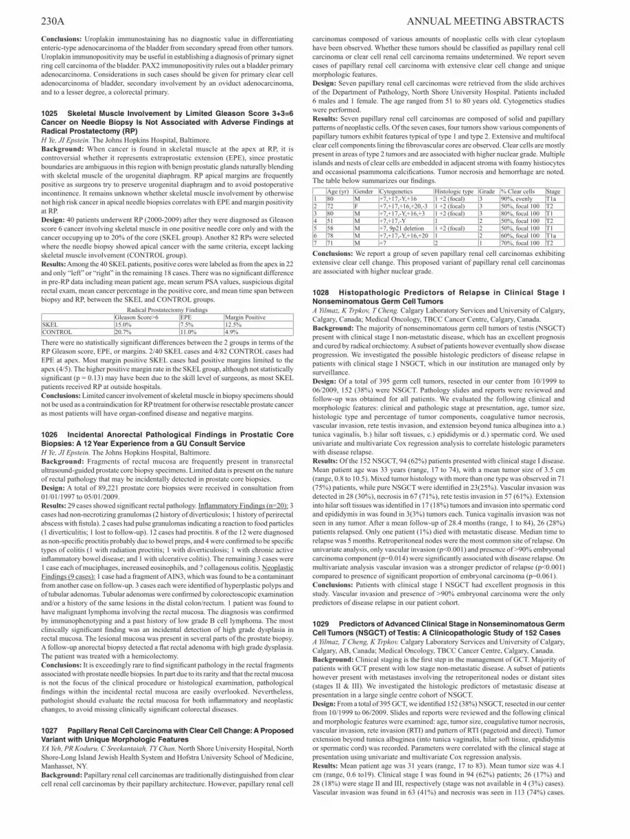
230A ANNUAL MEETING ABSTRACTS Conclusions: Uroplakin immunostaining has no diagnostic value in differentiating enteric-type adenocarcinoma of the bladder from secondary spread from other tumors. Uroplakin immunopositivity may be useful in establishing a diagnosis of primary signet ring cell carcinoma of the bladder. PAX2 immunopositivity rules out a bladder primary adenocarcinoma. Considerations in such cases should be given for primary clear cell adenocarcinoma of bladder, secondary involvement by an oviduct adenocarcinoma, and to a lesser degree, a colorectal primary.
1025 Skeletal Muscle Involvement by Limited Gleason Score 3+3=6 Cancer on Needle Biopsy Is Not Associated with Adverse Findings at Radical Prostatectomy (RP)H Ye, JI Epstein. The Johns Hopkins Hospital, Baltimore.Background: When cancer is found in skeletal muscle at the apex at RP, it is controversial whether it represents extraprostatic extension (EPE), since prostatic boundaries are ambiguous in this region with benign prostatic glands naturally blending with skeletal muscle of the urogenital diaphragm. RP apical margins are frequently positive as surgeons try to preserve urogenital diaphragm and to avoid postoperative incontinence. It remains unknown whether skeletal muscle involvement by otherwise not high risk cancer in apical needle biopsies correlates with EPE and margin positivity at RP.Design: 40 patients underwent RP (2000-2009) after they were diagnosed as Gleason score 6 cancer involving skeletal muscle in one positive needle core only and with the cancer occupying up to 20% of the core (SKEL group). Another 82 RPs were selected where the needle biopsy showed apical cancer with the same criteria, except lacking skeletal muscle involvement (CONTROL group).Results: Among the 40 SKEL patients, positive cores were labeled as from the apex in 22 and only “left” or “right” in the remaining 18 cases. There was no significant difference in pre-RP data including mean patient age, mean serum PSA values, suspicious digital rectal exam, mean cancer percentage in the positive core, and mean time span between biopsy and RP, between the SKEL and CONTROL groups.
Radical Prostatectomy FindingsGleason Score>6 EPE Margin Positive
SKEL 15.0% 7.5% 12.5%CONTROL 20.7% 11.0% 4.9%
There were no statistically significant differences between the 2 groups in terms of the RP Gleason score, EPE, or margins. 2/40 SKEL cases and 4/82 CONTROL cases had EPE at apex. Most margin positive SKEL cases had positive margins limited to the apex (4/5). The higher positive margin rate in the SKEL group, although not statistically significant (p = 0.13) may have been due to the skill level of surgeons, as most SKEL patients received RP at outside hospitals.Conclusions: Limited cancer involvement of skeletal muscle in biopsy specimens should not be used as a contraindication for RP treatment for otherwise resectable prostate cancer as most patients will have organ-confined disease and negative margins.
1026 Incidental Anorectal Pathological Findings in Prostatic Core Biopsies: A 12 Year Experience from a GU Consult ServiceH Ye, JI Epstein. The Johns Hopkins Hospital, Baltimore.Background: Fragments of rectal mucosa are frequently present in transrectal ultrasound-guided prostate core biopsy specimens. Limited data is present on the nature of rectal pathology that may be incidentally detected in prostate core biopsies.Design: A total of 89,221 prostate core biopsies were received in consultation from 01/01/1997 to 05/01/2009.Results: 29 cases showed significant rectal pathology. Inflammatory Findings (n=20): 3 cases had non-necrotizing granulomas (2 history of diverticulosis; 1 history of perirectal abscess with fistula). 2 cases had pulse granulomas indicating a reaction to food particles (1 diverticulitis; 1 lost to follow-up). 12 cases had proctitis. 8 of the 12 were diagnosed as non-specific proctitis probably due to bowel preps, and 4 were confirmed to be specific types of colitis (1 with radiation proctitis; 1 with diverticulosis; 1 with chronic active inflammatory bowel disease; and 1 with ulcerative colitis). The remaining 3 cases were 1 case each of muciphages, increased eosinophils, and ? collagenous colitis. Neoplastic Findings (9 cases): 1 case had a fragment of AIN3, which was found to be a contaminant from another case on follow-up. 3 cases each were identified of hyperplastic polyps and of tubular adenomas. Tubular adenomas were confirmed by colorectoscopic examination and/or a history of the same lesions in the distal colon/rectum. 1 patient was found to have malignant lymphoma involving the rectal mucosa. The diagnosis was confirmed by immunophenotyping and a past history of low grade B cell lymphoma. The most clinically significant finding was an incidental detection of high grade dysplasia in rectal mucosa. The lesional mucosa was present in several parts of the prostate biopsy. A follow-up anorectal biopsy detected a flat rectal adenoma with high grade dysplasia. The patient was treated with a hemicolectomy.Conclusions: It is exceedingly rare to find significant pathology in the rectal fragments associated with prostate needle biopsies. In part due to its rarity and that the rectal mucosa is not the focus of the clinical procedure or histological examination, pathological findings within the incidental rectal mucosa are easily overlooked. Nevertheless, pathologist should evaluate the rectal mucosa for both inflammatory and neoplastic changes, to avoid missing clinically significant colorectal diseases.
1027 Papillary Renal Cell Carcinoma with Clear Cell Change: A Proposed Variant with Unique Morphologic FeaturesYA Yeh, PR Koduru, C Sreekantaiah, TY Chan. North Shore University Hospital, North Shore-Long Island Jewish Health System and Hofstra University School of Medicine, Manhasset, NY.Background: Papillary renal cell carcinomas are traditionally distinguished from clear cell renal cell carcinomas by their papillary architecture. However, papillary renal cell
carcinomas composed of various amounts of neoplastic cells with clear cytoplasm have been observed. Whether these tumors should be classified as papillary renal cell carcinoma or clear cell renal cell carcinoma remains undetermined. We report seven cases of papillary renal cell carcinoma with extensive clear cell change and unique morphologic features.Design: Seven papillary renal cell carcinomas were retrieved from the slide archives of the Department of Pathology, North Shore University Hospital. Patients included 6 males and 1 female. The age ranged from 51 to 80 years old. Cytogenetics studies were performed.Results: Seven papillary renal cell carcinomas are composed of solid and papillary patterns of neoplastic cells. Of the seven cases, four tumors show various components of papillary tumors exhibit features typical of type 1 and type 2. Extensive and multifocal clear cell components lining the fibrovascular cores are observed. Clear cells are mostly present in areas of type 2 tumors and are associated with higher nuclear grade. Multiple islands and nests of clear cells are embedded in adjacent stroma with foamy histiocytes and occasional psammoma calcifications. Tumor necrosis and hemorrhage are noted. The table below summarizes our findings.
Age (yr) Gender Cytogenetics Histologic type Grade % Clear cells Stage1 80 M +7,+17,-Y,+16 1 +2 (focal) 3 90%, evenly T1a2 72 F +7,+17,+16,+20,-3 1 +2 (focal) 3 50%, focal 100 T23 80 M +7,+17,-Y,+16,+3 1 +2 (focal) 3 80%, focal 100 T14 51 M +7,+17,-Y 1 2 50%, focal 100 T25 58 M +7, 9p21 deletion 1 +2 (focal) 2 50%, focal 100 T16 78 M +7,+17,-Y,+16,+20 1 2 60%, focal 100 T1a7 71 M +7 2 1 70%, focal 100 T2
Conclusions: We report a group of seven papillary renal cell carcinomas exhibiting extensive clear cell change. This proposed variant of papillary renal cell carcinomas are associated with higher nuclear grade.
1028 Histopathologic Predictors of Relapse in Clinical Stage I Nonseminomatous Germ Cell TumorsA Yilmaz, K Trpkov, T Cheng. Calgary Laboratory Services and University of Calgary, Calgary, Canada; Medical Oncology, TBCC Cancer Centre, Calgary, Canada.Background: The majority of nonseminomatous germ cell tumors of testis (NSGCT) present with clinical stage I non-metastatic disease, which has an excellent prognosis and cured by radical orchiectomy. A subset of patients however eventually show disease progression. We investigated the possible histologic predictors of disease relapse in patients with clinical stage I NSGCT, which in our institution are managed only by surveillance.Design: Of a total of 395 germ cell tumors, resected in our center from 10/1999 to 06/2009, 152 (38%) were NSGCT. Pathology slides and reports were reviewed and follow-up was obtained for all patients. We evaluated the following clinical and morphologic features: clinical and pathologic stage at presentation, age, tumor size, histologic type and percentage of tumor components, coagulative tumor necrosis, vascular invasion, rete testis invasion, and extension beyond tunica albuginea into a.) tunica vaginalis, b.) hilar soft tissues, c.) epididymis or d.) spermatic cord. We used univariate and multivariate Cox regression analysis to correlate histologic parameters with disease relapse.Results: Of the 152 NSGCT, 94 (62%) patients presented with clinical stage I disease. Mean patient age was 33 years (range, 17 to 74), with a mean tumor size of 3.5 cm (range, 0.8 to 10.5). Mixed tumor histology with more than one type was observed in 71 (75%) patients, while pure NSGCT were identified in 23(25%). Vascular invasion was detected in 28 (30%), necrosis in 67 (71%), rete testis invasion in 57 (61%). Extension into hilar soft tissues was identified in 17 (18%) tumors and invasion into spermatic cord and epididymis in was found in 3(3%) tumors each. Tunica vaginalis invasion was not seen in any tumor. After a mean follow-up of 28.4 months (range, 1 to 84), 26 (28%) patients relapsed. Only one patient (1%) died with metastatic disease. Median time to relapse was 5 months. Retroperitoneal nodes were the most common site of relapse. On univariate analysis, only vascular invasion (p<0.001) and presence of >90% embryonal carcinoma component (p=0.014) were significantly associated with disease relapse. On multivariate analysis vascular invasion was a stronger predictor of relapse (p<0.001) compared to presence of significant proportion of embryonal carcinoma (p=0.061).Conclusions: Patients with clinical stage I NSGCT had excellent prognosis in this study. Vascular invasion and presence of >90% embryonal carcinoma were the only predictors of disease relapse in our patient cohort.
1029 Predictors of Advanced Clinical Stage in Nonseminomatous Germ Cell Tumors (NSGCT) of Testis: A Clinicopathologic Study of 152 CasesA Yilmaz, T Cheng, K Trpkov. Calgary Laboratory Services and University of Calgary, Calgary, AB, Canada; Medical Oncology, TBCC Cancer Centre, Calgary, Canada.Background: Clinical staging is the first step in the management of GCT. Majority of patients with GCT present with low stage non-metastatic disease. A subset of patients however present with metastases involving the retroperitoneal nodes or distant sites (stages II & III). We investigated the histologic predictors of metastasic disease at presentation in a large single centre cohort of NSGCT.Design: From a total of 395 GCT, we identified 152 (38%) NSGCT, resected in our center from 10/1999 to 06/2009. Slides and reports were reviewed and the following clinical and morphologic features were examined: age, tumor size, coagulative tumor necrosis, vascular invasion, rete invasion (RTI) and pattern of RTI (pagetoid and direct). Tumor extension beyond tunica albuginea (into tunica vaginalis, hilar soft tissue, epididymis or spermatic cord) was recorded. Parameters were correlated with the clinical stage at presentation using univariate and multivariate Cox regression analysis.Results: Mean patient age was 31 years (range, 17 to 83). Mean tumor size was 4.1 cm (range, 0.6 to19). Clinical stage I was found in 94 (62%) patients; 26 (17%) and 28 (18%) were stage II and III, respectively (stage was not available in 4 (3%) cases). Vascular invasion was found in 63 (41%) and necrosis was seen in 113 (74%) cases.

ANNUAL MEETING ABSTRACTS 231ARTI was seen in 97 (64%) tumors: in 72 (47%) cases invasion was direct pattern and in 25 (16%) it was pagetoid only. RTI was not found in 44 (29%) cases; in 11 (7%) rete could not be seen. Tumor invasion into hilar soft tissue was found in 41 (27%) cases. Epididymis and spermatic cord extension were identified in 12 (8%) tumors each. Tunica vaginalis invasion was not seen in any case. On univariate analysis, the following parameters had a significant association with the clinical stage at presentation: tumor size (p=0.010), vascular invasion (p<0.001), RTI with direct pattern (p<0.001), hilar soft tissue extension (p=0.001), epididymis invasion (p=0.009), spermatic cord invasion (p=0.009) and necrosis (p=0.050). On multivariate analysis, only vascular invasion (p=0.002) and the RTI with direct pattern showed significant association with advanced clinical stage.Conclusions: Although multiple parameters correlated with advanced clinical stage of NSGCT at presentation, on multivariate analysis, only vascular invasion and RTI with direct pattern were associated with metastases. Our findings suggest that the pattern of RTI in NSGCT should be routinely reported.
1030 Renal Cell Carcinoma with Rhabdoid Features: Clinicopathologic Features and Molecular ProfilesCW Yoo, JY Ro, K Alvarez, GM Quiroga-Garza, SS Shen, LD Truong, AG Ayala, FA Monzon. The Methodist Hospital, Houston, TX; Weill Cornell Medical College, New York, NY.Background: Tumor cells with rhabdoid morphology are often associated with high-grade renal cell carcinoma (RCC). It has been suggested that rhabdoid cells represent a subclonal proliferation of RCC with a dedifferentiated morphology. If this is true, rhabdoid cells should show unique genomic aberrations when compared to non-rhabdoid areas. In this study, we utilized virtual karyotyping to evaluate the genomic profiles of the rabdoid tumor cells in clear cell RCCs and compare them to those of the non-rhabdoid areas from the same tumors.Design: Nine cases of clear cell RCC with rhabdoid features were identified. DNA extraction from paraffin-embedded tissues was performed from rhabdoid and non-rhabdoid areas of the tumors. All cases were analyzed with virtual-karyotyping using the Affymetrix GeneChip 250K Nsp mapping arrays. Three cases yielded results for both rhabdoid and clear cell components, and 3 cases only gave results on the rhabdoid component and 3 cases failed analysis. Chromosome copy number data were reviewed and compared between rhabdoid and non-rhabdoid cells.Results: The average age of patients was 57.4 years (range, 38-75 years) and male to female ratio was 3:1. Rhabdoid cells were present in 3% to 60% of the entire tumor. Non-rhabdoid tumor cells were clear cell type. Average size of the tumor was 8.4 cm (range, 4.0-11.6 cm). All tumor showed necrosis, and had Fuhrman nuclear grade 3 or 4 and all were stages III or IV. Three patients had distant metastasis (bone, lung and heart) Virtual karyotyping data identified loss of 3p in all cases, consistent with clear cell RCC. All but one case showed chromosomal abnormalities commonly found in high-grade RCC tumors (such as loss of 9p and 14q), while one tumor showed 3p loss as the only abnormality. In all three cases in which a rhabdoid and a non-rhabdoid sample were analyzed, the chromosomal profiles were identical. No specific chromosomal abnormalities were seen that were common among tumors with rhabdoid morphology.Conclusions: Clinicopathologic characteristics of our eight cases are similar to those reported in the literature. Virtual karyotyping identified chromosomal profiles often found in high-grade RCC tumors. However, we did not identify any specific chromosomal changes associated with rhabdoid tumor cells. These results suggest that rhabdoid morphology does not originate from a genomic event involving large chromosomal gains or losses.
1031 The miR-17-92 Cluster Is Overexpressed and Has an Oncogenic Effect on Renal Cell CarcinomaY Youssef, NM White, M Mankaruos, TF Chow, A Girgis, S Metias, J Honey, R Stewart, KT Pace, S Mossad, MY Gabril, GM Yousef. St. Michael’s Hospital, Toronto, ON, Canada; London Health Sciences Centre, London, ON, Canada; St. Michael’s Hospital, Toronto, Canada.Background: MicroRNAs (miRNAs) are small non-protein coding RNAs that are differentially expressed in many malignancies. We have previously identified 80 miRNAs that are dysregulated in ccRCC. The purpose of this study was to validate the overexpression of the miR-17-92 cluster in clear cell renal cell carcinoma (ccRCC), and test the effect of two members of this cluster (miR-17-5p and miR-20a) on tumor proliferation. In addition, we also aim to elucidate the role of miRNAs in ccRCC pathogenesis through bioinformatics analysis.Design: miRNA expression was validated by qRT-PCR. Cell proliferation effects of miR-17-5p and miR-20a were tested in the ACHN renal adenocarcinoma cell line model. Cells were transfected with the miR precursor molecules, miR inhibitors, and appropriate controls. Also, indepth in-silico analyses were performed on the dysregulated miRNAs.Results: Transfection of miR-20a inhibitor significantly reduced cell proliferation in a dose dependent manner. Transfection of miR-17-5p, which is not endogenously expressed in the ACHN cell line, lead to increased cell proliferation compared to control, and this effect was suppressed by miR-17-5p inhibitor. Bioinformatics analyses identified ten clusters of miRNAs dysregulated in ccRCC that followed the same expression patterns. We also identified matching patterns between reported chromosomal aberration in ccRCC and miRNA dysregulation for 37.5% of the miRNAs. Target prediction analyses identified many key molecules in ccRCC pathogenesis including HIFs, mTOR, VEGF and VHL were found to be potential targets for dysregulated miRNAs. A significant number of dysregulated proteins in ccRCC are potential miRNA targets. Also, many of the ccRCC-dysregulated miRNAs were found to be phylogenetically conserved.Conclusions: Our data strongly supports that the members of the miR-17-92 cluster, which is found to be overexpressed in ccRCC, have an oncogenic effect on kidney
cancer cell growth and they may do so through mediating the expression of known key molecules in ccRCC pathogenesis.
1032 The Expression of GPR 30, a G Protein-Coupled Receptor, in Prostate CancerM Zhang, HM Lam, MQ Yu, JH Wang, B Ouyang, S Jain, G Daniels, V Reuter, A Gopalan, I Osman, P Lee, SM Ho. New York Univeristy School of Medicine, New York, NY; University of Cincinnati, Cincinnati, OH; Memorial Sloan Kettering Cancer Center, New York, NY.Background: The role of estrogen and its receptors in prostate cancer has now been recognized. There are distinct expressions and functions of estrogen receptors ERα and ERβ in prostate cancer epithelial cells and in tumor associated stromal cells. In this study, we determined the expression of G protein-coupled receptor for E2, GPR30. Our group demonstrated that GPR30 was down-regulated in prostate cancer when compared to the normal tissue by RT-PCR, which is in concordance with the public microarray database Oncomine. Herein we examined the expression of GPR30 protein in prostate cancer in relation to clinicopathological features.Design: Immunohistochemistry was performed using antibody against GPR30 to characterize its expression pattern in prostate cancer to compare GPR30 levels in prostate cancer with adjacent benign prostate using tissue microarray (TMA) (n= 199), to correlate with various clinocopathological parameters. The levels of GRP30 expression, membranous, cytoplasmic or stromal stain, were scored semi-quantitatively; 0: negative, 1+: weak, 2+: moderate and 3+: strong expression.Results: We observed distinct patterns of GPR30 expression for membranous and cytoplasmic stain in cancer epithelial cells, as well as stromal cells. The membranous stain is decreased in cancer cells in 40 of 190 (21%) cancer cases compared to 29 of 69 (42%) cases in benign prostate. When correlated with clinicopathological parameters, the level of GPR30 expression in the membrane of tumor cells was significantly lower in prostate cancer with advanced tumor grade and stages (p<0.05). Strikingly, the stromal GPR30 expression is significantly decreased in cancer from 68 of 69 benign cases (98.5%) to 19 of 190 (10%) of cancer cases with an average 19% cells in benign stromal cells and 4% in cancer stromal cells (p<0.05). These results correlated with the growth inhibitory effect of GPR30 on prostate cancer cells.Conclusions: This study demonstrates significant down-regulation of GPR30 expression in the membrane of cancer epithelial cells and stroma of prostate cancer. These results strongly suggest a role of GPR30 in high grade and advanced stage prostate cancer.
1033 Overexpression of Phospholipase D1 in Prostatic Adenocarcinoma: Pathogenetic and Therapeutic ImplicationsB Zhao, RE Brown. University of Texas - Medical School, Houston, TX.Background: Phospholipase D1 (PLD1) catalyzes the conversion of phosphatidylcholine to form phosphatidic acid (PA). Both PLD1 and PA are involved in the signaling by mammalian target of rapamycin complex1 (mTORC1) and in addition, PA, per se has been shown to bind to and phosphorylate p70S6K at threonine 389 . In light of this, and because we had previously demonstrated the expression of constitutively activated p-mTOR (Ser 2448) in the plasmalemmal and/or cytoplasmic compartments of prostatic adenocarcinoma (PAC) accompanied by the constitutive activation with nuclear translocation of p70S6K phosphorylated on threonine 389, we postulated that PLD1 may be a factor in such constitutive activation of mTOR and p70S6K analytes in PAC.Design: Tissue microarrays containing 37 cases of PAC and 24 cases on non-neoplastic prostatic tissue were included in the study. An immunohistochemical probe for PLD1 (PC-PLD1 [sc-28314], Santa Cruz Biotechnology, Inc., Santa Cruz, CA) was applied to representative sections of each using a standard diaminobenzidine chromogen detection system. An automated cellular imaging system (ACIS III, DAKO Corporation, Carpinteria, CA) was employed, and gating of representative areas was carried out by one of us (BZ).The threshold was set to assess the percentage of immunopositive pixels and scoring intensity of the gated areas. The total staining score of the cells with immunopositivity was generated from the product of the two measured parameters. A mean total score of PLD 1 expression with standard deviation (SD) was generated for each subset. Statistical analysis was carried out using the student t-test.Results: PLD1 total expression scores in the cytoplasmic/plasmalemmal and nucleolar compartments from each of the subsets were as follows: PAC (4572 +/-2109) versus non-neoplastic/BPH glandular epithelium (1052+/-879). Statistically, this revealed PLD1 expression to be significantly greater in PAC versus the non-neoplastic subset (p=1.55x10⁻¹⁰).Conclusions: PLD1 is overexpressed in prostatic adenocarcinoma. This coincides with our previous observation concerning the constitutive activation of p-mTOR (Ser 2448) and p-p70S6K (Thr 389) in PAC. Activation of this mitogenic pathway has pathogenetic implications for the progression of PAC. Therefore, agents that could interfere in PLD1-PA-mTORC1-PA-p70S6K signaling such as curcumin, triptolide and metformin offer preventive and therapeutic opportunities in retarding the progression of prostate cancer and warrant further investigation.
1034 Amplification of Ribosomal RNA Gene Loci in Human Prostate CancerQ Zheng, S Yegnasubramanian, AM De Marzo. Johns Hopkins, Baltimore, MD.Background: Nucleolar enlargement is a consistent structural change in many neoplastic cells, and this is especially common in prostatic adenocarcinoma (CaP) and prostatic intraepithelial neoplasia (PIN). Since the nucleolus is organzied by the rDNA, this raises the question of whether part of the nucleolar size expansion is cancer is related to amplification of the rDNA locus. The aim of this study was to quantify ribosomal gene copy number in clinical samples of prostate cancer.Design: Copy number of rDNA repeats was quantified by real-time PCR using separate primer sets designed to amplify regions coding for all 3 rDNA gene products
232A ANNUAL MEETING ABSTRACTS (28S, 18S, and 5.8S). To control for overall ploidy, 3 different single copy reference genes were used for normalization (HBB, PTGS2, and STK19). Copy numbers were determined using the using the relative standard curve method. Genomic DNA was isolated from frozen tissue sections of radical prostatectomy specimens from n = 21 patients (Gleason scores 6-9). Relative normalized target gene quantity in tumor DNA was determined from the standard curves and divided by the relative normalized target gene quantity of the calibrator, which consisted of DNA from each patient’s matched normal prostate tissue.Results: The median ratio of rDNA copies in tumor vs. normal was 1.45 for 5.8S, 1.43 for 18S, and 1.56 for 28S. The difference between these values and the expected tumor/normal ratio of 1.0 (null hypothesis) was significant for each target (5.8S p=0.0055, 18S p=0.0072, 28S p=0.0129; Wilcoxon Sign Rank test). Overall tumor/normal ratios were increased in 16 of the 21 cases. There was a high level of concordance in median relative copy number (tumor/normal) using different reference genes (e.g. HBB, PTGS2, RP1) with the same target (e.g. 18S rDNA), and using the different target genes in the locus (e.g. for a given patient if 5.8S rDNA was amplified then the 18S and 28S genes were also amplified).Conclusions: In the majority of cases of prostate cancer there was an increase in copy number of the entire rDNA locus (relative increase ∼1.5 fold). Since there are approximately 400 copies of this repeat unit in normal cells, these increases are on the order of hundreds of extra copies. These results raise the possibility that rDNA gene copy number may: i) influence nucleolar size expansion in cancer cells, and/or ii) play an important role in the development and/or progression prostate cancer.
1035 The Study of Xp11 Translocation Renal Cell Carcinoma (RCC) in Adults by TMA, IHC and FISHM Zhong, L Osborne, MJ Merino, M Hameed, S Aisner. University of Medicine and Dentistry of New Jersey-New Jersey Medical School, Newark; National Cancer Institute, Bethesda; Memorial Sloan-Kettering Cancer Center, New York.Background: Xp11 translocation renal cell carcinomas (RCC), a distinctive entity in the 2004 WHO renal tumor classification, are rare neoplasms and are often encountered in the pediatric and young adult population. The incidence is probably underestimated partly due to unavailable genetic studies and overlapping histopathological morphology with clear cell and papillary RCC. TFE3 immunohistochemical assay has been used for the diagnosis of the Xp11.2 translocation RCC. However, for a definitive diagnosis, one has to rely on identifying the translocation by genetic and molecular studies.Design: A total of 120 consecutive adult (>18Y) RCC patient specimens (FFPE during the period 2001-2008) from our institute, were collected to construct a tissue micro array(TMA). IHC was performed on TMA using TFE3 antibody. The 2nd TMA was constructed from all the TFE3 positive specimens, 5 TFE3 negative and 5 TFE3 weak positive specimens. A dual-color, break-apart FISH assay, which can detect the gene rearrangement at Xp11 region, was applied to the 2nd TMA.Results: Among the 120 RCC, 11(9.2%) cases were TFE3 positive . Among these 11 cases, FISH assay showed split signal, which confirmed chromosome translocations involving TFE3, in 5(4.2%) cases; fusion signal in 1 case; non-conclusive result in the rest 5 cases(most likely due to the ages of the blocks); no split signal in all tested TFE3 negative and weak positive cases. Further more, no TFE3 fusion transcripts (ASPL, PRCC, CLTC, PSF and Nono), but TFE3 wide type were detected by RT-PCR in the tested case with weak TFE3 nuclear stain.Conclusions: 1) Xp11.2 translocation RCC is not an uncommon neoplasm. The incidence in this group of 120 RCC patients is at least 4.2% in adults (confirmed by FISH). 2) TFE3 IHC is a relatively sensitive and specific assay for the diagnosis Xp11.2 translocation RCC. The strong nuclear TFE3 stain is most likely indicative of Xp11.2 translocation. The weaker nuclear stain appears to be due to expression of full length TFE3 protein, rather than chimeric fusion protein due to translocation. 3) FISH assay is the most reliable method to detect Xp11 translocation RCC, when only FFPE material is available.
Gynecologic & Obstetrics1036 Inter-Observer Agreement among Pathologists for Assessing Invasion in Early Vulvar Squamous Cell Carcinoma: Still a Diagnostic ChallengeA Abdel-Mesih, D Daya, K Onuma, N Akhtar-Danesh, O Boutross-Tadross, K Ceballos, W Chapman, T Colgan, P Deb, MR Nucci, E Oliva, M Sur, S Tang, A Lytwyn. McMaster University, Hamilton, ON, Canada; University of British Columbia, Vancouver, BC, Canada; University of Toronto, Toronto, ON, Canada; Harvard University, Boston, MA.Background: Despite published criteria, measuring depth of invasion to identify early squamous cell carcinoma of the vulva remains difficult, yet critical for surgical management. Tumours with ≤1mm depth of invasion have minimal risk of nodal involvement, and therefore surgery is limited to local excision without lymphadenectomy. Interobserver agreement for determining the presence of invasion and its measurement has not been studied. Our aim was to assess agreement among pathologists for (1) determining whether invasion is present, and (2) measuring the depth of invasion.Design: We searched the pathology database from 2000-2008, and identified 72 vulvar squamous cell carcinomas diagnosed as showing up to 5mm depth of invasion. From these, 45 slides were selected for this study, with preferential selection of challenging examples. Eleven gynecologic pathologists, from both Canadian and American institutions, independently reviewed the slides. Each slide was categorized as: (1) not invasive, (2) invasive with depth of invasion ≤1 mm, (3) invasive >1 mm, (4) invasive but depth cannot be measured, or (5) cannot determine if invasive or not invasive.Results: There was only fair agreement (mean κ =0.24) among pathologists for diagnosing a vulvar carcinoma as being invasive. Of the 45 cases, only 13 (29%) were
unanimously diagnosed by all 11 pathologists as being invasive. There was not a single case where all 11 pathologists agreed that there was no invasion. In 32 cases (71%), the pathologists did not unanimously agree on the presence or absence of invasion. Mean agreement for depth of invasion was only moderate (mean κ =0.50), and ranged from poor (κ= 0.12) to excellent (κ =0.92).Conclusions: There was only fair agreement among gynecologic pathologists in determining the presence of invasion in vulvar carcinoma. In cases where pathologists agreed there was invasion, agreement on depth was only moderate. The fair to moderate agreement in this study may, in part, be due to the preferential selection of a larger proportion of diagnostically challenging cases. This study highlights that assessing whether or not there is invasion, and ascertaining the critical ≤ 1mm depth, still poses a diagnostic challenge for pathologists.
1037 Endometrial Adenosarcomas: Diagnostic Use of Ki-67 Proliferation Marker as an Adjunct to Morphologic DiagnosisN Aggarwal, R Bhargava, E Elishaev. University of Pittsburgh School of Medicine, Pittsburgh, PA.Background: Endometrial adenosarcomas (AS) are rare biphasic neoplasms of the female genital tract containing benign glands and a low grade sarcomatous mesenchymal component. Diagnostic criteria include increased stromal cellularity in periglandular distribution (periglandular stromal cuffing) accompanied by variable degrees of cytologic atypia and mitotic activity. When not all the criteria are fulfilled a diagnosis of benign lesions are given and AS is considered in retrospect upon recurrence of the tumor.Design: Eight cases of AS were identified at Magee-Womens Hospital between 2004 and 2008 and were compared to 15 cases of endometrial polyps (EP) and 14 cases of atypical polypoid adenomyomas (APA). Pertinent 1-3 blocks/case were selected and stained for Ki-67 proliferation marker, caldesmon, smooth muscle actin (SMA), desmin, and CD10 with appropriate negative and positive controls.Results: All cases of AS had a polypoid growth pattern with round or slit-like glands with or without papillary projections into gland lumen. Increased periglandular stromal cellularity was observed focally or diffusely in all cases (8/8) of adenosarcoma, which also demonstrated variable degrees of stromal nuclear atypia. The mitotic activity ranged from 1/10 high power fields (HPF) to 15/10HPF. All cases of AS demonstrated distinct increase in Ki-67 positive nuclei in the peri-glandular zone compared to adjacent stroma, regardless of the mitotic count. The average Ki-67 labeling index in peri-glandular zones of AS was 20 % compared to less than 5 % in adjacent stroma. This zonation was not observed in any case of APA or EP all of which showed scattered positive cells with less than 5% Ki 67 proliferation index. The AS stroma was positive for CD10, with variable focal positive staining for SMA and desmin, and was negative for caldesmon. APA showed positivity for all muscle markers with SMA being the most consistent and diffuse. EPs were negative for caldesmon, variably positive for desmin and SMA and diffusely positive for CD10.Conclusions: Distinct increase in peri-glandular Ki-67 staining pattern is very helpful and Ki 67 should be considered as an adjunct to the routine morphologic diagnosis of AS. This could be especially useful in curettage specimens and other challenging lesions that lack some of the classic criteria of an AS.
1038 New Antibodies for Clear Cell Carcinoma (Pax-8, HNF-α, vHL) Are Also Positive in Arias-Stella ReactionCE Aguilar, E Silva. Cedars-Sinai Medical Center, Los Angeles, CA.Background: Recent studies have shown clear cell carcinoma of müllerian origin to be positive for Pax-8, hepatocyte nuclear factor- α (HNF-α), and von Hippel Lindau (vHL). Clear cell carcinoma of müllerian origin can present a diagnostic challenge in endometrial specimens showing areas of Arias-Stella. Therefore, Pax-8, HNF-α, vHL were applied to products of conception showing areas of Arias-Stella reaction.Design: Ten products of conception specimens showing Arias-Stella reaction and ten cases of clear cell carcinoma were stained with Pax-8, HNF-α, vHL.Results: In all ten cases, Arias-Stella cells and secretory glands displayed Pax-8 and HNF-α nuclear positivity and vHL cytoplasmic staining. All three immunostains were positive in each case of clear cell carcinoma with the expected pattern of staining.Conclusions: Clear cell carcinoma antibodies, Pax-8, HNF-α, and vHL, are positive in Arias-Stella reaction. These antibodies therefore cannot be applied to endometrial specimens in which the differential diagnosis includes clear cell carcinoma and Arias-Stella reaction. Future immunohistochemical studies to determine whether other molecular targets involved in the clear cell carcinoma pathway(s) can also be detected in Arias-Stella reaction would be useful in this unusual but challenging and important distinction.
1039 Prognostic Factors of Adenocarcinoma of the Endocervix: Pattern of Invasion vs Depth of InvasionI Aguilera-Barrantes, EG Silva. M.D.Anderson Cancer Center, Houston, TX.Background: The treatment of endocervical adenocarcinoma (EA) depends primarily on the depth of invasion. The grade of the tumor is typically considered a less important prognostic factor and the pattern of invasion has not been evaluated. The purpose of our study is to evaluate whether these features impact prognosis.Design: We reviewed 43 cases of invasive adenocarcinoma of the endocervix seen at our institution. For each case 1 to 49 slides (median 9) of cone/ LEEP and/or hysterectomy specimens were available for review. Follow up (FU) was obtained in all cases. These cases were divided in two groups. One group with pushing border (PB) of invasion or PB with focal infiltration (defined as 1 or 2 foci of infiltration) and the other group with diffuse infiltration (DI) of the stroma. Tumors were graded observing only architectural features and were classified as well differentiated (WD) if composed only of glands, moderated differentiated (MD) composed of glands and solid tumor and poorly differentiated (PD) predominantly solid.


