Embed Size (px)
Citation preview

Gene expression analysis of osteoblastic cells contacted byorthopedic implant particles
Dominique P. Pioletti,1,2,3* Lorenzo Leoni,4* Davide Genini,4 Hiroshi Takei,5 Pinyi Du,6 Jacques Corbeil4,6,7
1Department of Bioengineering, University of California San Diego, 9500 Gilman Drive, La Jolla, California 920932Bone Biophysics Group, Hopital Orthopedique de la Suisse Romande, Av. Pierre Decker 4, 1005 Lausanne, Switzerland3Institute for Biomedical Engineering, Swiss Federal Institute of Technology, SGAAB, 1015 Lausanne, Switzerland4Department of Medicine and the Stein Institute for Research on Aging, University of California San Diego, 9500Gilman Drive, La Jolla, California 920935Department of Orthopedics, University of California San Diego, 9500 Gilman Drive, La Jolla, California 920936Center for AIDS Research, University of California San Diego, 9500 Gilman Drive, La Jolla, California 920937Veterans Medical Research Foundation, University of California San Diego, 9500 Gilman Drive, La Jolla, California92093
Received 27 July 2001; revised 10 December 2001; accepted 10 January 2002
Abstract: Particles generated from orthopedic implantsthrough years of wear play an essential role in the asepticloosening of a prosthesis. We have investigated the biocom-patibility of these orthopedic particles on different osteo-blast-like cells representative of different stages of osteoblastmaturation. We found the particles induced a caspase-dependent apoptosis of osteoblasts, with less mature osteo-blasts being the most susceptible. An analysis of gene ex-pression was performed on the less mature osteoblasts,which were in contact with the particles. We found that theparticles had a profound impact on genes that code for in-flammatory cytokines and genes involved in controlling the
nuclear architecture. Results from this study suggest that theperi-implant osteolysis after a total joint replacement can bedue in part to a decrease of bone formation and not solely toan overstimulation of bone resorption as is generally pro-posed. Development of new drugs that promote normalbone formation and osteoblast survival would possibly con-trol peri-implant osteolysis, resulting in a better prognosisfor patients with orthopedic implants. © 2002 Wiley Periodi-cals, Inc. J Biomed Mater Res 61: 408–420, 2002
Key words: genomic study; osteoblast; particle; apoptosis;cytokine
INTRODUCTION
The peri-implant osteolysis after a total joint arthro-plasty (TJA) is responsible for the majority of ortho-pedic implant loosening.1 Failure rates of hip arthro-plasty can exceed 30% after 15 years for patients <50
years old,2 thus, requiring a revision surgery wherethe old implant is replaced by a new one. Joint disor-ders treated with TJA can reasonably be expected togive satisfactory results for 20 to 30 years with aninitial and revision surgery, but a subsequent proce-dure cannot be performed in a routine manner. Thecurrent trend is to propose TJA to younger patientswith the caveat that a second surgery is likely to berequired.
The response of cells to particles has been identifiedas a major cause of implant loosening.3 These particlesare generated through years of wear. Any of the ma-terials (metallic, polyethylene, methacrylate, ceramic)used for the implants or even the calcium phosphatecement4 can be involved. In cases of loose Ti implants,the mean concentration of Ti particles retrieved fromtissue surrounding the implants can reach 0.1% of thedry tissue weight.5
Normal bone function is assured when there is anequilibrium between bone formation and bone resorp-tion. Bone resorption has been mostly studied in rela-
*These authors have contributed equally.Correspondence to: D.P. Pioletti; e-mail: dominique.
[email protected] grant sponsor: Swiss Biology and Medicine
FoundationContract grant sponsor: Leenaards Foundation; contract
grant number: 309Contract grant sponsor: National Institutes of Health; con-
tract grant number: GM23200Contract grant sponsor: Veterans Medical Research Foun-
dationContract grant sponsor: UCSD Center for AIDS Research
Genomic Core Laboratory; contract grant number:AI36214
© 2002 Wiley Periodicals, Inc.

tion with wear particles. Macrophages,6 monocytes,7
or giant cells8 in contact with particles can producepotent osteolytic factors. Effects of implant particleson bone formation have been neglected and need to beexplored with a particular emphasis on biocompatibil-ity testing.
Particles from orthopedic implants have beenshown to influence the expression of some extracellu-lar matrix proteins in osteoblasts.9 It has been pro-posed that biocompatibility testing should include ap-optosis studies.10 Apoptosis, or programmed celldeath, is characterized by the activation of cysteineproteases called caspases, which cleave proteins es-sential for the survival of the cell. A member of thisfamily, caspase-3, has been identified as a key media-tor of apoptosis of mammalian cells.11 Furthermore,these caspases activate the endonuclease responsiblefor the internucleosomal cleavage of genomic DNA.12
The TUNEL assay (TdT-mediated dUTP-biotin nickend labeling) can detect the amount of internucleoso-mal DNA fragments and allow for apoptosis quanti-fication. Apoptosis quantification is important as it isthe normal fate for the majority of osteoblasts.13 More-over, an in vitro study showed that Ti particles caninduce apoptosis in osteoblasts.14 The analysis of thisapoptotic pathway and the stimuli that can trigger itwarrants investigation.
The recent advances in molecular biology tech-niques allow for a more comprehensive study of thebiological alterations induced by wear particles inphysiologically relevant cells such as osteoblasts.15 Inthis study, we quantified the apoptotic response totitanium (Ti) and polymethylmethacrylate (PMMA)particles in three osteoblast-like cells (human MG-63,rat osteoblast, and human SaOS-2). These are repre-sentative of the different maturation stages of the os-teoblasts. The choice of the particles type was based onthe fact that Ti is a widely used metal for orthopedicimplants and that it generates a large amount of par-
ticles5 that directly interact with osteoblasts duringdifferent stages of their differentiation. PMMA, thebone cement used to seal orthopedic implants, alsogenerates particles.16 A gene expression analysis wasperformed for the MG-63 osteoblast-like cells while incontact with these particles.
Our results demonstrate that implant particles in-duced and promoted apoptosis, especially in less ma-ture osteoblasts, and that caspase-3 was involved inthis process. The particles had a strong gene expres-sion effect in osteoblasts with an overall trend favor-ing bone resorption. These results clearly suggest thatwear particles from orthopedic implants are involvedin the osteolysis not only by favoring bone resorption,as it is generally accepted, but also by inhibiting boneformation and interfering with osteoblast prolifera-tion. Future drugs development aimed to control theperi-implant osteolysis should enhance the function-ing and survival of osteoblasts when particles arepresent.
MATERIALS AND METHODS
Particles
Commercially pure titanium particles of −325-mesh nomi-nal diameter were purchased (Aldrich, Milwaukee, WI). ThePMMA particles were a gift from Zimmer (Worthington,OH). Particle size distributions were performed by laser dif-fraction using a Malvern MasterSizer equipment. The valuesobtained for the different powders are given in Figure 1. Theparticles, sterilized by overnight UV irradiation, were mixedwith the culture medium under sterile conditions at a con-centration of 0.1% (w/w). This concentration can be consid-ered as representative of the particle concentration found inthe surrounding tissue of loose implant in biopsy study.5
The particle solutions were sonicated for 20 min in a sealedsterile container to minimize their agglomeration before be-ing added to the cell culture. Endotoxin contamination ofparticles was negligible as tested by limulus assay (QCL-
Figure 1. Frequency plot showing the size distribution of the Ti and PMMA particles used. The plain line represents thecumulative particle percentage. The size distribution was measured by laser diffraction (Malvern). For the Ti particles, 10%of the particles had a diameter smaller than 10.7 �m, 50% smaller than 30.2 �m, and 90% smaller than 176.3 �m. For thePMMA particles, 10% of the particles had a diameter smaller than 8.9 �m, 50% smaller than 56.9 �m, and 90% smaller than105.6 �m.
409GENE EXPRESSION OF OSTEOBLASTS CONTACTED BY PARTICLES

1000 Chromogenic LAL; BioWhittaker, Emerainville,France).
Cell cultures
The gene expression of the osteoblast has been shown todepend on its maturation stage.17 The osteoblast maturationcan be evaluated through the production of alkaline phos-phatase.18 Three types of osteoblast-like cell lines were usedspanning three different stages of osteoblast maturation. Thehuman MG-63 osteoblast-like cell (American Type CultureCollection, Rockville, MD) can be considered as the leastmature osteoblast used in this study (alkaline phosphataseactivity: 2.7 �U/�g protein).19 The rat osteoblast, isolated inour laboratory from neonatal Sprague-Dawley calvaria ratfollowing the procedure of Puelo,18 is an intermediate ma-ture osteoblast (alkaline phosphatase activity: 60 �U/�gprotein; measurement performed in our laboratory using theDiagnostic kit 245 of Sigma, St. Louis, MO). Finally, the hu-man SaOS-2 osteoblast-like cell (American Type CultureCollection) is the most mature osteoblast (alkaline phospha-tase activity: 5264.4 �U/�g protein).19 The osteoblasts wereseeded at a concentration of 50,000 cells/cm2 in 50-mm petridishes and incubated for 4 h to allow cell adhesion. Thesupernatant was removed, and 5 mL of particles solutionand normal medium was added, defining the time 0. Cellsamples were collected at 4 and 24 h for the gene expressionanalysis, fibronetin (protein level), osteocalcin (protein level)as well as alkaline phosphatase activity and at 24, 48, and 72h for the apoptosis, Bcl2/Bax ratio, and caspase activityquantification.
Confocal imaging (MG-63 osteoblast-like cells)
Cells were cultured 24 h with 0.1% Ti particles on cover-slips. The cells were fixed 10 min in 4% paraformaldehydeand washed in PBS. To visualize actin filaments, cells wereincubated for 1 h at 37°C with Alexa-488-conjugated phal-loidin (Molecular Probes, Eugene, OR); tubulin was visual-ized using anti-B-tubulin monoclonal antibody, followed byincubation with an Alexa anti-mouse secondary antibody(Molecular Probes) as previously described.20 Coverslipswere washed successively in PBS and deionized H2O for 5min and mounted in Flouromount (Fisher, Santa Clara, CA).Images were acquired using a confocal laser scanning mi-croscope LSM 510 equipped with an argon laser module(Carl Zeiss Inc., Thornwood, NY) using a 40× 1.3-na oil ob-jective.
Measurement of DNA fragmentation as apoptosisquantification (MG-63 osteoblast-like cells, ratosteoblast, SaOS-2 osteoblast-like cells)
The internucleosomal cleavage of genomic DNA has beendemonstrated to be a hallmark of apoptosis.21 This was used
as quantification of osteoblast apoptosis. Before the analysis,cells were fixed in ice-cold 30% ethanol and incubated with100 �g/mL of RNAse A and 50 �g/mL propidium iodidefor 1 h at 37°C. FSC/SSC gateway was used to discriminatebetween particles and cells. Hypodiploid cells were visual-ized using a Becton-Dickinson FACScalibur and the pro-gram ModFit LT 2.0 (Verity Software House, Topsham).DNA fragmentation was assessed by flow cytometry.
Cellular assay for caspase activity (MG-63osteoblast-like cells, rat osteoblast, SaOS-2osteoblast-like cells)
At the indicated time points, cells were washed twice withPBS, and the pellet was resuspended in caspase buffer (50mM Hepes, pH 7.4, 100 mM NaCl, 1 mM EDTA, 0.1% Chaps,and 5 mM dithiothreitol) for 30 min at 4°C. Lysates werethen stored at −80°C. The caspases enzymatic assays wereperformed in 96-well plates. Lysates (10–20 �g of total pro-tein) were mixed with 50 �L of caspase buffer, and reactionswere initiated by the addition of 100 �M of the specificsubstrate. After a 1-h incubation at 37°C, caspase-3-like pro-tease activity was measured with the substrate Ac-DEVD-AFC. Activity was measured by the release of 7-amino-4-trifluoromethyl-coumarin (AFC) monitoring fluorescence atexcitation and emission wavelengths of 400 and 505 nm,respectively.
Bcl2/Bax ratio (MG-63 osteoblast-like cells)
At the indicated time points, cells were washed twice withPBS and lysed. Proteins were resolved by SDS-PAGE on a4–20% Tris-Gly gel and transferred to a PVDF membrane.The membrane was probed with specific antibodies againstBcl-XL (mouse monoclonal clone 4; Transduction Lab), Bcl-2(mouse monoclonal clone 7, Transduction Lab), Bax (rabbitpolyclonal; gift from J. Reed of The Burnham Research In-stitute, San Diego, CA), and actin (mouse monoclonal,Sigma). The bands were resolved with specific HRP-conjugated secondary antibodies followed by enhanced che-miluminescence (ECL; Amersham). The intensity of eachband was quantified by densitometry and compared withthe actin band. The intensity of each Bcl-2 family memberwas then normalized to the control treated group (indicatedas 1) at each time point.
Analysis of gene expression (MG-63osteoblast-like cells)
We used Genefilters (GF211; Research Genetics, Hunts-ville, AL) and monitored the expression of approximately4000 genes. The samples (5 �g of total RNA per condition)were processed according to the manufacturer’s recommen-dations. A filter was used twice with the same sample inorder to verify the consistency of the results. There was good
410 PIOLETTI ET AL.

concordance between the two measurements. The data wereanalyzed using the pathway software developed by Re-search Genetics. Eighty genes were selected by fold modu-lation over the control and were classified into eight groups(extracellular matrix, cytokines, receptors, enzymes, nucleararchitecture regulation, cell adhesion, apoptosis, and oth-ers).17,22,23 Three analyses were performed at 4 and 24 h:control versus Ti, control versus PMMA, and Ti versusPMMA. The difference in gene expression was based on ourexperimental finding that genes had to be modulated at least1.5- and 2.5-fold for the 4- and 24-h experiments, respec-tively, to be significant in the assay used.
We tested the reliability of the Genefilters’ results by se-lecting genes identified as being modulated significantly foranalysis and validation by quantitative real time RT-PCR(TaqMan ABI Prism 7700; Applied Biosystem, Foster City,CA). The GAPDH housekeeping gene was used to controlfor input, thereby, allowing a comparison between samples.
Protein levels of fibronectin and osteocalcin andalkaline phosphatase (ALP) activity (MG-63osteoblast-like cells)
After a 4-h incubation, the supernatant was removed, andthe cells were washed three times with PBS. One milliliter ofparticles solution (serum free) and medium (serum free) wasadded defining the time 0. Supernatant and cell lysate (ob-tained with 1% Triton-X) were collected at 4 and 24 h. Thelysate was sonicated for 30 s on ice. Before measuring, su-pernatant and lysate were centrifuged at 800g for 2 min.
A volume of 100 �L (respectively, 25 �L) of supernatant ofthe different samples was used to determine the level offibronectin (respectively, osteocalcin) with a commercial en-zyme-linked immunoassay (Biomedical Technologies Inc.,Stoughton, MA). In parallel, a volume of 2 �L of lysatesample was added to p-nitrophenyl phosphate solution(Sigma) within a 96-well plate at 37°C for 3 min. p-Nitrophenol is produced in the presence of ALP, and theabsorbance can be measured with a plate reader at 405 nm.The change in rate of absorbance is directly proportional tothe activity of ALP. Data for fibronectin and osteonectin andALP activity were normalized by the total cell protein (DCprotein Assay Kit; Bio-Rad, Hercules, CA). Experimentswere performed in triplicate, and measurements were madein duplicate.
RESULTS
Confocal imaging (MG-63 osteoblast-like cells)
The views of the MG-63 osteoblasts at different z-levels clearly showed that the cytoplasm contained theTi particles, representing phagocytosis. A large num-ber of particles were also found adjacent to thenucleus (Fig. 2). Rhodamin-phalloidin stainingshowed that the organization of the actin filaments
was profoundly altered when particles were present(data not shown).
Apoptosis and caspase activity (MG-63osteoblast-like cells, rat osteoblast, SaOS-2osteoblast-like cells)
The particles induced apoptosis in osteoblast cells asdemonstrated by the appearance of hypodiploidDNA-containing cells, which were measured by flowcytometry in permeabilized cells stained with prop-idium iodide (Fig. 3). The fluorometric measurementof caspase-3-like activity using the synthetic substrateAc-DEVD-AFC confirmed the DNA content results(Fig. 4). The Ti particles had the greatest effect uponless mature MG-63 and mature SaOS-2 osteoblast-likecells, whereas the rat osteoblasts appeared more resis-tant. The PMMA particles were less potent to induceapoptosis than the Ti particles. Control latex particlesdid not induce apoptosis or caspase activity despitebeing phagocytosed (results not shown).
Bcl2/Bax ratio (MG-63 osteoblast-like cells)
Different biological responses were observed be-tween materials when expression of Bcl-2 familymembers was monitored in particle-treated MG-63cells (Fig. 5). Ti particles induced a slight downregu-lation of the Bcl-XL levels after a 24-h incubation. Con-versely, PMMA induced a strong induction of the Bcl-XL levels that was prolonged up to 48 h postincuba-tion, thus favoring survival. The Bcl-2 level alsochanged when cells were incubated with Ti andPMMA particles but its relative level of expression,when compared with Bcl-XL, was much lower. Fi-nally, Bax levels, although somewhat increased inPMMA-treated cells at 24 h concomitantly to the Bcl-XL and Bcl-2 increase, did not change dramaticallyduring the incubation period. It is conceivable that theupregulation of the two anti-apoptotic proteins Bcl-XLand Bcl-2 observed at 24 and 48 h in PMMA-treatedcells may be related to the delay in activation ofcaspases.
Gene expression (MG-63 osteoblast-like cells)
Modulation of gene expression after a 4-hco-incubation with Ti particles
After a 4-h co-incubation of Ti particles with MG-63osteoblast-like cells, 853 genes of the 3964 queried had
411GENE EXPRESSION OF OSTEOBLASTS CONTACTED BY PARTICLES

their expression modulated significantly. Of these, 32were of the 80 initially selected genes. Twenty-onegenes were upregulated, and 11 were downregulatedin the Ti-treated group compared with the controlgroup. Little effect on the gene expression of extracel-lular matrix proteins (type I collagen, osteonectin, os-teopontin, and alkaline phosphatase) was observed,except for an upregulation of the fibronectin and os-teocalcin genes (Table I). The Ti particles had a strongimpact on cytokine gene expression with an upregu-lation of the bone resorption inducers, interleukin 1�and 1�,23 and TGF-� (stimulates osteoblasts growthand differentiation24), and a downregulation of fibro-blast growth factor (promoter of osteoblast survival25)gene (Table II). Gene expression for receptors of TNF
(bone resorption inducer23 and proapoptotic agent25),interleukin 1 antagonist, insulin-like growth factor(stimulates osteoblast proliferation and differentia-tion26) and tyrosine-protein kinase (stimulate cellularproliferation27) were downregulated, while TNF andlaminin receptor genes expression were upregulated(Table III). A downregulation of the tissue inhibitor ofmetalloproteinase (TIMP-1) gene expression was de-tected (Table IV). This enzyme neutralizes the effect ofMMP-1, which can degrade type I collagen.28 Inconglomerate, the profile of genes modulated inMG-63 osteoblasts in contact with Ti particles appearsto favor bone resorption. The Ti particles also had aprofound impact on the gene expression of proteinsthat regulate the nuclear architecture (Table V).
Figure 2. MG-63 osteoblast-like cells were cultured 24 h with 0.1% Ti particles, cells were then fixed, and the cytoskeletalalterations were visualized by staining with an antitubulin antibody (red, microtubules) and FITC-labeled phalloidin (green,actin microfilaments). (a) Confocal fluorescence image showed the distribution of the actin microfilaments of a MG-63 cell.Three dark spots at the center of the cell clearly show the lack of actin filaments in correspondence to the “ingested” Tiparticles. (b) Confocal fluorescence image shows the distribution of the microtubules of an MG-63 cell. The three bright redspots at the center of the cells represent the Ti particles. (c) Nomarsky phase contrast image of the same cell demonstrates thepresence of the Ti particles ingested by the cell, indicated by the white arrows. (d) A combined image of the panels a, b, andc illustrates the colocalization of the beads with the cytoskeletal alterations.
412 PIOLETTI ET AL.
An upregulation of the Pou and NuMA genes and adownregulation of the HMG and SATB1 genes wereobserved. These genes belong to the nuclear matrixgene family.22 A downregulation was induced in theactin-depolymerizing gene expression as well. The Tiparticles also modulated the expression of genes thatcode for cell adhesion proteins with a downregulationof the integrin �1 (�2�1 is the major receptor for type Icollagen29) and an upregulation of laminin �1 (TableVI). The apoptosis-related genes p53-binding proteingene was upregulated, the anti- apoptotic Bcl-2 wasdownregulated, possibly rendering the cells moreprone to undergo apoptotic cell death (Table VII). TheTi particles had no effect on proliferation genes (c-fos,c-myc, c-jun) (Table VIII).
Modulation of gene expression after 24 hco-incubation with Ti particles
After a 24-h incubation of MG-63 osteoblast-likecells with Ti particles, a less pronounced effect wasobserved on gene modulation than at the 4-h timepoint. Twenty-six genes of the 80 initially selectedgenes had their expression modulated. Three geneswere upregulated, and 23 were downregulated in theTi group compared with the control group. For theextracellular matrix protein group, no significant ef-fect was observed except a downregulation of type XIcollagen (Table I). The largest difference in gene ex-pression, as compared with the 4-h results, werefound in the cytokines group with a downregulationof the macrophage-stimulating 1 and PDGF genes. Astrong downregulation of TGF-� inducible early pro-tein, and PDGF were also measured (Table II). Somechanges between the 4- and 24-h incubation werefound in the receptor group with a downregulation inthe gene expression of the receptors for interleukin 10,colony-stimulating factor 3, TGF-�, BMP type II, ste-roid hormone, epidermal growth factor, and tyrosine-protein kinase (Table III). MMP-7 gene expression wasalso downregulated (Table IV) as well as the Pou and
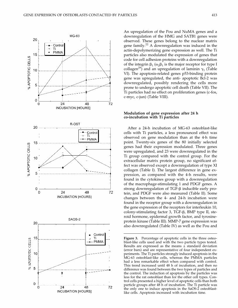
Figure 3. Percentage of apoptotic cells in the three osteo-blast-like cells used and with the two particle types tested.Results are expressed as the means ± standard deviation(error bars) and are representative of four independent ex-periments. The Ti particles strongly induced apoptosis in theMG-63 osteoblast-like cells, whereas the PMMA particleshad a less remarkable effect when compared with control.This trend increased until 48 h of incubation, and then nodifference was found between the two types of particles andthe control. The induction of apoptosis by the particles wasless for the rat osteoblast than for the other cell types. Con-trol cells presented a higher level of apoptotic cells than bothparticle groups after 48 h of incubation. The Ti particle wasthe only one to induce apoptosis in the SaOS-2 osteoblast-like cells. Apoptosis increased with incubation time.
413GENE EXPRESSION OF OSTEOBLASTS CONTACTED BY PARTICLES

YY1 genes (Table V). No differences with control wereobserved for the expression of genes in the adhesiongroup except for a downregulation of integrin �M(Table VI). For the apoptotic genes at 24 h, only BCL-2was downregulated (Table VII). The proliferation genec-myc was downregulated as well as two heat shockproteins (1 and 2), fast kinase, and serine/threonineprotein kinase SAK genes (Table VIII).
Comparison of gene modulation induced by Ti andPPMA particles at both 4 and 24 h
After a 4-h incubation of MG-63 osteoblast-like cellswith PMMA and Ti particles, no modulation differ-ence was observed except for the extracellular matrixgroup. A downregulation was observed for TGF-�,TGF-� receptor, steroid hormone receptor, tyrosine-protein kinase receptor, YY1, BCL2, heat shock protein
Figure 5. Particle-induced modulation of Bcl-2 familymembers expression for the MG-63 osteoblast-like cells.
Figure 4. Caspase-3-like activity for the three osteoblastcell lines used and with the two particle types tested. Resultsare expressed as the means ± standard deviation (error bars)and are representative of four experiments. The caspase ac-tivity of the MG-63 osteoblast-like cells in contact withPMMA particles increased over time reaching a peak at 48 hand then returned to its initial level. Unlike the PMMA par-ticles, the Ti particles increased the caspase activity of osteo-blasts up to the last timepoint of 72 h. The caspase activity ofthe control group remained stable for the duration of theexperiment. The rat osteoblasts had a similar caspase activ-ity for the two particles types with a constant increase overtime. The control group followed the same pattern with alower value of caspase activity. The caspase activity ofSaOS-2 cells peaked after 24 h and then decreased and re-mained stable until 72 h. PMMA particles decreased thecaspase activity of SaOS-2 cells when compared with con-trol. However, after 72 h of incubation, no difference werefound between the caspase activity of the SaOS-2 in contactwith the two particle types and the control group.
414 PIOLETTI ET AL.

TABLE I
Gene NameC-Ti4 h
C-PMMA4 h
Ti-PMMA4 h
C-Ti24 h
C-PMMA24 h
Ti-PMMA24 h
Collagen, type I, alpha-2 +1.14 +1.10 −1.03 −1.91 −2.35 −1.25Human pro-a2 chain of collagen type XI (COL11A2)
gene, complete cds −1.21 −1.37 −1.11 +3.06 +3.30 +1.07Fibronectin 1 −1.58 −1.45 +1.04 +1.04 −1.09 −1.13SPARC/osteonectin +1.29 −1.03 −1.32 −2.27 −2.47 −1.08Secreted phosphoprotein 1 (osteopontin, bone
sialoprotein I) −1.35 −1.52 −1.12 −1.51 −1.38 +1.09Biliary glycoprotein (BGP) (ostoecalcin) −1.52 −1.16 +1.3 +1.32 +2.39 +1.81Alkaline phosphatase, liver/bond/kidney −1.19 +1.15 +1.37 +1.06 +1.17 +1.09
TABLE II
Gene NameC-Ti4 h
C-PMMA4 h
Ti-PMMA4 h
C-Ti24 h
C-PMMA24 h
Ti-PMMA24 h
Homo sapiens TNF-related ligand TRANCEmRNA, partial cds −1.17 −1.2 −1.03 +1.61 +2.66 +1.65
Bone morphogenetic protein 2 (BMP2) +1.31 (+1.61) +1.82 (+1.40) +1.39 +1.60 +2.09 +1.30Transforming growth factor beta (BMP6) −1.30 −1.32 −1.01 −1.70 −2.02 −1.10Transforming growth factor, beta 3 −1.97 −1.42 +1.38 +1.40 +1.70 +1.21Human TGF-beta inducible early protein
(TIEG) mRNA, complete cds −1.86 (+1.11) −1.43 (−1.21) +1.30 +4.90 (+1.04) +5.28 (−2.95) +1.07Macrophage stimulating 1 (hepatocyte growth
factor-like) −1.57 −1.91 −2.13 +3.0 +4.98 −1.22Human PDGF associated protein mRNA,
complete cds −1.29 +1.04 +1.36 +4.02 +2.76 −1.45Platelet-derived growth factor PDGF-A −1.75 −1.43 +1.22 +1.74 +1.44 −1.20Interleukin 1 � −1.59 −1.47 +1.08 −1.98 −1.45 +1.37Interleukin 1 � −1.56 −1.46 +1.07 −1.69 −1.32 +1.27Interleukin-1 � convertase precursor −1.30 −1.28 +1.01 +1.29 +1.34 +1.04Interleukin 6 (B cell stimulatory factor 2) −1.12 −1.04 +1.11 −1.24 −1.37 −1.10Fibroblast growth factor 7 (keratinocyte
growth factor) +1.51 +1.30 −1.13 −1.96 −1.64 +1.15Granulocyte colony-stimulating factor
induced gene −1.0 −1.82 −1.80 −1.09 −1.66 −1.52
TABLE III
Gene NameC-Ti4 h
C-PMMA4 h
Ti-PMMA4 h
C-Ti24 h
C-PMMA24 h
Ti-PMMA24 h
Interleukin 1 receptor antagonist −1.80 −2.10 −1.17 −1.90 −1.72 +1.10Interleukin 4 receptor +1.10 −1.15 −1.28 −1.24 −1.33 −1.07Interleukin 10 receptor +1.21 +1.50 +1.25 +3.46 +3.37 −1.02Colony stimulating factor 3 receptor (granulocyte) +1.01 +1.32 +1.33 +4.05 +4.87 +1.2Homo sapiens TNF receptor-1 associated protein
(TRADD) mRNA, 3� end of cds +1.5 −1.45 −2.16 −1.56 −2.74 −1.76Human TNF receptor associated factor 6 (TRAF6)
mRNA, complete cds −1.35 −1.5 −1.09 −1.98 −2.08 −1.05Prostaglandin E receptor 2 (subtype EP2), 53kD −1.40 −1.33 +1.06 +1.31 +1.08 −1.20Transforming growth factor, beta receptor II (70–80 kD) −1.51 −1.01 +1.5 +4.06 +3.52 −1.15Insulin-like growth factor 2 receptor −1.67 −2.15 −1.28 −1.4 −1.55 −1.10Bone morphogenetic protein receptor, type II
(serine/threonine kinase) +1.42 +1.69 +1.19 +3.63 +3.16 −1.15Steroid hormone receptor err1 −1.20 +1.58 +1.91 +3.71 +3.53 −1.051.05Epidermal growth factor receptor −1.12 +1.26 +1.42 +3.18 +3.74 +1.17Tyrosine-protein kinase receptor eph precursor −2.1 −1.40 +1.50 +4.87 +3.93 −1.24Laminin receptor (2H5 epitope) +1.71 +1.02 −1.67 −1.65 −1.40 +1.18Human clone pSK1 interferon gamma receptor
accessory factor-1 (AF-1) +1.32 +1.08 −1.22 −1.13 −1.9 −1.68
415GENE EXPRESSION OF OSTEOBLASTS CONTACTED BY PARTICLES

TABLE IV
Gene NameC-Ti4 h
C-PMMA4 h
Ti-PMMA4 h
C-Ti24 h
C-PMMA24 h
Ti-PMMA24 h
Matrix metalloproteinase 2 (gelatinase A; collagenasetype IV) (MMP2) −1.32 −1.25 −1.05 −1.56 +2.69 +1.72
Matrix metalloproteinase 7 (matrilysin, uterine) (MMP7) −1.26 −1.03 +1.21 +3.10 +5.00 +1.61Matrix metalloproteinase 10 (stromelysin 2) (MMP10) −1.27 −1.3 −1.01 −1.82 −1.04 +1.73Matrix metalloproteinase 13 (collagenase 3) (MMP13) −1.17 −1.45 −1.23 −1.76 −1.71 +1.03TIMP1 +1.54 +1.20 −1.28 +1.06 −1.68 −1.78tissue inhibitor of metalloproteinase 2 (TIMP2) +1.10 −1.18 −1.30 −1.32 −1.63 −1.23
TABLE V
Gene NameC-Ti4 h
C-PMMA4 h
Ti-PMMA4 h
C-Ti24 h
C-PMMA24 h
Ti-PMMA24 h
Homo sapiens HMG box containing protein 1mRNA, complete cds +1.58 +1.31 −1.20 −1.24 −2.13 −1.70
SATB1 +1.51 −1.06 −1.42 −1.60 −1.75 −1.09POU homeobox protein −1.64 −1.11 +1.46 +4.03 +6.28 +1.55Human YY1-associated factor 2 (YAF2) mRNA,
complete cds −1.36 +1.18 +1.62 +2.60 +3.05 +1.17Clone PEBP2aA1) core-binding factor, runt domain,
alpha subunit 1 (CBFA1) −1.15 −1.63 −1.39 −2.40 −1.83 +1.31H. sapiens NuMA gene (Clone T33) −1.52 +1.07 −1.41 −1.24 −1.36 +1.02Lamin B receptor −1.79 −1.53 +1.16 −1.60 +1.00 +1.58
TABLE VI
Gene NameC-Ti4 h
C-PMMA4 h
Ti-PMMA4 h
C-Ti24 h
C-PMMA24 h
Ti-PMMA24 h
Integrin, alpha 2 (CD49B,alpha 2 subunit of VLA-2receptor) +1.19 −1.24 −1.49 −1.36 −1.70 −1.24
Integrin, beta 1 +1.56 (+1.33) −1.3 (−1.28) −2.04 −1.19 (−2.12) −1.82 (+1.12) −1.53Integrin, beta 3 (platelet
glycoprotein IIIa, antigenCD61) +1.45 +1.06 −1.37 −1.17 +3.91 −2.01
Integrin, alpha 4 (antigenCD49D, alpha 4 subunit ofVLA-4 receptor) −1.48 −1.30 +1.13 +1.22 +1.28 +1.04
Integrin, alpha V (vitronectinreceptor, alpha polypeptide,antigen CD51) +1.04 −1.25 −1.3 +1.79 +1.26 −1.41
Integrin, alpha M (complementcomponent rece. 3, alpha;CD11b (p170)) +1.26 +1.31 +1.04 +3.08 +2.20 −1.39
Integrin, alpha 7B −1.07 −1.28 −1.18 −1.07 −1.45 −1.36Integrin beta-5 subunit +1.01 −1.1 −1.11 −1.71 −1.51 +1.12Homo sapiens integrin
cytoplasmic domainassociated protein (Icap-1a) −1.02 +1.1 +1.12 −1.50 −1.31 +1.14
Human metalloprotease/disintegrin/cysteine-richprotein precursor (MDC9) +1.27 +1.20 −1.05 −1.87 −1.87 −1.00
Integrin, alpha E (antigenCD103, human mucosallymphocyte antigen 1) −1.13 −1.05 +1.06 −2.12 −1.41 +1.50
Laminin, gamma 1 (formerlyLAMB2) −1.57 −1.51 +1.04 +1.25 +1.16 −1.07
Laminin, beta 2 (laminin S) −1.33 −1.74 −1.31 −1.67 −1.95 +1.04
416 PIOLETTI ET AL.

1, and serine/threonine protein kinase SAK gene ex-pression. An upregulation of macrophage stimulating1, granulocyte colony-stimulating factor, TNF receptor1, lamin receptor, integrin �1, and c-fos genes expres-sion was induced by the PMMA particles comparedwith Ti. After 24 h of coincubation, there was no no-ticeable difference in expression of the 80 selectedgenes by the two types of particles.
Protein levels of fibronectin and osteocalcin andalkaline phosphatase activity (MG-63osteoblast-like cells)
A slight increase of fibronectin and osteocalcin pro-duction by the Ti group at 4 h in comparison to controlat 4 h was observed corresponding to the Genefiltersresults (Fig. 6). However, these differences were not
TABLE VII
Gene NameC-Ti4 h
C-PMMA4 h
Ti-PMMA4 h
C-Ti24 h
C-PMMA24 h
Ti-PMMA24 h
Human Bcl-2 related (Bfl-1) mRNA,complete cds +1.77 +3.02 +1.76 +3.21 +2.01 −1.59
Nucl. fact of kappa light polypeptide geneenhancer in B-cells 1 (p105) (NF-��) −1.43 (−1.88) −1.02 (−1.20) +1.40 −1.40 (−1.01) +1.00 (−1.12) +1.39
Mouse double minute 2, human homolog of;p53-binding protein −1.67 −1.40 +1.19 −1.15 −1.02 +1.12
Hum cysteine protease CPP32 isoform alphamRNA, complete cds (caspase-3) −1.03 −1.15 −1.11 −1.00 −1.17 −1.18
TABLE VIII
Gene NameC-Ti4 h
C-PMMA4 h
Ti-PMMA4 h
C-Ti24 h
C-PMMA24 h
Ti-PMMA24 h
Heat shock factor protein 2 −1.56 −1.52 +1.02 +4.63 +6.18 +1.33Heat shock 760 kd protein
1−1.41 +1.12 +1.59 +4.21 +4.21 −1.00
Transcription factor p65 −1.58 (−1.68) −1.48 (−1.47) +1.06 +3.18 (+1.03) +3.51 (−1.13) +1.10H. sapiens mRNA for FAST
kinase+1.22 +1.41 +1.15 +3.91 +3.51 −1.27
H. s. intermediateconductancecalcium-activatedpotassium channel(hKCa4)
−1.36 +1.06 +1.44 +4.06 +5.26 +1.29
Homo sapiens mRNA forserine/threonine proteinkinase SAK
−1.58 −1.03 +1.53 +4.08 +6.21 +1.52
Human protein kinase(MLK-3) mRNA,complete cds
+1.82 +2.09 +1.15 +4.36 +3.31 −1.31
40S ribosomal protein S15A +1.51 +1.50 −1.00 −2.65 −1.52 +1.71Human NADH:ubiquinone
oxidoreductase MLRQsubunit mRNA, completecds
+1.25 +1.45 +1.14 −2.84 −1.77 +1.60
H. sapiens RFXAP mRNA +1.12 −1.42 −1.60 −2.50 −1.75 +1.42Human mRNA for c-myc
binding protein, completecds
−1.10 −1.32 −1.19 +2.64 +3.54 +1.34
P55-c-fos proton-oncogeneprotein
−1.04 −1.59 −1.53 −1.50 −1.88 −1.25
Human c-jun protooncogene (JUN),complete cds, clone hCJ-1
+1.05 −1.21 −1.28 +1.39 +1.45 +1.04
Capping protein (actinfilament), gelsolin-like
+1.42 +1.02 −1.39 −1.58 −1.57 +1.00
Actin depolymerizing factor[human, fetal brain,mRNA, 1452 nt]
+1.57 +1.73 +1.10 −1.62 −1.37 +1.18
417GENE EXPRESSION OF OSTEOBLASTS CONTACTED BY PARTICLES

statistically significant. Indeed, no statistical differencewas observed between the groups for fibronectin andosteocalcin production. Also, no statistical differencewas observed between all the samples for the ALPactivity, which followed the trend given by the Gen-efilters results (Fig. 7).
DISCUSSION
The cells reaction to orthpedic implant particles hasbeen shown to be involved in implant loosening. Mostof the previous works studied the process of bone re-sorption related to the presence of particles. Effects ofparticles on bone formation have been negleted. In thepresent study, we investigate the osteoblasts reactionto Ti and PMMA particles.
Orthopedic implant particles induced apoptosis inosteoblasts in our in vitro model system, and caspase-3was involved in this process. This finding extends theresults obtained from previous studies with rat osteo-blasts using the TUNEL assay.14 A higher percentageof apoptosis induced by the particles was observed inthe MG-63 osteoblast-like cells. The increased suscep-tibility to apoptosis of the less mature osteoblast couldhave important consequences for bone remodeling. In-deed, if less mature osteoblasts undergo apoptosis in
vivo as our results may indicate, the number of matureosteoblasts that have to synthesize new bone will ob-viously thereby decrease, favoring the resorption pro-cess. Particle-induced apoptosis in osteoblast maythus be partly responsible for the osteolysis surround-ing orthopedic implants.
The apparent discrepancy between the levels ofcaspase activity (Fig. 4) and the percentage of apop-totic cells (Fig. 3) can be explained by the nature of theassays. The percentage of apoptotic cells is a cumula-tive measurement of cells currently undergoing apop-tosis as well as cells that are dead but not completelydegraded. On the other hand, the measurement ofcaspase activity is a snapshot of current enzymaticactivity, but the activated caspases will initiate a pro-teolytic cascade that will eventually lead to their com-plete cleavage and inactivation. At the protein levelusing Western blot analysis, different biological re-sponses were observed between materials when ex-pression of Bcl-2 family members was monitored inparticle-treated MG-63 cells. The particles did alter therelative levels of the Bcl-2 family members in thetreated cells, but the intensity of the changes do notseem to be sufficient to explain the induction of anapoptotic program.
Twelve of the 18 genes tested by real time RT-PCRconfirmed the results obtained in the cDNA Genefil-ters. Differences may occur because cDNA microarraydoes not discriminate between highly related genes(>80% homology). In fact the signal of related genescontribute in an additive manner to the measure-ments. This is a limitation of the technology and whyvalidation by quantitative RT-PCR was performed.Protein production of fibronectin by ELISA kit, osteo-calcin by EIA kit, and alkaline phosphatase activity byp-nitrophenyl phosphate solution gave trends similarto the one observed with Genefilters.
Ti particles had a stronger effect on transcriptionalinduction of genes than PMMA particles. This effectcould be partially explained by the slightly smallersize distribution of the Ti particles compared with thePMMA ones. It should be noted that the mean size of
Figure 6. Mean production ± SD (n = 3) of fibronectin (A) and osteocalcin (B) normalized by the total protein (TP) for MG-63osteoblast-like cells. The differences were not statistically significant.
Figure 7. Mean ALP activity ± SD (n = 3) normalized bythe total protein (TP) for MG-63 osteoblast-like cells. Thedifferences were not statistically significant.
418 PIOLETTI ET AL.

the particles used in this study was larger than par-ticles retrieved from peri-implant tissues obtained atrevision of failed prosthesis.30,31 Ninety percent of theused Ti and PMMA particles had a diameter >10 �m,rendering phagocytosis by the cells difficult. There-fore, it is possible that most of the effects quantified inthis study could be due more to cell-particle contactthan to phagocytosis. However, because of the largedistribution of particles size, it is difficult to draw adefinitive conclusion on the size effect in this study. Aminority of particles (smallest) could induce the moststricking effect.
A profound effect of particles on genes that regulatethe nuclear and cell architecture was noted. This ob-servation was confirmed by confocal imaging, whichdemonstrated a reorganization of actin filamentswhen particles were present. This effect on architec-ture could be explained by particles being phagocy-tosed by osteoblasts imposed deformations on thecytoskeleton. These deformations can modulate theexpression of genes involved in controlling the archi-tecture of the cell as described previously.32
The most striking effect of the particles was on themodulation of cytokine genes, which was noticeableafter only 4 h of exposure of the cells to the particles.It is difficult to interpret this effect on bone remodel-ing. However, there was a trend toward downregula-tion of cytokines or cytokine receptors involved inbone formation (FGF, TIMP-1, ILGF) and an upregu-lation of cytokines involved in bone resorption (IL-1�,IL-�, TNF-�). Thus, it is likely that the contact of par-ticles with osteoblasts would favor the bone resorp-tion in vivo.
Interestingly, this study showed that the expressionof the integrin �1, a major integrin responsible for os-teoblast adhesion, was downregulated. Because osteo-blasts are anchorage dependent cells, downregulationof adhesion proteins can be responsible for cell dys-function. Especially for less mature osteoblasts, it hasbeen shown that during early maturation the osteo-blast adhesion proteins are usually upregulated.17 Thedownregulation of integrin could explain our recentfinding that the adhesion strength of osteoblasts incontact with particles was decreased.33 The successionof events when osteoblasts are in contact with par-ticles can therefore be proposed: osteoblast-particlecontact, phagocytosis of particles, decrease of cell ad-hesion, and subsequent induction of apoptosis. How-ever, this hypothetical schema needs further valida-tion.
Our study demonstrates the undeniable impact oforthopedic implant particles on osteoblasts. The peri-implant osteolysis after a total joint replacement couldin part be attributed to a decrease of bone formationand not only to an overstimulation of bone resorptionas it was generally believed. Consequently, develop-ment of new drugs designed to control peri-implant
osteolysis should target the osteoblasts by promotingtheir survival, increasing their adherence, favoring theproduction of extracellular bone matrix, and decreas-ing the synthesis of osteolytic cytokines.
The authors thank Davey Smith for critical review of thismanuscript.
References
1. Clarke IC, Campbell P, Kossovsky N. Debris-mediated oste-olysis—A cascade phenomenon involving motion, wear, par-ticulates, macrophage induction, and bone lysis. In: St. JohnKR, editor. Particulate debris from medical implants: mecha-nisms of formation and biological consequences, ASTM STP1144. Philadelphia: American Society for Testing and Materi-als; 1992. p 7–26.
2. Amstutz H, Dorey FJ, Finerman GAM. The cemented T-28/TR-28 prosthesis. In: Finerman GAM, Dorey FJ, Grigoris P,McKellop HA, editors. Total hip arthroplasty outcomes. NewYork: Churchill Livingston; 1998. p 55–63.
3. Friedman RJ, Black J, Galante JO, Jacobs JJ, Skinner HB. Cur-rent concepts in orthopaedics biomaterials and implant fixa-tion. J Bone Joint Surg 1993;75-A:1086–1109.
4. Pioletti DP, Takei H, Lin T, Van Landuyt P, Ma QJ, Kwon SY,Sung KL The effects of calcium phosphate cement particles onosteoblast functions. Biomaterials 2000;21:1103–1114.
5. Salvati EA, Foster B, Doty SB. Particulate metallic debris incemented total hip arthroplasty. Clin Orthop 1993;293:160–173.
6. Glant TT, Jacobs JJ. Response of three murine macrophagepopulations to particulate debris: bone resorption in organ cul-tures. J Orthop Res 1994;12:720–731.
7. Lee SH, Brennan FR, Jacobs JJ, Urban RM, Ragasa DR, GlantTT. Human monocyte/macrophage response to cobalt-chromium corrosion products and titanium particles in pa-tients with total joint replacements. J Orthop Res 1997;15:40–49.
8. Goodman SB, Fornasier VL. Clinical and experimental studiesin the biology of aseptic loosening of joint arthroplasties andthe role of polymer particles. In: St. John KR, editor. Particulatedebris from medical implants: mechanisms of formation andbiological consequences, ASTM STP 1144. Philadelphia:American Society for Testing and Materials; 1992. p 27–37.
9. Yao J, Cs-Szabo G, Jacobs JJ, Kuettner KE, Glant TT. Suppres-sion of osteoblast function by titanium particles. J Bone JointSurg 1997;79-A:107–112.
10. Eloy R, Weill N. Cytotoxicity part II: apoptosis. In: BraybrookJH, editor. Biocompatibility assessment of medical devices andmaterials. London: John Wiley & Sons; 1997. p 124–128.
11. Kothakota S, Azuma T, Reinhard C, Klippel A, Tang J, Chu K,McGarry TJ, Kirschner MW, Koths K, Kwiatkowski DJ, Willi-ams LT. Caspase-3-generated fragment of gelsolin: effector ofmorphological change in apoptosis. Science 1997;278:294–298.
12. Salvesen GS, Dixit VM. Caspases: intracellular signaling byproteolysis. Cell 1997;91:443–446.
13. Jilka RL, Weinstein RS, Bellido T, Parfitt AM, Manolagas SC.Osteoblast programmed cell death (apoptosis): modulation bygrowth factors and cytokines. J Bone Miner Res 1998;13:793–802.
14. Pioletti DP, Takei H, Kwon SY, Wood D, Sung K-LP. The cy-totoxic effect of titanium particles phagocytosed by osteoblasts.J Biomed Mater Res 1999;46:399–407.
15. Puelo DA, Preston KE, Shaffer JB, Bizios R. Examination ofosteoblast-orthopaedic biomaterial interactions using molecu-lar techniques. Biomaterials 1993;14:111–114.
16. Horowitz SM, Gonzales JB. Inflammatory response to implant
419GENE EXPRESSION OF OSTEOBLASTS CONTACTED BY PARTICLES

particulates in a macrophage/osteoblast coculture model. CalcTissue Int 1996;59:392–396.
17. Stein GS, Lian JB, Stein JL, Van Wijnen AJ, Montecino M. Tran-scriptional control of osteoblast growth and differentiation.Physiol Rev 1996;76:593–629.
18. Puleo DA, Holleran LA, Domerus RH, Bizios R. Osteoblastresponse to orthopaedic implants in vitro. J Biomed Mater Res1991;25:711–723.
19. Rifas L, Fausto A, Scott MJ, Avioli LV, Welgus HG. Expressionof metalloproteinases and tissue inhibitors of metalloprotein-ases in human osteoblast-like cells: differentiation is associatedwith repression of metalloproteinase biosynthesis. Endocrinol-ogy 1994;134:213–221.
20. Sanders MC, Wang YLJ. Exogenous nucleation sites fail to in-duce detectable polymerization of actin in living cells. J CellBiol 1990;110:359–365.
21. Enari M, Sakahira H, Yokoyama H, Okawa K, Iwamatsu A,Nagata S. A caspase-activated DNase that degrades DNA dur-ing apoptosis, and its inhibitor ICAD. Nature 1998;391:43–50.
22. Bidwell JP, Alvarez M, Feister H, Onyia J, Hock J. Nuclearmatrix proteins and osteoblast gene expression. J Bone MinerRes 1998;13:155–167.
23. Gowen M. Cytokines and cellular interactions in the control ofbone remodelling. In: Heersche JNM, Kanis JA, editors. Boneand mineral research, Vol. 8. New York: Elsevier Science; 1994.p 77–114.
24. Roodman GD. Role of cytokines in the regulation of bone re-sorption. Calcif Tissue Int 1993;53:S94–S98.
25. Hill PA, Tumber A, Meikle MC. Multiple extracellular signalspromote osteoblast survival and apoptosis. Endocrinology1997;138:3849–3858.
26. Chevalley TH, Rizzoli R, Manen D, Caverzasio J, Bonjour J-P.Arginine increases insulin-like growth factor-1 production andcollagen synthesis in osteoblast-like cells. Bone 1998;23:103–109.
27. Siddhanti SR, Quarles LD. Molecular to pharmacologic controlof osteoblast proliferation and differentiation. J Cell Biotech1994;55:310–320.
28. Takagi M, Santavirta S, Ida H, Ishii M, Mandelin J, KonttinenYT. Matrix metalloproteinases and tissue inhibitors of metal-loproteinases in loose artificial hip joints. Clin Orthop 1998;352:35–45.
29. Xia G, Wang D, Bensar MD, Karsenty G. Role of alpha-2-integrin in osteoblast-specific gene expression and activation ofthe Osf2 transcription factor. J Biol Chem 1988;273:32988–32994.
30. Lee JM, Salvati EA, Betts F, DiCarlo EF, Doty SB, Bullough PG.Size of metallic and polyethylene debris particles in failed ce-mented total hip replacements. J Bone Joint Surg Br 1992;74:380–384.
31. Maloney WJ, Smith RL, Schmalzried TP, Chiba J, Huene D,Rubash H. Isolation and characterization of wear particles gen-erated in patients who have had failure of a hip arthroplastywithout cement. J Bone Joint Surg Am 1995;77:1301–1310.
32. Chen CS, Mrksich M, Huang S, Whitesides GM, Ingber DE.Geometric control of cell life and death. Science 1997;276:1425–1428.
33. Kwon SY, Takei H, Pioletti DP, Lin T, Ma QJ, Akeson WH,Wood DJ, Sung KLP Titanium particles inhibit osteoblast ad-hesion to fibronectin-coated substrates. J Orthop Res 2000;18:203–211.
420 PIOLETTI ET AL.


![[Prophylaxis of venous thromboembolic disease in high-risk orthopedic surgery]](https://img.dokumen.tips/doc/110x75/634c3cc71983efcda6055095/prophylaxis-of-venous-thromboembolic-disease-in-high-risk-orthopedic-surgery.jpg)


