Embed Size (px)
Citation preview
Received: 9 January, 2007. Accepted: 15 February, 2007. Review
Dynamic Biochemistry, Process Biotechnology and Molecular Biology ©2007 Global Science Books
Galactose Metabolism in Saccharomyces cerevisiae
David J. Timson
School of Biological Sciences, Queen's University Belfast, Medical Biology Centre, 97 Lisburn Road, Belfast, BT9 7BL, UK
Correspondence: [email protected]
ABSTRACT Galactose is metabolised to the more metabolically useful glucose 6-phosphate by the enzymes of the Leloir pathway. This pathway is necessary as the initial enzymes of glycolysis are unable to recognise galactose. In most organisms, including Saccharomyces cerevisiae, five enzymes are required to catalyse the conversion: galactose mutarotase, galactokinase, galactose 1-phosphate uridyltransferase, UDP-galactose 4-epimerase and phosphoglucomutase. The pathway has attracted interest in S. cerevisiae as it is under very strict genetic control and thus provides an excellent model for the study of gene expression in eukaryotes. In the presence of glucose the genes encoding the Leloir pathway enzymes (the GAL genes) are completely repressed through the action of a transcription factor Mig1p. Only in the presence of galactose and the absence of glucose do the concerted actions of Gal4p, Gal80p and Gal3p enable the rapid and high level activation of the GAL genes. The exact mechanism of action of these three proteins is controversial. Galactose metabolism in S. cerevisiae is also of interest because it can be exploited both in the laboratory (for high level expression of heterologous proteins and in the yeast two hybrid screen) and industrially (increasing flux through the Leloir pathway in order to make more efficient use of feedstocks with high galactose content). Recent work on the structures of the various proteins, their mechanisms of action and attempts to gain an integrated understanding of transcriptional and metabolic events will assist our understanding of both the fundamental biochemical processes and how these might be exploited commercially. _____________________________________________________________________________________________________________ Keywords: galactokinase, galactose mutarotase, galactose 1-phosphate uridyltransferase, GAL genes, Leloir pathway, UDP-galactose 4-epimerase CONTENTS INTRODUCTION........................................................................................................................................................................................ 63 CONTROL OF GAL GENE EXPRESSION ................................................................................................................................................ 64 TRANSPORT OF GALACTOSE INTO THE YEAST CELL..................................................................................................................... 66 THE ENZYMES OF GALACTOSE METABOLISM IN YEAST .............................................................................................................. 67
Galactose mutarotase (Gal10p)................................................................................................................................................................ 67 Galactokinase (Gal1p) ............................................................................................................................................................................. 67 Galactose 1-phosphate uridyltransferase (Gal7p) .................................................................................................................................... 68 UDP-galactose 4-epimerase (Gal10p) ..................................................................................................................................................... 68 Phosphoglucomutase (Pgm1p and Pgm2p).............................................................................................................................................. 69 Bleomycin hydrolase (Lap3p/Gal6p)....................................................................................................................................................... 69 Alternative pathways of galactose metabolism........................................................................................................................................ 70
LINKAGES BETWEEN GENE EXPRESSION AND METABOLISM ..................................................................................................... 70 EXPLOITATION OF GALACTOSE METABOLISM IN YEAST ............................................................................................................. 70 FUTURE PERSPECTIVES AND CHALLENGES..................................................................................................................................... 71 ACKNOWLEDGEMENTS ......................................................................................................................................................................... 71 REFERENCES............................................................................................................................................................................................. 71 _____________________________________________________________________________________________________________ INTRODUCTION The central pathways of carbohydrate metabolism have e-volved to process the hexose monosaccharide glucose. Many of the enzymes of the glycolytic pathway are so spe-cific for glucose that other sugars, even other hexoses, are not processed at any appreciable rate. To overcome this problem, there are number of short pathways which convert other common sugars (e.g. galactose and fructose) into gly-colytic intermediates. Galactose is metabolised by the en-zymes of the Leloir pathway (Frey 1996). This pathway, which was named after the Nobel Prize-winning Argenti-nean biochemist Louis Leloir (Cabib 1970), requires five enzymes to convert galactose to glucose 6-phosphate (Fig. 1; Table 1). In mammals, mutations in some of these en-zymes can result in the genetic disease galactosemia (Leslie
2003; Holden et al. 2004; Timson 2006). In higher plants, enzymes from the pathway are required for the synthesis of galactose containing components of the cell wall (Dormann and Benning 1998; Seifert et al. 2002; Barber et al, 2006)
In the budding yeast Saccharomyces cerevisiae, these five enzyme activities are provided by five proteins – Gal1p, Gal7p, Gal10p Pgm1p and Pgm2p. Gal1p is a galactokinase and catalyses the stereospecific phosphorylation of �-D-ga-lactose to give �-D-galactose 1-phosphate (Howard and Heinrich 1965; Schell and Wilson 1977). This compound reacts with UDP-glucose to give D-glucose 1-phosphate and UDP-galactose in a reaction catalysed by galactose 1-phos-phate uridyltransferase, Gal7p (Segawa and Fukasawa 1979). UDP-glucose is regenerated from UDP-galactose by the action of UDP-galactose 4-epimerase which is encoded by Gal10p (Fukasawa et al. 1980). This protein also en-

Dynamic Biochemistry, Process Biotechnology and Molecular Biology 1(1), 63-73 ©2007 Global Science Books
codes galactose mutarotase activity which catalyses the at-tainment of equilibrium between �- and �-galactose (Ma-jumdar et al. 2004). This dual activity is an oddity of S. ce-revisiae and some other yeast species. In both bacteria and higher eukaryotes, the two enzyme activities are provided by two separate proteins. The final stage in the pathway is the isomerisation of glucose 1-phosphate to glucose 6-phos-
phate catalysed by phosphoglucomutase. In S. cerevisae there are two phosphoglucomutase iso-
forms, Pgm1p and Pgm2p. About 80% of the total activity is provided by Pgm2p (Tsoi and Douglas 1964). Phospho-glucomutase is not exclusive to the Leloir pathway; it also plays a role in glycogen metabolism.
Over the years the metabolism of galactose in yeast has attracted considerable attention. It is not just the metabolic pathway itself which has been of interest. The control of the expression of the genes encoding the enzymes of the Leloir pathway (the GAL genes) was one of the first eukaryotic gene expression systems to be studied in any detail. Indeed, to this day, this still represents a genetically and biochem-ically amenable system for elucidating mechanisms of gene expression that extend far beyond the regulation of sugar metabolism in a single celled organism. In addition, in re-cent years, the organism has been exploited as a model sys-tem to study the effects of disease-causing mutations in the Leloir pathway enzymes. CONTROL OF GAL GENE EXPRESSION
S. cerevisiae’s preferred carbon and energy source is glu-cose. If this sugar is present, even at low levels compared to alternatives, it will be metabolised exclusively. The protein Mig1p is responsible for repressing the expression of the GAL genes (and others) in the presence of glucose. Two other transcription factors, Mig2p and Nrg1p may be able to substitute partially for Mig1p (Lutfiyya et al. 1998; Wu and Trumbly 1998; Zhou and Winston 2001). However, the role of these proteins compared to Mig1p appears to be minor in wild type yeast. Mig1p does not, directly, bind to glucose. Indeed the precise mechanism for sensing the presence of glucose is not known. Mig1p is a two cysteine/two histidine zinc finger DNA binding protein which binds at many sites in the genome, including upstream from the GAL genes. These sites are close to, or in some cases overlap with, the binding sites for a transcriptional activator, Gal4p (Frolova et al. 1999). Gal4p binds specifically at several sites in the yeast genome, all upstream from the various GAL genes. One hypothesis to explain the action of Mig1p is that the binding of Mig1p and Gal4p at these sites is mutually exclu-sive, possibly due to steric hindrance (Nehlin et al. 1991). However, recent work suggests that Mig1p can remain bound upstream of GAL genes, even when those genes are transcriptionally active (Papamichos-Chronakis et al. 2004).
Table 1 Genes and proteins important in galactose metabolism in S. cerevisiae. * Not an enzyme. Protein Function Gene (common
aliases)a Systematic gene name
Enzyme commission (EC) number
Protein databank accession number
Gal1p Galactokinase. Can also substitute for Gal3p GAL1 YBR020W 2.7.1.6 2AJ4 Gal2p Galactose permease GAL2 YLR081W * Gal3p Ligand sensor in the GAL genetic switch. No
kinase activity GAL3 YDR009W *
Gal4p Transcription factor (activator) in the GAL genetic switch
GAL4 (GAL81) YPL248C * 1D66 (DNA binding domain plus DNA); 1AW6 (DNA binding domain); 1HBW (Dimerisation domain)
Lap3p Aminopeptidase; Bleomycin hydrolase LAP3 (GAL6) YNL239W 3.4.22.40 1GCB Gal7p Galactose 1-phosphate uridyltransferase GAL7 YBR018C 2.7.7.12 Gal10p Galactose mutarotase and UDP-galactose 4-
epimerase GAL10 YBR019C 5.1.3.2 and 5.1.3.3. 1Z45
Gal80p Transcription factor (repressor) in the GAL genetic switch
GAL80 YML051W * 2NVWb
Mig1p Transcription factor which represses numerous systems including the GAL genes in the presence of glucose
MIG1 YGL035C *
Pgm1p Phosphoglucomutase (minor isoform) PGM1 YKL127W 5.4.2.2 Pgm2p Phosphoglucomutase (major isoform) PGM2 (GAL5) YMR105C 5.4.2.2 Cyc8p Transcriptional co-repressor; acts in complex
with Tup1p CYC8 (SSN6) YBR112C *
Tup1p General transcriptional repressor; acts with Cyc8p
TUP1 YCR084C * 1ERJ (WD40 domain)
Snf1p Protein kinase, phosphorylates Mig1p SNF1 YDR477W 2.7.11.1 2EUE (kinase domain); 2FH9 (kinase domain dimer)
Fig. 1 (A) The pyranose ring structures of D-glucose and D-galactose shown as Haworth projections. Note that these compounds differ only in the configuration about carbon-4. Both sugars are shown as the �-ano-mers. The �-anomers are identical, except that the configuration at carbon-1 is reversed. The numbers around the rings refer to the convention for numbering the carbon atoms. Note that, in the pyranose ring forms, car-bons 1 through 5 are chiral centres. (B) The Leloir pathway of galactose metabolism. The names of the yeast proteins which catalyse the various reactions are given.
64
Galactose metabolism in Saccharomyces cerevisiae. David J. Timson
Mig1p also represses the transcription of the GAL genes by interacting with the general transcriptional co-repressor complex Ssn6p-Tup1p (Keleher et al. 1992; Treitel and Carlson 1995). This complex recruits the histone deacetyl-ases Hda1p, Rpd3p, Hos1p and Hos2p (Wu et al. 2001; Davie et al. 2003; Malave and Dent 2006) which maintain the chromatin in its deacetylated, compact, transcriptionally inert form. Furthermore, the localisation and phosphoryla-tion state of Mig1p is dependant upon the concentration of glucose within the cell. In high glucose concentrations, the protein is dephosphorylated and located within the nucleus. When glucose concentrations fall, the AMP-activated pro-tein kinase Snf1p phosphorylates Mig1p (at serines 108, 278 and 311) (Ostling and Ronne 1998; Treitel et al. 1998), an event which causes dissociation from the Ssn6p-Tup1p complex (Papamichos-Chronakis et al. 2004) and transport out of the nucleus into the cytoplasm (De Vit et al. 1997). Currently, it is unclear if there is a protein which measures, directly, the concentration of glucose in the yeast cell. Al-though there are several proteins (e.g. hexose transporters, glycolytic enzymes) which interact with this sugar none have been shown to have a role in signalling to the Mig1p-mediated system. It is possible that the cell detects glucose levels indirectly, for example through the ratio of the con-centrations of ATP and ADP (or AMP). In this light it may be important that the Snf1p kinase is structurally and func-tionally related to mammalian AMP-dependent kinases (Woods et al. 1994). Although mammalian AMP-dependent kinase is allosterically activated by micromolar concentra-tions of AMP (Ferrer et al. 1985), purified Snf1p appears not to be (Wilson et al. 1996). However, in vivo, the system responds to the AMP:ATP ratio and it is assumed that this is sensed by either an upstream kinase or one of the regulatory subunits associated with Snf1p (Wilson et al. 1996). In mammals the �-subunit of the AMP-dependent kinase com-plex confers AMP sensitivity. The yeast homologue is Snf4p. However, key residues in the mammalian �-subunit are not present in Snf4p and when a histidine residue in the mammalian protein was changed to glycine (the equivalent residue in Snf4p), AMP sensitivity was lost (Adams et al. 2004). This shows that there are clear differences between the yeast and mammalian systems, despite both being in-volved in regulating the cell’s response to glucose concen-trations.
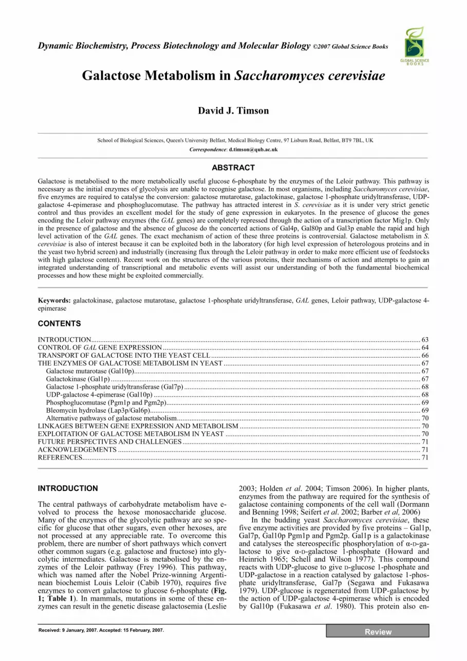
The absence of glucose is necessary, but not sufficient, for the induction of galactose metabolising enzymes. The presence of galactose is also required and this is sensed and responded to by a genetic switch containing three main components: Gal4p (a transcriptional activator), Gal80p (a transcriptional repressor) and Gal3p which is believed to act as a direct sensor of cellular galactose concentration. Gal4p binds to conserved sequences upstream of the GAL genes known as the upstream activating sequences (UASGAL). The protein is a dimer and each monomer can be divided into three main regions – an N-terminal two-Zn2+ six cysteine DNA binding and dimerisation domain, a cen-tral section of unknown function and a C-terminal trans-criptional activation domain. The N- and C-termini can be fused together to produce a protein which is an active trans-criptional activator both in vitro and in vivo (Ding and Johnston 1997). The structures of the DNA binding and di-merisation regions are known (Fig. 2), but the rest of the protein has yet to be characterised structurally (Marmor-stein et al. 1992; Hidalgo et al. 2001). Indeed it is likely that the activation domain, which contains a high propor-tion of negatively charged residues, may be essentially un-structured in the absence of a binding partner (Ansari et al. 1998). Such unstructured regions are common in biological switches. Specificity requires that numerous contacts are made between proteins involved in a switch. Generally speaking this will have a favourable enthalpy change (�H highly negative). When unstructured polypeptide chains bind to their targets, they suffer a considerable loss of con-formational freedom which is reflected in a large decrease in entropy (�S). This energetically unfavourable cones-
quence of binding largely balances out the enthalpy change resulting in an overall free energy change (�G) close to zero and, therefore, an equilibrium constant close to one. Thus the presence of an unstructured region in the binding site between two proteins can result in a highly specific, but rea-dily reversible interaction (Dyson and Wright 2002). This reversibility can be critical in achieving sensitivity to chan-ging concentrations of metabolites and, critically, enables to system to be easily switched off when it is no longer re-quired.
Gal4p has a number of targets. When the GAL genetic switch is on, it interacts through its C-terminal domain, with the general transcription factors Gal11p and TFIID subunits (Sua7p, Taf4p, Taf6p, Taf12p) along with subunits of the chromatin remodelling SAGA complex (Ada2p, Tra1p, Taf4p, Taf6p, Taf12p; note that these last three polypeptides are considered to be part of both TFIID and SAGA) and possibly TATA-binding protein (TBP, Spt15p) (Wu et al. 1996; Xie et al. 2000; Hidalgo et al. 2001; Jeong et al. 2001; Larschan and Winston 2001; Klein et al. 2003; Bhau-mik et al. 2004). The exact binding partners and the order in which they bind remain to be determined. Thus its mecha-nism of action as a transcriptional activator is two-fold: it recruits proteins which will relax the structure of the chro-matin and it begins the process of assembly of an active transcription complex upstream of each of the GAL genes. The end result of these two events is the recruitment of RNA polymerase II, which is able to gain access to the re-laxed chromatin and transcribe the genes. Gal4p is only fully activated when phsophorylated at serines 691, 696 and 699. Of these, serine 699 appears to be the most important (Sadowski et al. 1996).
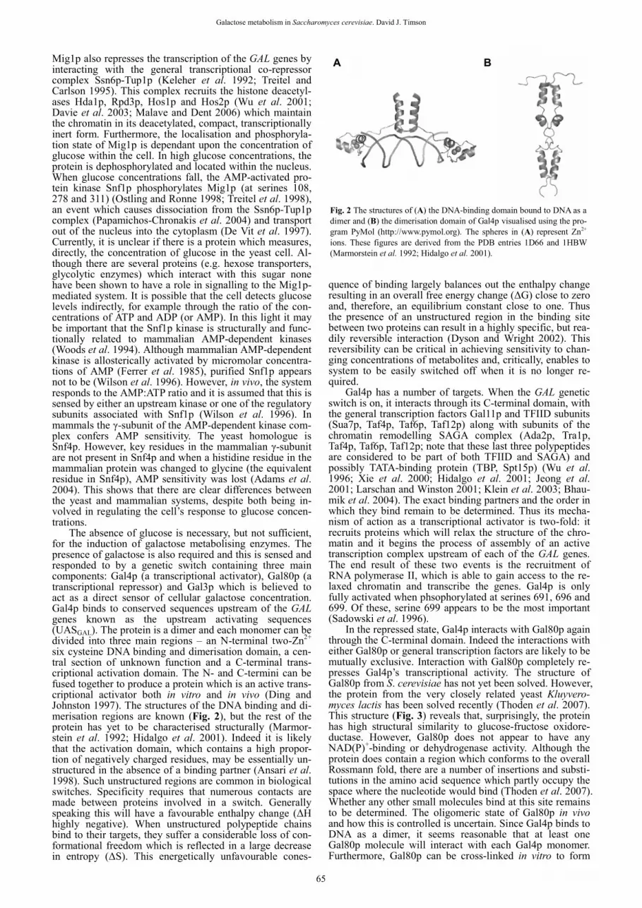
In the repressed state, Gal4p interacts with Gal80p again through the C-terminal domain. Indeed the interactions with either Gal80p or general transcription factors are likely to be mutually exclusive. Interaction with Gal80p completely re-presses Gal4p’s transcriptional activity. The structure of Gal80p from S. cerevisiae has not yet been solved. However, the protein from the very closely related yeast Kluyvero-myces lactis has been solved recently (Thoden et al. 2007). This structure (Fig. 3) reveals that, surprisingly, the protein has high structural similarity to glucose-fructose oxidore-ductase. However, Gal80p does not appear to have any NAD(P)+-binding or dehydrogenase activity. Although the protein does contain a region which conforms to the overall Rossmann fold, there are a number of insertions and substi-tutions in the amino acid sequence which partly occupy the space where the nucleotide would bind (Thoden et al. 2007). Whether any other small molecules bind at this site remains to be determined. The oligomeric state of Gal80p in vivo and how this is controlled is uncertain. Since Gal4p binds to DNA as a dimer, it seems reasonable that at least one Gal80p molecule will interact with each Gal4p monomer. Furthermore, Gal80p can be cross-linked in vitro to form
A B
Fig. 2 The structures of (A) the DNA-binding domain bound to DNA as a dimer and (B) the dimerisation domain of Gal4p visualised using the pro-gram PyMol (http://www.pymol.org). The spheres in (A) represent Zn2+ ions. These figures are derived from the PDB entries 1D66 and 1HBW (Marmorstein et al. 1992; Hidalgo et al. 2001).
65
Dynamic Biochemistry, Process Biotechnology and Molecular Biology 1(1), 63-73 ©2007 Global Science Books
dimers and dimers could also be detected in a modified yeast two hybrid assay (Melcher and Xu 2001). Native gel shift assays suggested a very high affinity interaction (Kd < 10-9 M) but a short half life (<1 min). These dimers are like-ly to remain intact when Gal80p interacts with Gal4p resul-ting in a Gal4p:Gal80p stoichiometry of 2:2. Indeed, Gal80p-Gal4p interaction appears to increase the half life of the Gal80p homodimer and increase the affinity of Gal4p for DNA. Transient associations between Gal80p dimers were also observed and these higher order oligomers may play a role in strongly repressed GAL promoters where there are more than one UASGAL (Melcher and Xu 2001). The transient nature of isolated Gal80p dimers was also ob-served in gel filtration experiments. The protein either e-luted as a monomer (Yun et al. 1991) or failed to elute as a discrete peak (Timson et al. 2002). The homologous protein from K. lactis also failed to elute as a discrete peak, unless millimolar concentrations of either citrate or EDTA were added. Under these conditions, the protein behaved like a dimer (Anders et al. 2006). Interestingly, the experiments of Melcher and Xu were carried out in the presence of EDTA which may explain why dimers were so readily ob-served in their experiments (Melcher and Xu 2001). The crystal structure of K. lactis Gal80p reveals a large interface (4400 Å2) between the two monomers in the homodimer (Thoden et al. 2007). This presumably accounts for the very high affinity observed in vitro. Since, by definition, the structure seen in the crystals must be a long lived one, it is likely that the structure represents a situation similar to that seen when Gal80p dimers bind to Gal4p even though no ci-trate or EDTA was included in the crystallisation solutions. Presumably the process of crystallisation “locks” the pro-tein into this conformation.
The repressive action of Gal80p is relieved by Gal3p. This relief of repression requires two small molecules – ga-lactose and ATP – which are also the substrates for the Le-loir pathway enzyme galactokinase. It is believed (although it has never been formally proven) that Gal3p interacts di-rectly with both these ligands and that this interaction re-sults in a conformational change which enables Gal3p to interact with Gal80p (Yano and Fukasawa 1997). Gal3p has a high level of sequence similarity to the yeast galactoki-nase, Gal1p, and this protein can also substitute for Gal3p in the GAL genetic switch (Bhat and Hopper 1992). In K. lactis there is only one protein, Gal1p, which carries out
both functions (Meyer et al. 1991). S. cerevisiae Gal3p has no galactokinase activity (Platt et al. 2000). The structure of Gal3p is not yet known, but homology models based on the structure of Gal1p (see below) have been built (Thoden et al. 2005).
Although it is well established that a galactose and ATP-dependent interaction between Gal80p and Gal3p is respon-sible for the activation of the GAL genetic switch, the cellu-lar location of this interaction is controversial. Initially, it was believed that the interaction occurred within the nu-cleus and this hypothesis was supported by the detection of a quaternary DNA-Gal4p-Gal80p-Gal3p complex by native gel electrophoresis (Platt and Reece 1998). This evidence is consistent with a model in which, in the repressed state Gal4p and Gal80p are in a DNA-bound complex. In the pre-sence of the activating ligands, Gal3p adds to this complex and, through a series of conformational changes, relieves Gal80p’s repression of Gal4p. In a variant of this model Gal3p causes the complete dissociation of Gal80p from Gal4p in the presence of galactose and ATP. It should be noted that if a multi-protein complex does form on the DNA it is quite likely that, in the transcriptionally active form, the Gal4p-Gal80p interaction is likely to be weakened and this might result in dissociation in some in vitro experiments. This is especially the case in protocols such as GST pull downs or co-immunoprecipitation where the procedure re-quires the isolation of a complex at equilibrium. However, subsequent experiments suggested that Gal3p was excluded from the nucleus in both the presence and absence of galac-tose. In contrast Gal80p was shown to be located in both the nucleus and the cytoplasm (Peng and Hopper 2000; Peng and Hopper 2002). The absence of a Gal80p-Gal4p interact-tion in vivo under inducing conditions was also suggested by chromatin immunoprecipitation (ChIP) experiments (Peng and Hopper 2002). This lead to a model being pro-posed in which Gal80p is able to move freely between the nucleus and the cytoplasm. In the presence of galactose and ATP it binds to Gal3p in the cytoplasm and becomes trapped in this compartment. Once it is trapped in the cytoplasm it is unable to interact with Gal4p in the nucleus and thus trans-cription can occur (Peng and Hopper 2002). Recent experi-ments have tended to point back towards a model where key changes take place in the nucleus and not the cytoplasm. Fluorescence resonance energy transfer has been observed in vivo between Gal4p and Gal80p labelled with different variants of green fluorescent protein (GFP), regardless of whether galactose is present, or not (Bhaumik et al. 2004). Since this effect is only observed over the Förster distance (typically 5-6 nm), the two proteins must remain bound together for it to occur. This suggests that galactose does not cause complete Gal4p-Gal80p dissociation and that at least some Gal3p must be in the nucleus in order to transduce the signal.
The availability of partial structures for Gal4p, the K. lactis Gal80p structure and homology models for Gal3p, means that we are moving towards being able to describe the molecular details of the GAL genetic switch. However, to do this it will be necessary to resolve some of the uncer-tainties described here. TRANSPORT OF GALACTOSE INTO THE YEAST CELL In order for galactose to be metabolised by S. cerevisiae it must first be transported into the cell. Yeast cells have at least 19 hexose transporters in their membranes – Hxt1p to Hxt17p, Mal11p and Gal2p (Ozcan and Johnston 1999; Wieczorke et al. 1999). All of these are integral membrane proteins. They all act as channels facilitating diffusion of hexoses down a concentration gradient rather than pumps which use energy to drive molecules across the membrane. However not all the hexose transporters are expressed under the same conditions. Some (e.g. Hxt1p) are expressed in conditions of high glucose concentrations whereas others (e.g. Hxt2p and Hxt4p) are expressed at lower concentra-
Fig. 3 The structure of the K. lactis Gal80p dimer derived from PDB entry 2NVW (Thoden et al. 2007). The two views are related by an ap-proximate 90° rotation about a horizontal axis. Note the extensive inter-face between the two monomers which is composed almost entirely of �-sheet.
66
Galactose metabolism in Saccharomyces cerevisiae. David J. Timson
tions of the sugar. Some of the sequences may be pseudo-genes. Those expressed in high glucose concentrations tend to have lower affinities for glucose than those expressed in low glucose concentrations. This enables the yeast cell to grow in a wide range of different glucose concentrations (micromolar to molar) while still taking up appropriate con-centrations of the sugar into the cell (Ozcan and Johnston 1995; Ozcan and Johnston 1999). Mal11p is high affinity maltose transporter, which is induced in the presence of maltose (Cheng and Michels 1991). Gal2p is one of the GAL genes and is a high affinity galactose transporter (Tschopp et al. 1986; Huibregtse et al. 1993). It is ex-pressed in the presence of galactose and repressed (through the Mig1p system described above) in the presence of glu-cose. In addition to these mechanisms, the protein is rapidly ubiquitinated, endocytosed and degraded if the yeast cells are shifted from an environment rich in galactose to one where glucose predominates (Horak and Wolf 1997). How-ever, it is not highly specific for galactose and can also transport glucose (Reifenberger et al. 1997; Maier et al. 2002). Furthermore, it is likely that when the organism is growing in galactose, the sugar is transported into the cell by both Gal2p and the less selective hexose transporters of the Hxt family.
Gal2p is predicted to have 11 transmembrane segments and the tenth segment contains two aromatic residues (Tyr446 and Trp455) responsible for discriminating in fa-vour of galactose (Nishizawa et al. 1995; Kasahara and Ma-eda 1998). Replacement of Tyr446 with a phenylalanine re-sidue reduced the transport of galactose to less than 20% of wild type levels, but increased the transport of glucose (Ka-sahara and Maeda 1998). In addition to these two residues in the tenth putative transmembrane segment, Tyr352 and Phe504 (in segments 7 and twelve respectively) must be bulky hydrophobic residues in order for the protein to func-tion as a galactose transporter (Kasahara and Kasahara 2000a). Double site-directed mutagenesis experiments sug-gest that these two residues interact with each other (Kasa-hara and Kasahara 2000b). As yet, no residues have been unequivocally identified as interacting directly with galac-tose. The results of the various site-directed mutagenesis experiments targeting hydrophobic residues could infer either that these residues interact with the sugar, or that they help create a structural environment for the interaction of other residues with galactose. Ideally, this question would be answered by solving the structure of Gal2p in complex with galactose. Of course, the solution of structures of in-tegral membrane proteins is difficult and even if this was achieved it would only show a “snap-shot” of the channel in action as presumably the sugar must interact with a num-ber of different residues as it passes through the protein. THE ENZYMES OF GALACTOSE METABOLISM IN YEAST Galactose mutarotase (Gal10p) Hexose sugars in the six-membered, pyranose ring configu-ration have five chiral centres at carbons 1 to 5 (Fig. 1A). The chiral centres at carbons 2 to 4 determine the identity of the sugar. For example, the two possible stereoisomers at carbon 4 are glucose and galactose. Carbon 5 determines whether the sugar is a D- or a L-hexose and carbon 1 (which is only a chiral centre in the ring form) determines whether the molecule is the �- or �-anomer. Unlike the other chiral centres the �- and �-anomers can be interconverted, through the straight chain form of the sugar, in aqueous solution. This reaction is catalysed by both acids and bases and the equilibrium constant is usually close to unity (ie there is not much energetic difference between the two configurations). Thus, there are four possible pyranose configurations of ga-lactose – �-L, �-L, �-D and �-D. In common with most other monosaccharides, only the D-form is usually found in living systems. Furthermore some enzymes, such as galactokinase, are highly specific for one configuration (the �-D form in
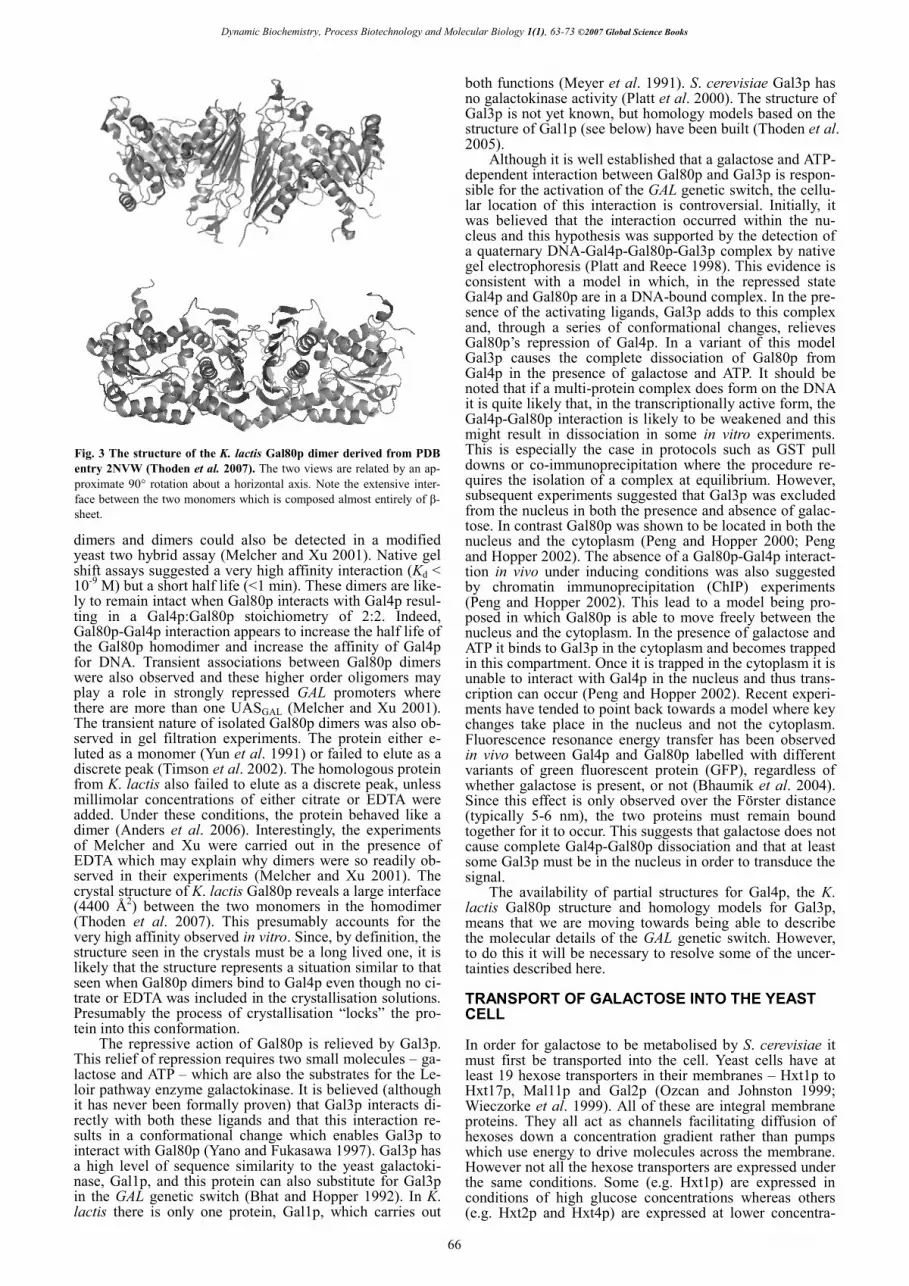
the case of galactokinase). Although the �- and �-anomers inter-convert at a measurable rate in aqueous solution, this rate does not appear to be great enough to supply the rest of the Leloir pathway with sufficient �-D-galactose and, for this reason, most organisms express a galactose mutarotase (aldose 1-epimerase) to catalyse this interconversion or mu-tarotation. In the case of S. cerevisiae, this enzyme activity is encoded by the C-terminal half of Gal10p.
In common with galactose mutarotase enzymes purified from other species, the yeast enzyme is characterised by a high value of the specificity constant, kcat/Km, when galac-tose is the substrate (68,000 l.mol-1.s-1) (Scott and Timson 2007). It appears to be more selective for galactose over glucose than the enzyme from some other species: the ratio of the specificity constants for the yeast enzyme is approxi-mately 90 compared to 4 for the human enzyme (Timson and Reece 2003). Given that the enzyme is only expressed in the presence of galactose and the absence of glucose, it seems unlikely that it plays are major role in maintaining the anomeric equilibrium of glucose, or any other monosac-charide. Indeed there are at least two other mutarotase-like sequences in the yeast genome (YHR210c and YNR071c) (Goffeau et al. 1996). Of these, YHR210c is more likely to be involved in catalysing anomeric interconversions as YNR071c lacks a critical residue in the active site. Thus it is YHR210c which is most likely to be account for the muta-rotase activity observed in extracts from yeast cells grown in 2% (w/v) glucose (Brahma and Bhattacharyya 2004).
The overall structure of the galactose mutarotase do-main of Gal10p is very similar to that of other mutarotases being largely composed of �-sheets with only a small pro-portion of �-helix (Fig. 4) (Thoden and Holden 2005). The active sites of galactose mutarotases also show a high de-gree of structural similarity and they are all believed to share the same mechanism of action. No co-factors are re-quired to bring about catalysis. Instead, the reaction is initi-ated by the abstraction of a proton from the hydroxyl group attached to carbon-1 of the sugar by a glutamate residue ac-ting as an active site Brønstead base. A histidine residue, ac-ting a Brønstead acid donates a proton to the oxygen of the pyranose ring and the combination of these two events is to cause breakage of the ring. Reversing these events results in the ring reforming, but the enzyme is not stereospecific for this action and so either the �- or �-anomer may be formed and thus an equilibrium mixture of products are formed. Galactokinase (Gal1p) Galactokinases belong to the GHMP (galactokinase, homo-serine kinase, mevalonate kinase and phosphomevalonate kinase) family of enzymes (Bork et al. 1993; Timson 2007).
Mutarotase domain
Epimerase domain
Mutarotase domain
Epimerase domain
Fig. 4 The structure of S. cerevisiae Gal10p showing the mutarotase and epimerase domains (PDB entry 1Z45 (Thoden and Holden 2005)). The protein forms a stable homodimer through contacts which are entirely lo-cated in the epimerase domain.
67
Dynamic Biochemistry, Process Biotechnology and Molecular Biology 1(1), 63-73 ©2007 Global Science Books
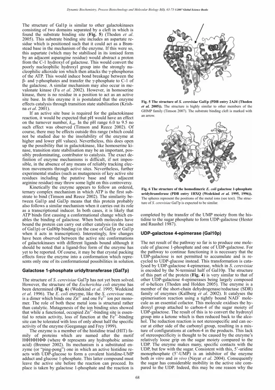
The structure of Gal1p is similar to other galactokinases consisting of two domains separated by a cleft in which is found the substrate binding site (Fig. 5) (Thoden et al. 2005). This substrate binding site includes an aspartate re-sidue which is positioned such that it could act as a Brøn-stead base in the mechanism of the enzyme. If this were so, this aspartate (which may be stabilised in its ionised form by an adjacent asparagine residue) would abstract a proton from the C-1 hydroxyl of galactose. This would convert the poorly nucleophilic hydroxyl group into the strongly nu-cleophilic alkoxide ion which then attacks the �-phosphorus of the ATP. This would induce bond breakage between the �- and �-phosphates and transfer the �-phosphate to C-1 of the galactose. A similar mechanism may also occur in me-valonate kinase (Fu et al. 2002). However, in homoserine kinase, there is no residue in a position to act as an active site base. In this enzyme it is postulated that the enzyme effects catalysis through transition state stabilisation (Krish-na et al. 2001).
If an active site base is required for the galactokinase reaction, it would be expected that pH would have an effect on the turnover number, kcat. In the pH range 6.0 to 9.5 no such effect was observed (Timson and Reece 2002). Of course, there may be effects outside this range (which could not be studied due to the insolubility of the enzyme at higher and lower pH values). Nevertheless, this does open up the possibility that in galactokinase, like homoserine ki-nase, transition state stabilisation may be an important, pos-sibly predominating, contributor to catalysis. The exact de-finition of enzyme mechanisms is difficult, if not impos-sible, in the absence of any means of reliably tracking elec-tron movements through active sites. Nevertheless, further experimental studies (such as mutagenesis of key active site residues including the putative base and the adjacent arginine residue) may throw some light on this controversy.
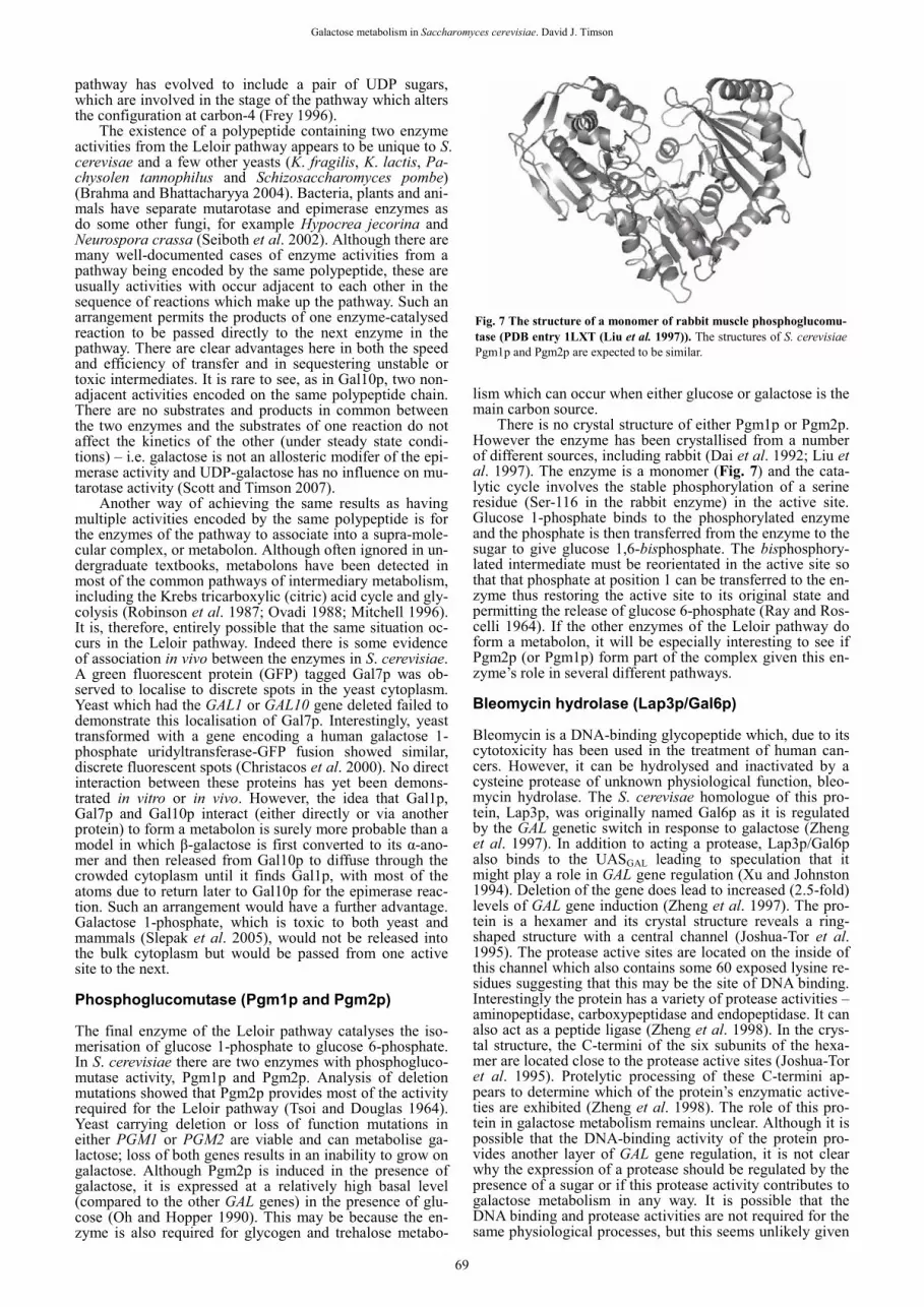
Kinetically the enzyme appears to follow an ordered, ternary complex mechanism in which ATP is the first sub-strate to bind (Timson and Reece 2002). The similarity be-tween Gal1p and Gal3p means that this protein probably also follows a similar mechanism when it carries out its role as a transcriptional inducer. In both cases, it is likely that ATP binds first causing a conformational change which en-ables the binding of galactose. When both molecules have bound the protein can carry out either catalysis (in the case of Gal1p) or Gal80p binding (in the case of Gal3p or Gal1p when it acts in transcription). Interestingly, few changes have been observed between the active site conformations of galactokinases with different ligands bound although it should be noted that a ligand-free form of the enzyme has yet to be reported. Of course, it may be that crystal packing effects force the enzyme into a conformation which repre-sents only one of its conformational possibilities in solution. Galactose 1-phosphate uridyltransferase (Gal7p) The structure of S. cerevisiae Gal7p has not yet been solved. However, the structure of the Escherichia coli enzyme has been determined (Fig. 6) (Wedekind et al. 1995; Wedekind et al. 1996). The E. coli enzyme, like the S. cerevisae one, is a dimer which binds one Zn2+ and one Fe3+ ion per mono-mer. The role of both these metal ions is structural rather than catalytic. Mutagenesis of the ion binding sites showed that while a functional, occupied Zn2+-binding site is essen-tial to retain activity, loss of function at the Fe3+-binding site can be tolerated with loss of only about half the specific activity of the enzyme (Geeganage and Frey 1999).
The enzyme is a member of the histidine triad (HIT) fa-mily of proteins which are named after the motif H�H�H�� (where � represents any hydrophobic amino acid) (Brenner 2002). Its mechanism is a substituted en-zyme (or “ping-pong”) one in which an active histidine re-acts with UDP-glucose to form a covalent histidine-UMP adduct and glucose 1-phosphate. This latter compound must leave the active site before the reaction can proceed. Its place is taken by galactose 1-phosphate and the reaction is
completed by the transfer of the UMP moiety from the his-tidine to the sugar phosphate to form UDP-galactose (Hester and Raushel 1987). UDP-galactose 4-epimerase (Gal10p) The net result of the pathway so far is to produce one mole-cule of glucose 1-phosphate and one of UDP-galactose. For the pathway to continue functioning it is necessary that the UDP-galactose is not permitted to accumulate and is re-cycled to UDP-glucose instead. This transformation is cata-lysed by UDP-galactose 4-epimerase, which in S. cerevisiae is encoded by the N-terminal half of Gal10p. The structure of this part of the protein (Fig. 4) is very similar to that of other UDP-galactose 4-epimerases being largely composed of �-helices (Thoden and Holden 2005). The enzyme is a member of the short-chain dehydrogenase/reductase (SDR) family of enzymes (Kallberg et al. 2002). It catalyses the epimerisation reaction using a tightly bound NAD+ mole-cule as an essential cofactor. This molecule oxidises the hy-droxyl group attached to carbon-4 of the sugar moiety of UDP-galactose. The result of this is to convert the hydroxyl group into a ketone which is then reduced back to the alco-hol. The reduction reaction is not stereospecific and can oc-cur at either side of the carbonyl group, resulting in a mix-ture of configurations at carbon-4 in the products. This lack of stereospecificity is thought to be caused by the enzyme’s relatively loose grip on the sugar moiety compared to the UDP. The enzyme makes many, specific contacts with the UDP, but few with the sugar. Consistent with this, 5’-uridine monophosphate (5’-UMP) is an inhibitor of the enzyme both in vitro and in vivo (Nayar et al. 2004). Consequently the sugar has considerable mobility in the active site com-pared to the UDP. Indeed, this may be one reason why the
Fig. 5 The structure of S. cerevisiae Gal1p (PDB entry 2AJ4 (Thoden et al. 2005)). The structure is highly similar to other members of the GHMP family (Timson 2007). The substrate binding cleft is marked with an arrow.
Fig. 6 The structure of the homodimeric E. coli galactose 1-phosphate uridyltransferase (PDB entry 1HXQ (Wedekind et al. 1995, 1996)). The spheres represent the positions of the metal ions (see text). The struc-ture of S. cerevisiae Gal7p is expected to be similar.
68
Galactose metabolism in Saccharomyces cerevisiae. David J. Timson
pathway has evolved to include a pair of UDP sugars, which are involved in the stage of the pathway which alters the configuration at carbon-4 (Frey 1996).
The existence of a polypeptide containing two enzyme activities from the Leloir pathway appears to be unique to S. cerevisae and a few other yeasts (K. fragilis, K. lactis, Pa-chysolen tannophilus and Schizosaccharomyces pombe) (Brahma and Bhattacharyya 2004). Bacteria, plants and ani-mals have separate mutarotase and epimerase enzymes as do some other fungi, for example Hypocrea jecorina and Neurospora crassa (Seiboth et al. 2002). Although there are many well-documented cases of enzyme activities from a pathway being encoded by the same polypeptide, these are usually activities with occur adjacent to each other in the sequence of reactions which make up the pathway. Such an arrangement permits the products of one enzyme-catalysed reaction to be passed directly to the next enzyme in the pathway. There are clear advantages here in both the speed and efficiency of transfer and in sequestering unstable or toxic intermediates. It is rare to see, as in Gal10p, two non-adjacent activities encoded on the same polypeptide chain. There are no substrates and products in common between the two enzymes and the substrates of one reaction do not affect the kinetics of the other (under steady state condi-tions) – i.e. galactose is not an allosteric modifer of the epi-merase activity and UDP-galactose has no influence on mu-tarotase activity (Scott and Timson 2007).
Another way of achieving the same results as having multiple activities encoded by the same polypeptide is for the enzymes of the pathway to associate into a supra-mole-cular complex, or metabolon. Although often ignored in un-dergraduate textbooks, metabolons have been detected in most of the common pathways of intermediary metabolism, including the Krebs tricarboxylic (citric) acid cycle and gly-colysis (Robinson et al. 1987; Ovadi 1988; Mitchell 1996). It is, therefore, entirely possible that the same situation oc-curs in the Leloir pathway. Indeed there is some evidence of association in vivo between the enzymes in S. cerevisiae. A green fluorescent protein (GFP) tagged Gal7p was ob-served to localise to discrete spots in the yeast cytoplasm. Yeast which had the GAL1 or GAL10 gene deleted failed to demonstrate this localisation of Gal7p. Interestingly, yeast transformed with a gene encoding a human galactose 1-phosphate uridyltransferase-GFP fusion showed similar, discrete fluorescent spots (Christacos et al. 2000). No direct interaction between these proteins has yet been demons-trated in vitro or in vivo. However, the idea that Gal1p, Gal7p and Gal10p interact (either directly or via another protein) to form a metabolon is surely more probable than a model in which �-galactose is first converted to its �-ano-mer and then released from Gal10p to diffuse through the crowded cytoplasm until it finds Gal1p, with most of the atoms due to return later to Gal10p for the epimerase reac-tion. Such an arrangement would have a further advantage. Galactose 1-phosphate, which is toxic to both yeast and mammals (Slepak et al. 2005), would not be released into the bulk cytoplasm but would be passed from one active site to the next. Phosphoglucomutase (Pgm1p and Pgm2p) The final enzyme of the Leloir pathway catalyses the iso-merisation of glucose 1-phosphate to glucose 6-phosphate. In S. cerevisiae there are two enzymes with phosphogluco-mutase activity, Pgm1p and Pgm2p. Analysis of deletion mutations showed that Pgm2p provides most of the activity required for the Leloir pathway (Tsoi and Douglas 1964). Yeast carrying deletion or loss of function mutations in either PGM1 or PGM2 are viable and can metabolise ga-lactose; loss of both genes results in an inability to grow on galactose. Although Pgm2p is induced in the presence of galactose, it is expressed at a relatively high basal level (compared to the other GAL genes) in the presence of glu-cose (Oh and Hopper 1990). This may be because the en-zyme is also required for glycogen and trehalose metabo-
lism which can occur when either glucose or galactose is the main carbon source.
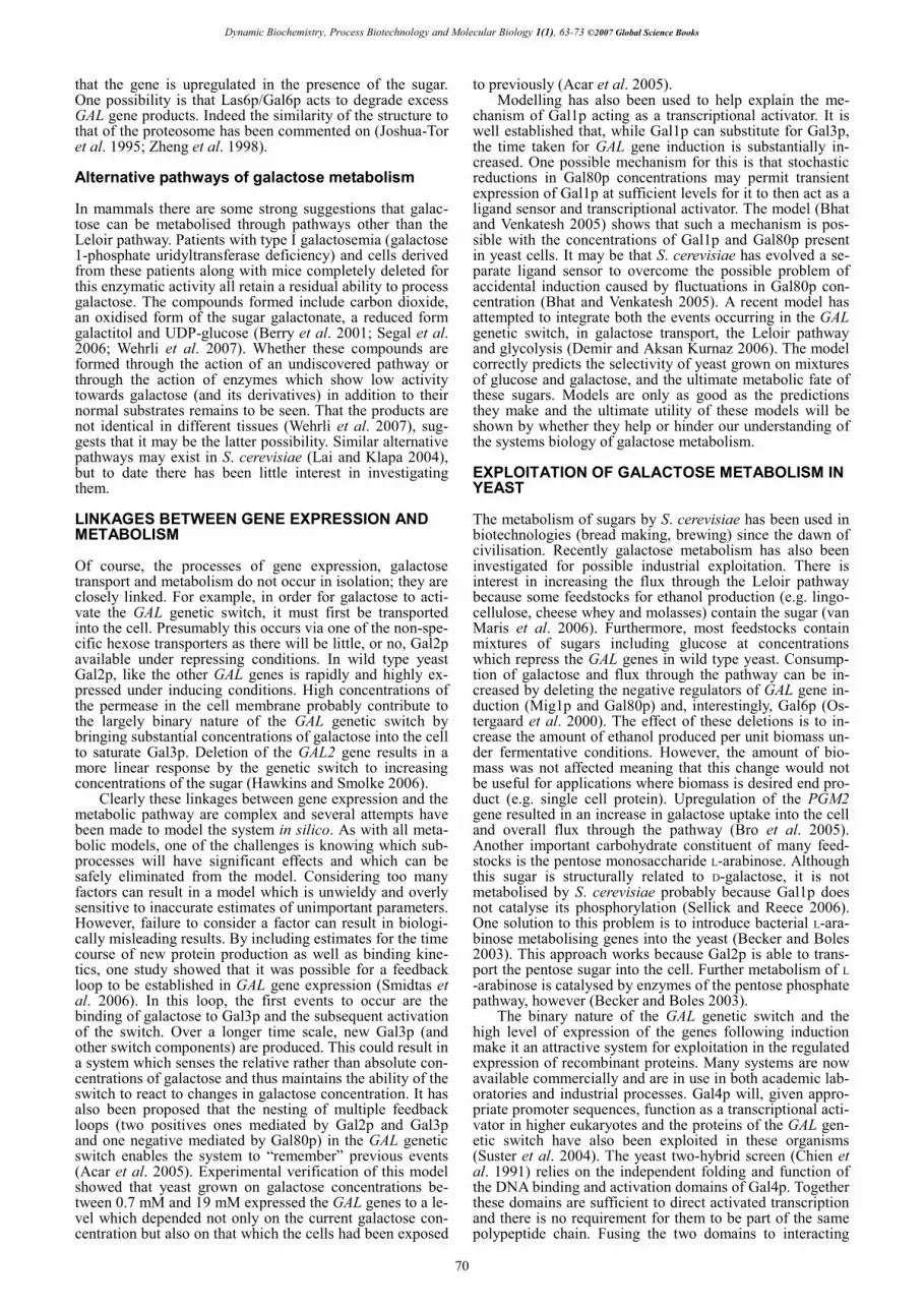
There is no crystal structure of either Pgm1p or Pgm2p. However the enzyme has been crystallised from a number of different sources, including rabbit (Dai et al. 1992; Liu et al. 1997). The enzyme is a monomer (Fig. 7) and the cata-lytic cycle involves the stable phosphorylation of a serine residue (Ser-116 in the rabbit enzyme) in the active site. Glucose 1-phosphate binds to the phosphorylated enzyme and the phosphate is then transferred from the enzyme to the sugar to give glucose 1,6-bisphosphate. The bisphosphory-lated intermediate must be reorientated in the active site so that that phosphate at position 1 can be transferred to the en-zyme thus restoring the active site to its original state and permitting the release of glucose 6-phosphate (Ray and Ros-celli 1964). If the other enzymes of the Leloir pathway do form a metabolon, it will be especially interesting to see if Pgm2p (or Pgm1p) form part of the complex given this en-zyme’s role in several different pathways. Bleomycin hydrolase (Lap3p/Gal6p) Bleomycin is a DNA-binding glycopeptide which, due to its cytotoxicity has been used in the treatment of human can-cers. However, it can be hydrolysed and inactivated by a cysteine protease of unknown physiological function, bleo-mycin hydrolase. The S. cerevisae homologue of this pro-tein, Lap3p, was originally named Gal6p as it is regulated by the GAL genetic switch in response to galactose (Zheng et al. 1997). In addition to acting a protease, Lap3p/Gal6p also binds to the UASGAL leading to speculation that it might play a role in GAL gene regulation (Xu and Johnston 1994). Deletion of the gene does lead to increased (2.5-fold) levels of GAL gene induction (Zheng et al. 1997). The pro-tein is a hexamer and its crystal structure reveals a ring-shaped structure with a central channel (Joshua-Tor et al. 1995). The protease active sites are located on the inside of this channel which also contains some 60 exposed lysine re-sidues suggesting that this may be the site of DNA binding. Interestingly the protein has a variety of protease activities – aminopeptidase, carboxypeptidase and endopeptidase. It can also act as a peptide ligase (Zheng et al. 1998). In the crys-tal structure, the C-termini of the six subunits of the hexa-mer are located close to the protease active sites (Joshua-Tor et al. 1995). Protelytic processing of these C-termini ap-pears to determine which of the protein’s enzymatic active-ties are exhibited (Zheng et al. 1998). The role of this pro-tein in galactose metabolism remains unclear. Although it is possible that the DNA-binding activity of the protein pro-vides another layer of GAL gene regulation, it is not clear why the expression of a protease should be regulated by the presence of a sugar or if this protease activity contributes to galactose metabolism in any way. It is possible that the DNA binding and protease activities are not required for the same physiological processes, but this seems unlikely given
Fig. 7 The structure of a monomer of rabbit muscle phosphoglucomu-tase (PDB entry 1LXT (Liu et al. 1997)). The structures of S. cerevisiae Pgm1p and Pgm2p are expected to be similar.
69
Dynamic Biochemistry, Process Biotechnology and Molecular Biology 1(1), 63-73 ©2007 Global Science Books
that the gene is upregulated in the presence of the sugar. One possibility is that Las6p/Gal6p acts to degrade excess GAL gene products. Indeed the similarity of the structure to that of the proteosome has been commented on (Joshua-Tor et al. 1995; Zheng et al. 1998). Alternative pathways of galactose metabolism In mammals there are some strong suggestions that galac-tose can be metabolised through pathways other than the Leloir pathway. Patients with type I galactosemia (galactose 1-phosphate uridyltransferase deficiency) and cells derived from these patients along with mice completely deleted for this enzymatic activity all retain a residual ability to process galactose. The compounds formed include carbon dioxide, an oxidised form of the sugar galactonate, a reduced form galactitol and UDP-glucose (Berry et al. 2001; Segal et al. 2006; Wehrli et al. 2007). Whether these compounds are formed through the action of an undiscovered pathway or through the action of enzymes which show low activity towards galactose (and its derivatives) in addition to their normal substrates remains to be seen. That the products are not identical in different tissues (Wehrli et al. 2007), sug-gests that it may be the latter possibility. Similar alternative pathways may exist in S. cerevisiae (Lai and Klapa 2004), but to date there has been little interest in investigating them. LINKAGES BETWEEN GENE EXPRESSION AND METABOLISM Of course, the processes of gene expression, galactose transport and metabolism do not occur in isolation; they are closely linked. For example, in order for galactose to acti-vate the GAL genetic switch, it must first be transported into the cell. Presumably this occurs via one of the non-spe-cific hexose transporters as there will be little, or no, Gal2p available under repressing conditions. In wild type yeast Gal2p, like the other GAL genes is rapidly and highly ex-pressed under inducing conditions. High concentrations of the permease in the cell membrane probably contribute to the largely binary nature of the GAL genetic switch by bringing substantial concentrations of galactose into the cell to saturate Gal3p. Deletion of the GAL2 gene results in a more linear response by the genetic switch to increasing concentrations of the sugar (Hawkins and Smolke 2006).
Clearly these linkages between gene expression and the metabolic pathway are complex and several attempts have been made to model the system in silico. As with all meta-bolic models, one of the challenges is knowing which sub-processes will have significant effects and which can be safely eliminated from the model. Considering too many factors can result in a model which is unwieldy and overly sensitive to inaccurate estimates of unimportant parameters. However, failure to consider a factor can result in biologi-cally misleading results. By including estimates for the time course of new protein production as well as binding kine-tics, one study showed that it was possible for a feedback loop to be established in GAL gene expression (Smidtas et al. 2006). In this loop, the first events to occur are the binding of galactose to Gal3p and the subsequent activation of the switch. Over a longer time scale, new Gal3p (and other switch components) are produced. This could result in a system which senses the relative rather than absolute con-centrations of galactose and thus maintains the ability of the switch to react to changes in galactose concentration. It has also been proposed that the nesting of multiple feedback loops (two positives ones mediated by Gal2p and Gal3p and one negative mediated by Gal80p) in the GAL genetic switch enables the system to “remember” previous events (Acar et al. 2005). Experimental verification of this model showed that yeast grown on galactose concentrations be-tween 0.7 mM and 19 mM expressed the GAL genes to a le-vel which depended not only on the current galactose con-centration but also on that which the cells had been exposed
to previously (Acar et al. 2005). Modelling has also been used to help explain the me-
chanism of Gal1p acting as a transcriptional activator. It is well established that, while Gal1p can substitute for Gal3p, the time taken for GAL gene induction is substantially in-creased. One possible mechanism for this is that stochastic reductions in Gal80p concentrations may permit transient expression of Gal1p at sufficient levels for it to then act as a ligand sensor and transcriptional activator. The model (Bhat and Venkatesh 2005) shows that such a mechanism is pos-sible with the concentrations of Gal1p and Gal80p present in yeast cells. It may be that S. cerevisiae has evolved a se-parate ligand sensor to overcome the possible problem of accidental induction caused by fluctuations in Gal80p con-centration (Bhat and Venkatesh 2005). A recent model has attempted to integrate both the events occurring in the GAL genetic switch, in galactose transport, the Leloir pathway and glycolysis (Demir and Aksan Kurnaz 2006). The model correctly predicts the selectivity of yeast grown on mixtures of glucose and galactose, and the ultimate metabolic fate of these sugars. Models are only as good as the predictions they make and the ultimate utility of these models will be shown by whether they help or hinder our understanding of the systems biology of galactose metabolism. EXPLOITATION OF GALACTOSE METABOLISM IN YEAST The metabolism of sugars by S. cerevisiae has been used in biotechnologies (bread making, brewing) since the dawn of civilisation. Recently galactose metabolism has also been investigated for possible industrial exploitation. There is interest in increasing the flux through the Leloir pathway because some feedstocks for ethanol production (e.g. lingo-cellulose, cheese whey and molasses) contain the sugar (van Maris et al. 2006). Furthermore, most feedstocks contain mixtures of sugars including glucose at concentrations which repress the GAL genes in wild type yeast. Consump-tion of galactose and flux through the pathway can be in-creased by deleting the negative regulators of GAL gene in-duction (Mig1p and Gal80p) and, interestingly, Gal6p (Os-tergaard et al. 2000). The effect of these deletions is to in-crease the amount of ethanol produced per unit biomass un-der fermentative conditions. However, the amount of bio-mass was not affected meaning that this change would not be useful for applications where biomass is desired end pro-duct (e.g. single cell protein). Upregulation of the PGM2 gene resulted in an increase in galactose uptake into the cell and overall flux through the pathway (Bro et al. 2005). Another important carbohydrate constituent of many feed-stocks is the pentose monosaccharide L-arabinose. Although this sugar is structurally related to D-galactose, it is not metabolised by S. cerevisiae probably because Gal1p does not catalyse its phosphorylation (Sellick and Reece 2006). One solution to this problem is to introduce bacterial L-ara-binose metabolising genes into the yeast (Becker and Boles 2003). This approach works because Gal2p is able to trans-port the pentose sugar into the cell. Further metabolism of L -arabinose is catalysed by enzymes of the pentose phosphate pathway, however (Becker and Boles 2003).
The binary nature of the GAL genetic switch and the high level of expression of the genes following induction make it an attractive system for exploitation in the regulated expression of recombinant proteins. Many systems are now available commercially and are in use in both academic lab-oratories and industrial processes. Gal4p will, given appro-priate promoter sequences, function as a transcriptional acti-vator in higher eukaryotes and the proteins of the GAL gen-etic switch have also been exploited in these organisms (Suster et al. 2004). The yeast two-hybrid screen (Chien et al. 1991) relies on the independent folding and function of the DNA binding and activation domains of Gal4p. Together these domains are sufficient to direct activated transcription and there is no requirement for them to be part of the same polypeptide chain. Fusing the two domains to interacting
70

Galactose metabolism in Saccharomyces cerevisiae. David J. Timson
partners is often sufficient to bring them into close enough proximity that the non-covalent complex can bind specific-ally to UASGAL and activate transcription. Although the assay has a number of well-documented problems (especi-ally a high background of false-positives) it has found widespread applications in the detection of new protein-protein interactions, the mapping of interaction domains within proteins and in global interaction screens.
The Leloir pathway in S. cerevisiae has also been used as a model system for studying the human genetic disease galactosemia. This disease is caused by mutations in the Leloir pathway enzymes. S. cerevisae offers some advanta-ges for these experiments. The ease of genetic manipulation means that strains can be constructed lacking one or more GAL gene and these strains then complemented with plas-mids containing wild type or mutant human genes. The use of diploid yeast means that the effects of heterozygosity can be studied – something which is difficult using either trans-fected human cell lines or isolated proteins. (Note that the dimeric nature of the uridyltransferase and the epimerase mean that three possible dimers are possible in heterozy-gotes – two different homodimers and a heterodimer. Relia-ble methods for dissociating and reforming dimers of these proteins in vitro or in vivo have yet to be developed. Re-combinant expression in heterozygous yeast overcomes this problem as all possible dimers will be synthesised in vivo.) The organism’s short doubling time means that the effects of the mutations can be studied on cells grown on galactose as the only carbon source over many generations. Rela-tively simple experiments such are comparisons of doub-ling times can be carried out and the proteins can be iso-lated if necessary for in vitro work. The system has been used successfully to characterise mutations in both the uri-dyltransferase and epimerase enzymes (Quimby et al. 1997; Riehman et al. 2001; Christacos and Fridovich-Keil 2002; Wasilenko et al. 2005). If there are alternative pathways of galactose metabolism in S. cerevisiae (see above), these may need to be taken into account in the future interpre-tation of these kinds of experiments. FUTURE PERSPECTIVES AND CHALLENGES The study of the galactose metabolism in yeast remains an important field. Although we have learned a lot over the last 25 years about the mechanisms of gene induction in the GAL genetic switch – much of which is applicable to a wide range of eukaryotic gene expression systems – many facts remain to be clarified. The precise mechanism of ac-tion remains controversial and more details are required about the nature of the interactions between the various components in atomic detail. Perhaps most importantly, the unambiguous identification of the general transcription fac-tors which Gal4p interacts with is required along with de-tails of the affinities and time courses of these interactions. We have learned much about the structures and mecha-nisms of the individual enzymes, but much less about how they work together as pathway. More practical and theore-tical studies on the control of flux through the pathway are required and the question of whether, or not, the enzymes form a metabolon needs to be tackled conclusively. The ac-quisition of this information will have clear scientific and industrial benefits. A better understanding of how galactose metabolism in yeast works as an integrated system will en-hance our ability to exploit it both in industrial biotechno-logy and in the research laboratory. ACKNOWLEDGEMENTS Research in my laboratory on galactose metabolism has been fun-ded, in part, by the Royal Society (London). REFERENCES Acar M, Becskei A, van Oudenaarden A (2005) Enhancement of cellular me-
mory by reducing stochastic transitions. Nature 435, 228-232
Adams J, Chen ZP, van Denderen BJ, Morton CJ, Parker MW, Witters LA, Stapleton D, Kemp BE (2004) Intrasteric control of AMPK via the gamma1 subunit AMP allosteric regulatory site. Protein Science 13, 155-165
Anders A, Lilie H, Franke K, Kapp L, Stelling J, Gilles ED, Breunig KD (2006) The galactose switch in Kluyveromyces lactis depends on nuclear competition between Gal4 and Gal1 for Gal80 binding. The Journal of Bio-logical Chemistry 281, 29337-29348
Ansari AZ, Reece RJ, Ptashne M (1998) A transcriptional activating region with two contrasting modes of protein interaction. Proceedings of the Nat-ional Academy of Sciences USA 95, 13543-13548
Barber C, Rosti J, Rawat A, Findlay K, Roberts K, Seifert GJ (2006) Dis-tinct properties of the five UDP-D-glucose/UDP-D-galactose 4-epimerase iso-forms of Arabidopsis thaliana. The Journal of Biological Chemistry 281, 17276-17285
Becker J, Boles E (2003) A modified Saccharomyces cerevisiae strain that con-sumes l-Arabinose and produces ethanol. Applied and Environmental Micro-biology 69, 4144-4150
Berry GT, Leslie N, Reynolds R, Yager CT, Segal S (2001) Evidence for alter-nate galactose oxidation in a patient with deletion of the galactose-1-phos-phate uridyltransferase gene. Molecular Genetics and Metabolism 72, 316-321
Bhat PJ, Venkatesh KV (2005) Stochastic variation in the concentration of a repressor activates GAL genetic switch: implications in evolution of regula-tory network. FEBS Letters 579, 597-603
Bhat PJ, Hopper JE (1992) Overproduction of the GAL1 or GAL3 protein causes galactose-independent activation of the GAL4 protein: evidence for a new model of induction for the yeast GAL/MEL regulon. Molecular and Cel-lular Biology 12, 2701-2707
Bhaumik SR, Raha T, Aiello DP, Green MR (2004) In vivo target of a trans-criptional activator revealed by fluorescence resonance energy transfer. Genes and Development 18, 333-343
Bork P, Sander C, Valencia A (1993) Convergent evolution of similar enzyme-tic function on different protein folds: the hexokinase, ribokinase, and galac-tokinase families of sugar kinases. Protein Science 2, 31-40
Brahma A, Bhattacharyya D (2004) UDP-galactose 4-epimerase from Kluyve-romyces fragilis. Evidence for independent mutarotation site. European Jour-nal of Biochemistry 271, 58-68
Brenner C (2002) Hint, Fhit, and GalT: function, structure, evolution, and me-chanism of three branches of the histidine triad superfamily of nucleotide hy-drolases and transferases. Biochemistry 41, 9003-9014
Bro C, Knudsen S, Regenberg B, Olsson L, Nielsen J (2005) Improvement of galactose uptake in Saccharomyces cerevisiae through overexpression of phosphoglucomutase: example of transcript analysis as a tool in inverse meta-bolic engineering. Applied and Environmental Microbiology 71, 6465-6472
Cabib E (1970) Research on sugar nucleotides brings honor to Argentinian bio-chemist (Luis Leloir). Science 170, 608-609
Cheng Q, Michels CA (1991) MAL11 and MAL61 encode the inducible high-affinity maltose transporter of Saccharomyces cerevisiae. Journal of Bacteri-ology 173, 1817-1820
Chien CT, Bartel PL, Sternglanz R, Fields S (1991) The two-hybrid system: a method to identify and clone genes for proteins that interact with a protein of interest. Proceedings of the National Academy of Sciences USA 88, 9578-9582
Christacos NC, Fridovich-Keil JL (2002) Impact of patient mutations on hete-rodimer formation and function in human galactose-1-P uridylyltransferase. Molecular Genetics and Metabolism 76, 319-326
Christacos NC, Marson MJ, Wells L, Riehman K, Fridovich-Keil JL (2000) Subcellular localization of galactose-1-phosphate uridylyltransferase in the yeast Saccharomyces cerevisiae. Molecular Genetics and Metabolism 70, 272-280
Dai JB, Liu Y, Ray WJ Jr., Konno M (1992) The crystal structure of muscle phosphoglucomutase refined at 2.7-angstrom resolution. Journal of Biologi-cal Chemistry 267, 6322-6337
Davie JK, Edmondson DG, Coco CB, Dent SY (2003) Tup1-Ssn6 interacts with multiple class I histone deacetylases in vivo. Journal of Biological Che-mistry 278, 50158-50162
De Vit MJ, Waddle JA, Johnston M (1997) Regulated nuclear translocation of the Mig1 glucose repressor. Molecular Biology of the Cell 8, 1603-1618
Demir O, Aksan Kurnaz I (2006) An integrated model of glucose and galac-tose metabolism regulated by the GAL genetic switch. Computational Bio-logy and Chemistry 30, 179-192
Ding WV, Johnston SA (1997) The DNA binding and activation domains of Gal4p are sufficient for conveying its regulatory signals. Molecular and Cellular Biology 17, 2538-2549
Dormann P, Benning C (1998) The role of UDP-glucose epimerase in carbohy-drate metabolism of Arabidopsis. Plant Journal 13, 641-52
Dyson HJ, Wright PE (2002) Coupling of folding and binding for unstructured proteins. Current Opinion in Structural Biology 12, 54-60
Ferrer A, Caelles C, Massot N, Hegardt FG (1985) Activation of rat liver cy-tosolic 3-hydroxy-3-methylglutaryl coenzyme A reductase kinase by adeno-sine 5'-monophosphate. Biochemical and Biophysical Research Communica-tions 132, 497-504
Frey PA (1996) The Leloir pathway: a mechanistic imperative for three en-
71
Dynamic Biochemistry, Process Biotechnology and Molecular Biology 1(1), 63-73 ©2007 Global Science Books
zymes to change the stereochemical configuration of a single carbon in ga-lactose. FASEB Journal 10, 461-470
Frolova E, Johnston M, Majors J (1999) Binding of the glucose-dependent Mig1p repressor to the GAL1 and GAL4 promoters in vivo: regulation by glucose and chromatin structure. Nucleic Acids Research 27, 1350-1358
Fu Z, Wang M, Potter D, Miziorko HM, Kim JJ (2002) The structure of a bi-nary complex between a mammalian mevalonate kinase and ATP: insights into the reaction mechanism and human inherited disease. The Journal of Biological Chemistry 277, 18134-18142
Fukasawa T, Obonai K, Segawa T, Nogi Y (1980) The enzymes of the galac-tose cluster in Saccharomyces cerevisiae. II. Purification and characterization of uridine diphosphoglucose 4-epimerase. The Journal of Biological Che-mistry 255, 2705-2707
Geeganage S, Frey PA (1999) Significance of metal ions in galactose-1-phos-phate uridylyltransferase: an essential structural zinc and a nonessential structural iron. Biochemistry 38, 13398-13406
Goffeau A, Barrell BG, Bussey H, Davis RW, Dujon B, Feldmann H, Gali-bert F, Hoheisel JD, Jacq C, Johnston M, Louis EJ, Mewes HW, Mura-kami Y, Philippsen P, Tettelin H, Oliver SG (1996) Life with 6000 genes. Science 274, 546, 563-567
Hawkins KM, Smolke CD (2006) The regulatory roles of the galactose perme-ase and kinase in the induction response of the GAL network in Saccharo-myces cerevisiae. Journal of Biological Chemistry 281, 13485-13492
Hester LS, Raushel FM (1987) Analysis of ping-pong reaction mechanisms by positional isotope exchange. Application to galactose-1-phosphate uridyl-transferase. The Journal of Biological Chemistry 262, 12092-12095
Hidalgo P, Ansari AZ, Schmidt P, Hare B, Simkovich N, Farrell S, Shin EJ, Ptashne M, Wagner G (2001) Recruitment of the transcriptional machinery through GAL11P: structure and interactions of the GAL4 dimerization do-main. Genes and Development 15, 1007-1020
Holden HM, Thoden JB, Timson DJ, Reece RJ (2004) Galactokinase: struc-ture, function and role in type II galactosemia. Cell Molecular Life Sciences 61, 2471-2484
Horak J, Wolf DH (1997) Catabolite inactivation of the galactose transporter in the yeast Saccharomyces cerevisiae: ubiquitination, endocytosis, and de-gradation in the vacuole. Journal of Bacteriology 179, 1541-1549
Howard SM, Heinrich MR (1965) The anomeric specificity of yeast galacto-kinase. Archives of Biochemistry and Biophysics 110, 395-400
Huibregtse JM, Good PD, Marczynski GT, Jaehning JA, Engelke DR (1993) Gal4 protein binding is required but not sufficient for derepression and induction of GAL2 expression. The Journal of Biological Chemistry 268, 22219-22222
Jeong CJ, Yang SH, Xie Y, Zhang L, Johnston SA, Kodadek T (2001) Evi-dence that Gal11 protein is a target of the Gal4 activation domain in the me-diator. Biochemistry 40, 9421-9427
Joshua-Tor L, Xu HE, Johnston SA, Rees DC (1995) Crystal structure of a conserved protease that binds DNA: the bleomycin hydrolase, Gal6. Science 269, 945-950
Kallberg Y, Oppermann U, Jornvall H, Persson B (2002) Short-chain dehy-drogenases/reductases (SDRs). European Journal of Biochemistry 269, 4409-4417
Kasahara M, Maeda M (1998) Contribution to substrate recognition of two aromatic amino acid residues in putative transmembrane segment 10 of the yeast sugar transporters Gal2 and Hxt2. The Journal of Biological Chemistry 273, 29106-29112
Kasahara T, Kasahara M (2000a) Three aromatic amino acid residues critical for galactose transport in yeast Gal2 transporter. The Journal of Biological Chemistry 275, 4422-4428
Kasahara T, Kasahara M (2000b) Interaction between the critical aromatic amino acid residues Tyr(352) and Phe(504) in the yeast Gal2 transporter. FEBS Letters 471, 103-107
Keleher CA, Redd MJ, Schultz J, Carlson M, Johnson AD (1992) Ssn6-Tup1 is a general repressor of transcription in yeast. Cell 68, 709-719
Klein J, Nolden M, Sanders SL, Kirchner J, Weil PA, Melcher K (2003) Use of a genetically introduced cross-linker to identify interaction sites of acidic activators within native transcription factor IID and SAGA. The Journal of Biological Chemistry 278, 6779-6786
Krishna SS, Zhou T, Daugherty M, Osterman A, Zhang H (2001) Structural basis for the catalysis and substrate specificity of homoserine kinase. Bio-chemistry 40, 10810-10818
Lai K, Klapa MI (2004) Alternative pathways of galactose assimilation: could inverse metabolic engineering provide an alternative to galactosemic pati-ents? Metabolic Engineering 6, 239-244
Larschan E, Winston F (2001) The S. cerevisiae SAGA complex functions in vivo as a coactivator for transcriptional activation by Gal4. Genes and Dev-elopment 15, 1946-1956
Leslie ND (2003) Insights into the pathogenesis of galactosemia. Annual Re-view of Nutrition 23, 59-80
Liu Y, Ray WJ Jr., Baranidharan S (1997) Structure of rabbit muscle phos-phoglucomutase refined at 2.4 Å resolution. Acta Crystallographica Section D Biological Crystallography 53, 392-405
Lutfiyya LL, Iyer VR, DeRisi J, DeVit MJ, Brown PO, Johnston M (1998) Characterization of three related glucose repressors and genes they regulate
in Saccharomyces cerevisiae. Genetics 150, 1377-1391 Maier A, Volker B, Boles E, Fuhrmann GF (2002) Characterisation of glucose
transport in Saccharomyces cerevisiae with plasma membrane vesicles (coun-tertransport) and intact cells (initial uptake) with single Hxt1, Hxt2, Hxt3, Hxt4, Hxt6, Hxt7 or Gal2 transporters. FEMS Yeast Research 2, 539-550
Majumdar S, Ghatak J, Mukherji S, Bhattacharjee H, Bhaduri A (2004) UDPgalactose 4-epimerase from Saccharomyces cerevisiae. A bifunctional enzyme with aldose 1-epimerase activity. European Journal of Biochemistry 271, 753-759
Malave TM, Dent SY (2006) Transcriptional repression by Tup1-Ssn6. Bioche-mistry and Cell Biology 84, 437-443
Marmorstein R, Carey M, Ptashne M, Harrison SC (1992) DNA recognition by GAL4: structure of a protein-DNA complex. Nature 356, 408-414
Melcher K, Xu HE (2001) Gal80-Gal80 interaction on adjacent Gal4p binding sites is required for complete GAL gene repression. The EMBO Journal 20, 841-851
Meyer J, Walker-Jonah A, Hollenberg CP (1991) Galactokinase encoded by GAL1 is a bifunctional protein required for induction of the GAL genes in Kluyveromyces lactis and is able to suppress the gal3 phenotype in Sac-charomyces cerevisiae. Molecular and Cellular Biology 11, 5454-5461
Mitchell CG (1996) Identification of a multienzyme complex of the tricarboxy-lic acid cycle enzymes containing citrate synthase isoenzymes from Pseudo-monas aeruginosa. Biochemical Journal 313, 769-774
Nayar S, Brahma A, Barat B, Bhattacharyya D (2004) UDP-galactose 4-epi-merase from Kluyveromyces fragilis: analysis of its hysteretic behavior during catalysis. Biochemistry 43, 10212-10223
Nehlin JO, Carlberg M, Ronne H (1991) Control of yeast GAL genes by MIG1 repressor: a transcriptional cascade in the glucose response. The EMBO Journal 10, 3373-3377
Nishizawa K, Shimoda E, Kasahara M (1995) Substrate recognition domain of the Gal2 galactose transporter in yeast Saccharomyces cerevisiae as re-vealed by chimeric galactose-glucose transporters. The Journal of Biological Chemistry 270, 2423-2426
Oh D, Hopper JE (1990) Transcription of a yeast phosphoglucomutase iso-zyme gene is galactose inducible and glucose repressible. Molecular and Cel-lular Biology 10, 1415-1422
Ostergaard S, Olsson L, Johnston M, Nielsen J (2000) Increasing galactose consumption by Saccharomyces cerevisiae through metabolic engineering of the GAL gene regulatory network. Nature Biotechnology 18, 1283-1286
Ostling J, Ronne H (1998) Negative control of the Mig1p repressor by Snf1p-dependent phosphorylation in the absence of glucose. European Journal of Biochemistry 252, 162-168
Ovadi J (1988) Old pathway-new concept: control of glycolysis by metabolite-modulated dynamic enzyme associations. Trends in Biochemical Sciences 13, 486-490
Ozcan S, Johnston M (1999) Function and regulation of yeast hexose trans-porters. Microbiology and Molecular Biology Reviews: MMBR 63, 554-569
Ozcan S, Johnston M (1995) Three different regulatory mechanisms enable yeast hexose transporter (HXT) genes to be induced by different levels of glu-cose. Molecular and Cellular Biology 15, 1564-1572
Papamichos-Chronakis M, Gligoris T, Tzamarias D (2004) The Snf1 kinase controls glucose repression in yeast by modulating interactions between the Mig1 repressor and the Cyc8-Tup1 co-repressor. EMBO Reports 5, 368-372
Peng G, Hopper JE (2002) Gene activation by interaction of an inhibitor with a cytoplasmic signaling protein. Proceedings of the National Academy of Sci-ences USA 99, 8548-8553
Peng G, Hopper JE (2000) Evidence for Gal3p's cytoplasmic location and Gal80p's dual cytoplasmic-nuclear location implicates new mechanisms for controlling Gal4p activity in Saccharomyces cerevisiae. Molecular and Cellu-lar Biology 20, 5140-5148
Platt A, Reece RJ (1998) The yeast galactose genetic switch is mediated by the formation of a Gal4p-Gal80p-Gal3p complex. The EMBO Journal 17, 4086-4091
Platt A, Ross HC, Hankin S, Reece RJ (2000) The insertion of two amino acids into a transcriptional inducer converts it into a galactokinase. Pro-ceedings of the National Academy of Sciences USA 97, 3154-3159
Quimby BB, Alano A, Almashanu S, DeSandro AM, Cowan TM, Fridovich-Keil JL (1997) Characterization of two mutations associated with epimerase-deficiency galactosemia, by use of a yeast expression system for human UDP-galactose-4-epimerase. American Journal of Human Genetics 61, 590-598
Ray WJ Jr., Roscelli GA (1964) A kinetic study of the phosphoglucomutase pathway. The Journal of Biological Chemistry 239, 1228-1236
Reifenberger E, Boles E, Ciriacy M (1997) Kinetic characterization of indivi-dual hexose transporters of Saccharomyces cerevisiae and their relation to the triggering mechanisms of glucose repression. European Journal of Bioche-mistry 245, 324-333
Riehman K, Crews C, Fridovich-Keil JL (2001) Relationship between geno-type, activity, and galactose sensitivity in yeast expressing patient alleles of human galactose-1-phosphate uridylyltransferase. The Journal of Biological Chemistry 276, 10634-10640
Robinson JB Jr., Inman L, Sumegi B, Srere PA (1987) Further characteri-zation of the Krebs tricarboxylic acid cycle metabolon. The Journal of
72
Galactose metabolism in Saccharomyces cerevisiae. David J. Timson
Biological Chemistry 262, 1786-1790 Sadowski I, Costa C, Dhanawansa R (1996) Phosphorylation of Ga14p at a
single C-terminal residue is necessary for galactose-inducible transcription. Molecular and Cellular Biology 16, 4879-4887
Schell MA, Wilson DB (1977) Purification and properties of galactokinase from Saccharomyces cerevisiae. The Journal of Biological Chemistry 252, 1162-1166
Scott A, Timson DJ (2007) Characterisation of the Saccharomyces cerevisiae galactose mutarotase/UDP-galactose 4-epimerase protein, Gal10p. FEMS Yeast Research 7, 366-371
Segal S, Wehrli S, Yager C, Reynolds R (2006) Pathways of galactose meta-bolism by galactosemics: evidence for galactose conversion to hepatic UDPglucose. Molecular Genetics and Metabolism 87, 92-101
Segawa T, Fukasawa T (1979) The enzymes of the galactose cluster in Sac-charomyces cerevisiae. Purification and characterization of galactose-1-phos-phate uridylyltransferase. The Journal of Biological Chemistry 254, 10707-10709
Seiboth B, Karaffa L, Sandor E, Kubicek C (2002) The Hypocrea jecorina gal10 (uridine 5'-diphosphate-glucose 4-epimerase-encoding) gene differs from yeast homologues in structure, genomic organization and expression. Gene 295, 143-149
Seifert GJ, Barber C, Wells B, Dolan L, Roberts K (2002) Galactose biosyn-thesis in Arabidopsis: genetic evidence for substrate channeling from UDP-D-galactose into cell wall polymers. Current Biology 12, 1840-1845
Sellick CA, Reece RJ (2006) Contribution of amino acid side chains to sugar binding specificity in a galactokinase, gal1p, and a transcriptional inducer, gal3p. The Journal of Biological Chemistry 281, 17150-17155
Slepak T, Tang M, Addo F, Lai K (2005) Intracellular galactose-1-phosphate accumulation leads to environmental stress response in yeast model. Mole-cular Genetics and Metabolism 86, 360-371
Smidtas S, Schachter V, Kepes F (2006) The adaptive filter of the yeast galac-tose pathway. Journal of Theoretical Biology 242, 372-381
Suster ML, Seugnet L, Bate M, Sokolowski MB (2004) Refining GAL4-dri-ven transgene expression in Drosophila with a GAL80 enhancer-trap. Gene-sis 39, 240-245
Thoden JB, Holden HM (2005) The molecular architecture of galactose muta-rotase/UDP-galactose 4-epimerase from Saccharomyces cerevisiae. The Journal of Biological Chemistry 280, 21900-21907
Thoden JB, Sellick CA, Reece RJ, Holden HM (2007) Understanding a trans-criptional paradigm at the molecular level: the structure of yeast Gal80p. The Journal of Biological Chemistry 282, 1534-1538
Thoden JB, Sellick CA, Timson DJ, Reece RJ, Holden HM (2005) Molecu-lar structure of Saccharomyces cerevisiae Gal1p, a bifunctional galactokinase and transcriptional inducer. The Journal of Biological Chemistry 280, 36905-36911
Timson DJ (2007) GHMP kinases - structures, mechanisms and potential for therapeutically relevant inhibition. Current Enzyme Inhibition 3, 77-94
Timson DJ (2006) The structural and molecular biology of type III galacto-semia. IUBMB Life 58, 83-89
Timson DJ, Reece RJ (2003) Identification and characterisation of human al-dose 1-epimerase. FEBS Letters 543, 21-24
Timson DJ, Reece RJ (2002) Kinetic analysis of yeast galactokinase: implica-tions for transcriptional activation of the GAL genes. Biochimie 84, 265-272
Timson DJ, Ross HC, Reece RJ (2002) Gal3p and Gal1p interact with the transcriptional repressor Gal80p to form a complex of 1:1 stoichiometry. Bio-chemical Journal 363, 515-520
Treitel MA, Carlson M (1995) Repression by SSN6-TUP1 is directed by MIG1, a repressor/activator protein. Proceedings of the National Academy of Sciences USA 92, 3132-3136
Treitel MA, Kuchin S, Carlson M (1998) Snf1 protein kinase regulates phos-phorylation of the Mig1 repressor in Saccharomyces cerevisiae. Molecular and Cellular Biology 18, 6273-6280
Tschopp JF, Emr SD, Field C, Schekman R (1986) GAL2 codes for a mem-
brane-bound subunit of the galactose permease in Saccharomyces cerevisiae. Journal of Bacteriology 166, 313-318
Tsoi A, Douglas HC (1964) The effect of mutation of two forms of phosphor-glucomutase in Saccharomyces. Biochimica et Biophysica Acta 92, 513-520
van Maris AJ, Abbott DA, Bellissimi E, van den Brink J, Kuyper M, Luttik MA, Wisselink HW, Scheffers WA, van Dijken JP, Pronk JT (2006) Alco-holic fermentation of carbon sources in biomass hydrolysates by Saccharo-myces cerevisiae: current status. Antonie Van Leeuwenhoek 90, 391-418
Wasilenko J, Lucas ME, Thoden JB, Holden HM, Fridovich-Keil JL (2005) Functional characterization of the K257R and G319E-hGALE alleles found in patients with ostensibly peripheral epimerase deficiency galactosemia. Mo-lecular Genetics and Metabolism 84, 32-38
Wedekind JE, Frey PA, Rayment I (1996) The structure of nucleotidylated histidine-166 of galactose-1-phosphate uridylyltransferase provides insight into phosphoryl group transfer. Biochemistry 35, 11560-11569
Wedekind JE, Frey PA, Rayment I (1995) Three-dimensional structure of ga-lactose-1-phosphate uridylyltransferase from Escherichia coli at 1.8 Å resolu-tion. Biochemistry 34, 11049-11061
Wehrli S, Reynolds R, Segal S (2007) Metabolic fate of administered [13C]ga-lactose in tissues of galactose-1-phosphate uridyl transferase deficient mice determined by nuclear magnetic resonance. Molecular Genetics and Metabo-lism 90, 42-48
Wieczorke R, Krampe S, Weierstall T, Freidel K, Hollenberg CP, Boles E (1999) Concurrent knock-out of at least 20 transporter genes is required to block uptake of hexoses in Saccharomyces cerevisiae. FEBS Letters 464, 123-128
Wilson WA, Hawley SA, Hardie DG (1996) Glucose repression/derepression in budding yeast: SNF1 protein kinase is activated by phosphorylation under derepressing conditions, and this correlates with a high AMP:ATP ratio. Cur-rent Biology 6, 1426-1434
Woods A, Munday MR, Scott J, Yang X, Carlson M, Carling D (1994) Yeast SNF1 is functionally related to mammalian AMP-activated protein kinase and regulates acetyl-CoA carboxylase in vivo. The Journal of Biological Che-mistry 269, 19509-19515
Wu J, Trumbly RJ (1998) Multiple regulatory proteins mediate repression and activation by interaction with the yeast Mig1 binding site. Yeast 14, 985-1000
Wu J, Suka N, Carlson M, Grunstein M (2001) TUP1 utilizes histone H3/ H2B-specific HDA1 deacetylase to repress gene activity in yeast. Molecular Cell 7, 117-126
Wu Y, Reece RJ, Ptashne M (1996) Quantitation of putative activator-target af-finities predicts transcriptional activating potentials. The EMBO Journal 15, 3951-3963
Xie Y, Denison C, Yang SH, Fancy DA, Kodadek T (2000) Biochemical cha-racterization of the TATA-binding protein-Gal4 activation domain complex. The Journal of Biological Chemistry 275, 31914-31920
Xu HE, Johnston SA (1994) Yeast bleomycin hydrolase is a DNA-binding cys-teine protease. Identification, purification, biochemical characterization. The Journal of Biological Chemistry 269, 21177-21183
Yano K, Fukasawa T (1997) Galactose-dependent reversible interaction of Gal3p with Gal80p in the induction pathway of Gal4p-activated genes of Saccharomyces cerevisiae. Proceedings of the National Academy of Sciences USA 94, 1721-1726
Yun SJ, Hiraoka Y, Nishizawa M, Takio K, Titani K, Nogi Y, Fukasawa T (1991) Purification and characterization of the yeast negative regulatory pro-tein GAL80. The Journal of Biological Chemistry 266, 693-697
Zheng W, Johnston SA, Joshua-Tor L (1998) The unusual active site of Gal6/ bleomycin hydrolase can act as a carboxypeptidase, aminopeptidase, and pep-tide ligase. Cell 93, 103-109
Zheng W, Xu HE, Johnston SA (1997) The cysteine-peptidase bleomycin hy-drolase is a member of the galactose regulon in yeast. The Journal of Bio-logical Chemistry 272, 30350-30355
Zhou H, Winston F (2001) NRG1 is required for glucose repression of the SUC2 and GAL genes of Saccharomyces cerevisiae. BMC Genetics 2, 5
73


![[8] Cytochrome-c oxidase from Saccharomyces cerevisiae](https://img.dokumen.tips/doc/110x75/635b7a829d85dc43cb0777af/8-cytochrome-c-oxidase-from-saccharomyces-cerevisiae.jpg)


