Embed Size (px)
Citation preview

RESEARCH ARTICLE
Functional Role of the Disulfide IsomeraseERp57 in Axonal RegenerationValentina Castillo1,2☯, Maritza Oñate1,2,3☯, Ute Woehlbier1,2, Pablo Rozas1,2,Catherine Andreu1,2, Danilo Medinas1,2, Pamela Valdés1,2, Fabiola Osorio4,Gabriela Mercado1,2, René L. Vidal1,5, Bredford Kerr6, Felipe A. Court3, Claudio Hetz1,2,7¤*
1 Biomedical Neuroscience Institute, Faculty of Medicine, University of Chile, Santiago, Chile, 2 Program ofCellular and Molecular Biology, Center for Molecular Studies of the Cell, Institute of Biomedical Sciences,University of Chile, Santiago, Chile, 3 Millenium Nucleus for Regenerative Biology, Faculty of Biology,Pontificia Universidad Católica de Chile, Santiago, Chile, 4 Program of Immunology, Institute of BiomedicalSciences, University of Chile, Santiago, Chile, 5 Neurounion Biomedical Foundation, CENPAR, Santiago,Chile, 6 Centro de Estudios Científicos, Valdivia, Chile, 7 Department of Immunology and Infectiousdiseases, Harvard School of Public Health, Boston MA, United States of America
☯ These authors contributed equally to this work.¤ Current address: Institute of Biomedical Sciences, University of Chile. Independencia 1027, Santiago,Chile.* [email protected]
AbstractERp57 (also known as grp58 and PDIA3) is a protein disulfide isomerase that catalyzes
disulfide bonds formation of glycoproteins as part of the calnexin and calreticulin cycle.
ERp57 is markedly upregulated in most common neurodegenerative diseases downstream
of the endoplasmic reticulum (ER) stress response. Despite accumulating correlative evi-
dence supporting a neuroprotective role of ERp57, the contribution of this foldase to the
physiology of the nervous system remains unknown. Here we developed a transgenic
mouse model that overexpresses ERp57 in the nervous system under the control of the
prion promoter. We analyzed the susceptibility of ERp57 transgenic mice to undergo neuro-
degeneration. Unexpectedly, ERp57 overexpression did not affect dopaminergic neuron
loss and striatal denervation after injection of a Parkinson’s disease-inducing neurotoxin. In
sharp contrast, ERp57 transgenic animals presented enhanced locomotor recovery after
mechanical injury to the sciatic nerve. These protective effects were associated with
enhanced myelin removal, macrophage infiltration and axonal regeneration. Our results
suggest that ERp57 specifically contributes to peripheral nerve regeneration, whereas its
activity is dispensable for the survival of a specific neuronal population of the central ner-
vous system. These results demonstrate for the first time a functional role of a component of
the ER proteostasis network in peripheral nerve regeneration.
IntroductionThe accumulation of abnormal protein aggregates in the form of oligomers and large inclusionsis the hallmark of several neurodegenerative diseases including Alzheimer’s disease (AD),
PLOSONE | DOI:10.1371/journal.pone.0136620 September 11, 2015 1 / 23
OPEN ACCESS
Citation: Castillo V, Oñate M, Woehlbier U, Rozas P,Andreu C, Medinas D, et al. (2015) Functional Role ofthe Disulfide Isomerase ERp57 in AxonalRegeneration. PLoS ONE 10(9): e0136620.doi:10.1371/journal.pone.0136620
Editor: Thomas H Gillingwater, University ofEdinburgh, UNITED KINGDOM
Received: March 9, 2015
Accepted: August 3, 2015
Published: September 11, 2015
Copyright: © 2015 Castillo et al. This is an openaccess article distributed under the terms of theCreative Commons Attribution License, which permitsunrestricted use, distribution, and reproduction in anymedium, provided the original author and source arecredited.
Data Availability Statement: All relevant data arewithin the paper and its Supporting Information files.
Funding: This work was funded by Ring InitiativeACT1109 (FC and CH), FONDEF D11I1007,Millennium Institute No. P09-015-F, the FrickFoundation, FONDECYT no. 1140549, Michael J FoxFoundation for Parkinson Research, the Alzheimer'sAssociation, the Muscular Dystrophy Association,COPEC-UC Foundation, CONICYT grant USA2013-0003, ECOS-CONICYT C13S02, and ALS TherapyAlliance (CH). Millennium Nucleus P-07-011-F,FONDECYT no. 1110987 (FC), FONDECYTno.1150608 (RV). FONDECYT no. 3130351 (DM),

Parkinson’s disease (PD), amyotrophic lateral sclerosis (ALS), among other brain pathologies;and are now classified as protein misfolding disorders (PMDs) [1]. Alteration to the proteosta-sis network is a salient feature of most PMDs, where we highlight perturbations to the functionof the endoplasmic reticulum (ER) as an emerging driver of neurodegeneration [2]. Aroundone third of the proteome is synthesized and folded at the ER, where a complex network of res-ident chaperones, foldases, quality control mechanisms, and co-factors ensure the correct fold-ing of proteins to prevent abnormal aggregation and proteotoxicity [3]. Many conditions canalter the protein folding status of the ER, generating a condition known as ER stress [4]. Tocope with ER stress cells activate the unfolded protein response (UPR) as an adaptive reactionto modulate the expression of hundreds of genes involved in almost every aspect of the secre-tory pathway [5, 6]. Protein disulfide isomerases (PDIs) represent a group of well-knownUPR-target genes induced in the nervous system under pathological conditions. Members ofthe PDI family are often upregulated in tissues derived from patients affected with PMDs, inaddition to mouse models of the disease (reviewed in [7]). However, most evidence linking thebiology of PDIs with neurodegeneration remains highly correlative and only a few functionalreports are available in cell culture models.
One of the most studied PDIs is ERp57 (also known as Grp58 or PDIA3). ERp57 is a multi-functional protein located mostly at the ER lumen where it operates as a foldase and chaperone[8]. As a component of the calnexin (CNX) and calreticulin (CRT) cycle, ERp57 is predicted toparticipate in the folding of numerous cysteine-rich glycoproteins [9]. ERp57 can also functionas a molecular chaperone preventing the formation of protein aggregates [10–13]. Besides,alternative roles of ERp57 are described beyond assisting protein folding, including the regula-tion of cell signaling, assembly of MHC complexes as a scaffold, and the regulation of apoptosis[7, 13, 14]. Accumulating evidence highlights the possible contribution of ERp57 to neurode-generative diseases. For example, a proteomic study of brain samples derived from patientsaffected with a Prion-related disorder indicated that ERp57 is one of the most upregulatedproteins [15]. We confirmed these findings and further validated the upregulation of ERp57 inanimal models of the disease [16]. We also described that targeting ERp57 function in cell cul-ture models revealed a neuroprotective activity against misfolded prions [17]. ERp57, and itsclosest homologue PDIA1, are also upregulated in the spinal cord from sporadic ALS cases[18, 19]. Consistent with these findings, proteomic analyses of spinal cord from an ALSmouse model revealed that ERp57 and PDIA1 are among the strongest induced proteins insymptomatic animals [20, 21]. Remarkably, PDIA1 and ERp57 were also identified as possiblebiomarkers to monitor disease progression in blood samples from ALS cases [22]. In addition,inactivation of PDIA1 by S-nitrosylation is observed in postmortem tissue derived frompatients affected with ALS, PD and AD; a posttranslational modification that may ablate itsneuroprotective activity [23, 24]. Moreover, we recently identified mutations in the genesencoding ERp57 and PDIA1 in ALS cases [25]. Intronic variants of Pdia1 were also proposedas a risk factor to develop ALS [26]. In contrast, another report suggested a proapoptotic roleof PDIA1 and ERp57 in models of Huntington’s disease and AD [27]. Despite all this evidencelinking PDIs to neurodegenerative conditions, the specific contribution of these foldases to thediseases process in vivo still remains elusive.
Only a few studies have evaluated the contribution of the ER chaperone network to the biol-ogy of the nervous system. CNX deficient animals develop motor function impairment, whichis caused by the loss of large and medium myelinated nerve fibers [28]. This phenomenonresults in demyelination and reduced nerve conduction velocity in the sciatic nerve [29]. In thecontext of ALS, deletion of one copy of the CRT gene accelerates the progression of the disease,possibly involving muscle denervation and enhanced protein aggregation in the spinal cord[30]. In addition, mutation in SIL1, a cofactor of BiP, results in spontaneous degeneration of
ERp57 Enhances Axonal Regeneration
PLOS ONE | DOI:10.1371/journal.pone.0136620 September 11, 2015 2 / 23
FONDECYT no. 3120146 (GM), CONICYT PAI No82130031. VC, PR and MO are postgraduate fellowssupported by a CONICYT fellowship. The fundershad no role in study design, data collection andanalysis, decision to publish, or preparation of themanuscript.
Competing Interests: The authors have declaredthat no competing interests exist.

cerebellar mutant Purkinje cells [31]. SIL1 was recently shown to protect motoneurons fromdegeneration on an ALS mouse model [32]. Furthermore, a knock-in mutant mouse modelto inactivate BiP was generated, where heterozygous animals showed a normal life span butdeveloped serious motor problems and neurodegeneration during aging [33]. Although thesereports have demonstrated the relevance of the ER proteostasis network to the physiology ofthe nervous system and neurodegeneration, no studies are available defining the therapeuticpotential of PDIs in vivo. To address this question, here we generated a neural-specific ERp57transgenic mouse (Tg-ERp57) to explore the possible contribution of this central PDI to neuro-degenerative processes. Remarkably, Tg-ERp57 mice displayed accelerated locomotor recoveryafter peripheral nerve injury associated with improved axonal regeneration. In contrast, over-expression of ERp57 had no functional consequences on the survival of dopaminergic neuronsin animals treated with a PD-inducing neurotoxin. Our results suggest a selective impact ofERp57 in maintaining protein homeostasis in the peripheral nervous system.
Results
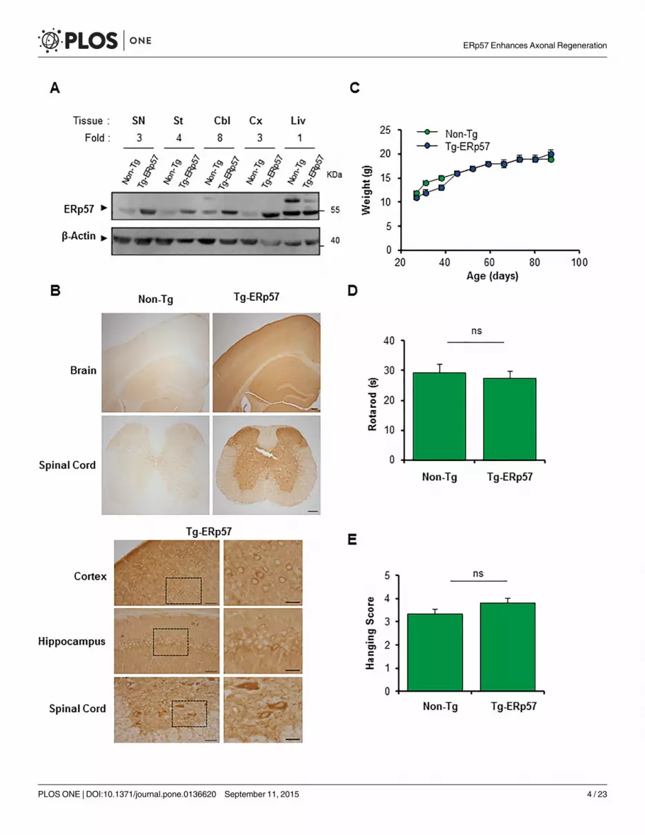
Generation of an ERp57 transgenic mouse model in the nervous systemTo investigate the possible role of ERp57 in the nervous system we generated a transgenicmouse model that overexpresses the human Erp57/Pdia3 cDNA under the control of the PrPpromoter (termed Tg-ERp57) to express the protein at high levels in the nervous system. Theexogenous human ERp57 was designed to carry a FLAG-tag at the C-terminus for detectionpurposes. Several founder lines were generated, however only one line was used in the presentstudy because it showed the highest expression in spinal cord, peripheral nerve, and brain tis-sue that was maintained over several generations. Routinely, Tg-ERp57 heterozygous animalswere crossed with C57BL/6 mice to obtain transgenic and non-transgenic offspring in B6 back-ground with 98% purity. Animals were viable and born in a Mendelian ratio (Table 1). To con-firm the overexpression of ERp57 in the nervous system, we dissected tissue from substantianigra (SN), striatum, cerebellum, cortex, as well as the liver as negative control. An increase inERp57 expression was observed in all tissues of the nervous system to different extends asdetermined by real-time PCR andWestern blot analysis (Fig 1A, S1A and S1B Fig). Quantifica-tion revealed at least a three-fold increase in ERp57 protein levels in transgenic animals com-pared to littermate control mice (Fig 1A). We determined the distribution of ERp57-FLAG indifferent regions of the nervous system including cortex, hippocampus and spinal cord usingan anti-FLAG antibody by immunohistochemistry (Fig 1B). A neuronal-staining pattern wasobserved in Tg-ERp57 mice in all CNS tissues based on morphological analysis. The stainingwas cytosolic and excluded the nucleus consistent with the known subcellular distribution ofERp57 (Fig 1B). Co-immunofluorescence analysis of brain tissue indicated that ERp57-FLAGexpression was mostly restricted to the neuronal compartment and not astrocytes (S1C Fig).Body weight measurements were performed once a week starting at 30 days of age. Normalgrowth curves were obtained for Tg-ERp57 animals (Fig 1C). Furthermore, evaluation of themotor performance of Tg-ERp57 mice using the rotarod assay showed no alterations to this
Table 1. Ratio of births of ERp57 transgenic mice.
Animals Non-Tg Tg-ERp57 Total
N° Observed 54 58 112
Expected (%) 50 50 100
Observed (%) 48.2 51.8 100
doi:10.1371/journal.pone.0136620.t001
ERp57 Enhances Axonal Regeneration
PLOS ONE | DOI:10.1371/journal.pone.0136620 September 11, 2015 3 / 23
ERp57 Enhances Axonal Regeneration
PLOS ONE | DOI:10.1371/journal.pone.0136620 September 11, 2015 4 / 23
parameter (Fig 1D). Similarly, coordination and muscle strength measurements using thehanging test did not reveal any gross effects after ERp57 overexpression in vivo (Fig 1E).
Genotypic distribution (percentage) of the offspring obtained from crosses, carried out toamplify the colony under study (see Materials and Methods). Non-Tg: ERp57 non-transgenicmice Tg-ERp57: ERp57 transgenic mice. The data correspond to generation 3 to 6 of thecolony.
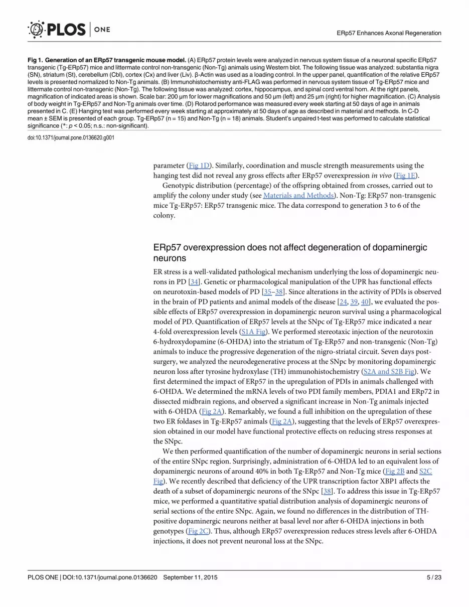
ERp57 overexpression does not affect degeneration of dopaminergicneuronsER stress is a well-validated pathological mechanism underlying the loss of dopaminergic neu-rons in PD [34]. Genetic or pharmacological manipulation of the UPR has functional effectson neurotoxin-based models of PD [35–38]. Since alterations in the activity of PDIs is observedin the brain of PD patients and animal models of the disease [24, 39, 40], we evaluated the pos-sible effects of ERp57 overexpression in dopaminergic neuron survival using a pharmacologicalmodel of PD. Quantification of ERp57 levels at the SNpc of Tg-ERp57 mice indicated a near4-fold overexpression levels (S1A Fig). We performed stereotaxic injection of the neurotoxin6-hydroxydopamine (6-OHDA) into the striatum of Tg-ERp57 and non-transgenic (Non-Tg)animals to induce the progressive degeneration of the nigro-striatal circuit. Seven days post-surgery, we analyzed the neurodegenerative process at the SNpc by monitoring dopaminergicneuron loss after tyrosine hydroxylase (TH) immunohistochemistry (S2A and S2B Fig). Wefirst determined the impact of ERp57 in the upregulation of PDIs in animals challenged with6-OHDA. We determined the mRNA levels of two PDI family members, PDIA1 and ERp72 indissected midbrain regions, and observed a significant increase in Non-Tg animals injectedwith 6-OHDA (Fig 2A). Remarkably, we found a full inhibition on the upregulation of thesetwo ER foldases in Tg-ERp57 animals (Fig 2A), suggesting that the levels of ERp57 overexpres-sion obtained in our model have functional protective effects on reducing stress responses atthe SNpc.
We then performed quantification of the number of dopaminergic neurons in serial sectionsof the entire SNpc region. Surprisingly, administration of 6-OHDA led to an equivalent loss ofdopaminergic neurons of around 40% in both Tg-ERp57 and Non-Tg mice (Fig 2B and S2CFig). We recently described that deficiency of the UPR transcription factor XBP1 affects thedeath of a subset of dopaminergic neurons of the SNpc [38]. To address this issue in Tg-ERp57mice, we performed a quantitative spatial distribution analysis of dopaminergic neurons ofserial sections of the entire SNpc. Again, we found no differences in the distribution of TH-positive dopaminergic neurons neither at basal level nor after 6-OHDA injections in bothgenotypes (Fig 2C). Thus, although ERp57 overexpression reduces stress levels after 6-OHDAinjections, it does not prevent neuronal loss at the SNpc.
Fig 1. Generation of an ERp57 transgenic mousemodel. (A) ERp57 protein levels were analyzed in nervous system tissue of a neuronal specific ERp57transgenic (Tg-ERp57) mice and littermate control non-transgenic (Non-Tg) animals usingWestern blot. The following tissue was analyzed: substantia nigra(SN), striatum (St), cerebellum (Cbl), cortex (Cx) and liver (Liv). β-Actin was used as a loading control. In the upper panel, quantification of the relative ERp57levels is presented normalized to Non-Tg animals. (B) Immunohistochemistry anti-FLAG was performed in nervous system tissue of Tg-ERp57 mice andlittermate control non-transgenic (Non-Tg). The following tissue was analyzed: cortex, hippocampus, and spinal cord ventral horn. At the right panels,magnification of indicated areas is shown. Scale bar: 200 μm for lower magnifications and 50 μm (left) and 25 μm (right) for higher magnification. (C) Analysisof body weight in Tg-ERp57 and Non-Tg animals over time. (D) Rotarod performance was measured every week starting at 50 days of age in animalspresented in C. (E) Hanging test was performed every week starting at approximately at 50 days of age as described in material and methods. In C-Dmean ± SEM is presented of each group. Tg-ERp57 (n = 15) and Non-Tg (n = 18) animals. Student’s unpaired t-test was performed to calculate statisticalsignificance (*: p < 0.05; n.s.: non-significant).
doi:10.1371/journal.pone.0136620.g001
ERp57 Enhances Axonal Regeneration
PLOS ONE | DOI:10.1371/journal.pone.0136620 September 11, 2015 5 / 23
Fig 2. Effects of ERp57 overexpression on the survival of dopaminergic neurons after exposure to 6-OHDA. (A) Pdia1 and Erp72mRNA levels weredetermined in dissected SNpc of Non-Tg and Tg-ERp57 mice 7 d after 6-OHDA injection. Injected and non-injected sides were analyzed using total cDNAand real-time PCR (n = 3 per group). Data is presented as mean and SEM. (B) Non-Tg and Tg-ERp57 mice were injected with 8 μg of 6-OHDA in the rightstriatum, and after 7d dopaminergic neurons (TH+) were quantified by anti-TH immunohistochemistry. Total content of TH-positive somas was measured inmidbrain sections covering the entire SN, in the non-injected (control) and injected (6-OHDA) side, for the indicated genotype (n = 8, Non-Tg; n = 5, Tg-ERp57). Scale bar: 200 μm. (C) Histograms show the number of TH-positive neurons of injected and non-injected sides in 25 μmmidbrain serial sectionsseparated by 100 μm and covering the entire SNpc. The numbers of serial sections indicate the orientation from anterior to posterior. Statistical analysis wasperformed using Mann-Whitney test for all quantifications except for (C) where two-way ANOVA was used followed by Bonferroni posttest (*: p < 0.05; **:p < 0.01; ***: p < 0.001. n.s.: not significant.).
doi:10.1371/journal.pone.0136620.g002
ERp57 Enhances Axonal Regeneration
PLOS ONE | DOI:10.1371/journal.pone.0136620 September 11, 2015 6 / 23
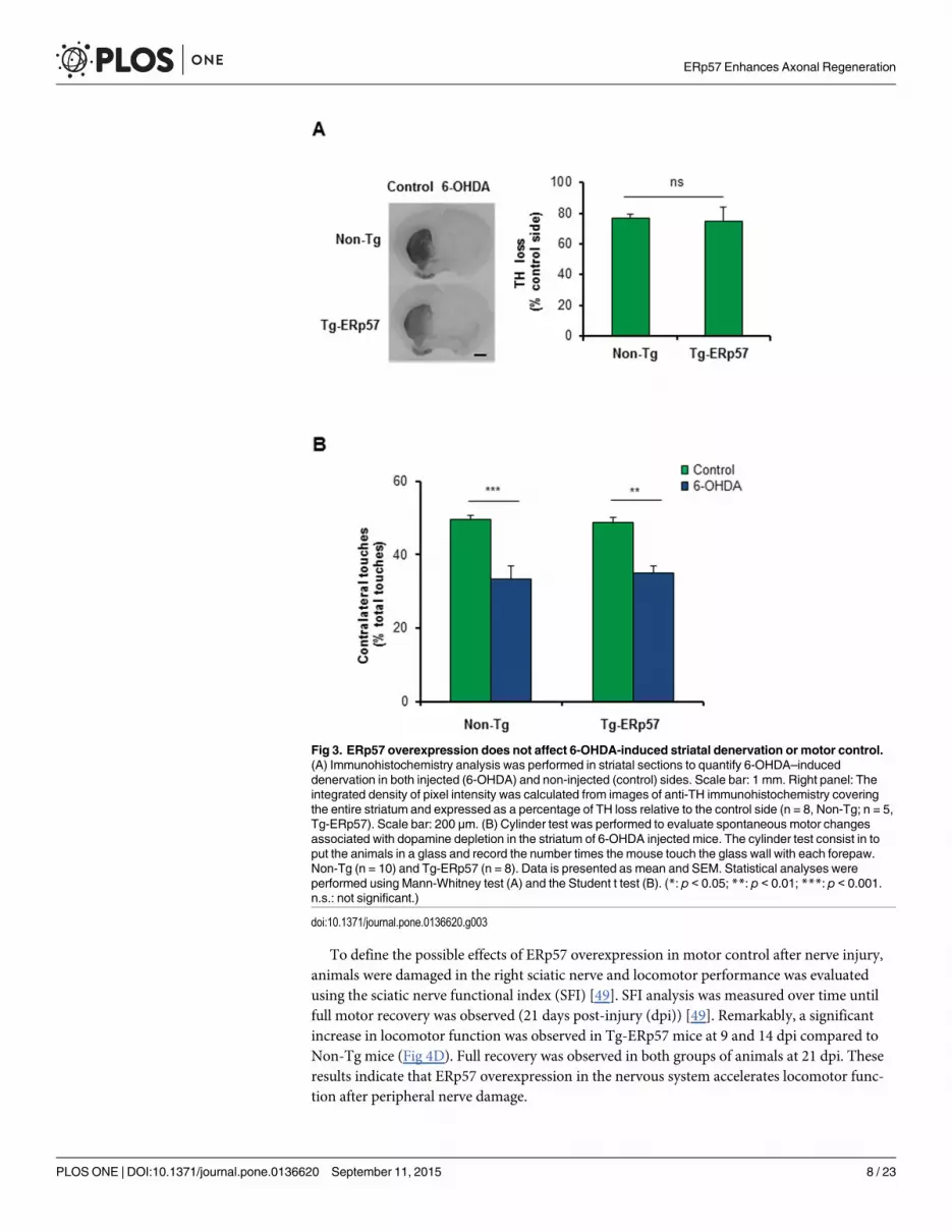
ERp57 overexpression does not affect striatal denervation and motorcontrolAn early event observed in PD is axonal degeneration, leading to the loss of innervation ofstriatal neurons before dopaminergic neuron death [41]. We therefore determined the extentof striatal denervation triggered by 6-OHDA administration in Tg-ERp57 mice. We quantifiedTH staining of the whole striatum using serial sections (S2B Fig), and observed that the per-centage of TH loss in the striatum was similar in the Tg-ERp57 and Non-Tg groups, reaching areduction of near 80% of TH staining (Fig 3A). To evaluate the functionality of the nigro-stria-tal circuit we performed the cylinder test. This assay measures the spontaneous motor changestriggered by dopamine depletion in the striatum, associated to an asymmetrical use of bothforepaws (see methods). Analysis of this behavioral test revealed no differences between Tg-ERp57 and control animals challenged with 6-OHDA (Fig 3B). Thus, the overexpression ofERp57 does not protect dopaminergic neurons against the PD-triggering neurotoxin 6-OHDA.
Based on these negative results, we decided to explore whereas 6-OHDA triggers a classicalER stress response. We monitored the expression levels of Chop using immunohistochemistry.In contrast to previous findings [42], we did not observe an upregulation of CHOP at the SNpcin both Tg-ERp57 and Non-Tg animals (S3A Fig, upper panel). As positive control we injectedthe SNpc with tunicamycin, a pharmacological inducer of ER stress (S3A Fig, bottom panel).We complemented this analysis by monitoring ChopmRNA levels by real time PCR of dis-sected midbrain tissue, obtaining virtually identical results (S3B Fig). We also measured thelevels of Xbp1mRNA splicing in the striatum and SNpc of 6-OHDA injected animals usingRT-PCR in time course experiments. Again, no signs of UPR activation were observed (S3Cand S3D Fig). Since ER stress has been extensively reported in neuronal cell lines treated withPD-inducing neurotoxins, we exposed SH-SY5Y human dopaminergic neurons to differentconcentrations of 6-OHDA, in addition to perform kinetic analysis. In contrast to our previousresults in vivo, treatment of cells with 6-OHDA induced a robust but transient activation ofXbp1mRNA splicing (S3E Fig). This extensive characterization suggests that 6-OHDA modu-lates the expression of PDIs at the SNpc in the absence of a global ER stress response.
Overexpression of ERp57 accelerates functional recovery afterperipheral nerve injuryRecent reports indicate that ER proteostasis is also altered when the peripheral nervous system(PNS) is damaged after mechanical injury or due to neurodegenerative conditions [41, 43].Peripheral nerve injury initiates a tissular reaction distal to the damage site known as Walleriandegeneration that is followed by successful axonal regeneration [44, 45]. We studied the contri-bution of ERp57 to sciatic nerve degeneration and regeneration triggered by mechanical dam-age (Fig 4A). We confirmed the overexpression of ERp57 in the soma of sensory neurons inthe dorsal root ganglia (DRG) and sciatic nerve using anti-FLAG immunohistochemistry (Fig4B) or real-time PCR (S1B Fig). Of note, FLAG immunoreactivity in nerve preparations wasobserved in axons but also in Schwann cells (Fig 4B). To determine the possible modulation ofERp57 during peripheral nerve damage, the sciatic nerve was crushed and then the expressionof ERp57 was analyzed by western blot after 12 or 24 hours post-injury (hpi), which corre-sponds to the time points when Wallerian degeneration is taking place [46–48]. Differentnerve fragments were dissected including the medial region (M) containing the damage site, inaddition to the proximal (P) and distal regions (D) (see schema in Fig 4A). The uninjured con-tralateral nerve was used as control (C). We observed an increase in ERp57 expression in thesciatic after nerve injury using western blot analysis that was higher in the medial region(Fig 4C).
ERp57 Enhances Axonal Regeneration
PLOS ONE | DOI:10.1371/journal.pone.0136620 September 11, 2015 7 / 23
To define the possible effects of ERp57 overexpression in motor control after nerve injury,animals were damaged in the right sciatic nerve and locomotor performance was evaluatedusing the sciatic nerve functional index (SFI) [49]. SFI analysis was measured over time untilfull motor recovery was observed (21 days post-injury (dpi)) [49]. Remarkably, a significantincrease in locomotor function was observed in Tg-ERp57 mice at 9 and 14 dpi compared toNon-Tg mice (Fig 4D). Full recovery was observed in both groups of animals at 21 dpi. Theseresults indicate that ERp57 overexpression in the nervous system accelerates locomotor func-tion after peripheral nerve damage.
Fig 3. ERp57 overexpression does not affect 6-OHDA-induced striatal denervation or motor control.(A) Immunohistochemistry analysis was performed in striatal sections to quantify 6-OHDA–induceddenervation in both injected (6-OHDA) and non-injected (control) sides. Scale bar: 1 mm. Right panel: Theintegrated density of pixel intensity was calculated from images of anti-TH immunohistochemistry coveringthe entire striatum and expressed as a percentage of TH loss relative to the control side (n = 8, Non-Tg; n = 5,Tg-ERp57). Scale bar: 200 μm. (B) Cylinder test was performed to evaluate spontaneous motor changesassociated with dopamine depletion in the striatum of 6-OHDA injected mice. The cylinder test consist in toput the animals in a glass and record the number times the mouse touch the glass wall with each forepaw.Non-Tg (n = 10) and Tg-ERp57 (n = 8). Data is presented as mean and SEM. Statistical analyses wereperformed using Mann-Whitney test (A) and the Student t test (B). (*: p < 0.05; **: p < 0.01; ***: p < 0.001.n.s.: not significant.)
doi:10.1371/journal.pone.0136620.g003
ERp57 Enhances Axonal Regeneration
PLOS ONE | DOI:10.1371/journal.pone.0136620 September 11, 2015 8 / 23
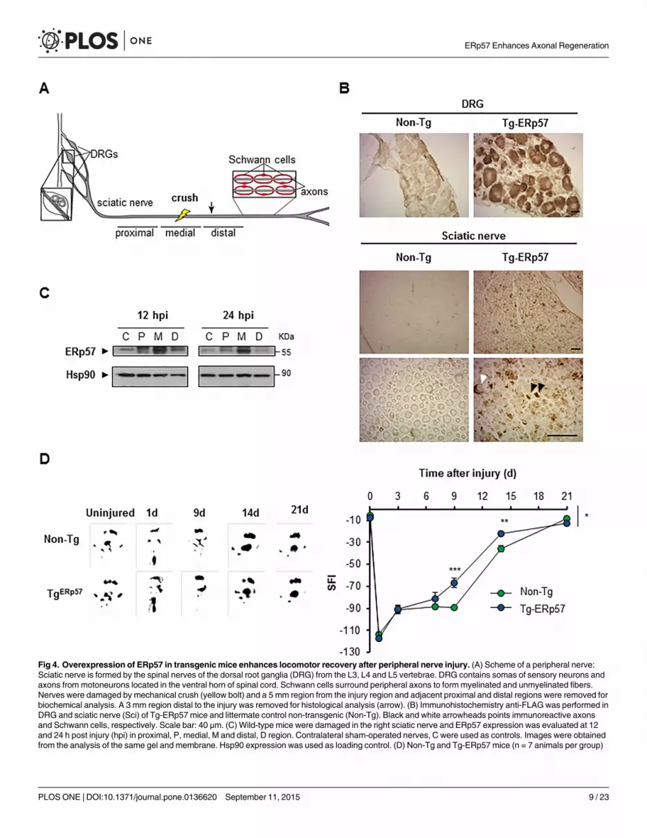
Fig 4. Overexpression of ERp57 in transgenicmice enhances locomotor recovery after peripheral nerve injury. (A) Scheme of a peripheral nerve:Sciatic nerve is formed by the spinal nerves of the dorsal root ganglia (DRG) from the L3, L4 and L5 vertebrae. DRG contains somas of sensory neurons andaxons frommotoneurons located in the ventral horn of spinal cord. Schwann cells surround peripheral axons to form myelinated and unmyelinated fibers.Nerves were damaged by mechanical crush (yellow bolt) and a 5 mm region from the injury region and adjacent proximal and distal regions were removed forbiochemical analysis. A 3 mm region distal to the injury was removed for histological analysis (arrow). (B) Immunohistochemistry anti-FLAGwas performed inDRG and sciatic nerve (Sci) of Tg-ERp57 mice and littermate control non-transgenic (Non-Tg). Black and white arrowheads points immunoreactive axonsand Schwann cells, respectively. Scale bar: 40 μm. (C) Wild-type mice were damaged in the right sciatic nerve and ERp57 expression was evaluated at 12and 24 h post injury (hpi) in proximal, P, medial, M and distal, D region. Contralateral sham-operated nerves, C were used as controls. Images were obtainedfrom the analysis of the same gel and membrane. Hsp90 expression was used as loading control. (D) Non-Tg and Tg-ERp57 mice (n = 7 animals per group)
ERp57 Enhances Axonal Regeneration
PLOS ONE | DOI:10.1371/journal.pone.0136620 September 11, 2015 9 / 23

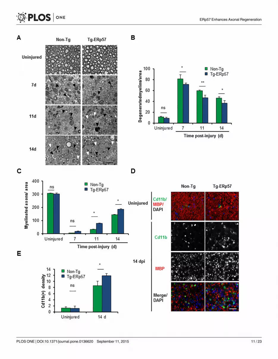
ERp57 overexpression enhances axonal regeneration after peripheralnerve injuryWe then evaluated myelin removal and regeneration of nerve fibers of Tg-ERp57 mice after sci-atic nerve crush. Animals were injured and the morphology of myelinated axons was visualizedand quantified in peripheral nerves at 3 mm distal from the injury site using semi-thin sections(Fig 5A). At basal levels, a homogeneous distribution of intact myelinated axons with con-densed myelin was observed in both Tg-ERp57 and littermate control mice (Fig 5A, upperpanel). Electron microscopy analysis also showed no differences in the ultrastructure of unin-jured nerves (S4A Fig). We then quantified the content of myelinated and degenerating axonsover time after sciatic nerve injury. In our crush model, an initial increase in degeneratedmyelin is observed that then decays progressively as myelin removal and axonal regenerationproceeds [44]. Consistent with the locomotor measurements, Tg-ERp57 mice exhibited a sig-nificant reduction in the number of degenerated myelin (Fig 5B), with a concomitant increasein the number of re-myelinated axons (Fig 5C). Qualitative electron microscopy analysis at 14dpi corroborated these results (S4A Fig).
Myelin removal and subsequent axonal regeneration is mediated in part by Schwann cellsand through the infiltration of macrophages at later stages after nerve injury [50, 51]. To ana-lyze the impact of ERp57 overexpression on the cellular immune response associated to Wal-lerian degeneration, we evaluated the infiltration of Cd11b+-macrophages into injured nerves.Measurement of Cd11b+ density in the distal stump of Tg-ERp57 mice revealed a significantincrease in macrophage content at 14 dpi when compared to Non-Tg littermates (Fig 5D and5E). As control we monitored the levels of ERp57 in macrophages. Importantly, macrophagesisolated from the lung of Tg-ERp57 animal (S4B Fig). Similarly, co-immunofluorescence analy-sis of FLAG-tagged ERp57 and the macrophage marker Cd11b did not show any co-expressionin Tg-ERp57 mice at the sciatic nerve after peripheral nerve crush (S4C Fig). In contrast, aclear colocalization was observed between ERp57-FLAG and the Schwann cell marker MBP(S4D Fig). Taken together, these results indicate that overexpression of ERp57 modulates theaxonal regeneration process, improving locomotor recovery after damage to the PNS.
DiscussionPDIs are emerging as interesting therapeutic targets and biomarkers in multiple neurodegener-ative diseases associated with protein misfolding and ER stress. However, so far no functionalstudies are available addressing the actual role of PDIs in neurodegeneration. ERp57 is a majorPDI family member expressed in the nervous system and has been identified as one of themain proteins upregulated in tissue derived from ALS and Creutzfeldt-Jacob patients [15, 16,18, 19, 22]. A few knockout mouse models for PDIs have been described to date, includingERp57, ERp29, ERdj5, PDIA1 and AGR2 [31, 52–57]. ERdj5 knockout mice develop alteredER proteostasis in salivary glands [58], whereas AGR2/PDIA17 knockout mice are viable, butshow decreased levels of mucin, an essential component of the protective mucus in the intes-tine, resulting on severe colitis [56, 57]. Complete ERp57 deficiency in mice is lethal at embry-onic day 13.5, possibly due to altered STAT3 signaling [59]. Conditional deletion of ERp57 inB cells leads to altered MHC class I peptide loading. However, no effects were detected on thesecretion of immunoglobulins, a highly glycosylated protein containing disulfide bonds [52].
were injured and the locomotor performance was analyzed using the sciatic nerve functional index (SFI). (Left panel) SFI was measured for 21 days afterinjury. (Right panel) Representative footprints of the damaged hind limb of Non-Tg and Tg-ERp57 mice at 0 (uninjured), 1, 9, 14 and 21 days post-injury. Dataare shown as mean ± S.E.M. * p < 0.05, ** p < 0.01, *** p < 0.001. For SFI analysis data were analyzed by two-way ANOVA followed by Bonferroni posttest.
doi:10.1371/journal.pone.0136620.g004
ERp57 Enhances Axonal Regeneration
PLOS ONE | DOI:10.1371/journal.pone.0136620 September 11, 2015 10 / 23
ERp57 Enhances Axonal Regeneration
PLOS ONE | DOI:10.1371/journal.pone.0136620 September 11, 2015 11 / 23
ERp29 deficiency alters the susceptibility of cells to apoptosis [53], whereas PDIA1 deficiencyis lethal, and conditional deletion of this gene in platelets impairs thrombus formation [54].Overall, in none of these studies CNS-related phenotypes were reported so far when the expres-sion of PDIs was targeted in vivo.
Here we addressed the impact of ERp57 on pathological conditions affecting the nervoussystem by creating a transgenic mouse model. Tg-ERp57 animals presented increased locomo-tor recovery after peripheral nerve injury, associated with improved regeneration of axons andmyelin removal. According with previous evidence, PNS damage triggers the upregulation ofcomponents of the ER proteostasis network such as calnexin, BiP and ERp29 [60, 61]. Consis-tent with this, we observed an upregulation of ERp57 levels in injured nerves. We speculatethat our transgenic model may generate a preconditioned environment, enhancing the abilityof neurons to handle protein-folding stress generated by the mechanical injury to the axon.Consistent with our results, a recent report described the occurrence of ER stress in models ofperipheral nerve injury triggered by diabetes [62]. Treatment of mice with chemical chaperonsreduced axonal degeneration, correlating with an attenuated ER stress response [62]. Impor-tantly, another study identified the ER stress-related sensor LUMAN/CREB3 as an ER-locatedprotein that is activated by mechanical injury to the axon [63]. LUMANmay communicatestress signals to the nucleus to enforce an adaptive reaction to reduce nerve damage.
In contrast to the protective consequences of ERp57 overexpression in the PNS, and despiteexpectations, our Tg-ERp57 mouse model did not show any protection on a pharmacologicalmodel of PD. Although ERp57 reduced the upregulation of Pdia1 and Erp72mRNA levelsafter exposing animals to 6-OHDA, we were unable to detect any differential effects on dopa-minergic neuron survival or striatal denervation in Tg-ERp57 animals. However, in our modelwe did not observe clear signs of UPR activation, suggesting that the modulation of ERp72 andPDIA1 expression by 6-OHDA may be ER stress-independent. Oxidative stress is the majorcontributor to 6-OHDA neurotoxicity [64, 65] and this condition has been associated with sec-ondary proteostasis alterations in PD [66]. Oxidative conditions also result in ERp57 inactiva-tion by S-nitrosylation, similarly to the effects reported for PDIA1 in human PD-derived tissue[24]. Thus, although we succeeded to overexpress ERp57 in the striatum and SNpc, it might bepossible that ERp57 was post-translationally modified and inhibited by the oxidative stressgenerated by 6-OHDA, which may explain our negative results. It remains to be determined ifERp57 overexpression protects dopaminergic neurons on PD models based on alpha Synucleinaggregation.
ERp57 deficiency may result in impaired folding and function of myelin protein 0 (P0), oneof the major peripheral myelin components [67]. The ERp57 binding partner, CNX, interactsand assists the folding of peripheral myelin proteins, contributing to peripheral neuropathiesin mouse models [68]. CNX deficiency leads to myelinopathy associated with the occurrence ofmotor defects [28, 29]. Importantly, Schwann cells and oligodendrocytes are highly prone toundergo ER stress as demonstrated in many different disease models due to their high demandfor protein and lipid synthesis [2, 69]. Since the PrP promoter is also active in Schwann cells[70, 71], we propose that ERp57 may have a relevant activity in sustaining glial proteostasis as
Fig 5. Overexpression of ERp57 in transgenicmice increases axonal regeneration after peripheral nerve injury. (A) Non-Tg and Tg-ERp57mice weredamaged and sciatic nerves were extracted at 7, 11 and 14 days post-injury (dpi). Transversal semi-thin sections were obtained from the distal region andremyelinated axons (white arrows) and degenerated myelins (black arrows) were analyzed. (B) Quantification of remyelinated axons and (C) degeneratedmyelin density was measured at 7, 11 and 14 days post-injury. (D) Tg- ERp57 and Non-Tg sciatic nerves were processed for immunofluorescence in uninjuredconditions and at 14 dpi. Sciatic nerves were analyzed for MBP (red), Cd11b (green) and nuclei were stained using DAPI (blue). (E) The staining density forCd11b was quantified 14 days after injury (right panel). Data is presented as mean ± S.E.M. * p < 0.05, ** p < 0.01, *** p < 0.001. For histological analysis,statistical differences were obtained using a student’s t-test (n = 3 animals per group). Scale bar: 20 μm.
doi:10.1371/journal.pone.0136620.g005
ERp57 Enhances Axonal Regeneration
PLOS ONE | DOI:10.1371/journal.pone.0136620 September 11, 2015 12 / 23

a central component of the CNX-CRT cycle. In this context, ERp57 may contribute to maintainmyelin structure or to reduce stress levels in Schwann cells. Previous evidence uncovered theconsequences of altering Schwann cell proteostasis in the peripheral nerve. For example, invitro studies using Chop deficient Schwann cells revealed an important proapoptotic activity ofthis factor upon pro-inflammatory challenges [72]. In the context of a demyelinating pathol-ogy, targeting PERK signaling in Charcot-Marie-Tooth-1B disease at the level of Chop [73] oreIF2 alpha phosphorylation provide strong protection [74, 75]. Taken together, our resultsdescribe a novel genetic model to manipulate ERp57 levels in the nervous system, demonstrat-ing for the first time a relevant neuroprotective effect of this essential PDI in the PNS aftermechanical injury.
Materials and Methods
Generation of the ERp57 transgenic mouse modelThis study was carried out in strict accordance with the recommendations in the Guide for theCare and Use of Laboratory Animals of the National Institutes of Health. The protocol wasapproved by the Committee on the Ethics of Animal Experiments of the University of Chile(protocol number CBA#0265 FMUCH). Mice were housed under a 12:12 h light-dark cyclewith access to food and water ad libitum.
The expression plasmid MoPrP.XhoI was kindly provided by David Borchelt of the JohnsHopkins School of Medicine, Baltimore, USA (Borchelt et al., 1996). Human ERp57 cDNA(Gene ID: 2923) was introduced into the MoPrP.XhoI plasmid and fused with the sequencecoding for a FLAG tag using the XhoI restriction site. The resulting construct expresses FLAG-tag ERp57 at the N-terminus under the control of the prion (PrP) promoter. ERp57 transgenicmice were generated in the “Centro de Estudios Científicos” (CECS), Valdivia, Chile. The fol-lowing primers were used for genotyping: Erp57 866 forward 5’-AATTCCTGGATGCTGGGCACAAAC -3’ and Erp57 1535 reverse 5’-TCTGCTTGTCATCGTCGTCCTTGT-3’. Briefly,1 μL of DNA was incubated with 10 μL of Go Taq Master Mix (Promega) and 1 μM of eachprimer in a final volume of 20 μL. The PCR program used consisted in one cycle of denatur-ation for 5 min at 94°C, 20 cycles of 30 sec at 94°C, 30 sec at 65°C (decreasing 0.5°C per cycle),50 sec at 72°C; 16 cycles of 50 sec at 94°C, 30 sec at 55°C, 50 sec at 72°C and a final cycle ofelongation for 5 min at 72°C.
Motor and behavioral testsFor the Rotarod test, mice were placed into a rod rotating (Model LE8500, Panlab SL) at anaccelerating speed starting with 4 rpm up to 40 rpm within 2 min [76]. The time until micecould not maintain themselves on the rod was measured. Training was performed 3 times perday for 5 consecutive days. The test was applied once per week starting at 52 days of age until80 days of age.
For the hanging test, individual mice were placed hanging with their forepaws on a horizon-tal bar (39 cm length and 35 cm height). The performance of the mouse and the body positionwas observed for 30 seconds and recorded with a video-camera. The test was performed 3times in one day once per week starting at 53 days of age until 81 days. A score was derivedfrom each video and averaged for the three measurements done at the same day as describedbefore [77]: score 0 was given when the mouse could not hold onto the bar for more than 10seconds; score 1, when the mouse maintained itself on the bar with the forelimbs for a mini-mum of 10 sec but not longer than 30 sec; score 2 if the mouse maintained itself with the fore-limbs and completed 30 sec; score 3 if the mouse used the forelimbs and one or two of the
ERp57 Enhances Axonal Regeneration
PLOS ONE | DOI:10.1371/journal.pone.0136620 September 11, 2015 13 / 23

hindlimbs but not the tail; score 4 if the mouse used all four limbs and the tail; and score 5 ifthe mouse actively escaped the bar in 30 sec.
Pharmacological model of PDStereotaxic injections were performed in 3 month-old male and female Non-Tg and Tg-ERp57mice. Mice were intraperitoneally anesthetized with ketamine/xylazine (ketamine 100 mg/kg,xylazine 10 mg/kg; Vetcom) and placed into a stereotaxic frame (David Kopf Instruments).Unilateral injections of 4 μg/μL 6-hydroxydopamine (6-OHDA; Sigma-Aldrich) dissolved in0.2% ascorbic acid were administered as described before [38]. 6-OHDA injections were per-formed in a single point, injecting 2 μL in the right striatum using a 5 μL Hamilton syringe atthe following coordinates: anteroposterior (AP) +0.07 cm; mediolateral (ML) −0.17 cm; dorso-ventral (DV) −0.31 cm. For histological analysis mice were euthanized by CO2 narcosis 7 daysafter the surgical procedure.
The Cylinder test is designed to evaluate locomotor asymmetry in rodent models of PD. Asthe animal moves within an open-top, clear glass cylinder, its forelimb activity while rearingagainst the wall of the arena is recorded. Forelimb use is defined by the placement of the wholepalm on the wall of the arena, which indicates its use for body support. Forelimb contacts whilerearing were scored. The analysis was performed blinded. The result is plotted as the percent-age of contralateral touches relative to total touches with both paws.
Peripheral nerve crush modelSciatic nerve injury was performed in 3-month-old male and female Non-Tg and Tg-ERp57mice as described before [78]. Animals were intraperitoneally anesthetized with 2-2-2 tribro-moethanol (330 mg/Kg, Sigma, St. Louis, MO, USA) and treated with Tramadol (30 mg/Kg) asanalgesic. Surgical dissection of the skin and muscle was performed at the level of the sciaticnotch to expose the right sciatic nerve. The sciatic nerve was crushed three times for 5 secondswith a Dumont #5 forceps (Fine Science Tools INC. CA, USA) and the skin was sutured withmouse metal clips. During recovery, mice were placed in a temperature-controlled chamber. Atdifferent days post-surgery, animals were euthanized by overdose of anesthesia and the sciaticnerve was removed for different analyses.
The sciatic nerve functional index (SFI) was performed to determine locomotor capabilityafter sciatic nerve injury. Paw prints of individual mice were obtained by moistening the hin-dlimbs with black ink and having them walk unassisted along an 11 X 56 cm white paper corri-dor. Tracks were obtained before surgery (day 0), and 1, 3, 7, 9, 14 and 21 days after nerveinjury. All paw prints were obtained and analyzed in a blinded fashion. Two different parame-ters were analyzed: toe spread (TS), the distance between the first and fifth toes, and printlength (PL), the distance between the third toe and the hind pad. Measurements of all parame-ters were made for the right injured hindpaw (experimental; E) and the left uninjured hindpaw(normal; N) and the SFI was calculated according to the following formula: SFI = 11.89 ((ETS–NTS) / NTS) – 51.2 ((EPL–NPL) / NPL) –7.5 as previously described [79].
Tissue extractThe ventral midbrain (containing the entire Substancia Nigra pars-compacta, SNpc), striatum,and cortex from both hemispheres were dissected and homogenized in 100 μL of ice-cold0.1 M PBS (pH 7.4) supplemented with a protease inhibitor cocktail (Roche). The homogenatewas divided into two fractions for total mRNA and protein extraction, followed by standardpurification and quantification protocols [80]. Protein extraction was performed in RIPAbuffer (20 mM Tris pH 8.0, 150 mMNaCl, 0.1% SDS, 0.5% deoxycholate, and 0.5% Triton
ERp57 Enhances Axonal Regeneration
PLOS ONE | DOI:10.1371/journal.pone.0136620 September 11, 2015 14 / 23
X-100) containing a protease inhibitor cocktail and phosphatase inhibitor cocktail (Sigma-Aldrich).
For sciatic nerves extracts, the nerve segments containing the injury region (5 mm of medialregion), and proximal and distal nerve segments of the same size were collected. The contralat-eral sciatic nerve was used as a control. Sciatic nerves were homogenized in extraction buffer(95 mM NaCl, 25 mM Tris-HCl pH 7.4, 10 mM EDTA pH 8.0, 1% SDS, 1 mM NaF, 1 mMNa3VO4 and 1% Protease Inhibitor Cocktail [PIC, Sigma-Aldrich, #P8340]). Lysates were soni-cated, centrifuged at 13,000 rpm for 10 min at 4°C and the supernatants were used for proteinanalysis. Samples were loaded onto SDS/PAGE gels and blotted onto PVDF membranes. Thefollowing antibodies and dilutions were used: anti-HSP90, (1:5000, sc-7947, H114, Santa Cruz,Santa Cruz, CA, USA); anti-ERp57 (1:3000, SPA-585, Stressgen). Band intensities were quanti-fied and normalized to Hsp90 as a loading control. Densitometry analysis was performed usingImageJ software (NIH, Bethesda, MD, USA).
RNA extraction and real time PCRTotal RNA was isolated from ventral midbrain (containing entire SNpc), dorsal root ganglia(from lumbar vertebrae L3 and L4) and medial sciatic nerves. After homogenization in PBS,total mRNA was purified using TRIzol (Life Technologies), and cDNA was synthesized from1 μg of RNA using an Applied Biosystems Reverse-Transcription Kit. Quantitative real-timePCR was performed in a Stratagene lightcycler system using SYBR Green (Applied Biosystems)using the following primers: Erp72 forward, 50-ACTCTCCGGGAATTTGTCACA-30; Erp72reverse, 50-ATGTCGTTGGCGAGTAGCATC-30; Pdia1 forward, 5’-CAAGATCAAGCCCCACCTGAT-3’; Pdia1 reverse, 5’-AGTTCGCCCCAACCAGTACTT-3’;-human Erp57 for-ward, 5’- GCC TCC GAC GTG CTA GAA C -3’; human Erp57 reverse 5’- GCG AAG AACTCG ACG AGC AT -3’; Chop forward 5’-TGGAGAGCGAGGGCTTTG-3’, Chop reverse 5’-GTCCCTAGCTTGGCTGACAGA-3’.
Xbp1mRNA splicing assay was performed as previously described [81] using the followingprimers: Xbp1 forward 5’-ACACGCTTGGGAATGGACAC-3’; Xbp1 reverse: 5’-CCATGGAAGATGTTCTGGG-3’.
Histological analysisFor ERp57-FLAG expression analysis, Tg-ERp57 and Non-Tg control littermates were anes-thetized and perfused through the ascending aorta with isotonic saline, followed by ice-cold 4%paraformaldehyde in 0.1 M PBS (pH 7.4). Tissue was sectioned using a Leica cryostat (Leica,Nussloch, Germany) as follow: brain, 25 μm coronal sections; spinal cord, 18 μm transversalsections; and dorsal root ganglia (DRG) and sciatic nerve, 10 μm transversal sections. Forimmunohystochemistry, tissue was incubated 30 min at room temperature (RT) with 3% H2O2
in 10% methanol in PBS, followed by epitope retrieval using 10 mM citrate buffer pH 6.0(home made) for 15 min at 95°C. After overnight (ON) incubation in blocking solution (BSA5%, Triton X-100 0.3% in PBS) at 4°C, sections were incubated with rabbit anti-FLAG antibody(1:250, F7245, Sigma) for 2 h at RT, followed by incubation with HRP-conjugated goat anti-rabbit antibody (1:1000, Invitrogen) for 1 h incubation at RT. Immunoreactivity was developedusing DAB HRP substrate kit (Vector Laboratories). Tissue staining was visualized with aninverted microscope (Olympus IX71). For co-immunofluorescence analysis, tissue was incu-bated with 10 mM citrate buffer pH 6.0 (home made) for 15 min at 95°C for epitope retrieval.After 1 h in blocking solution (5% BSA, 0.3% Triton X-100 in PBS) at RT, sections were incu-bated with rabbit anti-FLAG antibody (1:250, F7245, Sigma) for 2 h at RT, followed by incuba-tion with mouse anti-TH (1:300,Millipore) or anti-GFAP (1:300, Sigma) ON at RT. Slices were
ERp57 Enhances Axonal Regeneration
PLOS ONE | DOI:10.1371/journal.pone.0136620 September 11, 2015 15 / 23
then incubated with goat anti-rabbit (1:1000, Alexa 568, Invitrogen) and goat anti-mouse anti-body (1:1000, Alexa 488, Invitrogen) for 1 h incubation at RT. Slices were visualized with aconfocal microscope (Olympus Spectral FV1000 confocal microscope).
To analyze CHOP expression, tissue was incubated 30 min at room temperature with 3%H2O2 in 10% methanol in PBS, followed by epitope retrieval using buffer citrate pH 6.0(DAKO) for 20 min at 95°C. After incubation in blocking solution (10% Goat serum, 4% BSA,0.1% Triton X-100 in PBS) for 1.5 h at RT, sections were incubated with rabbit anti-CHOPantibody (1:100, Santa Cruz) ON at 4°C, followed by incubation with HRP-conjugated goatanti-rabbit antibody (1:1000, Invitrogen) for 1 h incubation at RT. Immunoreactivity wasdeveloped using DAB HRP substrate kit (Vector Laboratories). Tissue staining was visualizedwith an inverted microscope (Olympus IX71).
For the quantification of dopaminergic neuron degeneration in the 6-OHDA model, thebrain was sectioned into 25 μm coronal frozen sections containing the rostral striatum andmidbrain on a Leica cryostat (Leica, Nussloch, Germany). Free-floating midbrain and striataltissue sections were obtained and processed for immunohistochemistry. For immunohisto-chemical analysis, sections were incubated overnight at 4°C in blocking solution with tyrosinehydroxylase (TH) (1:2500; Calbiochem) primary antibody and developed with biotinylatedsecondary anti-rabbit antibody (1:500; Vector Laboratories) and avidin-biotin peroxidase com-plex (ABC Elite Kit; Vector Laboratories). Tissue was visualized with an inverted microscope(Olympus IX71).
Sciatic nerves were extracted at 14 days post injury (dpi). A 3 mm segment located 3 mmdistal to the crush site was removed and fixed for 1 h in 4% paraformaldehyde in 0.1 M PBS(pH 7.4). Nerves were then subjected to a sucrose gradient (10, 20 and 30% sucrose in PBS),included in optimal cutting temperature compound (OCT, Sakura Finetek) and fast frozen inliquid nitrogen. The tissue was transversally sectioned (10 μm thickness) using a cryostat(Leica, Nussloch, Germany) and mounted on Superfrost Plus slides (Thermo Fisher Scientific).For immunofluorescence, sections were blocked/permeabilized with 2% fish skin gelatin(Sigma-Aldrich) and 0.1% Triton X-100 in PBS for 1 h at RT and incubated with primary anti-bodies in the same solution overnight at 4°C. Then, incubated in secondary antibodies for 2 hat RT and mounted in Vectashield (Vector Laboratories) as previously described [82]. Sectionswere immunostained using the following antibodies and dilutions: rabbit anti-myelin basicprotein (MBP) (1:500, M3821, Sigma), rat anti-MBP (1:50, a gift from ML Feltri, Hunter JamesKelly Research Institute & Biochemistry, University at Buffalo, New York, USA), rat anti-Cd11b (1:500, MCA74G, Serotec) and rabbit anti-FLAG (1:250, F7245, Sigma). Electronmicroscopy analyses in sciatic nerves were made at 7, 11 and 14 dpi. A 3 mm region of the sci-atic nerve, located 6 mm distal to the injury site was removed and fixed overnight with 2.5%glutaraldehyde, 0.01% picric acid and 0.1 M cacodylate buffer, pH 7.4. Nerves were incubatedin the same buffer with 1% OsO4 for 1 h and then immersed in 2% uranyl acetate for 2h, dehy-drated in a gradient of ethanol and propylene oxide and infiltrated in Epon (Ted Pella) as pre-viously described [83]. Transversal semi-thin and ultra-thin sections were obtained using anultramicrotome. All images of tissue sections were obtained in an inverted Olympus fluores-cent microscope and analyzed using ImageJ software (NIH, Bethesda, MD, USA).
Dopaminergic neuron countingEstimation of the number of TH-positive neurons stained by immunohistochemistry was per-formed manually. Results are expressed as the total number of TH-positive neurons per hemi-sphere. To determine the percentage of TH-positive cell loss in the SNpc of 6-OHDA injectedmice, the number of dopaminergic cells in the injected and non-injected side was determined
ERp57 Enhances Axonal Regeneration
PLOS ONE | DOI:10.1371/journal.pone.0136620 September 11, 2015 16 / 23
by counting in a blinded manner the total number of TH-positive cells in midbrain serial sec-tions containing the entire SNpc (between the AP−0.29 and AP−0.35 cm coordinates). Resultswere expressed as the percentage of TH-positive neurons in the injected hemisphere comparedwith the non-injected side. In addition, striatal denervation was quantified by measuring theoptic density from serial sections covering the entire striatum using ImageJ software (NIH,Bethesda, MD, USA). The total integrated density per hemisphere was quantified. Results areexpressed as the percentage of the integrated density in the injected hemisphere compared tothe non-injected side.
Isolation of alveolar macrophagesLungs were minced and then were digested for 30 min at 37°C with Collagenase (1 mg/ml;Roche) and recombinant DNase I (0.01 U/μL) without serum, followed by lysis of red bloodcells. Samples were stained with allophycocyanin anti-CD11c (N418; BD Pharmingen), allo-phycocyanin-indotricarbocyanine–conjugated antibody to MHC class II (M5/114, 15, 2; Biole-gend), 7-aminoactinomycin D (7-AAD) viability dye (Life technologies) and Fc Block antibody(2.4G2, BD Pharmingen). Alveolar macrophages were isolated by cell sorting as reported [84].Briefly, cells were isolated with gating on live cells, FSC/SSC high granularity, CD11c+, MHCIIintermediate and high auto fluorescent signal. Cells were sorted on a FACSAria (BDBiosciences).
Models of ER stressSH-SY5Y cells were cultured in DMEM supplemented with 10% fetal bovine serum and antibi-otics (10,000 U/ml Penicillin, 10 μg/ml streptomycin), at 37°C and 5% CO2. For induction ofER stress in vitro, the ER stressors tunicamycin, was added to the cell culture medium ofSH-SY5Y cells followed by the measurement of ER stress markers by PCR. For induction of ERstress in vivo, mice received a single intracerebral injection of 2 μL of tunicamycin (5 5 μg/ μL)diluted in DMSO at the following coordinates: AP: -0,29 cmML: -0,13 cm and DV: -0,42 cmand sacrificed 24 h post injection.
Supporting InformationS1 Fig. Generation of a human ERp57-FLAG transgenic mice. (A, left panel) ERp57 proteinlevels were analyzed in the substancia nigra of ERp57 transgenic (Tg-ERp57) mice (n = 5) andlittermate control non-transgenic (Non-Tg) animals (n = 6) using Western blot. β-Actin wasused as a loading control. (A, right Panel) quantification of expression levels normalizedagainst Non-Tg protein levels. (B) hERp57mRNA levels were determined in dissected dorsalroot ganglia (DRG) (left) and sciatic nerve (right) of Non-Tg (n = 3) and Tg-ERp57 (n = 5)mice using real-time PCR. (C) Co-immunofluorescence anti-FLAG(red) and anti-TH(green)in midbrain tissue (upper panel) and anti-FLAG (red) and anti-GFAP (green) in hippocampus(bottom panel), showing co-localization of ERp57-FLAG transgene and TH dopaminergicneuron marker (yellow) but not co-localization with GFAP astrocyte marker. Scale bar: 50 μm.In A and B data are shown as mean ± S.E.M. � p< 0.05, �� p< 0.01, ��� p< 0.001.(TIF)
S2 Fig. The 6-OHDAmodel. Immunohistochemistry anti-TH in serial sections of 25 μm ofthick spaced by 100 μm covering the entire substantia nigra (A) and striatum (B) of one repre-sentative Non-Tg and Tg-ERp57 animal injected with 8 μg of 6-OHDA into the right striatumand sacrificed 7 days later.(TIF)
ERp57 Enhances Axonal Regeneration
PLOS ONE | DOI:10.1371/journal.pone.0136620 September 11, 2015 17 / 23
S3 Fig. Lack of an ER stress in the SNpc of animals exposed to 6-OHDA. (A) Analysis ofChop expression levels using immunohistochemistry of animals injected with 8 μg of 6-OHDAinto the right striatum and sacrificed 7 days later (upper panel). The image is representative ofNon-Tg and Tg-ERp57 (n = 3 per group). As control to induce Chop, animals were injected withtunicamycin (Tm) and sacrificed 24 h later (bottom panel). Scale bar: 200 μm. (B) ChopmRNAlevels were determined by real-time PCR in dissected SNpc fromWT animals injected with 8 μgde 6-OHDA after indicated time post injection. As positive animals were injected with 10 μg ofTm directly into the SNpc and sacrificed 24 h post-injection. Values were normalized by actinand are shown as fold of induction relative to non-injected side. (C) Xbp1mRNA splicing wasmonitored in the SNpc or the striatum (D) of animals injected with 6-OHDA for indicated timepoints. The injected and non-injected side of the same animal was compared in two independentanimals per group. As positive control for the assay, MEFs cells treated with 2,5 μg/ml of Tm for16 h. (D) Xbp1mRNA splicing was also monitored in SH-SY5Y cells treated with 6-OHDA forindicated time points and concentrations. NT: not treated SH-SY5Y cells, (-) negative controlwithout template, Xbp1u: unspliced Xbp1mRNA, Xbp1s: spliced Xbp1mRNA.(TIF)
S4 Fig. ERp57 overexpression reduces axonal degeneration. (A) Electron microscopy ofNon-Tg and Tg-ERp57 uninjured and 14 days-damaged nerves. In uninjured conditions whitearrowheads indicate axoplasm of myelinated fibers, black arrowheads, compact myelin sheathsand asterisks, unmyelinated fibers. At 14 days post-injury black arrows indicated degeneratedmyelins and white arrows, remyelinated axons. Scale bar: 4 μm. (B) hERp57mRNA levels weredetermined in macrophages isolated from alveoli of Non-Tg and Tg-ERp57 mice after cell sort-ing of Cd11b-positive cells using real-time PCR (n = 2 per group). Cortex tissue from these ani-mals was used as positive control. (C) Non-Tg and Tg-ERp57 mice were damaged and sciaticnerves were extracted at 14 days post-injury. Contralateral uninjured nerves were used as con-trol. Transversal slides were processed for immunofluorescence for FLAG (red), MBP (green)and DAPI (blue) to identify Schwann cells. (D) Animals described in C were used for immuno-fluorescence to stain FLAG (red), Cd11b (green) and DAPI (blue) to analyse the infiltratingmacrophage population. At the right panels, magnifications of indicated areas are shown. Scalebar: 20 mm. In B data are shown as mean.(TIF)
AcknowledgmentsWe thank Monica Perez for excellent EM processing. Javiera Ponce for technical support inanimal studies. This work was funded by Ring Initiative ACT1109 (FC and CH), FONDEFD11I1007, Millennium Institute No. P09-015-F, the Frick Foundation, FONDECYT no.1140549, Michael J Fox Foundation for Parkinson Research, COPEC-UC Foundation, CONI-CYT grant USA2013-0003, ECOS-CONICYT C13S02, and ALS Therapy Alliance (CH). Mil-lennium Nucleus P-07-011-F, FONDECYT no. 1110987 (FC), FONDECYT no.1150608 (RV).FONDECYT no. 3130351 (DM), FONDECYT no. 3120146 (GM), CONICYT PAI No82130031. VC, PR and MO are postgraduate fellows supported by a CONICYT fellowship.”
The funders had no role in study design, data collection and analysis, decision to publish, orpreparation of the manuscript"
Author ContributionsConceived and designed the experiments: VC MOUW PR DM PV FO GM RLV CH. Per-formed the experiments: VC MO UW PR CA DM PV FO. Analyzed the data: VC MO UW PR
ERp57 Enhances Axonal Regeneration
PLOS ONE | DOI:10.1371/journal.pone.0136620 September 11, 2015 18 / 23
DM PV RLV CH. Contributed reagents/materials/analysis tools: FO BK. Wrote the paper: VCMO FAC CH.
References1. Soto C. Unfolding the role of protein misfolding in neurodegenerative diseases. Nature reviews Neuro-
science. 2003; 4(1):49–60. PMID: 12511861
2. Hetz C, Mollereau B. Disturbance of endoplasmic reticulum proteostasis in neurodegenerative dis-eases. Nature reviews Neuroscience. 2014; 15(4):233–49. doi: 10.1038/nrn3689 PMID: 24619348
3. BalchWE, Morimoto RI, Dillin A, Kelly JW. Adapting proteostasis for disease intervention. Science.2008; 319(5865):916–9. doi: 10.1126/science.1141448 PMID: 18276881
4. Hetz C, Martinon F, Rodriguez D, Glimcher LH. The unfolded protein response: integrating stress sig-nals through the stress sensor IRE1alpha. Physiological reviews. 2011; 91(4):1219–43. doi: 10.1152/physrev.00001.2011 PMID: 22013210
5. Walter P, Ron D. The unfolded protein response: from stress pathway to homeostatic regulation. Sci-ence. 2011; 334(6059):1081–6. doi: 10.1126/science.1209038 PMID: 22116877
6. Hetz C, Chevet E, Oakes SA. Proteostasis control by the unfolded protein response. Nature cell biol-ogy. 2015; 17(7):829–38. doi: 10.1038/ncb3184 PMID: 26123108
7. Andreu CI, Woehlbier U, Torres M, Hetz C. Protein disulfide isomerases in neurodegeneration: fromdisease mechanisms to biomedical applications. FEBS letters. 2012; 586(18):2826–34. doi: 10.1016/j.febslet.2012.07.023 PMID: 22828277
8. Rutkevich LA, Williams DB. Participation of lectin chaperones and thiol oxidoreductases in protein fold-ing within the endoplasmic reticulum. Current opinion in cell biology. 2011; 23(2):157–66. doi: 10.1016/j.ceb.2010.10.011 PMID: 21094034
9. Ellgaard L, Frickel EM. Calnexin, calreticulin, and ERp57: teammates in glycoprotein folding. Cell bio-chemistry and biophysics. 2003; 39(3):223–47. PMID: 14716078
10. Ferrari DM, Soling HD. The protein disulphide-isomerase family: unravelling a string of folds. The Bio-chemical journal. 1999; 339 (Pt 1):1–10. PMID: 10085220
11. Grubb S, Guo L, Fisher EA, Brodsky JL. Protein disulfide isomerases contribute differentially to theendoplasmic reticulum-associated degradation of apolipoprotein B and other substrates. Molecularbiology of the cell. 2012; 23(4):520–32. doi: 10.1091/mbc.E11-08-0704 PMID: 22190736
12. Maattanen P, Gehring K, Bergeron JJ, Thomas DY. Protein quality control in the ER: the recognition ofmisfolded proteins. Seminars in cell & developmental biology. 2010; 21(5):500–11.
13. Turano C, Coppari S, Altieri F, Ferraro A. Proteins of the PDI family: unpredicted non-ER locations andfunctions. Journal of cellular physiology. 2002; 193(2):154–63. PMID: 12384992
14. Grek C, Townsend DM. Protein Disulfide Isomerase Superfamily in Disease and the Regulation of Apo-ptosis. Endoplasmic reticulum stress in diseases. 2014; 1(1):4–17. PMID: 25309899
15. Yoo BC, Krapfenbauer K, Cairns N, Belay G, Bajo M, Lubec G. Overexpressed protein disulfide isomer-ase in brains of patients with sporadic Creutzfeldt-Jakob disease. Neuroscience letters. 2002; 334(3):196–200. PMID: 12453628
16. Hetz C, Russelakis-Carneiro M, Maundrell K, Castilla J, Soto C. Caspase-12 and endoplasmic reticu-lum stress mediate neurotoxicity of pathological prion protein. The EMBO journal. 2003; 22(20):5435–45. PMID: 14532116
17. Hetz C, Russelakis-Carneiro M, Walchli S, Carboni S, Vial-Knecht E, Maundrell K, et al. The disulfideisomerase Grp58 is a protective factor against prion neurotoxicity. The Journal of neuroscience: the offi-cial journal of the Society for Neuroscience. 2005; 25(11):2793–802.
18. Atkin JD, Farg MA, Walker AK, McLean C, Tomas D, Horne MK. Endoplasmic reticulum stress andinduction of the unfolded protein response in human sporadic amyotrophic lateral sclerosis. Neurobiol-ogy of disease. 2008; 30(3):400–7. doi: 10.1016/j.nbd.2008.02.009 PMID: 18440237
19. Hetz C, Thielen P, Matus S, Nassif M, Court F, Kiffin R, et al. XBP-1 deficiency in the nervous systemprotects against amyotrophic lateral sclerosis by increasing autophagy. Genes & development. 2009;23(19):2294–306.
20. Atkin JD, Farg MA, Turner BJ, Tomas D, Lysaght JA, Nunan J, et al. Induction of the unfolded proteinresponse in familial amyotrophic lateral sclerosis and association of protein-disulfide isomerase withsuperoxide dismutase 1. The Journal of biological chemistry. 2006; 281(40):30152–65. PMID:16847061
ERp57 Enhances Axonal Regeneration
PLOS ONE | DOI:10.1371/journal.pone.0136620 September 11, 2015 19 / 23

21. Ilieva EV, Ayala V, Jove M, Dalfo E, Cacabelos D, Povedano M, et al. Oxidative and endoplasmic retic-ulum stress interplay in sporadic amyotrophic lateral sclerosis. Brain: a journal of neurology. 2007; 130(Pt 12):3111–23.
22. Nardo G, Pozzi S, Pignataro M, Lauranzano E, Spano G, Garbelli S, et al. Amyotrophic lateral sclerosismultiprotein biomarkers in peripheral blood mononuclear cells. PloS one. 2011; 6(10):e25545. doi: 10.1371/journal.pone.0025545 PMID: 21998667
23. Walker AK, Farg MA, Bye CR, McLean CA, Horne MK, Atkin JD. Protein disulphide isomerase protectsagainst protein aggregation and is S-nitrosylated in amyotrophic lateral sclerosis. Brain: a journal ofneurology. 2010; 133(Pt 1):105–16.
24. Uehara T, Nakamura T, Yao D, Shi ZQ, Gu Z, Ma Y, et al. S-nitrosylated protein-disulphide isomeraselinks protein misfolding to neurodegeneration. Nature. 2006; 441(7092):513–7. PMID: 16724068
25. Gonzalez-Perez P, Woehlbier U, Chian RJ, Sapp P, Rouleau GA, Leblond CS, et al. Identification ofrare protein disulfide isomerase gene variants in amyotrophic lateral sclerosis patients. Gene. 2015;566(2):158–65. doi: 10.1016/j.gene.2015.04.035 PMID: 25913742
26. Yang Q, Guo ZB. Polymorphisms in protein disulfide isomerase are associated with sporadic amyotro-phic lateral sclerosis in the Chinese Han population. The International journal of neuroscience. 2015:1–19.
27. Hoffstrom BG, Kaplan A, Letso R, Schmid RS, Turmel GJ, Lo DC, et al. Inhibitors of protein disulfideisomerase suppress apoptosis induced by misfolded proteins. Nature chemical biology. 2010; 6(12):900–6. doi: 10.1038/nchembio.467 PMID: 21079601
28. Denzel A, Molinari M, Trigueros C, Martin JE, Velmurgan S, Brown S, et al. Early postnatal death andmotor disorders in mice congenitally deficient in calnexin expression. Molecular and cellular biology.2002; 22(21):7398–404. PMID: 12370287
29. Kraus A, Groenendyk J, Bedard K, Baldwin TA, Krause KH, Dubois-Dauphin M, et al. Calnexin defi-ciency leads to dysmyelination. The Journal of biological chemistry. 2010; 285(24):18928–38. doi: 10.1074/jbc.M110.107201 PMID: 20400506
30. Bernard-Marissal N, Sunyach C, Marissal T, Raoul C, Pettmann B. Calreticulin levels determine onsetof early muscle denervation by fast motoneurons of ALSmodel mice. Neurobiology of disease. 2015;73:130–6. doi: 10.1016/j.nbd.2014.09.009 PMID: 25277755
31. Zhao L, Longo-Guess C, Harris BS, Lee JW, Ackerman SL. Protein accumulation and neurodegenera-tion in the woozy mutant mouse is caused by disruption of SIL1, a cochaperone of BiP. Nature genetics.2005; 37(9):974–9. PMID: 16116427
32. Filezac de L'Etang A, Maharjan N, Cordeiro Brana M, Ruegsegger C, Rehmann R, Goswami A, et al.Marinesco-Sjogren syndrome protein SIL1 regulates motor neuron subtype-selective ER stress in ALS.Nature neuroscience. 2015.
33. Jin H, Mimura N, Kashio M, Koseki H, Aoe T. Late-onset of spinal neurodegeneration in knock-in miceexpressing a mutant BiP. PloS one. 2014; 9(11):e112837. doi: 10.1371/journal.pone.0112837 PMID:25405877
34. Mercado G, Valdes P, Hetz C. An ERcentric view of Parkinson's disease. Trends Mol Med. 2013; 19(3):165–75. doi: 10.1016/j.molmed.2012.12.005 PMID: 23352769
35. Egawa N, Yamamoto K, Inoue H, Hikawa R, Nishi K, Mori K, et al. The endoplasmic reticulum stresssensor, ATF6alpha, protects against neurotoxin-induced dopaminergic neuronal death. The Journal ofbiological chemistry. 2011; 286(10):7947–57. doi: 10.1074/jbc.M110.156430 PMID: 21131360
36. Gorbatyuk MS, Shabashvili A, ChenW, Meyers C, Sullivan LF, Salganik M, et al. Glucose regulatedprotein 78 diminishes alpha-synuclein neurotoxicity in a rat model of Parkinson disease. Molecular ther-apy: the journal of the American Society of Gene Therapy. 2012; 20(7):1327–37.
37. Hashida K, Kitao Y, Sudo H, Awa Y, Maeda S, Mori K, et al. ATF6alpha promotes astroglial activationand neuronal survival in a chronic mouse model of Parkinson's disease. PloS one. 2012; 7(10):e47950.doi: 10.1371/journal.pone.0047950 PMID: 23112876
38. Valdes P, Mercado G, Vidal RL, Molina C, Parsons G, Court FA, et al. Control of dopaminergic neuronsurvival by the unfolded protein response transcription factor XBP1. Proceedings of the National Acad-emy of Sciences of the United States of America. 2014; 111(18):6804–9. doi: 10.1073/pnas.1321845111 PMID: 24753614
39. Conn KJ, GaoW, McKee A, Lan MS, Ullman MD, Eisenhauer PB, et al. Identification of the proteindisulfide isomerase family member PDIp in experimental Parkinson's disease and Lewy body pathol-ogy. Brain research. 2004; 1022(1–2):164–72. PMID: 15353226
40. Wu XF, Wang AF, Chen L, Huang EP, XieWB, Liu C, et al. S-Nitrosylating protein disulphide isomerasemediates alpha-synuclein aggregation caused by methamphetamine exposure in PC12 cells. Toxicol-ogy letters. 2014; 230(1):19–27. doi: 10.1016/j.toxlet.2014.07.026 PMID: 25090657
ERp57 Enhances Axonal Regeneration
PLOS ONE | DOI:10.1371/journal.pone.0136620 September 11, 2015 20 / 23
41. Kordower JH, Olanow CW, Dodiya HB, Chu Y, Beach TG, Adler CH, et al. Disease duration and theintegrity of the nigrostriatal system in Parkinson's disease. Brain: a journal of neurology. 2013; 136(Pt8):2419–31.
42. Silva RM, Ries V, Oo TF, Yarygina O, Jackson-Lewis V, Ryu EJ, et al. CHOP/GADD153 is a mediatorof apoptotic death in substantia nigra dopamine neurons in an in vivo neurotoxin model of parkinson-ism. Journal of neurochemistry. 2005; 95(4):974–86. PMID: 16135078
43. Roussel BD, Kruppa AJ, Miranda E, Crowther DC, Lomas DA, Marciniak SJ. Endoplasmic reticulumdysfunction in neurological disease. The Lancet Neurology. 2013; 12(1):105–18. doi: 10.1016/S1474-4422(12)70238-7 PMID: 23237905
44. Chen ZL, YuWM, Strickland S. Peripheral regeneration. Annual review of neuroscience. 2007;30:209–33. PMID: 17341159
45. Court FA, ColemanMP. Mitochondria as a central sensor for axonal degenerative stimuli. Trends inneurosciences. 2012; 35(6):364–72. doi: 10.1016/j.tins.2012.04.001 PMID: 22578891
46. Court F, Alvarez J. Nerve regeneration in Wld(s) mice is normalized by actinomycin D. Brain research.2000; 867(1–2):1–8. PMID: 10837792
47. Coleman M. Molecular signaling how do axons die? Advances in genetics. 2011; 73:185–217. doi: 10.1016/B978-0-12-380860-8.00005-7 PMID: 21310297
48. Geuna S, Raimondo S, Ronchi G, Di Scipio F, Tos P, Czaja K, et al. Chapter 3: Histology of the periph-eral nerve and changes occurring during nerve regeneration. International review of neurobiology.2009; 87:27–46. doi: 10.1016/S0074-7742(09)87003-7 PMID: 19682632
49. Yao M, Inserra MM, Duh MJ, Terris DJ. A longitudinal, functional study of peripheral nerve recovery inthe mouse. The Laryngoscope. 1998; 108(8 Pt 1):1141–5. PMID: 9707232
50. Hoke A, Redett R, Hameed H, Jari R, Zhou C, Li ZB, et al. Schwann cells express motor and sensoryphenotypes that regulate axon regeneration. The Journal of neuroscience: the official journal of theSociety for Neuroscience. 2006; 26(38):9646–55.
51. Barrette B, Hebert MA, Filali M, Lafortune K, Vallieres N, Gowing G, et al. Requirement of myeloid cellsfor axon regeneration. The Journal of neuroscience: the official journal of the Society for Neuroscience.2008; 28(38):9363–76.
52. Garbi N, Hammerling G, Tanaka S. Interaction of ERp57 and tapasin in the generation of MHC class I-peptide complexes. Current opinion in immunology. 2007; 19(1):99–105. PMID: 17150345
53. Hirsch I, Weiwad M, Prell E, Ferrari DM. ERp29 deficiency affects sensitivity to apoptosis viaimpairment of the ATF6-CHOP pathway of stress response. Apoptosis: an international journal on pro-grammed cell death. 2014; 19(5):801–15.
54. Kim K, Hahm E, Li J, Holbrook LM, Sasikumar P, Stanley RG, et al. Platelet protein disulfide isomeraseis required for thrombus formation but not for hemostasis in mice. Blood. 2013; 122(6):1052–61. doi:10.1182/blood-2013-03-492504 PMID: 23788140
55. Nemere I, Garbi N, Hammerling GJ, Khanal RC. Intestinal cell calcium uptake and the targeted knock-out of the 1,25D3-MARRS (membrane-associated, rapid response steroid-binding) receptor/PDIA3/Erp57. The Journal of biological chemistry. 2010; 285(41):31859–66. doi: 10.1074/jbc.M110.116954PMID: 20682787
56. Park SW, Zhen G, Verhaeghe C, Nakagami Y, Nguyenvu LT, Barczak AJ, et al. The protein disulfideisomerase AGR2 is essential for production of intestinal mucus. Proceedings of the National Academyof Sciences of the United States of America. 2009; 106(17):6950–5. doi: 10.1073/pnas.0808722106PMID: 19359471
57. Zhao F, Edwards R, Dizon D, Afrasiabi K, Mastroianni JR, Geyfman M, et al. Disruption of Paneth andgoblet cell homeostasis and increased endoplasmic reticulum stress in Agr2-/- mice. Developmentalbiology. 2010; 338(2):270–9. doi: 10.1016/j.ydbio.2009.12.008 PMID: 20025862
58. Hosoda A, Tokuda M, Akai R, Kohno K, Iwawaki T. Positive contribution of ERdj5/JPDI to endoplasmicreticulum protein quality control in the salivary gland. The Biochemical journal. 2010; 425(1):117–25.
59. Coe H, Jung J, Groenendyk J, Prins D, Michalak M. ERp57 modulates STAT3 signaling from the lumenof the endoplasmic reticulum. The Journal of biological chemistry. 2010; 285(9):6725–38. doi: 10.1074/jbc.M109.054015 PMID: 20022947
60. Noel F, Frost WN, Tian LM, Colicos MA, Dash PK. Recovery of tail-elicited siphon-withdrawal reflex fol-lowing unilateral axonal injury is associated with ipsi- and contralateral changes in gene expression inAplysia californica. The Journal of neuroscience: the official journal of the Society for Neuroscience.1995; 15(10):6926–38.
61. Willis D, Li KW, Zheng JQ, Chang JH, Smit AB, Kelly T, et al. Differential transport and local translationof cytoskeletal, injury-response, and neurodegeneration protein mRNAs in axons. The Journal of neu-roscience: the official journal of the Society for Neuroscience. 2005; 25(4):778–91.
ERp57 Enhances Axonal Regeneration
PLOS ONE | DOI:10.1371/journal.pone.0136620 September 11, 2015 21 / 23
62. Penas C, Verdu E, Asensio-Pinilla E, Guzman-Lenis MS, Herrando-Grabulosa M, Navarro X, et al.Valproate reduces CHOP levels and preserves oligodendrocytes and axons after spinal cord injury.Neuroscience. 2011; 178:33–44. doi: 10.1016/j.neuroscience.2011.01.012 PMID: 21241777
63. Ying Z, Misra V, Verge VM. Sensing nerve injury at the axonal ER: Activated Luman/CREB3 serves asa novel axonally synthesized retrograde regeneration signal. Proceedings of the National Academy ofSciences of the United States of America. 2014; 111(45):16142–7. doi: 10.1073/pnas.1407462111PMID: 25349404
64. Bove J, Perier C. Neurotoxin-based models of Parkinson's disease. Neuroscience. 2012; 211:51–76.doi: 10.1016/j.neuroscience.2011.10.057 PMID: 22108613
65. Lu X, Kim-Han JS, Harmon S, Sakiyama-Elbert SE, O'Malley KL. The Parkinsonian mimetic, 6-OHDA,impairs axonal transport in dopaminergic axons. Molecular neurodegeneration. 2014; 9:17. doi: 10.1186/1750-1326-9-17 PMID: 24885281
66. Niforou K, Cheimonidou C, Trougakos IP. Molecular chaperones and proteostasis regulation duringredox imbalance. Redox biology. 2014; 2:323–32. doi: 10.1016/j.redox.2014.01.017 PMID: 24563850
67. Jung J, Coe H, Michalak M. Specialization of endoplasmic reticulum chaperones for the folding andfunction of myelin glycoproteins P0 and PMP22. FASEB journal: official publication of the Federation ofAmerican Societies for Experimental Biology. 2011; 25(11):3929–37.
68. Dickson KM, Bergeron JJ, Shames I, Colby J, Nguyen DT, Chevet E, et al. Association of calnexin withmutant peripheral myelin protein-22 ex vivo: a basis for "gain-of-function" ER diseases. Proceedings ofthe National Academy of Sciences of the United States of America. 2002; 99(15):9852–7. PMID:12119418
69. Lin W, Popko B. Endoplasmic reticulum stress in disorders of myelinating cells. Nature neuroscience.2009; 12(4):379–85. doi: 10.1038/nn.2273 PMID: 19287390
70. Bradford BM, Tuzi NL, Feltri ML, McCorquodale C, Cancellotti E, Manson JC. Dramatic reduction ofPrP C level and glycosylation in peripheral nerves following PrP knock-out from Schwann cells doesnot prevent transmissible spongiform encephalopathy neuroinvasion. The Journal of neuroscience: theofficial journal of the Society for Neuroscience. 2009; 29(49):15445–54.
71. Follet J, Lemaire-Vieille C, Blanquet-Grossard F, Podevin-Dimster V, Lehmann S, Chauvin JP, et al.PrP expression and replication by Schwann cells: implications in prion spreading. Journal of virology.2002; 76(5):2434–9. PMID: 11836421
72. Mantuano E, Henry K, Yamauchi T, Hiramatsu N, Yamauchi K, Orita S, et al. The unfolded proteinresponse is a major mechanism by which LRP1 regulates Schwann cell survival after injury. The Jour-nal of neuroscience: the official journal of the Society for Neuroscience. 2011; 31(38):13376–85.
73. Pennuto M, Tinelli E, Malaguti M, Del Carro U, D'Antonio M, Ron D, et al. Ablation of the UPR-mediatorCHOP restores motor function and reduces demyelination in Charcot-Marie-Tooth 1B mice. Neuron.2008; 57(3):393–405. doi: 10.1016/j.neuron.2007.12.021 PMID: 18255032
74. Das I, Krzyzosiak A, Schneider K, Wrabetz L, D'Antonio M, Barry N, et al. Preventing proteostasis dis-eases by selective inhibition of a phosphatase regulatory subunit. Science. 2015; 348(6231):239–42.doi: 10.1126/science.aaa4484 PMID: 25859045
75. D'Antonio M, Musner N, Scapin C, Ungaro D, Del Carro U, Ron D, et al. Resetting translational homeo-stasis restores myelination in Charcot-Marie-Tooth disease type 1Bmice. The Journal of experimentalmedicine. 2013; 210(4):821–38. doi: 10.1084/jem.20122005 PMID: 23547100
76. Castillo K, Nassif M, Valenzuela V, Rojas F, Matus S, Mercado G, et al. Trehalose delays the progres-sion of amyotrophic lateral sclerosis by enhancing autophagy in motoneurons. Autophagy. 2013; 9(9):1308–20. doi: 10.4161/auto.25188 PMID: 23851366
77. Voikar V, Rauvala H, Ikonen E. Cognitive deficit and development of motor impairment in a mousemodel of Niemann-Pick type C disease. Behavioural brain research. 2002; 132(1):1–10. PMID:11853852
78. Barrientos SA, Martinez NW, Yoo S, Jara JS, Zamorano S, Hetz C, et al. Axonal degeneration is medi-ated by the mitochondrial permeability transition pore. The Journal of neuroscience: the official journalof the Society for Neuroscience. 2011; 31(3):966–78.
79. Inserra MM, Bloch DA, Terris DJ. Functional indices for sciatic, peroneal, and posterior tibial nervelesions in the mouse. Microsurgery. 1998; 18(2):119–24. PMID: 9674927
80. Hetz C, Bernasconi P, Fisher J, Lee AH, Bassik MC, Antonsson B, et al. Proapoptotic BAX and BAKmodulate the unfolded protein response by a direct interaction with IRE1alpha. Science. 2006; 312(5773):572–6. PMID: 16645094
81. Rodriguez DA, Zamorano S, Lisbona F, Rojas-Rivera D, Urra H, Cubillos-Ruiz JR, et al. BH3-only pro-teins are part of a regulatory network that control the sustained signalling of the unfolded protein
ERp57 Enhances Axonal Regeneration
PLOS ONE | DOI:10.1371/journal.pone.0136620 September 11, 2015 22 / 23
response sensor IRE1alpha. The EMBO journal. 2012; 31(10):2322–35. doi: 10.1038/emboj.2012.84PMID: 22510886
82. Valenzuela V, Collyer E, Armentano D, Parsons GB, Court FA, Hetz C. Activation of the unfolded pro-tein response enhances motor recovery after spinal cord injury. Cell death & disease. 2012; 3:e272.
83. Villegas R, Martinez NW, Lillo J, Pihan P, Hernandez D, Twiss JL, et al. Calcium release from intra-axo-nal endoplasmic reticulum leads to axon degeneration through mitochondrial dysfunction. The Journalof neuroscience: the official journal of the Society for Neuroscience. 2014; 34(21):7179–89.
84. Plantinga M, Guilliams M, Vanheerswynghels M, Deswarte K, Branco-Madeira F, Toussaint W, et al.Conventional and monocyte-derived CD11b(+) dendritic cells initiate and maintain T helper 2 cell-medi-ated immunity to house dust mite allergen. Immunity. 2013; 38(2):322–35. doi: 10.1016/j.immuni.2012.10.016 PMID: 23352232
ERp57 Enhances Axonal Regeneration
PLOS ONE | DOI:10.1371/journal.pone.0136620 September 11, 2015 23 / 23


