Embed Size (px)
Citation preview
10.1128/MCB.24.24.10792-10801.2004.
2004, 24(24):10792. DOI:Mol. Cell. Biol. Andrei Gudkov and Parmjit S. JatMarina Tarunina, Lynsey Alger, Grace Chu, Karl Munger, Survival
, in Senescence and Celll(2)tidSuppressor TumorDrosophilaHomologue of the
Involved in Senescence: Role of Tid1, a Functional Genetic Screen for Genes
http://mcb.asm.org/content/24/24/10792Updated information and services can be found at:
These include:
REFERENCEShttp://mcb.asm.org/content/24/24/10792#ref-list-1at:
This article cites 51 articles, 21 of which can be accessed free
CONTENT ALERTS more»articles cite this article),
Receive: RSS Feeds, eTOCs, free email alerts (when new
http://journals.asm.org/site/misc/reprints.xhtmlInformation about commercial reprint orders: http://journals.asm.org/site/subscriptions/To subscribe to to another ASM Journal go to:
on Septem
ber 30, 2014 by guesthttp://m
cb.asm.org/
Dow
nloaded from
on Septem
ber 30, 2014 by guesthttp://m
cb.asm.org/
Dow
nloaded from
MOLECULAR AND CELLULAR BIOLOGY, Dec. 2004, p. 10792–10801 Vol. 24, No. 240270-7306/04/$08.00�0 DOI: 10.1128/MCB.24.24.10792–10801.2004Copyright © 2004, American Society for Microbiology. All Rights Reserved.
Functional Genetic Screen for Genes Involved in Senescence: Role ofTid1, a Homologue of the Drosophila Tumor Suppressor l(2)tid, in
Senescence and Cell SurvivalMarina Tarunina,1 Lynsey Alger,2 Grace Chu,1 Karl Munger,3 Andrei Gudkov,4
and Parmjit S. Jat1,5*Ludwig Institute for Cancer Research, University College Branch of Cell and Molecular Biology,1 Department of Biochemistry andMolecular Biology,2 and Department of Neurodegenerative Disease, Institute of Neurology,5 University College London, London,
United Kingdom; Department of Pathology and Harvard Center for Cancer Biology, Harvard Medical School, Boston,Massachusett3; and Department of Molecular Biology, Lerner Research Institute, The Cleveland Clinic
Foundation, Cleveland, Ohio4
Received 22 March 2004/Returned for modification 3 May 2004/Accepted 17 September 2004
We performed a genetic suppressor element screen to identify genes whose inhibition bypasses cellularsenescence. A normalized library of fragmented cDNAs was used to select for elements that promote immor-talization of rat embryo fibroblasts. Fragments isolated by the screen include those with homology to genes thatfunction in intracellular signaling, cellular adhesion and contact, protein degradation, and apoptosis. Theyinclude mouse Tid1, a homologue of the Drosophila tumor suppressor gene l(2)tid, recently implicated inmodulation of apoptosis as well as gamma interferon and NF-�B signaling. We show that GSE-Tid1 enhancesimmortalization by human papillomavirus E7 and simian virus 40 T antigen and cooperates with activated rasfor transformation. Expression of Tid1 is upregulated upon cellular senescence in rat and mouse embryofibroblasts and premature senescence of REF52 cells triggered by activated ras. In accordance with this,spontaneous immortalization of rat embryo fibroblasts is suppressed upon ectopic expression of Tid1. Mod-ulation of endogenous Tid1 activity by GSE-Tid1 or Tid1-specific RNA interference alleviates the suppressionof tumor necrosis factor alpha-induced NF-�B activity by Tid1. We also show that NF-�B sequence-specificbinding is strongly downregulated upon senescence in rat embryo fibroblasts. We therefore propose that Tid1contributes to senescence by acting as a repressor of NF-�B signaling.
As normal cells proliferate in vitro, they progressively losethe potential to divide until they reach a state of irreversiblegrowth arrest or senescence. The number of divisions a cell canundergo in vitro, or the so-called Hayflick limit, is particular toeach cell type and varies between species (19). Senescent cellsremain metabolically active, have a distinct enlarged flat mor-phology, and express a number of senescence-associated mark-ers, such as senescence-associated �-galactosidase activity,plasminogen activator inhibitor 1 (PAI-1), p53, p21cip1,p19ARF, and p16INK4A (45).
Proteins of the p16INK4A/Rb and p19ARF/p53 pathways arecrucial for establishing the senescence phenotype (for a reviewsee reference 31). Disruption of these pathways through mu-tations or expression of viral oncogenes, such as human pap-illomavirus (HPV) E6 and E7, simian virus 40 (SV40) T anti-gen, or adenovirus E1A, leads to elimination of senescence andextension of life span in human cells and immortalization inrodent cells (31). There are intrinsic differences between hu-man and rodent cells regarding the upstream signals that trig-ger senescence. Telomere shortening or maintenance of thetelomere structure at the ends of chromosomes is the mostlikely candidate to signal replicative senescence in human cells(32), and reconstitution of telomerase activity in most human
diploid fibroblasts is sufficient to bypass senescence and conferimmortality (3). In contrast, rodent fibroblasts have long telo-meres and can undergo senescence upon serial passaging with-out evidence of telomere shortening, suggesting that signalsother than telomere dysfunction induce senescence (2). Fur-thermore, bypass of senescence in human keratinocytes, epi-thelial cells, and adult mammary fibroblasts and endothelialcells requires inactivation of the RB-p16 pathway in addition totelomerase activity (12, 22, 35).
It has been proposed that the telomere-independent mech-anisms that limit replicative life span observed in rodent orhuman cells are either a stress response or a form of prematuresenescence triggered by inappropriate in vitro culture condi-tions (tissue culture stress) (6, 33, 45). Premature senescencecan be induced by DNA damage (38), oxidative stress (8), oroverexpression of oncogenic ras (43). Premature senescenceinduced by oncogenic ras is provoked by excessive mitogenicsignaling through the MEK/mitogen-activated protein kinasecascade and depends on the p19ARF/p53 tumor suppressorpathway (13, 29). However, very little is known about thesignaling mechanisms that trigger the so-called stress-associ-ated senescence upon serial passaging.
A number of genetic screens were performed to identifygenes that allow bypass of senescence, either triggered withoncogenic ras or induced in conditionally immortal cell linesupon temperature shift (4, 37, 46). Genes identified in thesescreens are mainly involved in modulation of the p19ARF orRb-E2F1 pathway. To identify genes involved in the onset of
* Corresponding author. Mailing address: Ludwig Institute for Can-cer Research, 91 Riding House St., London W1W 7BS, United King-dom. Phone: 44 207 878 4099. Fax: 44 207 878 4040. E-mail:[email protected].
10792
on Septem
ber 30, 2014 by guesthttp://m
cb.asm.org/
Dow
nloaded from
cellular senescence upon serial passaging, we used a geneticsuppressor elements (GSE) approach, a functional geneticmethodology based on the expression of cDNA fragments en-coding either peptides that act as inhibitors of protein functionor antisense RNA segments that inhibit gene expression (17,40). It has been successfully used for the identification of genesinvolved in negative control of cell growth and cell survival,including drug sensitivity and candidate tumor suppressorgenes (15, 34, 41). Inactivation of genes that are crucial for theonset of cellular senescence by corresponding GSEs wouldlead to bypass of senescence. Rat embryo fibroblasts (REFs)were chosen as the recipient cells, because they senesce withinfour to five passages in vitro and do not readily undergo spon-taneous immortalization but can be immortalized by a singlegenetic event.
Here we present the isolation of eight functional GSEs,seven of which correspond to known genes. They include genesthat function in signal transduction, intracellular contacts,protein degradation, and apoptosis. Two of them, Tid1 andKIAA1389, a RapGAP-related protein, are homologous to theproduct of well-characterized tumor suppressor gene Drosoph-ila lethal(2)tumorous imaginal disks [l(2)tid] (24) and tuberoussclerosis 2 (TSC2) (25).
We show that GSE-Tid1, derived from a C-terminal frag-ment of mouse Tid1, interferes with the activity of endogenousTid1 proteins, facilitates cell immortalization, and promotescell survival. We found that endogenous Tid1 was upregulatedupon senescence in both REFs and mouse embryonic fibro-blasts (MEFs), and ectopic expression of recombinant Tid1S inprimary REFs suppressed spontaneous immortalization. Mod-ulation of endogenous Tid1 activity by GSE-Tid1- or Tid1-specific small interfering RNAs (siRNAs) alleviated the sup-pression effect of Tid1 on NF-�B activity. We also found thatsequence-specific DNA binding of NF-�B protein complex wasgradually decreased upon serial passaging. Therefore, we pro-pose that Tid1 contributes to senescence by acting as a repres-sor of NF-�B signaling.
MATERIALS AND METHODS
Cell lines and primary cultures. REFs prepared from 13-day-old Sprague-Dawley rat embryos and MEFs isolated from 13-day-old C57BL/6 mice weremaintained and serially passaged as described previously (20). All cell lines,including primary cells, were cultured in Dulbecco’s modified Eagle’s mediumsupplemented with 10% fetal calf serum (10% donor calf serum was used forNIH 3T3 cells). All media and supplements were from Invitrogen.
Plasmids and libraries. A normalized retroviral GSE library of randomlyfragmented NIH 3T3 cDNAs was made in pLNCX vector (17). GSE-p53(GSE56) was previously isolated in a GSE screen (36). Human Ha-Ras inpEJ6.6, HPV16 E7 in pMoE7, and pSE encoding the SV40 whole early regionwere used in immortalization assays. Tid1S cDNA was isolated from a REF�-phage cDNA library and was inserted into pcDNA3-1 expression vector (In-vitrogen). Tid1Sex3 and Tid1Sex5 cDNAs were produced by reverse transcrip-tion-PCR (RT-PCR) by using oligonucleotides designed to overlap the bordersof corresponding exons (52). Human Tid1L and Tid1S cDNAs amplified from ahuman fetal brain cDNA library (48) were recloned into pLHCX vector. Togenerate knockdown vectors, hairpin oligonucleotides capable of producingsiRNAs to target Tid1L and Tid1S were designed and inserted into thepRetroSuper vector (Oligoengine). The sequences targeted were GTGCTTCTTTGGCCAAAGA (ri1), GGATGATCCCAAAGCCAAG (ri2), CGAGCCTGGAACCAAAGTG (ri3), GCAAGGATAGGCGAGAGGC (ri6), and GCACTGGAAAGCGGTCAAC (ri9).
Retrovirus infection and DNA transfections. Retroviral vectors were trans-fected into Bosc23 ecotropic packaging cells by using FuGene 6 reagent (Roche).Infection was accomplished by incubating cells in virus-containing medium sup-
plemented with 8 �g of polybrene (Sigma)/ml. To induce silencing, pSuperRetroconstructs were transfected into GSE1 cells by using Lipofectamine 2000 reagent(Invitrogen). All other transfections and cotransfections of primary cells and celllines were conducted with FuGene 6.
Library screening for GSEs capable of bypassing senescence. Bosc23 cells (7� 106) were transfected with 12 �g of GSE library or pLNCX vector. Virus-containing medium was collected 48 h posttransfection and filtered. Virus titerwas determined by infection of NIH 3T3 cells. REFs at passage 2 (total of 108
cells) were infected with the GSE library, and 48 h postinfection cells were split1:10 for G418 selection (300 �g/ml). GSE inserts were retrieved from immortalclones by PCR using orientation-specific primers 5�-CCAAGCTTTGTTTACATCGATGGATG-3� (sense) and 5�-ATGGCGTTAACTTAAGCTAGCTAGCTTGC-3� (antisense), sequenced, and recloned back into pLNCX vector in bothorientations.
Analysis of RNA and protein expression. The level of Tid1 RNA expressionwas analyzed in MEFs and REFs by RT-PCR using several sets of primers thatdiscriminate between the splice variants. Protein extracts were prepared in ra-dioimmunoprecipitation assay (RIPA) buffer supplemented with protease inhib-itor cocktail (Sigma) and were analyzed by immunoblotting using standard pro-cedures (18). Antibodies Tid-1 Ab-2 (Clone RS13) (NeoMarkers), Tid-1s (S-9)(sc-5874) (Santa Cruz), and anti-actin clone AC-40 (Sigma) were used.
EMSA. Nuclear extracts prepared as described previously (26) were used forelectrophoretic mobility shift assay (EMSA) with P32 end-labeled NF-�B con-sensus oligonucleotides (E3291; Promega). Poly(dI-dC) (Amersham Bio-sciences) was used as a nonspecific DNA competitor. In competition assays,unlabeled NF-�B oligonucleotide was used as a specific competitor (sc), and AP1oligonucleotide (E3201; Promega) was used as a nonspecific competitor (nsc).
NF-�B luciferase reporter assay. Reporter assays were carried out by cotrans-fection of 0.4 �g of NF-�B-Luc (Stratagene) with 2 �g of corresponding expres-sion vector. Forty-eight hours after transfection, cells were treated with 20 ng ofrat tumor necrosis factor alpha (TNF-�) (Insight Biotechnology)/ml for 4 h andluciferase activity was measured by using a luciferase detection kit (Promega).
RESULTS
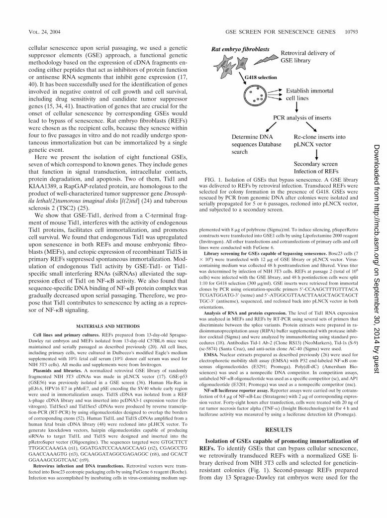
Isolation of GSEs capable of promoting immortalization ofREFs. To identify GSEs that can bypass cellular senescence,we retrovirally transduced REFs with a normalized GSE li-brary derived from NIH 3T3 cells and selected for geneticin-resistant colonies (Fig. 1). Second-passage REFs preparedfrom day 13 Sprague-Dawley rat embryos were used for the
FIG. 1. Isolation of GSEs that bypass senescence. A GSE librarywas delivered to REFs by retroviral infection. Transduced REFs wereselected for colony formation in the presence of G418. GSEs wererescued by PCR from genomic DNA after colonies were isolated andserially propagated for 5 or 6 passages, recloned into pLNCX vector,and subjected to a secondary screen.
VOL. 24, 2004 GSE SCREEN FOR SENESCENCE GENES 10793
on Septem
ber 30, 2014 by guesthttp://m
cb.asm.org/
Dow
nloaded from
screen, because they have a high proliferation rate that ensureshigh transduction efficiencies upon infection with recombinantretroviruses. To achieve maximal representation of GSEs, thenormalized retroviral NIH 3T3 GSE library (17) was tran-siently transfected into the highly transfectable ecotropic pack-aging cell line BOSC23 to prepare high-titer virus stocks.Empty pLNCX control vector was used as a negative control,and pLNCX GSE56, which encodes a C-terminal fragment ofp53 and is known for its strong immortalization activity, wasused as a positive control (36).
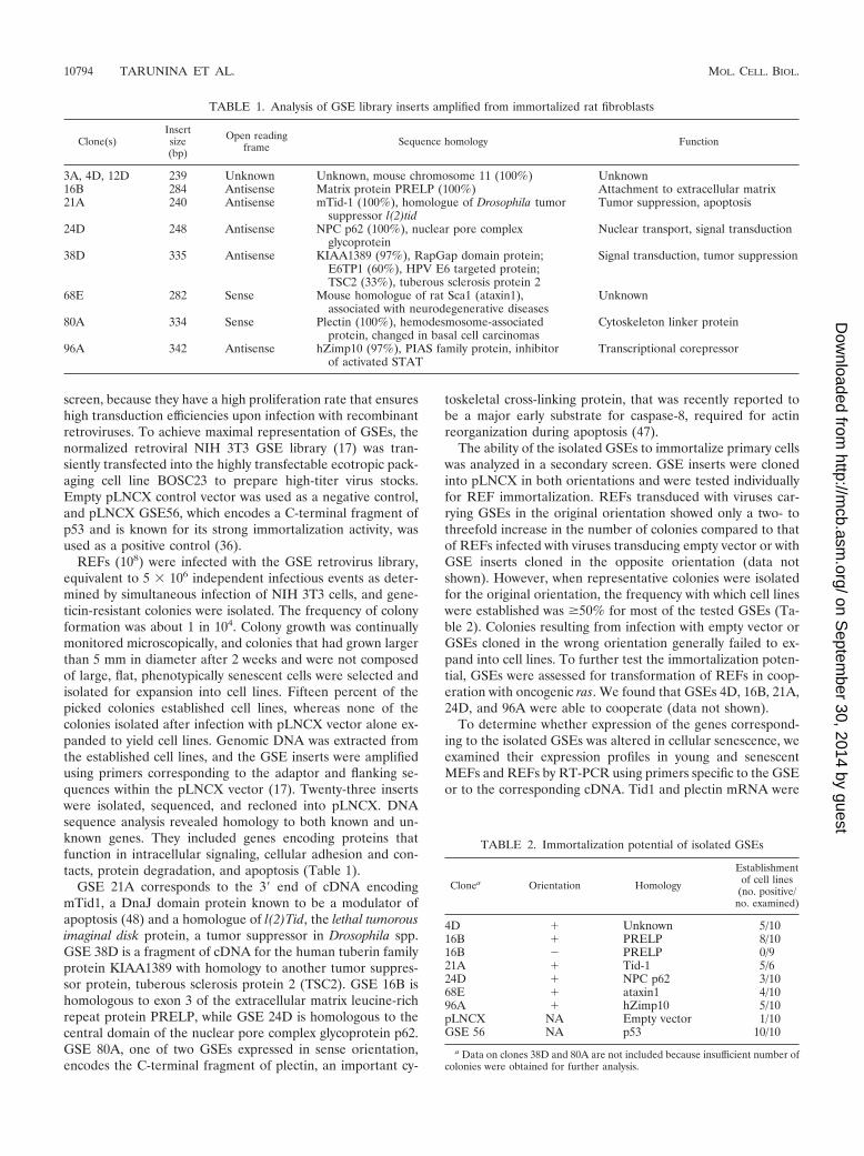
REFs (108) were infected with the GSE retrovirus library,equivalent to 5 � 106 independent infectious events as deter-mined by simultaneous infection of NIH 3T3 cells, and gene-ticin-resistant colonies were isolated. The frequency of colonyformation was about 1 in 104. Colony growth was continuallymonitored microscopically, and colonies that had grown largerthan 5 mm in diameter after 2 weeks and were not composedof large, flat, phenotypically senescent cells were selected andisolated for expansion into cell lines. Fifteen percent of thepicked colonies established cell lines, whereas none of thecolonies isolated after infection with pLNCX vector alone ex-panded to yield cell lines. Genomic DNA was extracted fromthe established cell lines, and the GSE inserts were amplifiedusing primers corresponding to the adaptor and flanking se-quences within the pLNCX vector (17). Twenty-three insertswere isolated, sequenced, and recloned into pLNCX. DNAsequence analysis revealed homology to both known and un-known genes. They included genes encoding proteins thatfunction in intracellular signaling, cellular adhesion and con-tacts, protein degradation, and apoptosis (Table 1).
GSE 21A corresponds to the 3� end of cDNA encodingmTid1, a DnaJ domain protein known to be a modulator ofapoptosis (48) and a homologue of l(2)Tid, the lethal tumorousimaginal disk protein, a tumor suppressor in Drosophila spp.GSE 38D is a fragment of cDNA for the human tuberin familyprotein KIAA1389 with homology to another tumor suppres-sor protein, tuberous sclerosis protein 2 (TSC2). GSE 16B ishomologous to exon 3 of the extracellular matrix leucine-richrepeat protein PRELP, while GSE 24D is homologous to thecentral domain of the nuclear pore complex glycoprotein p62.GSE 80A, one of two GSEs expressed in sense orientation,encodes the C-terminal fragment of plectin, an important cy-
toskeletal cross-linking protein, that was recently reported tobe a major early substrate for caspase-8, required for actinreorganization during apoptosis (47).
The ability of the isolated GSEs to immortalize primary cellswas analyzed in a secondary screen. GSE inserts were clonedinto pLNCX in both orientations and were tested individuallyfor REF immortalization. REFs transduced with viruses car-rying GSEs in the original orientation showed only a two- tothreefold increase in the number of colonies compared to thatof REFs infected with viruses transducing empty vector or withGSE inserts cloned in the opposite orientation (data notshown). However, when representative colonies were isolatedfor the original orientation, the frequency with which cell lineswere established was �50% for most of the tested GSEs (Ta-ble 2). Colonies resulting from infection with empty vector orGSEs cloned in the wrong orientation generally failed to ex-pand into cell lines. To further test the immortalization poten-tial, GSEs were assessed for transformation of REFs in coop-eration with oncogenic ras. We found that GSEs 4D, 16B, 21A,24D, and 96A were able to cooperate (data not shown).
To determine whether expression of the genes correspond-ing to the isolated GSEs was altered in cellular senescence, weexamined their expression profiles in young and senescentMEFs and REFs by RT-PCR using primers specific to the GSEor to the corresponding cDNA. Tid1 and plectin mRNA were
TABLE 1. Analysis of GSE library inserts amplified from immortalized rat fibroblasts
Clone(s)Insertsize(bp)
Open readingframe Sequence homology Function
3A, 4D, 12D 239 Unknown Unknown, mouse chromosome 11 (100%) Unknown16B 284 Antisense Matrix protein PRELP (100%) Attachment to extracellular matrix21A 240 Antisense mTid-1 (100%), homologue of Drosophila tumor
suppressor l(2)tidTumor suppression, apoptosis
24D 248 Antisense NPC p62 (100%), nuclear pore complexglycoprotein
Nuclear transport, signal transduction
38D 335 Antisense KIAA1389 (97%), RapGap domain protein;E6TP1 (60%), HPV E6 targeted protein;TSC2 (33%), tuberous sclerosis protein 2
Signal transduction, tumor suppression
68E 282 Sense Mouse homologue of rat Sca1 (ataxin1),associated with neurodegenerative diseases
Unknown
80A 334 Sense Plectin (100%), hemodesmosome-associatedprotein, changed in basal cell carcinomas
Cytoskeleton linker protein
96A 342 Antisense hZimp10 (97%), PIAS family protein, inhibitorof activated STAT
Transcriptional corepressor
TABLE 2. Immortalization potential of isolated GSEs
Clonea Orientation Homology
Establishmentof cell lines
(no. positive/no. examined)
4D � Unknown 5/1016B � PRELP 8/1016B PRELP 0/921A � Tid-1 5/624D � NPC p62 3/1068E � ataxin1 4/1096A � hZimp10 5/10pLNCX NA Empty vector 1/10GSE 56 NA p53 10/10
a Data on clones 38D and 80A are not included because insufficient number ofcolonies were obtained for further analysis.
10794 TARUNINA ET AL. MOL. CELL. BIOL.
on Septem
ber 30, 2014 by guesthttp://m
cb.asm.org/
Dow
nloaded from
increased upon senescence, whereas the level of mRNA for theothers was either decreased or remained unaltered (data notshown). This raised the possibility that GSEs for Tid1 andplectin cause alterations in senescence-associated activities ofthese genes, leading to bypass of senescence.
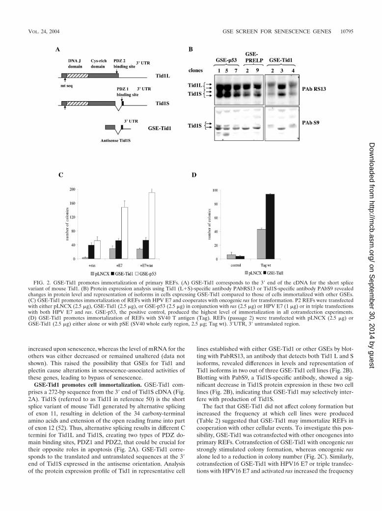
GSE-Tid1 promotes cell immortalization. GSE-Tid1 com-prises a 272-bp sequence from the 3� end of Tid1S cDNA (Fig.2A). Tid1S (referred to as Tid1I in reference 50) is the shortsplice variant of mouse Tid1 generated by alternative splicingof exon 11, resulting in deletion of the 34 carboxy-terminalamino acids and extension of the open reading frame into partof exon 12 (52). Thus, alternative splicing results in different Ctermini for Tid1L and Tid1S, creating two types of PDZ do-main binding sites, PDZ1 and PDZ2, that could be crucial fortheir opposite roles in apoptosis (Fig. 2A). GSE-Tid1 corre-sponds to the translated and untranslated sequences at the 3�end of Tid1S expressed in the antisense orientation. Analysisof the protein expression profile of Tid1 in representative cell
lines established with either GSE-Tid1 or other GSEs by blot-ting with PabRS13, an antibody that detects both Tid1 L and Sisoforms, revealed differences in levels and representation ofTid1 isoforms in two out of three GSE-Tid1 cell lines (Fig. 2B).Blotting with PabS9, a Tid1S-specific antibody, showed a sig-nificant decrease in Tid1S protein expression in these two celllines (Fig. 2B), indicating that GSE-Tid1 may selectively inter-fere with production of Tid1S.
The fact that GSE-Tid1 did not affect colony formation butincreased the frequency at which cell lines were produced(Table 2) suggested that GSE-Tid1 may immortalize REFs incooperation with other cellular events. To investigate this pos-sibility, GSE-Tid1 was cotransfected with other oncogenes intoprimary REFs. Cotransfection of GSE-Tid1 with oncogenic rasstrongly stimulated colony formation, whereas oncogenic rasalone led to a reduction in colony number (Fig. 2C). Similarly,cotransfection of GSE-Tid1 with HPV16 E7 or triple transfec-tions with HPV16 E7 and activated ras increased the frequency
FIG. 2. GSE-Tid1 promotes immortalization of primary REFs. (A) GSE-Tid1 corresponds to the 3� end of the cDNA for the short splicevariant of mouse Tid1. (B) Protein expression analysis using Tid1 (L�S)-specific antibody PAbRS13 or Tid1S-specific antibody PAbS9 revealedchanges in protein level and representation of isoforms in cells expressing GSE-Tid1 compared to those of cells immortalized with other GSEs.(C) GSE-Tid1 promotes immortalization of REFs with HPV E7 and cooperates with oncogenic ras for transformation. P2 REFs were transfectedwith either pLNCX (2.5 �g), GSE-Tid1 (2.5 �g), or GSE-p53 (2.5 �g) in conjunction with ras (2.5 �g) or HPV E7 (1 �g) or in triple transfectionswith both HPV E7 and ras. GSE-p53, the positive control, produced the highest level of immortalization in all cotransfection experiments.(D) GSE-Tid1 promotes immortalization of REFs with SV40 T antigen (Tag). REFs (passage 2) were transfected with pLNCX (2.5 �g) orGSE-Tid1 (2.5 �g) either alone or with pSE (SV40 whole early region, 2.5 �g; Tag wt). 3�UTR, 3� untranslated region.
VOL. 24, 2004 GSE SCREEN FOR SENESCENCE GENES 10795
on Septem
ber 30, 2014 by guesthttp://m
cb.asm.org/
Dow
nloaded from
of immortalized clones (Fig. 2C). Cotransfection of GSE-Tid1with SV40 T antigen also yielded two times more colonies thanT antigen alone (Fig. 2D). Together these results suggest thatGSE-Tid1 promotes survival and facilitates immortalization ofprimary REFs.
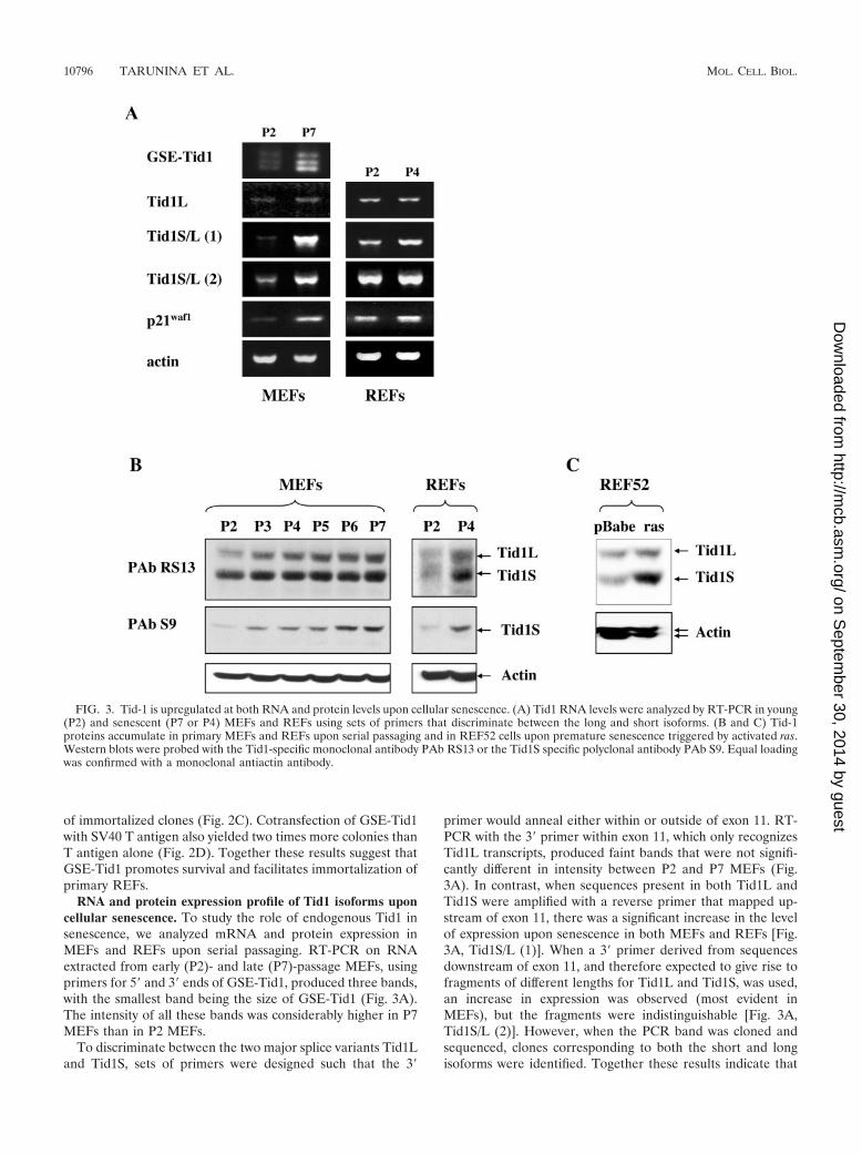
RNA and protein expression profile of Tid1 isoforms uponcellular senescence. To study the role of endogenous Tid1 insenescence, we analyzed mRNA and protein expression inMEFs and REFs upon serial passaging. RT-PCR on RNAextracted from early (P2)- and late (P7)-passage MEFs, usingprimers for 5� and 3� ends of GSE-Tid1, produced three bands,with the smallest band being the size of GSE-Tid1 (Fig. 3A).The intensity of all these bands was considerably higher in P7MEFs than in P2 MEFs.
To discriminate between the two major splice variants Tid1Land Tid1S, sets of primers were designed such that the 3�
primer would anneal either within or outside of exon 11. RT-PCR with the 3� primer within exon 11, which only recognizesTid1L transcripts, produced faint bands that were not signifi-cantly different in intensity between P2 and P7 MEFs (Fig.3A). In contrast, when sequences present in both Tid1L andTid1S were amplified with a reverse primer that mapped up-stream of exon 11, there was a significant increase in the levelof expression upon senescence in both MEFs and REFs [Fig.3A, Tid1S/L (1)]. When a 3� primer derived from sequencesdownstream of exon 11, and therefore expected to give rise tofragments of different lengths for Tid1L and Tid1S, was used,an increase in expression was observed (most evident inMEFs), but the fragments were indistinguishable [Fig. 3A,Tid1S/L (2)]. However, when the PCR band was cloned andsequenced, clones corresponding to both the short and longisoforms were identified. Together these results indicate that
FIG. 3. Tid-1 is upregulated at both RNA and protein levels upon cellular senescence. (A) Tid1 RNA levels were analyzed by RT-PCR in young(P2) and senescent (P7 or P4) MEFs and REFs using sets of primers that discriminate between the long and short isoforms. (B and C) Tid-1proteins accumulate in primary MEFs and REFs upon serial passaging and in REF52 cells upon premature senescence triggered by activated ras.Western blots were probed with the Tid1-specific monoclonal antibody PAb RS13 or the Tid1S specific polyclonal antibody PAb S9. Equal loadingwas confirmed with a monoclonal antiactin antibody.
10796 TARUNINA ET AL. MOL. CELL. BIOL.
on Septem
ber 30, 2014 by guesthttp://m
cb.asm.org/
Dow
nloaded from
Tid1 RNA and, in particular, the RNA for the Tid1S splicevariant is upregulated upon senescence.
To determine whether there were changes in protein expres-sion of the Tid1 isoforms upon senescence, total extracts wereprepared from cultures of MEFs at each passage and wereanalyzed by Western blotting. To ensure that only changes inexpression due to senescence were being examined, extractswere prepared from subconfluent cultures that had been pas-saged the previous day and, thus, should not contain quiescentcells. Western blots with antibody PAb RS13 revealed a steadyincrease in the level of both Tid1L and Tid1S isoforms inMEFs upon serial passaging (Fig. 3B). A similar accumulationof both isoforms was observed in REFs when senescent P4cells were compared with actively proliferating P2 cells. Accu-mulation of the Tid1S isoform upon passaging was confirmedby blotting the extracts with PAb S9, which specifically recog-nizes only this isoform (Fig. 3B).
Premature senescence triggered by ectopic expression ofactivated ras induces a pattern of gene expression similar tothat of cells undergoing replicative senescence upon serial pas-saging, and these patterns are distinct from those in quiescentcells (44). To determine whether Tid1 isoforms are also up-regulated upon premature senescence, oncogenic ras was in-troduced into REF-52 cells by retroviral infection. Seven dayspostinfection with activated ras, REF52 cells underwentgrowth arrest and phenotypic changes consistent with senes-cence. A parallel culture of REF52 cells infected with thebackbone retrovirus did not undergo growth arrest but contin-ued to proliferate until the cells became contact inhibited.Western blotting of lysates prepared from the REF52 cultureswith the anti-Tid1 monoclonal antibody PAb RS13 showedthat both Tid1 isoforms were upregulated; however, the Tid1Sisoform accumulated to a much higher level, thereby alteringthe ratio between the two proteins (Fig. 3C).
Thus, both replicative senescence and premature senescencetriggered by oncogenic ras were associated with upregulationof Tid1 proteins. While the protein levels of both isoformsshowed a steady increase upon serial passaging of MEFs andREFs, ras-induced premature senescence of REF52 was ac-companied by a predominant accumulation of Tid1S.
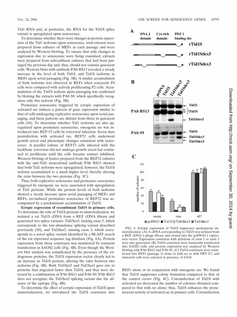
Ectopic expression of recombinant Tid1S in primary cells.To determine the role of Tid1S proteins in immortalization, weisolated a rat Tid1S cDNA from a REF cDNA library andgenerated two splice variants: Tid1Sex5, lacking exon 5, whichcorresponds to the low-abundance splicing variant describedpreviously (50), and Tid1Sex3, missing exon 3, which corre-sponds to a novel splice variant identified by a BLAST searchof the rat expressed sequence tag database (Fig. 4A). Proteinexpression from these constructs was monitored by transienttransfection in SAOS2 cells (Fig. 4B). Even though the West-ern blot analysis was complicated by the presence of the en-dogenous proteins, the Tid1S expression vector clearly led toan increase in Tid1S protein, altering the ratio between twoisoforms (Fig. 4B). Both Tid1Sex5 and Tid1Sex3 gave rise toproteins that migrated faster than Tid1S, and they were de-tected by a combination of PAb RS13 and PAb S9. PAb RS13does not recognize the Tid1Sex3 splicing variant due the ab-sence of the epitope (Fig. 4B).
To determine the effect of ectopic expression of Tid1S uponimmortalization, we introduced the Tid1S constructs into
REFs alone or in conjunction with oncogenic ras. We foundthat Tid1S suppresses colony formation compared to that ofthe control vector (Fig. 4C). Cotransfection of Tid1S withactivated ras decreased the number of colonies obtained com-pared to that with ras alone; thus, Tid1S enhances the prose-nescent activity of activated ras in primary cells. Cotransfection
FIG. 4. Ectopic expression of Tid1S suppresses spontaneous im-mortalization. (A) A cDNA corresponding to Tid1S was isolated froma REF cDNA �-phage library and cloned into the pcDNA3.1 expres-sion vector. Expression constructs with deletions of exon 5 or exon 3were also generated. (B) Tid1S constructs were transiently transfectedinto SAOS2 cells, and protein expression was analyzed by Westernblotting with PAb RS13 and PAb S9. (C) Tid1S constructs were trans-fected into REFs (passage 2) alone or with ras or with HPV E7, andimmortal cells were selected in presence of G418.
VOL. 24, 2004 GSE SCREEN FOR SENESCENCE GENES 10797
on Septem
ber 30, 2014 by guesthttp://m
cb.asm.org/
Dow
nloaded from
of the deletion constructs, where Tid1S lacked either exon 3 orexon 5, with oncogenic ras did not have the same inhibitoryaffect as intact Tid1S, suggesting that both the DnaJ and cys-tine-rich domains are essential for the suppression of growth.This suppression of colony formation by Tid1S was overcomeby the HPV16 E7 gene, indicating that the immortalizing func-tions of HPV16 E7 are dominant to the inhibitory effects ofTid1S.
NF-�B is a potential target of Tid1 in senescence. Previouslypublished data has indicated that Tid1 may have multiple cel-lular targets and may modulate activity of proteins involved inthe ras, gamma interferon, and NF-�B signaling pathways (10,42, 50). Because ectopic expression of GSE-Tid1 promoted cellimmortalization and cell survival, we explored the possibilitythat GSE-Tid1 may act through modulation of NF-�B signal-ing, a pathway known to be linked to inhibition of apoptosisand promotion of cell survival.
We first examined NF-�B DNA binding activity in REFsupon serial passaging, and we found that the sequence-specificDNA binding activity of the NF-�B transcription factor com-plex gradually decreased (Fig. 5A). It was recently reportedthat the Tid1L protein suppresses NF-�B signaling by repress-ing I�B� phosphorylation and nuclear translocation of NF-�B(10). The reduction in NF-�B activity in cells undergoing se-nescence is in accordance with the increase in Tid1 proteinexpression (Fig. 3B) and may reflect a functional link betweenTid1 and the NF-�B complex. NF-�B DNA binding activitywas also reduced in REFs conditionally immortalized with atemperature-sensitive SV40 tsA58 T antigen when they enter asenescence-like state upon inactivation of T antigen (Fig. 5A).SV4, a REF line immortalized with wild-type T antigen andused as a control, did not show a reduction in NF-�B DNAbinding activity.
We determined whether ectopic overexpression of Tid1Scan also affect NF-�B activity, as has been reported for Tid1L(10). U2OS cells were cotransfected with an NF-�B luciferasereporter, and human Tid1 expression vectors and luciferaseactivity were examined 4 h after treatment of transfected cellswith TNF-�. Ectopic expression of human Tid1S resulted in amore than twofold reduction of TNF-�-induced NF-�B activity(Fig. 5B, panel 1). The reduction was similar to that observedwith human Tid1L. This suppression of NF-�B activity by ec-topic expression of human Tid1S was also observed in ratGSEp53-1 cells, with Tid1S being a stronger repressor thanTid1L (data not shown).
To modulate expression of endogenous Tid1 proteins, wedesigned siRNA oligonucleotides that would either silenceboth major isoforms or selectively suppress expression of oneof them (Fig. 5C). The corresponding oligonucleotides werecloned into pRetroSuper vector and introduced into GSEp53-1cells. Western blot analysis confirmed siRNA-mediated knock-down of endogenous Tid1 proteins (Fig. 5C). Coexpression ofNF-�B luciferase reporter vector with the siRNA constructs ri1and ri2, which target both Tid1 isoforms, resulted in a signif-icant increase in TNF-�-induced NF-�B activity, while selec-tive silencing of one isoform by ri6 or ri9 had only a slight effect(Fig. 5B, panel 2). The siRNA construct ri3, which inducedonly a partial reduction of protein expression of both isoforms,showed a reduced effect on NF-�B activity. These results in-
dicate that alterations in the level of endogenous Tid1 proteinscan directly affect TNF-�-induced NF-�B activity.
Finally, we cotransfected the NF-�B reporter with a GSE-Tid1 expression vector and observed an approximately 50%increase in TNF-�-induced NF-�B activity (Fig. 5B, panel 3),confirming that GSE-Tid1 modulates endogenous Tid1 pro-teins and reduces their inhibitory effect on NF-�B. Thus, theability of GSE-Tid1 to promote immortalization and cell sur-vival could be due to the activation of the NF-�B complex.However, interaction between Tid1 proteins and other cellulartargets may also be important for the role of Tid1 in senes-cence and for the immortalization properties of GSE-Tid1, butthey remain to be clarified.
DISCUSSION
Here we employed a functional genetic screen to identifynew genes involved in cellular senescence. A normalized GSElibrary generated from mRNA of NIH 3T3 cells (17) wasdelivered into REFs by retroviral infection, and cells that by-passed senescence were selected. A secondary screen was em-ployed to ensure that the isolated GSEs were truly capable ofbypassing senescence. This involved recloning the GSEs into aretroviral vector in both sense and antisense orientations andrepeating the REF immortalization assay. The stringency ofthe secondary screen was further increased by isolating colo-nies and assessing their ability to establish cell lines.
We identified eight GSEs corresponding to eight indepen-dent genes. Several of these genes, like nuclear pore proteinp62 or cytoskeleton protein plectin, are well characterized,while the functions of other proteins, such as KIAA1389 andhZimp10, are predicted on the basis of their sequence andremain to be elucidated. Most of the isolated GSEs correspondto genes involved in signal transduction, consistent with theview of rodent cell senescence as a stress-related phenomenon(31, 33). Interestingly, one of the identified genes, PRELP, wasrecently proposed to be involved in the development ofHutchinson-Gilford progeria, accelerated aging syndrome(27).
Two of the eight isolated GSEs show a high level of homol-ogy to known tumor suppressor genes. The protein product ofKIAA1389 harbors a putative RapGAP domain and is closelyrelated to RapGTPase-activating protein E6TP1 (1), which istargeted for degradation by the E6 protein of high-risk humanpapilloma viruses (14). Both KIAA1389 and E6TP1 are alsorelated to tuberin, the product of the tumor suppressor TSC2,whose inactivation is associated with familial benign tumors(25). Tuberin controls mTOR signaling by acting as part of theGTPase-activating complex for Rheb (Ras homologue en-riched in brain) (49).
We also isolated a GSE fragment from a candidate tumorsuppressor gene of Tid1. The tumor suppressor function of theDrosophila homologue l(2)tid is well established, as homozy-gous mutations in this gene result in hyperproliferation andtumor formation in imaginal disks of Drosophila larvae. Severalfacts point towards a similar role for mammalian Tid1 in tumordevelopment: expression of endogenous Tid1 was found al-tered in basal cell carcinomas (7) while ectopic expression ofhuman Tid1 in tumor cell lines led to suppression of theirtransformed phenotype (9). Our results indicate that Tid1 has
10798 TARUNINA ET AL. MOL. CELL. BIOL.
on Septem
ber 30, 2014 by guesthttp://m
cb.asm.org/
Dow
nloaded from
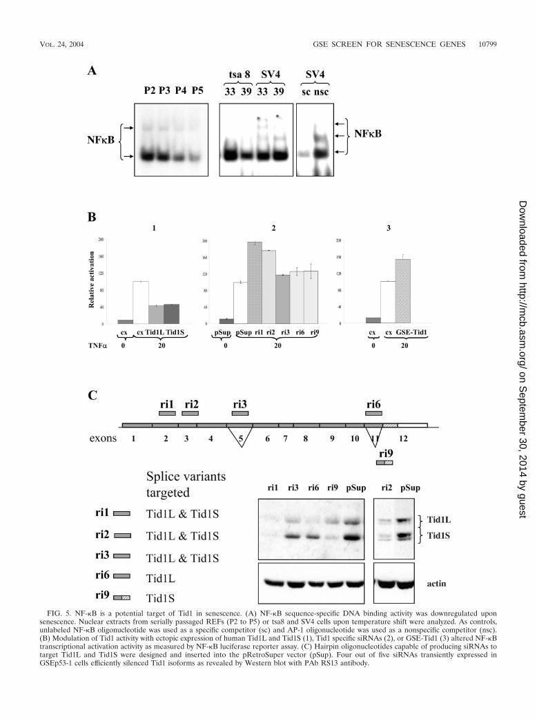
FIG. 5. NF-�B is a potential target of Tid1 in senescence. (A) NF-�B sequence-specific DNA binding activity was downregulated uponsenescence. Nuclear extracts from serially passaged REFs (P2 to P5) or tsa8 and SV4 cells upon temperature shift were analyzed. As controls,unlabeled NF-�B oligonucleotide was used as a specific competitor (sc) and AP-1 oligonucleotide was used as a nonspecific competitor (nsc).(B) Modulation of Tid1 activity with ectopic expression of human Tid1L and Tid1S (1), Tid1 specific siRNAs (2), or GSE-Tid1 (3) altered NF-�Btranscriptional activation activity as measured by NF-�B luciferase reporter assay. (C) Hairpin oligonucleotides capable of producing siRNAs totarget Tid1L and Tid1S were designed and inserted into the pRetroSuper vector (pSup). Four out of five siRNAs transiently expressed inGSEp53-1 cells efficiently silenced Tid1 isoforms as revealed by Western blot with PAb RS13 antibody.
VOL. 24, 2004 GSE SCREEN FOR SENESCENCE GENES 10799
on Septem
ber 30, 2014 by guesthttp://m
cb.asm.org/
Dow
nloaded from
the capacity to promote growth arrest and suppress immortal-ization, because GSE-Tid1 promoted immortalization and ec-topic expression of Tid1S cDNA in REFs reduced spontaneousimmortalization.
Recent studies have implicated mammalian Tid1 proteins inseveral signaling pathways, including those of ras, gamma in-terferon, and NF-�B (10, 42, 50). Our data suggest that therole of Tid1 proteins as positive regulators of cellular senes-cence may be linked to their ability to modulate NF-�B sig-naling. Cheng et al. (10) showed that hTid1 antagonizes activ-ities of various NF-�B activators, including human T-cellleukemia virus (HTLV) Tax, TNF-�, and Bcl10, by repressingI�B kinase (IKK�) activity and enhancing the stability of I�Bmolecules. Consistent with these results, we found that silenc-ing of Tid1 with Tid1-specific siRNAs considerably enhancedNF-�B activity induced by treatment of cells with TNF-�. Ec-topic expression of GSE-Tid1 had a similar enhancing effect onNF-�B activity that could be responsible for its role in promot-ing cell survival and immortalization. The Tid1 siRNAs thatenhance NF-�B activity could not be directly tested for immor-talization of REFs as they were lethal, possibly due to theinvolvement of Tid1 in other signaling pathways. This is sup-ported by a recent publication showing that homozygous lossof Tid1 causes embryonic lethality (30). Unlike siRNAs whichsilence expression of the protein, GSE fragments may onlyperturb the functions relevant for the genetic screen utilized.
NF-�B signaling was only recently recognized to play animportant role in tumorigenesis (for recent reviews see refer-ences 21 and 28). Constitutive activation of NF-�B is found ina variety of human cancers and can contribute to the malignantphenotype by driving cell proliferation, increasing resistance toapoptosis, and promoting cell survival and migration. Multiplemechanisms could disrupt the normal regulation of NF-�Bactivity in cancer cells. The Tax oncoprotein of HTLV-1 thatcauses adult T-cell leukemia (ATL) has been shown to directlyinteract with the IKK complex, resulting in enhancement ofIKK� kinase activity and subsequent activation of the NF-�Bcomplex (11). It was then reported that Tax associates withhTid1/Hsp70 and antagonizes the suppressive activity of hTid1on IKK�-mediated phosphorylation of I�B� (9, 10). Anothertumor suppressor protein, CYLD, that is mutated in familialcylindromatosis, skin appendage cancer predisposition syn-drome, was recently described to be a negative regulator ofNF-�B signaling by targeting the activity of the IKK complexthrough binding to IKK (or NEMO) and deubiquitination ofsignaling proteins that function upstream of IKKs (5, 23, 51).
There is presently no consensus about the role of NF-�Bsignaling in cellular senescence. Some groups have reportedrather dramatic tissue-specific increases or decreases in NF-�Bactivity in humans and animals upon ageing (reviewed in ref-erence 16). Here we have found that senescence in REFs wasaccompanied by a steady decline in NF-�B sequence-specificDNA binding activity, and we have proposed that this was dueto increased expression of Tid1. Interestingly, it was recentlypublished that p14ARF/p19ARF proteins that are crucial forcellular senescence suppress NF-�B function and its antiapop-totic activity independently of MDM2 and p53 by direct inhi-bition of RelA transactivation without affecting NF-�B se-quence-specific DNA binding (39). Taken together, these datasuggest that downregulation of NF-�B activity upon senes-
cence could be controlled by a number of mechanisms, includ-ing Tid1 activity in the cytoplasm and ARF activity in thenucleus, and the perturbation of these regulatory mechanismscan lead to aberrantly active NF-�B and contribute to tumor-igenesis. Isolation of the Tid1 fragment capable of promotingcell survival and immortalization validates the use of the GSEapproach in the search for genes involved in control of irre-versible growth arrest and senescence. The role of other genesidentified in this screen in regulation of cellular senescence andimmortalization remains to be elucidated.
ACKNOWLEDGMENTS
We thank Silvia Benvenuti for REF52 protein extracts.This work was supported by ReNeuron, Ltd. (Guildford, United
Kingdom), to P.S.J. and NIH grant CA60730 to A.V.G.
REFERENCES
1. Bernards, A. 2003. GAPs galore! A survey of putative Ras superfamilyGTPase activating proteins in man and Drosophila. Biochim. Biophys. Acta1603:47–82.
2. Blasco, M. A. 2003. Telomeres in cancer and aging: lessons from the mouse.Cancer Lett. 194:183–188.
3. Bodnar, A. G., M. Ouellette, M. Frolkis, S. E. Holt, C. P. Chiu, G. B. Morin,C. B. Harley, J. W. Shay, S. Lichtsteiner, and W. E. Wright. 1998. Extensionof life-span by introduction of telomerase into normal human cells. Science279:349–352.
4. Brummelkamp, T. R., R. M. Kortlever, M. Lingbeek, F. Trettel, M. E.MacDonald, M. van Lohuizen, and R. Bernards. 2002. TBX-3, the genemutated in ulnar-mammary syndrome, is a negative regulator of p19ARFand inhibits senescence. J. Biol. Chem. 277:6567–6572.
5. Brummelkamp, T. R., S. M. Nijman, A. M. Dirac, and R. Bernards. 2003.Loss of the cylindromatosis tumour suppressor inhibits apoptosis by activat-ing NF-�B. Nature 424:797–801.
6. Campisi, J. 2001. From cells to organisms: can we learn about aging fromcells in culture? Exp. Gerontol. 36:607–618.
7. Canamasas, I., A. Debes, P. G. Natali, and U. Kurzik-Dumke. 2003. Under-standing human cancer using Drosophila: Tid47, a cytosolic product of theDnaJ-like tumor suppressor gene l2Tid, is a novel molecular partner ofpatched related to skin cancer. J. Biol. Chem. 278:30952–30960.
8. Chen, Q. M., J. C. Bartholomew, J. Campisi, M. Acosta, J. D. Reagan, andB. N. Ames. 1998. Molecular analysis of H2O2-induced senescent-like growtharrest in normal human fibroblasts: p53 and Rb control G1 arrest but not cellreplication. Biochem J. 332:43–50.
9. Cheng, H., C. Cenciarelli, Z. Shao, M. Vidal, W. P. Parks, M. Pagano, andC. Cheng-Mayer. 2001. Human T cell leukemia virus type 1 Tax associateswith a molecular chaperone complex containing hTid-1 and Hsp70. Curr.Biol. 11:1771–1775.
10. Cheng, H., C. Cenciarelli, M. Tao, W. P. Parks, and C. Cheng-Mayer. 2002.HTLV-1 Tax-associated hTid-1, a human DnaJ protein, is a repressor of I�Bkinase � subunit. J. Biol. Chem. 277:20605–20610.
11. Chu, Z. L., Y. A. Shin, J. M. Yang, J. A. DiDonato, and D. W. Ballard. 1999.IKK mediates the interaction of cellular I�B kinases with the tax trans-forming protein of human T cell leukemia virus type 1. J. Biol. Chem.274:15297–15300.
12. Dickson, M. A., W. C. Hahn, Y. Ino, V. Ronfard, J. Y. Wu, R. A. Weinberg,D. N. Louis, F. P. Li, and J. G. Rheinwald. 2000. Human keratinocytes thatexpress hTERT and also bypass a p16(INK4a)-enforced mechanism thatlimits life span become immortal yet retain normal growth and differentia-tion characteristics. Mol. Cell. Biol. 20:1436–1447.
13. Ferbeyre, G., E. de Stanchina, A. W. Lin, E. Querido, M. E. McCurrach, G. J.Hannon, and S. W. Lowe. 2002. Oncogenic ras and p53 cooperate to inducecellular senescence. Mol. Cell. Biol. 22:3497–3508.
14. Gao, Q., S. Srinivasan, S. N. Boyer, D. E. Wazer, and V. Band. 1999. The E6oncoproteins of high-risk papillomaviruses bind to a novel putative GAPprotein, E6TP1, and target it for degradation. Mol. Cell. Biol. 19:733–744.
15. Garkavtsev, I., A. Kazarov, A. Gudkov, and K. Riabowol. 1996. Suppressionof the novel growth inhibitor p33ING1 promotes neoplastic transformation.Nat. Genet. 14:415–420.
16. Giardina, C., and A. K. Hubbard. 2002. Growing old with nuclear factor-�B.Cell Stress Chaperones 7:207–212.
17. Gudkov, A. V., A. R. Kazarov, R. Thimmapaya, S. A. Axenovich, I. A. Mazo,and I. B. Roninson. 1994. Cloning mammalian genes by expression selectionof genetic suppressor elements: association of kinesin with drug resistanceand cell immortalization. Proc. Natl. Acad. Sci. USA 91:3744–3748.
18. Harlow, E., and D. Lane. 1988. Antibodies: a laboratory manual. Cold SpringHarbor Laboratory Press, Cold Spring Harbor, N.Y.
10800 TARUNINA ET AL. MOL. CELL. BIOL.
on Septem
ber 30, 2014 by guesthttp://m
cb.asm.org/
Dow
nloaded from
19. Hayflick, L., and P. S. Moorehead. 1961. The serial cultivation of humandiploid cell strains. Exp. Cell Res. 25:585–621.
20. Ikram, Z., T. Norton, and P. S. Jat. 1994. The biological clock that measuresthe mitotic life-span of mouse embryo fibroblasts continues to function in thepresence of simian virus 40 large tumor antigen. Proc. Natl. Acad. Sci. USA91:6448–6452.
21. Karin, M., Y. Cao, F. R. Greten, and Z. W. Li. 2002. NF-�B in cancer: frominnocent bystander to major culprit. Nat. Rev. Cancer 2:301–310.
22. Kiyono, T., S. A. Foster, J. I. Koop, J. K. McDougall, D. A. Galloway, andA. J. Klingelhutz. 1998. Both Rb/p16INK4a inactivation and telomeraseactivity are required to immortalize human epithelial cells. Nature 396:84–88.
23. Kovalenko, A., C. Chable-Bessia, G. Cantarella, A. Israel, D. Wallach, andG. Courtois. 2003. The tumour suppressor CYLD negatively regulatesNF-�B signalling by deubiquitination. Nature 424:801–805.
24. Kurzik-Dumke, U., A. Debes, M. Kaymer, and P. Dienes. 1998. Mitochon-drial localization and temporal expression of the Drosophila melanogasterDnaJ homologous tumor suppressor Tid50. Cell Stress Chaperones 3:12–27.
25. Kwiatkowski, D. J. 2003. Tuberous sclerosis: from tubers to mTOR. Ann.Hum. Genet. 67:87–96.
26. Lassar, A. B., R. L. Davis, W. E. Wright, T. Kadesch, C. Murre, A. Voronova,D. Baltimore, and H. Weintraub. 1991. Functional activity of myogenic HLHproteins requires hetero-oligomerization with E12/E47-like proteins in vivo.Cell 66:305–315.
27. Lewis, M. 2003. PRELP, collagen, and a theory of Hutchinson-Gilford prog-eria. Ageing Res. Rev. 2:95–105.
28. Lin, A., and M. Karin. 2003. NF-�B in cancer: a marked target. Semin.Cancer Biol. 13:107–114.
29. Lin, A. W., M. Barradas, J. C. Stone, L. van Aelst, M. Serrano, and S. W.Lowe. 1998. Premature senescence involving p53 and p16 is activated inresponse to constitutive MEK/MAPK mitogenic signaling. Genes Dev. 12:3008–3019.
30. Lo, J. F., M. Hayashi, S. Woo-Kim, B. Tian, J. F. Huang, C. Fearns, S.Takayama, J. M. Zapata, Y. Yang, and J. D. Lee. 2004. Tid1, a cochaperoneof the heat shock 70 protein and the mammalian counterpart of the Dro-sophila tumor suppressor l(2)tid, is critical for early embryonic developmentand cell survival. Mol. Cell. Biol. 24:2226–2236.
31. Marcotte, R., and E. Wang. 2002. Replicative senescence revisited. J. Ger-ontol. A Biol. Sci. Med. Sci. 57:B257—B269.
32. Masutomi, K., E. Y. Yu, S. Khurts, I. Ben-Porath, J. L. Currier, G. B. Metz,M. W. Brooks, S. Kaneko, S. Murakami, J. A. DeCaprio, R. A. Weinberg,S. A. Stewart, and W. C. Hahn. 2003. Telomerase maintains telomere struc-ture in normal human cells. Cell 114:241–253.
33. Mathon, N. F., and A. C. Lloyd. 2001. Cell senescence and cancer. Nat. Rev.Cancer 1:203–213.
34. Neznanov, N., L. Neznanova, R. V. Kondratov, L. Burdelya, E. S. Kandel,D. M. O’Rourke, A. Ullrich, and A. V. Gudkov. 2003. Dominant negativeform of signal-regulatory protein-alpha (SIRP�/SHPS-1) inhibits tumor ne-crosis factor-mediated apoptosis by activation of NF-�B. J. Biol. Chem.278:3809–3815.
35. O’Hare, M. J., J. Bond, C. Clarke, Y. Takeuchi, A. J. Atherton, C. Berry, J.Moody, A. R. Silver, D. C. Davies, A. E. Alsop, A. M. Neville, and P. S. Jat.2001. Conditional immortalization of freshly isolated human mammary fi-broblasts and endothelial cells. Proc. Natl. Acad. Sci. USA 98:646–651.
36. Ossovskaya, V. S., I. A. Mazo, M. V. Chernov, O. B. Chernova, Z. Strezoska,
R. Kondratov, G. R. Stark, P. M. Chumakov, and A. V. Gudkov. 1996. Useof genetic suppressor elements to dissect distinct biological effects of sepa-rate p53 domains. Proc. Natl. Acad. Sci. USA 93:10309–10314.
37. Peeper, D. S., A. Shvarts, T. Brummelkamp, S. Douma, E. Y. Koh, G. Q.Daley, and R. Bernards. 2002. A functional screen identifies hDRIL1 as anoncogene that rescues RAS-induced senescence. Nat. Cell Biol. 4:148–153.
38. Robles, S. J., and G. R. Adami. 1998. Agents that cause DNA double strandbreaks lead to p16INK4a enrichment and the premature senescence ofnormal fibroblasts. Oncogene 16:1113–1123.
39. Rocha, S., K. J. Campbell, and N. D. Perkins. 2003. p53- and Mdm2-independent repression of NF-�B transactivation by the ARF tumor sup-pressor. Mol. Cell 12:15–25.
40. Roninson, I. B., and A. V. Gudkov. 2003. Genetic suppressor elements in thecharacterization and identification of tumor suppressor genes. Methods Mol.Biol. 222:413–436.
41. Sanz, G., L. Mir, and A. Jacquemin-Sablon. 2002. Bleomycin resistance inmammalian cells expressing a genetic suppressor element derived from theSRPK1 gene. Cancer Res. 62:4453–4458.
42. Sarkar, S., B. P. Pollack, K. T. Lin, S. V. Kotenko, J. R. Cook, A. Lewis, andS. Pestka. 2001. hTid-1, a human DnaJ protein, modulates the interferonsignaling pathway. J. Biol. Chem. 276:49034–49042.
43. Serrano, M., A. W. Lin, M. E. McCurrach, D. Beach, and S. W. Lowe. 1997.Oncogenic ras provokes premature cell senescence associated with accumu-lation of p53 and p16INK4a. Cell 88:593–602.
44. Shelton, D. N., E. Chang, P. S. Whittier, D. Choi, and W. D. Funk. 1999.Microarray analysis of replicative senescence. Curr. Biol. 9:939–945.
45. Sherr, C. J., and R. A. DePinho. 2000. Cellular senescence: mitotic clock orculture shock? Cell 102:407–410.
46. Shvarts, A., T. R. Brummelkamp, F. Scheeren, E. Koh, G. Q. Daley, H. Spits,and R. Bernards. 2002. A senescence rescue screen identifies BCL6 as aninhibitor of anti-proliferative p19(ARF)-p53 signaling. Genes Dev. 16:681–686.
47. Stegh, A. H., H. Herrmann, S. Lampel, D. Weisenberger, K. Andra, M.Seper, G. Wiche, P. H. Krammer, and M. E. Peter. 2000. Identification of thecytolinker plectin as a major early in vivo substrate for caspase 8 duringCD95- and tumor necrosis factor receptor-mediated apoptosis. Mol. Cell.Biol. 20:5665–5679.
48. Syken, J., T. De-Medina, and K. Munger. 1999. TID1, a human homolog ofthe Drosophila tumor suppressor l(2)tid, encodes two mitochondrial modu-lators of apoptosis with opposing functions. Proc. Natl. Acad. Sci. USA96:8499–8504.
49. Tee, A. R., B. D. Manning, P. P. Roux, L. C. Cantley, and J. Blenis. 2003.Tuberous sclerosis complex gene products, Tuberin and Hamartin, controlmTOR signaling by acting as a GTPase-activating protein complex towardRheb. Curr. Biol. 13:1259–1268.
50. Trentin, G. A., X. Yin, S. Tahir, S. Lhotak, J. Farhang-Fallah, Y. Li, and M.Rozakis-Adcock. 2001. A mouse homologue of the Drosophila tumor sup-pressor l(2)tid gene defines a novel Ras GTPase-activating protein (Ras-GAP)-binding protein. J. Biol. Chem. 276:13087–13095.
51. Trompouki, E., E. Hatzivassiliou, T. Tsichritzis, H. Farmer, A. Ashworth,and G. Mosialos. 2003. CYLD is a deubiquitinating enzyme that negativelyregulates NF-�B activation by TNFR family members. Nature 424:793–796.
52. Yin, X., and M. Rozakis-Adcock. 2001. Genomic organization and expressionof the human tumorous imaginal disc (TID1) gene. Gene 278:201–210.
VOL. 24, 2004 GSE SCREEN FOR SENESCENCE GENES 10801
on Septem
ber 30, 2014 by guesthttp://m
cb.asm.org/
Dow
nloaded from