Embed Size (px)
Citation preview
Cover Page
The handle http://hdl.handle.net/1887/50874 holds various files of this Leiden University dissertation Author: Ibrahim, M. Title: Development of an in vitro vascular network using zebrafish embryonic cells Issue Date: 2017-06-13

i
Development of an in vitro vascular network
using zebrafish embryonic cells
Muhammad Ibrahim

ii
Cover: development of vascular network-like structure from kdrl:GFP+ endothelial cells in
zebrafish embryoid body culture.
ISBN: 978-94-6182-805-7
Copyright © 2017 Muhammad Ibrahim. All rights reserved.
Printed by Off Page, Amsterdam, www.offpage.nl
iii
Development of an in vitro vascular network using
zebrafish embryonic cells
Proefschrift
ter verkrijging van
de graad van Doctor aan de Universiteit Leiden,
op gezag van de Rector Magnificus Prof. mr. C.J.J.M. Stolker,
volgens besluit van het College voor Promoties
te verdedigen op dinsdag 13 juni 2017
klokke 10.00 uur
door
Muhammad Ibrahim
geboren te Mardan, Pakistan
17 February 1982
iv
Promotiecommissie
Promotor
Prof. dr. Michael K. Richardson
Co-promotor
Dr. Anna Pavlina Haramis
Overige leden
Prof. dr. Gilles van Wezel
Prof. dr. Annemarie Meijer
Prof. dr. Robert E. Poelmann
Dr. Beerend P. Hierck (Leiden University Medical Center)
Dr. Neil Vargesson (University of Aberdeen)
The work described in this thesis was supported by the faculty development programme of
Institute of Biotechnology and Genetic Engineering, The University of Agriculture
Peshawar, Pakistan [scholarship number 06/SIBGE]; the Smart Mix programme of the
Netherlands Ministry of Economic Affairs and the Netherlands Scientific Research Council
(NWO) [grant number SSM06010]; and the Generade programme of the Centre of
Expertise Genomics in Leiden, The Netherlands [grant number 2016_004].

v
For For For For
Prof. Zahoor Ahmad SwatiProf. Zahoor Ahmad SwatiProf. Zahoor Ahmad SwatiProf. Zahoor Ahmad Swati

vi
vii
Table of Contents
Chapter Contents Page
1 General introduction
Culturing zebrafish vascular networks
1
2 Beyond organoids: in vitro vasculogenesis and angiogenesis using
cells from mammals and zebrafish
9
3 Zebrafish blastocyst cell culture and differentiation of fli:GFP+ and
kdrl:GFP+ cells
47
4 Influence of medium composition and substratum on the growth of
fli:GFP+ and kdrl:GFP+ cells in zebrafish blastocyst cell culture
77
5 Zebrafish fli:GFP and kdrl:GFP embryoid bodies – a model for
vasculogenesis and angiogenesis
103
6 In vitro development of zebrafish vascular networks 129
7 Summary and conclusions 159
Nederlandse samenvatting 166
Curriculum Vitae 171

viii

1
Chapter 1
General introduction
Culturing zebrafish vascular networks
Muhammad Ibrahim and Michael K. Richardson
2
Tissue engineering is one of the most important areas of biomedical research [1].
Strategies to develop complex tissues or organs in vitro will help our understanding of
organ physiology and pathology [2-4]. Development of organ cultures could also have
applications in organ transplantation and regenerative medicine [5]. Furthermore, these
organ cultures can be used to test candidate drugs which might ultimately reduce the use
of animals in research [6, 7].
One of the main issues in culturing complex organs is the lack of a vascular system [8]. In
multicellular organisms, the vascular system allows the growth and function of organs by
supplying nutrients and growth factors and by removing waste products [9]. The limited
diffusion of nutrients and oxygen into the un-vascularized tissue mass, developed in vitro,
hinders its growth and function into something resembling an organ [10]. To this end,
several techniques have been developed to culture vascular networks, largely using
mammalian cells and tissues.
The mammalian cells and tissues commonly used for this purpose are: (i) endothelial cell
lines (most commonly human umbilical vein endothelial cells or HUVECs [11-22]); (ii)
stem cells (embryonic stem cells [23], mesenchymal stem cells [24] or induced pluripotent
stem cells [25]); and (iii) tissue explants [26]. However, there are certain limitations to
these techniques. The endothelial cell lines are extensively adapted to growth and
proliferation in vitro, and therefore vascular cultures derived from these cells does not
truly represent the in vivo vasculature [26]. The use of embryonic stem cells from
mammals, especially from humans, raises ethical issues [27]. Furthermore, adult stem
cells (mesenchymal and induced pluripotent stem cells) possess technical challenges in
their isolation and derivation procedures [28, 29]. Similarly, the isolation of tissue
explants from mammals (in most cases, rodents) requires invasive surgical procedures
and therefore raises ethical concerns [27, 30].
For these reasons, it is important to develop alternative models for studying vascular
morphogenesis in vitro. The zebrafish is one such emerging model species in the field of
vascular development and regeneration [31]. In contrast to rodents, zebrafish embryos
are externally fertilized allowing easy access to large number of embryos [32]. The
embryos are fast-developing and transparent in early life stages, allowing easy access to
cells and tissues (for in vitro manipulation) at different developmental stages [33].
Furthermore, genome comparison studies have shown significant similarities in the
functional domains of many protein-coding genes of zebrafish and humans, and have
shown that many human disease genes are also present in the zebrafish genome [34]. For
these and other reasons, there is growing interest in the zebrafish as a model for human
disease [32].
Several factors have been identified which influence the formation of blood vessels. These
factors include: (i) growth factors (such as vascular endothelial growth factors, fibroblast
3
growth factors, angiopoietins and transforming growth factors [35]); (ii) extracellular
matrix components (such as collagen type I and IV, fibronectin and laminin [36]); (iii)
supporting cell types (such as pericytes and smooth muscle cells [37, 38]); and (iv)
haemodynamic forces caused by blood flow [39]. In order to develop a physiologically
relevant and functional vascular network in vitro, endothelial or stem cells, or tissue
explants are cultured in the presence of naturally derived vascular growth factors,
supporting cell types and extracellular matrix [12, 40, 41].
Recently-developed microfluidic technology mimics the haemodynamic forces exerted by
the blood flow in vivo, by culturing the cells in a closed system with circulating medium
[42]. Using a combination of these factors, great advances have been made in recent years
in developing a functional in vitro vascular network. In a landmark study, the vascular
network developed inside a 3D scaffold connected to a microfluidic system allowed the
growth and function of cardiac and hepatic tissues cultured on the outside of the scaffold
[22].
Little is known about using zebrafish cells for culturing vascular network. The benefits
associated with zebrafish cell culture makes it a model of choice for in vitro studies. The
availability of large number of externally fertilized embryos allow easy access to primary
embryonic cells and tissues [33]. Zebrafish cell cultures are maintained at relatively low
temperatures (26-28 °C) and do not require extra CO2 in the atmosphere for buffering the
medium. In principle, this allows the zebrafish cells to be grown at room temperature,
although the use of a simple incubator is recommended to maintain sterile conditions
[43]. However, this can also be considered a disadvantage as these conditions are not ideal
for human cells.
A further advantage is the availability of transgenic lines such as fli:GFP [44] and kdrl:GFP
[45], expressing green fluorescence protein in endothelial cells. These transgenic lines
allow direct observation of vascular development in living embryos and in cell cultures
[46, 47]. In contrast to endothelial cell lines such as HUVECs, zebrafish primary embryonic
cells are closer to the in vivo state. Furthermore, the development of vascular networks in
zebrafish embryonic cell culture takes place in a complex environment of other cell types,
which is difficult to achieve working with isolated cell lines. On the other hand, culturing a
mixture of cell types allows less control over the cell culture environment compared to
pure endothelial cell lines. In this thesis, I have studied the use of zebrafish embryonic
cells and tissues as a complementary model to the mammalian cells and tissues used for
vascular development in vitro.
In Chapter 2 I have reviewed the current advances in the field of developing in vitro
vasculature. The review includes a brief overview of vascular development and
requirements of the process in vivo. This is followed by an extensive survey of the
developed techniques using endothelial cell lines, stem cells and tissue explants for the
4
formation of vascular networks in vitro. Then I have argued the importance of zebrafish as
a complementary model for such studies. Finally I have discussed the advances in the
microfluidic technology making breakthroughs in developing functional vascular cultures.
In order to establish the basal requirements of zebrafish cell culture, in Chapter 3 I have
cultured primary blastocyst cells in media supplemented with different concentrations of
fetal bovine serum and zebrafish embryo extract. The concentrations of these nutrients in
the media showing optimal growth of the blastocyst cells were used in further
experiments. Furthermore, the growth of putative endothelial cells (fli:GFP+ or kdrl:GFP+
cells) was analysed in the blastocyst cell culture under basal conditions (without the
additional growth factors).
In Chapter 4 I have used different media compositions, growth factors and extracellular
matrix components to analyse their effect on the generation of fli:GFP+ and kdrl:GFP+ cells
in zebrafish blastocyst cell culture. Different media compositions tested were LDF medium
(commonly used medium for zebrafish cell culture) and endothelial growth medium
(commonly used medium for mammalian endothelial cells). The effect of different
substrates i.e. gelatin and collagen type-I was compared to the uncoated polystyrene
substratum. Finally, the effect of different concentrations of recombinant zebrafish
vascular endothelial growth factor in the media on the percentage of fli:GFP+ and kdrl:GFP+
cells in cultures was analysed.
In Chapter 5, I have analysed the effect of culturing blastocyst cells in suspension culture
(to form embryoid body aggregates) compared to the adherent cultures on the generation
of fli:GFP+ and kdrl:GFP+ cells. The migration of fli:GFP+ cells from the EB culture on
collagen type-I, gelatin and fibrin substrates was analysed. The kdrl:GFP embryoid bodies
showed the formation of vascular network-like structures. The dimensions of these
networks varied on different substrates (collagen type-I and Geltrex™).
Finally, in Chapter 6 I have developed a zebrafish EB model for sprouting vascular
networks in 3D gel matrix. The effect of microfluidic flow on the growth of vascular
sprouts in the 3D embryoid body cultures was examined. The results show an effect of
microfluidic flow on the length and width of vascular sprouts. In addition, I have
developed a technique for the sterile isolation and culture of liver and heart tissues from 5
days post fertilization zebrafish larvae. The isolated tissue explants developed vascular
sprouts when cultured in a 3D gel matrix.
References
[1] Fisher MB, Mauck RL. Tissue engineering and regenerative medicine: recent
innovations and the transition to translation. Tissue Eng Part B Rev 2013; 19(1): 1-
13.
5
[2] Bhatia SN, Ingber DE. Microfluidic organs-on-chips. Nat Biotechnol 2014; 32(8):
760-72.
[3] Haraguchi Y, Shimizu T, Yamato M, Okano T. Concise review: cell therapy and tissue
engineering for cardiovascular disease. Stem Cells Transl Med 2012; 1(2): 136-41.
[4] Ingram M, Techy GB, Ward BR, Imam SA, Atkinson R, Ho H, et al. Tissue engineered
tumor models. Biotech Histochem 2010; 85(4): 213-29.
[5] Marx V. Tissue engineering: organs from the lab. Nature 2015; 522(7556): 373-7.
[6] Hansen A, Eder A, Bonstrup M, Flato M, Mewe M, Schaaf S, et al. Development of a
drug screening platform based on engineered heart tissue. Circ Res 2010; 107(1):
35-44.
[7] Groeber F, Engelhardt L, Lange J, Kurdyn S, Schmid FF, Rucker C, et al. A first
vascularized skin equivalent for as an alternative to animal experimentation. ALTEX
2016; 33(4): 415-422.
[8] Kannan RY, Salacinski HJ, Sales K, Butler P, Seifalian AM. The roles of tissue
engineering and vascularisation in the development of micro-vascular networks: a
review. Biomaterials 2005; 26(14): 1857-75.
[9] Levick JR. The microcirculation and solute exchange. In: Eds. An Introduction to
cardiovascular physiology. 5 ed, Taylor & Francis 2010, pp. 166-187.
[10] Lovett M, Lee K, Edwards A, Kaplan DL. Vascularization strategies for tissue
engineering. Tissue Eng Part B Rev 2009; 15(3): 353-70.
[11] Chen Y, Zhang Y, Deng Q, Shan N, Peng W, Luo X, et al. Inhibition of Wnt Inhibitory
Factor 1 Under Hypoxic Condition in Human Umbilical Vein Endothelial Cells
Promoted Angiogenesis in Vitro. Reprod Sci 2016; 23(10): 1348-58.
[12] Rohringer S, Hofbauer P, Schneider KH, Husa AM, Feichtinger G, Peterbauer-Scherb
A, et al. Mechanisms of vasculogenesis in 3D fibrin matrices mediated by the
interaction of adipose-derived stem cells and endothelial cells. Angiogenesis 2014;
17(4): 921-33.
[13] Kim S, Lee H, Chung M, Jeon NL. Engineering of functional, perfusable 3D
microvascular networks on a chip. Lab Chip 2013; 13(8): 1489-500.
[14] Nguyen DH, Stapleton SC, Yang MT, Cha SS, Choi CK, Galie PA, et al. Biomimetic
model to reconstitute angiogenic sprouting morphogenesis in vitro. Proc Natl Acad
Sci U S A 2013; 110(17): 6712-7.
[15] Li H, Chang J. Bioactive silicate materials stimulate angiogenesis in fibroblast and
endothelial cell co-culture system through paracrine effect. Acta Biomaterialia
2013; 9(6): 6981-6991.
[16] Zheng Y, Chen J, Craven M, Choi NW, Totorica S, Diaz-Santana A, et al. In vitro
microvessels for the study of angiogenesis and thrombosis. Proc Natl Acad Sci U S A
2012; 109(24): 9342-7.
[17] Nakatsu MN, Sainson RC, Aoto JN, Taylor KL, Aitkenhead M, Perez-del-Pulgar S, et
al. Angiogenic sprouting and capillary lumen formation modeled by human
umbilical vein endothelial cells (HUVEC) in fibrin gels: the role of fibroblasts and
Angiopoietin-1. Microvasc Res 2003; 66(2): 102-12.
[18] Stamati K, Priestley JV, Mudera V, Cheema U. Laminin promotes vascular network
formation in 3D in vitro collagen scaffolds by regulating VEGF uptake. Exp Cell Res
2014; 327(1): 68-77.
[19] Boyd NL, Nunes SS, Krishnan L, Jokinen JD, Ramakrishnan VM, Bugg AR, et al.
Dissecting the role of human embryonic stem cell-derived mesenchymal cells in
human umbilical vein endothelial cell network stabilization in three-dimensional
environments. Tissue Eng Part A 2013; 19(1-2): 211-23.
6
[20] Arnaoutova I, Kleinman HK. In vitro angiogenesis: endothelial cell tube formation
on gelled basement membrane extract. Nat Protoc 2010; 5(4): 628-35.
[21] Rao RR, Peterson AW, Ceccarelli J, Putnam AJ, Stegemann JP. Matrix composition
regulates three-dimensional network formation by endothelial cells and
mesenchymal stem cells in collagen/fibrin materials. Angiogenesis 2012; 15(2):
253-64.
[22] Zhang B, Montgomery M, Chamberlain MD, Ogawa S, Korolj A, Pahnke A, et al.
Biodegradable scaffold with built-in vasculature for organ-on-a-chip engineering
and direct surgical anastomosis. Nat Mater 2016; 15(6): 669-78.
[23] Hammoud L, Adams JR, Loch AJ, Marcellus RC, Uehling DE, Aman A, et al.
Identification of RSK and TTK as modulators of blood vessel morphogenesis using
an embryonic stem cell-based vascular differentiation assay. Stem Cell Reports
2016; 7(4): 787-801.
[24] Tancharoen W, Aungsuchawan S, Pothacharoen P, Markmee R, Narakornsak S,
Kieodee J, et al. Differentiation of mesenchymal stem cells from human amniotic
fluid to vascular endothelial cells. Acta Histochem 2016.
[25] Kusuma S, Gerecht S. Recent progress in the use of induced pluripotent stem cells in
vascular regeneration. Expert Rev Cardiovasc Ther 2013; 11(6): 661-3.
[26] Staton CA, Reed MW, Brown NJ. A critical analysis of current in vitro and in vivo
angiogenesis assays. Int J Exp Pathol 2009; 90(3): 195-221.
[27] Zou T, Fan J, Fartash A, Liu H, Fan Y. Cell-based strategies for vascular regeneration.
J Biomed Mater Res A 2016; 104(5): 1297-314.
[28] Lin L, Bolund L, Luo Y. Towards Personalized Regenerative Cell Therapy:
Mesenchymal Stem Cells Derived from Human Induced Pluripotent Stem Cells. Curr
Stem Cell Res Ther 2016; 11(2): 122-30.
[29] Yamanaka S. Induced pluripotent stem cells: past, present, and future. Cell Stem Cell
2012; 10(6): 678-84.
[30] Rezzola S, Belleri M, Gariano G, Ribatti D, Costagliola C, Semeraro F, et al. In vitro
and ex vivo retina angiogenesis assays. Angiogenesis 2014; 17(3): 429-42.
[31] Chavez MN, Aedo G, Fierro FA, Allende ML, Egana JT. Zebrafish as an emerging
model organism to study angiogenesis in development and regeneration. Front
Physiol 2016; 7: 56.
[32] Wilkinson RN, van Eeden FJ. The zebrafish as a model of vascular development and
disease. Prog Mol Biol Transl Sci 2014; 124: 93-122.
[33] Grunow B, Mohamet L, Shiels HA. Generating an in vitro 3D cell culture model from
zebrafish larvae for heart research. J Exp Biol 2015; 218(Pt 8): 1116-21.
[34] Howe K, Clark MD, Torroja CF, Torrance J, Berthelot C, Muffato M, et al. The
zebrafish reference genome sequence and its relationship to the human genome.
Nature 2013; 496(7446): 498-503.
[35] Yancopoulos GD, Davis S, Gale NW, Rudge JS, Wiegand SJ, Holash J. Vascular-specific
growth factors and blood vessel formation. Nature 2000; 407(6801): 242-8.
[36] Eble JA, Niland S. The extracellular matrix of blood vessels. Curr Pharm Des 2009;
15(12): 1385-400.
[37] Birbrair A, Zhang T, Wang ZM, Messi ML, Mintz A, Delbono O. Pericytes at the
intersection between tissue regeneration and pathology. Clin Sci (Lond) 2015;
128(2): 81-93.
[38] Campbell GR, Campbell JH. Development of the vessel wall: overview. In: Mecham
RP, Eds. The Vascular Smooth Muscle Cell. ed, San Diego, Academic Press 1995, pp.
1-15.
7
[39] Davies PF. Hemodynamic shear stress and the endothelium in cardiovascular
pathophysiology. Nat Clin Pract Cardiovasc Med 2009; 6(1): 16-26.
[40] Shin Y, Jeon JS, Han S, Jung GS, Shin S, Lee SH, et al. In vitro 3D collective sprouting
angiogenesis under orchestrated ANG-1 and VEGF gradients. Lab Chip 2011;
11(13): 2175-81.
[41] Lutolf MP, Hubbell JA. Synthetic biomaterials as instructive extracellular
microenvironments for morphogenesis in tissue engineering. Nat Biotechnol 2005;
23(1): 47-55.
[42] Hasan A, Paul A, Vrana NE, Zhao X, Memic A, Hwang YS, et al. Microfluidic
techniques for development of 3D vascularized tissue. Biomaterials 2014; 35(26):
7308-25.
[43] Choorapoikayil S, Overvoorde J, den Hertog J. Deriving cell lines from zebrafish
embryos and tumors. Zebrafish 2013; 10(3): 316-25.
[44] Lawson ND, Weinstein BM. In vivo imaging of embryonic vascular development
using transgenic zebrafish. Dev Biol 2002; 248(2): 307-318.
[45] Jin SW, Beis D, Mitchell T, Chen JN, Stainier DY. Cellular and molecular analyses of
vascular tube and lumen formation in zebrafish. Development 2005; 132(23):
5199-209.
[46] Tal T, Kilty C, Smith A, LaLone C, Kennedy B, Tennant A, et al. Screening for
angiogenic inhibitors in zebrafish to evaluate a predictive model for developmental
vascular toxicity. Reprod Toxicol 2016: http://dx.doi.org/10.1016/j.reprotox.-
2016.12.004.
[47] Huang HG, Lindgren A, Wu XR, Liu NA, Lin SO. High-throughput screening for
bioactive molecules using primary cell culture of transgenic zebrafish embryos. Cell
Rep 2012; 2(3): 695-704.

8

9
Chapter 2
Beyond organoids: in vitro
vasculogenesis and angiogenesis using
cells from mammals and zebrafish
Muhammad Ibrahim and Michael K. Richardson
This chapter has been accepted for publication as following:
Ibrahim M, Richardson MK. Beyond organoids: in vitro vasculogenesis and angiogenesis
using cells from mammals and zebrafish. Reproductive Toxicology (in press).
10
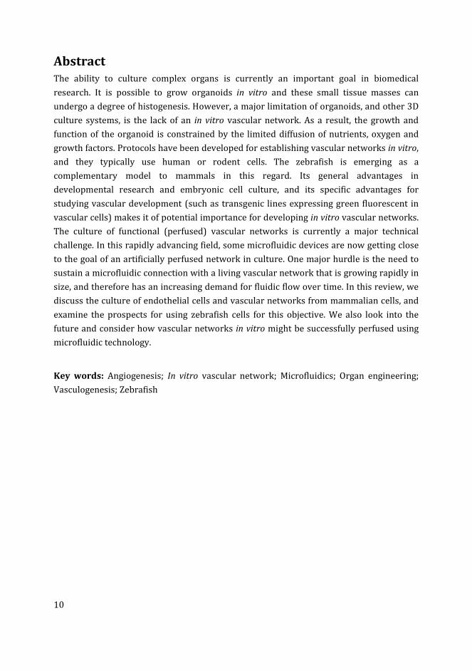
Abstract
The ability to culture complex organs is currently an important goal in biomedical
research. It is possible to grow organoids in vitro and these small tissue masses can
undergo a degree of histogenesis. However, a major limitation of organoids, and other 3D
culture systems, is the lack of an in vitro vascular network. As a result, the growth and
function of the organoid is constrained by the limited diffusion of nutrients, oxygen and
growth factors. Protocols have been developed for establishing vascular networks in vitro,
and they typically use human or rodent cells. The zebrafish is emerging as a
complementary model to mammals in this regard. Its general advantages in
developmental research and embryonic cell culture, and its specific advantages for
studying vascular development (such as transgenic lines expressing green fluorescent in
vascular cells) makes it of potential importance for developing in vitro vascular networks.
The culture of functional (perfused) vascular networks is currently a major technical
challenge. In this rapidly advancing field, some microfluidic devices are now getting close
to the goal of an artificially perfused network in culture. One major hurdle is the need to
sustain a microfluidic connection with a living vascular network that is growing rapidly in
size, and therefore has an increasing demand for fluidic flow over time. In this review, we
discuss the culture of endothelial cells and vascular networks from mammalian cells, and
examine the prospects for using zebrafish cells for this objective. We also look into the
future and consider how vascular networks in vitro might be successfully perfused using
microfluidic technology.
Key words: Angiogenesis; In vitro vascular network; Microfluidics; Organ engineering;
Vasculogenesis; Zebrafish
11
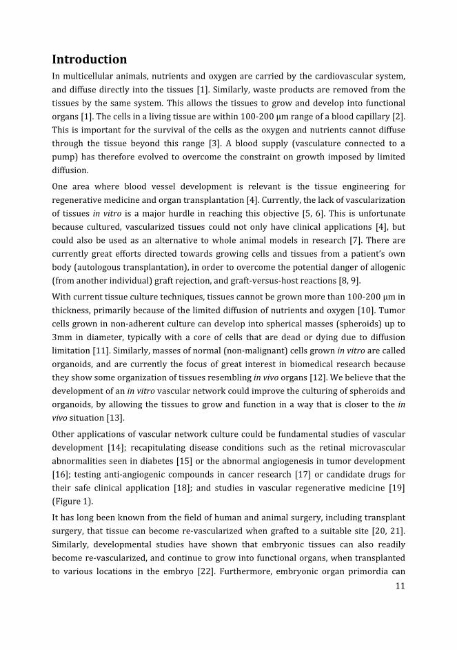
Introduction
In multicellular animals, nutrients and oxygen are carried by the cardiovascular system,
and diffuse directly into the tissues [1]. Similarly, waste products are removed from the
tissues by the same system. This allows the tissues to grow and develop into functional
organs [1]. The cells in a living tissue are within 100-200 µm range of a blood capillary [2].
This is important for the survival of the cells as the oxygen and nutrients cannot diffuse
through the tissue beyond this range [3]. A blood supply (vasculature connected to a
pump) has therefore evolved to overcome the constraint on growth imposed by limited
diffusion.
One area where blood vessel development is relevant is the tissue engineering for
regenerative medicine and organ transplantation [4]. Currently, the lack of vascularization
of tissues in vitro is a major hurdle in reaching this objective [5, 6]. This is unfortunate
because cultured, vascularized tissues could not only have clinical applications [4], but
could also be used as an alternative to whole animal models in research [7]. There are
currently great efforts directed towards growing cells and tissues from a patient’s own
body (autologous transplantation), in order to overcome the potential danger of allogenic
(from another individual) graft rejection, and graft-versus-host reactions [8, 9].
With current tissue culture techniques, tissues cannot be grown more than 100-200 µm in
thickness, primarily because of the limited diffusion of nutrients and oxygen [10]. Tumor
cells grown in non-adherent culture can develop into spherical masses (spheroids) up to
3mm in diameter, typically with a core of cells that are dead or dying due to diffusion
limitation [11]. Similarly, masses of normal (non-malignant) cells grown in vitro are called
organoids, and are currently the focus of great interest in biomedical research because
they show some organization of tissues resembling in vivo organs [12]. We believe that the
development of an in vitro vascular network could improve the culturing of spheroids and
organoids, by allowing the tissues to grow and function in a way that is closer to the in
vivo situation [13].
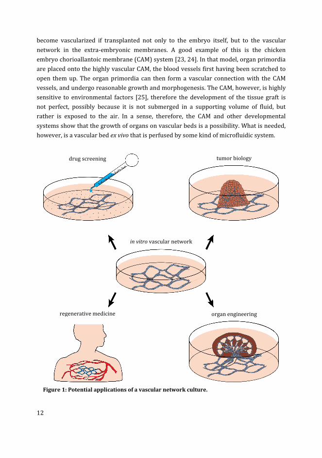
Other applications of vascular network culture could be fundamental studies of vascular
development [14]; recapitulating disease conditions such as the retinal microvascular
abnormalities seen in diabetes [15] or the abnormal angiogenesis in tumor development
[16]; testing anti-angiogenic compounds in cancer research [17] or candidate drugs for
their safe clinical application [18]; and studies in vascular regenerative medicine [19]
(Figure 1).
It has long been known from the field of human and animal surgery, including transplant
surgery, that tissue can become re-vascularized when grafted to a suitable site [20, 21].
Similarly, developmental studies have shown that embryonic tissues can also readily
become re-vascularized, and continue to grow into functional organs, when transplanted
to various locations in the embryo [22]. Furthermore, embryonic organ primordia can
12
become vascularized if transplanted not only to the embryo itself, but to the vascular
network in the extra-embryonic membranes. A good example of this is the chicken
embryo chorioallantoic membrane (CAM) system [23, 24]. In that model, organ primordia
are placed onto the highly vascular CAM, the blood vessels first having been scratched to
open them up. The organ primordia can then form a vascular connection with the CAM
vessels, and undergo reasonable growth and morphogenesis. The CAM, however, is highly
sensitive to environmental factors [25], therefore the development of the tissue graft is
not perfect, possibly because it is not submerged in a supporting volume of fluid, but
rather is exposed to the air. In a sense, therefore, the CAM and other developmental
systems show that the growth of organs on vascular beds is a possibility. What is needed,
however, is a vascular bed ex vivo that is perfused by some kind of microfluidic system.
Figure 1: Potential applications of a vascular network culture.
in vitro vascular network
drug screening tumor biology
organ engineering regenerative medicine
13
Most of the current research describing vasculogenesis (de novo formation of blood
vessels from progenitor cells) and angiogenesis (formation of blood vessels from existing
blood vessels) uses mammalian models, mainly mice. However, these models are fairly
expensive, time consuming and require ethical and other permissions [26]. Endothelial
cell lines such as human umbilical vein endothelial cells (HUVECs) are commonly used for
developing in vitro vascular networks [27]. Other sources for developing such cultures
include embryonic or adult stem cells or tissue explants. The uses and limitations of these
techniques are discussed in detail in the following sections.
Zebrafish can be an alternative to mammalian models [28]. The zebrafish produce a large
number of fertilized eggs at low cost; the embryos are externally fertilized and therefore
readily accessible for experiments [29]. In some jurisdictions, zebrafish embryos has
fewer ethical restrictions. For example, in the European Union, the Directive 2010/63/EU
on the protection of experimental animals allows zebrafish embryos to be used until 5
days post fertilization (dpf) without restriction [30]. Finally, the zebrafish genome has
been sequenced and there is a high level of conservation between zebrafish and human
protein coding genes [31]. This similarity supports the use of zebrafish to model various
human diseases [32, 33]. Because of these advantages, the zebrafish is currently emerging
as a model species to study vasculogenesis and angiogenesis in vivo [28]. Transgenic
reporter lines are proving very useful in these studies [28].
In this review we give a general overview of vascular development in vivo and the role of
various factors in the development of vasculature. Then, we review the current
procedures used to culture vascular networks using mammalian endothelial cells and
tissue explants. Then, we review the use of zebrafish to study various aspects of
vasculogenesis and angiogenesis in vivo. Finally we look forward by summarizing the
potential use of zebrafish model for in vitro studies of vascular development.
Development of vasculature in vivo
Formation of a vascular system is an essential process in embryonic development.
Because multicellular tissues cannot survive without a blood supply, the cardiovascular
system is one of the earliest systems formed during embryogenesis [34, 35]. The
endothelial precursor cells (angioblasts) differentiate into endothelial cells and undergo
the process of vasculogenesis in early embryos to form the primitive blood vessels [36].
Studies on zebrafish have shown that the angioblasts appear in the lateral mesoderm,
migrate to the midline of the embryo and form the first blood vessels [37]. In adult mice
and humans, endothelial progenitor cells reside in the bone marrow as multipotent adult
progenitor cells, and contribute to the formation of new blood vessels [38].
Further development of blood vessels takes place by the extension of the pre-existing
vascular network through the process of sprouting and non-sprouting angiogenesis [39].
14
During angiogenic sprouting, some endothelial cells within the existing blood vessel are
selected as tip cells, and migrate in the direction of angiogenic stimuli [40]. The
surrounding extracellular matrix is degraded by specific proteases released during the
process [41]. Meanwhile, the stalk cells (endothelial cells following the tip cells)
proliferate to extend the blood vessel [40]. Further in development the vascular network
also extends through intussusceptive or non-sprouting angiogenesis [42]. The mature
blood vessels attain arterial, venous and lymphatic differentiation types having different
structures and functions [43]. Endothelial differentiation and blood vessel formation is a
complex process which requires a number of growth factors, cell types and extracellular
matrix (ECM) components, discussed in the following section.
Factors controlling vasculogenesis and angiogenesis in vivo
Exogenous protein factors influencing vascular development
The differentiation of endothelial cells and the formation of blood vessels is mainly
controlled by several protein factors [44]. Some of these factors are released by the
endothelial cells themselves, other factors are stabilizing signals released by other cell
types [44]. The differentiation of angioblasts is induced mainly by fibroblast growth factor
2 (FGF2) and bone morphogenic protein 4 (BMP4) [43]. FGF2 induces the expression of
vascular endothelial growth factor (VEGF) and other important chemokines required to
control vascular morphogenesis [45]. The importance of FGF2 for vascular formation has
been shown in studies on quail and zebrafish embryos [46, 47]. Similarly, BMP4 deficiency
is associated with severe abnormalities in early mouse embryos, including the lack of a
well-organized vasculature [48].
Among the endothelial growth factors, VEGFs play the predominant role in regulating the
formation of blood vessels [49]. The VEGF family consists of several VEGF genes of which
VEGF-A, which interacts with endothelial cells through VEGF receptor 2 (VEGFR2 also
known as KDR or FLK1), is the main component responsible for the viability and
proliferation of endothelial cells [50]. VEGFs also have important roles in the
differentiation, migration and cell-cell adhesion of endothelial cells, as well as stimulating
sprouting angiogenesis and the activation of tip cells [51]. Placental growth factor (PlGF),
a member of VEGF family expressed in the placenta of early mammalian embryos, has a
role in the activation of VEGFR2 and establishing interaction between VEGF-A and
VEGFR2 [52]. PlGF has been demonstrated to increase the angiogenic potential of VEGF in
ischemic myocardium in mouse [53]. PlGF expression is normally low in adult tissues, but
high in pathological conditions, especially in cancer, where it promotes tumour
angiogenesis [54].
15
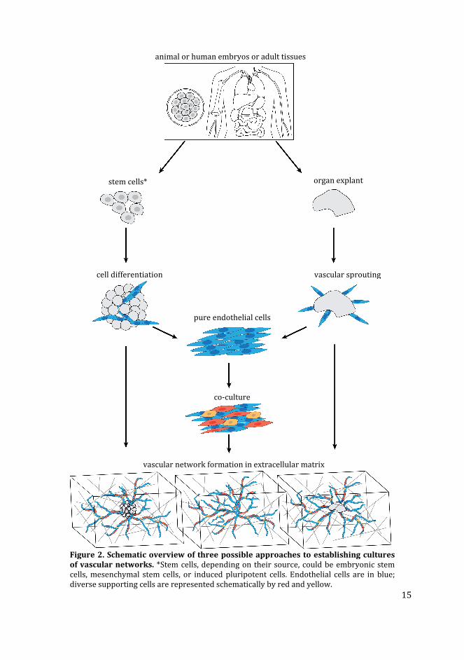
Figure 2. Schematic overview of three possible approaches to establishing cultures
of vascular networks. *Stem cells, depending on their source, could be embryonic stem
cells, mesenchymal stem cells, or induced pluripotent cells. Endothelial cells are in blue;
diverse supporting cells are represented schematically by red and yellow.
animal or human embryos or adult tissues
stem cells* organ explant
cell differentiation
pure endothelial cells
vascular sprouting
co-culture
vascular network formation in extracellular matrix

16
Other growth factors involved in the spreading and maturation of blood vessels include
angiopoietins (Ang-1 and Ang-2) [55], platelet-derived growth factor-B (PDGF-B) [56] and
transforming growth factor β (TGF-β) [57], reviewed in Refs. [44, 49]. Many other
transcription factors and signalling molecules have been identified to be involved in the
differentiation of endothelial cells and the regulation of vascular development reviewed in
Ref. [58]. In response to low oxygen levels in the tissues, Hypoxia inducible factors (HIFs)
regulates the expression of a number of pro-angiogenic factors including VEGF, PlGF, Ang-
1, Ang-2 and PDGF-B [59]. The HIFs are considered to be the principle mediators of in vivo
vasculogenesis and angiogenesis at all developmental stages [59].
Role of membrane proteins and other cell types on vascular
development
Membrane proteins on the surface of endothelial cells also play an important role in
vascular morphogenesis. Examples of these membrane proteins include vascular
endothelial cadherin (VE-cadherin) which functions to maintain endothelial cell-cell
contact during VEGF-induced migration [60], epidermal growth factor like domain-7,
which facilitates the formation of endothelial tubes [61], and delta like ligand-4 which
specifies the tip cells for sprouting angiogenesis [62].
In addition to the soluble and bound protein factors, cell types other than endothelial cells
also contribute to the formation of blood vessels. Pericytes and smooth muscle cells
promote the proliferation and survival of endothelial cells and provide structural support
to the blood vessels [63, 64]. Macrophages are reported to be involved in connecting two
blood vessel sprouts in the process called anastomosis [65]. Under certain conditions (e.g.
hypoxia), the parenchymal cells (neurons, hepatocytes, myocytes etc.) release angiogenic
growth factors to initiate sprouting angiogenesis [66].
Role of extracellular matrix
Extracellular matrix (ECM) contributes to the formation and diversity of blood vessels in
several ways including: (i) maintaining the histological structure and elasticity of the
vessels, (ii) regulating the proliferation and differentiation of endothelial cells, and (iii)
transporting, modifying or blocking the angiogenic growth factors [67]. The ECM is a
complex network of macromolecules and its composition and properties are highly
variable among different tissues, affecting the tissue specific differentiation of stem cells
[68]. Research on the ECM of blood vessels have shown the presence of different ECM
components at different stages of vascular development [69]. In the beginning of the
process, the endothelial cells adhere to and migrate on a laminin-rich ECM which is later
replaced by a collagen type-I rich ECM to support vascular tube formation [69].
17
Haemodynamic factors
Shear stress generated by blood flow on the luminal surface of endothelial cells is a
mechanical factor that induces intracellular biochemical pathways resulting in gene
expression changes and the modulation of the structure and function of blood vessels [70].
Heparin binding EGF-like growth factor is one such factor which is expressed in response
to reduced blood flow and induces vessel narrowing [71]. Other molecular pathways
involved in vascular remodelling are reported to be regulated by changes in shear stress
leading to the expression of PlGF [72], Notch1 [73], and Smad6 (involved in TGB-β
signalling) proteins [74].
Microfluidic culture of endothelial cells is currently an emerging technology which mimics
the physiological shear stress on cultured cells to achieve the goal of culturing functional
blood vessels for tissue engineering [13, 75]. A number of techniques for culturing
vascular networks have been described in which endothelial growth factors, ECM
components and microfluidics are combined; however, the development of fully functional
blood vessels still remains a challenge [75].
Culture of vascular networks using endothelial cells
Pure endothelial cell populations can develop into vascular network-like structures in
culture [27]. However, these networks are not sufficiently robust to be used for tissue
engineering; they are mainly used to screen pro- and anti-angiogenic compounds for
activity. Pure endothelial cell populations are derived from various sources including
embryonic stem cells, induced pluripotent stem cells and adult tissues [76] (Figure 2).
Human macro- and micro-vascular endothelial cells are commercially available
(http://www.promocell.com/products-/human-primary-cells/) and have the ability to
form vascular networks in vitro. The most commonly used endothelial cells in this regard
are the HUVECs [27, 77-87], derived from the veins of the umbilical cord (Table 1). Other
endothelial cell types such as bovine aortic endothelial cells [88] and rat aortic endothelial
cells [89] have also been used to culture vascular networks.
By contrast, the culture of well-defined vascular networks with a lumen requires the co-
culture of multiple cell types with endothelial cells [76]. The important supporting cell
types, known to induce network formation by endothelial cells, include pericytes [90],
mesenchymal stem cells [85], fibroblasts [79], hepatocytes [91], smooth muscle cells [92]
and adipose-derived stem cells [78]. The importance of fibroblasts in enhancing
angiogenesis has been shown in co-culture with HUVECs [81, 93]. The ECM components
and angiogenic growth factors secreted by fibroblasts have been found to be critical for
vascular tube formation from HUVECs [94].
18
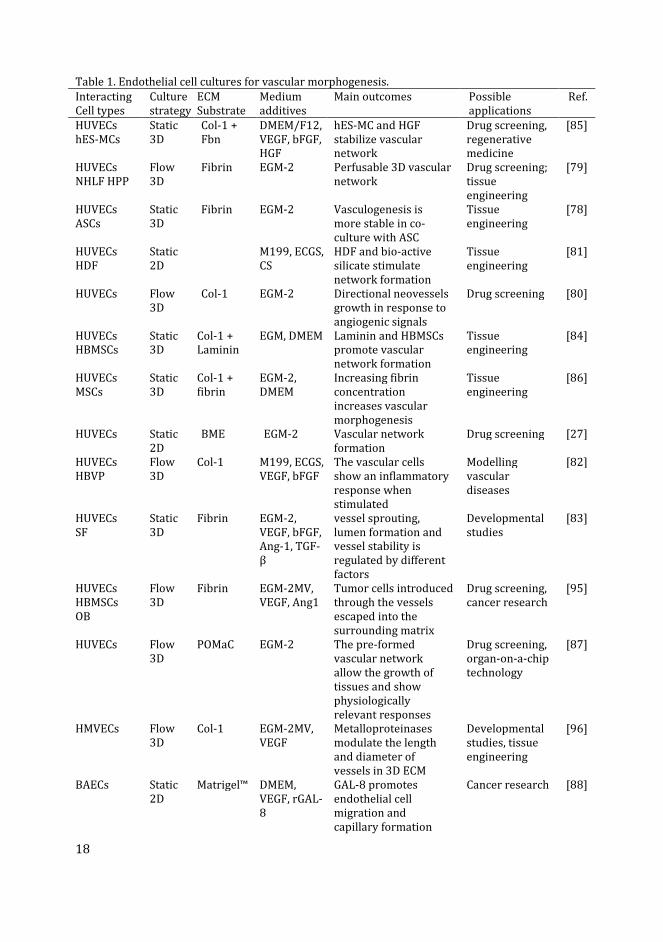
Table 1. Endothelial cell cultures for vascular morphogenesis.
Interacting
Cell types
Culture
strategy
ECM
Substrate
Medium
additives
Main outcomes Possible
applications
Ref.
HUVECs
hES-MCs
Static
3D
Col-1 +
Fbn
DMEM/F12,
VEGF, bFGF,
HGF
hES-MC and HGF
stabilize vascular
network
Drug screening,
regenerative
medicine
[85]
HUVECs
NHLF HPP
Flow
3D
Fibrin EGM-2 Perfusable 3D vascular
network
Drug screening;
tissue
engineering
[79]
HUVECs
ASCs
Static
3D
Fibrin EGM-2 Vasculogenesis is
more stable in co-
culture with ASC
Tissue
engineering
[78]
HUVECs
HDF
Static
2D
M199, ECGS,
CS
HDF and bio-active
silicate stimulate
network formation
Tissue
engineering
[81]
HUVECs Flow
3D
Col-1 EGM-2 Directional neovessels
growth in response to
angiogenic signals
Drug screening [80]
HUVECs
HBMSCs
Static
3D
Col-1 +
Laminin
EGM, DMEM Laminin and HBMSCs
promote vascular
network formation
Tissue
engineering
[84]
HUVECs
MSCs
Static
3D
Col-1 +
fibrin
EGM-2,
DMEM
Increasing fibrin
concentration
increases vascular
morphogenesis
Tissue
engineering
[86]
HUVECs Static
2D
BME EGM-2 Vascular network
formation
Drug screening [27]
HUVECs
HBVP
Flow
3D
Col-1 M199, ECGS,
VEGF, bFGF
The vascular cells
show an inflammatory
response when
stimulated
Modelling
vascular
diseases
[82]
HUVECs
SF
Static
3D
Fibrin EGM-2,
VEGF, bFGF,
Ang-1, TGF-
β
vessel sprouting,
lumen formation and
vessel stability is
regulated by different
factors
Developmental
studies
[83]
HUVECs
HBMSCs
OB
Flow
3D
Fibrin EGM-2MV,
VEGF, Ang1
Tumor cells introduced
through the vessels
escaped into the
surrounding matrix
Drug screening,
cancer research
[95]
HUVECs Flow
3D
POMaC EGM-2 The pre-formed
vascular network
allow the growth of
tissues and show
physiologically
relevant responses
Drug screening,
organ-on-a-chip
technology
[87]
HMVECs Flow
3D
Col-1 EGM-2MV,
VEGF
Metalloproteinases
modulate the length
and diameter of
vessels in 3D ECM
Developmental
studies, tissue
engineering
[96]
BAECs Static
2D
Matrigel™ DMEM,
VEGF, rGAL-
8
GAL-8 promotes
endothelial cell
migration and
capillary formation
Cancer research [88]
19
Interacting
Cell types
Culture
strategy
ECM
Substrate
Medium
additives
Main outcomes Possible
applications
Ref.
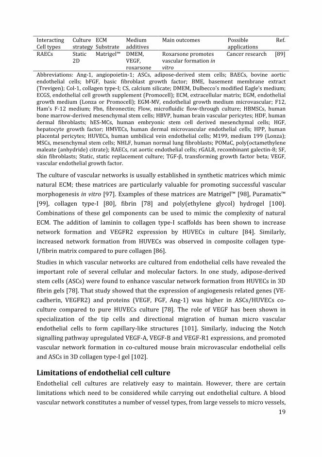
RAECs Static
2D
Matrigel™ DMEM,
VEGF,
roxarsone
Roxarsone promotes
vascular formation in
vitro
Cancer research [89]
Abbreviations: Ang-1, angiopoietin-1; ASCs, adipose-derived stem cells; BAECs, bovine aortic
endothelial cells; bFGF, basic fibroblast growth factor; BME, basement membrane extract
(Trevigen); Col-1, collagen type-I; CS, calcium silicate; DMEM, Dulbecco’s modified Eagle’s medium;
ECGS, endothelial cell growth supplement (Promocell); ECM, extracellular matrix; EGM, endothelial
growth medium (Lonza or Promocell); EGM-MV, endothelial growth medium microvascular; F12,
Ham’s F-12 medium; Fbn, fibronectin; Flow, microfluidic flow-through culture; HBMSCs, human
bone marrow-derived mesenchymal stem cells; HBVP, human brain vascular pericytes; HDF, human
dermal fibroblasts; hES-MCs, human embryonic stem cell derived mesenchymal cells; HGF,
hepatocyte growth factor; HMVECs, human dermal microvascular endothelial cells; HPP, human
placental pericytes; HUVECs, human umbilical vein endothelial cells; M199, medium 199 (Lonza);
MSCs, mesenchymal stem cells; NHLF, human normal lung fibroblasts; POMaC, poly(octamethylene
maleate (anhydride) citrate); RAECs, rat aortic endothelial cells; rGAL8, recombinant galectin-8; SF,
skin fibroblasts; Static, static replacement culture; TGF-β, transforming growth factor beta; VEGF,
vascular endothelial growth factor.
The culture of vascular networks is usually established in synthetic matrices which mimic
natural ECM; these matrices are particularly valuable for promoting successful vascular
morphogenesis in vitro [97]. Examples of these matrices are Matrigel™ [98], Puramatix™
[99], collagen type-I [80], fibrin [78] and poly(ethylene glycol) hydrogel [100].
Combinations of these gel components can be used to mimic the complexity of natural
ECM. The addition of laminin to collagen type-I scaffolds has been shown to increase
network formation and VEGFR2 expression by HUVECs in culture [84]. Similarly,
increased network formation from HUVECs was observed in composite collagen type-
I/fibrin matrix compared to pure collagen [86].
Studies in which vascular networks are cultured from endothelial cells have revealed the
important role of several cellular and molecular factors. In one study, adipose-derived
stem cells (ASCs) were found to enhance vascular network formation from HUVECs in 3D
fibrin gels [78]. That study showed that the expression of angiogenesis related genes (VE-
cadherin, VEGFR2) and proteins (VEGF, FGF, Ang-1) was higher in ASCs/HUVECs co-
culture compared to pure HUVECs culture [78]. The role of VEGF has been shown in
specialization of the tip cells and directional migration of human micro vascular
endothelial cells to form capillary-like structures [101]. Similarly, inducing the Notch
signalling pathway upregulated VEGF-A, VEGF-B and VEGF-R1 expressions, and promoted
vascular network formation in co-cultured mouse brain microvascular endothelial cells
and ASCs in 3D collagen type-I gel [102].
Limitations of endothelial cell culture
Endothelial cell cultures are relatively easy to maintain. However, there are certain
limitations which need to be considered while carrying out endothelial culture. A blood
vascular network constitutes a number of vessel types, from large vessels to micro vessels,
20
and each vessel type has its own unique properties (including endothelial cell subtypes, as
discussed below). Therefore it is challenging to attempt to recapitulate the formation of
different vessel types using a homogeneous endothelial cell population [103]. Primary
endothelial cell cultures are usually derived from terminally differentiated tissues; these
cells have limited proliferative and regenerative capacity, and a short life span in vitro
[104, 105].
Endothelial cells derived from different sites within the same tissue express different
genes and respond differently to the same pro- or anti- angiogenic factors [106]. This
supports the idea of functional subtypes among endothelial cells. Endothelial cells can be
immortalised; however, this may change their behaviour and response to stimuli [107].
Immortalised endothelial cells may alter their gene expression and physiological
properties with repeated passaging in vitro, resulting in loss of vasculogenesis efficiency
[107]. The non-endothelial cell types that support in vitro vascular network formation
from endothelial cells (e.g. fibroblasts), may represent an undesirable cell type if the
resultant tissue is to be used for tissue engineering [76].
Use of stem cells for in vitro vasculogenesis
In recent years, stem cells have increasingly been used to develop vascular cultures; this is
because stem cells have several advantages over terminally differentiated endothelial cells
[19]. Stem cells are multipotent or pluripotent in nature, they show self-renewal, and their
differentiation along various cell lineages can be manipulated by fine-tuning the culture
conditions [19]. A few examples of the stem cells that can be used for endothelial, and
ultimately vascular, differentiation are summarized in Table 2. Three main stem cell types
used are: (i) embryonic stem cells (ESCs) [108], (ii) induced pluripotent stem cells (iPSCs)
[109] and (iii) mesenchymal stem cells (MSCs) [110].
In addition, endothelial progenitor cells (EPCs), which originate in the bone marrow and
contribute to the formation of new blood vessels in adults, are also useful in the study of in
vitro vasculogenesis [105]. The differentiated endothelial cells arising from these stem
cells directly undergo vasculogenesis because of the presence of other cell types that have
also differentiated from the stem cells; alternatively, the endothelial cells can be isolated
from the stem cell culture, without the unwanted additional cell types, and used for
vascular morphogenesis (either in pure culture or co-culture with defined cell types)
[111].
One of the advantages of using ESCs is that they can differentiate into multiple vascular
cell lineages simultaneously in culture. In principle, these different lineages can contribute
to the newly-formed vessels (neovessels) in a way that closely resembles the in vivo
vasculogenesis in early embryos [19, 112]. Endothelial differentiation and vascular
morphogenesis in ESCs is controlled by culture conditions (such as the presence of growth
21
factors in the medium and the use of feeder layers of stromal cells, or a substratum
consisting of a synthetic hydrogel [112, 113]).
One approach to inducing the differentiation of ESCs in culture is to allow them to first
aggregate into spherical cell masses, called embryoid bodies (EBs), in suspension culture
[114]. The use of EBs as an intermediate step is common when ESCs are cultured for
vascular differentiation (Table 2) [115]. In the absence of anti-differentiation factors (e.g.
leukaemia inhibitory factor in mouse and feeder cell layer in human), ESCs differentiate
into EBs consisting of mesodermal, ectodermal and endodermal lineages, similar to early
embryogenesis [116]. In 2D (adherent) cultures the EB cells tend to proliferate and give
rise to undesired cell types such as fibroblasts [114]. By contrast, in 3D culture
(suspension or gels), the proliferation of EB cells is limited, allowing greater control of the
differentiation of the desired cell type [114]. Significant effects of different factors, such as
culture substrate (collagen type-IV or fibronectin), cell seeding density, concentration of
VEGF and FGF in medium, and culture duration, have been observed on the endothelial
differentiation in human, mouse and zebrafish ESC culture [117, 118]. Similarly, TGF-β has
been identified to induce vascular differentiation in human ESCs [119].
Another important stem cell type, similar to ESCs in pluripotency and differentiation
events, is the iPSCs [120]. An advantage of iPSCs is that they can be generated by genetic
reprogramming of any adult somatic cell population, and therefore raise fewer ethical
concerns compared to ESCs [19]. Endothelial differentiation in iPSCs can be induced by
applying similar methods used for differentiation of ESCs [121]. Furthermore, gene
expression in endothelial cells derived from ESCs and iPSCs is very similar [121]. MSCs are
multipotent stem cells residing in adult tissues; they have limited differentiation potential
compared to ESCs and iPSCs [19]. Endothelial differentiation in human amniotic fluid
derived MSCs has been shown to be inducible by VEGF [110]. MSCs derived from various
tissues (bone marrow, hair follicle, adipose tissue and muscles) have been used for
vascular regeneration studies reviewed in Ref. [19]. In some studies the MSCs have been
reported to promote and stabilize vascular network formation from HUVECs (Table 1).
In addition to the use of pluripotent and multipotent stem cells for endothelial
differentiation and in vitro vasculogenesis, the unipotent EPCs also have the ability to
differentiate into mature endothelial cells and form vascular tubes in culture [105]. The
advantage of EPCs for culturing vascular networks is that these cells can be easily
obtained from adult tissues such as peripheral blood [19]. In vitro studies have shown
that the early EPCs do not directly undergo vascularization, but release factors to
stimulate angiogenesis in distantly-cultured endothelial cells in a transwell [105]. Co-
culture with MSCs has been proven to enhance vascular formation from EPCs both in vitro
and after implantation in vivo [122, 123].
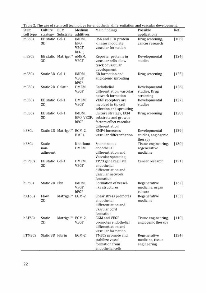
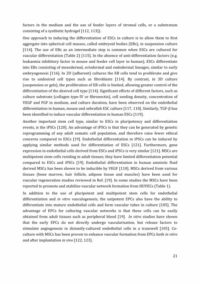
22
Table 2. The use of stem cell technology for endothelial differentiation and vascular development.
Stem
cell type
Culture
strategy
ECM
Substrate
Medium
additives
Main findings Possible
applications
Ref.
mESCs EB static
3D
Col-1 IMDM,
EPO,
VEGF,
bFGF,
RSK and TTK protein
kinases modulate
vascular formation
Drug screening,
cancer research
[108]
mESCs EB static
3D
Matrigel™ αMEM,
VEGF
Reporter proteins in
vascular cells allow
track of vascular
development
Developmental
studies
[124]
mESCs Static 3D Col-1 IMDM,
VEGF,
bFGF
EB formation and
angiogenic sprouting
Drug screening [125]
mESCs Static 2D Gelatin DMEM,
VEGF
Endothelial
differentiation, vascular
network formation
Developmental
studies, Drug
screening
[126]
mESCs EB static
2D
Col-1 DMEM,
VEGF
VEGF receptors are
involved in tip cell
selection and sprouting
Developmental
studies
[127]
mESCs EB static
3D
Col-1 IMDM,
EPO, VEGF,
bFGF
Culture strategy, ECM
substrate and growth
factors effect vascular
differentiation
Drug screening [128]
hESCs Static 2D Matrigel™ EGM-2,
BMP4
BMP4 increases
vascular differentiation
Developmental
studies, angiogenic
therapy
[129]
hESCs Static
non-
adherent
Knockout
DMEM
Spontaneous
endothelial
differentiation and
Vascular sprouting
Tissue engineering,
regenerative
medicine
[130]
miPSCs EB static
3D
Col-1 DMEM,
VEGF
TP73 gene regulate
endothelial
differentiation and
vascular network
formation
Cancer research [131]
hiPSCs Static 2D Fbn IMDM,
VEGF,
bFGF
Formation of vessel-
like structures
Regenerative
medicine, organ
culture
[132]
hAFSCs Flow
2D
Matrigel™ EGM-2 Shear stress promotes
endothelial
differentiation and
vascular cord
formation
Regenerative
medicine
[133]
hAFSCs Static
2D
Matrigel™ EGM-2,
VEGF
EGM and VEGF
promotes endothelial
differentiation and
vascular formation
Tissue engineering,
angiogenic therapy
[110]
hTMSCs Static 3D Fibrin EGM-2 TMSCs promote and
stabilize vessel
formation from
endothelial cells
Regenerative
medicine, tissue
engineering
[134]
23
Abbreviations: αMEM, alpha-minimal essential medium (Cellgro); bFGF, basic fibroblast growth
factor; BMP4, bone morphogenetic protein-4; Col-1, collagen type-I; DMEM, Dulbecco’s modified
Eagle’s medium; EB, embryoid body intermediate; ECM, extracellular matrix; EGM, endothelial
growth medium (Cambrex or Clonetics); EPO, erythropoietin; Fbn, fibronectin; Flow, under flow of
medium; hAFSCs, human amniotic fluid-derived stem cells; hESCs, human embryonic stem cells;
IMDM, Iscove’s modified Dulbecco’s medium; mESCs, mouse embryonic stem cells; miPSCs, mouse
induced pluripotent stem cells; RSK, ribosomal S6 kinase; Static, static replacement culture; TP73,
tumor protein-73; TTK, threonine and tyrosine kinase; VEGF, vascular endothelial growth factor.
Issues and drawbacks with stem cell culture
Although stem cell technology has several advantages for vascular engineering and
regenerative therapy, there are some limitations to its use [19]. Thus, while ESCs have
been extensively studied in laboratory animals such as mouse and rats, and stable cell
lines have been developed form these animals, the technique has been proven less
successful for other species such as cattle, goat and dogs [120]. Furthermore, the very
complexity of vascular differentiation from ESCs means that the growth factors necessary
to support the generation and maintenance of multiple cell types need to be laboriously
optimised [135]. Furthermore, the use of human ESCs for research raise ethical concerns
[19].
ESCs and iPSCs are both pluripotent, and therefore it is challenging to direct the
differentiation towards a specific lineage, and to obtain high quality pure cell cultures
[120]. The iPSCs are developed by transfection of somatic cells with pluripotency genes;
however, the efficiency of the process is very low (less than 1%) [136]. The iPSCs (in
contrast to ESCs) are derived from adult differentiated cells by de-differentiation. Then, if
re-differentiated into a specific cell type, they attain some of the characteristics of that cell
type but are not identical to their normal counterparts [136]. Other issues with stem cells
is that the isolation of MSCs from adult tissues requires invasive surgical procedures, and
only yields small numbers of cells; the proliferation of these cells is also limited in vitro
[137]. Furthermore, the MSCs isolated from different tissues or life stages are not the
same, and therefore have different culture requirements and angiogenic potentials [138].
The isolation and culture methods for EPCs are only relatively recently developed
(Asahara et al., 1997 [139]). For this reason, there is no standard protocol among
researchers. It should be noted that there are no specific markers for EPCs because many
of the genes expressed by EPCs are also expressed in hematopoietic progenitors [140].
Similar to the iPSCs and MSCs, the number of EPCs found in isolated adult tissue cells is
very low, and this greatly limits their study [19]. Finally, analysis of EPCs in long-term
culture has shown that the late passage cells (45 days after the initiation of the primary
culture) have changed morphology, reduced their proliferation rate, show high β-
galactosidase expression and loss of vascular network formation ability, compared to the
early passaged cells [141].

24
Use of tissue explants for in vitro angiogenesis
An important strategy for vascular morphogenesis in vitro is to stimulate the growth of
the blood vessels existing in isolated sections or fragments of specific tissues [107]. The
development of a well-defined blood vascular network requires the incorporation of
multiple cell types both in vivo and in vitro, as discussed in the previous sections. The use
of tissue explants is important in this context, because these explants already contain
multiple cell types, and the angiogenesis stimulated in these cultures closely represents
the corresponding process in vivo [107]. Furthermore, tissue explant experiments are
relatively easy to perform and allow a large number of cultures to be derived from a single
tissue sample [142].
Various tissue explants have been shown to have the ability to develop vascular sprouts in
vitro (Table 3). Examples include cross sections of aorta called aortic rings [143];
metatarsal bones [144]; retina fragments [145]; choroid-sclera fragments [146]; and
adipose tissue [147]. In most cases, the tissues for explant preparation are isolated from
developing rodent embryos or neonates. Tissue explants from other species such as chick
embryo aortic arch [148], rabbit aorta[149] and pig carotid artery [150] have also been
adapted for sprouting angiogenesis. Furthermore, angiogenic sprouting has also been
reported from human tissue explants e.g. adipose tissue [147], aortic explants from
aborted embryos [151], placental explants [152, 153] and umbilical artery rings [154].
Explant cultures are usually established in a 3D gel matrix in the presence of angiogenic
growth factors, and are examined for microvessel outgrowth (vascular sprouting)[107,
155]. The aortic ring model from various species is the most commonly used explant for
studying in vitro angiogenesis (Table 3). The stimulatory effect of various factors, such as
angiogenic growth factors (especially VEGF) and ECM components, on the growth of
vascular sprouts from aortic ring have been extensively studied, reviewed in Ref. [156].
Recently developed explant cultures, using fetal metatarsals from mice, have shown
advantages over the aortic ring model, in that they do not require a 3D matrix and
exogenous growth factors for vascular sprouting [144]. In general, explant cultures can
serve as an intermediate between the endothelial cell culture on the one hand, and in vivo
models on the other. They are also thought to be more reliable for studying the
mechanisms of angiogenesis and testing the role of regulatory factors [157].
Limitations of explant cultures
Besides the advantages of explant cultures, certain limitations need to be addressed
before the technique can be fully accepted for research in tissue engineering and
regenerative medicine. The mouse aortic ring model shows significant variability in
microvessel sprouting from explants isolated from different age and strain of animals
[158]. Variability in outcome has also been reported using explants isolated from different
vessel types (artery or vein) of the same individual animal [159]. The vascular sprouts in
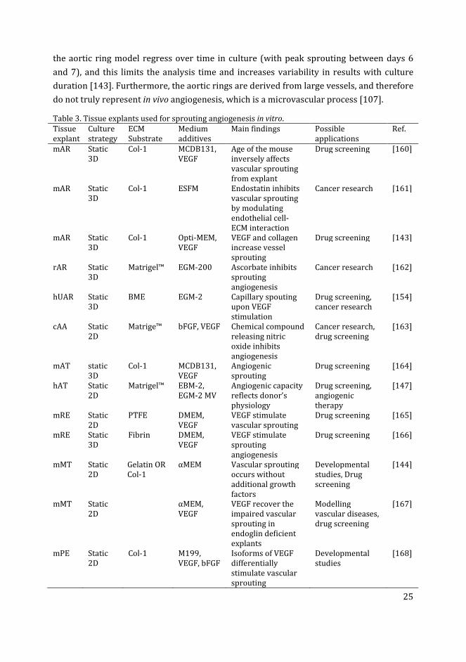
25
the aortic ring model regress over time in culture (with peak sprouting between days 6
and 7), and this limits the analysis time and increases variability in results with culture
duration [143]. Furthermore, the aortic rings are derived from large vessels, and therefore
do not truly represent in vivo angiogenesis, which is a microvascular process [107].
Table 3. Tissue explants used for sprouting angiogenesis in vitro.
Tissue
explant
Culture
strategy
ECM
Substrate
Medium
additives
Main findings Possible
applications
Ref.
mAR Static
3D
Col-1 MCDB131,
VEGF
Age of the mouse
inversely affects
vascular sprouting
from explant
Drug screening [160]
mAR Static
3D
Col-1 ESFM Endostatin inhibits
vascular sprouting
by modulating
endothelial cell-
ECM interaction
Cancer research [161]
mAR Static
3D
Col-1 Opti-MEM,
VEGF
VEGF and collagen
increase vessel
sprouting
Drug screening [143]
rAR Static
3D
Matrigel™ EGM-200 Ascorbate inhibits
sprouting
angiogenesis
Cancer research [162]
hUAR Static
3D
BME EGM-2 Capillary spouting
upon VEGF
stimulation
Drug screening,
cancer research
[154]
cAA Static
2D
Matrige™ bFGF, VEGF Chemical compound
releasing nitric
oxide inhibits
angiogenesis
Cancer research,
drug screening
[163]
mAT static
3D
Col-1 MCDB131,
VEGF
Angiogenic
sprouting
Drug screening [164]
hAT Static
2D
Matrigel™ EBM-2,
EGM-2 MV
Angiogenic capacity
reflects donor’s
physiology
Drug screening,
angiogenic
therapy
[147]
mRE Static
2D
PTFE DMEM,
VEGF
VEGF stimulate
vascular sprouting
Drug screening [165]
mRE Static
3D
Fibrin DMEM,
VEGF
VEGF stimulate
sprouting
angiogenesis
Drug screening [166]
mMT Static
2D
Gelatin OR
Col-1
αMEM Vascular sprouting
occurs without
additional growth
factors
Developmental
studies, Drug
screening
[144]
mMT Static
2D
αMEM,
VEGF
VEGF recover the
impaired vascular
sprouting in
endoglin deficient
explants
Modelling
vascular diseases,
drug screening
[167]
mPE Static
2D
Col-1 M199,
VEGF, bFGF
Isoforms of VEGF
differentially
stimulate vascular
sprouting
Developmental
studies
[168]
26
Abbreviations: αMEM, alpha-minimal essential medium; bFGF, basic fibroblast growth factor; BME,
basement membrane extract (BD Biosciences); cAA, chick aortic arch; Col-1, collagen type-I; DMEM,
Dulbecco’s modified Eagle’s medium; EBM, endothelial basal medium (Lonza); ECM, extracellular
matrix; EGM MV, endothelial growth medium microvascular; EGM-200, endothelial growth medium-
200 (Cascade Biologics); ESFM, endothelial serum-free medium (Life Technologies); hAT, human
adipose tissue explant; hUAR, human umbilical arterial ring; M199, medium 199 (Gibco); mAR,
mouse aortic ring; mAT, mouse adipose tissue explant; MCDB131, basal medium (Invitrogen); mMT,
mouse metatarsal explant; mPE, mouse proepicardium explant; mRE, mouse retinal explant; Opti-
MEM, minimal essential reduced-serum medium (Gibco); PTFE, polytetrafluroethylene membrane;
rAR, rat aortic ring; Static, static replacement culture; VEGF, vascular endothelial growth factor.
Tissues containing microvascular networks (e.g. adipose tissue and retina) can be used for
explant preparation. However, these tissues are more difficult to isolate and, like the
aortic explant, show variability between experiments [145]. The high levels of capillary
sprouting observed in adipose tissue explant cultures are in many senses an advantage;
however they do make it difficult to identify all the sprouts individually and interpret the
results [147]. Similarly, angiogenic sprouts from fetal mouse metatarsal explants present
microvascular features; however, their isolation and culture procedures also require
advanced technical skills, which are key to the reproducibility of the research [144].
Finally, the metatarsal and chick aortic arch explants are isolated from developing
embryos and have high proliferative capacity; therefore, angiogenesis in these models
does not represent the in vivo situation in adults [107].
Zebrafish: a new model species for studying in vivo
vasculogenesis and angiogenesis
The zebrafish is a freshwater teleost fish [169] that is emerging as a model of choice for
studying vasculogenesis and angiogenesis [170]. The embryos and larvae are often used in
these studies because of their external fertilization, optical transparency at early stages,
and the ease of exposure to test substances (by simply adding the compound to the
swimming water) [171]. Furthermore, the genome comparison study has revealed that
there is at least one orthologue in zebrafish genome for more than 70% of human protein
coding genes [31]. Vascular development and function in zebrafish are relatively
conserved, compared to the same processes in other vertebrates [170].
The embryos develop a simple vascular system with circulating blood as early as 24 hours
post fertilization (hpf) [37]. Vascular development can be directly observed non-
invasively in the living, transparent embryo [28]. Enhanced visualization of vascular
development can be achieved by injecting fluorescent micro particles into the blood
stream, or by using transgenic lines such as kdrl:GFP and fli:GFP that express green
fluorescent protein (GFP) in vascular cells [28].
For these and other reasons, vascular development in zebrafish — from early
differentiation of angioblasts to the maturation of blood vessels — has been extensively
27
studied [32, 37, 172, 173]. Studies have shown similar angiogenic responses to the test
substance irisin, in zebrafish embryos in vivo, and in HUVECs in vitro [174]. In another
example, the genetic mutation (gridlock), which causes aortic malformations and
congenital heart defects in humans, showed similar phenotypic effects in zebrafish [175].
Zebrafish have been successfully utilized to model several human vascular diseases
reviewed in Ref. [32].
Similarly, a zebrafish in vivo xenograft model has been developed to study human
carcinomas [176]. These studies have shown successful invasion, metastasis and
extravasation of various human tumor cells in zebrafish embryos and adults [176]. It has
been demonstrated that the transplantation of human WM-266-4 melanoma cells and
breast adenocarcinoma cells in zebrafish embryos induced angiogenesis in the host
vasculature; this led to the formation and infiltration of neovessels into the tumor masses
[177, 178]. Other examples of human carcinomas studied in zebrafish include breast
cancer bone metastasis [179], uveal melanoma [180] and retinoblastoma [181].
The zebrafish possesses remarkable regenerative capacity in several organs (including the
caudal fin and heart [182, 183]) which makes it a useful model for studying regeneration
[184]. The regeneration of organs also involves the regeneration of blood vessels, and
therefore the regenerative capacity of zebrafish is also important for vascular
regeneration studies [28].
Zebrafish transgenic reporter lines for vascular studies
Several transgenic lines have been developed for zebrafish which express fluorescent
proteins under vascular cell specific promoters [28]. These transgenic lines allow the
tracking of the differentiation, proliferation and migration of individual cells during
vascular development in vivo and in vitro [28]. Moreover, different transgenes can be
combined in the same embryo, as was done with a line (scl-PAC:GFP) expressing
fluorescent proteins in both endothelial and blood cells; that line permitted the
observation of the development of the vascular system and blood flow simultaneously
[185].
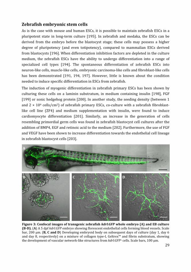
The most important transgenic line that we are utilizing to study vascular development in
zebrafish is the kdrl:GFP line (Figure 3A). This transgenic line is also known as
Tg(kdr:eGFP) or Tg(flk1:eGFP) [186]. In this line, GFP is specifically expressed in
endothelial cells under the control of the VEGFR2 or kdr-like gene [186]. The kdrl:GFP line
allows high resolution analysis of single cell migration and vascular development in living
embryos [186]. The utility of kdrl:GFP zebrafish embryos has been confirmed as a high-
throughput toxicology screening model [187].
Other transgenic zebrafish lines are available for vascular studies, although they have
some limitations. For example, In Tg(Tie2:eGFP) the GFP expression is relatively weak in
the vascular cells [188]. Similarly, in Tg(fli1:eGFP) the GFP is expressed in certain non-
28
vascular cells which interferes with the results especially in the head region of the embryo
[186]. Furthermore, studies on Tg(fli1:eGFP) have shown changes in the gene expression
of a number of genes, compared to the wild type embryos, which may affect the results
while using transgenic zebrafish for experiments [189].
In summary, zebrafish is a high-throughput, easily quantifiable, fast developing and
relatively inexpensive in vivo model for vascular studies. However, there are some
drawbacks associated with this model. The relevance of zebrafish embryo model to
understand human angiogenesis is questioned, as there is a large evolutionary time
difference between the two species [157]. Therefore, preclinical drug screening in
zebrafish should always be followed by validation in mammalian models before going to
clinical trials [28].
Future prospects for using zebrafish cells for in vitro
vasculogenesis and angiogenesis
In principle, many of the techniques discussed in the previous sections for in vitro
vasculogenesis and angiogenesis (using mammalian endothelial cells, ESCs and tissue
explants), can also be adapted for use in the zebrafish model. With the availability of
primary embryonic cells due to high fecundity of the species, and easy cell isolation
procedures, the drawbacks associated with adapted cell lines can be avoided [190]. In
addition to the above mentioned characteristics, zebrafish also possess specific desirable
features for in vitro applications.
Cell culture techniques in zebrafish
The external fertilization and large number of fast developing embryos, allow easy
harvesting of large numbers of cells and quantities of tissues from different
developmental stages [191]. Zebrafish cells grow at a lower temperature (26-28 °C) than
chick and mouse cells and do not usually require a CO2-enriched atmosphere [191]. These
properties allow zebrafish cells to be grown at room temperature, although the use of a
simple incubator is recommended to help maintain sterile conditions [191]. The
protective covering of the chorion, which is present until hatching at around 48 hpf, partly
isolates the embryos from the environment [192]. This is important for in vitro studies
because it maintains the embryos in an aseptic condition [193].
To harvest sterile cells or tissues from zebrafish embryos it is necessary to decontaminate
the surface of the chorion. Using this approach, it is possible to isolate and culture sterile
cells from blastula (3 hpf) or gastrula (24 hpf) stage embryos [191, 194]. In a recent study,
we have shown that embryos with a chorion decontaminated at 24 hpf could be further
cultured to 5 dpf under aseptic conditions [155]. The tissues and cells isolated from these
embryos were successfully maintained free of contamination for eight days in culture.
29
Zebrafish embryonic stem cells
As is the case with mouse and human ESCs, it is possible to maintain zebrafish ESCs in a
pluripotent state in long-term culture [195]. In zebrafish and medaka, the ESCs can be
derived from the embryo before the blastocyst stage; these cells may possess a higher
degree of pluripotency (and even totipotency), compared to mammalian ESCs derived
from blastocysts [196]. When differentiation inhibition factors are depleted in the culture
medium, the zebrafish ESCs have the ability to undergo differentiation into a range of
specialized cell types [194]. The spontaneous differentiation of zebrafish ESCs into
neuron-like cells, muscle-like cells, embryonic carcinoma-like cells and fibroblast-like cells
has been demonstrated [191, 194, 197]. However, little is known about the condition
needed to induce specific differentiation in ESCs from zebrafish.
The induction of myogenic differentiation in zebrafish primary ESCs has been shown by
culturing these cells on a laminin substratum, in medium containing insulin [198], FGF
[199] or sonic hedgehog protein [200]. In another study, the seeding density (between 1
and 2 × 10⁴ cells/cm2) of zebrafish primary ESCs, co-culture with a zebrafish fibroblast-
like cell line (ZF4) and medium supplementation with insulin, were found to induce
cardiomyocyte differentiation [201]. Similarly, an increase in the generation of cells
resembling primordial germ cells was found in zebrafish blastocyst cell cultures after the
addition of BMP4, EGF and retinoic acid to the medium [202]. Furthermore, the use of FGF
and VEGF have been shown to increase differentiation towards the endothelial cell lineage
in zebrafish blastocyst cells [203].
Figure 3: Confocal images of transgenic zebrafish kdrl:GFP whole embryo (A) and EB culture
(B-D). (A) A 5 dpf kdrl:GFP embryo showing florescent endothelial cells forming blood vessels. Scale
bar, 200 µm. (B, C and D) Developing embryoid body on subsequent days of culture (day 1, day 6
and day 8, respectively) on a mixture of collagen type-I, Geltrex™ and fibrin substratum, showing
the development of vascular network-like structures from kdrl:GFP+ cells. Scale bars, 100 µm.
A
B C D
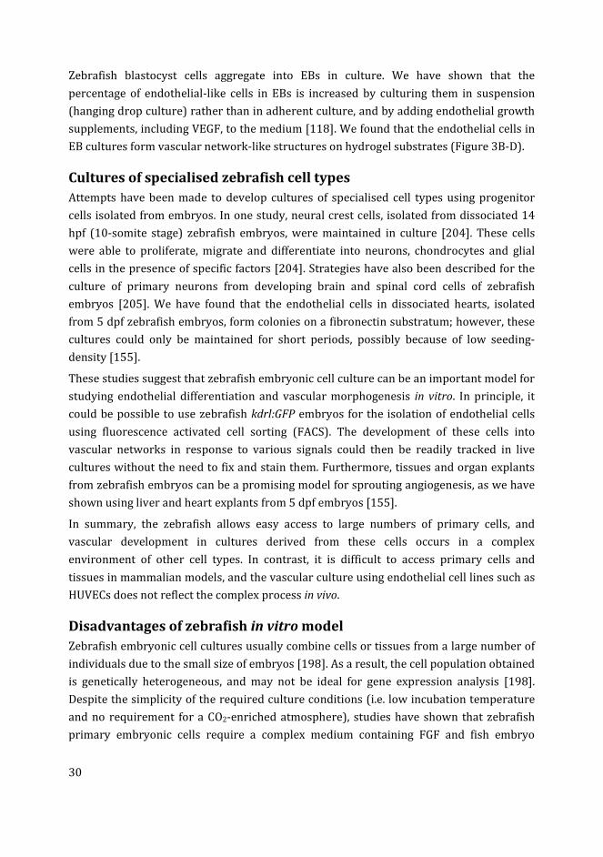
30
Zebrafish blastocyst cells aggregate into EBs in culture. We have shown that the
percentage of endothelial-like cells in EBs is increased by culturing them in suspension
(hanging drop culture) rather than in adherent culture, and by adding endothelial growth
supplements, including VEGF, to the medium [118]. We found that the endothelial cells in
EB cultures form vascular network-like structures on hydrogel substrates (Figure 3B-D).
Cultures of specialised zebrafish cell types
Attempts have been made to develop cultures of specialised cell types using progenitor
cells isolated from embryos. In one study, neural crest cells, isolated from dissociated 14
hpf (10-somite stage) zebrafish embryos, were maintained in culture [204]. These cells
were able to proliferate, migrate and differentiate into neurons, chondrocytes and glial
cells in the presence of specific factors [204]. Strategies have also been described for the
culture of primary neurons from developing brain and spinal cord cells of zebrafish
embryos [205]. We have found that the endothelial cells in dissociated hearts, isolated
from 5 dpf zebrafish embryos, form colonies on a fibronectin substratum; however, these
cultures could only be maintained for short periods, possibly because of low seeding-
density [155].
These studies suggest that zebrafish embryonic cell culture can be an important model for
studying endothelial differentiation and vascular morphogenesis in vitro. In principle, it
could be possible to use zebrafish kdrl:GFP embryos for the isolation of endothelial cells
using fluorescence activated cell sorting (FACS). The development of these cells into
vascular networks in response to various signals could then be readily tracked in live
cultures without the need to fix and stain them. Furthermore, tissues and organ explants
from zebrafish embryos can be a promising model for sprouting angiogenesis, as we have
shown using liver and heart explants from 5 dpf embryos [155].
In summary, the zebrafish allows easy access to large numbers of primary cells, and
vascular development in cultures derived from these cells occurs in a complex
environment of other cell types. In contrast, it is difficult to access primary cells and
tissues in mammalian models, and the vascular culture using endothelial cell lines such as
HUVECs does not reflect the complex process in vivo.
Disadvantages of zebrafish in vitro model
Zebrafish embryonic cell cultures usually combine cells or tissues from a large number of
individuals due to the small size of embryos [198]. As a result, the cell population obtained
is genetically heterogeneous, and may not be ideal for gene expression analysis [198].
Despite the simplicity of the required culture conditions (i.e. low incubation temperature
and no requirement for a CO2-enriched atmosphere), studies have shown that zebrafish
primary embryonic cells require a complex medium containing FGF and fish embryo
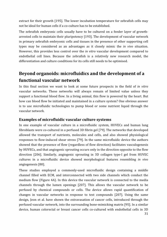
31
extract for their growth [195]. The lower incubation temperature for zebrafish cells may
not be ideal for human cells if a co-culture has to be established.
The zebrafish embryonic cells usually have to be cultured on a feeder layer of growth-
arrested cells to maintain their pluripotency [193]. The development of vascular network
in primary zebrafish embryonic cells and tissues in the presence of other supporting cell
types may be considered as an advantages as it closely mimic the in vivo situation.
However, this provides less control over the in vitro vascular development compared to
endothelial cell lines. Because the zebrafish is a relatively new research model, the
differentiation and culture conditions for its cells still needs to be optimised.
Beyond organoids: microfluidics and the development of a
functional vascular network
In this final section we want to look at some future prospects in the field of in vitro
vascular networks. These networks will always remain of limited value unless they
support a functional blood flow. In a living animal, this flow is powered by the heart. But
how can blood flow be initiated and maintained in a culture system? One obvious answer
is to use microfluidic technologies to pump blood or some nutrient liquid through the
vascular network.
Examples of microfluidic vascular culture systems
In one example of vascular culture in a microfluidic system, HUVECs and human lung
fibroblasts were co-cultured in a perfused 3D fibrin gel [79]. The networks that developed
allowed the transport of nutrients, molecules and cells, and also showed physiological
responses to flow-induced shear stress [79]. In the same microfluidic device the authors
showed that the presence of flow (regardless of flow direction) facilitates vasculogenesis
by HUVECs, and that angiogenic sprouting occurs only in the direction opposite to the flow
direction [206]. Similarly, angiogenic sprouting in 3D collagen type-I gel from HUVEC
cultures in a microfluidic device showed morphological features resembling in vivo
angiogenesis [80].
These studies employed a commonly-used microfluidic design containing a middle
channel filled with ECM, and interconnected with two side channels which conduct the
medium flow (Figure 4A). In this device the vascular network is connected to the media
channels through the lumen openings [207]. This allows the vascular network to be
perfused by chemical compounds or cells. The device allows rapid quantification of
changes in vascular networks in response to test compounds [207]. Using the same
design, Jeon et al. have shown the extravasation of cancer cells, introduced through the
perfused vascular network, into the surrounding bone-mimicking matrix [95]. In a similar
device, human colorectal or breast cancer cells co-cultured with endothelial cells in 3D
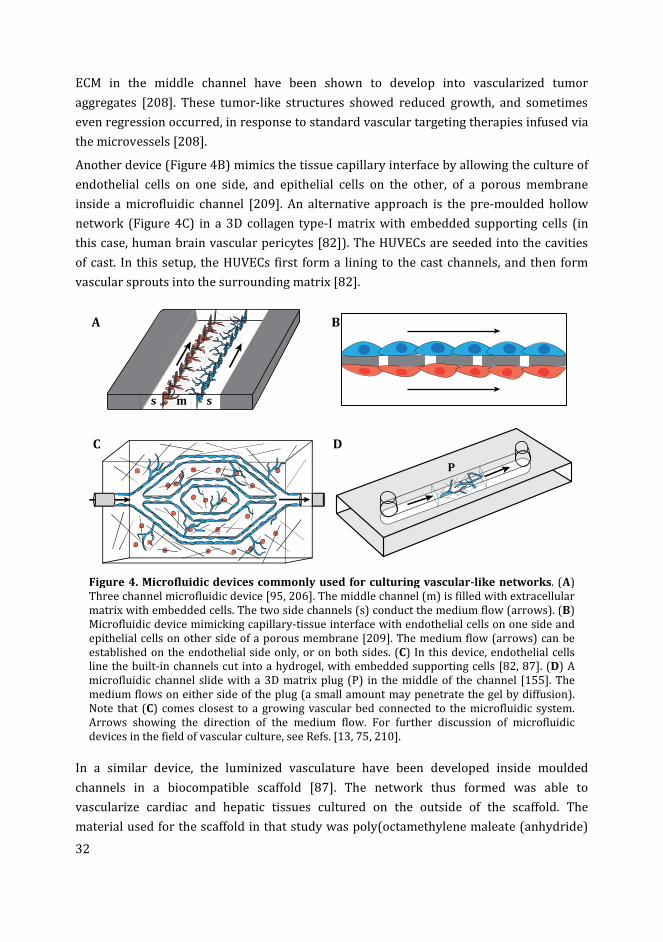
32
ECM in the middle channel have been shown to develop into vascularized tumor
aggregates [208]. These tumor-like structures showed reduced growth, and sometimes
even regression occurred, in response to standard vascular targeting therapies infused via
the microvessels [208].
Another device (Figure 4B) mimics the tissue capillary interface by allowing the culture of
endothelial cells on one side, and epithelial cells on the other, of a porous membrane
inside a microfluidic channel [209]. An alternative approach is the pre-moulded hollow
network (Figure 4C) in a 3D collagen type-I matrix with embedded supporting cells (in
this case, human brain vascular pericytes [82]). The HUVECs are seeded into the cavities
of cast. In this setup, the HUVECs first form a lining to the cast channels, and then form
vascular sprouts into the surrounding matrix [82].
Figure 4. Microfluidic devices commonly used for culturing vascular-like networks. (A)
Three channel microfluidic device [95, 206]. The middle channel (m) is filled with extracellular
matrix with embedded cells. The two side channels (s) conduct the medium flow (arrows). (B)
Microfluidic device mimicking capillary-tissue interface with endothelial cells on one side and
epithelial cells on other side of a porous membrane [209]. The medium flow (arrows) can be
established on the endothelial side only, or on both sides. (C) In this device, endothelial cells
line the built-in channels cut into a hydrogel, with embedded supporting cells [82, 87]. (D) A
microfluidic channel slide with a 3D matrix plug (P) in the middle of the channel [155]. The
medium flows on either side of the plug (a small amount may penetrate the gel by diffusion).
Note that (C) comes closest to a growing vascular bed connected to the microfluidic system.
Arrows showing the direction of the medium flow. For further discussion of microfluidic
devices in the field of vascular culture, see Refs. [13, 75, 210].
In a similar device, the luminized vasculature have been developed inside moulded
channels in a biocompatible scaffold [87]. The network thus formed was able to
vascularize cardiac and hepatic tissues cultured on the outside of the scaffold. The
material used for the scaffold in that study was poly(octamethylene maleate (anhydride)
A B
C D
s m s
P
33
citrate) (POMaC; a biodegrade-able and biocompatible scaffold) [87]. In that setup, the
micro pores incorporated into the scaffold allowed the uptake of nutrients, chemical
compounds and cells from the vessels, and the release of metabolites into the vessels by
the surrounding tissue [87].
In most cases, endothelial cell lines are used to culture vascular networks in a microfluidic
system. The stem cells and explant cultures have been very little used in such studies. We
have shown that zebrafish EBs embedded in 3D gel matrix, cultured in a microfluidic
channel (Figure 4D) formed longer and wider vascular sprouts compared to the EBs
cultured in conventional (static) 96-well plates [155].
Technical challenges for the future
As we have discussed above, scientists are beginning to realize the goal of a functional
vascular network perfused by microfluidics in vitro. However, there are some severe
technical challenges to be overcome. Presumably, some kind of synthetic interface or
connector will be needed to connect the living vessels with microfluidic system. Another
major challenge will be to maintain an increasing blood flow as the tissue explant,
attached to the vascular network, grows in size. Thus, the growing tissue will require
vessels of increasing diameter, and this in turn will require an expanding connection to
the microfluidic system. Solutions to these problems will require intensive research.
Conclusions
In summary, the techniques developed for in vitro vascular network formation in
mammals are producing rapid advances in our understanding. They can also be applied to
zebrafish cells and tissues. For its benefits such as easy access to primary embryonic cells,
availability of transgenic lines to visualize endothelial cells and conserved molecular
pathways, the zebrafish can be a significant first-step model for studying in vitro vascular
development. In recent years, microfluidic technology have shown great advances in
developing in vitro vascular networks. Ultimately, the future of tissue culture and organ
culture will be greatly extended if functional vascular networks with a fluidic flow can be
grown in vitro. The major challenges in establishing such a system would be to
recapitulate the key features of vascular system such as barrier function and vasoactivity,
as wells as to compensate for the increasing demands of the growing tissue.
References
[1] Levick JR. The microcirculation and solute exchange. In: Eds. An Introduction to
cardiovascular physiology. 5 ed, Taylor & Francis 2010, pp. 166-187.
34
[2] Kannan RY, Salacinski HJ, Sales K, Butler P, Seifalian AM. The roles of tissue
engineering and vascularisation in the development of micro-vascular networks: a
review. Biomaterials 2005; 26(14): 1857-75.
[3] Jain RK, Au P, Tam J, Duda DG, Fukumura D. Engineering vascularized tissue. Nat
Biotechnol 2005; 23(7): 821-3.
[4] Marx V. Tissue engineering: organs from the lab. Nature 2015; 522(7556): 373-7.
[5] Kim JJ, Hou L, Huang NF. Vascularization of three-dimensional engineered tissues
for regenerative medicine applications. Acta Biomater 2016; 41: 17-26.
[6] Bae H, Puranik AS, Gauvin R, Edalat F, Carrillo-Conde B, Peppas NA, et al. Building
vascular networks. Sci Transl Med 2012; 4(160): 160ps23.
[7] Groeber F, Engelhardt L, Lange J, Kurdyn S, Schmid FF, Rucker C, et al. A first
vascularized skin equivalent for as an alternative to animal experimentation. ALTEX
2016; 33(4): 415-422.
[8] Rezvani M, Grimm AA, Willenbring H. Assessing the therapeutic potential of lab-
made hepatocytes. Hepatology 2016; 64(1): 287-94.
[9] Nayak DK, Saravanan PB, Bansal S, Naziruddin B, Mohanakumar T. Autologous and
allogenous antibodies in lung and islet cell transplantation. Front Immunol 2016; 7:
650.
[10] Lovett M, Lee K, Edwards A, Kaplan DL. Vascularization strategies for tissue
engineering. Tissue Eng Part B Rev 2009; 15(3): 353-70.
[11] Weiswald LB, Bellet D, Dangles-Marie V. Spherical cancer models in tumor biology.
Neoplasia 2015; 17(1): 1-15.
[12] Clevers H. Modeling development and disease with organoids. Cell 2016; 165(7):
1586-97.
[13] Ibrahim M, Richardson MK. Microfluidic devices for cell, tissue and embryo culture.
Rec Pat Regen Med 2013; 3(3): 249-263.
[14] Rosenfeld D, Landau S, Shandalov Y, Raindel N, Freiman A, Shor E, et al.
Morphogenesis of 3D vascular networks is regulated by tensile forces. Proc Natl
Acad Sci U S A 2016; 113(12): 3215-20.
[15] Shan K, Li CP, Liu C, Liu X, Yan B. RNCR3: a regulator of diabetes mellitus-related
retinal microvascular dysfunction. Biochem Biophys Res Commun 2017; 482(4):
777-783.
[16] Roudsari LC, West JL. Studying the influence of angiogenesis in in vitro cancer
model systems. Adv Drug Deliv Rev 2016; 97: 250-9.
[17] Azimi MS, Lacey M, Mondal D, Murfee WL. An ex vivo tissue culture model for anti-
angiogenic drug testing. Methods Mol Biol 2016; 1464: 85-95.
[18] Therapontos C, Erskine L, Gardner ER, Figg WD, Vargesson N. Thalidomide induces
limb defects by preventing angiogenic outgrowth during early limb formation. Proc
Natl Acad Sci U S A 2009; 106(21): 8573-8.
[19] Zou T, Fan J, Fartash A, Liu H, Fan Y. Cell-based strategies for vascular regeneration.
J Biomed Mater Res A 2016; 104(5): 1297-314.
[20] Antonopoulos IM, Yamacake KG, Oliveira LM, Piovesan AC, Kanashiro H, Nahas WC.
Revascularization of living-donor kidney transplant with multiple arteries: long-
term outcomes using the inferior epigastric artery. Urology 2014; 84(4): 955-9.
[21] Takebe T, Zhang RR, Koike H, Kimura M, Yoshizawa E, Enomura M, et al. Generation
of a vascularized and functional human liver from an iPSC-derived organ bud
transplant. Nat Protoc 2014; 9(2): 396-409.
[22] Rugh R. Intra-embryonic transplantations. In: Eds. Experimental embryology:
techniques and procedures. ed, Burgess Pub. Co. 1962, pp. 443-448.
35
[23] Searls RL, Smith AA. Evidence that ectoderm influences the differentiation of
muscle in the limb of the embryonic chick. J Exp Zool 1982; 220(3): 343-51.
[24] Rugh R. The methods of chorio-allantoic grafting. In: Eds. Experimental
embryology: techniques and procedures. ed, Burgess Pub. Co. 1962, pp. 437-442.
[25] Ribatti D. The chick embryo chorioallantoic membrane (CAM). A multifaceted
experimental model. Mech Dev 2016; 141: 70-7.
[26] Festing S, Wilkinson R. The ethics of animal research. Talking Point on the use of
animals in scientific research. EMBO Rep 2007; 8(6): 526-30.
[27] Arnaoutova I, Kleinman HK. In vitro angiogenesis: endothelial cell tube formation
on gelled basement membrane extract. Nat Protoc 2010; 5(4): 628-35.
[28] Chavez MN, Aedo G, Fierro FA, Allende ML, Egana JT. Zebrafish as an emerging
model organism to study angiogenesis in development and regeneration. Front
Physiol 2016; 7: 56.
[29] Dahm R, Geisler R. Learning from small fry: the zebrafish as a genetic model
organism for aquaculture fish species. Mar Biotechnol (NY) 2006; 8(4): 329-45.
[30] Strahle U, Scholz S, Geisler R, Greiner P, Hollert H, Rastegar S, et al. Zebrafish
embryos as an alternative to animal experiments--a commentary on the definition
of the onset of protected life stages in animal welfare regulations. Reprod Toxicol
2012; 33(2): 128-32.
[31] Howe K, Clark MD, Torroja CF, Torrance J, Berthelot C, Muffato M, et al. The
zebrafish reference genome sequence and its relationship to the human genome.
Nature 2013; 496(7446): 498-503.
[32] Wilkinson RN, van Eeden FJ. The zebrafish as a model of vascular development and
disease. Prog Mol Biol Transl Sci 2014; 124: 93-122.
[33] Lieschke GJ, Currie PD. Animal models of human disease: zebrafish swim into view.
Nat Rev Genet 2007; 8(5): 353-67.
[34] Ribatti D, Nico B, Crivellato E. The development of the vascular system: a historical
overview. Methods Mol Biol 2015; 1214: 1-14.
[35] Richardson MK. Heterochrony and the phylotypic period. Dev Biol 1995; 172(2):
412-21.
[36] Schmidt A, Brixius K, Bloch W. Endothelial precursor cell migration during
vasculogenesis. Circ Res 2007; 101(2): 125-36.
[37] Gore AV, Monzo K, Cha YR, Pan W, Weinstein BM. Vascular development in the
zebrafish. Cold Spring Harb Perspect Med 2012; 2(5): a006684.
[38] Ribatti D. The discovery of endothelial progenitor cells. An historical review. Leuk
Res 2007; 31(4): 439-44.
[39] Risau W. Mechanisms of angiogenesis. Nature 1997; 386(6626): 671-4.
[40] Adams RH, Alitalo K. Molecular regulation of angiogenesis and lymphangiogenesis.
Nat Rev Mol Cell Biol 2007; 8(6): 464-78.
[41] Lu P, Takai K, Weaver VM, Werb Z. Extracellular matrix degradation and
remodeling in development and disease. Cold Spring Harb Perspect Biol 2011;
3(12).
[42] Mentzer SJ, Konerding MA. Intussusceptive angiogenesis: expansion and
remodeling of microvascular networks. Angiogenesis 2014; 17(3): 499-509.
[43] Marcelo KL, Goldie LC, Hirschi KK. Regulation of endothelial cell differentiation and
specification. Circ Res 2013; 112(9): 1272-87.
[44] Yancopoulos GD, Davis S, Gale NW, Rudge JS, Wiegand SJ, Holash J. Vascular-specific
growth factors and blood vessel formation. Nature 2000; 407(6801): 242-8.
36
[45] Murakami M, Simons M. Fibroblast growth factor regulation of neovascularization.
Curr Opin Hematol 2008; 15(3): 215-20.
[46] Cox CM, Poole TJ. Angioblast differentiation is influenced by the local environment:
FGF-2 induces angioblasts and patterns vessel formation in the quail embryo. Dev
Dyn 2000; 218(2): 371-82.
[47] Nicoli S, De Sena G, Presta M. Fibroblast growth factor 2-induced angiogenesis in
zebrafish: the zebrafish yolk membrane (ZFYM) angiogenesis assay. J Cell Mol Med
2009; 13(8B): 2061-8.
[48] Chang H, Huylebroeck D, Verschueren K, Guo Q, Matzuk MM, Zwijsen A. Smad5
knockout mice die at mid-gestation due to multiple embryonic and extraembryonic
defects. Development 1999; 126(8): 1631-42.
[49] Carmeliet P, Jain RK. Molecular mechanisms and clinical applications of
angiogenesis. Nature 2011; 473(7347): 298-307.
[50] Ferrara N. VEGF-A: a critical regulator of blood vessel growth. Eur Cytokine Netw
2009; 20(4): 158-63.
[51] Phng LK, Gerhardt H. Angiogenesis: a team effort coordinated by notch. Dev Cell
2009; 16(2): 196-208.
[52] Newell LF, Holtan SG. Placental growth factor: what hematologists need to know.
Blood Rev 2016.
[53] Autiero M, Waltenberger J, Communi D, Kranz A, Moons L, Lambrechts D, et al. Role
of PlGF in the intra- and intermolecular cross talk between the VEGF receptors Flt1
and Flk1. Nat Med 2003; 9(7): 936-43.
[54] Schmidt T, Carmeliet P. Angiogenesis: a target in solid tumors, also in leukemia?
Hematology Am Soc Hematol Educ Program 2011; 2011: 1-8.
[55] Zadeh G, Koushan K, Pillo L, Shannon P, Guha A. Role of Ang1 and its interaction
with VEGF-A in astrocytomas. J Neuropathol Exp Neurol 2004; 63(9): 978-89.
[56] Kim J, Wu Q, Zhang Y, Wiens KM, Huang Y, Rubin N, et al. PDGF signaling is required
for epicardial function and blood vessel formation in regenerating zebrafish hearts.
Proc Natl Acad Sci U S A 2010; 107(40): 17206-10.
[57] Nguyen HL, Lee YJ, Shin J, Lee E, Park SO, McCarty JH, et al. TGF-beta signaling in
endothelial cells, but not neuroepithelial cells, is essential for cerebral vascular
development. Lab Invest 2011; 91(11): 1554-63.
[58] Park C, Kim TM, Malik AB. Transcriptional regulation of endothelial cell and
vascular development. Circ Res 2013; 112(10): 1380-400.
[59] Krock BL, Skuli N, Simon MC. Hypoxia-induced angiogenesis: good and evil. Genes
Cancer 2011; 2(12): 1117-33.
[60] Vestweber D. VE-cadherin: the major endothelial adhesion molecule controlling
cellular junctions and blood vessel formation. Arterioscler Thromb Vasc Biol 2008;
28(2): 223-32.
[61] Parker LH, Schmidt M, Jin SW, Gray AM, Beis D, Pham T, et al. The endothelial-cell-
derived secreted factor Egfl7 regulates vascular tube formation. Nature 2004;
428(6984): 754-8.
[62] Hellstrom M, Phng LK, Hofmann JJ, Wallgard E, Coultas L, Lindblom P, et al. Dll4
signalling through Notch1 regulates formation of tip cells during angiogenesis.
Nature 2007; 445(7129): 776-80.
[63] Birbrair A, Zhang T, Wang ZM, Messi ML, Mintz A, Delbono O. Pericytes at the
intersection between tissue regeneration and pathology. Clin Sci (Lond) 2015;
128(2): 81-93.
37
[64] Campbell GR, Campbell JH. Development of the vessel wall: overview. In: Mecham
RP, Eds. The Vascular Smooth Muscle Cell. ed, San Diego, Academic Press 1995, pp.
1-15.
[65] Schmidt T, Carmeliet P. Blood-vessel formation: bridges that guide and unite.
Nature 2010; 465(7299): 697-9.
[66] Adair TH, Montani JP. Chapter 1. Overview of Angiogenesis. In: Eds. Angiogenesis.
ed, San Rafael (CA), 2010.
[67] Eble JA, Niland S. The extracellular matrix of blood vessels. Curr Pharm Des 2009;
15(12): 1385-400.
[68] Gattazzo F, Urciuolo A, Bonaldo P. Extracellular matrix: a dynamic
microenvironment for stem cell niche. Biochim Biophys Acta 2014; 1840(8): 2506-
19.
[69] Davis GE, Senger DR. Endothelial extracellular matrix: biosynthesis, remodeling,
and functions during vascular morphogenesis and neovessel stabilization. Circ Res
2005; 97(11): 1093-107.
[70] Davies PF. Hemodynamic shear stress and the endothelium in cardiovascular
pathophysiology. Nat Clin Pract Cardiovasc Med 2009; 6(1): 16-26.
[71] Zhang H, Sunnarborg SW, McNaughton KK, Johns TG, Lee DC, Faber JE. Heparin-
binding epidermal growth factor-like growth factor signaling in flow-induced
arterial remodeling. Circ Res 2008; 102(10): 1275-85.
[72] Rashdan NA, Lloyd PG. Fluid shear stress upregulates placental growth factor in the
vessel wall via NADPH oxidase 4. Am J Physiol Heart Circ Physiol 2015; 309(10):
H1655-66.
[73] Jahnsen ED, Trindade A, Zaun HC, Lehoux S, Duarte A, Jones EA. Notch1 is pan-
endothelial at the onset of flow and regulated by flow. PLoS One 2015; 10(4):
e0122622.
[74] Topper JN, Cai J, Qiu Y, Anderson KR, Xu YY, Deeds JD, et al. Vascular MADs: two
novel MAD-related genes selectively inducible by flow in human vascular
endothelium. Proc Natl Acad Sci U S A 1997; 94(17): 9314-9.
[75] Hasan A, Paul A, Vrana NE, Zhao X, Memic A, Hwang YS, et al. Microfluidic
techniques for development of 3D vascularized tissue. Biomaterials 2014; 35(26):
7308-25.
[76] Sun X, Altalhi W, Nunes SS. Vascularization strategies of engineered tissues and
their application in cardiac regeneration. Adv Drug Deliv Rev 2016; 96: 183-94.
[77] Chen Y, Zhang Y, Deng Q, Shan N, Peng W, Luo X, et al. Inhibition of Wnt Inhibitory
Factor 1 Under Hypoxic Condition in Human Umbilical Vein Endothelial Cells
Promoted Angiogenesis in Vitro. Reprod Sci 2016; 23(10): 1348-58.
[78] Rohringer S, Hofbauer P, Schneider KH, Husa AM, Feichtinger G, Peterbauer-Scherb
A, et al. Mechanisms of vasculogenesis in 3D fibrin matrices mediated by the
interaction of adipose-derived stem cells and endothelial cells. Angiogenesis 2014;
17(4): 921-33.
[79] Kim S, Lee H, Chung M, Jeon NL. Engineering of functional, perfusable 3D
microvascular networks on a chip. Lab Chip 2013; 13(8): 1489-500.
[80] Nguyen DH, Stapleton SC, Yang MT, Cha SS, Choi CK, Galie PA, et al. Biomimetic
model to reconstitute angiogenic sprouting morphogenesis in vitro. Proc Natl Acad
Sci U S A 2013; 110(17): 6712-7.
[81] Li H, Chang J. Bioactive silicate materials stimulate angiogenesis in fibroblast and
endothelial cell co-culture system through paracrine effect. Acta Biomaterialia
2013; 9(6): 6981-6991.
38
[82] Zheng Y, Chen J, Craven M, Choi NW, Totorica S, Diaz-Santana A, et al. In vitro
microvessels for the study of angiogenesis and thrombosis. Proc Natl Acad Sci U S A
2012; 109(24): 9342-7.
[83] Nakatsu MN, Sainson RC, Aoto JN, Taylor KL, Aitkenhead M, Perez-del-Pulgar S, et
al. Angiogenic sprouting and capillary lumen formation modeled by human
umbilical vein endothelial cells (HUVEC) in fibrin gels: the role of fibroblasts and
Angiopoietin-1. Microvasc Res 2003; 66(2): 102-12.
[84] Stamati K, Priestley JV, Mudera V, Cheema U. Laminin promotes vascular network
formation in 3D in vitro collagen scaffolds by regulating VEGF uptake. Exp Cell Res
2014; 327(1): 68-77.
[85] Boyd NL, Nunes SS, Krishnan L, Jokinen JD, Ramakrishnan VM, Bugg AR, et al.
Dissecting the role of human embryonic stem cell-derived mesenchymal cells in
human umbilical vein endothelial cell network stabilization in three-dimensional
environments. Tissue Eng Part A 2013; 19(1-2): 211-23.
[86] Rao RR, Peterson AW, Ceccarelli J, Putnam AJ, Stegemann JP. Matrix composition
regulates three-dimensional network formation by endothelial cells and
mesenchymal stem cells in collagen/fibrin materials. Angiogenesis 2012; 15(2):
253-64.
[87] Zhang B, Montgomery M, Chamberlain MD, Ogawa S, Korolj A, Pahnke A, et al.
Biodegradable scaffold with built-in vasculature for organ-on-a-chip engineering
and direct surgical anastomosis. Nat Mater 2016; 15(6): 669-78.
[88] Delgado VM, Nugnes LG, Colombo LL, Troncoso MF, Fernandez MM, Malchiodi EL, et
al. Modulation of endothelial cell migration and angiogenesis: a novel function for
the "tandem-repeat" lectin galectin-8. FASEB J 2011; 25(1): 242-54.
[89] Zhu J, Cui W, Liu X, Ying J, Hu C, Zhang Y. In vitro and ex vivo angiogenic effects of
roxarsone on rat endothelial cells. Toxicol Lett 2013; 223(2): 175-82.
[90] Geevarghese A, Herman IM. Pericyte-endothelial crosstalk: implications and
opportunities for advanced cellular therapies. Transl Res 2014; 163(4): 296-306.
[91] Sudo R, Chung S, Zervantonakis IK, Vickerman V, Toshimitsu Y, Griffith LG, et al.
Transport-mediated angiogenesis in 3D epithelial coculture. FASEB J 2009; 23(7):
2155-64.
[92] Ucuzian AA, Bufalino DV, Pang Y, Greisler HP. Angiogenic endothelial cell invasion
into fibrin is stimulated by proliferating smooth muscle cells. Microvasc Res 2013;
90: 40-7.
[93] Costa-Almeida R, Gomez-Lazaro M, Ramalho C, Granja PL, Soares R, Guerreiro SG.
Fibroblast-endothelial partners for vascularization strategies in tissue engineering.
Tissue Eng Part A 2015; 21(5-6): 1055-65.
[94] Newman AC, Nakatsu MN, Chou W, Gershon PD, Hughes CC. The requirement for
fibroblasts in angiogenesis: fibroblast-derived matrix proteins are essential for
endothelial cell lumen formation. Mol Biol Cell 2011; 22(20): 3791-800.
[95] Jeon JS, Bersini S, Gilardi M, Dubini G, Charest JL, Moretti M, et al. Human 3D
vascularized organotypic microfluidic assays to study breast cancer cell
extravasation. Proc Natl Acad Sci U S A 2015; 112(1): 214-9.
[96] Wood LB, Ge R, Kamm RD, Asada HH. Nascent vessel elongation rate is inversely
related to diameter in in vitro angiogenesis. Integr Biol (Camb) 2012; 4(9): 1081-9.
[97] Lutolf MP, Hubbell JA. Synthetic biomaterials as instructive extracellular
microenvironments for morphogenesis in tissue engineering. Nat Biotechnol 2005;
23(1): 47-55.
39
[98] Lesman A, Habib M, Caspi O, Gepstein A, Arbel G, Levenberg S, et al. Transplantation
of a tissue-engineered human vascularized cardiac muscle. Tissue Eng Part A 2010;
16(1): 115-25.
[99] Allen P, Melero-Martin J, Bischoff J. Type I collagen, fibrin and PuraMatrix matrices
provide permissive environments for human endothelial and mesenchymal
progenitor cells to form neovascular networks. J Tissue Eng Regen Med 2011; 5(4):
e74-86.
[100] Zanotelli MR, Ardalani H, Zhang J, Hou Z, Nguyen EH, Swanson S, et al. Stable
engineered vascular networks from human induced pluripotent stem cell-derived
endothelial cells cultured in synthetic hydrogels. Acta Biomater 2016; 35: 32-41.
[101] Shin Y, Jeon JS, Han S, Jung GS, Shin S, Lee SH, et al. In vitro 3D collective sprouting
angiogenesis under orchestrated ANG-1 and VEGF gradients. Lab Chip 2011;
11(13): 2175-81.
[102] Zhao D, Xue C, Lin S, Shi S, Li Q, Liu M, et al. Notch signaling pathway regulates
angiogenesis via endothelial cell in 3D co-culture model. J Cell Physiol 2016.
[103] Auerbach R, Lewis R, Shinners B, Kubai L, Akhtar N. Angiogenesis assays: a critical
overview. Clin Chem 2003; 49(1): 32-40.
[104] Fernandez CE, Achneck HE, Reichert WM, Truskey GA. Biological and engineering
design considerations for vascular tissue engineered blood vessels (TEBVs). Curr
Opin Chem Eng 2014; 3: 83-90.
[105] Sieveking DP, Buckle A, Celermajer DS, Ng MK. Strikingly different angiogenic
properties of endothelial progenitor cell subpopulations: insights from a novel
human angiogenesis assay. J Am Coll Cardiol 2008; 51(6): 660-8.
[106] Stewart EA, Samaranayake GJ, Browning AC, Hopkinson A, Amoaku WM.
Comparison of choroidal and retinal endothelial cells: characteristics and response
to VEGF isoforms and anti-VEGF treatments. Exp Eye Res 2011; 93(5): 761-6.
[107] Staton CA, Reed MW, Brown NJ. A critical analysis of current in vitro and in vivo
angiogenesis assays. Int J Exp Pathol 2009; 90(3): 195-221.
[108] Hammoud L, Adams JR, Loch AJ, Marcellus RC, Uehling DE, Aman A, et al.
Identification of RSK and TTK as modulators of blood vessel morphogenesis using
an embryonic stem cell-based vascular differentiation assay. Stem Cell Reports
2016; 7(4): 787-801.
[109] Kusuma S, Gerecht S. Recent progress in the use of induced pluripotent stem cells in
vascular regeneration. Expert Rev Cardiovasc Ther 2013; 11(6): 661-3.
[110] Tancharoen W, Aungsuchawan S, Pothacharoen P, Markmee R, Narakornsak S,
Kieodee J, et al. Differentiation of mesenchymal stem cells from human amniotic
fluid to vascular endothelial cells. Acta Histochem 2016.
[111] Kusuma S, Shen YI, Hanjaya-Putra D, Mali P, Cheng L, Gerecht S. Self-organized
vascular networks from human pluripotent stem cells in a synthetic matrix. Proc
Natl Acad Sci U S A 2013; 110(31): 12601-6.
[112] Tan KS, Tamura K, Lai MI, Veerakumarasivam A, Nakanishi Y, Ogawa M, et al.
Molecular pathways governing development of vascular endothelial cells from
ES/iPS cells. Stem Cell Rev 2013; 9(5): 586-98.
[113] Noghero A, Bussolino F, Gualandris A. Role of the microenvironment in the
specification of endothelial progenitors derived from embryonic stem cells.
Microvasc Res 2010; 79(3): 178-83.
[114] Kurosawa H. Methods for inducing embryoid body formation: in vitro
differentiation system of embryonic stem cells. J Biosci Bioeng 2007; 103(5): 389-
98.
40
[115] Jakobsson L, Kreuger J, Claesson-Welsh L. Building blood vessels--stem cell models
in vascular biology. J Cell Biol 2007; 177(5): 751-5.
[116] Bratt-Leal AM, Carpenedo RL, McDevitt TC. Engineering the embryoid body
microenvironment to direct embryonic stem cell differentiation. Biotechnol Prog
2009; 25(1): 43-51.
[117] Glaser DE, Turner WS, Madfis N, Wong L, Zamora J, White N, et al. Multifactorial
optimizations for directing endothelial fate from stem cells. PLoS One 2016; 11(12):
e0166663.
[118] Ibrahim M, Richardson MK. The growth of endothelial-like cells in zebrafish
embryoid body culture. 2017: in prep.
[119] Wang Y, Qian DJ, Zhong WY, Lu JH, Guo XK, Cao YL, et al. TGF-beta1 induces the
formation of vascular-like structures in embryoid bodies derived from human
embryonic stem cells. Exp Ther Med 2014; 8(1): 52-58.
[120] Koh S, Piedrahita JA. From "ES-like" cells to induced pluripotent stem cells: a
historical perspective in domestic animals. Theriogenology 2014; 81(1): 103-11.
[121] Taura D, Sone M, Homma K, Oyamada N, Takahashi K, Tamura N, et al. Induction
and isolation of vascular cells from human induced pluripotent stem cells--brief
report. Arterioscler Thromb Vasc Biol 2009; 29(7): 1100-3.
[122] Eman RM, Meijer HA, Oner FC, Dhert WJ, Alblas J. Establishment of an early vascular
network promotes the formation of ectopic bone. Tissue Eng Part A 2016; 22(3-4):
253-62.
[123] Duttenhoefer F, Lara de Freitas R, Meury T, Loibl M, Benneker LM, Richards RG, et
al. 3D scaffolds co-seeded with human endothelial progenitor and mesenchymal
stem cells: evidence of prevascularisation within 7 days. Eur Cell Mater 2013; 26:
49-64; discussion 64-5.
[124] Glaser DE, Burns AB, Hatano R, Medrzycki M, Fan Y, McCloskey KE. Specialized
mouse embryonic stem cells for studying vascular development. Stem Cells Cloning
2014; 7: 79-88.
[125] Hermant B, Desroches-Castan A, Dubessay ML, Prandini MH, Huber P, Vittet D.
Development of a one-step embryonic stem cell-based assay for the screening of
sprouting angiogenesis. BMC Biotechnol 2007; 7: 20.
[126] Li J, Stuhlmann H. In vitro imaging of angiogenesis using embryonic stem cell-
derived endothelial cells. Stem Cells Dev 2012; 21(2): 331-42.
[127] Jakobsson L, Franco CA, Bentley K, Collins RT, Ponsioen B, Aspalter IM, et al.
Endothelial cells dynamically compete for the tip cell position during angiogenic
sprouting. Nat Cell Biol 2010; 12(10): 943-53.
[128] Feraud O, Prandini MH, Vittet D. Vasculogenesis and angiogenesis from in vitro
differentiation of mouse embryonic stem cells. Methods Enzymol 2003; 365: 214-
28.
[129] Boyd NL, Dhara SK, Rekaya R, Godbey EA, Hasneen K, Rao RR, et al. BMP4 promotes
formation of primitive vascular networks in human embryonic stem cell-derived
embryoid bodies. Exp Biol Med (Maywood) 2007; 232(6): 833-43.
[130] Levenberg S, Golub JS, Amit M, Itskovitz-Eldor J, Langer R. Endothelial cells derived
from human embryonic stem cells. Proc Natl Acad Sci U S A 2002; 99(7): 4391-6.
[131] Fernandez-Alonso R, Martin-Lopez M, Gonzalez-Cano L, Garcia S, Castrillo F, Diez-
Prieto I, et al. p73 is required for endothelial cell differentiation, migration and the
formation of vascular networks regulating VEGF and TGFbeta signaling. Cell Death
Differ 2015; 22(8): 1287-99.
41
[132] Kawamoto T, Kobayashi Y, Nakajima H, Yamagishi Y. Generation of robust vascular
networks from cardiovascular blast populations derived from human induced
pluripotent stem cells in vivo and ex vivo organ culture system. Biochem Biophys
Res Commun 2013; 441(1): 180-5.
[133] Zhang P, Baxter J, Vinod K, Tulenko TN, Di Muzio PJ. Endothelial differentiation of
amniotic fluid-derived stem cells: synergism of biochemical and shear force stimuli.
Stem Cells Dev 2009; 18(9): 1299-308.
[134] Wang S, Mundada L, Johnson S, Wong J, Witt R, Ohye RG, et al. Characterization and
angiogenic potential of human neonatal and infant thymus mesenchymal stromal
cells. Stem Cells Transl Med 2015; 4(4): 339-50.
[135] Keller G. Embryonic stem cell differentiation: emergence of a new era in biology and
medicine. Genes Dev 2005; 19(10): 1129-55.
[136] Yamanaka S. Induced pluripotent stem cells: past, present, and future. Cell Stem Cell
2012; 10(6): 678-84.
[137] Lin L, Bolund L, Luo Y. Towards Personalized Regenerative Cell Therapy:
Mesenchymal Stem Cells Derived from Human Induced Pluripotent Stem Cells. Curr
Stem Cell Res Ther 2016; 11(2): 122-30.
[138] Watt SM, Gullo F, van der Garde M, Markeson D, Camicia R, Khoo CP, et al. The
angiogenic properties of mesenchymal stem/stromal cells and their therapeutic
potential. Br Med Bull 2013; 108: 25-53.
[139] Asahara T, Murohara T, Sullivan A, Silver M, van der Zee R, Li T, et al. Isolation of
putative progenitor endothelial cells for angiogenesis. Science 1997; 275(5302):
964-7.
[140] Yoder MC. Differentiation of pluripotent stem cells into endothelial cells. Curr Opin
Hematol 2015; 22(3): 252-7.
[141] Bertelsen LB, Bohn AB, Smith M, Molgaard B, Moller B, Stodkilde-Jorgensen H, et al.
Are endothelial outgrowth cells a potential source for future re-vascularization
therapy? Exp Gerontol 2014; 58: 132-8.
[142] Kruger EA, Duray PH, Price DK, Pluda JM, Figg WD. Approaches to preclinical
screening of antiangiogenic agents. Semin Oncol 2001; 28(6): 570-6.
[143] Baker M, Robinson SD, Lechertier T, Barber PR, Tavora B, D'Amico G, et al. Use of
the mouse aortic ring assay to study angiogenesis. Nat Protoc 2011; 7(1): 89-104.
[144] Song W, Fhu CW, Ang KH, Liu CH, Johari NA, Lio D, et al. The fetal mouse metatarsal
bone explant as a model of angiogenesis. Nat Protoc 2015; 10(10): 1459-73.
[145] Rezzola S, Belleri M, Gariano G, Ribatti D, Costagliola C, Semeraro F, et al. In vitro
and ex vivo retina angiogenesis assays. Angiogenesis 2014; 17(3): 429-42.
[146] Shao Z, Friedlander M, Hurst CG, Cui Z, Pei DT, Evans LP, et al. Choroid sprouting
assay: an ex vivo model of microvascular angiogenesis. PLoS One 2013; 8(7):
e69552.
[147] Rojas-Rodriguez R, Gealekman O, Kruse ME, Rosenthal B, Rao K, Min S, et al.
Adipose tissue angiogenesis assay. Methods Enzymol 2014; 537: 75-91.
[148] Auerbach R, Muthukkaruppan V. The chick embryo aortic arch assay. In: Zudaire E
and F Cuttitta, Eds. The Textbook of Angiogenesis and Lymphangiogenesis:
Methods and Applications. ed, Dordrecht, Springer Netherlands 2012, pp. 149-157.
[149] Chen CH, Cartwright J, Jr., Li Z, Lou S, Nguyen HH, Gotto AM, Jr., et al. Inhibitory
effects of hypercholesterolemia and ox-LDL on angiogenesis-like endothelial
growth in rabbit aortic explants. Essential role of basic fibroblast growth factor.
Arterioscler Thromb Vasc Biol 1997; 17(7): 1303-12.
42
[150] Stiffey-Wilusz J, Boice JA, Ronan J, Fletcher AM, Anderson MS. An ex vivo
angiogenesis assay utilizing commercial porcine carotid artery: modification of the
rat aortic ring assay. Angiogenesis 2001; 4(1): 3-9.
[151] Alessandri G, Girelli M, Taccagni G, Colombo A, Nicosia R, Caruso A, et al. Human
vasculogenesis ex vivo: embryonal aorta as a tool for isolation of endothelial cell
progenitors. Lab Invest 2001; 81(6): 875-85.
[152] Williams PJ, Bulmer JN, Innes BA, Broughton Pipkin F. Possible roles for folic acid in
the regulation of trophoblast invasion and placental development in normal early
human pregnancy. Biol Reprod 2011; 84(6): 1148-53.
[153] Jung SP, Siegrist B, Wade MR, Anthony CT, Woltering EA. Inhibition of human
angiogenesis with heparin and hydrocortisone. Angiogenesis 2001; 4(3): 175-86.
[154] Seano G, Chiaverina G, Gagliardi PA, di Blasio L, Sessa R, Bussolino F, et al. Modeling
human tumor angiogenesis in a three-dimensional culture system. Blood 2013;
121(21): e129-37.
[155] Ibrahim M, Richardson MK. In vitro development of zebrafish vascular networks.
Reprod Toxicol 2017: DOI: 10.1016/j.reprotox.2017.02.008.
[156] Nicosia RF. The aortic ring model of angiogenesis: a quarter century of search and
discovery. J Cell Mol Med 2009; 13(10): 4113-36.
[157] Irvin MW, Zijlstra A, Wikswo JP, Pozzi A. Techniques and assays for the study of
angiogenesis. Exp Biol Med (Maywood) 2014; 239(11): 1476-88.
[158] Zhu WH, Iurlaro M, MacIntyre A, Fogel E, Nicosia RF. The mouse aorta model:
influence of genetic background and aging on bFGF- and VEGF-induced angiogenic
sprouting. Angiogenesis 2003; 6(3): 193-9.
[159] Nicosia RF, Zhu WH, Fogel E, Howson KM, Aplin AC. A new ex vivo model to study
venous angiogenesis and arterio-venous anastomosis formation. J Vasc Res 2005;
42(2): 111-9.
[160] Reed MJ, Karres N, Eyman D, Vernon RB. Culture of murine aortic explants in 3-
dimensional extracellular matrix: a novel, miniaturized assay of angiogenesis in
vitro. Microvasc Res 2007; 73(3): 248-52.
[161] Li Q, Olsen BR. Increased angiogenic response in aortic explants of collagen
XVIII/endostatin-null mice. Am J Pathol 2004; 165(2): 415-24.
[162] Mikirova NA, Casciari JJ, Riordan NH. Ascorbate inhibition of angiogenesis in aortic
rings ex vivo and subcutaneous Matrigel plugs in vivo. J Angiogenes Res 2010; 2: 2.
[163] Kiziltepe T, Anderson KC, Kutok JL, Jia L, Boucher KM, Saavedra JE, et al. JS-K has
potent anti-angiogenic activity in vitro and inhibits tumour angiogenesis in a
multiple myeloma model in vivo. J Pharm Pharmacol 2010; 62(1): 145-51.
[164] Reed MJ, Damodarasamy M, Vernon RB. Angiogenesis in vitro utilizing murine
vascular explants in miniaturized 3-dimensional collagen gels. Open Circ Vasc J
2011; 4: 12-17.
[165] Sawamiphak S, Ritter M, Acker-Palmer A. Preparation of retinal explant cultures to
study ex vivo tip endothelial cell responses. Nat Protoc 2010; 5(10): 1659-65.
[166] Rezzola S, Belleri M, Ribatti D, Costagliola C, Presta M, Semeraro F. A novel ex vivo
murine retina angiogenesis (EMRA) assay. Exp Eye Res 2013; 112: 51-6.
[167] Liu Z, Lebrin F, Maring JA, van den Driesche S, van der Brink S, van Dinther M, et al.
ENDOGLIN is dispensable for vasculogenesis, but required for vascular endothelial
growth factor-induced angiogenesis. PLoS One 2014; 9(1): e86273.
[168] Niderla-Bielinska J, Ciszek B, Jankowska-Steifer E, Flaht-Zabost A, Gula G,
Radomska-Lesniewska DM, et al. Mouse proepicardium exhibits a sprouting
43
response to exogenous proangiogenic growth factors in vitro. J Vasc Res 2016;
53(1-2): 83-93.
[169] Spence R, Gerlach G, Lawrence C, Smith C. The behaviour and ecology of the
zebrafish, Danio rerio. Biol Rev Camb Philos Soc 2008; 83(1): 13-34.
[170] Hasso S, Chan J. Chemical approaches to angiogenesis in development and
regeneration. Methods Cell Biol 2011; 101: 181-95.
[171] Ali S, Champagne DL, Spaink HP, Richardson MK. Zebrafish embryos and larvae: a
new generation of disease models and drug screens. Birth Defects Res C Embryo
Today 2011; 93(2): 115-33.
[172] Schuermann A, Helker CS, Herzog W. Angiogenesis in zebrafish. Semin Cell Dev Biol
2014; 31: 106-14.
[173] Ellertsdottir E, Lenard A, Blum Y, Krudewig A, Herwig L, Affolter M, et al. Vascular
morphogenesis in the zebrafish embryo. Dev Biol 2010; 341(1): 56-65.
[174] Wu F, Song H, Zhang Y, Zhang Y, Mu Q, Jiang M, et al. Irisin induces angiogenesis in
human umbilical vein endothelial cells in Vitro and in zebrafish embryos in vivo via
activation of the ERK signaling pathway. PLoS One 2015; 10(8): e0134662.
[175] Peterson RT, Shaw SY, Peterson TA, Milan DJ, Zhong TP, Schreiber SL, et al.
Chemical suppression of a genetic mutation in a zebrafish model of aortic
coarctation. Nat Biotechnol 2004; 22(5): 595-9.
[176] Konantz M, Balci TB, Hartwig UF, Dellaire G, Andre MC, Berman JN, et al. Zebrafish
xenografts as a tool for in vivo studies on human cancer. Ann N Y Acad Sci 2012;
1266: 124-37.
[177] Haldi M, Ton C, Seng WL, McGrath P. Human melanoma cells transplanted into
zebrafish proliferate, migrate, produce melanin, form masses and stimulate
angiogenesis in zebrafish. Angiogenesis 2006; 9(3): 139-51.
[178] Nicoli S, Presta M. The zebrafish/tumor xenograft angiogenesis assay. Nat Protoc
2007; 2(11): 2918-23.
[179] Mercatali L, La Manna F, Groenewoud A, Casadei R, Recine F, Miserocchi G, et al.
Development of a patient-derived xenograft (PDX) of breast cancer bone metastasis
in a zebrafish model. Int J Mol Sci 2016; 17(8).
[180] van der Ent W, Burrello C, de Lange MJ, van der Velden PA, Jochemsen AG, Jager MJ,
et al. Embryonic zebrafish: different phenotypes after injection of human uveal
melanoma Cells. Ocul Oncol Pathol 2015; 1(3): 170-81.
[181] Chen X, Wang J, Cao Z, Hosaka K, Jensen L, Yang H, et al. Invasiveness and metastasis
of retinoblastoma in an orthotopic zebrafish tumor model. Sci Rep 2015; 5: 10351.
[182] Poss KD, Keating MT, Nechiporuk A. Tales of regeneration in zebrafish. Dev Dyn
2003; 226(2): 202-10.
[183] Poss KD, Wilson LG, Keating MT. Heart regeneration in zebrafish. Science 2002;
298(5601): 2188-90.
[184] Shi W, Fang Z, Li L, Luo L. Using zebrafish as the model organism to understand
organ regeneration. Sci China Life Sci 2015; 58(4): 343-51.
[185] Zhang XY, Rodaway AR. SCL-GFP transgenic zebrafish: in vivo imaging of blood and
endothelial development and identification of the initial site of definitive
hematopoiesis. Dev Biol 2007; 307(2): 179-94.
[186] Jin SW, Beis D, Mitchell T, Chen JN, Stainier DY. Cellular and molecular analyses of
vascular tube and lumen formation in zebrafish. Development 2005; 132(23):
5199-209.
[187] Tal T, Kilty C, Smith A, LaLone C, Kennedy B, Tennant A, et al. Screening for
angiogenic inhibitors in zebrafish to evaluate a predictive model for developmental
44
vascular toxicity. Reprod Toxicol 2016: http://dx.doi.org/10.1016/j.reprotox.-
2016.12.004.
[188] Motoike T, Loughna S, Perens E, Roman BL, Liao W, Chau TC, et al. Universal GFP
reporter for the study of vascular development. Genesis 2000; 28(2): 75-81.
[189] Liu Z, Liu F. Cautious use of fli1a:EGFP transgenic zebrafish in vascular research.
Biochem Biophys Res Commun 2012; 427(1): 223-6.
[190] Ciarlo CA, Zon LI. Chapter 1 - Embryonic cell culture in zebrafish. In: H. William
Detrich MW and IZ Leonard, Eds. Methods in Cell Biology. ed, Academic Press 2016,
pp. 1-10.
[191] Choorapoikayil S, Overvoorde J, den Hertog J. Deriving cell lines from zebrafish
embryos and tumors. Zebrafish 2013; 10(3): 316-25.
[192] Henn K. Limits of fish embryo toxicity test with Danio rerio as an alternative to the
acute fish toxicity test. PhD thesis, 2011. Center of Organismal Studies Heidelberg
(COS), Ruperto-Carola University Heidelberg, Germany.
[193] Fan L, Collodi P. Zebrafish embryonic stem cells. Methods Enzymol 2006; 418: 64-
77.
[194] Hong N, Schartl M, Hong Y. Derivation of stable zebrafish ES-like cells in feeder-free
culture. Cell Tissue Res 2014; 357(3): 623-32.
[195] Alvarez MC, Bejar J, Chen S, Hong Y. Fish ES cells and applications to biotechnology.
Mar Biotechnol (NY) 2007; 9(2): 117-27.
[196] Hong N, Li Z, Hong Y. Fish stem cell cultures. Int J Biol Sci 2011; 7(4): 392-402.
[197] Xing JG, Lee LEJ, Fan LC, Collodi P, Holt SE, Bols NC. Initiation of a zebrafish blastula
cell line on rainbow trout stromal cells and subsequent development under feeder-
free conditions into a cell line, ZEB2J. Zebrafish 2008; 5(1): 49-63.
[198] Myhre JL, Pilgrim DB. Cellular differentiation in primary cell cultures from single
zebrafish embryos as a model for the study of myogenesis. Zebrafish 2010; 7(3):
255-66.
[199] Xu C, Tabebordbar M, Iovino S, Ciarlo C, Liu J, Castiglioni A, et al. A zebrafish embryo
culture system defines factors that promote vertebrate myogenesis across species.
Cell 2013; 155(4): 909-921.
[200] Norris W, Neyt C, Ingham PW, Currie PD. Slow muscle induction by Hedgehog
signalling in vitro. J Cell Sci 2000; 113 ( Pt 15): 2695-703.
[201] Xiao Y, Gao M, Gao L, Zhao Y, Hong Q, Li Z, et al. Directed differentiation of zebrafish
pluripotent embryonic cells to functional cardiomyocytes. Stem Cell Reports 2016;
7(3): 370-82.
[202] Riesco MF, Valcarce DG, Alfonso J, Herraez MP, Robles V. In vitro generation of
zebrafish PGC-like cells. Biol Reprod 2014; 91(5): 114.
[203] Huang HG, Lindgren A, Wu XR, Liu NA, Lin SO. High-throughput screening for
bioactive molecules using primary cell culture of transgenic zebrafish embryos. Cell
Rep 2012; 2(3): 695-704.
[204] Kinikoglu B, Kong Y, Liao EC. Characterization of cultured multipotent zebrafish
neural crest cells. Exp Biol Med (Maywood) 2014; 239(2): 159-68.
[205] Chen Z, Lee H, Henle SJ, Cheever TR, Ekker SC, Henley JR. Primary neuron culture
for nerve growth and axon guidance studies in zebrafish (Danio rerio). PLoS One
2013; 8(3): e57539.
[206] Kim S, Chung M, Ahn J, Lee S, Jeon NL. Interstitial flow regulates the angiogenic
response and phenotype of endothelial cells in a 3D culture model. Lab Chip 2016;
16(21): 4189-4199.

45
[207] Chen MB, Whisler JA, Frose J, Yu C, Shin Y, Kamm RD. On-chip human
microvasculature assay for visualization and quantification of tumor cell
extravasation dynamics. Nat Protoc 2017; 12(5): 865-880.
[208] Sobrino A, Phan DT, Datta R, Wang X, Hachey SJ, Romero-Lopez M, et al. 3D
microtumors in vitro supported by perfused vascular networks. Sci Rep 2016; 6:
31589.
[209] Huh D, Matthews BD, Mammoto A, Montoya-Zavala M, Hsin HY, Ingber DE.
Reconstituting organ-level lung functions on a chip. Science 2010; 328(5986):
1662-8.
[210] Bhatia SN, Ingber DE. Microfluidic organs-on-chips. Nat Biotechnol 2014; 32(8):
760-72.

46

47
Chapter 3
Zebrafish blastocyst cell culture and
differentiation of fli:GFP+ and kdrl:GFP+
cells
Muhammad Ibrahim and Michael K. Richardson
48
Abstract
Different strategies have been developed to induce specific differentiation pathways in
zebrafish embryonic cell culture. Our ultimate goal is to develop in vitro vascular networks
using zebrafish embryonic cells. Therefore, we need to establish the basal, optimal
conditions under which endothelial cells can be maintained in zebrafish embryonic cell
culture. To achieve this aim, we describe here procedures for culture of zebrafish
blastocyst cells and the quantification of putative endothelial cells in these cultures. To
determine the basic nutrient requirements for zebrafish primary cells, we first cultured
the blastocyst fragments (explants) in LDF medium (combination of Leibowitz’s L-15,
Dulbecco’s modified Eagle’s and Ham’s F12 media) supplemented with different
concentrations of heat-inactivated fetal bovine serum (FBS) or zebrafish embryo extract
(ZEE). In the second experiment we analysed the development of putative endothelial
cells (expressing green fluorescence protein), under basal culture conditions, in
trypsinized blastocyst cell cultures derived from the transgenic zebrafish fli:GFP and
kdrl:GFP lines. In the third experiment we analysed the characteristic and growth of
blastocyst secondary cell cultures established by passaging primary cultures. The
blastocyst explant cultures showed significant expansion in the area covered by cells in
media supplemented with 15% FBS and 60 µg/mL ZEE. The trypsinized blastocyst cells
formed embryoid body (EB) aggregates in culture which contained different cell types
including fli:GFP+ and kdrl:GFP+ cells. The percentage of these GFP+ cells dropped in
cultures after day 4, which is probably because of the thriving fibroblast-like cells
emigrating from the EBs. These fibroblast-like cells were maintained for 8 passages with
12 population doublings. These cells showed a homogeneous morphology and higher
growth rate compared to the primary blastocyst cells.
49
Introduction
In many vertebrates, embryonic stem cells (ESCs) are cells isolated from early embryos
[1]. These ESCs may form, when cultured, embryoid bodies (EBs) containing endoderm
and mesoderm cells which differentiate into different cell types including hematopoietic,
endothelial, muscle and neural cells [2]. This differentiating property of ESCs can be used
to study the differentiation and lineage commitment events in embryos [2]. Mouse ESCs
have been extensively studied and used in various experiments [3]. Because of the their
considerable potential, techniques for the culture of pluripotent ESCs are being developed
in other mammals [4] including humans [5]. However, the use of these species in research
is relatively expensive and raise ethical concerns [6]. As an alternative, the zebrafish can
be used for ESC research because of it has advantages over mammalian cells as a culture
system [7].
The manipulation of zebrafish is relatively simple and cell isolation procedures are well
described [7]. Zebrafish are small, have a short generation time of about 2 to 3 months
and one female produces about 200 eggs per week [8]. Zebrafish eggs are externally
fertilized so there is no need to kill the mother for embryo isolation as in the mouse.
Furthermore, zebrafish cells can be maintained at a lower temperature (26 °C) in
atmospheric CO2, in contrast to mammalian cells [7]. Being a relatively new research
model, few cell cultures have been developed for zebrafish [9-17]. The methods used to
culture zebrafish cells often vary between researchers. One of the important
inconsistencies is the composition of the culture medium (Table 1). It will be important in
this field to define a standard medium for efficient growth of zebrafish embryonic cell
culture.
A commonly-used zebrafish cell culture medium is composed of LDF (combined
Leibowitz’s L-15, Dulbecco’s modified Eagle’s medium (DMEM) and Ham’s F12) with
added HEPES buffer (4-(2-hydroxyethyl)-1-piperazine-ethanesulfonic acid), sodium
bicarbonate, sodium pyruvate and antibiotics [18]. The medium is usually supplemented
with fetal bovine serum (FBS), fish serum, fish embryo extract and basic fibroblast growth
factor (bFGF) [7]. The concentrations of these ingredients, and the type of antibiotic used,
may vary for different zebrafish cell culture experiments (Table 1). Primary cells from
zebrafish embryos tend to require a nutrient-rich medium for their growth [7]. Therefore,
the media are sometimes enriched with additional supplements and growth factors,
including L-glutamine [19], bovine insulin [20], trout plasma, human epidermal growth
factor (hEGF) [21] and murine epidermal growth factor (mEGF) [22].
When studying cellular differentiation in zebrafish ESC culture, transgenic zebrafish lines
expressing green fluorescence protein (GFP) under fli-1 [23] and kdrl [24, 25] promoters,
may be used as a marker to observe real time differentiation of vascular progenitor cells.
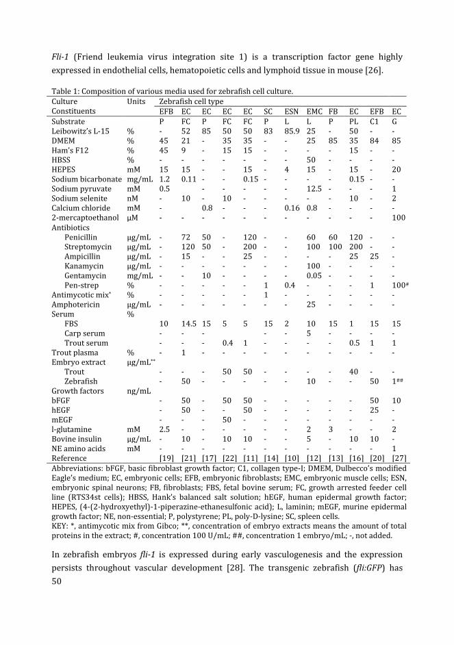
50
Fli-1 (Friend leukemia virus integration site 1) is a transcription factor gene highly
expressed in endothelial cells, hematopoietic cells and lymphoid tissue in mouse [26].
Table 1: Composition of various media used for zebrafish cell culture.
Culture
Constituents
Units Zebrafish cell type
EFB EC EC EC EC SC ESN EMC FB EC EFB EC
Substrate P FC P FC FC P L L P PL C1 G
Leibowitz’s L-15 % - 52 85 50 50 83 85.9 25 - 50 - -
DMEM % 45 21 - 35 35 - - 25 85 35 84 85
Ham’s F12 % 45 9 - 15 15 - - - - 15 - -
HBSS % - - - - - - - 50 - - - -
HEPES mM 15 15 - - 15 - 4 15 - 15 - 20
Sodium bicarbonate mg/mL 1.2 0.11 - - 0.15 - - - - 0.15 - -
Sodium pyruvate mM 0.5 - - - - - 12.5 - - - 1
Sodium selenite nM - 10 - 10 - - - - - 10 - 2
Calcium chloride mM - 0.8 - - - 0.16 0.8 - - - -
2-mercaptoethanol µM - - - - - - - - - - - 100
Antibiotics
Penicillin µg/mL - 72 50 - 120 - - 60 60 120 - -
Streptomycin µg/mL - 120 50 - 200 - - 100 100 200 - -
Ampicillin µg/mL - 15 - - 25 - - - - 25 25 -
Kanamycin µg/mL - - - - - - - 100 - - - -
Gentamycin mg/mL - - 10 - - - - 0.05 - - - -
Pen-strep % - - - - - 1 0.4 - - - 1 100#
Antimycotic mix* % - - - - - 1 - - - - - -
Amphotericin µg/mL - - - - - - - 25 - - - -
Serum %
FBS 10 14.5 15 5 5 15 2 10 15 1 15 15
Carp serum - - - - - 5 - - - -
Trout serum - - - 0.4 1 - - - - 0.5 1 1
Trout plasma % - 1 - - - - - - - - - -
Embryo extract µg/mL**
Trout - - - 50 50 - - - - 40 - -
Zebrafish - 50 - - - - - 10 - - 50 1##
Growth factors ng/mL
bFGF - 50 - 50 50 - - - - - 50 10
hEGF - 50 - - 50 - - - - - 25 -
mEGF - - - 50 - - - - - - - -
l-glutamine mM 2.5 - - - - - - 2 3 - - 2
Bovine insulin µg/mL - 10 - 10 10 - - 5 - 10 10 -
NE amino acids mM - - - - - - - - - - - 1
Reference [19] [21] [17] [22] [11] [14] [10] [12] [13] [16] [20] [27]
Abbreviations: bFGF, basic fibroblast growth factor; C1, collagen type-I; DMEM, Dulbecco’s modified
Eagle’s medium; EC, embryonic cells; EFB, embryonic fibroblasts; EMC, embryonic muscle cells; ESN,
embryonic spinal neurons; FB, fibroblasts; FBS, fetal bovine serum; FC, growth arrested feeder cell
line (RTS34st cells); HBSS, Hank’s balanced salt solution; hEGF, human epidermal growth factor;
HEPES, (4-(2-hydroxyethyl)-1-piperazine-ethanesulfonic acid); L, laminin; mEGF, murine epidermal
growth factor; NE, non-essential; P, polystyrene; PL, poly-D-lysine; SC, spleen cells.
KEY: *, antimycotic mix from Gibco; **, concentration of embryo extracts means the amount of total
proteins in the extract; #, concentration 100 U/mL; ##, concentration 1 embryo/mL; -, not added.
In zebrafish embryos fli-1 is expressed during early vasculogenesis and the expression
persists throughout vascular development [28]. The transgenic zebrafish (fli:GFP) has
51
been shown to express GFP in endothelial and hematopoietic cells, as well as in cells on
the yolk sac and neural crest cells i.e. mesenchyme and jaw cartilages [23]. Kdrl, also
known as flk-1 or VEGFR2, is a vascular endothelial growth factor receptor, expressed
particularly in endothelial cells during vascular development [25, 29].
Zebrafish embryos at the blastocyst stage of development (approximately 3.5 hours post
fertilization or hpf) are usually dechorionated and trypsinized to isolate ESCs. Culturing
blastocyst explants is equally important in studying cellular interactions, organization and
lineage commitment in early embryos [30]. Embryo explant culture has also been applied
to invertebrate embryos such as Caenorhabditis elegans [31] and the sea urchin [32].
However, in most of the procedures for ESC research, the embryos are dissociated
(trypsinized) into single cells for culturing. These early embryonic cells are used to
establish pluripotent ESC cultures which may be used for various applications e.g. genetic
manipulation of cells and organisms and production of germ line chimeras [21]. By
culturing these ESCs under differentiation conditions, specific cell lines can also be
developed [12, 17].
In order to maintain the ESCs in a pluripotent, undifferentiated state, they may be grown
on a feeder monolayer of growth-arrested cells [33]. Different feeder cell lines used to
culture zebrafish embryonic cells include rainbow trout spleen cells (RTS34st ) [11, 22],
zebrafish spleen stromal cells (ZSSJ) [14], buffalo rat liver cells [34] and zebrafish
embryonic fibroblasts [18]. For specific differentiation of ESCs, the feeder layer is replaced
by coating the culture plate with adhesive molecules. Fibronectin, laminin, collagen and
poly-D-lysine are the molecules known to promote attachment and differentiation of
zebrafish cells [10, 12, 20]. Zebrafish embryonic cells have also been successfully cultured
on polystyrene culture dishes without any feeder layer or substrate coating [17].
In this chapter we have optimized the culture conditions for zebrafish blastocyst cells in
the following steps: (1) The optimization of FBS and zebrafish embryo extract (ZEE)
concentrations in the media for the expansion of blastocyst explant cultures. (2) The
quantification of fli:GFP+ and kdrl:GFP+ cells under basic medium conditions, in the
blastocyst cell culture developed from the transgenic zebrafish embryos. (3) The effect of
different concentrations of FBS, ZEE and gelatin substratum on the growth and viability of
blastocyst secondary cell cultures.
Materials and methods
Zebrafish rearing and mating setup for embryo isolation
All the animal experiments were performed according to the Netherland Experiments on
Animals Act [35], based on guidelines laid by the Council of European Union [36]. Adult
52
zebrafish were maintained, as previously described [37], in 5 liter tanks having
continuously circulating egg water (“Instant Ocean” sea salt 60 µg/mL demi water), on 14
h light: 10 h dark cycle. Temperature of the water and air was controlled at 26 and 23 °C,
respectively. Wild type zebrafish embryos were used to characterize general growth of
blastocyst cells. Transgenic fli:GFP and kdrl:GFP embryos were used to analyse
differentiation of blastocyst cells into putative endothelial and hematopoietic cells. To
obtain embryos, adult male and female fish, at a proportion of 1:1, were transferred to
small breeding tanks in the evening. The zebrafish usually laid eggs when light turns on.
The eggs were collected at the bottom of the tank, separated from adults using a cotton
mesh to protect the eggs from being eaten.
Embryos were transferred to a temperature controlled room (28 °C) and were distributed
in 9 cm Petri dishes at a final density of 100 embryos per dish, after removing dead and
unfertilized eggs. The embryos were washed thoroughly with clean egg water to remove
any debris.
Sterilization of embryos
Embryos were allowed to develop to the high blastula stage of Kimmel et al. [38]
(approximately 3 hpf) at 28 °C. These embryos were then sterilized using 70% ethanol for
10 sec and 0.05% bleach solution (Table 2) for 2 × 5 min. Between the sterilization steps
the embryos were washed with LDF medium (Table 2). The sterilization procedure
described in Ref. [21] was followed. The embryos were finally left in 0.5 mL of LDF
medium and any dead embryos or embryos with visible contamination inside the chorion
were removed. These embryos were dechorionated using a pair of No. 5 watchmaker’s
forceps for the isolation of blastocyst cells.
Overview of experiments
The procedure of cell isolation and culture from embryos is shown in Figure 1. Three
different experiments were performed (Figure 1). In the first experiment the wild type
blastocysts were triturated gently with a P-200 Gilson micropipette (Gilson, B.V., Europe:
Den Haag) without enzymatic digestion to form fragments (explants). These explants
were cultured in LDF medium (Table 2) supplemented with different amounts of FBS
(Invitrogen, 10500) and ZEE to optimize the concentration of these nutrients for zebrafish
primary cell culture (see below, Experiment 1: Primary explant cultures). In the second
experiment (see below, Experiment 2: Quantification of fli:GFP+ and kdrl:GFP+ cells in
cultures) trypsinized blastocyst cells isolated from fli:GFP and kdrl:GFP embryos were
cultured to analyse the generation of fli:GFP+ and kdrl:GFP+ cells in these cultures. In the
third experiment (see below, Experiment 3: Secondary blastocyst cell culture) the effect of
FBS and ZEE supplementation and gelatin substratum was analysed on the growth and
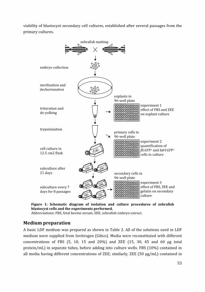
53
viability of blastocyst secondary cell cultures, established after several passages from the
primary cultures.
Figure 1: Schematic diagram of isolation and culture procedures of zebrafish
blastocyst cells and the experiments performed.
Abbreviations: FBS, fetal bovine serum; ZEE, zebrafish embryo extract.
Medium preparation
A basic LDF medium was prepared as shown in Table 2. All of the solutions used in LDF
medium were supplied from Invitrogen (Gibco). Media were reconstituted with different
concentrations of FBS (5, 10, 15 and 20%) and ZEE (15, 30, 45 and 60 µg total
protein/mL) in separate tubes, before adding into culture wells. FBS (10%) contained in
all media having different concentrations of ZEE; similarly, ZEE (50 µg/mL) contained in
zebrafish matting
embryo collection
sterilization and
dechorionation
trituration and
de-yolking
trypsinization
cell culture in
12.5 cm2 flask
subculture after
21 days
subculture every 7
days for 8 passages
explants in
96-well plate
primary cells in
96-well plate
secondary cells in
96-well plate
experiment 1
effect of FBS and ZEE
on explant culture
experiment 2
quantification of
fli:GFP+ and kdrl:GFP+
cells in culture
experiment 3
effect of FBS, ZEE and
gelatin on secondary
culture
54
all media having different concentrations of FBS. Cells were cultured in each distinct
medium in six wells (replicates) of the 96 well microplate. The FBS used in the
experiments was qualified, heat inactivated, E.U.-approved, South America Origin from
Invitrogen, catalog number 10500. ZEE was prepared using 36 hpf wild type zebrafish
embryos using the following procedure.
Preparation of zebrafish embryo extract
The ZEE was prepared according to the procedure described in Ref. [21]. The 36 hpf wild
type zebrafish embryos were washed thoroughly with egg water. About 500 embryos
suspended in 0.5 mL LDF medium were transferred to a 2 mL Eppendorf tube having a 5
mm glass bead. The embryos were homogenized in a QIAGEN TissueLyser II at 30
oscillations/min for 1 min. After homogenization, 1 mL LDF was added to the embryo
lysate. The tubes were then centrifuged at 20,000 g for 10 min at 4 °C. The supernatant
was transferred to a new tube, and the total protein content of the extract was quantified
using bovine serum albumin protein assay kit (Thermo scientific, product 23227). Finally,
the extract was diluted to a final concentration of 1 mg total protein/mL, filter sterilized
and stored in aliquots at -20 °C.
Experiment 1: Primary explant culture
The wild type embryos were dechorionated using sterile No. 5 watchmaker forceps and
each set of 50 embryos was then transferred to a separate 1.5 mL sterile Eppendorf tube.
The embryos were triturated using a P-200 Gilson micropipette, in the LDF medium, to
fragmentize the blastocysts. The tubes were then centrifuged at 300 g for 2 min and the
supernatant, including the yolk, was discarded. The blastocyst fragments (explants) were
washed with CMF-PBS (calcium magnesium free – phosphate buffered saline) and then
with LDF to remove any remaining yolk particles. Finally, the explants were re-suspended
in 300 µL of LDF medium containing 10% FBS, and distributed in a 96-well plate such
that, on average, the explants from 2 blastocysts were plated per well. Finally 200 µL LDF
media, having varying concentrations of FBS and ZEE (as described above in Medium
preparation) were added to the wells. The cultures were maintained in an incubator at 28
°C in atmospheric CO2 until data collection.
Trypsinized blastocyst cell culture
The concentrations of FBS and ZEE in the medium optimized for explant culture in
experiment 1 (above) was used to culture the completely dissociated (trypsinized)
blastocyst cells. For this purpose, the embryos were sterilized, dechorionated and de-
yolked as described above. Then, the blastocysts were given a rinse with CMF-PBS and
incubated in 1 mL of 0.25% trypsin solution (Table 2) for 2 min. The trypsin-blastocysts
solution was triturated several times using p-1000 Gilson micropipette, to facilitate
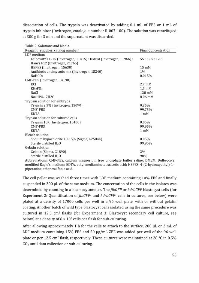
55
dissociation of cells. The trypsin was deactivated by adding 0.1 mL of FBS or 1 mL of
trypsin inhibitor (Invitrogen, catalogue number R-007-100). The solution was centrifuged
at 300 g for 3 min and the supernatant was discarded.
Table 2: Solutions and Media.
Reagent (supplier, catalog number) Final Concentration
LDF medium
Leibowitz’s L-15 (Invitrogen, 11415) : DMEM (Invitrogen, 11966) :
Ham’s F12 (Invitrogen, 21765)
55 : 32.5 : 12.5
HEPES (Invitrogen, 15630) 15 mM
Antibiotic antimycotic mix (Invitrogen, 15240) 1%
NaHCO3 0.015%
CMF-PBS (Invitrogen, 14190)
KCl 2.7 mM
KH2PO4 1.5 mM
NaCl 138 mM
Na2HPO4-7H2O 8.06 mM
Trypsin solution for embryos
Trypsin 2.5% (Invitrogen, 15090) 0.25%
CMF-PBS 99.75%
EDTA 1 mM
Trypsin solution for cultured cells
Trypsin 10X (Invitrogen, 15400) 0.05%
CMF-PBS 99.95%
EDTA 1 mM
Bleach solution
Sodium hypochlorite 10-15% (Sigma, 425044) 0.05%
Sterile distilled H2O 99.95%
Gelatin solution
Gelatin (Sigma, G1890) 2%
Sterile distilled H2O 98%
Abbreviations: CMF-PBS, calcium magnesium free phosphate buffer saline; DMEM, Dulbecco’s
modified Eagle’s medium; EDTA, ethylenediaminetetraacetic acid; HEPES, 4-(2-hydroxyethyl)-1-
piperazine-ethanesulfonic acid.
The cell pellet was washed three times with LDF medium containing 10% FBS and finally
suspended in 300 µL of the same medium. The concertation of the cells in the isolates was
determined by counting in a heamocytometer. The fli:GFP or kdrl:GFP blastocyst cells (for
Experiment 2: Quantification of fli:GFP+ and kdrl:GFP+ cells in cultures, see below) were
plated at a density of 17000 cells per well in a 96 well plate, with or without gelatin
coating. Another batch of wild type blastocyst cells isolated using the same procedure was
cultured in 12.5 cm2 flasks (for Experiment 3: Blastocyst secondary cell culture, see
below) at a density of 6 × 10⁵ cells per flask for sub-culturing.
After allowing approximately 1 h for the cells to attach to the surface, 200 µL or 2 mL of
LDF medium containing 15% FBS and 50 µg/mL ZEE was added per well of the 96 well
plate or per 12.5 cm2 flask, respectively. These cultures were maintained at 28 °C in 0.5%
CO2 until data collection or sub-culturing.
56
Media refreshment
To optimize the time for media refreshment, in a separate experiment, the zebrafish
blastocyst explants were cultured without replacement of media. We changed to a
refreshment interval of 4 days for the primary cultures when we observed a decrease in
the surface area covered by the emigrating cells around the explants after day 4 in the
non-replacement cultures. For the secondary cultures the medium was refreshed weekly.
Experiment 2: Quantification of fli:GFP+ and kdrl:GFP+ cells in culture
Two different cultures were established to analyse spontaneous differentiation in primary
blastocyst cultures: one from the cells isolated from zebrafish fli:GFP embryos and the
other from kdrl:GFP embryos. The cells cultured in the wells of 96 well plate, with or
without gelatin coating, were isolated for the quantification of fli:GFP+ or kdrl:GFP+ at
successive time-points (days 2, 4, 6 and 8). The gelatin was used at a concentration of 0.1
mg/cm2 (1.7 µL of 2% gelatin solution per well). For each of the above mentioned
condition (i.e. transgene line, gelatin coating and time-points), the blastocyst cells cultured
in six replicate wells were transferred to separate Eppendorf tubes (see below,
Trypsinization of cultured cells). Each cells isolate was subjected to quantification of
fli:GFP+ or kdrl:GFP+ using the procedure described below (Data collection).
Experiment 3: Secondary blastocyst cell culture
The blastocyst cell culture in 12.5 cm2 flasks was maintained for 21 days with media
refreshment every four days. The cells were then isolated from the flask using 2mL of
0.05% trypsin solution (Trypsinization of cultured cells), and sub-cultured in a new flask
in LDF medium containing 10% FBS. These secondary cells were then passaged every
seven days. At least three 12.5 cm2 flasks were platted, for the primary culture as wells as
for the secondary cultures, to allow for statistical analysis of growth rates at successive
passages. During transfer a small amount of cell isolate at each passage was used to
determine the total number of cells harvested and the population doubling time (see
below, Statistical analysis).
At passage eight the cells were isolated from the flask and distributed at 17000 cells per
well in a 96-well plate (Figure 1). These cells were maintained in media supplemented
with different concentrations of FBS (5, 10, 15 and 20%) or ZEE (15, 30, 45 and 60
µg/mL), or on different concentrations of gelatin substratum (see below, Gelatin coating of
wells). The effect of FBS, ZEE and gelatin substratum was analysed by calculating the total
number of cells harvested per well and the viability of the isolated cells on day 7 of culture
(see below, Data collection).
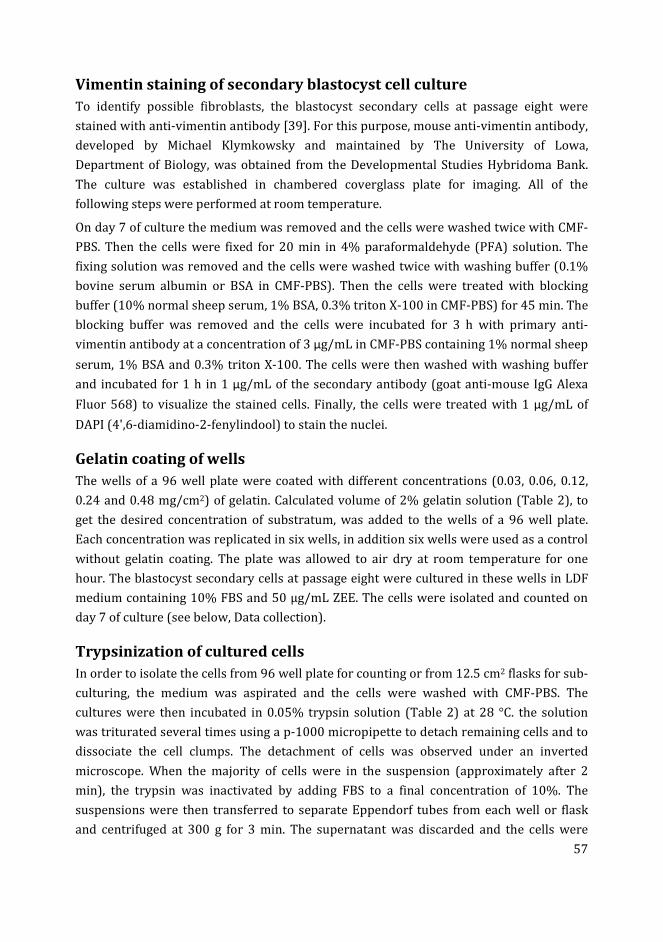
57
Vimentin staining of secondary blastocyst cell culture
To identify possible fibroblasts, the blastocyst secondary cells at passage eight were
stained with anti-vimentin antibody [39]. For this purpose, mouse anti-vimentin antibody,
developed by Michael Klymkowsky and maintained by The University of Lowa,
Department of Biology, was obtained from the Developmental Studies Hybridoma Bank.
The culture was established in chambered coverglass plate for imaging. All of the
following steps were performed at room temperature.
On day 7 of culture the medium was removed and the cells were washed twice with CMF-
PBS. Then the cells were fixed for 20 min in 4% paraformaldehyde (PFA) solution. The
fixing solution was removed and the cells were washed twice with washing buffer (0.1%
bovine serum albumin or BSA in CMF-PBS). Then the cells were treated with blocking
buffer (10% normal sheep serum, 1% BSA, 0.3% triton X-100 in CMF-PBS) for 45 min. The
blocking buffer was removed and the cells were incubated for 3 h with primary anti-
vimentin antibody at a concentration of 3 µg/mL in CMF-PBS containing 1% normal sheep
serum, 1% BSA and 0.3% triton X-100. The cells were then washed with washing buffer
and incubated for 1 h in 1 µg/mL of the secondary antibody (goat anti-mouse IgG Alexa
Fluor 568) to visualize the stained cells. Finally, the cells were treated with 1 µg/mL of
DAPI (4',6-diamidino-2-fenylindool) to stain the nuclei.
Gelatin coating of wells
The wells of a 96 well plate were coated with different concentrations (0.03, 0.06, 0.12,
0.24 and 0.48 mg/cm2) of gelatin. Calculated volume of 2% gelatin solution (Table 2), to
get the desired concentration of substratum, was added to the wells of a 96 well plate.
Each concentration was replicated in six wells, in addition six wells were used as a control
without gelatin coating. The plate was allowed to air dry at room temperature for one
hour. The blastocyst secondary cells at passage eight were cultured in these wells in LDF
medium containing 10% FBS and 50 µg/mL ZEE. The cells were isolated and counted on
day 7 of culture (see below, Data collection).
Trypsinization of cultured cells
In order to isolate the cells from 96 well plate for counting or from 12.5 cm2 flasks for sub-
culturing, the medium was aspirated and the cells were washed with CMF-PBS. The
cultures were then incubated in 0.05% trypsin solution (Table 2) at 28 °C. the solution
was triturated several times using a p-1000 micropipette to detach remaining cells and to
dissociate the cell clumps. The detachment of cells was observed under an inverted
microscope. When the majority of cells were in the suspension (approximately after 2
min), the trypsin was inactivated by adding FBS to a final concentration of 10%. The
suspensions were then transferred to separate Eppendorf tubes from each well or flask
and centrifuged at 300 g for 3 min. The supernatant was discarded and the cells were
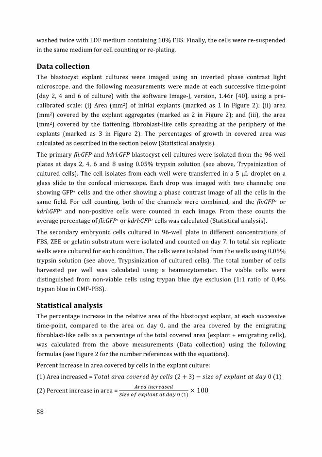
58
washed twice with LDF medium containing 10% FBS. Finally, the cells were re-suspended
in the same medium for cell counting or re-plating.
Data collection
The blastocyst explant cultures were imaged using an inverted phase contrast light
microscope, and the following measurements were made at each successive time-point
(day 2, 4 and 6 of culture) with the software Image-J, version, 1.46r [40], using a pre-
calibrated scale: (i) Area (mm2) of initial explants (marked as 1 in Figure 2); (ii) area
(mm2) covered by the explant aggregates (marked as 2 in Figure 2); and (iii), the area
(mm2) covered by the flattening, fibroblast-like cells spreading at the periphery of the
explants (marked as 3 in Figure 2). The percentages of growth in covered area was
calculated as described in the section below (Statistical analysis).
The primary fli:GFP and kdrl:GFP blastocyst cell cultures were isolated from the 96 well
plates at days 2, 4, 6 and 8 using 0.05% trypsin solution (see above, Trypsinization of
cultured cells). The cell isolates from each well were transferred in a 5 µL droplet on a
glass slide to the confocal microscope. Each drop was imaged with two channels; one
showing GFP+ cells and the other showing a phase contrast image of all the cells in the
same field. For cell counting, both of the channels were combined, and the fli:GFP+ or
kdrl:GFP+ and non-positive cells were counted in each image. From these counts the
average percentage of fli:GFP+ or kdrl:GFP+ cells was calculated (Statistical analysis).
The secondary embryonic cells cultured in 96-well plate in different concentrations of
FBS, ZEE or gelatin substratum were isolated and counted on day 7. In total six replicate
wells were cultured for each condition. The cells were isolated from the wells using 0.05%
trypsin solution (see above, Trypsinization of cultured cells). The total number of cells
harvested per well was calculated using a heamocytometer. The viable cells were
distinguished from non-viable cells using trypan blue dye exclusion (1:1 ratio of 0.4%
trypan blue in CMF-PBS).
Statistical analysis
The percentage increase in the relative area of the blastocyst explant, at each successive
time-point, compared to the area on day 0, and the area covered by the emigrating
fibroblast-like cells as a percentage of the total covered area (explant + emigrating cells),
was calculated from the above measurements (Data collection) using the following
formulas (see Figure 2 for the number references with the equations).
Percent increase in area covered by cells in the explant culture:
(1) Area increased = ��������������� ����(2 + 3) − ����������������� 0(1)
(2) Percent increase in area = ��� !"#�� $�%
&!'�()�*+, "- -% ./(0)× 100
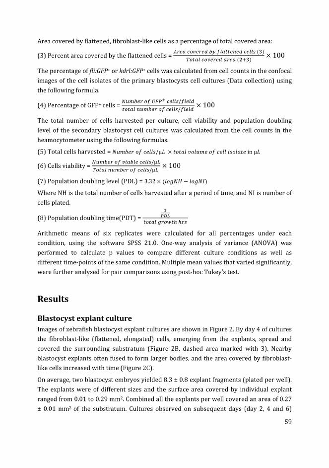
59
Area covered by flattened, fibroblast-like cells as a percentage of total covered area:
(3) Percent area covered by the flattened cells = ��� #(2���%3.), --�"�%#�,,$(4)
5(- ,#(2���% �� (674)× 100
The percentage of fli:GFP+ or kdrl:GFP+ cells was calculated from cell counts in the confocal
images of the cell isolates of the primary blastocysts cell cultures (Data collection) using
the following formula.
(4) Percentage of GFP+ cells = 89:3��();<=>#�,,$/)!�,%
-(- ,"9:3��()#�,,$/)!�,%× 100
The total number of cells harvested per culture, cell viability and population doubling
level of the secondary blastocyst cell cultures was calculated from the cell counts in the
heamocytometer using the following formulas.
(5) Total cells harvested = @AB���������/μD × �������AB�������������inμD
(6) Cells viability = 89:3��()2! 3,�#�,,$/GH
5(- ,"9:3��()#�,,$/GH× 100
(7) Population doubling level (PDL) = 3.32 × (��J@K − ��J@L)
Where NH is the total number of cells harvested after a period of time, and NI is number of
cells plated.
(8) Population doubling time(PDT) =
M
NOP
-(- ,Q�(R-SS�$
Arithmetic means of six replicates were calculated for all percentages under each
condition, using the software SPSS 21.0. One-way analysis of variance (ANOVA) was
performed to calculate p values to compare different culture conditions as well as
different time-points of the same condition. Multiple mean values that varied significantly,
were further analysed for pair comparisons using post-hoc Tukey’s test.
Results
Blastocyst explant culture
Images of zebrafish blastocyst explant cultures are shown in Figure 2. By day 4 of cultures
the fibroblast-like (flattened, elongated) cells, emerging from the explants, spread and
covered the surrounding substratum (Figure 2B, dashed area marked with 3). Nearby
blastocyst explants often fused to form larger bodies, and the area covered by fibroblast-
like cells increased with time (Figure 2C).
On average, two blastocyst embryos yielded 8.3 ± 0.8 explant fragments (plated per well).
The explants were of different sizes and the surface area covered by individual explant
ranged from 0.01 to 0.29 mm2. Combined all the explants per well covered an area of 0.27
± 0.01 mm2 of the substratum. Cultures observed on subsequent days (day 2, 4 and 6)
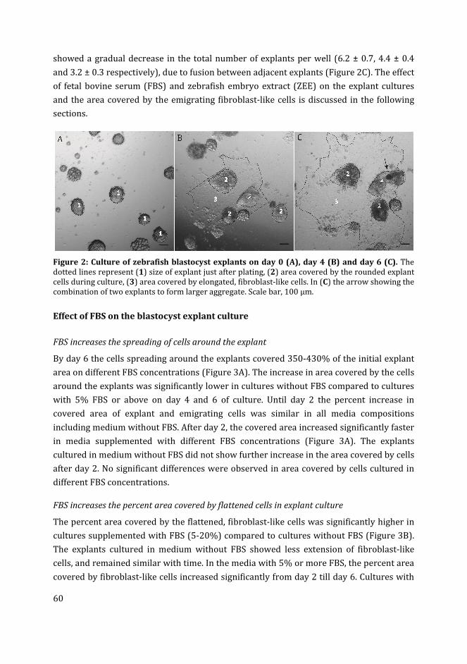
60
showed a gradual decrease in the total number of explants per well (6.2 ± 0.7, 4.4 ± 0.4
and 3.2 ± 0.3 respectively), due to fusion between adjacent explants (Figure 2C). The effect
of fetal bovine serum (FBS) and zebrafish embryo extract (ZEE) on the explant cultures
and the area covered by the emigrating fibroblast-like cells is discussed in the following
sections.
Figure 2: Culture of zebrafish blastocyst explants on day 0 (A), day 4 (B) and day 6 (C). The
dotted lines represent (1) size of explant just after plating, (2) area covered by the rounded explant
cells during culture, (3) area covered by elongated, fibroblast-like cells. In (C) the arrow showing the
combination of two explants to form larger aggregate. Scale bar, 100 µm.
Effect of FBS on the blastocyst explant culture
FBS increases the spreading of cells around the explant
By day 6 the cells spreading around the explants covered 350-430% of the initial explant
area on different FBS concentrations (Figure 3A). The increase in area covered by the cells
around the explants was significantly lower in cultures without FBS compared to cultures
with 5% FBS or above on day 4 and 6 of culture. Until day 2 the percent increase in
covered area of explant and emigrating cells was similar in all media compositions
including medium without FBS. After day 2, the covered area increased significantly faster
in media supplemented with different FBS concentrations (Figure 3A). The explants
cultured in medium without FBS did not show further increase in the area covered by cells
after day 2. No significant differences were observed in area covered by cells cultured in
different FBS concentrations.
FBS increases the percent area covered by flattened cells in explant culture
The percent area covered by the flattened, fibroblast-like cells was significantly higher in
cultures supplemented with FBS (5-20%) compared to cultures without FBS (Figure 3B).
The explants cultured in medium without FBS showed less extension of fibroblast-like
cells, and remained similar with time. In the media with 5% or more FBS, the percent area
covered by fibroblast-like cells increased significantly from day 2 till day 6. Cultures with
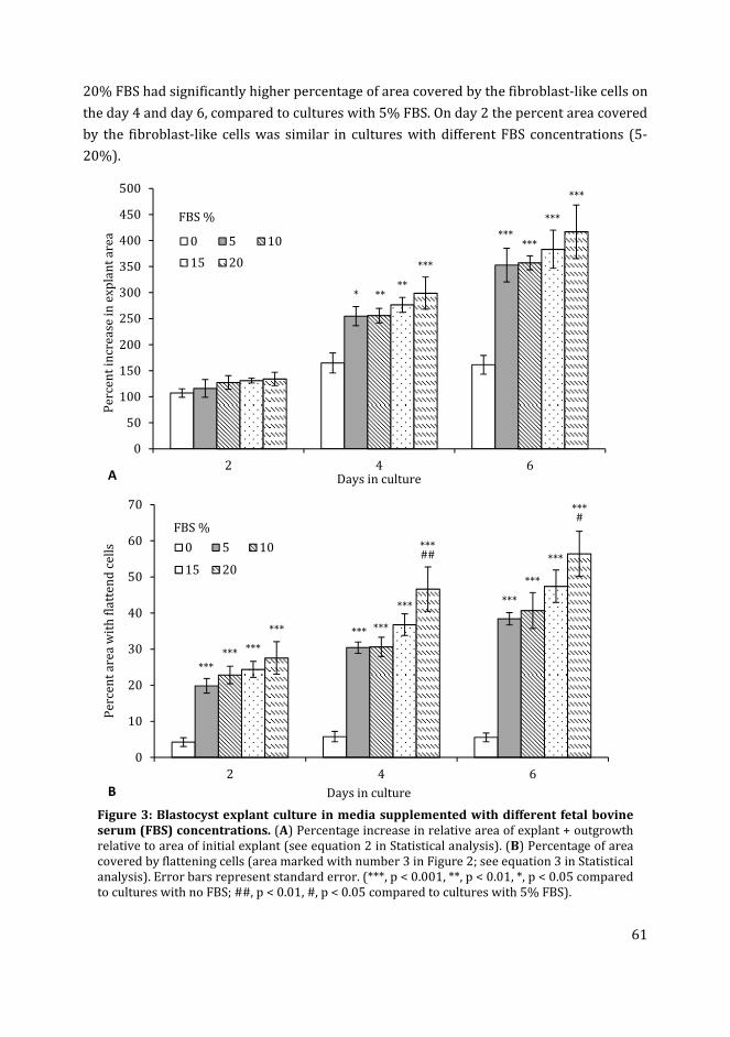
61
20% FBS had significantly higher percentage of area covered by the fibroblast-like cells on
the day 4 and day 6, compared to cultures with 5% FBS. On day 2 the percent area covered
by the fibroblast-like cells was similar in cultures with different FBS concentrations (5-
20%).
Figure 3: Blastocyst explant culture in media supplemented with different fetal bovine
serum (FBS) concentrations. (A) Percentage increase in relative area of explant + outgrowth
relative to area of initial explant (see equation 2 in Statistical analysis). (B) Percentage of area
covered by flattening cells (area marked with number 3 in Figure 2; see equation 3 in Statistical
analysis). Error bars represent standard error. (***, p < 0.001, **, p < 0.01, *, p < 0.05 compared
to cultures with no FBS; ##, p < 0.01, #, p < 0.05 compared to cultures with 5% FBS).
0
50
100
150
200
250
300
350
400
450
500
2 4 6
Pe
rce
nt
incr
ea
se i
n e
xp
lan
t a
rea
Days in culture
0 5 10
15 20
FBS %
* ****
***
******
***
***
A
0
10
20
30
40
50
60
70
2 4 6
Pe
rce
nt
are
a w
ith
fla
tte
nd
ce
lls
Days in culture
0 5 10
15 20
B
***
*** ***
*** *** ***
***
***##
***
***
***
***#
FBS %
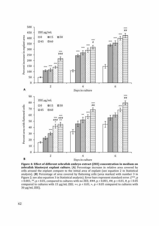
62
Figure 4: Effect of different zebrafish embryo extract (ZEE) concentration in medium on
zebrafish blastocyst explant culture. (A) Percentage increase in relative area covered by
cells around the explant compare to the initial area of explant (see equation 2 in Statistical
analysis). (B) Percentage of area covered by flattening cells (area marked with number 3 in
Figure 2; see also equation 3 in Statistical analysis). Error bars represent standard error. (***, p
< 0.001, **, p < 0.01, compared to cultures with no ZEE; ###, p < 0.001, ##, p < 0.01, #, p < 0.05
compared to cultures with 15 µg/mL ZEE; ++, p < 0.01, +, p < 0.05 compared to cultures with
30 µg/mL ZEE).
0
50
100
150
200
250
300
350
400
450
500
2 4 6
Pe
rce
nt
incr
ea
se i
n e
xp
lan
t a
rea
Days in culture
0 15 30
45 60
ZEE µg/mL
*** ******
***###
***
******
***# ***
***
***
***##
A
0
10
20
30
40
50
60
70
80
90
2 4 6
Pe
rce
nt
are
a w
ith
fla
tte
ne
d c
ell
s
Days in culture
0 15 30
45 60
ZEE µg/mL
***
***###
+
***###++
*****
***## ***
***##
B
63
Effect of zebrafish embryo extract on blastocyst explant culture
ZEE increases area covered by cells around blastocyst explant
Similar to FBS, ZEE also showed significant effect on the expansion in area covered by the
emigrating cells from the blastocyst explants (Figure 4A). The increase in the area covered
by cells around the explant was higher on day 2 in cultures with 15 µg/mL ZEE compared
to cultures without ZEE in the medium. More expansion of the fibroblast-like cells around
the explant was observed in cultures supplemented with 60 µg/mL compared to 15
µg/mL ZEE (Figure 4A). No significant differences between 15, 30 and 45 µg/mL ZEE
concentrations was observed on the expansion of the cultures. Without ZEE in the
medium the area of the explant remained similar from day 2 to day 6. In the presence of
ZEE (15-60 µg/mL), significant increase was observed in the explant area from day 2 to
day 6 (p<0.001).
Percent area covered by flattened cells is increased by ZEE
The area covered by the flattened cells, as a percentage of the total area covered by the
explant (including the fibroblastic cells), was higher with 30-60 µg/mL ZEE concentration,
compared to no ZEE in the medium (Figure 4B). Cultures maintained with 60 µg/mL ZEE
showed higher percentage of area covered by fibroblast-like cells compared to cultures
with 15 µg/mL ZEE. No significant differences were observed between cultures with zero
and 15 µg/mL ZEE. The percent area covered by fibroblast-like cells increased
significantly from day 2 to day 4 under all ZEE conditions (p<0.01). However, after day 4
to day 6 a significant increase was observed only with 60 µg/mL ZEE (p<0.05).
Trypsinized blastocyst primary cell culture
The trypsinized blastocyst cells formed aggregates known as EBs within the first 24 h of
culture and then the elongated, fibroblast-like cells started to emigrate on the culture
substratum as a monolayer from these EBs (Figure 5A-E). The primary cultures
predominantly contained two types of cells: (i) flattened, elongated, fibroblast-like cells;
(ii) round, EB cells. At this stage other cell types such as neuron-like (long thread-like)
cells, and melanocytes (pigment cells; Figure 5F) were also visible in the EBs. The
appearance of fli:GFP+ or kdrl:GFP+ cells in the EBs was also observed in cultures (Figure
5B). By day 4 both the EBs and the proliferating fibroblast like cells contained cells
expressing fli:GFP or kdrl:GFP marker (Figure 5C and G).
Expression of fli:GFP+ cells in blastocyst primary cell culture
The fli:GFP blastocyst cell culture showed the fli:GFP expression in cells in culture medium
without supplementation of endothelial/hematopoietic growth factors (Figure 5B-E). By
day 2 of culture the fli:GFP+ cells were observed on the periphery of the EBs (Figure 5B).
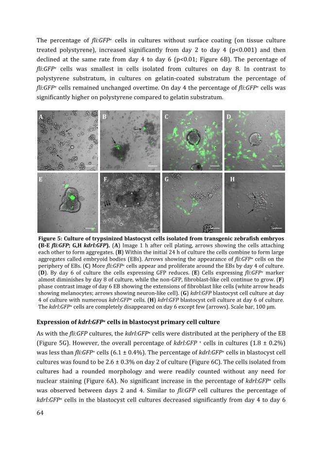
64
The percentage of fli:GFP+ cells in cultures without surface coating (on tissue culture
treated polystyrene), increased significantly from day 2 to day 4 (p<0.001) and then
declined at the same rate from day 4 to day 6 (p<0.01; Figure 6B). The percentage of
fli:GFP+ cells was smallest in cells isolated from cultures on day 8. In contrast to
polystyrene substratum, in cultures on gelatin-coated substratum the percentage of
fli:GFP+ cells remained unchanged overtime. On day 4 the percentage of fli:GFP+ cells was
significantly higher on polystyrene compared to gelatin substratum.
Figure 5: Culture of trypsinized blastocyst cells isolated from transgenic zebrafish embryos
(B-E fli:GFP; G,H kdrl:GFP). (A) Image 1 h after cell plating, arrows showing the cells attaching
each other to form aggregates. (B) Within the initial 24 h of culture the cells combine to form large
aggregates called embryoid bodies (EBs). Arrows showing the appearance of fli:GFP+ cells on the
periphery of EBs. (C) More fli:GFP+ cells appear and proliferate around the EBs by day 4 of culture.
(D). By day 6 of culture the cells expressing GFP reduces. (E) Cells expressing fli:GFP+ marker
almost diminishes by day 8 of culture, while the non-GFP, fibroblast-like cell continue to grow. (F)
phase contrast image of day 6 EB showing the extensions of fibroblast like cells (white arrow heads
showing melanocytes; arrows showing neuron-like cell). (G) kdrl:GFP blastocyst cell culture at day
4 of culture with numerous kdrl:GFP+ cells. (H) kdrl:GFP blastocyst cell culture at day 6 of culture.
The kdrl:GFP+ cells are completely disappeared on day 6 except few (arrows). Scale bar, 100 µm.
Expression of kdrl:GFP+ cells in blastocyst primary cell culture
As with the fli:GFP cultures, the kdrl:GFP+ cells were distributed at the periphery of the EB
(Figure 5G). However, the overall percentage of kdrl:GFP + cells in cultures (1.8 ± 0.2%)
was less than fli:GFP+ cells (6.1 ± 0.4%). The percentage of kdrl:GFP+ cells in blastocyst cell
cultures was found to be 2.6 ± 0.3% on day 2 of culture (Figure 6C). The cells isolated from
cultures had a rounded morphology and were readily counted without any need for
nuclear staining (Figure 6A). No significant increase in the percentage of kdrl:GFP+ cells
was observed between days 2 and 4. Similar to fli:GFP cell cultures the percentage of
kdrl:GFP+ cells in the blastocyst cell cultures decreased significantly from day 4 to day 6
F
A B C D
E G H
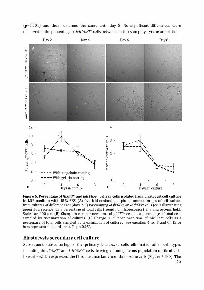
65
(p<0.001) and then remained the same until day 8. No significant differences were
observed in the percentage of kdrl:GFP+ cells between cultures on polystyrene or gelatin.
Day 2 Day 4 Day 6 Day 8
fli:
GF
P+ c
ell
co
un
ts
kd
rl:G
FP
+ c
ell
co
un
ts
Figure 6: Percentage of fli:GFP+ and kdrl:GFP+ cells in cells isolated from blastocyst cell culture
in LDF medium with 15% FBS. (A) Overlaid confocal and phase contrast images of cell isolates
from cultures of different ages (days 2-8) for counting of fli:GFP+ or kdrl:GFP+ cells (cells illuminating
green fluorescence) as a percentage of total cells (round non-fluorescence) in a microscopic field..
Scale bar, 100 µm. (B) Change in number over time of fli:GFP+ cells as a percentage of total cells
sampled by trypsinization of cultures. (C) Change in number over time of kdrl:GFP+ cells as a
percentage of total cells sampled by trypsinization of cultures (see equation 4 for B and C). Error
bars represent standard error. (*, p < 0.05).
Blastocysts secondary cell culture
Subsequent sub-culturing of the primary blastocyst cells eliminated other cell types
including the fli:GFP+ and kdrl:GFP+ cells, leaving a homogeneous population of fibroblast-
like cells which expressed the fibroblast marker vimentin in some cells (Figure 7 B-D). The
0
2
4
6
8
10
12
2 4 6 8
Pe
rce
nt
fli:
GF
P+
cell
s
Days in culture
Without gelatin coating
With gelatin coating
*
B
0
1
2
3
4
2 4 6 8
Pe
rce
nt
kd
rl:G
FP
+ce
lls
Days in cultureC
A
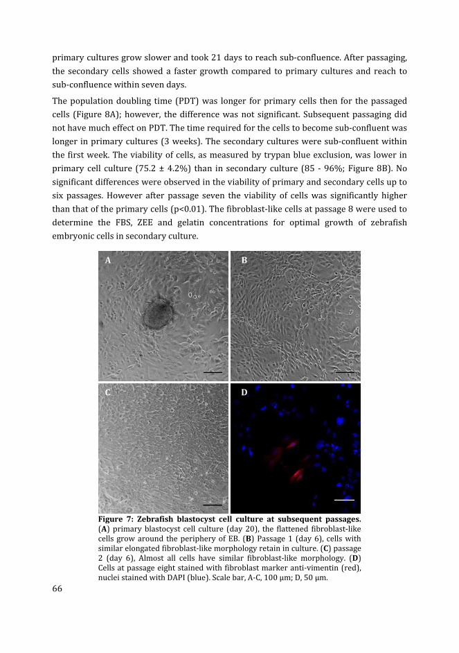
66
primary cultures grow slower and took 21 days to reach sub-confluence. After passaging,
the secondary cells showed a faster growth compared to primary cultures and reach to
sub-confluence within seven days.
The population doubling time (PDT) was longer for primary cells then for the passaged
cells (Figure 8A); however, the difference was not significant. Subsequent passaging did
not have much effect on PDT. The time required for the cells to become sub-confluent was
longer in primary cultures (3 weeks). The secondary cultures were sub-confluent within
the first week. The viability of cells, as measured by trypan blue exclusion, was lower in
primary cell culture (75.2 ± 4.2%) than in secondary culture (85 - 96%; Figure 8B). No
significant differences were observed in the viability of primary and secondary cells up to
six passages. However after passage seven the viability of cells was significantly higher
than that of the primary cells (p<0.01). The fibroblast-like cells at passage 8 were used to
determine the FBS, ZEE and gelatin concentrations for optimal growth of zebrafish
embryonic cells in secondary culture.
Figure 7: Zebrafish blastocyst cell culture at subsequent passages.
(A) primary blastocyst cell culture (day 20), the flattened fibroblast-like
cells grow around the periphery of EB. (B) Passage 1 (day 6), cells with
similar elongated fibroblast-like morphology retain in culture. (C) passage
2 (day 6), Almost all cells have similar fibroblast-like morphology. (D)
Cells at passage eight stained with fibroblast marker anti-vimentin (red),
nuclei stained with DAPI (blue). Scale bar, A-C, 100 µm; D, 50 µm.
A B
C D
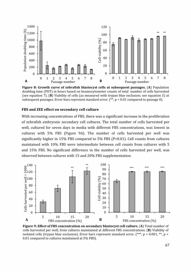
67
Figure 8: Growth curve of zebrafish blastocyst cells at subsequent passages. (A) Population
doubling time (PDT) in hours based on heamocytometer counts of total number of cells harvested
(see equation 7). (B) Viability of cells (as measured with trypan blue exclusion; see equation 5) at
subsequent passages. Error bars represent standard error. (**, p < 0.01 compared to passage 0).
FBS and ZEE effect on secondary cell culture
With increasing concentrations of FBS, there was a significant increase in the proliferation
of zebrafish embryonic secondary cell cultures. The total number of cells harvested per
well, cultured for seven days in media with different FBS concentrations, was lowest in
cultures with 5% FBS (Figure 9A). The number of cells harvested per well was
significantly higher in 15% FBS compared to 5% FBS (P<0.01). Cell counts from cultures
maintained with 10% FBS were intermediate between cell counts from cultures with 5
and 15% FBS. No significant difference in the number of cells harvested per well, was
observed between cultures with 15 and 20% FBS supplementation.
Figure 9: Effect of FBS concentration on secondary blastocyst cell culture. (A) Total number of
cells harvested per well, from cultures maintained at different FBS concentrations. (B) Viability of
isolated cells (trypan blue exclusion). Error bars represent standard error. (***, p < 0.001, **, p <
0.01 compared to cultures maintained at 5% FBS).
0
200
400
600
800
1000
1200
1400
0 1 2 3 4 5 6 7 8
Po
pu
lati
on
do
ub
lin
g t
ime
(h
)
Passage numberA
0
20
40
60
80
100
120
0 1 2 3 4 5 6 7 8
Ce
ll v
iab
ilit
y (
%)
Passage number
** **
B
0
20
40
60
80
100
120
140
5 10 15 20
Ce
lls
ha
rve
ste
d p
er
we
ll ×
10
00
FBS concentration (%)
****
A
0
10
20
30
40
50
60
70
80
90
100
5 10 15 20
Ce
ll v
iab
ilit
y (
%)
FBS concentration (%)
*** *** ***
B
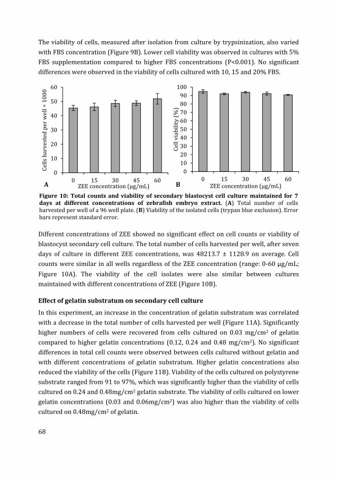
68
The viability of cells, measured after isolation from culture by trypsinization, also varied
with FBS concentration (Figure 9B). Lower cell viability was observed in cultures with 5%
FBS supplementation compared to higher FBS concentrations (P<0.001). No significant
differences were observed in the viability of cells cultured with 10, 15 and 20% FBS.
Figure 10: Total counts and viability of secondary blastocyst cell culture maintained for 7
days at different concentrations of zebrafish embryo extract. (A) Total number of cells
harvested per well of a 96 well plate. (B) Viability of the isolated cells (trypan blue exclusion). Error
bars represent standard error.
Different concentrations of ZEE showed no significant effect on cell counts or viability of
blastocyst secondary cell culture. The total number of cells harvested per well, after seven
days of culture in different ZEE concentrations, was 48213.7 ± 1128.9 on average. Cell
counts were similar in all wells regardless of the ZEE concentration (range: 0-60 µg/mL;
Figure 10A). The viability of the cell isolates were also similar between cultures
maintained with different concentrations of ZEE (Figure 10B).
Effect of gelatin substratum on secondary cell culture
In this experiment, an increase in the concentration of gelatin substratum was correlated
with a decrease in the total number of cells harvested per well (Figure 11A). Significantly
higher numbers of cells were recovered from cells cultured on 0.03 mg/cm2 of gelatin
compared to higher gelatin concentrations (0.12, 0.24 and 0.48 mg/cm2). No significant
differences in total cell counts were observed between cells cultured without gelatin and
with different concentrations of gelatin substratum. Higher gelatin concentrations also
reduced the viability of the cells (Figure 11B). Viability of the cells cultured on polystyrene
substrate ranged from 91 to 97%, which was significantly higher than the viability of cells
cultured on 0.24 and 0.48mg/cm2 gelatin substrate. The viability of cells cultured on lower
gelatin concentrations (0.03 and 0.06mg/cm2) was also higher than the viability of cells
cultured on 0.48mg/cm2 of gelatin.
0
10
20
30
40
50
60
0 15 30 45 60
Ce
lls
ha
rve
ste
d p
er
we
ll ×
10
00
ZEE concentration (µg/mL) A
0
10
20
30
40
50
60
70
80
90
100
0 15 30 45 60C
ell
via
bil
ity
(%
)
ZEE concentration (µg/mL)B
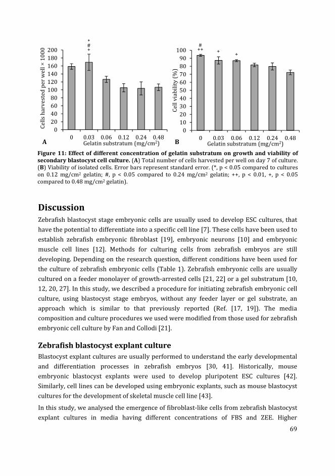
69
Figure 11: Effect of different concentration of gelatin substratum on growth and viability of
secondary blastocyst cell culture. (A) Total number of cells harvested per well on day 7 of culture.
(B) Viability of isolated cells. Error bars represent standard error. (*, p < 0.05 compared to cultures
on 0.12 mg/cm2 gelatin; #, p < 0.05 compared to 0.24 mg/cm2 gelatin; ++, p < 0.01, +, p < 0.05
compared to 0.48 mg/cm2 gelatin).
Discussion
Zebrafish blastocyst stage embryonic cells are usually used to develop ESC cultures, that
have the potential to differentiate into a specific cell line [7]. These cells have been used to
establish zebrafish embryonic fibroblast [19], embryonic neurons [10] and embryonic
muscle cell lines [12]. Methods for culturing cells from zebrafish embryos are still
developing. Depending on the research question, different conditions have been used for
the culture of zebrafish embryonic cells (Table 1). Zebrafish embryonic cells are usually
cultured on a feeder monolayer of growth-arrested cells [21, 22] or a gel substratum [10,
12, 20, 27]. In this study, we described a procedure for initiating zebrafish embryonic cell
culture, using blastocyst stage embryos, without any feeder layer or gel substrate, an
approach which is similar to that previously reported (Ref. [17, 19]). The media
composition and culture procedures we used were modified from those used for zebrafish
embryonic cell culture by Fan and Collodi [21].
Zebrafish blastocyst explant culture
Blastocyst explant cultures are usually performed to understand the early developmental
and differentiation processes in zebrafish embryos [30, 41]. Historically, mouse
embryonic blastocyst explants were used to develop pluripotent ESC cultures [42].
Similarly, cell lines can be developed using embryonic explants, such as mouse blastocyst
cultures for the development of skeletal muscle cell line [43].
In this study, we analysed the emergence of fibroblast-like cells from zebrafish blastocyst
explant cultures in media having different concentrations of FBS and ZEE. Higher
0
20
40
60
80
100
120
140
160
180
200
0 0.03 0.06 0.12 0.24 0.48
Ce
lls
ha
rve
ste
d p
er
we
ll ×
10
00
Gelatin substratum (mg/cm2)
*#+
A
0
10
20
30
40
50
60
70
80
90
100
0 0.03 0.06 0.12 0.24 0.48
Ce
ll v
iab
ilit
y (
%)
Gelatin substratum (mg/cm2)
#++ +
+
B
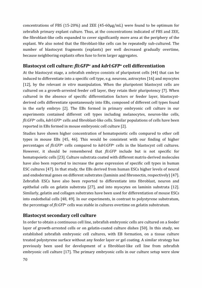
70
concentrations of FBS (15-20%) and ZEE (45-60µg/mL) were found to be optimum for
zebrafish primary explant culture. Thus, at the concentrations indicated of FBS and ZEE,
the fibroblast-like cells expanded to cover significantly more area at the periphery of the
explant. We also noted that the fibroblast-like cells can be repeatedly sub-cultured. The
number of blastocyst fragments (explants) per well decreased gradually overtime,
because neighboring explants often fuse to form larger aggregates.
Blastocyst cell culture: fli:GFP+ and kdrl:GFP+ cell differentiation
At the blastocyst stage, a zebrafish embryo consists of pluripotent cells [44] that can be
induced to differentiate into a specific cell type, e.g. neurons, astrocytes [16] and myocytes
[12], by the relevant in vitro manipulation. When the pluripotent blastocyst cells are
cultured on a growth-arrested feeder cell layer, they retain their pluripotency [7]. When
cultured in the absence of specific differentiation factors or feeder layer, blastocyst-
derived cells differentiate spontaneously into EBs, composed of different cell types found
in the early embryo [2]. The EBs formed in primary embryonic cell culture in our
experiments contained different cell types including melanocytes, neuron-like cells,
fli:GFP+ cells, kdrl:GFP+ cells and fibroblast-like cells. Similar populations of cells have been
reported in EBs formed in mouse embryonic cell culture [2].
Studies have shown higher concentration of hematopoietic cells compared to other cell
types in mouse EBs [45, 46]. This would be consistent with our finding of higher
percentages of fli:GFP+ cells compared to kdrl:GFP+ cells in the blastocyst cell cultures.
However, it should be remembered that fli:GFP include but is not specific for
hematopoietic cells [23]. Culture substrata coated with different matrix-derived molecules
have also been reported to increase the gene expression of specific cell types in human
ESC cultures [47]. In that study, the EBs derived from human ESCs higher levels of neural
and endodermal genes on different substrates (laminin and fibronectin, respectively) [47].
Zebrafish ESCs have also been reported to differentiate into fibroblast, neuron and
epithelial cells on gelatin substrata [27], and into myocytes on laminin substrata [12].
Similarly, gelatin and collagen substrates have been used for differentiation of mouse ESCs
into endothelial cells [48, 49]. In our experiments, in contrast to polystyrene substratum,
the percentage of fli:GFP+ cells was stable in cultures overtime on gelatin substratum.
Blastocyst secondary cell culture
In order to obtain a continuous cell line, zebrafish embryonic cells are cultured on a feeder
layer of growth-arrested cells or on gelatin-coated culture dishes [50]. In this study, we
established zebrafish embryonic cell cultures, with EB formation, on a tissue culture
treated polystyrene surface without any feeder layer or gel coating. A similar strategy has
previously been used for development of a fibroblast-like cell line from zebrafish
embryonic cell culture [17]. The primary embryonic cells in our culture setup were slow
71
growing, being characterized by longer population doubling times, and lower cell viability.
This may be because of the considerable amount of unattached cells observed in the
primary cultures, which were washed away while refreshing the media.
Another reason for the slower growth rate of zebrafish primary cell cultures, could be the
occurrence of cellular differentiation, because different cell types have different growth
requirements [17]. In the present study, the most abundant fibroblast-like cells developed
into a monolayer on the surface of the culture dish when passaged. The secondary cells,
we obtained after passaging the primary blastocyst embryonic cells, were fast growing,
mostly homogeneous and were similar in morphology to the fibroblast-like cell line
derived from zebrafish embryos in ref. [17]. The mouse anti-vimentin antibody that we
used to stain fibroblasts in the blastocyst secondary culture, has been previously reported
to detect fibroblasts in zebrafish [39]. Similar cultures derived from zebrafish embryonic
cells after several passages were referred to as a subpopulation of the primary cells that
suited best or they have adapted themselves to the culture conditions [18].
Effect of FBS on blastocyst secondary cell culture
FBS is the most widely used supplement for cell cultures including zebrafish cell culture
[21]. It contains growth factors, hormones, amino acids, vitamins, trace minerals as well as
cell attachment, proliferating and binding factors [51]. In our study, the presence of higher
than 15% FBS significantly increased the area covered by proliferating cells around the
explant overtime. Similar FBS concentration (15%) have been used previously for
zebrafish embryonic cell cultures [20, 21]. Secondary zebrafish embryonic cells we
obtained after passaging the primary cells also showed higher growth rate with increasing
FBS concentration. For these secondary cells, the use of 10% FBS in the medium was
optimum, in our hands, for the growth of cultures. Similar to these findings, total cell
counts of zebrafish embryonic cell lines (Z428 and ZEB2J) increased with increasing FBS
concentrations in the culture media [52, 53]. Contrary to these findings, in early studies
[18, 54], higher FBS concentrations were shown to inhibit the growth of zebrafish ESC
culture. However, similar to our results, omitting FBS from the media significantly
reduced the growth of these cells [18].
Effect of zebrafish embryo extract on blastocyst secondary cell culture
Fish embryo extract has been shown to support the attachment and growth of medaka
[50, 55] and turbot [56] ESCs. Zebrafish embryo extract is particularly important for the
growth of some of the zebrafish cell lines, e.g. in the case of zebrafish caudal fin cells [18].
However, this is not critical for most of the zebrafish cells cultures (Table 1). In our study,
the emigrating cells around the blastocyst explants covered significantly more area in
media supplemented with 60 µg ZEE protein per mL medium compared to lower ZEE
concentrations. Similar ZEE concentrations have been used previously for zebrafish
72
embryonic cell cultures [20, 21]. Different concentrations of ZEE did not have significant
effects on the growth rate and viability of the zebrafish secondary embryonic cells in our
experiments. These results recommend the use of ZEE for zebrafish primary embryonic
cell cultures. However, for secondary cell cultures ZEE is not important for population
growth. Previous studies have shown significant mitogenic effect of medaka fish embryo
extract on the zebrafish embryonic cell line ZF428 [52].
Effect of gelatin substratum on blastocyst secondary cell culture
Gelatin coating has been used as an alternative to a feeder cell layer for medaka [50],
turbot [56] and zebrafish [52] embryonic cell cultures. In the current study, we cultured
the zebrafish secondary embryonic cells on different concentrations of gelatin substrate.
At higher gelatin concentrations lower number of cells were harvested, although the
number was not significantly different compared to cells cultured without gelatin
substrate. Cell viability was significantly higher in cultures without gelatin coating
compared to cultures on 0.24 and 0.48 mg/cm2 gelatin. Cells cultured on a range of gelatin
substrates up to 0.12 mg/cm2, or cultured without gelatin, had similar growth rates and
viability. These results suggest that gelatin concentrations up to 0.12 mg/cm2 may be used
to coat the culture dish for the attachment and proliferation of zebrafish cells. Similar
gelatin concentrations have been previously used for zebrafish cell culture [52]. A reason
for lower cell growth and viability on high gelatin concentrations might because of the
change in substrate after growing the cells for several passages on a polystyrene surface
of the tissue culture flask without gelatin coating.
Conclusions
Zebrafish blastocyst cells differentiate in vitro into cell types including vimentin+
fibroblastic cells, kdrl:GFP+ cells, and fli:GFP+ cells. However, in the absence of a selective
medium, the latter two cell types decline in abundance over time in the primary cultures,
and disappear with subsequent passaging. Only the fast growing fibroblast-like cells
remain in the cultures. Zebrafish primary embryonic cells require a high FBS
concentration (15%) in medium, while secondary fibroblast-like cell grow at a lower
(10%) FBS concentrations. ZEE at a total protein concentration of 45-60 µg/mL medium is
important for the maintenance of primary cell cultures. However, ZEE does not affect the
growth of the zebrafish secondary cells. Gelatin can be used at low concentrations (0.03-
0.12 mg/cm2) as a substratum for zebrafish blastocyst cell culture.
73
References
[1] Bethesda MD. What are embryonic stem cells? In Stem Cell Information, 2015;
Available from: http://stemcells.nih.gov/info/basics/pages/basics3.aspx.
[2] Keller GM. In vitro differentiation of embryonic stem cells. Curr Opin Cell Biol 1995;
7(6): 862-9.
[3] Evans M. Discovering pluripotency: 30 years of mouse embryonic stem cells. Nat
Rev Mol Cell Biol 2011; 12(10): 680-6.
[4] Koh S, Piedrahita JA. From "ES-like" cells to induced pluripotent stem cells: a
historical perspective in domestic animals. Theriogenology 2014; 81(1): 103-11.
[5] Tachibana M, Amato P, Sparman M, Gutierrez NM, Tippner-Hedges R, Ma H, et al.
Human embryonic stem cells derived by somatic cell nuclear transfer. Cell 2013;
153(6): 1228-38.
[6] Festing S, Wilkinson R. The ethics of animal research. Talking Point on the use of
animals in scientific research. EMBO Rep 2007; 8(6): 526-30.
[7] Alvarez MC, Bejar J, Chen S, Hong Y. Fish ES cells and applications to biotechnology.
Mar Biotechnol (NY) 2007; 9(2): 117-27.
[8] Barut BA, Zon LI. Realizing the potential of zebrafish as a model for human disease.
Physiological Genomics 2000; 2(2): 49-51.
[9] Fan L, Crodian J, Collodi P. Culture of embryonic stem cell lines from zebrafish.
Methods Cell Biol 2004; 76: 151-60.
[10] Chen Z, Lee H, Henle SJ, Cheever TR, Ekker SC, Henley JR. Primary neuron culture
for nerve growth and axon guidance studies in zebrafish (Danio rerio). PLoS One
2013; 8(3): e57539.
[11] Fan L, Crodian J, Liu X, Alestrom A, Alestrom P, Collodi P. Zebrafish embryo cells
remain pluripotent and germ-line competent for multiple passages in culture.
Zebrafish 2004; 1(1): 21-6.
[12] Myhre JL, Pilgrim DB. Cellular differentiation in primary cell cultures from single
zebrafish embryos as a model for the study of myogenesis. Zebrafish 2010; 7(3):
255-66.
[13] Paw BH, Zon LI. Primary fibroblast cell culture. Methods Cell Biol 1999; 59: 39-43.
[14] Xing JG, El-Sweisi W, Lee LE, Collodi P, Seymour C, Mothersill C, et al. Development
of a zebrafish spleen cell line, ZSSJ, and its growth arrest by gamma irradiation and
capacity to act as feeder cells. In Vitro Cell Dev Biol Anim 2009; 45(3-4): 163-74.
[15] Sander V, Sune G, Jopling C, Morera C, Izpisua Belmonte JC. Isolation and in vitro
culture of primary cardiomyocytes from adult zebrafish hearts. Nat Protoc 2013;
8(4): 800-9.
[16] Ghosh C, Liu Y, Ma CG, Collodi P. Cell cultures derived from early zebrafish embryos
differentiate in vitro into neurons and astrocytes. Cytotechnology 1997; 23(1-3):
221-230.
[17] Choorapoikayil S, Overvoorde J, den Hertog J. Deriving cell lines from zebrafish
embryos and tumors. Zebrafish 2013; 10(3): 316-25.
[18] Collodi P, Kamei Y, Ernst T, Miranda C, Buhler DR, Barnes DW. Culture of cells from
zebrafish (Brachydanio rerio) embryo and adult tissues. Cell Biol Toxicol 1992;
8(1): 43-61.
[19] He S, Salas-Vidal E, Rueb S, Krens SF, Meijer AH, Snaar-Jagalska BE, et al. Genetic
and transcriptome characterization of model zebrafish cell lines. Zebrafish 2006;
3(4): 441-53.
74
[20] Badakov R, Jazwinska A. Efficient transfection of primary zebrafish fibroblasts by
nucleofection. Cytotechnology 2006; 51(2): 105-10.
[21] Fan L, Collodi P. Zebrafish embryonic stem cells. Methods Enzymol 2006; 418: 64-
77.
[22] Ma C, Fan L, Ganassin R, Bols N, Collodi P. Production of zebrafish germ-line
chimeras from embryo cell cultures. Proc Natl Acad Sci U S A 2001; 98(5): 2461-6.
[23] Lawson ND, Weinstein BM. In vivo imaging of embryonic vascular development
using transgenic zebrafish. Dev Biol 2002; 248(2): 307-318.
[24] Renaud O, Herbomel P, Kissa K. Studying cell behavior in whole zebrafish embryos
by confocal live imaging: application to hematopoietic stem cells. Nat Protoc 2011;
6(12): 1897-1904.
[25] Jin SW, Beis D, Mitchell T, Chen JN, Stainier DY. Cellular and molecular analyses of
vascular tube and lumen formation in zebrafish. Development 2005; 132(23):
5199-209.
[26] Melet F, Motro B, Rossi DJ, Zhang L, Bernstein A. Generation of a novel Fli-1 protein
by gene targeting leads to a defect in thymus development and a delay in Friend
virus-induced erythroleukemia. Mol Cell Biol 1996; 16(6): 2708-18.
[27] Ho SY, Goh CW, Gan JY, Lee YS, Lam MK, Hong N, et al. Derivation and long-term
culture of an embryonic stem cell-like line from zebrafish blastomeres under
feeder-free condition. Zebrafish 2014; 11(5): 407-20.
[28] Thompson MA, Ransom DG, Pratt SJ, MacLennan H, Kieran MW, Detrich HW, 3rd, et
al. The cloche and spadetail genes differentially affect hematopoiesis and
vasculogenesis. Dev Biol 1998; 197(2): 248-69.
[29] Bussmann J, Lawson N, Zon L, Schulte-Merker S, Committee ZN. Zebrafish VEGF
receptors: a guideline to nomenclature. PLoS Genet 2008; 4(5): e1000064.
[30] Grinblat Y, Lane ME, Sagerstrom C, Sive H. Analysis of zebrafish development using
explant culture assays. Methods Cell Biol 1999; 59: 127-56.
[31] Edgar LG, Goldstein B. Culture and manipulation of embryonic cells. Methods Cell
Biol 2012; 107: 151-75.
[32] Sweet H, Amemiya S, Ransick A, Minokawa T, McClay DR, Wikramanayake A, et al.
Blastomere isolation and transplantation. Methods Cell Biol 2004; 74: 243-71.
[33] Schmidt JV. Embryonic stem (ES) cell culture basics. Curr Protoc Toxicol 2001;
Chapter 15: Unit15 1.
[34] Sun L, Bradford CS, Ghosh C, Collodi P, Barnes DW. ES-like cell cultures derived
from early zebrafish embryos. Mol Mar Biol Biotechnol 1995; 4(3): 193-9.
[35] Experiments on Animal Act, 2014. The State Secretary of Health and Environmental
Protection, Soestdijk. http://wetten.overheid.nl/BWBR0003081/2014-12-18
[36] Directive 2010/63/EU of the European parliament and of the council of 22
September 2010 on the protection of animals used for scientific purposes Text with
EEA relevance, 2010. The European Parliament and the council of the European
Union, Official Journal of the European Union. http://eur-lex.europa.eu/legal-
content/EN/TXT/?uri=celex:32010L0063
[37] Westerfield M. The zebrafish book: a guide for the laboratory use of zebrafish
(Danio Rerio). 4 ed, University of Oregon Press, Eugene 2000, pp.
[38] Kimmel CB, Ballard WW, Kimmel SR, Ullmann B, Schilling TF. Stages of embryonic
development of the zebrafish. Dev Dyn 1995; 203(3): 253-310.
[39] Chablais F, Veit J, Rainer G, Jazwinska A. The zebrafish heart regenerates after
cryoinjury-induced myocardial infarction. BMC Dev Biol 2011; 11: 21.
75
[40] Schneider CA, Rasband WS, Eliceiri KW. NIH Image to ImageJ: 25 years of image
analysis. Nat Methods 2012; 9(7): 671-5.
[41] Sagerstrom CG, Grinbalt Y, Sive H. Anteroposterior patterning in the zebrafish,
Danio rerio: an explant assay reveals inductive and suppressive cell interactions.
Development 1996; 122(6): 1873-83.
[42] Evans MJ, Kaufman MH. Establishment in culture of pluripotential cells from mouse
embryos. Nature 1981; 292(5819): 154-6.
[43] Smith J, Merrick D. Embryonic skeletal muscle microexplant culture and isolation of
skeletal muscle stem cells. Methods Mol Biol 2010; 633: 29-56.
[44] Kimmel CB, Warga RM. Tissue-specific cell lineages originate in the gastrula of the
zebrafish. Science 1986; 231(4736): 365-8.
[45] Keller G, Kennedy M, Papayannopoulou T, Wiles MV. Hematopoietic commitment
during embryonic stem cell differentiation in culture. Mol Cell Biol 1993; 13(1):
473-86.
[46] Wang L, Cerdan C, Menendez P, Bhatia M. Derivation and characterization of
hematopoietic cells from human embryonic stem cells. Methods Mol Biol 2006; 331:
179-200.
[47] Laperle A, Masters KS, Palecek SP. Influence of substrate composition on human
embryonic stem cell differentiation and extracellular matrix production in
embryoid bodies. Biotechnol Prog 2014.
[48] Blancas AA, Lauer NE, McCloskey KE. Endothelial differentiation of embryonic stem
cells. Curr Protoc Stem Cell Biol 2008; Chapter 1: Unit 1F 5.
[49] Liu Z, Lebrin F, Maring JA, van den Driesche S, van der Brink S, van Dinther M, et al.
ENDOGLIN is dispensable for vasculogenesis, but required for vascular endothelial
growth factor-induced angiogenesis. PLoS One 2014; 9(1): e86273.
[50] Hong N, Li Z, Hong Y. Fish stem cell cultures. Int J Biol Sci 2011; 7(4): 392-402.
[51] Gstraunthaler G. Alternatives to the use of fetal bovine serum: serum-free cell
culture. ALTEX 2003; 20(4): 275-81.
[52] Hong N, Schartl M, Hong Y. Derivation of stable zebrafish ES-like cells in feeder-free
culture. Cell Tissue Res 2014; 357(3): 623-32.
[53] Xing JG, Lee LEJ, Fan LC, Collodi P, Holt SE, Bols NC. Initiation of a zebrafish blastula
cell line on rainbow trout stromal cells and subsequent development under feeder-
free conditions into a cell line, ZEB2J. Zebrafish 2008; 5(1): 49-63.
[54] Ghosh C, Collodi P. Culture of cells from zebrafish (Brachydanio rerio) blastula-stage
embryos. Cytotechnology 1994; 14(1): 21-6.
[55] Hong Y, Schartl M. Isolation and differentiation of medaka embryonic stem cells.
Methods Mol Biol 2006; 329: 3-16.
[56] Holen E, Hamre K. Towards obtaining long term embryonic stem cell like cultures
from a marine flatfish, Scophtalmus maximus. Fish Physiol Biochem 2003; 29(3):
245-252.

76
77
Chapter 4
Influence of medium composition and
substratum on the growth of fli:GFP+
and kdrl:GFP+ cells in zebrafish
blastocyst cell culture
Muhammad Ibrahim and Michael K. Richardson
This chapter is part of our following submission:
Ibrahim M, Richardson MK. The growth of endothelial-like cells in zebrafish embryoid
body culture. Experimental cell research (in preparation).
78
Abstract
Embryonic stem cells (ESCs) are important tools to study lineage commitment and cellular
differentiation in early embryos. The differentiation of ESCs can be directed towards a
specific cell type by culturing them in appropriate conditions. In this study we analysed
the effect of different media formulations and culture substrates on the quantification of
fli:GFP+ and kdrl:GFP+ cells in zebrafish blastocyst cell cultures. These cultures were
derived from transgenic zebrafish (fli:GFP and kdrl:GFP) lines which express green
fluorescent protein under the fli1 and VEGFR2 promoters, respectively. The different
media compositions used to culture the blastocyst cells were (i) LDF medium (a
combination of Leibowitz’s L-15, Dulbecco’s modified Eagle’s and Ham’s F12 media); (ii)
combined LDF and endothelial growth medium (LDF:EGM); and (iii) LDF medium
supplemented with endothelial growth supplement mix (LDF:EGS). The blastocyst cells
formed embryoid body aggregates within the first 24 h of culture containing fli:GFP+ or
kdrl:GFP+ cells. The percentage of fli:GFP+ cells was higher in cultures compared to
kdrl:GFP+ cells. This is presumably because the kdrl:GFP line is specific for endothelial
cells, while the fli:GFP is expressed in multiple cell types. On day 6 of cultures, higher
percentages of fli:GFP+ and kdrl:GFP+ cells were observed in LDF:EGS medium (31.8 ±
1.6% and 3.7 ± 0.4%, respectively) compared to LDF medium (21.6 ± 1.9% and 2.1 ± 0.2%,
respectively). In LDF:EGM medium the percentage of fli:GFP+ and kdrl:GFP+ cells was
comparable to that in LDF:EGS medium; however, the total cell yield was significantly less
in LDF:EGM cultures. The percentage of fli:GFP+ and kdrl:GFP+ cells was higher in cultures
on collagen type-I substratum compared to gelatin substratum. Recombinant zebrafish
vascular endothelial growth factor protein was also found to increase the concentration of
fli:GFP+ and kdrl:GFP+ cells in cultures. Both fli:GFP+ and kdrl:GFP+ cells were no longer
detectable in cultures after 14 days and eight days, respectively. Possible reasons for this
may be the down regulation of the transgenes, change in endothelial identity and
occurrence of cell death.
79
Introduction
Embryonic stem cells (ESCs) are cells, derived from blastocyst embryos, that have not
started to differentiate yet [1]. By specific in vitro manipulation these ESCs can maintain
their growth and pluripotency (the ability of cells to differentiated into multiple cell types)
almost indefinitely [2]. ESCs are important tools for regenerative medicine [3], genome
manipulation in animals [4], development of transgenic animals [5] and toxicity testing
[6]. The ESCs have the ability to differentiated into the cells of three germ layers
(ectoderm, endoderm and mesoderm), and further into a specific cell type by
manipulating culture conditions [1]. Examples of differentiated cell types derived from
ESCs in vitro include human cardiomyocytes [7], human neural progenitor cells [8], mouse
hematopoietic progenitor cells [9], alveolar epithelial cells [10] and endothelial progenitor
cells [11].
Research into endothelial cells is fundamental for understanding important processes
regulated by these cells e.g. tissue homeostasis, blood cell activation and coagulation [12].
Endothelial cell culture can be used for important applications such as tissue
regeneration. In one study, endothelial cells derived from ESCs modified into organ
specific endothelial cells and helped regeneration of liver sinusoidal vessels in mice, after
transplantation [13]. Similarly vascular networks cultured in 3D hydrogel matrix using
endothelial cells derived from human pluripotent ESCs, were able to incorporate with the
microvasculature of mouse and sustain blood flow after implantation [14].
Culturing endothelial cells alone is difficult to maintain, to overcome this problem co-
culturing techniques have been developed, in which endothelial cells are cultured in the
presence of supporting cells including fibroblasts, mural cells, pericytes and mesenchymal
stem cells [15]. These endothelial co-culture techniques may be used to engineer
vascularized organ culture. In one example, human umbilical vein endothelial cells
(HUVECs) co-cultured with human mesenchymal stem cells, in a combination of
endothelial growth medium (EGM) and osteogenic medium, formed an in vitro
vascularized bone model [16]. It has been suggested that such vascularized organ cultures
may one day be used for tissue transplantation [17].
Heamangioblasts, the common progenitors of endothelial and hematopoietic lineages,
differentiate from the mesoderm during the early development of embryos [18]. The
differentiation of heamangioblasts is initiated by various factors including vascular
endothelial growth factor (VEGF), basic fibroblast growth factor (bFGF) and bone
morphogenic protein 4 (BMP-4) [19]. Haemangioblasts can be generated in vitro by
treating ESCs with these various differentiation factors just mentioned, as well as others
used in the differentiating media (Table 1; [20, 21]).
In vitro differentiation of ESCs into vascular cells have potential applications in studying
vascular development, proposing vascular regenerative therapy, culturing vascularized

80
organs and developing endothelial cell line [22]. In order to induce differentiation the
ESCs are usually cultured in differentiation media in a suspension culture to form
embryoid bodies (EBs); on a feeder cell layer of stromal cell line; or on an artificial matrix
e.g. collagen type-IV [23, 24].
Table 1: Culture substrates and media composition used for in vitro differentiation of ESCs into
endothelial lineage.
Culture components Units Sources of ESCs
Mouse Human Human Mouse Mouse Human Mouse
Substrate Col-I MEF Fbn Col-IV Gelatin PEC Gelatin
IMDM - - - - -
Lv-EGM - - - - - -
α-MEM - - - - - -
SP-34 medium - - - - - -
DMEM/F12 - - - - - -
DMEM (high glucose) - - - - - -
Methyl cellulose % 1 - - - - - -
FBS % 15 - - 5 - - 20
Knockout serum
replacement
% - - - - - 20 -
BIT 9500 % - 20 - - - - -
Insulin µg/mL 10 - - - - - -
Penicillin U/mL 50 50 - - - - -
Streptomycin µg/mL 50 50 - - - - -
PS % - - - 1 1 1 100X
Monothioglycerol µM 450 450 - - - - -
VEGF ng/mL 50 50 - - 20 10 50
FGF2 ng/mL 100 50 3 - 10 5 -
BMP4 ng/mL - 50 - - 5 10 -
hEGF ng/mL - - 10 - - - -
ActivinA ng/mL - - - - 5 - -
Erythropoietin U/mL 2 - - - - - -
Interleukin 6 ng/mL 10 - - - - - -
BHT µg/mL - - - - 200 - -
L-glutamin - 2mM - 1% 1% 1% 2mM
NE amino acids - 0.1mM - - - 1% 100X
Hydrocortisone µg/mL - - 1 - - - -
Heparin µg/mL - - 10 - - - -
Ascorbic acid mM - - - - 0.5 - -
β-mercaptoethanol - - - 50µM 0.1% 1% 50µM
ESC qualified
nucleotides
- - - - - - 100X
References [25, 26] [27] [28] [20] [13] [13] [29]
Abbreviations: α-MEM, minimum essential medium (sigma); BHT, Bovine holo-transferrin; BMP,
bone morphogenic protein; Col-I, collagen type-I; Col-IV, collagen type-IV; DMEM, Dulbecco’s
modified Eagle’s medium; ESC, embryonic stem cell; F12, Ham’s F12 medium; Fbn, fibronectin; FBS,
fetal bovine serum, BIT, combined bovine serum albumin, insulin and transferrin (Stem Cell
Technologies); FGF, fibroblast growth factor; hEGF, human epidermal growth factor; IMDM, Iscove’s
modified Dulbecco’s medium; Lv-EGM, large-vessel endothelial growth medium; MEF, mouse
embryonic fibroblasts; NE, non-essential amino acids; PEC, primary endothelial cells; PS, penicillin
streptomycin mix; SP-34, Stem Pro-34 medium (Invitrogen); VEGF, vascular endothelial growth
factor; grey boxes indicate the base medium; -, not added.
81
Mouse ESCs cultured on collagen type-IV substrate have been shown to differentiate along
the mesodermal lineage with higher efficiency compared to EB culture [30]. In living
embryos the mesodermal cells differentiate into endothelial and hematopoietic progenitor
cells [12]. In in vitro studies, collagen type-IV has been successfully used for differentiation
of endothelial cells from mouse ESCs [20]. As an alternative to collagen type-IV, gelatin has
also been used as a substratum for mouse ESCs to induce endothelial differentiation [20].
The differentiated endothelial cells are identified using specific antibodies to stain CD31+,
CD34+, or VE-cadherin+ cells [31]. These cells are then enriched by florescence cells
sorting or by other means e.g. magnetic beads, which isolates the stained cells from the
rest of the cells [32]. Using the same strategy, vascular progenitor cells have been derived
from mouse ESCs using flk1 marker [33].
Various ESC cultures have been utilized to develop endothelial cell cultures using
differentiating media (Table 1). ESCs from the mouse and other mammals are usually used
for these studies [21]. However, it is desirable to develop alternative models in order to
reduce the use of mammals in research. Zebrafish can be an excellent model for various
cell culture applications because there is no need to sacrifice the mother to get embryos,
as would be the case in mice. Also, the zebrafish embryo model provides easy and large-
scale availability of embryos for cell isolation and comparatively simple conditions
required for cell culture [34].
Beside the general advantages of zebrafish for cell culture applications, there are
transgenic zebrafish lines that express green fluorescence protein (GFP) in a specific cell
type. Two of the transgenic zebrafish reporter lines: (i) Kdrl:GFP, which expresses GFP
under the promoter of VEGF receptor (VEGFR2), also known as flk-1 (fetal liver kinase 1)
or KDR-like (kinase insert domain receptor like) gene i.e. specifically expressed in
endothelial cells [35]; and (ii) fli:GFP, which expresses GFP under the promoter of friend
leukemia virus integration site 1, i.e. expressed in endothelial, hematopoietic and neural
crest cells [36]. Being a relatively new research model, in vitro studies on zebrafish
hematopoietic and endothelial cells are rare, except for a recently developed zebrafish
embryonic stromal trunk cell line that was reported to support proliferation and
differentiation of zebrafish hematopoietic stem cells [37].
In this chapter, we examine the development of fli:GFP+ and kdrl:GFP+ cells in zebrafish
blastocyst cell cultures. We analyse the yield of these cells under different culture
conditions. We used different media compositions to test their potential to generate
fli:GFP+ and kdrl:GFP+ cells in blastocyst cell culture. The three media compositions used
were: (i) LDF (combined Lebovitz-15, Dulbecco’s modified Eagle’s medium and Ham’s F-
12 medium) medium; (ii) 1:1 mixture of LDF and EGM (endothelial growth medium)
medium; and (iii) LDF medium supplemented with endothelial growth supplement.
Furthermore, we analyse the effect of different substrates (gelatin and collagen type-I),
82
and different concentrations of recombinant zebrafish vascular endothelial growth factor
(VEGF165), on the percentage of fli:GFP+ and kdrl:GFP+ cells in culture.
Materials and methods
Embryo collection
All the animal experiments were performed according to the Netherland Experiments on
Animals Act [38], based on the guidelines on the protection of experimental animals, laid
by the Council of European Union [39]. Adult zebrafish were maintained as previously
described [40]. Two different transgenic zebrafish lines fli:GFP and kdrl:GFP were used. To
obtain embryos, adult male and female fish, at a proportion of 1 to 1, were transferred to
small breeding tanks in the evening. The zebrafish usually laid eggs when the light comes
on. The eggs were collected at the bottom of the tank, separated from adults using a cotton
mesh.
Embryos were transferred to a temperature controlled room (28 °C) and were distributed
in 9cm Petri dishes at a final density of 100 embryos per dish, after removing dead and
unfertilized eggs. The embryos were washed thoroughly with clean egg water to remove
any debris. These embryos were allowed to develop to the high blastula stage of Kimmel
et al. [41] (approximately 3 h after fertilization) at 28 °C.
Sterilization of embryos
The embryos were transferred to a flow cabinet at room temperature for sterilization and
cell isolation. For sterilization, the embryos, with the chorion intact, were immersed in
70% ethanol for 10 sec and then in two changes of 0.05% sodium hypochlorite (available
chlorine 10-15%, Sigma, catalog 425044 ) for 4 min each. The sterilization was done
according to the procedure described in Ref. [42]. After each immersion in ethanol or
sodium hypochlorite, the embryos were rinsed with LDF medium (see below, Media
combinations and composition). Finally the embryos were left in 0.5 mL of LDF medium
for dechorionation. Before dechorionating, the embryos were observed under a dissecting
microscope and any dead or abnormal embryos (with cloudiness in the perivitelline fluid)
were removed.
Blastocyst cells isolation and culture
The following steps were all conducted at room temperature. The embryos were
dechorionated in LDF medium using sterile No. 5 watchmaker’s forceps and then
transferred to Eppendorf tubes. The LDF medium was gently triturated with a P-200
Gilson micropipette (Gilson, B.V., Europe: Den Haag) to remove the yolk. The solution was
then centrifuged at 300 g for 1 min and the supernatant was discarded. The blastocysts
were washed once with CMF-PBS (calcium magnesium free phosphate buffered saline;
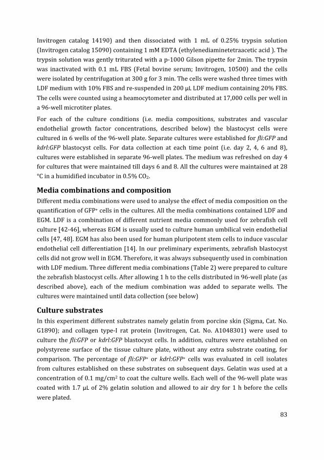
83
Invitrogen catalog 14190) and then dissociated with 1 mL of 0.25% trypsin solution
(Invitrogen catalog 15090) containing 1 mM EDTA (ethylenediaminetetraacetic acid ). The
trypsin solution was gently triturated with a p-1000 Gilson pipette for 2min. The trypsin
was inactivated with 0.1 mL FBS (Fetal bovine serum; Invitrogen, 10500) and the cells
were isolated by centrifugation at 300 g for 3 min. The cells were washed three times with
LDF medium with 10% FBS and re-suspended in 200 µL LDF medium containing 20% FBS.
The cells were counted using a heamocytometer and distributed at 17,000 cells per well in
a 96-well microtiter plates.
For each of the culture conditions (i.e. media compositions, substrates and vascular
endothelial growth factor concentrations, described below) the blastocyst cells were
cultured in 6 wells of the 96-well plate. Separate cultures were established for fli:GFP and
kdrl:GFP blastocyst cells. For data collection at each time point (i.e. day 2, 4, 6 and 8),
cultures were established in separate 96-well plates. The medium was refreshed on day 4
for cultures that were maintained till days 6 and 8. All the cultures were maintained at 28
°C in a humidified incubator in 0.5% CO2.
Media combinations and composition
Different media combinations were used to analyse the effect of media composition on the
quantification of GFP+ cells in the cultures. All the media combinations contained LDF and
EGM. LDF is a combination of different nutrient media commonly used for zebrafish cell
culture [42-46], whereas EGM is usually used to culture human umbilical vein endothelial
cells [47, 48]. EGM has also been used for human pluripotent stem cells to induce vascular
endothelial cell differentiation [14]. In our preliminary experiments, zebrafish blastocyst
cells did not grow well in EGM. Therefore, it was always subsequently used in combination
with LDF medium. Three different media combinations (Table 2) were prepared to culture
the zebrafish blastocyst cells. After allowing 1 h to the cells distributed in 96-well plate (as
described above), each of the medium combination was added to separate wells. The
cultures were maintained until data collection (see below)
Culture substrates
In this experiment different substrates namely gelatin from porcine skin (Sigma, Cat. No.
G1890); and collagen type-I rat protein (Invitrogen, Cat. No. A1048301) were used to
culture the fli:GFP or kdrl:GFP blastocyst cells. In addition, cultures were established on
polystyrene surface of the tissue culture plate, without any extra substrate coating, for
comparison. The percentage of fli:GFP+ or kdrl:GFP+ cells was evaluated in cell isolates
from cultures established on these substrates on subsequent days. Gelatin was used at a
concentration of 0.1 mg/cm2 to coat the culture wells. Each well of the 96-well plate was
coated with 1.7 µL of 2% gelatin solution and allowed to air dry for 1 h before the cells
were plated.
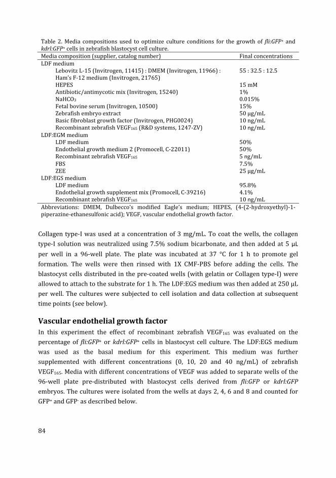
84
Table 2. Media compositions used to optimize culture conditions for the growth of fli:GFP+ and
kdrl:GFP+ cells in zebrafish blastocyst cell culture.
Media composition (supplier, catalog number) Final concentrations
LDF medium
Lebovitz L-15 (Invitrogen, 11415) : DMEM (Invitrogen, 11966) :
Ham’s F-12 medium (Invitrogen, 21765)
55 : 32.5 : 12.5
HEPES 15 mM
Antibiotic/antimycotic mix (Invitrogen, 15240) 1%
NaHCO3 0.015%
Fetal bovine serum (Invitrogen, 10500) 15%
Zebrafish embryo extract 50 µg/mL
Basic fibroblast growth factor (Invitrogen, PHG0024) 10 ng/mL
Recombinant zebrafish VEGF165 (R&D systems, 1247-ZV) 10 ng/mL
LDF:EGM medium
LDF medium 50%
Endothelial growth medium 2 (Promocell, C-22011) 50%
Recombinant zebrafish VEGF165 5 ng/mL
FBS 7.5%
ZEE 25 µg/mL
LDF:EGS medium
LDF medium 95.8%
Endothelial growth supplement mix (Promocell, C-39216) 4.1%
Recombinant zebrafish VEGF165 10 ng/mL
Abbreviations: DMEM, Dulbecco’s modified Eagle’s medium; HEPES, (4-(2-hydroxyethyl)-1-
piperazine-ethanesulfonic acid); VEGF, vascular endothelial growth factor.
Collagen type-I was used at a concentration of 3 mg/mL. To coat the wells, the collagen
type-I solution was neutralized using 7.5% sodium bicarbonate, and then added at 5 µL
per well in a 96-well plate. The plate was incubated at 37 °C for 1 h to promote gel
formation. The wells were then rinsed with 1X CMF-PBS before adding the cells. The
blastocyst cells distributed in the pre-coated wells (with gelatin or Collagen type-I) were
allowed to attach to the substrate for 1 h. The LDF:EGS medium was then added at 250 µL
per well. The cultures were subjected to cell isolation and data collection at subsequent
time points (see below).
Vascular endothelial growth factor
In this experiment the effect of recombinant zebrafish VEGF165 was evaluated on the
percentage of fli:GFP+ or kdrl:GFP+ cells in blastocyst cell culture. The LDF:EGS medium
was used as the basal medium for this experiment. This medium was further
supplemented with different concentrations (0, 10, 20 and 40 ng/mL) of zebrafish
VEGF165. Media with different concentrations of VEGF was added to separate wells of the
96-well plate pre-distributed with blastocyst cells derived from fli:GFP or kdrl:GFP
embryos. The cultures were isolated from the wells at days 2, 4, 6 and 8 and counted for
GFP+ and GFP- as described below.
85
Isolation of cultured cells for data collection
The percentage of fli:GFP+ or kdrl:GFP+ cells, total number of cells harvested per well and
cell viability was calculated in cell isolates from the cultures at subsequent time-points. To
isolate the cells, the medium was aspirated from each well. The wells were then rinsed
with 200 µL of 1X CMF-PBS. Then 250 µL of 0.05% trypsin solution containing 1mM EDTA
was added to each well. The solution was triturated in the wells several times and then the
plate was incubated for 2 min at 28 °C. The degree of detachment was monitored under an
inverted microscope. When most cells were rounded up, the trypsin was inactivated by
adding 25 µL of FBS, and the cell suspension from each well was transferred to separate
1.5 mL Eppendorf tubes. The suspension was centrifuged at 300 g for 3 min and the
supernatant discarded. The cell pellet was washed twice with LDF medium (without
growth factors) and re-suspended in 15-20 µL of the same medium. These cells were used
for further analysis.
Cell counts and data collection
For the cultures maintained in different media compositions (i.e. LDF, LDF:EGM and
LDF:EGS media), the total number of cells harvested per well and the cell viability was
calculated in addition to the percentage of fli:GFP+ or kdrl:GFP+ cells. For other conditions
(i.e. different substrates and VEGF concentrations) only the percentage of fli:GFP+ or
kdrl:GFP+ cells was calculated in cell isolates. The total number of cells harvested per well
was estimated by counting the cells in a heamocytometer. Viability of the cells was
determined by using trypan blue exclusion dye (0.4% trypan blue in CMF-PBS) to
differentiate viable and non-viable cells.
The percentage of fli:GFP+ or kdrl:GFP+ cells in the living cell suspensions was calculated
using a confocal microscope. A 5 µL drop from each of the cell isolates was transferred to a
cover glass slide. The slide was placed under the confocal microscope and the cells were
allowed to settle for 30 s. An image was then captured from the center of each drop. Each
image was taken in duplex, one fluorescence image showing GFP+ cells and one phase
contrast image showing all the cells. For both fli:GFP and kdrl:GFP cultures, number of
GFP+ and GFP- cells per microscopic field (image) were then counted in image J software.
From these counts the percentage of fli:GFP+ or kdrl:GFP+ cells was calculated for each
sample.
Statistical analysis
Number of EBs formed, total number of cells harvested, percentage of viable cells and the
percentages of fli:GFP+ and kdrl:GFP+ cells per well, collected from 6 replicate wells for
each culture condition, were analysed for means and standard errors using SPSS software
version 21.0. One way ANOVA was performed to calculate the probability values to
analyse differences between different culture conditions, as well as differences at different
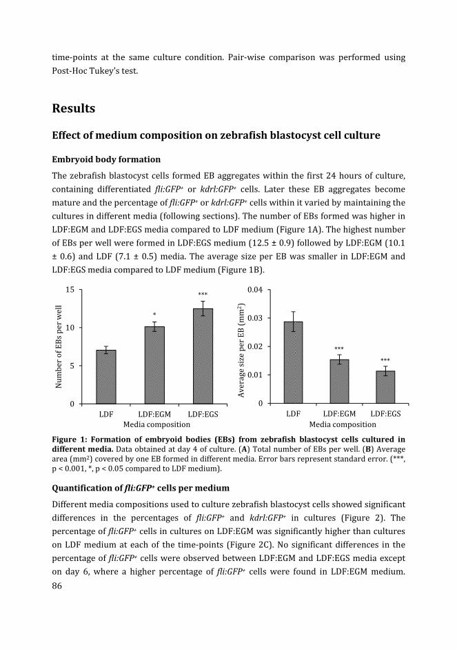
86
time-points at the same culture condition. Pair-wise comparison was performed using
Post-Hoc Tukey’s test.
Results
Effect of medium composition on zebrafish blastocyst cell culture
Embryoid body formation
The zebrafish blastocyst cells formed EB aggregates within the first 24 hours of culture,
containing differentiated fli:GFP+ or kdrl:GFP+ cells. Later these EB aggregates become
mature and the percentage of fli:GFP+ or kdrl:GFP+ cells within it varied by maintaining the
cultures in different media (following sections). The number of EBs formed was higher in
LDF:EGM and LDF:EGS media compared to LDF medium (Figure 1A). The highest number
of EBs per well were formed in LDF:EGS medium (12.5 ± 0.9) followed by LDF:EGM (10.1
± 0.6) and LDF (7.1 ± 0.5) media. The average size per EB was smaller in LDF:EGM and
LDF:EGS media compared to LDF medium (Figure 1B).
Figure 1: Formation of embryoid bodies (EBs) from zebrafish blastocyst cells cultured in
different media. Data obtained at day 4 of culture. (A) Total number of EBs per well. (B) Average
area (mm2) covered by one EB formed in different media. Error bars represent standard error. (***,
p < 0.001, *, p < 0.05 compared to LDF medium).
Quantification of fli:GFP+ cells per medium
Different media compositions used to culture zebrafish blastocyst cells showed significant
differences in the percentages of fli:GFP+ and kdrl:GFP+ in cultures (Figure 2). The
percentage of fli:GFP+ cells in cultures on LDF:EGM was significantly higher than cultures
on LDF medium at each of the time-points (Figure 2C). No significant differences in the
percentage of fli:GFP+ cells were observed between LDF:EGM and LDF:EGS media except
on day 6, where a higher percentage of fli:GFP+ cells were found in LDF:EGM medium.
0
5
10
15
LDF LDF:EGM LDF:EGS
Nu
mb
er
of
EB
s p
er
we
ll
Media composition
*
***
0
0.01
0.02
0.03
0.04
LDF LDF:EGM LDF:EGS
Av
era
ge
siz
e p
er
EB
(m
m2)
Media composition
***
***
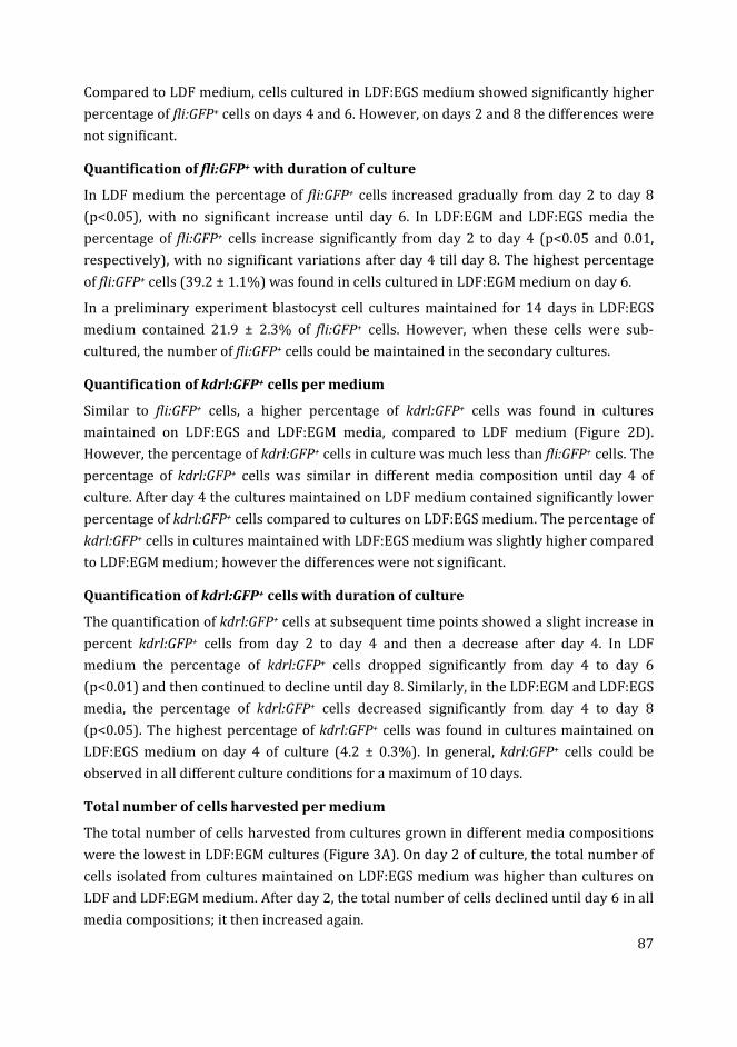
87
Compared to LDF medium, cells cultured in LDF:EGS medium showed significantly higher
percentage of fli:GFP+ cells on days 4 and 6. However, on days 2 and 8 the differences were
not significant.
Quantification of fli:GFP+ with duration of culture
In LDF medium the percentage of fli:GFP+ cells increased gradually from day 2 to day 8
(p<0.05), with no significant increase until day 6. In LDF:EGM and LDF:EGS media the
percentage of fli:GFP+ cells increase significantly from day 2 to day 4 (p<0.05 and 0.01,
respectively), with no significant variations after day 4 till day 8. The highest percentage
of fli:GFP+ cells (39.2 ± 1.1%) was found in cells cultured in LDF:EGM medium on day 6.
In a preliminary experiment blastocyst cell cultures maintained for 14 days in LDF:EGS
medium contained 21.9 ± 2.3% of fli:GFP+ cells. However, when these cells were sub-
cultured, the number of fli:GFP+ cells could be maintained in the secondary cultures.
Quantification of kdrl:GFP+ cells per medium
Similar to fli:GFP+ cells, a higher percentage of kdrl:GFP+ cells was found in cultures
maintained on LDF:EGS and LDF:EGM media, compared to LDF medium (Figure 2D).
However, the percentage of kdrl:GFP+ cells in culture was much less than fli:GFP+ cells. The
percentage of kdrl:GFP+ cells was similar in different media composition until day 4 of
culture. After day 4 the cultures maintained on LDF medium contained significantly lower
percentage of kdrl:GFP+ cells compared to cultures on LDF:EGS medium. The percentage of
kdrl:GFP+ cells in cultures maintained with LDF:EGS medium was slightly higher compared
to LDF:EGM medium; however the differences were not significant.
Quantification of kdrl:GFP+ cells with duration of culture
The quantification of kdrl:GFP+ cells at subsequent time points showed a slight increase in
percent kdrl:GFP+ cells from day 2 to day 4 and then a decrease after day 4. In LDF
medium the percentage of kdrl:GFP+ cells dropped significantly from day 4 to day 6
(p<0.01) and then continued to decline until day 8. Similarly, in the LDF:EGM and LDF:EGS
media, the percentage of kdrl:GFP+ cells decreased significantly from day 4 to day 8
(p<0.05). The highest percentage of kdrl:GFP+ cells was found in cultures maintained on
LDF:EGS medium on day 4 of culture (4.2 ± 0.3%). In general, kdrl:GFP+ cells could be
observed in all different culture conditions for a maximum of 10 days.
Total number of cells harvested per medium
The total number of cells harvested from cultures grown in different media compositions
were the lowest in LDF:EGM cultures (Figure 3A). On day 2 of culture, the total number of
cells isolated from cultures maintained on LDF:EGS medium was higher than cultures on
LDF and LDF:EGM medium. After day 2, the total number of cells declined until day 6 in all
media compositions; it then increased again.
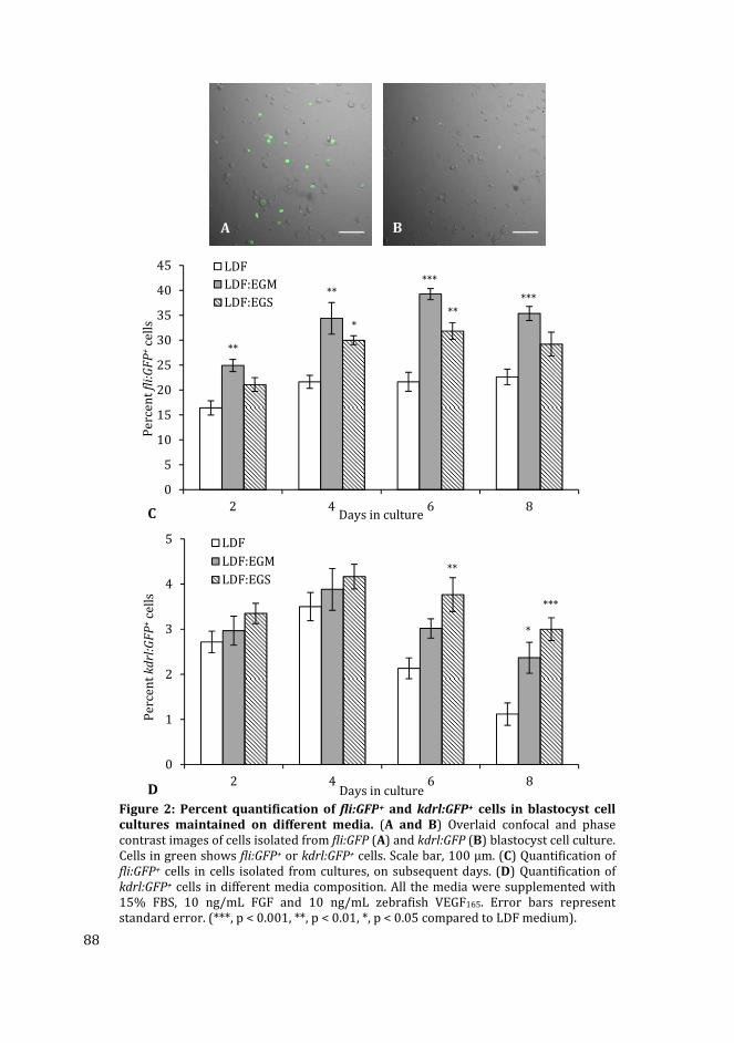
88
Figure 2: Percent quantification of fli:GFP+ and kdrl:GFP+ cells in blastocyst cell
cultures maintained on different media. (A and B) Overlaid confocal and phase
contrast images of cells isolated from fli:GFP (A) and kdrl:GFP (B) blastocyst cell culture.
Cells in green shows fli:GFP+ or kdrl:GFP+ cells. Scale bar, 100 µm. (C) Quantification of
fli:GFP+ cells in cells isolated from cultures, on subsequent days. (D) Quantification of
kdrl:GFP+ cells in different media composition. All the media were supplemented with
15% FBS, 10 ng/mL FGF and 10 ng/mL zebrafish VEGF165. Error bars represent
standard error. (***, p < 0.001, **, p < 0.01, *, p < 0.05 compared to LDF medium).
0
5
10
15
20
25
30
35
40
45
2 4 6 8
Pe
rce
nt
fli:
GF
P+
cell
s
Days in culture
LDF
LDF:EGM
LDF:EGS
**
**
***
***
***
C
0
1
2
3
4
5
2 4 6 8
Pe
rce
nt
kd
rl:G
FP
+ce
lls
Days in culture
LDF
LDF:EGM
LDF:EGS**
*
***
D
A B
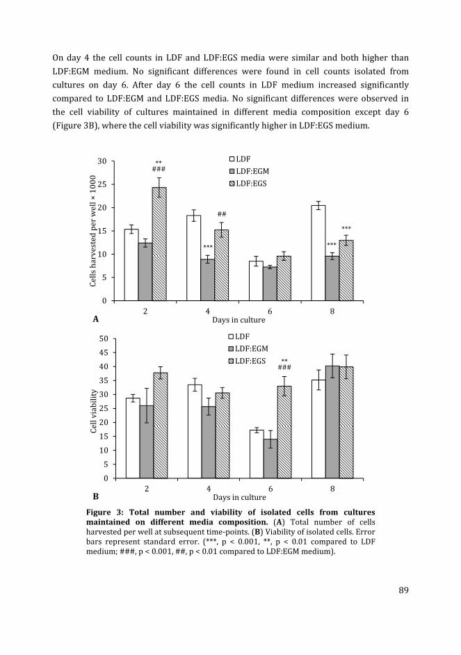
89
On day 4 the cell counts in LDF and LDF:EGS media were similar and both higher than
LDF:EGM medium. No significant differences were found in cell counts isolated from
cultures on day 6. After day 6 the cell counts in LDF medium increased significantly
compared to LDF:EGM and LDF:EGS media. No significant differences were observed in
the cell viability of cultures maintained in different media composition except day 6
(Figure 3B), where the cell viability was significantly higher in LDF:EGS medium.
Figure 3: Total number and viability of isolated cells from cultures
maintained on different media composition. (A) Total number of cells
harvested per well at subsequent time-points. (B) Viability of isolated cells. Error
bars represent standard error. (***, p < 0.001, **, p < 0.01 compared to LDF
medium; ###, p < 0.001, ##, p < 0.01 compared to LDF:EGM medium).
0
5
10
15
20
25
30
2 4 6 8
Ce
lls
ha
rve
ste
d p
er
we
ll ×
10
00
Days in culture
LDF
LDF:EGM
LDF:EGS
**###
***
##
***
***
A
0
5
10
15
20
25
30
35
40
45
50
2 4 6 8
Ce
ll v
iab
ilit
y
Days in culture
LDF
LDF:EGM
LDF:EGS **###
B
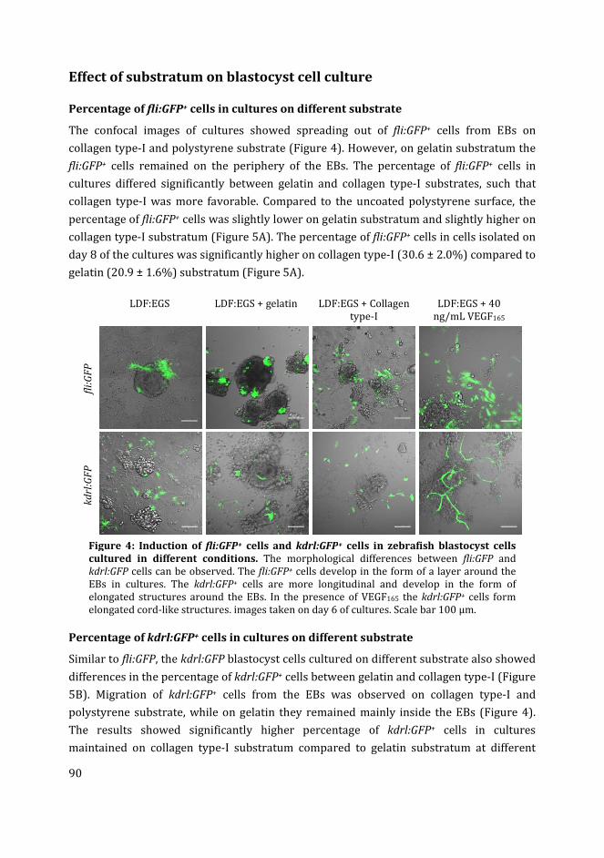
90
Effect of substratum on blastocyst cell culture
Percentage of fli:GFP+ cells in cultures on different substrate
The confocal images of cultures showed spreading out of fli:GFP+ cells from EBs on
collagen type-I and polystyrene substrate (Figure 4). However, on gelatin substratum the
fli:GFP+ cells remained on the periphery of the EBs. The percentage of fli:GFP+ cells in
cultures differed significantly between gelatin and collagen type-I substrates, such that
collagen type-I was more favorable. Compared to the uncoated polystyrene surface, the
percentage of fli:GFP+ cells was slightly lower on gelatin substratum and slightly higher on
collagen type-I substratum (Figure 5A). The percentage of fli:GFP+ cells in cells isolated on
day 8 of the cultures was significantly higher on collagen type-I (30.6 ± 2.0%) compared to
gelatin (20.9 ± 1.6%) substratum (Figure 5A).
LDF:EGS LDF:EGS + gelatin LDF:EGS + Collagen
type-I
LDF:EGS + 40
ng/mL VEGF165
fli:
GF
P
kd
rl:G
FP
Figure 4: Induction of fli:GFP+ cells and kdrl:GFP+ cells in zebrafish blastocyst cells
cultured in different conditions. The morphological differences between fli:GFP and
kdrl:GFP cells can be observed. The fli:GFP+ cells develop in the form of a layer around the
EBs in cultures. The kdrl:GFP+ cells are more longitudinal and develop in the form of
elongated structures around the EBs. In the presence of VEGF165 the kdrl:GFP+ cells form
elongated cord-like structures. images taken on day 6 of cultures. Scale bar 100 µm.
Percentage of kdrl:GFP+ cells in cultures on different substrate
Similar to fli:GFP, the kdrl:GFP blastocyst cells cultured on different substrate also showed
differences in the percentage of kdrl:GFP+ cells between gelatin and collagen type-I (Figure
5B). Migration of kdrl:GFP+ cells from the EBs was observed on collagen type-I and
polystyrene substrate, while on gelatin they remained mainly inside the EBs (Figure 4).
The results showed significantly higher percentage of kdrl:GFP+ cells in cultures
maintained on collagen type-I substratum compared to gelatin substratum at different
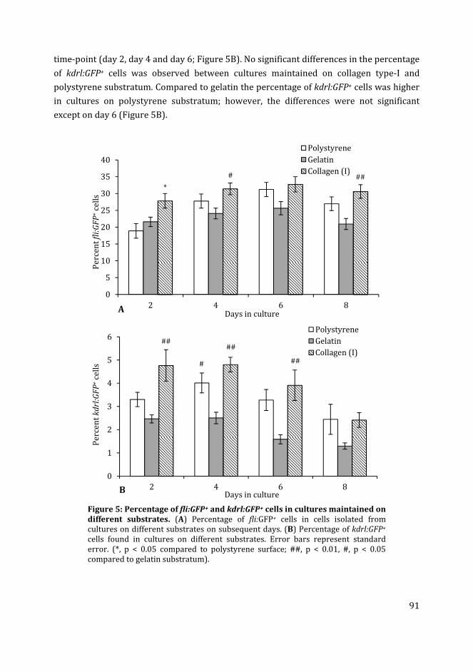
91
time-point (day 2, day 4 and day 6; Figure 5B). No significant differences in the percentage
of kdrl:GFP+ cells was observed between cultures maintained on collagen type-I and
polystyrene substratum. Compared to gelatin the percentage of kdrl:GFP+ cells was higher
in cultures on polystyrene substratum; however, the differences were not significant
except on day 6 (Figure 5B).
Figure 5: Percentage of fli:GFP+ and kdrl:GFP+ cells in cultures maintained on
different substrates. (A) Percentage of fli:GFP+ cells in cells isolated from
cultures on different substrates on subsequent days. (B) Percentage of kdrl:GFP+
cells found in cultures on different substrates. Error bars represent standard
error. (*, p < 0.05 compared to polystyrene surface; ##, p < 0.01, #, p < 0.05
compared to gelatin substratum).
0
5
10
15
20
25
30
35
40
2 4 6 8
Pe
rce
nt
fli:
GF
P+
cell
s
Days in culture
Polystyrene
Gelatin
Collagen (I)
*
# ##
0
1
2
3
4
5
6
2 4 6 8
Pe
rce
nt
kd
rl:G
FP
+ce
lls
Days in culture
Polystyrene
Gelatin
Collagen (I)
####
###
A
B
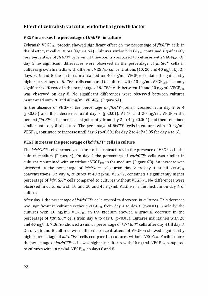
92
Effect of zebrafish vascular endothelial growth factor
VEGF increases the percentage of fli:GFP+ in culture
Zebrafish VEGF165 protein showed significant effect on the percentage of fli:GFP+ cells in
the blastocyst cell cultures (Figure 6A). Cultures without VEGF165 contained significantly
less percentage of fli:GFP+ cells on all time-points compared to cultures with VEGF165. On
day 2 no significant differences were observed in the percentage of fli:GFP+ cells in
cultures grown in media with different VEGF165 concentrations (10, 20 and 40 ng/mL). On
days 4, 6 and 8 the cultures maintained on 40 ng/mL VEGF165 contained significantly
higher percentage of fli:GFP+ cells compared to cultures with 10 ng/mL VEGF165. The only
significant difference in the percentage of fli:GFP+ cells between 10 and 20 ng/mL VEGF165
was observed on day 8. No significant differences were observed between cultures
maintained with 20 and 40 ng/mL VEGF165 (Figure 6A).
In the absence of VEGF165 the percentage of fli:GFP+ cells increased from day 2 to 4
(p<0.05) and then decreased until day 8 (p<0.01). At 10 and 20 ng/mL VEGF165 the
percent fli:GFP+ cells increased significantly from day 2 to 4 (p<0.001) and then remained
similar until day 8 of culture. The percentage of fli:GFP+ cells in cultures with 40 ng/mL
VEGF165 continued to increase until day 6 (p<0.001 for day 2 to 4; P<0.05 for day 4 to 6).
VEGF increases the percentage of kdrl:GFP+ cells in culture
The kdrl:GFP+ cells formed vascular cord-like structures in the presence of VEGF165 in the
culture medium (Figure 4). On day 2 the percentage of kdrl:GFP+ cells was similar in
cultures maintained with or without VEGF165 in the medium (Figure 6B). An increase was
observed in the percentage of kdrl:GFP+ cells from day 2 to day 4 at all VEGF165
concentrations. On day 4, cultures at 40 ng/mL VEGF165 contained a significantly higher
percentage of kdrl:GFP+ cells compared to cultures without VEGF165. No differences were
observed in cultures with 10 and 20 and 40 ng/mL VEGF165 in the medium on day 4 of
culture.
After day 4 the percentage of kdrl:GFP+ cells started to decrease in cultures. This decrease
was significant in cultures without VEGF165 from day 4 to day 6 (p<0.01). Similarly, the
cultures with 10 ng/mL VEGF165 in the medium showed a gradual decrease in the
percentage of kdrl:GFP+ cells from day 4 to day 8 (p<0.05). Cultures maintained with 20
and 40 ng/mL VEGF165 showed a similar percentage of kdrl:GFP+ cells after day 4 till day 8.
On days 6 and 8 cultures with different concentrations of VEGF165 showed significantly
higher percentage of kdrl:GFP+ cells compared to cultures without VEGF165. Furthermore,
the percentage of kdrl:GFP+ cells was higher in cultures with 40 ng/mL VEGF165 compared
to cultures with 10 ng/mL VEGF165 on days 6 and 8.
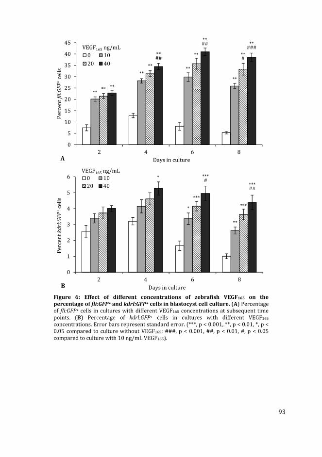
93
Figure 6: Effect of different concentrations of zebrafish VEGF165 on the
percentage of fli:GFP+ and kdrl:GFP+ cells in blastocyst cell culture. (A) Percentage
of fli:GFP+ cells in cultures with different VEGF165 concentrations at subsequent time
points. (B) Percentage of kdrl:GFP+ cells in cultures with different VEGF165
concentrations. Error bars represent standard error. (***, p < 0.001, **, p < 0.01, *, p <
0.05 compared to culture without VEGF165; ###, p < 0.001, ##, p < 0.01, #, p < 0.05
compared to culture with 10 ng/mL VEGF165).
0
5
10
15
20
25
30
35
40
45
2 4 6 8
Pe
rce
nt
fli:
GF
P+
cell
s
Days in culture
0 10
20 40
VEGF165 ng/mL
**
****
**
**##
**
**
** **
**##
**#
**###
A
0
1
2
3
4
5
6
2 4 6 8
Pe
rce
nt
kd
rl:G
FP
+ce
lls
Days in culture
0 10
20 40
VEGF165 ng/mL*
*
**
***
***
***#
***##
B
94
Discussion
Relationship of culture conditions, EB formation and fli:GFP+ and
kdrl:GFP+ cell quantification in culture
We have investigated different culture conditions for zebrafish blastocyst cells, with the
objective to generate differentiated fli:GFP+ or kdrl:GFP+ cells in relatively high numbers.
The results showed important information for a controlled differentiation process in
zebrafish blastocyst cell cultures. Studies have shown that without any anti-differentiation
factors or feeder cells mouse and human ESCs undergo cellular differentiation (similar in
some respects to embryogenesis) and form EBs [49]. In this study, we found that medium
composition affects the development of EBs and the percentages of fli:GFP+ and kdrl:GFP+
cells in zebrafish blastocyst cell cultures.
Study on mouse ESC culture have shown that EBs developed in adherent cultures
contained a higher number of total cells and a lower percentage of hematopoietic and
endothelial cells, compared to the EBs grown in suspension cultures [50]. In our study,
zebrafish blastocyst cells cultured in LDF medium developed a few, large-sized EBs, while
cells cultured in LDF:EGM and LDF:EGS media developed more numerous, but smaller EBs
with a higher percentage of fli:GFP+ or kdrl:GFP+ cells. These results suggest a direct
relation between the number of EBs and the percentage of fli:GFP+ or kdrl:GFP+ cells.
Previous studies have shown that the relative less cell-to-cell interaction, in the adherent
cultures compared to 3D or suspension cultures, may favor the growth or differentiation
of cell types other than hematopoietic and endothelial cells [50]. This conclusion is
supported by our hanging-drop experiments in which there is by definition no outgrowth,
but there is a high percentage of fli:GFP+ or kdrl:GFP+ cells (see Chapter 5). In our
experiments described in this chapter, the supplementation of the LDF medium with
endothelial growth supplement resulted in higher percentages of fli:GFP+ and kdrl:GFP+
cells in cultures. This could be because the supplementation leads to increased cell-cell-
interactions in the plated blastocyst cells as they are aggregating into EBs.
Differences between fli:GFP+ and kdrl:GFP+ cells in culture
The blastocyst cell cultures initiated from fli:GFP embryos in our experiments expressed
GFP in a higher percentage of cells compare to kdrl:GFP. This might be explained by the
lineage-specificity of GFP expression in kdrl:GFP transgenic lines (endothelial cells only
[51]), compared to the fli:GFP line (endothelial, lymphatic, hematopoietic and neural crest
cells [36]). The kdrl:GFP+ cells almost vanished by 8 days of culture, a similar phenomenon
was recorded in a recent study using kdrl:GFP blastocyst cell culture for screening
angiogenic compounds [52].
95
The fli:GFP signals by contrast, could be observed for up to 14 days. However, they also
could not be maintained after that. This can be considered in the light of previous studies,
describing fli:GFP expression persists in zebrafish embryos during early developmental
stages [36]. Similarly, studies on mouse embryos and ESCs have shown significant
reduction in the expression levels of flk1/kdr gene at later life stages [53]. These reports
suggest as the cells matures in our zebrafish blastocyst cell cultures, they down regulate
the transgenes, as these genes are expressed mainly in early stages of differentiation.
However, other studies have reported that the expression of fli1 and kdrl genes persists
throughout vascular development in zebrafish [51, 54]. One possible explanation can be
changes in gene expression and properties of endothelial cells, in our study, particularly in
vitro, as previously described for other endothelial cells [55].
Studies have shown that primary endothelial cells have a short life span, which is usually
overcome by immortalizing these cells using genetic manipulation [56]. Thus, an
alternative explanation for the loss of fli:GFP and kdrl:GFP signals may be the occurrence
of cell death in our primary blastocyst cell culture. This phenomena was observed as high
number of floating cells in culture and long population doubling time of the primary
blastocyst cell culture, discussed in chapter 3. A previous study also reported the loss of
kdrl:GFP signal in zebrafish blastocyst cell culture due to apoptosis [57]. Another reason
for this may be the development of fast growing fibroblast-like cells in our cultures, which
depleted other cell types in subsequent passages (Chapter 3).
Media composition and endothelial growth supplementation
LDF is a commonly-used medium for zebrafish ESC culture [42-46]. It is also sometimes
referred to as limiting dilution factor (LDF) medium, used to retain the homogeneity of a
cell culture [43, 58]. The zebrafish primary blastocyst cells may have more complex
nutrient requirements for their growth and attachment therefore supplements including
FBS, fish embryo extract, fish serum and bFGF are usually added to the medium [59]. A
nutrient rich medium is required to culture the primary blastocyst cells as the initial cell
death is high in these cells because of bleaching the embryos [60]. The blastocyst cells are
pluripotent in nature, therefore specific differentiation can be promoted by changing the
culture conditions [34]. In some experiments additional supplementation or a substrate
coating may be required to induce specific differentiation in these pluripotent ESCs.
Examples include sonic hedgehog protein for myocyte differentiation [61], and poly-D-
lysine coating for neurons and astrocytes differentiation [45].
Endothelial growth medium (EGM) is usually used to culture human umbilical vein
endothelial cells (HUVECs; [47, 48]), as well as for the differentiation of human
pluripotent stem cells into vascular endothelial cells [14]. The complete EGM is a
combination of endothelial basal medium and endothelial growth supplement (EGS) mix.
The EGS is composed of growth factors including human epidermal growth factor, bFGF,
96
insulin-like growth factor (IGF-1) and human VEGF. These components are usually used in
differentiation media to induce endothelial differentiation in mouse [13, 25] and human
[13, 27, 28] ESCs. Other components of EGS are heparin and hydrocortisone, which have
been used for endothelial differentiation in human ESCs [28]. Similarly, ascorbic acid (also
a component of EGS) have also been used in endothelial differentiation medium for mouse
ESC culture [13].
In our experiments, the LDF medium increased the total cell counts after day 6 of the
cultures. However, the percentages of fli:GFP+ and kdrl:GFP+ cells was significantly lower
in this medium compared to LDF:EGM and LDF:EGS media. Previous studies on zebrafish
blastocyst cell culture have shown the necessity of a feeder layer of growth arrested
stromal cells in combination with LDF medium, to obtain a pluripotent zebrafish ESC
culture [42, 44, 62]. Without the support of a feeder layer the blastocyst cells differentiate
into EBs (that contain various cell types), and adherent fibroblast-like cells [59]. In further
passages only the adherent cell type that suit best or adapt itself to the medium remains in
culture [43, 58]. These studies suggest the suitability of LDF medium for growth and
differentiation of cells other than fli:GFP+ and kdrl:GFP+ cells. However, we found that the
addition of EGS and VEGF165 to the LDF medium, increased the percentages of fli:GFP+ and
kdrl:GFP+ cells compared to cultures in basic LDF medium.
When combined with the EGM, the LDF:EGM (1:1) medium increased the percentage of
fli:GFP+ and kdrl:GFP+ cells in cultures. However, the total cells counts were significantly
less in LDF:EGM medium compared to LDF medium. One of the reasons for this can be that
EGM is an optimized medium for human endothelial cell lines [47, 48], and not for primary
zebrafish cells. Therefore under an optimum medium condition (which is LDF for
zebrafish cells) the cells grow at a normal rate. However, when combined with a non-
specific medium (EGM in this case), the nutrient balance in the media changes and this
then causes the slower growth rate of cultures in our study. On the other hand, EGM
contains constituents to support endothelial cells, that may stimulate zebrafish blastocyst
cells to differentiate into hematopoietic and endothelial lineages. This is why we obtained
higher percentages of fli:GFP+ and kdrl:GFP+ cells in cultures with LDF:EGM compared to
LDF medium.
LDF medium supplemented with endothelial growth supplement (EGS) significantly
increased the percentages of fli:GFP+ and kdrl:GFP+ cells in our experiments as expected.
As noted above, LDF is defined as a standard medium in many zebrafish cell culture
procedures [42-46]. By using the LDF:EGS medium for our experiments, the full strength
of LDF medium was ensured for the optimum growth of the cell cultures, which we
quantified by total cell counts. The EGS contains the necessary factors required for the
growth of endothelial and hematopoietic cells as discussed above. Therefore, the
combination of LDF medium and EGS, represents a medium that can support cell growth
97
and viability, while at the same time induces maximum differentiation of fli:GFP+ and
kdrl:GFP+ cells in cultures.
Culture substrate for endothelial differentiation
Extracellular matrix (ECM) is an important component of tissues in vivo, which directly
interact with cells by cell receptors and support their growth and differentiation [63].
Different tissues possess ECM of differing composition and physical properties (stiffness,
elasticity etc.), that influences the behavior and differentiation of cells in that tissue [63].
The same principle applies to cells in vitro. Different ECM substrates have been identified
to direct differentiation of ESCs towards different cell lineages, as is reviewed in Ref. [64].
Some ECM substrates including collagen type-I [25, 26], collagen type-IV [20], and gelatin
[13, 29], have been used to stimulate endothelial differentiation in mouse ESCs.
Fibronectin has been used to promote the differentiation of human ESCs along endothelial
lineage [28].
In our experiments the blastocyst cells cultured on collagen type-I substratum contained
higher percentage of fli:GFP+ or kdrl:GFP+ cells, compared to gelatin substratum. The
percentages of these cells were slightly higher on collagen type-I compared to polystyrene
substratum. The only significant difference in percentage of fli:GFP+ cells between collagen
type-I and polystyrene substrate was observed on day 2 of culture. A similar phenomena
was observed in kdrl:GFP cultures. Where the percentage of kdrl:GFP+ cells was higher on
collagen type-I substratum compared to gelatin; however, the differences were not
significant compared to polystyrene.
This may suggests that collagen type-I is not necessary for the differentiation of fli:GFP+
and kdrl:GFP+ cells in zebrafish. However, based on our results collagen type-I is favorable
compared to gelatin substratum for the differentiation of these cells. Previous research on
zebrafish ECM have shown the production of fibronectin and laminin in early developing
embryos, and the synthesis of collagen at later stages [65]. Similarly, other studies
describe the role of collagen type-I in formation of tubular blood vessels from endothelial
cells at advance developmental stages [66]. This may explain why collagen type-I did not
show significant effect on the percentage of fli:GFP+ and kdrl:GFP+ cells in zebrafish
blastocyst cell culture in our study.
Zebrafish vascular endothelial growth factor
VEGF is a known factor for the differentiation and growth of endothelial cells in early
embryogenesis, as well as for the development of vascular networks in embryos and adult
tissues [67]. VEGF has also been identified to increase endothelial differentiation in
human ESC culture [68]. In our experiments, on day 6 of cultures the percentages of
fli:GFP+ cells was 5.0 fold higher, and kdrl:GFP+ cells 2.9 fold higher, in cultures with 40
ng/mL VEGF165 compared to cultures without VEGF165 in medium. These results are
98
comparable with a previous study on human ESCs where VEGF at 50 ng/mL has been
reported to increase endothelial differentiation by 4.7 folds [68].
Conclusions
The growth of fli:GFP+ and kdrl:GFP+ cells in zebrafish blastocyst cell culture can be
increased by manipulating culture conditions. Supplementation of cell culture medium
with EGS (used as medium supplementation for human endothelial cell cultures)
increases the percentages of fli:GFP+ and kdrl:GFP+ cells in zebrafish blastocyst cell culture.
Collagen type-I substratum should be preferred over gelatin for the differentiation of
fli:GFP+ and kdrl:GFP+ cells in zebrafish blastocyst cell culture. However, compared to
polystyrene substratum the effect of collagen is not significant for this purpose.
Recombinant zebrafish VEGF165 also increases the percentages of fli:GFP+ and kdrl:GFP+
cells in culture.
References
[1] Thomson JA, Itskovitz-Eldor J, Shapiro SS, Waknitz MA, Swiergiel JJ, Marshall VS, et
al. Embryonic stem cell lines derived from human blastocysts. Science 1998;
282(5391): 1145-7.
[2] Chen J, Zhou L, Pan SY. A brief review of recent advances in stem cell biology.
Neural Regen Res 2014; 9(7): 684-7.
[3] Schroeder IS. Stem cells: are we ready for therapy? Methods Mol Biol 2014; 1213:
3-21.
[4] Rossant J, Bernelot-Moens C, Nagy A. Genome manipulation in embryonic stem
cells. Philos Trans R Soc Lond B Biol Sci 1993; 339(1288): 207-15.
[5] Pirity M, Hadjantonakis AK, Nagy A. Embryonic stem cells, creating transgenic
animals. Methods Cell Biol 1998; 57: 279-93.
[6] Yu GY, Cao T, Ouyang HW, Peng SQ, Deng XL, Li SL, et al. Development of human
embryonic stem cell model for toxicity evaluation. Beijing Da Xue Xue Bao 2013;
45(1): 9-11.
[7] Lam AT, Chen AK, Li J, Birch WR, Reuveny S, Oh SK. Conjoint propagation and
differentiation of human embryonic stem cells to cardiomyocytes in a defined
microcarrier spinner culture. Stem Cell Res Ther 2014; 5(5): 110.
[8] Liao MC, Diaconu M, Monecke S, Collombat P, Timaeus C, Kuhlmann T, et al.
Embryonic stem cell-derived neural progenitors as non-tumorigenic source for
dopaminergic neurons. World J Stem Cells 2014; 6(2): 248-55.
[9] Thompson HL, McLelland BT, Manilay JO. Indirect immune recognition of mouse
embryonic stem cell-derived hematopoietic progenitors in vitro. Exp Hematol 2014;
42(5): 347-359 e5.
[10] Sun H, Quan Y, Yan Q, Peng X, Mao Z, Wetsel RA, et al. Isolation and characterization
of alveolar epithelial type II cells derived from mouse embryonic stem cells. Tissue
Eng Part C Methods 2014; 20(6): 464-72.
99
[11] Nishikawa S. Embryonic stem cells as a source of hematopoietic and vascular
endothelial cells in vitro. J Allergy Clin Immunol 1997; 100(6 Pt 2): S102-4.
[12] Risau W. Differentiation of endothelium. FASEB J 1995; 9(10): 926-33.
[13] Nolan DJ, Ginsberg M, Israely E, Palikuqi B, Poulos MG, James D, et al. Molecular
signatures of tissue-specific microvascular endothelial cell heterogeneity in organ
maintenance and regeneration. Dev Cell 2013; 26(2): 204-19.
[14] Kusuma S, Shen YI, Hanjaya-Putra D, Mali P, Cheng L, Gerecht S. Self-organized
vascular networks from human pluripotent stem cells in a synthetic matrix. Proc
Natl Acad Sci U S A 2013; 110(31): 12601-6.
[15] Costa-Almeida R, Granja PL, Soares R, Guerreiro SG. Cellular strategies to promote
vascularisation in tissue engineering applications. Eur Cell Mater 2014; 28: 51-66;
discussion 66-7.
[16] Correia C, Grayson WL, Park M, Hutton D, Zhou B, Guo XE, et al. In vitro model of
vascularized bone: synergizing vascular development and osteogenesis. PLoS One
2011; 6(12): e28352.
[17] Rivron NC, Liu JJ, Rouwkema J, de Boer J, van Blitterswijk CA. Engineering
vascularised tissues in vitro. Eur Cell Mater 2008; 15: 27-40.
[18] Marcelo KL, Goldie LC, Hirschi KK. Regulation of endothelial cell differentiation and
specification. Circ Res 2013; 112(9): 1272-87.
[19] Coultas L, Chawengsaksophak K, Rossant J. Endothelial cells and VEGF in vascular
development. Nature 2005; 438(7070): 937-45.
[20] Blancas AA, Lauer NE, McCloskey KE. Endothelial differentiation of embryonic stem
cells. Curr Protoc Stem Cell Biol 2008; Chapter 1: Unit 1F 5.
[21] Bai H, Wang ZZ. Directing human embryonic stem cells to generate vascular
progenitor cells. Gene Ther 2008; 15(2): 89-95.
[22] Descamps B, Emanueli C. Vascular differentiation from embryonic stem cells: novel
technologies and therapeutic promises. Vascul Pharmacol 2012; 56(5-6): 267-79.
[23] Nishikawa S, Jakt LM, Era T. Embryonic stem-cell culture as a tool for
developmental cell biology. Nat Rev Mol Cell Biol 2007; 8(6): 502-7.
[24] Noghero A, Bussolino F, Gualandris A. Role of the microenvironment in the
specification of endothelial progenitors derived from embryonic stem cells.
Microvasc Res 2010; 79(3): 178-83.
[25] Feraud O, Prandini MH, Vittet D. Vasculogenesis and angiogenesis from in vitro
differentiation of mouse embryonic stem cells. Methods Enzymol 2003; 365: 214-
28.
[26] Feraud O, Cao Y, Vittet D. Embryonic stem cell-derived embryoid bodies
development in collagen gels recapitulates sprouting angiogenesis. Lab Invest
2001; 81(12): 1669-81.
[27] Wang ZZ, Au P, Chen T, Shao Y, Daheron LM, Bai H, et al. Endothelial cells derived
from human embryonic stem cells form durable blood vessels in vivo. Nat
Biotechnol 2007; 25(3): 317-8.
[28] Kane NM, Meloni M, Spencer HL, Craig MA, Strehl R, Milligan G, et al. Derivation of
endothelial cells from human embryonic stem cells by directed differentiation:
analysis of microRNA and angiogenesis in vitro and in vivo. Arterioscler Thromb
Vasc Biol 2010; 30(7): 1389-97.
[29] Li J, Stuhlmann H. In vitro imaging of angiogenesis using embryonic stem cell-
derived endothelial cells. Stem Cells Dev 2012; 21(2): 331-42.
[30] Tada S, Era T, Furusawa C, Sakurai H, Nishikawa S, Kinoshita M, et al.
Characterization of mesendoderm: a diverging point of the definitive endoderm and
100
mesoderm in embryonic stem cell differentiation culture. Development 2005;
132(19): 4363-4374.
[31] Wilson HK, Canfield SG, Shusta EV, Palecek SP. Concise review: tissue-specific
microvascular endothelial cells derived from human pluripotent stem cells. Stem
Cells 2014; 32(12): 3037-45.
[32] Yoder MC. Differentiation of pluripotent stem cells into endothelial cells. Curr Opin
Hematol 2015; 22(3): 252-7.
[33] Yamashita J, Itoh H, Hirashima M, Ogawa M, Nishikawa S, Yurugi T, et al. Flk1-
positive cells derived from embryonic stem cells serve as vascular progenitors.
Nature 2000; 408(6808): 92-96.
[34] Alvarez MC, Bejar J, Chen S, Hong Y. Fish ES cells and applications to biotechnology.
Mar Biotechnol (NY) 2007; 9(2): 117-27.
[35] Renaud O, Herbomel P, Kissa K. Studying cell behavior in whole zebrafish embryos
by confocal live imaging: application to hematopoietic stem cells. Nat Protoc 2011;
6(12): 1897-1904.
[36] Lawson ND, Weinstein BM. In vivo imaging of embryonic vascular development
using transgenic zebrafish. Dev Biol 2002; 248(2): 307-318.
[37] Campbell C, Su T, Lau RP, Shah A, Laurie PC, Avalos B, et al. Zebrafish embryonic
stromal trunk (ZEST) cells support hematopoietic stem and progenitor cell (HSPC)
proliferation, survival, and differentiation. Exp Hematol 2015.
[38] Experiments on Animal Act, 2014. The State Secretary of Health and Environmental
Protection, Soestdijk. http://wetten.overheid.nl/BWBR0003081/2014-12-18
[39] Directive 2010/63/EU of the European parliament and of the council of 22
September 2010 on the protection of animals used for scientific purposes Text with
EEA relevance, 2010. The European Parliament and the council of the European
Union, Official Journal of the European Union. http://eur-lex.europa.eu/legal-
content/EN/TXT/?uri=celex:32010L0063
[40] Westerfield M. The zebrafish book: a guide for the laboratory use of zebrafish
(Danio Rerio). 4 ed, University of Oregon Press, Eugene 2000, pp.
[41] Kimmel CB, Ballard WW, Kimmel SR, Ullmann B, Schilling TF. Stages of embryonic
development of the zebrafish. Dev Dyn 1995; 203(3): 253-310.
[42] Fan L, Collodi P. Zebrafish embryonic stem cells. Methods Enzymol 2006; 418: 64-
77.
[43] Collodi P, Kamei Y, Ernst T, Miranda C, Buhler DR, Barnes DW. Culture of cells from
zebrafish (Brachydanio rerio) embryo and adult tissues. Cell Biol Toxicol 1992;
8(1): 43-61.
[44] Fan L, Crodian J, Liu X, Alestrom A, Alestrom P, Collodi P. Zebrafish embryo cells
remain pluripotent and germ-line competent for multiple passages in culture.
Zebrafish 2004; 1(1): 21-6.
[45] Ghosh C, Liu Y, Ma CG, Collodi P. Cell cultures derived from early zebrafish embryos
differentiate in vitro into neurons and astrocytes. Cytotechnology 1997; 23(1-3):
221-230.
[46] Ma C, Fan L, Ganassin R, Bols N, Collodi P. Production of zebrafish germ-line
chimeras from embryo cell cultures. Proc Natl Acad Sci U S A 2001; 98(5): 2461-6.
[47] Allen P, Melero-Martin J, Bischoff J. Type I collagen, fibrin and PuraMatrix matrices
provide permissive environments for human endothelial and mesenchymal
progenitor cells to form neovascular networks. J Tissue Eng Regen Med 2011; 5(4):
e74-86.
101
[48] Arnaoutova I, Kleinman HK. In vitro angiogenesis: endothelial cell tube formation
on gelled basement membrane extract. Nat Protoc 2010; 5(4): 628-35.
[49] Kurosawa H. Methods for inducing embryoid body formation: in vitro
differentiation system of embryonic stem cells. J Biosci Bioeng 2007; 103(5): 389-
98.
[50] Dang SM, Kyba M, Perlingeiro R, Daley GQ, Zandstra PW. Efficiency of embryoid
body formation and hematopoietic development from embryonic stem cells in
different culture systems. Biotechnol Bioeng 2002; 78(4): 442-53.
[51] Jin SW, Beis D, Mitchell T, Chen JN, Stainier DY. Cellular and molecular analyses of
vascular tube and lumen formation in zebrafish. Development 2005; 132(23):
5199-209.
[52] Zhao H, Huang H, Lin S. Chemical approaches to angiogenesis in development and
regeneration. Methods Cell Biol 2016; 134: 369-76.
[53] Kabrun N, Buhring HJ, Choi K, Ullrich A, Risau W, Keller G. Flk-1 expression defines
a population of early embryonic hematopoietic precursors. Development 1997;
124(10): 2039-48.
[54] Thompson MA, Ransom DG, Pratt SJ, MacLennan H, Kieran MW, Detrich HW, 3rd, et
al. The cloche and spadetail genes differentially affect hematopoiesis and
vasculogenesis. Dev Biol 1998; 197(2): 248-69.
[55] Jackson CJ, Nguyen M. Human microvascular endothelial cells differ from
macrovascular endothelial cells in their expression of matrix metalloproteinases.
Int J Biochem Cell Biol 1997; 29(10): 1167-77.
[56] Nisato RE, Harrison JA, Buser R, Orci L, Rinsch C, Montesano R, et al. Generation and
characterization of telomerase-transfected human lymphatic endothelial cells with
an extended life span. Am J Pathol 2004; 165(1): 11-24.
[57] Huang HG, Lindgren A, Wu XR, Liu NA, Lin SO. High-throughput screening for
bioactive molecules using primary cell culture of transgenic zebrafish embryos. Cell
Rep 2012; 2(3): 695-704.
[58] Choorapoikayil S, Overvoorde J, den Hertog J. Deriving cell lines from zebrafish
embryos and tumors. Zebrafish 2013; 10(3): 316-25.
[59] Helmrich A, Barnes D. Zebrafish embryonal cell culture. Methods Cell Biol 1999; 59:
29-37.
[60] Myhre JL, Pilgrim DB. Cellular differentiation in primary cell cultures from single
zebrafish embryos as a model for the study of myogenesis. Zebrafish 2010; 7(3):
255-66.
[61] Norris W, Neyt C, Ingham PW, Currie PD. Slow muscle induction by Hedgehog
signalling in vitro. J Cell Sci 2000; 113 ( Pt 15): 2695-703.
[62] Fan L, Crodian J, Collodi P. Culture of embryonic stem cell lines from zebrafish.
Methods Cell Biol 2004; 76: 151-60.
[63] Gattazzo F, Urciuolo A, Bonaldo P. Extracellular matrix: a dynamic
microenvironment for stem cell niche. Biochim Biophys Acta 2014; 1840(8): 2506-
19.
[64] Wang H, Luo X, Leighton J. Extracellular matrix and integrins in embryonic stem cell
differentiation. Biochem Insights 2015; 8(Suppl 2): 15-21.
[65] Jessen JR. Recent advances in the study of zebrafish extracellular matrix proteins.
Dev Biol 2015; 401(1): 110-21.
[66] Whelan MC, Senger DR. Collagen I initiates endothelial cell morphogenesis by
inducing actin polymerization through suppression of cyclic AMP and protein
kinase A. J Biol Chem 2003; 278(1): 327-34.

102
[67] Olsson AK, Dimberg A, Kreuger J, Claesson-Welsh L. VEGF receptor signalling - in
control of vascular function. Nat Rev Mol Cell Biol 2006; 7(5): 359-71.
[68] Nourse MB, Halpin DE, Scatena M, Mortisen DJ, Tulloch NL, Hauch KD, et al. VEGF
induces differentiation of functional endothelium from human embryonic stem
cells: implications for tissue engineering. Arterioscler Thromb Vasc Biol 2010;
30(1): 80-9.

103
Chapter 5
Zebrafish fli:GFP and kdrl:GFP embryoid
bodies – a model for vasculogenesis and
angiogenesis
Muhammad Ibrahim and Michael K. Richardson
This chapter is part of our following submission:
Ibrahim M, Richardson MK. The growth of endothelial-like cells in zebrafish embryoid
body culture. Experimental cell research (in preparation).
104
Abstract
Embryoid body (EB) culture is a commonly used procedure to differentiate embryonic
stem cells into the endothelial lineage. Under appropriate conditions the differentiating
endothelial cells form a vascular network inside the EBs, that resembles vasculogenesis in
early embryos. Here, we developed a method for culturing zebrafish EBs, as a low cost and
promising model for vasculogenesis and angiogenesis research. Embryonic cells isolated
from the high blastula stage of transgenic fli:GFP or kdrl:GFP zebrafish embryos, were
cultured in hanging drops, in endothelial differentiating medium. Under these conditions
the embryonic cells aggregated into EBs. These EBs contained fli:GFP+ (putative
hematopoietic, lymphatic, neural crest and endothelial) or kdrl:GFP+ (endothelial
precursor) cells, dependent on which transgenic zebrafish was used. When transferred to
various substrates, the EBs showed spreading of fli:GFP+ or kdrl:GFP+ cells. Different
substrate molecules used in this study were gelatin, collagen type-I and Geltrex™.
Significant decrease in the percentage of fli:GFP+ and kdrl:GFP+ cells was observed, after
the EBs were transferred from hanging drops to an adherent culture. The development
and morphology of fli:GFP+ and kdrl:GFP+ cell colonies in EB cultures, varied on different
culture substrates, as observed at subsequent time-points. The fli:GFP+ cells formed a
monolayer around the EBs in culture. In contrast, the kdrl:GFP+ cells formed network-like
structures in culture. The network formation of the kdrl:GFP+ cells was enhanced on a
mixture of collagen type-I and Geltrex™ compared to pure substrates. These results
suggest the importance of EB aggregates for differentiation of endothelial cells in
zebrafish blastocyst cells. Furthermore, the choice of a complex substratum is critical for
this particular differentiation event.
105
Introduction
Embryonic stem cells (ESCs) are pluripotent cells derived from blastocyst stage embryos
[1]. The ability of ESCs to differentiate into vascular progenitor cells makes them of
potential importance in regenerative medicine for treating cardiovascular disorders [2, 3].
Different techniques have been developed for the in vitro differentiation of ESCs into
various cell types. These techniques include embryoid body culture (ESC aggregates
formed in suspension culture); culture on a feeder cell layer; or culture on an extracellular
matrix (ECM) substrate [4]. Embryoid bodies (EBs) are three-dimensional (3D) aggregates
of ESCs [5]. These EBs are cultured using various non-adherent culturing strategies,
reviewed in Ref. [6]. Inside the EBs the ESCs differentiate in the three germ layers i.e.
ectoderm, mesoderm and endoderm, and then further in hematopoietic, endothelial,
neuronal and muscle cells, mimicking cellular organization during early embryogenesis [7,
8].
Cellular differentiation within EBs can be controlled by regulating their size; by adding
soluble growth factors or ECM components to the medium; and by enhancing or inhibiting
cellular interactions within the EB [5]. The EB is an important model for studying the basis
of endothelium differentiation and blood vessel formation [9], for screening angiogenic
and antiangiogenic compounds [10-12], and for the development of an organ culture
system [13]. The EB system as a model for vascular differentiation has advantages over
other systems such as the possibility to control the microenvironment, easy access to the
differentiated cells and the possibility to study vascular morphogenesis [14]. Using EBs as
the intermediate, vascular endothelial differentiation systems have been developed for
human [15, 16] and mouse [9, 17, 18] ESCs.
Formation of vascular networks starts at early stages of embryo development from the
endothelial precursor cells called angioblasts through the process of vasculogenesis [19].
Mature blood vessels (lined with endothelial cells) are then formed from the existing
vasculature by the process of angiogenesis via sprouting and non-sprouting mechanism
[20]. Various growth factors are known to be involved in the differentiation of endothelial
cells from angioblasts and the formation of blood vessels [21]. The most recognizable of
which are vascular endothelial growth factors (VEGF) [22]. Isoforms of VEGF attaches to
the extracellular matrix at different locations generating a VEGF gradient, producing
chemical signals for the directional migration of newly forming capillaries [23].
Recombinant VEGF is an important medium constituent for differentiation of ESCs into
endothelial lineage [17, 24-26].
Another important component required for the formation of vascular networks in vivo is
the extracellular matrix [27]. ECM plays important roles in the binding and diffusion of
growth factors, in the differentiation and proliferation of endothelial cells and in the
maintenance of blood vessel shape [27]. For culturing complex tissues in vitro, several
106
naturally derived and synthetic biomaterials that mimic the natural ECM, have been
developed [28]. Various ECM components used for the vascular differentiation of ESCs
include collagen type-I [17], collagen type-IV [29] and gelatin [24, 25] for mouse ESCs; and
fibronectin [30] for human ESCs. These materials promote the attachment, proliferation,
migration and differentiation of ESCs in a physiologically relevant way [31].
In previous studies, the in vitro formation of vascular networks has been achieved by
using various cells and tissues in the presence of growth factors and ECM, reviewed in Ref.
[32, 33]. In most of the current techniques human umbilical endothelial cells, either alone
or in co-culture with other cell types are used [34-40]. Other endothelial cell lines used to
culture blood vessels in vitro include rat mesenteric microvascular endothelial cells [41],
rat retinal capillary endothelial cells [42] and bovine aortic endothelial cells [43].
Although assays using endothelial cell lines are easy to perform, reproducible and easily
quantifiable, these assays does not represent the complex biology of vascular formation in
vivo [33]. Furthermore, these cell lines are relatively expensive and are subject to changes
in their gene expression and behaviour with time in culture, which makes it difficult to
control certain assays [33].
Alternatively, in an EB culture the endothelial cells differentiate and form blood vessels in
a complex environment, which reflects the vascular formation in early embryos [32].
Unlike endothelial cell culture, multiple cell types are involved in vessel formation in EB
culture [10]. The most commonly used culture method which allows control over the
microenvironment and size of the EBs is the hanging drop (HD) method [6].
Vasculogenesis starts at day 3 of HD cultures in mouse EBs [44]. These EBs show
sprouting of blood vessel-like structures, into the surrounding matrix, when transferred to
two-dimensional (2D) or 3D collagen gel [44].
Mouse and human ESCs are commonly used to culture EBs for vasculogenesis and
angiogenesis research. However, these models are relatively expensive, raise ethical
concerns and may require special licenses [45]. For these reasons, the development of
alternatives models is a matter of current interest. The zebrafish has a number of practical
advantages as a model species [46]. For example, its eggs are externally fertilized so the
embryonic cells can be easily isolated without harming the mother [46]. In contrast to
mammalian cells, the zebrafish cells can be cultured at 26-28 °C in atmospheric air [47].
Furthermore, a genome comparison study has revealed a high level of sequence similarity
in the majority protein coding genes of zebrafish and humans [48].
In addition to the above mentioned advantages as a model laboratory animal, there are
several transgenic lines of zebrafish available such as fli:GFP and kdrl:GFP for
vasculogenesis and angiogenesis research. The zebrafish fli:GFP and kdrl:GFP lines express
green fluorescence protein (GFP) in putative endothelial cells, which allows real-time
observation of the development of vascular system [49]. The fli:GFP zebrafish express GFP
107
under the fli1 (Friend leukemia integration site 1) promoter which is expressed in
endothelial, hematopoietic and neural crest cells [50]. The kdrl:GFP zebrafish express GFP
under the VEGFR2 (vascular endothelial growth factor receptor 2) promoter which is
expressed in endothelial cells [51].
In summary, zebrafish embryonic cells can be a potential low-cost, high-throughput model
to study in vitro vasculogenesis and angiogenesis. In this chapter, we describe a method
for promoting the growth of fli:GFP+ and kdrl:GFP+ cells in zebrafish EB cultures. The effect
of different substrate molecules on the development of fli:GFP+ and kdrl:GFP+ cells in the
EB cultures is evaluated. We demonstrate that the morphological characteristics and
growth rate of fli:GFP+ cells are different than the kdrl:GFP+ cells in vitro.
Materials and methods
Embryo collection
All the animal experiments were performed according to the Netherland Experiments on
Animals Act [52], based on the guidelines on the protection of experimental animals [53].
Adult zebrafish were maintained in continuously circulating egg water (“Instant Ocean”
sea salt 60 µg/mL demi water), according to previously described [54]. Temperature of
the water and air was controlled at 26 °C and 23 °C, respectively. Two different transgenic
zebrafish lines fli:GFP and kdrl:GFP were used. To obtain embryos, adult male and female
fish, at a proportion of 1:1, were transferred to small breeding tanks in the evening. The
zebrafish usually laid eggs when the light came on. The eggs were collected at the bottom
of the tank, separated from adults using a cotton mesh to protect the eggs from being
eaten.
Embryos were transferred to a temperature controlled room (28 °C) and were distributed
in 9 cm Petri dishes (100 embryos per dish), after removing dead and unfertilized eggs.
The embryos were washed thoroughly with clean egg water to remove any debris. These
embryos were allowed to develop to the high blastula stage of Kimmel et al. [55] (3.5 h
after fertilization) at 28 °C.
Decontamination of embryos
The embryos were transferred to a flow cabinet at room temperature for sterilization and
cell isolation. For sterilization, the embryos, with the chorion intact, were immersed in
70% ethanol for 10 sec and then in two changes of 0.05% sodium hypochlorite (available
chlorine 10-15%, Sigma, Cat. No. 425044 ) for 4 min each. The sterilization was done
according to the procedure described in Ref. [47]. After each immersion in ethanol or
sodium hypochlorite, the embryos were rinsed with LDF medium (see below, Media
composition). Finally the embryos were left in 0.5 mL LDF medium for dechorionation.
108
Before dechorionating, the embryos were observed under a dissecting microscope and
any dead or abnormal embryos (with cloudiness in the perivitelline fluid) were removed.
Cell isolation
The sterilized embryos were dechorionated using a pair of sterile No.5 watchmaker’s
forceps. The dechorionated blastocysts were then transferred to an 1.5 mL Eppendorf
tube. The blastocysts were triturated using a P-200 Gilson Pipette (Gilson, B.V., Europe:
Den Haag) to remove the yolk. The blastocysts were then isolated by centrifugation (300 g
for 1 min). The supernatant was discarded and the blastocysts were washed with CMF-
PBS (calcium magnesium free phosphate buffered saline; Invitrogen Cat. No. 14190). After
centrifugation the CMF-PBS was discarded and the blastocysts were dissociated into
single cells by triturating in 1 mL of 0.25% trypsin solution (Invitrogen Cat. No. 15090)
containing 1 mM EDTA (ethylene diamine tetra acetic acid ). The trypsin was inactivated
using 0.1 mL FBS (Fetal bovine serum). The cells were pelleted by centrifugation at 300 g
for 3 min. The cell pellet was washed 3 times with the washing medium (see below, Media
composition). Finally the cells were re-suspended in 200 µL of washing medium. The cell
concentration in the suspension was calculated using a heamocytometer.
Media composition
For all the experiments described here we used LDF medium. The basic LDF medium is
composed of Lebovitz L-15, Dulbecco’s modified Eagle’s and Ham’s F-12 media (in a ratio
of 55 : 32.5 : 12.5, respectively), supplemented with 15 mM of HEPES (4-(2-hydroxyethyl)-
1-piperazine-ethanesulfonic acid), 1% antibiotic/antimycotic mix (Invitrogen, 15240) and
0.015% NaHCO3. The basic LDF medium without extra supplementation was used for
rinsing and dechorionating the embryos. For further experiments with the blastocyst cells
the LDF medium was enriched with three levels of supplementation as following.
1. Washing medium: for rinsing and re-suspension of blastocyst cells after
trypsinization the LDF medium was supplemented with 15% FBS.
2. EB differentiation medium: for HD cultures (see below) the LDF medium was
supplemented with 4.1% endothelial growth supplement mix (EGS; Promocell, Cat. No.
C-39216), 20% FBS (Invitrogen, Cat. No. 10500), 50 µg/mL zebrafish embryo extract
(ZEE), 50 ng/mL recombinant zebrafish vascular endothelial growth factor (VEGF165;
R&D systems, Cat. No. 1247-ZV) and 10 ng/mL recombinant human basic fibroblast
growth factor (bFGF; Invitrogen, Cat. No. PHG0024).
3. EB maturation medium: for EBs culture on different substrates the LDF medium was
supplemented with 15% FBS, 4.1% EGS, 50 µg/mL ZEE, 50 ng/mL VEGF165 and 10
ng/mL bFGF.
109
Cell culture conditions
All cultures described here were carried out in a forced draft, humidified incubator at 28
°C in 0.5% CO2. The media was refreshed on day 4, for the cultures that were maintained
longer than four days.
Hanging drop culture
The blastocyst cells were re-suspended (at a final concentration of 50 cells/µL) in EB
differentiation medium. All the cultures were established twice (one for fli:GFP and one
for kdrl:GFP line) unless otherwise specified. The cell suspension was distributed in 20 µL
droplets (containing 1000 blastocyst cells) onto the inside of the lids of 60 mm Petri
dishes. The lids with the droplets were places inverted on the Petri dishes to initiate the
HD cultures. To diminish evaporation from the droplets, CMF-PBS was added to each Petri
dish. The cultures were maintained in the incubator for four days to allow the aggregation
of cells in the drops to form EBs. These EBs were then further cultured on different
substrates as described below.
Isolation of EBs
To isolate the EBs the Petri dish lid was carefully inverted and held at a 45° angle, the
droplets containing the EBs were dropped to one side of the lid by gently tapping the lid.
The EBs were transferred to a 1.5 mL Eppendorf tube using a p-1000 micropipette, and
were allowed to settle to the bottom of the tube by gravity. The medium was removed and
the EBs were washed with CMF-PBS and then with washing medium. Finally, the EBs were
re-suspended at 1 EB per µL of EB maturation medium.
EB culture for quantification of fli:GFP+ or kdrl:GFP+ cells
In order to quantify fli:GFP+ or kdrl:GFP+ cells, the EBs were sub-cultured in 96 well plates,
without extra substrate coating. The EB suspension was diluted with EB maturation
medium such that 250 µL of medium contain 20 EBs. This EB suspension was distributed
at 250 µL per well in 96-well plates. For each transgenic line a total of four 96-well plates
were seeded with the EBs, to analyse cell counts on four consecutive time-points (day 2, 4,
6 and 8; one plate for one time-point). For each time-point six wells of the 96-well plate
were cultured with EBs. The cultures were maintained in EB maturation medium for a
maximum of 8 days, with media refreshment on day 4. In addition to these time-points,
the percentage of fli:GFP+ or kdrl:GFP+ cells was also calculated in the EBs on day 0 (the
time of harvesting of the EBs from the HD culture, that is day 4 of HD culture).
Cell isolation from EB culture
For day 0 counts, the EBs were distributed at 20 EBs per tube in six Eppendorf tubes. The
EBs isolated on day 0 and the EB cultures at each time-point were washed with 250 µL of
CMF-PBS, and trypsinized using 250 µL of 0.25% trypsin solution containing 1 mM EDTA,
110
for 2 min. Cell dissociation was observed in cultures using an inverted microscope. The
trypsin was deactivated using FBS to a final concentration of 10%. The dissociated
cells/trypsin suspension from the cultures were transferred from each well to an
Eppendorf tube. The cells were isolated by centrifugation at 300 g for 3 min. The cells
were washed twice with 200 µL, and finally re-suspended with 20 µL of washing medium.
These cells were subjected to counting for the percentage of fli:GFP+ or kdrl:GFP+ cells as
following.
Quantification of fli:GFP+ or kdrl:GFP+ cells in EB culture
To count the fli:GFP+ or kdrl:GFP+ cells, a 5 µL drop from each cell suspension (isolated
cells per replicate) was transferred onto a cover glass under a confocal microscope. The
cell population in each drop was imaged with 488 nm wavelength excitation light to
visualize the fli:GFP+ or kdrl:GFP+ cells, and with phase contrast. For each image, the
percentage of fli:GFP+ or kdrl:GFP+ cells was calculated by counting the number of GFP+
and GFP- cells. These values from six culture replicates were then used to calculate the
mean percentages of fli:GFP+ or kdrl:GFP+ cells on each time-point.
Quantification of fli:GFP+ cells in EB secondary culture
For this experiment, the fli:GFP EBs cultures were sub-cultured for two passages and
quantified at each passage for percentage of fli:GFP+ cells. These cultures were established
in three replicate wells of a 96-well plate. At each passage cells isolated from one well
were sub-cultured in a single well of a new plate. On day 8 of primary cultures, the cells
were isolated from each well by trypsinization as above (Cell isolation from EB cultures).
The isolated cells were re-suspended in EB maturation medium for sub-culture (passage
1). These secondary cultures were maintained in EB maturation medium for 4 days. On
day 4 the cells were isolated and sub-cultured in a fresh plate (passage 2). During transfer,
a small volume of cell suspension from each replicate was withdrawn and used to count
fli:GFP+ cells in cultures (as described above; Quantification of fli:GFP+ or kdrl:GFP+ cells in
EB cultures). The passage 2 cells were cultured again for 4 days, then the cells were
isolated and counted for the percentage of fli:GFP+ cells.
Culture of fli:GFP EBs on different substrate
In order to observe the development and morphological appearance of fli:GFP+ cells, the
fli:GFP EBs were cultured on three different substrates: (i) gelatin (Sigma, Cat. No. G1890);
(ii) collagen type-I (Invitrogen, Cat. No. A1048301); (iii) fibrin, made with bovine
fibrinogen (Sigma, Cat. No. F8630). A 96-well format plate with a glass bottom (CS16-
chambered coverglass plate; Grace Bio; Cat. No. 112358) was used to culture the EBs for
imaging. To coat with 0.1 mg/cm2 gelatin, 1.7 µL of 2% gelatin solution was added to a
well and then allowed to air dry. The collagen type-I solution was prepared at 3 mg/mL
and neutralized with 0.0125 mL/mL of 7.5% sodium bicarbonate. For coating, 5 µL of the

111
collagen type-I gel solution was added to a well and allowed to polymerize at 37 °C for 30
min.
The fibrin gel was prepared by mixing fibrinogen solution (at a final concentration of 2.5
mg/mL) with thrombin solution (Sigma, Cat. No. T4648; final concentration 3 Units/mL).
A well of the chambered coverglass plate was coated with 5 µL of the mixture. The plate
was incubated at 37 °C for 30 min. The coated wells were rinsed with washing medium
before adding the fli:GFP EBs. The EBs isolated from HD cultures were distribute at 20 EBs
per well with different substrate coating and maintained on EB maturation medium.
Culture of kdrl:GFP EBs on different substrate
The kdrl:GFP EBs were transferred from HD cultures to three different substrate coated
wells: (i) collagen type-I; (ii) Geltrex™ (Invitrogen, Cat. No. A1413201); and (iii) collage
type-I + Geltrex™. A well of chambered coverglass plate was coated with collagen type-I as
above. Similarly, 5 µL Geltrex™ was used to coat another well. Geltrex™ has, according to
the manufacturer’s documentation, as its major components, laminin, collagen type-IV,
entactin and heparin sulphate proteoglycans and has a total protein concentration 12-18
mg/mL. Collage type-I + Geltrex™ mixture was prepared by combining equal volumes of
the pure gel solutions to make final concentrations of 1.5 mg/mL and 6-9 mg/mL,
respectively. Sodium bicarbonate (7.5%) was added to the mixture at a final concentration
of 0.0125 mL/mL of collagen type-I used. Five microliter of the gel mixture was used to
coat one well of the plate. The plate was incubated at 37 °C for 30 min to allow the gels to
polymerize. The wells were then rinsed with washing medium. Finally, 20 kdrl:GFP EBs in
250 µL of EB maturation medium were added to each well prepared with different
substrate coating.
Image analysis
Selected EBs showing colonies of fli:GFP+ or kdrl:GFP+ cells were imaged on consecutive
days using a confocal microscope (Axio observer inverted microscope A1). The fli:GFP+ or
kdrl:GFP+ cells were visualized with 488 nm wavelength excitation light for imaging.
Image-J software, version 1.46r [56] was used to reconstruct the images for further
analysis. The EBs were tracked from day one till the end of the experiment (day 12) to
observe changes in the growth and morphology of fli:GFP+ or kdrl:GFP+ cells in culture. For
all the measurements the pre-calibrated scale was used. The images were analysed for the
following parameters:
Total area covered by fli:GFP+ or kdrl:GFP+ cells per EB
The area covered by fli:GFP+ or kdrl:GFP+ cells, for each EB, at each time-point, on different
culture substrates, was measured from confocal images using image-J software. From
these measurements, the percentage change in area covered by fli:GFP+ or kdrl:GFP+ cells
at each time-point, compared to day one, was calculated for each individual EB.
112
Measurement of kdrl:GFP+ cell networks in EBs
The kdrl:GFP+ cell network formed in the EBs was measured from the confocal images at
consecutive days using image-J software. The parameters for these measurements were:
lengths of individual branches of the cell network in each EB, average width of the
branches per EB, number of network branches per EB and total length of the network per
EB.
Calculation of connectedness of the kdrl:GFP+ cell network
The connectedness of the endothelial cell networks formed in kdrl:GFP EB cultures was
calculated using the following formula:
@��T��Uonnectedness = ]^_`abcdaefgcheij
89:3��()k9"#-!("$
Theoretically, for a well-connected network, the value obtained should be close to zero
[57].
Statistical analysis
The percentages of fli:GFP+ or kdrl:GFP+ cell in EB cultures, increase in area covered by
fli:GFP+ or kdrl:GFP+ cells per EB on different substrates and the measurements of
kdrl:GFP+ cell network per EB, were used to calculate means and standard errors per
culture condition, using SPSS software version 21.0. One-way ANOVA was performed to
calculate the p-values to analyse differences between different culture conditions and
subsequent time-points. Pairs of different conditions were compared using the Post-Hoc
Tukey’s test.
Results
Development of fli:GFP+ cells in EB culture
The percentage of fli:GFP+ cells in EB cultures declined significantly over time (Figure 1).
In total the percentage of fli:GFP+ cells in the EBs dropped from 45% to 20% during eight
days of culture. Results from the quantification of fli:GFP+ cells in EB primary and
secondary cultures, and migration of the fli:GFP+ cells from EBs on different culture
substrates, were as follows.
Percentage of fli:GFP+ cells in EB primary culture
The fli:GFP EBs contained a high percentage of fli:GFP+ cells on day 0 (45.0 ± 3.1%),
directly after isolation from HD cultures. When transferred to a conventional 96-well
plate, the percentage of fli:GFP+ cells dropped gradually with time (Figure 1). In the first
four days of the adherent culture no significant decrease in the percentage of fli:GFP+ cells
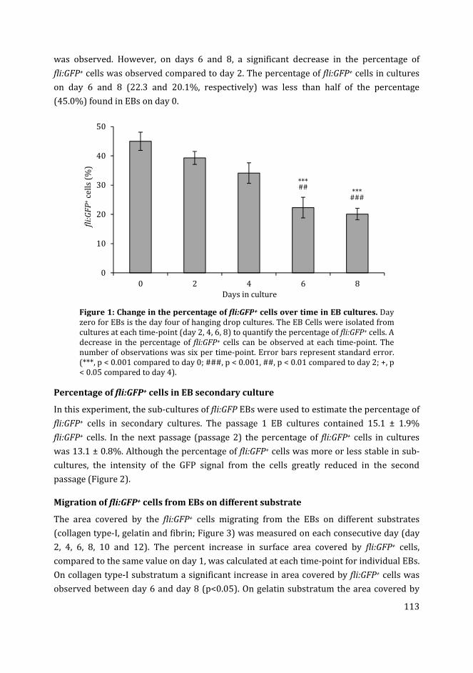
113
was observed. However, on days 6 and 8, a significant decrease in the percentage of
fli:GFP+ cells was observed compared to day 2. The percentage of fli:GFP+ cells in cultures
on day 6 and 8 (22.3 and 20.1%, respectively) was less than half of the percentage
(45.0%) found in EBs on day 0.
Figure 1: Change in the percentage of fli:GFP+ cells over time in EB cultures. Day
zero for EBs is the day four of hanging drop cultures. The EB Cells were isolated from
cultures at each time-point (day 2, 4, 6, 8) to quantify the percentage of fli:GFP+ cells. A
decrease in the percentage of fli:GFP+ cells can be observed at each time-point. The
number of observations was six per time-point. Error bars represent standard error.
(***, p < 0.001 compared to day 0; ###, p < 0.001, ##, p < 0.01 compared to day 2; +, p
< 0.05 compared to day 4).
Percentage of fli:GFP+ cells in EB secondary culture
In this experiment, the sub-cultures of fli:GFP EBs were used to estimate the percentage of
fli:GFP+ cells in secondary cultures. The passage 1 EB cultures contained 15.1 ± 1.9%
fli:GFP+ cells. In the next passage (passage 2) the percentage of fli:GFP+ cells in cultures
was 13.1 ± 0.8%. Although the percentage of fli:GFP+ cells was more or less stable in sub-
cultures, the intensity of the GFP signal from the cells greatly reduced in the second
passage (Figure 2).
Migration of fli:GFP+ cells from EBs on different substrate
The area covered by the fli:GFP+ cells migrating from the EBs on different substrates
(collagen type-I, gelatin and fibrin; Figure 3) was measured on each consecutive day (day
2, 4, 6, 8, 10 and 12). The percent increase in surface area covered by fli:GFP+ cells,
compared to the same value on day 1, was calculated at each time-point for individual EBs.
On collagen type-I substratum a significant increase in area covered by fli:GFP+ cells was
observed between day 6 and day 8 (p<0.05). On gelatin substratum the area covered by
0
10
20
30
40
50
0 2 4 6 8
fli:
GF
P+
cell
s (%
)
Days in culture
***###
***##
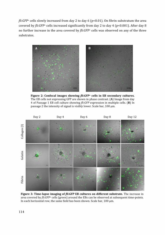
114
fli:GFP+ cells slowly increased from day 2 to day 6 (p<0.01). On fibrin substratum the area
covered by fli:GFP+ cells increased significantly from day 2 to day 4 (p<0.001). After day 8
no further increase in the area covered by fli:GFP+ cells was observed on any of the three
substrates.
Figure 2: Confocal images showing fli:GFP+ cells in EB secondary cultures.
The EB cells not expressing GFP are shown in phase contrast. (A) Image from day
4 of Passage 1 EB cell culture showing fli:GFP expression in multiple cells. (B) In
passage 2 the intensity of signal is visibly lower. Scale bar, 100 µm.
Day 2 Day 4 Day 6 Day 8 Day 12
Co
lla
ge
n (
I)
Ge
lati
n
Fib
rin
Figure 3: Time-lapse imaging of fli:GFP EB cultures on different substrate. The increase in
area covered by fli:GFP+ cells (green) around the EBs can be observed at subsequent time-points.
In each horizontal row, the same field has been shown. Scale bar, 100 µm.
A B
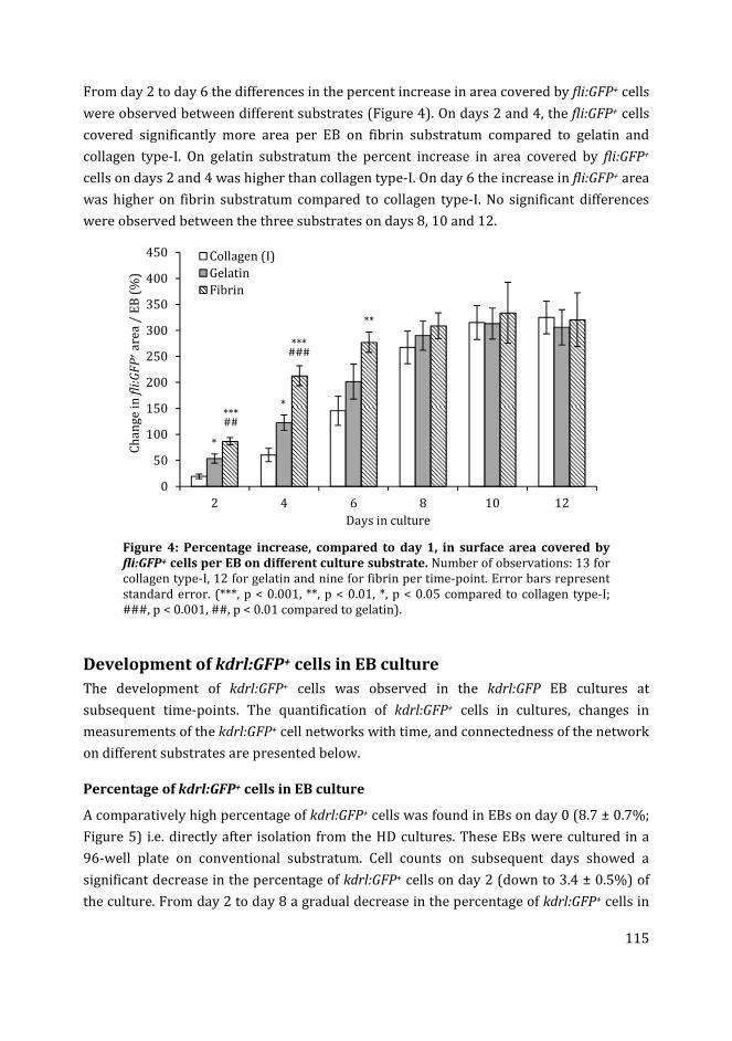
115
From day 2 to day 6 the differences in the percent increase in area covered by fli:GFP+ cells
were observed between different substrates (Figure 4). On days 2 and 4, the fli:GFP+ cells
covered significantly more area per EB on fibrin substratum compared to gelatin and
collagen type-I. On gelatin substratum the percent increase in area covered by fli:GFP+
cells on days 2 and 4 was higher than collagen type-I. On day 6 the increase in fli:GFP+ area
was higher on fibrin substratum compared to collagen type-I. No significant differences
were observed between the three substrates on days 8, 10 and 12.
Figure 4: Percentage increase, compared to day 1, in surface area covered by
fli:GFP+ cells per EB on different culture substrate. Number of observations: 13 for
collagen type-I, 12 for gelatin and nine for fibrin per time-point. Error bars represent
standard error. (***, p < 0.001, **, p < 0.01, *, p < 0.05 compared to collagen type-I;
###, p < 0.001, ##, p < 0.01 compared to gelatin).
Development of kdrl:GFP+ cells in EB culture
The development of kdrl:GFP+ cells was observed in the kdrl:GFP EB cultures at
subsequent time-points. The quantification of kdrl:GFP+ cells in cultures, changes in
measurements of the kdrl:GFP+ cell networks with time, and connectedness of the network
on different substrates are presented below.
Percentage of kdrl:GFP+ cells in EB culture
A comparatively high percentage of kdrl:GFP+ cells was found in EBs on day 0 (8.7 ± 0.7%;
Figure 5) i.e. directly after isolation from the HD cultures. These EBs were cultured in a
96-well plate on conventional substratum. Cell counts on subsequent days showed a
significant decrease in the percentage of kdrl:GFP+ cells on day 2 (down to 3.4 ± 0.5%) of
the culture. From day 2 to day 8 a gradual decrease in the percentage of kdrl:GFP+ cells in
0
50
100
150
200
250
300
350
400
450
2 4 6 8 10 12
Ch
an
ge
in
fli
:GF
P+
are
a /
EB
(%
)
Days in culture
Collagen (I)
Gelatin
Fibrin
*
***##
*
***###
**
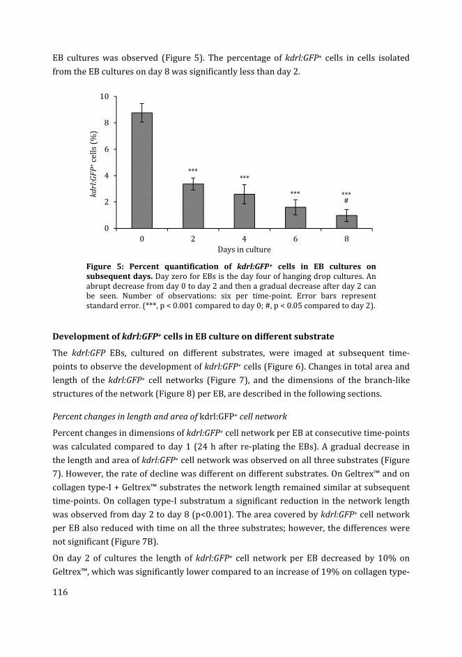
116
EB cultures was observed (Figure 5). The percentage of kdrl:GFP+ cells in cells isolated
from the EB cultures on day 8 was significantly less than day 2.
Figure 5: Percent quantification of kdrl:GFP+ cells in EB cultures on
subsequent days. Day zero for EBs is the day four of hanging drop cultures. An
abrupt decrease from day 0 to day 2 and then a gradual decrease after day 2 can
be seen. Number of observations: six per time-point. Error bars represent
standard error. (***, p < 0.001 compared to day 0; #, p < 0.05 compared to day 2).
Development of kdrl:GFP+ cells in EB culture on different substrate
The kdrl:GFP EBs, cultured on different substrates, were imaged at subsequent time-
points to observe the development of kdrl:GFP+ cells (Figure 6). Changes in total area and
length of the kdrl:GFP+ cell networks (Figure 7), and the dimensions of the branch-like
structures of the network (Figure 8) per EB, are described in the following sections.
Percent changes in length and area of kdrl:GFP+ cell network
Percent changes in dimensions of kdrl:GFP+ cell network per EB at consecutive time-points
was calculated compared to day 1 (24 h after re-plating the EBs). A gradual decrease in
the length and area of kdrl:GFP+ cell network was observed on all three substrates (Figure
7). However, the rate of decline was different on different substrates. On Geltrex™ and on
collagen type-I + Geltrex™ substrates the network length remained similar at subsequent
time-points. On collagen type-I substratum a significant reduction in the network length
was observed from day 2 to day 8 (p<0.001). The area covered by kdrl:GFP+ cell network
per EB also reduced with time on all the three substrates; however, the differences were
not significant (Figure 7B).
On day 2 of cultures the length of kdrl:GFP+ cell network per EB decreased by 10% on
Geltrex™, which was significantly lower compared to an increase of 19% on collagen type-
0
2
4
6
8
10
0 2 4 6 8
kd
rl:G
FP
+ce
lls
(%)
Days in culture
***
***#
***
***
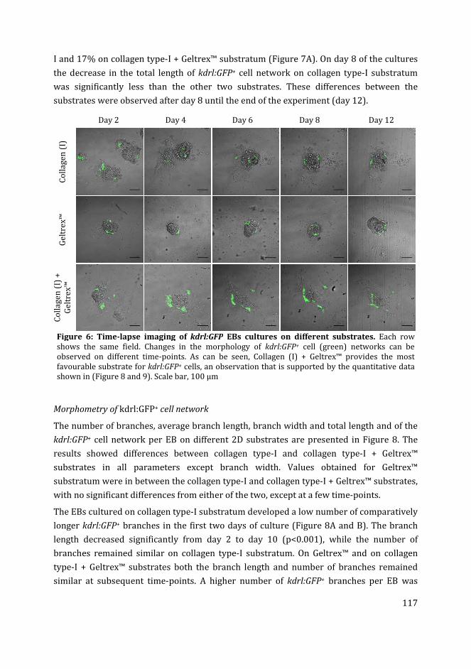
117
I and 17% on collagen type-I + Geltrex™ substratum (Figure 7A). On day 8 of the cultures
the decrease in the total length of kdrl:GFP+ cell network on collagen type-I substratum
was significantly less than the other two substrates. These differences between the
substrates were observed after day 8 until the end of the experiment (day 12).
Day 2 Day 4 Day 6 Day 8 Day 12
Co
lla
ge
n (
I)
Ge
ltre
x™
Co
lla
ge
n (
I) +
Ge
ltre
x™
Figure 6: Time-lapse imaging of kdrl:GFP EBs cultures on different substrates. Each row
shows the same field. Changes in the morphology of kdrl:GFP+ cell (green) networks can be
observed on different time-points. As can be seen, Collagen (I) + Geltrex™ provides the most
favourable substrate for kdrl:GFP+ cells, an observation that is supported by the quantitative data
shown in (Figure 8 and 9). Scale bar, 100 µm
Morphometry of kdrl:GFP+ cell network
The number of branches, average branch length, branch width and total length and of the
kdrl:GFP+ cell network per EB on different 2D substrates are presented in Figure 8. The
results showed differences between collagen type-I and collagen type-I + Geltrex™
substrates in all parameters except branch width. Values obtained for Geltrex™
substratum were in between the collagen type-I and collagen type-I + Geltrex™ substrates,
with no significant differences from either of the two, except at a few time-points.
The EBs cultured on collagen type-I substratum developed a low number of comparatively
longer kdrl:GFP+ branches in the first two days of culture (Figure 8A and B). The branch
length decreased significantly from day 2 to day 10 (p<0.001), while the number of
branches remained similar on collagen type-I substratum. On Geltrex™ and on collagen
type-I + Geltrex™ substrates both the branch length and number of branches remained
similar at subsequent time-points. A higher number of kdrl:GFP+ branches per EB was
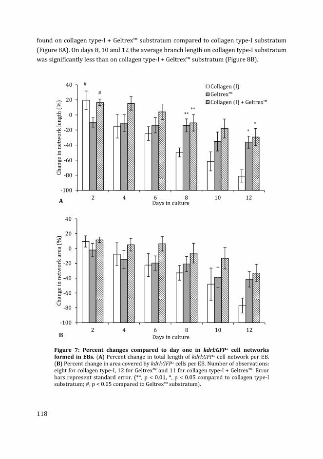
118
found on collagen type-I + Geltrex™ substratum compared to collagen type-I substratum
(Figure 8A). On days 8, 10 and 12 the average branch length on collagen type-I substratum
was significantly less than on collagen type-I + Geltrex™ substratum (Figure 8B).
Figure 7: Percent changes compared to day one in kdrl:GFP+ cell networks
formed in EBs. (A) Percent change in total length of kdrl:GFP+ cell network per EB.
(B) Percent change in area covered by kdrl:GFP+ cells per EB. Number of observations:
eight for collagen type-I, 12 for Geltrex™ and 11 for collagen type-I + Geltrex™. Error
bars represent standard error. (**, p < 0.01, *, p < 0.05 compared to collagen type-I
substratum; #, p < 0.05 compared to Geltrex™ substratum).
-100
-80
-60
-40
-20
0
20
40
2 4 6 8 10 12
Ch
an
ge
in
ne
two
rk l
en
gth
(%
)
Days in culture
Collagen (I)
Geltrex™
Collagen (I) + Geltrex™
#
#
****
*
*
A
-100
-80
-60
-40
-20
0
20
40
2 4 6 8 10 12
Ch
an
ge
in
ne
two
rk a
rea
(%
)
Days in cultureB
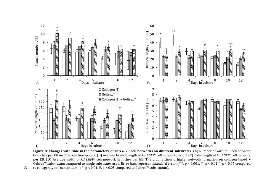
119
Figure 8: Changes with time in the parameters of kdrl:GFP+ cell networks on different substrates. (A) Number of kdrl:GFP+ cell network
branches per EB on different time-points. (B) Average branch length of kdrl:GFP+ cell network per EB. (C) Total length of kdrl:GFP+ cell network
per EB. (D) Average width of kdrl:GFP+ cell network branches per EB. The graphs show a higher network formation on collagen type-I +
Geltrex™ substratum compared to single substrates used. Error bars represent standard error. (***, p < 0.001, **, p < 0.01, *, p < 0.05 compared
to collagen type-I substratum; ##, p < 0.01, #, p < 0.05 compared to Geltrex™ substratum).
0
2
4
6
8
10
12
1 2 4 6 8 10 12
Bra
nch
nu
mb
er
/ E
B
Days in culture
*
*
*
*
A
0
10
20
30
40
50
60
1 2 4 6 8 10 12
Bra
nch
le
ng
th /
EB
(µ
m)
Days in cultureB
###
*#
*
*
***#
**
0
50
100
150
200
250
300
350
400
1 2 4 6 8 10 12
Ne
two
rk l
en
gth
/ E
B (
µm
)
Days in culture
Collagen (I)
Geltrex™
Collagen (I) + Geltrex™
C
#
#
***
0
1
2
3
4
5
6
7
8
9
1 2 4 6 8 10 12B
ran
ch w
idth
/ E
B (
µm
)Days in cultureD
11
9
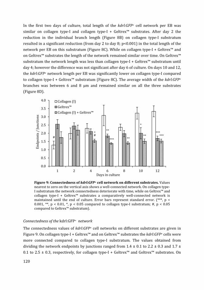
120
In the first two days of culture, total length of the kdrl:GFP+ cell network per EB was
similar on collagen type-I and collagen type-I + Geltrex™ substrates. After day 2 the
reduction in the individual branch length (Figure 8B) on collagen type-I substratum
resulted in a significant reduction (from day 2 to day 8; p<0.001) in the total length of the
network per EB on this substratum (Figure 8C). While on collagen type-I + Geltrex™ and
on Geltrex™ substrates the length of the network remained similar over time. On Geltrex™
substratum the network length was less than collagen type-I + Geltrex™ substratum until
day 4; however the difference was not significant after day 6 of culture. On days 10 and 12,
the kdrl:GFP+ network length per EB was significantly lower on collagen type-I compared
to collagen type-I + Geltrex™ substratum (Figure 8C). The average width of the kdrl:GFP+
branches was between 6 and 8 µm and remained similar on all the three substrates
(Figure 8D).
Figure 9: Connectedness of kdrl:GFP+ cell network on different substrates. Values
nearest to zero on the vertical axis shows a well-connected network. On collagen type-
I substratum the network connectedness deteriorate with time, while on Geltrex™ and
collagen type-I + Geltrex™ substrates a comparatively well-connected network is
maintained until the end of culture. Error bars represent standard error. (***, p <
0.001, **, p < 0.01, *, p < 0.05 compared to collagen type-I substratum; #, p < 0.05
compared to Geltrex™ substratum).
Connectedness of the kdrl:GFP+ network
The connectedness values of kdrl:GFP+ cell networks on different substrates are given in
Figure 9. On collagen type-I + Geltrex™ and on Geltrex™ substrates the kdrl:GFP+ cells were
more connected compared to collagen type-I substratum. The values obtained from
dividing the network endpoints by junctions ranged from 1.4 ± 0.1 to 2.2 ± 0.3 and 1.7 ±
0.1 to 2.5 ± 0.3, respectively, for collagen type-I + Geltrex™ and Geltrex™ substrates. On
0.0
0.5
1.0
1.5
2.0
2.5
3.0
3.5
4.0
1 2 4 6 8 10 12
En
dp
oin
ts /
Ju
nct
ion
s
Days in culture
Collagen (I)
Geltrex™
Collagen (I) + Geltrex™
#
*#
**
*** **
*
*****
***
121
these substrates the connectedness values remained constant over the 12 days of culture.
On collagen type-I substratum the kdrl:GFP+ cell network per EB was well connected on
day 1 of culture with lower endpoints divided by junctions value. However, the
connectedness of the network on this substratum was lost with the duration of culture.
Discussion
EB culture in hanging drops
In this section the results from EBs cultures, in the current study, will be discussed in
comparison with the blastocyst cell cultures in our previous experiments (Chapter 4). In
studies on mouse ESCs, EBs grown in suspension culture have shown a higher percentage
of hematopoietic and endothelial cells compared to EBs grown in adherent culture [58].
Our results from zebrafish cells are consistent with these findings. In our previous
experiments (Chapter 4) zebrafish blastocyst cells in adherent culture showed fewer
fli:GFP+ and kdrl:GFP+ cells compared to the EBs developed in HD suspension culture in
this chapter.
In HD culture the dispersed cells collect by gravity in the bottom center of the drop,
forming EB aggregates, where there is presumably more cell-to-cell contact than in the
adherent culture, which is a monolayer. We suggest that the increased cellular contact in
HD cultures may be favorable for the growth and differentiation of fli:GFP+ and kdrl:GFP+
cells. previous studies have shown that in suspension cultures the proliferation of EB cells
is limited compared to the adherent cultures, allowing greater control of the
differentiation of specific cell types [6]. This would explain why the percentage of
transgenic cells dropped significantly in our EB cultures after transferring to adherent
culture. The medium composition used here for the EB culture was selected based on the
optimization experiments described in chapter 4. In those experiments, this medium gave
rise to a higher percentage of fli:GFP+ and kdrl:GFP+ cells in zebrafish blastocyst cell
culture.
Development of fli:GFP+ and kdrl:GFP+ cells in EB culture
The percentage of fli:GFP+ cells in EB culture was always higher than kdrl:GFP+ cells. This
is presumably because the kdrl:GFP transgene is expressed only in endothelial cells [51],
whereas the fli:GFP is expressed in endothelial, lymphatic, hematopoietic and neural crest
cells [50]. In contrast to the fibroblast-like morphology of fli:GFP+ cells, the endothelial-
like properties of kdrl:GFP+ cells is supported by the fact that they form network-like
structures in the EB culture developed here. This difference in morphology of fli:GFP+ and
kdrl:GFP+ cells also suggests that the kdrl:GFP is specific for endothelial cells.
122
In our previous experiments (Chapter 4) the kdrl:GFP+ cells were no longer found after
eight days of primary blastocyst cell culture. The numbers of fli:GFP+ cells, on the other
hand, were comparatively stable in the primary culture. However, these cells could also
not be maintained in secondary culture (Chapter 4). The possible explanation for these
findings could include apoptosis in kdrl:GFP+ cells [59], or down regulation of kdrl:GFP
[60] and fli:GFP [50] transgenes as previously suggested. In contrast to primary blastocyst
cultures (Chapter 4), in the EB cultures described here, the kdrl:GFP+ cell networks could
be observed up to 12 days in culture. Similarly, the fli:GFP+ cells could be maintained in
secondary culture for up to three passages. Thus, under appropriate conditions the
fli:GFP+ and kdrl:GFP+ cells can be maintained for a longer time in vitro.
Effect of substratum on fli:GFP+ and kdrl:GFP+ cells in EB culture
In our previous experiments, the blastocyst cells cultured on collagen type-I substratum
contained a higher percentage of fli:GFP+ and kdrl:GFP+ cells compared to those cultured
on gelatin substratum (Chapter 4). In contrast, the increase in area covered by the
outgrowing fli:GFP+ cells from EBs in the current study was lower on collagen type-I,
compared to gelatin and fibrin substrates. A reason for this may be the softness of collagen
type-I gel, where the cells are in a more elongated shape and cover less area, compared to
the more flattened cells on a gelatin and fibrin substrates. The area covered by fli:GFP+
cells per EB was significantly higher on a fibrin substratum compared to gelatin and
collagen type-I, in our study. These results are in comparison with the previous studies
where fibrin substrata have been shown to enhance the formation of blood vessel-like
structures from human dermal microvascular endothelial cells [61].
Similarly in our other experiments, the kdrl:GFP+ cells formed network-like structures in
3D gel matrix containing fibrin (chapter 6), compared to the 2D substrates tested here. In
those experiments (Chapter 6), the network length of kdrl:GFP+ cells was higher compared
to the network length on Geltrex™ and collagen type-I substrata described here. These
results are in agreement with a previous study where a 3D fibrin scaffold enhanced
network formation from human umbilical vein endothelial cells (HUVECs) compared to
collagen type-I scaffolds [62]. Fibrin 3D matrices have also been demonstrated to increase
angiogenesis during wound healing in pigs [63]. This suggest that fibrin could be an
important part of the culture substrate for angiogenesis assays.
The selection of an appropriate substratum is critical for a particular assay, as different
cell types may have different requirements for extra cellular matrix [64]. Of different
substrate compositions used for the EB culture in the experiments in this chapter, longer
and more number of kdrl:GFP+ strands were observed on collagen type-I + Geltrex™
substratum, compared to pure substrates. The formation of kdrl:GFP+ cell network was
principally observed in the matrix composed of a combination of collagen type-I, Geltrex™
and fibrin, described in chapter 6. This may suggest the requirement of a complex
123
extracellular matrix substrate for the development of vascular networks in zebrafish EB
culture. Similar to our findings, previous studies on HUVECs also recommend the use of
composite 3D matrix containing collagen and fibrin, because it offers advantages over a
pure matrix in the culture of vascular networks [65].
Fibronectin is a major constituent of zebrafish extracellular matrix (ECM) in early
embryonic stages [66]. Furthermore, the cells in early zebrafish embryo start expressing
membrane proteins of the integrin family, which bind fibronectin [67]. Studies in other
animals and humans have shown that fibronectin cross-link to fibrinogen and form a clot
which serve as a substrate for wound healing [68]. This may explain why an increased
migration of fli:GFP+ cells was observed on fibrin substratum in this study. Furthermore,
the addition of fibrinogen to the 3D matrix favoured the development of kdrl:GFP+ cell
networks in our other experiments (chapter 6). Another possible cause of enhanced
network formation or cell migration in fibrin containing matrices may be the vulnerability
of fibrin to the proteases released from the EB cells. In previous studies, testing of pro-
angiogenic compounds on zebrafish embryos has shown increased expression of matrix
metalloproteinases MMP-2 and MMP-9 [69]. These metalloproteinases are suggested to be
involved in the degradation of fibrin matrix [70].
Conclusions
Suspension culture of zebrafish blastocyst cells allows better control of cellular
differentiation, compared to the adherent cultures. Using the EB intermediate (developed
in suspension culture), the development of fli:GFP+ and kdrl:GFP+ cells could be increased
and maintained for longer duration in culture. Different substrate composition effect the
spreading and development of fli:GFP+ and kdrl:GFP+ cells in cultures. We recommend the
use of a complex substratum for the growth of these cells in culture. Fibrin substratum has
an effect on growth of fli:GFP+ cells in cultures, as well as on the development of network-
like structures from kdrl:GFP+ cells. The fli:GFP+ and kdrl:GFP+ cells showed differences in
their number and morphology, suggesting that these are diverse cell types.
References
[1] Bethesda MD. What are embryonic stem cells? In Stem Cell Information, 2015;
Available from: http://stemcells.nih.gov/info/basics/pages/basics3.aspx.
[2] Descamps B, Emanueli C. Vascular differentiation from embryonic stem cells: novel
technologies and therapeutic promises. Vascul Pharmacol 2012; 56(5-6): 267-79.
[3] Rumpold H, Wolf D, Koeck R, Gunsilius E. Endothelial progenitor cells: a source for
therapeutic vasculogenesis? J Cell Mol Med 2004; 8(4): 509-18.
[4] Nishikawa S, Jakt LM, Era T. Embryonic stem-cell culture as a tool for
developmental cell biology. Nat Rev Mol Cell Biol 2007; 8(6): 502-7.
124
[5] Bratt-Leal AM, Carpenedo RL, McDevitt TC. Engineering the embryoid body
microenvironment to direct embryonic stem cell differentiation. Biotechnol Prog
2009; 25(1): 43-51.
[6] Kurosawa H. Methods for inducing embryoid body formation: in vitro
differentiation system of embryonic stem cells. J Biosci Bioeng 2007; 103(5): 389-
98.
[7] Desbaillets I, Ziegler U, Groscurth P, Gassmann M. Embryoid bodies: an in vitro
model of mouse embryogenesis. Exp Physiol 2000; 85(6): 645-51.
[8] Itskovitz-Eldor J, Schuldiner M, Karsenti D, Eden A, Yanuka O, Amit M, et al.
Differentiation of human embryonic stem cells into embryoid bodies compromising
the three embryonic germ layers. Mol Med 2000; 6(2): 88-95.
[9] Glaser DE, Burns AB, Hatano R, Medrzycki M, Fan Y, McCloskey KE. Specialized
mouse embryonic stem cells for studying vascular development. Stem Cells Cloning
2014; 7: 79-88.
[10] Goodwin AM. In vitro assays of angiogenesis for assessment of angiogenic and anti-
angiogenic agents. Microvasc Res 2007; 74(2-3): 172-83.
[11] Hermant B, Desroches-Castan A, Dubessay ML, Prandini MH, Huber P, Vittet D.
Development of a one-step embryonic stem cell-based assay for the screening of
sprouting angiogenesis. BMC Biotechnol 2007; 7: 20.
[12] Matsubara K, Mori M, Akagi R, Kato N. Anti-angiogenic effect of pyridoxal 5'-
phosphate, pyridoxal and pyridoxamine on embryoid bodies derived from mouse
embryonic stem cells. Int J Mol Med 2004; 14(5): 819-23.
[13] Ogawa S, Tagawa Y, Kamiyoshi A, Suzuki A, Nakayama J, Hashikura Y, et al. Crucial
roles of mesodermal cell lineages in a murine embryonic stem cell-derived in vitro
liver organogenesis system. Stem Cells 2005; 23(7): 903-13.
[14] Noghero A, Bussolino F, Gualandris A. Role of the microenvironment in the
specification of endothelial progenitors derived from embryonic stem cells.
Microvasc Res 2010; 79(3): 178-83.
[15] Levenberg S, Golub JS, Amit M, Itskovitz-Eldor J, Langer R. Endothelial cells derived
from human embryonic stem cells. Proc Natl Acad Sci U S A 2002; 99(7): 4391-6.
[16] Boyd NL, Dhara SK, Rekaya R, Godbey EA, Hasneen K, Rao RR, et al. BMP4 promotes
formation of primitive vascular networks in human embryonic stem cell-derived
embryoid bodies. Exp Biol Med (Maywood) 2007; 232(6): 833-43.
[17] Feraud O, Cao Y, Vittet D. Embryonic stem cell-derived embryoid bodies
development in collagen gels recapitulates sprouting angiogenesis. Lab Invest
2001; 81(12): 1669-81.
[18] Liu Z, Lebrin F, Maring JA, van den Driesche S, van der Brink S, van Dinther M, et al.
ENDOGLIN is dispensable for vasculogenesis, but required for vascular endothelial
growth factor-induced angiogenesis. PLoS One 2014; 9(1): e86273.
[19] Schmidt A, Brixius K, Bloch W. Endothelial precursor cell migration during
vasculogenesis. Circ Res 2007; 101(2): 125-36.
[20] Risau W. Mechanisms of angiogenesis. Nature 1997; 386(6626): 671-4.
[21] Yancopoulos GD, Davis S, Gale NW, Rudge JS, Wiegand SJ, Holash J. Vascular-specific
growth factors and blood vessel formation. Nature 2000; 407(6801): 242-8.
[22] Wang N, Zhang R, Wang SJ, Zhang CL, Mao LB, Zhuang CY, et al. Vascular endothelial
growth factor stimulates endothelial differentiation from mesenchymal stem cells
via Rho/myocardin-related transcription factor--a signaling pathway. Int J Biochem
Cell Biol 2013; 45(7): 1447-56.
125
[23] Shin Y, Jeon JS, Han S, Jung GS, Shin S, Lee SH, et al. In vitro 3D collective sprouting
angiogenesis under orchestrated ANG-1 and VEGF gradients. Lab Chip 2011;
11(13): 2175-81.
[24] Li J, Stuhlmann H. In vitro imaging of angiogenesis using embryonic stem cell-
derived endothelial cells. Stem Cells Dev 2012; 21(2): 331-42.
[25] Nolan DJ, Ginsberg M, Israely E, Palikuqi B, Poulos MG, James D, et al. Molecular
signatures of tissue-specific microvascular endothelial cell heterogeneity in organ
maintenance and regeneration. Dev Cell 2013; 26(2): 204-19.
[26] Wang ZZ, Au P, Chen T, Shao Y, Daheron LM, Bai H, et al. Endothelial cells derived
from human embryonic stem cells form durable blood vessels in vivo. Nat
Biotechnol 2007; 25(3): 317-8.
[27] Eble JA, Niland S. The extracellular matrix of blood vessels. Curr Pharm Des 2009;
15(12): 1385-400.
[28] Lutolf MP, Hubbell JA. Synthetic biomaterials as instructive extracellular
microenvironments for morphogenesis in tissue engineering. Nat Biotechnol 2005;
23(1): 47-55.
[29] Blancas AA, Lauer NE, McCloskey KE. Endothelial differentiation of embryonic stem
cells. Curr Protoc Stem Cell Biol 2008; Chapter 1: Unit 1F 5.
[30] Kane NM, Meloni M, Spencer HL, Craig MA, Strehl R, Milligan G, et al. Derivation of
endothelial cells from human embryonic stem cells by directed differentiation:
analysis of microRNA and angiogenesis in vitro and in vivo. Arterioscler Thromb
Vasc Biol 2010; 30(7): 1389-97.
[31] Wang H, Luo X, Leighton J. Extracellular matrix and integrins in embryonic stem cell
differentiation. Biochem Insights 2015; 8(Suppl 2): 15-21.
[32] Vailhe B, Vittet D, Feige JJ. In vitro models of vasculogenesis and angiogenesis. Lab
Invest 2001; 81(4): 439-52.
[33] Staton CA, Reed MW, Brown NJ. A critical analysis of current in vitro and in vivo
angiogenesis assays. Int J Exp Pathol 2009; 90(3): 195-221.
[34] Arnaoutova I, Kleinman HK. In vitro angiogenesis: endothelial cell tube formation
on gelled basement membrane extract. Nat Protoc 2010; 5(4): 628-35.
[35] Kim S, Lee H, Chung M, Jeon NL. Engineering of functional, perfusable 3D
microvascular networks on a chip. Lab Chip 2013; 13(8): 1489-500.
[36] Nguyen DH, Stapleton SC, Yang MT, Cha SS, Choi CK, Galie PA, et al. Biomimetic
model to reconstitute angiogenic sprouting morphogenesis in vitro. Proc Natl Acad
Sci U S A 2013; 110(17): 6712-7.
[37] Li H, Chang J. Bioactive silicate materials stimulate angiogenesis in fibroblast and
endothelial cell co-culture system through paracrine effect. Acta Biomaterialia
2013; 9(6): 6981-6991.
[38] Zheng Y, Chen J, Craven M, Choi NW, Totorica S, Diaz-Santana A, et al. In vitro
microvessels for the study of angiogenesis and thrombosis. Proc Natl Acad Sci U S A
2012; 109(24): 9342-7.
[39] Nakatsu MN, Sainson RC, Aoto JN, Taylor KL, Aitkenhead M, Perez-del-Pulgar S, et
al. Angiogenic sprouting and capillary lumen formation modeled by human
umbilical vein endothelial cells (HUVEC) in fibrin gels: the role of fibroblasts and
Angiopoietin-1. Microvasc Res 2003; 66(2): 102-12.
[40] Stamati K, Priestley JV, Mudera V, Cheema U. Laminin promotes vascular network
formation in 3D in vitro collagen scaffolds by regulating VEGF uptake. Exp Cell Res
2014; 327(1): 68-77.
126
[41] Ashley RA, Dubuque SH, Dvorak B, Woodward SS, Williams SK, Kling PJ.
Erythropoietin stimulates vasculogenesis in neonatal rat mesenteric microvascular
endothelial cells. Pediatr Res 2002; 51(4): 472-8.
[42] Rezzola S, Belleri M, Gariano G, Ribatti D, Costagliola C, Semeraro F, et al. In vitro
and ex vivo retina angiogenesis assays. Angiogenesis 2014; 17(3): 429-42.
[43] Delgado VM, Nugnes LG, Colombo LL, Troncoso MF, Fernandez MM, Malchiodi EL, et
al. Modulation of endothelial cell migration and angiogenesis: a novel function for
the "tandem-repeat" lectin galectin-8. FASEB J 2011; 25(1): 242-54.
[44] Jakobsson L, Kreuger J, Claesson-Welsh L. Building blood vessels--stem cell models
in vascular biology. J Cell Biol 2007; 177(5): 751-5.
[45] Festing S, Wilkinson R. The ethics of animal research. Talking Point on the use of
animals in scientific research. EMBO Rep 2007; 8(6): 526-30.
[46] Alvarez MC, Bejar J, Chen S, Hong Y. Fish ES cells and applications to biotechnology.
Mar Biotechnol (NY) 2007; 9(2): 117-27.
[47] Fan L, Collodi P. Zebrafish embryonic stem cells. Methods Enzymol 2006; 418: 64-
77.
[48] Howe K, Clark MD, Torroja CF, Torrance J, Berthelot C, Muffato M, et al. The
zebrafish reference genome sequence and its relationship to the human genome.
Nature 2013; 496(7446): 498-503.
[49] Chavez MN, Aedo G, Fierro FA, Allende ML, Egana JT. Zebrafish as an emerging
model organism to study angiogenesis in development and regeneration. Front
Physiol 2016; 7: 56.
[50] Lawson ND, Weinstein BM. In vivo imaging of embryonic vascular development
using transgenic zebrafish. Dev Biol 2002; 248(2): 307-318.
[51] Jin SW, Beis D, Mitchell T, Chen JN, Stainier DY. Cellular and molecular analyses of
vascular tube and lumen formation in zebrafish. Development 2005; 132(23):
5199-209.
[52] Experiments on Animal Act, 2014. The State Secretary of Health and Environmental
Protection, Soestdijk. http://wetten.overheid.nl/BWBR0003081/2014-12-18
[53] Directive 2010/63/EU of the European parliament and of the council of 22
September 2010 on the protection of animals used for scientific purposes Text with
EEA relevance, 2010. The European Parliament and the council of the European
Union, Official Journal of the European Union. http://eur-lex.europa.eu/legal-
content/EN/TXT/?uri=celex:32010L0063
[54] Westerfield M. The zebrafish book: a guide for the laboratory use of zebrafish
(Danio Rerio). 4 ed, University of Oregon Press, Eugene 2000, pp.
[55] Kimmel CB, Ballard WW, Kimmel SR, Ullmann B, Schilling TF. Stages of embryonic
development of the zebrafish. Dev Dyn 1995; 203(3): 253-310.
[56] Schneider CA, Rasband WS, Eliceiri KW. NIH Image to ImageJ: 25 years of image
analysis. Nat Methods 2012; 9(7): 671-5.
[57] Moya ML, Hsu YH, Lee AP, Hughes CC, George SC. In vitro perfused human capillary
networks. Tissue Eng Part C Methods 2013; 19(9): 730-7.
[58] Dang SM, Kyba M, Perlingeiro R, Daley GQ, Zandstra PW. Efficiency of embryoid
body formation and hematopoietic development from embryonic stem cells in
different culture systems. Biotechnol Bioeng 2002; 78(4): 442-53.
[59] Zhao H, Huang H, Lin S. Chemical approaches to angiogenesis in development and
regeneration. Methods Cell Biol 2016; 134: 369-76.
127
[60] Kabrun N, Buhring HJ, Choi K, Ullrich A, Risau W, Keller G. Flk-1 expression defines
a population of early embryonic hematopoietic precursors. Development 1997;
124(10): 2039-48.
[61] Feng X, Tonnesen MG, Mousa SA, Clark RA. Fibrin and collagen differentially but
synergistically regulate sprout angiogenesis of human dermal microvascular
endothelial cells in 3-dimensional matrix. Int J Cell Biol 2013; 2013: 231279.
[62] Park YK, Tu TY, Lim SH, Clement IJ, Yang SY, Kamm RD. In Vitro Microvessel Growth
and Remodeling within a Three-dimensional Microfluidic Environment. Cell Mol
Bioeng 2014; 7(1): 15-25.
[63] Roy S, Driggs J, Elgharably H, Biswas S, Findley M, Khanna S, et al. Platelet-rich
fibrin matrix improves wound angiogenesis via inducing endothelial cell
proliferation. Wound Repair Regen 2011; 19(6): 753-66.
[64] Haycock JW. 3D cell culture: a review of current approaches and techniques.
Methods Mol Biol 2011; 695: 1-15.
[65] Rao RR, Peterson AW, Ceccarelli J, Putnam AJ, Stegemann JP. Matrix composition
regulates three-dimensional network formation by endothelial cells and
mesenchymal stem cells in collagen/fibrin materials. Angiogenesis 2012; 15(2):
253-64.
[66] Latimer A, Jessen JR. Extracellular matrix assembly and organization during
zebrafish gastrulation. Matrix Biol 2010; 29(2): 89-96.
[67] Jessen JR. Recent advances in the study of zebrafish extracellular matrix proteins.
Dev Biol 2015; 401(1): 110-21.
[68] Bergmeier W, Hynes RO. Extracellular matrix proteins in hemostasis and
thrombosis. Cold Spring Harb Perspect Biol 2012; 4(2).
[69] Wu F, Song H, Zhang Y, Zhang Y, Mu Q, Jiang M, et al. Irisin induces angiogenesis in
human umbilical vein endothelial cells in Vitro and in zebrafish embryos in vivo via
activation of the ERK signaling pathway. PLoS One 2015; 10(8): e0134662.
[70] Wang MF, Lai SC. Fibronectin degradation by MMP-2/MMP-9 in the serum of
pregnant women and umbilical cord with Toxoplasma gondii infection. J Obstet
Gynaecol 2013; 33(4): 370-4.

128

129
Chapter 6
In vitro development of zebrafish
vascular networks
Muhammad Ibrahim and Michael K. Richardson
This chapter has been published as following:
Ibrahim M, Richardson MK. In vitro development of zebrafish vascular networks.
Reproductive Toxicology (2017). DOI: 10.1016/j.reprotox.2017.02.008.
130
Abstract
A major limitation to culturing tissues and organs is the lack of a functional vascular
network in vitro. The zebrafish possess many useful properties which makes it a
promising model for such studies. Unfortunately, methods of culturing endothelial cells
from this species are not well characterised. Here, we tried two methods ( embryoid body
culture and organ explants from transgenic zebrafish kdrl:GFP embryos) to develop in
vitro vascular networks. In the kdrl:GFP line, endothelial cells expresses green fluorescent
protein, which allows to track the vascular development in live cultures. We found that
embryoid bodies showed significantly longer and wider branches of connected
endothelial cells when grown in a microfluidic system than in static culture. Similarly,
sprouting of kdrl:GFP+ cells from the tissue explants was observed in a 3D hydrogel matrix.
This study is a step towards the development of zebrafish vascular networks in vitro.
Key words: Angiogenesis; Embryoid bodies; Explant culture; Microfluidics;
Vasculogenesis; Zebrafish.
131
Introduction
There are a number of reasons why it could be useful to develop culture systems
containing functional vascular networks. For example, tissue engineering is a very
important area of biomedical research that may have applications in regenerative
medicine and organ transplantation [1]. The in vitro culture of complex tissues might also
help our understanding of physiological aspects of organ function [2]; disease conditions
such as cardiac disorders [3]; and drug screening [4]. In vivo the vascular system is
essential for the growth and development of functional tissues and organs [5]. A major
obstacle to engineering an organ in vitro with the current tissue culture procedures is the
lack of a vascular network [6]. Development of three-dimensional (3D) culture systems
with a functional capillary bed could overcome this problem [7, 8].
Development of an in vitro vascular network could also have other applications e.g.
vascular regenerative therapy [9] and modelling diseases such as retinal microvascular
abnormalities in diabetes [10] and abnormal angiogenesis in tumor development [11].
Vascular culture techniques are important in cancer research for the screening of
compounds that inhibit angiogenesis [12]. Furthermore, in vitro vascular networks could
also serve as a screening model for candidate drugs, as some of the drugs approved for
clinical trials may disturb vascular development. An example of such a drug is
thalidomide, whose teratogenicity is linked to anti-angiogenic effects [13].
Protocols for culturing vascular networks have been successfully developed using
endothelial cell lines and embryonic tissues [14]. Commonly, human umbilical vein
endothelial cells (HUVEC) are used in pure culture or in co-culture with other cells (Table
1). These cultures are established on biological matrices that mimic some of the
properties of endogenous extracellular matrix [15]. Blood vessel sprouting has been
shown to take place from beads coated with HUVECs, and cultured on a fibrin gel, in media
supplemented with vascular endothelial growth factor (VEGF), fibroblast growth factor
(FGF), angiopoietin-1 (Ang-1) and transforming growth factor beta (TGF-β) [16].
Endothelial cells from other species have also been adapted for in vitro vasculogenesis;
examples include bovine aortic endothelial cells [17] and rat aortic endothelial cells [18].
Another method for culturing vascular networks is the embryoid body culture. Embryoid
bodies (EBs) are three-dimensional (3D) aggregates of embryonic stem cells isolated from
blastocyst stage embryos [19]. In an embryoid body culture the endothelial cells
differentiate and form blood vessels in a complex environment, which reflects vascular
formation in early embryos [20]. Unlike pure endothelial cell cultures, multiple cell types
are involved in vessel formation in EB culture [21]. Vasculogenesis starts at day 3 of
hanging drop cultures in mouse EBs [22]. These EBs show sprouting of blood vessel-like
structures, into the surrounding matrix, when transferred to two-dimensional (2D) or 3D
collagen gel [22].
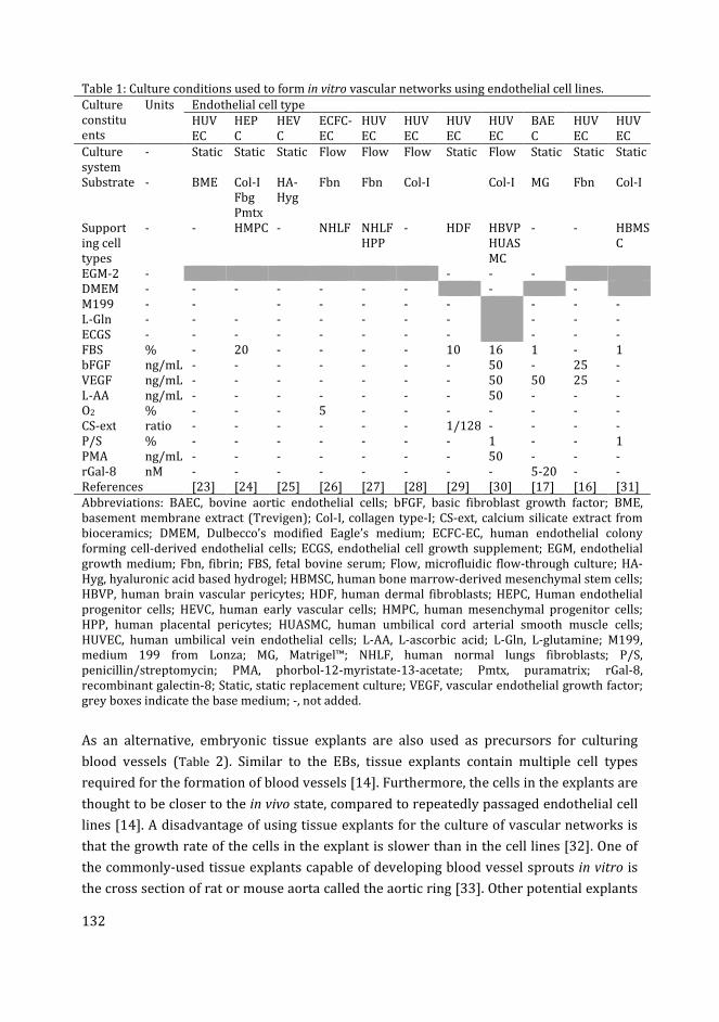
132
Table 1: Culture conditions used to form in vitro vascular networks using endothelial cell lines.
Culture
constitu
ents
Units Endothelial cell type
HUV
EC
HEP
C
HEV
C
ECFC-
EC
HUV
EC
HUV
EC
HUV
EC
HUV
EC
BAE
C
HUV
EC
HUV
EC
Culture
system
- Static Static Static Flow Flow Flow Static Flow Static Static Static
Substrate - BME Col-I
Fbg
Pmtx
HA-
Hyg
Fbn Fbn Col-I Col-I MG Fbn Col-I
Support
ing cell
types
- - HMPC - NHLF NHLF
HPP
- HDF HBVP
HUAS
MC
- - HBMS
C
EGM-2 - - - -
DMEM - - - - - - - - -
M199 - - - - - - - - - -
L-Gln - - - - - - - - - - -
ECGS - - - - - - - - - - -
FBS % - 20 - - - - 10 16 1 - 1
bFGF ng/mL - - - - - - - 50 - 25 -
VEGF ng/mL - - - - - - - 50 50 25 -
L-AA ng/mL - - - - - - - 50 - - -
O2 % - - - 5 - - - - - - -
CS-ext ratio - - - - - - 1/128 - - - -
P/S % - - - - - - - 1 - - 1
PMA ng/mL - - - - - - - 50 - - -
rGal-8 nM - - - - - - - - 5-20 - -
References [23] [24] [25] [26] [27] [28] [29] [30] [17] [16] [31]
Abbreviations: BAEC, bovine aortic endothelial cells; bFGF, basic fibroblast growth factor; BME,
basement membrane extract (Trevigen); Col-I, collagen type-I; CS-ext, calcium silicate extract from
bioceramics; DMEM, Dulbecco’s modified Eagle’s medium; ECFC-EC, human endothelial colony
forming cell-derived endothelial cells; ECGS, endothelial cell growth supplement; EGM, endothelial
growth medium; Fbn, fibrin; FBS, fetal bovine serum; Flow, microfluidic flow-through culture; HA-
Hyg, hyaluronic acid based hydrogel; HBMSC, human bone marrow-derived mesenchymal stem cells;
HBVP, human brain vascular pericytes; HDF, human dermal fibroblasts; HEPC, Human endothelial
progenitor cells; HEVC, human early vascular cells; HMPC, human mesenchymal progenitor cells;
HPP, human placental pericytes; HUASMC, human umbilical cord arterial smooth muscle cells;
HUVEC, human umbilical vein endothelial cells; L-AA, L-ascorbic acid; L-Gln, L-glutamine; M199,
medium 199 from Lonza; MG, Matrigel™; NHLF, human normal lungs fibroblasts; P/S,
penicillin/streptomycin; PMA, phorbol-12-myristate-13-acetate; Pmtx, puramatrix; rGal-8,
recombinant galectin-8; Static, static replacement culture; VEGF, vascular endothelial growth factor;
grey boxes indicate the base medium; -, not added.
As an alternative, embryonic tissue explants are also used as precursors for culturing
blood vessels (Table 2). Similar to the EBs, tissue explants contain multiple cell types
required for the formation of blood vessels [14]. Furthermore, the cells in the explants are
thought to be closer to the in vivo state, compared to repeatedly passaged endothelial cell
lines [14]. A disadvantage of using tissue explants for the culture of vascular networks is
that the growth rate of the cells in the explant is slower than in the cell lines [32]. One of
the commonly-used tissue explants capable of developing blood vessel sprouts in vitro is
the cross section of rat or mouse aorta called the aortic ring [33]. Other potential explants
133
include fragments of embryonic mouse metatarsal bones [34], mouse retina [35] and rat
kidney [36] tissues.
Haemodynamic, or the mechanical forces produced by blood flow, influence the
expression of several biochemical pathways in the endothelial cells; these in turn can
modulate the structure and function of blood vessels [37]. In addition to the different
techniques discussed above for culturing vascular networks, the role of haemodynamic
factors has been studied using microfluidic or lab-on-a-chip technology [38]. By
combining 3D culture in a hydrogel (which mimics the natural ECM) with microfluidics
(which mimics the blood flow), an in vitro environment can be created which could be in
principle, close to the in vivo environment for vascular morphogenesis [8, 39]. Advances in
microfluidics and 3D culture technologies have greatly increased the possibilities for
developing functional vascular models and vascularized tissues [39]. However, the
challenges in selecting an appropriate microfluidic system and 3D matrix for culturing
blood vessels, that can vascularize complex tissues in vitro, still need to be resolved [39].
Most of the current procedures for culturing blood vessels discussed above, involve cells
or tissues from mouse, humans or other species. These techniques are associated with
certain limitations. Human endothelial cell lines are not thought to closely represent the in
vivo state of the endothelial cells [14]. Furthermore, these cell lines change their gene
expression and physiological properties with repeated passaging in vitro, and may lose
their ability to form vascular networks [14]. Mouse embryonic tissues are difficult to
isolate because of the internal fertilisation and in utero development of the embryo.
Techniques for the isolation of embryonic stem cells and organ explants from mammals
are more costly, require invasive surgical procedures and can raise ethical concerns [9,
40]. Furthermore, mouse aortic explant cultures have shown significant variability
between the experiments [41].
For these reasons, it is desirable to explore the possibilities offered by alternative models.
The zebrafish is one such emerging model species [42]. In contrast to the mouse, the
external fertilization in zebrafish allows easy access to a large number of embryos, as well
as cells or tissues isolated from these embryos, for in vitro studies [43, 44]. Zebrafish early
embryonic cells or adult stem cells have been used for ex vivo experiments; fewer have
used cells from larvae [44, 45]. Zebrafish whole embryos and isolated cells are currently
being developed as potential alternative screening models for toxicity analysis [46, 47].
Many of the organ primordia of zebrafish are formed during the first 72 h of embryo
development [48]. There are practical advantages of zebrafish for cell culture e.g. they can
be maintained in a simple incubator without additional CO2 supply [49]. Zebrafish
embryos are optically transparent until early larval stages [50]. Furthermore, the
genetically-modified zebrafish line kdrl:GFP expresses green fluorescent protein (GFP) in
its endothelial cells [51]. In this line, the development of blood vessels can be tracked
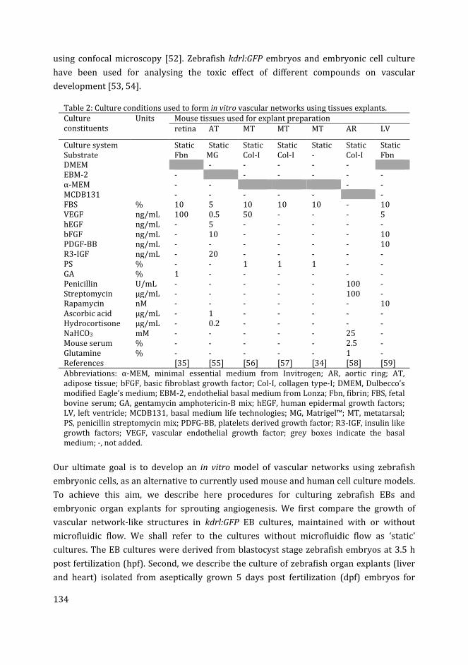
134
using confocal microscopy [52]. Zebrafish kdrl:GFP embryos and embryonic cell culture
have been used for analysing the toxic effect of different compounds on vascular
development [53, 54].
Table 2: Culture conditions used to form in vitro vascular networks using tissues explants.
Culture
constituents
Units Mouse tissues used for explant preparation
retina AT MT MT MT AR LV
Culture system Static Static Static Static Static Static Static
Substrate Fbn MG Col-I Col-I - Col-I Fbn
DMEM - - - - -
EBM-2 - - - - - -
α-MEM - - - -
MCDB131 - - - - - -
FBS % 10 5 10 10 10 - 10
VEGF ng/mL 100 0.5 50 - - - 5
hEGF ng/mL - 5 - - - - -
bFGF ng/mL - 10 - - - - 10
PDGF-BB ng/mL - - - - - - 10
R3-IGF ng/mL - 20 - - - - -
PS % - - 1 1 1 - -
GA % 1 - - - - - -
Penicillin U/mL - - - - - 100 -
Streptomycin µg/mL - - - - - 100 -
Rapamycin nM - - - - - - 10
Ascorbic acid µg/mL - 1 - - - - -
Hydrocortisone µg/mL - 0.2 - - - - -
NaHCO3 mM - - - - - 25 -
Mouse serum % - - - - - 2.5 -
Glutamine % - - - - - 1 -
References [35] [55] [56] [57] [34] [58] [59]
Abbreviations: α-MEM, minimal essential medium from Invitrogen; AR, aortic ring; AT,
adipose tissue; bFGF, basic fibroblast growth factor; Col-I, collagen type-I; DMEM, Dulbecco’s
modified Eagle’s medium; EBM-2, endothelial basal medium from Lonza; Fbn, fibrin; FBS, fetal
bovine serum; GA, gentamycin amphotericin-B mix; hEGF, human epidermal growth factors;
LV, left ventricle; MCDB131, basal medium life technologies; MG, Matrigel™; MT, metatarsal;
PS, penicillin streptomycin mix; PDFG-BB, platelets derived growth factor; R3-IGF, insulin like
growth factors; VEGF, vascular endothelial growth factor; grey boxes indicate the basal
medium; -, not added.
Our ultimate goal is to develop an in vitro model of vascular networks using zebrafish
embryonic cells, as an alternative to currently used mouse and human cell culture models.
To achieve this aim, we describe here procedures for culturing zebrafish EBs and
embryonic organ explants for sprouting angiogenesis. We first compare the growth of
vascular network-like structures in kdrl:GFP EB cultures, maintained with or without
microfluidic flow. We shall refer to the cultures without microfluidic flow as ‘static’
cultures. The EB cultures were derived from blastocyst stage zebrafish embryos at 3.5 h
post fertilization (hpf). Second, we describe the culture of zebrafish organ explants (liver
and heart) isolated from aseptically grown 5 days post fertilization (dpf) embryos for
135
sprouting angiogenesis. These explant cultures were developed in order to further
optimize the culture conditions for these tissues, as cells derived from different tissues
and at different developmental stage of embryo may have different culture requirements.
Using the knowledge gained from these studies in zebrafish, we hope one day to extend
the techniques to cells from other species.
Materials and methods
Zebrafish embryos
All the animal experiments were performed according to the Netherland experiments on
Animals act [60], based on EU directives [61]. Adult kdrl:GFP zebrafish were maintained in
circulating water according to previously described protocols [62]. Adult male and female
fish, at a proportion of 1:1, were transferred to breeding tanks in the evening. The eggs
were collected, next morning, at the bottom of the tank, separated from adults using a
mesh to prevent the eggs from being eaten. Fertilized, healthy embryos were distributed
in 9 cm Petri dishes (100 embryos per dish for EB culture and 50 embryos per dish for
liver and heart isolation). The embryos used for EB culture were allowed to grow for 3.5 h,
and the embryos for liver and heart isolation for 24 h in a temperature controlled room at
28 °C.
When zebrafish eggs are laid, they are exposed to a wide range of pathogens in the water
including faecal pathogens from the adults [63]. The chorion represents a barrier to the
entry of microorganisms into the perivitelline space and embryo [64]. Therefore, before
isolating cells and tissues we decontaminated the eggs with their intact chorion.
Embryo sterilization
The embryos were surface decontaminated, with the chorion intact, using a procedure
modified from Ref. [65]. Briefly, the embryos were transferred to a small net and
immersed in 70% ethanol for 10 sec. The embryos were then washed with L15 medium
(Table 3) to remove the ethanol. The embryos were then immersed twice, for 4 min each,
in sodium hypochlorite solution (Table 3), with a change of L15 medium in between. After
the last treatment with sodium hypochlorite the embryos were washed three times with
L15 medium and finally left in 500 µL of L15 medium for dechorionation.
Embryo dechorionation
The embryos decontaminated in the previous step, still with their chorions intact, were
subjected to manual dechorionation under a dissecting microscope using a pair of sterile
No.5 watchmaker’s forceps. The dead embryos, or embryos with cloudy perivitelline fluid
were removed before dechorionation.
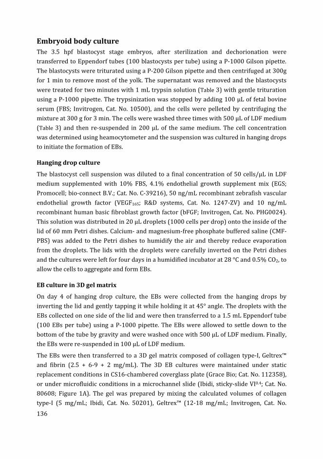
136
Embryoid body culture
The 3.5 hpf blastocyst stage embryos, after sterilization and dechorionation were
transferred to Eppendorf tubes (100 blastocysts per tube) using a P-1000 Gilson pipette.
The blastocysts were triturated using a P-200 Gilson pipette and then centrifuged at 300g
for 1 min to remove most of the yolk. The supernatant was removed and the blastocysts
were treated for two minutes with 1 mL trypsin solution (Table 3) with gentle trituration
using a P-1000 pipette. The trypsinization was stopped by adding 100 µL of fetal bovine
serum (FBS; Invitrogen, Cat. No. 10500), and the cells were pelleted by centrifuging the
mixture at 300 g for 3 min. The cells were washed three times with 500 µL of LDF medium
(Table 3) and then re-suspended in 200 µL of the same medium. The cell concentration
was determined using heamocytometer and the suspension was cultured in hanging drops
to initiate the formation of EBs.
Hanging drop culture
The blastocyst cell suspension was diluted to a final concentration of 50 cells/µL in LDF
medium supplemented with 10% FBS, 4.1% endothelial growth supplement mix (EGS;
Promocell; bio-connect B.V.; Cat. No. C-39216), 50 ng/mL recombinant zebrafish vascular
endothelial growth factor (VEGF165; R&D systems, Cat. No. 1247-ZV) and 10 ng/mL
recombinant human basic fibroblast growth factor (bFGF; Invitrogen, Cat. No. PHG0024).
This solution was distributed in 20 µL droplets (1000 cells per drop) onto the inside of the
lid of 60 mm Petri dishes. Calcium- and magnesium-free phosphate buffered saline (CMF-
PBS) was added to the Petri dishes to humidify the air and thereby reduce evaporation
from the droplets. The lids with the droplets were carefully inverted on the Petri dishes
and the cultures were left for four days in a humidified incubator at 28 °C and 0.5% CO2, to
allow the cells to aggregate and form EBs.
EB culture in 3D gel matrix
On day 4 of hanging drop culture, the EBs were collected from the hanging drops by
inverting the lid and gently tapping it while holding it at 45° angle. The droplets with the
EBs collected on one side of the lid and were then transferred to a 1.5 mL Eppendorf tube
(100 EBs per tube) using a P-1000 pipette. The EBs were allowed to settle down to the
bottom of the tube by gravity and were washed once with 500 µL of LDF medium. Finally,
the EBs were re-suspended in 100 µL of LDF medium.
The EBs were then transferred to a 3D gel matrix composed of collagen type-I, Geltrex™
and fibrin (2.5 + 6-9 + 2 mg/mL). The 3D EB cultures were maintained under static
replacement conditions in CS16-chambered coverglass plate (Grace Bio; Cat. No. 112358),
or under microfluidic conditions in a microchannel slide (Ibidi, sticky-slide VI0.4; Cat. No.
80608; Figure 1A). The gel was prepared by mixing the calculated volumes of collagen
type-I (5 mg/mL; Ibidi, Cat. No. 50201), Geltrex™ (12-18 mg/mL; Invitrogen, Cat. No.
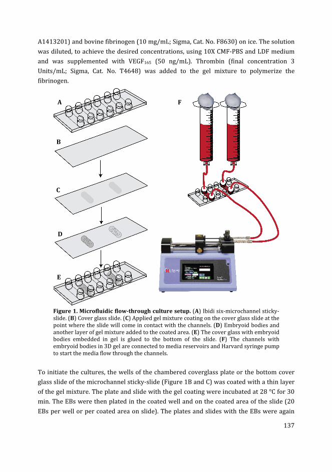
137
A1413201) and bovine fibrinogen (10 mg/mL; Sigma, Cat. No. F8630) on ice. The solution
was diluted, to achieve the desired concentrations, using 10X CMF-PBS and LDF medium
and was supplemented with VEGF165 (50 ng/mL). Thrombin (final concentration 3
Units/mL; Sigma, Cat. No. T4648) was added to the gel mixture to polymerize the
fibrinogen.
Figure 1. Microfluidic flow-through culture setup. (A) Ibidi six-microchannel sticky-
slide. (B) Cover glass slide. (C) Applied gel mixture coating on the cover glass slide at the
point where the slide will come in contact with the channels. (D) Embryoid bodies and
another layer of gel mixture added to the coated area. (E) The cover glass with embryoid
bodies embedded in gel is glued to the bottom of the slide. (F) The channels with
embryoid bodies in 3D gel are connected to media reservoirs and Harvard syringe pump
to start the media flow through the channels.
To initiate the cultures, the wells of the chambered coverglass plate or the bottom cover
glass slide of the microchannel sticky-slide (Figure 1B and C) was coated with a thin layer
of the gel mixture. The plate and slide with the gel coating were incubated at 28 °C for 30
min. The EBs were then plated in the coated well and on the coated area of the slide (20
EBs per well or per coated area on slide). The plates and slides with the EBs were again
A
B
C
D
E
F
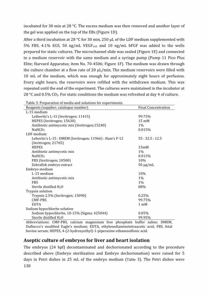
138
incubated for 30 min at 28 °C. The excess medium was then removed and another layer of
the gel was applied on the top of the EBs (Figure 1D).
After a third incubation at 28 °C for 30 min, 250 µL of the LDF medium supplemented with
5% FBS, 4.1% EGS, 50 ng/mL VEGF165 and 10 ng/mL bFGF was added to the wells
prepared for static cultures. The microchannel slide was sealed (Figure 1E) and connected
to a medium reservoir with the same medium and a syringe pump (Pump 11 Pico Plus
Elite; Harvard Apparatus; item No. 70-4506; Figure 1F). The medium was drawn through
the culture chamber at a flow rate of 20 µL/min. The medium reservoirs were filled with
10 mL of the medium, which was enough for approximately eight hours of perfusion.
Every eight hours, the reservoirs were refilled with the withdrawn medium. This was
repeated until the end of the experiment. The cultures were maintained in the incubator at
28 °C and 0.5% CO2. For static conditions the medium was refreshed at day 4 of culture.
Table 3: Preparation of media and solutions for experiments.
Reagents (supplier; catalogue number) Final Concentration
L-15 medium
Leibovitz’s L-15 (Invitrogen; 11415) 99.75%
HEPES (Invitrogen; 15630) 15 mM
Antibiotic antimycotic mix (Invitrogen;15240) 1%
NaHCO3 0.015%
LDF medium
Lebovitz’s L-15 : DMEM (Invitrogen; 11966) : Ham’s F-12
(Invitrogen; 21765)
55 : 32.5 : 12.5
HEPES 15mM
Antibiotic antimycotic mix 1%
NaHCO3 0.015%
FBS (Invitrogen; 10500) 10%
Zebrafish embryo extract 50 µg/mL
Embryo medium
L-15 medium 10%
Antibiotic antimycotic mix 1%
FBS 1%
Sterile distilled H2O 88%
Trypsin solution
Trypsin 2.5% (Invitrogen; 15090) 0.25%
CMF-PBS 99.75%
EDTA 1 mM
Sodium hypochlorite solution
Sodium hypochlorite, 10-15% (Sigma; 425044) 0.05%
Sterile distilled H2O 99.95%
Abbreviations: CMF-PBS, calcium magnesium free phosphate buffer saline; DMEM,
Dulbecco’s modified Eagle’s medium; EDTA, ethylenediaminetetraacetic acid; FBS, fetal
bovine serum; HEPES, 4-(2-hydroxyethyl)-1-piperazine-ethanesulfonic acid.
Aseptic culture of embryos for liver and heart isolation
The embryos (24 hpf) decontaminated and dechorionated according to the procedure
described above (Embryo sterilization and Embryo dechorionation) were raised for 5
days in Petri dishes in 25 mL of the embryo medium (Table 3). The Petri dishes were
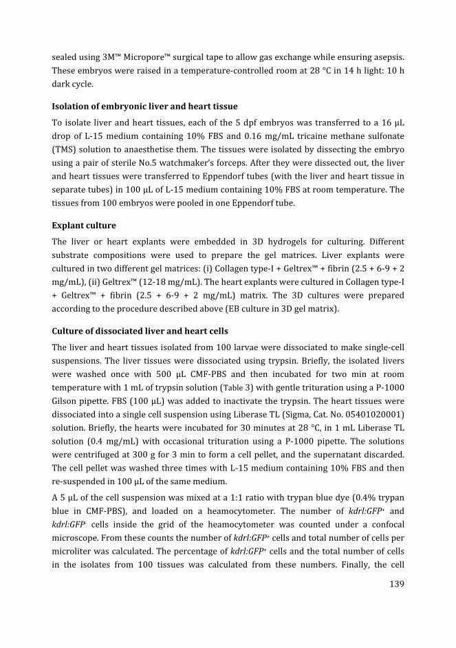
139
sealed using 3M™ Micropore™ surgical tape to allow gas exchange while ensuring asepsis.
These embryos were raised in a temperature-controlled room at 28 °C in 14 h light: 10 h
dark cycle.
Isolation of embryonic liver and heart tissue
To isolate liver and heart tissues, each of the 5 dpf embryos was transferred to a 16 µL
drop of L-15 medium containing 10% FBS and 0.16 mg/mL tricaine methane sulfonate
(TMS) solution to anaesthetise them. The tissues were isolated by dissecting the embryo
using a pair of sterile No.5 watchmaker’s forceps. After they were dissected out, the liver
and heart tissues were transferred to Eppendorf tubes (with the liver and heart tissue in
separate tubes) in 100 µL of L-15 medium containing 10% FBS at room temperature. The
tissues from 100 embryos were pooled in one Eppendorf tube.
Explant culture
The liver or heart explants were embedded in 3D hydrogels for culturing. Different
substrate compositions were used to prepare the gel matrices. Liver explants were
cultured in two different gel matrices: (i) Collagen type-I + Geltrex™ + fibrin (2.5 + 6-9 + 2
mg/mL), (ii) Geltrex™ (12-18 mg/mL). The heart explants were cultured in Collagen type-I
+ Geltrex™ + fibrin (2.5 + 6-9 + 2 mg/mL) matrix. The 3D cultures were prepared
according to the procedure described above (EB culture in 3D gel matrix).
Culture of dissociated liver and heart cells
The liver and heart tissues isolated from 100 larvae were dissociated to make single-cell
suspensions. The liver tissues were dissociated using trypsin. Briefly, the isolated livers
were washed once with 500 µL CMF-PBS and then incubated for two min at room
temperature with 1 mL of trypsin solution (Table 3) with gentle trituration using a P-1000
Gilson pipette. FBS (100 µL) was added to inactivate the trypsin. The heart tissues were
dissociated into a single cell suspension using Liberase TL (Sigma, Cat. No. 05401020001)
solution. Briefly, the hearts were incubated for 30 minutes at 28 °C, in 1 mL Liberase TL
solution (0.4 mg/mL) with occasional trituration using a P-1000 pipette. The solutions
were centrifuged at 300 g for 3 min to form a cell pellet, and the supernatant discarded.
The cell pellet was washed three times with L-15 medium containing 10% FBS and then
re-suspended in 100 µL of the same medium.
A 5 µL of the cell suspension was mixed at a 1:1 ratio with trypan blue dye (0.4% trypan
blue in CMF-PBS), and loaded on a heamocytometer. The number of kdrl:GFP+ and
kdrl:GFP- cells inside the grid of the heamocytometer was counted under a confocal
microscope. From these counts the number of kdrl:GFP+ cells and total number of cells per
microliter was calculated. The percentage of kdrl:GFP+ cells and the total number of cells
in the isolates from 100 tissues was calculated from these numbers. Finally, the cell
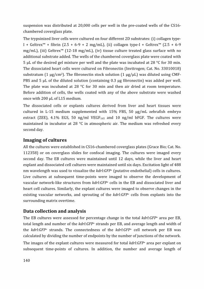
140
suspension was distributed at 20,000 cells per well in the pre-coated wells of the CS16-
chambered coverglass plate.
The trypsinized liver cells were cultured on four different 2D substrates: (i) collagen type-
I + Geltrex™ + fibrin (2.5 + 6-9 + 2 mg/mL), (ii) collagen type-I + Geltrex™ (2.5 + 6-9
mg/mL), (iii) Geltrex™ (12-18 mg/mL), (iv) tissue culture treated glass surface with no
additional substrate added. The wells of the chambered coverglass plate were coated with
5 µL of the desired gel mixture per well and the plate was incubated at 28 °C for 30 min.
The dissociated heart cells were cultured on Fibronectin (Invitrogen; Cat. No. 33010018)
substratum (1 µg/cm2). The fibronectin stock solution (1 µg/µL) was diluted using CMF-
PBS and 5 µL of the diluted solution (containing 0.3 µg fibronectin) was added per well.
The plate was incubated at 28 °C for 30 min and then air dried at room temperature.
Before addition of cells, the wells coated with any of the above substrate were washed
once with 200 µL of L15 medium.
The dissociated cells or explants cultures derived from liver and heart tissues were
cultured in L-15 medium supplemented with 15% FBS, 50 µg/mL zebrafish embryo
extract (ZEE), 4.1% EGS, 50 ng/ml VEGF165 and 10 ng/ml bFGF. The cultures were
maintained in incubator at 28 °C in atmospheric air. The medium was refreshed every
second day.
Imaging of cultures
All the cultures were established in CS16-chambered coverglass plates (Grace Bio; Cat. No.
112358) or on coverglass slides for confocal imaging. The cultures were imaged every
second day. The EB cultures were maintained until 12 days, while the liver and heart
explant and dissociated cell cultures were maintained until six days. Excitation light of 488
nm wavelength was used to visualize the kdrl:GFP+ (putative endothelial) cells in cultures.
Live cultures at subsequent time-points were imaged to observe the development of
vascular network-like structures from kdrl:GFP+ cells in the EB and dissociated liver and
heart cell cultures. Similarly, the explant cultures were imaged to observe changes in the
existing vascular networks, and sprouting of the kdrl:GFP+ cells from explants into the
surrounding matrix overtime.
Data collection and analysis
The EB cultures were assessed for percentage change in the total kdrl:GFP+ area per EB,
total length and number of the kdrl:GFP+ strands per EB, and average length and width of
the kdrl:GFP+ strands. The connectedness of the kdrl:GFP+ cell network per EB was
calculated by dividing the number of endpoints by the number of junctions of the network.
The images of the explant cultures were measured for total kdrl:GFP+ area per explant on
subsequent time-points of cultures. In addition, the number and average length of
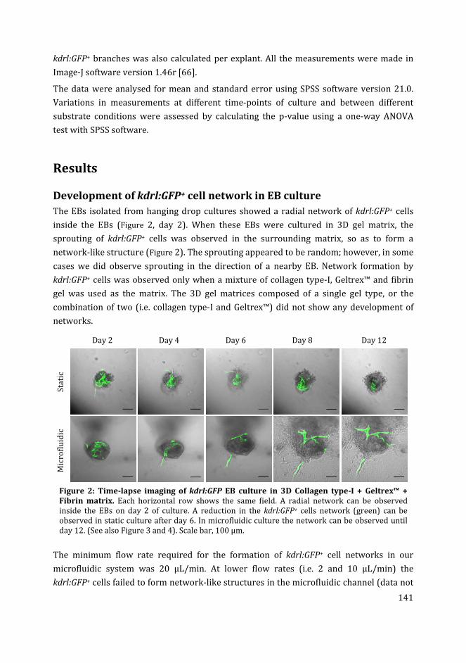
141
kdrl:GFP+ branches was also calculated per explant. All the measurements were made in
Image-J software version 1.46r [66].
The data were analysed for mean and standard error using SPSS software version 21.0.
Variations in measurements at different time-points of culture and between different
substrate conditions were assessed by calculating the p-value using a one-way ANOVA
test with SPSS software.
Results
Development of kdrl:GFP+ cell network in EB culture
The EBs isolated from hanging drop cultures showed a radial network of kdrl:GFP+ cells
inside the EBs (Figure 2, day 2). When these EBs were cultured in 3D gel matrix, the
sprouting of kdrl:GFP+ cells was observed in the surrounding matrix, so as to form a
network-like structure (Figure 2). The sprouting appeared to be random; however, in some
cases we did observe sprouting in the direction of a nearby EB. Network formation by
kdrl:GFP+ cells was observed only when a mixture of collagen type-I, Geltrex™ and fibrin
gel was used as the matrix. The 3D gel matrices composed of a single gel type, or the
combination of two (i.e. collagen type-I and Geltrex™) did not show any development of
networks.
Day 2 Day 4 Day 6 Day 8 Day 12
Sta
tic
Mic
rofl
uid
ic
Figure 2: Time-lapse imaging of kdrl:GFP EB culture in 3D Collagen type-I + Geltrex™ +
Fibrin matrix. Each horizontal row shows the same field. A radial network can be observed
inside the EBs on day 2 of culture. A reduction in the kdrl:GFP+ cells network (green) can be
observed in static culture after day 6. In microfluidic culture the network can be observed until
day 12. (See also Figure 3 and 4). Scale bar, 100 µm.
The minimum flow rate required for the formation of kdrl:GFP+ cell networks in our
microfluidic system was 20 µL/min. At lower flow rates (i.e. 2 and 10 µL/min) the
kdrl:GFP+ cells failed to form network-like structures in the microfluidic channel (data not
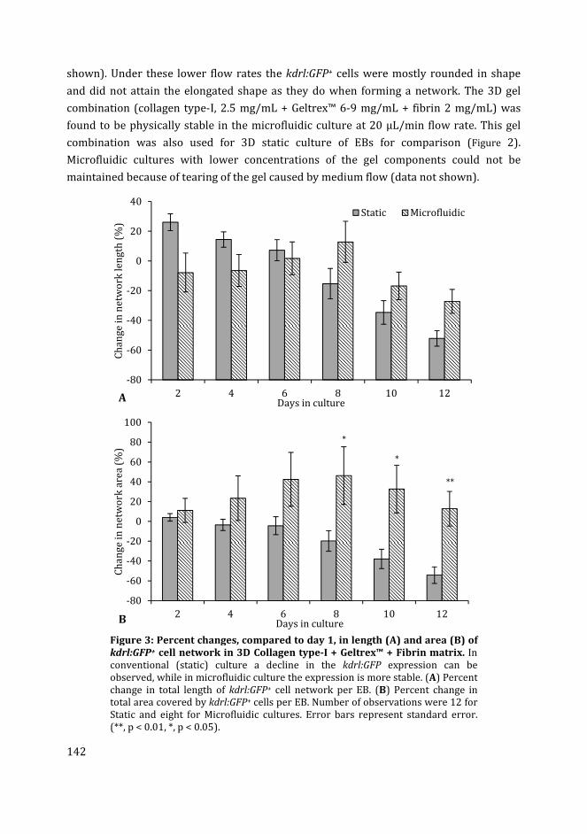
142
shown). Under these lower flow rates the kdrl:GFP+ cells were mostly rounded in shape
and did not attain the elongated shape as they do when forming a network. The 3D gel
combination (collagen type-I, 2.5 mg/mL + Geltrex™ 6-9 mg/mL + fibrin 2 mg/mL) was
found to be physically stable in the microfluidic culture at 20 µL/min flow rate. This gel
combination was also used for 3D static culture of EBs for comparison (Figure 2).
Microfluidic cultures with lower concentrations of the gel components could not be
maintained because of tearing of the gel caused by medium flow (data not shown).
Figure 3: Percent changes, compared to day 1, in length (A) and area (B) of
kdrl:GFP+ cell network in 3D Collagen type-I + Geltrex™ + Fibrin matrix. In
conventional (static) culture a decline in the kdrl:GFP expression can be
observed, while in microfluidic culture the expression is more stable. (A) Percent
change in total length of kdrl:GFP+ cell network per EB. (B) Percent change in
total area covered by kdrl:GFP+ cells per EB. Number of observations were 12 for
Static and eight for Microfluidic cultures. Error bars represent standard error.
(**, p < 0.01, *, p < 0.05).
-80
-60
-40
-20
0
20
40
2 4 6 8 10 12
Ch
an
ge
in
ne
two
rk l
en
gth
(%
)
Days in culture
Static Microfluidic
A
-80
-60
-40
-20
0
20
40
60
80
100
2 4 6 8 10 12
Ch
an
ge
in
ne
two
rk a
rea
(%
)
Days in culture
*
*
**
B
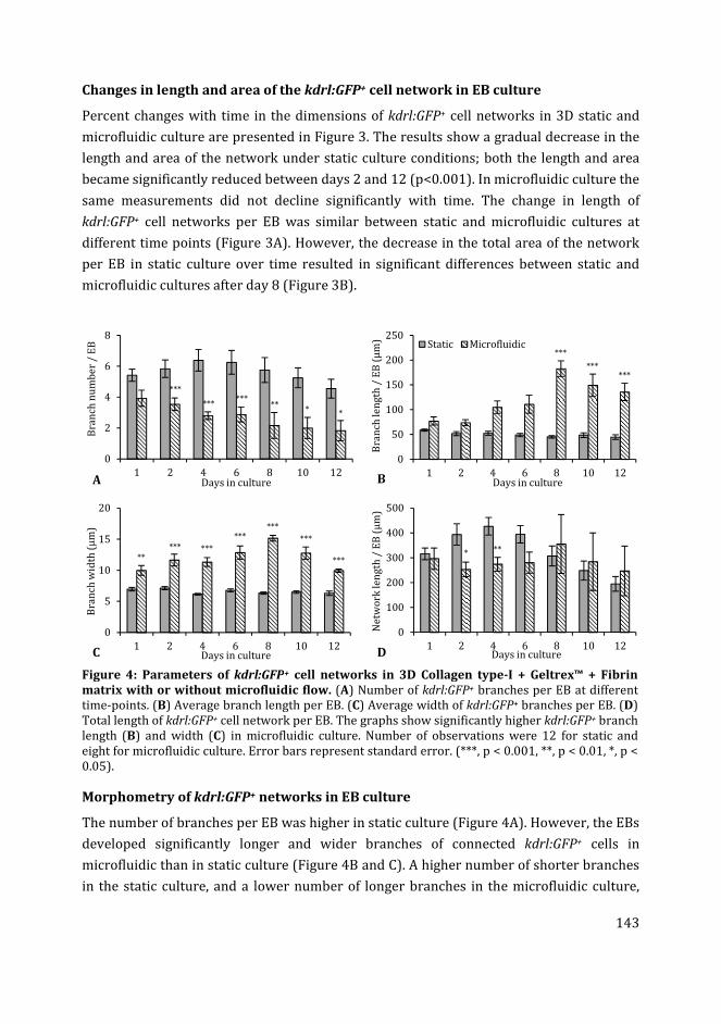
143
Changes in length and area of the kdrl:GFP+ cell network in EB culture
Percent changes with time in the dimensions of kdrl:GFP+ cell networks in 3D static and
microfluidic culture are presented in Figure 3. The results show a gradual decrease in the
length and area of the network under static culture conditions; both the length and area
became significantly reduced between days 2 and 12 (p<0.001). In microfluidic culture the
same measurements did not decline significantly with time. The change in length of
kdrl:GFP+ cell networks per EB was similar between static and microfluidic cultures at
different time points (Figure 3A). However, the decrease in the total area of the network
per EB in static culture over time resulted in significant differences between static and
microfluidic cultures after day 8 (Figure 3B).
Figure 4: Parameters of kdrl:GFP+ cell networks in 3D Collagen type-I + Geltrex™ + Fibrin
matrix with or without microfluidic flow. (A) Number of kdrl:GFP+ branches per EB at different
time-points. (B) Average branch length per EB. (C) Average width of kdrl:GFP+ branches per EB. (D)
Total length of kdrl:GFP+ cell network per EB. The graphs show significantly higher kdrl:GFP+ branch
length (B) and width (C) in microfluidic culture. Number of observations were 12 for static and
eight for microfluidic culture. Error bars represent standard error. (***, p < 0.001, **, p < 0.01, *, p <
0.05).
Morphometry of kdrl:GFP+ networks in EB culture
The number of branches per EB was higher in static culture (Figure 4A). However, the EBs
developed significantly longer and wider branches of connected kdrl:GFP+ cells in
microfluidic than in static culture (Figure 4B and C). A higher number of shorter branches
in the static culture, and a lower number of longer branches in the microfluidic culture,
0
2
4
6
8
1 2 4 6 8 10 12
Bra
nch
nu
mb
er
/ E
B
Days in cultureA
***
******
*** *
0
50
100
150
200
250
1 2 4 6 8 10 12
Bra
nch
len
gth
/ E
B (
µm
)
Days in culture
Static Microfluidic
B
***
******
0
5
10
15
20
1 2 4 6 8 10 12
Bra
nch
wid
th (
µm
)
Days in cultureC
***** ***
******
***
***
0
100
200
300
400
500
1 2 4 6 8 10 12
Ne
two
rk le
ng
th /
EB
(µ
m)
Days in cultureD
* **
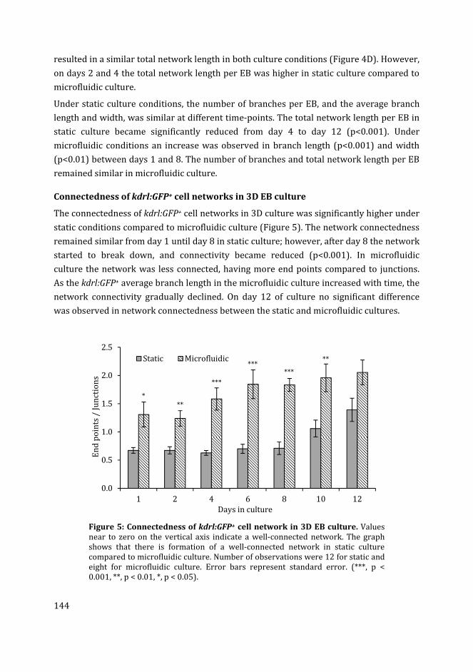
144
resulted in a similar total network length in both culture conditions (Figure 4D). However,
on days 2 and 4 the total network length per EB was higher in static culture compared to
microfluidic culture.
Under static culture conditions, the number of branches per EB, and the average branch
length and width, was similar at different time-points. The total network length per EB in
static culture became significantly reduced from day 4 to day 12 (p<0.001). Under
microfluidic conditions an increase was observed in branch length (p<0.001) and width
(p<0.01) between days 1 and 8. The number of branches and total network length per EB
remained similar in microfluidic culture.
Connectedness of kdrl:GFP+ cell networks in 3D EB culture
The connectedness of kdrl:GFP+ cell networks in 3D culture was significantly higher under
static conditions compared to microfluidic culture (Figure 5). The network connectedness
remained similar from day 1 until day 8 in static culture; however, after day 8 the network
started to break down, and connectivity became reduced (p<0.001). In microfluidic
culture the network was less connected, having more end points compared to junctions.
As the kdrl:GFP+ average branch length in the microfluidic culture increased with time, the
network connectivity gradually declined. On day 12 of culture no significant difference
was observed in network connectedness between the static and microfluidic cultures.
Figure 5: Connectedness of kdrl:GFP+ cell network in 3D EB culture. Values
near to zero on the vertical axis indicate a well-connected network. The graph
shows that there is formation of a well-connected network in static culture
compared to microfluidic culture. Number of observations were 12 for static and
eight for microfluidic culture. Error bars represent standard error. (***, p <
0.001, **, p < 0.01, *, p < 0.05).
0.0
0.5
1.0
1.5
2.0
2.5
1 2 4 6 8 10 12
En
d p
oin
ts /
Ju
nct
ion
s
Days in culture
Static Microfluidic
*
**
***
******
**
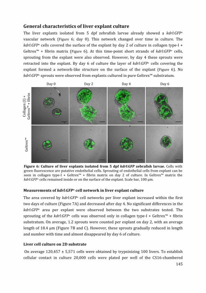
145
General characteristics of liver explant culture
The liver explants isolated from 5 dpf zebrafish larvae already showed a kdrl:GFP+
vascular network (Figure 6; day 0). This network changed over time in culture. The
kdrl:GFP+ cells covered the surface of the explant by day 2 of culture in collagen type-I +
Geltrex™ + fibrin matrix (Figure 6). At this time-point short strands of kdrl:GFP+ cells,
sprouting from the explant were also observed. However, by day 4 these sprouts were
retracted into the explant. By day 6 of culture the layer of kdrl:GFP+ cells covering the
explant formed a network-like structure on the surface of the explant (Figure 6). No
kdrl:GFP+ sprouts were observed from explants cultured in pure Geltrex™ substratum.
Day 0 Day 2 Day 4 Day 6
Co
lla
ge
n (
I) +
Ge
ltre
x™
+ f
ibri
n
Ge
ltre
x™
Figure 6: Culture of liver explants isolated from 5 dpf kdrl:GFP zebrafish larvae. Cells with
green fluorescence are putative endothelial cells. Sprouting of endothelial cells from explant can be
seen in collagen type-I + Geltrex™ + fibrin matrix on day 2 of culture. In Geltrex™ matrix the
kdrl:GFP+ cells remained inside or on the surface of the explant. Scale bar, 100 µm.
Measurements of kdrl:GFP+ cell network in liver explant culture
The area covered by kdrl:GFP+ cell networks per liver explant increased within the first
two days of culture (Figure 7A) and decreased after day 4. No significant differences in the
kdrl:GFP+ area per explant were observed between the two substrates tested. The
sprouting of the kdrl:GFP+ cells was observed only in collagen type-I + Geltrex™ + fibrin
substratum. On average, 1.2 sprouts were counted per explant on day 2, with an average
length of 18.4 µm (Figure 7B and C). However, these sprouts gradually reduced in length
and number with time and almost disappeared by day 6 of culture.
Liver cell culture on 2D substrate
On average 120,457 ± 5,571 cells were obtained by trypsinizing 100 livers. To establish
cellular contact in culture 20,000 cells were plated per well of the CS16-chambered
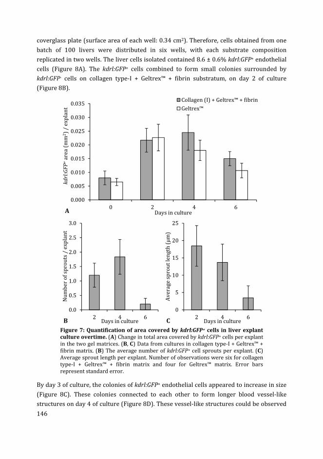
146
coverglass plate (surface area of each well: 0.34 cm2). Therefore, cells obtained from one
batch of 100 livers were distributed in six wells, with each substrate composition
replicated in two wells. The liver cells isolated contained 8.6 ± 0.6% kdrl:GFP+ endothelial
cells (Figure 8A). The kdrl:GFP+ cells combined to form small colonies surrounded by
kdrl:GFP- cells on collagen type-I + Geltrex™ + fibrin substratum, on day 2 of culture
(Figure 8B).
Figure 7: Quantification of area covered by kdrl:GFP+ cells in liver explant
culture overtime. (A) Change in total area covered by kdrl:GFP+ cells per explant
in the two gel matrices. (B, C) Data from cultures in collagen type-I + Geltrex™ +
fibrin matrix. (B) The average number of kdrl:GFP+ cell sprouts per explant. (C)
Average sprout length per explant. Number of observations were six for collagen
type-I + Geltrex™ + fibrin matrix and four for Geltrex™ matrix. Error bars
represent standard error.
By day 3 of culture, the colonies of kdrl:GFP+ endothelial cells appeared to increase in size
(Figure 8C). These colonies connected to each other to form longer blood vessel-like
structures on day 4 of culture (Figure 8D). These vessel-like structures could be observed
0.000
0.005
0.010
0.015
0.020
0.025
0.030
0.035
0 2 4 6
kd
rl:G
FP
+a
rea
(m
m2)
/ e
xp
lan
t
Days in culture
Collagen (I) + Geltrex™ + fibrin
Geltrex™
A
0.0
0.5
1.0
1.5
2.0
2.5
3.0
2 4 6
Nu
mb
er
of
spro
uts
/ e
xp
lan
t
Days in cultureB
0
5
10
15
20
25
2 4 6
Av
era
ge
sp
rou
t le
ng
th (
µm
)
Days in cultureC
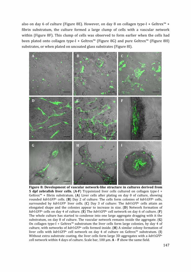
147
also on day 6 of culture (Figure 8E). However, on day 8 on collagen type-I + Geltrex™ +
fibrin substratum, the culture formed a large clump of cells with a vascular network
within (Figure 8F). This clump of cells was observed to form earlier when the cells had
been plated onto collagen type-I + Geltrex™ (Figure 8G) and pure Geltrex™ (Figure 8H)
substrates, or when plated on uncoated glass substrates (Figure 8I).
Figure 8: Development of vascular network-like structure in cultures derived from
5 dpf zebrafish liver cells. (A-F) Trypsinized liver cells cultured on collagen type-I +
Geltrex™ + fibrin substratum. (A) Liver cells after plating on day 0 of culture, showing
rounded kdrl:GFP+ cells. (B) Day 2 of culture: The cells form colonies of kdrl:GFP+ cells,
surrounded by kdrl:GFP- liver cells. (C) Day 3 of culture: The kdrl:GFP+ cells attain an
elongated shape and the colonies appear to increase in size. (D) Network formation of
kdrl:GFP+ cells on day 4 of culture. (E) The kdrl:GFP+ cell network on day 6 of culture. (F)
The whole culture has started to condense into one large aggregate dragging with it the
substratum, on day 8 of culture. The vascular network remains inside the aggregate. (G)
On collagen type-I + Geltrex™ substratum the liver cells form large colonies, by day 4 of
culture, with networks of kdrl:GFP+ cells formed inside. (H) A similar colony formation of
liver cells with kdrl:GFP+ cell network on day 4 of culture on Geltrex™ substratum. (I)
Without extra substrate coating, the liver cells form large 3D aggregates with a kdrl:GFP+
cell network within 4 days of culture. Scale bar, 100 µm. A – F show the same field.
A B
C
D E F
G H I
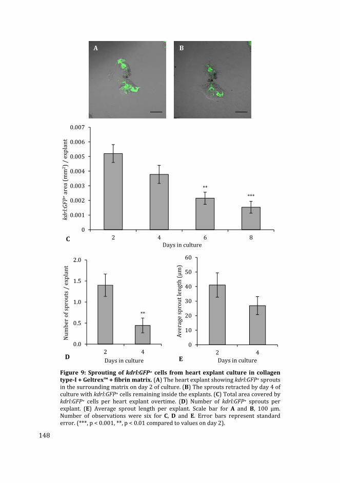
148
Figure 9: Sprouting of kdrl:GFP+ cells from heart explant culture in collagen
type-I + Geltrex™ + fibrin matrix. (A) The heart explant showing kdrl:GFP+ sprouts
in the surrounding matrix on day 2 of culture. (B) The sprouts retracted by day 4 of
culture with kdrl:GFP+ cells remaining inside the explants. (C) Total area covered by
kdrl:GFP+ cells per heart explant overtime. (D) Number of kdrl:GFP+ sprouts per
explant. (E) Average sprout length per explant. Scale bar for A and B, 100 µm.
Number of observations were six for C, D and E. Error bars represent standard
error. (***, p < 0.001, **, p < 0.01 compared to values on day 2).
0
0.001
0.002
0.003
0.004
0.005
0.006
0.007
2 4 6 8
kd
rl:G
FP
+a
rea
(m
m2)
/ e
xp
lan
t
Days in culture
**
***
C
0.0
0.5
1.0
1.5
2.0
2 4
Nu
mb
er
of
spro
uts
/ e
xp
lan
t
Days in cultureD
**
0
10
20
30
40
50
60
2 4
Av
era
ge
sp
rou
t le
ng
th (
µm
)
Days in cultureE
A
B
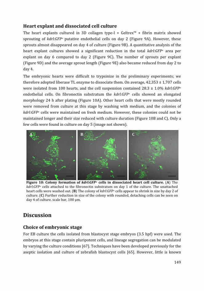
149
Heart explant and dissociated cell culture
The heart explants cultured in 3D collagen type-I + Geltrex™ + fibrin matrix showed
sprouting of kdrl:GFP+ putative endothelial cells on day 2 (Figure 9A). However, these
sprouts almost disappeared on day 4 of culture (Figure 9B). A quantitative analysis of the
heart explant cultures showed a significant reduction in the total kdrl:GFP+ area per
explant on day 6 compared to day 2 (Figure 9C). The number of sprouts per explant
(Figure 9D) and the average sprout length (Figure 9E) also became reduced from day 2 to
day 4.
The embryonic hearts were difficult to trypsinize in the preliminary experiments; we
therefore adopted liberase TL enzyme to dissociate them. On average, 42,353 ± 1,707 cells
were isolated from 100 hearts, and the cell suspension contained 28.3 ± 1.0% kdrl:GFP+
endothelial cells. On fibronectin substratum the kdrl:GFP+ cells showed an elongated
morphology 24 h after plating (Figure 10A). Other heart cells that were mostly rounded
were removed from culture at this stage by washing with medium, and the colonies of
kdrl:GFP+ cells were maintained on fresh medium. However, these colonies could not be
maintained longer and their size reduced with culture duration (Figure 10B and C). Only a
few cells were found in culture on day 5 (image not shown).
Figure 10: Colony formation of kdrl:GFP+ cells in dissociated heart cell culture. (A) The
kdrl:GFP+ cells attached to the fibronectin substratum on day 1 of the culture. The unattached
heart cells were washed out. (B) The colony of kdrl:GFP+ cells appear to shrink in size by day 2 of
culture. (C) Further reduction in size of the colony with rounded, detaching cells can be seen on
day 4 of culture. scale bar, 100 µm.
Discussion
Choice of embryonic stage
For EB culture the cells isolated from blastocyst stage embryos (3.5 hpf) were used. The
embryos at this stage contain pluripotent cells, and lineage segregation can be modulated
by varying the culture conditions [67]. Techniques have been developed previously for the
aseptic isolation and culture of zebrafish blastocyst cells [65]. However, little is known
A B C
150
about the differentiation events and culture strategies needed for the lineage-specific
differentiation of these cells.
For embryonic liver and heart tissue culture, zebrafish embryos at an early larval stage (5
dpf) were used. At this stage, there is relatively little yolk remaining in the larva. This
makes it easier to isolate the liver, which is more hidden by the yolk in earlier stages. We
found that, at 5 dpf, the tissues isolated from 100 larvae contained sufficient numbers of
cells to allow for replicates. Zebrafish larvae up to 5 dpf do not engage in exogenous
feeding but rely instead on yolk nutrients; therefore, it is easier to keep them sterile in
closed Petri dishes. Furthermore, at this stage the tissues presumably contain more
precursor cells, with the potential to grow and differentiate, compared to the tissues of
more advance stages.
EB culture in microfluidic setup
Mouse EB cultures can undergo the formation of blood islands and vascular
morphogenesis [20]. In our study, the zebrafish EBs developed in hanging drop culture,
also show some degree of vascular organization, i.e. a well-connected radial network
inside the EBs. However, when the EBs were transferred to 3D culture, the radial pattern
and connectivity of the network was lost due to the extension of the vascular sprouts into
the surrounding matrix.
Vascular sprouting is a physiological process in which selection of endothelial tip cells, as
well as migration and vascular extension, occurs in existing blood vessels in response to
angiogenic stimuli [68]. The phenomenon of vascular sprouting has been previously
reported in mouse EB cultures [56]. Similarly, we also observed angiogenic sprouting in
our zebrafish EB cultures; however, the extent of sprouting was less compared to the
mouse model. This may because we used primary blastocyst cells to establish EB cultures,
while mouse embryonic stem cell lines, which are adapted to proliferation in vitro, have
been used in the other studies. Previous studies on zebrafish primary blastocyst cell
culture have reported high cell death rates and low proliferative capacity in these cells
[69]; this may explain the low level of sprouting in our cultures.
Studies on mouse EBs in vitro, and zebrafish embryos in vivo, have shown the directional
migration of vascular sprouts towards the highest concentration of VEGF [70]. In our EB
cultures, the selection of tip cells and the direction of sprout extension appeared to be
random. This may because of the presence of angiogenic stimuli (growth factors)
dispersed throughout the medium. However, we observed in some cases the extension of
sprouts from one EB in the direction of a nearby EB. This may correlate with the in vivo
situation in which the release of angiogenic growth factors from a distant cell population
directs the migration of vascular sprouting.
151
The EB cultures described here with kdrl:GFP+ sprouts were maintained under
microfluidic conditions for a maximum of 12 days. In our previous studies, the growth of
EB cultures could be maintained for longer time in primary culture, as well as in
subculture [71]. However, the percentage of kdrl:GFP+ cells in those cultures dropped
significantly because they became overgrown by fibroblast-like cells [71]. Furthermore,
the regression of kdrl:GFP+ sprouts was observed after day 6 in our static cultures
described here. These results are in accordance with a study of angiogenic sprouting in
mouse EB cultures, in which the cells continued to degrade the 3D matrix after 12 days of
culture, and differentiate into a variety of cells, making it difficult to interpret the vascular
sprouts [72].
The medium was refreshed in our static culture at four day intervals. This interval was
chosen after our preliminary studies indicated that cell growth was hindered after four
days in non-replacement cultures. By contrast, the microfluidic cultures continued to grow
with recycling of the medium. Medium replacement at four day intervals in the static
culture may not be ideal for screening drugs or molecules that have a short half-life. For
those experiments, shorter interval between the medium refreshment may be needed.
It is possible that the microfluidic system described here can be adapted for toxicity
screening. For this purpose, the molecules to be tested can be easily added to the medium
reservoir. Depending on the exposure time, the final volume of the medium in the
reservoir can be adjusted according to the flow rate (20 µL/min in our case). Once the
medium is withdrawn through the culture chamber, the reservoir can be refilled with
fresh medium and the exhausted medium discarded or used for further analysis.
In the experiments described here, we formed a 3D gel matrix containing zebrafish EBs in
a microfluidic channel slide. The open design of the microfluidic channel allows direct
contact at the interface between the matrix and the medium. This is different to the
microfluidic systems currently used for 3D cell culture, in which the medium can only
diffuse into the matrix [73-76]. One of the drawbacks with the latter systems is that they
do not mimic the dynamic environment of the tissue, but represent a rather static
condition [38]. The flow of medium around the 3D matrix in our system presumably
exerted a shear stress on the EB cells inside the matrix and allowed the extension of
kdrl:GFP+ sprouts.
Under physiological conditions, endothelial cells produce secreted factors in response to
the shear stress induced by the blood flow [37]. These factors are essential for the
development, regulation and maintenance of the blood vessels [37]. In a microfluidic
culture, the flow rate of the medium is critical for cell proliferation, viability and function
[77]. This was observed in our microfluidic cultures, where the kdrl:GFP+ cells failed to
form networks at low flow rates (2 and 10 µL/min). One possible explanation for this
could be poor viability of cells at such low flow rates. Thus, under these low flow rates the
152
kdrl:GFP+ cells were mostly rounded in shape compared to 20 µL/min flow rate, where the
cells acquired an elongated shape and formed connected networks.
One of the challenges with our microfluidic culture was to find a balance between the flow
rate of the medium and the mechanical stability of the 3D matrix. The gel matrices
comprising lower concentrations of collagen type-I (1.5 mg/mL) and fibrin (1 mg/mL)
were not mechanically stable at the 20 µL/min flow rate required for network formation.
When the concentrations of the above mentioned substrates were increased to 2.5 mg/mL
and 2 mg/mL, respectively, the matrix formed was stable at the desired flow rate. Beside
the stability of the matrix of higher concentration, the stiffness of the 3D matrix itself is
presumably important in the culturing of vascular networks; thus, previous studies have
shown that stiffness of the matrix promotes the organization of endothelial cells into
capillary networks in vitro [78].
In our experiments, the differences between the microfluidic and static cultures was seen
mainly in the length and width of kdrl:GFP+ strands. In general, network formation by
kdrl:GFP+ cells in static cultures was limited to the periphery of the EBs. In microfluidic
culture by contrast, the kdrl:GFP+ cells extended more into the matrix and formed longer
branches. A possible explanation for this may be that the shear stress causes the cells to
secrete factors (as discussed above) which modulate the surrounding matrix and allow
the cells to grow further into the matrix. The wider diameter of kdrl:GFP+ strands in
microfluidic cultures may indicate the formation of blood vessel-like structures with a
lumen. By contrast, in the static cultures, the thinner branches formed are more consistent
with a solid chain of cells connected end-to-end than with a continuous vessel.
A network having fewer end points and more junctions is considered to be a well-
connected network [26]. The connectedness of a network can be determined by dividing
the number of endpoints by the number of junctions [26]. We find that the connectedness
of the networks depends on whether the network is more confined (as in the static
culture) or spread (as in the microfluidic culture). In principle, the more confined network
will be well-connected compared to a network with longer branches. In the microfluidic
cultures in our experiments, as the branch length increased, the network connectivity was
lost.
Culture of embryonic liver and heart tissues
The zebrafish liver and heart explant cultures described here might be suitable for
development as alternative to mouse aortic ring culture for sprouting angiogenesis. One
possible application of these explants could be to test the stimulatory or inhibitory effect
of various substances on angiogenesis. Mouse aortic ring cultures are currently being used
for such studies [79, 80]. Variability between cultures remains an issue with the mouse
aortic ring model [14]. Although we found variability in the explants isolated from
zebrafish embryos, the zebrafish model can easily yield a large more number of explants
153
which may help minimise this issue The small size of zebrafish embryos makes it
technically difficult to dissect the embryos for tissue isolation. However, with practice, we
were able to isolate tissues from 100 embryos in approximately three hours.
Under standard conditions, zebrafish embryos hatch at 48 hpf [48]. We found it important
to decontaminate the embryos before hatching (at 24 hpf), because preliminary
experiments showed that explants, isolated from the embryos decontaminated after
hatching at 5 dpf, could not be maintained free from contamination in culture. We also
found that embryos treated with sodium hypochlorite at 24 hpf have greatly impaired
hatching; it is therefore necessary to manually dechorionate these embryos before sealing
them into the Petri dish. Using this procedure the tissues isolated from these embryos at 5
dpf were successfully maintained sterile in culture.
The sprouting of kdrl:GFP+ cells from liver explants could only be observed in matrices
composed of collagen type-I, Geltrex™ and fibrin. Explants cultured in pure Geltrex™
matrix did not show any sprouting. These results are in accordance with mouse aortic ring
cultures which have been shown to produce higher microvessel sprouting in collagen
type-I and fibrin matrices compared to Matrigel™ [81]. The Geltrex™ we used in our
experiments is similar to Matrigel™, and is a mixture of laminin, collagen type-IV, entactin
and heparin sulphate proteoglycans (manufacturer’s documentation).
Similar to microvessel formation in mouse aortic ring culture [81], the dimensions of the
network-like structure formed by kdrl:GFP+ cells in trypsinized liver cells cultured on pure
Geltrex™ substratum were thinner and different from the much broader networks formed
on collagen type-I + Geltrex™ + fibrin substratum. The sprouting of kdrl:GFP+ cells in our
explant cultures was observed on day 2 which is earlier than in the mouse aortic ring
culture, where the peak sprouting of microvessels is reported to take place on day 6 of
culture [81]. Regression of the sprouts from the explants occurred on day 4 in our
experiments, whereas in mouse aortic ring culture this phenomena occurs on day 9 of
culture [81].
The average length of the kdrl:GFP+ cell sprouts emerging from the heart explants was
higher compared to the liver explants (41.1 ± 8.4 vs 18.4 ± 5.8 µm). This may be because
of a higher percentage of kdrl:GFP+ cells in the isolated hearts compared to the livers, as
indicated by our quantification of the cell isolates of these tissues. The colonies of
kdrl:GFP+ cells in the dissociated heart cell culture could not be maintained longer,
probably due to low seeding density.
Conclusions
Zebrafish embryoid body culture is a promising model for in vitro vasculogenesis and
angiogenesis. Microfluidic flow seems to have an effect on the growth of blood vessels in
154
EB culture. The use of a complex extracellular matrix with fibrin as a crucial part was, in
our hands, optimal for culturing vascular networks. The zebrafish liver and heart explant
cultures are promising models for sprouting angiogenesis. More experiments are needed
to optimize the substrate and medium composition for these cultures so that the cultures
can be expanded and be maintained for longer periods. This goal may also require co-
culture with other cell- or tissue-types. In principle, pure populations of viable kdrl:GFP+
cells could also be isolated from these cultures using fluorescence activated cell sorting
assay. These cells could then be used for further analysis (e.g. gene expression profiling),
as wells as for co-culturing with the EBs to improve the formation of vascular networks.
Acknowledgements
We would like to thank Prof. Stefan Schulte-Merker (Institute for Cardiovascular
Organogenesis and Regeneration, Westfalische Wilhelms University, Munster, Germany)
for providing the zebrafish kdrl:GFP line for this research.
This work was supported by the Smart Mix programme of the Netherlands Ministry of
Economic Affairs and the Netherlands Scientific Research Council (NWO)[grant number
SSM06010]; and the Generade programme of the Centre of Expertise Genomics in Leiden,
The Netherlands[grant number 2016_004].
References
[1] Fisher MB, Mauck RL. Tissue engineering and regenerative medicine: recent
innovations and the transition to translation. Tissue Eng Part B Rev 2013; 19(1): 1-
13.
[2] Bhatia SN, Ingber DE. Microfluidic organs-on-chips. Nat Biotechnol 2014; 32(8):
760-72.
[3] Haraguchi Y, Shimizu T, Yamato M, Okano T. Concise review: cell therapy and tissue
engineering for cardiovascular disease. Stem Cells Transl Med 2012; 1(2): 136-41.
[4] Hansen A, Eder A, Bonstrup M, Flato M, Mewe M, Schaaf S, et al. Development of a
drug screening platform based on engineered heart tissue. Circ Res 2010; 107(1):
35-44.
[5] Levick JR. The microcirculation and solute exchange. In: Eds. An Introduction to
cardiovascular physiology. 5 ed, Taylor & Francis 2010, pp. 166-187.
[6] Kannan RY, Salacinski HJ, Sales K, Butler P, Seifalian AM. The roles of tissue
engineering and vascularisation in the development of micro-vascular networks: a
review. Biomaterials 2005; 26(14): 1857-75.
[7] Vunjak-Novakovic G, Scadden DT. Biomimetic platforms for human stem cell
research. Cell Stem Cell 2011; 8(3): 252-61.
[8] Ibrahim M, Richardson MK. Microfluidic devices for cell, tissue and embryo culture.
Rec Pat Regen Med 2013; 3(3): 249-263.
[9] Zou T, Fan J, Fartash A, Liu H, Fan Y. Cell-based strategies for vascular regeneration.
J Biomed Mater Res A 2016; 104(5): 1297-314.
155
[10] Shan K, Li CP, Liu C, Liu X, Yan B. RNCR3: a regulator of diabetes mellitus-related
retinal microvascular dysfunction. Biochem Biophys Res Commun 2017; 482(4):
777-783.
[11] Roudsari LC, West JL. Studying the influence of angiogenesis in in vitro cancer
model systems. Adv Drug Deliv Rev 2016; 97: 250-9.
[12] Azimi MS, Lacey M, Mondal D, Murfee WL. An ex vivo tissue culture model for anti-
angiogenic drug testing. Methods Mol Biol 2016; 1464: 85-95.
[13] Vargesson N. Thalidomide-induced teratogenesis: history and mechanisms. Birth
Defects Res C Embryo Today 2015; 105(2): 140-56.
[14] Staton CA, Reed MW, Brown NJ. A critical analysis of current in vitro and in vivo
angiogenesis assays. Int J Exp Pathol 2009; 90(3): 195-221.
[15] Lutolf MP, Hubbell JA. Synthetic biomaterials as instructive extracellular
microenvironments for morphogenesis in tissue engineering. Nat Biotechnol 2005;
23(1): 47-55.
[16] Nakatsu MN, Sainson RC, Aoto JN, Taylor KL, Aitkenhead M, Perez-del-Pulgar S, et
al. Angiogenic sprouting and capillary lumen formation modeled by human
umbilical vein endothelial cells (HUVEC) in fibrin gels: the role of fibroblasts and
Angiopoietin-1. Microvasc Res 2003; 66(2): 102-12.
[17] Delgado VM, Nugnes LG, Colombo LL, Troncoso MF, Fernandez MM, Malchiodi EL, et
al. Modulation of endothelial cell migration and angiogenesis: a novel function for
the "tandem-repeat" lectin galectin-8. FASEB J 2011; 25(1): 242-54.
[18] Zhu J, Cui W, Liu X, Ying J, Hu C, Zhang Y. In vitro and ex vivo angiogenic effects of
roxarsone on rat endothelial cells. Toxicol Lett 2013; 223(2): 175-82.
[19] Bratt-Leal AM, Carpenedo RL, McDevitt TC. Engineering the embryoid body
microenvironment to direct embryonic stem cell differentiation. Biotechnol Prog
2009; 25(1): 43-51.
[20] Vailhe B, Vittet D, Feige JJ. In vitro models of vasculogenesis and angiogenesis. Lab
Invest 2001; 81(4): 439-52.
[21] Goodwin AM. In vitro assays of angiogenesis for assessment of angiogenic and anti-
angiogenic agents. Microvasc Res 2007; 74(2-3): 172-83.
[22] Jakobsson L, Kreuger J, Claesson-Welsh L. Building blood vessels--stem cell models
in vascular biology. J Cell Biol 2007; 177(5): 751-5.
[23] Arnaoutova I, Kleinman HK. In vitro angiogenesis: endothelial cell tube formation
on gelled basement membrane extract. Nat Protoc 2010; 5(4): 628-35.
[24] Allen P, Melero-Martin J, Bischoff J. Type I collagen, fibrin and PuraMatrix matrices
provide permissive environments for human endothelial and mesenchymal
progenitor cells to form neovascular networks. J Tissue Eng Regen Med 2011; 5(4):
e74-86.
[25] Kusuma S, Shen YI, Hanjaya-Putra D, Mali P, Cheng L, Gerecht S. Self-organized
vascular networks from human pluripotent stem cells in a synthetic matrix. Proc
Natl Acad Sci U S A 2013; 110(31): 12601-6.
[26] Moya ML, Hsu YH, Lee AP, Hughes CC, George SC. In vitro perfused human capillary
networks. Tissue Eng Part C Methods 2013; 19(9): 730-7.
[27] Kim S, Lee H, Chung M, Jeon NL. Engineering of functional, perfusable 3D
microvascular networks on a chip. Lab Chip 2013; 13(8): 1489-500.
[28] Nguyen DH, Stapleton SC, Yang MT, Cha SS, Choi CK, Galie PA, et al. Biomimetic
model to reconstitute angiogenic sprouting morphogenesis in vitro. Proc Natl Acad
Sci U S A 2013; 110(17): 6712-7.
156
[29] Li H, Chang J. Bioactive silicate materials stimulate angiogenesis in fibroblast and
endothelial cell co-culture system through paracrine effect. Acta Biomaterialia
2013; 9(6): 6981-6991.
[30] Zheng Y, Chen J, Craven M, Choi NW, Totorica S, Diaz-Santana A, et al. In vitro
microvessels for the study of angiogenesis and thrombosis. Proc Natl Acad Sci U S A
2012; 109(24): 9342-7.
[31] Stamati K, Priestley JV, Mudera V, Cheema U. Laminin promotes vascular network
formation in 3D in vitro collagen scaffolds by regulating VEGF uptake. Exp Cell Res
2014; 327(1): 68-77.
[32] Lodish H, Berk A, Zipursky SL, Matsudaira P, Baltimore D, Darnell J. Section 6.2
Growth of Animal cells in culture. In: Eds. Molecular cell biology. 4 ed, New York,
Freeman, W. H. 2000.
[33] Nicosia RF. The aortic ring model of angiogenesis: a quarter century of search and
discovery. J Cell Mol Med 2009; 13(10): 4113-36.
[34] Deckers M, van der Pluijm G, Dooijewaard S, Kroon M, van Hinsbergh V, Papapoulos
S, et al. Effect of angiogenic and antiangiogenic compounds on the outgrowth of
capillary structures from fetal mouse bone explants. Lab Invest 2001; 81(1): 5-15.
[35] Rezzola S, Belleri M, Ribatti D, Costagliola C, Presta M, Semeraro F. A novel ex vivo
murine retina angiogenesis (EMRA) assay. Exp Eye Res 2013; 112: 51-6.
[36] Brodsky SV, Smith M, Kashgarian M, Goligorsky MS. A model for ex vivo renal
angiogenesis. Nephron Exp Nephrol 2003; 93(1): e46-52.
[37] Davies PF. Hemodynamic shear stress and the endothelium in cardiovascular
pathophysiology. Nat Clin Pract Cardiovasc Med 2009; 6(1): 16-26.
[38] Gupta N, Liu JR, Patel B, Solomon DE, Vaidya B, Gupta V. Microfluidics-based 3D cell
culture models: Utility in novel drug discovery and delivery research.
Bioengineering & Translational Medicine 2016; 1(1): 63-81.
[39] Hasan A, Paul A, Vrana NE, Zhao X, Memic A, Hwang YS, et al. Microfluidic
techniques for development of 3D vascularized tissue. Biomaterials 2014; 35(26):
7308-25.
[40] Rezzola S, Belleri M, Gariano G, Ribatti D, Costagliola C, Semeraro F, et al. In vitro
and ex vivo retina angiogenesis assays. Angiogenesis 2014; 17(3): 429-42.
[41] Zhu WH, Iurlaro M, MacIntyre A, Fogel E, Nicosia RF. The mouse aorta model:
influence of genetic background and aging on bFGF- and VEGF-induced angiogenic
sprouting. Angiogenesis 2003; 6(3): 193-9.
[42] Chavez MN, Aedo G, Fierro FA, Allende ML, Egana JT. Zebrafish as an emerging
model organism to study angiogenesis in development and regeneration. Front
Physiol 2016; 7: 56.
[43] Wilkinson RN, van Eeden FJ. The zebrafish as a model of vascular development and
disease. Prog Mol Biol Transl Sci 2014; 124: 93-122.
[44] Grunow B, Mohamet L, Shiels HA. Generating an in vitro 3D cell culture model from
zebrafish larvae for heart research. J Exp Biol 2015; 218(Pt 8): 1116-21.
[45] Yue MS, Plavicki JS, Li XY, Peterson RE, Heideman W. A co-culture assay of
embryonic zebrafish hearts to assess migration of epicardial cells in vitro. BMC Dev
Biol 2015; 15: 50.
[46] Selderslaghs IW, Van Rompay AR, De Coen W, Witters HE. Development of a
screening assay to identify teratogenic and embryotoxic chemicals using the
zebrafish embryo. Reprod Toxicol 2009; 28(3): 308-20.
157
[47] Ciarlo CA, Zon LI. Chapter 1 - Embryonic cell culture in zebrafish. In: H. William
Detrich MW and IZ Leonard, Eds. Methods in Cell Biology. ed, Academic Press 2016,
pp. 1-10.
[48] Kimmel CB, Ballard WW, Kimmel SR, Ullmann B, Schilling TF. Stages of embryonic
development of the zebrafish. Dev Dyn 1995; 203(3): 253-310.
[49] Choorapoikayil S, Overvoorde J, den Hertog J. Deriving cell lines from zebrafish
embryos and tumors. Zebrafish 2013; 10(3): 316-25.
[50] Simmich J, Staykov E, Scott E. Zebrafish as an appealing model for optogenetic
studies. Prog Brain Res 2012; 196: 145-62.
[51] Li R, Beebe T, Jen N, Yu F, Takabe W, Harrison M, et al. Shear stress-activated Wnt-
angiopoietin-2 signaling recapitulates vascular repair in zebrafish embryos.
Arterioscler Thromb Vasc Biol 2014; 34(10): 2268-75.
[52] Harrison MR, Bussmann J, Huang Y, Zhao L, Osorio A, Burns CG, et al. Chemokine-
guided angiogenesis directs coronary vasculature formation in zebrafish. Dev Cell
2015; 33(4): 442-54.
[53] Tal T, Kilty C, Smith A, LaLone C, Kennedy B, Tennant A, et al. Screening for
angiogenic inhibitors in zebrafish to evaluate a predictive model for developmental
vascular toxicity. Reprod Toxicol 2016: http://dx.doi.org/10.1016/j.reprotox.-
2016.12.004.
[54] Huang HG, Lindgren A, Wu XR, Liu NA, Lin SO. High-throughput screening for
bioactive molecules using primary cell culture of transgenic zebrafish embryos. Cell
Rep 2012; 2(3): 695-704.
[55] Rojas-Rodriguez R, Gealekman O, Kruse ME, Rosenthal B, Rao K, Min S, et al.
Adipose tissue angiogenesis assay. Methods Enzymol 2014; 537: 75-91.
[56] Liu Z, Lebrin F, Maring JA, van den Driesche S, van der Brink S, van Dinther M, et al.
ENDOGLIN is dispensable for vasculogenesis, but required for vascular endothelial
growth factor-induced angiogenesis. PLoS One 2014; 9(1): e86273.
[57] Song W, Fhu CW, Ang KH, Liu CH, Johari NA, Lio D, et al. The fetal mouse metatarsal
bone explant as a model of angiogenesis. Nat Protoc 2015; 10(10): 1459-73.
[58] Masson VV, Devy L, Grignet-Debrus C, Bernt S, Bajou K, Blacher S, et al. Mouse aortic
ring assay: a new approach of the molecular genetics of angiogenesis. Biol Proced
Online 2002; 4: 24-31.
[59] Kiefer FN, Munk VC, Humar R, Dieterle T, Landmann L, Battegay EJ. A versatile in
vitro assay for investigating angiogenesis of the heart. Exp Cell Res 2004; 300(2):
272-82.
[60] Experiments on Animal Act, 2014. The State Secretary of Health and Environmental
Protection, Soestdijk. http://wetten.overheid.nl/BWBR0003081/2014-12-18
[61] Directive 2010/63/EU of the European parliament and of the council of 22
September 2010 on the protection of animals used for scientific purposes Text with
EEA relevance, 2010. The European Parliament and the council of the European
Union, Official Journal of the European Union. http://eur-lex.europa.eu/legal-
content/EN/TXT/?uri=celex:32010L0063
[62] Westerfield M. The zebrafish book: a guide for the laboratory use of zebrafish
(Danio Rerio). 4 ed, University of Oregon Press, Eugene 2000, pp.
[63] Pham LN, Kanther M, Semova I, Rawls JF. Methods for generating and colonizing
gnotobiotic zebrafish. Nat Protoc 2008; 3(12): 1862-75.
[64] Henn K. Limits of fish embryo toxicity test with Danio rerio as an alternative to the
acute fish toxicity test. PhD thesis, 2011. Center of Organismal Studies Heidelberg
(COS), Ruperto-Carola University Heidelberg, Germany.
158
[65] Fan L, Collodi P. Zebrafish embryonic stem cells. Methods Enzymol 2006; 418: 64-
77.
[66] Schneider CA, Rasband WS, Eliceiri KW. NIH Image to ImageJ: 25 years of image
analysis. Nat Methods 2012; 9(7): 671-5.
[67] Thomson JA, Itskovitz-Eldor J, Shapiro SS, Waknitz MA, Swiergiel JJ, Marshall VS, et
al. Embryonic stem cell lines derived from human blastocysts. Science 1998;
282(5391): 1145-7.
[68] Adams RH, Alitalo K. Molecular regulation of angiogenesis and lymphangiogenesis.
Nat Rev Mol Cell Biol 2007; 8(6): 464-78.
[69] Myhre JL, Pilgrim DB. Cellular differentiation in primary cell cultures from single
zebrafish embryos as a model for the study of myogenesis. Zebrafish 2010; 7(3):
255-66.
[70] Jakobsson L, Franco CA, Bentley K, Collins RT, Ponsioen B, Aspalter IM, et al.
Endothelial cells dynamically compete for the tip cell position during angiogenic
sprouting. Nat Cell Biol 2010; 12(10): 943-53.
[71] Ibrahim M, Richardson MK. The growth of endothelial-like cells in zebrafish
embryoid body culture. 2017: in prep.
[72] Gu A, Tsark W, Holmes KV, Shively JE. Role of Ceacam1 in VEGF induced
vasculogenesis of murine embryonic stem cell-derived embryoid bodies in 3D
culture. Exp Cell Res 2009; 315(10): 1668-82.
[73] Zanotelli MR, Ardalani H, Zhang J, Hou Z, Nguyen EH, Swanson S, et al. Stable
engineered vascular networks from human induced pluripotent stem cell-derived
endothelial cells cultured in synthetic hydrogels. Acta Biomater 2016; 35: 32-41.
[74] Shin Y, Yang K, Han S, Park HJ, Seok Heo Y, Cho SW, et al. Reconstituting vascular
microenvironment of neural stem cell niche in three-dimensional extracellular
matrix. Adv Healthc Mater 2014; 3(9): 1457-64.
[75] Hsu YH, Moya ML, Abiri P, Hughes CC, George SC, Lee AP. Full range physiological
mass transport control in 3D tissue cultures. Lab Chip 2013; 13(1): 81-9.
[76] Park YK, Tu TY, Lim SH, Clement IJ, Yang SY, Kamm RD. In Vitro Microvessel Growth
and Remodeling within a Three-dimensional Microfluidic Environment. Cell Mol
Bioeng 2014; 7(1): 15-25.
[77] Tehranirokh M, Kouzani AZ, Francis PS, Kanwar JR. Microfluidic devices for cell
cultivation and proliferation. Biomicrofluidics 2013; 7(5): 51502.
[78] Sieminski AL, Hebbel RP, Gooch KJ. The relative magnitudes of endothelial force
generation and matrix stiffness modulate capillary morphogenesis in vitro. Exp Cell
Res 2004; 297(2): 574-84.
[79] Gopinathan G, Milagre C, Pearce OM, Reynolds LE, Hodivala-Dilke K, Leinster DA, et
al. Interleukin-6 Stimulates Defective Angiogenesis. Cancer Res 2015; 75(15):
3098-107.
[80] Park SL, Won SY, Song JH, Lee SY, Kim WJ, Moon SK. Esculetin inhibits VEGF-
induced angiogenesis both in Vitro and in Vivo. Am J Chin Med 2016; 44(1): 61-76.
[81] Baker M, Robinson SD, Lechertier T, Barber PR, Tavora B, D'Amico G, et al. Use of
the mouse aortic ring assay to study angiogenesis. Nat Protoc 2011; 7(1): 89-104.

159
Chapter 7
Summary and conclusions
160
Summary
Development of an in vitro vascular network is currently one of the major challenges in
tissue engineering and regenerative medicine [1]. Several techniques have been
developed to culture vascular networks using mammalian cells and tissues [2-12].
However, it is desirable to develop alternative models which are cost effective, easily
accessible and available in large numbers. The zebrafish possess these benefits and is
emerging as a model laboratory animal in various fields of research. For in vitro studies
the externally fertilized, abundant zebrafish embryos are an excellent source of primary
embryonic cells, which are not always easy to access in other species [13]. The availability
of a number of vascular transgenic lines and the regenerative capacity of zebrafish makes
it of even greater importance in this field [14].
Development of blood vessels is a complex process which involves the differentiation,
migration and arrangement of endothelial cells into a tubular form [15]. The process is
complemented by supporting cell types [16, 17], protein factors [18], extracellular matrix
molecules [19] and blood flow [20]. By combining these components in vitro, some of the
researchers have recently developed cultures of perfusable vascular network [3, 10];
however, the formation of a functional vasculature which is close in resemblance to the
physiological blood vessels remains a challenge [21].
The cultures that we have established in this study are a step towards helping our
understanding of zebrafish in vitro vascular network development. The zebrafish
embryoid body and tissue explant models we have developed are promising
complementary models to mammalian embryonic stem cell and tissue explant models, for
studying in vitro vascular development. The embryoid body cultures can easily be
established in large numbers; however, the isolation of tissue explants from small
embryos require technical expertise which can be acquired with practice.
Based on our results, zebrafish embryonic cells can be used to culture vascular networks.
In its current state however, the zebrafish cannot completely replace the mammalian cell
culture models, but can be used as a complementary model. The size of the vascular
network formed in our cultures was smaller than previously reported for mouse and
human embryonic cell and tissue culture. This may because of the limited availability of
techniques for zebrafish in this regard. Further research in zebrafish model for this
purpose should provide information that will improve the formation of in vitro vascular
network in this species.
In Chapter 2 we reviewed the available techniques for culturing vascular networks and
considered their potential applications. There are very few studies using zebrafish cells
for this purpose [22, 23]. However, in the light of general cell culture techniques
developed in zebrafish, we believe that most of the techniques developed for culturing
vascular networks using mammalian cells and tissues can be adapted for zebrafish. And,
161
because of its general benefits (such as low cost, external fertilization) the zebrafish can
be a significant first step model for studying vascular development in vitro. Nevertheless,
there are a few disadvantages with the zebrafish model such as combining cells from
genetically diverse embryos in culture (the zebrafish is genetically polymorphic) [24], the
requirement of a nutrient rich medium [25] and the need, reported in some studies [26],
of a feeder layer of stromal cells to maintain an undifferentiated state of the primary
embryonic cell cultures.
In Chapter 3 we optimized the basic nutrient requirements for zebrafish blastocyst cell
culture. We found that, for their optimal growth, the primary blastocyst cells require 15%
fetal bovine serum (FBS) and 60 µg/mL zebrafish embryo extract (ZEE) in the medium.
We found that endothelial cells differentiated spontaneously in zebrafish blastocyst cell
cultures. Using transgenic lines (fli:GFP+ and kdrl:GFP+ cells) we could track the further
development of these cells until day 4 of culture under basal culture conditions (when
they start to reduce in number).
During the course of culture, most of the differentiated cell types (we also observed
neuron-like cells and pigment cells along with the fli:GFP+ and kdrl:GFP+ cells) diminished
and a single subpopulation of cells appear to increase in number. By continuous passaging
of the blastocyst cell cultures, the secondary cells attain a homogenous fibroblast-like
morphology. These cells shows an optimal growth at a lower FBS concentration (10%)
and without ZEE in the medium.
In Chapter 4 we attempted to increase the fli:GFP+ and kdrl:GFP+ cell populations in the
blastocyst cell cultures by manipulating the culture conditions. We used different media
compositions, substrate molecules and vascular endothelial growth factor concentrations
to find optimum conditions for the growth of fli:GFP+ and kdrl:GFP+ cells in cultures. We
found that the LDF medium previously optimized for the zebrafish blastocyst cell culture
[26-30], with added endothelial growth supplement mix (used in supplementation with
endothelial growth medium for human endothelial cell culture) increases the percentages
of fli:GFP+ and kdrl:GFP+ cells in cultures.
Collagen type-I substratum also increased the percentages of putative endothelial cells in
culture; however, the effect was not significant compared to the uncoated polystyrene
substratum. Furthermore, the recombinant zebrafish vascular endothelial growth factor
also increased the percentages of fli:GFP+ and kdrl:GFP+ cells when added to the culture
medium. With all these optimization steps we were able to maintain the fli:GFP+ and
kdrl:GFP+ cells for longer duration (until eight days) in cultures.
In Chapter 5 we utilised the possibilities of suspension culture of zebrafish blastocyst
cells to see its effect on the development of fli:GFP+ and kdrl:GFP+ cells in cultures. The
blastocyst cells were cultured in hanging drops on the inside of the lid of a Petri dish in the
zebrafish endothelial differentiation medium (optimized in the experiments described in
162
the previous chapters). The results were promising: we found significantly higher
percentages of fli:GFP+ and kdrl:GFP+ cells in the embryoid body (EB) aggregates of the
blastocyst cells developed in hanging drop cultures compared to the adherent cultures.
When transferred from hanging drops to a 2D substratum the percentages of fli:GFP+ and
kdrl:GFP+ cells in the EB cultures declined with time.
Under optimal conditions the fli:GFP+ cells could be observed in the primary EB cultures as
well as in secondary cultures until two passages. In contrast, the kdrl:GFP+ cells could only
be observed until 12 days of primary cultures. Different substrates tested for the EB
cultures showed variations in the development of fli:GFP+ of kdrl:GFP+ cells. In the first six
days of the EB adherent cultures, the fli:GFP+ cells, migrating in a monolayer around the
EBs, covered significantly more area on fibrin substratum compared to collagen type-I and
gelatin substratum.
Similarly, the kdrl:GFP+ cells developed significantly longer strands of connected kdrl:GFP+
cells forming network-like structures on mixed collagen type-I and Geltrex™ substratum
compared to pure substrates. These results suggest the requirement of a complex
substratum including fibrin as a critical component for the development of endothelial-
like cells in zebrafish blastocyst cell culture.
In Chapter 6 we demonstrated the development of vascular network-like structures in
zebrafish EB cultures in a 3D gel matrix composed of a mixture of collagen type-I, Geltrex™
and fibrin. We found that in the microfluidic culture the kdrl:GFP+ cells in the EBs formed
longer branches, and these branches were maintained for a longer period, compared to
the static cultures. In addition, the sprouting of kdrl:GFP+ cells was observed from
zebrafish embryonic liver and heart explant cultures in 3D matrices. We found that the
kdrl:GFP+ cells in the dissociated liver cell cultures also form vascular network-like
structures on 2D collagen type-I + Geltrex™ + fibrin substratum. These models are of
potential importance to test the candidate drugs for their effect on vascular development.
Conclusions
• Zebrafish are a promising complementary model for studying vascular
development in vivo as wells as in vitro (Chapter 2).
• The blastocyst cell cultures can be maintained without a feeder cell layer in basic
LDF medium supplemented with 15% FBS and 60 µg/mL ZEE (Chapter 3).
• Under basic culture conditions the fli:GFP+ and kdrl:GFP+ cells can be observed in
culture from day 1 until day 4 (Chapter 3).
• The growth of fli:GFP+ and kdrl:GFP+ cells in zebrafish blastocyst cell culture can
be enhanced by manipulating culture conditions (Chapter 4).
163
• The development of EB intermediate in suspension culture allows better control
on the generation and development of fli:GFP+ and kdrl:GFP+ cells in blastocyst cell
culture (Chapter 5).
• The kdrl:GFP+ cells shows properties of endothelial cells by making network-like
structures. In contrast, the fli:GFP+ cells shows properties of fibroblast-like cells by
growing in a monolayer around the EBs (Chapter 5).
• Culture in a 3D matrix allows the extension of kdrl:GFP+ vascular sprouts
emerging from the EBs (Chapter 6).
• The EBs cultured in 3D matrix developed longer and wider sprouts of kdrl:GFP+
cells in microfluidic culture compared to static culture, suggesting the effect of
microfluidic flow on vascular development (Chapter 6).
• The EB and organ explant models developed in this study showing vascular
sprouting could be developed as screening platform for various chemical
compounds and candidate drugs (Chapter 6).
Future prospects
The zebrafish embryo is currently emerging as a promising model for studies of vascular
development. Much research has been done to develop transgenic lines for this species,
and to study the in vivo development of its vascular system.
The small size of the zebrafish, and the relatively simple rearing conditions it requires,
make this model of great interest for developmental studies using a whole animal model,
although the species is still a relatively new model. However, for its general benefits (such
as high fecundity, external fertilization) and specific benefits for cell culture applications
(such as the requirement of simple culture conditions, easy access to a large number of
primary embryonic cells for culture), the zebrafish can be developed as a promising in
vitro model in several fields.
The availability of transgenic lines allows for the fluorescence activated cell sorting
(FACS) of live endothelial cells; in principle, these could be developed as endothelial cell
lines for in vitro studies of vascular development. However, the culture conditions for the
differentiation or maintenance of zebrafish endothelial cells needs further optimization. In
principle, the zebrafish cell culture systems developed in this study could also be
subjected to FACS, in order to isolate and analyse pure endothelial cell population.
One of the experiments that can readily be performed with the zebrafish EB cultures
developed here, is to study the effect of various chemical compounds that are known to
inhibit or enhance vascular development in vitro in mammalian cell culture models. This
would increase the validity of this model for future research on the screening of
164
compounds. Another advantage of the zebrafish is the ability to use mutant fish lines or
genetically modified fish embryos to develop in vitro vascular networks. This could help
uncovering the molecular processes involved in vascular development.
Microfluidic cultures mimicking the physiological dynamic environment of tissues are
facilitating considerable contributions to the field of vascular engineering. The
development of a microfluidic system connected to a perfused vascularized tissue
construct would be an area of great interest in the future. The major challenge in
establishing such a system would be to develop the ability to remodel the vascular-to-
hardware connections in the microfluidic system, in order to accomplish the increasing
demands of the growing tissue overtime.
References
[1] Kim JJ, Hou L, Huang NF. Vascularization of three-dimensional engineered tissues
for regenerative medicine applications. Acta Biomater 2016; 41: 17-26.
[2] Rohringer S, Hofbauer P, Schneider KH, Husa AM, Feichtinger G, Peterbauer-Scherb
A, et al. Mechanisms of vasculogenesis in 3D fibrin matrices mediated by the
interaction of adipose-derived stem cells and endothelial cells. Angiogenesis 2014;
17(4): 921-33.
[3] Kim S, Lee H, Chung M, Jeon NL. Engineering of functional, perfusable 3D
microvascular networks on a chip. Lab Chip 2013; 13(8): 1489-500.
[4] Nguyen DH, Stapleton SC, Yang MT, Cha SS, Choi CK, Galie PA, et al. Biomimetic
model to reconstitute angiogenic sprouting morphogenesis in vitro. Proc Natl Acad
Sci U S A 2013; 110(17): 6712-7.
[5] Zheng Y, Chen J, Craven M, Choi NW, Totorica S, Diaz-Santana A, et al. In vitro
microvessels for the study of angiogenesis and thrombosis. Proc Natl Acad Sci U S A
2012; 109(24): 9342-7.
[6] Nakatsu MN, Sainson RC, Aoto JN, Taylor KL, Aitkenhead M, Perez-del-Pulgar S, et
al. Angiogenic sprouting and capillary lumen formation modeled by human
umbilical vein endothelial cells (HUVEC) in fibrin gels: the role of fibroblasts and
Angiopoietin-1. Microvasc Res 2003; 66(2): 102-12.
[7] Stamati K, Priestley JV, Mudera V, Cheema U. Laminin promotes vascular network
formation in 3D in vitro collagen scaffolds by regulating VEGF uptake. Exp Cell Res
2014; 327(1): 68-77.
[8] Arnaoutova I, Kleinman HK. In vitro angiogenesis: endothelial cell tube formation
on gelled basement membrane extract. Nat Protoc 2010; 5(4): 628-35.
[9] Rao RR, Peterson AW, Ceccarelli J, Putnam AJ, Stegemann JP. Matrix composition
regulates three-dimensional network formation by endothelial cells and
mesenchymal stem cells in collagen/fibrin materials. Angiogenesis 2012; 15(2):
253-64.
[10] Zhang B, Montgomery M, Chamberlain MD, Ogawa S, Korolj A, Pahnke A, et al.
Biodegradable scaffold with built-in vasculature for organ-on-a-chip engineering
and direct surgical anastomosis. Nat Mater 2016; 15(6): 669-78.
[11] Hammoud L, Adams JR, Loch AJ, Marcellus RC, Uehling DE, Aman A, et al.
Identification of RSK and TTK as modulators of blood vessel morphogenesis using
165
an embryonic stem cell-based vascular differentiation assay. Stem Cell Reports
2016; 7(4): 787-801.
[12] Song W, Fhu CW, Ang KH, Liu CH, Johari NA, Lio D, et al. The fetal mouse metatarsal
bone explant as a model of angiogenesis. Nat Protoc 2015; 10(10): 1459-73.
[13] Choorapoikayil S, Overvoorde J, den Hertog J. Deriving cell lines from zebrafish
embryos and tumors. Zebrafish 2013; 10(3): 316-25.
[14] Chavez MN, Aedo G, Fierro FA, Allende ML, Egana JT. Zebrafish as an emerging
model organism to study angiogenesis in development and regeneration. Front
Physiol 2016; 7: 56.
[15] Schmidt A, Brixius K, Bloch W. Endothelial precursor cell migration during
vasculogenesis. Circ Res 2007; 101(2): 125-36.
[16] Birbrair A, Zhang T, Wang ZM, Messi ML, Mintz A, Delbono O. Pericytes at the
intersection between tissue regeneration and pathology. Clin Sci (Lond) 2015;
128(2): 81-93.
[17] Campbell GR, Campbell JH. Development of the vessel wall: overview. In: Mecham
RP, Eds. The Vascular Smooth Muscle Cell. ed, San Diego, Academic Press 1995, pp.
1-15.
[18] Yancopoulos GD, Davis S, Gale NW, Rudge JS, Wiegand SJ, Holash J. Vascular-specific
growth factors and blood vessel formation. Nature 2000; 407(6801): 242-8.
[19] Eble JA, Niland S. The extracellular matrix of blood vessels. Curr Pharm Des 2009;
15(12): 1385-400.
[20] Davies PF. Hemodynamic shear stress and the endothelium in cardiovascular
pathophysiology. Nat Clin Pract Cardiovasc Med 2009; 6(1): 16-26.
[21] Hasan A, Paul A, Vrana NE, Zhao X, Memic A, Hwang YS, et al. Microfluidic
techniques for development of 3D vascularized tissue. Biomaterials 2014; 35(26):
7308-25.
[22] Huang HG, Lindgren A, Wu XR, Liu NA, Lin SO. High-throughput screening for
bioactive molecules using primary cell culture of transgenic zebrafish embryos. Cell
Rep 2012; 2(3): 695-704.
[23] Ibrahim M, Richardson MK. In vitro development of zebrafish vascular networks.
Reprod Toxicol 2017: DOI: 10.1016/j.reprotox.2017.02.008.
[24] Myhre JL, Pilgrim DB. Cellular differentiation in primary cell cultures from single
zebrafish embryos as a model for the study of myogenesis. Zebrafish 2010; 7(3):
255-66.
[25] Alvarez MC, Bejar J, Chen S, Hong Y. Fish ES cells and applications to biotechnology.
Mar Biotechnol (NY) 2007; 9(2): 117-27.
[26] Fan L, Collodi P. Zebrafish embryonic stem cells. Methods Enzymol 2006; 418: 64-
77.
[27] Collodi P, Kamei Y, Ernst T, Miranda C, Buhler DR, Barnes DW. Culture of cells from
zebrafish (Brachydanio rerio) embryo and adult tissues. Cell Biol Toxicol 1992;
8(1): 43-61.
[28] Fan L, Crodian J, Liu X, Alestrom A, Alestrom P, Collodi P. Zebrafish embryo cells
remain pluripotent and germ-line competent for multiple passages in culture.
Zebrafish 2004; 1(1): 21-6.
[29] Ghosh C, Liu Y, Ma CG, Collodi P. Cell cultures derived from early zebrafish embryos
differentiate in vitro into neurons and astrocytes. Cytotechnology 1997; 23(1-3):
221-230.
[30] Ma C, Fan L, Ganassin R, Bols N, Collodi P. Production of zebrafish germ-line
chimeras from embryo cell cultures. Proc Natl Acad Sci U S A 2001; 98(5): 2461-6.
166
Nederlandse samenvatting
De ontwikkeling van een in vitro vasculair netwerk is momenteel een van de grootste
uitdagingen in weefselkweek en regeneratieve geneeskunde. Er zijn meerdere technieken
ontwikkeld om vasculaire netwerken te kweken waarbij cellen en weefsel van zoogdieren
worden gebruikt, maar het is wenselijk om alternatieve modellen te ontwikkelen die
doeltreffend, toegankelijk en grootschalig beschikbaar zijn. De zebravis bezit deze
voordelen en ontwikkelt zich tot een model proefdier in verschillende
onderzoeksgebieden. Voor in vitro studies zijn de extern bevruchte grote hoeveelheden
aan zebravisembryo's een uitstekende bron van primaire embryonale cellen, die in andere
soorten minder makkelijk te verkrijgen zijn. Beschikbaarheid van een aantal vasculaire
transgene lijnen en de regeneratieve capaciteit van de zebravis maakt dit model nog
belangrijker in dit onderzoeksgebied.
De ontwikkeling van bloedvaten is een complex proces, wat onder andere bestaat uit
differentiatie en migratie van endotheelcellen, die vervolgens een buis vormen. Het proces
wordt verder gestuurd door ondersteunende celtypen, eiwitfactoren, extracellulaire
matrix moleculen en de bloedstroom. Door deze componenten in vitro te combineren
hebben onderzoekers recent een op celkweek gebaseerd vasculair netwerk ontwikkeld,
inclusief doorbloeding. De vorming van een functioneel vasculair netwerk dat grote
gelijkenis vertoont met fysiologische bloedvaten blijft echter een uitdaging.
De celculturen die we in deze studie hebben ontwikkeld zijn een stap in de richting van
het beter begrijpen van de in vitro ontwikkeling van het vasculaire netwerk van de
zebravis. De zebravis embryoid body (EB) en weefsel explantatie modellen die wij hebben
ontwikkeld, zijn veelbelovende, aanvullende modellen naast de huidige zoogdieren
embryonale stamcel en weefsel explantatie modellen, voor het bestuderen van in vitro
vasculaire ontwikkeling. De EB celkweken kunnen gemakkelijk in grote aantallen worden
opgezet. De isolatie van weefsel explantaten van kleine embryo's vereist echter technische
expertise die door oefening moet worden verkregen.
Op basis van onze resultaten kunnen embryonale cellen van de zebravis worden gebruikt
om vasculaire netwerken te groeien. In de huidige situatie kan de zebravis echter niet het
zoogdier celcultuur model vervangen, maar wel als een complementair model worden
gebruikt. De grootte van het vasculaire netwerk dat in onze culturen werd gevormd, was
kleiner dan eerder beschreven voor muis en menselijke embryonale cel- en weefselkweek.
Dit kan komen door de beperkte beschikbaarheid van technieken voor zebravissen in dit
onderzoeksveld. Verder onderzoek naar het zebravismodel met betrekking tot dit
onderwerp moet meer informatie verstrekken over de vorming van in vitro vasculaire
netwerken.
In Hoofdstuk 2 hebben we een overzicht gegeven van de beschikbare technieken voor het
kweken van vasculaire netwerken en hun mogelijke toepassingen onderzocht. Er zijn
167
maar weinig studies met dit doel waarin gebruik wordt gemaakt van zebravis cellen.
Gebaseerd op de algemene celkweek technieken die ontwikkeld zijn voor de zebravis,
geloven wij dat het merendeel van de technieken voor het kweken van vasculaire
netwerken met behulp van zoogdiercellen en -weefsels kan worden aangepast voor de
zebravis. Vanwege diens algemene voordelen (zoals lage kosten, externe bevruchting) kan
de zebravis een belangrijke eerste stap zijn voor het bestuderen van in vitro vasculaire
ontwikkeling. Er zijn echter enkele nadelen van het zebravismodel, zoals het combineren
van cellen van genetisch diverse embryo's in de celcultuur (de zebravis is genetisch
polymorf), een vereist voedselrijk medium en de vereiste van, zoals in sommige studies
aangegeven, een voedingslaag van stromale cellen om de ongedifferentieerde toestand
van de primaire embryonale celkweken te behouden.
In Hoofdstuk 3 hebben we de minimale vereisten van de voedingsstoffen voor de
zebravis blastocyst celkweek geoptimaliseerd. Wij vonden voor optimale groei, dat de
primaire blastocyst cellen 15% foetaal runder serum (FBS) en 60 ug/mL zebravisembryo
extract (ZEE) in het medium nodig habben. We vonden dat endotheelcellen spontaan
differentieerden in de zebravis blastocyst celculturen. Met behulp van transgene lijnen
(fli:GFP+ en kdrl:GFP+ cellen) konden we de verdere ontwikkeling van deze cellen volgen
tot dag 4 van de kweek onder basale kweekcondities (wanneer ze in hoeveelheid
afnemen).
Gedurende de kweek verminderden de meeste van de gedifferentieerde cel types (we
namen ook neuron-achtige cellen en pigment cellen samen met de fli:GFP+ en kdrl:GFP+
cellen waar) en een subpopulatie van cellen bleek in aantal toe te nemen. Door continue
de blastocyst celculturen door te kweken, krijgen de secundaire cellen een fibroblast-
achtige morfologie. Deze cellen vertonen een optimale groei bij een lagere concentratie
FBS (10%) en zonder ZEE in het medium.
In Hoofdstuk 4 hebben we geprobeerd om de hoeveelheid fli:GFP+ en kdrl:GFP+
celpopulaties in de blastocyst celculturen te verhogen door de kweekomstandigheden te
manipuleren. We gebruikten verschillende media samenstellingen, substraten en
vasculair endotheel groeifactor (VEGF) concentraties om optimale omstandigheden voor
de groei van fli:GFP+ en kdrl:GFP+ cellen te vinden. We vonden dat het LDF medium dat
eerder is geoptimaliseerd voor de zebravis blastocyst celkweek aangevuld met een mix
van endothele groeifactoren (in aanvulling met endotheel groeimedium voor humane
endotheel celculturen) de percentages van fli:GFP+ en kdrl:GFP+ cellen in de celkweek
verhoogt.
Collageen type-I als substraat verhoogde ook de percentages van de mogelijke
endotheelcellen in de celkweek; dit effect was echter niet significant in vergelijking met
het ongecoatte polystyreen substraat. Ook de recombinante zebravis VEGF verhoogde de
percentages van fli:GFP+ en kdrl:GFP+ cellen indien toegevoegd aan het kweekmedium. Met
168
al deze optimalisatie stappen konden we de fli:GFP+ en kdrl:GFP+ cellen voor een langere
duur (tot acht dagen) kweken.
In Hoofdstuk 5 hebben we gebruik gemaakt van de mogelijkheden van een suspensie
cultuur van zebravis blastocyst cellen om het effect daarvan op de ontwikkeling van
fli:GFP+ en kdrl:GFP+ cellen in cultuur te onderzoeken. De blastocyst cellen werden
gekweekt in hangende druppels op de binnenkant van de deksel van een petrischaal in het
zebravis endotheel differentiatiemedium (geoptimaliseerd in de experimenten zoals die
zijn beschreven in voorgaande hoofdstukken). De resultaten waren veelbelovend: we
vonden significant hogere percentages fli:GFP+ en kdrl:GFP+ cellen in de EB aggregaten van
de in een hangende druppel opgegroeide blastocyst cellen vergeleken met de gehechte
culturen. Wanneer de celculturen uit de hangende druppel worden overgebracht op een
2D-ondergrond dalen de percentages van fli:GFP+ en kdrl:GFP+ cellen in de EB culturen
met de tijd.
Onder optimale omstandigheden kunnen de fli:GFP+ cellen worden waargenomen in de
primaire EB celculturen en in secundaire kweken tot en met de tweede doorkweek. De
kdrl:GFP+ cellen konden daarentegen slechts worden waargenomen tot 12 dagen in de
primaire celculturen. De verschillende onderzochte substraten resulteerden in variaties in
de ontwikkeling van fli:GFP+ en kdrl:GFP+ cellen. Gedurende de eerste zes dagen van de EB
gehechte culturen, bedekten de fli:GFP+ cellen, die migreren in een enkele laag rond de
EBS, beduidend meer oppervlakte op fibrine substraat ten opzichte van het type-I
collageen- en gelatine substraat.
Op een vergelijkbare manier ontwikkelden de kdrl:GFP+ cellen significant langere strengen
van aan elkaar verbonden kdrl:GFP+ cellen, die zo een netwerk-achtige structuur vormden,
op mixed collageen type-I en Geltrex ™ substraat in vergelijking met pure substraten. Deze
resultaten suggereren dat een complex substraat, waarvan fibrine een essentieel
onderdeel is, een vereiste is voor de ontwikkeling van endotheel-achtige cellen in de
zebravis blastocyst celkweek.
In Hoofdstuk 6 toonden we de ontwikkeling van vasculaire netwerkachtige structuren in
zebravis EB celculturen in een 3D gelmatrix, bestaande uit een mengsel van collageen type
I, Geltrex ™ en fibrine. We vonden dat in de microfluïdische cultuur kdrl:GFP+ cellen in de
EBs langere vertakkingen vormden en deze takken werden gedurende een langere
periode onderhouden in vergelijking met de statische celkweken. Daarnaast hebben we
het ontstaan van kdrl:GFP+ cellen uit embryonale lever en hart explantatie culturen van
zebravissen waargenomen in 3D matrices. We vonden dat de kdrl:GFP+ cellen in de
gedissocieerde levercelkweken ook vasculaire netwerkachtige structuren vormen op 2D
collageen type-I + Geltrex ™ + fibrine substraat. Deze modellen zijn van mogelijk belang
voor het testen van potentiele geneesmiddelen op hun effect op vasculaire ontwikkeling.
169
Conclusies
• Zebravissen zijn een veelbelovend aanvullend model voor het bestuderen van de
ontwikkeling van bloedvaten in vivo en in vitro (Hoofdstuk 2).
• De blastocyst celkweek kan in stand gehouden worden zonder een voedende
cellaag, in LDF medium gecomplementeerd met 15% FBS en 60 µg/mL ZEE
(Hoofdstuk 3).
• Onder standaard groeicondities kunnen fli:GFP+ en kdrl:GFP+ cellen in vitro
waargenomen worden vanaf dag 1 tot dag 4 (Hoofdstuk 3)
• De groei van fli:GFP+ en kdrl:GFP+ cellen in een zebravis blastocyst celkweek
kunnen gestimuleerd worden door het manipuleren van de kweekcondities
(Hoofdstuk 4).
• De ontwikkeling van EBs in een hangende kweek zorgt voor betere controle over
de aanmaak en ontwikkeling van fli:GFP+ en kdrl:GFP+ cellen in een blastocyst
celkweek(Hoofdstuk 5)
• Kdrl:GFP+ cellen vertonen eigenschappen van endotheel cellen door het maken
van netwerk-achtige structuren. Fli:GFP+ cellen daarentegen vertonen fibroblast-
achtige eigenschappen door in een cellaag rondom de EBs te groeien (Hoofdstuk
5).
• Het kweken in een 3D matrix faciliteert de uitgroei van kdrl:GFP+ vasculaire
strengen die ontstaan vanuit de EBs (Hoofdstuk 6).
• De EBs die gegroeid worden in een 3D matrix in een microfluïdisch kweek
systeem, ontwikkelen langere en bredere vasculaire strengen van kdrl:GFP+ cellen
in vergelijking met statische kweek, wat duidt op een effect van de
microfluïdische stroom op vatontwikkeling (Hoofdstuk 6)
• De EBs en orgaan explantatie modellen zoals ontwikkeld in deze studie laten zien
dat het uitgroeien van vaatstructuren ontwikkeld zou kunnen worden als
screening platform voor verschillende chemische stoffen en potentiële medicijnen
(Hoofdstuk 6).
Toekomstperspectief
Het zebravis embryo is momenteel in opkomst als een veelbelovend model voor het
bestuderen van (bloed)vat ontwikkeling. Veel onderzoek is gedaan om transgene lijnen te
ontwikkelen voor deze soort, en de ontwikkeling van zijn bloedvaten in vivo te
bestuderen.
170
De grootte van de zebravis en de relatief simpele behuizingscondities maken dit model
zeer interessant voor studies in ontwikkelingsbiologie ondanks dat het nog een relatief
nieuw model is. Door zijn vele voordelen (zoals hoge vruchtbaarheid en externe
bevruchting) en specifieke voordelen voor celkweek toepassingen (zoals simpele kweek
condities en grote aantallen primaire cellen om te kweken), kan de zebravis ontwikkeld
worden als een veelbelovend in vitro model voor verschillende onderzoeksgebieden.
De beschikbaarheid van transgene lijnen maakt het mogelijk om fluorescent geactiveerde
cel sortering (FACS) uit te voeren op levende endotheelcellen. Deze gesorteerde cellen
kunnen vervolgens verder ontwikkeld worden tot cellijnen voor in vitro studies van
bloedvatontwikkeling. Echter, de kweek condities voor de differentiatie en groei van
zebravis endotheelcellen moet nog verder geoptimaliseerd worden. In principe zouden de
zebravis celkweek systemen beschreven in deze studie met FACS gesorteerd kunnen
worden om alleen de endotheel cellen te isoleren en analyseren.
Een experiment dat op dit moment uitgevoerd zou kunnen worden, met de in deze studie
beschreven zebravis EB culturen, is het bestuderen van effecten van verschillende
chemische stoffen waarvan bekend is dat ze een remmend of stimulerend effect hebben
op bloedvatontwikkeling in in vitro zoogdier celcultuur modellen. Dit zou de potentie van
dit model bevestigen voor het gebruik als screening platform in de toekomst. Een ander
voordeel van de zebravis is de mogelijkheid van het gebruik van mutanten voor het
ontwikkelen van een in vitro vasculair netwerk. Dit zou kunnen bijdragen aan het
ontrafelen van de moleculaire processen die een rol spelen in de vasculaire ontwikkeling.
Microfluïdische culturen die de fysiologisch dynamische omgeving van weefsel nabootsen
dragen bij aan het veld van bloedvatvorming. De ontwikkeling van een microfluïdisch
systeem verbonden aan een weefsel met vaatstructuren zou een interessant
onderzoeksgebied zijn voor de toekomst. De voornaamste uitdaging bij het ontwikkelen
van een dergelijk system zouden liggen in de mogelijkheid om ‘vaatstructuur’ aan
‘hardware’ te verbinden en dit aan te passen aan de toenemende benodigdheden van het
groeiende weefsel over de tijd.
171
Curriculum Vitae
Muhammad Ibrahim was born on 17th of February 1982, in Mardan, Pakistan. He
completed his secondary school majoring in science from the Government Centennial
Model School, Mardan in 1997. He did his intermediate studies in the pre-medical group at
the Mardan Model School and College, Mardan in 1999. He obtained the title of Doctor of
Veterinary Medicine from Gomal College of Veterinary Science, Gomal University, Dera
Ismail Khan, Pakistan in 2005. Then he did an M.Phil in biotechnology from Institute of
Biotechnology and Genetic Engineering, The University of Agriculture Peshawar, Pakistan
in 2009. He was appointed in 2010 as Lecturer at the same institute where he obtained his
M.Phil. degree. In 2012 he received a faculty development scholarship from The University
of Agriculture Peshawar for his PhD studies. In August 2012 he started his PhD at the
Institute of Biology Leiden, Leiden University, under the supervision of Prof. dr. Michael
Richardson. After his PhD he intends to continue his career at The University of
Agriculture Peshawar, Pakistan.
List of Publications
• Ibrahim M, Richardson MK. Beyond organoids: in vitro vasculogenesis and
angiogenesis using cells from mammals and zebrafish. Reprod Toxicol (in press).
• Ibrahim M, Richardson MK. The growth of endothelial-like cells in zebrafish embryoid
body culture. (in preparation).
• Ibrahim M, Richardson MK. In vitro development of zebrafish vascular network.
Reprod Toxicol (2017) DOI: 10.1016/j.reprotox.2017-.02.008.
• Ibrahim M, Richardson MK. Microfluidic devices for cell, tissue and embryo culture.
Rec Pat Regen Med (2013) 3(3): 249-263.
• Musavi SAA, Ahmad S, Ibrahim M. Documentation and morphology of Hazaragie sheep
native to central Afghanistan. Indian J Anim Sci (2013) 83(9): 934-941.
• Musavi SAA, Ahmad S, Ibrahim M. Molecular characterization of Hazaragie sheep
native to central Afghanistan. Indian J Anim Sci (2011) 81(7): 711-717.
• Ibrahim M, Ahmad S, Swati ZA, Ullah G. Fat Tailed Sheep Production Systems in the
Khyber Pakhtunkhwa Province of Pakistan. Trop Anim Health Prod (2011) 43: 1395-
1403.
• Ibrahim M, Ahmad S, Swati ZA, Khan MS. Genetic diversity in Balkhi, Hashtnagri and
Michni sheep populations using SSR markers. Afr J Biotechnol (2010) 9(45): 7617-
7628.


