Embed Size (px)
Citation preview
From milk to diet: Feed recognition for milk authenticity E. Ponzoni ,*† S. Gianì ,† F. Mastromauro ,† and D. Breviario †1
* Istituto Sperimentale Italiano “Lazzaro Spallanzani,” Rivolta d’Adda (CR) 26027, Italy † Istituto di Biologia e Biotecnologia Agraria (IBBA), Consiglio Nazionale delle Ricerche (CNR), Milano 20133, Italy
ABSTRACT
The presence of plastidial DNA fragments of plant origin in animal milk samples has been confirmed. An experimental plan was arranged with 4 groups of goats, each provided with a different monophytic diet: 3 fresh forages (oats, ryegrass, and X-triticosecale) and one 2-wk-old silage (X-triticosecale). Feed-derived rubisco (ribulose bisphosphate carboxylase, rbcL) DNA frag-ments were detected in 100% of the analyzed goat milk samples, and the nucleotide sequence of the PCR-am-plified fragments was found to be 100% identical to the corresponding fragments amplified from the plant spe-cies consumed in the diet. Two additional chloroplast-based molecular markers were used to set up an assay for distinctiveness, conveniently based on a simple PCR. In one case, differences in single nucleotides occurring within the gene encoding for plant maturase K (matK) were exploited. In the other, plant species recognition was based on the difference in the length of the intron present within the transfer RNA leucine (trnL) gene. The presence of plastidial plant DNA, ascertained by the PCR-based amplification of the rbcL fragment, was also assessed in raw cow milk samples collected directly from stock farms or taken from milk sold on the com-mercial market. In this case, the nucleotide sequence of the amplified DNA fragments reflected the multiple forages present in the diet fed to the animals. Key words: deoxyribonucleic acid detection , feed , milk , traceability
INTRODUCTION
Analytical methods based on PCR have gained in-creasing significance in the surveillance of food quality and safety assurance (Meyer et al., 1996; Mafra et al., 2008; Marshfield Food Safety, Marshfield, WI: http://www.marshfieldlaboratories.org; Joint Research Cen-ter, European Commission: http://www.jrc.ec.europa.eu). Polymerase chain reaction-based methods can also
be used to address whether plant DNA fragments are detectable in animal tissues. Until a few years ago, the presence in animal tissues of DNA fragments derived from feed was assumed a rare event. Investigations in animals have suggested that after passage through the duodenum, more than 95% of the DNA ingested in food is hydrolyzed (McAllan, 1982; Beever and Kemp, 2000; Jonas et al., 2001). Nevertheless, it has been re-ported that fragments of food-derived DNA can cross the intestinal wall and enter into the blood system through the Peyer’s patches (Schubbert et al., 1994, 1997; Beever and Kemp, 2000). Einspanier et al. (2001) identified the presence of short (<200-bp) DNA frag-ments of a plant chloroplast gene in cow blood lympho-cytes. Similarly, fragments of up to 1,600 bp in length of the rubisco gene encoding for the large subunit of the enzyme (ribulose bisphosphate carboxylase, rbcL) were found in the intestinal tracts of mice (Hohlweg and Doerfler, 2001). Mazza et al. (2005) reported the detection, with different frequencies, of fragments of specific corn (Zea mays) genes (Zein, Sh-2) and of the cry1A(B) transgene in DNA extracted from piglet liver, spleen, kidney, and blood. These studies suggest that feed-derived DNA fragments may also be present in low concentrations in animal-derived foods such as meat, eggs, and milk. Several studies have reported conflict-ing data regarding the presence of feed-derived DNA in milk (Einspanier et al., 2001; Klotz and Einspanier, 2001; Jennings et al., 2003; Poms et al., 2003; Castillo et al., 2004). However, evidence for the presence of plastidial DNA fragments of plant origin in milk was provided by Phipps et al. (2003) and Nemeth et al. (2004), who detected chloroplast-based DNA sequences in more than 80% of the cow milk samples they ana-lyzed. Both laboratories consistently amplified rubisco DNA fragments derived from corn while reporting the unsuccessful search for plant DNA sequences of nuclear origin. This latter result was subsequently confirmed (Phipps et al., 2005; Rizzi et al., 2008).
To further substantiate these findings and to prove that only chloroplast-based DNA fragments can be suc-cessfully used for traceability of the feed components, we analyzed milk samples from 4 distinct experimental groups of goats, each provided with a specific mono-
J. Dairy Sci. 92 :5583–5594doi: 10.3168/jds.2009-2239 © American Dairy Science Association, 2009 .
5583
Received March 25, 2009. Accepted July 23, 2009. 1 Corresponding author: [email protected]
phytic diet. In this paper, we demonstrate that in milk it is possible, by either DNA sequencing or a PCR-based distinctiveness assay, to identify the plant species in the diet supplied to animals.
MATERIALS AND METHODS
Experimental Plan
Goats. Four groups of lactating goats of the Siriana breed (Basilicata region, local breed) at the same stage of lactation (105 to 115 d of lactation), kept under standard conditions, were used for the feeding studies, which were carried out for 2 consecutive years. Each group contained 10 healthy individuals. The animals, bred in the province of Potenza, southern Italy [Cen-tro Ricerche in Agricoltura (CRA), Bella, Italy], were housed in separated box pens (13 m2 each) in the same stable. For 1 mo, each group of goats was fed one of the following monophytic diets in continuum: peren-nial ryegrass hay, Lolium perenne L.; oat hay, Avena sativa L.; X-triticosecale hay, (Triticum ae. L. × Secale cereale L.) and a 2-wk-old X-triticosecale silage. For-age diets and water were offered ad libitum with no supplemental ingredients. All the plant species given as fodder were chosen for their agronomic and zootechni-cal interest in the stock farm areas. Deoxyribonucleic acid analysis was performed on the plant species pro-vided as feed. At the end of the third and fourth weeks, milk samples (100 mL) from each independently fed goat group were manually collected, pooled into sterile tubes, and quickly frozen on dry ice. Afterward, they were permanently stored at −20°C. Milk collection was done using gloves, by cleaning the mammary glands with laboratory-distilled water before direct sampling into sterile tubes. The first milk, forestripped from the teat, was discarded before taking the sample.
Cows. Stock farms were located in the Mantova and Aosta provinces of Italy. Raw milk samples (50 mL) were collected into sterile tubes and quickly frozen, as reported for goats. Cows were fed with a known poly-phytic diet, except for an integrating element whose composition was not detailed on the analytical tag. For the 2 brands of raw milk samples bought on the northern Italian market, compositions of the fodder fed to cows were unknown. Deoxyribonucleic acid was extracted from 4 replicates of each sample on the day of purchase. Once extracted, the DNA solution was stored at +4°C.
Extraction and Purification of Genomic DNA
To avoid any possible laboratory-derived plant DNA contamination that could have occurred during sample
handling, PCR analyses done on DNA extracted from vegetative material were kept separate from those performed on milk-derived DNA samples. Laboratory sites, timing, reagents, and pipettes were different for the 2 procedures.
From Plant. Three grams of each diet was finely ground in liquid nitrogen with a mortar and a pestle. Extraction of DNA with CTAB buffer [1.4 M NaCl, 2% (wt/vol) cetyltrimethylammonium bromide, 100 mM Tris, 15 mM EDTA, pH 8.0] was carried out according to the procedure described by Doyle and Doyle (1987).
From Milk. Total DNA from both goat and cow milk samples was extracted according to the protocol mentioned above. Goat milk samples were extracted in triplicate. Twenty milliliters of each milk sample was directly mixed with 60 mL of CTAB buffer and 240 μL of proteinase K (20 mg/mL; Sigma-Aldrich, St. Louis, MO). The solution was kept overnight at 65°C under constant agitation. A 75-mL (vol/vol) quantity of phenol:chloroform:isoamyl alcohol (25:24:1) was then added to the samples and mixed gently. The aqueous phase was recovered after centrifugation for 15 min at 8,000 × g. The extraction-centrifugation step was repeated twice. Deoxyribonucleic acid was precipitated overnight at −20°C with 1 vol of 2-propanol and 500 μL of glycogen (20 mg/mL). Samples were then centri-fuged at 8,000 × g for 20 min at 4°C. The pellet was washed twice with 75% (vol/vol) ethanol and dissolved in 300 μL of double-distilled sterile water. Samples were finally incubated for 30 min at 50°C with 25 μL of RNase A (100 mg/mL; Sigma-Aldrich).
Purity and quantity of the extracted DNA were de-termined with a spectrophotometer by measuring the absorbance (A) at A260 and the A260/A280 ratio (DU 730 Beckman spectrophotometer, Fullerton, CA). Only milk samples with a satisfactory DNA purity (A260/A280 = 1.7 to 1.9) were used. Aliquots of each DNA sample were also loaded on a 0.8% (wt/vol) agarose gel, and the DNA quantity was further verified by comparison with known amounts of standard DNA (high molecular weight phage lambda DNA, M-Medical, Cornaredo, Italy).
PCR Methodology and Primer Design
Each PCR-based DNA amplification was carried out in a final volume of 30 μL, in 1× Taq Advanced buffer (Eppendorf HQ, Hamburg, Germany), 2 mM magnesium solution, 200 μM each deoxynucleotide 5′-triphosphate, 1 μM each primer, and 1 U of Taq DNA polymerase (Eppendorf HQ). The amount per reaction of DNA was between 200 and 1,200 ng if extracted from milk (depending on the specific set of primers used) or 0.5 ng when extracted from plant material. The cycling
Journal of Dairy Science Vol. 92 No. 11, 2009
PONZONI ET AL.5584
conditions for each primer pair are described below. Polymerase chain reaction amplification was performed in an Eppendorf Mastercycler Gradient thermal cycler (Eppendorf HQ). All PCR were “hot started” by plac-ing the PCR tubes on the preheated (99°C) PCR ther-mocycler machine.
Two controls for negative amplification (no DNA) were systematically added to every set of PCR. The DNA fragments amplified by PCR were loaded onto 1.5 to 2% (wt/vol) agarose gels with a DNA molecular marker (pUC8 Mix Marker or O’RangeRuler, both from Fermentas, Glen Burnie, MD) that would allow for the rapid identification of amplicon size.
Diverse sets of primers resulting in amplicons of dif-ferent sizes were used for detection of the chloroplast sequences of rubisco (rbcL), matK, and the intron re-gion of the transfer RNA (tRNA) leucine (trnL) gene. The complete list and description of the primers used in this study is given in Table 1, with the exception of primer pairs β-cas E7F/cas2R and RUB-F2/RUB-R2, which have been reported by Klotz and Einspanier (2001) and Phipps et al. (2003), respectively. Three dif-ferent DNA extractions and several PCR amplification reactions were performed for each pool of milk samples. The RUB-F2/RUB-R2 set of primers was first tested on the total DNA extracted from the different plant species present in the animal diets, as well as from some cow and goat milk samples. Two newly designed sets of primers (Arub fw/Arub rv and Lrub fw/Lrub rv), both matching the rbcL gene sequence in proximity of the ATG translational initiation codon, were then used for plant DNA detection. Primer specificity was initially evaluated on total DNA separately extracted from each of the 4 different monophytic diets (oats, ryegrass, hay, and silage X-triticosecale).
The nucleotide sequences of the 3 chloroplast-based genes, specific for each of the crop species used as a diet,
were searched and analyzed using the tBLASTn pro-gram (http://www.ncbi.nlm.nih.gov/BLAST/). Primer design was based on multiple sequence alignments car-ried out with the Vector NTi AlignX (Invitrogen Ltd., Paisley, UK) or EBI/ClustalW programs (http://www.ebi.ac.uk/tools/clustalw/index.html).
When using the RUB-F2/RUB-R2, the Lrub fw/Lrub rv, and the Arub fw/Arub rv combinations of primers, the thermal cycle conditions were as follows: 5 min at 95°C for the initial denaturation, 45 cycles of amplifica-tion (30 s at 95°C, 40 s at 60°C, and 30 s at 72°C), and 5 min at 72°C for the final extension. Except for an annealing step performed for 90 s at 60°C, the same amplification conditions were used with the rbcL-ex fw and rbcL-ex rv and the casE7F/cas2R sets of primers to allow for amplification of the whole rbcL sequence or the β-CN fragments, respectively.
The temperature profile of PCR amplifications car-ried out with the matK F/matK R combination of primers included an initial denaturation step at 95°C for 5 min, followed by 5 cycles at 94°C for 30 s and 62°C for 30 s. Forty additional cycles at 92°C for 20 s and 62°C for 20 s were then performed. All reactions were brought to completion with a final extension step of 3 min at 72°C.
The PCR conditions for the amplification of the trnL target intron, carried out with the use of the Plant2-forward/trnL-rv set of primers, were as follows: 5 min at 95°C for the initial denaturation, followed by 45 cycles at 94°C for 30 s and 66°C for 30 s, concluding with a final extension performed at 72°C for 3 min.
Sequence Analyses
To confirm the nucleotide sequence composition of the amplicons, DNA bands resulting from PCR were separated by agarose gel electrophoresis, excised, and
5585FEED RECOGNITION FOR MILK AUTHENTICITY
Journal of Dairy Science Vol. 92 No. 11, 2009
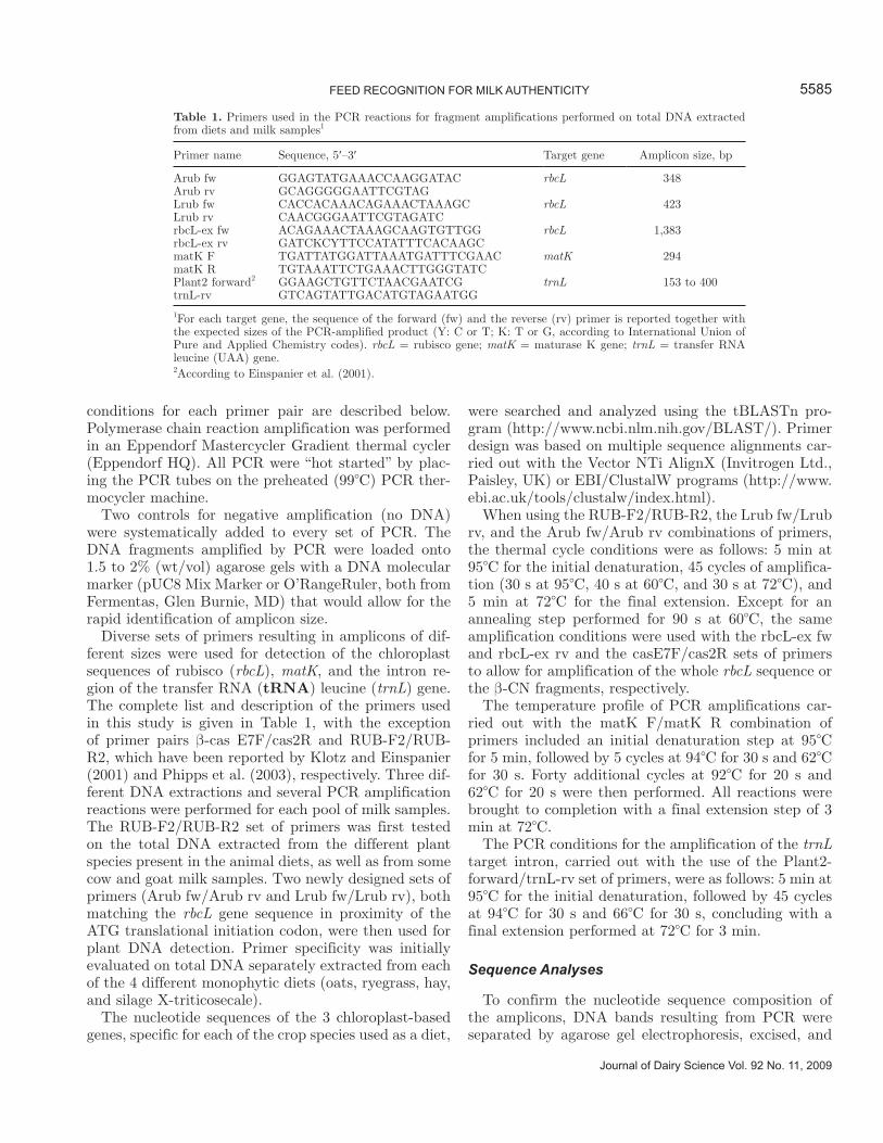
Table 1. Primers used in the PCR reactions for fragment amplifications performed on total DNA extracted from diets and milk samples1
Primer name Sequence, 5′–3′ Target gene Amplicon size, bp
Arub fw GGAGTATGAAACCAAGGATAC rbcL 348Arub rv GCAGGGGGAATTCGTAGLrub fw CACCACAAACAGAAACTAAAGC rbcL 423Lrub rv CAACGGGAATTCGTAGATCrbcL-ex fw ACAGAAACTAAAGCAAGTGTTGG rbcL 1,383rbcL-ex rv GATCKCYTTCCATATTTCACAAGCmatK F TGATTATGGATTAAATGATTTCGAAC matK 294matK R TGTAAATTCTGAAACTTGGGTATCPlant2 forward2 GGAAGCTGTTCTAACGAATCG trnL 153 to 400trnL-rv GTCAGTATTGACATGTAGAATGG
1For each target gene, the sequence of the forward (fw) and the reverse (rv) primer is reported together with the expected sizes of the PCR-amplified product (Y: C or T; K: T or G, according to International Union of Pure and Applied Chemistry codes). rbcL = rubisco gene; matK = maturase K gene; trnL = transfer RNA leucine (UAA) gene.2According to Einspanier et al. (2001).
purified using a QIAquick Gel Extraction Kit (Qiagen, Hamburg, Germany) according to the instructions of the manufacturer. Direct sequencing of the purified PCR products was performed at PRIMM srl (Biomedi-cal Science Park San Raffaele, Milan, Italy). Deoxy-ribonucleic acid sequencing on both strands of the amplified fragments was repeated at least 3 times from 3 independent PCR amplification experiments.
RESULTS
Rubisco Large Subunit Gene Identification in Goat Milk Samples
Milk is a complex food matrix with an abundance of potential PCR inhibitors (Rossen et al., 1992) that can contaminate DNA extracts. In addition to the natural protein and lipid components, DNA from a large num-ber of bacterial and somatic animal cells can interfere with the detection of very low amounts of target plant
DNA sequences. Because of the complexity of milk, several key assay parameters affecting specificity and sensitivity needed to be determined to amplify plant-derived DNA from milk.
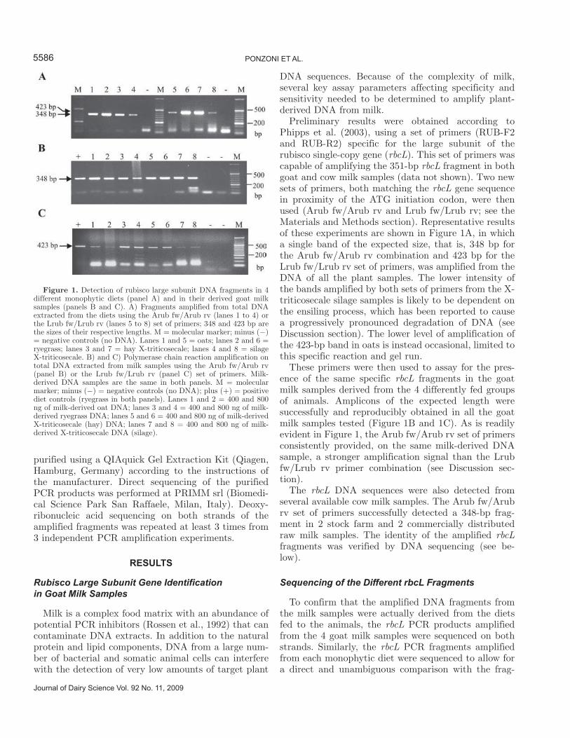
Preliminary results were obtained according to Phipps et al. (2003), using a set of primers (RUB-F2 and RUB-R2) specific for the large subunit of the rubisco single-copy gene (rbcL). This set of primers was capable of amplifying the 351-bp rbcL fragment in both goat and cow milk samples (data not shown). Two new sets of primers, both matching the rbcL gene sequence in proximity of the ATG initiation codon, were then used (Arub fw/Arub rv and Lrub fw/Lrub rv; see the Materials and Methods section). Representative results of these experiments are shown in Figure 1A, in which a single band of the expected size, that is, 348 bp for the Arub fw/Arub rv combination and 423 bp for the Lrub fw/Lrub rv set of primers, was amplified from the DNA of all the plant samples. The lower intensity of the bands amplified by both sets of primers from the X-triticosecale silage samples is likely to be dependent on the ensiling process, which has been reported to cause a progressively pronounced degradation of DNA (see Discussion section). The lower level of amplification of the 423-bp band in oats is instead occasional, limited to this specific reaction and gel run.
These primers were then used to assay for the pres-ence of the same specific rbcL fragments in the goat milk samples derived from the 4 differently fed groups of animals. Amplicons of the expected length were successfully and reproducibly obtained in all the goat milk samples tested (Figure 1B and 1C). As is readily evident in Figure 1, the Arub fw/Arub rv set of primers consistently provided, on the same milk-derived DNA sample, a stronger amplification signal than the Lrub fw/Lrub rv primer combination (see Discussion sec-tion).
The rbcL DNA sequences were also detected from several available cow milk samples. The Arub fw/Arub rv set of primers successfully detected a 348-bp frag-ment in 2 stock farm and 2 commercially distributed raw milk samples. The identity of the amplified rbcL fragments was verified by DNA sequencing (see be-low).
Sequencing of the Different rbcL Fragments
To confirm that the amplified DNA fragments from the milk samples were actually derived from the diets fed to the animals, the rbcL PCR products amplified from the 4 goat milk samples were sequenced on both strands. Similarly, the rbcL PCR fragments amplified from each monophytic diet were sequenced to allow for a direct and unambiguous comparison with the frag-
Journal of Dairy Science Vol. 92 No. 11, 2009
PONZONI ET AL.5586
Figure 1. Detection of rubisco large subunit DNA fragments in 4 different monophytic diets (panel A) and in their derived goat milk samples (panels B and C). A) Fragments amplified from total DNA extracted from the diets using the Arub fw/Arub rv (lanes 1 to 4) or the Lrub fw/Lrub rv (lanes 5 to 8) set of primers; 348 and 423 bp are the sizes of their respective lengths. M = molecular marker; minus (−) = negative controls (no DNA). Lanes 1 and 5 = oats; lanes 2 and 6 = ryegrass; lanes 3 and 7 = hay X-triticosecale; lanes 4 and 8 = silage X-triticosecale. B) and C) Polymerase chain reaction amplification on total DNA extracted from milk samples using the Arub fw/Arub rv (panel B) or the Lrub fw/Lrub rv (panel C) set of primers. Milk-derived DNA samples are the same in both panels. M = molecular marker; minus (−) = negative controls (no DNA); plus (+) = positive diet controls (ryegrass in both panels). Lanes 1 and 2 = 400 and 800 ng of milk-derived oat DNA; lanes 3 and 4 = 400 and 800 ng of milk-derived ryegrass DNA; lanes 5 and 6 = 400 and 800 ng of milk-derived X-triticosecale (hay) DNA; lanes 7 and 8 = 400 and 800 ng of milk-derived X-triticosecale DNA (silage).
ments amplified from the corresponding milk samples. Incidentally, this procedure permitted the identification of an entirely novel nucleotide sequence, the rbcL frag-ment of X-triticosecale, registered in GenBank with accession number FM202540.
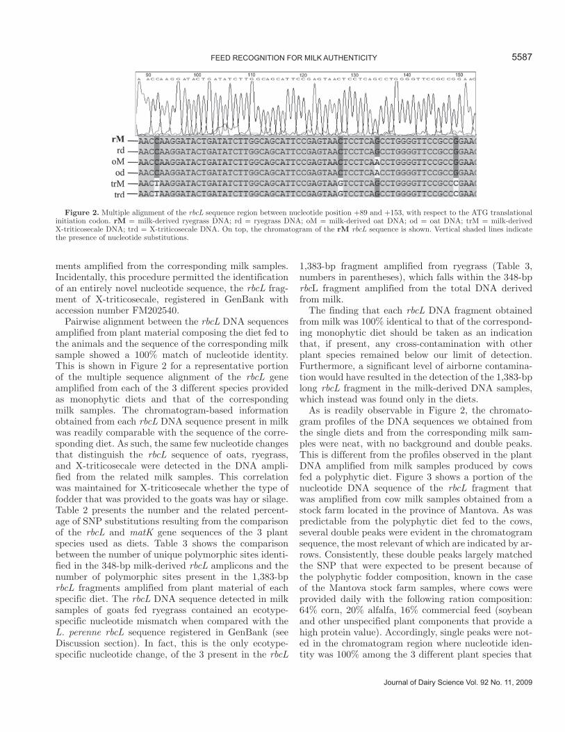
Pairwise alignment between the rbcL DNA sequences amplified from plant material composing the diet fed to the animals and the sequence of the corresponding milk sample showed a 100% match of nucleotide identity. This is shown in Figure 2 for a representative portion of the multiple sequence alignment of the rbcL gene amplified from each of the 3 different species provided as monophytic diets and that of the corresponding milk samples. The chromatogram-based information obtained from each rbcL DNA sequence present in milk was readily comparable with the sequence of the corre-sponding diet. As such, the same few nucleotide changes that distinguish the rbcL sequence of oats, ryegrass, and X-triticosecale were detected in the DNA ampli-fied from the related milk samples. This correlation was maintained for X-triticosecale whether the type of fodder that was provided to the goats was hay or silage. Table 2 presents the number and the related percent-age of SNP substitutions resulting from the comparison of the rbcL and matK gene sequences of the 3 plant species used as diets. Table 3 shows the comparison between the number of unique polymorphic sites identi-fied in the 348-bp milk-derived rbcL amplicons and the number of polymorphic sites present in the 1,383-bp rbcL fragments amplified from plant material of each specific diet. The rbcL DNA sequence detected in milk samples of goats fed ryegrass contained an ecotype-specific nucleotide mismatch when compared with the L. perenne rbcL sequence registered in GenBank (see Discussion section). In fact, this is the only ecotype-specific nucleotide change, of the 3 present in the rbcL
1,383-bp fragment amplified from ryegrass (Table 3, numbers in parentheses), which falls within the 348-bp rbcL fragment amplified from the total DNA derived from milk.
The finding that each rbcL DNA fragment obtained from milk was 100% identical to that of the correspond-ing monophytic diet should be taken as an indication that, if present, any cross-contamination with other plant species remained below our limit of detection. Furthermore, a significant level of airborne contamina-tion would have resulted in the detection of the 1,383-bp long rbcL fragment in the milk-derived DNA samples, which instead was found only in the diets.
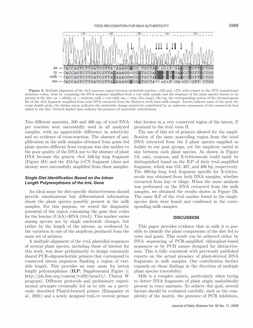
As is readily observable in Figure 2, the chromato-gram profiles of the DNA sequences we obtained from the single diets and from the corresponding milk sam-ples were neat, with no background and double peaks. This is different from the profiles observed in the plant DNA amplified from milk samples produced by cows fed a polyphytic diet. Figure 3 shows a portion of the nucleotide DNA sequence of the rbcL fragment that was amplified from cow milk samples obtained from a stock farm located in the province of Mantova. As was predictable from the polyphytic diet fed to the cows, several double peaks were evident in the chromatogram sequence, the most relevant of which are indicated by ar-rows. Consistently, these double peaks largely matched the SNP that were expected to be present because of the polyphytic fodder composition, known in the case of the Mantova stock farm samples, where cows were provided daily with the following ration composition: 64% corn, 20% alfalfa, 16% commercial feed (soybean and other unspecified plant components that provide a high protein value). Accordingly, single peaks were not-ed in the chromatogram region where nucleotide iden-tity was 100% among the 3 different plant species that
5587FEED RECOGNITION FOR MILK AUTHENTICITY
Journal of Dairy Science Vol. 92 No. 11, 2009
Figure 2. Multiple alignment of the rbcL sequence region between nucleotide position +89 and +153, with respect to the ATG translational initiation codon. rM = milk-derived ryegrass DNA; rd = ryegrass DNA; oM = milk-derived oat DNA; od = oat DNA; trM = milk-derived X-triticosecale DNA; trd = X-triticosecale DNA. On top, the chromatogram of the rM rbcL sequence is shown. Vertical shaded lines indicate the presence of nucleotide substitutions.
constituted the main components of the diet (Figure 3, from nucleotide +224 to +244). We could then observe double peaks, corresponding to those nucleotides that, according to the available sequence data, were differ-ent and could therefore identify 2 or 3 of the different plant species present in the diet. The thick arrow in Figure 3 points to nucleotide position +263, where the nucleotide polymorphism could not be assigned to any of the 3 plant species but is a likely contribution of one of the unknown components of the commercial mix fed to the cows. Similar data were obtained by sequencing the rbcL fragment amplified from samples of raw cow milk sold on the northern Italian market.
Differential PCR Analysis Based on matK Gene Sequences
Sequencing data support the hypothesis that plas-tidial DNA fragments from the diet of an animal can be detected in milk samples. Hence, the molecular traces left by the feed can be tracked.
We attempted to define an assay for distinctiveness depending on a simple PCR run, with primer combina-tions that could possibly differentiate the 3 monophytic diets, based on the minimal differences found when comparing the 3 rbcL DNA sequences. Although we met with occasional success, our efforts to establish a simple rbcL PCR-based assay for distinctiveness were hampered by the high degree of nucleotide identity (more than 97%; National Center for Biotechnology
Information (NCBI)/Nucleotide DataBase and Vec-tor NTi tool (http://www.invitrogen.com/site/us/en/home/Products-and-Services/Applications/Cloning.html); see also Table 2) shared by the 3 DNA sequenc-es. Polymerase chain reactions were successful but not discriminatory.
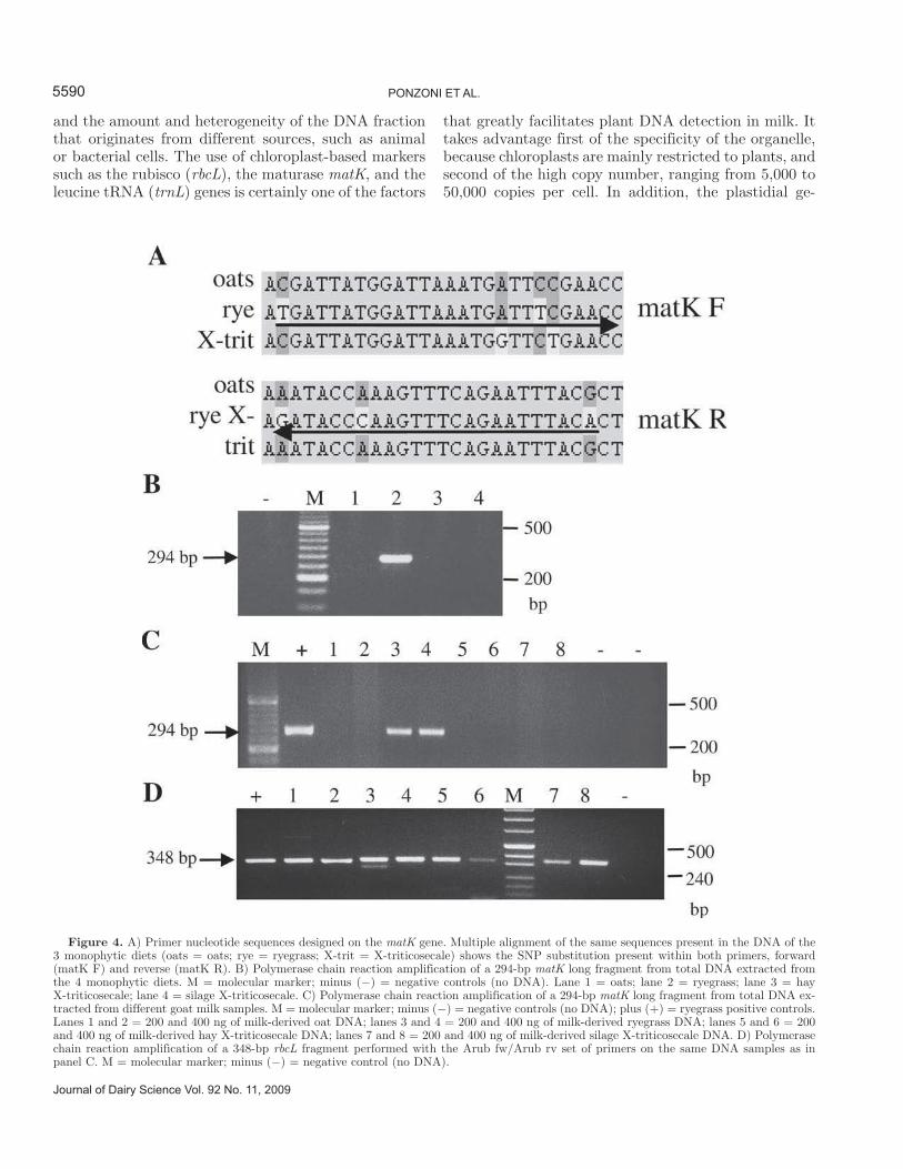
Exploration of both the literature and nucleic acid da-tabase (Hilu et al., 2003; NCBI/Nucleotide DataBase) suggested the use of matK, another single-copy chloro-plast-based molecular marker gene. Multiple alignment of the 3 matK gene sequences of oats, ryegrass, and X-triticosecale allowed us to design a set of primers that could better discriminate ryegrass from the other 2 plant species because of the presence of 3 to 4 nucle-otide mismatches in both the forward and reverse prim-ers. The primers were also longer than previously used primers, 26 and 24 nucleotides, respectively, allowing for greater specificity (Figure 4A). A ryegrass-specific combination of matK primers (matK F/matK R; Table 1) was first tested for successful amplification on total DNA extracted from plant material that composed the 4 monophytic diets. Consistent with this, Figure 4B shows that the combination of ryegrass-specific matK primers was capable of exclusively amplifying a 294-bp matK fragment from the total DNA extracted from ryegrass. The same assay for ryegrass distinctiveness was then applied to total DNA extracted from each of the 4 different goat milk samples. As shown in Figure 4C, the 294-bp fragment was amplified only from milk-derived DNA samples obtained from goats fed ryegrass.
Journal of Dairy Science Vol. 92 No. 11, 2009
PONZONI ET AL.5588
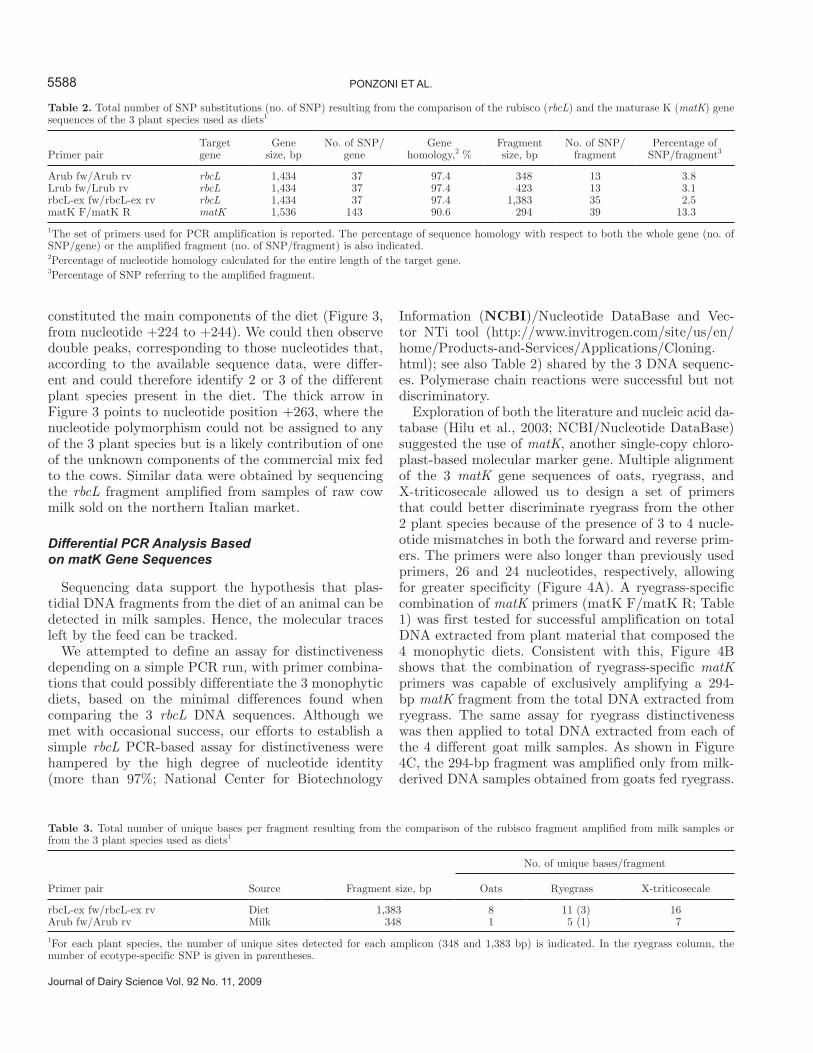
Table 2. Total number of SNP substitutions (no. of SNP) resulting from the comparison of the rubisco (rbcL) and the maturase K (matK) gene sequences of the 3 plant species used as diets1
Primer pairTarget gene
Gene size, bp
No. of SNP/gene
Gene homology,2 %
Fragment size, bp
No. of SNP/fragment
Percentage of SNP/fragment3
Arub fw/Arub rv rbcL 1,434 37 97.4 348 13 3.8Lrub fw/Lrub rv rbcL 1,434 37 97.4 423 13 3.1rbcL-ex fw/rbcL-ex rv rbcL 1,434 37 97.4 1,383 35 2.5matK F/matK R matK 1,536 143 90.6 294 39 13.3
1The set of primers used for PCR amplification is reported. The percentage of sequence homology with respect to both the whole gene (no. of SNP/gene) or the amplified fragment (no. of SNP/fragment) is also indicated.2Percentage of nucleotide homology calculated for the entire length of the target gene.3Percentage of SNP referring to the amplified fragment.
Table 3. Total number of unique bases per fragment resulting from the comparison of the rubisco fragment amplified from milk samples or from the 3 plant species used as diets1
Primer pair Source Fragment size, bp
No. of unique bases/fragment
Oats Ryegrass X-triticosecale
rbcL-ex fw/rbcL-ex rv Diet 1,383 8 11 (3) 16Arub fw/Arub rv Milk 348 1 5 (1) 7
1For each plant species, the number of unique sites detected for each amplicon (348 and 1,383 bp) is indicated. In the ryegrass column, the number of ecotype-specific SNP is given in parentheses.
Two different amounts, 200 and 400 ng, of total DNA per reaction were successfully used in all analyzed samples, with no appreciable difference in selectivity and no evidence of cross-reaction. The absence of am-plification in the milk samples obtained from goats fed plant species different from ryegrass was due neither to the poor quality of the DNA nor to the absence of plant DNA because the generic rbcL 348-bp long fragment (Figure 4D) and the 453-bp β-CN fragment (data not shown) were successfully amplified from these samples.
Single Diet Identification Based on the Intron Length Polymorphisms of the trnL Gene
An ideal assay for diet-specific distinctiveness should provide simultaneous but differentiated information about the plant species possibly present in the milk samples. For this purpose, we tested the diagnostic potential of the region containing the gene that codes for the leucine (UAA) tRNA (trnL). This marker varies among species not by single nucleotide changes, but rather by the length of the introns, as evidenced by the variation in size of the amplicon produced from the same set of primers.
A multiple alignment of the trnL plastidial sequences of several plant species, including those of interest for this work, was done preliminarily to design commonly shared PCR-oligonucleotide primers that correspond to conserved intron sequences flanking a region of vari-able length. This provides an easy assay for intron length polymorphisms (ILP; Supplemental Figure 1: http://jds.fass.org/content/vol92/issue11/; Clustal W program). Different protocols and preliminary experi-mental attempts eventually led us to rely on a previ-ously described Plant2-forward primer (Einspanier et al., 2001) and a newly designed trnL-rv reverse primer
that locates in a very conserved region of the intron, 3′ proximal to the trnL exon II.
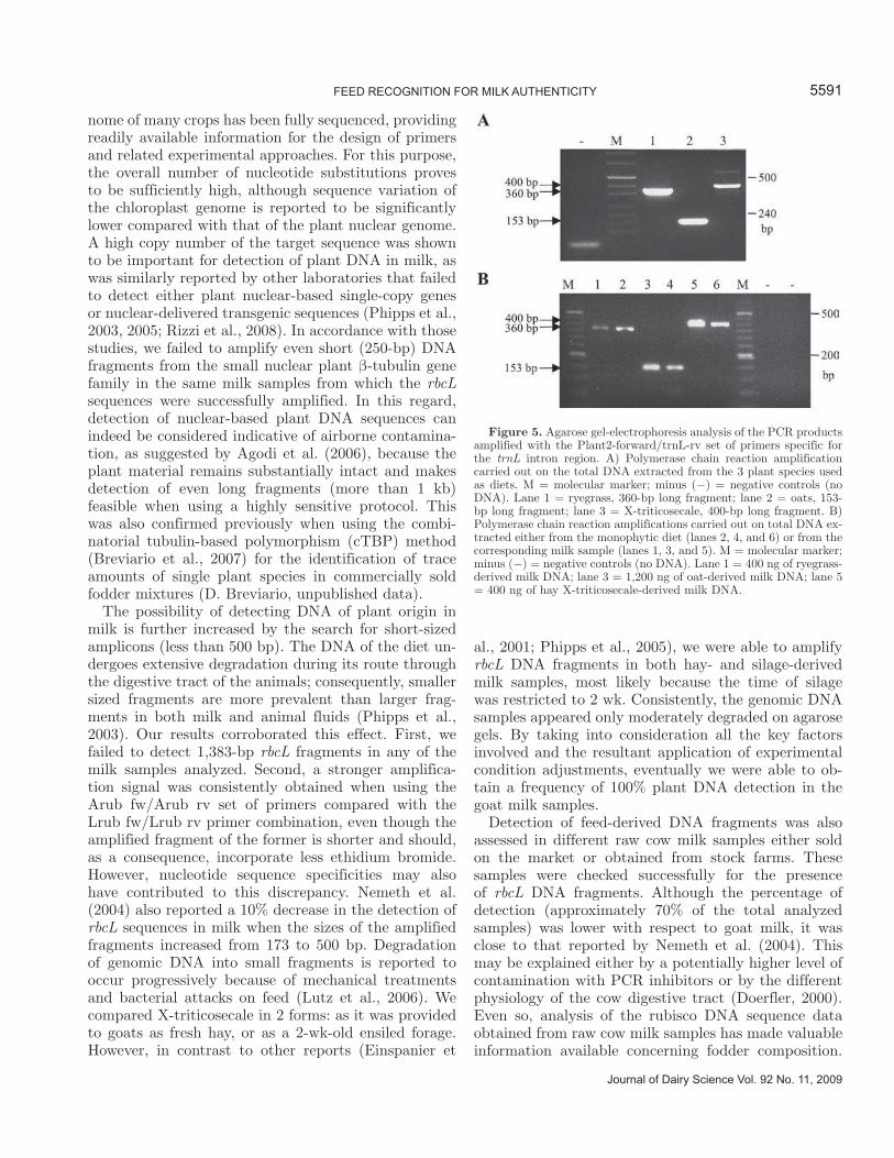
The use of this set of primers allowed for the ampli-fication of the same noncoding region from the total DNA extracted from the 3 plant species supplied as fodder to our goat groups, yet the amplicon varied in size between each plant species. As shown in Figure 5A, oats, ryegrass, and X-triticosecale could easily be distinguished based on the ILP of their trnL-amplified fragment, which was 153, 367, and 400 bp, respectively. The 400-bp long trnL fragment specific for X-tritico-secale was obtained from both DNA samples, whether extracted from hay or silage. When the same analysis was performed on the DNA extracted from the milk samples, we obtained the results shown in Figure 5B. The same ILP of the trnL marker found in the single-species diets were found and confirmed in the corre-sponding milk samples.
DISCUSSION
This paper provides evidence that in milk it is pos-sible to identify the plant components of the diet fed to cows and goats. This result can be achieved either by DNA sequencing of PCR-amplified chloroplast-based sequences or by PCR assays designed for distinctive-ness. This is fully consistent with previously published reports on the actual presence of plant-derived DNA fragments in milk samples. Our contribution further expands on those findings in the direction of multiple plant species traceability.
Milk is a complex matrix, particularly when trying to detect DNA fragments of plant origin undoubtedly present in trace amounts. To achieve this goal, several factors should be evaluated carefully, such as the com-plexity of the matrix, the presence of PCR inhibitors,
5589FEED RECOGNITION FOR MILK AUTHENTICITY
Journal of Dairy Science Vol. 92 No. 11, 2009
Figure 3. Multiple alignment of the rbcL sequence region between nucleotide position +222 and +275, with respect to the ATG translational initiation codon, done by comparing the DNA sequence amplified from a cow milk sample and the sequence of the plant species known to be present in the diet. aa = alfalfa; sy = soybean; milk = cow milk; ma = corn (Zea mays). On top, the corresponding section of the chromatogram file of the rbcL fragment amplified from total DNA extracted from the Mantova stock farm milk sample. Arrows indicate some of the most rel-evant double picks; the thicker arrow indicates the nucleotide change putatively contributed by an unknown component of the commercial feed added to the diet. Vertical shaded lines indicate the presence of nucleotide substitutions.
and the amount and heterogeneity of the DNA fraction that originates from different sources, such as animal or bacterial cells. The use of chloroplast-based markers such as the rubisco (rbcL), the maturase matK, and the leucine tRNA (trnL) genes is certainly one of the factors
that greatly facilitates plant DNA detection in milk. It takes advantage first of the specificity of the organelle, because chloroplasts are mainly restricted to plants, and second of the high copy number, ranging from 5,000 to 50,000 copies per cell. In addition, the plastidial ge-
Journal of Dairy Science Vol. 92 No. 11, 2009
PONZONI ET AL.5590
Figure 4. A) Primer nucleotide sequences designed on the matK gene. Multiple alignment of the same sequences present in the DNA of the 3 monophytic diets (oats = oats; rye = ryegrass; X-trit = X-triticosecale) shows the SNP substitution present within both primers, forward (matK F) and reverse (matK R). B) Polymerase chain reaction amplification of a 294-bp matK long fragment from total DNA extracted from the 4 monophytic diets. M = molecular marker; minus (−) = negative controls (no DNA). Lane 1 = oats; lane 2 = ryegrass; lane 3 = hay X-triticosecale; lane 4 = silage X-triticosecale. C) Polymerase chain reaction amplification of a 294-bp matK long fragment from total DNA ex-tracted from different goat milk samples. M = molecular marker; minus (−) = negative controls (no DNA); plus (+) = ryegrass positive controls. Lanes 1 and 2 = 200 and 400 ng of milk-derived oat DNA; lanes 3 and 4 = 200 and 400 ng of milk-derived ryegrass DNA; lanes 5 and 6 = 200 and 400 ng of milk-derived hay X-triticosecale DNA; lanes 7 and 8 = 200 and 400 ng of milk-derived silage X-triticosecale DNA. D) Polymerase chain reaction amplification of a 348-bp rbcL fragment performed with the Arub fw/Arub rv set of primers on the same DNA samples as in panel C. M = molecular marker; minus (−) = negative control (no DNA).
nome of many crops has been fully sequenced, providing readily available information for the design of primers and related experimental approaches. For this purpose, the overall number of nucleotide substitutions proves to be sufficiently high, although sequence variation of the chloroplast genome is reported to be significantly lower compared with that of the plant nuclear genome. A high copy number of the target sequence was shown to be important for detection of plant DNA in milk, as was similarly reported by other laboratories that failed to detect either plant nuclear-based single-copy genes or nuclear-delivered transgenic sequences (Phipps et al., 2003, 2005; Rizzi et al., 2008). In accordance with those studies, we failed to amplify even short (250-bp) DNA fragments from the small nuclear plant β-tubulin gene family in the same milk samples from which the rbcL sequences were successfully amplified. In this regard, detection of nuclear-based plant DNA sequences can indeed be considered indicative of airborne contamina-tion, as suggested by Agodi et al. (2006), because the plant material remains substantially intact and makes detection of even long fragments (more than 1 kb) feasible when using a highly sensitive protocol. This was also confirmed previously when using the combi-natorial tubulin-based polymorphism (cTBP) method (Breviario et al., 2007) for the identification of trace amounts of single plant species in commercially sold fodder mixtures (D. Breviario, unpublished data).
The possibility of detecting DNA of plant origin in milk is further increased by the search for short-sized amplicons (less than 500 bp). The DNA of the diet un-dergoes extensive degradation during its route through the digestive tract of the animals; consequently, smaller sized fragments are more prevalent than larger frag-ments in both milk and animal fluids (Phipps et al., 2003). Our results corroborated this effect. First, we failed to detect 1,383-bp rbcL fragments in any of the milk samples analyzed. Second, a stronger amplifica-tion signal was consistently obtained when using the Arub fw/Arub rv set of primers compared with the Lrub fw/Lrub rv primer combination, even though the amplified fragment of the former is shorter and should, as a consequence, incorporate less ethidium bromide. However, nucleotide sequence specificities may also have contributed to this discrepancy. Nemeth et al. (2004) also reported a 10% decrease in the detection of rbcL sequences in milk when the sizes of the amplified fragments increased from 173 to 500 bp. Degradation of genomic DNA into small fragments is reported to occur progressively because of mechanical treatments and bacterial attacks on feed (Lutz et al., 2006). We compared X-triticosecale in 2 forms: as it was provided to goats as fresh hay, or as a 2-wk-old ensiled forage. However, in contrast to other reports (Einspanier et
al., 2001; Phipps et al., 2005), we were able to amplify rbcL DNA fragments in both hay- and silage-derived milk samples, most likely because the time of silage was restricted to 2 wk. Consistently, the genomic DNA samples appeared only moderately degraded on agarose gels. By taking into consideration all the key factors involved and the resultant application of experimental condition adjustments, eventually we were able to ob-tain a frequency of 100% plant DNA detection in the goat milk samples.
Detection of feed-derived DNA fragments was also assessed in different raw cow milk samples either sold on the market or obtained from stock farms. These samples were checked successfully for the presence of rbcL DNA fragments. Although the percentage of detection (approximately 70% of the total analyzed samples) was lower with respect to goat milk, it was close to that reported by Nemeth et al. (2004). This may be explained either by a potentially higher level of contamination with PCR inhibitors or by the different physiology of the cow digestive tract (Doerfler, 2000). Even so, analysis of the rubisco DNA sequence data obtained from raw cow milk samples has made valuable information available concerning fodder composition.
5591FEED RECOGNITION FOR MILK AUTHENTICITY
Journal of Dairy Science Vol. 92 No. 11, 2009
Figure 5. Agarose gel-electrophoresis analysis of the PCR products amplified with the Plant2-forward/trnL-rv set of primers specific for the trnL intron region. A) Polymerase chain reaction amplification carried out on the total DNA extracted from the 3 plant species used as diets. M = molecular marker; minus (−) = negative controls (no DNA). Lane 1 = ryegrass, 360-bp long fragment; lane 2 = oats, 153-bp long fragment; lane 3 = X-triticosecale, 400-bp long fragment. B) Polymerase chain reaction amplifications carried out on total DNA ex-tracted either from the monophytic diet (lanes 2, 4, and 6) or from the corresponding milk sample (lanes 1, 3, and 5). M = molecular marker; minus (−) = negative controls (no DNA). Lane 1 = 400 ng of ryegrass-derived milk DNA; lane 3 = 1,200 ng of oat-derived milk DNA; lane 5 = 400 ng of hay X-triticosecale-derived milk DNA.
The chromatogram profile was not uniformly clear, as was evident for the goat milk. Few but significant overlapping single base peaks, representative of the different SNP expected to be present because of the multispecies composition of the diet, were detected. Where the nucleotide sequence was identical for all the components of the fodder, the chromatogram was clean and unambiguous, but double peaks emerged, corresponding to predicted species-specific nucleotide changes. In addition, DNA sequencing data allowed us to infer a priori the composition of the diet supplied to those cows that produce a brand-labeled raw milk com-monly sold on the northern Italian market. Our diag-nosis, based on the rbcL sequence data, was compatible with the information that was later recovered from the producer. This also highlights the possibility of using DNA sequencing for the identification and diagnosis of uncertified components (undisclosed on the fodder tag) possibly present as contaminants in animal feed.
To our knowledge, this is the first time that nucle-otide sequencing data, with reference to components of the diet detectable in milk samples, have actually been shown. This potentially provides insight into the differ-ences that can be found when comparing milk samples produced by animals fed different diets.
We have also shown and discussed our DNA sequence data with reference to the different diets consumed by the animal, whether polyphytic or monophytic. With regard to the latter, DNA sequences were found to be 100% identical when comparing the amplified rbcL DNA fragments of the goat milk sample with that of the corresponding diet. This was found for all 3 plant species investigated in the present study. Of additional relevance is the finding that the DNA amplified from milk samples contained the same few nucleotide changes that distinguish the rubisco DNA fragments of oats, ryegrass, and X-triticosecale. For oats, the amplified rbcL sequence was identical to the sequence registered in GenBank. For ryegrass, the rbcL DNA sequence am-plified from the milk of goats fed ryegrass contained the same nucleotide change (at nucleotide position +334) found in the corresponding sequence of the ryegrass ecotype used as the diet. This sequence is different from the rbcL DNA sequence of the ryegrass reference variety in GenBank. In fact, this is 1 of 3 mismatches (the other 2 located at positions +678 and +1,144) we found when comparing our ryegrass variety with that registered in the database (NCBI, accession number NC009950) for the whole length of the rbcL 1,383-bp fragment (Tables 1 and 3). For X-triticosecale, no reference sequence was present in the database, as mentioned above.
The rubisco sequencing data demonstrate that DNA fragments of plastidial origin can be recovered equally in cow and goat milk samples and that DNA sequence
analysis can be used to assess the composition of mono-phytic as well as polyphytic diets. Therefore, DNA se-quencing can be used as a tool for traceability of plant sequences in milk.
The unambiguous nucleotide correlations found when comparing rbcL fragments amplified from the diet and the corresponding milk sample indicate that contami-nation did not interfere with our data. The occurrence of contamination or cross-reactions with rubisco se-quences of other organisms, such as photosynthetic bacteria, fungi, and algae, was excluded by our data on matK. The latter is a gene that encodes for the only known maturase of higher plant plastids (Vogel et al., 1997), with no corresponding paralogous or orthologous sequences in organisms different from higher plants.
Although the issue regarding the length of feed-de-rived DNA fragments that can be detected in the ani-mal gastrointestinal track is still ongoing (Chowdhury et al., 2004), and despite the precautions taken when collecting the milk samples, we cannot rule out with absolute confidence (100%) that negligible amounts of feces could have been accidentally included in the milk samples. Such a possibility, although much less prob-able than that of some DNA release during milking, would have resulted in greater variation in the detec-tion of diet-specific DNA fragments in all the samples analyzed.
The alignment of the 3 rbcL DNA sequences obtained from the different plant species used to feed goats re-vealed the presence of sporadic SNP occurring within an overall nucleotide identity amounting to 97.4%. De-spite this, we tried to exploit these SNP to determine PCR conditions that would permit us to selectively dis-tinguish each of the 3 diets. However, specific detection was not consistently achieved. For this reason, it was necessary to use matK because the ryegrass sequence of this gene contains a higher number of nucleotide mis-matches, which could be useful in designing selective primers.
The matK gene, located in the chloroplast genome within an intron of group II, nested between the 5′ and the 3′ exons of the trnK (tRNA-lysine) gene (Barthet and Hilu, 2008), represents an excellent tool for the se-lective identification of plant-specific DNA fragments. The reason is that its sequence variability allows for the design of distinctiveness assays based on PCR, a much more convenient and faster means of detection compared with DNA sequencing (see below).
With regard to the use of a matK-based distinctive-ness assay, it must also be pointed out that detection is favored by the fact that the amount of DNA used for each PCR reaction can be 10-fold higher than that used for the rbcL marker. The greater number of polymorphisms in matK is likely a consequence of the
Journal of Dairy Science Vol. 92 No. 11, 2009
PONZONI ET AL.5592
higher mutational rate that has affected the matK gene sequence compared with rbcL (Soltis and Soltis, 1998), which is attributable to minor selective pressure on the former. Table 2 provides some data regarding the number and frequency of SNP substitutions, which bet-ter support this view and explain the success obtained using the matK-based primers for a ryegrass-specific distinctiveness assay. In fact, although the total num-ber of SNP found when comparing the whole rbcL gene sequences of the 3 species amounted to 37, this number was increased to 143 when comparing the matK gene sequences. This discrepancy was also reflected in the frequency of SNP present in the PCR-amplified frag-ments, which ranged between 2.5 and 3.8% for the dif-ferently sized rbcL fragments, and increased to 13.3% for matK, thereby confirming that matK is potentially a better and more promising target for the development of PCR-based distinctiveness assays.
Feed traceability done using matK sequences success-fully confirmed the unambiguous correlation between the milk and the diet from which the milk was eventu-ally produced. The higher percentage of SNP present in the plant matK compared with the rbcL nucleotide sequence raises an important point for the future devel-opment of distinctiveness assays that aim to increase the number of species that can be detected.
In fact, this possibility was anticipated with the data obtained when using the intron of the trnL chloroplast-located gene as the source for detection of ILP. The trnL gene has already been used successfully for evolution-ary studies (Tsai et al., 2006) and as a genetic marker (Taberlet et al., 1991; Gielly and Taberlet, 1994). Here, the different sizes of the trnL-amplified bands were more conveniently used for the identification of a plant species-specific length-based polymorphism in what was a true assay for distinctiveness.
Further improvements along the line of plant species traceability in milk can also be foreseen. This approach for multiple-species detection could effectively replace DNA sequencing, the success of which always depends on the clear output and reading of the multiple peaks found in the chromatogram.
CONCLUSIONS
We showed that feed-derived chloroplast-based DNA fragments can be detected in milk samples and that their plant origin can be traced by both DNA sequenc-ing and PCR-based distinctiveness assays. This was done using 3 chloroplast-based molecular markers. The discriminatory power of each was used selectively in 3 different applications, DNA sequencing for rbcL, SNP for matK, and ILP for trnL.
ACKNOWLEDGMENTS
This work was partially supported by the Italian Ministry of Agriculture (Rome, Italy) within the frame shift of the “IDENTILAT” project. The authors thank S. Claps and V. Fedele (CRA, Potenza, Italy), P. Leone (IBBA-CNR, Lodi, Italy), and M. Feligini (Istituto Spallanzani, Rivolta d’Adda, Italy) for their assistance in delivering the material and in providing useful in-formation. The authors also thank Mona Monfared (USDA, Albany, CA) and Anna Whittaker (University of Florence, Florence, Italy) for their critical reading.
REFERENCESAgodi, A., M. Barchetta, A. Grillo, and S. Sciacca. 2006. Detection
of genetically modified DNA sequences in milk from the Italian market. Int. J. Hyg. Environ. Health 209:81–88.
Barthet, M. M., and K. W. Hilu. 2008. Evaluating evolutionary constraint on the rapidly evolving gene matK using protein composition. J. Mol. Evol. 66:85–97.
Beever, D. E., and C. F. Kemp. 2000. Safety issues associated with the DNA in animal feed derived from genetically modified crops. A review of scientific and regulatory procedures. Nutr. Abstr. Rev. Ser. Livest. Feeds Feeding 70:175–182.
Breviario, D., W. V. Baird, S. Sangoi, K. Hilu, P. Blumetti, and S. Gianì. 2007. High polymorphism and resolution in targeted fingerprinting with combined beta-tubulin introns. Mol. Breed. 20:249–259.
Castillo, A. R., M. R. Gallardo, M. Maciel, J. M. Giordano, G. A. Conti, M. C. Gaggiotti, O. Quaino, C. Gianni, and G. F. Hartnell. 2004. Effects of feeding rations with genetically modified whole cottonseed to lactating dairy cows. J. Dairy Sci. 87:1778–1785.
Chowdhury, E. H., O. Mikami, H. Murata, P. Sultana, N. Shimada, M. Yoshioka, K. S. Guruge, S. Yamamoto, S. Miyazaki, N. Yamanaka, and Y. Nakajima. 2004. Fate of maize intrinsic and recombinant genes in calves fed genetically modified maize Bt11. J. Food Prot. 67:365–370.
Doerfler, W. 2000. Foreign DNA in Mammalian Systems. 1st ed. Wiley VCH, Weinheim, Germany.
Doyle, J. J., and J. L. Doyle. 1987. A rapid DNA isolation procedure for small quantities of fresh leaf tissue. Phytochem. Bull. 19:11–15.
Einspanier, R., A. Klotz, J. Kraft, K. Aulrich, R. Poser, F. Schwägele, G. Jahreis, and G. Flachowsky. 2001. The fate of forage plant DNA in farm animals: A collaborative case-study investigating cattle and chicken fed recombinant plant material. Eur. Food Res. Technol. 212:129–134.
Gielly, L., and P. Taberlet. 1994. The use of chloroplast DNA to resolve plant phylogenies: Noncoding versus rbcL sequences. Mol. Biol. Evol. 11:769–777.
Hilu, K. W., T. Borsch, K. Müller, D. E. Soltis, P. S. Soltis, V. Savolainen, M. W. Chase, M. P. Powell, L. A. Alice, R. Evans, H. Sauquet, C. Neinhuis, T. A. B. Slotta, J. G. Rohwer, C. S. Campbell, and L. W. Chatrou. 2003. Angiosperm phylogeny based on matK sequence information. Am. J. Bot. 90:1758–1776.
Hohlweg, U., and W. Doerfler. 2001. On the fate of plant or other foreign genes upon uptake in food or after intramuscular injection in mice. Mol. Genet. Genomics 265:225–233.
Jennings, J. C., A. J. Whetsell, N. R. Nicholas, B. M. Sweeney, M. B. Klaften, S. B. Kays, G. F. Hartnell, R. P. Lirette, and K. C. Glenn. 2003. Determining whether transgenic or endogenous plant DNA is detectable in dairy milk or beef organs. Pages 41–46 in Bull. 383/2003. Int. Dairy Fed., Brussels, Belgium.
Jonas, D. A., I. Elmadfa, K.-H. Engel, K. J. Heller, G. Kozianowski, A. König, D. J. Müller, F. Narbonne, W. Wackernagel, and J.
5593FEED RECOGNITION FOR MILK AUTHENTICITY
Journal of Dairy Science Vol. 92 No. 11, 2009
Kleiner. 2001. Safety considerations of DNA in food. Ann. Nutr. Metab. 45:235–254.
Klotz, A., and R. Einspanier. 2001. Development of a DNA-based screening method to detect cow milk in ewe, goat and buffalo milk and dairy products using PCR-LCR-EIA-technique. Milchwissenschaft 56:67–70.
Lutz, B., S. Wiedemann, and C. Albrecht. 2006. Degradation of transgenic Cry1Ab DNA and protein in Bt-176 maize during the ensiling process. J. Anim. Physiol. Anim. Nutr. (Berl.) 90:116–123.
Mafra, I., I. M. P. L. V. O. Ferreira, and M. B. P. P. Oliveira. 2008. Food authentication by PCR-based methods. Eur. Food Res. Technol. 227:649–665.
Mazza, R., M. Soave, M. Morlacchini, G. Piva, and A. Marocco. 2005. Assessing the transfer of genetically modified DNA from feed to animal tissues. Transgenic Res. 14:775–784.
McAllan, A. B. 1982. The fate of nucleic acids in ruminants. Proc. Nutr. Soc. 41:309–317.
Meyer, R., F. Chardonnes, P. Hubner, and J. Luthy. 1996. Polymerase chain reaction (PCR) in the quality and safety assurance of food: Detection of soya in processed meat products. Z. Lebensm. Unters. Forsch. 203:339–344.
Nemeth, A., A. Wurz, L. Artim, S. Charlton, G. Dana, K. Glenn, P. Hunst, J. Jennings, R. Shilito, and P. Song. 2004. Sensitive PCR analysis of animal tissue samples for fragments of endogenous and transgenic plant DNA. J. Agric. Food Chem. 52:6129–6135.
Phipps, R. H., E. R. Deaville, and B. C. Maddison. 2003. Detection of transgenic and endogenous plant DNA in rumen fluid, duodenal digesta, milk, blood, and feces of lactating dairy cows. J. Dairy Sci. 86:4070–4078.
Phipps, R. H., A. K. Jones, A. P. Tingey, and S. Abeyasekera. 2005. Effect of corn silage from an herbicide-tolerant genetically modified variety on milk production and absence of transgenic DNA in milk. J. Dairy Sci. 88:2870–2878.
Poms, R. E., W. Hochsteiner, K. Luger, J. Glössl, and H. Foissy. 2003. Model studies on the detectability of genetically modified feeds in milk. J. Food Prot. 66:304–310.
Rizzi, A., L. Brusetti, S. Arioli, K. M. Nielsen, A. Tamburini, C. Sorlini, and D. Daffonchio. 2008. Detection of feed-derived maize DNA in goat milk and evaluation of the potential of horizontal transfer to bacteria. Eur. Food Res. Technol. 277:1699–1709.
Rossen, L., P. Norskov, K. Holmstrom, and O. F. Rasmussen. 1992. Inhibition of PCR by components of food samples, microbial diagnostic assays and DNA extraction solutions. Int. J. Food Microbiol. 17:37–45.
Schubbert, R., C. Lettmann, and W. Doerfler. 1994. Ingested foreign (phage M13) DNA survives transiently in the gastrointestinal tract and enters the bloodstream of mice. Mol. Gen. Genet. 242:495–504.
Schubbert, R., D. Renz, B. Schmitz, and W. Doerfler. 1997. Foreign (M13) DNA ingested by mice reaches peripheral leukocytes, spleen, and liver via the intestinal wall mucosa and can be covalently linked to mouse DNA. Proc. Natl. Acad. Sci. USA 94:961–966.
Soltis, D. E., and P. S. Soltis. 1998. Choosing an approach and an appropriate gene for phylogenetic analysis. Pages 1–42 in Molecular Systematics of Plants II. DNA Sequencing. D. E. Soltis, P. S. Soltis, and J. J. Doyle, ed. Kluwer Academic Publishers, Boston, MA.
Taberlet, P., L. Gielly, G. Pautou, and J. Bouvet. 1991. Universal primers for amplification of 3 non-coding regions of chloroplast DNA. Plant Mol. Biol. 17:1105–1109.
Tsai, L. C., Y. C. Yu, H. M. Hsieh, J. C. Wang, A. Linacre, and J. C. Lee. 2006. Species identification using sequences of the trnL intron and the trnL-trnF IGS of chloroplast genome among popular plants in Taiwan. Forensic Sci. Int. 164:193–200.
Vogel, J., T. Hubschmann, T. Borner, and W. R. Hess. 1997. Splicing and intron-internal RNA editing of trnK-matK transcripts in barley plastids: Support for MatK as an essential splicing factor. J. Mol. Biol. 270:179–187.
Journal of Dairy Science Vol. 92 No. 11, 2009
PONZONI ET AL.5594


