Embed Size (px)
Citation preview
Ras Trokovic, Nina Trokovic,Sanna Hernesniemi, Ulla Pirvola,Daniela M.Vogt Weisenhorn1,2,Janet Rossant3, Andrew P.McMahon4,Wolfgang Wurst1,2 and Juha Partanen5
Institute of Biotechnology, Viikki Biocenter, PO Box 56, 00014University of Helsinki, Finland, 3Samuel Lunenfeld Research Institute,Mount Sinai Hospital, 600 University Avenue, Toronto, OntarioM5G 1X5, Canada, 4Department of Molecular and Cellular Biology,Harvard University, 16 Divinity Avenue, Cambridge, MA 02138,USA, 1GSF-Research Centre for Environment and Health, TechnicalUniversity Munich, Institute of Mammalian Genetics, IngolstaedterLandstrasse 1, D-85764 Neuhenberg and 2Max Planck Institute ofPsychiatry, Kapelinstrasse 2-16, D-80804 Munich, Germany
5Corresponding authore-mail: [email protected]
Fibroblast growth factors (FGFs) are signalingmolecules of the isthmic organizer, which regulatesdevelopment of the midbrain and cerebellum. Tissue-speci®c inactivation of one of the FGF receptor(FGFR) genes, Fgfr1, in the midbrain and rhombo-mere 1 of the hindbrain of mouse embryos results indeletion of the inferior colliculi in the posterior mid-brain and vermis of the cerebellum. Analyses of bothmidbrain±hindbrain and midbrain-speci®c Fgfr1mutants suggest that after establishment of the isthmicorganizer, FGFR1 is needed for continued response tothe isthmic signals, and that it has direct functions onboth sides of the organizer. In addition, FGFR1appears to modify cell adhesion properties critical formaintaining a coherent organizing center. This maybe achieved by regulating expression of speci®ccell-adhesion molecules at the midbrain±hindbrainborder.Keywords: cerebellum/Cre recombinase/development/FGF/isthmic organizer/midbrain
Introduction
Organizing centers established at the borders betweendevelopmental units are commonly used for tissuepatterning during embryogenesis. One such organizingcenter is the isthmic organizer, which forms at the junctionbetween developing midbrain and rhombomere 1 of thehindbrain. Transplantation studies with avian embryoshave demonstrated that tissue containing the midbrain±hindbrain junction can induce cells in more anterior andposterior regions of the brain to adopt fates characteristicfor midbrain and rhombomere 1 (Nakamura et al., 1988;Martinez et al., 1991, 1995). Under the control of theisthmic organizer the dorsal midbrain develops intosuperior and inferior colliculi, relaying visual and auditorystimuli, respectively. In turn, dorsal rhombomere 1 forms
the cerebellum involved in processes such as motorcoordination. Development of some of the ganglia in theventral brainstem is also thought to be regulated by theisthmic organizer.
The molecular basis for the development and functionof the isthmic organizer is beginning to be understood. Theborder of expression of Otx2 and Gbx2, two homeodomaintranscription factors, separates the cells of the futuremidbrain from the hindbrain and determines the positionof the isthmic organizer (Millet et al., 1996, 1999;Broccoli et al., 1999). In addition to the early regionaliza-tion of the neurectoderm, signals from the mesoderm arerequired for the induction of genes, such as En1 and En2,in the mid- and hindbrain region (Hemmati-Brivanlouet al., 1990; Ang and Rossant, 1993). Studies with avianembryos have suggested that ®broblast growth factors(FGFs), potentially FGF4 transiently expressed in theanterior notochord, are important for the induction of mid-and hindbrain-speci®c gene expression (Shamim et al.,1999). In addition, Fgf8, expressed in the cardiogenicmesoderm, has been suggested to play a role in theinduction of the midbrain (Crossley et al., 1996). Inaddition to the En genes, the paired box transcriptionfactor genes Pax2 and Pax5 are also activated early in theentire midbrain±hindbrain region.
Later in development, patterning and growth of themidbrain and hindbrain rely on the isthmic organizer,which forms in the neurectoderm at the Otx2/Gbx2 border.An important signaling molecule of the isthmic organizeris FGF8. In the mouse embryo, Fgf8 expression isactivated after Pax2 and En1 in the entire rhombomere 1of the hindbrain, and later restricted to a stripe in the mostanterior hindbrain (Crossley and Martin, 1995). Both gain-and loss-of-function experiments have suggested thatFGF8 is essential for the activity of the isthmic organizer.FGF8 containing beads can mimic the isthmic transplantsin induction of midbrain and cerebellum in the dience-phalon or cerebellum in the posterior hindbrain (Crossleyet al., 1996; Martinez et al., 1999; Irving and Mason,2000). In addition, zebra®sh Fgf8 mutants fail to maintainisthmic gene expression (Reifers et al., 1998), and ahypomorphic mutation in the mouse Fgf8 gene causesmidbrain and cerebellar defects (Meyers et al., 1998).Other FGF family members, such as Fgf17 and Fgf18, arealso expressed in the isthmic region and may contribute toorganizer function (Maruoka et al., 1998; Xu et al., 2000).
Another important signaling molecule of the isthmicorganizer is WNT1. Initially, Wnt1 is detected in the entiremidbrain, but later Wnt1-positive cells are found as a narrowstripe in the most posterior midbrain next to the Fgf8-expressing cells in the anterior hindbrain. FGF-containingbeads can induce Wnt1 expression, implicating Wnt1 as oneof the targets of FGF signaling (Crossley et al., 1996). On theother hand, Fgf8 expression is down-regulated in the Wnt1
FGFR1 is independently required in both developingmid- and hindbrain for sustained response toisthmic signals
The EMBO Journal Vol. 22 No. 8 pp. 1811±1823, 2003
ã European Molecular Biology Organization 1811
mutants, suggesting that a midbrain-derived signal in turnmaintains Fgf8 (Lee et al., 1997). Other FGF-regulatablegenes include Pax2/5 and En1/2, which are expressedaround the isthmic organizer in a graded manner. Thesesignaling molecules and transcription factors appear to beinvolved in a complex regulatory network responsible forthe maintenance of their expression and development of themidbrain±hindbrain region (reviewed in Wurst and Bally-Cuif, 2001; Liu and Joyner, 2002).
Cells sense the FGFs by tyrosine kinase-type cellsurface receptors. Four FGF receptor (FGFR) genes existin the mammalian genome (Fgfr1±Fgfr4). All of the
FGFRs can bind several FGF family members and thereceptor±ligand interaction is affected by the proteoglycanco-receptors expressed on the target cell. Studies withmice carrying null mutations in each of the Fgfr geneshave suggested that two of these, Fgfr1 and Fgfr2, carryout the majority of FGF receptor functions during earlyembryonic development. Embryos homozygous for aFgfr1-null allele fail in gastrulation (Deng et al., 1994;Yamaguchi et al., 1994), and FGFR1 has been suggestedto regulate adhesive and migratory properties of meso-dermal cells during their traversal of the primitive streak(Ciruna and Rossant, 2001). In addition to the primitivestreak, Fgfr1 is widely expressed in other embryonictissues, including developing nervous system (Yamaguchiet al., 1992; Walshe and Mason, 2000).
As described above, FGF signaling has been implicatedat several stages of development of the mid- and hindbrain.However, the direct target tissues and the receptors of FGFsignals are still poorly understood. In the current work wedemonstrate by tissue-speci®c mutagenesis that Fgfr1 isrequired after establishment of the midbrain±hindbrainregion for the response to the signals from the isthmicorganizer. Our results further suggest that FGF signalingthrough FGFR1 is directly involved in regulation of gene-expression in both the mid- and hindbrain. In addition,FGFR1 appears to be important for speci®c cell-adhesivecharacteristics at the midbrain±hindbrain boundary.
Results
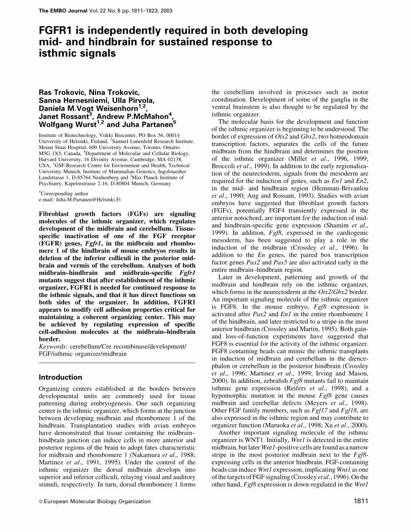
Expression of Fgfr1 and Fgfr2 during earlydevelopment of the mid- and hindbrainWe ®rst analyzed the expression of two potential medi-ators of isthmic signaling, Fgfr1 and Fgfr2, in mouseembryos by in situ mRNA hybridization. At a late head-fold stage [embryonic day (E) 7.5], around the stage whenthe isthmic organizer is induced, Fgfr1 expression wasdetected in the head folds (Figure 1A). Prominentexpression was also detected in other regions of thedeveloping embryo, including the primitive streak. Theseresults are consistent with the earlier studies of Fgfr1expression (Yamaguchi et al., 1992). At E7.5, Fgfr2 wasalso found to be expressed in the head folds (Figure 1B).Strong Fgfr2 expression was also detected in the extra-embryonic ectoderm. Later, at E8.5±9.5, widespread Fgfr1expression was observed in the developing central nervoussystem, including the midbrain±hindbrain region (Figures1C and E, and 2J and K). Fgfr2 was detected in thediencephalon, hindbrain and spinal cord, especially in thedorsal region (Figure 1D). In contrast to Fgfr1, no Fgfr2expression was detected at the midbrain±hindbrain bound-ary in the anterior rhombomere 1 or posterior midbrain(Figure 1F).
Generation of a conditional Fgfr1 alleleTo study the function of Fgfr1 in the development of themid- and hindbrain, we wanted to inactivate it in a tissue-speci®c fashion. To generate a Fgfr1 allele, Fgfr1¯ox,which can be inactivated by the site-speci®c recombinaseCre, we employed targeting vectors described earlier(Partanen et al., 1998). Using these vectors and transientCre expression in embryonic stem cells, we sequentiallyintroduced recognition sites of Cre (loxP sites) into two
Fig. 1. Expression of Fgfr1 and Fgfr2. Whole-mount in situ hybridiza-tion analysis of the expression of (A) Fgfr1 and (B) Fgfr2 at E7.5.Expression of both genes is detected in the headfolds (arrowheads). Inaddition, Fgfr1 is strongly expressed in the primitive streak region (PS)and Fgfr2 in the extra-embryonic ectoderm (EE). At E9.5, Fgfr1 iswidely expressed (C), whereas Fgfr2 expression is not detected in theanterior rhombomere 1 and the midbrain (D). In situ hybridizationanalysis of (E) Fgfr1 and (F) Fgfr2 expression on sagittal sections ofE9.5 embryos. Fgfr1 is widely expressed in the neural tube, whereasFgfr2 appears to be absent from the tissue around the isthmus. Arrowsin (C)±(F) mark the midbrain±hindbrain boundary (MHB).
R.Trokovic et al.
1812

Fig. 2. The conditional Fgfr1 allele, Fgfr1¯ox, and its inactivation by En1-Cre and Wnt1-Cre. (A) Schematic presentation of the Fgfr1¯ox allele and itsinactivation by the Cre-recombinase. The structures of the FGFR1 protein and the wild-type Fgfr1 locus are shown at the top. Only exons 7, 8, 15 and16 are depicted. LoxP sites were introduced into introns 7 and 15 by sequential gene targeting to generate the Fgfr1¯ox allele. Cre-mediated recombina-tion of the Fgfr1¯ox deletes the transmembrane and most of the intracellular region encoding exons resulting in the inactive Fgfr1D¯ox allele. EC, extra-cellular domain; TM, transmembrane domain; TK, tyrosine kinase domain. To characterize the Cre activity expressed by the En1-Cre and Wnt1-Cremice, they were crossed with mice carrying a Z/AP reporter allele. (B) Cre-mediated recombination between LoxP sites in the Z/AP allele results inalkaline phosphatase (AP) expression in the midbrain (MB) and rhombomere 1 (R1) of an E8.5 En1-Cre/+; Z/AP/+ embryo. (C) In an E8.5 Wnt1-Cre/+; Z/AP/+ embryo, AP activity is detected in the midbrain and neural crest cells (NCC). Frozen sections of E10.5 (D) En1-Cre/+; Z/AP/+ and (E andF) Wnt1-Cre/+; Z/AP/+ embryos are shown. In the En1-Cre/+; Z/AP/+ embryos, AP was expressed in the midbrain and rhombomere 1. In Wnt1-Cre/+; Z/AP/+ embryos, AP activity was detected in the midbrain and scattered cells of the rhombomere 1. A boundary is observed between the AP-posi-tive midbrain and mostly AP-negative rhombomere 1 (F). Arrowheads in (B)±(F) point to the midbrain±hindbrain boundary. (G±L) Inactivation ofFgfr1 expression by En1-Cre and Wnt1-Cre. Whole-mount in situ hybridization analysis of E8.5 (10 somite stage) En1-Cre/+; Fgfr1¯ox/¯ox (G) andwild-type (WT) embryos reveals inactivation of Fgfr1 transcription in the midbrain±hindbrain region by En1-Cre (arrowheads). In situ hybridizationanalysis of Fgfr1 expression in sagittal sections of E9.5 En1-Cre/+; Fgfr1¯ox/¯ox (H), Wnt1-Cre/+; Fgfr1¯ox/D¯ox (I) and wild-type (K) embryos. In En1-Cre/+; Fgfr1¯ox/¯ox embryos, inactivation of Fgfr1 expression occurs both in the midbrain and rhombomere 1, whereas in Wnt1-Cre/+; Fgfr1¯ox/D¯ox
embryos rhombomere 1 still expresses Fgfr1. Regions of affected Fgfr1 expression are indicated by arrowheads (H and I). A parallel section to theone shown in (I) hybridized with the Fgf8 probe (L). A double-headed arrow in (H)±(L) marks the midbrain±hindbrain boundary.
FGF signaling in development of the mid- and hindbrain
1813

different introns of the Fgfr1 gene (Figure 2A; andSupplementary ®gure 1 available at The EMBO JournalOnline). In the resulting allele, Fgfr1¯ox, exons 8±15encoding the transmembrane domain, juxtamembranedomain and most of the tyrosine kinase domain ofFGFR1, are ¯anked by two loxP sites. Mice hetero- orhomozygous for the Fgfr1¯ox allele are phenotypicallyindistinguishable from their wild-type littermates. Thus,the introduced loxP sites themselves do not appear tointerfere with Fgfr1 expression.
To test the functionality of the Fgfr1¯ox allele, wecrossed the Fgfr1¯ox/+ mice with mice carrying a Pgk-Cretransgene driving ubiquitous Cre expression (Lallemandet al., 1998). In mice heterozygous for both the Fgfr1¯ox
allele and the Pgk-Cre transgene, recombination betweenthe loxP sites resulted in excision of the genomic DNA¯anked by the loxP sites, generating a novel allele,Fgfr1D¯ox. The mice heterozygous for the Fgfr1D¯ox allelewere normal. Thus, the remaining Fgfr1 sequences in theFgfr1D¯ox do not appear to drive expression of dominant-negative gene products, which could signi®cantly interferewith FGF signaling. Embryos homozygous for theFgfr1D¯ox allele have gastrulation defects and die at aroundE9.5, closely resembling the Fgfr1-null mutants reportedpreviously (Deng et al., 1994; Yamaguchi et al., 1994;data not shown). Therefore, Fgfr1¯ox behaves as a condi-tional allele, which can be fully inactivated by the Crerecombinase.
Tissue-speci®c inactivation of Fgfr1 in themidbrain±hindbrain and midbrainTo study the role of FGFR1 in the isthmic organizersignaling, we wanted to inactivate Fgfr1 in the neuro-epithelium of the mid- and hindbrain after their regionalspeci®cation. For this purpose, we used the En1-Cre mice,which express the Cre recombinase from the En1 locus(Kimmel et al., 2000). We also wanted to inactivate Fgfr1speci®cally in the midbrain. For this, we used micecarrying a transgene expressing Cre recombinase under theWnt1 promoter (Danielian et al., 1998). To analyze thepatterns of Cre activity in the En1-Cre and Wnt1-Cre mice,we ®rst crossed them with the Z/AP reporter mouse line(Lobe et al., 1999). The Z/AP reporter allele was observedto be recombined ef®ciently and speci®cally in themidbrain±hindbrain region of the En1-Cre/+; Z/AP/+embryos already at the 8 somite stage (E8.5; Figure 2B).Analyses at E9.5 and E10.5 revealed ef®cient recombina-tion both in the midbrain and entire rhombomere 1(Figure 2D; data not shown). Ef®cient Cre-mediatedrecombination of the Z/AP allele was also detected in themidbrain of Wnt1-Cre/+; Z/AP/+ mice. In contrast, exceptfor a few scattered cells, cells in the rhombomere 1 carriedmostly the unrecombined Z/AP allele. At E9.5±12.5, allthe neuroepithelial cells of the midbrain appeared to carrythe recombinant allele (Figure 2E and F; data not shown).Although isolated recombinant cells were also observed inthe rhombomere 1 (Figure 2E), a boundary betweenrecombinant cells in the midbrain and mostly unrecombi-nant cells in the hindbrain could be observed (Figure 2Eand F).
We then crossed the Fgfr1¯ox mice with the En1-Cre andWnt1-Cre mice to inactivate Fgfr1 in a tissue-speci®cmanner. To determine the pattern of Cre-mediated
recombination and inactivation of the Fgfr1¯ox allele, wecarried out in situ hybridization analyses of En1-Cre/+;Fgfr1¯ox/¯ox and Wnt1-Cre/+; Fgfr1¯ox/D¯ox embryos with aFgfr1 cDNA probe containing exonic sequences betweenthe loxP sites in the Fgfr1¯ox allele. In En1-Cre/+;Fgfr1¯ox/¯ox embryos at E8.5, reduction in Fgfr1 signalwas observed already at the 8 somite stage, and at the10±11 somite stage the midbrain±hindbrain regionappeared negative for Fgfr1 expression (Figure 2G andJ; Supplementary ®gure 3). Radioactive in situ hybridiza-tion analysis on tissue sections of En1-Cre/+; Fgfr1¯ox/¯ox
embryos at E9.5 further demonstrated that midbrain andthe entire rhombomere 1 were negative for Fgfr1 expres-sion (Figure 2H and K). In contrast, the Wnt1-Cre/+;Fgfr1¯ox/D¯ox embryos lacked Fgfr1 expression in themidbrain, but still expressed abundant Fgfr1 in therhombomere 1 (Figure 2I and L). Thus, our experimentalapproaches allow us to inactivate Fgfr1 by E9.5 in themidbrain±hindbrain and midbrain using the En1-Cre andWnt1-Cre mice, respectively.
Inactivation of Fgfr1 in the mid- and hindbrainresults in ataxiaThe majority of En1-Cre/+; Fgfr1¯ox/¯ox mice surviveduntil adulthood. However, they were visibly uncoordin-ated, showing abnormal gait and wide stance. The impairedmotor coordination of the En1-Cre/+; Fgfr1¯ox/¯ox mice(n = 11) was further demonstrated by behavioral tests,including stationary beam and rotarod assays (Table I).
Cerebellar and midbrain defects caused byinactivation of Fgfr1 in the midbrain±hindbrainregionPossibly contributing to the behavioral defects, weobserved severe abnormalities in the cerebellum of theEn1-Cre/+; Fgfr1¯ox/¯ox mice. In adults, the vermis of thecerebellum was completely absent (n = 5; Figure 3A±D).The cerebellar hemispheres were present, although theirfoliation was abnormal (Figure 3E and F). The defect inthe cerebellar vermis was also obvious in the newbornEn1-Cre/+; Fgfr1¯ox/¯ox mice (n = 3). In addition to thecerebellum, extensive deletions including the inferiorcolliculi were evident in the posterior midbrain(Figure 3G, H, J and K). A comparable phenotype wasobserved in newborn En1-Cre/+; Fgfr1¯ox/D¯ox mice (n = 2;data not shown).
We also analyzed the development of the midbrain andcerebellum in mice homozygous for the hypomorphicFgfr1 alleles, Fgfr1n7 and Fgfr1n15YF, in which the Fgfr1
Table I. Behavioral analysis of Fgfr1 midbrain±hindbrain mutants
Wild type(n = 12)
En1-Cre/+;Flox/Flox (n = 11)
P
Rotarod assayt [mean (SD)]a 176 (59) 49 (79) <0.001
Stationary beam assayt [mean (SD)]a 60 (1.4) 4.3 (5.4) <0.001d [mean (SD)]b 90 (80) 0.40 (1.0) <0.005
aTime on rotarod/beam (s).bDistance walked on beam (cm).
R.Trokovic et al.
1814

transcript levels are reduced by ~80 and 90%, respectively(Partanen et al., 1998). Defects in the midline vermisand partial deletions of the inferior colliculi of the
midbrain were observed in both Fgfr1n7/n7 (n = 8) andFgfr1n15YF/n15YF (n = 10) newborn mice (Figure 3I and L;data not shown).
Fig. 3. Morphology of the midbrain and cerebellum of the midbrain±hindbrain-speci®c and hypomorphic Fgfr1 mutants. Whole-mount view of(A) wild-type and (B) En1-Cre/+; Fgfr1¯ox/¯ox adult brains. The entire vermis is missing in En1-Cre/+; Fgfr1¯ox/¯ox mice (arrow). (C and D) Midsagittaland (E and F) parasagittal sections of adult wild-type (C and E) and En1-Cre/+; Fgfr1¯ox/¯ox (D and F) brains. Complete aplasia of the vermis is evi-dent in the En1-Cre/+; Fgfr1¯ox/¯ox mice [arrow in (D)]. The En1-Cre/+; Fgfr1¯ox/¯ox mice have cerebellar hemispheres, but their pattern of foliation isaltered (F). Whole-mount views and corresponding midsagittal sections of new-born wild-type (G and J), En1-Cre/+; Fgfr1¯ox/¯ox (H and K) andFgfr1n15YF/n15YF (I and L) mice. Aplasia of the vermis and deletions of the inferior colliculi can be seen in both types of Fgfr1 mutants (arrows). Locuscoeruleus, identi®ed by Dopamine-b-hydroxylase mRNA in situ hybridization of adult brain sections (M and N) and anti-tyrosine hydroxylase immuno-staining of newborn brain sections (O and P) appears disorganized in the En1-Cre/+; Fgfr1¯ox/¯ox mutants (N and P) compared with wild type (M andO). cI, crus I; cII, crusII; ch, cerebellar hemisphere; cp, choroids plexus; ic, inferior colliculus; mb, midbrain; PM, paramedian lobule; S, simplex; v,vermis. The mutant hemisphere lobes are labeled with asterisks.
FGF signaling in development of the mid- and hindbrain
1815

In addition to the dorsal midbrain and cerebellum, weanalyzed several ganglia in the alar and basal plates of themidbrain±hindbrain region of En1-Cre/+; Fgfr1¯ox/¯ox
mutants, including locus coeruleus, substantia nigra,cranial nerves III and IV, pontine nucleus, the nucleuspedunculoponinus tegmentalis and nucleus parabigemina-lis. Tyrosine hydroxylase-positive neurons were found innewborn and adult mutants, in both the substantia nigra ofthe midbrain and the locus coeruleus of the rhombomere 1.Also, Dopamine-b-hydroxylase mRNA in situ hybridiza-tion revealed locus coeruleus in the mutants. However,locus coeruleus appeared to be disorganized comparedwith the wild type (Figure 3M±P). Whole-mount neuro-®lament staining of E10.5 En1-Cre/+; Fgfr1¯ox/¯ox em-bryos revealed both the oculomotor (III) nerve from themidbrain and trochlear (IV) nerve from the anteriorrhombomere 1. Consistently, the oculomotor and trochlearnuclei also appeared unaltered in adult mutants. Inaddition, pontine nuclei as well as mesopontine nucleisuch as nucleus pedunculopontinus tegmentalis andnucleus parabigeminalis were found to be present in themutants (Supplementary ®gure 2; data not shown). Thus,in contrast to the dorsal brain structures, no extensivedeletions or alterations were observed in the basal plate ofthe midbrain±hindbrain region.
Inactivation of Fgfr1 in the midbrain±hindbrainregion does not affect cellular survival at E9.5In order to understand the developmental basis forthe deletions in the dorsal midbrain and cerebellum,we analyzed the pattern of cell death in En1-Cre/+;Fgfr1¯ox/¯ox embryos. TUNEL analysis of apoptosis at E9.5revealed no statistically signi®cant difference in thenumber of apoptotic cells in the midbrain±hindbrainregion of En1-Cre/+; Fgfr1¯ox/¯ox embryos (n = 10)compared with the wild type (n = 8). In addition, analysisof semi-thin sections of E9.5 mutants (n = 3) and whole-mount Nile Blue staining of E10.5 mutants (n = 2) showedno difference in the cellular viability compared with wild-type littermates (data not shown). Therefore, although wecannot completely rule out any effect on the apoptoticindex in the midbrain±hindbrain region, cell death ataround E9.5 does not appear to be a major component ofthe phenotype of En1-Cre/+; Fgfr1¯ox/¯ox embryos.
Fgfr1 is required for sustained expression ofisthmic organizer dependent genesTo analyze how the mid- and hindbrain-speci®c inactiva-tion of Fgfr1 affects the early development of the isthmicregion, we analyzed gene expression in E8.5±10.5 En1-Cre/+; Fgfr1¯ox/¯ox embryos by whole-mount in situhybridization. At E8.5 (8±10 somite stage), Pax2, Wnt1and Sprouty1, a FGF-inducible inhibitor of receptortyrosine kinase signaling (Minowada et al., 1999), wereexpressed in En1-Cre/+; Fgfr1¯ox/¯ox embryos at a leveland pattern comparable to the wild type (Figure 4A and B;data not shown). At E9.5, an anterior marker Otx2(Figure 4C) and a posterior marker Gbx2 (Figure 4D)were expressed in the correct domains in the En1-Cre/+;Fgfr1¯ox/¯ox embryos. In addition, Fgf8 was expressed inthe anterior hindbrain of E9.5 En1-Cre/+; Fgfr1¯ox/¯ox
embryos (Figure 4E), although its expression decreasedat later stages especially in the dorsal isthmus (data not
shown). Thus, the expression of a speci®c set of genes,including signaling molecules thought to be important foristhmic organizer activity, was established around themidbrain±hindbrain boundary in the En1-Cre/+; Fgfr1¯ox/¯ox
embryos at E8.5±9.5.In contrast to E8.5, expression of several genes thought
to depend on isthmic signals failed to be maintained atlater stages. Down-regulation of Sprouty1 was ®rstobserved at the 12 somite stage and Sprouty expressionwas completely abolished from the isthmic domain at E9.5(Figure 4G; Supplementary ®gure 3). Interestingly, someSprouty1 expression was still detected further away fromthe isthmus in dorsal and ventral regions. Also, expressionof Pax2 was virtually absent by E9.5 (Figure 4H). Otheristhmic target genes were down-regulated slightly later.En1 and En2 were still expressed at E9.5 (Figure 4I and J).However, their expression rapidly decreased thereafter,and by E10 En1 was barely detectable in either mid- orhindbrain (Figure 4K). In addition, expression of Wnt1 wasdown-regulated in the posterior midbrain, especially in itsdorsal region, after E9.5 (data not shown). Interestingly,before the down-regulation of the Wnt1 signal, Wnt1expression was observed as a broadened stripe in theposterior midbrain (Figure 4F). In contrast to the wild-typeembryos, in which the Wnt1-positive cells formed a tightband next to the Fgf8 expressing anterior hindbrain, theWnt1-positive and -negative cells were extensively mixedin the En1-Cre/+; Fgfr1¯ox/¯ox embryos. Also, Otx2 andespecially Gbx2 (Figure 4D) showed heterogeneousexpression borders.
Tissue-speci®c inactivation of Fgfr1 in themidbrain causes deletion of posterior midbrainand cerebellar abnormalitiesTo determine whether Fgfr1 directly regulates develop-ment of the midbrain, we crossed the Fgfr1¯ox mice withmice carrying a Wnt1-Cre transgene. The Wnt1-Cre/+;Fgfr1¯ox/¯ox mice die neonatally, possibly due to defects inthe craniofacial neural crest (Trokovic et al., 2003).Analysis of the brains of the newborn Wnt1-Cre/+;Fgfr1¯ox/¯ox mice (n = 5) revealed deletion of the inferiorcolliculi of the midbrain, reminiscent of the En1-Cre/+;Fgfr1¯ox/¯ox mice (Figure 5A±D). Development of thedorsal cerebellum was also abnormal. However, in con-trast to the En1-Cre/+; Fgfr1¯ox/¯ox mice, the vermis wasnot completely missing although it was severely mal-formed in the Wnt1-Cre/+; Fgfr1¯ox/¯ox mutants(Figure 5C±F). Some cellular differentiation was stillobserved in the mutant vermis by immunohistochemistrywith anti-calbindin antibodies (Figure 5F). A comparablephenotype was seen in the Wnt1-Cre/+; Fgfr1¯ox/D¯ox mice(n = 3; data not shown).
Fgfr1 is autonomously required in the midbrain forthe maintenance of expression of isthmus-regulated genesWe next analyzed isthmic gene expression in Wnt1-Cre/+;Fgfr1¯ox/D¯ox embryos at E9.5. As judged by the expressionof Otx2, Gbx2 and Fgf8 (Figure 6A and C; data notshown), the isthmic organizer forms and is correctlypositioned in the Wnt1-Cre/+; Fgfr1¯ox/D¯ox embryos.However, expression of isthmus-regulated genes wasabnormal speci®cally in the midbrain. In contrast to the
R.Trokovic et al.
1816

En1-Cre/+; Fgfr1¯ox/¯ox embryos, which showed markeddown-regulation of Sprouty1 in both mid- and hindbrain,E9.5 Wnt1-Cre/+; Fgfr1¯ox/D¯ox embryos still expressedSprouty1 in the hindbrain, but its expression was down-regulated in the midbrain (Figure 6G). Wnt1 showed asimilar pattern of expression as in the En1-Cre/+;
Fgfr1¯ox/¯ox mutants, with early broadened and heteroge-neous expression and down-regulation dorsally after E9.5(Figure 6E and F). The initial broadening of Wnt1expression was accompanied by slightly expanded expres-sion of En1 and En2 (Figure 6I and J). En1 was thereafterdown-regulated speci®cally in the midbrain by E10
Fig. 4. Analysis of gene expression in the midbrain±hindbrain speci®c Fgfr1 mutants. Whole-mount in situ hybridization of E8.5 (A and B),E9.5 (C±J) and E10 (K) wild-type and En1-Cre/+; Fgfr1¯ox/¯ox embryos with Sprouty1 (A, G), Pax2 (B, H), Otx2 (C), Gbx2 (D), Fgf8 (E), Wnt1 (F),En1 (I, K) and En2 (J) probes. Red arrows indicate altered gene expression. Small arrowheads in (G) indicate remaining Sprouty1 expression inregions distal to the isthmus. See text for details.
FGF signaling in development of the mid- and hindbrain
1817

(Figure 6K). In contrast to the En1-Cre/+; Fgfr1¯ox/¯ox
embryos, expression of Pax2 was still detected as a fuzzyband at the isthmus (Figure 6H). Interestingly, theposterior border of Otx2 expression, as well as the anteriorborder of Gbx2 and Fgf8 expression, was found to beuneven, showing mixing of positive and negative cellpopulations in Wnt1-Cre/+; Fgfr1¯ox/D¯ox embryos(Figure 6B and D).
Fgfr1 regulates expression of PB-cadherin, anisthmic cell adhesion moleculeTo gain insight into the mechanisms responsible forenhanced cell mixing in the Fgfr1 mutants we nextanalyzed expression of genes thought to mediate celladhesion and repulsion. One of these is Ephrin-A5, whichis speci®cally expressed in the midbrain. No change inEphrin-A5 expression could be observed in E9.5 En1-Cre/+; Fgfr1¯ox/¯ox embryos compared with wild type(Figure 7A and B).
Other candidates for regulators of cellular dispersioninclude Cadherins, a family of mostly homotypic cell-
adhesion molecules. One member of the cadherin family,PB-cadherin, has been reported to be expressed in theisthmic region in a pattern overlapping with Wnt1expression at E10.5 (Kitajima et al., 1999). As the Wnt1-positive cells failed to form a tight band of cells in the En1-Cre/+; Fgfr1¯ox/¯ox and Wnt1-Cre/+; Fgfr1¯ox/D¯ox em-bryos, we wanted to analyze whether the expression of PB-cadherin was altered in the isthmus. We found PB-cadherin expression at the midbrain±hindbrain boundaryof wild-type embryos already at E8.5 (data not shown). AtE9.5, PB-cadherin was expressed as a stripe throughoutthe isthmic region (Figure 7C). Weaker expression wasdetected throughout the midbrain. In contrast, both in En1-Cre/+; Fgfr1¯ox/¯ox (n = 5; Figure 7D) and Wnt1-Cre/+;Fgfr1¯ox/D¯ox (n = 4; Figure 7E) embryos, PB-cadherinexpression was markedly down-regulated, except for apatch of ventral expression.
Discussion
Genetic loss-of-function studies both in zebra®sh (Reiferset al., 1998) and mouse (Meyers et al., 1998; Xu et al.,2000), have demonstrated the importance of FGFs, andFGF8 in particular, in the development of the midbrain±hindbrain region. However, the timing and primary targetsof FGF signaling are still unclear. Here we have analyzedthe consequences of tissue-speci®c inactivation of Fgfr1 inthe mid- and hindbrain after the establishment of theirregional identity. Loss of Fgfr1 results in aplasia ofcerebellar vermis and inferior colliculi. Our results suggestthat FGFR1 is required both in the mid- and hindbrainfor their correct response to signals from the isthmicorganizer.
Tissue-speci®c inactivation of Fgfr1The tissue-speci®c inactivation of a conditional Fgfr1allele, Fgfr1¯ox, was achieved by crosses with En1-Cre andWnt1-Cre mice, which allowed apparently completeinactivation of Fgfr1 transcription by E9.5 in the midbrainand anterior hindbrain. Several additional observationssuggest ef®cient recombination by En1-Cre and Wnt1-Cre.First, recombination of the Z/AP reporter allele occurredby E9.5 in virtually all En1- and Wnt1-positive cells in theEn1-Cre/+; Z/AP/+ and Wnt1-Cre/+; Z/AP/+ embryos,respectively (Figure 2; data not shown). Secondly, thephenotypes of En1-Cre/+; Fgfr1¯ox/D¯ox and Wnt1-Cre/+;Fgfr1¯ox/D¯ox mice, in which a single recombination eventis enough to make a cell null-mutant for Fgfr1, arecomparable to the En1-Cre/+; Fgfr1¯ox/¯ox and the Wnt1-Cre/+; Fgfr1¯ox/¯ox mice, respectively. Thirdly, the phe-notype of the hypomorphic Fgfr1 mutants, expressing only~10% of the wild-type Fgfr1 mRNA levels, is less severethan the phenotype of the En1-Cre/+; Fgfr1¯ox/¯ox mice.Finally, the early changes in the gene expression inmidbrain were similar in En1-Cre/+; Fgfr1¯ox/¯ox andWnt1-Cre/+; Fgfr1¯ox/D¯ox embryos.
Isolated recombinant cells were observed also in therhombomere 1 in the Wnt1-Cre/+; Z/AP/+ embryos bothat E8.5 and E10.5. It has been proposed that themidbrain±hindbrain border is not a boundary of cell-lineage restriction (Jungbluth et al., 2001). It is thereforepossible that the recombinant cells in rhombomere 1 havetheir origin in the midbrain. Alternatively, the scattered
Fig. 5. Morphology of the midbrain and cerebellum of the midbrain-speci®c Fgfr1 mutants. Whole-mount views (A and B) and mid-sagittalsections (C and D) of brains of newborn wild-type (A and C) andWnt1-Cre/+; Fgfr1¯ox/¯ox (B and D) mice. Calbindin expression in wild-type (E) and Wnt1-Cre/+; Fgfr1¯ox/¯ox mice (F). Arrows indicate thedeletion of the inferior colliculi and malformed vermis. ch, cerebellarhemisphere; cp, choroid plexus; mb, midbrain; v, vermis.
R.Trokovic et al.
1818

Fig. 6. Analysis of gene expression in the midbrain speci®c Fgfr1 mutants. Whole-mount in situ hybridization of E9.5 (A±J) and E10.0 (K) wild-typeand Wnt1-Cre/+; Fgfr1¯ox/D¯ox embryos with Otx2 (A and B), Fgf8 (C and D), Wnt1 (E and F), Sprouty1 (G), Pax2 (H), En1 (I and K) and En2 (J)probes. Close-up side views of embryos hybridized with Otx2 (B) and Wnt1 (F), as well as a slightly oblique dorsal view of embryos hybridized withFgf8 probe (D). Arrowheads indicate the isthmus, red arrows indicate altered gene expression. See text for details.
FGF signaling in development of the mid- and hindbrain
1819

recombinant cells may result from transient or weakexpression of the Wnt1 promoter in rhombomere 1. Thepresence of a border between recombinant cells of themidbrain and mostly unrecombined rhombomere 1 sug-gests that mixing of cells between mid- and hindbrain isalready restricted to some extent by E10.5. Our in situhybridization analysis suggested that the majority of thecells in rhombomere 1 still expressed Fgfr1 in the Wnt1-Cre/+; Fgfr1¯ox/D¯ox embryos. We cannot rule outinactivation of Fgfr1 in isolated cells, which couldcontribute to observed cerebellar defects in these mutants.However, studies of the expression of both Fgfr1 and itstarget genes are consistent with ef®cient and speci®cinactivation of Fgfr1 in midbrain±hindbrain and midbrainby En1-Cre and Wnt1-Cre, respectively.
Regulation of isthmus dependent gene expressionby Fgfr1In the En1-Cre/+; Fgfr1¯ox/¯ox embryos, Fgfr1 transcrip-tion was inactivated by the 8±10 somite stage. Sprouty1 isa FGF-responsive gene, which is normally expressed nextto the FGF source in several regions of the developingembryo and can be induced by ectopic Fgf expression
(Minowada et al., 1999). Expression of Sprouty1 in E8.5En1-Cre/+; Fgfr1¯ox/¯ox embryos suggested that FGFsignaling was still active in the isthmic region at thisstage. This could be due to expression of residual FGFR1protein or other Fgfrs, perhaps Fgfr2, expression of whichwas detected in the headfolds at E7.5. Initial expression ofresidual FGFR1 or other FGFRs may also explain whymore extensive deletions of midbrain and cerebellum areobserved in Fgf8 mutants (Meyers et al., 1998; Reiferset al., 1998). It is also possible that Fgf8 affects multipletarget tissues regulating development of the mid- andhindbrain. This is perhaps a less likely explanation, sincetissue-speci®c inactivation of Fgf8 in the mid- andhindbrain also results in early loss of the entire midbrainand anterior hindbrain (G.Martin, personal communication).
Our results suggest that signaling through Fgfr1 regu-lates the maintenance of expression of a set of genes nearthe isthmic organizer at E9.5. As judged based on isthmus-speci®c gene expression, the organizer itself is establishedin both midbrain±hindbrain and midbrain-speci®c Fgfr1mutants, and expression of Fgf8 and other Fgfs (ourunpublished observations) is activated in the anteriorhindbrain of the mutants. However, FGF signals fail to
Fig. 7. Expression of Ephrin-A5 and PB-cadherin in Fgfr1 mutants and a model for FGFR1 function during the maintenance of the isthmic organizer.Whole-mount in situ hybridization analysis of Ephrin-A5 (A, B) and PB-cadherin (C±E) expression in E9.5 wild-type (A, C), En1-Cre/+; Fgfr1¯ox/¯ox
(B, D) and Wnt1-Cre/+; Fgfr1¯ox/D¯ox (E) embryos. In both types of Fgfr1 mutants PB-cadherin expression is down-regulated in the midbrain, espe-cially in its dorsal part. Arrowheads in (A)±(E) point to the midbrain±hindbrain boundary. (F) A model for the function of FGF signaling throughFGFR1 during maintenance of the isthmic organizer. We suggest that FGFR1 regulates gene expression independently in the midbrain (Mes) and hind-brain (Met). Some of the FGFR1-regulated genes confer speci®c adhesive characteristics to the cells next to the midbrain±hindbrain border.
R.Trokovic et al.
1820

elicit normal transcriptional response in the Fgfr1 mutanttarget cells. In the E9.5 En1-Cre/+; Fgfr1¯ox/¯ox mutants,expression of Sprouty1 and Pax2 was not detected in theisthmus. This suggests that very little FGF signaling occursat the midbrain±hindbrain junction of E9.5 embryos in theabsence of FGFR1. Thus, at this stage, FGFR1 appears tobe the primary FGF receptor receiving isthmic signals andmaintaining isthmus-dependent gene expression.
In Wnt1-Cre/+; Fgfr1¯ox/D¯ox embryos, expression ofgenes such as Sprouty1 and En1 was speci®cally alteredin the midbrain. This suggests that Fgfr1 is independ-ently involved in the regulation of gene expression inthe midbrain. As these genes are down-regulated inboth the mid- and hindbrain of En1-Cre/+; Fgfr1¯ox/¯ox
embryos at the same stage, Fgfr1 must also have adirect role in the regulation of the anterior hindbraindevelopment.
Later, defects in the vermis of the cerebellum, aderivative of rhombomere 1, were also observed inWnt1-Cre/+; Fgfr1¯ox/¯ox mutants. As discussed above,inactivation of Fgfr1 in some cells of the hindbrain mightcontribute to this phenotype. However, the observed geneexpression changes in the midbrain, for example down-regulation of Wnt1, are likely to also have secondaryeffects on the cerebellar development in Wnt1-Cre/+;Fgfr1¯ox/¯ox mice.
Regulation of cell-adhesive properties by Fgfr1?What could be the molecular and cellular processesregulated by FGFR1? The observed changes in geneexpression suggest alterations in cellular identities, i.e.tissue patterning. The behavior of Wnt1-positive cells inboth midbrain±hindbrain and midbrain-speci®c Fgfr1mutants suggests yet another mechanism. In the wild-type embryos Wnt1 is expressed as a tight band of cellsin the posterior midbrain next to Fgf8-expressing cellsin the anterior hindbrain. Some mixing of the Wnt1-positive and Wnt1-negative midbrain cells is observedat the anterior margin of the Wnt1 expression domain.In the Fgfr1 mutants, Wnt1-positive cells appear to mixextensively with Wnt1-negative cells. It is possible thatsignaling through FGFR1 gives Wnt1-positive cellsadhesive characteristics, which allow them to sort outof the Wnt1-negative cells. In the absence of Fgfr1,such characteristics are lost and segregation of Wnt1-positive and -negative cells fails. The fact that Otx2,Gbx2 and Fgf8 also showed heterogenous expressionborders suggests that loss of Fgfr1 also leads to mixingof midbrain and hindbrain cells.
An alternative hypothesis is suggested by the experi-ments by Jungbluth et al. (2001), who demonstrated thatcells in the avian midbrain and rhombomere 1 were able tomix with each other. If the midbrain±hindbrain borderdoes not represent a true compartment boundary, FGFR1may regulate readjustment of identities of the cellstraversing the midbrain±hindbrain border. However, pre-vious fate-mapping studies (Millet et al., 1996), as well asour analyses of the distribution of genetically markedmidbrain cells in Wnt1-Cre/+; Z/AP/+ embryos, do notsupport extensive cell mixing at the midbrain±hindbrainboundary. In addition, in chimeric mouse embryosconsisting of both wild-type and Otx2-null mutant cells,Otx2-negative cells were found to segregate from wild-
type cells in the midbrain, perhaps due to change in theexpression of cell surface adhesion molecules (Rhinn et al.,1999). Nevertheless, it is still possible that differential celladhesion is not a general property of the midbrain andrhombomere 1 cells, but is restricted to the cells at theborder of these two domains.
A speci®c cell adhesion molecule, PB-cadherin, isexpressed in Wnt1-positive cells at the midbrain±hindbrain junction, arguing for unique adhesive prop-erties of these cells (Figure 7; Kitajima et al., 1999).Interestingly, our results suggest that PB-cadherin isone of the transcriptional targets of FGFR1 signaling,and thus its loss may contribute to the mixing of cellsat the midbrain±hindbrain border in the Fgfr1 mutants(see Figure 7F). In addition to the transcriptionalresponse, it is possible that intercellular signalingpathways of FGFR1 more directly regulate cell-adhe-sive properties. For example, tyrosine phosphorylationhas been reported to regulate the activity of catenins,which in turn affect cadherin function (Lilien et al.,2002).
A failure in the segregation between mid- and hindbraincells has been reported in hypomorphic Wnt1Sw mutants(Bally-Cuif et al., 1995). Our results suggest that thebehavior of the midbrain cells is affected prior to loss ofWnt1 expression in En1-Cre/+; Fgfr1¯ox/¯ox and Wnt1-Cre/+; Fgfr1¯ox/D¯ox embryos. It is therefore possible that thecell-sorting defect in Wnt1Sw/Sw mice results from down-regulation of isthmic FGF signals. The broadening of theWnt1 expression domain may also explain the initial slightexpansion of the midbrain in Wnt1-Cre/+; Fgfr1¯ox/D¯ox
embryos, as WNT1 has been suggested to stimulatecellular proliferation in the midbrain±hindbrain region(W.Wurst, unpublished data).
An interesting parallel in organizer regulation can perhapsbe found in the wing imaginal disc of Drosophila. In thewing disc the signaling molecule Hedgehog, expressed bythe cells in the posterior compartment, not only inducesexpression of the morphogen Decapentaplegic but alsoaffects adhesive properties in the adjacent cells of theanterior compartment (Blair and Ralston, 1997; Rodriguezand Basler, 1997). Regulation of adhesive properties byFGF signaling could explain in part how the isthmicorganizer is kept as a straight and coherent signalingcenter, and how the distinct mid- and hindbrain domainsare maintained.
Materials and methods
Homologous and site-speci®c recombination in embryonicstem cellsSee Supplementary ®gure 1 for detailed description of generation of theFgfr1¯ox allele by gene targeting.
Generation and genotyping of mutant miceFgfr1¯ox/+ embryonic stem cells were aggregated with morula stageembryos of the ICR strain to produce chimeric mice, which transmittedthe Fgfr1¯ox allele through the germ line. All the experiments wereperformed in an outbred (129sv/ICR) background. The Fgfr1¯ox allele wasdetected by PCR with primers LoxP-1 (5¢-AATAGGTCCCTCGA-CGGTATC-3¢) and Fgfr1 i73 (5¢-CTGGGTCAGTGTGGACAGTGT-3¢). The wild-type Fgfr1 allele was detected with the primers Fgfr1 wt5¢(5¢-CCCCATCCCATTTCCTTACCT-3¢) and Fgfr1 wt3¢ (5¢-TTCTGGT-GTGTCTGAAAACAGCT-3¢). The different Cre alleles were detectedwith the primers Cre5¢ (5¢-AATCTCCCACCGTCAGTACG-3¢) and
FGF signaling in development of the mid- and hindbrain
1821

Cre3¢ (5¢-CGTTTTCTGAGCATACCTGGA-3¢). The Z/AP allele wasdetected by b-galactosidase staining.
b-galactosidase and alkaline phosphatase stainingb-galactosidase and alkaline phosphatase staining of whole-mountE8.5±10.5 embryos as well as frozen sections was performed as describedpreviously (Lobe et al., 1999).
Behavioral analysesIn the rotarod assay, the mice were placed on a rotating rod (diameter15 cm, speed 15 r.p.m.). The time the mice were able to stay on top of therod was measured. The experiment was terminated after 5 min. Eachmouse was assayed nine times (Table I).
In the stationary beam assay the mice were placed in the middle of ahorizontal beam (diameter 2 cm, length 150 cm). The time the micestayed on the beam and the distance they walked on the beam weremeasured. The experiment was terminated after 1 min. Each mouse wasassayed nine times (Table I).
Histology and immunohistochemistryAdult tissues were ®xed by intracardial perfusion with 4% paraformalde-hyde. Brain tissues of newborn mice were dissected in phosphate-buffered saline (PBS) and ®xed overnight in 4% paraformaldehyde. Thetissues were dehydrated, embedded in paraf®n and sectioned in 5 mmslices. Hematoxylin±eosin was used for counterstaining. Immuno-histochemistry on paraf®n sections with anti-calbindin (Swant cat.CB38) and anti-tyrosine hydroxylase (Chemicon AB152) antibodieswas performed according to standard procedures. For semi-thin sections,embryos were ®xed in 2.5% glutaraldehyde in 0.1 M phosphate buffer,pH 7.2, overnight at 4°C, post-®xed in 1% osmium tetroxide, andembedded in Epon. One micrometer plastic sagittal sections were cut andstained with toluidine blue.
Analysis of cell deathTUNEL assays. To detect apoptotic cells, TUNEL assays were performedon paraf®n sections of E9.5 embryos using the in situ cell death detectionkit (Roche cat. 1684 795).
Nile Blue sulfate (NBS) staining. Following dissection, embryos (E10.5)were washed in PBS, and incubated for 30 min at 37°C in ®ltered NBS(Sigma N-5632) saturated water diluted 1:1000 in PBT (PBS containing0.1% Tween-20). Embryos were then washed several times in PBT atroom temperature and photographed immediately.
mRNA in situ hybridization analysesWhole-mount mRNA in situ hybridization of E8.5±10.5 embryos withEn1, En2 (Davis and Joyner, 1988), EphrinA5 (a gift from DavidWilkinson), Fgfr1 (bp 1152±1724 of NM010206), Fgfr2 (a gift from AlkaMansukhani), Fgf8 (Crossley and Martin, 1995), Gbx2, Otx2 (Acamporaet al., 1997), Pax2 (a gift from Gregory Dressler), PB-cadherin (clone ID:UI-M-BH1-akr-h-03-0-UI), Sprouty1 (a gift from Seppo Vainio) andWnt1 (McMahon et al., 1992) riboprobes was performed as describedpreviously (Henrique et al., 1995). At least four mutants and fourlittermate controls were hybridized with each of the probes. Hybridizationand subsequent treatments of the mutants and the littermate controls werecarried out simultaneously in the same vial. Radioactive in situhybridizations on paraf®n sections with Fgf8, Fgfr1, Fgfr2 andDopamine-b-hydroxylase (bp 620±1018 of S50200) probes were carriedout as described by Wilkinson and Green (1990).
Supplementary dataSupplementary data are available at The EMBO Journal Online.
Acknowledgements
We thank Lois Schwartz, Eija Koivunen, Outi Koljonen, Mona Augustin,PaÈivi Hannuksela, Maria von Numers, Stefanie Pirrung and NadineTrepesch for expert technical assistance. We also wish to thank AndrasNagy for the Z/AP mice, Peter Lonai for the Pgk-Cre mice, HenriHuttunen for help with immunohistochemistry and Vootele Vojkar forhelp in behavioral analyses. This work was supported by the Academy ofFinland, the Sigrid Juselius foundation and Biocentrum Helsinki (J.P.),the Viikki Graduate School in Biosciences (R.T., N.T.), theBundesministerium fuÈr Bildung und Forschung and the DeutscheForschungsgemeinschaft (W.W.).
References
Acampora,D., Avantaggiato,V., Tuorto,F. and Simeone,A. (1997)Genetic control of brain morphogenesis through Otx gene dosagerequirement. Development, 124, 3639±3650.
Ang,S.L. and Rossant,J. (1993) Anterior mesendoderm induces mouseEngrailed genes in explant cultures. Development, 118, 139±149.
Bally-Cuif,L., Cholley,B. and Wassef,M. (1995) Involvement of Wnt-1in the formation of the mes/metencephalic boundary. Mech. Dev., 53,23±34.
Blair,S.S. and Ralston,A. (1997) Smoothened-mediated Hedgehogsignalling is required for the maintenance of the anterior±posteriorlineage restriction in the developing wing of Drosophila.Development, 124, 4053±4063.
Broccoli,V., Boncinelli,E. and Wurst,W. (1999) The caudal limit of Otx2expression positions the isthmic organizer. Nature, 401, 164±168.
Ciruna,B. and Rossant,J. (2001) FGF signaling regulates mesoderm cellfate speci®cation and morphogenetic movement at the primitivestreak. Dev. Cell, 1, 37±49.
Crossley,P.H. and Martin,G.R. (1995) The mouse Fgf8 gene encodes afamily of polypeptides and is expressed in regions that directoutgrowth and patterning in the developing embryo. Development,121, 439±451.
Crossley,P.H., Martinez,S. and Martin,G.R. (1996) Midbraindevelopment induced by FGF8 in the chick embryo. Nature, 380,66±68.
Danielian,P.S., Muccino,D., Rowitch,D.H., Michael,S.K. andMcMahon,A.P. (1998) Modi®cation of gene activity in mouseembryos in utero by a tamoxifen-inducible form of Crerecombinase. Curr. Biol., 8, 1323±1326.
Davis,C.A. and Joyner,A.L. (1988) Expression patterns of thehomeobox-containing genes En-1 and En-2 and the proto-oncogeneint-1 diverge during mouse development. Genes Dev., 2, 1736±1744.
Deng,C.X., Wynshaw-Boris,A., Shen,M.M., Daugherty,C., Ornitz,D.M.and Leder,P. (1994) Murine FGFR-1 is required for earlypostimplantation growth and axial organization. Genes Dev., 8,3045±3057.
Hemmati-Brivanlou,A., Stewart,R.M. and Harland,R.M. (1990) Region-speci®c neural induction of an engrailed protein by anterior notochordin Xenopus. Science, 250, 800±802.
Henrique,D., Adam,J., Myat,A., Chitnis,A., Lewis,J. and Ish-Horowicz,D. (1995) Expression of a Delta homologue in prospectiveneurons in the chick. Nature, 375, 787±790.
Irving,C. and Mason,I. (2000) Signalling by FGF8 from the isthmuspatterns anterior hindbrain and establishes the anterior limit of Hoxgene expression. Development, 127, 177±186.
Jungbluth,S., Larsen,C., Wizenmann,A. and Lumsden,A. (2001) Cellmixing between the embryonic midbrain and hindbrain. Curr. Biol.,11, 204±207.
Kimmel,R.A., Turnbull D.H., Blanquet V., Wurst W., Loomis C.A. andJoyner A.L. (2000) Two lineage boundaries coordinate vertebrateapical ectodermal ridge formation. Genes Dev., 14, 1377±1389.
Kitajima,K., Koshimizu,U. and Nakamura,T. (1999) Expression of anovel type of classic cadherin, PB-cadherin in developing brain andlimb buds. Dev. Dyn., 215, 206±214.
Lallemand,Y., Luria,V., Haffner-Krausz,R. and Lonai,P. (1998)Maternally expressed PGK-Cre transgene as a tool for early anduniform activation of the Cre site-speci®c recombinase. TransgenicRes., 7, 105±112.
Lee,S.M., Danielian,P.S., Fritzsch,B. and McMahon,A.P. (1997)Evidence that FGF8 signalling from the midbrain±hindbrain junctionregulates growth and polarity in the developing midbrain.Development, 124, 959±969.
Lilien,J., Balsamo,J., Arregui,C. and Xu,G. (2002) Turn-off, drop-out:functional state switching of cadherins. Dev. Dyn., 224, 18±29.
Liu,A. and Joyner,A.L. (2001) Early anterior/posterior patterning of themidbrain and cerebellum. Annu. Rev. Neurosci., 24, 869±896.
Lobe,C.G., Koop,K.E., Kreppner,W., Lomeli,H., Gertsenstein,M. andNagy,A. (1999) Z/AP, a double reporter for cre-mediatedrecombination. Dev. Biol., 208, 281±292.
Martinez,S., Wassef,M. and Alvarado-Mallart,R.M. (1991) Induction ofa mesencephalic phenotype in the 2-day-old chick prosencephalon ispreceded by the early expression of the homeobox gene en. Neuron, 6,971±981.
Martinez,S., Marin,F., Nieto,M.A. and Puelles,L. (1995) Induction ofectopic engrailed expression and fate change in avian
R.Trokovic et al.
1822

rhombomeres: intersegmental boundaries as barriers. Mech. Dev.,51, 289±303.
Martinez,S., Crossley,P.H., Cobos,I., Rubenstein,J.L. and Martin,G.R.(1999) FGF8 induces formation of an ectopic isthmic organizer andisthmocerebellar development via a repressive effect on Otx2expression. Development, 126, 1189±1200.
Maruoka,Y., Ohbayashi,N., Hoshikawa,M., Itoh,N., Hogan,B.M. andFuruta,Y. (1998) Comparison of the expression of three highly relatedgenes, Fgf8, Fgf17 and Fgf18, in the mouse embryo. Mech. Dev., 74,175±177.
McMahon,A.P., Joyner,A.L., Bradley,A. and McMahon,J.A. (1992) Themidbrain±hindbrain phenotype of Wnt-1±/Wnt-1± mice results fromstepwise deletion of engrailed-expressing cells by 9.5 dayspostcoitum. Cell, 69, 581±595.
Meyers,E.N., Lewandoski,M. and Martin,G.R. (1998) An Fgf8 mutantallelic series generated by Cre- and Flp-mediated recombination. Nat.Genet., 18, 136±141.
Millet,S., Bloch-Gallego,E., Simeone,A. and Alvarado-Mallart,R.M.(1996) The caudal limit of Otx2 gene expression as a marker ofthe midbrain/hindbrain boundary: a study using in situhybridisation and chick/quail homotopic grafts. Development, 122,3785±3797.
Millet,S., Campbell,K., Epstein,D.J., Losos,K., Harris,E. and Joyner,A.L.(1999) A role for Gbx2 in repression of Otx2 and positioning the mid/hindbrain organizer. Nature, 401, 161±164.
Minowada,G., Jarvis,L.A., Chi,C.L., Neubuser,A., Sun,X., Hacohen,N.,Krasnow,M.A. and Martin,G.R. (1999) Vertebrate Sprouty genes areinduced by FGF signaling and can cause chondrodysplasia whenoverexpressed. Development, 126, 4465±4475.
Nakamura,H., Takagi,S., Tsuji,T., Matsui,K.A. and Fujisawa,H. (1988)The prosencephalon has the capacity to differentiate into optic tectum:analysis in quail-chick-chimeric brains. Dev. Growth Differ., 30,717±725.
Partanen,J., Schwartz,L. and Rossant,J. (1998) Opposite phenotypes ofhypomorphic and Y766 phosphorylation site mutations reveal afunction for Fgfr1 in anteroposterior patterning of mouse embryos.Genes Dev., 12, 2332±2344.
Reifers,F., Bohli,H., Walsh,E.C., Crossley,P.H., Stainier,D.Y. andBrand,M. (1998) Fgf8 is mutated in zebra®sh acerebellar (ace)mutants and is required for maintenance of midbrain±hindbrainboundary development and somitogenesis. Development, 125,2381±2395.
Rhinn,M., Dierich,A., LeMeur,M. and Ang,S.-L. (1999) Cellautonomous and non-cell autonomous functions of Otx2 inpatterning the rostral brain. Development, 126, 4295±4304.
Rodriguez,I. and Basler,K. (1997) Control of compartmental af®nityboundaries by hedgehog. Nature, 389, 614±618.
Shamim,H., Mahmood,R., Logan,C., Doherty,P., Lumsden,A. andMason,I. (1999) Sequential roles for Fgf4, En1 and Fgf8 inspeci®cation and regionalisation of the midbrain. Development, 126,945±959.
Trokovic,N., Trokovic,R., Mai,P. and Partanen,J. (2003) Fgfr1 regulatespatterning of the pharyngeal region. Genes Dev., 17, 141±153.
Walshe,J. and Mason,I. (2000) Expression of FGFR1, FGFR2 andFGFR3 during early neural development in the chick embryo. Mech.Dev., 90, 103±110.
Wilkinson,D.G. and Green,J. (1990) In situ hybridization and the three-dimensional construction of serial sections. In A.J. Copp and D.L.Cockroft (eds), Postimplantation Mammalian Embryos. OxfordUniversity Press, Oxford, UK, p. 155±171.
Wurst,W. and Bally-Cuif,L. (2001) Neural plate patterning: upstreamand downstream of the isthmic organizer. Nat. Rev. Neurosci., 2,99±108.
Xu,J., Liu,Z. and Ornitz,D.M. (2000) Temporal and spatial gradients ofFgf8 and Fgf17 regulate proliferation and differentiation of midlinecerebellar structures. Development, 127, 1833±1843.
Yamaguchi,T.P., Conlon,R.A. and Rossant,J. (1992) Expression of the®broblast growth factor receptor FGFR-1/¯g during gastrulation andsegmentation in the mouse embryo. Dev. Biol., 152, 75±88.
Yamaguchi,T.P., Harpal,K., Henkemeyer,M. and Rossant,J. (1994) fgfr-1is required for embryonic growth and mesodermal patterning duringmouse gastrulation. Genes Dev., 8, 3032±3044.
Received September 28, 2002; revised January 31, 2003;accepted February 18, 2003
FGF signaling in development of the mid- and hindbrain
1823


