Embed Size (px)
Citation preview
Type of the Paper (Article, Review, Communication, etc.)
Extensive Limb Lengthening for Achondroplasia and Hypo-
chondroplasia:
Dror Paley, MD, FRCSC
1 Paley Orthopedic & Spine Institute
* Correspondence: [email protected]; Tel.: 561-844-5255
901 45th St, West Palm Beach, Florida, 33407
Abstract: Extensive limb lengthening (ELL) was completed in 75 patients: 66 achondroplasia and 9
hypochondroplasia. The average lengthening was 27cm for achondroplasia (12-40cm) and 17cm for
hypochondroplasia (range 10-25cm). There were 48 females and 27 males. Lengthening was done
either by 2-segment (14 patients; both tibias and/or both femurs) or by serial 4-segment lengthen-
ings (64 patients; both femurs and tibias same time). Most patients also had bilateral humeral
lengthening. Lengthenings were either juvenile-onset (31), adolescent-onset (38) or adult-onset (6).
The average age at final follow-up was 26 years old (range 17-43 years). There were few permanent
sequelae of complications. The most serious was one paraparesis. All patients returned to activities
of normal living and only one was made worse by the surgery (paraparesis). This is the first study
to show that ELL can lead to increase of height into the normal height range. Previous studies
showed mean increases of height of up to 20cm, while this study consistently showed an average
increase of 30 cm (range 15-40cm) for juvenile-onset and increase of 26cm (range 15-30cm) for ad-
olescent-onset. This results in lower normal height at skeletal maturity for males and females. The
adult-onset had a mean increase of 16.8 (range 12-22cm). This long-term follow-up study shows
ELL can be done safely even with large lengthenings and that 4-segment lengthening may offer
advantages over 2-segment lengthening. While the majority of cases were performed using exter-
nal fixation, implantable limb lengthening promises to be an excellent alternative and perhaps an
improvement.
Keywords: achondroplasia, hypochondroplasia, dwarfism, short-limb, short stature, FGFR3,
skeletal dysplasia, genetic condition, extensive limb lengthening.
1. Introduction
Limb lengthening for achondroplasia is controversial [1-3]. Modern techniques of
limb lengthening, using distraction osteogenesis, have been able to add significant length
to the lower and upper limbs of children and adults with achondroplasia and hypo-
chondroplasia [1-6]. The long-term results of these treatments have remained unknown.
There has been concern that while the increased stature may be obtained, the patients
will eventually develop degeneration of their joints or other long-term complications
[2,3]. The purpose of this study is to determine what are the long-term results of exten-
sive limb lengthening (ELL) in achondroplasia and hypochondroplasia.
From 1987 to 1997 all the cases of ELL performed by the author were by two seg-
ment lengthenings; bilateral tibial lengthening [4] (double level lengthening with external
fixation in most cases) followed by single level bilateral femoral lengthening (Fig 1).
Preprints (www.preprints.org) | NOT PEER-REVIEWED | Posted: 12 May 2021 doi:10.20944/preprints202105.0268.v1
© 2021 by the author(s). Distributed under a Creative Commons CC BY license.
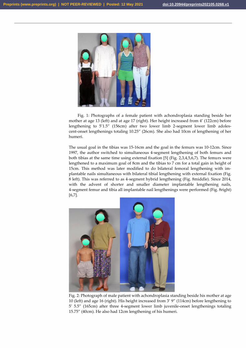
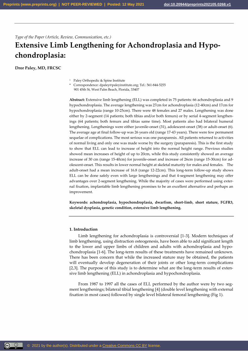
Fig. 1: Photographs of a female patient with achondroplasia standing beside her
mother at age 13 (left) and at age 17 (right). Her height increased from 4’ (122cm) before
lengthening to 5’1.5” (156cm) after two lower limb 2-segment lower limb adoles-
cent-onset lengthenings totaling 10.25” (26cm). She also had 10cm of lengthening of her
humeri.
The usual goal in the tibias was 15-16cm and the goal in the femurs was 10-12cm. Since
1997, the author switched to simultaneous 4-segment lengthening of both femurs and
both tibias at the same time using external fixation [5] (Fig. 2,3,4,5,6,7). The femurs were
lengthened to a maximum goal of 8cm and the tibias to 7 cm for a total gain in height of
15cm. This method was later modified to do bilateral femoral lengthening with im-
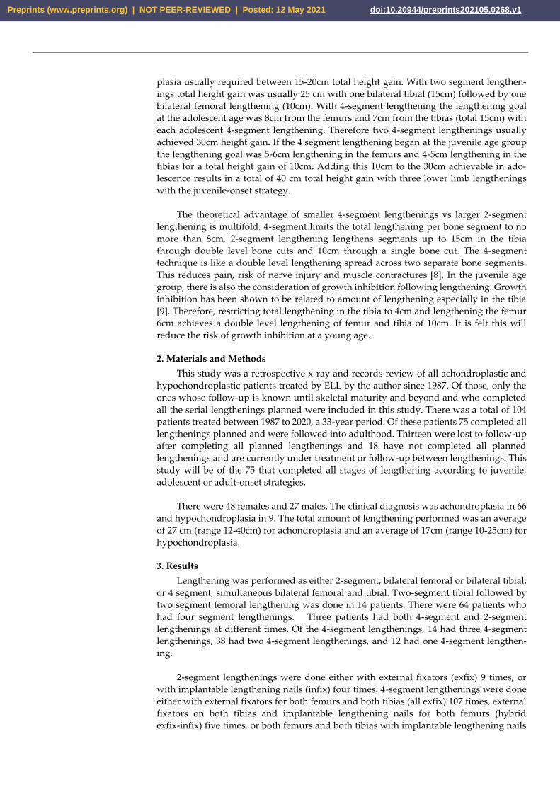
plantable nails simultaneous with bilateral tibial lengthening with external fixation (Fig.
8 left). This was referred to as 4-segment hybrid lengthening (Fig. 8middle). Since 2014,
with the advent of shorter and smaller diameter implantable lengthening nails,
4-segment femur and tibia all implantable nail lengthenings were performed (Fig. 8right)
[6,7].
Fig. 2: Photograph of male patient with achondroplasia standing beside his mother at age
10 (left) and age 16 (right). His height increased from 3’ 9” (114cm) before lengthening to
5’ 5.5” (165cm) after three 4-segment lower limb juvenile-onset lengthenings totaling
15.75” (40cm). He also had 12cm lengthening of his humeri.
Preprints (www.preprints.org) | NOT PEER-REVIEWED | Posted: 12 May 2021 doi:10.20944/preprints202105.0268.v1
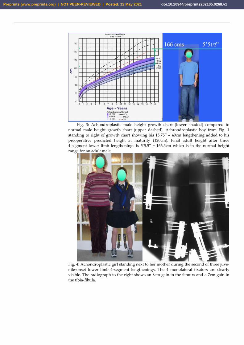
Fig. 3: Achondroplastic male height growth chart (lower shaded) compared to
normal male height growth chart (upper dashed). Achrondroplastic boy from Fig. 1
standing to right of growth chart showing his 15.75” = 40cm lengthening added to his
preoperative predicted height at maturity (120cm). Final adult height after three
4-segment lower limb lengthenings is 5’5.5” = 166.3cm which is in the normal height
range for an adult male.
Fig. 4: Achondroplastic girl standing next to her mother during the second of three juve-
nile-onset lower limb 4-segment lengthenings. The 4 monolateral fixators are clearly
visible. The radiograph to the right shows an 8cm gain in the femurs and a 7cm gain in
the tibia-fibula.
Preprints (www.preprints.org) | NOT PEER-REVIEWED | Posted: 12 May 2021 doi:10.20944/preprints202105.0268.v1
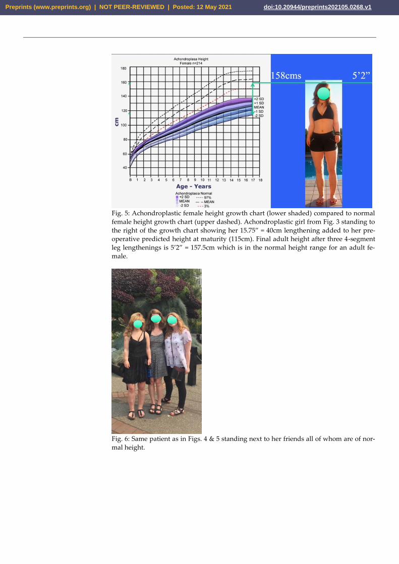
Fig. 5: Achondroplastic female height growth chart (lower shaded) compared to normal
female height growth chart (upper dashed). Achondroplastic girl from Fig. 3 standing to
the right of the growth chart showing her 15.75” = 40cm lengthening added to her pre-
operative predicted height at maturity (115cm). Final adult height after three 4-segment
leg lengthenings is 5’2” = 157.5cm which is in the normal height range for an adult fe-
male.
Fig. 6: Same patient as in Figs. 4 & 5 standing next to her friends all of whom are of nor-
mal height.
Preprints (www.preprints.org) | NOT PEER-REVIEWED | Posted: 12 May 2021 doi:10.20944/preprints202105.0268.v1
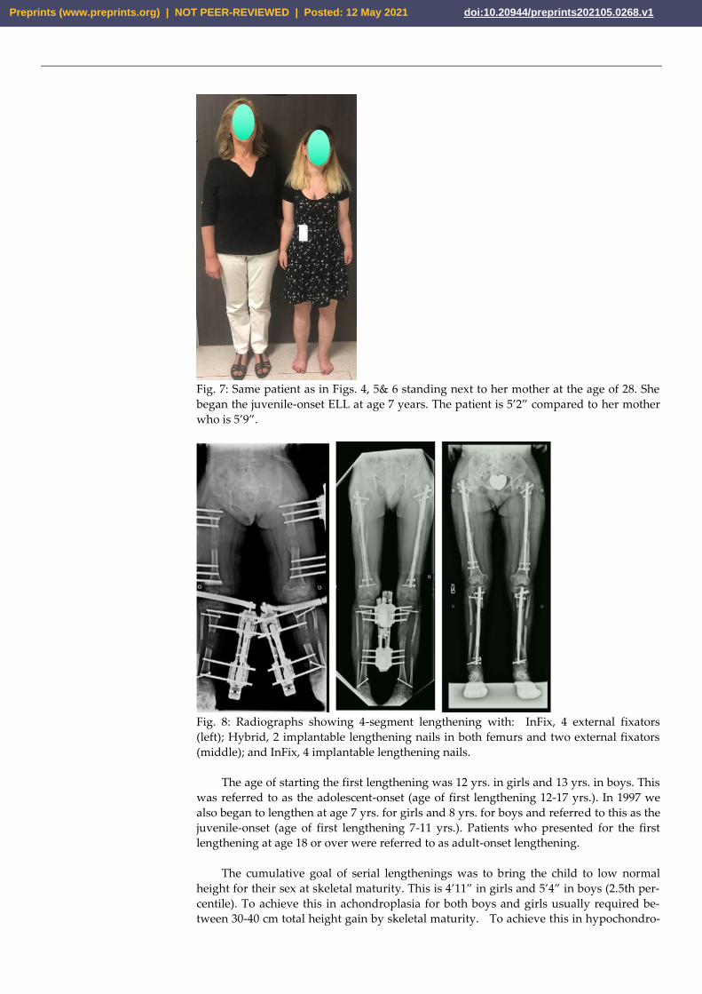
Fig. 7: Same patient as in Figs. 4, 5& 6 standing next to her mother at the age of 28. She
began the juvenile-onset ELL at age 7 years. The patient is 5’2” compared to her mother
who is 5’9”.
Fig. 8: Radiographs showing 4-segment lengthening with: InFix, 4 external fixators
(left); Hybrid, 2 implantable lengthening nails in both femurs and two external fixators
(middle); and InFix, 4 implantable lengthening nails.
The age of starting the first lengthening was 12 yrs. in girls and 13 yrs. in boys. This
was referred to as the adolescent-onset (age of first lengthening 12-17 yrs.). In 1997 we
also began to lengthen at age 7 yrs. for girls and 8 yrs. for boys and referred to this as the
juvenile-onset (age of first lengthening 7-11 yrs.). Patients who presented for the first
lengthening at age 18 or over were referred to as adult-onset lengthening.
The cumulative goal of serial lengthenings was to bring the child to low normal
height for their sex at skeletal maturity. This is 4’11” in girls and 5’4” in boys (2.5th per-
centile). To achieve this in achondroplasia for both boys and girls usually required be-
tween 30-40 cm total height gain by skeletal maturity. To achieve this in hypochondro-
Preprints (www.preprints.org) | NOT PEER-REVIEWED | Posted: 12 May 2021 doi:10.20944/preprints202105.0268.v1
plasia usually required between 15-20cm total height gain. With two segment lengthen-
ings total height gain was usually 25 cm with one bilateral tibial (15cm) followed by one
bilateral femoral lengthening (10cm). With 4-segment lengthening the lengthening goal
at the adolescent age was 8cm from the femurs and 7cm from the tibias (total 15cm) with
each adolescent 4-segment lengthening. Therefore two 4-segment lengthenings usually
achieved 30cm height gain. If the 4 segment lengthening began at the juvenile age group
the lengthening goal was 5-6cm lengthening in the femurs and 4-5cm lengthening in the
tibias for a total height gain of 10cm. Adding this 10cm to the 30cm achievable in ado-
lescence results in a total of 40 cm total height gain with three lower limb lengthenings
with the juvenile-onset strategy.
The theoretical advantage of smaller 4-segment lengthenings vs larger 2-segment
lengthening is multifold. 4-segment limits the total lengthening per bone segment to no
more than 8cm. 2-segment lengthening lengthens segments up to 15cm in the tibia
through double level bone cuts and 10cm through a single bone cut. The 4-segment
technique is like a double level lengthening spread across two separate bone segments.
This reduces pain, risk of nerve injury and muscle contractures [8]. In the juvenile age
group, there is also the consideration of growth inhibition following lengthening. Growth
inhibition has been shown to be related to amount of lengthening especially in the tibia
[9]. Therefore, restricting total lengthening in the tibia to 4cm and lengthening the femur
6cm achieves a double level lengthening of femur and tibia of 10cm. It is felt this will
reduce the risk of growth inhibition at a young age.
2. Materials and Methods
This study was a retrospective x-ray and records review of all achondroplastic and
hypochondroplastic patients treated by ELL by the author since 1987. Of those, only the
ones whose follow-up is known until skeletal maturity and beyond and who completed
all the serial lengthenings planned were included in this study. There was a total of 104
patients treated between 1987 to 2020, a 33-year period. Of these patients 75 completed all
lengthenings planned and were followed into adulthood. Thirteen were lost to follow-up
after completing all planned lengthenings and 18 have not completed all planned
lengthenings and are currently under treatment or follow-up between lengthenings. This
study will be of the 75 that completed all stages of lengthening according to juvenile,
adolescent or adult-onset strategies.
There were 48 females and 27 males. The clinical diagnosis was achondroplasia in 66
and hypochondroplasia in 9. The total amount of lengthening performed was an average
of 27 cm (range 12-40cm) for achondroplasia and an average of 17cm (range 10-25cm) for
hypochondroplasia.
3. Results
Lengthening was performed as either 2-segment, bilateral femoral or bilateral tibial;
or 4 segment, simultaneous bilateral femoral and tibial. Two-segment tibial followed by
two segment femoral lengthening was done in 14 patients. There were 64 patients who
had four segment lengthenings. Three patients had both 4-segment and 2-segment
lengthenings at different times. Of the 4-segment lengthenings, 14 had three 4-segment
lengthenings, 38 had two 4-segment lengthenings, and 12 had one 4-segment lengthen-
ing.
2-segment lengthenings were done either with external fixators (exfix) 9 times, or
with implantable lengthening nails (infix) four times. 4-segment lengthenings were done
either with external fixators for both femurs and both tibias (all exfix) 107 times, external
fixators on both tibias and implantable lengthening nails for both femurs (hybrid
exfix-infix) five times, or both femurs and both tibias with implantable lengthening nails
Preprints (www.preprints.org) | NOT PEER-REVIEWED | Posted: 12 May 2021 doi:10.20944/preprints202105.0268.v1
(all infix) 16 times. Lengthening of the humerus [10,11] for between 10-12 cm was done in
all patients with achondroplasia and all but two patients with hypochondroplasia for
between 10-12 cm (exfix in all but two cases which were done with infix).
The average age at final follow-up was 26 years old (range 17-43 years). This repre-
sented a follow-up average of 13 years in the adult-onset group, vs 10 years in the com-
bined adolescent and juvenile-onset groups (range 2-33 years).
While there were various complications during lengthening [4,12], for the purpose
of this end result study, we only tabulated the ones that led to permanent sequalae. One
female patient aged 16, developed paraparesis upon waking up from anesthesia at the
time of removal of the external fixators after a successful second 2-segment lower limb
lengthening (total length gain was 25 cm). She did not have any neurologic dysfunction
during the distraction or consolidation phases. She did have a recognized thoracolumbar
kyphosis which her neurosurgeon elected not to treat since she was asymptomatic. The
paraparesis was treated by decompression and fusion and did improve although she was
left with some permanent lower limb weakness. A second patient developed RSD after
humeral lengthening. This eventually resolved after decompression of ulnar and radial
nerves.
Other long-term findings not directly related to lengthening included one patient
who committed suicide several years after finishing lengthening. He had always ex-
pressed satisfaction with the lengthening. He apparently struggled with depression
while studying in university unbeknownst to his parents and friends. Three patients had
significant hip dysplasia. Two of these were treated by periacetabular osteotomy imme-
diately after completing lengthenings. A third patient went on to develop arthritis of one
hip which will likely require a total hip replacement. No other patients developed de-
generative joints of hips knees or ankles. There was no evidence of radiographic deteri-
oration of the joints after recovering from lengthening.
Patients started lengthening between ages 6-11 (juvenile-onset 31), 12-17 years (ad-
olescent-onset 38), or 18 years and older (adult-onset 6). Five patients developed spinal
stenosis requiring surgery, either prior to, during treatment or at a later date remote from
the lengthening.
All patients had completed high-school, and many went on to higher education. All
patients had returned to normal activities of daily living. Many were also participating in
sporting activities. No patient was negatively impacted by the lengthening physically or
psychologically, except the one patient who became paraparetic. Many patients were
employed, and some were married and had children. Every patient said they would do
this again despite the hardships. This included the patients with adult-onset ELL surgery.
4. Discussion
There have been numerous reviews of lengthening results in achondroplasia and
hypochondroplasia [13-20]. Most focused on the short-term results and specific length-
ening complications. Park et al 2015 from Korea [13], reported on 28 achondroplastic pa-
tients who underwent lengthening as adolescents (bilateral tibias followed by bilateral
femurs). The average height gain was 18.2cm (8.4 femurs, 9.8 tibias). Fewer complications
were found with lengthening of the femur than with lengthening of the tibia. Chibule et
al from India [14] presented 8 cases with an average of 15.4cm lengthening of the femurs,
9.9cm of the tibias and 9.6cm of the humeri. Donaldson et al from the UK [15], reported
on 10 achondroplastic children lengthened a total average of 20cm with good final re-
sults. Yasui et al from Japan [16], published on 35 patients with achondroplasia and hy-
pochondroplasia lengthened with external fixation. The average total length gain was
Preprints (www.preprints.org) | NOT PEER-REVIEWED | Posted: 12 May 2021 doi:10.20944/preprints202105.0268.v1
14.3cm (7.2cm femurs, 7.1 cm tibias). Aldegheri et al from Italy [17] reported on 100 pa-
tients with achondroplasia and hypochondroplasia who underwent an average of 20.5cm
lengthening for the former and 17.5cm for the latter. Lee and Chow from Hong Kong [18]
published on 8 patients with an average gain of 5.2cm in the femurs and 5.8cm gain in the
tibias. Schiedel and Rodl from Germany [19] did a meta-analysis of 172 patients reported
in the literature. The average length gain ranged from 5.7 to 20.5cm. Leiva-Gea etal from
Spain [20] reported on the use of 4 segment lengthening for achondroplasia in 17 patients.
The average height gain was 14.4 cm.
The average male height in achondroplasia is 132cm with the low end of height
range being 118cm and the high end 146cm [21,22]. The low end of normal height range
for a male is 160cm. The difference between the low end, average and high-end height for
a male with achondroplasia vs the low-end normal height for a male is 42cm, 28cm, and
14cm respectively.
The average female height in achondroplasia is 125cm with the low end of height
range being 114cm and the high end 138cm. The low end of normal height range for a
female is 150cm. The difference between the low end, average and high-end height for a
female with achondroplasia vs the low-end normal height for a female is 36cm, 25cm,
and 12cm respectively.
What, therefore, should be the height gain goal of extensive limb lengthening? Our
goal was to achieve the low end of normal height for their sex. This requires between 14
to 42cm in males and between 12 and 36cm in females. Using Schiedel and Rodl’s [19]
metanalysis as the range of height gain from lengthening from most studies (5.7-20.5 cm)
suggests that few if any achondroplastic patients achieve low normal height for their sex
with the methods used by most authors (2-segment: bilateral femoral and bilateral tibial
lengthening) [13-18]. Even the one 4-segment lengthening study by Leiva-Gea et al [20]
from Spain, only achieved 14.4cm average height gain. In our study the average gain was
27cm for achondroplasia and 17cm for hypochondroplasia. The 29 patients with achon-
droplasia who underwent the juvenile-onset strategy had an average lower limb length
gain of 30.1cm (median 28cm, range 15-40cm). Twenty-five of the 29 gained 25cm or
greater. The 33 achondroplasia patients who underwent the adolescent-onset strategy
gained an average of 26 cm (median 27cm; range 15-30cm). Twenty-seven of the 33
gained 25cm or greater. The 4 adult achondroplastic patients gained an average of 16.8cm
(range 12-22cm). It is clear that both the adolescent-onset as well as the juvenile-onset
strategies gain sufficient height to reach the low normal height for their sex. Almost all
patients in both groups achieved height gain of 25cm or greater. Half the patients in the
juvenile-onset group achieved gains of 30cm or greater up to 40cm. This study is there-
fore the first study to demonstrate that extensive limb lengthening for achondroplasia
can result in safe increase of height to normal adult levels for each sex. In contrast pre-
vious methods reported, have fallen short of that goal [13-20]. This study demonstrates
that the criticism that ELL can only produce ‘tall dwarfs’ is false [23]. ELL can add suffi-
cient height to restore achondroplastic patients to normal adult heights for each sex in the
majority of cases. Although we did not do quality of life (QOL) analysis, prior studies of
QOL have shown that despite frequent complications and despite the limited height gain
as previously described, the QOL improved with ELL [24].
There are no published growth charts for hypochondroplasia. In this author’s expe-
rience the final adult height in hypochondroplasia is 10-15cm taller than in achondro-
plasia. In this study the average length gain in hypochondroplasia was 16.8cm (range
10-25 cm). All of these patients reached normal adult height for their sex.
The 4-segment lengthening method is currently our preferred lengthening strategy
to achieve maximum total length gain in the shortest possible time without exceeding
Preprints (www.preprints.org) | NOT PEER-REVIEWED | Posted: 12 May 2021 doi:10.20944/preprints202105.0268.v1
8cm lengthening in any single bone segment. While this strategy was originally used
with 4-segment external fixation application it can also be done with 4-segment im-
plantable limb lengthening devices or with hybrid 4-segment lengthening; two segments
with external and two segments with internal devices. When four intramedullary nails
are inserted, we stagger the insertion by doing the two tibias, followed three weeks later
by the two femurs. This is to reduce the risk of fat embolism syndrome from reaming 4
bones at one time. When the nails are exchanged for a second lengthening the bones do
not have to be reamed again and therefore all four rods can be inserted in one surgery.
Most recently the extramedullary lengthening with Precice nail or Precice plate has al-
lowed us to use implantable lengthening nails in the femurs and lengthening plates in the
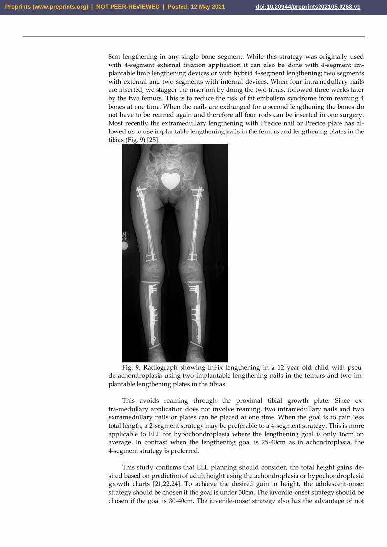
tibias (Fig. 9) [25].
Fig. 9: Radiograph showing InFix lengthening in a 12 year old child with pseu-
do-achondroplasia using two implantable lengthening nails in the femurs and two im-
plantable lengthening plates in the tibias.
This avoids reaming through the proximal tibial growth plate. Since ex-
tra-medullary application does not involve reaming, two intramedullary nails and two
extramedullary nails or plates can be placed at one time. When the goal is to gain less
total length, a 2-segment strategy may be preferable to a 4-segment strategy. This is more
applicable to ELL for hypochondroplasia where the lengthening goal is only 16cm on
average. In contrast when the lengthening goal is 25-40cm as in achondroplasia, the
4-segment strategy is preferred.
This study confirms that ELL planning should consider, the total height gains de-
sired based on prediction of adult height using the achondroplasia or hypochondroplasia
growth charts [21,22,24]. To achieve the desired gain in height, the adolescent-onset
strategy should be chosen if the goal is under 30cm. The juvenile-onset strategy should be
chosen if the goal is 30-40cm. The juvenile-onset strategy also has the advantage of not
Preprints (www.preprints.org) | NOT PEER-REVIEWED | Posted: 12 May 2021 doi:10.20944/preprints202105.0268.v1
allowing the achondroplastic or hypochondroplastic child to fall too far behind their
peers in height. Lengthening during adolescence offers children with achondroplasia and
hypochondroplasia a ‘growth spurt’ in parallel with their peers. Lengthening younger
than age 7 in girls and 8 in boys risks growth inhibition and also is limited in total
amount of length gain [9]. In this authors opinion it should be avoided. Parents who want
ELL should also avoid having osteotomy for genu varum at a young age since the genu
varum can be corrected at the time of the juvenile-onset lengthening [26]. This is another
advantage of the juvenile-onset strategy.
With the introduction of pharmacotherapeutics for achondroplasia [27], ELL is likely
to become an abandoned treatment. Since the height gain from these new drugs may not
achieve full normal adult height (especially depending on what age these drugs are
started, e.g. girls achieve half their lower and upper limb growth by age 3 yrs and boys by
age 4 yrs; by age 1 both boys and girls have one third of their total adult limb lengths.
Therefore, if the therapeutics don’t start until age 4 they are unlikely to regain the inhib-
ited growth that already occurred. For this reason, it is likely that stature lengthening for
children with achondroplasia will still be needed but to a lesser extent and only at skel-
etal maturity. This may involve one 2 or 4-segment lengthening with fully implantable
lengthening devices.
Authors should discuss the results and how they can be interpreted from the per-
spective of previous studies and of the working hypotheses. The findings and their im-
plications should be discussed in the broadest context possible. Future research direc-
tions may also be highlighted.
Institutional Review Board Statement: The study was conducted according to the guidelines of
the Declaration of Helsinki, and approved by the Institutional Review Board MetroWest Medical
Center. IRB# 2018-135. IRB Address: 115 Lincoln Street, Framingham, MA 01702 FWA#: 00003242 /
FWA Expiration Date: 08/29/2023 IORG: 0002308 / IORG Expiration Date: 06/10/2023
Informed Consent Statement: Patient consent was waived due to this study was a retrospective
chart review with minimal risk.
Conflicts of Interest: The authors declare no conflict of interest.
References
References must be numbered in order of appearance in the text (including citations in tables and legends) and listed indi-
vidually at the end of the manuscript. We recommend preparing the references with a bibliography software package, such as
EndNote, ReferenceManager or Zotero to avoid typing mistakes and duplicated references. Include the digital object identifier
(DOI) for all references where available.
Citations and references in the Supplementary Materials are permitted provided that they also appear in the reference list here.
1. Paley D: Current techniques of limb lengthening. J Pediatr Orthop 8:73-92, 1988.
2. Herzenberg J, Paley D: Methods and strategies in limb lengthening and realignment for skeletal dysplasia. In, Laron Z,
Mastragostino S, Romano C (ed): Limb Lengthening: For Whom, When & How? Tel Aviv, Freund Publishing, Ltd., 1995,
pp 181-199.
3. Herzenberg JE, Paley D: Stature lengthening: skeletal dysplasia. In, Rozbruch SR, Ilizarov S (eds): Limb Lengthening and
Reconstruction Surgery. New York, Informa Healthcare, 2007, ch 43, pp 575–596.
4. Rolf D. Burghardt , Koichi Yoshino , Naoya Kashiwagi , Shigeo Yoshino b, Anil Bhave , Dror Paley , John E. Her-
zenberg: Bilateral double level tibial lengthening in dwarfism. J. Orthop 2015 Dec; 12(4): 242–247.
5. Paley D: Progress in and from Limb Lengthening. Current Progress in Orthopedics Tree Life Media: Kothari Medical
Subscription Services, Pvt. Ltd., 2014 Ch 4, pp 55-80.
Preprints (www.preprints.org) | NOT PEER-REVIEWED | Posted: 12 May 2021 doi:10.20944/preprints202105.0268.v1
6. Paley D, Harris M, Debiparshad K, Prince D: Limb Lengthening by Implantable Limb Lengthening Devices. Techniques in
Orthopaedics: June 2014 - Volume 29 - Issue 2 - pp 72-85.
7. Paley D. PRECICE intramedullary limb lengthening system. Expert Rev Med Devices. 2015 May;12(3):231-49. Epub 2015
Feb 18.
8. Venkatesh K.P., Modi H.N., Devmurari K., Yoon J.Y., Anupama B.R., Song H.R.: Femoral lengthening in achondroplasia.
Magnitude of Lengthening in Relation to Patterns of Callus, Stiffness of Adjacent Joints and Fracture. J. Bone Joint Surg.
VOL. 91-B, No. 12, DECEMBER 2009, pp 1612-17.
9. Song SH, Kim SE, Agashe MV, Lee H, Refai MA, Park YE, Choi HJ, Park JH, Song HR: Growth disturbance after length-
ening of the lower limb and quantitative assessment of physeal closure in skeletally immature patients with achondro-
plasia. J. Bone and Joint Surg. VOL. 94-B, No. 4, April 2012 , pp 556-63.
10. Tetsworth K, Krome J, Paley D: Lengthening and deformity correction of the upper extremity by the Ilizarov technique.
Orthop Clin North Am 22:689-713, 1991.
11. Paley D, Kelly D: Lengthening and deformity correction in the upper extremities. In, Raskin K (ed): Atlas of the Hand
Clinics. Philadelphia, WB Saunders, 2000, vol 5, pp 117-172.
12. Paley D: Problems, obstacles, and complications of limb lengthening by the Ilizarov technique. Clin Orthop 250:81-104,
1990.
13. Kwang-Won Park, Rey-an Niño Garcia, Chastity Amor Rejuso, Jung-Woo Choi, and Hae-Ryong Song: Limb Lengthening
in Patients with Achondroplasia. Yonsei Med J 2015 Nov;56(6):1656-1662.
14. Sanjay K Chilbule, Vivek Dutt, and Vrisha Madhuri: Limb lengthening in achondroplasia. Indian J Orthop. 2016 Jul-Aug;
50(4): 397–405.
15. James Donaldson, Syed Aftab, Christopher Bradish: Achondroplasia and limb lengthening: Results in a UK cohort and
review of the literature. J. Orthop. 2015, 12, 31-34.
16. Yasui N, Kawabata H, Kojimoto H, Ohno H, Matsuda S, Araki N, Shimomura Y, Ochi T: Lengthening of the Lower Limbs
in Patients with Achondroplasia and Hypochondroplasia. Clin. Orthop. & Rel. Res. 1997, 344, pp 298-306.
17. Aldegheri R, Dall’Orca C: Limb Lengthening in Short Stature Patients. J. Pediatr. Orthop. B, 10: 238-247, 2001.
18. Lie WH, Chow W: Limb lengthening in short-stature patients using monolateral and circular external fixators. Hong Kong
Med J Vol 15, No 4, August 2009, pp 280-84.
19. Schiedel F, Rödl R: Lower limb lengthening in patients with disproportionate short stature with achondroplasia: a sys-
tematic review of the last 20 years. Disability & Rehabilitation, 2012; 34(12): 982–987 © 2012 Informa UK, Ltd.
20. Leiva-Gea A, Borja Delgado-Rufino F, Queipo-de-Llano A, Mariscal-Lara J, Lombardo-Torre M, Luna-González F:
Staged upper and lower limb lengthening performing bilateral simultaneous surgery of the femur and tibia in achondro-
plastic patients. Archives of Orthopaedic and Trauma Surgery 2020 https://doi.org/10.1007/s00402-020-03360-3.
21. Horton WA, Rotter JI, Rimoin DL, et al. Standard growth curves for achondroplasia. J Pediatr. 1978;93:435–438.
22. Paley D, Matz AL, Kurland DB, Lamm BM, Herzenberg JE: The multiplier method for prediction of adult height in pa-
tients with achondroplasia. J Pediatr Orthop 25:539–542, 2005.
23. Fernandez S, Gomez A, Branscombe N, Morales JF: Influence of the Social Context on Use of Surgical-Lengthening and
Group-Empowering Coping Strategies Among People With Dwarfism. Rehabilitation Psychology 2012, Vol. 57. No. 3,
224-235..
24. Seung-Ju Kim, Gracia Cielo Balce, Mandar Vikas Agashe, Sang-Heon Song, Hae-Ryong Song: Is Bilateral Lower Limb
Lengthening Appropriate for Achondroplasia? Midterm Analysis of the Complications and Quality of Life. Clin Orthop
Relat Res (2012) 470:616–621.
Preprints (www.preprints.org) | NOT PEER-REVIEWED | Posted: 12 May 2021 doi:10.20944/preprints202105.0268.v1
25. Shannon C, Paley D: Extramedullary Internal Limb Lengthening. Techniques in Orthopaedics,Vol. 35, No. 3, September
2020.
26. Ali Al Kaissi, Sebastian Farr, Rudolf Ganger, Jochen G. Hofstaetter Klaus Klaushofer, Franz Grill: Treatment of Varus
Deformities of the Lower Limbs in Patients with Achondroplasia and Hypochondroplasia. The Open Orthopaedics Jour-
nal, 2013, 7, 33-39.
27. Ravi Savarirayan, Louise Tofts, Melita Irving, William Wilcox, Carlos A Bacino, Julie Hoover-Fong, Rosendo Ullot Font,
Paul Harmatz, Frank Rutsch, Michael B Bober, Lynda E Polgreen, Ignacio Ginebreda, Klaus Mohnike, Joel Charrow,
Daniel Hoernschmeyer, Keiichi Ozono, Yasemin Alanay, Paul Arundel, Shoji Kagami, Natsuo Yasui, Klane K White,
Howard M Saal, Antonio Leiva-Gea, Felipe Luna-González, Hiroshi Mochizuki, Donald Basel, Dania M Porco, Kala
Jayaram, Elena Fisheleva, Alice Huntsman-Labed, Jonathan Day. Once-daily, subcutaneous vosoritide therapy in children
with achondroplasia: a randomised, double-blind, phase 3, placebo-controlled, multicentre trial. The Lancet, 2020; 396
(10252): 684 DOI: 10.1016/S0140-6736(20)31541-5
Preprints (www.preprints.org) | NOT PEER-REVIEWED | Posted: 12 May 2021 doi:10.20944/preprints202105.0268.v1


