Embed Size (px)
Citation preview
Exercise Training Prevents Arterial Baroreflex Dysfunctionin Rats Treated With Central Angiotensin II
Yan-Xia Pan, Lie Gao, Wei-Zhong Wang, Hong Zheng, Dongmei Liu, Kaushik P. Patel,Irving H. Zucker, Wei Wang
Abstract—Angiotensin II (Ang II)–induced arterial baroreflex dysfunction is associated with superoxide generation in thebrain. Exercise training (EX) improves baroreflex function and decreases oxidative stress in cardiovascular diseaseslinked to elevated central Ang II. The aim of this study was to determine whether previous EX prevents barorefleximpairment caused by central administration of exogenous Ang II via an Ang II–superoxide mechanism. Four groupsof rats were used: non-EX artificial cerebrospinal fluid infused, non-EX Ang II infused, EX artificial cerebrospinal fluidinfused, and EX Ang II infused. Rats were treadmill trained for 3 to 4 weeks and subjected to intracerebroventricularinfusion of Ang II over the last 3 days of EX. Twenty-four hours after the end of EX, the arterial baroreflex was assessedin anesthetized rats. Compared with non-EX artificial cerebrospinal fluid–infused rats, Ang II significantly decreasedbaroreflex sensitivity (maximum gain: 3.0�0.2% of maximum per millimeter of mercury versus 1.6�0.1% of maximumper millimeter of mercury; P�0.01), which was abolished by acute intracerebroventricular infusion of the Ang II type1 receptor antagonist losartan and the reduced nicotinamide-adenine dinucleotide phosphate oxidase inhibitor apocynin.EX prevented the decrease in baroreflex sensitivity and downregulated Ang II type 1 receptor and NADPH oxidasesubunit protein expression in the paraventricular nucleus of Ang II–infused rats. Finally, EX decreased superoxideproduction in the paraventricular nucleus of Ang II–infused rats. These results indicate that EX improves arterialbaroreflex function in conditions of high brain Ang II, which is mediated by the central Ang II type 1 receptor andassociated with a reduction in central oxidative stress. (Hypertension. 2007;49:519-527.)
Key Words: exercise � baroreflex � sympathetic nerve activity � reactive oxygen species � AT1 receptor
Impairment of arterial baroreflex function is an importantfeature in cardiovascular diseases, such as chronic heart
failure (CHF) and hypertension.1,2 This abnormality increasescardiovascular risk.3 Recent studies indicate that the bluntedarterial baroreflex is related to an enhanced central angioten-sin II (Ang II) mechanism, because blockade of Ang II type1 receptors (AT1Rs) in the brain restored baroreflex sensitiv-ity.4 Intracerebroventricular (icv) infusion of Ang II depressesarterial baroreflex function in normal animals.5 However,specific brain nuclei and the intracellular mechanism for AngII–induced baroreflex dysfunction have not been fully iden-tified. The paraventricular nucleus (PVN) of the hypothala-mus plays an important role in the control of baroreflexfunction and sympathetic drive.6 Furthermore, the PVNcontains a high density of AT1R7 and is a candidate region torespond to Ang II in the cerebrospinal fluid.8
Several lines of evidence show that reduced nicotinamide-adenine dinucleotide phosphate (NAD[P]H) oxidase-derivedreactive oxygen species (ROS) are novel mediators of Ang IIsignaling in the central nervous system.9 Pretreatment withadenoviral-mediated superoxide dismutase prevents Ang II–induced hypertension and the increased superoxide produc-
tion.10 On the other hand, the AT1R antagonist losartanabolishes Ang II–stimulated superoxide generation.9 In-creases in superoxide production may contribute to impairedarterial baroreflex function.5,11 Conversely, antioxidant treat-ment improves baroreflex sensitivity.11
Exercise training (EX) has been demonstrated to alter neuralcontrol of the circulation, including influencing arterial barore-flex function.12 Although the effects of EX on baroreflexfunction in normal subjects and animals are increased,12 de-creased,13,14 or unchanged,15 EX consistently increases barore-flex sensitivity in CHF and hypertensive subjects.16–18 Themechanisms responsible for the training-induced improvementof baroreflex function involve changes in central and peripheralcomponents of the baroreflex arc. It has been shown that EX notonly increases baroreceptor sensitivity16 and decreases ROS inperipheral tissue19,20 but also decrease central sympathetic out-flow.17,21 Furthermore, EX improves NO bioactivity within thePVN in CHF,22 which contributes to sympathoinhibition. Inaddition, EX also reduces AT1R mRNA expression in centralcardiovascular regulatory regions, such as the PVN, the nucleusof the solitary tract (NTS), and the rostral ventrolateral medulla(RVLM) in CHF animals.18 However, the central mechanisms
Received July 25, 2006; first decision August 14, 2006; revision accepted December 18, 2006.From the Department of Cellular and Integrative Physiology, University of Nebraska Medical Center, Omaha.Correspondence to Wei Wang, Department of Cellular and Integrative Physiology, University of Nebraska Medical Center, Omaha, NE 68198-5850.
E-mail [email protected]© 2007 American Heart Association, Inc.
Hypertension is available at http://www.hypertensionaha.org DOI: 10.1161/01.HYP.0000256955.74461.93
519 by guest on August 13, 2015http://hyper.ahajournals.org/Downloaded from
mediating baroreflex function after EX are not well understood.In this study, we hypothesized that EX improves arterial barore-flex function via reducing central ROS in a rat model of chronicicv infusion of Ang II. To test this hypothesis, we examined theeffects of both EX and central superoxide anion on arterialbaroreflex function and measured the central superoxide produc-tion and protein expression of AT1R and NAD(P)H oxidasesubunits in Ang II–infused rats.
MethodsAnimalsEighty-eight male Sprague–Dawley rats weighing 300 to 310 g wereassigned randomly to 4 groups (n�22 in each group): non-EX AngII–infused rats, non-EX artificial cerebrospinal fluid (aCSF)–infusedrats, EX Ang II–infused rats, and EX aCSF-infused rats. Experi-ments were approved by the University of Nebraska Medical CenterInstitutional Animal Care and Use Committee and conformed to theGuidelines for the Care and Use of Experimental Animals of theAmerican Physiological Society and the National Institutesof Health.
EX ProtocolRats were trained on a treadmill 5 days per week for �3 to 4 weeksaccording to the program used by Zheng et al.22 The treadmill speedand incline were gradually increased from 10 m/min, 0% grade, to 25m/min, 10% grade. Exercise duration was increased from 10 to 20min/day to 60 min/day to produce a significant endurance effectduring EX. To test the efficiency of EX, the soleus muscle was takenat the end of EX and frozen at �80°C. The citrate synthase activityfrom muscle homogenate was measured spectrophotometrically.23
icv Infusion of Ang IIThree days before the end of EX, the rats were anesthetized with aketamine/xylazine mixture (90 mg/kg and 5 mg/kg, IP) and placed ina stereotaxic apparatus (Stoelting). After the bregma was identified,a sterile brain cannula using the Alzet Brain Infusion Kit IIconnected to an osmotic minipump (model 1003D) was inserted intothe right lateral cerebral ventricle and fixed to the skull with dentalcement. The periosteums of both sides were sutured together tofasten the brain cannula. The coordinates were determined from thePaxinos and Watson rat atlas,24 which were 0.8-mm posterior,1.4-mm lateral to the bregma and 3.8-mm ventral to the 0 level. Thebrain cannula, connecting catheter and minipump, were prefilledwith Ang II or aCSF. Ang II was delivered at a rate of 60 ng/�L perhour. aCSF and Ang II were infused at 1 �L/h through theminipump. Twenty hours after implantation, the rats continued to betrained. In a preliminary study, the cannula tip placement wasconfirmed by infusion of 2% Chicago blue dye at the end of theexperiment. Water intake was measured daily during infusion of AngII or aCSF.
Acute Experiments
General Animal PreparationTwenty-four hours after the end of EX, the rats were anesthetizedwith urethane (800 mg/kg, IP) and �-chloralose (40 mg/kg, IP). Thetrachea was intubated, and the rats were artificially ventilated byinhalation of an air–O2 mixture using a ventilator (model SAR-830,Ardmore, PA). The right common carotid artery was catheterizedwith a pressure transducer (model SPR-524, Millar Instruments) formeasurement of mean arterial pressure (MAP). Heart rate (HR) wasderived from the arterial pressure pulse using the cardiotachometerfunction of the PowerLab data acquisition system (model 16SP, ADInstruments). The femoral veins were cannulated bilaterally foradministration of drugs. The rat head was stabilized in a stereotaxicapparatus (Stoelting). After the minipump was removed, the braincannula was connected to a microsyringe (model 7001, Hamilton)and flushed with 2 �L of aCSF.
Recording of Renal Sympathetic Nerve ActivityThe renal sympathetic nerve activity (RSNA) was recorded asdescribed by Gao et al.25 The left kidney was exposed retroperito-neally. A branch of the renal nerve was isolated and placed on abipolar platinum electrode. RSNA was amplified (�1000) andfiltered (bandwidth: 30 to 1000 Hz) via a Grass P55C preamplifier.The nerve signal was further amplified (5 to 10 mV/DIV), filtered(30 to 1000 Hz), and monitored on a Tektronix oscilloscope (model7313). The signal from the oscilloscope was displayed on a computerwhere RSNA was rectified, integrated, sampled (1 kHz), andconverted to a digital signal by the PowerLab data acquisitionsystem. When an optimal nerve signal was observed, the nerve andelectrode were covered with Kwik-Sil gel (World Precision Instru-ments). The maximum nerve activity was determined by injection ofnitroglycerin (60 �g/kg, IV). The noise level for RSNA wasdetermined at the end of the experiment when the rat was dead. Thevalue of RSNA was calculated by subtracting the noise from therecorded value. The baseline RSNA was expressed as a percentageof the maximum.
Assessment of Arterial Baroreflex FunctionBaroreflex function was assessed by determining the changes in theRSNA response to decreases and increases in MAP. MAP wasinitially decreased by intravenous injection of nitroglycerin (25 �g)and then immediately elevated by phenylephrine (10 �g). Theinjection duration of each drug is 30 to 40 seconds. The data forMAP and RSNA (expressed as a percentage of maximum nerveactivity) were collected every 2s over the MAP range from �50 to�140 mm Hg. The resultant sigmoidal relationship of RSNA andMAP was analyzed using a nonlinear regression equation26:%RSNA�A/�1�exp[B(MAP-C)]�D where A is the RSNA range(maximum response minus minimum response); B is the slopecoefficient (average slope of the reflex); C is the pressure at themidrange of the curve (BP50); and D is the minimum response ofRSNA. Values of A to D were derived in each rat, and these valueswere averaged to give a mean value that was used to construct acomposite sigmoidal baroreflex curve. The gain of the function atany give MAP was calculated from the derivative of the aboveequation. The maximum gain (or peak slope) was calculated usingthe following equation26: Gainmax�A�B �1/4, where A is the rangeand B is the average slope.
Losartan (500 nmol in 1 �L; n�8 in each group) was injectedthrough the brain cannula. The arterial baroreflex was evaluatedbefore and after losartan. In separate groups, the NAD(P)H oxidaseinhibitor apocynin (600 nmol in 1 �L/min; n�6 in each group) wasinfused through the brain cannula for 10 minutes. The arterialbaroreflex was assessed before and after apocynin. After each icvinjection, the brain cannula was flushed with 2 �L of aCSF. At theend of the experiment, the brain was removed and immediatelyfrozen at �80°C for Western blot analysis.
Western Blot Analysis of AT1R and NAD(P)H Oxidase inthe PVNThe brains were cut into 100-�m coronal sections with a cryostat(Leica) at �18°C, and 6 consecutive sections at the level of the PVNwere collected. The PVN was punched27 and homogenized inradioimmunoprecipitation assay buffer. The protein concentrationwas measured using a protein assay kit (Pierce Chemical). Theproteins were loaded onto a 10% SDS-PAGE gel along with proteinstandards (Bio-Rad Laboratories) in a separate lane for electrophore-sis and then transferred to polyvinylidene fluoride membrane. Themembrane was probed with rabbit polyclonal antibody against AT1R,gp91phox, p67phox, p47phox, p22phox (1:500 to 1:1000 dilutions, SantaCruz Biotechnology) and secondary antibody of goat anti-rabbit IgG(1:5000 dilutions, Pierce Chemical). The protein signals were de-tected by enhanced chemiluminescence reagent (Pierce Chemical)and analyzed using UVP BioImaging Systems. The levels of targetproteins were normalized with GAPDH as a loading control (1:1000dilutions, Santa Cruz Biotechnology).
520 Hypertension March 2007
by guest on August 13, 2015http://hyper.ahajournals.org/Downloaded from
Measurement of Superoxide Production in theHypothalamus and the PVNIn 4 additional groups, the superoxide production in the hypothala-mus was measured using lucigenin-enhanced chemiluminescence.28
Rats (n�6 for each group) were anesthetized and the brain wasremoved and immersed in cold Krebs/HEPES buffer (pH 7.4)28 onice. The hypothalamus papilla between the optic chiasm and the ponswas dissected and cut into small pieces. One piece of hypothalamuswas placed in preheated Krebs/HEPES buffer (37°C) containing5 �mol/L of lucigenin and then read in a Sirius luminometer, whichreports relative light units emitted over a 30-s interval for 5 minutes. Thevalue was subtracted from background activity and normalized to tissueweight. NAD(P)H (10 �mol/L) was used to stimulate NAD(P)Hoxidase. To determine the source of superoxide, hypothalamic sampleswere preincubated with the NAD(P)H oxidase inhibitor apocynin(1mmol/L) and the NO synthase inhibitor NG-monomethyl-L-arginine(1 mmol/L), respectively, for 30 minutes.
To detect superoxide in situ, the brain (n�2 for each group) wasremoved and immediately frozen at �80°C for 1 hour, blocked in thecoronal plane, and sectioned into 30-�m slices with a cryostat. Thesections at the level of the PVN were mounted on microscope slidesand incubated with dihydroethidium (2 �mol/L, Molecular Probes)for 30 minutes at 37°C in a light-protected humidified chamber.Images were visualized using a fluorescent microscope (Leica).
DrugsApocynin and NG-monomethyl-L-arginine were obtained from theCalbiochem Co. Losartan was a gift from Merck and Co. Ketaminewas obtained from Ft Dodge Laboratories Inc. Nitroglycerin wasobtained from ETHEX Co. Ang II and other chemicals wereobtained from Sigma-Aldrich Co.
Statistical AnalysesData are presented as mean�SE. Differences between groups weredetermined by a 2-way ANOVA followed by the Newman–Keulstest for posthoc analysis of significance. Responses before and aftera given intervention were compared with a paired t test. A P �0.05was considered statistically significant.
ResultsResponses to EXTable 1 shows that 3 to 4 weeks of EX significantly increasedcitrate synthase activity in soleus muscle in Ang II- andaCSF-infused rats compared with their respective non-EXgroups, indicating an increase in skeletal muscle oxidativecapacity and adaptation to EX. Basal MAP, HR, and body
weight were not different between EX groups and non-EXgroups. The ratios of left ventricular weight to body weightwere not altered in EX groups compared with non-EX groups.
Effects of EX on RSNA and Water Intake inAng II–Infused RatsIn non-EX rats, chronic icv infusion of Ang II increased thebaseline RSNA and water intake by 66% and 35% comparedwith aCSF infusion, indicating that changes in non-EX AngII–infused rats are significantly different from non-EX aCSF-infused rats, as reflected in Table 1. In contrast in EX rats,chronic Ang II infusion did not evoke the increase in baselineRSNA but still increased water intake. There was no differ-ence in the baseline RSNA between EX Ang II–infused ratsand EX or non-EX aCSF-infused rats (Table 1).
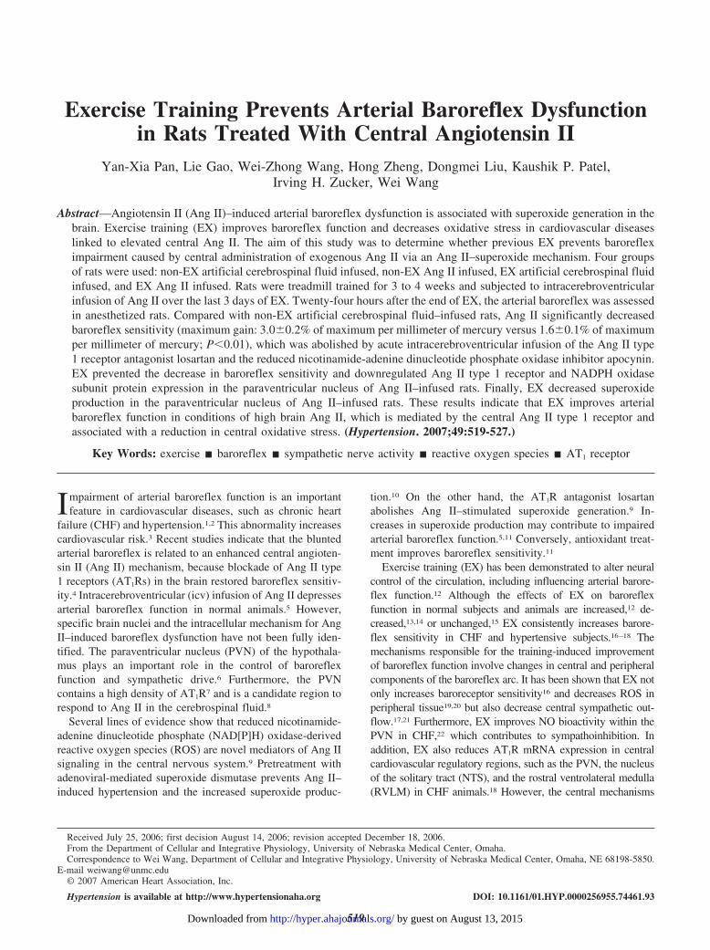
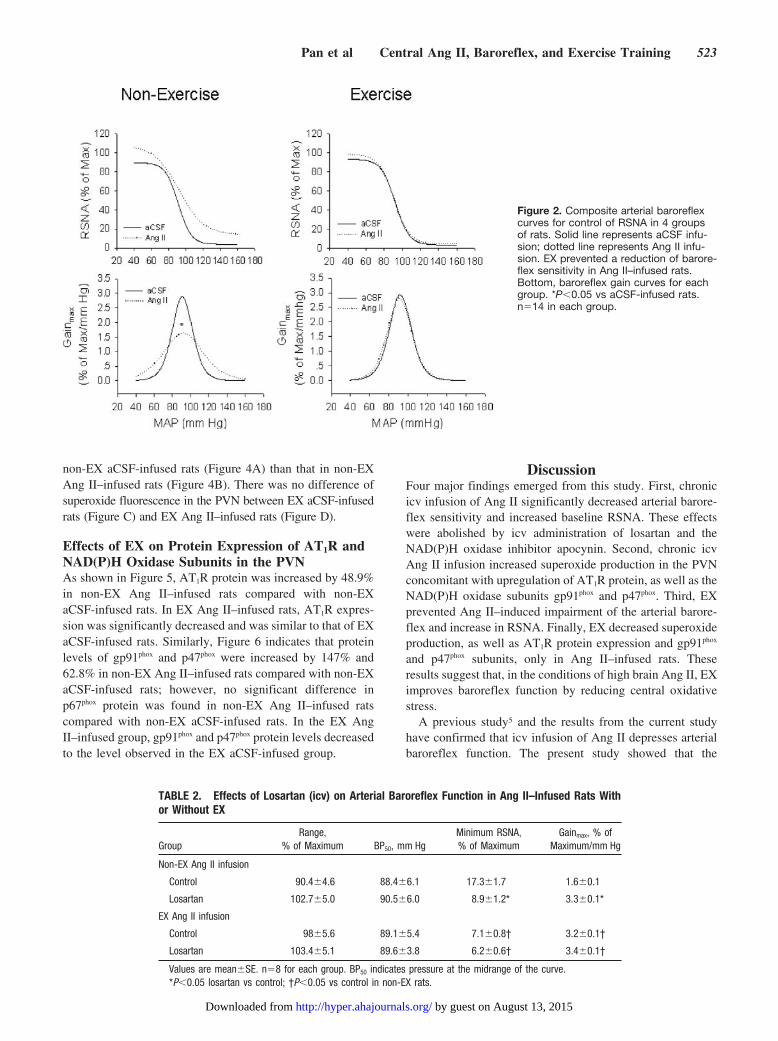
Effects of EX on Arterial Baroreflex Function inAng II–Infused RatsThe original recordings of baroreflex control of RSNA in the4 groups are shown in Figure 1. It is evidence that the reflexRSNA response to phenylephrine in non-EX Ang II–infusedrats was attenuated compared with non-EX aCSF-infusedrats. There was no difference in the reflex RSNA responsebetween the EX Ang II group and the EX aCSF group. Themaximum gain of baroreflex control of RSNA in the non-EXAng II group was decreased compared with the non-EX aCSFgroup, as shown in Figure 2. Baroreflex function was restoredin the EX Ang II–infused rats compared with the non-EXAng II–infused rats. Baroreflex gain in the non-EX aCSFgroup was similar to that of the EX aCSF group.
Effects of Losartan and Apocynin onBaroreflex FunctionTable 2 shows that acute icv infusion of losartan increased themaximum gain and decreased the minimum RSNA only innon-EX Ang II–infused rats. Losartan did not alter baroreflexfunction in EX Ang II–infused rats, EX aCSF-infused rats,and non-EX aCSF-infused rats. Meanwhile, losartan de-creased the basal RSNA in the non-EX Ang II group(31.4�5.8% of maximum versus 67.4�4.7% of maximum;
TABLE 1. Baseline Characteristics of EX and Non-EX Rats Infused With Eithericv aCSF or Ang II
Parameters
Non-EX EX
aCSF Ang II aCSF Ang II
Body weight, g 408�9 397�5 381�6 376�6
Left ventricle, g 0.85�0.03 0.77�0.04 0.81�0.06 0.9�0.05
LV/BW, mg/g 2.1�0.1 1.9�0.1 2.3�0.1 2.4�0.1
Basal MAP, mm Hg 88.7�5.3 95.7�3.8 86.1�4.2 89.4�5.1
Basal HR, bpm 375�13 392�10 360�14 370�9
Basal RSNA, % of maximum 40.6�5.3 67.4�4.7* 37.9�4.3 46.2�6.0†
Water intake, mL/kg per day 45.7�3.4 61.8�3.5* 48.5�4.1 62.2�3.0*
CS, �mol/min per gram 14.4�3.0 15.0�1.5 21.3�1.3* 24.3�2.2*
Values are mean�SE. n�14 in each group. LV indicates left ventricle; BW, body weight; CS, citratesynthase activity.
*P�0.05 vs non-EX aCSF-infused rats; †P�0.05 vs non-EX Ang II–infused rats.
Pan et al Central Ang II, Baroreflex, and Exercise Training 521
by guest on August 13, 2015http://hyper.ahajournals.org/Downloaded from
n�8; P�0.05) but not in the EX Ang II group (37.1�5.7% ofmaximum versus 46.2�6.0% of the maximum; P0.05).
In separate groups, icv infusion of apocynin improved themaximum gain of the baroreflex and minimum RSNA innon-EX Ang II–treated rats (Table 3). There were no differ-ences in baroreflex sensitivity before and after administrationof apocynin in non-EX and EX aCSF-infused rats. Apocyninalso reduced the basal RSNA in the non-EX Ang II group(33.4�6.4% of maximum versus 62.2�8.9% of maximum;n�6; P�0.05) but not in EX Ang II groups (25.7�3.3% ofmaximum versus 34.9�4.5% of maximum; P0.05).
Effects of EX on Superoxide Production in theHypothalamus and the PVNThe basal superoxide level was lower in EX Ang II–infusedrats than in non-EX Ang II–infused rats, which was similar to
that in non-EX aCSF-infused rats (Figure 3A). In the pres-ence of NAD(P)H (10 �mol/L), EX also depressed theincrease in superoxide production in Ang II–infused rats(Figure 3B). Pretreatment with apocynin markedly reducedsuperoxide generation in both basal conditions and in thepresence of NAD(P)H in non-EX Ang II–infused rats. How-ever, NG-monomethyl-L-arginine (1mmol/L) did not altersuperoxide production both in basal conditions and thepresence of NAD(P)H in non-EX Ang II–infused rats andnon-EX aCSF-infused rats. These data indicate that AngII–induced superoxide is mainly derived from activation ofNAD(P)H oxidase.
In situ detection of superoxide using dihydroethidiumfluorescence indicated an intense signal in the PVN. Further-more, superoxide fluorescence in the PVN was lower in
Figure 1. An original recording of the RSNA response to changes in arterial blood pressure induced by nitroglycerin (25 �g IV) and thenphenylephrine (10 �g IV). An attenuated RSNA response to an increase in MAP was observed in non-EX Ang II–infused rats (top) com-pared with non-EX aCSF-infused rats. However, there were no differences of RSNA between EX rats infused with aCSF or Ang II(bottom).
522 Hypertension March 2007
by guest on August 13, 2015http://hyper.ahajournals.org/Downloaded from
non-EX aCSF-infused rats (Figure 4A) than that in non-EXAng II–infused rats (Figure 4B). There was no difference ofsuperoxide fluorescence in the PVN between EX aCSF-infusedrats (Figure C) and EX Ang II–infused rats (Figure D).
Effects of EX on Protein Expression of AT1R andNAD(P)H Oxidase Subunits in the PVNAs shown in Figure 5, AT1R protein was increased by 48.9%in non-EX Ang II–infused rats compared with non-EXaCSF-infused rats. In EX Ang II–infused rats, AT1R expres-sion was significantly decreased and was similar to that of EXaCSF-infused rats. Similarly, Figure 6 indicates that proteinlevels of gp91phox and p47phox were increased by 147% and62.8% in non-EX Ang II–infused rats compared with non-EXaCSF-infused rats; however, no significant difference inp67phox protein was found in non-EX Ang II–infused ratscompared with non-EX aCSF-infused rats. In the EX AngII–infused group, gp91phox and p47phox protein levels decreasedto the level observed in the EX aCSF-infused group.
DiscussionFour major findings emerged from this study. First, chronicicv infusion of Ang II significantly decreased arterial barore-flex sensitivity and increased baseline RSNA. These effectswere abolished by icv administration of losartan and theNAD(P)H oxidase inhibitor apocynin. Second, chronic icvAng II infusion increased superoxide production in the PVNconcomitant with upregulation of AT1R protein, as well as theNAD(P)H oxidase subunits gp91phox and p47phox. Third, EXprevented Ang II–induced impairment of the arterial barore-flex and increase in RSNA. Finally, EX decreased superoxideproduction, as well as AT1R protein expression and gp91phox
and p47phox subunits, only in Ang II–infused rats. Theseresults suggest that, in the conditions of high brain Ang II, EXimproves baroreflex function by reducing central oxidativestress.
A previous study5 and the results from the current studyhave confirmed that icv infusion of Ang II depresses arterialbaroreflex function. The present study showed that the
Figure 2. Composite arterial baroreflexcurves for control of RSNA in 4 groupsof rats. Solid line represents aCSF infu-sion; dotted line represents Ang II infu-sion. EX prevented a reduction of barore-flex sensitivity in Ang II–infused rats.Bottom, baroreflex gain curves for eachgroup. *P�0.05 vs aCSF-infused rats.n�14 in each group.
TABLE 2. Effects of Losartan (icv) on Arterial Baroreflex Function in Ang II–Infused Rats Withor Without EX
GroupRange,
% of Maximum BP50, mm HgMinimum RSNA,% of Maximum
Gainmax, % ofMaximum/mm Hg
Non-EX Ang II infusion
Control 90.4�4.6 88.4�6.1 17.3�1.7 1.6�0.1
Losartan 102.7�5.0 90.5�6.0 8.9�1.2* 3.3�0.1*
EX Ang II infusion
Control 98�5.6 89.1�5.4 7.1�0.8† 3.2�0.1†
Losartan 103.4�5.1 89.6�3.8 6.2�0.6† 3.4�0.1†
Values are mean�SE. n�8 for each group. BP50 indicates pressure at the midrange of the curve.*P�0.05 losartan vs control; †P�0.05 vs control in non-EX rats.
Pan et al Central Ang II, Baroreflex, and Exercise Training 523
by guest on August 13, 2015http://hyper.ahajournals.org/Downloaded from

attenuated baroreflex control of RSNA was primarily becauseof the decreased gain of the baroreflex and an increase in theminimum RSNA. The maximum RSNA achieved during adecrease in MAP (nitroglycerin administration) was in-creased so that the range of the baroreflex curve was notchanged. Chronic Ang II infusion increased resting RSNAand water intake without a rise in resting MAP. After 3 to 4weeks of EX, Ang II failed to cause a decrease in thebaroreflex gain but increased water intake. Furthermore, themaximum and minimum RSNAs were decreased in the EXAng II group compared with the non-EX Ang II group. Therewas no difference in these parameters between EX and
non-EX aCSF-infused rats. This reflects the fact that basallevels of RSNA were altered in different groups and startedfrom higher basal RSNA in the non-EX Ang II group andlower basal RSNA in the EX Ang II, EX aCSF, and non-EXaCSF groups. The reduced maximum gain of the baroreflexwas restored in EX Ang II–infused rats and reached the levelof non-EX aCSF infusion. This is in agreement with previousresults obtained in CHF and hypertensive animals afterEX.16–18 The effects of EX on baroreflex function in normalsubjects has been inconsistent, with either an increase12,16 ordecrease13,14 being reported. This discrepancy may be be-cause of differences in the EX workload, the way of testingbaroreflex function, and whether the animal is in the anes-thetized or conscious state. Regardless of the reported differ-ent effects of EX on baroreflex function under normalconditions, EX appears to improve arterial baroreflex func-tion, at least in the conditions of high brain Ang II.
The mechanisms by which EX prevents the impairment ofbaroreflex function induced by chronic central Ang II infu-sion are not clear. In view of the fact that resting RSNA wasdecreased after EX, the reduced central sympathetic drivemay play an initial role in the improvement of baroreflexfunction. The hypothalamic PVN is a key site for control ofsympathetic outflow and the regulation of cardiovascularreflexes. PVN neurons project to the RVLM, NTS, andintermediolateral cell column of the spinal cord.29 Michelini30
and Jackson et al31 demonstrated that the oxytocinergic andvasopresinergic projections from the PVN to the NTS wereenhanced after EX, which might change neuronal activity inthe NTS. A previous study has shown that EX downregulatesAT1R mRNA expression in central cardiovascular regulatoryregions, such as the PVN, the NTS, and the RVLM, in CHFanimals.18 In this study, we focused on the PVN and foundthat the AT1R protein level in the PVN was significantlylower in the EX Ang II group than in the non-EX Ang IIgroup. However, there was no difference in AT1R proteinbetween the EX and non-EX aCSF groups. Consistent withthis result, icv infusion of the AT1R antagonist losartanimproved baroreflex function in the non-EX Ang II group butnot in the EX Ang II, EX aCSF, and non-EX aCSF groups,which exhibited lower AT1R expression. These data suggestthat EX can inhibit sympathoexcitation in the PVN under theconditions of sympathetic overactivity. Because of angioten-ergic projections from the PVN to the RVLM,32 we speculate
TABLE 3. Effects of Apocynin (icv) on Arterial Baroreflex Function in Ang II–Infused RatsWith or Without EX
GroupRange,
% of Maximum BP50, mm HgMinimum RSNA,% of Maximum
Gainmax, % ofMaximum/mm Hg
Non-EX Ang II infusion
Control 91.6�4.1 89.2�3.6 18.5�2.1 1.8�0.1
Apocynin 99�6.2 90.5�4.6 11.4�1.1* 3.0�0.1*
EX Ang II infusion
Control 100.7�4.3 90.7�3.4 9.6�1.1† 3.0�0.1†
Apocynin 103.4�5.1 91.5�4.5 7.5�1.0† 3.2�0.1†
Values are mean�SE. n�6 for each group. BP50 indicates pressure at the midrange of the curve.*P�0.05 apocynin vs control; †P�0.05 vs control in non-EX rats.
Figure 3. Hypothalamic superoxide production in the basalstate (A) or in the presence of NAD(P)H (10 �mol/L; B). Apocy-nin (1 mmol/L) and NG-monomethyl-L-arginine NG-monomethyl-L-arginine (1 mmol/L) were used to inhibit the activity ofNAD(P)H oxidase and NOS, respectively. *P�0.001 vs aCSF-infused rats; †P�0.001 vs Ang II–infused rats; ‡P�0.001, com-parison within a group. n�6, in each group.
524 Hypertension March 2007
by guest on August 13, 2015http://hyper.ahajournals.org/Downloaded from

that EX inhibits exogenous Ang II action in the PVN, which,in turn, affects baroreflex integration in the RVLM to alterbaroreflex function. Additional studies have shown that EXenhances sympathoinhibition in the PVN via an increase inneuronal NOS expression in CHF rats22 and an increase in thenumber of NOS-positive neurons in hypertensive rats.33 Theroles of PVN-NTS and PVN-RVLM neuronal pathways afterEX in the modulation of baroreflex control remain to beexamined.
In addition to a central mechanism, EX exerts peripheraleffects on the baroreflex. Brum et al16 and Krieger et al12
demonstrated that EX increased baroreceptor afferent activityin normal and spontaneous hypertensive rats. In CHF ani-mals, plasma Ang II was decreased after EX, which posi-tively correlated with sympathetic nerve activity and con-versely correlated with arterial baroreflex function.17,18 Thereis evidence that EX reduces myocardial oxidative stress,
Figure 4. Effect of EX on superoxide pro-duction in the PVN in chronic Ang II–in-fused rats. An intense dihydroethidiumfluorescent signal is clearly visible innon-EX Ang II–treated rats comparedwith all other groups. A, Non-EX aCSF-infused rats; B, Non-EX Ang II–infusedrats; C, EX aCSF-infused rats; D, EX AngII–infused rats. Scale bar�200 �m.
Figure 5. The influence of EX on AT1R protein expression in thePVN in the 4 groups of rats examined. Top, RepresentativeWestern blot for AT1R protein (lanes 1 to 4 presents treatmentwith aCSF, Ang II, aCSF�EX, and Ang II�EX, respectively). Bot-tom, Mean data from densitometric analysis. Values aremean�SE. *P�0.01 vs aCSF; †P�0.01 vs Ang II. n�8 in eachgroup.
Figure 6. The influence of EX on protein expression of NAD(P)Hoxidase subunits in the PVN in the 4 groups of rats studied.Top, Representative Western blot for NAD(P)H subunit expres-sion (lanes 1 to 4 are aCSF, Ang II, aCSF�EX, and Ang II�EX,respectively). Bottom, Mean data from densitometric analysis.Values are mean�SE. *P�0.05, **P�0.01 vs aCSF; †P�0.05,††P�0.01 vs Ang II. n�6 in each group.
Pan et al Central Ang II, Baroreflex, and Exercise Training 525
by guest on August 13, 2015http://hyper.ahajournals.org/Downloaded from
contributing to the improved baroreflex control of HR.20 Inaddition, we cannot exclude the possibility that EX changesthe reactivity of effectors, contributing to the improvement ofbaroreflex function.
A growing body of evidence indicates that superoxide is anovel signaling molecule that mediates central Ang II–induced hypertension, thirst, and sympathoexcitation.9 How-ever, the influence of superoxide in the brain on the arterialbaroreflex and the effect of EX on superoxide generation arenot identified. In the present study, icv infusion of theNAD(P)H oxidase inhibitor apocynin increased the gain ofbaroreflex control of RSNA and restored the blunted arterialbaroreflex in non-EX Ang II–infused rats where superoxideproduction in the hypothalamus and the PVN was increased.Importantly, superoxide production in the hypothalamus andthe PVN were lower in the EX Ang II group than in thenon-EX Ang II group and near to the level of the non-EXaCSF control group. These results suggest that EX-inducedbaroreflex improvement is associated with a reduction inoxidative stress within the brain.
Superoxide anions are generated by multiple enzymes,such as NAD(P)H oxidase, xanthine oxidase, and uncoupledNOS. Zimmerman et al34 have identified NAD(P)H oxidaseas the major source of a central Ang II–induced superoxideincrease. In this study, we found that when superoxide levelwas in the reference range, apocynin did not affect superoxidegeneration; however, in non-EX Ang II–infused rats, in-creased superoxide production in the hypothalamus underbasal conditions and in the presence of NAD(P)H wassuppressed by apocynin.
NAD(P)H oxidase consists of membrane-bound subunits(gp91phox and p22 phox) and cytosolic subunits (p40phox, p47phox,p67phox, and rac1). After stimulation of AT1R by Ang II,p47phox is phosphorylated, which mediates assembly of en-zyme subunits into the active enzyme complex.35 Althoughthe activity of NAD(P)H oxidase was not determined in thecurrent study, we found that EX prevented the increasedprotein levels of the NAD(P)H oxidase subunits gp91phox andp47phox in the PVN of Ang II infused rats.
Citrate synthase activity of the soleus muscle was used toassess the effectiveness of EX, which was increased by47.9% and 62% in EX aCSF- and Ang II–infused ratscompared with their respective non-EX groups. These resultsare similar to a previous report,22 suggesting increasedoxidative capacity of skeletal muscle and adaptation to EX.On the other hand, the resting HR has been reported to bereduced16,17,20 or unchanged22,36 after various EX regimens.These differences may be related to different training pro-grams (intensity, duration, and workload), species, and thestates of the animal (eg, conscious or anesthetized and normalor diseased). We failed to observe a decrease in resting HR,probably related to anesthesia.
In conclusion, the results of the present study indicate thatcentral Ang II–mediated ROS production is involved inmodulation of arterial baroreflex function and that the im-provement of baroreflex function after EX is associated withreduced ROS only in conditions of high brain Ang II. Thesedata suggest a preventative role of EX in the pathogenesis of
cardiovascular diseases dependent on an increase in centralAng II.
PerspectivesActivation of the renin–angiotensin system plays an impor-tant role in various cardiovascular diseases, including hyper-tension, CHF, diabetes mellitus, and coronary artery disease.Although pharmacological therapies have had some successin reducing the mortality of these diseases, the morbidity ratefrom these diseases remains high. EX may be an importantfactor in the prevention and treatment of cardiovasculardiseases. In the present study, we demonstrated that EXprevents the impairment of the arterial baroreflex induced bychronic Ang II infusion, which is associated with a reductionin oxidative stress in the brain. The current study indicatesthat 1 of the mechanisms for the improvement of cardiovas-cular function by EX involves the Ang II–NAD(P)H–ROSpathway. This finding also supports EX as adjunctive therapyin several cardiovascular diseases where Ang II is known tobe increased.
Source of FundingThis study was supported by National Heart, Lung, and BloodInstitute Grant PO1-HL-62222.
DisclosuresNone.
References1. Grassi G, Seravalle G, Dell’Oro R, Facchini A, Ilardo V, Mancia G.
Sympathetic and baroreflex function in hypertensive or heart failurepatients with ventricular arrhythmias. J Hypertens. 2004;22:1747–1753.
2. Wang W, Chen JS, Zucker IH. Carotid sinus baroreceptor sensitivity inexperimental heart failure. Circulation. 1990;81:1959–1966.
3. La Rovere MT, Bigger JT Jr, Marcus FI, Mortara A, Schwartz PJ.Baroreflex sensitivity and heart-rate variability in prediction of totalcardiac mortality after myocardial infarction. ATRAMI (Autonomic Toneand Reflexes After Myocardial Infarction) Investigators. Lancet. 1998;351:478–484.
4. DiBona GF, Jones SY, Brooks VL. ANG II receptor blockade and arterialbaroreflex regulation of renal nerve activity in cardiac failure. Am JPhysiol. 1995;269:R1189–R1196.
5. Gao L, Wang W, Li YL, Schultz HD, Liu D, Cornish KG, Zucker IH.Sympathoexcitation by central ANG II: roles for AT1 receptor upregu-lation and NAD(P)H oxidase in RVLM. Am J Physiol Heart Circ Physiol.2005;288:H2271–H2279.
6. Patel KP, Schmid PG. Role of paraventricular nucleus (PVH) inbaroreflex-mediated changes in lumbar sympathetic nerve activity andheart rate. J Auton Nerv Syst. 1988;22:211–219.
7. Allen AM, Moeller I, Jenkins TA, Zhuo J, Aldred GP, Chai SY,Mendelsohn FA. Angiotensin receptors in the nervous system. Brain ResBull. 1998;47:17–28.
8. Porter JP. Chronic intracerebroventricular infusion of angiotensin IIincreases brain AT1 receptor expression in young rats. Brain Res DevBrain Res. 1999;112:293–295.
9. Zimmerman MC, Lazartigues E, Lang JA, Sinnayah P, Ahmad IM, SpitzDR, Davisson RL. Superoxide mediates the actions of angiotensin II inthe central nervous system. Circ Res. 2002;91:1038–1045.
10. Zimmerman MC, Lazartigues E, Sharma RV, Davisson RL. Hypertensioncaused by angiotensin II infusion involves increased superoxide produc-tion in the central nervous system. Circ Res. 2004;95:210–216.
11. Li Z, Mao HZ, Abboud FM, Chapleau MW. Oxygen-derived free radicalscontribute to baroreceptor dysfunction in atherosclerotic rabbits. CircRes. 1996;79:802–811.
12. Krieger EM, Da Silva GJ, Negrao CE. Effects of exercise training onbaroreflex control of the cardiovascular system. Ann N Y Acad Sci. 2001;940:338–347.
526 Hypertension March 2007
by guest on August 13, 2015http://hyper.ahajournals.org/Downloaded from
13. Chen CY, DiCarlo SE, Scislo TJ. Daily spontaneous running attenuatedthe central gain of the arterial baroreflex. Am J Physiol. 1995;268:H662–H669.
14. DiCarlo SE, Bishop VS. Exercise training attenuates baroreflex regu-lation of nerve activity in rabbits. Am J Physiol. 1988;255:H974–H979.
15. Sheldahl LM, Ebert TJ, Cox B, Tristani FE. Effect of aerobic training onbaroreflex regulation of cardiac and sympathetic function. J Appl Physiol.1994;76:158–165.
16. Brum PC, Da Silva GJ, Moreira ED, Ida F, Negrao CE, Krieger EM.Exercise training increases baroreceptor gain sensitivity in normal andhypertensive rats. Hypertension. 2000;36:1018–1022.
17. Liu JL, Irvine S, Reid IA, Patel KP, Zucker IH. Chronic exercise reducessympathetic nerve activity in rabbits with pacing-induced heart failure: Arole for angiotensin II. Circulation. 2000;102:1854–1862.
18. Zucker IH, Patel KP, Schultz HD, Li YF, Wang W, Pliquett RU. Exercisetraining and sympathetic regulation in experimental heart failure. ExercSport Sci Rev. 2004;32:107–111.
19. Adams V, Linke A, Krankel N, Erbs S, Gielen S, Mobius-Winkler S,Gummert JF, Mohr FW, Schuler G, Hambrecht R. Impact of regularphysical activity on the NAD(P)H oxidase and angiotensin receptorsystem in patients with coronary artery disease. Circulation. 2005;111:555–562.
20. Irigoyen MC, Paulini J, Flores LJ, Flues K, Bertagnolli M, Moreira ED,Consolim-Colombo F, Bello-Klein A, Angelis KD. Exercise trainingimproves baroreflex sensitivity associated with oxidative stress reductionin ovariectomized rats. Hypertension. 2005;46:998–1003.
21. Zucker IH, Wang W, Pliquett RU, Liu JL, Patel KP. The regulation ofsympathetic outflow in heart failure. The roles of angiotensin II, nitricoxide, and exercise training. Ann N Y Acad Sci. 2001;940:431–443.
22. Zheng H, Li YF, Cornish KG, Zucker IH, Patel KP. Exercise trainingimproves endogenous nitric oxide mechanisms within the paraventricularnucleus in rats with heart failure. Am J Physiol Heart Circ Physiol.2005;288:H2332–H2341.
23. Srere PA. Citrate synthase. Methods Enzymol. 1969;13:3–11.24. Paxinos G, Watson C. The Rat Brain in Stereotaxic Coordinates.
Orlando, FL: Academic; 1986.
25. Gao L, Zhu Z, Zucker IH, Wang W. Cardiac sympathetic afferent stim-ulation impairs baroreflex control of renal sympathetic nerve activity inrats. Am J Physiol Heart Circ Physiol. 2004;286:H1706–H1711.
26. Kent BB, Drane JW, Blumenstein B, Manning JW. A mathematicalmodel to assess changes in the baroreceptor reflex. Cardiology. 1972;57:295–310.
27. Palkovits M, Brownstein M. Brain microdissection techniques. In: BrainMicrodissection Technique. Cuello AC, ed. Chichester, United Kingdom:Wiley; 1983.
28. Sun H, Molacek E, Zheng H, Fang Q, Patel KP, Mayhan WG. Alcohol-induced impairment of neuronal nitric oxide synthase (nNOS)-dependentdilation of cerebral arterioles: role of NAD(P)H oxidase. J Mol CellCardiol. 2006;40:321–328.
29. Pyner S, Coote JH. Identification of branching paraventricular neurons ofthe hypothalamus that project to the rostroventrolateral medulla andspinal cord. Neuroscience. 2000;100:549–556.
30. Michelini LC. Oxytocin in the NTS. A new modulator of cardiovascularcontrol during exercise. Ann N Y Acad Sci. 2001;940:206–220.
31. Jackson K, Silva HM, Zhang W, Michelini LC, Stern JE. Exercisetraining differentially affects intrinsic excitability of autonomic and neu-roendocrine neurons in the hypothalamic paraventricular nucleus. J Neu-rophysiol. 2005;94:3211–3220.
32. Tagawa T, Dampney RA. AT(1) receptors mediate excitatory inputs torostral ventrolateral medulla pressor neurons from hypothalamus. Hyper-tension. 1999;34:1301–1307.
33. DiCarlo SE, Zheng H, Collins HL, Rodenbaugh DW, Patel KP. Dailyexercise normalizes the number of diaphorase (NOS) positive neurons inthe hypothalamus of hypertensive rats. Brain Res. 2002;955:153–160.
34. Zimmerman MC, Dunlay RP, Lazartigues E, Zhang Y, Sharma RV,Engelhardt JF, Davisson RL. Requirement for Rac1-dependent NADPHoxidase in the cardiovascular and dipsogenic actions of angiotensin II inthe brain. Circ Res. 2004;95:532–539.
35. Li JM, Shah AM. Mechanism of endothelial cell NADPH oxidase acti-vation by angiotensin II. Role of the p47phox subunit. J Biol Chem.2003;278:12094–12100.
36. Negrao CE, Irigoyen MC, Moreira ED, Brum PC, Freire PM, KriegerEM. Effect of exercise training on RSNA, baroreflex control, and bloodpressure responsiveness. Am J Physiol. 1993;265:R365–R370.
Pan et al Central Ang II, Baroreflex, and Exercise Training 527
by guest on August 13, 2015http://hyper.ahajournals.org/Downloaded from
H. Zucker and Wei WangYan-Xia Pan, Lie Gao, Wei-Zhong Wang, Hong Zheng, Dongmei Liu, Kaushik P. Patel, Irving
Angiotensin IIExercise Training Prevents Arterial Baroreflex Dysfunction in Rats Treated With Central
Print ISSN: 0194-911X. Online ISSN: 1524-4563 Copyright © 2007 American Heart Association, Inc. All rights reserved.
is published by the American Heart Association, 7272 Greenville Avenue, Dallas, TX 75231Hypertension doi: 10.1161/01.HYP.0000256955.74461.93
2007;49:519-527; originally published online January 15, 2007;Hypertension.
http://hyper.ahajournals.org/content/49/3/519World Wide Web at:
The online version of this article, along with updated information and services, is located on the
http://hyper.ahajournals.org//subscriptions/
is online at: Hypertension Information about subscribing to Subscriptions:
http://www.lww.com/reprints Information about reprints can be found online at: Reprints:
document. Permissions and Rights Question and Answer this process is available in the
click Request Permissions in the middle column of the Web page under Services. Further information aboutOffice. Once the online version of the published article for which permission is being requested is located,
can be obtained via RightsLink, a service of the Copyright Clearance Center, not the EditorialHypertensionin Requests for permissions to reproduce figures, tables, or portions of articles originally publishedPermissions:
by guest on August 13, 2015http://hyper.ahajournals.org/Downloaded from


