Embed Size (px)
Citation preview

ACE inhibition prevents the release of monocytes from theirsplenic reservoir in mice with myocardial infarction
Florian Leuschner, MD, Peter Panizzi, PhD, Isabel Chico-Calero, PhD, Won Woo Lee, MDPhD, Takuya Ueno, MD PhD, Virna Cortez-Retamozo, PhD, Peter Waterman, BS, RosticGorbatov, BS, Brett Marinelli, BS, Yoshiko Iwamoto, BS, Aleksey Chudnovskiy, MS, Jose-Luiz Figueiredo, MD, David E. Sosnovik, MD, Mikael J. Pittet, PhD, Filip K. Swirski, PhD,Ralph Weissleder, MD PhD, and Matthias Nahrendorf, MD, PhDCenter for Systems Biology, Massachusetts General Hospital and Harvard Medical School,Simches Research Building, 185 Cambridge St., Boston, MA 02114
AbstractRationale—Monocytes recruited to ischemic myocardium originate from a reservoir in thespleen, and the release from their splenic niche relies on angiotensin-II (Ang-II) signaling.
Objective—Since monocytes are centrally involved in tissue repair after ischemia, we herehypothesized that early ACE inhibitor therapy impacts healing after myocardial infarction partlyvia effects on monocyte traffic.
Methods and Results—In a mouse model of permanent coronary ligation, Enalapril arrestedthe release of monocytes from the splenic reservoir and consequently reduced their recruitmentinto the healing infarct by 45%, as quantified by flow cytometry of digested infarcts. Time-lapseintravital microscopy revealed that Enalapril reduces monocyte motility in the spleen. In vitromigration assays and Western blotting showed that this was caused by reduced signaling throughthe Ang-II receptor subtype 1. We then studied the long-term consequences of blocked splenicmonocyte release in atherosclerotic apoE-/- mice, in which infarct healing is impaired due toexcessive inflammation in the cardiac wound. Enalapril improved histological healing biomarkersand reduced inflammation in infarcts measured by fluorescence molecular tomography (FMT-CT)of proteolytic activity. ACE inhibition improved MRI-derived ejection fraction by 14% on day 21,despite initially comparable infarct size. In apoE-/- mice, ischemia reperfusion injury resulted inlarger infarct size, enhanced monocyte recruitment and was reversible by Enalapril treatment.Splenectomy reproduced anti-inflammatory effects of Enalapril.
Conclusion—This study suggests that benefits of early ACE inhibition after MI can partially beattributed to its potent anti-inflammatory impact on the splenic monocyte reservoir.
KeywordsMonocyte; spleen; ACE inhibitor; myocardial infarction; heart failure; wound healing
ACE inhibitors, which lower systemic and tissue levels of angiotensin II (Ang-II) byreducing cleavage of the C-terminal dipeptide from Ang-1, were developed asantihypertensive agents. They have evolved into a mainstay of heart failure therapy; current
Corresponding author: Matthias Nahrendorf or Ralph Weissleder Center for Systems Biology 185 Cambridge Street Boston, MA02114 Tel: (617) 643-0500 Fax: (617) 643-6133 [email protected] [email protected].
NIH Public AccessAuthor ManuscriptCirc Res. Author manuscript; available in PMC 2010 November 26.
Published in final edited form as:Circ Res. 2010 November 26; 107(11): 1364–1373. doi:10.1161/CIRCRESAHA.110.227454.
NIH
-PA Author Manuscript
NIH
-PA Author Manuscript
NIH
-PA Author Manuscript
guidelines recommend to start therapy early after MI 1, and are based on clinical trialsshowing that ACE inhibition decreases mortality 2. Initially, these effects were attributed tohemodynamic unloading of the left ventricle 3. Subsequently, it became clear that ACEinhibitors also decrease hypertrophy and fibrosis through direct effects on heart tissue 4.These well described effects reduce left ventricular remodeling following MI, and therapy istherefore continued indefinitely.
The effect of ACE inhibitors on the monocyte response during the first week after MI is lesswell understood. There is emerging evidence that Ang-II, which sharply increases incirculation after myocardial infarction, is a pro-inflammatory peptide that closely interactswith the immune system, including monocytes 4-7. Furthermore, Ang-II can modulate theregional cytokine milieu8-11. It has recently been shown that 40-70% of monocytes acutelyrecruited to the infarct originate from a splenic reservoir 6. In the steady state, monocytesreside in clusters of ~50 cells in the subcapsular red pulp of the spleen. Upon interaction ofAng-II with its cell surface receptor, Ang-II receptor subtype 1 (AT1R), splenic monocytesincrease their motility and intravasate into nearby splenic veins. In the mouse, this“emergency reservoir” releases up to one million monocytes within 24 hours post MI 6,which are subsequently recruited into the infarct mainly via interaction of the chemokineMCP-1 with its cognate receptor CCR2 12. The cells then assume a central role inorchestrating the healing wound 13-16. They promote removal of cell debris and extracellularmatrix through proteolysis and phagocytosis and regulate tissue repair by stimulatingangiogenesis and fibroblasts. However, an excessive inflammatory response is deleterious.In atherosclerotic apoE-/- mice with coronary ligation, the co-existing systemic inflammationassociated with hyperlipidemia increases levels of circulating monocytes and theirrecruitment to the infarct 17. Consequently, scar healing is compromised and development ofheart failure accentuated 17, 18. In patients, increased levels of blood monocytes during MIalso correlate with enhanced left ventricular dilation and adverse outcome 19-21.
Here we hypothesized that ACE inhibition reduces splenic release of monocytes, decreasestheir recruitment into the infarct, curbs excessive infarct inflammation, and promotescoordinated wound healing. We further propose that the effect of ACE inhibition on theinnate immune system presents a new mechanism that at least partially explains thereduction of heart failure following MI.
MethodsA detailed method section is available online.
Animal modelsFemale C57BL/6J and apoE-/- mice were purchased from Jackson Labs. Cx3cr1gfp/+ micewere obtained by breeding Cx3cr1gfp/gfp mice with C57BL/6J mice. Cx3cr1gfp/+ mice haveone Cx3cr1 allele replaced with cDNA encoding eGFP, and can be used to track monocytes22. ApoE-/- mice had an average age of 45 weeks and were on a high-cholesterol diet. Theinstitutional animal welfare committee approved the research reported.
MI, ischemia reperfusion injury and splenectomy were performed as described online 6, 23.Mice were treated with a dose of 100 mg/kg 24, 25 Enalapril daily for the acute studies(sacrifice one day after MI) or 20 mg/kg Enalapril daily 26, 27 for the chronic studies.Treatment was started two days before MI and continued for seven days thereafter. Inadditional cohorts, we initiated Enalapril treatment one hour or twenty-four hours aftercoronary ligation. Hydralazine was given at a dose of 15 mg/kg 28 daily. Treatment wasdone by gastric gavage.
Leuschner et al. Page 2
Circ Res. Author manuscript; available in PMC 2010 November 26.
NIH
-PA Author Manuscript
NIH
-PA Author Manuscript
NIH
-PA Author Manuscript
HistologyHistology of spleens and hearts was assessed as described in the online methods.
Flow CytometryMice were sacrificed on days 1, 2, 3 and 5 after MI and 1 day after ischemia reperfusioninjury for flow cytometry (n = 3 – 5 mice per group and time point). Spleens, hearts andblood were prepared and analyzed for total organ monocyte numbers by multicolor flowcytometry 18.
ELISA for serum Ang-II levelsAng-II concentration in blood was determined with an Ang-II ELISA.
Intravital microscopyIntravital microscopy of splenic monocytes was done in Cx3cr1gfp/+ mice. During isofluraneanesthesia, the spleen was exteriorized and imaged with a prototypical intravital laserscanning microscope (IV100, Olympus Corporation) 6, 29.
In vitro migrationMigration experiments using Ang-II as a chemoattractant were performed using sortedmonocytes from spleen. For the receptor blocking condition, cells were pretreated with theAT1R antagonist losartan.
Western for Ang-II Type 1 (AT1R) receptor on splenic monocytesSplenic monocytes were isolated by FACS and dimerization of the AT1 receptor wasassessed by Western blotting.
Quantitative PCRInfarct tissue was examined for expression of markers of inflammatory monocytes (Ly-6C),differentiated macrophages (CD68; Mac3), TNF-α, TGF-β, myeloperoxidase, andappropriate controls (GAPDH). TGF-β mRNA was also measured in monocytes isolatedfrom the heart.
FMT-CTOn day 1 after MI we performed FMT-CT imaging 17, 30, 31 to interrogate the magnitude ofinflammation 24 hours after injection of 5 nmol of a pan-cathepsin protease sensor(Prosense-680, PerkinElmer) in apoE-/- mice with and without Enalapril treatment andapoE-/- mice that were splenectomized at the time of coronary ligation.
MRIWe performed in vivo MRI on day 21 after MI in apoE-/- mice with and without Enalapriltreatment and in apoE-/- mice that were splenectomized at the time of coronary ligation 23.
Ischemia reperfusion injuryIschemia reperfusion injury (IRI) was studied in wild type mice, apoE-/- with and withoutEnalapril treatment, and in apoE-/- that were splenectomized immediately prior to ischemia.The area at risk was assessed using fluorescent microspheres, and the resulting infarct sizeby triphenyltetrazolium chloride (TTC) staining.
Leuschner et al. Page 3
Circ Res. Author manuscript; available in PMC 2010 November 26.
NIH
-PA Author Manuscript
NIH
-PA Author Manuscript
NIH
-PA Author Manuscript

Blood pressure measurementsBlood pressure was monitored using the non-invasive tail cuff method 32.
StatisticsResults are expressed as mean ± SEM. Statistical comparisons between two groups wereevaluated by Student's t-test and corrected by ANOVA for multiple comparisons. A value ofp < 0.05 was considered to indicate statistical significance.
ResultsACE inhibition prevents the release of splenic monocytes in acute MI
Myocardial injury or administration of Ang-II results in increased motility of monocytes inthe spleen 6. We therefore investigated if lowering levels of Ang-II through ACE inhibitionimpacts splenic monocytes. First, we assessed monocyte numbers in the spleen on day 1after permanent coronary ligation with and without Enalapril treatment. The subcapsularregion is the site where monocytes are stored in the spleen 6. Immunoflurescence stainingfor CD11b showed significant reduction of CD11b+ cells in the subcapsular region after MI.Treatment with Enalapril completely abolished splenic loss of monocytes after MI(p<0.0001; Figure 1A).
To quantify total monocyte numbers in the whole spleen, we used flow cytometry one dayafter coronary ligation. MI induced massive release of monocytes from the spleen.Monocyte numbers in spleens of mice with MI were reduced by 42% compared to spleensfrom naive mice (Figure 1B, p = 0.01). In contrast, infarcted mice treated with Enalaprilshowed practically no release of splenic monocytes (no MI, 1.24 × 10⁶ monocytes perspleen; MI plus Enalapril, 1.22 × 10⁶ monocytes per spleen; p = n.s.; Figure 1B). The effectwas observed for both subsets, Ly6-Chigh as well as Ly6-Clow monocytes.
Next we investigated if the effect on splenic monocytes was related to vasodilatoryproperties of ACE inhibitors. We confirmed that Enalapril significantly reduced Ang-IIserum levels in plasma (p < 0.05; Figure 1C). We then treated mice with the directvasodilator Hydralazine and chose a dose of 15mg/kg/day, which had previously beenshown to reduce systolic blood pressure to < 80 mmHg 28. Flow cytometric analysis of thespleen showed that Hydralazine did not reduce the release of monocytes after MI (p = n.s.versus untreated MI, Figure 1D). We thus concluded that monocyte retention in the spleenby Enalapril is likely not related to its vasodilatory properties, since lowering of bloodpressure with a direct vasodilator did not reproduce its effect. Blood pressure data in variouscohorts are shown in Online Table I.
ACE inhibition reduces the motility of splenic monocytesNext we aimed to investigate how ACE inhibition reduces monocyte release from thespleen. We employed time-lapse intravital microscopy, a techique that allows to trackendogenous monocytes in the subcapsular red pulp of the spleen in live animals 6. Weinvestigated the spleens of Cx3cr1gfp/+ mice, in which one Cx3cr1 allele is replaced withcDNA encoding eGFP. Thus, cells that express the fractalkine receptor are rendered GFP+and can be followed with fluorescence imaging. Within the splenic capsule, 95% of the GFP+ cell population consists of monocytes, macrophages and dendritic cells 6. The presentstudy confirmed that splenic monocytes increase motility after MI (4-fold increase, p < 0.01,Figure 2A, Online Movie I). Previously, it was shown that this increase in motility leads tointravasation into sinusoids and splenic veins and departure of the cells from the organ 6.When treated with Enalapril, monocytes did not increase motility after MI (Figure 2A,
Leuschner et al. Page 4
Circ Res. Author manuscript; available in PMC 2010 November 26.
NIH
-PA Author Manuscript
NIH
-PA Author Manuscript
NIH
-PA Author Manuscript

Online Movie I). Splenic macrophages or dendritic cells showed very low displacement andthere was no difference between both groups, as expected (Figure 2A).
In vitro cell migration experiments corroborated the importance of Ang-II signaling formonocyte motility. Ang-II induced rigorous migration of monocytes isolated from thespleen; however, co-treatment with the AT1R antagonist Losartan reduced this effectsignificantly (Figure 2B), suggesting that mobilization of splenic monocytes requires Ang-II/AT1R signaling.
Next, we investigated dimeric forms of the AT1R on monocytes sorted from spleens byWestern blot. Ang-II/AT1R signaling requires dimerization and covalent cross-linking of thereceptor, an event that can be quantified by Western blot using reduced gels 6, 33. We foundthat MI triggers AT1R dimerization in splenic monocytes. However, Enalapril reduced thiseffect (Figure 2C). These data describe the mechanism how ACE inhibition blocks splenicmonocyte release: they indicate that ACE inhibition reduces Ang II/AT1R signaling inmonocytes, which directly affects monocyte motility, and thus departure from the splenicreservoir.
Blocking of the splenic monocyte release diminishes recruitment to the infarctReduced mobilization of splenic monocytes by Enalpril should decrease accumulation ofthese cells in ischemic myocardium. Flow cytometry confirmed a marked reduction in thenumber of monocytes accumulating in the infarct on day 1, 3 and 5 after permanentcoronary ligation (Figure 3A). The inflammatory response on day 1 after MI is dominatedby Ly-6Chigh monocytes 18. Accordingly, the impact of Enalapril was more profound on thissubset (Figure 3A). However, Ly-6Clow monocyte numbers were also reduced in the heart,especially at later time points (Figure 3A). Immunoreactive staining for CD11b and MAC-3confirmed that Enalapril reduced the number of myeloid cells in one-day old infarcts (Figure3B). The number of CD11b+ cells per high power field was reduced from 52 ± 4 to 31 ± 4(p < 0.01), and the area positive for MAC-3 from 5.8 ± 0.7 to 3.5 ± 0.4% (p < 0.01).
Since some patients will not be on ACE inhibitor therapy prior to their first MI, we nexttested the effects of Enalapril 1 hour or 24 hours after coronary ligation. This experimentmodels the situation of a first MI, when treatment is started early after ischemia. The impactof ACE inhibition on monocyte flux was preserved, but somewhat attenuated when startedwith a 24 hour delay, probably because there was a period of uninhibited monocyte releaseafter MI (Online Figure I).
Quantitative PCR of infarct tissue showed that Enalapril profoundly reduced expression ofTGF-β, myeloperoxidase, and TNF-α (Figure 4). MAC-3, CD68, and Ly-6C, which aregenes primarily expressed by inflammatory monocytes and macrophages, were also reduced(Figure 4). Further, we assessed the mRNA level of TGF-β in monocytes that were isolatedby flow activated cell sorting and found levels that were 4.3-fold higher than in infarct tissue(p < 0.01 versus infarct tissue). Thus, we concluded that monocytes may be a source ofTGF-β after MI.
ACE inhibition reduces inflammation in infarcts of apoE-/- miceWe next tested whether inhibiting the mobilization of the splenic monocyte reservoir affectscardiac wound healing. We chose to investigate infarct healing in apoE-/- mice, because theirheightened systemic inflammation, specifically the increased availability of monocytes incirculation, may resemble patients with atherosclerosis better 17. This model is particularlyhelpful when innate immune responses to ischemia and tissue repair are studied 34. Flowcytometric analysis of apoE-/- mice on day 1 after permanent coronary ligation showed thatEnalapril prevented the release of splenic monocytes and reduced blood monocytosis
Leuschner et al. Page 5
Circ Res. Author manuscript; available in PMC 2010 November 26.
NIH
-PA Author Manuscript
NIH
-PA Author Manuscript
NIH
-PA Author Manuscript
(Figure 5). Notably, monocyte numbers in the infarct were reduced to levels seen in wildtype mice (wild type, 0.42 × 10⁶ ± 0.1 × 10⁶; apoE-/-, 1.23 × 10⁶ ± 0.1 × 10⁶; apoE-/-
Enalapril, 0.35 × 10⁶ ± 0.1 × 10⁶). The treatment reduced the number of both, Ly-6Chigh andLy-6Clow monocytes (Figure 5B).
Wound healing was then assessed by histology on day 4, 7 and 10 after coronary ligation.Immunoreactive staining of the infarcted tissue from apoE-/- mice showed that Enalaprilsupported the resolution of inflammation, as numbers of inflammatory cells, includingmonocytes, neutrophils and macrophages were reduced (Figure 6). We had previously foundthat the infarcts of apoE-/- mice contain more microvessels and less collagen, likely due toincreased delivery of pro-angiogenic factors and proteases by monocytes 18. This wasreversed in the current study: Enalapril decreased the number of neovessels and augmentedcollagen content in the border zone (Figure 6). Thus, reduction of inflammation by Enalaprilimproved infarct healing in apoE-/- mice.
Early ACE inhibitor treatment reduces protease activity and improves LV remodeling inapoE-/- mice with MI
We next evaluated the effects of improved healing in Enalapril-treated mice on leftventricular remodeling. Infarct size and proteolytic activity were measured by FMT-CT 17
on day 1 after MI, followed by MRI on day 21 to evaluate left ventricular anatomy andfunction. A group of apoE-/- mice received 20 mg/kg Enalapril (see flow chart in Figure 7)and was compared to untreated apoE-/- mice. Importantly, treatment was terminated on day7 to study ACE inhibitor effects during the early healing phase after MI. CT imaging foundthat infarct size 35 on day 1 was similar between study groups. To assess infarctinflammation in vivo, we quantitated proteolytic activity in infarcts using an activatablefluorescent protease sensor and FMT-CT 17, 30. Regions of interest were identified in theFMT data set based on anatomical information provided by fusion with CT images. In linewith flow cytometric results showing lower monocyte numbers, Enalapril reducedproteolytic activity in the infarct (Figure 7, p < 0.05). Enalapril therapy was discontinued onday 7, when inflammation in murine infarcts subsides and actions of the ACE inhibitor onthe remote myocardium would predominate. Mice were then followed up on day 21 after MIby MRI volumetry. Untreated mice showed substantial left ventricular remodeling and areduced ejection fraction. However, MRI parameters improved in the Enalapril treatedcohort. These mice had higher ejection fractions and smaller end-diastolic volumes (Figure7, p < 0.05). Increased scar thickness (Figure 7, p < 0.05) indicated that the treatment hadinfluenced infarct healing, as infarct expansion and thinning is a hallmark of impairedhealing 15. Left ventricular mass was also reduced (Figure 7).
When Enalapril treatment was started one hour after coronary ligation, the strong anti-inflammtory effects seen by FMT-CT were preserved (protease sensor activation: MI 67.6 ±17.0 pmol, MI Enalapril 26.2 ± 8.2 pmol, p < 0.05). In the same mice, MRI derived ejectionfraction on day 21 was higher when compared to untreated mice (control MI, 34±4%, MIEnalapril 43±2%, p < 0.05).
Early ACE inhibitor treatment has beneficial effects on left ventricular hemodynamics, andreduces local ACE activity in the heart. We wanted to explore to what extent the impact onthe monocyte flux contributes to the overall benefits of Enalapril. We thus neutralized thesplenic monocyte reservoir by surgical removal of the spleen at the time of coronaryligation. As in Enalapril treated mice, this results in a reduced availability of splenicmonocytes, but leaves non-spleen targets of Enalapril untouched. Hence, this cohort modelsthe isolated splenic effect of ACE inhibition. FMT-CT showed a similar reduction ofprotease activity in the infarct of apoE-/- mice by Enalapril and splenectomy (Figure 7, p <0.05). This is in line with the notion that both procedures reduce the recruitment of
Leuschner et al. Page 6
Circ Res. Author manuscript; available in PMC 2010 November 26.
NIH
-PA Author Manuscript
NIH
-PA Author Manuscript
NIH
-PA Author Manuscript

monocytes into the infarct, cells that are the major source of proteases in MI 31. In apoE-/-
mice, splenectomy had a beneficial effect on left ventricular remodeling, albeit to a lesserdegree than treatment with Enalapril (Figure 7, p < 0.05). Compared to untreated apoE-/-
mice, the EF was improved by 9% in splenectomized mice, and by 16% in mice treated withEnalapril. Similar trends were observed for the end-diastolic volume, left ventricular massand scar thickness, all measured by MRI (Figure 7).
Ischemia reperfusion injury is increased in apoE-/- mice and ameliorated by Enalapril aswell as splenectomy
Most patients with acute MI reach the hospital in time for reperfusion therapy. We thereforeinvestigated the role of monocytes in ischemia reperfusion injury. During thirty-five minutesof ischemia, mice were injected with fluorescent microspheres to delineate the area at risk.Twenty-four hours after reperfusion, infarct size was determined by TTC staining, and areaat risk by fluorescence imaging of myocardial rings (Figure 8A). Compared to wild typemice, the infarct/area at risk ratio was profoundly increased in apoE-/- mice (Figure 8B), andthis was paralleled by an increased number of monocytes recruited to the injuredmyocardium (Figure 8C, D). Treatment with Enalapril and surgical removal of the splenicmonocyte reservoir reduced the infarct/area at risk ratio in a similar fashion. Likewise, thenumber of monocytes recruited to the heart was reduced in parallel by Enalapril treatmentand splenectomy (Figure 8C, D). Analysis of spleens revealed that Enalapril inhibited therelease of the splenic monocyte reservoir following ischemia reperfusion injury (Figure 8E).
DiscussionIn 2010, an estimated 785,000 Americans will have a new acute coronary syndrome, oneevery 25 seconds 36. Most of these patients will receive ACE inhibitor therapy within 24hours. Here we identified a new mechanism that contributes to the beneficial effects of earlyACE inhibitor therapy after myocardial infarction. In mice with coronary ligation, Enalaprildecreased Ang-II/AT1R signaling on monocytes in the spleen, and thus inhibited theirmassive mobilization from the splenic reservoir. Preventing the release of splenic monocytessubsequently controlled their recruitment in infarcts, and short-term treatment during thewound healing phase after MI had a profound long-term impact on LV remodeling.
Coronary ligation in apoE-/- mice allows the study of MI in the context of pre-existingchronic inflammation 17. In these mice, hyperlipidemia induces a progressive increase of themonocyte pool 37. The cells are recruited into the vessel wall, a process that is now wellunderstood to drive atherosclerotic lesion formation and progression 38, 39. We chose thismodel because it likely reproduces innate immune processes during infarct repair in manwith higher fidelity than a model of coronary ligation in otherwise ‘healthy’ wild type mice34, as patients experience infarcts due to thrombotic complication of an atheroscleroticplaque. The pre-existing heightened immune activity and the increased levels of bloodmonocytes at the time of MI result in enhanced recruitment of monocytes into the infarct 17.Blood monocytosis by itself (i.e., in the absence of atherosclerosis or hypercholesterolemia,but induced by LPS injections) causes excessive monocyte recruitment in infarcts, impairedinfarct healing and accelerated left ventricular dilation 17. Of note, a parallel relationship ofincreased blood monocyte levels and left ventricular remodeling was recently described inpatients after MI 21. In the current study, Enalapril substantially reduced the mobilization ofmonocytes from the spleen and lowered their numbers in blood and infarct of wild type aswell as apoE-/- mice. Therefore, the anti-inflammatory properties of ACE inhibition maymitigate the accentuated immune activity in patients with acute coronary syndromes andthus partly contribute to improving their prognosis 19-21.
Leuschner et al. Page 7
Circ Res. Author manuscript; available in PMC 2010 November 26.
NIH
-PA Author Manuscript
NIH
-PA Author Manuscript
NIH
-PA Author Manuscript

Histological assessment of Enalapril-treated apoE–/– mice after MI showed faster resolutionof inflammation, and normalization of wound healing parameters such as number ofneovessels and collagen content in the border zone. Direct action of Ang-II on fibroblastspromotes collagen synthesis and ACE inhibition reduces collagen content in the remote zone40, 41 and scar 42. In the current study, we found increased collagen content in the scar oftreated apoE-/- mice. Because Enalapril drastically reduced numbers of inflammatorymonocytes and protease activity, it likely changed the balance of matrix breakdown andsynthesis 43. Increased scar thickness measured by MRI indicates that improved healingreduced infarct expansion, which then resulted in attenuated left ventricular remodeling andhigher ejection fraction in Enalapril treated mice. Anatomic and functional MR datameasured 3 weeks after MI were improved although therapy was limited to the first week,suggesting that treatment during the early period of infarct healing garners long-termbenefits. ACE inhibition has also been shown to modulate inflammatory cytokineexpression, for instance TNF-α or TGF-β 8, which may influence left ventricular remodelingand heart failure. Since these regulators of innate immunity 14 are found in monocytes, it islikely that the effects of ACE inhibition on monocyte traffic, at least partially, contribute to achange in the regional cytokine milieu. This is supported by the 4.3-fold higher TGF-βmRNA levels in monocytes when compared to infarct tissue levels.
Similar as in previous reports which showed increased recruitment of monocytes to theinfarct after permanent coronary artery occlusion in apoE-/- mice with atherosclerosis andblood monocytosis 17, we found that monocyte recruitment is more than doubled afterischemia reperfusion injury in these mice. The majority of recruited cells belonged to theinflammatory Ly-6Chigh subset, which carry high payloads of potentially harmfulinflammatory mediators. When compared to wild type, the infarct size was increased inapoE-/- mice. Treatment with Enalapril as well as splenectomy reversed the number ofrecruited monocytes and the quantity of infarcted tissue to levels seen in wild type mice. Thelower number of monocytes in heart tissue was due to inhibited splenic release followingACE inhibitor treatment. These findings position monocytes as therapeutic targets inischemia reperfusion injury, and confirm that ACE inhibitors reduce infarct size 44-46 in amodel that accounts for the heightened activity of the immune system in atherosclerosis 38.
ACE inhibitor therapy has many reported targets, including beneficial systemichemodynamic effects and decrease of Ang-II tissue levels 4. To study the relativecontribution of splenic effects, we removed the splenic monocyte reservoir surgically inapoE-/- mice. Infarct protease activity was reduced to the same extent by splenectomy andEnalapril, which suggests dominance of the splenic effects in the acute anti-inflammatoryproperties of Enalapril. Ejection fraction on day 21 post MI was improved partially whencompared to Enalapril treatment (9 versus 16% improvement over untreated mice with MI).It is important to stress that these beneficial effects of splenectomy were observed in apoE-/-
mice, which recruit too many monocytes to the healing infarct 17. In healthy individuals, theexistence of a rapidly deployable reservoir of myeloid cells is likely an evolutionaryadvantage as it allows the immune system to respond to injury quickly. The spleen may havemany favorable effects for wound healing, and health in general. This is highlighted by astudy of veterans that lost their spleen due to a World War II injury and consequently had anincreased cardiovascular mortality 47. Also, the wholesale removal of the organ maycompromise other, not yet known protective functions in infarct healing.
Current guidelines of the American College of Cardiology (ACC) and the American HeartAssociation (AHA) recommend that oral ACE inhibitor therapy should be started within thefirst 24 hours of suspected acute myocardial infarction in patients without contraindications.If the observations of this study translate into humans, the treatment would also curbinflammation in patients with a raised level of immune activity due to co-existing
Leuschner et al. Page 8
Circ Res. Author manuscript; available in PMC 2010 November 26.
NIH
-PA Author Manuscript
NIH
-PA Author Manuscript
NIH
-PA Author Manuscript
atherosclerotic disease. The data presented here provide additional mechanistic insight intothe mortality reduction by early ACE inhibitor treatment found in clinical studies (HEART48, SMILE 49), and suggest that systemic or local infarct inflammation may be an importanttherapeutic target for guiding therapy in individual patients. The level of inflammation in theinfarct could be monitored by monocyte/macrophage imaging. To this end, the phagocyticor inflammatory properties of myeloid cells could be harnessed for MR imaging 50, 51.Alternatively, as done in this study, a key monocyte function could serve as an imagingbiomarker. We used fluorescence molecular tomography to quantify protease-dependentactivation of an optical beacon in the myocardium, which can be accomplished with nuclearimaging in patients 52. While these technologies are not yet translated into clinical care, thelevel of circulating monocytes may offer an approximation for the number of cells in theinfarct. However, this association needs to be confirmed in patients.
In conclusion, we show that ACE inhibitor treatment has a profound impact on the innateimmune response after MI, and that this newly discovered mechanism contributessubstantially to the benefits of early ACE inhibitor therapy after MI. The inhibition ofmonocyte mobilization from their splenic reservoir represents a powerful anti-inflammatoryaction which may have therapeutic implications beyond treatment of hypertension and heartfailure.
Supplementary MaterialRefer to Web version on PubMed Central for supplementary material.
AcknowledgmentsWe gratefully acknowledge the help of Elisabeth Zhang, MS; Rainer Kohler, PhD, Martin Etzrodt, MS, Brena Sena,MS.
Funding Sources
This work was funded in part by grants from NHLBI (R01HL095629 and R01HL096576) and American HeartAssociation (SDG0835623D) to MN, R24-CA92782 and UO1-HL08073 to RW, and Deutsche Herzstiftung e. V. toFL, and the Korea Research Foundation Grant (KRF-2009-013-E00027) to WWL.
Non-standard Abbreviations and Acronyms
ACEi Angiotensin Converting Enzyme Inhibitor
Ang-II angiotensin II
AT1R Ang-II type 1 receptor
IVM Intravital Microscopy
IRI Ischemia reperfusion injury
FMT-CT Fluorescence Molecular Tomography in conjunction with Xray ComputedTomography
MRI Magnetic Resonance Imaging
SPX Splenectomy
References1. Antman EM, Anbe DT, Armstrong PW, Bates ER, Green LA, Hand M, Hochman JS, Krumholz
HM, Kushner FG, Lamas GA, Mullany CJ, Ornato JP, Pearle DL, Sloan MA, Smith SCJ, Alpert JS,Anderson JL, Faxon DP, Fuster V, Gibbons RJ, Gregoratos G, Halperin JL, Hiratzka LF, Hunt SA,
Leuschner et al. Page 9
Circ Res. Author manuscript; available in PMC 2010 November 26.
NIH
-PA Author Manuscript
NIH
-PA Author Manuscript
NIH
-PA Author Manuscript
Jacobs AK. ACC/AHA guidelines for the management of patients with ST-elevation myocardialinfarction: a report of the American College of Cardiology/American Heart Association Task Forceon Practice Guidelines (Committee to Revise the 1999 Guidelines for the Management of Patientswith Acute Myocardial Infarction). Circulation 2004;110:e82–292. [PubMed: 15339869]
2. Pfeffer MA, Braunwald E, Moye LA, Basta L, Brown EJ Jr. Cuddy TE, Davis BR, Geltman EM,Goldman S, Flaker GC, et al. Effect of captopril on mortality and morbidity in patients with leftventricular dysfunction after myocardial infarction. Results of the survival and ventricularenlargement trial. The SAVE Investigators. N Engl J Med 1992;327:669–677. [PubMed: 1386652]
3. Pfeffer MA, Braunwald E. Ventricular remodeling after myocardial infarction. Experimentalobservations and clinical implications. Circulation 1990;81:1161–1172. [PubMed: 2138525]
4. Dzau VJ, Bernstein K, Celermajer D, Cohen J, Dahlof B, Deanfield J, Diez J, Drexler H, Ferrari R,Van Gilst W, Hansson L, Hornig B, Husain A, Johnston C, Lazar H, Lonn E, Luscher T, Mancini J,Mimran A, Pepine C, Rabelink T, Remme W, Ruilope L, Ruzicka M, Schunkert H, Swedberg K,Unger T, Vaughan D, Weber M. Pathophysiologic and therapeutic importance of tissue ACE: aconsensus report. Cardiovasc Drugs Ther 2002;16:149–160. [PubMed: 12090908]
5. Platten M, Youssef S, Hur EM, Ho PP, Han MH, Lanz TV, Phillips LK, Goldstein MJ, Bhat R,Raine CS, Sobel RA, Steinman L. Blocking angiotensin-converting enzyme induces potentregulatory T cells and modulates TH1- and TH17-mediated autoimmunity. Proc Natl Acad Sci U SA 2009;106:14948–14953. [PubMed: 19706421]
6. Swirski F, Nahrendorf M, Etzrodt M, Wildgruber M, Cortez-Retamozo V, Panizzi P, Figueiredo J,Kohler R, Chudnovskiy A, Waterman P, Aikawa E, Mempel T, Libby P, Weissleder R, Pittet M.Identification of Splenic Reservoir Monocytes and Their Deployment to Inflammatory Sites.Science 2009;325:612–616. [PubMed: 19644120]
7. Tsubakimoto Y, Yamada H, Yokoi H, Kishida S, Takata H, Kawahito H, Matsui A, Urao N,Nozawa Y, Hirai H, Imanishi J, Ashihara E, Maekawa T, Takahashi T, Okigaki M, Matsubara H.Bone marrow angiotensin AT1 receptor regulates differentiation of monocyte lineage progenitorsfrom hematopoietic stem cells. Arterioscler Thromb Vasc Biol 2009;29:1529–1536. [PubMed:19628784]
8. Blais CJ, Lapointe N, Rouleau JL, Clement R, Bachvarov DR, Adam A. Effects of captopril andomapatrilat on early post-myocardial infarction survival and cardiac hemodynamics in rats:interaction with cardiac cytokine expression. Can J Physiol Pharmacol 2002;80:48–58. [PubMed:11911226]
9. Peng H, Carretero OA, Vuljaj N, Liao TD, Motivala A, Peterson EL, Rhaleb NE. Angiotensin-converting enzyme inhibitors: a new mechanism of action. Circulation 2005;112:2436–2445.[PubMed: 16216963]
10. Young JB. Angiotensin-converting enzyme inhibitors and cytokines in heart failure: dose andeffect? J Am Coll Cardiol 1999;34:2068–2071. [PubMed: 10588225]
11. Bujak M, Frangogiannis NG. The role of TGF-beta signaling in myocardial infarction and cardiacremodeling. Cardiovasc Res 2007;74:184–195. [PubMed: 17109837]
12. Dewald O, Zymek P, Winkelmann K, Koerting A, Ren G, Abou-Khamis T, Michael LH, RollinsBJ, Entman ML, Frangogiannis NG. CCL2/Monocyte Chemoattractant Protein-1 regulatesinflammatory responses critical to healing myocardial infarcts. Circ Res 2005;96:881–889.[PubMed: 15774854]
13. Cleutjens JP, Blankesteijn WM, Daemen MJ, Smits JF. The infarcted myocardium: simply deadtissue, or a lively target for therapeutic interventions. Cardiovasc Res 1999;44:232–241. [PubMed:10690298]
14. Frangogiannis NG, Smith CW, Entman ML. The inflammatory response in myocardial infarction.Cardiovasc Res 2002;53:31–47. [PubMed: 11744011]
15. Frantz S, Bauersachs J, Ertl G. Post-infarct remodelling: contribution of wound healing andinflammation. Cardiovasc Res 2009;81:474–481. [PubMed: 18977766]
16. French BA, Kramer CM. Mechanisms of Post-Infarct Left Ventricular Remodeling. Drug DiscovToday Dis Mech 2007;4:185–196. [PubMed: 18690295]
Leuschner et al. Page 10
Circ Res. Author manuscript; available in PMC 2010 November 26.
NIH
-PA Author Manuscript
NIH
-PA Author Manuscript
NIH
-PA Author Manuscript

17. Panizzi P, Swirski FK, Figueiredo JL, Waterman P, Sosnovik DE, Aikawa E, Libby P, Pittet M,Weissleder R, Nahrendorf M. Impaired infarct healing in atherosclerotic mice with Ly-6C(hi)monocytosis. J Am Coll Cardiol 2010;55:1629–1638. [PubMed: 20378083]
18. Nahrendorf M, Swirski FK, Aikawa E, Stangenberg L, Wurdinger T, Figueiredo JL, Libby P,Weissleder R, Pittet MJ. The healing myocardium sequentially mobilizes two monocyte subsetswith divergent and complementary functions. J Exp Med 2007;204:3037–3047. [PubMed:18025128]
19. Maekawa Y, Anzai T, Yoshikawa T, Asakura Y, Takahashi T, Ishikawa S, Mitamura H, Ogawa S.Prognostic significance of peripheral monocytosis after reperfused acute myocardial infarction:apossible role for left ventricular remodeling. J Am Coll Cardiol 2002;39:241–246. [PubMed:11788214]
20. Mariani M, Fetiveau R, Rossetti E, Poli A, Poletti F, Vandoni P, D'Urbano M, Cafiero F, MarianiG, Klersy C, De Servi S. Significance of total and differential leucocyte count in patients withacute myocardial infarction treated with primary coronary angioplasty. Eur Heart J 2006;27:2511–2515. [PubMed: 16923741]
21. Tsujioka H, Imanishi T, Ikejima H, Kuroi A, Takarada S, Tanimoto T, Kitabata H, Okochi K, AritaY, Ishibashi K, Komukai K, Kataiwa H, Nakamura N, Hirata K, Tanaka A, Akasaka T. Impact ofheterogeneity of human peripheral blood monocyte subsets on myocardial salvage in patients withprimary acute myocardial infarction. J Am Coll Cardiol 2009;54:130–138. [PubMed: 19573729]
22. Luster AD, Alon R, von Andrian UH. Immune cell migration in inflammation: present and futuretherapeutic targets. Nature Immunology 2005;6:1182–1190. [PubMed: 16369557]
23. Nahrendorf M, Hu K, Frantz S, Jaffer FA, Tung CH, Hiller KH, Voll S, Nordbeck P, Sosnovik D,Gattenlöhner S, Novikov M, Dickneite G, Reed GL, Jakob P, Rosenzweig A, Bauer WR,Weissleder R, Ertl G. Factor XIII deficiency causes cardiac rupture, impairs wound healing, andaggravates cardiac remodeling in mice with myocardial infarction. Circulation 2006;113:1196–1202. [PubMed: 16505171]
24. Gard PR, Mandy A, Sutcliffe MA. Evidence of a possible role of altered angiotensin function inthe treatment, but not etiology, of depression. Biol Psychiatry 1999;45:1030–1034. [PubMed:10386186]
25. Nascimben L, Friedrich J, Liao R, Pauletto P, Pessina AC, Ingwall JS. Enalapril treatmentincreases cardiac performance and energy reserve via the creatine kinase reaction in myocardiumof Syrian myopathic hamsters with advanced heart failure. Circulation 1995;91:1824–1833.[PubMed: 7882493]
26. Liu YH, Xu J, Yang XP, Yang F, Shesely E, Carretero OA. Effect of ACE inhibitors andangiotensin II type 1 receptor antagonists on endothelial NO synthase knockout mice with heartfailure. Hypertension 2002;39:375–381. [PubMed: 11882576]
27. Xu J, Carretero OA, Liu YH, Shesely EG, Yang F, Kapke A, Yang XP. Role of AT2 receptors inthe cardioprotective effect of AT1 antagonists in mice. Hypertension 2002;40:244–250. [PubMed:12215461]
28. Kanamori H, Takemura G, Li Y, Okada H, Maruyama R, Aoyama T, Miyata S, Esaki M, Ogino A,Nakagawa M, Ushikoshi H, Kawasaki M, Minatoguchi S, Fujiwara H. Inhibition of Fas-associatedapoptosis in granulation tissue cells accompanies attenuation of postinfarction left ventricularremodeling by olmesartan. Am J Physiol Heart Circ Physiol 2007;292:H2184–94. [PubMed:17208988]
29. Dunay IR, Damatta RA, Fux B, Presti R, Greco S, Colonna M, Sibley LD. Gr1(+) inflammatorymonocytes are required for mucosal resistance to the pathogen Toxoplasma gondii. Immunity2008;29:306–317. [PubMed: 18691912]
30. Nahrendorf M, Waterman P, Thurber G, Groves K, Rajopadhye M, Panizzi P, Marinelli B, AikawaE, Pittet MJ, Swirski FK, Weissleder R. Hybrid in vivo FMT-CT imaging of protease activity inatherosclerosis with customized nanosensors. Arterioscler Thromb Vasc Biol 2009;29:1444–1451.[PubMed: 19608968]
31. Nahrendorf M, Sosnovik DE, Waterman P, Swirski FK, Pande AN, Aikawa E, Figueiredo JL,Pittet MJ, Weissleder R. Dual channel optical tomographic imaging of leukocyte recruitment andprotease activity in the healing myocardial infarct. Circ Res 2007;100:1218–1225. [PubMed:17379832]
Leuschner et al. Page 11
Circ Res. Author manuscript; available in PMC 2010 November 26.
NIH
-PA Author Manuscript
NIH
-PA Author Manuscript
NIH
-PA Author Manuscript
32. Daugherty A, Rateri D, Hong L, Balakrishnan A. Measuring blood pressure in mice using volumepressure recording, a tail-cuff method. J Vis Exp 2009;15:1291. [PubMed: 19488026]
33. AbdAlla S, Lother H, Langer A, el Faramawy Y, Quitterer U. Factor XIIIA transglutaminasecrosslinks AT1 receptor dimers of monocytes at the onset of atherosclerosis. Cell 2004;119:343–354. [PubMed: 15507206]
34. Nahrendorf M, Pittet MJ, Swirski FK. Monocytes: protagonists of infarct inflammation and repairafter myocardial infarction. Circulation 2010;121:2437–2445. [PubMed: 20530020]
35. Nahrendorf M, Badea C, Hedlund LW, Figueiredo JL, Sosnovik DE, Johnson GA, Weissleder R.High-resolution imaging of murine myocardial infarction with delayed-enhancement cine micro-CT. Am J Physiol Heart Circ Physiol 2007;292:H3172–8. [PubMed: 17322414]
36. Lloyd-Jones D, Adams RJ, Brown TM, Carnethon M, Dai S, De Simone G, Ferguson TB, Ford E,Furie K, Gillespie C, Go A, Greenlund K, Haase N, Hailpern S, Ho PM, Howard V, Kissela B,Kittner S, Lackland D, Lisabeth L, Marelli A, McDermott MM, Meigs J, Mozaffarian D,Mussolino M, Nichol G, Roger V, Rosamond W, Sacco R, Sorlie P, Stafford R, Thom T,Wasserthiel-Smoller S, Wong ND, Wylie-Rosett J. Heart Disease and Stroke Statistics--2010Update. A Report From the American Heart Association. Circulation 2010;121:e46–e215.[PubMed: 20019324]
37. Swirski FK, Libby P, Aikawa E, Alcaide P, Luscinskas FW, Weissleder R, Pittet MJ. Ly-6Chimonocytes dominate hypercholesterolemia-associated monocytosis and give rise to macrophagesin atheromata. The Journal of Clinical Investigation 2007;117:195–205. [PubMed: 17200719]
38. Libby P. Inflammation in atherosclerosis. Nature 2002;420:868–874. [PubMed: 12490960]39. Swirski FK, Pittet MJ, Kircher MF, Aikawa E, Jaffer FA, Libby P, Weissleder R. Monocyte
accumulation in mouse atherogenesis is progressive and proportional to extent of disease. ProcNatl Acad Sci U S A 2006;103:10340–10345. [PubMed: 16801531]
40. Mulder P, Devaux B, Richard V, Henry JP, Wimart MC, Thibout E, Mace B, Thuillez C. Earlyversus delayed angiotensin-converting enzyme inhibition in experimental chronic heart failure.Effects on survival, hemodynamics, and cardiovascular remodeling. Circulation 1997;95:1314–1319. [PubMed: 9054865]
41. Wollert KC, Studer R, Doerfer K, Schieffer E, Holubarsch C, Just H, Drexler H. Differentialeffects of kinins on cardiomyocyte hypertrophy and interstitial collagen matrix in the survivingmyocardium after myocardial infarction in the rat. Circulation 1997;95:1910–1917. [PubMed:9107180]
42. Jugdutt BI, Lucas A, Khan MI. Effect of angiotensin-converting enzyme inhibition on infarctcollagen deposition and remodelling during healing after transmural canine myocardial infarction.Can J Cardiol 1997;13:657–668. [PubMed: 9251578]
43. Jugdutt BI. Ventricular remodeling after infarction and the extracellular collagen matrix: when isenough enough? Circulation 2003;108:1395–1403. [PubMed: 12975244]
44. Zughaib ME, Sun JZ, Bolli R. Effect of angiotensin-converting enzyme inhibitors on myocardialischemia/reperfusion injury: an overview. Basic Res Cardiol 1993;88(Suppl 1):155–167.[PubMed: 8357331]
45. Przyklenk K, Kloner RA. Relationships between structure and effects of ACE inhibitors:comparative effects in myocardial ischaemic/reperfusion injury. Br J Clin Pharmacol1989;28(Suppl 2):167S–175S. [PubMed: 2690907]
46. Lazar HL, Bao Y, Rivers S, Colton T, Bernard SA. High tissue affinity angiotensin-convertingenzyme inhibitors improve endothelial function and reduce infarct size. Ann Thorac Surg2001;72:548–53. [PubMed: 11515896]
47. Robinette CD, Fraumeni JFJ. Splenectomy and subsequent mortality in veterans of the 1939-45war. Lancet 1977;2:127–129. [PubMed: 69206]
48. Pfeffer MA, Greaves SC, Arnold JM, Glynn RJ, LaMotte FS, Lee RT, Menapace FJJ, Rapaport E,Ridker PM, Rouleau JL, Solomon SD, Hennekens CH. Early versus delayed angiotensin-converting enzyme inhibition therapy in acute myocardial infarction. The healing and earlyafterload reducing therapy trial. Circulation 1997;95:2643–2651. [PubMed: 9193433]
49. Ambrosioni E, Borghi C, Magnani B. The effect of the angiotensin-converting-enzyme inhibitorzofenopril on mortality and morbidity after anterior myocardial infarction. The Survival of
Leuschner et al. Page 12
Circ Res. Author manuscript; available in PMC 2010 November 26.
NIH
-PA Author Manuscript
NIH
-PA Author Manuscript
NIH
-PA Author Manuscript

Myocardial Infarction Long-Term Evaluation (SMILE) Study Investigators. N Engl J Med1995;332:80–85. [PubMed: 7990904]
50. Sosnovik DE, Nahrendorf M, Weissleder R. Molecular magnetic resonance imaging incardiovascular medicine. Circulation 2007;115:2076–2086. [PubMed: 17438163]
51. Swirski FK, Wildgruber M, Ueno T, Figueiredo JL, Panizzi P, Iwamoto Y, Zhang E, Stone JR,Rodriguez E, Chen JW, Pittet MJ, Weissleder R, Nahrendorf M. Myeloperoxidase-rich Ly-6C+myeloid cells infiltrate allografts and contribute to an imaging signature of organ rejection in mice.J Clin Invest 2010;120:2627–34. [PubMed: 20577051]
52. Su H, Spinale FG, Dobrucki LW, Song J, Hua J, Sweterlitsch S, Dione DP, Cavaliere P, Chow C,Bourke BN, Hu XY, Azure M, Yalamanchili P, Liu R, Cheesman EH, Robinson S, Edwards DS,Sinusas AJ. Noninvasive targeted imaging of matrix metalloproteinase activation in a murinemodel of postinfarction remodeling. Circulation 2005;112:3157–3167. [PubMed: 16275862]
Leuschner et al. Page 13
Circ Res. Author manuscript; available in PMC 2010 November 26.
NIH
-PA Author Manuscript
NIH
-PA Author Manuscript
NIH
-PA Author Manuscript

Figure 1. ACE inhibition abolishes the release of splenic monocytes in acute MIA. Splenic sections stained with CD11b-specific antibodies (red) and 4′,6′-diamidino-2-phenylindole (DAPI) (blue) showing the subcapsular red pulp. Right panel showsenumeration of CD11b+ cells in the subcapsular red pulp.B. Flow cytometric analysis of spleens. Rectangle indicates monocyte gate, Lin: lineagemarkers. Bars are divided to display contribution of monocyte subsets (upper part / lightercolor encodes Ly-6Chigh monocytes, whereas the lower / darker part shows Ly-6Clow
monocytes).C. Serum levels of Angiotensin II measured by ELISA.D. Total number of monocytes in the spleen in control mice (no MI) or 1 day after MI withand without Hydralazine (HYDR) treatment, a direct vasodilator. Mean ± SEM; * p < 0.05.
Leuschner et al. Page 14
Circ Res. Author manuscript; available in PMC 2010 November 26.
NIH
-PA Author Manuscript
NIH
-PA Author Manuscript
NIH
-PA Author Manuscript

Figure 2. ACE inhibition curtails motility and release of splenic monocytes through reducedAng-II/AT1R signallingA. Intravital microscopy of GFP+ cells in the spleen subcapsular red pulp of Cx3cr1gfp/+
mice. Example from a control mouse (no MI, upper left panel): cells expressing thefractalkine receptor are GFP+ (green), vessels are shown in red as highlighted by afluorescent blood pool agent. Tracks are shown for monocytes in spleens of mice 1 day aftercoronary ligation (upper middle and right panel). Average displacement over time of allGFP+ splenic monocytes is shown on lower left (mean ± SEM, * p < 0.01). Displacementover time of single splenic monocytes is shown in lower middle panel and splenicmacrophages or DCs on the right.B. In vitro migration of splenic monocytes without the presence of Ang II (control), inresponse to Ang II and after treatment with AT1R blocker Losartan.C. Western blot analysis of the AT1R on splenic monocytes in the steady state (no MI), and1 day after MI with or without Enalapril. GAPDH was used as loading control.
Leuschner et al. Page 15
Circ Res. Author manuscript; available in PMC 2010 November 26.
NIH
-PA Author Manuscript
NIH
-PA Author Manuscript
NIH
-PA Author Manuscript

Figure 3. Reduction of infarct inflammation by EnalaprilA. Flow cytometric analysis of hearts after MI: dot plots from untreated animals (top row)and Enalapril treated animals (lower row) and the according enumeration (bottom) on days1, 3 and 5 after coronary ligation. Bars are divided to display contribution of monocytesubsets (upper part / lighter color encodes Ly-6Chigh monocytes, whereas the lower / darkerpart shows Ly-6Clow monocytes). Mean ± SEM; * p < 0.05.B. Immunohistochemistry for CD11b+ cells in a one day old infarct.
Leuschner et al. Page 16
Circ Res. Author manuscript; available in PMC 2010 November 26.
NIH
-PA Author Manuscript
NIH
-PA Author Manuscript
NIH
-PA Author Manuscript
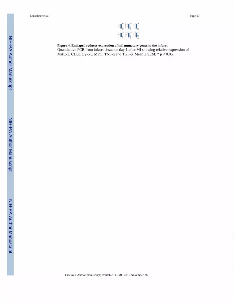
Figure 4. Enalapril reduces expression of inflammatory genes in the infarctQuantitative PCR from infarct tissue on day 1 after MI showing relative expression ofMAC-3, CD68, Ly-6C, MPO, TNF-α and TGF-β. Mean ± SEM; * p < 0.05.
Leuschner et al. Page 17
Circ Res. Author manuscript; available in PMC 2010 November 26.
NIH
-PA Author Manuscript
NIH
-PA Author Manuscript
NIH
-PA Author Manuscript

Figure 5. Impact of Enalapril on monocyte flux in apoE-/- miceA. Representative flow cytometry dot plots of infarct tissue from apoE-/- mice 1 day afterMI. The gate shows CD11b positive lineage negative monocytes and macrophages. Lin:Lineage markers.B. Quantitation of monocytes and their subsets by flow cytometry in heart, spleen and bloodof apoE-/- mice 1 day after MI. Mean ± SEM; * p < 0.05.
Leuschner et al. Page 18
Circ Res. Author manuscript; available in PMC 2010 November 26.
NIH
-PA Author Manuscript
NIH
-PA Author Manuscript
NIH
-PA Author Manuscript
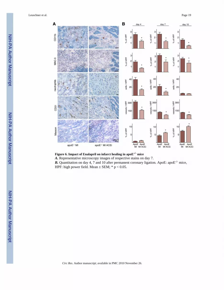
Figure 6. Impact of Enalapril on infarct healing in apoE-/- miceA. Representative microscopy images of respective stains on day 7.B. Quantitation on day 4, 7 and 10 after permanent coronary ligation. ApoE: apoE-/- mice,HPF: high power field. Mean ± SEM; * p < 0.05.
Leuschner et al. Page 19
Circ Res. Author manuscript; available in PMC 2010 November 26.
NIH
-PA Author Manuscript
NIH
-PA Author Manuscript
NIH
-PA Author Manuscript

Figure 7. Enalapril reduces early protease activity and subsequently LV remodeling in apoE-/-
mice with MITop: Flow chart illustrating the set up of the study.Left: FMT-CT 1 day after MI. Arrows denote apical infarct signal. Additional activation ofthe protease sensor is seen in the vicinity of the decending aorta and may be due toinflammatory atherosclerotic lesions. Infarct size was measured by contrast-enhanced CT.Middle and right panels: Cardiac MRI, LA: long axis, SA: short axis view. ACEi: Enalapriltreatment, SPX: splenectomy. Mean ± SEM; * p < 0.05.
Leuschner et al. Page 20
Circ Res. Author manuscript; available in PMC 2010 November 26.
NIH
-PA Author Manuscript
NIH
-PA Author Manuscript
NIH
-PA Author Manuscript

Figure 8. Ischemia reperfusion injury is enhanced in apoE-/- mice with inflammatoryatherosclerosis, and attenuated by Enalapril and splenectomyA: Fluorescence reflectance images display the area at risk, which is void of microspheresinjected during ischemia. Lower panel shows TTC staining of the same myocardial shortaxis slice.B: Quantification of ischemia reperfusion injury as a percentage of the area at risk. Mean ±SEM; * p < 0.05 versus all other groups.C: Representative flow cytometry dot plots of heart tissue from apoE-/- mice 1 day afterischemia. The gate shows CD11b positive lineage negative monocytes and macrophages.D: Quantification of CD11b positive lineage negative monocytes/macrophages. * p < 0.05versus all other groups.E: Flow cytometric quantification of splenic monocytes 24 hours after ischemia in apoE-/-
mice with and without Enalapril treatment (ACEi). Mean ± SEM; * p < 0.05.
Leuschner et al. Page 21
Circ Res. Author manuscript; available in PMC 2010 November 26.
NIH
-PA Author Manuscript
NIH
-PA Author Manuscript
NIH
-PA Author Manuscript


