Embed Size (px)
Citation preview

Presentation
Evidence-Based Practice in the Management of Vascular AccessDevices for Home Parenteral Nutrition Therapy
Marcia Ryder, PhD, MS, RN
From Research and Consulting, Healthcare-acquired Infections/Vascular Access, San Mateo, California
ABSTRACT. Catheter-related bloodstream infection andcatheter occlusion are potential significant complications ofparenteral nutrition therapy. The increased incidence andassociated morbidity, mortality, increased costs, and quality-of-life issues experienced with these adverse events necessi-tate specialized management of vascular access devices. Thehost coagulation response to biomaterials and the associateddevelopment of biofilm on vascular devices are complex phe-nomena. Multiple interventions are required to prevent
access of bacteria to both intraluminal and extraluminalcatheter surfaces, and the occurrence of catheter occlusion.The discovery of the biofilm form of microbial life and theassociated recalcitrance of biofilm bacteria to antimicrobialshas provided insight into the failure of current prevention,diagnostic, and treatment protocols. Critical interventionsare presented correlating current evidence with new discov-eries in pathogenesis. ( Journal of Parenteral and Enteral Nutri-tion 30:S82–S93, 2006)
The revolution in health-care delivery systems overthe last 2 decades has shifted the care of patients fromthe acute care setting to alternate sites. Provision ofhealthcare in the home has become the fastest-growingsegment of the healthcare system to the extent thatnearly as many patients are receiving care in the homeas in the hospital setting.1
Nearly eight million people in the United Statesreceived medical care at home in 1996,2 of which774,113 (10%) were estimated to have at least 1indwelling medical device.3 The use of a medical deviceis the greatest predictor (exogenous) of healthcare-as-sociated infection.3 Complications related to vascularaccess devices (VADs) have reportedly been the pri-mary cause of morbidity, mortality, and rehospitaliza-tion related to parenteral nutrition therapy in hospi-talized patients,4,5 home patients—including adults6–8
and pediatrics9,10—in the United States andabroad.11–13 Unfortunately, the transfer of care toalternate sites was not accompanied by the develop-ment of national surveillance systems to monitor out-comes and adverse events or with the establishment offormal infection control programs for standardizationin the prevention, diagnosis, and treatment of compli-cations.3,14
The safe administration of parenteral nutrition (PN)requires the use of a central venous catheter (CVC) dueto the hypertonic and acidic properties of the solution.CVCs most appropriate for PN therapy in the homeinclude peripherally inserted central catheters (PICC),
tunneled catheters, and implanted ports15,16 (seeRyder Appendix). However, the use of these devices isnot without serious risk. Thrombotic catheter occlusionand catheter-related infections are the most frequentlyreported catheter complications for all types of CVCs inall healthcare settings. In an analysis of data from theStrategic Health Care Programs National Database(April 1999 to September 2000) that included 50,470patients receiving home infusion care (2.83 millioncatheter-days), the rate of CVC complication was 1.5per 1000 catheter-days.17 The most common events(per 1000 catheter-days) were catheter dysfunction(0.83; nonthrombotic 0.6, thrombotic 0.23), catheter-site infections (0.26), and bloodstream infections (0.19).In the face of the increasing shift of care for the moreacutely ill and immunocompromised patients to thenonhospital setting, an increase in the rate of thesecomplications might be expected.
Prevention of complications remains the cornerstoneof quality patient care and improved outcomes. Har-barth et al18 conducted a systematic review of theliterature published in the last decade to generate acrude estimate of the proportion of potentially prevent-able nosocomial infections under current healthcareconditions. The evaluation of 30 reports suggests thatat least 20%, ranging from 10% to 70%, of all nosoco-mial infections are preventable. The most importantreduction effect was discovered for catheter-relatedbloodstream infection (CRBSI). Little is known aboutthe proportion of preventable infections in the home-care setting.
With continued concern for the increased morbidity,mortality, and risk of device-related complications andthe lack of standardization of care in alternate sites, itis prudent to identify key evidence-based strategiesapplicable to home PN patients for the care and man-
Received for publication April 14, 2005.Accepted for publication October 3, 2005.Correspondence: Marcia A. Ryder, RN, Research and Consulting,Healthcare-acquired Infections/Vascular Access, 1504 Forge Road,San Mateo, CA 94402. Electronic mail may be sent [email protected].
0148-6071/06/3001-0S82$03.00/0 Vol. 30, No. 1JOURNAL OF PARENTERAL AND ENTERAL NUTRITION Printed in U.S.A.Copyright © 2006 by the American Society for Parenteral and Enteral Nutrition
S82

agement of VADs. The purpose of this paper is toreview the evidence for implementation of critical pre-ventative strategies linked to the pathogenesis of themost common VAD complications, catheter-relatedinfections and thrombotic catheter occlusion.
CATHETER-RELATED INFECTIONS
The estimated 20% prevention rate for nosocomialinfections raises the question of why 80% are not pre-ventable. Recent discoveries related to microbial sur-vival strategies and antimicrobial resistance provideinsight into the pathogenesis of CRBSIs. Understand-ing pathogenesis gives clear direction to prevention.
Pathogenesis of Catheter-Related Infections
IV catheters inserted into the bloodstream are sub-ject to the hydrodynamics of 2 flow systems. The exter-nal surface of the catheter interfaces with the circulat-ing blood, whereas the internal surface interacts with avariety of infusates, including crystalloid solutions,drug admixtures, blood and blood products, and nutri-tive solutions. The rate of blood flow is dependent onthe diameter of the catheterized vessel and thepatient’s physiologic status. The rate of flow within thecatheter is highly variable, depending on the infusiontherapy, or there may be no flow when the catheter is“locked.” Both Silastic and polyurethane are negativelycharged, and hydrophobic biomaterials that promoteadherence of various contacting particles in solutionsand host products form a “conditioning film” on thecatheter surface.
Under any circumstance, the external and internalcatheter luminal surfaces are not mirror images.Microorganisms in contact with either surface interactwith the biomaterial under very different conditions.The pathogenesis of infection at each surface must beconsidered independently in order to develop effectivemeasures for prevention. Catheter-related infectionsoccur as the result of a complex series of events: (a)microbial contamination of the internal or externalsurface of the catheter or add-on devices, (b) microbialadherence, (c) biofilm development, and (d) dispersaland dissemination of biofilm bacteria into the blood-stream.19
The patient’s skin is the primary source of contami-nation of the external catheter surface. During inser-tion, bacteria are impacted on the tip and externalcatheter surface as the catheter transcends the epider-mis. Thus, the catheter arrives in the bloodstream witha specific quantity of adherent bacteria. Elliott et al20
verified this phenomenon in a study of 30 cardiac sur-gical patients requiring central venous catheterization.After insertion, the skin at the insertion site and alldevices used during the procedure were cultured. Thetip of each catheter was cultured in situ within 90minutes of the insertion during surgery. Sixty-sevenpercent of cultures from the insertion site were posi-tive, as well as 50% of guidewires, 4% of skin dilators,36% of insertion needles, and 17% of the catheter tips.Within 3 days, 11% of catheters had !15 colony form-ing units (cfu) on the external surface despite rigorousskin antisepsis and aseptic technique. These results
are further substantiated in 2 subsequent studiesusing pulsed gel electrophoresis techniques to matchorganisms attached to the tip of the catheter withorganisms at the insertion site.21,22
Arrival of the catheter into the bloodstream triggersa well-defined host response.23 Plasma proteinsinstantly adhere to the catheter surface upon contactwith the blood. Attachment of arriving platelets, neu-trophils, and fibrin(ogen) forms a “conditioning layer”on the catheter surface over the next few hours.Thrombus may then form to a variable extent over thefibrin sheath. After approximately 1 week, migratoryfibroblasts and smooth muscle cells from the injuredvessel wall cover the fibrin sheath/pericatheter throm-bus. By 2 weeks, a layer of migratory endothelial cellsthat may then be protective against microbial attach-ment encases the host-derived sheath. Planktonic bac-teria “free floating” in the bloodstream from distantsources may attach to the developing conditioninglayer or pericatheter thrombus and further colonize thecatheter.24–26 The preattached bacteria immediatelydevelop a biofilm for survival in a new hostile environ-ment.
Concurrently, contamination and colonization of theskin tract or subcutaneous tunnel may continue tooccur during the inflammatory phase of wound healingwithin the first few days of catheterization. Microor-ganisms from the skin surface at the insertion site arepassively transported within the edematous skin tractby capillary action.27 The arriving microorganismsattach to the catheter surface or surrounding trauma-tized tissue and form colonizing biofilm. The progres-sion from colonization to infection depends on the bac-terial count, the species present, the virulence of theorganisms, and the host immune response.28
Microorganisms gain entrance to the internal lumenof the catheter at any entry point, anywhere along thefluid path where the system is manipulated (ie, IVsolution connection sites, administration tubing junc-tions, access portals, and needleless connectors). Thesource of contamination is primarily the hands of med-ical personnel and the patient’s own skin or body fluidsin contact with the access sites. Bacteria flowingthrough any of the administration devices that come indirect contact with the inner lumen attach to surfaceand form colonizing biofilms. The same process of pro-tein attachment, fibrin deposition, and clotting occurswithin the lumen when used for blood sampling andblood product administration or when blood is allowedto remain within the lumen. The host conditioned sur-face then provides attachment sites for arriving bacte-ria.25,28
Biofilm: Microbial Life on Surfaces
The initial event in the formation of biofilm is theattachment of microbes to the surface of the biomate-rial or conditioned surface. Within 10–20 minutes ofdirect contact, phenotypic changes within the micro-bial cell wall initiate the production of species-depen-dent adhesins and accumulation proteins.29,30 Self-pro-duced exopolymer saccharides embed the proliferatingcells into cell clusters or microcolonies.
January–February 2006 VASCULAR ACCESS DEVICES FOR HOME PN THERAPY S83

Although each biofilm is unique in structure, mostbiofilms develop as multilayered cell clusters with acomplex architecture of towers and flow channels forthe delivery of nutrients and removal of waste. Thisstructure sustains an environment heterogeneous tooxygen level, nutrient availability, and metabolic state,depending on the location of the cell within the biofilm.The parent cells adherent to the biomaterial surfaceare the most deprived of nutrient availability and arethe most metabolically altered into a slow-growing ornongrowing, dormant lifestyle. Development of the bio-film evolves according to the local microenvironmentconditions and is often incorporated structurallywithin host conditioning layers or tissue matrices. Therate of growth is influenced by flow rate, nutrient com-position of the liquid (blood or infusate), and tempera-ture.31 The bloodstream provides ideal conditions tosupport biofilm growth on indwelling devices. Depend-ing on the location and number of attached or “sessile”bacteria, the biofilm forms in patchy sections or devel-ops in a contiguous layer completely covering the sur-face.
Biofilms harbor large numbers of organisms within asmall scale, and pathogen cell densities can reach asmany as 107 cells/cm2 on a surface.32 Increasing cellu-lar density within the biofilm triggers an elaboratecell-to-cell communication that regulates biofilm struc-ture and progeny cell dispersal.33 Dissemination ofbiofilm cells is species dependent but typically occursby the shedding of single daughter cells or detachmentof clumps of biofilm cells by hydrodynamic shear forcesor by cell-cell signaling that directs the production ofsubstances that lyse the biofilm matrix.34 Cells dis-persed as single planktonic cells are readily killed bynormal host defense mechanisms, and the biofilmremains nonpathogenic. However, when the dissemi-nation becomes extensive or if the host becomesimmunosuppressed, colonization develops into overtinfection.30 Dispersal in clumps, particularly Staphy-lococcus aureus, containing hundreds of resistant cellsmay result in metastatic infections.35
Biofilms mature at variable rates dependent on themicrobial species. Staphylococcus biofilms maturewithin 7 days, whereas Pseudomonas biofilms maturelater, around 10–12 days.36,37 Extraluminal catheter-related infections are typically evidenced within thefirst week of catheterization.38 This correlates wellwith heavy initial colonization of the external cathetersurface that was most likely inserted through poorlydisinfected skin. Infection from the internal lumen typ-ically occurs after 1 week as the number of manipula-tions increase; however, more recently the internallumen has been shown to be the primary source ofbloodstream infection as early as 3 and 6 days in short-term catheters.39 The mean time to infection in long-term catheters is !10 days and implicates the internallumen as the major site of CRBSI.40
It has been estimated that as many as 65% of bacte-rial infections treated by physicians in the developedworld are related to biofilms.34 Clinical implications forprevention, diagnosis, and treatment of vascular cath-eter-related infections can be derived from understand-ing the pathogenesis of biofilm infections. The follow-
ing characteristics of biofilm infections should beconsidered in the management of CVCs25,29,35,41:! virtually any organism in contact with a biomaterial
can form a biofilm;! microbial attachment to surfaces results in extensive
phenotypic changes profoundly different from unat-tached cells;
! bacteria growing in biofilm may be in a dormant butviable state and initially may fail to grow in culture;
! biofilm infections are inherently resistant to all anti-microbial agents (by 10–1000 times) and to the host’simmune system;
! aging biofilms become increasingly more difficult totreat;
! in general, exposure of biofilm to prolonged and ele-vated concentrations of antibiotic agents killsapproximately 90% of biofilm cells; the persistingcells survive and regenerate the biofilm after cessa-tion of antibiotic therapy.
Evidence-Based Prevention Strategies
The Centers for Disease Control and Prevention’s(CDC) Guidelines for the Prevention of IntravascularCatheter-Related Infections offers 113 recommenda-tions for implementation in all healthcare settings.15
This extensive set of guidelines represents the com-plexity of effort required for the safe use of thesedevices. Harbarth et al18 found that the most effectiveapproach to the reduction of nosocomial infectionsincludes the implementation of a multimodal qualityimprovement program applying standardized policiesand, if necessary, mandatory practice changes. Consid-ering the pathogenesis of catheter-related infections,interventions should be designed to prevent microbialcontact with the external catheter surface and micro-bial entry to the internal surfaces of the entire deliverysystem. Given that the major sources of microorgan-isms are the patient’s own skin and the hands of med-ical personnel, a multimodal intervention packagemust be implemented to prevent microbial access fromthese sources.
Extraluminal contamination: skin antisepsis. Contami-nation of the external lumen during insertion andthroughout the duration of use is most effectively min-imized by systematic skin antisepsis and the use of anantimicrobial dressing (Table I). Preoperative skinpreparation is probably the most important interven-tion for the prevention of CRBSI. Protocol developmentfor effective skin antisepsis requires an understandingof the anatomy, physiology, and microbiology of theskin at the chosen site of insertion.
The basic structure of the skin from outer- to inner-most layer includes the superficial horny cell layer ofthe stratum corneum (1–2 mm thick), the viable orstratified cell layer of epidermis (50–100 mm thick),the dermis (1–2 mm thick), and the hypodermis(1–2 mm thick).42 The stratum corneum is composed ofapproximately 15 layers of corneocytes that provide thebarrier function of the skin. The corneocytes are rem-nants of terminally differentiated keratinocytes gener-ated by the stratified epidermis positioned directlyunder the stratum corneum. The stratified epidermis is
S84 RYDER Vol. 30, No. 1
composed of 10–20 layers of keratinized epithelialcells. The stratum corneum receives a new basal layerof cells to replace the outermost surface layer of deadcells (squames) shed from the skin surface each day.The stratum corneum is replaced in total approxi-mately every 2 weeks.43 Healthy skin disseminatesapproximately 107 squames daily, 10% of which con-tain viable bacteria.
The microbiology of the skin varies widely, depend-ing on body location and nutrient and water availabil-ity. Normal colony count of the skin at the subclavianand jugular insertion sites is approximately 1000–10,000 cfu/cm2 compared with approximately 10 cfuper cm2 at the antecubital space.44 The transient skinflora arrives from the environment and may includebacteria, fungi, and virus. The resident flora is foundmainly in the stratum corneum, 80% of which arelocated within the first 5 layers.45 The remaining 20%inhabit the deeper reservoirs of sebaceous glands and
hair follicles sustained within biofilms that provideadded protection against antiseptic agents.29,46,47 Thedominant species of resident flora is the coagulase neg-ative staphylococci (CNS; mostly Staphylococcus epi-dermidis). S epidermidis grows in prolific biofilmsbetween the squamous cells of the outer 3–10 layers ofthe stratified epithelium and colonize the hair folliclesand sebaceous glands quite successfully.32
Topical application of antimicrobial agents elimi-nates CNS on the skin surface but does not sterilize theunderlying stratum corneum, sebaceous glands, or hairfollicles.48 The bacterial concentration of the skin ismost effectively reduced by the combination of physicalremoval, along with antimicrobial activity by antisep-tic exposure.49 The CDC guidelines15 and the 2004AORN (Association of Operating Room Nurses) Stan-dards, Recommended Practices, and Guidelines50 rec-ommend a 2-step process for preoperative skin prepa-ration and continued catheter insertion site care. The 2
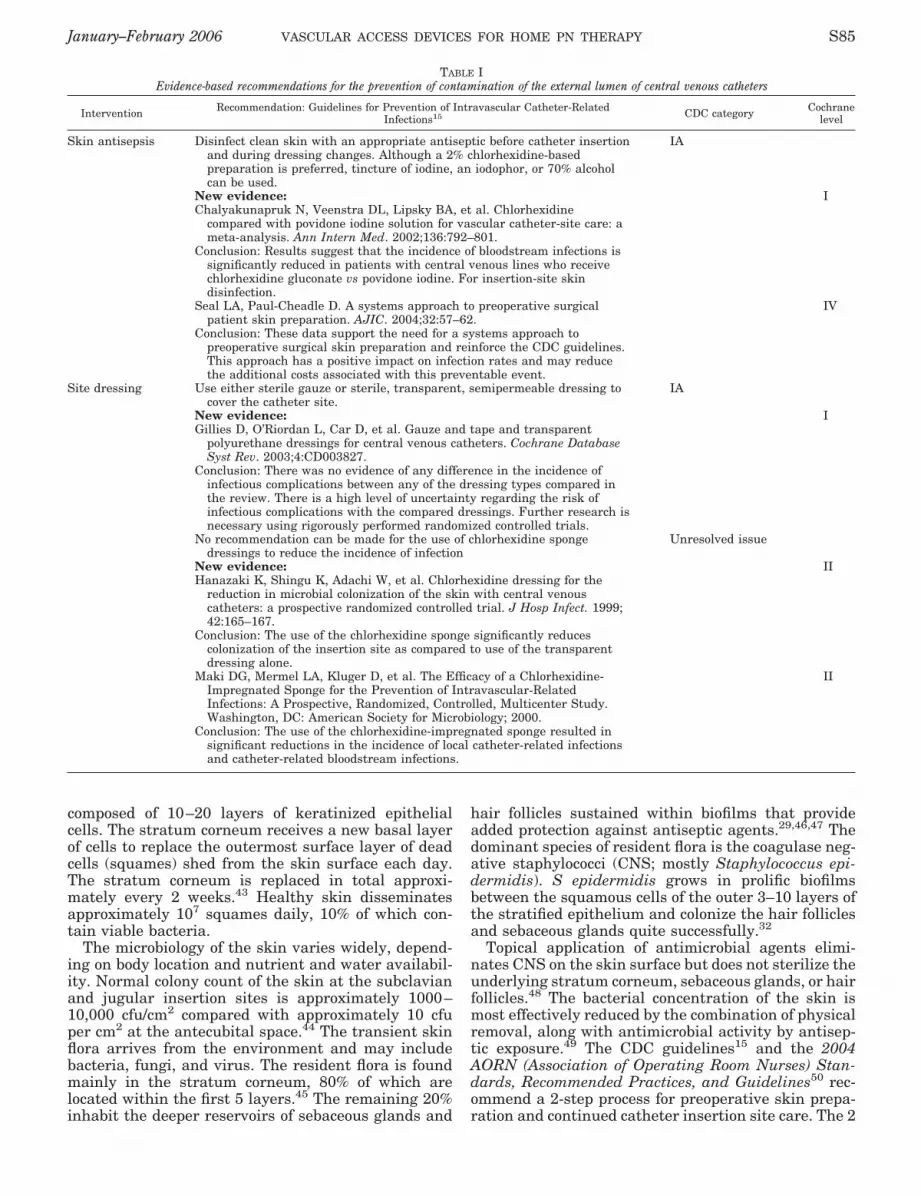
TABLE IEvidence-based recommendations for the prevention of contamination of the external lumen of central venous catheters
Intervention Recommendation: Guidelines for Prevention of Intravascular Catheter-RelatedInfections15 CDC category Cochrane
level
Skin antisepsis Disinfect clean skin with an appropriate antiseptic before catheter insertionand during dressing changes. Although a 2% chlorhexidine-basedpreparation is preferred, tincture of iodine, an iodophor, or 70% alcoholcan be used.
IA
New evidence:Chalyakunapruk N, Veenstra DL, Lipsky BA, et al. Chlorhexidine
compared with povidone iodine solution for vascular catheter-site care: ameta-analysis. Ann Intern Med. 2002;136:792–801.
Conclusion: Results suggest that the incidence of bloodstream infections issignificantly reduced in patients with central venous lines who receivechlorhexidine gluconate vs povidone iodine. For insertion-site skindisinfection.
I
Seal LA, Paul-Cheadle D. A systems approach to preoperative surgicalpatient skin preparation. AJIC. 2004;32:57–62.
Conclusion: These data support the need for a systems approach topreoperative surgical skin preparation and reinforce the CDC guidelines.This approach has a positive impact on infection rates and may reducethe additional costs associated with this preventable event.
IV
Site dressing Use either sterile gauze or sterile, transparent, semipermeable dressing tocover the catheter site.
IA
New evidence:Gillies D, O’Riordan L, Car D, et al. Gauze and tape and transparent
polyurethane dressings for central venous catheters. Cochrane DatabaseSyst Rev. 2003;4:CD003827.
Conclusion: There was no evidence of any difference in the incidence ofinfectious complications between any of the dressing types compared inthe review. There is a high level of uncertainty regarding the risk ofinfectious complications with the compared dressings. Further research isnecessary using rigorously performed randomized controlled trials.
I
No recommendation can be made for the use of chlorhexidine spongedressings to reduce the incidence of infection
Unresolved issue
New evidence:Hanazaki K, Shingu K, Adachi W, et al. Chlorhexidine dressing for the
reduction in microbial colonization of the skin with central venouscatheters: a prospective randomized controlled trial. J Hosp Infect. 1999;42:165–167.
Conclusion: The use of the chlorhexidine sponge significantly reducescolonization of the insertion site as compared to use of the transparentdressing alone.
II
Maki DG, Mermel LA, Kluger D, et al. The Efficacy of a Chlorhexidine-Impregnated Sponge for the Prevention of Intravascular-RelatedInfections: A Prospective, Randomized, Controlled, Multicenter Study.Washington, DC: American Society for Microbiology; 2000.
Conclusion: The use of the chlorhexidine-impregnated sponge resulted insignificant reductions in the incidence of local catheter-related infectionsand catheter-related bloodstream infections.
II
January–February 2006 VASCULAR ACCESS DEVICES FOR HOME PN THERAPY S85
steps include skin cleansing, followed by application ofan antiseptic. The CDC guidelines recommend specificantiseptics for use on clean skin but do not addressmethods for cleansing the skin. The AORN guidelinesprovide specific recommended practice techniques forboth skin cleansing and surgical site preparation. Rec-ommendations for skin cleansing include (a) patientshowering before arrival at the practice setting, (b)washing the surgical site before arrival in the practicesetting, and (c) washing the surgical site immediatelybefore applying the antiseptic agent.50
Data presented by Seal and Paul-Cheadle51 furthersupport the utility of a systems approach to surgical-site preparation. Use of a combination of antisepticshower(s) or bath(s), followed by antiseptic surgicalsite preparation with alcohol-based antisepticsresulted in a positive impact on the incidence of surgi-cal-site infections.
Substantial evidence indicates that chlorhexidinegluconate (CHG) solutions are the superior agents foruse in vascular catheter insertion care for the reduc-tion of CRBSIs. The CDC guidelines recommend 2%CHG as the preferred antiseptic for skin preparationand designate its use as a performance indicator forreducing CRBSI. The economic benefits of CHG use forvascular catheter site care have been compared withpovidone iodine use in a decision analysis model.52 Themodel estimates that the use of CHG compared withpovidone iodine results in a 1.6% decrease in the inci-dence of CRBSI, a 0.23% decrease in the incidence ofdeath, and a cost savings of $113 per catheter used.
CHG in combination with alcohol increases thepotential activity of the antiseptics. The alcohol pro-vides rapid reduction in bacterial counts but has min-imal persistence. The CHG remains active for at least6 hours and is minimally affected by the presence oforganic material.53,54 Repeated use of CHG increaseseffectiveness over time due to the binding and reten-tion of active antiseptic to the surface epithelial cellwalls.55 A preoperative 4% CHG skin scrub (Hibiclensscrub sponge, Regent Medical Ltd, Irlam, UK) followedby the application of a 2% CHG/70% alcohol antiseptic(ChloraPrep, Medi-Flex Inc., Kansas City, KS) is sug-gested for maximum physical and chemical reductionof transient and resident flora before passage of thecatheter through the skin.
Within 18 hours of antiseptic application, residentbacteria surface from the deeper reservoirs and recol-onize the skin surface, regardless of the type of steriledressing applied over the insertion site.45,48,53
Repeated skin antisepsis may be important within thefirst 24–48 hours of insertion to remove repopulatingbacteria from the insertion site avoiding migration intothe skin tract by capillary action. Postinsertion sitecare is accomplished by using the same 2-step process.Skin cleansing with gentle mechanical friction accom-plishes removal of desquamed epithelial cells, repopu-lating bacteria, inactive antiseptic, oils, sweat, and anydrainage if present. The antiseptic is then applied toclean skin. A multidirectional, back-and-forth cleans-ing using alcohol saturated swab sticks, followed bycircular application of a 2% CHG/70% alcohol combi-nation, is suggested.56
Extraluminal contamination: antimicrobial dressing.Considerable debate over the role of gauze and tapedressings vs transparent polyurethane film dressingsin the prevention of catheter-related infections hasbeen observed in the literature. Gillies et al57 recentlycompleted a Cochran database systematic review toidentify by meta-analysis any differences betweengauze and tape dressings and transparent polyure-thane film dressings in the incidence of CVC-relatedlocal infection or CRBSI, catheter security, dressingcondition, tolerance to the material, and ease of appli-cation in hospitalized patients. There was no evidenceof any difference in the incidence of infectious compli-cations between any of the dressing types compared inthe review. Traditional sterile gauze and tape dress-ings and transparent polyurethane film dressings pro-vide protection for the catheter site from trauma andtransient bacteria and prevent the accumulation ofmoisture; however, neither have any antimicrobialproperties. This illustrates the critical importance ofeffective skin antisepsis as the major intervention inthe prevention of catheter site infection, tunnel andport pocket infection, and CRBSI from the extralumi-nal source.
The use of a CHG-impregnated polyurethane foamdisc applied around the catheter and in direct contactwith the skin surface at the insertion site has demon-strated the ability to maintain a sterile skin surface atthe insertion site over the lifetime of the catheter.58 Ina randomized clinical trial, 50 patients undergoingabdominal surgery had a CVC placed for PN andreceived either a transparent polyurethane film dress-ing (Bioclusive, Johnson & Johnson, Inc, New Bruns-wick, NJ) or a CHG-impregnated disc (Biopatch, John-son & Johnson) covered with a transparent filmdressing. Two skin cultures were taken once a weekduring the dressing change, 1 from under the CHG discand 1 from a distant site under the transparent dress-ing. Contamination was detected under the transpar-ent dressing in 14 of 60 cases (23.3%), whereas nobacterial contamination was observed under the CHGdisc (p " .0001). In the control group (transparentdressing alone), bacterial contamination was detectedin 7 of 64 cases (10.9%) at the insertion site and in 17of 64 cases (26.6%) at the distant site under the dress-ing (p " .0001). The difference in contamination at theinsertion site between the CHG disc and control wassignificant in favor of the CHG disc (p " .01). Therewas no difference in contamination under the dressingat the distant site between the 2 groups.
In a second, randomized, blinded, controlled, multi-center trial by Maki et al,59 use of a CHG-impregnatedfoam disc (Biopatch) was compared with a controltransparent polyurethane film dressing in 589 hospi-talized patients receiving short- and medium-termCVCs and arterial catheters.59,60 All CRBSIs were con-firmed by concordance between microorganisms iso-lated from peripheral blood and the catheter tip, hub,or infusate demonstrated by DNA subtyping. The CHGdressing significantly reduced the risk of local cathe-ter-related infection (CHG disc, 28.14%; and control,45.24%; p " .001) and CRBSI (CHG disc, 2.37%; andcontrol, 6.12%; p " .05). Not surprisingly, the greatest
S86 RYDER Vol. 30, No. 1
benefit was the prevention of local infection and theextraluminal source of CRBSI.
The cost benefit and impact on CRBSI mortality hasalso been assessed. A cost-benefit sensitivity analysisestimates potential US net benefits from CHG dressinguse to range from $275 million to approximately $1.97billion, and a preventable mortality between 329 and3906 deaths annually.60
The combination of the 2-step protocol of cleansingand antiseptic site preparation for both preoperativeand insertion site preparation using 2% CHG and 70%alcohol combinations along with application of theCHG-impregnated disc appears to be very powerfuland cost-effective for the prevention of catheter-relatedinfections in short-term transcutaneous catheters.This combination may be beneficial for prevention oftunnel infections in cuffed catheters, particularly
within the first 2 weeks until adhesion of the cuff to thesubcutaneous tissue is complete.
Intraluminal contamination: hand hygiene. Entry ofmicroorganisms through contaminated access sites ofthe infusion system is the major source of intraluminalcontamination and the major source of CRBSI in long-term catheters. Interventions should be focused on pre-vention of touch contamination, access-site disinfec-tion, and use of prophylactic flush solutions (Table II).
As early as 1 week after admission, hospitalizedpatients become colonized with antimicrobial-resistantpathogens that may be transferred into the home onhospital discharge.61 Fundamental to developing infec-tion-control policies for the delivery of medical care inthe home is the need to recognize that people live in anenvironment where all types of human activities areongoing and that pathogens are continually introduced
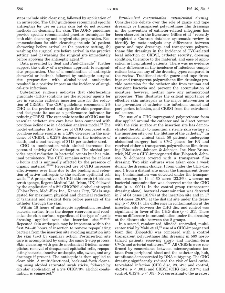
TABLE IIEvidence-based recommendations for the prevention of contamination of the internal lumen of central venous catheters
Intervention Recommendation: Guidelines for Prevention of Intravascular Catheter-related Infections15 CDCcategory
Cochranelevel
Hand hygiene Observe proper hand hygiene procedures by washing hands with eitherconventional antiseptic-containing soap and water or with waterless alcohol-based gels or foams. Observe hand hygiene before and after palpating catheterinsertion sites, as well as before and after inserting, replacing, accessing,repairing, or dressing an intravascular catheter.
IA
New evidence:Boyce JM, Pittett D. HICPAC Committee and HICPAC/SHEA/APIC/IDSA Hand
Hygiene Task Force: guideline for hand hygiene in health-care settings. MMWRRecommend Rep. 2002;51(RR16):1–44.
Recommendation:Decontaminate hands before having direct contact with patients.
IB
When decontaminating hands with an alcohol-based hand rub, apply product topalm of one hand and rub hands together, covering all surfaces of hands andfingers, until hands are dry. Follow manufacturer’s recommendations regardingthe volume of product to use.
IB
When washing hands with soap and water, wet hands first with water, apply theamount of product recommended by the manufacturer to hands, and rub handsvigorously for at least 15 seconds, covering all surfaces of the hands and fingers.Rinse hands with water and dry thoroughly with a disposable towel.
IB
Access-site disinfection Clean injection ports with 70% alcohol or an iodophor before accessing the system. IAMinimize contamination risk by wiping the access port with an appropriate
antiseptic and accessing the port with only sterile devices.IB
New evidence:Casey AL, Worthington T, Lambert PA, Quinn D, Faroqui MH, Elliott TS. A
randomized, prospective clinical trial to assess the potential infection riskassociated with the PosiFlow needleless connector. J Hosp Infect. 2003;54:288–293.
Conclusion: Disinfection of needleless connectors with either chlorhexidine/alcoholor povidone iodine significantly reduced external microbial contamination.
I
Prophylactic flush solution Do not routinely use antibiotic lock solutions to prevent CRBSI. Use prophylacticantibiotic solution only in special circumstances (eg, in the treating a patientwith long-term cuffed or tunneled catheter or port who has a history of multipleCRBSIs despite optimal maximal adherence to antiseptic technique).
II
New evidence:Chatzinikolaou I, Zipf TF, Hanna H, et al. Minocycline-ethylenediamine-
tetraacetate lock solution for the prevention of implantable port infections inchildren with cancer. Clin Infect Dis. 2003;36:116–119.
Conclusion: M-EDTA lock solution was efficacious in preventing port-relatedinfections in children with cancer, without causing any adverse events. Largeprospective, randomized trials are needed to further test the ability of M-EDTAto prevent long-term catheter-related infections.
III
Kite P, Eastwood K, Sugden S, Percival SL. Use of in vivo generated biofilms fromhemodialysis catheters to test the efficacy of a novel antimicrobial catheter lockfor biofilm eradication in vitro. J Clin Microbiol. 2004;42:3073–3076.
Conclusion: The efficacy of tetrasodium EDTA as a catheter lock potentially showsthat this agent could substantially reduce catheter-related infections and be usedto treat patients with limited access.
IV
January–February 2006 VASCULAR ACCESS DEVICES FOR HOME PN THERAPY S87
into the home on people, food and water, pets, insects,and by air transmission.62 Hand hygiene is intended todecrease contamination of the hands with transientorganisms from the local environment.
The term hand hygiene includes handwashing, anti-septic hand wash, antiseptic hand rub, or surgicalhand antisepsis. In both the CDC Guidelines for thePrevention of Intravascular Device-Related Infections15
and the CDC Guideline for Hand Hygiene in Health-Care Settings,42 decontamination of hands is recom-mended before and after providing care procedures forintravascular devices. An antiseptic hand rub or anti-septic hand wash is recommended for hand decontam-ination.
Hand hygiene is the simplest, most effective mea-sure for preventing healthcare-acquired infections.53,54
However, it is well known that compliance with handhygiene by medical and nurse clinicians has histori-cally been dismal.42,54 Hand hygiene protocols forhome patients should be incorporated into the pro-vider’s infection control program and should includeeducation and compliance monitoring of the nursingstaff, education and compliance monitoring of thepatient/caregiver, provision of appropriate handhygiene products, and routine observation and feed-back of technique to patients and caregivers.62,63
Intraluminal contamination: access-site disinfection.Access-site disinfection is probably the most importantstep in prevention of CRBSI in long-term catheters.The CDC guidelines strongly recommend cleaningaccess ports with 70% alcohol or an iodophor beforeaccessing the system; however, no recommendationregarding the duration or method for cleaning is pro-vided (Table II). Three studies were cited in support ofthe Category IA recommendation. The study by Salz-man et al64 compared the efficacy of CHG (1% with andwithout 70% alcohol), ethanol (70% and 97%), and nor-mal saline in eradicating microorganisms in an in vitromodel of catheter hub contamination. They found that70% ethanol was more effective than 1% CHG andconcluded that ethanol is likely to be the safest treat-ment.
In the second in vitro study by Luebke et al,65 theseptum of 2 devices, one a conventional latex injectionport and the other a split-septum injection system(Interlink, Baxter Healthcare Corp, Deerfield, IL), wasinoculated with an Enterococcus faecium suspension of104–105 cfu/mL. Each system was swabbed with a 70%alcohol-saturated pad using either a single-motionwipe or a 5-second wipe followed by a 1-minute dryingperiod before puncture for flushing. The control grouphad no cleansing before puncture and flushing. Thedevices were accessed by either a needle (injectionport) or blunt cannula (Interlink).
When 1 single-motion wipe was performed, therecovery fluid from the needleless device was positivein 6%, and 4% were positive in the conventional sys-tem. In the 5-second wipe/1-minute drying group, therecovery fluid was positive in 4% of needleless devices,whereas none of the conventional system cultures werepositive. When no disinfection was performed, thetransfer of organisms into the fluid path of the splitseptum was positive in 31%–80% of the needleless
devices and 72%–90% of the conventional injectionports. The authors concluded that the needleless sys-tem performed like the conventional system, but rein-forced the need for an appropriate disinfection proce-dure before accessing either system.
The third cited study documented the potentialspread of iatrogenic infection through contaminatedmultidose vials but did not examine the effect of anti-septics for disinfection before entry into the vial.66
Casey et al,67 in a more recent randomized, prospec-tive, controlled trial, compared the microbial contami-nation rate of standard injection caps and a needlelesspositive-pressure valve. Seventy-seven patients under-going cardiac surgery and requiring a CVC were ran-domly allocated to receive either needleless connectors(BD PosiFlow, BD Medical, Sandy, UT) or standardinjection caps attached to stopcock entry points at thecatheter hub. The microbial contamination rate of theexternal compression seals of 274 needleless connec-tors and 306 standard caps was assessed to comparethe efficacy of 3 disinfectants: 70% isopropyl alcohol,0.5% CHG gluconate in 70% isopropyl alcohol, and 10%povidone iodine. Each device was cleaned before andafter each manipulation, allowing the disinfectant todry for 2 minutes on each occasion. Each device wasexchanged after 72 hours.
Forty-one percent of the needleless valves wereexternally contaminated at exchange. Contaminationof the external compression seals was significantlylower when disinfected with CHG (p " .0001) andpovidone iodine (p " .0001). There was no statisticallysignificant difference in contamination rates betweenthe CHG and povidone iodine group (p # .4). Sevenpercent of the needleless-valve stopcock entry pointswere internally contaminated, with no statistical dif-ference between any of the disinfectants.
In the standard-cap group, 18% of the septa wereexternally contaminated, with no significant differencebetween the rate and extent of microbial contamina-tion after swabbing with each of the disinfectants.Eighteen percent of the stopcock entry points werecontaminated. Disinfection of the entry ports witheither CHG or povidone iodine resulted in a reducedrate of internal contamination compared with alcohol.Overall, the use of 0.5% CHG gluconate in 70% isopro-pyl alcohol before and after each manipulation resultedin the lowest contamination rates.
These results are comparable to the results in a trialby Maki et al,68 who compared the use of 2% aqueousCHG, 10% povidone iodine, and 70% alcohol for prein-sertion skin antisepsis and access-site disinfection.The use of 2% CHG was associated with the lowestrates of localized infection and bacteremia.
Intraluminal contamination: prophylactic flush solution.The third critical intervention for prevention ofintraluminal contamination is the instillation of ananti-infective locking solution when the catheter is notin use. The current standard includes normal saline orheparinized saline for maintaining catheter patency.Neither of these solutions inhibit microbial growth. Tothe contrary, heparin has been shown to support micro-bial growth in solution and in biofilm.69–72 Preliminaryfindings by Hostetler et al have raised concern that
S88 RYDER Vol. 30, No. 1

heparin used in intravascular catheters may play arole in triggering a series of events that result in theproduction of a life-threatening toxic shock–like reac-tion with fungal (Candida) infections.73
In at least 3 studies, prophylactic antibiotic catheterlocking has demonstrated efficacy in the prevention ofCRBSI74–76; however, with the rapid emergence of Gram-positive, Gram-negative, and fungal antibiotic resistantstrains, frontline antibiotics such as vancomycin, qui-nilones, $ lactams and aminoglycosides should bereserved for treatment of systemic infections.40,77 Thus,the strong recommendation of the CDC is to not routinelyuse antibiotic lock solutions to prevent CRBSI.15
The efficacy of a combination solution of minocycline(Wyeth-Ayerst, Pearl River, NY) and disodium EDTA(Endrate; Abbott Laboratories, Chicago, IL) (M-EDTA)as a broad-spectrum antimicrobial/antibiofilm andantithrombotic agent has been thoroughly studied invitro.69–71 Raad et al70 investigated the prophylacticuse of minocycline (3 mg/mL) and disodium EDTA (30mg/mL) in 14 children with cancer for whom the solu-tion was used to lock their ports. They found thatM-EDTA significantly decreased the risk of CRBSI incomparison to the control group of 48 children usingheparin (p # .05). These results are promising; how-ever, IV minocycline has recently been discontinued bythe manufacturer and is no longer available.
Tetrasodium EDTA (tEDTA) has been investigatedas an antimicrobial agent in both in vitro and ex vivostudies. Ryder et al78 compared ciprofloxacin 10, 100,1000, and 5000 % MIC to tEDTA 40 mg/mL for theeradication of coagulase negative staphylococcus andPseudomonas aeruginosa (PA) biofilm bacteria grownon glass fiber membranes. The mean log reduction(MLR) of CNS after 6 hours of exposure to ciprofloxacinwas 7% at 10 % MIC, 15% at 100 % MIC, 26% at 1000 %MIC, and 35% for 5000 % MIC. The MLR of PA at 6hours was 58% at 10 % MIC, 74% at 100 % MIC, 68%at 1000 % MIC, and 82% for 5000 % MIC. The MLR ofthe tEDTA at 6 hours was 100%, a statistically signif-icant reduction against all other tested concentrationsof ciprofloxacin (p ! .001), except for CNS at 100 %MIC at 6 hours (p # .06).
Kite et al79 investigated the effect of tEDTA in an exvivo study of 20 clinically infected hemodialysis cathe-ters. The explanted catheters were screened by a cul-ture of through-catheter flush technique. Bacteriaidentified in the biofilms were Gram-positive, Gram-negative, and mixed species. The initial biofilm cellcount levels averaged above 105 cfu/1 cm of intralumi-nal catheter surface. tEDTA 40 mg was instilled intoequal catheter sections and remained “locked” for 24hours. tEDTA was effective at complete eradication ofthe total viable count in almost all cases. tEDTAappears to be a very promising agent for the prophy-laxis and treatment of vascular catheters, but random-ized clinical trials are needed.
THROMBOTIC CATHETER OCCLUSIONS
Pathogenesis of Intraluminal Thrombotic Catheter Occlusions
Catheter occlusion may be partial or complete and istypically evidenced by inability to infuse or aspirate,
sluggish flow, or frequent pump alarms. Thromboticcatheter occlusion occurs as a result of clotted bloodwithin the lumen or from the buildup of fibrin on theintraluminal surface over time. Plasma proteins andfibrin(ogen) are deposited during aspiration or admin-istration of blood or blood products. Clotting of wholeblood within the lumen is usually a consequence of aninadequate volume of flush solution, inadequate flush-ing technique, or retrograde blood flow on disconnectfrom needless connectors. Clotting directly at andslightly within the tip of the catheter may result fromconvex blood flow and fluid displacement that occurswhile the catheter is locked, regardless of flushingmethod or needleless connector design.80
The correlation between thrombosis and infectionhas been well described.81 Some microbial speciesquickly attach directly to polymer surfaces, whereasothers more readily adhere to a fibrin/platelet matrix.The biofilm/fibrin matrix formation may become thickenough to cause partial or complete occlusion. Sherertzet al82 investigated the sensitivity of various culturemethods in the diagnosis of triple-lumen catheterinfections. A strong correlation was identified betweenfailed blood aspiration and the titer of microorganismscultured from each lumen (r # .85). The inability toaspirate blood for culture was experienced in 51% ofaspiration attempts, a likely indicator of partial orcomplete occlusion. The frequency of failed blood aspi-ration was 91% in catheters with significantly positivelumen cultures (100 cfu) compared to 58% when thecultures were negative ("100 cfu; p # .001).
Evidence-Based Prevention Strategies
Strategies for prevention of thrombotic occlusionshould be focused on methods to maintain patency bykeeping blood out of the catheter (Table III). The pre-vention of thrombotic catheter occlusion is centeredprimarily on 2 interventions: catheter flushing and theuse of antireflux needleless connectors and valves(Table III).
Intraluminal thrombotic occlusion: prophylactic flush solu-tions. There are 3 components important to the flushingprotocol for maintaining patency of vascular catheters:the flush solution, the volume of solution, and theflushing technique. The use of normal saline and hep-arin has been studied extensively over the last 2decades. Two meta-analyses published in the early1990s set the current standard specifically for periph-eral IV catheters. The results of the analysis by Peter-son and Kirchhoff83 found no significant difference induration of patency between IV catheters flushed withsaline solution and those flushed with a heparinizedsolution. Goode et al84 concluded that saline is as effec-tive as heparin in maintaining patency, preventingphlebitis, and increasing duration of use in peripheralIV locks. Saline has been used successfully in main-taining patency of CVCs as well.85
Despite these findings, the rate of catheter occlu-sion,15 the incidence of intraluminal clots,86–88 the riskof heparin-induced thrombocytopenia,89 and the lack ofantimicrobial activity of saline and heparin continue to
January–February 2006 VASCULAR ACCESS DEVICES FOR HOME PN THERAPY S89
be of great concern. EDTA has been explored as apotential agent for protection against catheter infec-tion. EDTA is a calcium and iron chelator with veryeffective anticoagulation activity. Along with infectionrates, Chatzinikolaou et al also compared occlusionrates of implanted vascular ports in the pediatric can-cer patients using either a heparin lock or M-EDTA.90
Two thrombotic episodes occurred in 48 patients in theheparin group, whereas no thrombotic events occurredin the M-EDTA group of 14 patients. tEDTA alsoappears to be a promising agent with dual applicationfor both antimicrobial and anticoagulant capability.79
The volume of flush solution is an important factornot only to prevent intraluminal clotting but also cath-eter tip occlusion, a phenomenon that occurs as a resultof laminar flow and the flow distribution as predictedby the Hagen-Poiseuille law when the catheter islocked.91 The IV Nursing Society’s Standards of Prac-tice recommend that the volume of flush solution beequal to at least twice the volume capacity of the cath-eter.91 The findings of Polaschegg and Shah92 supportthis standard. In an in vitro study using dye and salinedilution, the investigators demonstrated that approxi-mately 14% of the injected flush solution spills from thecatheter when the exact priming volume is injected,resulting in a mean concentration of approximately90% of the locking solution’s concentration remainingin the fluid at the tip of the catheter. They concludedthat the injection volume must exceed 120% of the
catheter lumen to achieve the full strength of the lock-ing solution at the tip.
Intraluminal thrombotic occlusion: antireflux needlelessconnectors. Needleless connectors are important devicesin the reduction of needlestick injuries in healthcareworkers. However, the net benefit of these devices hasbeen called into question as a result of several reportsof associated increased infection risk.93,94 Early devicedesigns also reportedly increased the incidence of cath-eter occlusion, particularly in the smaller-lumen cath-eters where a longer length of catheter is filled by areflux displacement volume of blood on disconnectionof the syringe.95
Currently, at least 5 needleless connectors rede-signed with an end positive-pressure mechanism and 2devices incorporating a neutral displacement valveexist in the marketplace; however, evidence-based lit-erature involving each of these devices is limited. Fourrandomized or prospective controlled trials investigat-ing 3 antireflux devices—2 testing a neutral valve and2 a positive-end-pressure valve—report a reduced inci-dence of catheter occlusion with the use of thesedevices.96–99 Reduced occlusion rates were alsoreported in 3 clinical studies evaluating use of 2 posi-tive-end-pressure needleless connectors.95,100,101
Currently, at least 2 marketed devices have not beenvalidated in well-designed clinical trials or descriptiveclinical investigations regarding infection risk or effi-cacy in the reduction of occlusion rates. Clinicians
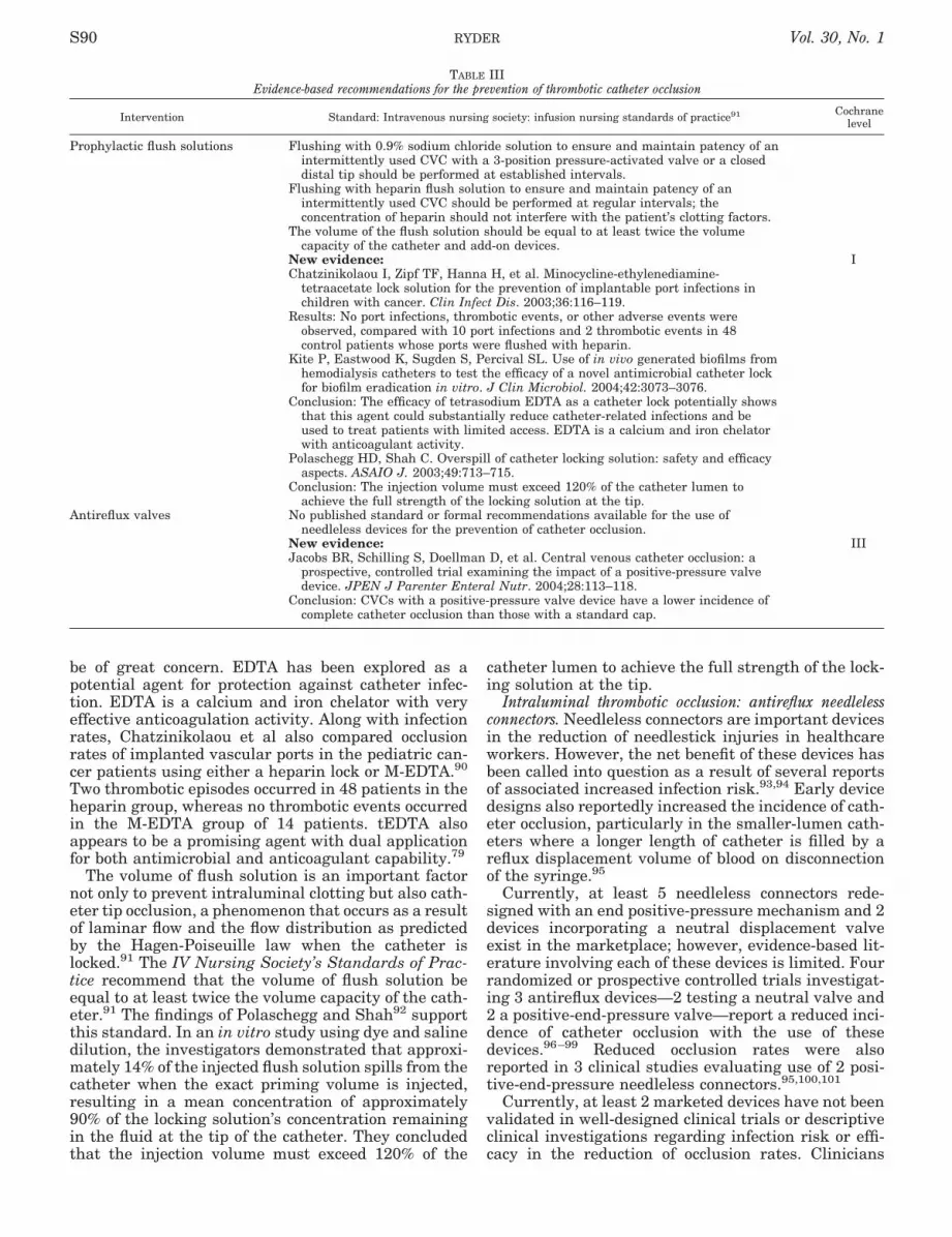
TABLE IIIEvidence-based recommendations for the prevention of thrombotic catheter occlusion
Intervention Standard: Intravenous nursing society: infusion nursing standards of practice91 Cochranelevel
Prophylactic flush solutions Flushing with 0.9% sodium chloride solution to ensure and maintain patency of anintermittently used CVC with a 3-position pressure-activated valve or a closeddistal tip should be performed at established intervals.
Flushing with heparin flush solution to ensure and maintain patency of anintermittently used CVC should be performed at regular intervals; theconcentration of heparin should not interfere with the patient’s clotting factors.
The volume of the flush solution should be equal to at least twice the volumecapacity of the catheter and add-on devices.
New evidence:Chatzinikolaou I, Zipf TF, Hanna H, et al. Minocycline-ethylenediamine-
tetraacetate lock solution for the prevention of implantable port infections inchildren with cancer. Clin Infect Dis. 2003;36:116–119.
Results: No port infections, thrombotic events, or other adverse events wereobserved, compared with 10 port infections and 2 thrombotic events in 48control patients whose ports were flushed with heparin.
I
Kite P, Eastwood K, Sugden S, Percival SL. Use of in vivo generated biofilms fromhemodialysis catheters to test the efficacy of a novel antimicrobial catheter lockfor biofilm eradication in vitro. J Clin Microbiol. 2004;42:3073–3076.
Conclusion: The efficacy of tetrasodium EDTA as a catheter lock potentially showsthat this agent could substantially reduce catheter-related infections and beused to treat patients with limited access. EDTA is a calcium and iron chelatorwith anticoagulant activity.
Polaschegg HD, Shah C. Overspill of catheter locking solution: safety and efficacyaspects. ASAIO J. 2003;49:713–715.
Conclusion: The injection volume must exceed 120% of the catheter lumen toachieve the full strength of the locking solution at the tip.
Antireflux valves No published standard or formal recommendations available for the use ofneedleless devices for the prevention of catheter occlusion.
New evidence:Jacobs BR, Schilling S, Doellman D, et al. Central venous catheter occlusion: a
prospective, controlled trial examining the impact of a positive-pressure valvedevice. JPEN J Parenter Enteral Nutr. 2004;28:113–118.
Conclusion: CVCs with a positive-pressure valve device have a lower incidence ofcomplete catheter occlusion than those with a standard cap.
III
S90 RYDER Vol. 30, No. 1
should be cautious when using these devices withoutimplementation of strict protocols and close monitoringof clinical outcomes. Access-site disinfection and timelychanges of the devices has been stressed as a safetymeasure in the prevention of needleless device-associ-ated CRBSI.67,94,102–106 The use of well-designed anti-reflux devices is an effective strategy for the preventionof CRBSI when appropriately disinfected before useand replaced at recommended intervals.
In summary, the prevention of catheter-relatedinfections and thrombotic intraluminal occlusionrequires strict adherence to evidence-based protocols.The development of effective protocols for the preven-tion, diagnosis, and treatment of CRI requires anunderstanding of the pathogenic mechanisms of micro-bial access to both the external and internal catheterlumen and the subsequent development of biofilm. Amultimodal intervention strategy is required toaddress the multiple potential sources of microbialaccess to the catheter and delivery system. Recom-mended strategies critical in the prevention of extralu-minal contamination include skin antisepsis and anti-microbial dressings. Hand hygiene, access-sitedisinfection, and antimicrobial flush solutions addressprevention of intraluminal contamination. Althoughsome of these are based on strong evidence, others arebased on best practice theory and clinical evaluation.
Interventions to reduce the incidence of thromboticcatheter occlusions improve outcomes related to infec-tion, delayed therapy, cost of treatment, and loss ofaccess. Although not well studied, needleless devicesdesigned to eliminate the presence of blood within thecatheter while not increasing the risk of infectionshould be used with active outcome monitoring andquality-improvement controls. The use of normalsaline as a flush solution may be a prudent choice inthe face of the current concerns with heparin use.Continued investigation regarding the efficacy of newand promising flush solutions is urgently needed.
REFERENCES
1. Jarvis WR. Infection control and changing health-care deliverysystems. Emerg Infect Dis. 2001;7:170–173.
2. Pearson ML, Banerjee SN. Home care in the United States: anational perspective [abstract]. Infect Control Hosp Epidemiol.2000;21:114–115.
3. Manangan LP, Pearson ML, Tokars JI, Miller E, Jarvis WR.Feasibility of national surveillance of health-care-associatedinfections in home-care settings. Emerg Infect Dis. 2002;8:233–236.
4. Ryan JA, Abel RM, Abbott WM, et al. Catheter complications intotal parenteral nutrition: a prospective study of 200 consecu-tive patients. N Engl J Med. 1974;290:757–761.
5. Wolfe BM, Ryder MA, Nishikawa RA, Halstead CH, SchmidtBF. Complications of parenteral nutrition. Am J Surg. 1986;152:93–99.
6. Steiger E. North American Home Parenteral and Enteral Nutri-tion Patient Registry: Annual Report: 1992. Albany, NY: OleyFoundation; 1994.
7. Richards DM, Deeks JJ, Sheldon TA, Shaffer JL. Home paren-teral nutrition: a systematic review. Health Technol Assess.1997;1:i–iii, 1–59.
8. Scolappio JS, Fleming SR, Kelly DJ, et al. Survival of homeparenteral nutrition-treated patients: 20 years of experience atthe Mayo Clinic. Mayo Clinic Proc. 1999;74:217–222.
9. Moukarzel AA, Haddad I, Ament ME, et al. 230 Patient years ofexperience with home long-term parenteral nutrition in child-
hood: natural history and life of central venous catheters.J Pediatr Surg. 1994;29:1323–1327.
10. Knafelz D, Gambarara M, Diamanti A, et al. Complications ofhome parenteral nutrition in a large pediatric series. Trans-plant Proc. 2003;35:3050–3051.
11. Reimund JM, Arondel Y, Finck G, Zimmerman F, Duclos B,Baumann R. Catheter-related infection in patients on homeparenteral nutrition: results of a prospective survey. Clin Nutr.2002;21:33–38.
12. Bozzetti F, Mariani L, Bertinet DB, et al. Central venous cath-eter complications in 447 patients on home parenteral nutri-tion: an analysis of over 100,000 catheter days. Clin Nutr.2002;21:475–485.
13. Planas M, Castella M, Leon M, et al. Parenteral nutrition athome: NADYA register for the year 2000. Nutr Hosp. 2003;18:29–33.
14. Rhinehart E. Infection control in home care. Emerg Infect Dis.2001;7:208–211.
15. O’Grady NP, Alexander M, Dellinger EP, et al. Guidelines forthe prevention of intravascular catheter-related infections.Infect Control Hosp Epidemiol. 2002;23:759–769.
16. Ryder MA. Peripheral access options. Surg Oncol Clin NorthAm. 1995;4:395–427.
17. Moureau N, Poole S, Murdock MA, Gray SM, Semba CP. Cen-tral venous catheters in home infusion care: outcomes analysisin 50,470 patients. J Vasc Interv Radiol. 2002;13:1009–1016.
18. Harbarth S, Sax H, Gastmeier P. The preventable portion ofnosocomial infections: an overview of the published reports. JHosp Infect. 2003;54:258–266.
19. Habash M, Reid G. Microbial biofilms: their development andsignificance for medical device-related infections. J Clin Phar-macol. 1999;39:887–889.
20. Elliott TS, Moss HA, Tebbs SE, et al. Novel approach to inves-tigate a source of microbial contamination of central venouscatheters. Eur J Clin Microbiol Infect Dis. 1997;16:210–213.
21. Livesley MA, Tebbs SE, Moss Ha, Farouqi MH, Lambert PA,Elliott TS. Use of pulsed electrophoresis to determine thesource of microbial contamination of central venous catheters.Eur J Clin Microbiol Infect Dis. 1998;17:108–112.
22. Jeske C, Raedler C, von Goedecke A, et al. Early identificationof bacteria leading to central venous catheter contamination.Anesth Analg. 2003;97:940–943.
23. Ryder M. The role of biofilm in vascular catheter-related infec-tions. New Dev Vasc Dis. 2001;2:15–25. Available at: http://www.medpub.com. Accessed on March 2, 2005.
24. Herwaldt LA, Hollis RJ, Boyken LD, Pfaller MA. Molecularepidemiology of coagulase negative staphylococci isolated fromimmunocompromised patients. Infect Control Hosp Epidemiol.1992;13:86–92.
25. Costa SF, Miceli MH, Anaissie EJ. Mucosa or skin as source ofcoagulase-negative staphylococcal bacteremia? Lancet InfectDis. 2004;4:278–286.
26. Cooper GL, Schiller AL, Hopkins CC. Possible role of capillaryaction in pathogenesis of experimental catheter-associated der-mal tunnel infections. J Clin Microbiol. 1988;26:8–12.
27. Edwards R, Harding KG. Bacteria and wound healing. CurrOpin Infect Dis. 2004;17:91–96.
28. Mueller-Premru M, Gubina M, Kaufman ME, Primozic J, Cook-son BD. Use of semiquantitative and quantitative culture meth-ods and typing for studying the epidemiology of central venouscatheter-related infections in neonates on parenteral nutrition.J Med Microbiol. 1999;48:451–460.
29. Williams I, Paul F, Lloyd D, et al. Flow cytometry and othertechniques show that Staphylococcus aureus undergoes signif-icant physiological changes in the early stages of surface-at-tached culture. Microbiology. 1999;145(pt 6):1325–1333.
30. Costerton W, Veeh R, Shirtliff M, Pasmore M, Post C, EhrlichG. The application of biofilm science to the study and control ofchronic bacterial infections. J Clin Invest. 2003;112:1466–1477.
31. Donlan RM. Biofilms and device-associated infections. EmergInfect Dis. 2001;7:277–281.
32. Hall-Stoodley L, Stoodley P. Biofilm formation and dispersaland the transmission of human pathogens. Trends Micrbiol.2005;13:7–10.
33. Hall-Stoodley L, Stoodley P. Developmental regulation ofmicrobial biofilms. Curr Opin Biotechnol. 2002;13:228–233.
January–February 2006 VASCULAR ACCESS DEVICES FOR HOME PN THERAPY S91

34. Hall-Stoodley L, Costerton JW, Stoodley P. Bacterial biofilms:from the natural environment to infectious diseases. Nat RevMicrobiol. 2004;2:95–108.
35. Fux CA, Wilson S, Stoodley P. Detachment characteristics andoxacillin resistance of Staphylococcus aureus biofilm emboli inan in vitro catheter infection model. J Bacteriol. 2004;186:4486–4491.
36. Khoury AE, Lam K, Ellis B, Costerton JW. Prevention andcontrol of bacterial infections associated with medical devices.ASAIO J. 1992;38:M174–M178.
37. Sauer K, Camper AK, Ehrlich GD, Costerton JW, Davies JD.Pseudomonas aeruginosa displays multiple phenotypes duringdevelopment as a biofilm. J Bacteriol. 2002;184:1140–1154.
38. Darouiche RO, Raad II, Heard SO. A comparison of two anti-microbial-impregnated central venous catheters: CatheterStudy Group. N Engl J Med. 1999;340:1–8.
39. Safdar N, Maki DG. The pathogenesis of catheter-related blood-stream infection with noncuffed short-term central venouscatheters. Intensive Care Med. 2004;30:62–67.
40. De Cicco M, Campisf C, Matovic M. Central venous catheter-related bloodstream infections: pathogenesis factors, new per-spectives in prevention and early diagnosis. J Vasc Access.2003;4:83–91.
41. Roberts ME, Stewart PS. Modelling protection from antimicro-bial agents in biofilms through the formation of persister cells.Microbiology. 2005;151:75–80.
42. Boyce JM, Pittett D. Guideline for hand hygiene in health-caresettings: recommendations of the Healthcare Infection ControlPractices Advisory Committee and the HICPAC/SHEA/APIC/IDSA/ Hand Hygiene Task Force. Am J Infect Control. 2002;30:S1–S46.
43. Larson E. Hygiene of the skin: when is clean too clean? EmergInfect Dis. 2001;7:225–230.
44. Maki DG, Ringer M. Evaluation of dressing regimes for preven-tion of infection with peripheral intravenous catheters: gauze, atransparent polyurethane dressing, and an iodophor-transpar-ent dressing. JAMA. 1987;258:2396–2403.
45. Brown E, Wenzel RP, Hendley JO. Exploration of the microbialanatomy of normal human skin by using plasmid profiles ofcoagulase-negative staphylococci: search for the reservoir ofresident skin flora. J Infect Dis. 1989;160:644–650.
46. Baquero F, Patron C, Canton R, Martinez Ferrer M. Laboratoryand in-vitro testing of skin antiseptics: a prediction for in-vivoactivity? J Hosp Infect. 1991;18(suppl B):5–11.
47. Messager S, Hann AC, Goddard PA, Dettmar PW, Maillard JY.Use of the “ex vivo” test to study long-term bacterial survival onhuman skin and their sensitivity to antisepsis. J Appl Micro-biol. 2004;97:1149–1160.
48. Hendley JO, Ashe KM. Effect of topical antimicrobial treatmenton aerobic bacteria in the stratum corneum of human skin.Antimicrob Agents Chemother. 1991;35:627–631.
49. Messager S, Goddard PA, Dettmar PW, Maillard JY. Compar-ison of two in vivo and two ex vivo tests to assess the antibac-terial activity of several antiseptics. J Hosp Infect. 2004;58:115–121.
50. AORN Recommended Practices Committee. 2004 Standards,Recommended Practices and Guidelines. Denver, CO: AORN.
51. Seal LA, Paul-Cheadle D. A systems approach to preoperativesurgical patient skin preparation. Am J Infect Control. 2004;32:57–68.
52. Chaiyakunapruk N, Veenstra DL, Lipsky BA, Sullivan SD,Saint S. Vascular catheter site care: the clinical and economicbenefits of chlorhexidine gluconate compared with povidoneiodine. Clin Infect Dis. 2003;37:764–771.
53. Rotter ML. 150 Years of hand disinfection: Semmelweis’ heri-tage. Hygiene Med. 1997;22:332–339.
54. Pittet D. Improving adherence to hand hygiene practice: amultidisciplinary approach. Emerg Infect Dis. 2001;7:234–240.
55. Aly R, Bayles C, Maibach H. Restriction of bacterial growthunder commercial catheter dressings. Am J Infect Control.1988;16:95–100.
56. Crosby CT, Mares AK. Skin antisepsis: past, present, andfuture. JVAD. 2001;spring:26–31.
57. Gillies D, O’Riordan E, Carr D, Frost J, Gunning R, O’Brien I.Central venous catheter dressings: a systematic review. J AdvNurs. 2003;44:623–632.
58. Hanazaki K, Shingu K, Adachi W, Miyazaki T, Amano J. Chlo-rhexidine dressing for reduction in microbial colonization of theskin with central venous catheters: a prospective randomizedcontrolled trial. J Hosp Infect. 1999;42:165–167.
59. Maki DG, Mermel LA, Kluger D, et al. The Efficacy of a Chlo-rhexidine-Impregnated Sponge for the Prevention of Intravas-cular-Related Infection: A Prospective, Randomized, Con-trolled, Multicenter Study. Washington, DC: American Societyfor Microbiology; 2000.
60. Crawford AG, Fuhr JP, Rao B. Cost-benefit analysis of chlo-rhexidine gluconate dressing in the prevention of catheter-related bloodstream infections. Infect Control Hosp Epidemiol.2004;25:668–674.
61. Terpstra S, Noordhoek GT, Voesten HG, Hendriks B, DegenerJE. Rapid emergence of resistant coagulase-negative staphylo-cocci on the skin after antibiotic prophylaxis. J Hosp Infect.1999;43:195–202.
62. Bloomfield SF, Scott EA. Developing an effective policy forhome hygiene: a risk-based approach. Int J Environ Health Res.2003;13(suppl 1):S57–S66.
63. Boyer CL, Wade DC. The impact of compliance on qualityoutcomes in the home infusion population. J Intraven Nurs.1998;21(5 suppl):S161–S165.
64. Salzman MB, Isenberg HD, Rubin LG. Use of disinfectants toreduce microbial contamination of hubs of vascular catheters.J Clin Microbiol. 1993;31:475–479.
65. Luebke MA, Arduino MJ, Duda, DL, et al. Comparison of themicrobial barrier properties of a needleless and a conventionalneedle-based intravenous access system. Am J Infect Control.1998;26:437–441.
66. Plott RT, Wagner RF, Tyring SK. Iatrogenic contamination ofmultidose vials in simulated use: a reassessment of currentpatient injection technique. Arch Dermatol. 1990;126:1441–1444.
67. Casey AL, Worthington T, Lambert PA, Quinn D, Faroqui MH,Elliott TS. A randomized, prospective clinical trial to assess thepotential infection risk associated with the PosiFlow needlelessconnector. J Hosp Infect. 2003;54:288–293.
68. Maki DG, Ringer M, Alvarado CJ. Prospective randomized trialof povidone-iodine, alcohol, and chlorhexidine for prevention ofinfection associated with central venous and arterial catheters.Lancet. 1991;338:339–343.
69. Root JL, McIntyre OR, Jacobs NJ, Daghlian CP. Inhibitoryeffect of disodium EDTA upon the growth of Staphylococcusepidermidis in vitro: relation to infection prophylaxis of Hick-man catheters. Antimicrob Agents Chemother. 1988;32:1627–1631.
70. Raad I, Hachem R, Tcholakian RK, et al. Efficacy of minocyclineand EDTA lock solution in preventing catheter-related bacte-remia, septic phlebitis, and endocarditis in rabbits. AntimicrobAgents Chemother. 2002;46:327–332.
71. Raad I, Chatzinikolaou I, Chaiban G, et al. In vitro and ex vivoactivities of minocycline and EDTA against microorganismsembedded in biofilm on catheter surfaces. Antimicrob AgentsChemother. 2003;47:3580–3585.
72. Shah CB, Mittelman MW, Costerton JW, et al. Antimicrobialactivity of a novel catheter lock solution. Antimicrob AgentsChemother. 2002;46:1674–1679.
73. Stephenson J. Can common medical practice transform Can-dida infections from benign to deadly? JAMA. 2001;286:2531–2532.
74. Henrickson KJ, Axtell RA, Hoover SM, et al. Prevention ofcentral venous catheter-related infections and thromboticevents in immunocompromised children by use of vancomycin/ciprofloxacin/heparin flush solution: a randomized, multicenter,double-blind trial. J Clin Oncol. 2000;18:1269–1278.
75. Carratala J, Niubo J, Fernandez-Sevilla A, et al. Randomized,double-blind trial of an antibiotic-lock technique for the preven-tion of gram-positive central venous catheter-related infectionin neutropenic patients with cancer. Antimicrob Agents Che-mother. 1999;43:2200–2204.
76. Schwartz C, Henrickson KJ, Roghmann K, Powell K. Preven-tion of bacteremia attributed to luminal colonization of tun-neled central venous catheters with vancomycin-susceptibleorganisms. J Clin Oncol. 1990;8:1591–1597.
77. Grohskopf LA, Maki DG, Sohn AH, Sinkowitz-Cochran RL,
S92 RYDER Vol. 30, No. 1

Jarvis WR, Goldmann DA. Reality check: should we use vanco-mycin for the prophylaxis of intravascular catheter-associatedinfections? Infect Control Hosp Epidemiol. 2001;22:176–179.
78. Ryder M, Nishikawa R, Liu YL, et al. Tetra sodium EDTA: anovel antimicrobial agent superior to ciprofloxacin in eradicat-ing biofilm bacteria: implications for catheter-related blood-stream infections [abstract]. JPEN J Parenter Enteral Nutr.2005;29:S18.
79. Kite P, Eastwood K, Sugden S, Percival SL. Use of in vivogenerated biofilms from hemodialysis catheters to test the effi-cacy of a novel antimicrobial catheter lock for biofilm eradica-tion in vitro. J Clin Microbiol. 2004;42:3073–3076.
80. Friedrich P, Reininger AJ. Occlusive thrombus formation onindwelling catheters: in vitro investigation and computationalanalysis. Thromb Hemost. 1995;73:66–72.
81. Mohammad SF. Enhanced risk of infection with device-associ-ated thrombi. ASAIO J. 2000;46:S63–S68.
82. Sherertz RJ, Heard SO, Raad II. Diagnosis of triple-lumencatheter infection: comparison of roll plate, sonication, andflushing methodologies. J Clin Microbiol. 1997;35:641–646.
83. Peterson FY, Kirchhoff KT. Analysis of the research aboutheparinized versus nonheparinized intravascular lines. HeartLung. 1991;20:631–642.
84. Goode CJ, Titler M, Rakel, et al. A meta-analysis of effects ofheparin flush and saline flush: quality and cost implications.Nurs Res. 1991;40:324–330.
85. Smith S, Dawson S, Hennessey R. Maintenance of the patencyof indwelling central venous catheters: is heparin necessary?Am J Pediatr Hematol Oncol. 1991;13:141–143.
86. Anderson AJ, Krasnow SH, Boyer MW, et al. Hickman catheterclots: a common occurrence despite daily heparin flushing. Can-cer Treat Rep. 1987;71:651–653.
87. Biagi E, Arrigo C, Dellı́Orto MG, et al. Mechanical and infectivecentral catheter-related complications: a prospective non-ran-domized study using Hickman and Groshong catheters in chil-dren with hematological malignancies. Support Care Cancer.1997;5:228–233.
88. Cosca PA, Smith S, Chatfield S, et al. Reinfusion of discard fromvenous access devices. Oncol Nurs Forum. 1998;25:1073–1076.
89. Rosenthal K. Type II heparin-induced thrombocytopenia:toward developing a strategy to determine the role of routinevascular access device heparinization. JAVA. 2004;9:221–225.
90. Chatzinikolaou I, Zipf TF, Hanna H, et al. Minocycline-ethyl-enediamine-tetraacetate lock solution for the prevention ofimplantable port infections in children with cancer. Clin InfectDis. 2003;36:116–119.
91. Intravenous Nursing Society. Infusion nursing standards ofpractice. J Infusion Nurs. 2000;23(6 suppl):S53–S54.
92. Polaschegg H-D, Shah C. Overspill of catheter locking solution:safety and efficacy aspects. ASAIO J. 2003;49:713–715.
93. Donlan RM, Murga R, Bell M, et al. Protocol for detection ofbiofilms on needleless connectors attached to central venouscatheters. J Clin Microbiol. 2001;39:750–753.
94. Lenhart C. Preventing central venous access device occlusionswith saline only flush by use of an adapter. JVAD. 2001;summer:34–35.
95. Hanchett M, Kung L-Y. Do needleless intravenous systemsincrease the risk of infection? J Intraven Nurs. 1999;22:117–121.
96. Jacobs BR, Schilling S, Doellman D, Hutchinson N, Rickey M,Nelson S. Central venous catheter occlusion: a prospective,controlled trial examining the impact of a positive-pressurevalve device. JPEN J Parenter Enteral Nutr. 2004;28:113–118.
97. Buehrle D. A prospective randomized comparison of threeneedleless IV systems used in conjunction with peripherallyinserted central catheters. JAVA. 2004;9:35–38.
98. Hoffer EK, Bloch RD, Borsa JJ, Peripherally inserted centralcatheters with distal versus proximal valves: prospective ran-domized trial. J Vasc Interv Radiol. 2001;12:1173–1177.
99. Hoffer EK, Borsa J, Santulli P, Bloch R, Fontaine AB. Prospec-tive randomized comparison of valved versus nonvalved periph-erally inserted central vein catheters. AJR Am J Roentgenol.1999;173:1393–1398.
100. Rummel M, Donnelly P, Fortenbaugh C. Clinical evaluation ofa positive pressure device to prevent central venous catheterocclusion: results of a pilot study. Clin J Oncol Nurs. 2001;5:261–265.
101. Berger L. The effects of positive pressure devices on catheterocclusions. JVAD. 2000;5:31–33.
102. Yebenes JC, Vidaur L, Serra-Prat M, et al. Prevention of cath-eter-related bloodstream infection in critically ill patients usinga disinfectable, needle-free connector: a randomized controlledtrial. Am J Infect Control. 2004;32:291–295.
103. Do AN, Ray BJ, Banerjee SN, et al. Bloodstream infectionassociated with needleless device use and the importance ofinfection-control practices in the home healthcare setting.J Clin Infect Dis. 1999;179:442–448.
104. Cookson ST, Ihrig M, O’Mara EM, et al. Increased bloodstreaminfection rates in surgical patients associated with variationfrom recommended use and care following implementation of aneedleless device. Infect Control Hosp Epidemiol. 1998;19:23–27.
105. McDonald LC, Banerjee SN, Jarvis WR. Line-associated blood-stream infections in pediatric intensive-care-unit patients asso-ciated with a needleless device and intermittent intravenoustherapy. Infect Control Hosp Epidemiol. 1998;19:772–777.
106. Arduino MJ, Bland LA, Danzig LE, McAllister SK, Aguero SM.Microbiologic evaluation of needleless and needle-accessdevices. Am J Infect Control. 1997;26:377–380.
January–February 2006 VASCULAR ACCESS DEVICES FOR HOME PN THERAPY S93