Embed Size (px)
Citation preview
EMBRC-ERIC, 4 Place Jussieu, Tour 46/00, 75005 Paris
www.embrc.eu
European Marine Omics Biodiversity Observation Network
(EMO BON)
Handbook
European Marine Omics Biodiversity Observation Network (EMO BON) 1
European Marine Omics Biodiversity Observation Network (EMO
BON) – Handbook
Ioulia Santi1,2, Raffaella Casotti3, Thierry Comtet4, Michael Cunliffe5, Yolanda Koulouri2, Lara Macheriotou6,
Fabrice Not4, Matthias Obst7, Christina Pavloudi2, Sarah Romac4, Eric Thiebaut4, Jan Vanaverbeke8, Nicolas
Pade1
1 European Marine Biological Resource Centre (EMBRC-ERIC), Paris, France
2 Hellenic Centre for Marine Research (HCMR), Institute of Marine Biology, Biotechnology and Aquaculture
(IMBBC), Heraklion, Greece
3 Stazione Zoologica Anton Dohrn, Napoli, Italy
4 Sorbonne University, CNRS, Biologique de Roscoff, Roscoff, France
5 Marine Biological Association of the UK, School of Biological and Marine Sciences, University of Plymouth,
Plymouth, UK
6 Marine Biology Research Group, Department of Biology, Ghent University, Ghent, Belgium
7 Department of Marine Sciences, University of Gothenburg, Göteborg, Sweden
8 Royal Belgian Institute of Natural Sciences, Operational Directorate Natural Environment (OD Nature),
Marine Ecology and Management (MARECO), Brussels, Belgium
Version 1.0
April 2021
Contact:
Ioulia Santi
European Marine Biological
Resource Centre
Paris Headquarters
4 Place Jussieu,
Tour 46/00, 1er étage, bureau 101
75252 Paris Cedex 05 (FR)
www.embrc.eu
Email: [email protected]
Phone: +30 2 81 03 37 727
Mobile: +30 6 94 51 78 545
How to cite:
Ioulia Santi, Raffaella Casotti, Thierry Comtet, Michael Cunliffe, Yolanda Koulouri, Lara Macheriotou, Fabrice Not, Matthias Obst, Christina
Pavloudi, Sarah Romac, Eric Thiebaut, Jan Vanaverbeke, Nicolas Pade. European Marine Omics Biodiversity Observation Network (EMO
BON) – Handbook. Version 1.0, 2021.
European Marine Omics Biodiversity Observation Network (EMO BON) 2
Table of Contents
Summary ........................................................................................................................... 3
Introduction ....................................................................................................................... 4
Participation to EMO BON ............................................................................................... 6
Sampling Protocols .......................................................................................................... 7
Water Column .......................................................................................................................... 10
Water Column Standard Operating Procedures 1 ‒ WaSOP 1 (basic) .................................. 10
Water Column Standard Operating Procedures 2 ‒ WaSOP 2 (optional) .............................. 18
Water Column Standard Operating Procedures 3 ‒ WaSOP 3 (optional) .............................. 23
Soft Substrate.......................................................................................................................... 28
Soft Substrate Standard Operating Procedures 1 ‒ SoSOP 1 (intertidal sediments).............. 28
Soft substrate Standard Operating Procedures 2 ‒ SoSOP 2 (coastal sediments by diving) . 35
Soft substrate Standard Operating Procedures 3 ‒ SoSOP 3 (coastal sediments by research vessel) ................................................................................................................................... 36
Hard Substrate ........................................................................................................................ 43
Hard Substrate Standard Operating Procedures ‒ HaSOP .................................................... 43
Sample Labelling ............................................................................................................ 47
Collection and Documentation of Metadata ................................................................. 49
DNA extraction ................................................................................................................ 53
Sequencing ..................................................................................................................... 54
Replication and Biobanking .......................................................................................... 55
Calibration efforts ........................................................................................................... 56
References ...................................................................................................................... 57
Appendix 1: Materials and equipment .......................................................................... 58
Specifications of filters and filter holders ................................................................................ 58
DESS recipe .......................................................................................................................... 58
Hydrochloric acid (HCl) 10% recipe ....................................................................................... 58
Bleach (sodium hypochlorite, NaClO) 10% recipe ................................................................. 59
Appendix 2: Samples Summary .................................................................................... 60
European Marine Omics Biodiversity Observation Network (EMO BON) 3
Summary
The European Marine Omics Biodiversity Observation Network (EMO BON) is a European initiative
from the European Marine Biological Resource Centre (EMBRC) to establish a coordinated, long-
term biodiversity observatory. Currently there are many ongoing genomic observation stations in
Europe. The goal for EMO BON is to support the individual marine biodiversity observatories within
EMBRC and connect them under one centrally coordinated network, with shared protocols, data,
and metadata standards. EMBRC provides the context and opportunity for partner institutions to
participate and complement EMO BON by initiating biodiversity observation stations. EMO BON
includes marine stations from Polar regions to the Red Sea that will sample for genomic marine
biodiversity at frequent intervals. This network will contribute to the United Nations Decade of Ocean
Science for Sustainable Development and aims to be an important European component to the
global ocean observation networks.
Collection of marine water, sediment and organisms will take place at the EMBRC participating
observatory stations according to the protocols described in this document – the EMO BON
Handbook – setting a minimum standard for participation to the network. DNA extraction and
sequencing will be performed at a centralised facility to reduce biases and ensure consistency in
the high-quality of sequencing. The data generated within this initiative will follow the FAIR data
principles. The life cycle of the EMO BON data will be described in detail in the EMO BON Data
Management Plan. Overall, EMBRC aims to build a long-term genomic observatory, generating
cost-effective, high-quality, baseline genomic biodiversity data that will be produced in the long term.
This Handbook contains all the guidelines and procedures from sampling to sequencing that will be
followed within EMO BON. The purpose of this document is not only to ensure the rigorous adhesion
to the appropriate protocols within EMO BON, but also to provide all the necessary information to
potential external participants from the wider scientific community.
European Marine Omics Biodiversity Observation Network (EMO BON) 4
Introduction
Marine ecosystems are rich and complex, hosting a diverse array of life from the smallest
microorganisms to the largest mammals. Together, this biodiversity sustains marine ecosystems
and provides services to human society as well as an inherent natural beauty. Restoring and
protecting biodiversity is critical today as the wellbeing of humans is tightly linked to fauna, flora, and
ecosystem health. The health and status of marine biodiversity is becoming of increasing concern
because of a range of impacts, including climate change and pollution. The marine environment and
its biodiversity are proving to be even more susceptible than previously believed, with increasing
losses of biodiversity observed as temperature increases (Smale et al., 2019; Antão et al., 2020).
Eventually, holistic knowledge on marine biodiversity is essential for fully understanding the ocean
(Muller-Karger et al., 2018).
Across the European Union, the political will to allay environmental damage while protecting and
restoring natural ecosystems is growing, with promising and ambitious plans proposed to reduce
environmental impact and mitigate climate change (European Green Deal), and with pledges to
restore degraded ecosystems and biodiversity (European Biodiversity Strategy for 2030). Policy
makers require focused, sustained, consistent, quality-controlled, and transparent information with
regional coverage to rapidly detect the impact of their management and policy decisions on
biodiversity. Nowadays, at the European level, data on marine biodiversity are fragmented and
targeted at specific research questions. Furthermore, with the increased need to understand and
minimize anthropogenic impact on the oceans, policy makers need biological monitoring to provide
operational input to their decision-making processes.
To move towards a sustainable and environmentally conscious society, it is urgent that we rapidly
develop and organize marine biodiversity observing capabilities to complement the well-organised
physio-chemical observation networks (Heymans et al., 2020). There is a need for regional coverage
of established long-term biodiversity series, where traditional methods and emerging techniques are
combined (Claudet et al., 2020). Finally, biological data need to adopt a rigorous approach to
protocols and quality control to ensure the replicability of experiments and models.
Omics biodiversity defined as the collective identification of the species composition of an ecosystem
by using molecular biology omics approaches such as metagenomics and metabarcoding, is a
global point of discussion. Recognizing the need to continuously assess biodiversity, many
organisations and research infrastructures are expressing interest in establishing such an
observation network.
The core mission of EMBRC is to provide access to marine biodiversity for research and innovation
purposes. As a multi-domain research infrastructure, supporting both environmental and biological
research, EMBRC can pave the path to biological observation in the marine environment. Therefore,
as a contribution to the global omics observation, in 2021 EMBRC launches the first coordinated,
long-term European Marine Omics Biodiversity Observation Network (EMO BON). EMO-BON will
provide long-term baseline genomic biodiversity data, supporting biodiversity and EOVs (Essential
Ocean Variables) and EBVs (Essential Biological Variables) monitoring, and ensuring continuity
between the many current short-term genomics observatory projects. EMO BON will build on robust
methodologies and produce cross-checked and quality-controlled datasets following appropriate
data and metadata standards. By establishing EMO BON, EMBRC will mobilise a substantial
network and bring transformation to the marine biological science landscape. Long-term observation
European Marine Omics Biodiversity Observation Network (EMO BON) 5
will provide the backbone of large research projects and initiatives, from ocean science, and climate
change to microbiome research and bioprospecting.
Several EMBRC nodes are already carrying out genomic observation and have brought
considerable expertise on the subject. EMO BON will support the individual marine biodiversity
observatories present in EMBRC and connect them under one centrally coordinated network, thus
creating one of the few operational genomics observatory networks in the world. By widening the
distribution of the observatories along the European coastline, EMBRC can ensure a unique and
broad coverage of habitats and longitudes from northern Norway to tropical Israel. In addition,
EMBRC is involved in the international community, which has been heavily engaged in the
conceptualization and development of genomic observation. Therefore, EMBRC observatory will be
operational in Europe, but relevant in a global context drawing users of the planned outputs from
around the planet.
To launch the initiative, 2021 and 2022 will serve as pilot years to establish and improve operational
procedures, create, and apply a data management plan based on FAIR principles and, build the
tools required to publish the produced data in a useful manner to all end users. These two years will
serve as the baseline to establish the future operation of EMO BON and its position in the global
marine observation roadmap.
European Marine Omics Biodiversity Observation Network (EMO BON) 6
Participation to EMO BON
EMO BON will initiate in 2021 and operate as a pilot until December 2022. For this period, selected
EMBRC partner institutions are invited to establish operational observatories. The selection of
partners was done to obtain a broad geographic distribution and to include representatives from
each EMBRC node. After the pilot phase, the geographical coverage will be expanded by including
additional observatory stations. Observatories are invited to select their sampling sites and provide
information to EMBRC by filling in the EMO BON registration form.
Three different marine habitats will be sampled within the EMO BON: water column, soft substrates,
and hard substrates. The observatories are expected to participate by sampling the water column
as a minimum and they may also select whether to sample soft and/or hard substrates. Therefore,
they may select a water column, a soft substrate, and a hard substrate site to include to their EMO
BON observatory. It is recommended that observatories select sampling sites that have been
studied in the past and that are well characterized (i.e., include other marine variables such as
seawater chemistry and/or biological components). Prior knowledge on the system will help putting
EMO BON data in a broader context and will result into an integrated study of the sites. Therefore,
in case EMBRC partner institutions run long term time series of biological data, it is recommended
that EMO BON sampling is carried out at the same sites. In addition, it is suggested that the sampling
sites of the different habitats are located near each other or at least in the same marine area, if
possible.
Participating institutions should decide on a name and a short ID for their observatory that will be
maintained for the duration of EMO BON and will characterize all samples collected at this
observatory. A contact person should be appointed who will be responsible for any future
communication. This person should hold all information regarding sampling campaigns, samples
collection, laboratory analyses, shipping, environmental variable measurements and will be the link
between EMO BON coordinators and the observatory. The contact person should be reachable for
an extended period after sampling.
An EMO BON Operating Committee (OpCo) will be established and comprise of the EMBRC
executive director, the EMBRC EMO BON science officer, one representative from each EMBRC
node, and one representative from the e-Infrastructures EMBRC working group. The OpCo will
ensure connection and communication with all its national observatories. A member of the EMBRC
General Assembly will also follow the OpCo during the pilot phase. The OpCo will be the connection
between EMO BON coordinators and the observatories and will be meeting monthly to tackle
technical and operational issues.
European Marine Omics Biodiversity Observation Network (EMO BON) 7
Sampling Protocols
Three different marine habitats will be sampled within the EMO BON: water column, soft substrates,
and hard substrates. Considering the diverse interests, experience, and equipment within EMBRC,
this handbook contains different sampling modules ranging from simplified to more complex and
time demanding. The basic water column procedure is the only prerequisite for participating in the
observatories network. All other protocols are included to help guide the decision on future protocols
that observatories may wish to add depending on their local interests.
All modules are based on robust methodologies that generate reliable and versatile data and have
been priorly tested either in the EMBRC operating marine stations or through other EMBRC activities
[ASSEMBLE Plus: Ocean Sampling Day (OSD) and Autonomous Reef Monitoring Structures
(ARMS)]. Considerable effort has been put into making the methodologies as flexible and adaptable
as possible to accommodate all the observatories chosen for the pilot phase. The standardization
of the sampling procedures is important to avoid biases and to produce comparable results; thus,
EMO BON participating observatory stations should adhere to the Standardized Operating
Procedures (SOPs) described in this Handbook. Using the protocols in this Handbook will allow for
interoperability and comparability between observatory stations. In addition, EMBRC will organize
training workshops for its members and beyond, to support users during the sampling and laboratory
procedures and to ensure consistency in the implementation of the procedures.
During the pilot phase in 2021 and 2022, EMO BON will focus on creating and maintaining the
network of observatories, applying and testing methodologies and procedures, producing high
quality datasets, and generate a robust data management plan based on FAIR principles. In this
initial phase it is important to give perspective to this network and make it operational for the ongoing
decade. For a successful and cost-effective implementation phase EMO BON will have to
concentrate the efforts to a broad geographic coverage and the sampling of diverse habitats rather
than the thorough sampling of fewer sites. Therefore, EMO BON will cover the sampling based on
one SOP per sampled habitat and a fixed number of ARMS deployments. Particularly for the water
column, Water Column Standard Operating Procedures 1 (WaSOP 1 basic) will be covered as
a prerequisite for participation to the network. In addition, each observatory may select one of the
Soft Substrate Standard Operating Procedures (SoSOPs) to sample the communities of choice
(microorganisms and/or meiobenthos and/or macrobenthos). For Hard Substrates Standard
Operating Procedures (HaSOP), it will be possible to deploy ARMS three times in one year. The
additional SOPs are included in the Handbook not only as future approaches but also to help
standardization for any observatory who wishes to establish further operational procedures.
Observatories are welcome to follow them and either perform in-house analyses or biobank the
samples. This approach will be re-evaluated by the EMO BON OpCo and the EMBRC General
Assembly at the end of the pilot phase.
The different habitats will be sampled at different time intervals; this information is summarized in
Table 1. Water column sampling events will take place at the participating operating stations once
every two months during daytime hours. There will be three modules for genomic biodiversity
sampling of water column. Water Column Standard Operating Procedures 1 (WaSOP 1 basic)
will represent the basic module for water column sampling within EMO BON. It includes sequential
filtration for the collection of different sized microbial plankton organisms. With equipping the
appropriate filtration apparatuses, seawater filtration becomes simplified while ensuring adequate
biomass collection at minimum processing time. This protocol will constitute the EMBRC contribution
European Marine Omics Biodiversity Observation Network (EMO BON) 8
to European marine genomics observation and is the prerequisite for participating in the network.
All EMO BON participating stations are expected to follow this basic protocol.
In addition to the basic water column sampling (WaSOP 1 basic), EMOBON participating stations
may wish to optionally perform WaSOP 2 and/or WaSOP 3. WaSOP 2 and WaSOP 3 target plankton
organisms of larger sizes (micro-, meso- and macro-plankton) and include the use of plankton nets.
These protocols are included to assist observatories that are interested in sampling larger in size
plankton but cannot be covered by EMO BON during the pilot phase. After the pilot phase, the
possibilities of covering the costs of further SOPs will be re-evaluated.
Soft substrate sampling will be based on different modules, depending on the sampling capacities
of each operating station. Participating stations that have access to intertidal sediments are
encouraged to sample such suitable sites using the Soft Substrate Standard Operating
Procedures 1 for intertidal sediments (SoSOP 1). If access to intertidal sediments is limited or
unfeasible, operating stations may choose between SoSOP 2 for sampling by diving or SoSOP 3
for sampling onboard a research vessel. Operating stations are to select one or more modules for
soft substrate sampling and maintain it for the duration of EMO BON. Each of the Soft Substrate
SOPs describes the collection of samples to access the microbial community, the meiobenthos and
the macrobenthos. Participating stations may sample any of the communities they are interested in.
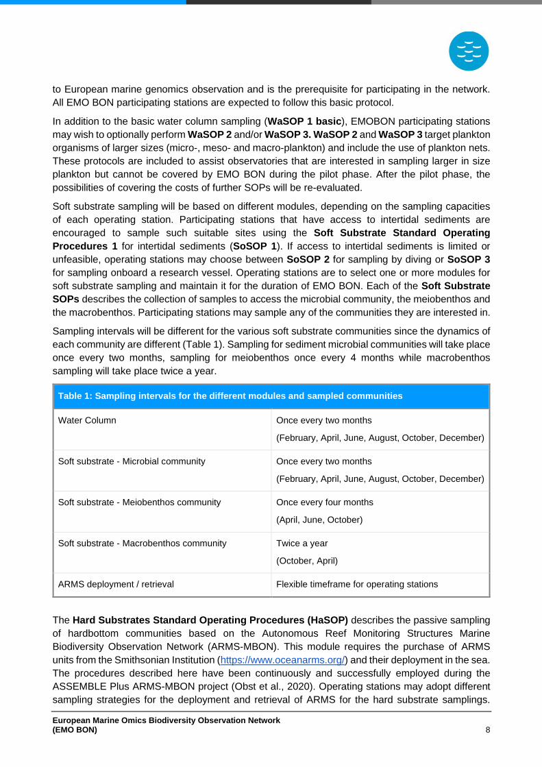
Sampling intervals will be different for the various soft substrate communities since the dynamics of
each community are different (Table 1). Sampling for sediment microbial communities will take place
once every two months, sampling for meiobenthos once every 4 months while macrobenthos
sampling will take place twice a year.
Table 1: Sampling intervals for the different modules and sampled communities
Water Column Once every two months
(February, April, June, August, October, December)
Soft substrate - Microbial community Once every two months
(February, April, June, August, October, December)
Soft substrate - Meiobenthos community Once every four months
(April, June, October)
Soft substrate - Macrobenthos community Twice a year
(October, April)
ARMS deployment / retrieval Flexible timeframe for operating stations
The Hard Substrates Standard Operating Procedures (HaSOP) describes the passive sampling
of hardbottom communities based on the Autonomous Reef Monitoring Structures Marine
Biodiversity Observation Network (ARMS-MBON). This module requires the purchase of ARMS
units from the Smithsonian Institution (https://www.oceanarms.org/) and their deployment in the sea.
The procedures described here have been continuously and successfully employed during the
ASSEMBLE Plus ARMS-MBON project (Obst et al., 2020). Operating stations may adopt different
sampling strategies for the deployment and retrieval of ARMS for the hard substrate samplings.
European Marine Omics Biodiversity Observation Network (EMO BON) 9
However, it is important that the sampling variations are well documented and linked to the dataset
as metadata.
The deployment and recovery of ARMS may be challenging for inexperienced users. Therefore,
observatories not familiar with ARMS protocols need to be trained in the deployment, recovery, and
data collection. This will be done by participating in a hard substrate sampling in an experienced
observatory before being able to apply HaSOP in EMO BON. EMO BON may help bring new and
experienced observatories in contact. EMBRC plans to host training events within the pilot phase of
EMO BON for additional training on sampling and laboratory procedures.
European Marine Omics Biodiversity Observation Network (EMO BON) 10
Water Column
Water Column Standard Operating Procedures 1 ‒ WaSOP 1 (basic)
Summary
Subsurface seawater (0-3 m depth) will be collected from the water column sampling site of each
observatory. The exact water sampling depth is not specified as observatories may not be able to
sample at a specific depth. In case an observatory can sample at specified seawater depth, then
sampling at 1 m depth is recommended. Plankton community will be prefiltered to screen out
organisms >200 μm and then concentrated by sequential filtration on polycarbonate (PC) membrane
filters. Initially seawater volumes will be filtered through 3 µm polycarbonate membrane filters (142
mm in diameter); the outflow seawater will be sequentially filtered through 0.2 µm polycarbonate
membrane filters (142 mm in diameter). This will result in collecting 2 different fractions of plankton:
3-200 μm and 0.2-3 μm organisms.
The required seawater volume to ensure adequate biomass collection may vary between geographic
locations. The observatories may decide on the filtration volume based on the characteristics of their
sampling site and on previous filtration experience in the area. As an indication, in the oligotrophic
open-sea, filtration volumes may reach 20 l, while in nearshore coastal areas filtration of 5 l may be
adequate. However, it is important that operating stations filter the same seawater volume in every
sampling event to obtain consistency among samples from each location. EMBRC can provide
further guidance on selecting filtration volumes upon request.
After filtration, each filter membrane will be cut into two pieces by using a sterile scalpel and each
piece will represent one replicate. In total 4 replicates should be collected (more information in
Replication and Biobanking section, a diagram representation is available in Figure 11); that is, 2
separate sequential filtrations will take place and the cutting of each filter into two will result in 4
replicates. Filtration should be performed by delicate pumping either using a peristaltic pump or by
manual hand pumping. After filtration, membranes should be left to dry, carefully folded, placed in
individual containers with the preservative DNA/RNA shield, flash frozen in liquid nitrogen and stored
at -80oC until shipping for further analysis.
Filtration may initiate in the field immediately after sampling or following the transportation of the
sampled seawater to the laboratory. Seawater should remain in a cool dark place during
transportation and until filtration. It is important to be consistent to when the filtration takes place
among the different sampling events, therefore observatories are expected to either always filter in
the field or always transport water and filter in the lab. For a diagram representation of the
procedures see Appendix 2.
Protocol
A. Materials, equipment, and supplies
Before starting, ensure all materials equipment, and supplies are available by checking the following
table (Table 2).
European Marine Omics Biodiversity Observation Network (EMO BON) 11
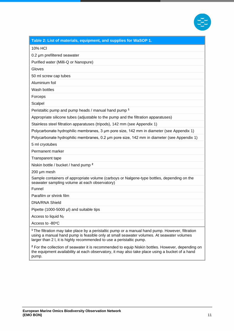
Table 2: List of materials, equipment, and supplies for WaSOP 1.
10% HCl
0.2 μm prefiltered seawater
Purified water (Milli-Q or Nanopure)
Gloves
50 ml screw cap tubes
Aluminium foil
Wash bottles
Forceps
Scalpel
Peristaltic pump and pump heads / manual hand pump 1
Appropriate silicone tubes (adjustable to the pump and the filtration apparatuses)
Stainless steel filtration apparatuses (tripods), 142 mm (see Appendix 1)
Polycarbonate hydrophilic membranes, 3 μm pore size, 142 mm in diameter (see Appendix 1)
Polycarbonate hydrophilic membranes, 0.2 μm pore size, 142 mm in diameter (see Appendix 1)
5 ml cryotubes
Permanent marker
Transparent tape
Niskin bottle / bucket / hand pump 2
200 μm mesh
Sample containers of appropriate volume (carboys or Nalgene-type bottles, depending on the seawater sampling volume at each observatory)
Funnel
Parafilm or shrink film
DNA/RNA Shield
Pipette (1000-5000 μl) and suitable tips
Access to liquid N2
Access to -80oC
1 The filtration may take place by a peristaltic pump or a manual hand pump. However, filtration using a manual hand pump is feasible only at small seawater volumes. At seawater volumes larger than 2 l, it is highly recommended to use a peristaltic pump.
2 For the collection of seawater it is recommended to equip Niskin bottles. However, depending on the equipment availability at each observatory, it may also take place using a bucket of a hand pump.
European Marine Omics Biodiversity Observation Network (EMO BON) 12
B. Preparation and Cleaning
• Always wear gloves.
• Prepare 10% HCl.
• Clean the workspace using 10% HCl.
• Filter seawater at 0.2 μm to prepare 0.2-prefiltered seawater. Ideally this should be seawater from the sampling site.
• Prepare a wash bottle filled with 0.2-prefiltered seawater.
• Prepare a 50 ml screw cap tube filled with 10% HCl and a 50 ml screw cap tube filled with 0.2-prefiltered seawater. Soak forceps and scalpel in the HCl tube and then in the 0.2-prefiltered seawater tube before every use. Store forceps in the 10% HCl tube after use.
• Follow the manufracturer’s instructions for proper cleaning and maintenance of the filtration apparatuses and the peristaltic pump. Clean the peristaltic pump with paper towel humidified with tapwater before and after use.
• Filtration apparatuses and tubes are cleaned at the end of each sampling event and if allowed to dry and stored properly (i.e., tube ends covered, tripod tube inlets/outlets covered, interior of the tripod does not touch other surfaces, all parts are left to dry) there is no need to clean again. It is recommended to rinse all parts of the filtration apparatuses using 70% ethanol after use and allow them to dry overnight.
• After each filtration sequence, the system is cleaned by passing purified water (Milli-Q or Nanopure) through.
• Never touch the filter (not even with gloves on).
• Avoid touching the inside of the filtration apparatuses.
• Prepare the appropriate volume of purified water (Milli-Q or Nanopure) to be used as negative control (see below, Negative Controls section).
• Clean sampling equipment (Niskin bottle and/or buckets) using tap water. Rinse vigorously and make sure material from previous samplings is washed away. Cleaning of sampling equipment (Niskin bottle and/or buckets) may also take place at the end of every sampling event. Make sure that equipment was cleaned and stored properly before using it.
• Rinse the 200 μm mesh and funnels vigorously with tap water and purified water (Milli-Q or Nanopure). Make sure material from previous samplings is washed away. Cleaning of the 200 μm mesh and funnels may also take place at the and of every sampling event. Make sure that materials were cleaned and stored properly before using them.
• Clean the sample containers (carboys or or Nalgene-type bottles) by incubating overnight with 10% HCl. The following day wash twice using distilled water and twice using purified water (Milli-Q or Nanopure). Rinse vigorously to ensure all interior surfaces are cleaned and acid is washed away. Cleaning and decontamination of the sample containers (carboys or or Nalgene-type bottles) may also take place at the and of every sampling event. Make sure they were cleaned and stored properly before using them.
C. Setting up
Follow the manufacturer’s instructions for fully setting up the filtration apparatuses and the pump.
Here, only a brief description is included, considering that a peristaltic pump is used. The equipment
set up is described in the following sketch (Figure 1). In addition, a filtration workspace is depicted
in Figure 2.
European Marine Omics Biodiversity Observation Network (EMO BON) 13
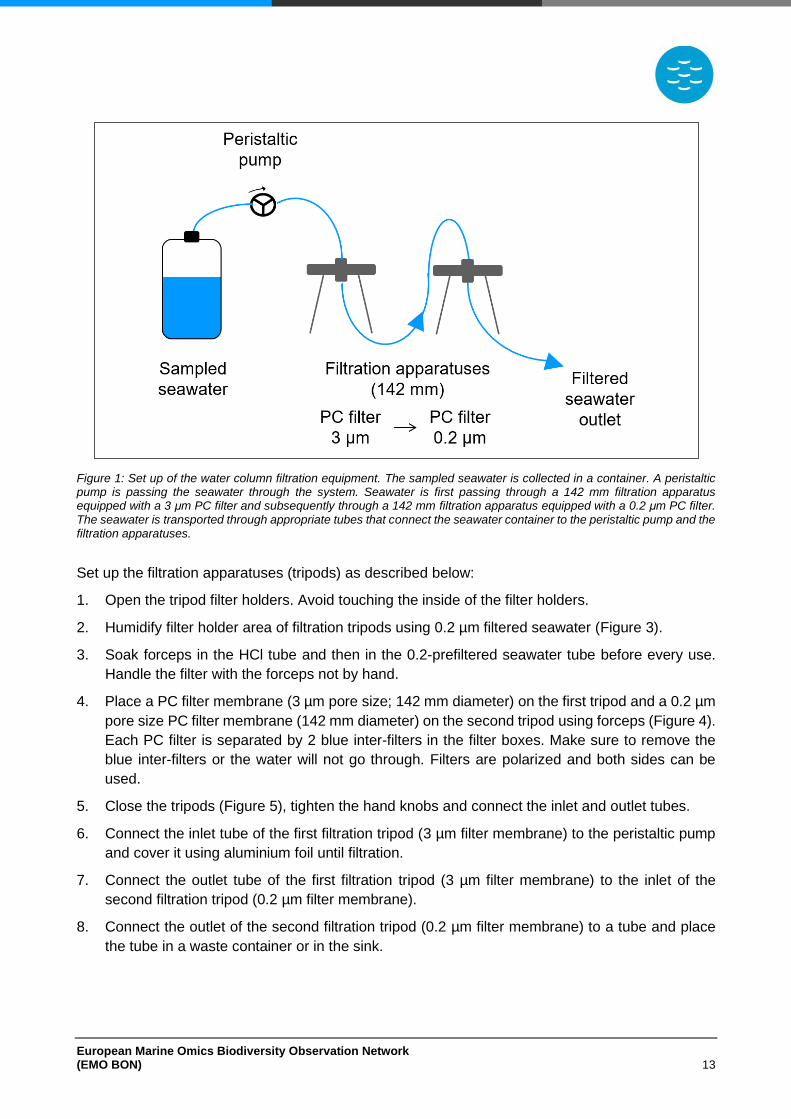
Figure 1: Set up of the water column filtration equipment. The sampled seawater is collected in a container. A peristaltic pump is passing the seawater through the system. Seawater is first passing through a 142 mm filtration apparatus equipped with a 3 μm PC filter and subsequently through a 142 mm filtration apparatus equipped with a 0.2 μm PC filter. The seawater is transported through appropriate tubes that connect the seawater container to the peristaltic pump and the filtration apparatuses.
Set up the filtration apparatuses (tripods) as described below:
1. Open the tripod filter holders. Avoid touching the inside of the filter holders.
2. Humidify filter holder area of filtration tripods using 0.2 µm filtered seawater (Figure 3).
3. Soak forceps in the HCl tube and then in the 0.2-prefiltered seawater tube before every use.
Handle the filter with the forceps not by hand.
4. Place a PC filter membrane (3 µm pore size; 142 mm diameter) on the first tripod and a 0.2 µm
pore size PC filter membrane (142 mm diameter) on the second tripod using forceps (Figure 4).
Each PC filter is separated by 2 blue inter-filters in the filter boxes. Make sure to remove the
blue inter-filters or the water will not go through. Filters are polarized and both sides can be
used.
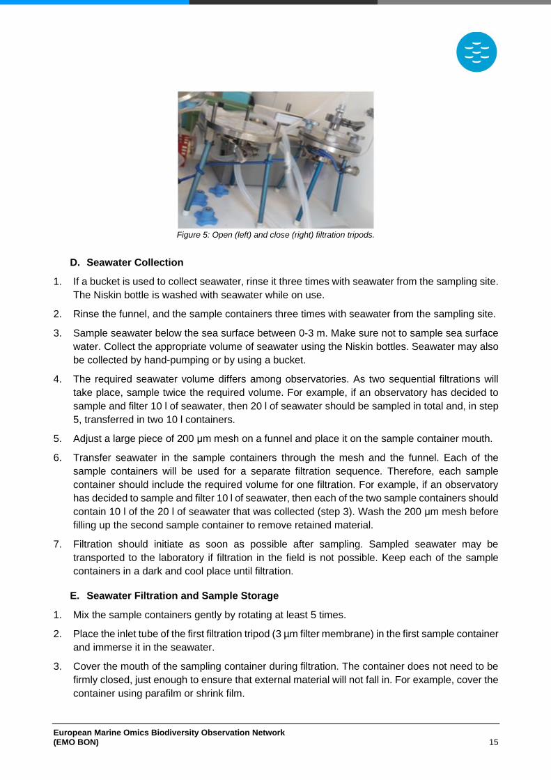
5. Close the tripods (Figure 5), tighten the hand knobs and connect the inlet and outlet tubes.
6. Connect the inlet tube of the first filtration tripod (3 µm filter membrane) to the peristaltic pump
and cover it using aluminium foil until filtration.
7. Connect the outlet tube of the first filtration tripod (3 µm filter membrane) to the inlet of the
second filtration tripod (0.2 µm filter membrane).
8. Connect the outlet of the second filtration tripod (0.2 µm filter membrane) to a tube and place
the tube in a waste container or in the sink.
European Marine Omics Biodiversity Observation Network (EMO BON) 14
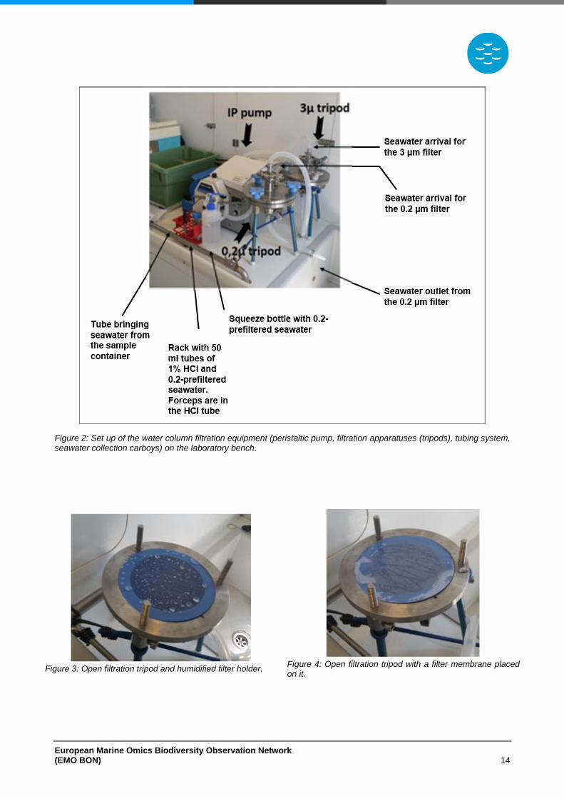
Figure 2: Set up of the water column filtration equipment (peristaltic pump, filtration apparatuses (tripods), tubing system, seawater collection carboys) on the laboratory bench.
Figure 3: Open filtration tripod and humidified filter holder.
Figure 4: Open filtration tripod with a filter membrane placed on it.
European Marine Omics Biodiversity Observation Network (EMO BON) 15
Figure 5: Open (left) and close (right) filtration tripods.
D. Seawater Collection
1. If a bucket is used to collect seawater, rinse it three times with seawater from the sampling site.
The Niskin bottle is washed with seawater while on use.
2. Rinse the funnel, and the sample containers three times with seawater from the sampling site.
3. Sample seawater below the sea surface between 0-3 m. Make sure not to sample sea surface
water. Collect the appropriate volume of seawater using the Niskin bottles. Seawater may also
be collected by hand-pumping or by using a bucket.
4. The required seawater volume differs among observatories. As two sequential filtrations will
take place, sample twice the required volume. For example, if an observatory has decided to
sample and filter 10 l of seawater, then 20 l of seawater should be sampled in total and, in step
5, transferred in two 10 l containers.
5. Adjust a large piece of 200 μm mesh on a funnel and place it on the sample container mouth.
6. Transfer seawater in the sample containers through the mesh and the funnel. Each of the
sample containers will be used for a separate filtration sequence. Therefore, each sample
container should include the required volume for one filtration. For example, if an observatory
has decided to sample and filter 10 l of seawater, then each of the two sample containers should
contain 10 l of the 20 l of seawater that was collected (step 3). Wash the 200 μm mesh before
filling up the second sample container to remove retained material.
7. Filtration should initiate as soon as possible after sampling. Sampled seawater may be
transported to the laboratory if filtration in the field is not possible. Keep each of the sample
containers in a dark and cool place until filtration.
E. Seawater Filtration and Sample Storage
1. Mix the sample containers gently by rotating at least 5 times.
2. Place the inlet tube of the first filtration tripod (3 µm filter membrane) in the first sample container
and immerse it in the seawater.
3. Cover the mouth of the sampling container during filtration. The container does not need to be
firmly closed, just enough to ensure that external material will not fall in. For example, cover the
container using parafilm or shrink film.
European Marine Omics Biodiversity Observation Network (EMO BON) 16
4. Check the pumping speed and the flow direction and turn on the peristaltic pump. Flow rate
should be adjusted to ensure a regular gentle continuous seawater flow.
5. Always initiate the filtration sequence with the filter holder vents open. Wait until all air bubbles
get expulsed from the filter holders through the vents and once the water flows regularly through
the vents, close them.
6. Seawater will be filtered sequentially through two filter membranes, first through the 3 μm and
subsequently through the 0.2 μm pore size membrane.
7. Check the tubing system and the filtration tripod connections regularly for overpressure that
may damage the cells and/or the filtration system. If pressure is too high, decrease the flow
rate.
8. Check for the presence of bubbles and use the filter holder vents to expulse them.
9. Ensure membranes always remain moistened during filtration.
10. After filtration, it is important to allow the filters to dry. To do that:
a. Keep the pump on and allow the system to pump air.
b. Disconnect the tube after the second tripod (0.2 μm tripod).
c. Empty the tube between the pump and the first tripod (3 μm tripod) by lifting it up.
d. Slowly open the vent of the second tripod (0.2 μm tripod) and empty the tube between the
two tripods by “waving it” a few times.
e. Close the vent of the second tripod (0.2 μm tripod). All the seawater that remained on the
second tripod filter will be pushed out by the increasing pressure.
f. Gently shake each tripod to make sure there is no water trapped in the filter holder.
g. Slowly open both vents while turning the peristaltic pump off.
11. Open the tripod filter holders. Avoid touching the inside of the filter holder area.
12. Use the forceps to handle the filter. Soak forceps in the HCl tube and then in the 0.2-prefiltered
seawater tube before every use.
13. To avoid contaminations, cut rapidly the filter into 2 visually equal pieces by using a sterile
scapel, after placing it on a clean, hard surface (e.g. in a Petri dish).
14. Immediately fold each piece twice and then fold again by rolling the filter in a cylinder. The two
filter pieces will be the sample replicates.
15. Carefully place the filters into separate 5 ml cryotubes and gently push the filters to the bottom
of the tube to minimize the space they occupy in the tube. Do not crumble the filter, just try to
gently fold it once more while in the tube.
16. Add 4.5 ml of DNA/RNA Shield by using a pipette and make sure the filter is covered by the
solution.
17. Label the tube appropriately using permanent marker (see Sample Labelling section). Each
filtration produces two sample replicates; therefore after 2 filtrations, 4 replicated samples will
be produced. The first filtration (for example, filtration A) will produce replicates 1 and 2 when
filter A is cut in half.
European Marine Omics Biodiversity Observation Network (EMO BON) 17
18. Apply transparent tape firmly on top of the label to avoid bleaching.
19. Transfer tubes into liquid N2 for flash freezing and subsequently store at -80oC.
20. Close the tripod and clean the filtration system by passing 2 l of purified water (Milli-Q or
Nanopure). Place new filters in the filtration tripods as described in the setting up section and
close the tripods.
21. Repeat steps 1-15 once more to filter all the collected seawater. Each filtration produces two
sample replicates; therefore after 2 filtrations, 4 replicated samples will be produced. The first
filtration (for example, filtration A) will produce replicates 1 and 2 when filter A is cut in half. The
second filtration (for example, filtration B) will produce replicates 3 and 4 when filter B is cut in
half.
22. Store the filter pieces for replicates 3 and 4 in 5 ml cryotubes without the addition of DNA/RNA
Shield. Replicates 3 and 4 will be stored for biobanking.
23. Label the tube appropriately using permanent marker (see Sample Labelling section).
24. Apply transparent tape firmly on top of the label to avoid bleaching.
25. Transfer tubes into liquid N2 for flash freezing and subsequently store at -80oC.
F. Negative Controls
As samples undergo different procedures at the operating stations and at the centralized sequencing
centre laboratories, it is important to check for contaminations. For this reason, blank samples will
be collected and processed together with the actual samples to serve as negative controls. The
collection of the blank samples will take place after the seawater filtrations to account for the cleaning
of the filtration apparatuses between different samples. After the seawater filtration follow the steps
bellow to collect and store the negative control sample.
1. After the second filtration, close the tripod and clean the filtration system by passing 2 l of
purified water (Milli-Q or Nanopure).
2. Place new filters in the filtration tripods as described in the setting up section above (Setting up
steps 1-4) and close the tripods.
3. Filter purified water (Milli-Q or Nanopure). The volume of the purified water (Milli-Q or Nanopure)
to be filtered should be equal to the volume of seawater filtered for one filtration. For example,
if an observatory has decided to sample and filter 10 l of seawater in one filtration sequence,
then 10 l of purified water (Milli-Q or Nanopure) should be filtered for the negative control as
well.
4. Follow filtration procedure as described in the seawater filtration section (E. Seawater Filtration
and Sample Storage, steps 6-10).
5. Open tripod filter holders. Avoid touching the inside of the filter holders.
6. Fold immediately each filter piece four times and then fold again by rolling the filter like a
cigarette paper.
7. Carefully place the filters into 5 ml cryotubes and gently push the filters to minimize the space
they occupy in the tube. Do not crumble the filter, just try to gently fold it once more while in the
tube.
European Marine Omics Biodiversity Observation Network (EMO BON) 18
8. Add 4.5 ml of DNA/RNA Shield by using a pipette and make sure the filter is covered by the
solution
9. Label the tube appropriately using permanent marker (see Sample Labelling section).
10. Apply transparent tape firmly on top of the label to avoid bleaching.
11. Transfer tubes into liquid N2 for flash freezing and subsequently store at -80oC.
Water Column Standard Operating Procedures 2 ‒ WaSOP 2 (optional)
Summary
This protocol aims at collecting plankton organisms sized >20 μm. EMO BON observatories have
different characteristics and may already follow specific routines for sampling plankton >20 μm.
Therefore, observatories will be allowed to deviate from the WaSOP 2 as long the procedures
followed are described in detail and adhere to the data as metadata.
Here we suggest vertical tows by a 20 µm mesh size plankton net from 100 m depth to the surface
of the water column or from bottom to the water column surface in case of a shallower site. The
material collected in the cod end should be diluted and separated into 4 subsamples. Each
subsample should be filtered through 10 µm polycarbonate membrane filters (47 mm in diameter)
under vacuum pressure. After filtration, membranes should be left to dry, carefully folded, placed in
individual tubes, preserved in DNA/RNA Shield, and stored in liquid nitrogen or at -80oC until further
analyses.
Filtration may initiate in the field immediately after sampling or after the transportation of sampled
seawater to the laboratory. Samples should remain in a cool dark place during transportation and
until filtration. It is important to be consistent to when the filtration takes place among the different
sampling events, therefore observatories are expected to either always filter at the field or always
transport water and filter it in the lab.
Observatories may deviate from the protocol and opt for horizontal instead of vertical tow, or by
introducing prefiltration at different mesh sizes. Nevertheless, it is important that all procedures are
documented in detail and kept consistent during EMOBON. For a diagram representation of the
procedures see Appendix 2.
Protocol
A. Materials, equipment, and supplies
Before starting, ensure all materials equipment, and supplies are available by checking the following
table (Table 3).
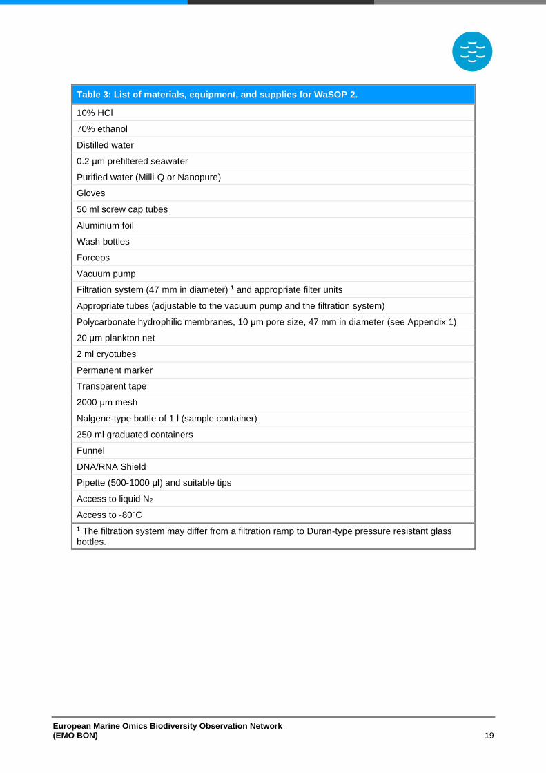
European Marine Omics Biodiversity Observation Network (EMO BON) 19
Table 3: List of materials, equipment, and supplies for WaSOP 2.
10% HCl
70% ethanol
Distilled water
0.2 μm prefiltered seawater
Purified water (Milli-Q or Nanopure)
Gloves
50 ml screw cap tubes
Aluminium foil
Wash bottles
Forceps
Vacuum pump
Filtration system (47 mm in diameter) 1 and appropriate filter units
Appropriate tubes (adjustable to the vacuum pump and the filtration system)
Polycarbonate hydrophilic membranes, 10 μm pore size, 47 mm in diameter (see Appendix 1)
20 μm plankton net
2 ml cryotubes
Permanent marker
Transparent tape
2000 μm mesh
Nalgene-type bottle of 1 l (sample container)
250 ml graduated containers
Funnel
DNA/RNA Shield
Pipette (500-1000 μl) and suitable tips
Access to liquid N2
Access to -80oC
1 The filtration system may differ from a filtration ramp to Duran-type pressure resistant glass bottles.
European Marine Omics Biodiversity Observation Network (EMO BON) 20
B. Preparation and Cleaning
• Always wear gloves
• Prepare 10% HCl.
• Disinfect the workspace using 10% HCl.
• Filter seawater at 0.2 μm to prepare 0.2-prefiltered seawater. Ideally this should be seawater from the sampling site.
• Prepare a wash bottle filled with 0.2-prefiltered seawater.
• Prepare a 50 ml screw cap tube filled with 10% HCl and a 50 ml screw cap tube filled with 0.2-prefiltered seawater. Soak forceps and scalpel in the HCl tube and then in the 0.2-prefiltered seawater tube before every use. Store forceps in the 10% HCl tube after use.
• Clean the filtration system:
− Clean the filtration ramp or the Duran-type pressure resistant glass bottles with tap water and distilled water.
− Dissasemble the filtration units and rinse with tap water, distilled water, and EtOH 70%. Allow them to dry before reassembling.
Cleaning of the filtration system may also take place at the end of every sampling event. Make sure it was cleaned and stored properly before using it.
• Rinse the 20 μm plankton net vigorously inside and outside using tap water and make sure material from previous samplings is washed away. Cleaning of the plankton net may also take place at the end of every sampling event. Make sure it was cleaned and stored properly before using it.
• Clean the sample containers (Nalgene-type bottles of 1 l) using 250 ml of 10% HCl and incubate overnight. The following day wash twice using distilled water and twice using purified water (Milli-Q or Nanopure). Rinse vigorously to ensure all interior surfaces are cleaned and acid is washed away. Cleaning and decontamination of the sample containers may also take place at the and of every sampling event. Make sure the containers were cleaned and stored properly before using them.
• Rinse funnels vigorously with tap water and purified water (Milli-Q or Nanopure). Cleaning and decontamination of funnels may also take place at the and of every sampling event. Make sure that materials were cleaned and stored properly before using them.
• Coordinate the appropriate adjustment of the plankton net to the winch cable and the proper towing with the RV sailing personel.
C. Setting up
Set up the filtration system as described below. Filtration may take place in parallel depending on
the available equipment. The filtration system used may differ from a filtration ramp to Duran-type
pressure resistant glass bottles. Filtration units of 47 mm in diameter are adjusted on the ramp or
on the bottles. Figure 6 depicts a filtration ramp and the 4 adjusted filtration units.
European Marine Omics Biodiversity Observation Network (EMO BON) 21
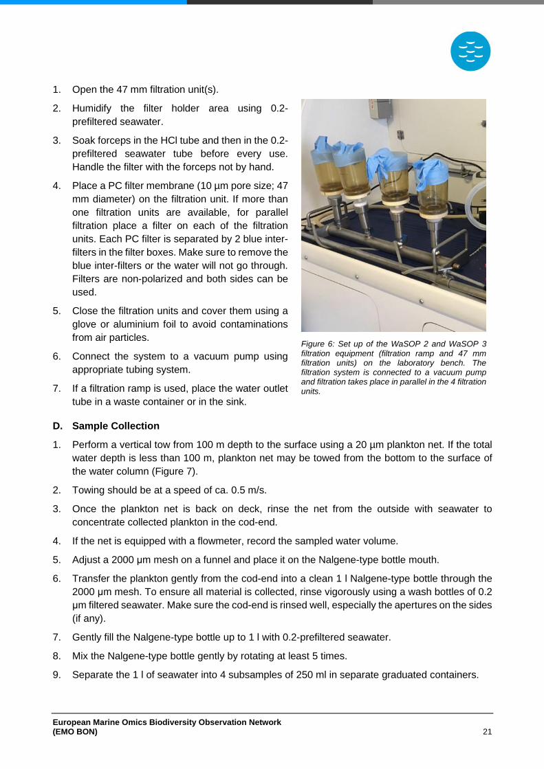
1. Open the 47 mm filtration unit(s).
2. Humidify the filter holder area using 0.2-
prefiltered seawater.
3. Soak forceps in the HCl tube and then in the 0.2-
prefiltered seawater tube before every use.
Handle the filter with the forceps not by hand.
4. Place a PC filter membrane (10 µm pore size; 47
mm diameter) on the filtration unit. If more than
one filtration units are available, for parallel
filtration place a filter on each of the filtration
units. Each PC filter is separated by 2 blue inter-
filters in the filter boxes. Make sure to remove the
blue inter-filters or the water will not go through.
Filters are non-polarized and both sides can be
used.
5. Close the filtration units and cover them using a
glove or aluminium foil to avoid contaminations
from air particles.
6. Connect the system to a vacuum pump using
appropriate tubing system.
7. If a filtration ramp is used, place the water outlet
tube in a waste container or in the sink.
D. Sample Collection
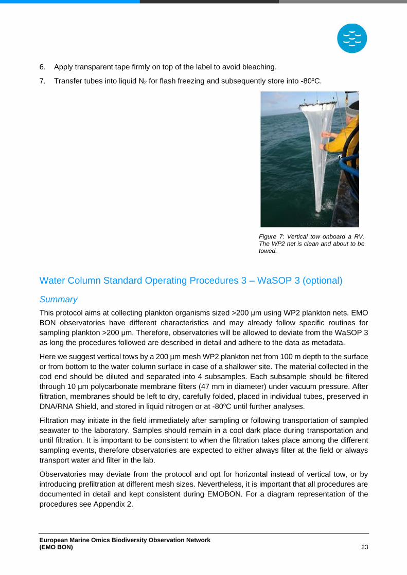
1. Perform a vertical tow from 100 m depth to the surface using a 20 µm plankton net. If the total
water depth is less than 100 m, plankton net may be towed from the bottom to the surface of
the water column (Figure 7).
2. Towing should be at a speed of ca. 0.5 m/s.
3. Once the plankton net is back on deck, rinse the net from the outside with seawater to
concentrate collected plankton in the cod-end.
4. If the net is equipped with a flowmeter, record the sampled water volume.
5. Adjust a 2000 μm mesh on a funnel and place it on the Nalgene-type bottle mouth.
6. Transfer the plankton gently from the cod-end into a clean 1 l Nalgene-type bottle through the
2000 μm mesh. To ensure all material is collected, rinse vigorously using a wash bottles of 0.2
μm filtered seawater. Make sure the cod-end is rinsed well, especially the apertures on the sides
(if any).
7. Gently fill the Nalgene-type bottle up to 1 l with 0.2-prefiltered seawater.
8. Mix the Nalgene-type bottle gently by rotating at least 5 times.
9. Separate the 1 l of seawater into 4 subsamples of 250 ml in separate graduated containers.
Figure 6: Set up of the WaSOP 2 and WaSOP 3 filtration equipment (filtration ramp and 47 mm filtration units) on the laboratory bench. The filtration system is connected to a vacuum pump and filtration takes place in parallel in the 4 filtration units.
European Marine Omics Biodiversity Observation Network (EMO BON) 22
10. Filtration should initiate as soon as possible after sampling. Subsamples may be transported to
the laboratory if filtration at the field is not possible. Keep each of the subsample containers in
a dark and cool place until filtration.
E. Filtration and Sample Storage
1. Filter subsamples through 10 μm pore size PC filter membranes (47 mm in diameter).
2. Process each subsample in multiple filtration steps by adding 50 ml to the filtration tower in each
step. This is because samples may be rich in plankton and clog the filter. Mix the Nalgene-type
bottle gently for each of the 50 ml filtration steps. In case the filter clogs, stop the filtration and
record the total volume filtered.
3. After filtration, allow filters to dry. Recover each membrane using forceps and fold membranes
three times. Soak forceps in the HCl tube and then in the 0.2-prefiltered seawater tube before
every use.
4. Place filter membrane into 2 ml cryotube.
5. Add 1.5 ml of DNA/RNA Shield by using a pipette and make sure the filter is covered by the
solution.
6. Label the tube appropriately using permanent marker (see Sample Labelling section).
7. Apply transparent tape firmly on top of the label to avoid bleaching.
8. Transfer tubes into liquid N2 for flash freezing and subsequently store into -80oC.
9. Repeat steps 1-8 for all 4 replicated subsamples. Vacuum filtrations of the replicates may also
take place in parallel if appropriate filtration system is available.
10. For the third and fourth filter (replicates 3 and 4) skip step 5. Replicates 3 and 4 will be stored
for biobanking and DNA/RNA Shield will not be used.
F. Negative Controls
As samples undergo different procedures at the operating stations and at the centralized sequencing
centre laboratories, it is important to check for contaminations. For this reason, blank samples will
be collected and processed together with the actual samples to serve as negative controls. The
collection of the blank samples will take place after the seawater filtrations to account for the cleaning
of the filtration apparatuses between different samples. After the seawater filtration follow the steps
bellow to collect and store the negative control sample.
1. Filter 250 ml of purified water (Milli-Q or Nanopure) through a 10 μm pore size PC filter
membrane (47 mm in diameter). Follow filtration procedure as described above.
2. After filtration, allow filter to dry. Recover the filter membrane using clean forceps and fold three
times. Soak forceps in the HCl tube and then in the 0.2-prefiltered seawater tube before every
use.
3. Place membrane into a 2 ml cryotube.
4. Add 1.5 ml of DNA/RNA Shield by using a pipette and make sure the filter is covered by the
solution.
5. Label the tube appropriately using permanent marker (see Sample Labelling section).
European Marine Omics Biodiversity Observation Network (EMO BON) 23
6. Apply transparent tape firmly on top of the label to avoid bleaching.
7. Transfer tubes into liquid N2 for flash freezing and subsequently store into -80oC.
Figure 7: Vertical tow onboard a RV. The WP2 net is clean and about to be towed.
Water Column Standard Operating Procedures 3 ‒ WaSOP 3 (optional)
Summary
This protocol aims at collecting plankton organisms sized >200 μm using WP2 plankton nets. EMO
BON observatories have different characteristics and may already follow specific routines for
sampling plankton >200 μm. Therefore, observatories will be allowed to deviate from the WaSOP 3
as long the procedures followed are described in detail and adhere to the data as metadata.
Here we suggest vertical tows by a 200 µm mesh WP2 plankton net from 100 m depth to the surface
or from bottom to the water column surface in case of a shallower site. The material collected in the
cod end should be diluted and separated into 4 subsamples. Each subsample should be filtered
through 10 µm polycarbonate membrane filters (47 mm in diameter) under vacuum pressure. After
filtration, membranes should be left to dry, carefully folded, placed in individual tubes, preserved in
DNA/RNA Shield, and stored in liquid nitrogen or at -80oC until further analyses.
Filtration may initiate in the field immediately after sampling or following transportation of sampled
seawater to the laboratory. Samples should remain in a cool dark place during transportation and
until filtration. It is important to be consistent to when the filtration takes place among the different
sampling events, therefore observatories are expected to either always filter at the field or always
transport water and filter in the lab.
Observatories may deviate from the protocol and opt for horizontal instead of vertical tow, or by
introducing prefiltration at different mesh sizes. Nevertheless, it is important that all procedures are
documented in detail and kept consistent during EMOBON. For a diagram representation of the
procedures see Appendix 2.
European Marine Omics Biodiversity Observation Network (EMO BON) 24
Protocol
A. Materials, equipment, and supplies
Before starting, ensure all materials equipment, and supplies are available by checking the following
table (Table 4).
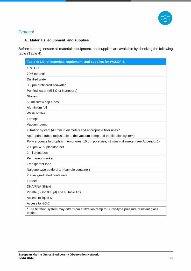
Table 4: List of materials, equipment, and supplies for WaSOP 3.
10% HCl
70% ethanol
Distilled water
0.2 μm prefiltered seawater
Purified water (Milli-Q or Nanopure)
Gloves
50 ml screw cap tubes
Aluminium foil
Wash bottles
Forceps
Vacuum pump
Filtration system (47 mm in diameter) and appropriate filter units 1
Appropriate tubes (adjustable to the vacuum pump and the filtration system)
Polycarbonate hydrophilic membranes, 10 μm pore size, 47 mm in diameter (see Appendix 1)
200 μm WP2 plankton net
2 ml cryotubes
Permanent marker
Transparent tape
Nalgene-type bottle of 1 l (sample container)
250 ml graduated containers
Funnel
DNA/RNA Shield
Pipette (500-1000 μl) and suitable tips
Access to liquid N2
Access to -80oC
1 The filtration system may differ from a filtration ramp to Duran-type pressure resistant glass bottles.
European Marine Omics Biodiversity Observation Network (EMO BON) 25
B. Preparation and Cleaning
• Always wear gloves
• Prepare 10% HCl.
• Disinfect the workspace using 10% HCl.
• Filter seawater at 0.2 μm to prepare 0.2-prefiltered seawater. Ideally this should be seawater from the sampling site.
• Prepare a wash bottle filled with 0.2-prefiltered seawater.
• Prepare a 50 ml screw cap tube filled with 10% HCl and a 50 ml screw cap tube filled with 0.2-prefiltered seawater. Soak forceps and scalpel in the HCl tube and then in the 0.2-prefiltered seawater tube before every use. Store forceps in the 10% HCl tube after use.
• Clean the filtration system:
− Clean the filtration ramp or the Duran-type pressure resistant glass bottles with tap water and distilled water.
− Dissasemble the filtration units and rinse with tap water, distilled water, and EtOH 70%. Allow them to dry before reassembling.
Cleaning of the filtration system may also take place at the end of every sampling event. Make sure it was cleaned and stored properly before using it.
• Rinse the 200 μm WP2 plankton net vigorously inside and outside using tap water and make sure material from previous samplings is washed away. Cleaning of the plankton net may also take place at the end of every sampling event. Make sure it was cleaned and stored properly before using it.
• Clean the sample containers (Nalgene-type bottles of 1 l) using 250 ml of 10% HCl and incubate overnight. The following day wash twice using distilled water and twice using purified water (Milli-Q or Nanopure). Rinse vigorously to ensure all interior surfaces are cleaned and acid is washed away. Cleaning and decontamination of the sample containers may also take place at the and of every sampling event. Make sure the containers were cleaned and stored properly before using them.
• Rinse funnels vigorously with tap water and purified water (Milli-Q or Nanopure). Cleaning and decontamination of funnels may also take place at the and of every sampling event. Make sure that materials were cleaned and stored properly before using them.
• Coordinate the appropriate adjustment of the plankton net to the winch cable and the proper towing with the RV sailing personel.
C. Setting up
Set up the filtration system as described below. Filtration may take place in parallel depending on
the available equipment. The filtration system used may differ from a filtration ramp to Duran-type
pressure resistant glass bottles. Filtration units of 47 mm in diameter are adjusted on the ramp or
on the bottles. Figure 6 depicts a filtration ramp and the 4 adjusted filtration units.
1. Open the 47 mm filtration unit(s).
2. Humidify the filter holder area using 0.2-prefiltered seawater.
3. Soak forceps in the HCl tube and then in the 0.2-prefiltered seawater tube before every use.
Handle the filter with the forceps not by hand.
European Marine Omics Biodiversity Observation Network (EMO BON) 26
4. Place a PC filter membrane (10 µm pore size; 47 mm diameter) on the filtration unit. If more
than one filtration units are available, for parallel filtration place a filter on each of the filtration
units. Each PC filter is separated by 2 blue inter-filters in the filter boxes. Make sure to remove
the blue inter-filters or the water will not go through. Filters are non-polarized and both sides
can be used.
5. Close the filtration units and cover them using a glove or aluminium foil to avoid contaminations
from air particles.
6. Connect the system to a vacuum pump using appropriate tubing system.
7. If a filtration ramp is used, place the water outlet tube in a waste container or in the sink.
D. Sample Collection
1. Perform a vertical tow from 100 m depth to surface using a WP2 200 µm plankton net. If the
total water depth is less than 100 m, plankton net may be towed from the bottom to the surface
of the water column (Figure 7).
2. Towing should be at a speed of ca. 0.5 m/s.
3. Once the plankton net is back on deck, rinse the net from the outside with seawater to
concentrate collected plankton to the cod-end.
4. If the net is equipped with a flowmeter, record the sampled water volume.
5. Transfer the plankton gently from the cod-end into a clean 1 l Nalgene-type bottle through the
2000 μm mesh. To ensure all material is collected, rinse vigorously using a wash bottles of 0.2
μm filtered seawater. Make sure the cod-end is rinsed well, especially the apertures on the sides
(if any).
6. Gently fill the Nalgene-type bottle up to 1 l with 0.2-prefiltered seawater.
7. Mix the Nalgene-type bottle gently by rotating at least 5 times.
8. Separate the 1 l of seawater into 4 subsamples of 250 ml in separate graduated containers.
9. Filtration should initiate as soon as possible after sampling. Subsamples may be transported to
the laboratory if filtration at the field is not possible. Keep each of the subsample containers in
a dark and cool place until filtration.
E. Filtration and Sample Storage
1. Filter subsamples through 10 μm pore size PC filter membranes (47 mm in diameter). Filtration
should proceed fast.
2. After filtration, allow filters to dry. Recover each membrane using forceps and fold membranes
three times. Soak forceps in the HCl tube and then in the 0.2-prefiltered seawater tube before
every use.
3. Place membranes into 2 ml cryotubes.
4. Add 1.5 ml of DNA/RNA Shield by using a pipette and make sure the filter is covered by the
solution.
5. Label the tube appropriately using permanent marker (see Sample Labelling section).
European Marine Omics Biodiversity Observation Network (EMO BON) 27
6. Apply transparent tape firmly on top of the label to avoid bleaching.
7. Transfer tubes into liquid N2 for flash freezing and subsequently store into -80oC.
8. Repeat steps 1-7 for all 4 replicated subsamples. Vacuum filtrations of the replicates may also
take place in parallel if appropriate filtration system is available.
9. For the third and fourth filter (replicates 3 and 4) skip step 5. Replicates 3 and 4 will be stored
for biobanking and DNA/RNA Shield will not be used.
F. Negative Controls
As samples undergo different procedures at the operating stations and at the centralized sequencing
centre laboratories, it is important to check for contaminations. For this reason, blank samples will
be collected and processed together with the actual samples to serve as negative controls. The
collection of the blank samples will take place after the seawater filtrations to account for the cleaning
of the filtration apparatuses between different samples. After the seawater filtration follow the steps
bellow to collect and store the negative control sample.
1. Filter 250 ml of purified water (Milli-Q or Nanopure) through a 10 μm pore size PC filter
membrane (47 mm in diameter). Follow filtration procedure as described above.
2. After filtration, allow filter to dry. Recover the filter membrane using clean forceps and fold three
times. Soak forceps in the HCl tube and then in the 0.2-prefiltered seawater tube before every
use.
3. Place membrane into a 2 ml cryotube.
4. Add 1.5 ml of DNA/RNA Shield by using a pipette and make sure the filter is covered by the
solution.
5. Label the tube appropriately using permanent marker (see Sample Labelling section).
6. Apply transparent tape firmly on top of the label to avoid bleaching.
7. Transfer tubes into liquid N2 for flash freezing and subsequently store into -80oC.
European Marine Omics Biodiversity Observation Network (EMO BON) 28
Soft Substrate
Soft Substrate Standard Operating Procedures 1 ‒ SoSOP 1 (intertidal sediments)
Summary
Operating stations that have access to intertidal sediments are encouraged to choose such habitats
as soft substrate sampling sites. Intertidal soft substrates will be sampled during low tides using
sediment corers to ensure the collection of undisturbed sediment. Sediment corers are
recommended to be at least 10 cm in diameter so that adequate sediment is collected. The top 5
cm of sediment will be sliced and sampled for the collection of microorganisms and meiobenthos.
For the collection of macrobenthos, all the sediment in the corers will be used. For each community
sampled, 4 replicated samples should be collected. These replicates are designed to represent
technical replicates and will be used for different purposes (more information on the Replication and
BiobankingReplication and BiobankingReplication and BiobankingReplication and Biobanking
section).
The finer the sampled sediment is, the more care should be given during handling. For example,
silty sediment might leak when the corer is retrieved from the sediment. In that case, corers handling
should be performed quickly. In case operating stations find it impossible to use sediment corers in
their site of choice, it will also be acceptable to use other means for sediment collection. For
example, a shovel might be used to dig out sediment from a defined area, or metal frames could
define the area from where the sediment will be collected. However, all deviations from the present
protocols should be explicitly described and follow the samples as metadata.
Sediment slicing or sieving may initiate in the field immediately after sampling or following the
transportation of the sediment corers to the laboratory. Sediment corers should remain cool and in
the dark during transportation and always be kept in a vertical position as retrieved from the field. It
is important to be consistent to when the slicing or sieving takes place among the different sampling
events, therefore observatories are expected to either always process samples in the field or always
transport corers and process in the lab.
Before shipment for DNA extraction and sequencing, the meiobenthic organisms should be
extracted from the sediment. After the meiobenthos extraction, the meiobenthic organisms will be
resuspended in the preservative DNA/RNA shield for shipment.
Sediment collected for macrobenthos sampling will have to be sieved through a 1 mm sieve. Some
marine areas, such as the Eastern Mediterranean, or polluted sediments are characterized by
smaller macrobenthic organisms. Using a 1 mm sieve alone would result to missing a large part of
the macrobenthos for those areas. In such cases, sequential sieving using both 1 mm and 0.5 mm
sieves should take place. The organisms retained in the 1 mm and in the 0.5 mm sieves will be
handled and stored separately as different fractions. After sieving all organisms will be crushed and
homogenized using mortar and pestle to obtain a mixture of tissues. For a diagram representation
of the procedures see Appendix 2.
European Marine Omics Biodiversity Observation Network (EMO BON) 29
Protocol
A. Materials, equipment, and supplies
Before starting, ensure all materials equipment, and supplies are available by checking the following
table (Table 5).
Table 5: List of materials, equipment, and supplies for SoSOP 1 (intertidal sediments).
Bleach solution
Distilled water
Purified water (Milli-Q or Nanopure)
Gloves
Aluminium foil
Plexiglass or PVC sediment corers of at least 10 cm in diameter (microbial and meiobenthos community) 1
Appropriate rubber stoppers for the 10 cm sediment corers (microbial and meiobenthos community)
Plexiglass or PVC sediment corers of at least 20 cm in diameter (macrobenthos community) 1
Appropriate rubber stoppers for the 20 cm sediment corers (macrobenthos community)
Spatula and/or spoon
Plunger
Clean surface for mixing the sediment (e.g., large Petri dish or plastic bowl)
Pipette (1000-5000 μl) and pre-cut 5000 μl tips (optional for silty sediments)
1 mm sieve (macrobenthos community)
0.5 mm sieve (optional; macrobenthos community)
Mortar and pestle (macrobenthos community)
15 ml screw cap tubes resistant to liquid N2 (microbial community)
Ziplock bags (meiobenthos community)
50 ml screw cap tubes (macrobenthos community)
Permanent marker
Transparent tape
Laboratory balance
DNA/RNA Shield (microbial and meiobenthos community)
DESS aqueous solution (macrobenthos community)
Pipette (500-1000 μl) and suitable tips
Cotton swabs
Access to liquid N2 (microbial community)
Access to -80oC (microbial community)
Access to -20oC (meiobenthos community)
Access to 4oC (macrobenthos community)
1 Alternatively, a multicorer may be used to collect several sediment corers simultaneously.
European Marine Omics Biodiversity Observation Network (EMO BON) 30
B. Preparation and Cleaning
• Always wear gloves.
• Prepare 10% bleach solution.
• Prepare DESS aquaous solution (see DESS recipe).
• Clean sediment corers and sieves using tap water. Rinse vigorously and make sure material from previous samplings is washed away.
• Desinfect plastic material (large Petri dish and/or plastic bowl) by incubating in 10% bleach solution overnight. Rinse with purified water (Milli-Q or Nanopure) to wash the solution away and cover in aluminium foil to store.
• Desinfect spatulas, spoons, and other metal tools by briefly soaking in 10% bleach solution. Rinse with purified water (Milli-Q or Nanopure) to wash the solution away and cover in aluminium foil to store.
C. Negative Control Samples
As samples undergo different procedures at the operating stations and at the centralized sequencing
centre laboratories, it is important to check for contaminations. For this reason, blank samples will
be collected and processed together with the sediment samples to serve as negative controls. Once
the sediment is collected and before further processing, collect and store a negative control sample
as described below:
Microbial community negative control
1. Use a cotton swab on the lab bench, the spatulas, and the spoons.
2. Cut the cotton part of the swab and store it in a 15 ml screw cap tube.
3. Add 10 ml of DNA/RNA Shield by using a pipette.
4. Label the tube appropriately using permanent marker (see Sample Labelling section).
5. Apply transparent tape firmly on top of the label to avoid bleaching.
6. Transfer tube into liquid N2 for flash freezing and subsequently store at -80oC.
Meiobenthos community negative control
1. Use a cotton swab on the lab bench, the spatulas, and the spoons.
2. Cut the cotton part of the swab and store it in a Ziplock bag.
3. Label the tube appropriately using permanent marker (see Sample Labelling section).
4. Apply transparent tape firmly on top of the label to avoid bleaching.
5. Store the bag at -20oC.
6. Process the negative control sample together with the sediment samples during the
meiobenthos extraction from sediment.
Macrobenthos community negative control
1. Use a cotton swab on the spatulas, the spoons, the sieves and the mortar and pestle.
2. Cut the cotton part of the swab and store it in a 50 ml screw cap tube.
European Marine Omics Biodiversity Observation Network (EMO BON) 31
3. Add 20 ml of DESS (see DESS recipe).
4. Label the tube appropriately using permanent marker (see Sample Labelling section).
5. Apply transparent tape firmly on top of the label to avoid bleaching.
6. Store at 4oC.
D. Microbial Community Sampling
1. Reach the sampling site during low tide.
2. Deploy 2 plexiglass or PVC sediment corers of at least 10 cm in diameter. Deploy the different
sediment corers in proximity and ensure that the sediment surface is not visually different among
the deployment points.
3. Use appropriate rubber stop to close the upper end of the corer while deployed in the sediment
to create vacuum.
4. Gently bring the corer to surface and place a rubber stop to its lower end. If sampling silt or clay
sediment, retrieving corers from the sediment will need extra care as sediment may leak. In that
case, the bottom end rubber stop should be placed quickly or, if possible, while the corer is still
deployed in the sediment. In case, it seems difficult to adjust the lower stop, loosen the upper
stop slightly, adjust the lower stop, and then firmly close the upper stop again.
5. If available, a multicorer may be used to collect several sediment corers simultaneously (Figure
8). In that case, all the necessary corers for microorganisms, meiobenthos and macrobenthos
may be collected during the multicorer deployment.
6. Ideally, the sediment core slicing should take place at the sampling site directly after collection.
If this is not possible, corers should be kept in a cool and dark environment until transportation
to the lab. Open the top rubber stop during transportation to prevent oxygen consumption which
may alter the microbial community. In any case, handle sediment corers carefully and always
keep them vertically (surface sediment on top) to avoid mixing surface to deep sediment (Figure
9).
7. Place a plunger in a sediment corer, push the corer to remove seawater and set the sediment
at the surface of the corer.
8. Push the corer to 6 cm and slice sediment at the top 5 cm and using a spoon and spatula place
the slice on a clean surface (e.g., Petri dish or plastic bowl).
9. Repeat steps 7-8 for both collected sediment corers and place the sediment slices on the same
surface.
10. Gently homogenize sediment using a spoon or a spatula.
11. Pre-weigh a 15 ml screw cap tube and using a spoon and spatula transfer 10 g of sediment in
the tube. An alternative, though less accurate, method would be to fill the 15 ml graduated screw
cap tubes to approximately 10 ml tube indication. In the case of silty sediment, pre-cut 5000 μl
tips might be used to transfer the sediment in the screw cap tubes by careful pipetting. Make
sure that the 15 ml screw cap tubes used are resistant to liquid N2.
12. Fill 4 screw cap tubes with the homogenized sediment.
European Marine Omics Biodiversity Observation Network (EMO BON) 32
10. Add 10 ml of DNA/RNA Shield by using a pipette and make sure the sediment is covered by the
solution. For the third and fourth tube of replicate samples skip this step. Replicates 3 and 4 will
be stored for biobanking and DNA/RNA Shield will not be used.
13. Label the tube appropriately using permanent marker (see Sample Labelling section).
14. Apply transparent tape firmly on top of the label to avoid bleaching.
E. Meiobenthos Community Sampling
1. Reach the sampling site during low tide.
2. Deploy 4 plexiglass or PVC sediment corers of at least 10 cm in diameter. Deploy the different
sediment corers in proximity and ensure that the sediment surface is visually similar among the
deployment points.
3. Follow steps 3-10 above (Microbial Community Sampling), for deploying and retrieving the
corers.
4. Distribute the homogenised sediment into 4 plastic Ziplock bags and weigh the bags. Flatten
the bags to allow stacking of samples.
5. Label the Ziplock bags appropriately using permanent marker (see Sample Labelling section)
and apply transparent tape firmly on top of the label to avoid bleaching.
6. Store bags at -20oC. Sediment can be stored at -20oC safely for 2 years until meiobenthos
extraction from sediment.
F. Meiobenthos Extraction from Sediment
Extract meiobenthos from the sampled sediment into purified water (Milli-Q or Nanopure) by density-
gradient centrifugation with the colloidal silica polymer Ludox HS-40 as a flotation medium, or
equivalent (specific density 1.18 g cm−1). The meiobenthos extraction from sediment may take
place any time after sampling and within 2 years. Before starting, ensure all materials equipment,
and supplies are available by checking the following table (Table 6).
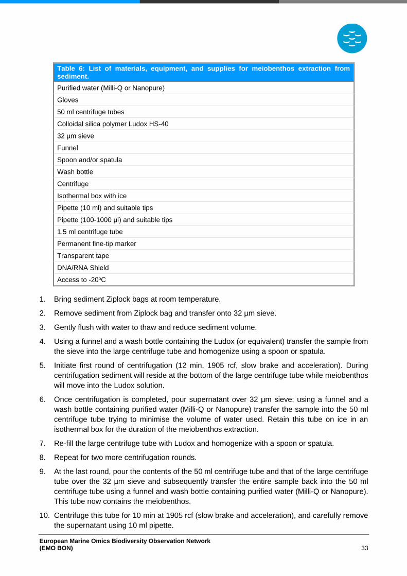
European Marine Omics Biodiversity Observation Network (EMO BON) 33
Table 6: List of materials, equipment, and supplies for meiobenthos extraction from sediment.
Purified water (Milli-Q or Nanopure)
Gloves
50 ml centrifuge tubes
Colloidal silica polymer Ludox HS-40
32 µm sieve
Funnel
Spoon and/or spatula
Wash bottle
Centrifuge
Isothermal box with ice
Pipette (10 ml) and suitable tips
Pipette (100-1000 μl) and suitable tips
1.5 ml centrifuge tube
Permanent fine-tip marker
Transparent tape
DNA/RNA Shield
Access to -20oC
1. Bring sediment Ziplock bags at room temperature.
2. Remove sediment from Ziplock bag and transfer onto 32 µm sieve.
3. Gently flush with water to thaw and reduce sediment volume.
4. Using a funnel and a wash bottle containing the Ludox (or equivalent) transfer the sample from
the sieve into the large centrifuge tube and homogenize using a spoon or spatula.
5. Initiate first round of centrifugation (12 min, 1905 rcf, slow brake and acceleration). During
centrifugation sediment will reside at the bottom of the large centrifuge tube while meiobenthos
will move into the Ludox solution.
6. Once centrifugation is completed, pour supernatant over 32 µm sieve; using a funnel and a
wash bottle containing purified water (Milli-Q or Nanopure) transfer the sample into the 50 ml
centrifuge tube trying to minimise the volume of water used. Retain this tube on ice in an
isothermal box for the duration of the meiobenthos extraction.
7. Re-fill the large centrifuge tube with Ludox and homogenize with a spoon or spatula.
8. Repeat for two more centrifugation rounds.
9. At the last round, pour the contents of the 50 ml centrifuge tube and that of the large centrifuge
tube over the 32 µm sieve and subsequently transfer the entire sample back into the 50 ml
centrifuge tube using a funnel and wash bottle containing purified water (Milli-Q or Nanopure).
This tube now contains the meiobenthos.
10. Centrifuge this tube for 10 min at 1905 rcf (slow brake and acceleration), and carefully remove
the supernatant using 10 ml pipette.
European Marine Omics Biodiversity Observation Network (EMO BON) 34
11. Resuspend the pellet containing the meiobenthos with 500 μl of DNA/RNA Shield and transfer
the sample to a 1.5 ml centrifuge tube.
12. For replicates 3 and 4 resuspend the pellet containing the meiobenthos with 500 μl of purified
water (Milli-Q or Nanopure) and transfer the sample to a 1.5 ml centrifuge tube. Replicates 3
and 4 will be stored for biobanking and DNA/RNA Shield will not be used.
13. Label the tubes appropriately using permanent marker (see Sample Labelling section).
14. Apply transparent tape firmly on top of the label to avoid bleaching.
15. Samples are now ready for DNA extraction. Store samples at -20oC until shipment.
G. Macrobenthos Sampling
1. Reach the sampling site during low tide.
2. Deploy 5 plexiglass or PVC sediment corers of at least 20 cm in diameter. Alternatively, deploy
10 plexiglass or PVC sediment corers of at least 10 cm in diameter. Deploy the different
sediment corers in proximity and ensure that the sediment surface is not visually different among
the deployment points.
3. Follow steps 3-5 above (Microbial Community Sampling), for deploying and retrieving the
corers.
4. Place the total sediment in a 1 mm sieve and sieve it (see step 4 for sequential sieving). Make
sure that the amount of remaining sediment in the sieve is minimized as much as possible.
5. Optional: Perform sequential sieving. This is recommended for stations that usually use smaller
sieve size for macrobenthos collection. Place a 1 mm sieve on top of a 0.5 mm sieve and sieve
sediment sequentially through both sieves. This way the material that flows through the 1 mm
sieve will be sequentially sieved through the 0.5 mm sieve. Make sure that the amount of
remaining sediment in both sieves is minimized as much as possible.
6. Transfer macrobenthic organisms retained in the sieve(s) in a mortar for homogenization. The
remaining sediment that may be transferred with the organisms will be removed during the DNA
extraction. Sediment homogenization may take place in the field directly after sieving. If this is
not possible, macrobenthic organisms may be collected in tubes and kept in a cool and dark
environment until transportation to the lab.
7. Use mortar and pestle to crush and homogenize organisms until a tissue mixture is obtained.
8. Transfer 10 g of the tissue mixture into a screw cup tube of 50 ml. Repeat this to obtain 4 screw
cup tubes of 50 ml each containing 10 g of the tissue mixture.
9. Fill tubes to 50 ml with DESS (see DESS recipe).
10. Label the tubes appropriately using permanent marker (see Sample Labelling section).
11. Apply transparent tape firmly on top of the label to avoid bleaching.
12. Store tubes at 4oC.
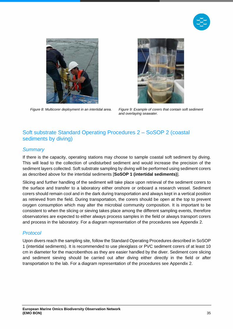
European Marine Omics Biodiversity Observation Network (EMO BON) 35
Figure 8: Multicorer deployment in an intertidal area.
Figure 9: Example of corers that contain soft sediment and overlaying seawater.
Soft substrate Standard Operating Procedures 2 ‒ SoSOP 2 (coastal sediments by diving)
Summary
If there is the capacity, operating stations may choose to sample coastal soft sediment by diving.
This will lead to the collection of undisturbed sediment and would increase the precision of the
sediment layers collected. Soft substrate sampling by diving will be performed using sediment corers
as described above for the intertidal sediments [SoSOP 1 (intertidal sediments)].
Slicing and further handling of the sediment will take place upon retrieval of the sediment corers to
the surface and transfer to a laboratory either onshore or onboard a research vessel. Sediment
corers should remain cool and in the dark during transportation and always kept in a vertical position
as retrieved from the field. During transportation, the corers should be open at the top to prevent
oxygen consumption which may alter the microbial community composition. It is important to be
consistent to when the slicing or sieving takes place among the different sampling events, therefore
observatories are expected to either always process samples in the field or always transport corers
and process in the laboratory. For a diagram representation of the procedures see Appendix 2.
Protocol
Upon divers reach the sampling site, follow the Standard Operating Procedures described in SoSOP
1 (intertidal sediments). It is recommended to use plexiglass or PVC sediment corers of at least 10
cm in diameter for the macrobenthos as they are easier handled by the diver. Sediment core slicing
and sediment sieving should be carried out after diving either directly in the field or after
transportation to the lab. For a diagram representation of the procedures see Appendix 2.
European Marine Omics Biodiversity Observation Network (EMO BON) 36
Soft substrate Standard Operating Procedures 3 ‒ SoSOP 3 (coastal sediments by research vessel)
Summary
Observatories may choose to sample coastal soft substrate onboard a research vessel. Soft
substrate will be collected using a van Veen or a Smith-McIntyre grab (0.07–0.1 m2) at the selected
site. Sediment collection by grab is suitable for different types of sediment (clay to sandy sediments).
The sediment should be brought onboard on a clean surface for subsampling. As the sediment
collected in the grab is disturbed, subsampling should be performed carefully, and materials should
not be immersed deep into the sediment to ensure the subsampling of surface sediment as much
as possible. For each community sampled, 4 replicated samples should be collected. These
replicates are designed to represent technical replicates and will be used for different purposes
(more information on the Sample Labelling section). Sediment processing will take place onboard
after collection.
In case other sediment sampling equipment is available on the research vessels, as for example a
multicorer, a box corer or a multi-box corer, the observatories may use this equipment to collect
sediment from the field. Once the sediment is brought onboard, proceed with handling the sediment
according to SoSOP 1 (intertidal sediments) if a multicorer was used, or proceed with sampling the
top sediment as described below (SoSOP 3).
Before shipment for DNA extraction and sequencing, the meiobenthic organisms should be
extracted from the sediment. After the meiobenthos extraction, the meiobenthic organisms will be
resuspended in the preservative DNA/RNA shield for shipment.
Sediment collected for macrobenthos sampling will have to be sieved through a 1 mm sieve. Some
marine areas, such as the Eastern Mediterranean, or polluted sediments are characterized by
smaller macrobenthic organisms. Using a 1 mm sieve alone would result to missing a large part of
the macrobenthos for those areas. In such cases, sequential sieving using both 1 mm and 0.5 mm
sieves will take place. The organisms retained in the 1 mm and in the 0.5 mm sieves will be handled
and stored separately as different fractions. After sieving all organisms will be crushed and
homogenized using mortar and pestle to obtain a mixture of tissues. For a diagram representation
of the procedures see Appendix 2.
Protocol
A. Materials, equipment, and supplies
Before starting, ensure all materials equipment, and supplies are available by checking the following
table (Table 7).
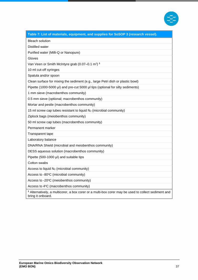
European Marine Omics Biodiversity Observation Network (EMO BON) 37
Table 7: List of materials, equipment, and supplies for SoSOP 3 (research vessel).
Bleach solution
Distilled water
Purified water (Milli-Q or Nanopure)
Gloves
Van Veen or Smith McIntyre grab (0.07–0.1 m2) 1
10 ml cut-off syringes
Spatula and/or spoon
Clean surface for mixing the sediment (e.g., large Petri dish or plastic bowl)
Pipette (1000-5000 μl) and pre-cut 5000 μl tips (optional for silty sediments)
1 mm sieve (macrobenthos community)
0.5 mm sieve (optional; macrobenthos community)
Mortar and pestle (macrobenthos community)
15 ml screw cap tubes resistant to liquid N2 (microbial community)
Ziplock bags (meiobenthos community)
50 ml screw cap tubes (macrobenthos community)
Permanent marker
Transparent tape
Laboratory balance
DNA/RNA Shield (microbial and meiobenthos community)
DESS aqueous solution (macrobenthos community)
Pipette (500-1000 μl) and suitable tips
Cotton swabs
Access to liquid N2 (microbial community)
Access to -80oC (microbial community)
Access to -20oC (meiobenthos community)
Access to 4oC (macrobenthos community)
1 Alternatively, a multicorer, a box corer or a multi-box corer may be used to collect sediment and bring it onboard.
European Marine Omics Biodiversity Observation Network (EMO BON) 38
B. Preparation and Cleaning
• Always wear gloves when handling samples dedicated to microbial community analyses.
• Prepare 10% bleach solution.
• Prepare DESS aquaous solution (see DESS recipe).
• Prepare 10 ml cut-off syringes for subsampling. Use a scalpel to cut off the luer lock end of the syringe so as the suringe is open at this end.
• Clean sediment grab, shovel, bucket and sieves using tap water. Rinse vigorously and make sure material from previous samplings is washed away.
• Desinfect plastic materials (cut-off syringes, large Petri dish and/or plastic bowl) by incubating in 10% bleach solution overnight. Rinse with purified water (Milli-Q or Nanopure) to wash the solution away and cover in aluminium foil to store.
• Desinfect spatulas, spoons, and other metal tools by briefly soaking in 10% bleach solution. Rinse with purified water (Milli-Q or Nanopure) to wash the solution away and cover in aluminium foil to store.
C. Negative Control Samples
As samples undergo different procedures at the operating stations and at the centralized sequencing
centre laboratories, it is important to check for contaminations. For this reason, blank samples will
be collected and processed together with the sediment samples to serve as negative controls. Once
the sediment is collected and before further processing, collect and store a negative control sample
as described below:
Microbial community negative control
1. Use a cotton swab on the lab bench, the spatulas, and the spoons.
2. Cut the cotton part of the swab and store it in a 15 ml screw cap tube.
3. Add 10 ml of DNA/RNA Shield by using a pipette.
4. Label the tube appropriately using permanent marker (see Sample Labelling section).
5. Apply transparent tape firmly on top of the label to avoid bleaching.
6. Transfer tube into liquid N2 for flash freezing and subsequently store at -80oC.
Meiobenthos community negative control
1. Use a cotton swab on the lab bench, the spatulas, and the spoons.
2. Cut the cotton part of the swab and store it in a Ziplock bag.
3. Label the tube appropriately using permanent marker (see Sample Labelling section).
4. Apply transparent tape firmly on top of the label to avoid bleaching.
5. Store the bag at -20oC.
6. Process the negative control sample together with the sediment samples during the
meiobenthos extraction from sediment.
Macrobenthos community negative control
European Marine Omics Biodiversity Observation Network (EMO BON) 39
1. Use a cotton swab on the spatulas, the spoons, the sieves and the mortar and pestle.
2. Cut the cotton part of the swab and store it in a 50 ml screw cap tube.
3. Add 20 ml of DESS (see DESS recipe).
4. Label the tube appropriately using permanent marker (see Sample Labelling section).
5. Apply transparent tape firmly on top of the label to avoid bleaching.
6. Store at 4oC.
D. Microbial Community Sampling
1. Collect soft sediment using a van Veen or a Smith-McIntyre grab (0.07–0.1 m2) equipped on
the research vessel (Figure 10).
2. Bring sediment onboard on a clean surface for subsampling. In case a subsampling port is
present at the top of the grab, perform subsampling from that port before the grab is opened.
3. Use 10 ml cut-off syringes to subsample for the sediment microbial community. Collect about
10 times a rather full 10 ml cut-off syringe from the same grab.
4. Transfer the sediment collected in the syringes on a clean surface (e.g., Petri dish or plastic
bowl) and homogenize gently using a spoon or a spatula.
5. Pre-weigh a 15 ml screw cap tube and using a spoon and spatula transfer 10 g of sediment in
the tube. An alternative, though less accurate, method would be to fill the 15 ml graduated screw
cap tubes to approximately 10 ml tube indication. In the case of silty sediment, pre-cut 5000 μl
tips might be used to transfer the sediment in the screw cap tubes by careful pipetting. Make
sure that the 15 ml screw cap tubes used are resistant to liquid N2.
6. In total, fill 4 screw cap tubes with the homogenized sediment.
7. Add 10 ml of DNA/RNA Shield by using a pipette and make sure the sediment is covered by the
solution. For the third and fourth tube of replicate samples skip this step. Replicates 3 and 4 will
be stored for biobanking and DNA/RNA Shield will not be used.
8. Label the tube appropriately using permanent marker (see Sample Labelling section).
9. Apply transparent tape firmly on top of the label to avoid bleaching.
10. Add 5 ml of Purified water (Milli-Q or Nanopure) in a 15 ml screw cap tube. Briefly dip a clean
spatula in the tube (clean and disinfected as described in the Preparation and Cleaning
11. section above). Add 10 ml of DNA/RNA Shield by using a pipette. This tube will be used as
negative control sample.
12. Label the tube appropriately using permanent marker (see Sample Labelling section).
13. Apply transparent tape firmly on top of the label to avoid bleaching.
14. Transfer tubes into liquid N2 for flash freezing and subsequently store into -80oC.
E. Meiobenthos Community Sampling
1. Collect soft sediment using a van Veen or a Smith-McIntyre grab (0.07–0.1 m2) equipped at the
research vessel (Figure 10).
European Marine Omics Biodiversity Observation Network (EMO BON) 40
2. Bring sediment onboard on a clean surface for subsampling.
3. Use a shovel to transfer approximately half of the grabbed sediment in a clean bucket.
4. Gently homogenize the sediment in the bucket by stirring it with a large spoon or spatula.
5. Deploy a plexiglass or PVC sediment corer of 3.6 cm in diameter into the sediment.
6. Transfer the sediment into a plastic Ziplock bag and weigh it. Flatten the bag before placing it
into the freezer to allow stacking of samples.
7. Repeat steps 5-6 to collect 4 subsamples in total.
8. Label the Ziplock bags appropriately (see Sample Labelling section).
9. Store the bags at -20oC. Sediment can be stored at -20oC safely for at least 2 years until
meiobenthos extraction.
F. Meiobenthos Extraction from Sediment
Extract meiobenthos from the sampled sediment into purified water (Milli-Q or Nanopure) by density-
gradient centrifugation with the colloidal silica polymer Ludox HS-40 as a flotation medium, or
equivalent (specific density 1.18 g cm−1). The meiobenthos extraction from sediment may take
place any time after sampling and within 2 years. Before starting, ensure all materials equipment,
and supplies are available by checking the following table (Table 8).
Table 8: List of materials, equipment, and supplies for meiobenthos extraction from sediment.
Purified water (Milli-Q or Nanopure)
Gloves
50 ml centrifuge tubes
Colloidal silica polymer Ludox HS-40
32 µm sieve
Funnel
Spoon and/or spatula
Wash bottle
Centrifuge
Isothermal box and ice
Pipette (10 ml) and suitable tips
Pipette (100-1000 μl) and suitable tips
1.5 ml centrifuge tube
Permanent fine-tip marker
Transparent tape
DNA/RNA Shield
Access to -20oC
1. Bring sediment Ziplock bags at room temperature.
2. Remove sediment from Ziplock bag and transfer onto 32 µm sieve.
European Marine Omics Biodiversity Observation Network (EMO BON) 41
3. Gently flush with water to thaw and reduce sediment volume.
4. Using a funnel and a wash bottle containing the Ludox (or equivalent) transfer the sample from
the sieve into the large centrifuge tube and homogenize using a spoon or spatula.
5. Initiate first round of centrifugation (12 min, 1905 rcf, slow brake and acceleration). During
centrifugation sediment will reside at the bottom of the large centrifuge tube while meiobenthos
will move into the Ludox solution.
6. Once centrifugation is completed, pour supernatant over 32 µm sieve; using a funnel and a
wash bottle containing purified water (Milli-Q or Nanopure) transfer the sample into the 50 ml
centrifuge tube trying to minimise the volume of water used. Retain this tube on ice in an
isothermal box for the duration of the meiobenthos extraction.
7. Re-fill the large centrifuge tube with Ludox and homogenize with a spoon or spatula.
8. Repeat for two more centrifugation rounds.
9. At the last round, pour the contents of the 50 ml centrifuge tube and that of the large centrifuge
tube over the 32 µm sieve and subsequently transfer the entire sample back into the 50 ml
centrifuge tube using a funnel and squeezing bottle containing purified water (Milli-Q or
Nanopure). This tube now contains the meiobenthos.
10. Centrifuge this tube for 10 min at 1905 rcf (slow brake and acceleration), and carefully remove
the supernatant using 10 ml pipette.
11. Resuspend the pellet containing the meiobenthos with 500 μl of DNA/RNA Shield and transfer
the sample to a 1.5 ml centrifuge tube.
12. For replicates 3 and 4 resuspend the pellet containing the meiobenthos with 500 μl of purified
water (Milli-Q or Nanopure) and transfer the sample to a 1.5 ml centrifuge tube. Replicates 3
and 4 will be stored for biobanking and DNA/RNA Shield will not be used.
13. Label the tubes appropriately using permanent marker (see Sample Labelling section).
14. Apply transparent tape firmly on top of the label to avoid bleaching.
15. Samples are now ready for DNA extraction. Store samples at -20oC until shipment.
G. Macrobenthos Sampling
1. Collect soft sediment using a van Veen or a Smith-McIntyre grab (0.07–0.1 m2) equipped at the
research vessel (Figure 10).
2. Bring sediment onboard and place it in a 1 mm sieve (see step 4 for sequential sieving).
3. Sieve sediment. Make sure that the amount of remaining sediment in the sieve is minimized as
much as possible.
4. Optional: Perform sequential sieving. This is recommended for stations that usually use smaller
sieve size for macrobenthos collection. Place a 1 mm sieve on top of a 0.5 mm sieve and sieve
sediment sequentially through both sieves. This way the material that flows through the 1 mm
sieve will be sequentially sieved through the 0.5 mm sieve. Make sure that the amount of
remaining sediment in both sieves is minimized as much as possible.
5. Transfer macrobenthic organisms retained in the sieve(s) in a mortar for homogenization. The
remaining sediment that may be transferred with the organisms will be removed during the DNA
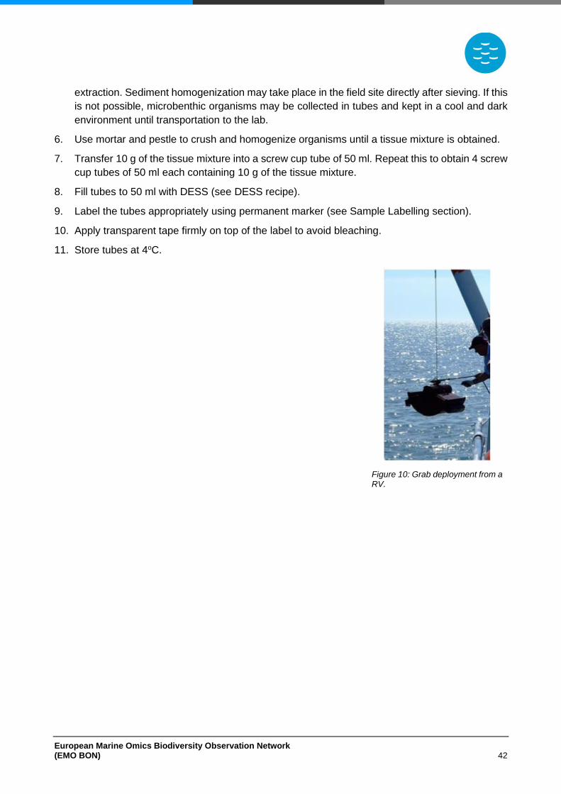
European Marine Omics Biodiversity Observation Network (EMO BON) 42
extraction. Sediment homogenization may take place in the field site directly after sieving. If this
is not possible, microbenthic organisms may be collected in tubes and kept in a cool and dark
environment until transportation to the lab.
6. Use mortar and pestle to crush and homogenize organisms until a tissue mixture is obtained.
7. Transfer 10 g of the tissue mixture into a screw cup tube of 50 ml. Repeat this to obtain 4 screw
cup tubes of 50 ml each containing 10 g of the tissue mixture.
8. Fill tubes to 50 ml with DESS (see DESS recipe).
9. Label the tubes appropriately using permanent marker (see Sample Labelling section).
10. Apply transparent tape firmly on top of the label to avoid bleaching.
11. Store tubes at 4oC.
Figure 10: Grab deployment from a RV.
European Marine Omics Biodiversity Observation Network (EMO BON) 43
Hard Substrate
Hard Substrate Standard Operating Procedures ‒ HaSOP
Summary
The passive sampling of hard substrate communities will take place using ARMS units. More details
on purchasing ARMS units, deployment and sample collection are available from the ARMS-MBON
(http://www.arms-mbon.eu/).
Different sampling strategies may be followed while setting up an ARMS observatory. Here are some
strategies that have been implemented in past:
1. Short-term surveys that aim at seasonal investigations and monitoring of alien species. ARMS
are deployed for a period of 2-3 months usually during spring to summer season.
2. Long-term surveys that aim at continuous biological monitoring and ecological research. ARMS
are deployed for a period of 12 months and at the time of retrieval the next ARMS unit is
deployed.
3. Survey on community succession of macro-organisms. In this case 3 or more ARMS units are
deployed on the same date and retrieved sequentially (i.e., 1 ARMS unit retrieved every 4
months) to investigate the succession of settled organisms over time.
Participating stations may opt for one of the sampling strategies or for a different sampling design
depending on their scientific interests. However, if an observatory station has not performed ARMS
deployment in the past, it is recommended to contact one of the current ARMS-MBON observatory
stations to discuss possible sampling strategies. The sampling strategies usually reflect the internal
research needs of the observatory stations.
Deployment and retrieval of ARMS in EMO BON will follow the standards and protocols of the
Smithsonian Institution (https://www.oceanarms.org/) and the additional guidelines of ARMS-MBON
(http://www.arms-mbon.eu/). Selecting the location of the ARMS observatory is of great importance
both for short-term and for long-term surveys. EMO BON recommends, if possible, that ARMS
sampling sites are located near the water column and/or soft substrate sampling sites, as well as
near long term monitoring sites. More information on the ARMS locations can be found in the ARMS-
MBON Handbook (http://www.arms-mbon.eu/). Deploying more than one ARMS unit, will give a
better representation of the biodiversity in the area. It is recommended that three ARMS units per
sampling site are deployed in close proximity.
Sample processing after retrieval will follow the standards and protocols of the Smithsonian
Institution (https://www.oceanarms.org/) and the additional guidelines of ARMS-MBON
(http://www.arms-mbon.eu/); here only a brief description of the procedures may be found. After
ARMS retrieval, units should be transferred in the laboratory where the disassembly and
photography of ARMS plates will take place. Using scalpel and tweezers, sessile organisms are
collected from each ARMS plate and placed in a collection tube. Motile fractions should be recovered
from the ARMS bin using a 2 mm sieve on top of a 0.5 mm on top of a 0.1 mm. Seawater and
sediment in the ARMS bin should gently pass sequentially through the three sieves (2 mm, 0.5 mm,
and 0.1 mm) and the bin should be rinsed thoroughly to ensure the collection of all organisms. The
material collected on the 0.5 mm sieve may be concentrated using a 40 μm nytex mesh. The material
European Marine Omics Biodiversity Observation Network (EMO BON) 44
collected in the sieves should be photographed prior to storage. All sievings should be carefully
performed to avoid overflow.
In addition to the sessile fraction retrieved by plate scraping, each sieve will collect a different fraction
of the motile organisms: a) motile fraction sieved with 2 mm (organisms >2 mm), b) motile fraction
sieved with 0.5 mm (2 mm > organisms > 0.5 mm), and c) motile fraction sieved with 0.1 mm (0.5
mm > organisms > 0.1 mm). The material in motile fraction a (organisms >2 mm) will be processed
only for optical identification by photographing the individual specimens. The sessile together with
the motile fractions b (2 mm > organisms > 0.5 mm) and c (0.5 mm > organisms > 0.1 mm) will be
homogenized and processed for DNA extraction and sequencing.
All tissues and material to be processed for sequencing (sessile fraction, b and c motile fractions)
should be homogenized. The sessile fraction homogenization is a straightforward procedure
performed using a blender. The motile fraction homogenization requires washing steps, and the
grinding is performed using mortar and pestle. The procedures are described in detail by the
Smithsonian Institution (https://www.oceanarms.org/). After thorough homogenization, the mixtures
are stored in DESS at 4oC.
At least 4 technical replicates should be collected from each sampling site, therefore material from
the sessile fraction and the motile fractions should be stored in 4 individual tubes. In case the
material is not enough to collect 4 technical replicates, collect as many as possible; make sure to
indicate the number of collected replicates in the metadata. In many cases, biomass of a few species
may dominate the community in ARMS. This may cause biases during DNA amplification and
sequencing and to decrease this bias it is recommended to reduce the amount of biomass included
during tissue homogenization (http://www.arms-mbon.eu/).
Protocol
All procedures regarding ARMS assembly, deployment, retrieval, disassembly, and sample
processing are described in detail by the Smithsonian Institution (https://www.oceanarms.org/).
Before starting, ensure all materials equipment, and supplies are available by checking the following
table (Table 9).
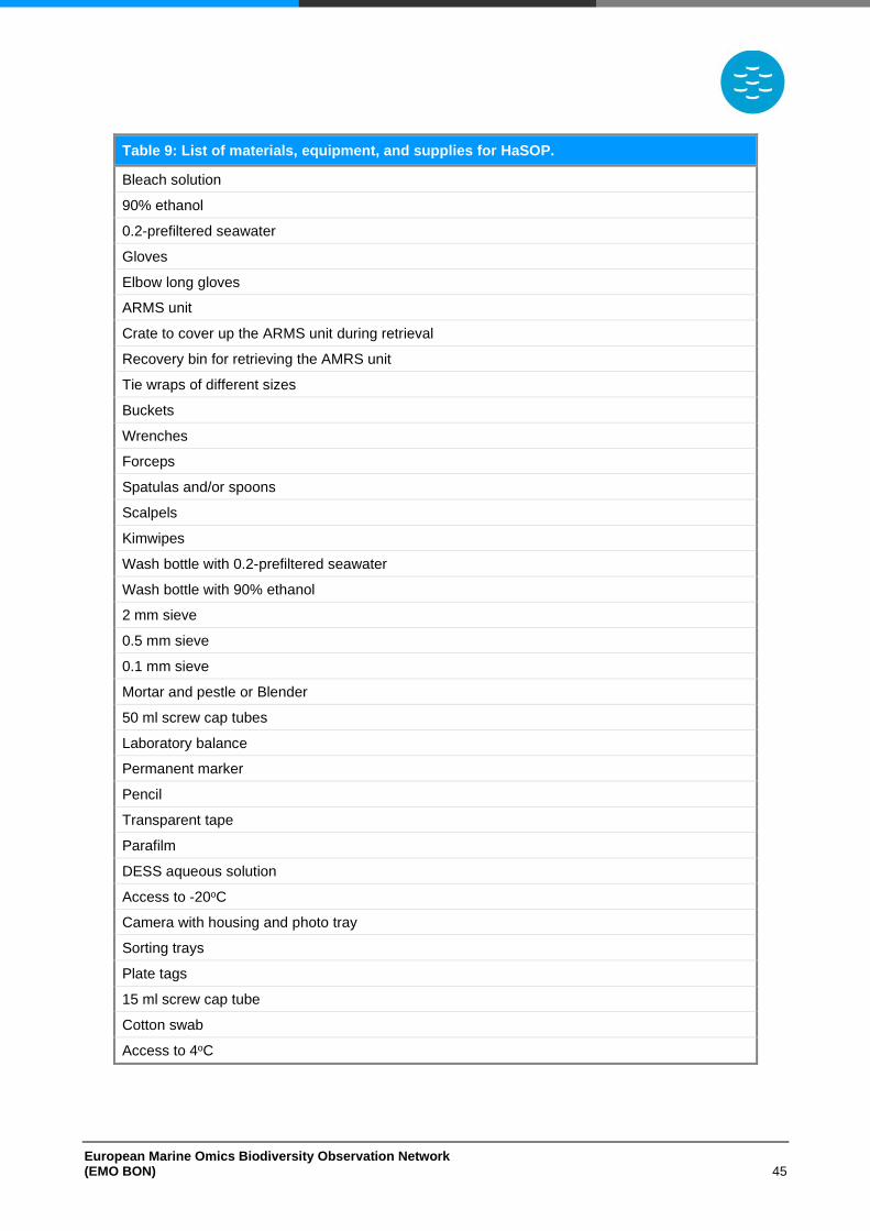
European Marine Omics Biodiversity Observation Network (EMO BON) 45
Table 9: List of materials, equipment, and supplies for HaSOP.
Bleach solution
90% ethanol
0.2-prefiltered seawater
Gloves
Elbow long gloves
ARMS unit
Crate to cover up the ARMS unit during retrieval
Recovery bin for retrieving the AMRS unit
Tie wraps of different sizes
Buckets
Wrenches
Forceps
Spatulas and/or spoons
Scalpels
Kimwipes
Wash bottle with 0.2-prefiltered seawater
Wash bottle with 90% ethanol
2 mm sieve
0.5 mm sieve
0.1 mm sieve
Mortar and pestle or Blender
50 ml screw cap tubes
Laboratory balance
Permanent marker
Pencil
Transparent tape
Parafilm
DESS aqueous solution
Access to -20oC
Camera with housing and photo tray
Sorting trays
Plate tags
15 ml screw cap tube
Cotton swab
Access to 4oC
European Marine Omics Biodiversity Observation Network (EMO BON) 46
Negative control sample
As samples undergo different procedures at the operating stations and at the centralized sequencing
centre laboratories, it is important to check for contaminations. For this reason, blank samples will
be collected and processed together with the ARMS samples to serve as negative controls. Once
the ARMS unit is brought in the laboratory and before samples processing, collect and store a
negative control sample as described below:
1. Use a cotton swab on the lab bench, the spatulas, the spoons, and the sieves.
2. Cut the cotton part of the swab and store it in a 15 ml screw cap tube.
3. Add 10 ml of DESS.
4. Label the tube appropriately using permanent marker (see Sample Labelling section).
5. Apply transparent tape firmly on top of the label to avoid bleaching.
6. Store at 4oC.
European Marine Omics Biodiversity Observation Network (EMO BON) 47
Sample Labelling
Correct labelling is of extreme importance when collecting samples and especially for a network of
more than ten operational stations running in the long-term. Proper labelling will ensure that samples
collected are organized and processed appropriately, that data and metadata produced in EMO
BON will be properly catalogued and analysed in the future, and that the data produced can be
correctly assigned to the sampling sites.
Participating institutions should decide on a name and a short ID for their observatory that will be
maintained for the duration of EMO BON and will characterize all samples collected at this
observatory. The name and ID of the observatory should be unique whether an observatory station
samples for all three habitats (Wa: Water column, So: Soft substrate, Ha: Hard substrate) or for just
one. For example, if “SMN99” is sampling water column 1 mile offshore and hard substrates in a
marina, the observatory ID should be “SMN99” for both water column and hard substrate samplings.
The different sampling sites of each observatory will be distinguished by the suffix “Wa” for water
column, “So” for soft substrates and “Ha” for hard substrates. Therefore, if observatory “SMN99” is
sampling all three habitats, it will include three different sites: “SMN99 Wa” (water column sampling
site), “SMN99 So” (soft substrate sampling site) and “SMN99 Ha” (hard bottom sampling site).
Five terms will constitute the label of each sample:
1. Project (“EMO BON”)
2. Sampling Site ID; that is the Observatory ID (for example “SMN99”) supplemented with the
sampling site indicator (“Wa” for water column, “So” for soft substrates and “Ha” for hard
substrates)
3. Sampling Campaign Date formated as YYMMDD.For example “220315” would be the campaign
on 15 March 2022. Especially for Hard substrates, both the deployment and retrieval dates have
to be recorded formated as YYMMDD-YYMMDD.
4. Size fraction (Wa: “3 μm” / “0.2 μm” / “20 μm” / “200 μm”) or organisms collected (So: “micro” for
microorganisms, “meio” for meiobenthos and “macro” for macrobenthos) or ARMS fraction (Ha:
“SF” for sessile fraction, “MF05” for motile fraction sieved through 0.5 mm, “MF01” for motile
fraction sieved through 0.1 mm)
5. Replicate (number 1-4). For labelling the negative control, this term will be replaced by the
notation “blank” in parenthesis.
A few examples of sample labelling may be found in Table 10. It is recommended to avoid hyphens,
commas, underscores and to separate easily confused digits (for example 1 and 7, 0 and O). The
combination of these terms will be unique for each individual sample within EMO BON. This unique
Sample ID created from the combination of those terms will follow the sample and the generated
data and metadata during all procedures and analyses.
Permanent markers should be used for labelling the tubes and Ziplock bags and transparent tape
should be applied firmly on top of the label to avoid bleaching. Samples that are not properly labelled
will be discarded prior to DNA extraction.
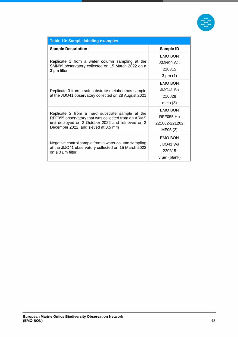
European Marine Omics Biodiversity Observation Network (EMO BON) 48
Table 10: Sample labeling examples
Sample Description Sample ID
Replicate 1 from a water column sampling at the SMN99 observatory collected on 15 March 2022 on a 3 μm filter
EMO BON
SMN99 Wa
220315
3 μm (1)
Replicate 3 from a soft substrate meiobenthos sample at the JIJO41 observatory collected on 28 August 2021
EMO BON
JIJO41 So
210828
meio (3)
Replicate 2 from a hard substrate sample at the RFF055 observatory that was collected from an ARMS unit deployed on 2 October 2022 and retrieved on 2 December 2022, and sieved at 0.5 mm
EMO BON
RFF055 Ha
221002-221202
MF05 (2)
Negative control sample from a water column sampling at the JIJO41 observatory collected on 15 March 2022 on a 3 μm filter
EMO BON
JIJO41 Wa
220315
3 μm (blank)
European Marine Omics Biodiversity Observation Network (EMO BON) 49
Collection and Documentation of Metadata
The collection of rich and detailed metadata is of great importance for the longstanding life cycle of
the data collected in EMOBON. Metadata are necessary in all the principles of FAIR data
management. To assure the correct data FAIRification, metadata should be considered even before
the collection of the biodivarsity data. EMO BON will adhere to the FAIR data principles for all data
generated within the network; therefore, the collection of metadata during sampling and laboratory
procedure is essential and mandatory. More details on the data and metadata life cycle will be
provided in the EMO BON Data Management Plan (DMP). EMO BON DMP will describe in detail
the handling of the data and their processing towards FAIRification.
Observatories will collect and document all information related to the data from sampling to
sequencing and quality control. There will be 5 categories of metadata that will adhere to the data:
1. Observatory Metadata
Here, all information regarding the observatory station, the sampling sites, their characteristics,
and the contact people will be included. This information will be provided with the registration form
that will confirm participation to EMO BON (see also Participation to EMO BON
section and the Registration Form document). Observatory Metadata refer to the observatory and
will not change among sampling events. Observatory Metadata will be digitalized and submitted as
described in the DMP.
2. Sampling Metadata
All information related to each sampling event will be documented as Sampling Metadata. This will
include date and time of sampling, sample handling details, deviation from protocols and other
characteristics of the sampling events. These metadata will be recorded in log sheet provided by
EMO BON during each sampling event and will be digitalized and submitted as described in the
DMP.
3. Measured Metadata
Observatories will have to measure additional variables that will make the sequencing dataset
relevant to its ecological context. Some variables are characterized as mandatory and are to be
measured during every sampling event. Observatories may choose to additionally measure any of
the recommended variables. The more environmental variables connect to the dataset, the more
relevant and reusable the dataset will be. Information regarding the sampling and laboratory
methodologies of the measured variables will be documented here. EMO BON will not give specific
instructions for the methodologies of the environmental variables measured, as observatory stations
are already equipped with specific instruments and follow specific procedures for their sampling
sites. However, it is important that the same procedure is followed for each variable for all EMO
BON samplings and the procedure is adequately described. Environmental variables are listed in
Table 11 for each habitat (Water column, Soft substrate, Hard substrate). The DMP will describe all
procedures to handle Measured Metadata after documentation.
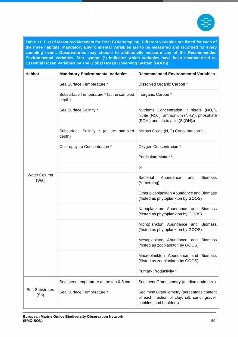
European Marine Omics Biodiversity Observation Network (EMO BON) 50
Table 11: List of Measured Metadata for EMO BON sampling. Different variables are listed for each of
the three habitats. Mandatory Environmental Variables are to be measured and recorded for every
sampling event. Observatories may choose to additionally measure any of the Recommended
Environmental Variables. Star symbol (*) indicates which variables have been characterized as
Essential Ocean Variables by The Global Ocean Observing System (GOOS).
Habitat Mandatory Environmental Variables Recommended Environmental Variables
Water Column
(Wa)
Sea Surface Temperature * Dissolved Organic Carbon *
Subsurface Temperature * (at the sampled
depth)
Inorganic Carbon *
Sea Surface Salinity * Nutrients Concentration *: nitrate (NO3-),
nitrite (NO2-), ammonium (NH4
+), phosphate
(PO43-) and silicic acid (Si(OH)4)
Subsurface Salinity * (at the sampled
depth)
Nitrous Oxide (N2O) Concentration *
Chlorophyll-a Concentration * Oxygen Concentration *
Particulate Matter *
pH
Bacterial Abundance and Biomass
(*emerging)
Other picoplankton Abundance and Biomass
(*listed as phytoplankton by GOOS)
Nanoplankton Abundance and Biomass
(*listed as phytoplankton by GOOS)
Microplankton Abundance and Biomass
(*listed as phytoplankton by GOOS)
Mesoplankton Abundance and Biomass
(*listed as zooplankton by GOOS)
Macroplankton Abundance and Biomass
(*listed as zooplankton by GOOS)
Primary Productivity *
Soft Substrates
(So)
Sediment temperature at the top 0-5 cm Sediment Granulometry (median grain size)
Sea Surface Temperature * Sediment Granulometry (percentage content
of each fraction of clay, silt, sand, gravel,
cobbles, and boulders)
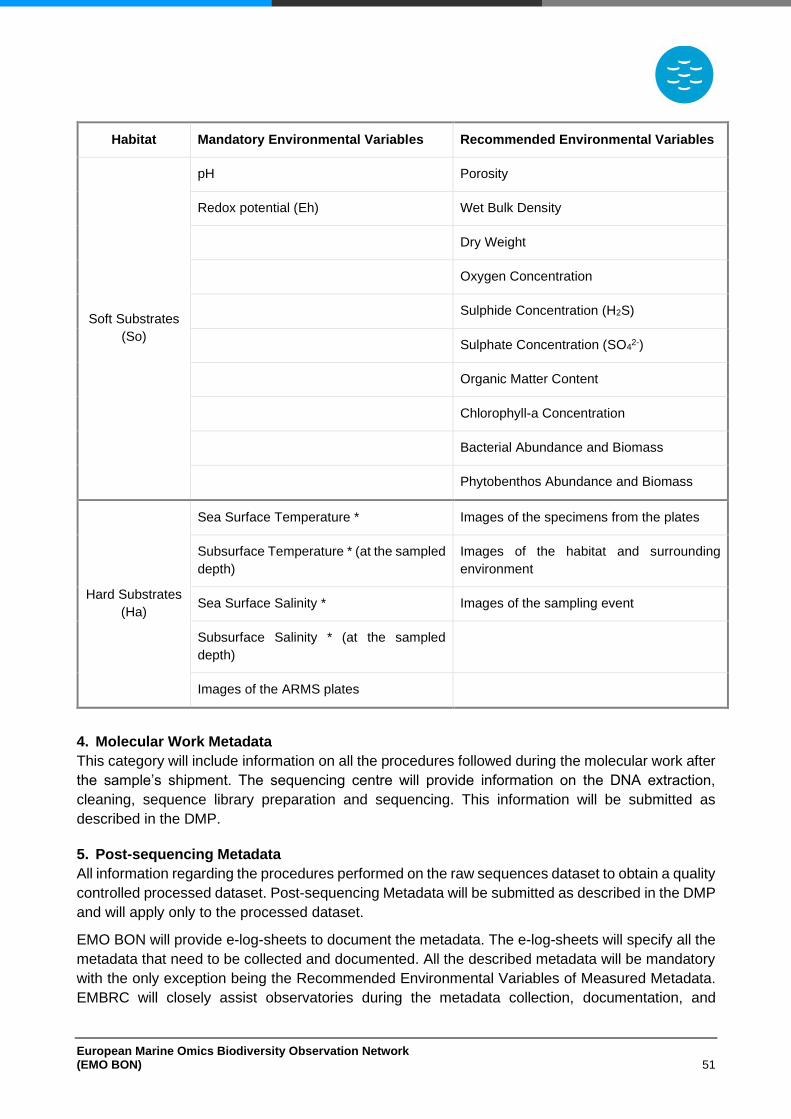
European Marine Omics Biodiversity Observation Network (EMO BON) 51
Habitat Mandatory Environmental Variables Recommended Environmental Variables
Soft Substrates
(So)
pH Porosity
Redox potential (Eh) Wet Bulk Density
Dry Weight
Oxygen Concentration
Sulphide Concentration (H2S)
Sulphate Concentration (SO42-)
Organic Matter Content
Chlorophyll-a Concentration
Bacterial Abundance and Biomass
Phytobenthos Abundance and Biomass
Hard Substrates
(Ha)
Sea Surface Temperature * Images of the specimens from the plates
Subsurface Temperature * (at the sampled
depth)
Images of the habitat and surrounding
environment
Sea Surface Salinity * Images of the sampling event
Subsurface Salinity * (at the sampled
depth)
Images of the ARMS plates
4. Molecular Work Metadata
This category will include information on all the procedures followed during the molecular work after
the sample’s shipment. The sequencing centre will provide information on the DNA extraction,
cleaning, sequence library preparation and sequencing. This information will be submitted as
described in the DMP.
5. Post-sequencing Metadata
All information regarding the procedures performed on the raw sequences dataset to obtain a quality
controlled processed dataset. Post-sequencing Metadata will be submitted as described in the DMP
and will apply only to the processed dataset.
EMO BON will provide e-log-sheets to document the metadata. The e-log-sheets will specify all the
metadata that need to be collected and documented. All the described metadata will be mandatory
with the only exception being the Recommended Environmental Variables of Measured Metadata.
EMBRC will closely assist observatories during the metadata collection, documentation, and
European Marine Omics Biodiversity Observation Network (EMO BON) 52
submission. Samples for which metadata are incomplete or improperly documented will be rejected
from the observatory network.
European Marine Omics Biodiversity Observation Network (EMO BON) 53
DNA extraction
DNA extraction will be performed at a centralized facility to minimize biases. It is important that all
EMO BON samples are processed in the same laboratory, under the same conditions and using the
same equipment to minimise operator bias in the study. As the laboratory analyses of samples will
take place at different time intervals, it is essential that the procedures followed are as standardized
as possible. DNA extraction will be performed using commercialized kits, as the standardized
solutions of the kits will minimize extraction deviations among samples.
For DNA extraction of the water column filter samples the NucleoSpin Plant II Mini kit (Macherey-
Nagel) will be used. This kit has been previously tested by EMBRC partners and was found to
perform well for DNA extraction from filters.
DNA extraction from sediment samples will be performed using the commercially available DNeasy
PowerSoil Pro kit (Qiagen). This kit has been tested multiple times for sediment DNA extraction by
EMBRC partners and has been found to result in high yields. During EMO BON preparation phase,
the DNeasy PowerSoil Pro kit was tested for DNA extraction from meiobenthos samples (Soft
Substrates) and results indicated that the kit performs well and can be used within EMO BON. The
DNeasy PowerSoil Pro kit will also be used for DNA extraction from macrobenthos tissues (Soft
Substrate) and the tissue mixtures from the Hard Substrates. This kit was used for DNA extraction
from tissues during ARMS-MBON.
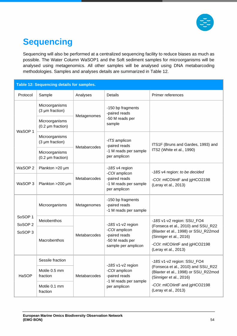
European Marine Omics Biodiversity Observation Network (EMO BON) 54
Sequencing
Sequencing will also be performed at a centralized sequencing facility to reduce biases as much as
possible. The Water Column WaSOP1 and the Soft sediment samples for microorganisms will be
analysed using metagenomics. All other samples will be analysed using DNA metabarcoding
methodologies. Samples and analyses details are summarized in Table 12.
Table 12: Sequencing details for samples.
Protocol Sample Analyses Details Primer references
WaSOP 1
Microorganisms
(3 μm fraction)
Metagenomes
-150 bp fragments
-paired reads
-50 M reads per
sample
Microorganisms
(0.2 μm fraction)
Microorganisms
(3 μm fraction)
Metabarcodes
-ITS amplicon
-paired reads
-1 M reads per sample
per amplicon
ITS1F (Bruns and Gardes, 1993) and
ITS2 (White et al., 1990) Microorganisms
(0.2 μm fraction)
WaSOP 2 Plankton >20 μm
Metabarcodes
-18S v4 region
-COI amplicon
-paired reads
-1 M reads per sample
per amplicon
-18S v4 region: to be decided
-COI: mlCOIintF and jgHCO2198
(Leray et al., 2013) WaSOP 3 Plankton >200 μm
SoSOP 1
SoSOP 2
SoSOP 3
Microorganisms Metagenomes
-150 bp fragments
-paired reads
-1 M reads per sample
Meiobenthos
Metabarcodes
-18S v1-v2 region
-COI amplicon
-paired reads
-50 M reads per
sample per amplicon
-18S v1-v2 region: SSU_FO4
(Fonseca et al., 2010) and SSU_R22
(Blaxter et al., 1998) or SSU_R22mod
(Sinniger et al., 2016)
-COI: mlCOIintF and jgHCO2198
(Leray et al., 2013)
Macrobenthos
HaSOP
Sessile fraction
Metabarcodes
-18S v1-v2 region
-COI amplicon
-paired reads
-1 M reads per sample
per amplicon
-18S v1-v2 region: SSU_FO4
(Fonseca et al., 2010) and SSU_R22
(Blaxter et al., 1998) or SSU_R22mod
(Sinniger et al., 2016)
-COI: mlCOIintF and jgHCO2198
(Leray et al., 2013)
Motile 0.5 mm
fraction
Motile 0.1 mm
fraction
European Marine Omics Biodiversity Observation Network (EMO BON) 55
Replication and Biobanking
The replicates collected within EMO BON are technical replicates, not true statistical replicates. They
represent identical samples and are to be used for different purposes. All 4 replicates per sample
are collected following the exact same procedures as described in the Sampling Protocols section.
Two of the replicates will be processed for the extraction of DNA and subsequently sequencing. The
replication of processed samples will allow the recognition of false or accidental laboratory practices
and ensure the highest data quality. Additionally, 1 replicate per sample will be provided as a service
from EMBRC to external users. The purpose of this service provision is to encourage the
collaboration among marine institutions, to give the chance for biodiversity information comparisons
across the globe and to allow samples to be analysed using different methodologies and under
alternative foci.
EMO BON aims to be part of global research effort in marine biodiversity and to interact with other
scientific entities and disciplines. It will contribute to the UN Decade of the Ocean not only by
producing accurate and complete time-series data on marine biodiversity but also by preserving
material samples for re-analyses at the end of the decade or beyond. Particularly, one replicate will
be collected and biobanked for decadal storage. Decadal samples will be stored at -80oC organized
in containers dedicated only to decadal preservation. All marine operating stations are required to
keep detailed records of the preserved samples.
European Marine Omics Biodiversity Observation Network (EMO BON) 56
Calibration efforts
The cross-validation of the generated data and the calibration of methods is of high importance for
EMO BON. Data produced within EMO BON will be a contribution to the United Nations Decade of
Ocean Science for Sustainable Development and to the marine science community. As data will
follow FAIR principles, will be reusable and will help reply to different scientific questions over the
years. Data may be analysed several times during the coming decade as different scientists may
access them. Therefore, it is essential that the quality of the data produced is high and that there is
the capacity to validate the sequencing results in various ways.
For this reason, during all samplings two of the technical replicates collected will be processed. DNA
extraction and sequencing of the replicates will take place under the same conditions and all followed
procedures will be the same. Results of the two processed replicated will be integrated to produce
final quality control datasets. In addition, a mock community comprised of eukaryotic
microorganisms and a mock community comprised of bacteria will be created at EMBRC culture
collections. The mock communities will be processed together with the samples and serve as
positive controls. As the diversity of the mock communities will be known, the sequencing results
will be validated by examining the sequencing reads of the mock communities
During sampling blank samples will be collected to serve as negative controls during DNA extraction
and sequencing. This will help detect contamination during sampling or laboratory processing. In
case a contamination is detected, it will be possible to go back to the sampling and processing of
samples and revise the procedures. Furthermore, the sequencing reads of the blank samples will
allow for corrections during the final dataset refining.
A sampling calibration exercise is planned to take place during EMO BON. The personnel
responsible for sampling will be exchanged among observatories. Personnel will participate in a
sampling event at one or more different observatories. The idea behind this is to standardize the
methods between EMO BON operating stations and to exchange expertise. In combination to the
training workshops planned, the sampling methods used in EMO BON will be harmonized among
the different observatories.
European Marine Omics Biodiversity Observation Network (EMO BON) 57
References
Antão, L. H., Pöyry, J., Leinonen, R., and Roslin, T. (2020). Contrasting latitudinal patterns in diversity and stability in a high-latitude species-rich moth community. Glob. Ecol. Biogeogr. 29, 896–907. doi:10.1111/geb.13073.
Blaxter, M. L., De Ley, P., Garey, J. R., Llu, L. X., Scheldeman, P., Vierstraete, A., et al. (1998). A molecular evolutionary framework for the phylum Nematoda. Nature 392, 71–75. doi:10.1038/32160.
Bruns, T. D., and Gardes, M. (1993). Molecular tools for the identification of ectomycorrhizal fungi — taxon‐specific oligonucleotide probes for suilloid fungi. Mol. Ecol. 2, 233–242. doi:10.1111/j.1365-294X.1993.tb00013.x.
Claudet, J., Bopp, L., Cheung, W. W. L., Devillers, R., Escobar-Briones, E., Haugan, P., et al. (2020). A Roadmap for Using the UN Decade of Ocean Science for Sustainable Development in Support of Science, Policy, and Action. One Earth 2, 34–42. doi:10.1016/j.oneear.2019.10.012.
Fonseca, V. G., Carvalho, G. R., Sung, W., Johnson, H. F., Power, D. M., Neill, S. P., et al. (2010). Second-generation environmental sequencing unmasks marine metazoan biodiversity. Nat. Commun. 1, 1–8. doi:10.1038/ncomms1095.
Heymans, J. J., Bundy, A., Christensen, V., Coll, M., de Mutsert, K., Fulton, E. A., et al. (2020). The Ocean Decade: A True Ecosystem Modeling Challenge. Front. Mar. Sci. 7, 766. doi:10.3389/fmars.2020.554573.
Leray, M., Yang, J. Y., Meyer, C. P., Mills, S. C., Agudelo, N., Ranwez, V., et al. (2013). A new versatile primer set targeting a short fragment of the mitochondrial COI region for metabarcoding metazoan diversity: Application for characterizing coral reef fish gut contents. Front. Zool. 10, 1–14. doi:10.1186/1742-9994-10-34.
Muller-Karger, F. E., Miloslavich, P., Bax, N. J., Simmons, S., Costello, M. J., Pinto, I. S., et al. (2018). Advancing marine biological observations and data requirements of the complementary Essential Ocean Variables (EOVs) and Essential Biodiversity Variables (EBVs) frameworks. Front. Mar. Sci. 5, 25. doi:10.3389/fmars.2018.00211.
Obst, M., Exter, K., Allcock, A. L., Arvanitidis, C., Axberg, A., Bustamante, M., et al. (2020). A Marine Biodiversity Observation Network for Genetic Monitoring of Hard-Bottom Communities (ARMS-MBON). Front. Mar. Sci. 7, 572680. doi:10.3389/fmars.2020.572680.
Seutin, G., White, B. N., and Boag, P. T. (1991). Preservation of avian blood and tissue samples for DNA analyses. Can. J. Zool. 69, 82–90. doi:10.1139/z91-013.
Sinniger, F., Pawlowski, J., Harii, S., Gooday, A. J., Yamamoto, H., Chevaldonné, P., et al. (2016). Worldwide analysis of sedimentary DNA reveals major gaps in taxonomic knowledge of deep-sea benthos. Front. Mar. Sci. 3, 92. doi:10.3389/fmars.2016.00092.
Smale, D. A., Wernberg, T., Oliver, E. C. J., Thomsen, M., Harvey, B. P., Straub, S. C., et al. (2019). Marine heatwaves threaten global biodiversity and the provision of ecosystem services. Nat. Clim. Chang. 9, 306–312. doi:10.1038/s41558-019-0412-1.
White, T. J., Bruns, T., Lee, S., and Taylor, J. (1990). “Amplification and direct sequencing of fungal ribosomal RNA genes for phylofenetics,” in PCR Protocols: A Guide to Methods and Applications, eds. M. A. Innis, D. H. Gelfland, J. J. Sninsky, and T. J. White (San Diego: Academic Press), 315–322.
European Marine Omics Biodiversity Observation Network (EMO BON) 58
Appendix 1: Materials and equipment
Specifications of filters and filter holders
• Polycarbonate hydrophilic membranes, 0.2 μm pore size, 142 mm (WaSOP 1)
− For example the following items may be used:
MERCK - GTTP14250
Sterlitech - PCT0214220
• Polycarbonate hydrophilic membranes, 3 μm pore size, 142 mm (WaSOP 1)
− For example the following items may be used:
MERCK - TSTP14250
Sterlitech - PCT3014220
• Polycarbonate hydrophilic membranes, 10.0 pore size, 47mm (WaSOP 2 and 3)
− For example the following items may be used:
MERCK TCTP04700
Sterlitech - PCT10047100
• Stainless steel filter holder, 142 mm (WaSOP 1)
− For example the following items may be used:
MERCK - YY3014236
Sterlitech - 301900
Sterlitech - 302000
Sterlitech - 302100
Sterlitech - 302200
DESS recipe
The recommended composition of DESS aqueous solution is 20% dimethyl sulfoxide (DMSO), 0.25
M ethylenediaminetetraacetic acid (EDTA), and saturated sodium chloride (NaCl) (Seutin et al.,
1991). The pH of the DESS solution should be adjusted to 8.
Hydrochloric acid (HCl) 10% recipe
Hydrochloric acid aquatic solution is usually found at 37% w/w concentration (density 1.2 g/mL at
25°C and molecular weight 36.5 g/mol). That corresponds to 12 M. To prepare 1 l of 10% HCl, add
270 ml of 37% HCl to 600 ml of distilled water. Stir gently to make sure the acid is evenly mixed.
Add distilled water to the final volume of 1 l and stir again gently. It is important to add acid to water,
never the opposite. Make sure to take all necessary precautions before working with acid (lab coat,
fume hood, safety goggles). Read the specifications of the HCl aquatic solution before starting.
European Marine Omics Biodiversity Observation Network (EMO BON) 59
Bleach (sodium hypochlorite, NaClO) 10% recipe
Sodium hypochlorite aquatic solution is usually found at 5-8.25% concentration. It is recommended
that to use a solution of at least 0.5% for disinfection. To prepare 10% bleach solution dilute the
stock solution at a ratio 1:10, that is add 1 part of bleach solution to 9 parts of distilled water. For
example, to prepare 1 l of 10% bleach, add 100 ml of bleach into 900 ml of distilled water. Make
sure to take all necessary precautions before working with bleach (lab coat, fume hood, safety
goggles). Read the specifications of the bleach solution before starting.
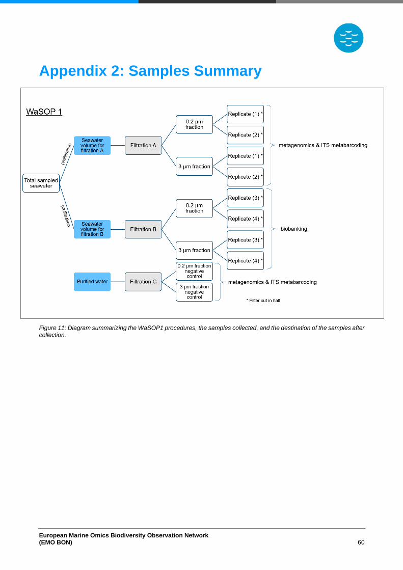
European Marine Omics Biodiversity Observation Network (EMO BON) 60
Appendix 2: Samples Summary
Figure 11: Diagram summarizing the WaSOP1 procedures, the samples collected, and the destination of the samples after collection.
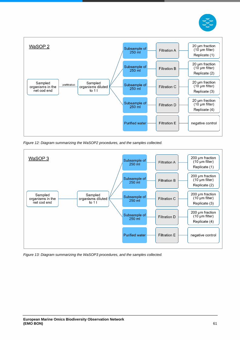
European Marine Omics Biodiversity Observation Network (EMO BON) 61
Figure 12: Diagram summarizing the WaSOP2 procedures, and the samples collected.
Figure 13: Diagram summarizing the WaSOP3 procedures, and the samples collected.
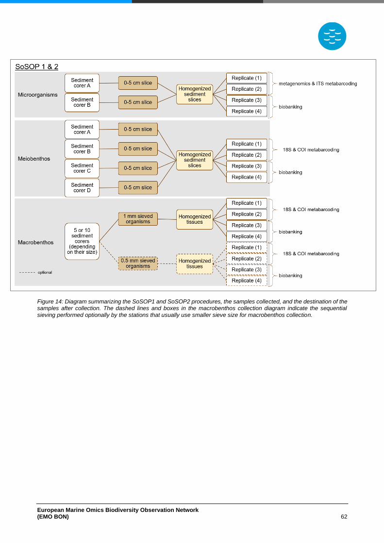
European Marine Omics Biodiversity Observation Network (EMO BON) 62
Figure 14: Diagram summarizing the SoSOP1 and SoSOP2 procedures, the samples collected, and the destination of the samples after collection. The dashed lines and boxes in the macrobenthos collection diagram indicate the sequential sieving performed optionally by the stations that usually use smaller sieve size for macrobenthos collection.
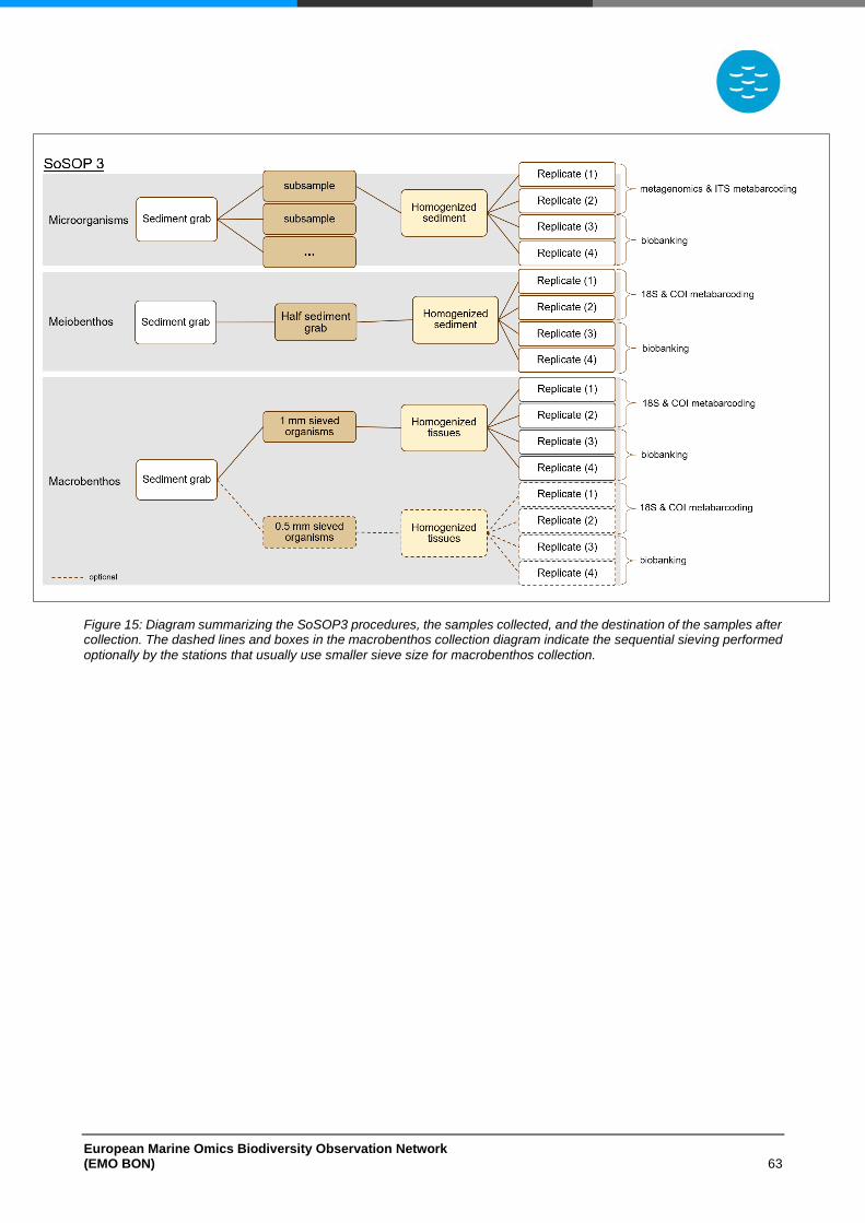
European Marine Omics Biodiversity Observation Network (EMO BON) 63
Figure 15: Diagram summarizing the SoSOP3 procedures, the samples collected, and the destination of the samples after collection. The dashed lines and boxes in the macrobenthos collection diagram indicate the sequential sieving performed
optionally by the stations that usually use smaller sieve size for macrobenthos collection.


