Embed Size (px)
Citation preview

Endothelial Nitric Oxide Synthase Tagging Single NucleotidePolymorphisms and Recovery from Aneurysmal SubarachnoidHemorrhage
Sheila A. Alexander, RN, PhD1, Samuel M. Poloyac, PhD2, Leslie Hoffman, RN, PhD1,Matthew Gallek, RN, PhD3, Dianxu Ren, PhD1, Jeffrey Balzer, PhD4, Amin B. Kassam, MD4,and Yvette P. Conley, PhD1Sheila A. Alexander: [email protected]; Samuel M. Poloyac: [email protected]; Leslie Hoffman: [email protected]; MatthewGallek: [email protected]; Dianxu Ren: [email protected]; Jeffrey Balzer: [email protected]; Amin B. Kassam:[email protected]; Yvette P. Conley: [email protected] of Pittsburgh, School of Nursing2University of Pittsburgh, Pharmaceutical Sciences3The University of Arizona, College of Nursing4University of Pittsburgh, Neurological Surgery
AbstractAneurysmal subarachnoid hemorrhage (SAH) is a hemorrhagic stroke subtype with a poor recoveryprofile. Cerebral vasospasm (CV), a narrowing of the cerebral vasculature, significantly contributesto the poor recovery profile. Variation in the endothelial nitric oxide (NO) synthase (eNOS) gene hasbeen implicated in CV and outcome after SAH. The purpose of this project was to explore thepotential association between three eNOS tagging single nucleotide polymorphisms (SNPs) andrecovery from SAH. We included 195 subjects with a diagnosis of SAH and DNA and 6-monthoutcome data available but without pre-existing neurologic disease/deficit. Genotyping wasperformed using an ABI Prism® 7000 Sequence Detection System and TaqMan® assays. CV wasverified by cerebral angiogram independently read by a neurosurgeon on 118 subjects. ModifiedRankin Scores (MRS) and Glasgow Outcome Scale (GOS) scores were collected 6 months posthemorrhage. Data were analyzed using descriptive statistics, ANOVA and chi-square analysis asappropriate. The sample was primarily female (n = 147; 75.4%) and Caucasian (n = 178; 91.3%)with a mean age of 54.6 years. Of the subjects with CV data, 56 (47.5%) developed CV within 14days of SAH. None of the SNPs individually were associated with CV presence; however, acombination of the three variant SNPs was significantly associated with CV (p = .017). Only oneSNP (RS1799983, variant allele) was associated with worse 6-month GOS scores (p < .001) andMRS (p < .001). These data indicate that the eNOS gene plays a role in the response to SAH, whichmay be explained by an influence on CV.
KeywordsSubarachnoid hemorrhage; cerebral vasospasm; nitric oxide; genotype; outcome
Corresponding author: Sheila A. Alexander, RN, PhD, Assistant Professor, Acute and Tertiary Care, School of Nursing, University ofPittsburgh, 336 Victoria Building, 3500 Victoria Street, Pittsburgh, PA 15261, Telephone: 412-624-3831, [email protected], Facsimile:412-383-7227.Carolyn B. Yucha, PhD, RN, FAAN, served as editor-in-chief for this article to avoid a conflict of interest for the special issue guesteditor, Yvette Conley, PhD, who is a coauthor.
NIH Public AccessAuthor ManuscriptBiol Res Nurs. Author manuscript; available in PMC 2010 September 13.
Published in final edited form as:Biol Res Nurs. 2009 July ; 11(1): 42–52. doi:10.1177/1099800409334751.
NIH
-PA Author Manuscript
NIH
-PA Author Manuscript
NIH
-PA Author Manuscript

Subarachnoid hemorrhage (SAH) is a devastating hemorrhagic stroke subtype associated withmortality rates of 20–70% and disability affecting up to 20% of survivors (Broderick, Brott,Duldner, Tomsick, & Leach, 1994; Dumont et al., 2003; Hop, Rinkel, Algra, & van Gijn,1997; Ingall, Asplund, Mahonen, & Bonita, 2000; Kassell, Sasaki, Colohan, & Nazar, 1985;Olafsson, Hauser, & Gudmundsson, 1997). SAH is most commonly due to the rupture of ananeurysm in the large vessels of the circle of Willis that provide blood flow to the cerebraltissue and results in blood in the subarachnoid space around the brain. While the initial increasein intracranial pressure associated with an increase in blood volume in the cranial vault accountsfor much of the early mortality, secondary injury occurring in the days to weeks after thehemorrhage contributes significantly to disability and mortality after SAH (Broderick et al.,1994; Dumont et al., 2003; Kassell et al., 1985; Olafsson et al., 1997).
Cerebral ischemia related to cerebral vasospasm (CV) is the most common delayed secondaryinjury occurring after SAH and significantly contributes to poor outcome. Within days afterthe initial hemorrhage, the blood or breakdown products of the blood in the cerebrospinal fluidbathing the blood vessels in the subarachnoid space alter vasomotor tone and promotevasoconstriction of the vessels leading to a spasmodic state in up to 70% of patients survivingthe initial hemorrhage (Adams, Kassell, Torner, & Haley, 1987; Al-Yamany & Wallace,1999; Dehdashti, Mermillod, Rufenacht, Reverdin, & de Tribolet, 2004; Dorsch, 2002). CV,a restriction in the internal lumen diameter of the cerebral blood vessels, can occur in one ormultiple cerebral blood vessels and often lasts for days (Kassell et al., 1985; Kassell, Torner,Jane, Haley, & Adams, 1990). The decrease in blood flow associated with CV leads to hypoxiaand decreased nutrient delivery and frequently to ischemia and infarction, which if not reversed,results in stroke (Haley, Kassell, Alves, Weir, & Hansen, 1995; Haley, Kassell, & Torner,1992; Lanzino & Kassell, 1999; Ohman, Servo, & Heiskanen, 1991). Clinical symptoms occurin 17–40% of patients experiencing CV (Lee et al., 2006; Mascia et al., 2003; Qureshi et al.,2000), suggesting that there are unknown factors influencing vasomotor tone and neuronalresponse involved. To date, only two predictors of CV have been identified – younger age(Torbey et al., 2001; Treggiari-Venzi, Suter, & Romand, 2001) and the amount and distributionof blood noted in the intracranial space on computed tomography (CT) scan (Fisher grade andFisher-Claassen grade) (Claassen et al., 2001; Fisher, Kistler, & Davis, 1980). While thesepredictors can identify patients at greater risk, they are not sensitive or specific. Other variablesthat have been implicated in the development of CV include hypertension, cigarette smokingand cocaine use (Conway & Tamargo, 2001; Lasner et al., 1997; Qureshi et al., 2001; Weir etal., 1998).
However, many individuals who are not at risk for CV based on this profile will develop CV.Current management involves attempting to prevention CV with oral Nimodipine (a calciumchannel blocker; Allen et al., 1983) and, in some institutions, statins and magnesium infusion.Additional therapy to prevent and decrease the ischemia associated with CV includeshypervolemia, hypertension and hemodilution. However this therapy, termed “Triple H”therapy, is difficult accomplish as many patients recovering from SAH also have comorbiditiesthat limit its use (e.g., cardiac or pulmonary disease; Sawada et al., 1997; Sayama, Liu, &Couldwell, 2006). There are also problems with other interventions aimed at reducing CV.Direct mechanical or pharmacologic (e.g., Papavarine) opening of the vessels with primaryinterventions are also problematic in that they are not consistently effective in preventing thenegative consequences of CV in all patients (Sawada et al., 1997; Sayama et al., 2006).
Because the physiologic mechanisms driving the development of CV after SAH are unknown,the search for more effective treatment has been difficult. CV most often occurs between 3 and7 days after SAH (Dorsch, 2002; Gerard, Frontera, & Wright, 2007; Haley, Kassell, Apperson-Hansen, Maile, & Alves, 1997; Haley et al., 1992), suggesting that it is a response to the releaseof one or more byproducts of blood breakdown in the subarachnoid space, where the exterior
Alexander et al. Page 2
Biol Res Nurs. Author manuscript; available in PMC 2010 September 13.
NIH
-PA Author Manuscript
NIH
-PA Author Manuscript
NIH
-PA Author Manuscript
surfaces of cerebral vessels are exposed to blood and products of its breakdown. The breakdownof blood in the subarachnoid space results in the release of oxyhemoglobin-derived freeradicals, which inhibit ATP-dependent calcium pumps (Arai, Takeyama, & Tanaka, 1999;Macdonald & Weir, 1991; Macdonald et al., 2001; Macdonald et al., 1991). This developmentis believed to lead to alteration in blood vessel tone and possibly influences development ofCV (Fujii & Fujitsu, 1988). Oxyhemoglobin has a direct effect on the arterial vessel wall,causing vessel spasm, and mediates the release of free radicals, such as NO, and vasoactivesubstances promoting a spasmodic state (Macdonald & Weir, 1991).
Recent work has implicated nitric oxide (NO) as a mediator of vasomotor tone that may impactCV development after SAH. NO is an endogenous vasodilator formed by its cleavage from L-arginine by nitric oxide synthases (NOS). The formation of NOS, and thereby NO, is drivenby gene transcription activated by many intra- and extracellular stimuli. NO availability isdecreased after SAH (Jung et al., 2004; Kajita et al., 1994; Kim, Schini, Sundt, & Vanhoutte,1992). NOS is produced by neurons (nNOS), endothelial cells (eNOS) and other cells(inducible NOS; iNOS). The decreased availability of NO after SAH may be related to adecrease in NOS production and/or activity and/or NO disruption or scavenging (Iuliano, Pluta,Jung, & Oldfield, 2004; Jung et al., 2004; Kim et al., 1992). After SAH, eNOS function isimpaired in cerebral vessels (Iuliano et al., 2004), limiting any increase in vessel relaxationinduced by the eNOS-associated increase in NO production. Administration of NO donors andNOS metabolites have shown efficacy in decreasing angiographic CV (Afshar, Pluta, Boock,Thompson, & Oldfield, 1995; Hino et al., 1996; Pluta, Oldfield, & Boock, 1997; Thomas,1997; Thomas & Rosenwasser, 1999; Thomas et al., 1999; Tierney et al., 2001). However, thetransformation of these clinical trials into widespread clinical treatment was limited by theshort half-life of NO, side effects and potential toxicity (Afshar et al., 1995; Dietrich & Dacey,2000; Hino et al., 1996).
The production of NOS is genetically driven by three genes. Specifically, endothelial NOS(eNOS) is produced by endothelial cells and this production is driven by the eNOS gene onchromosome 18. Neuronal NOS (nNOS) is produced by neurons and this enzyme productionis driven by the nNOS gene. Inducible NOS is produced by a variety of cell types includingendothelial cells and neurons during periods of exposure to hypoxia or other environmentalstimuli and production is driven by the iNOS gene. The eNOS gene is primarily active in theendothelial cells of the vasculature. These cells act to increase NOS production at the vascularlevel, increasing NO production and ultimately vasodilation. In this way, the endothelial cellsinfluence delivery of blood flow to tissues in need by expressing eNOS. In humans withcoronary artery disease, individuals with the variant of the eNOS single nucleotidepolymorphism (SNP) in the promoter region (T-687C) and in the G894T SNP had decreasedendothelium-dependent relaxation (Erbs et al., 2006). Additional studies have found theeNOS genotype to be inconsistently implicated in the development of CV. A study by Khuranaand associates (2004) on 28 human subjects recovering from SAH found that an SNP in theeNOS promoter region (T-786C) was associated with increased risk of CV (Khurana et al.,2004). Song and colleagues found the T-786C was not associated with SAH or CV but wasassociated with poorer outcomes in a study of 136 subjects with SAH and 110 control subjects(Song et al., 2006). Berra and colleagues showed that eNOS expression was upregulated afterSAH and that there was a further increase in expression in subjects with poor clinical conditions(Berra, Carcereri De Prati, Suzuki, & Pasqualin, 2007). No further work has been done toclarify these conflicting findings.
In the study described here, we used tagging SNPs to measure genetic variation in the eNOSgene. A tagging SNP (tSNP) is an SNP within a gene that serves as a surrogate for all geneticvariation in a region of DNA within the gene. By measuring genetic variation with thesesurrogates, it is possible to measure the influence of functional SNPs, such as T-786C, as well
Alexander et al. Page 3
Biol Res Nurs. Author manuscript; available in PMC 2010 September 13.
NIH
-PA Author Manuscript
NIH
-PA Author Manuscript
NIH
-PA Author Manuscript

as genetic variance that may influence phenotype but has not yet been identified. The threetSNPs that we chose are representative of all known variation within the eNOS gene at the timethis study was done. The overall purpose of the current project is to explore the potentialassociation between three eNOS tSNPs and recovery (CV and functional outcome) from SAH.
MethodsDesign
For this study, we used a prospective, between-group, within-subject repeated measures designto identify the potential relationship between NOS genotypes and outcome (CV developmentand gross functional outcome) in individuals recovering from SAH while controlling forcovariates such as age, severity of hemorrhage and comorbidities.
ParticipantsParticipants comprised patients, age 21–75, admitted to the University of Pittsburgh MedicalCenter (UPMC) neurovascular ICU with a diagnosis of aneurysmal SAH verified by CT scan(Fisher grade ≥ 2) and aneurysm presence and location verified by cerebral angiogram orcomputed tomographic angiogram and no pre-existing chronic neurologic diseases or deficits.In order to capture patients most at risk for CV, we excluded patients with non-aneurysmalSAH from this study. At our facility lumbar puncture and cerebral spinal fluid (CSF) analysesare used to determine presence of SAH only after a negative CT scan. All patients in this studyhad a CT scan positive for SAH on admission. A total of 195 patients meeting inclusion criteriahad DNA and outcome data available were enrolled in this study.
Collection of Demographic and Medical Condition DataProject personnel extracted demographic variables including age, gender, and race frommedical records. Upon admission to the UPMC, all participants had a head CT scan and theneurosurgeon assigned a Fisher grade. The Fisher grade is a measure of the amount anddistribution of blood noted in the intracranial space on CT scan. While the tool has a range of1–4, a Fisher grade of 3 is most highly correlated with CV development (Fisher et al., 1980).All participants had a cerebral angiogram or computed tomographic angiogram to identifyaneurysm size and location and, if appropriate, coil embolization for aneurysm securement.For patients not having coil embolization, surgical clipping for aneurysm securement wasperformed as soon as possible. The Hunt and Hess score was determined on admission by theneurosurgeon responsible for clinical care, and project personnel extracted this score frommedical records. Hunt and Hess score is a tool used to quantify clinical exam on admissionand predict long-term functional outcome. Scores range from 1 to 5, with higher scores beingpredictive of poorer outcome (Hunt, 1983).
GenotypingWe collected a single blood sample from each participant into EDTA-containing vacutainertubes and processed it for white blood cell removal. DNA was extracted from the first availableblood sample using a simple salting out procedure (Miller, Dykes, & Polesky, 1988). To assessall of the genetic variability within the eNOS gene as well as the promoter and 3’ flankingregion, we identified a total of three tSNPs with a minor allele frequency ≥ 20% with an r2 ≥ .80 using build 35 of the Hapmap database. The three tSNPs identified were rs1799983,rs1800779, and rs3918188. We used an ABI Prism® 7000 Sequence Detection System toconduct allele discrimination using TaqMan® assays (Applied Biosystems, Foster City, CA).Due to the sample size available and genotype distribution, we dichotomized the sample intogroups based on variant presence (homozygote for variant allele combined with heterozygotes)versus variant absence (homozygote for wild type allele) for each SNP analysis.
Alexander et al. Page 4
Biol Res Nurs. Author manuscript; available in PMC 2010 September 13.
NIH
-PA Author Manuscript
NIH
-PA Author Manuscript
NIH
-PA Author Manuscript
CV VerificationThe presence or absence of CV was verified by cerebral angiography read by a neurosurgeon.The population included in this sample had CV angiography to monitor for CV presenceroutinely on Day 5 after SAH. CV presence was assessed in the anterior cerebral, anteriorcommunicating, middle cerebral, internal carotid, posterior cerebral, posterior communicating,vertebral and basilar arteries and classified as cerebral vasospasm negative (0–24% constrictionin any portion of any of these vessels) or cerebral vasospasm positive (≥ 25% constriction inany portion of any of these vessels). CV angiograms were coded for percent of vesselconstriction for research purposes at a later time. Cerebral angiography is the gold standard forverification of CV.
Functional OutcomeGlasgow Outcome Scale (GOS) and Modified Rankin Score (MRS) were administered by aneuropsychological technician during a face-to-face or telephone interview at 3, 6, 12 and 24months following SAH. The neuropsychological technician was blinded to the results of NOSgenotyping and CV.
The GOS, a clinical observation scale, categorizes functional outcomes into five levels: 1--death, 2--persistent vegetative state, 3--severe disability, 4--moderate disability, 5--goodrecovery (Jennett, & Bond, 1975).
The MRS is another clinical observation scale, which categorizes functional outcomes intoseven levels: 0--no symptoms; 1--no significant disabling symptoms. no significant disabilitydespite symptoms, able to carry out all usual duties and activities; 2--slight disability, unableto carry out all previous activities but able to look after their own affairs without assistance;3--moderate disability, requiring some help but able to walk without assistance; 4--moderate/severe disability, unable to walk without assistance and unable to attend to own bodily needswithout assistance; 5--severe disability, bedridden, incontinent and requiring constant nursingcare and attention; and 6--death (Sulter et al., 1999; van Swieten, Koudstaal, Visser, Schouten,& van Gijn, 1988; Wilson et al., 2005).
Data AnalysisBasic descriptive statistics were used to identify differences in demographic characteristicsbased on genotype. Analysis of variance and Chi square analyses were used to determinedifferences in outcome variables by genotype while controlling for age and Fisher grade.
ResultsSample
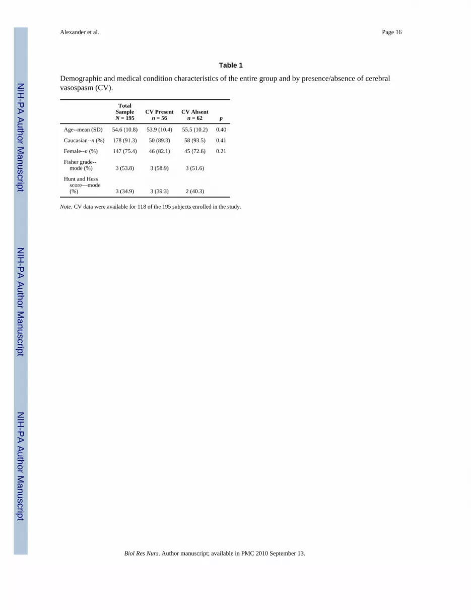
There was a total of 195 subjects with DNA and outcome data included in these analyses.Subjects were primarily Caucasian and female with a mean age of 54.6 years. The Fisher grademode for the entire sample was 3, indicating that over half of the sample was at the highestrisk for development of CV. The Hunt and Hess score mode for the entire sample was also 3,with 81% of the sample having scores of 1–3 indicating low risk of poor outcome. Demographicdata and admission severity of hemorrhage scores are presented in Table 1. The full range ofFisher grades and Hunt and Hess scores for the entire sample are presented in Table 2. Genotypefrequencies for each of the three SNPs are presented in Table 3. The three SNPs were separatelyexamined for adherence to Hardy-Weinberg equilibrium with a goodness-of-fit test and werefound to be in equilibrium for the entire sample.
Alexander et al. Page 5
Biol Res Nurs. Author manuscript; available in PMC 2010 September 13.
NIH
-PA Author Manuscript
NIH
-PA Author Manuscript
NIH
-PA Author Manuscript
CVThere were 118 subjects with available CV data. Of these, 56 (47.5%) developed CV within14 days of SAH. There were no significant differences in demographic variables based on CVpresence. Individuals who developed CV more often had a Fisher grade of 3 than those whodid not. See Table 1 for demographic and medical condition characteristics by CV presence.Number and percentage of subjects in each Fisher grade category and Hunt and Hess scorecategory by CV presence are presented in Table 2. None of the SNPs examined wereindividually associated with CV presence; however the combination of three SNPs wasassociated with CV presence. Subjects with variant copies of rs1799983, rs1800779, andrs3918188 more often developed CV (p = .017) than the other subjects.
GOSWe had 6-month GOS data available for all 195 subjects. Most subjects (n = 70, 40.4%) hada GOS of 5, indicating good recovery and resumption of normal life despite minor deficits.Only one SNP (rs1799983) was associated with 6-month GOS, with individuals with at leastone copy of the variant allele having poorer outcomes (p < .001). See Figure 1 for GOS byrs1799983 genotype.
MRSWe had 6-month MRS data for 167 subjects. Of those, 98 (55.1) had MRS scores of 0 or 1,indicating no symptoms or no significant disability despite minor symptoms. Only one SNP(rs1799983) was associated with 6-month MRS, with subjects with at least one copy of thevariant allele having poor MRS (p < .001). See Figure 2 for MRS by rs1799983 genotype.
DiscussionThis study had two important findings: 1) the three tagging SNPs within the eNOS gene arenot individually associated with the development of CV; the combination of variant alleles ofthe three SNPs, however, is; 2) the variant allele of the tagging SNP rs1799983 is associatedwith a poorer outcome as measured by both GOS and MRS. Thus, though the SNPs within theeNOS gene do not independently explain CV, when considered together they may serve as apredictor for CV.
Researchers have previously explored NOS gene expression (mRNA) after SAH. In a study ofhuman brain tissue taken from 23 subjects recovering from SAH, eNOS and iNOS expressionwere increased, while nNOS expression was unchanged compared to control samples (Berraet al., 2007). This altered expression of NOS genes may be driven by genetic variability. Inparticular, variation in either the coding regions or the promoter regions may alter mRNAlevels, protein levels and functional recovery of patients after SAH. This study explored allvariation within the eNOS gene and, as such, better reflects the genetic variance that may beinfluencing mRNA, eNOS production and amount of eNOS produced. Our findings suggestthat there is variance in other regions of the eNOS gene that may be influencing vasomotortone and recovery from SAH via an as yet unidentified mechanism not associated with thefunctional SNP (T-786C).
There have also been previous studies exploring the relationship between CV and eNOSgenotypes. Previous work has focused primarily on a single known functional SNP in theeNOS gene (Khurana et al., 2004). The findings of the current study support those of the studiesby Khurana and associates (2004) and Song (Song et al., 2006), who found that eNOS genotypewas associated with CV after SAH. While our work did not find an association between theregion containing the SNP explored by this work and CV, the combination of SNPs waspredictive of CV development. Utilizing tagging SNPs allowed us to assess variation
Alexander et al. Page 6
Biol Res Nurs. Author manuscript; available in PMC 2010 September 13.
NIH
-PA Author Manuscript
NIH
-PA Author Manuscript
NIH
-PA Author Manuscript
throughout the entire gene sequence and the relationship with CV, adding strength to ouranalyses.
Research in other areas has implicated variation in the T-786C SNP in hypertension(Kuhlencordt et al., 2001; Wang & Wang, 2000), myocardial infarction (Hingorani, 2001;Wang et al., 1996), and coronary vasospasm (Nakayama et al., 1999), all disorders influencedby vasomotor reactivity. The T-786->C polymorphism results in a 52% reduction in promoteractivity (Nakayama et al., 1999). The significant reduction in promoter activity ultimately leadsto decreased eNOS protein expression. eNOS production generates active NO, which leads tovasodilation. The 52% reduction in eNOS production resulting from this variant allele, whichresides within the haploblock tagged by SNP rs1799983 measured for this study, results indecreased vasodilation (Song et al., 2003). The variant (C) allele at the −786 position istranslated such that there is less eNOS produced, which results in a decrease in NO productionand decreased vasodilation. In this way, individuals with a variant (C) allele at the −786 positionare at increased risk of decreased vasodilation and therefore CV after SAH.
While our study did not find direct association between the rs1799983 variant allele containingthis SNP and CV, this tagging SNP was associated with poorer outcome. The −786 SNP maystill be influencing CV development or response to CV and ultimately drive the associationbetween the tagging SNP (rs1799983) and poor outcome. Variation in the gene sequence maylead to worse outcomes via alteration of CV risk or response to CV.
The confusion within the literature regarding the role of the T-786C allele and CV may alsobe influenced by the method of CV measurement. Khurana and associates (2004) defined CVas decline in neurologic status coupled with an elevation in cerebral blood flow velocity(measured by transcranial Doppler ultrasonography). This method of vasospasm measurementunderestimates angiographic vasospasm as a diagnosis of CV positive is dependent upon achange in the neurologic exam that results from tissue hypoxia/ischemia. It is, however, aclinically relevant definition as angiographic vasospasm without clinical signs and symptomsis often undiagnosed and untreated. Variation in neuronal response could be mediated by eNOSexpression, and this may be the phenomenon found by this body of literature. Ko and colleagues(2008) used only cerebral angiography to define CV and found similar results, showing thatdecreased eNOS expression is directly related to cerebral vessel reactivity. The regionaldecrease in NO after SAH and the variation in eNOS expression directed by the eNOS geneare highly suggestive of the influence of eNOS in CV.
It is also possible that the additional genetic variability explored in the current project maskedeffects. Using tagging SNPs for this analysis was more informative, as we were able to exploremore genetic variability than if we had, as with previous studies, examined a single SNP.However there may be additional SNPs within the tagging region with influence on expressionor functionality of eNOS that cancelled out the relationship at the single SNP level. As previousresearch has only explored the influence of a single SNP on CV development, it is also possiblethat variation in other regions of the eNOS gene were not adequately explored in previous workbut are influential on vasomotor tone and CV development.
A larger sample size may identify relationships more clearly as genetic variability is moreaccurately represented with more subjects with presence of the minor allele available foranalyses. The inconsistency in findings may also be influenced by inheritance. The Ko andSong studies utilized a Korean sample, with anticipated T-786C polymorphism presence lowerthan in a primarily Caucasian sample, while the Khurana study utilized an American sample(primarily Caucasian). Our sample is comparable to the subjects in the Khurana study, but theremay be additional genetic variability exerted by other SNPs with varying frequencies indifferent populations.
Alexander et al. Page 7
Biol Res Nurs. Author manuscript; available in PMC 2010 September 13.
NIH
-PA Author Manuscript
NIH
-PA Author Manuscript
NIH
-PA Author Manuscript
It may be that there is additional influence on NOS and NO production exerted by regionswithin the eNOS gene that are not yet recognized as having functional differences but explainthe discrepancy in the literature. As only a combination of SNPs was found to be associatedwith CV development, additional influence may be exerted by variance represented by taggingSNPs in these regions. Tagging SNPs represent variation within a large sequence of the gene,and the three SNPs in this project describe all variation in the eNOS gene. Variance within theregions represented by the tagging SNPs likely leads to variation in either eNOS expressionor eNOS function, which influences recovery after SAH.
The variant of the rs1799983 SNP was associated with poorer outcome in both measures (GOSand MRS) used in our study. The functional SNP found to be associated with recovery afterSAH by Song et al. (2006) is located within the region represented by rs1799983 SNP and thesame region that has previously been associated with CV in the literature. Song and associatesfound an association between the T-786C SNP and outcome as measured by GOS but notbetween the SNP and CV, similar to our findings. While there is inconsistency in the literatureregarding the relationship between this particular SNP and CV development, the relationshipbetween eNOS genotypes and outcome from SAH has not been well described. CV is a causeof secondary injury that is negatively correlated with long-term outcome from SAH; howeverthere are many other events that occur in the recovery process, and eNOS is likely influentialon long-term recovery in addition to its influence on CV. Certainly alterations in cerebral bloodflow may persist beyond the 2-week period during which CV risk is highest. Variation invasomotor reactivity later in the recovery process, for instance, may influence neuronalviability and long-term recovery. Genetically driven variability in eNOS production mayimpact cerebral blood flow and neuronal recovery throughout the recovery process. While thealtered eNOS expression in individuals developing CV has been documented, there is noliterature describing long term eNOS expression dysfunction. Slight decreases in cerebralblood flow due to altered eNOS expression may persist long after the hemorrhage and impairneuronal function leading to poorer outcome. There may be additional, unknown influenceson neuronal response to injury and recovery processes that are mediated by eNOS, NOS andNO that have not yet been identified or described.
It does appear, based on our findings in the context of the existing literature, that the decreasedNO formation from decreased eNOS production based on genetic variance leads to anenvironment that is favorable to CV development and poorer outcome after SAH. The variantin the region of the eNOS gene that has consistently been implicated in CV development andpoorer outcome leads to decreased eNOS production and ultimately a decrease in availableNO, which promotes vasoconstriction (Nakayama et al., 1999). This consistent relationship issuggestive of the strong role expression of this gene plays in the recovery process after SAHand could likely lead to therapeutic interventions to decrease CV and improve outcomes.
There are two other genes that influence NOS and NO production. Inducible NOS (iNOS) isexpressed by cells of varying types, including neurons, in response to hypoxia and otherenvironmental stimuli. Neuronal NOS (nNOS) is also expressed by neurons in response tohypoxia. The function of nNOS producing neurons at the neurovascular junction is impairedafter SAH, and variations in the nNOS gene may also exert influence on recovery from SAH(Pluta & Oldfield, 2007). The availability of NO after SAH is likely influenced to a variableextent by each of these genes. Additionally, the role of NOS and NO in development of CVafter SAH may be mediated by other mechanisms. NO scavenging may be the source ofdecreased NO (Pluta, 2006; Pluta & Oldfield, 2007). Alternatively, an increased expression ofasymmetric dimethylarginine (ADMA), an endogenous NOS inhibitor that has been implicatedin the development of CV (Jung et al., 2004), might be related to the decrease in available NO.Neither NO scavenging or ADMA expression were measured in this study. Further workexploring the expression of each gene and genotypic expression of these genes will provide
Alexander et al. Page 8
Biol Res Nurs. Author manuscript; available in PMC 2010 September 13.
NIH
-PA Author Manuscript
NIH
-PA Author Manuscript
NIH
-PA Author Manuscript

insight into mechanism of CV after SAH and ultimately lead to improved therapeutic strategiesto decrease incidence, severity and negative effects of CV.
The limitations of this study include sample size, method of defining CV, and outcomemeasurements. While our sample size of 195 subjects is one of the largest available (118 foranalyses including CV as a variable), it is still a relatively small sample size for a genetic study.The fact that we found significance despite this limitation supports the potential role of thesegenes’ influence on the recovery processes after SAH. We chose angiographic CV rather thansymptomatic CV as our study definition because it is the most appropriate definition of CV inrelation to the scientific rationale supporting the study. Studies exploring this relationship withsymptomatic CV may yield different results and are necessary for clinical relevance. Weoperationalized our outcome variable as GOS and MRS. These tools measure gross functionaloutcome but are not specific enough to describe the many neuropsychological impairmentsthat this population may encounter. Further work exploring the role of eNOS genotypes onthese other outcomes is needed to fully describe this relationship.
ConclusionThese data indicate that the eNOS gene plays a role in the response to SAH, which may beexplained by an influence on the development of CV. The identification of genetic biomarkersof secondary injury after SAH will likely lead to many changes in the nursing care of patientsrecovering from SAH. Knowledge about genotypic differences influencing CV will provideinsight into mechanism of CV and promote development of therapeutics to improve outcomes.This information will also permit more focused utilization of resources, allowing patients atlower risk to be discharged from an intensive care setting at an earlier time. Knowledge ofgenotypic differences in long-term outcome will also be instrumental in changing the outcomeprofile of patients with SAH. In addition to providing insight into other potential mechanismsof recovery from SAH, this knowledge may also lead to the development of genotype-specifictherapeutics for use in select populations. Further research validating this work and determiningsensitivity and specificity of genotypes in predicting CV and long-term outcome in SAH isneeded in order to translate these findings into biomarkers for use in clinical care.
ReferencesAdams HP Jr, Kassell NF, Torner JC, Haley EC Jr. Predicting cerebral ischemia after aneurysmal
subarachnoid hemorrhage: Influences of clinical condition, CT results, and antifibrinolytic therapy. Areport of the Cooperative Aneurysm Study. Neurology 1987;37(10):1586–1591. [PubMed: 3658161]
Afshar JK, Pluta RM, Boock RJ, Thompson BG, Oldfield EH. Effect of intracarotid nitric oxide onprimate cerebral vasospasm after subarachnoid hemorrhage. Journal of Neurosurgery 1995;83(1):118–122. [PubMed: 7782826]
Al-Yamany M, Wallace MC. Management of cerebral vasospasm in patients with aneurysmalsubarachnoid hemorrhage. Intensive Care Medicine 1999;25(12):1463–1466. [PubMed: 10702032]
Allen GS, Ahn HS, Preziosi TJ, Battye R, Boone SC, Boone SC, et al. Cerebral arterial spasm--acontrolled trial of nimodipine in patients with subarachnoid hemorrhage. New England Journal ofMedicine 1983;308(11):619–624. [PubMed: 6338383]
Arai T, Takeyama N, Tanaka T. Glutathione monoethyl ester and inhibition of the oxyhemoglobin-induced increase in cytosolic calcium in cultured smooth-muscle cells. Journal of Neurosurgery1999;90(3):527–532. [PubMed: 10067923]
Berra LV, Carcereri De Prati A, Suzuki H, Pasqualin A. The role of constitutive and inducible nitric oxidesynthase in the human brain after subarachnoid hemorrhage. Journal of Neurosurgical Sciences2007;51(1):1–9. [PubMed: 17369785]
Broderick JP, Brott TG, Duldner JE, Tomsick T, Leach A. Initial and recurrent bleeding are the majorcauses of death following subarachnoid hemorrhage. Stroke 1994;25(7):1342–1347. [PubMed:8023347]
Alexander et al. Page 9
Biol Res Nurs. Author manuscript; available in PMC 2010 September 13.
NIH
-PA Author Manuscript
NIH
-PA Author Manuscript
NIH
-PA Author Manuscript

Claassen J, Bernardini GL, Kreiter K, Bates J, Du YE, Copeland D, et al. Effect of cisternal and ventricularblood on risk of delayed cerebral ischemia after subarachnoid hemorrhage: The Fisher scale revisited.Stroke 2001;32(9):2012–2020. [PubMed: 11546890]
Conway JE, Tamargo RJ. Cocaine use is an independent risk factor for cerebral vasospasm afteraneurysmal subarachnoid hemorrhage. Stroke 2001;32(10):2338–2343. [PubMed: 11588323]
Dehdashti AR, Mermillod B, Rufenacht DA, Reverdin A, de Tribolet N. Does treatment modality ofintracranial ruptured aneurysms influence the incidence of cerebral vasospasm and clinical outcome?Cerebrovascular Disease 2004;17(1):53–60.
Dietrich HH, Dacey RG Jr. Molecular keys to the problems of cerebral vasospasm. Neurosurgery 2000;46(3):517–530. [PubMed: 10719847]
Dorsch NW. Therapeutic approaches to vasospasm in subarachnoid hemorrhage. Current Opinion inCritical Care 2002;8(2):128–133. [PubMed: 12386513]
Dumont AS, Dumont RJ, Chow MM, Lin CL, Calisaneller T, Ley KF, et al. Cerebral vasospasm aftersubarachnoid hemorrhage: Putative role of inflammation. Neurosurgery 2003;53(1):123–133.discussion 133-125. [PubMed: 12823881]
Erbs S, Mobius-Winkler S, Linke A, Adams V, Doll N, Gielen S, et al. Both T-786C and G894Tpolymorphism of endothelial nitric oxide synthase affect in-vitro endothelium-dependent relaxationof internal mammary artery rings from patients with coronary artery disease. European Journal ofCardiovascular Prevention & Rehabilitation 2006;13(5):826–831. [PubMed: 17001225]
Fisher CM, Kistler JP, Davis JM. Relation of cerebral vasospasm to subarachnoid hemorrhage visualizedby computerized tomographic scanning. Neurosurgery 1980;6(1):1–9. [PubMed: 7354892]
Fujii S, Fujitsu K. Experimental vasospasm in cultured arterial smooth-muscle cells. Part 1: Contractileand ultrastructural changes caused by oxyhemoglobin. Journal of Neurosurgery 1988;69(1):92–97.[PubMed: 3379479]
Gerard E, Frontera JA, Wright CB. Vasospasm and cerebral infarction following isolated intraventricularhemorrhage. Neurocritical Care 2007;7(3):257–259. [PubMed: 17522787]
Haley EC Jr, Kassell NF, Alves WM, Weir BK, Hansen CA. Phase II trial of tirilazad in aneurysmalsubarachnoid hemorrhage. A report of the Cooperative Aneurysm Study. Journal of Neurosurgery1995;82(5):786–790. [PubMed: 7714603]
Haley EC Jr, Kassell NF, Apperson-Hansen C, Maile MH, Alves WM. A randomized, double-blind,vehicle-controlled trial of tirilazad mesylate in patients with aneurysmal subarachnoid hemorrhage:A cooperative study in North America. Journal of Neurosurgery 1997;86(3):467–474. [PubMed:9046304]
Haley EC Jr. Kassell NF, Torner JC. The International Cooperative Study on the Timing of AneurysmSurgery. The North American experience. Stroke 1992;23(2):205–214. [PubMed: 1561649]
Hingorani AD. Polymorphisms in endothelial nitric oxide synthase and atherogenesis: John FrenchLecture 2000. Atherosclerosis 2001;154(3):521–527. [PubMed: 11257252]
Hino A, Tokuyama Y, Weir B, Takeda J, Yano H, Bell GI, et al. Changes in endothelial nitric oxidesynthase mRNA during vasospasm after subarachnoid hemorrhage in monkeys. Neurosurgery1996;39(3):562–567. discussion 567–568. [PubMed: 8875487]
Hop JW, Rinkel GJ, Algra A, van Gijn J. Case-fatality rates and functional outcome after subarachnoidhemorrhage: A systematic review. Stroke 1997;28(3):660–664. [PubMed: 9056628]
Hunt WE. Clinical assessment of SAH. Journal of Neurosurgery 1983;59(3):550–551. [PubMed:6886772]
Ingall T, Asplund K, Mahonen M, Bonita R. A multinational comparison of subarachnoid hemorrhageepidemiology in the WHO MONICA stroke study. Stroke 2000;31(5):1054–1061. [PubMed:10797165]
Iuliano BA, Pluta RM, Jung C, Oldfield EH. Endothelial dysfunction in a primate model of cerebralvasospasm. Journal of Neurosurgery 2004;100(2):287–294. [PubMed: 15086237]
Jennett B, Bond M. Assessment of outcome after severe brain damage. Lancet 1975;1(7905):480–484.[PubMed: 46957]
Jung CS, Iuliano BA, Harvey-White J, Espey MG, Oldfield EH, Pluta RM. Association betweencerebrospinal fluid levels of asymmetric dimethyl-L-arginine, an endogenous inhibitor of endothelial
Alexander et al. Page 10
Biol Res Nurs. Author manuscript; available in PMC 2010 September 13.
NIH
-PA Author Manuscript
NIH
-PA Author Manuscript
NIH
-PA Author Manuscript

nitric oxide synthase, and cerebral vasospasm in a primate model of subarachnoid hemorrhage.Journal of Neurosurgery 2004;101(5):836–842. [PubMed: 15543672]
Kajita Y, Suzuki Y, Oyama H, Tanazawa T, Takayasu M, Shibuya M, et al. Combined effect of L-arginineand superoxide dismutase on the spastic basilar artery after subarachnoid hemorrhage in dogs. Journalof Neurosurgery 1994;80(3):476–483. [PubMed: 8113861]
Kassell NF, Sasaki T, Colohan AR, Nazar G. Cerebral vasospasm following aneurysmal subarachnoidhemorrhage. Stroke 1985;16(4):562–572. [PubMed: 3895589]
Kassell NF, Torner JC, Jane JA, Haley EC Jr, Adams HP. The International Cooperative Study on theTiming of Aneurysm Surgery. Part 2: Surgical results. Journal of Neurosurgery 1990;73(1):37–47.[PubMed: 2191091]
Khurana VG, Sohni YR, Mangrum WI, McClelland RL, O'Kane DJ, Meyer FB, et al. Endothelial nitricoxide synthase gene polymorphisms predict susceptibility to aneurysmal subarachnoid hemorrhageand cerebral vasospasm. Journal of Cerebral Blood Flow & Metabolism 2004;24(3):291–297.[PubMed: 15091109]
Kim P, Schini VB, Sundt TM Jr, Vanhoutte PM. Reduced production of cGMP underlies the loss ofendothelium-dependent relaxations in the canine basilar artery after subarachnoid hemorrhage.Circulation Research 1992;70(2):248–256. [PubMed: 1310445]
Ko NU, Rajendran P, Kim H, Rutkowski M, Pawlikowska L, Kwok PY, et al. Endothelial nitric oxidesynthase polymorphism (−786T->C) and increased risk of angiographic vasospasm after aneurysmalsubarachnoid hemorrhage. Stroke 2008;39(4):1103–1108. [PubMed: 18309169]
Kuhlencordt PJ, Gyurko R, Han F, Scherrer-Crosbie M, Aretz TH, Hajjar R, et al. Acceleratedatherosclerosis, aortic aneurysm formation, and ischemic heart disease in apolipoprotein E/endothelial nitric oxide synthase double-knockout mice. Circulation 2001;104(4):448–454.[PubMed: 11468208]
Lanzino G, Kassell NF. Double-blind, randomized, vehicle-controlled study of high-dose tirilazadmesylate in women with aneurysmal subarachnoid hemorrhage. Part II. A cooperative study in NorthAmerica. Journal of Neurosurgy 1999;90(6):1018–1024.
Lasner TM, Weil RJ, Riina HA, King JT Jr, Zager EL, Raps EC, et al. Cigarette smoking-induced increasein the risk of symptomatic vasospasm after aneurysmal subarachnoid hemorrhage. Journal ofNeurosurgy 1997;87(3):381–384.
Lee JY, Lee MS, Whang K, Lee JM, Kim SH, Lee SS. Accuracy of transcranial Doppler sonography forpredicting cerebral infarction in aneurysmal subarachnoid hemorrhage. Journal of ClinicalUltrasound 2006;34(8):380–384. [PubMed: 16944480]
Macdonald RL, Weir BK. A review of hemoglobin and the pathogenesis of cerebral vasospasm. Stroke1991;22(8):971–982. [PubMed: 1866764]
Macdonald RL, Weir BK, Marton LS, Zhang ZD, Sajdak M, Johns LM, et al. Role of adenosine 5'-triphosphate in vasospasm after subarachnoid hemorrhage: Human investigations. Neurosurgery2001;48(4):854–862. discussion 862-853. [PubMed: 11322446]
Macdonald RL, Weir BK, Runzer TD, Grace MG, Findlay JM, Saito K, et al. Etiology of cerebralvasospasm in primates. Journal of Neurosurgery 1991;75(3):415–424. [PubMed: 1869943]
Mascia L, Fedorko L, terBrugge K, Filippini C, Pizzio M, Ranieri VM, et al. The accuracy of transcranialDoppler to detect vasospasm in patients with aneurysmal subarachnoid hemorrhage. Intensive CareMedicine 2003;29(7):1088–1094. [PubMed: 12774157]
Miller SA, Dykes DD, Polesky HF. A simple salting out procedure for extracting DNA from humannucleated cells. Nucleic Acids Research 1988;16(3):1215. [PubMed: 3344216]
Nakayama M, Yasue H, Yoshimura M, Shimasaki Y, Kugiyama K, Ogawa H, et al. T-786-->C mutationin the 5'-flanking region of the endothelial nitric oxide synthase gene is associated with coronaryspasm. Circulation 1999;99(22):2864–2870. [PubMed: 10359729]
Ohman J, Servo A, Heiskanen O. Long-term effects of nimodipine on cerebral infarcts and outcome afteraneurysmal subarachnoid hemorrhage and surgery. Journal of Neurosurgery 1991;74(1):8–13.[PubMed: 1984511]
Olafsson E, Hauser WA, Gudmundsson G. A population-based study of prognosis of ruptured cerebralaneurysm: Mortality and recurrence of subarachnoid hemorrhage. Neurology 1997;48(5):1191–1195. [PubMed: 9153441]
Alexander et al. Page 11
Biol Res Nurs. Author manuscript; available in PMC 2010 September 13.
NIH
-PA Author Manuscript
NIH
-PA Author Manuscript
NIH
-PA Author Manuscript

Pluta RM. Dysfunction of nitric oxide synthases as a cause and therapeutic target in delayed cerebralvasospasm after SAH. Neurological Research 2006;28(7):730–737. [PubMed: 17164036]
Pluta RM, Oldfield EH. Analysis of nitric oxide (NO) in cerebral vasospasm after aneursymal bleeding.Reviews on Recent Clinical Trials 2007;2(1):59–67. [PubMed: 18473989]
Pluta RM, Oldfield EH, Boock RJ. Reversal and prevention of cerebral vasospasm by intracarotidinfusions of nitric oxide donors in a primate model of subarachnoid hemorrhage. Journal ofNeurosurgery 1997;87(5):746–751. [PubMed: 9347984]
Qureshi AI, Sung GY, Razumovsky AY, Lane K, Straw RN, Ulatowski JA. Early identification of patientsat risk for symptomatic vasospasm after aneurysmal subarachnoid hemorrhage. Crit Care Med2000;28(4):984–990. [PubMed: 10809270]
Qureshi AI, Suri MF, Yahia AM, Suarez JI, Guterman LR, Hopkins LN, et al. Risk factors forsubarachnoid hemorrhage. Neurosurgery 2001;49(3):607–612. discussion 612-603. [PubMed:11523670]
Sawada M, Hashimoto N, Tsukahara T, Nishi S, Kaku Y, Yoshimura S. Effectiveness of intra-arteriallyinfused papaverine solutions of various concentrations for the treatment of cerebral vasospasm. ActaNeurochirurgica 1997;139(8):706–711. [PubMed: 9309284]
Sayama CM, Liu JK, Couldwell WT. Update on endovascular therapies for cerebral vasospasm inducedby aneurysmal subarachnoid hemorrhage. Neurosurgical Focus 2006;21(3):E12. [PubMed:17029336]
Song J, Yoon Y, Park KU, Park J, Hong YJ, Hong SH, et al. Genotype-specific influence on nitric oxidesynthase gene expression, protein concentrations, and enzyme activity in cultured human endothelialcells. Clinical Chemistry 2003;49(6 Pt 1):847–852. [PubMed: 12765978]
Song MK, Kim MK, Kim TS, Joo SP, Park MS, Kim BC, et al. Endothelial nitric oxide gene T-786Cpolymorphism and subarachnoid hemorrhage in Korean population. Journal of Korean MedicalScience 2006;21(5):922–926. [PubMed: 17043430]
Sulter G, Steen C, De Keyser J, Sulter G, Steen C, De Keyser J. Use of the Barthel index and modifiedRankin scale in acute stroke trials. Stroke 1999;30(8):1538–1541. [PubMed: 10436097]
Thomas JE. Molecular biological considerations in cerebral vasospasm following aneurysmalsubarachnoid hemorrhage. Neurosurgical Focus 1997;3(3):e3. [PubMed: 15104415]
Thomas JE, Rosenwasser RH. Reversal of severe cerebral vasospasm in three patients after aneurysmalsubarachnoid hemorrhage: Initial observations regarding the use of intraventricular sodiumnitroprusside in humans. Neurosurgery 1999;44(1):48–57. discussion 57-48. [PubMed: 9894963]
Thomas JE, Rosenwasser RH, Armonda RA, Harrop J, Mitchell W, Galaria I. Safety of intrathecal sodiumnitroprusside for the treatment and prevention of refractory cerebral vasospasm and ischemia inhumans. Stroke 1999;30(7):1409–1416. [PubMed: 10390315]
Tierney TS, Clatterbuck RE, Lawson C, Thai QA, Rhines LD, Tamargo RJ. Prevention and reversal ofexperimental posthemorrhagic vasospasm by the periadventitial administration of nitric oxide froma controlled-release polymer. Neurosurgery 2001;49(4):945–951. discussion 951-943. [PubMed:11564257]
Torbey MT, Hauser TK, Bhardwaj A, Williams MA, Ulatowski JA, Mirski MA, et al. Effect of age oncerebral blood flow velocity and incidence of vasospasm after aneurysmal subarachnoid hemorrhage.Stroke 2001;32(9):2005–2011. [PubMed: 11546889]
Treggiari-Venzi MM, Suter PM, Romand JA. Review of medical prevention of vasospasm afteraneurysmal subarachnoid hemorrhage: A problem of neurointensive care. Neurosurgery 2001;48(2):249–261. discussion 261-242. [PubMed: 11220367]
van Swieten JC, Koudstaal PJ, Visser MC, Schouten HJ, van Gijn J. Interobserver agreement for theassessment of handicap in stroke patients. Stroke 1988;19(5):604–607. [PubMed: 3363593]
Wang XL, Sim AS, Badenhop RF, McCredie RM, Wilcken DE, Wang XL, et al. A smoking-dependentrisk of coronary artery disease associated with a polymorphism of the endothelial nitric oxide synthasegene. Nature Medicine 1996;2(1):41–45.
Wang XL, Wang J. Endothelial nitric oxide synthase gene sequence variations and vascular disease.Molecular Genetics & Metabolism 2000;70(4):241–251. [PubMed: 10993711]
Alexander et al. Page 12
Biol Res Nurs. Author manuscript; available in PMC 2010 September 13.
NIH
-PA Author Manuscript
NIH
-PA Author Manuscript
NIH
-PA Author Manuscript

Weir BK, Kongable GL, Kassell NF, Schultz JR, Truskowski LL, Sigrest A. Cigarette smoking as a causeof aneurysmal subarachnoid hemorrhage and risk for vasospasm: A report of the CooperativeAneurysm Study. Journal of Neurosurgery 1998;89(3):405–411. [PubMed: 9724114]
Wilson JT, Hareendran A, Hendry A, Potter J, Bone I, Muir KW, et al. Reliability of the modified RankinScale across multiple raters: Benefits of a structured interview. Stroke 2005;36(4):777–781.[PubMed: 15718510]
Alexander et al. Page 13
Biol Res Nurs. Author manuscript; available in PMC 2010 September 13.
NIH
-PA Author Manuscript
NIH
-PA Author Manuscript
NIH
-PA Author Manuscript

Figure 1.
Alexander et al. Page 14
Biol Res Nurs. Author manuscript; available in PMC 2010 September 13.
NIH
-PA Author Manuscript
NIH
-PA Author Manuscript
NIH
-PA Author Manuscript

Figure 2.
Alexander et al. Page 15
Biol Res Nurs. Author manuscript; available in PMC 2010 September 13.
NIH
-PA Author Manuscript
NIH
-PA Author Manuscript
NIH
-PA Author Manuscript
NIH
-PA Author Manuscript
NIH
-PA Author Manuscript
NIH
-PA Author Manuscript
Alexander et al. Page 16
Table 1
Demographic and medical condition characteristics of the entire group and by presence/absence of cerebralvasospasm (CV).
TotalSampleN = 195
CV Presentn = 56
CV Absentn = 62 p
Age--mean (SD) 54.6 (10.8) 53.9 (10.4) 55.5 (10.2) 0.40
Caucasian--n (%) 178 (91.3) 50 (89.3) 58 (93.5) 0.41
Female--n (%) 147 (75.4) 46 (82.1) 45 (72.6) 0.21
Fisher grade-- mode (%) 3 (53.8) 3 (58.9) 3 (51.6)
Hunt and Hess score—mode (%) 3 (34.9) 3 (39.3) 2 (40.3)
Note. CV data were available for 118 of the 195 subjects enrolled in the study.
Biol Res Nurs. Author manuscript; available in PMC 2010 September 13.

NIH
-PA Author Manuscript
NIH
-PA Author Manuscript
NIH
-PA Author Manuscript
Alexander et al. Page 17
Table 2
Hunt and Hess scores and Fisher grades of the subsample with CV data present (N = 118) and based on thepresence (n = 56) or absence (n = 62) of cerebral vasospasm (CV).
Total Samplen (%)
CV Presentn (%)
CV Absentn (%)
Hunt and Hess score
1 16 (13.6) 6 (10.7) 10 (16.1)
2 38 (32.2) 13 (23.2) 25 (40.3)
3 38 (32.2) 22 (39.3) 16 (25.8)
4 19 (16.1) 11 (19.6) 8 (12.9)
5 7 (5.9) 4 (7.1) 3 (4.8)
Fisher grade
1 0 0 0
2 27 (22.9) 9 (16.1) 18 (29.0)
3 65 (55.1) 33 (58.9) 32 (51.6)
4 26 (22.0) 14 (25.0) 12 (19.4)
There were no significant differences in these variables between the two groups.
Biol Res Nurs. Author manuscript; available in PMC 2010 September 13.

NIH
-PA Author Manuscript
NIH
-PA Author Manuscript
NIH
-PA Author Manuscript
Alexander et al. Page 18
Table 3
Genotype frequencies with number of subjects and percentages of the entire sample (N = 195) used in this study.
SNP1 (rs1799983)n (%)
SNP2 (rs1800779)n (%)
SNP3 (rs3918188)n (%)
Wild type homozygous 86 (45.7) 71 (37.8) 24 (12.6)
Heterozygous 84 (44.7) 90 (47.9) 86 (45)
Variant homozygous 18 (9.6) 27 (14.4) 81 (42.4)a
Missing 7 7 4
aThere were more subjects in our sample with the variant allele of SNP3 than there were in the hapmap population used to identify the tagging SNPs.
Biol Res Nurs. Author manuscript; available in PMC 2010 September 13.


