Embed Size (px)
Citation preview
CRC CourseVEC, 30 October 2020
vec.efort.org/crc
EFORT SYLLABUSThe Comprehensive Orthopaedic Review CourseDuring the 1st Virtual EFORT Congress: 30 October 2020
Course highlights
Harmonisation and Diversity Basic Science Paediatrics Reconstruction Sport Injuries
#EOTEP
Musculoskeletal Infections & Tumours Spine (incl. Trauma) Trauma Lower Limb Trauma Upper Limb
EFORTTextbook.
EFORT Publications
info More updates available on: www.efort.org/education/publications
European Surgical Orthopaedics and Traumatology
This important reference textbook covers the surgical management of all major orthopaedic and traumatological conditions.
The book will act as the major source of education and guidance in surgical practice for surgeons and trainees, especially those preparing for higher surgical examinations and the Board of Orthopaedics and Traumatology examinations within and beyond Europe.
The emphasis throughout is on the application of current knowledge and research to technical problems, how to avoid operative problems, and how to salvage complications if they occur. The didactic text is complemented by abundant illustrations that highlight the essentials of each clinical scenario.
The authors are all recognised international authorities active at congresses and workshops as well as in universities and hospitals across the world.
Editor: G. Bentley, Edition 2014
The EFORT Textbook
Guides the reader through the total management of the patient,
including surgical techniques
Written by recognised international authorities
Didactic style appropriate for those preparing for examinations
Abundant illustrations highlighting the essentials of each clinical scenario
Edition 2014, 4983 pages. 3325 illus., 2033 illus. in color. 7 volumes, not available separately.
Print (Book)549.00 € | £494.50 | $749.00
*587.43 € (D) | 603.90 € (A) | CHF 731.71
eReference549.00 € | £494.50 | $749.00
*653.31 € (D) | 658.80 € (A) | CHF 731.60
Print + eReference823.00 € | £612.00 | $1,115.00
*913.20 € (D) | 921.76 € (A) | CHF 904.50
Order online at www.springer.com
For outside the Americas call +49 (0)6221 345 4301 | email: [email protected]
For the Americas call (toll free) +1-800-SPRINGER | email: [email protected]
The first € price and the £ and $ price are net prices, subject to local VAT. Prices indicated with * include VAT for books; the €(D) includes 7% for Germany, the €(A) includes 10% for Austria. Prices indicated with ** include VAT for electronic products; 19% for Germany, 20% for Austria. All prices exclusive of carriage charges. Prices and other details are subject to change without notice. All errors and omissions excepted.
SPRINGER REFERENCE
Ad_EFORT_188x250_textbook18.indd 1 09/04/2018 15:46:55
3
Welcome
Dear CRC participants!
Welcome to the first virtual edition of the EFORT Comprehensive Review Course (CRC) on Wednesday 30 October 2020, during the 1st Virtual EFORT Congress (VEC).
This course is a review of the essentials of our specialty with emphasize on fundamental knowledge of basic sciences, orthopaedic surgery and traumatology.
The content is presented in a concise and brief way, gathering knowledge of the orthopaedics and traumatology based on best evidence.
The CRC Presentations summarizes, the elements which are presented within the CRC syllabus in a brief and easy way to understand.
The programme starts with the fundamental knowledge of the basic sciences and general conditions, such as tumours, infections and metabolic diseases. Orthopaedics and Trauma are reviewed by each subspecialty within the most important information from the fields of spine, paediatrics and then the key aspects of the trauma and orthopaedics in the upper and lower limb. Reconstructive surgery of bones and joints and sports medicine are treated succinctly without forgetting complications.
The syllabus also includes an “Eye Opener” text, specifically linked to our Congress Main Theme Harmonisation & Diversity and its impact on Evidence-Based Medicine, provided by Prof. Dr. Søren Overgaard, Chair of the Science Committee of EFORT.
Each chapter of this reference document includes three to five questions under the MCQ exam format linked to the main highlights of the topic and addressing clearly established core knowledge. These questions aim to involve the audience and to give specific examples of a standard interim assessment. We hope this material will be useful to those willing to review the fundamentals of current orthopaedic surgery and traumatology, particularly to prepare to certification exams.
We would like acknowledge the collaboration of all the experts conducting the course whose participation and efforts in the preparation of this syllabus make the final result worth it.
We wish to thank all the presenters of the CRC, as well as the experts moderating the different topic sections of the virtual course and the EFORT Head Office staff.
We are sure that this particular EFORT Comprehensive Review Course (CRC) during the 21st virtual EFORT Congress will be a successful programme enhancing your online experience.
Prof. Enrique Gómez-BarrenaChairman of the EFORT Education Committee
Prof. Dr. Søren OvergaardCo-Chair of the EFORT
Science Committee
4
Index
Eye OpenerHarmonisation & Diversity within Evidence-Based Medicine ............................................................................................................................................. 05
Basic ScienceBiomechanics & Biomaterials For Musculoskeletal Application ........................................................................................................................................09Study Design & Statistics ..............................................................................................................................................................................................................16
PaediatricsClassification & Principles Of Childhood Fracture Treatment ............................................................................................................................................. 22Foot Disorders of Newborns .......................................................................................................................................................................................................... 27Hip Diseases In The Childhood ..................................................................................................................................................................................................... 33
ReconstructionHip Reconstruction .......................................................................................................................................................................................................................... 40Knee: Surgical Treatment For Degenerative Changes ............................................................................................................................................................ 46Ankle Osteoarthrosis, Adult Acquired Flatfoot Deformity & Hallux Valgus .................................................................................................................... 54Degenerative Disorders Of The Shoulder & Elbow .................................................................................................................................................................. 65
Sport InjuriesKnee Ligaments & Meniscii ........................................................................................................................................................................................................... 73
Musculoskeletal Infections & TumoursInfections After Total Joint Arthroplasty ....................................................................................................................................................................................81Diagnostic & Recognition Of Primary Bone Tumours ............................................................................................................................................................ 84Diagnostic Algorithm & Treatment Options In Bone Metastasis ....................................................................................................................................... 90
Spine (Including Trauma)Paediatric Spine Deformities ......................................................................................................................................................................................................... 94Degenerative Spine Diseases & Spine Fractures ................................................................................................................................................................... 100
Trauma Lower LimbFractures: Pelvic Ring & Acetabular Fractures .......................................................................................................................................................................110Fractures: Femur, Tibia & Open Fractures ................................................................................................................................................................................115Fractures: Pilon, Ankle, Talus & Calcaneus ..............................................................................................................................................................................119
Trauma Upper LimbFractures: Shoulder, Arm, Elbow & Forearm ...........................................................................................................................................................................124Fractures: Hand & Wrist .............................................................................................................................................................................................................. 138
Eye
Ope
ner
5
Eye Opener
Professor Søren Overgaard, MD, PhD
Department of Orthopaedic Surgery and Traumatology, Odense University Hospital Odense, Denmark
Harmonisation & Diversity within Evidence-Based Medicine
Introduction
The main theme of the 2020 educational programme is “Harmonisation and Diversity“.
Diversity in Europe is not only a matter of language, but also one of culture and education. Diversity may also be reflected through similar possibilities and duties, regardless of gender, race, religion and sexual orientation. It is expressed through different practices from one country to another.
Harmonisation becomes necessary when it comes to the standard of knowledge, as reflected in curricula & syllabus and is of utmost importance for the EBOT exam. These minimum requirements have no reason to be different anywhere in Europe.
There is a need for harmonisation in an organisation like a hospital department for the treatment of patients. This requires guidelines based on evidence.
Evidence
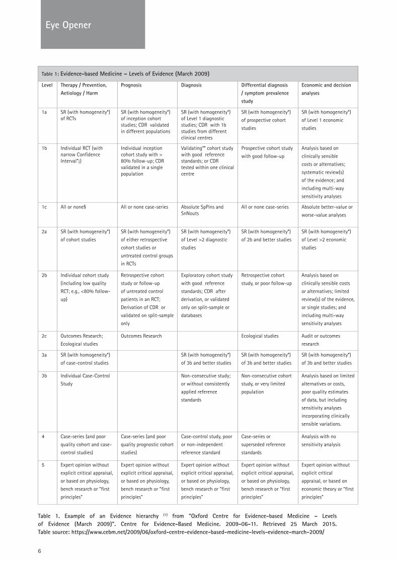
Scientific evidence is any knowledge that can support a statement, hypothesis or question. It can be strong or weak depending on the how the evidence has been collected. In the scientific setting you typically rank the strength of the evidence in a hierarchy based on the study design. The lowest evidence is expert opinion and statements based on personal observations (Table 1)
• 1a: Systematic reviews of randomized controlled trials• 1b: Individual randomized controlled trials• 1c: All or none randomized controlled trials• 2a: Systematic reviews of cohort studies• 2b: Individual cohort study or low quality randomized
controlled trials• 2c: "Outcomes" Research; ecological studies• 3a: Systematic review of case-control studies• 3b: Individual case-control study• 4: Case series• 5: Expert opinion without explicit critical appraisal, or based
on physiology, bench research or “first principles” The evidence has to be used systematically in order to avoid flaws
and biased treatments which are apparent in unsystematic expert based medical decision-making. This is the reason for the need of evidence based medicine.
Evidence based medicine (EBM)
EBM was developed as a new paradigm for medical practice 30 years ago (2). The working group from McMaster University, Canada,
had a focus on how to teach students.They said:“Evidence-based medicine de-emphasizes
• intuition• unsystematic clinical experience• pathophysiologic rationale
as sufficient grounds for clinical decision making and stresses the examination of evidence from clinical research”.
Sackett, who was part of the working group, moved EBM into the decision making in clinical practice. They defined EBM as “the conscientious, explicit, and judicious use of current best evidence in making decisions about the care of individual patients” (3).
It was pointed out that EBM is the common part of best external evidence, the patient value and expectations, as well as the individual clinical expertise of the profession.
Based on this, there will logically be some requirement for practising EBM
• Best external evidence. The evidence from literature has to be searched and evaluated systematically and in an unbiased way.
• The patient value and expectations. The patient has to be involved in the decision making in a dialogue with the surgeon and others involved in the treatment.
• The individual clinical expertise of the profession. Each surgeon has been educated according to national requirements. He/she will practice according to guidelines based on best external evidence.
Is there a need for EBM?
A natural way of learning is from the daily practice, where you recall and learn from the outcome of your cases. This is an important way of learning. You can get feedback from your colleagues and learn from x-rays, how well you have placed implant components or treated a fracture.
However, it is not the way of finding the best evidence for a treatment. Your memory is not systematic, it is biased on what you believe is the most suitable treatment of the patient and the case-mix of your patients. Trying to capture the success of a treatment from bits and pieces in an ongoing clinical practice is not a systematic evaluation of the best external evidence.
Thus, the need for EBM is obvious. It will guide the surgeon in the treatment based on best evidence and not on local traditions.
6
Eye Opener
Table 1. Example of an Evidence hierarchy (1) from "Oxford Centre for Evidence-based Medicine – Levels of Evidence (March 2009)". Centre for Evidence-Based Medicine. 2009-06-11. Retrieved 25 March 2015. Table source: https://www.cebm.net/2009/06/oxford-centre-evidence-based-medicine-levels-evidence-march-2009/
Table 1: Evidence-based Medicine – Levels of Evidence (March 2009)
Level Therapy / Prevention, Aetiology / Harm
Prognosis Diagnosis Differential diagnosis / symptom prevalence study
Economic and decision analyses
1a SR (with homogeneity*) of RCTs
SR (with homogeneity*) of inception cohort studies; CDR validated in different populations
SR (with homogeneity*) of Level 1 diagnostic studies; CDR with 1b studies from different clinical centres
SR (with homogeneity*)
of prospective cohort
studies
SR (with homogeneity*)
of Level 1 economic
studies
1b Individual RCT (with narrow Confidence Interval”¡)
Individual inception cohort study with > 80% follow-up; CDR validated in a single population
Validating** cohort study with good reference standards; or CDR tested within one clinical centre
Prospective cohort study
with good follow-up
Analysis based on
clinically sensible
costs or alternatives;
systematic review(s)
of the evidence; and
including multi-way
sensitivity analyses
1c All or none§ All or none case-series Absolute SpPins and SnNouts
All or none case-series Absolute better-value or
worse-value analyses
2a SR (with homogeneity*)
of cohort studies
SR (with homogeneity*)
of either retrospective
cohort studies or
untreated control groups
in RCTs
SR (with homogeneity*)
of Level >2 diagnostic
studies
SR (with homogeneity*)
of 2b and better studies
SR (with homogeneity*)
of Level >2 economic
studies
2b Individual cohort study
(including low quality
RCT; e.g., <80% follow-
up)
Retrospective cohort
study or follow-up
of untreated control
patients in an RCT;
Derivation of CDR or
validated on split-sample
only
Exploratory cohort study
with good reference
standards; CDR after
derivation, or validated
only on split-sample or
databases
Retrospective cohort
study, or poor follow-up
Analysis based on
clinically sensible costs
or alternatives; limited
review(s) of the evidence,
or single studies; and
including multi-way
sensitivity analyses
2c Outcomes Research;
Ecological studies
Outcomes Research Ecological studies Audit or outcomes
research
3a SR (with homogeneity*)
of case-control studies
SR (with homogeneity*)
of 3b and better studies
SR (with homogeneity*)
of 3b and better studies
SR (with homogeneity*)
of 3b and better studies
3b Individual Case-Control
Study
Non-consecutive study;
or without consistently
applied reference
standards
Non-consecutive cohort
study, or very limited
population
Analysis based on limited
alternatives or costs,
poor quality estimates
of data, but including
sensitivity analyses
incorporating clinically
sensible variations.
4 Case-series (and poor
quality cohort and case-
control studies)
Case-series (and poor
quality prognostic cohort
studies)
Case-control study, poor
or non-independent
reference standard
Case-series or
superseded reference
standards
Analysis with no
sensitivity analysis
5 Expert opinion without
explicit critical appraisal,
or based on physiology,
bench research or “first
principles”
Expert opinion without
explicit critical appraisal,
or based on physiology,
bench research or “first
principles”
Expert opinion without
explicit critical appraisal,
or based on physiology,
bench research or “first
principles”
Expert opinion without
explicit critical appraisal,
or based on physiology,
bench research or “first
principles”
Expert opinion without
explicit critical
appraisal, or based on
economic theory or “first
principles”
Eye
Ope
ner
7
Eye Opener
The surgeon has to involve the patient in the selection of the treatment method, based on guidelines. EBM and guidelines will help the patient to accept the treatment. Finally, EBM will have the ability to increase treatment quality and safety.
How is EBM applied?
Who should evaluate the best external evidence? It is a time-consuming process and requires certain skills. It is not feasible to have all surgeons doing this. This is the reason why guidelines have to be developed on a national or regional level. The process for this has to be transparent, to achieve acceptance from the orthopaedic society, patients, surgeons and health authorities. Certain principles, such as the GRADE approach (Grading of Recommendations Assessment, Development and Evaluation) and AGREE (The Appraisal of Guidelines for Research and Evaluation) facilitate this work (4,5). These statements are intended to assist practice guideline developers to improve the completeness and transparency of reporting in practice guidelines.
In the daily practice, the surgeon has to treat the patient according to guidelines. The treatment should involve the patient and a discussion on expectations is important. The surgeon should also consider his/her ability to perform the treatment.
Harmonisation & Diversity within EBM
It is important to understand that harmonisation has to respect diversity also in EBM.
Across countries, health care systems are different. Some countries have tax-supported public healthcare for all citizens, guaranteeing free or in-expensive medical care for both emergency and general hospital admission. Others have a mix, requiring payment and/or private insurance. Welfare is also different.
Although there is a European common EBOT exam, there is diversity within education, traditions among countries and availability of certain treatments. This may result in variation between guidelines although the same scientific evidence is available globally.
As part of the EFORT Mission & Vision, EBM guidelines should be developed and used on a local, national, or international level.
References:
1. "Oxford Centre for Evidence-based Medicine – Levels of Evidence (March 2009)". Centre for Evidence-Based Medicine. 2009-06-11. Retrieved 25 March 2015.
2. Evidence-based medicine. A new approach to teaching the practice of medicine. Evidence-Based Medicine Working Group. JAMA 1992 Nov 4;268(17):2420-5.
3. Sackett D, Rosenberg W, Gray J, Haynes R, Richardson W. Evidence based medicine: what it is and what it isn’t. BMJ 1996; 312:71–2.
4. https://www.gradeworkinggroup.org/5. https://www.agreetrust.org/

8
Basic Science
Basi
c Sc
ienc
e
9
Basic Science
Dr. Bernd Grimm, MEng, PhD
Sylvia Lawry Centre – The Human Motion Institute Munich, Germany
Biomechanics & Biomaterials For Musculoskeletal Application
1. Introduction
Biomechanics and Biomaterials are major fields of basic science which can cover semesters of teaching even when limited to musculoskeletal applications, orthopaedics or implants only. This lecture will focus on major concepts, terminology and applications of biomechanics and biomaterials most important to orthopaedic surgery and the curriculum.
This chapter will highlight the clinical relevance of basic biomechanics and biomaterials to motivate a reader of medical background. Concepts of biomechanics and biomaterials which are essential for understanding the beautiful interplay of biology, mechanics and materials in medicine are listed here.
For instance, all cells, in particular bone cells are mechanosensitive by mechanotransduction referring to the molecular mechanisms by which cells sense and respond to mechanical signals. This interplay of physical forces, movement and the mechanical or materials’ properties of cells and tissues and, the response in cell development, cell differentiation and thus tissue (re-) generation are referred to as mechanobiology. This interface of biology, mechanics and materials’ science is increasingly being recognized besides the traditional genetic and biochemical components as a basis of disease development and cure.
The Wolff’s law is a long established and commonly known expression of this principle, which says that the osseous bone structure, such as trabecular orientation and bone density optimizes itself in adaptation to the mechanical load.
2. Biomechanics
One common way to differentiate the disciplines of (bio)mechanics is referring to kinematics, statics and dynamics. Kinematics describes the motion of objects independent of the forces that originated it. Using kinematics, one can calculate a trajectory or momentary velocity of an object, using terminology such as translation for straight line movement, rotation for any curved movement and the degrees of freedom to refer to the number of independent translational and rotational coordinates available to describe an object’s position. In orthopaedics, a kinematic approach is common for instance in clinical movement analysis describing e.g. the phases of gait or outcome assessment of joint mobility such as range of motion or the screw-home mechanism common in natural knee kinematics.
Statics describe the forces and moments acting on an object at rest (or in steady motion with zero acceleration) keeping an
equilibrium. A force is defined as any influence that causes an object to undergo a certain change, either concerning its movement, direction, or geometrical construction (e.g. deformation, which depends on the material’s properties and linking biomechanics to biomaterials). Depending on the direction of effect, forces are described as in tension, compression or shear. When the force is calculated per area it is acting upon, the term stress is used.
A moment is defined as the tendency of a force to rotate an object around an axis. A moment (also called torque) affects rotational movement or causes bending or torsional deformation. In musculoskeletal applications, moments are introduced e.g. by the muscle contraction forces acting via their tendinous insertion points (deduced from insertion area) onto the bones via a lever arm around the joint axis biomechanically considered the fulcrum. Both moments and forces (and thus also stresses) are vectors which means we have a direction and orientation in space and a scalar magnitude. For force vectors one also refers to the line and point of application and the sense. Thus, a numerical value by its own does not sufficiently describe a situation in static biomechanics. Common units are: Newton [N] for force, Newtonmeter [Nm] for moments and [N/mm2, MPa] for stress.
A major principle in statics with many applications in orthopaedic considerations is the static equilibrium. For static equilibrium of an object (solid body), all forces and moments acting upon it cancel each other out, mathematically meaning that the sum (also called resultant) of forces are zero and the sum of moments about a point are also equal to zero. With forces and moments being 3D vectors, this leads to 6 scalar equations in space or 3 scalar equations in a simplified plane scenario (see Fig. 1). Being vectors, when calculating the lever arm length for a force causing a moment with reference to a fulcrum, the perpendicular distance to the “line of action” is considered.
To derive the relevant equilibrium equations, a so-called a free body diagram is made which is a graphical illustration used to visualize the forces and moments applied to a solid body and calculate the resulting reactions. As an example, the biceps force to statically and horizontally hold in hand an object can be calculated. For an object with mass m= 1kg, a weight (gravitational force) of 10N results. The forearm mass is assumed at 2kg resulting in a 20N weight, with its center of mass and thus line of action at 13cm horizontal distance (lever arm) to the elbow joint considered as fulcrum. Based on anatomic assumptions the distances (lever arms) from elbow joint to hand centre and biceps insertion point are 30cm and 5cm respectively. Resolving the equilibrium equations results in a biceps force of 112N, almost 4 times the weight of forearm and mass.

10
Basic Science
Figure 1: Free Body Diagram to calculate the biceps force required to hold in hand a 1kg mass
Dynamics (or kinetics) describes the interactions between forces and moments and the motions caused, taking into account the translational and rotational inertia of the moving objects such as swinging limbs or carried loads involved. In orthopaedics, dynamics is applied e.g. to calculate the loads and fracture risk of bone or implants during impacts from falls or accidents. Complex dynamic analysis is also applied e.g. in workplace or sports ergonomics, including also computer simulations.
In this context, the famous Newton’s laws of motion can be considered as the foundation of classic mechanics. The First law states that an object either remains at rest or continues to move at a constant velocity, unless acted upon by a force. The Second law states that the vector sum of the forces F on an object is equal to the mass m of that object multiplied by the acceleration a of the object: F = ma. The third law, brief in latin “actio est reactio”, states that when one body exerts a force on a second body, the second body simultaneously exerts a force equal in magnitude and opposite in direction on the first body. In an orthopaedic biomechanics application, this means for instance that for any movement of a limb, tissue or cell, a force must act upon it (1st law). The size of this force depends on the acceleration and inertia (mass, shape) of the object put in motion (2nd law). If for instance a person is pushing off the ground by toe off during gait, the floor experiences an equally sized but reversely orientated ground reaction force (3rd law), a principle used in laboratory force-plate measurements to calculate e.g. joint reaction forces (also called inverse dynamics). Newton’s second and third law lead directly to the “Law of conservation of momentum”, where momentum describes the product of mass and velocity, which in a closed system stays constant as a total; a law for instance applied in impact biomechanics. In combination with the “Law of conservation of energy” many dynamic situations in biomechanics can be calculated.
When the human body is modelled with many bones, multiple joints, muscles, muscle insertion points and for various movements or boundary conditions reflecting normal or pathological movement dynamics, many equations based on the principles above must be resolved simultaneously and numerically instead of analytically. The biomaterials’ properties of bones (see next chapter), muscles and tendons may be added. This complex task is now often performed using computer-based modelling programs such as Anybody (www.anybodytech.com).
3. Examples of biomechanics in orthopaedics
3.1 Wolff’s law, Davis’ law and Mechanostat
In the introduction, there was already a reference to Wolff’s law describing the remodeling of the osseous structure and density in an optimization process to the load regimen experienced by the bone. The involved process of mechanotransduction refers to mechanical signals such like forces being converted to biochemical signals in cellular signaling involving steps such as mechanocoupling, biochemical coupling, signal transmission, and cell response reflecting the inseparable interaction of biology and mechanics. It has been found that only cyclic loading can induce bone formation [Duncan and Turner, 1995] and the bone remodeling effect depends on duration, magnitude and rate of loading, factors also common in calculating e.g. fatigue of non-biological materials like steel.
In an extension of Wolff’s law for bone, the Davis’ law describes that also soft-tissues remodel along imposed mechanical demands including fibrous collagenous connective tissues such as ligaments, tendons and fascia [Frost, 2003]. Like with bone, the mechanical stimulation can fall into 4 categories, a) a level of insufficient mechanical challenge and subsequent loss of soft-tissue properties, such as the ones studied during space-flight, b) a low level sufficient for maintaining the status quo, c) a level of optimal training stimulus, succeeded by d), a level leading to overload or damage leading to edema and fibrosis.
This concept of mechanical load thresholds for maintaining, losing or gaining tissue was already introduced before for bone [Frost, 2003] referring to it as the Mechanostat or Utah Paradigm of Skeletal Physiology. For bone, thresholds values for stress and strain levels leading to bone loss, adaptation, gain or fracture as a result of (over-) load (modelling), normal use or disuse (remodeling) has been established for different bones such as the tibia or skull (see Fig 2). For instance, the modeling threshold referring to the mechanical stimulus leading to a gain in bone density is 1500 µstrain for the tibia and 250 µstrain for the skull, with µstrain being a dimensionless unit for the normalized deformation (elongation/compression) with 1000 µstrain referring to a 0.1% change in elongation. Feeding such thresholds into finite element computer simulations of loaded bones and implants has allowed the in-silico study of bone remodeling effects of various implant designs.
Figure 2: The Mechanostat model defining four regions and thresholds of elastic bone deformation and corresponding (re-)modeling response or fracture.
Basi
c Sc
ienc
e
11
Basic Science
3.2 Stress shielding
Stress shielding describes the reduction in bone density (osteopenia) due to mechanical stress removed or reduced below physiological levels or in other words, below the strain thresholds leading to bone loss as established by the Mechanostat. In orthopaedic surgery, stress shielding can be introduced by the resection of bone and the implant. The implant may be too stiff [Mellon and Tanner, 2012], a biomaterial’s property described below and referring to a relatively too low elastic deformation compared to the periprosthetic bone, a condition caused either by the material, the shape or size of the implant. Stress shielding may also be caused by the implant being incorrectly positioned or wrongly designed. Stress shielding associated bone loss may lead to mid- or long-term loosening of an initially stable implant and may require revision surgery.
3.3 Micromotion and implant fixation
Micromotion describes the relative cyclic motion between bone and implant during dynamic loading and is thus a phenomenon of kinetics and biomechanics. At the interface between bone and implant, there is always some level of relative motion depending on the loading scenario (activity, patient characteristics) and e.g. on the implant shape, bone/implant friction prior to osseointegration or the level of osteointegration combined with the stiffness of the surrounding bone and implant.
This level of micromotion is particularly important for the peri-implant bone healing during the direct post-operative phase when a mechanical stimulus is required for bone remodeling and formation at the bone/implant interface to foster osseointegration and secondary mechanical implant stability. When micromotion is too high, and such thresholds have been estimated at relative motion exceeding 150 µm [Pilliar et al., 1986], instead of bone fibrous tissue will form at interface, preventing osseointegration and leading to mechanically unstable implant and aseptic loosening.
One biomechanical way to prevent excessive micromotion beyond implant design is achieving high initial stability by seating the implant well like e.g. the uncemented hip stem with sufficient impact force during surgery (without causing periprosthetic fractures).
3.4 Gait analysis or knee kinematics
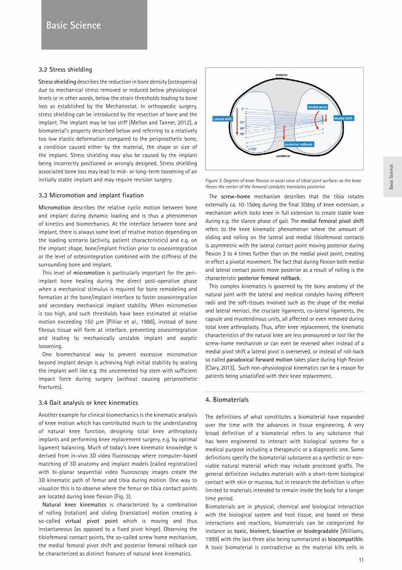
Another example for clinical biomechanics is the kinematic analysis of knee motion which has contributed much to the understanding of natural knee function, designing total knee arthroplasty implants and performing knee replacement surgery, e.g. by optimal ligament balancing. Much of today’s knee kinematic knowledge is derived from in-vivo 3D video fluoroscopy where computer-based matching of 3D anatomy and implant models (called registration) with bi-planar sequential video fluoroscopy images create the 3D kinematic path of femur and tibia during motion. One way to visualize this is to observe where the femur on tibia contact points are located during knee flexion (Fig. 3).
Natural knee kinematics is characterized by a combination of rolling (rotation) and sliding (translation) motion creating a so-called virtual pivot point which is moving and thus instantaneous (as opposed to a fixed pivot hinge). Observing the tibiofemoral contact points, the so-called screw home mechanism, the medial femoral pivot shift and posterior femoral rollback can be characterized as distinct features of natural knee kinematics.
Figure 3: Degrees of knee flexion in axial view of tibial joint surface: as the knee flexes the center of the femoral candyles translates posterior.
The screw-home mechanism describes that the tibia rotates externally ca. 10-15deg during the final 30deg of knee extension, a mechanism which locks knee in full extension to create stable knee during e.g. the stance phase of gait. The medial femoral pivot shift refers to the knee kinematic phenomenon where the amount of sliding and rolling on the lateral and medial tibiofemoral contacts is asymmetric with the lateral contact point moving posterior during flexion 3 to 4 times further than on the medial pivot point, creating in effect a pivotal movement. The fact that during flexion both medial and lateral contact points move posterior as a result of rolling is the characteristic posterior femoral rollback.
This complex kinematics is governed by the bony anatomy of the natural joint with the lateral and medical condyles having different radii and the soft-tissues involved such as the shape of the medial and lateral menisci, the cruciate ligaments, co-lateral ligaments, the capsule and myotendinous units, all affected or even removed during total knee arthroplasty. Thus, after knee replacement, the kinematic characteristics of the natural knee are less pronounced or lost like the screw-home mechanism or can even be reversed when instead of a medial pivot shift a lateral pivot is overserved, or instead of roll-back so called paradoxical forward motion takes place during high flexion [Clary, 2013]. Such non-physiological kinematics can be a reason for patients being unsatisfied with their knee replacement.
4. Biomaterials
The definitions of what constitutes a biomaterial have expanded over the time with the advances in tissue engineering. A very broad definition of a biomaterial refers to any substance that has been engineered to interact with biological systems for a medical purpose including a therapeutic or a diagnostic one. Some definitions specify the biomaterial substance as a synthetic or non-viable natural material which may include processed grafts. The general definition includes materials with a short-term biological contact with skin or mucosa, but in research the definition is often limited to materials intended to remain inside the body for a longer time period. Biomaterials are in physical, chemical and biological interaction with the biological system and host tissue, and based on these interactions and reactions, biomaterials can be categorized for instance as toxic, bioinert, bioactive or biodegradable [Williams, 1999] with the last three also being summarized as biocompatible. A toxic biomaterial is contradictive as the material kills cells in
12
Basic Science
contact with the implant. A bioinert biomaterial in the pure sense of its definition produces no response by the body but as there is always some interaction and a bodily response, bioinert refers to materials with minimal effect on the body. A bioactive biomaterial encourages a desired and advantageous response from the body which may depend on the location of the implant in the body. A biodegradable biomaterial breaks down in the body to non-toxic components or compounds which can be excreted by the body.
Common forms of interaction between biomaterials and the body include corrosion, especially of metals changing their properties and releasing metal ions, or protein absorption of e.g. polymers degrading their properties.
In the context of biomaterials, the body’s living, natural materials are also studied to understand the basis of their chemical, physical and biological interactions. In an orthopaedic context, the properties, in particular the biomechanical characteristics of bone, cartilage, tendon, ligaments and meniscus are studied at macro- and microscopic level.
4.1 Mechanical properties of biomaterials
Materials and biomaterials can be categorized into four basic groups based on their structure at molecular and atomic level: metals, ceramics, polymers and composites. These can be distinguished also by their characteristic mechanical properties so that understanding the related metrics is fundamental for understanding biomaterials.
Many essential biomaterial’s properties are derived from how the material deforms and eventually fails (breaks) under load. These properties are studied in standardized laboratory tests to derive comparable metrics via e.g. the so-called stress-strain curve during quasi-constant loading. As previously defined stress σ is the force per unit area [N/mm2, MPa] and strain ε the relative deformation [%]. Thus, the normalized parameters derived from this test characterize the material for comparison and feed into biomechanical calculations for implant specific dimensions and loads.
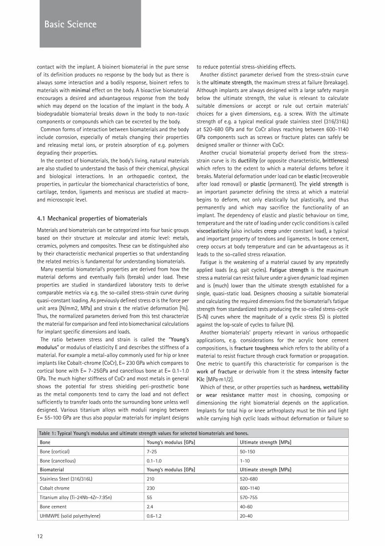
The ratio between stress and strain is called the “Young’s modulus” or modulus of elasticity E and describes the stiffness of a material. For example a metal-alloy commonly used for hip or knee implants like Cobalt-chrome (CoCr), E= 230 GPa which compares to cortical bone with E= 7-25GPa and cancellous bone at E= 0.1-1.0 GPa. The much higher stiffness of CoCr and most metals in general shows the potential for stress shielding peri-prosthetic bone as the metal components tend to carry the load and not deflect sufficiently to transfer loads onto the surrounding bone unless well designed. Various titanium alloys with moduli ranging between E= 55-100 GPa are thus also popular materials for implant designs
to reduce potential stress-shielding effects.Another distinct parameter derived from the stress-strain curve
is the ultimate strength, the maximum stress at failure (breakage). Although implants are always designed with a large safety margin below the ultimate strength, the value is relevant to calculate suitable dimensions or accept or rule out certain materials’ choices for a given dimensions, e.g. a screw. With the ultimate strength of e.g. a typical medical grade stainless steel (316/316L) at 520-680 GPa and for CoCr alloys reaching between 600-1140 GPa components such as screws or fracture plates can safely be designed smaller or thinner with CoCr.
Another crucial biomaterial property derived from the stress-strain curve is its ductility (or opposite characteristic, brittleness) which refers to the extent to which a material deforms before it breaks. Material deformation under load can be elastic (recoverable after load removal) or plastic (permanent). The yield strength is an important parameter defining the stress at which a material begins to deform, not only elastically but plastically, and thus permanently and which may sacrifice the functionality of an implant. The dependency of elastic and plastic behaviour on time, temperature and the rate of loading under cyclic conditions is called viscoelasticity (also includes creep under constant load), a typical and important property of tendons and ligaments. In bone cement, creep occurs at body temperature and can be advantageous as it leads to the so-called stress relaxation.
Fatigue is the weakening of a material caused by any repeatedly applied loads (e.g. gait cycles). Fatigue strength is the maximum stress a material can resist failure under a given dynamic load regimen and is (much) lower than the ultimate strength established for a single, quasi-static load. Designers choosing a suitable biomaterial and calculating the required dimensions find the biomaterial’s fatigue strength from standardized tests producing the so-called stress-cycle (S-N) curves where the magnitude of a cyclic stress (S) is plotted against the log-scale of cycles to failure (N).
Another biomaterials’ property relevant in various orthopaedic applications, e.g. considerations for the acrylic bone cement compositions, is fracture toughness which refers to the ability of a material to resist fracture through crack formation or propagation. One metric to quantify this characteristic for comparison is the work of fracture or derivable from it the stress intensity factor ΚIc [MPa·m1/2].
Which of these, or other properties such as hardness, wettability or wear resistance matter most in choosing, composing or dimensioning the right biomaterial depends on the application. Implants for total hip or knee arthroplasty must be thin and light while carrying high cyclic loads without deformation or failure so
Table 1: Typical Young’s modulus and ultimate strength values for selected biomaterials and bones.
Bone Young’s modulus [GPa] Ultimate strength [MPa]
Bone (cortical) 7-25 50-150
Bone (cancellous) 0.1-1.0 1-10
Biomaterial Young’s modulus [GPa] Ultimate strength [MPa]
Stainless Steel (316/316L) 210 520-680
Cobalt chrome 230 600-1140
Titanium alloy (Ti-24Nb-4Zr-7.9Sn) 55 570-755
Bone cement 2.4 40-60
UHMWPE (solid polyethylene) 0.6-1.2 20-40
Basi
c Sc
ienc
e
13
Basic Science
that fatigue and ultimate strength are most crucial. For a suture material, fatigue and ultimate strength seem less important, as failure would likely occur via pull-out through the soft-tissue rather than rupture of the suture. Here a high Young’s modulus defines the handling properties.
Besides a material’s bulk properties described above, biomaterials derive and influence specific relevant properties via their structure such as porosity (at macro- and micro-level, inter-connectivity), e.g. in porous metals, surface topography (at macro-, micro- or even nano-level such as scales or surface roughness to reduce biofilm formation and infection) or combine properties by coating a biomaterial onto a substrate.
4.2 Common Biomaterials
Following the four basic material categories - metals, ceramics, polymers and composites - some common biomaterials are discussed.
Metals
Metal biomaterials are all special alloys, a term which refers to a mixture of different metals or a mixture of a metal with another element such as carbon, all forming metallic bonds with the atoms in a crystalline lattice. Metal alloys are highly complex systems and may exhibit different phases similar to a composite material. They can be thus tuned regarding their desired properties (metallurgy). In general, metals have a high Young’s modulus (stiff), exhibit a high fatigue strength and mostly behave ductile which means they plastically deform (an advantageous property for molding implants such as e.g. fracture plates into shape or in service to prevent catastrophic breakage).
The major metals used in orthopaedics are alloys of stainless steels, cobalt chrome and titanium (see Table 1). In stainless steel alloys, the presence of chromium causes a chromium oxide surface layer preventing further corrosion. CoCr alloys are low or free of Nickel, important for patients with nickel sensitivity. Titanium alloys, especially those of TiNb and TiMo have low Young’s moduli to get closer to the stiffness of bone and prevent stress shielding. Porous metals such as titanium alloys or tantalum can even match the stiffness of cancellous bone. In recent years the adverse local tissue reactions (ALTR) around metal-on-metal (MoM) hip arthroplasties and the spread of modular implants such as hip stems with modular head-neck junctions have put new emphasis on the tribo-corrosive properties of metals.
Ceramics
A ceramic is an inorganic compound, non-metallic, solid material comprising metal or non-metal atoms, primarily held in ionic and covalent bonds. The crystallinity of ceramic materials ranges from highly oriented to semi-crystalline or even completely amorphous, e.g., (bio-)glasses.
Bioceramics can be divided into 2 major groups. Bioinert ceramics are mainly zirconia (ZrO2) and alumina (Al2O3) or combinations thereof, such as zirconia toughened alumina as used for articulating bearing surfaces in hip, shoulder and knee replacements. Bioactive ceramics are mainly hydroxyapatite (HA), such as the mineral phase of natural bone and tricalcium phosphates (TCP) and their composites to tune their strength and resorption properties to the
application. Ceramics, as a bearing surface against each other and against
polyethylene, have been in long-term successful clinical use. They benefit from the smoother surface roughness, superior hardness creating a scratch and third-body wear resistant articulation surface and their higher wettability, all causing very low wear rates of the ceramic and also lower wear of polyethylene if paired against it. Furthermore, ceramic bearings release no metal ions, have no allergenic risk, lower fretting corrosion at the modular neck and a pathogenic reaction to ceramic wear particles is unlikely. Reducing ceramic grain size and toughening with adding SrO, Y2O3 and Cr2O3
to the compositions has reduced early incidents of brittle ceramic component fracture.
Bioactive ceramics are used in six major applications: 1) bulk implants, that is space filling implants, 2) porous implants when used as implants for ingrowth or 3) scaffolds for tissue engineering, 4) granules used to supplement or to replace autologous bone graft such as in impaction grafting, 5) coatings which can be plain hydroxyapatite (HA - Ca10(PO4)6(OH)2), tricalcium phosphate (TCP – Ca3(PO4)2) or HA+TCP (also called biphasic calcium phosphate - BCP) and 6) as injectables where the calcium phosphate, with or without some calcium sulphate (plaster of Paris - CaSO4) and other additives, is mixed in the operating theatre, injected into the body and sets in situ. The degradation rate depends on the crystallinity of the ceramic phases and their relative amounts. Degradation rates increase from HA, to TCP, to calcium sulphate. The required degradation rate depends on the clinical application.
Polymers
Polymers are a class of materials defined by comprising large molecules, named macromolecules, composed of many repeated subunits. Their consequently large molecular mass and way of molecular order produce a broad range of physical properties, including toughness, viscoelasticity, and a tendency to form glasses and semicrystalline structures rather than crystals.
Polymers typically used in orthopaedics are primarily ultrahigh molecular weight polyethylene (UHMWPE), polymethylmethacrylate (PMMA), other methacrylates, polyesters, poly(glycolic acid) and poly(lactic acid) and hydrogels.
For the use of UHMWPE as a bearing surface, the wear resistance, oxidation resistance, fatigue strength and fracture toughness have been improved during the last 25 years by a variety of manufacturing steps with the most effective having been molecular cross-linking, to increase wear resistance and adding anti-oxidants such as Vitamin E to increase the oxidation resistance (Gu, Li et al. 2014).
PMMA is a thermoset polymer which in clinical application is created through exothermic polymerization of a powder phase, pre-polymerised polymethylmethacrylate beads plus liquid benzoyl peroxide which initiates the polymerization. PMMA is combined with a radiopacifier in the form of barium sulphate or zirconia. PMMA bone cement is used to fix (grout, not glue) joint replacements in place or is used to space fill and thus stabilize osteoporotic bone against fracture such as in spinal vertebroplasty and kyphoplasty.
Desired properties of PMMA bone cement are fatigue strength, resistance to crack initiation or propagation, and, the long, predictable window of workable viscosity in the operating theatre
14
Basic Science
before setting. Vacuum mixing to reduce weakening voids, retrograde cementing and cement pressurization during the hardening phase for high cement-bone interdigitation, have been identified as crucial surgical steps for an optimal PMMA mantle.
The main degradable polymers in clinical use are the thermoplastic Poly(lactic acid) PLA and Poly(glycolic acid) PGA. Chemically these break down into lactic and glycolic acid, which the body breaks down to CO2 and H2O and is excreted. In orthopaedic surgery PGA is used in resorbable sutures due to the fast degradation of PGA within the body. Due to its lower degradation rate, PLA is used for fracture fixation in low load bearing applications under the form of internal fixation plates. The current challenge with degradable polymers are their low strength and too fast degradation rate for e.g. fracture healing. In attempts to improve the strength, fibre reinforcement and ceramic reinforcement have been used [Bleach, Nazhat et al. 2002; Huttunen, Törmälä et al. 2008]. PLA/PGA composites are also available as resorbable meshes and screws for bone augmentation. Recently PLA has become popular as a biopolymer suitable for 3D printing. In orthopaedics the application as a template for pre-operative contouring of fracture plates has been shown.
Composites
A composite is a material made from two or more constituent materials with significantly different physical or chemical properties that, when combined, produce a material with characteristics different from the individual components. The individual components remain separate and distinct within the finished composite. Living bone is a composite material by nature combining collagen and HA to create an extremely tough, yet lightweight, adaptive and multi-functional material.
A major type of composite biomaterials used are polymers reinforced with ceramics such as glasses, HA or TCP for e.g. arthroscopic interference screws. Also in use are polymers reinforced, with another polymer or the same polymer but in another form such as drawn fibres in an amorphous matrix of the same polymer. One example of this is the PLLA in PLDLA used in some degradable fracture fixation plates.
Carbon-reinforced PEEK (Polyether ether ketone) is a polymer which has demonstrated good wear characteristics in experimental wear simulation in simple geometry pin-on-plate studies and in total hip joint replacement, but which may be less suited for low-conformity designs such as knee replacement.
With composite material compositions allowing in theory the creation of application-optimized biomaterials, including functionality such as controlled drug release, sensing, or self-repair, many novel composite biomaterials have been explored [Tanner, 2010]. One example of these manifold efforts are e.g. carbon nanotube, graphene and boron nitride nanotube reinforced bioactive ceramics for bone repair which have made great advances but are not yet available in orthopaedic clinic.
5. Conclusions
Orthopaedic surgery placing an implant into the body can only be performed successfully when the two interacting factors are considered and understood. One factor is how the implant is loaded, thus the biomechanics, and the other factor is the material the implant is made of and its properties, thus the biomaterials. While much of this is considered during design, development and testing, the orthopaedic surgeon is faced with many implant choices and the decisions for the optimal design, size, placement and fixation are all affected by the biomechanics and biomaterials interactions described here.
Questions
1. In biomechanics, what is the difference between kinematics and kinetics?
2. What is stress shielding and how can it be reduced?3. Which kinematic behaviour of the natural knee contributes
to joint stability during the stance phase of gait?4. What is the difference between a ductile and a brittle
biomaterial?5. How can advantageous properties of different biomaterials
be combined to best serve clinical demands? Give one example.
Answers
1. Kinematics describes the motion of objects independent of the forces that originated it. Kinetics (or dynamics) describes the interactions between forces and moments, and the motions caused taking into account the translational and rotational inertia of the moving objects.
2. Stress shielding describes a loss of bone (osteopenia) due to mechanical stress removed or reduced below physiological levels by an implant. Stress shielding can, for instance, be reduced by matching the stiffness or the implant and material to the surrounding bone or designing the implant to transfer load to the bone instead of shielding from it.
3. During stance, the knee is at or near full extension and, as defined by the tibiofemoral bony morphology and soft-tissue (ligaments, meniscus) properties, the tibia rotates externally ca. 10-15deg during the final 30deg of knee extension “locking” the knee in full extension. This kinematic characteristic is called “screw-home mechanism”.
4. Ductility and brittleness refer to the extent of deformation, elastic and plastic, a biomaterial undergoes before breaking under load. A ductile material deforms much before failure while a brittle material shows small deformation before failure. Most metals are usually ductile so that they can be molded into shape. Most ceramics behave usually brittle and instead of deforming near failure loads, may break by e.g. bursting.
Basi
c Sc
ienc
e
15
Basic Science
5. Advantageous properties of different biomaterials can be combined by coatings or creating a composite biomaterial. One example would be the use of a mechanically strong and elastic titanium hip stem coated with porous HA to enhance osseointegration. Another example is an arthroscopic interference screw, made composite of PLDLA polymer and calcium-phosphate, to make the screw strong and resorbable.
References
1. Bleach, N. C., S. N. Nazhat, et al. (2002). “Effect of Filler Content on Mechanical and Dynamic Mechanical Properties of Particulate Biphasic Calcium Phosphate Polylactide Composites”.” Biomaterials 23(7): 1579-1585.
2. Clary CW, Fitzpatrick CK, Maletsky LP, Rullkoetter PJ. The influence of total knee arthroplasty geometry on mid-flexion stability: an experimental and finite element study. J Biomech. 2013 Apr 26;46(7):1351-7.
3. Claire L. Brockett, Silvia Carbone, John Fisher, and Louise M. Jennings PEEK and CFR-PEEK as alternative bearing materials to UHMWPE in a fixed bearing total knee replacement: An experimental wear study Wear. 2017 Mar 15; 374-375: 86–91.
4. Currey, J. D. (2006). Bones: Structure and Mechanics, Princeton University Press.
5. Duncan, RL; CH Turner (November 1995). "Mechanotransduction and the functional response of bone to mechanical strain". Calcified Tissue International. 57 (5): 344–358.
6. Ellenbecker, Todd, "Effective Functional Progressions in Sport Rehabilitation", Human Kinetics 2009, ISBN 0-7360-6381-1
7. Frost, Harold "New targets for fascial, ligament and tendon research: A perspective from the Utah paradigm of skeletal physiology" J Musculoskel Neuron Interact 2003; 3(3):201-209
8. Gao C, Feng P, Peng S, Shuai C. Carbon nanotube, graphene and boron nitride nanotube reinforced bioactive ceramics for bone repair. Acta Biomater. 2017 Oct 1;61:1-20
9. Huttunen, M., P. Törmälä, et al. (2008). “Fiber-reinforced bioactive and bioabsorbable hybrid composites.” Biomedical Materials 3(3).
10. Mellon, S. and K. E. Tanner (2012) “Mechanical Adaptability of Bone in Vivo and in Vitro – A Review”, International Materials Reviews 25(5), 235-255
11. Nordin, M, Frankel, VH. Basic Biomechanics Of The Musculoskeletal System. Philadelphia: Lippincott Williams and Wilkins, 2012. 4th ed.
12. Pilliar RM, Lee JM, Maniatopoulos C. Observations on the effect of movement on bone ingrowth into porous-surfaced implants. Clin Orthop Relat Res. 1986;208:108–113
13. Tanner, K. E. (2010). Hard tissue applications of biocomposites. Biomedical Composites. Ed L. Ambrosio. Cambridge, UK, Woodhead Publishers.
14. Williams, D. F. (1999). The Williams Dictionary of Biomaterials. Liverpool, Liverpool University Press
16
Basic Science
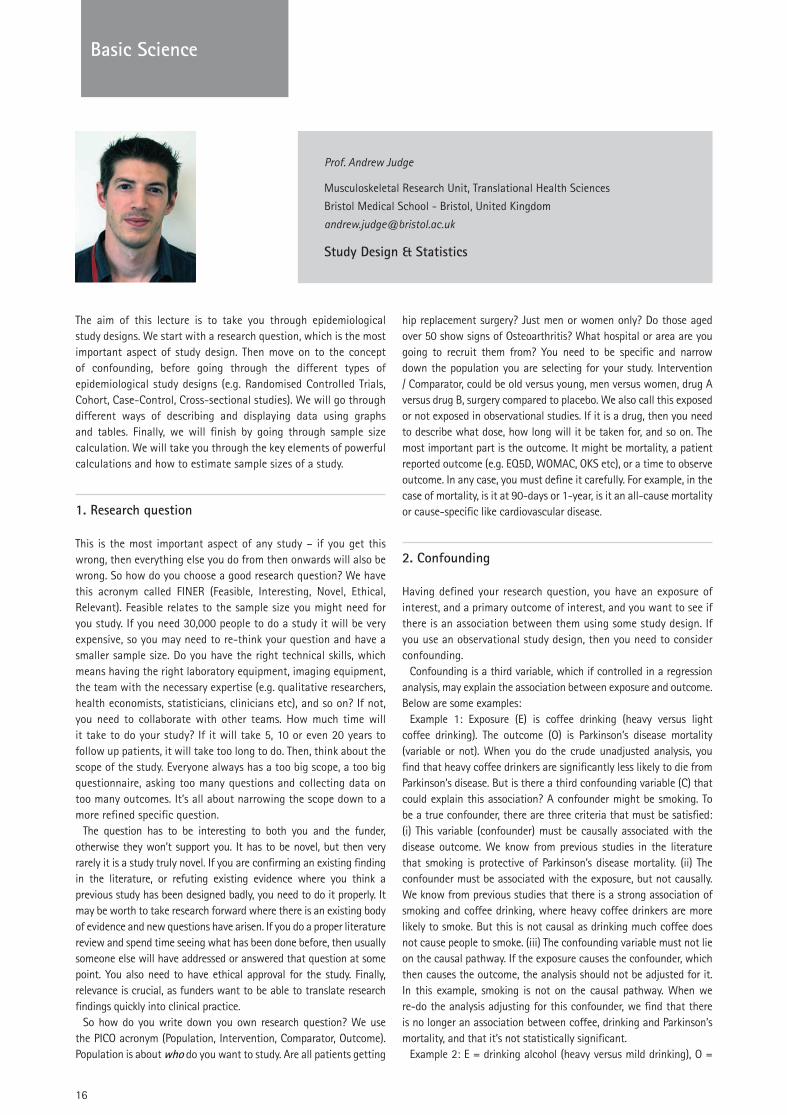
Prof. Andrew Judge
Musculoskeletal Research Unit, Translational Health Sciences
Bristol Medical School - Bristol, United Kingdom
Study Design & Statistics
The aim of this lecture is to take you through epidemiological study designs. We start with a research question, which is the most important aspect of study design. Then move on to the concept of confounding, before going through the different types of epidemiological study designs (e.g. Randomised Controlled Trials, Cohort, Case-Control, Cross-sectional studies). We will go through different ways of describing and displaying data using graphs and tables. Finally, we will finish by going through sample size calculation. We will take you through the key elements of powerful calculations and how to estimate sample sizes of a study.
1. Research question
This is the most important aspect of any study – if you get this wrong, then everything else you do from then onwards will also be wrong. So how do you choose a good research question? We have this acronym called FINER (Feasible, Interesting, Novel, Ethical, Relevant). Feasible relates to the sample size you might need for you study. If you need 30,000 people to do a study it will be very expensive, so you may need to re-think your question and have a smaller sample size. Do you have the right technical skills, which means having the right laboratory equipment, imaging equipment, the team with the necessary expertise (e.g. qualitative researchers, health economists, statisticians, clinicians etc), and so on? If not, you need to collaborate with other teams. How much time will it take to do your study? If it will take 5, 10 or even 20 years to follow up patients, it will take too long to do. Then, think about the scope of the study. Everyone always has a too big scope, a too big questionnaire, asking too many questions and collecting data on too many outcomes. It’s all about narrowing the scope down to a more refined specific question.
The question has to be interesting to both you and the funder, otherwise they won’t support you. It has to be novel, but then very rarely it is a study truly novel. If you are confirming an existing finding in the literature, or refuting existing evidence where you think a previous study has been designed badly, you need to do it properly. It may be worth to take research forward where there is an existing body of evidence and new questions have arisen. If you do a proper literature review and spend time seeing what has been done before, then usually someone else will have addressed or answered that question at some point. You also need to have ethical approval for the study. Finally, relevance is crucial, as funders want to be able to translate research findings quickly into clinical practice.
So how do you write down you own research question? We use the PICO acronym (Population, Intervention, Comparator, Outcome). Population is about who do you want to study. Are all patients getting
hip replacement surgery? Just men or women only? Do those aged over 50 show signs of Osteoarthritis? What hospital or area are you going to recruit them from? You need to be specific and narrow down the population you are selecting for your study. Intervention / Comparator, could be old versus young, men versus women, drug A versus drug B, surgery compared to placebo. We also call this exposed or not exposed in observational studies. If it is a drug, then you need to describe what dose, how long will it be taken for, and so on. The most important part is the outcome. It might be mortality, a patient reported outcome (e.g. EQ5D, WOMAC, OKS etc), or a time to observe outcome. In any case, you must define it carefully. For example, in the case of mortality, is it at 90-days or 1-year, is it an all-cause mortality or cause-specific like cardiovascular disease.
2. Confounding
Having defined your research question, you have an exposure of interest, and a primary outcome of interest, and you want to see if there is an association between them using some study design. If you use an observational study design, then you need to consider confounding.
Confounding is a third variable, which if controlled in a regression analysis, may explain the association between exposure and outcome. Below are some examples:
Example 1: Exposure (E) is coffee drinking (heavy versus light coffee drinking). The outcome (O) is Parkinson’s disease mortality (variable or not). When you do the crude unadjusted analysis, you find that heavy coffee drinkers are significantly less likely to die from Parkinson’s disease. But is there a third confounding variable (C) that could explain this association? A confounder might be smoking. To be a true confounder, there are three criteria that must be satisfied: (i) This variable (confounder) must be causally associated with the disease outcome. We know from previous studies in the literature that smoking is protective of Parkinson’s disease mortality. (ii) The confounder must be associated with the exposure, but not causally. We know from previous studies that there is a strong association of smoking and coffee drinking, where heavy coffee drinkers are more likely to smoke. But this is not causal as drinking much coffee does not cause people to smoke. (iii) The confounding variable must not lie on the causal pathway. If the exposure causes the confounder, which then causes the outcome, the analysis should not be adjusted for it. In this example, smoking is not on the causal pathway. When we re-do the analysis adjusting for this confounder, we find that there is no longer an association between coffee, drinking and Parkinson’s mortality, and that it’s not statistically significant.
Example 2: E = drinking alcohol (heavy versus mild drinking), O =

Basi
c Sc
ienc
e
17
Basic Science
lung cancer mortality. In an unadjusted crude analysis, we see heavy drinkers have an increased risk of lung cancer. The confounder in this case is smoking. In the adjusted analysis the association between drinking and lung cancer is no longer significant.
Example 3: E = Smoking (yes or no), O = Cardiovascular disease (yes or no), C = Atherosclerosis (yes or no). Is Atherosclerosis a confounder? No – it is on the causal pathway. Smoking causes Atherosclerosis, which causes Cardiovascular disease. Thus, the analysis should not be adjusted for this as a confounder.
It is important to read the literature to identify all potential confounders for your research question. If you have not collected the data on potential confounders, then you cannot later adjust for them in the analysis, and this may explain your study’s findings.
3. Study Design
There are two main types of study – interventional and observational. In an interventional study, we intervene to alter the course of disease, (e.g. by introducing a new treatment). Whereas in an observational study we just observe what happens.
3.1 Randomised Controlled Trials (RCT)
This is the interventional study design. To start with you need to define the population of interest (as in the PICO for the research question). From this group, you will then identify which patients are eligible to take part on the trial, based on your inclusion and exclusion criteria. Patients contacted need to have informed consent to take part in the study. These patients are then randomised to be in either the intervention group, or the control group. The randomisation means that each person has an equal chance (probability) of being selected to be in either the intervention or the control groups. Baseline measurements are taken, and patients are then followed up to collect data on outcomes. An RCT is the gold standard study design, and the main reason for that is that there should be no confounding. This is due to the randomisation: the only way the two groups will differ is if they get treatment or control. They will be balanced in respect of all other confounding factors. e.g. mean age, proportion of women, ethnicity groups, body mass index, etc should all be same in both intervention and control groups. This includes not just confounders that are known, measured and collected, but also unknown and unmeasured confounders.
Other important parameter to ensure in an RCT is ‘concealment of allocation’. What this means is that the person who is doing the randomisation, cannot influence who is allocated to treatment or control. For example, if ordered opaque envelopes are used, a clinician may be tempted to hold the envelope up to a light to see what treatment allocation is and be able to influence this by allocating a sicker patient to intervention. If this happens it will cause a selection bias. Therefore we use computer-generated randomisation at a distant site, not involved in the study. Importantly, we need to use an Intention-To-Treat (ITT) analysis, which analyses people in the groups as they were at the point of randomisation. If a patient switches groups (e.g. from control to intervention), doesn’t complete the follow-up, drops out or doesn’t comply with the treatment, the study is still accurate. If there is missing data on outcomes, you will need to impute the outcomes, so groups are the same sample size as they were at
the point of randomisation. If instead we did a completers or on-treatment analysis of only those who comply or complete the follow-up, this will introduce a bias to the study. This is in the form of selection bias, known as loss-to-follow-up bias. If loss-to-follow-up is similar in both sections of the trial (smaller than 5%)then this is unlikely to have much effect. But, if it is differential, and loss-to-follow-up is higher in one group, and this is related to the outcome (e.g. drug side effects), then selection bias may be introduced. ITT analyses help to avoid this. The final biases relate to measurement error in either treatment or outcome. To avoid this, we use patient and assessor blinding. If the patient works out, gets the active drug or the placebo, takes other medications or treatments, there is a bias. Likewise, if the person assessing outcomes is aware of treatment allocation, it may influence the way outcomes are recorded. Therefore, proper patient and assessor blinding are important to avoid measurement errors.
If all the above is taken into consideration, then a large well conducted RCT is the gold standard, as we minimise the possibility of confounding, selection bias, and measurement error. Then we can begin to consider that the RCT study findings may be causal. The main limitation of the RCT is the lack of generalisability. The type of people that are selected to take part in trials are not necessarily representative of those in the general population who would be offered treatment. Take for example bisphosphonates for treatment of osteoporosis. Adherence to therapy in trials is much higher than in the general population. Patients in trials are also less likely to have co-morbidities and contra-indications, due to strict inclusion and exclusion criteria. So even if a drug or treatment is shown to be clinically effective in a trial setting, it may not be
when introduced to the wider general population.
3.2 Prospective Cohort study
This is an observational study design. We start by defining the population of interest. The population is normally defined by some common characteristic (e.g. all went to the same school, were born in the same year, etc). At the start of the study patients must be disease free, meaning that they cannot have the outcome of interest. At baseline, we measure a wide range of exposure and confounding variables. Patients are then followed up until they get the outcome of interest.
It is the strongest type of observational study design because the temporal sequence of events is clear – cause precedes effect. It is also the best observational study if you have rare exposures. Further strengths are that the cohort study can be used to answer many different research questions. The limitations are that the sample sizes are usually very large, with long follow-up, making them expensive and time-consuming. An alternative to the prospective cohort study, is to consider a retrospective (historical cohort) study design. Here we use existing data (e.g. joint registries, hospital admissions, primary care datasets), whereby we start in the past when patients are disease free, measure and collect exposures, and follow patients to the present day to see associations with outcome. Limitations of this are that such datasets were not typically designed for research purposes, and so may lack data on important confounders, but they will be quicker and cheaper to do. Further limitations are in respect of confounding, and the potential for measurement error in either exposure or outcome. There will also be loss to follow up over the duration of the study, so again we
18
Basic Science
need to consider a possibility of selection bias. In an observational study we are only assessing associations, not causality, as there will always be potential limitations in respect of bias and confounding.
3.3 Case Control study
This observational study design is best used for rare outcomes. People are selected for inclusion in the study based on their outcome and data on past exposures is considered. The main limitation and difficulty is how to select controls who don’t have the outcome. Selecting the wrong controls can introduce selection bias. For example, in a study about the association between alcohol consumption and liver disease, if we choose our cases as patients with liver disease admitted to hospital, and hospital controls who don’t have liver disease, this could introduce a bias. The reason being that drinking is one of the most common reasons for hospital admissions, and the population of patients in a hospital setting are more likely to be drinkers than in the general population. Hence, with these controls, we may underestimate the association with outcome. The other problem with this study design is measurement error as we are asking people to recall past exposures. If for example we are looking at the association between having leukaemia and living near power lines, the parents of leukaemia patients may say they live close to power lines, whereas measure of the actual distance would not show closeness. Likewise, if a researcher is interviewing patients for a study on sun cream use and skin cancer, if the researcher knows who the cases and controls are, it may bias the way he collects data on past exposure, being more detailed for those cases with outcome. Analyses will also need to attempt to adjust for confounding. In this design, it is also unclear whether cause proceeds effect, and the temporal sequence of events is less clear than in a cohort study.
3.4 Cross-sectional study
In this observational design, there is a single point in time (a snap shot). Data on exposures and outcome are all collected at the same point in time, and hence the temporal sequence of events is unclear. For example, is cannabis consumption associated with schizophrenia, or are schizophrenics more likely to smoke cannabis? In this study design we can’t define if cause proceeds effect. Aside from this, it is very similar to a cohort study at baseline, because you identify your population of interest, take a sample of these to include in the study, and collect baseline data on exposure and outcome. This study design is very useful for estimating disease prevalence, but if doing so, you must be very careful in how you define your study population. Say for example you want to estimate the prevalence of Chlamydia. If you recruited patients attending a Genito-urinary medicine clinic, then you would only measure the prevalence of symptomatic disease, in those who have sought treatment. There will be people whose disease is asymptomatic, so you need to sample from the wider general population.
You may want to estimate the prevalence of hip pain that requires surgery. If you sampled those in a hospital setting, you would underestimate the true prevalence, as these are only the people who have sought help for their symptoms and managed to get access to surgery. There will be people with severe pain that do not seek help from a GP or surgeon, and have not accessed services, even though they are in clinical need. There will be people in
need that have sought help, but there are barriers to them accessing surgery.
4. Describing and displaying data
4.1 Types of data
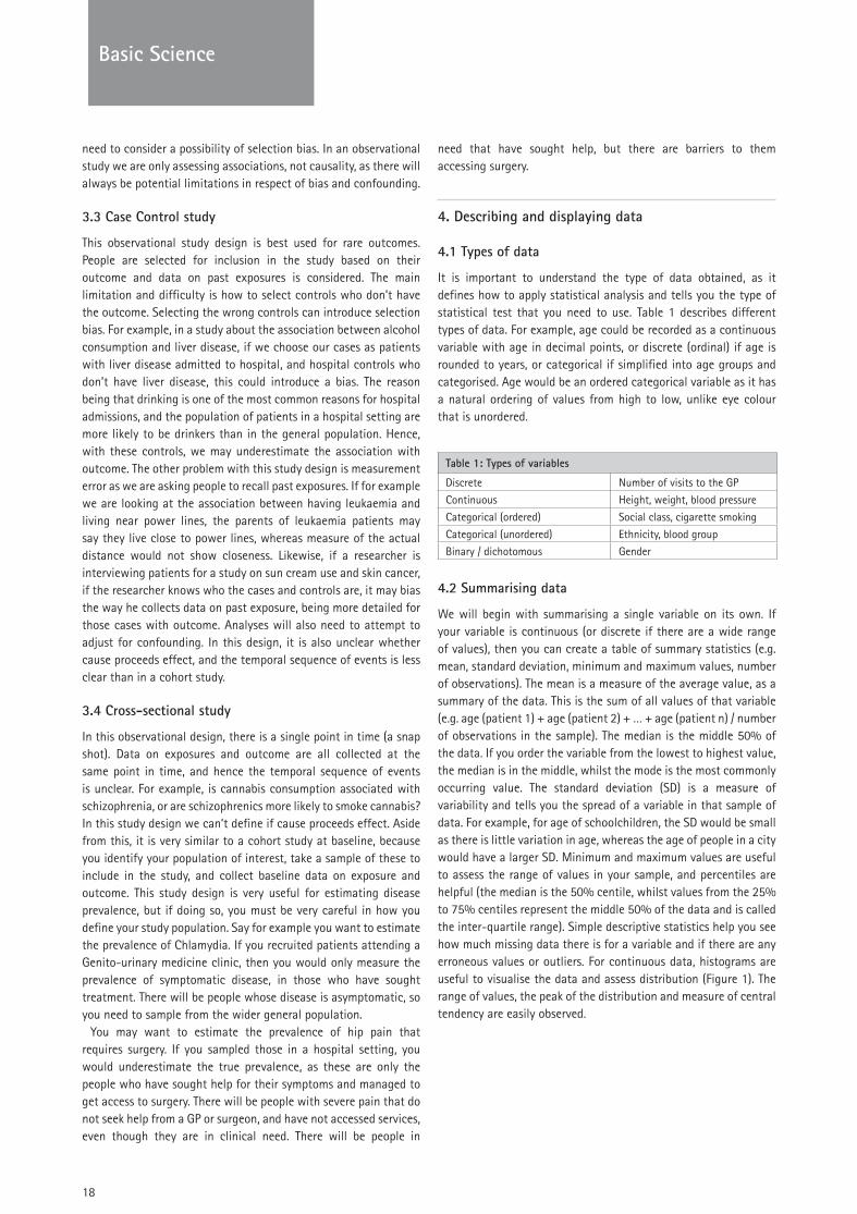
It is important to understand the type of data obtained, as it defines how to apply statistical analysis and tells you the type of statistical test that you need to use. Table 1 describes different types of data. For example, age could be recorded as a continuous variable with age in decimal points, or discrete (ordinal) if age is rounded to years, or categorical if simplified into age groups and categorised. Age would be an ordered categorical variable as it has a natural ordering of values from high to low, unlike eye colour that is unordered.
Table 1: Types of variables
Discrete Number of visits to the GP
Continuous Height, weight, blood pressure
Categorical (ordered) Social class, cigarette smoking
Categorical (unordered) Ethnicity, blood group
Binary / dichotomous Gender
4.2 Summarising data
We will begin with summarising a single variable on its own. If your variable is continuous (or discrete if there are a wide range of values), then you can create a table of summary statistics (e.g. mean, standard deviation, minimum and maximum values, number of observations). The mean is a measure of the average value, as a summary of the data. This is the sum of all values of that variable (e.g. age (patient 1) + age (patient 2) + … + age (patient n) / number of observations in the sample). The median is the middle 50% of the data. If you order the variable from the lowest to highest value, the median is in the middle, whilst the mode is the most commonly occurring value. The standard deviation (SD) is a measure of variability and tells you the spread of a variable in that sample of data. For example, for age of schoolchildren, the SD would be small as there is little variation in age, whereas the age of people in a city would have a larger SD. Minimum and maximum values are useful to assess the range of values in your sample, and percentiles are helpful (the median is the 50% centile, whilst values from the 25% to 75% centiles represent the middle 50% of the data and is called the inter-quartile range). Simple descriptive statistics help you see how much missing data there is for a variable and if there are any erroneous values or outliers. For continuous data, histograms are useful to visualise the data and assess distribution (Figure 1). The range of values, the peak of the distribution and measure of central tendency are easily observed.
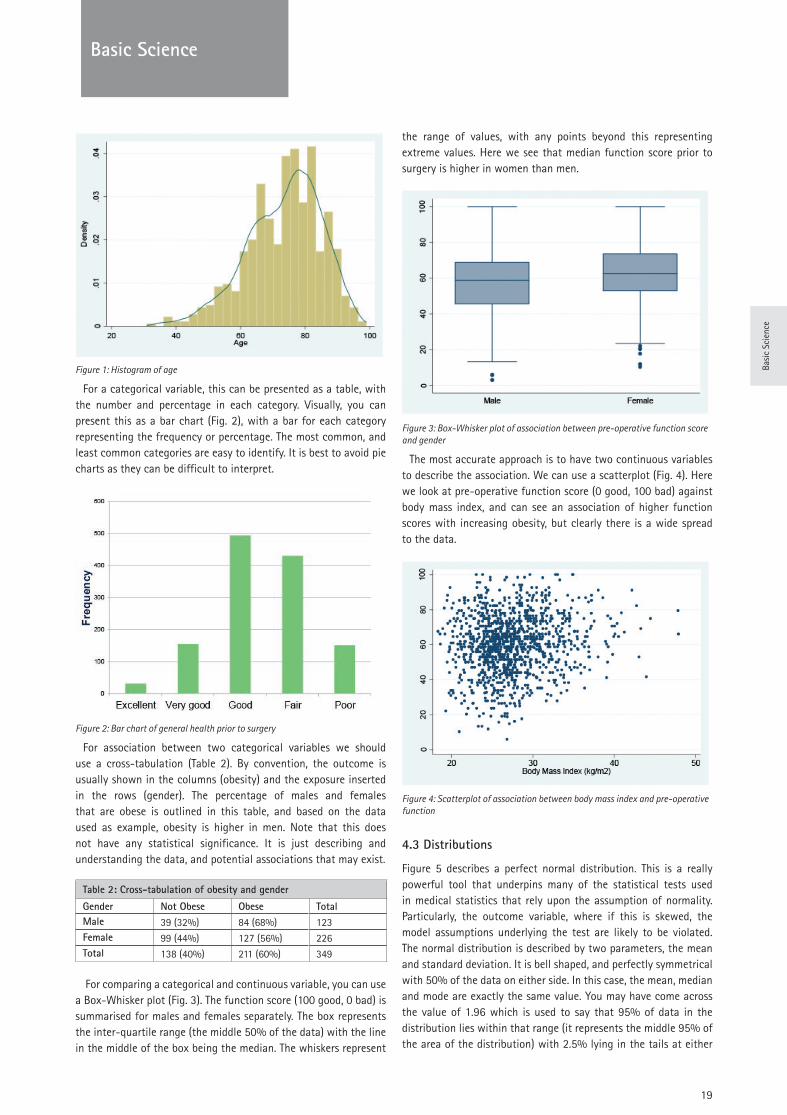
19
Basic Science
Basi
c Sc
ienc
e
Figure 1: Histogram of age
For a categorical variable, this can be presented as a table, with the number and percentage in each category. Visually, you can present this as a bar chart (Fig. 2), with a bar for each category representing the frequency or percentage. The most common, and least common categories are easy to identify. It is best to avoid pie charts as they can be difficult to interpret.
Figure 2: Bar chart of general health prior to surgery
For association between two categorical variables we should use a cross-tabulation (Table 2). By convention, the outcome is usually shown in the columns (obesity) and the exposure inserted in the rows (gender). The percentage of males and females that are obese is outlined in this table, and based on the data used as example, obesity is higher in men. Note that this does not have any statistical significance. It is just describing and understanding the data, and potential associations that may exist.
Table 2: Cross-tabulation of obesity and gender
Gender Not Obese Obese TotalMale 39 (32%) 84 (68%) 123Female 99 (44%) 127 (56%) 226Total 138 (40%) 211 (60%) 349
For comparing a categorical and continuous variable, you can use a Box-Whisker plot (Fig. 3). The function score (100 good, 0 bad) is summarised for males and females separately. The box represents the inter-quartile range (the middle 50% of the data) with the line in the middle of the box being the median. The whiskers represent
the range of values, with any points beyond this representing extreme values. Here we see that median function score prior to surgery is higher in women than men.
Figure 3: Box-Whisker plot of association between pre-operative function score and gender
The most accurate approach is to have two continuous variables to describe the association. We can use a scatterplot (Fig. 4). Here we look at pre-operative function score (0 good, 100 bad) against body mass index, and can see an association of higher function scores with increasing obesity, but clearly there is a wide spread to the data.
Figure 4: Scatterplot of association between body mass index and pre-operative function
4.3 Distributions
Figure 5 describes a perfect normal distribution. This is a really powerful tool that underpins many of the statistical tests used in medical statistics that rely upon the assumption of normality. Particularly, the outcome variable, where if this is skewed, the model assumptions underlying the test are likely to be violated. The normal distribution is described by two parameters, the mean and standard deviation. It is bell shaped, and perfectly symmetrical with 50% of the data on either side. In this case, the mean, median and mode are exactly the same value. You may have come across the value of 1.96 which is used to say that 95% of data in the distribution lies within that range (it represents the middle 95% of the area of the distribution) with 2.5% lying in the tails at either
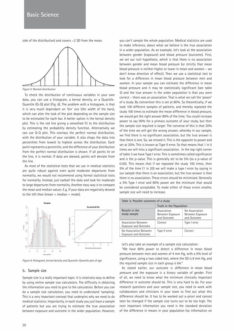
20
Basic Science
side of the distribution) and covers ~2 SD from the mean.
Figure 5: Normal distribution
To check the distribution of continuous variables in your own data, you can use a histogram, a kernal density, or a Quantile-Quantile (Q-Q) plot (Fig. 6). The problem with a histogram, is that it is very much dependant on ‘bin’ size (the width of the bars), which can alter the look of the plot depending on the sample size to be estimated for each bar. A better option is the kernal density plot. This is the red line giving a smoothed fit to the distribution by estimating the probability density function. Alternatively we can use Q-Q plot. This overlays the perfect normal distribution, with the distribution of your variable. It also chops the data into percentiles from lowest to highest across the distribution. Each point represents a percentile, and the difference of your distribution from the perfect normal distribution is shown. If all points lie on the line, it is normal. If data are skewed, points will deviate from the line.
As most of the statistical tests that we use in medical statistics are quite robust against even quite moderate departures from normality, we would not recommend using formal statistical tests for normality. Instead, just eyeball the data and look for moderate to large departures from normality. Another easy way is to compare the mean and median values. E.g. If your data are negatively skewed to the left then (mean < median < mode).
Figure 6: Histogram, kernal density and Quantile-Quantile plot of age .
5. Sample size
Sample size is a really important topic. It is relatively easy to define by using online sample size calculators. The difficulty is obtaining the information you need to give to the calculation. Before you can do a sample size calculation, you need to understand ‘sampling’. This is a very important concept that underpins why we need to do medical statistics. Importantly, in each study you just have a sample of patients but you are trying to estimate the true association between exposure and outcome in the wider population. However,
you can’t sample the whole population. Medical statistics are used to make inference, about what we believe is the true association in a wider population. As an example, let’s look at the association between gender (exposure) and blood pressure (outcome). First, we set our null hypothesis, which is that there is no association between gender and mean blood pressure (or strictly that mean blood pressure is neither higher or lower in mean and women – we don’t know direction of effect). Then we use a statistical test to look for a difference in mean blood pressure between men and women. In your sample you can estimate the difference in mean blood pressure and it may be statistically significant (see table 3) and the true answer in the wider population is that you were correct – there was an association. That is what we call the ‘power’ of a study. By convention this is set at 80%. So theoretically, if we took 100 different samples of patients, and thereby repeated the study 100 times to estimate the mean difference in blood pressure, we would get the right answer 80% of the time. You could increase power to say 90% for a primary outcome of your study, but then the sample size required is larger. The converse of this is that 20% of the time we will get the wrong answer, whereby in our sample, we find there is no significant association, but the true answer is that there is one. So, we missed it. This is the opposite to power and set at 20%. This is known as Type II error. So that means that 1 in 5 times we will miss a significant association. In the top right corner of table 3 we have Type I error. This is sometimes called significance and is the p-value. This is generally set to be 5% (so a p-value of 0.05). This means that if we repeated the study 100 times, then 5% of the time (1 in 20) we will make a type I error by saying in our sample that there is an association, but the true answer is that there is no association. These errors should be minimised. Generally a 5% Type I error and 80% power are the minimum that would be considered acceptable. To make either of those errors smaller, sample size will need to increase.
Table 3: Possible outcomes of a study
Truth in the PopulationResults in the study sample
Association Between Exposure and Outcome
No Association Between Exposure and Outcome
Association Between Exposure and Outcome
Correct Type I error
No Association Between Exposure and Outcome
Type II error Correct
Let’s also take an example of a sample size calculation:“We have 80% power to detect a difference in mean blood
pressure between men and women of 4 mm Hg, with a 5% level of significance, using a two-sided test, where the SD is 8 mm Hg, and the required sample size in each group is 64.”
As stated earlier, our outcome is difference in mean blood pressure and the exposure is a binary variable of gender. First of all, we need to know what the minimum clinically important difference in outcome should be. This is very hard to do. For your research questions and your sample size, you need to work with collaborators and clinicians in your team to find out what this difference should be. It has to be worked out a-priori and cannot later be changed if the sample size turns out to be too high. The next important information you need is the standard deviation of the difference in means in your population (so information on
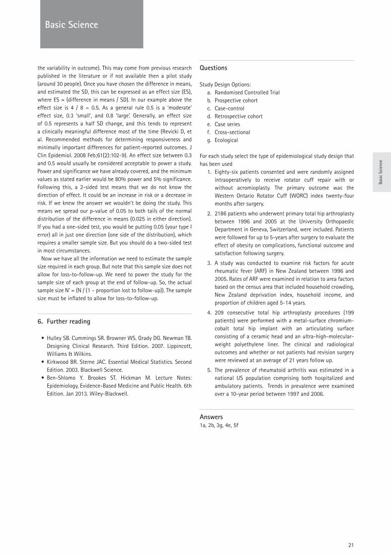
21
Basi
c Sc
ienc
e
Basic Science
the variability in outcome). This may come from previous research published in the literature or if not available then a pilot study (around 30 people). Once you have chosen the difference in means, and estimated the SD, this can be expressed as an effect size (ES), where ES = (difference in means / SD). In our example above the effect size is 4 / 8 = 0.5. As a general rule 0.5 is a ‘moderate’ effect size, 0.3 ‘small’, and 0.8 ‘large’. Generally, an effect size of 0.5 represents a half SD change, and this tends to represent a clinically meaningful difference most of the time (Revicki D, et al. Recommended methods for determining responsiveness and minimally important differences for patient-reported outcomes. J Clin Epidemiol. 2008 Feb;61(2):102-9). An effect size between 0.3 and 0.5 would usually be considered acceptable to power a study. Power and significance we have already covered, and the minimum values as stated earlier would be 80% power and 5% significance. Following this, a 2-sided test means that we do not know the direction of effect. It could be an increase in risk or a decrease in risk. If we knew the answer we wouldn’t be doing the study. This means we spread our p-value of 0.05 to both tails of the normal distribution of the difference in means (0.025 in either direction). If you had a one-sided test, you would be putting 0.05 (your type I error) all in just one direction (one side of the distribution), which requires a smaller sample size. But you should do a two-sided test in most circumstances.
Now we have all the information we need to estimate the sample size required in each group. But note that this sample size does not allow for loss-to-follow-up. We need to power the study for the sample size of each group at the end of follow-up. So, the actual sample size N’ = (N / (1 – proportion lost to follow-up)). The sample size must be inflated to allow for loss-to-follow-up.
6. Further reading
• Hulley SB. Cummings SR. Browner WS. Grady DG. Newman TB. Designing Clinical Research. Third Edition. 2007. Lippincott, Williams & Wilkins.
• Kirkwood BR. Sterne JAC. Essential Medical Statistics. Second Edition. 2003. Blackwell Science.
• Ben-Shlomo Y. Brookes ST. Hickman M. Lecture Notes: Epidemiology, Evidence-Based Medicine and Public Health. 6th Edition. Jan 2013. Wiley-Blackwell.
Questions
Study Design Options:a. Randomised Controlled Trialb. Prospective cohortc. Case-controld. Retrospective cohorte. Case seriesf. Cross-sectionalg. Ecological
For each study select the type of epidemiological study design that has been used
1. Eighty-six patients consented and were randomly assigned intraoperatively to receive rotator cuff repair with or without acromioplasty. The primary outcome was the Western Ontario Rotator Cuff (WORC) index twenty-four months after surgery.
2. 2186 patients who underwent primary total hip arthroplasty between 1996 and 2005 at the University Orthopaedic Department in Geneva, Switzerland, were included. Patients were followed for up to 5-years after surgery to evaluate the effect of obesity on complications, functional outcome and satisfaction following surgery.
3. A study was conducted to examine risk factors for acute rheumatic fever (ARF) in New Zealand between 1996 and 2005. Rates of ARF were examined in relation to area factors based on the census area that included household crowding, New Zealand deprivation index, household income, and proportion of children aged 5-14 years.
4. 209 consecutive total hip arthroplasty procedures (199 patients) were performed with a metal-surface chromium-cobalt total hip implant with an articulating surface consisting of a ceramic head and an ultra-high-molecular-weight polyethylene liner. The clinical and radiological outcomes and whether or not patients had revision surgery were reviewed at an average of 21 years follow up.
5. The prevalence of rheumatoid arthritis was estimated in a national US population comprising both hospitalized and ambulatory patients. Trends in prevalence were examined over a 10-year period between 1997 and 2006.
Answers1a, 2b, 3g, 4e, 5f
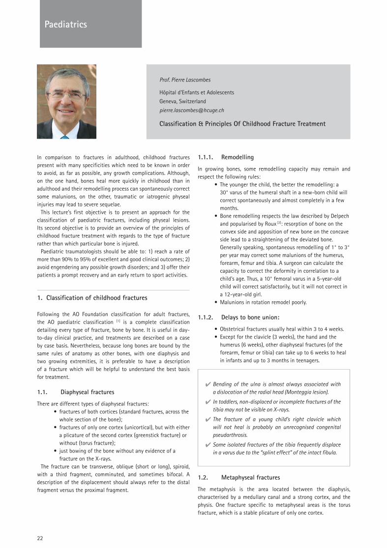
22
Paediatrics
Prof. Pierre Lascombes
Hôpital d’Enfants et Adolescents
Geneva, Switzerland
Classification & Principles Of Childhood Fracture Treatment
In comparison to fractures in adulthood, childhood fractures present with many specificities which need to be known in order to avoid, as far as possible, any growth complications. Although, on the one hand, bones heal more quickly in childhood than in adulthood and their remodelling process can spontaneously correct some malunions, on the other, traumatic or iatrogenic physeal injuries may lead to severe sequelae.
This lecture’s first objective is to present an approach for the classification of paediatric fractures, including physeal lesions. Its second objective is to provide an overview of the principles of childhood fracture treatment with regards to the type of fracture rather than which particular bone is injured.
Paediatric traumatologists should be able to: 1) reach a rate of more than 90% to 95% of excellent and good clinical outcomes; 2) avoid engendering any possible growth disorders; and 3) offer their patients a prompt recovery and an early return to sport activities.
1. Classification of childhood fractures
Following the AO Foundation classification for adult fractures, the AO paediatric classification [1] is a complete classification detailing every type of fracture, bone by bone. It is useful in day-to-day clinical practice, and treatments are described on a case by case basis. Nevertheless, because long bones are bound by the same rules of anatomy as other bones, with one diaphysis and two growing extremities, it is preferable to have a description of a fracture which will be helpful to understand the best basis for treatment.
1.1. Diaphyseal fractures
There are different types of diaphyseal fractures:• fractures of both cortices (standard fractures, across the
whole section of the bone);• fractures of only one cortex (unicortical), but with either
a plicature of the second cortex (greenstick fracture) or without (torus fracture);
• just bowing of the bone without any evidence of a fracture on the X-rays.
The fracture can be transverse, oblique (short or long), spiroid, with a third fragment, comminuted, and sometimes bifocal. A description of the displacement should always refer to the distal fragment versus the proximal fragment.
1.1.1. Remodelling
In growing bones, some remodelling capacity may remain and respect the following rules:
• The younger the child, the better the remodelling: a 30° varus of the humeral shaft in a new-born child will correct spontaneously and almost completely in a few months.
• Bone remodelling respects the law described by Delpech and popularised by Roux [2]: resorption of bone on the convex side and apposition of new bone on the concave side lead to a straightening of the deviated bone. Generally speaking, spontaneous remodelling of 1° to 3° per year may correct some malunions of the humerus, forearm, femur and tibia. A surgeon can calculate the capacity to correct the deformity in correlation to a child’s age. Thus, a 10° femoral varus in a 5-year-old child will correct satisfactorily, but it will not correct in a 12-year-old girl.
• Malunions in rotation remodel poorly.
1.1.2. Delays to bone union:
• Obstetrical fractures usually heal within 3 to 4 weeks.• Except for the clavicle (3 weeks), the hand and the
humerus (6 weeks), other diaphyseal fractures (of the forearm, femur or tibia) can take up to 6 weeks to heal in infants and up to 3 months in teenagers.
4 Bending of the ulna is almost always associated with a dislocation of the radial head (Monteggia lesion).
4 In toddlers, non-displaced or incomplete fractures of the tibia may not be visible on X-rays.
4 The fracture of a young child’s right clavicle which will not heal is probably an unrecognised congenital pseudarthrosis.
4 Some isolated fractures of the tibia frequently displace in a varus due to the “splint effect” of the intact fibula.
1.2. Metaphyseal fractures
The metaphysis is the area located between the diaphysis, characterised by a medullary canal and a strong cortex, and the physis. One fracture specific to metaphyseal areas is the torus fracture, which is a stable plicature of only one cortex.
23
Paed
iatr
ics
Paediatrics
1.2.1. Remodelling
Excellent capacity for remodelling requires the fracture’s close proximity to an active physis (far from the elbow and close to the knee) and involves the direction of the adjacent joint’s motion. According to the Hueter–Volkmann law, growth is faster on the concave side than on the convex side, and this leads to the straightening of the deformity [3, 4]. For example:
• a severe displacement of the proximal humerus has an excellent capacity for correction, whereas correction of a distal humerus deformity is usually very poor;
• an antecurvatum malunion of the distal radius may correct at more than 10° a year, but an ulnar varus will remain almost unchanged with growth.
1.2.2. Delays to bone union:
Cancellous bone union has an excellent capacity to heal, taking:• 3 to 4 weeks for
ᴑ a proximal humerus in teenagers, ᴑ a supracondylar elbow in children around 6 years old, ᴑ a torus fracture of the distal radius
• 6 weeks for other locations, including frequently occurring distal forearm fractures
4 An undisplaced medial proximal tibial fracture frequently heals with a progressive valgus deformity (Cozen’s effect) [5].
1.3. Physeal injuries
The literature describes more than six different classifications: the Salter–Harris classification, with five types of physeal injury, is the most often used [6]; Ogden’s classification, with eight types, is the most complete [7]. It should be emphasised that the weak area of the physis corresponds to the hypertrophic cartilage, close to the metaphyseal bone and far from the germinal cells. As long as the latter zone remains intact, the chance of a post-traumatic epiphysiodesis is rare. Nevertheless, diagnosing an injury to the germinal centre using X-ray remains impossible. This is the case with Smith–Petersen distal radius type I injuries [8], some Salter–Harris type II injuries with a compression of the physis itself during trauma, and of undetectable Salter–Harris type V injuries.
All transphyseal injuries (Salter–Harris type III and IV) are also joint fractures. It appears evident that these fractures must be perfectly reduced in order to: 1) regain the correct anatomical shape of the joint; and 2) avoid an epiphysiodesis due to a bony bar developing across the physis.
Each location presents specificities:• Proximal humerus and distal forearm Salter–Harris type
I and II injuries have great potential for remodelling.• Many distal femoral and proximal tibial fractures lead to
delayed deformities due to growth asymmetry.• Salter-Harris type II injuries to the distal tibia, with
a valgus displacement, are often associated with an entrapment of the periosteum into the fracture.
4 If not properly reduced, one frequent complication of a humeral lateral condyle fracture is non-union.
4 A triplane fracture of the distal tibia is a Salter–Harris type IV injury.
4 The medial malleolus fracture (MacFarland), Salter–Harris III or IV, is most often associated with a fracture of the distal fibula physis, Salter–Harris I.
1.4. Apophyseal injuries
Traumatic avulsions of the apophysis are lesions specific to teenagers. During the time separating the ossification of any apophysis and the closure of its physis, a sudden contraction of the attached tendon may lead to an apophyseal avulsion fracture. The main apophyses affected are the antero-inferior-iliac spine (rectus femoris), ischium (hamstrings), lesser trochanter (psoas iliac), tibial tuberosity (quadriceps, patellar ligament) and humeral medial epicondyle.
4 A dislocated elbow is frequently associated with a humeral medial epicondyle fracture. This fragment may become entrapped in the joint after reduction of the dislocation.
1.5. Small bones
1.5.1. Wrist
A fracture of the scaphoid bone may be difficult to diagnose. An MRI should be carried out if there are any positive clinical signs of a wrist fracture.
1.5.2. Foot
A non-displaced fracture of the talus, calcaneus or another tarsal bone is not easy to diagnose from an X-ray. For a displaced fracture, accurate information about the lesion is mandatory for preparing surgery. A CT scan may be helpful if any of these bones are fractured.
1.6. Intraarticular fractures and ligament injuries
Osteochondral fractures may appear during a traumatic dislocation of the patella, either on the patella itself or on a femoral condyle. The intraarticular fragment must be removed or fixed.Ligament injuries may lead to the avulsion of their bony attachment and require surgical repair. Examples are the pre- and retro-spinal bony avulsions of the knee’s cruciate ligaments, the insertion of the medial patella-femoral ligament on the medial patella and the tip of the lateral malleolus.
24
Paediatrics
2. Principles of childhood fracture treatment
The absolute primary rules for the treatment of any fracture are reduction and stabilisation. In children, the quality of the reduction can be adapted to the bone’s capacity for remodelling. For example, a residual posterior tilt of 20° in the distal radius in a boy of 10 years old is acceptable as it will correct completely with its remaining growth.
The secondary rules concern prompt recovery, an early return to school and sports activities, as well as some economic aspects. For example, a fracture of the femoral shaft in a 10-years-old child should not be treated with more than three weeks of traction in hospital, followed by 6 weeks in a spica cast.
The present lecture focuses mainly on isolated limb fractures, and not on the following:
• In cases involving polytrauma and multiple injuries, the indications recommend surgical fixation in order to avoid casts, which cause difficulties when screening and slow recovery.
• Gustilo Anderson type III open fractures should be fixed with external fixation, whereas in many type I and II injuries, intramedullary fixations can be performed.
• In cases of compartment syndrome, external fixation or intramedullary implants are good choices to manage fasciotomies.
2.1. Methods of treatment in children
2.1.1. Conservative treatment
Non-displaced fractures can be treated in an emergency room using a well-adapted cast. Except for a torus fracture of the distal radius, which can be immobilised with a simple splint, the rule is that upper and lower joints should be immobilised for at least the first 3 weeks.
Displaced fractures should either be reduced immediately in the emergency department or later in the operating room under general anaesthesia.
2.1.2. Paediatric surgical fixations
• Diaphyseal fractures ᴑ In children, diaphyseal fractures (of the humerus, forearm, femur or tibia) can be fixed using flexible intramedullary nailing (FIN), except when the diameter of the medullary canal exceeds 10 mm. In these circumstances, two flexible 4 mm nails would be too small to ensure the FIN technique’s usual biomechanical properties [9]. It should be remembered that the ratio of each nail’s size to that of the medullary canal should be over 40%. With regards to femoral shaft fractures, the AAOS recommends a FIN procedure for children from 5 to 11 years old.
ᴑ When the medullary canal is wider than 10 mm: - Fractures of the femur after 10 to 12-years-old can
be treated using a lateral trochanteric intramedullary locking nail.
- After the fusion of the tibial tuberosity physis, fractures of the tibia can also be treated with an intramedullary locking nail. In younger children with an open physis, an external fixator, or an association of external fixator and FIN or some plates may be used.
4 A strait intramedullary locking nail through the trochanteric digital fossa of the femur may lead to avascular necrosis in 5% of teenagers and is not indicated for adolescents.
4 Intramedullary locking through the tibial tuberosity physis before its fusion may lead to genu recurvatum.
• Metaphyseal, and Salter–Harris I and II fractures ᴑ Some metaphyseal and physeal fractures can be treated using a FIN, such as the upper humerus, radial neck (gold standard) and some distal third of the radius fractures with a posteromedial retrograde FIN. Other fractures, such as of the distal supracondylar humerus or the distal radius (Kapandji technique) should be fixed using percutaneous Kirschner wires.
ᴑ In lower limbs, metaphyseal fracture is better fixed with screws or plates and screws. In some distal, Salter–Harris II femur fractures in boys aged around 14 years old, surgeons should decide between:
- Either a “light” fixation with some screws and a cast, with the ensuing risk of secondary axial deviation due to growth disturbances,
- Or a plate, as in adults, which induces a deliberate epiphysiodesis and may later require a secondary treatment for a leg discrepancy.
4 A compartmental syndrome of the extensor hallux muscle is a complication of a distal tibia Salter II fracture with a posterior displacement.
• Physeal injuries: Salter–Harris III and IV ᴑ Reduction should be perfect, and fixation should be absolutely stable. Screws are more often better than K wires, which are preferable when crossing a physis is necessary. Resorbable screws are even more desirable as they do not require follow-up surgery for implant removal.
ᴑ Supra- and intercondylar fractures of both the distal humerus and femur should be fixed with specific plates, as in adults.
4 Screws across a physis may lead to severe growth disturbances and an extremity deformity.
• Apophyseal injuries ᴑ When indicated, fixation should be performed either with screws (medial epicondyle) or resorbable anchors (medial epicondyle in young children, pelvic apophysis).
Paed
iatr
ics
25
Paediatrics
4 A severely displaced apophysis may look like a pseudo bone tumour syndrome (ischium) or lead to femoroacetabular impingement (Antero-Inferior Iliac Spine) or non-union (medial epicondyle of the elbow).
2.2. Indications for the treatment of children
2.2.1. Non-displaced fractures: a conservative treatment is almost always the best choice. X-ray monitoring at 7 days is usually mandatory to screen for a possible secondary displacement.
4 X-ray monitoring of a non-displaced fracture of the humeral condyle on day seven is not easy. Being certain of an absence of secondary displacement may require removal of the cast in order to obtain good quality anteroposterior X-ray images.
2.2.2. Many fractures with a great capacity for remodelling can be treated conservatively, which means associating a reduction with immobilisation. ᴑ Proximal fractures of the humerus: metaphysis and Salter–Harris I-II. A cast is avoided and replaced by a simple Gerdy immobilisation.
ᴑ Distal fractures of the radius and ulna: metaphyseal and Salter–Harris I-II.
2.2.3. Any Salter–Harris type III or IV with a displacement of more than 2 mm should be reduced and fixed perfectly. In certain circumstances, a percutaneous reduction can be performed: i.e. a mild displacement of a distal tibia Tillaux fracture. A surgical approach is usually mandatory to obtain the reduction. The quality of the reduction should be checked by examining the reduction of the physis and the joint either visually or via an arthrogram.
2.2.4. When a cast is not certain to maintain the reduction, unstable diaphyseal fractures of the humerus, forearm, and leg are good indications for intramedullary fixation (FIN or locking nail, depending on size and age). Thus, by definition, any fracture of the femoral shaft belongs in this category. Severely displaced and multiple fragments of the clavicle also belong in this group.
2.2.5. Other fractures have either conservative or surgical indications. Controversies between surgeons about this group are ongoing, and there are pros and cons for both methods.
The advantages of surgery are: 1) stabilisation of the fracture with no risk of secondary displacement; 2) less time wearing a cast, or perhaps even no cast at all; and 3) earlier recovery.
The advantages of conservative treatment are: 1) there is no need to remove implants; and 2) there is no risk of infection.
This list is not limited. For example: ᴑ Diaphysis of the humerus, of the forearm before the age of 10 and of the femur before the age of 5 tolerate treatment via a FIN procedure far better than any type of cast.
ᴑ After a conservative treatment, unstable metaphyseal and Salter–Harris II fractures of the distal radius (and ulna) can avoid the issue of secondary displacement by treatment with a Kapandji pinning (or another type of pinning).
ᴑ Clavicle fracture in a teenager, with a shortening of up to 2 cm, can be fixed with an intramedullary nail.
3. Conclusion
Treating fractures in children is never an easy process, and malunion will not always correct spontaneously with growth. Comprehensive knowledge of the mechanisms of bone union can only help surgeons in their decision- making. On one hand, surgeons should spare no efforts to repair a child’s fracture and aim for the most favourable clinical outcome, but on the other, they should inform the parents about the possibilities of complications, including growth disturbances.
4. Further Reading
Walter P. Blount, 1955 [10], Mercer Rang, 1974 [11], and the “Rockwood & Wilkins”, 2012 [12].
References:
1. Slongo T, Audigé L, Schlickewei W, Clavert JM, Hunter J; International Association for Pediatric Traumatology. Development and validation of the AO pediatric comprehensive classification of long bone fractures by the Pediatric Expert Group of the AO Foundation in collaboration with AO Clinical Investigation and Documentation and the International Association for Pediatric Traumatology. J Pediatr Orthop. 2006; 26: 43-9.
2. Lee TC, Taylor D. Bone remodelling: should we cry Wolff? Ir J Med Sc. 1999; 168: 102-5.
3. Hueter C. Anatomische Studien an den Extremitätengelenken Neugeborener und Erwachsener. Virchow Arch. 1862; 26: 484–519.
4. Volkmann R. Chirurgische Erfahrungen über Knochenverbie- gung und Knochenwachstum. Virchow Archiv. 1862; 24: 512–40.
5. Cozen L. Fracture of the proximal portion of the tibia in children followed by valgus deformity. Surg Gynecol Obstet. 1953; 97: 183-8.
6. Salter RB, Harris R. Injuries involving the epiphyseal plate. J Bone Joint Surg Am. 1963; 48: 587-622.
7. Ogden JA. Injury to the growth mechanisms of the immature skeleton. Skeletal Radiol. 1981; 6: 237-53.
8. Peterson HA: Physeal fractures. III. Classification. J Pediatr Orthop. 1994; 14: 439-48.
26
Paediatrics
9. Lascombes P. Flexible Intramedullary Nailing. The Nancy University Manual. Springer-Verlag, Berlin, Heidelberg. 2010, 317p.
10. Blount WP. Fractures in children. The Williams and Wilkins Company. 1955, 279p.
11. Rang M. Children’s fractures. Lippincott, 1974, 238p.12. Rockwood and Wilkins’. Fractures in children, 7th edition. J
Beaty and JR Kasser, Lippincott Williams &Wilkins, 2012, 1076p.
Questions
1. Malunion capacity of remodelling after a fracture, girl, 5 years old (one valid answer) :a. 40° varus deformity of the upper humerus after a Salter II
fracture : poor capacityb. 20° varus deformity of the distal humerus after a supra
condylar fracture: good capacityc. 20° varus deformity of the midshaft forearm after fracture
of both bones: good capacityd. 20° posterior deformity of the distal radius after a fracture
of the distal metaphysis of the radius: good capacity
2. Closed displaced fracture of radius and ulna diaphysis, boy, 13 years old, normal neuro vascular status. The most appropriate treatment is (one valid answer): a. Reduction and cast immobilisation for 8-12 weeksb. Reduction and flexible intramedullary nailingc. Reduction and plating fixationd. Reduction and external fixator
3. Closed displaced fracture of the femur diaphysis, girl, 11 years old, normal neuro vascular status. The medullary canal diameter measures 12mm. The most appropriate treatment is (one valid answer): a. Traction 3 weeks and spica cast 6 weeksb. Reduction and flexible intramedullary nailingc. Reduction and lateral greater trochanter intramedullary
locking naild. Reduction and external fixator
4. Distal fracture of the tibia (one valid answer):a. A medial malleolus fracture (Mac Farland) with a 4 mm
displacement can be treated conservativelyb. An antero-lateral tibial fracture (Tillaux’) is frequent in
children 8 years oldc. The periosteum is frequently entrapped in Salter II types
fractures displaced in valgusd. Post-trauma epiphysiodesis is never observed after severely
displaced Salter type II fractures
Answers1d, 2b, 3c, 4c
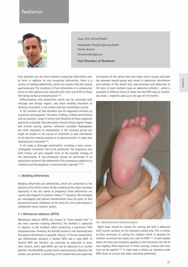
Paed
iatr
ics
27
Paediatrics
Assoc. Prof. Christof Radler
Orthopaedic Hospital Speising GmbH
Vienna, Austria
Foot Disorders of Newborns
Foot disorders are the most common congenital deformities seen at birth. In addition to true congenital deformities, there is a variety of molding deformities, which are usually mild and resolve spontaneously. The incidence of foot deformities in a consecutive series of 2401 patients was reported with 4.2% with 87% of those feet being normal at reexamination [41].
Differentiating mild deformities which can be corrected with massage and benign neglect, and those needing treatment or showing recurrence, is not always easy but nonetheless crucial.
In the newborn all foot disorders can be diagnosed clinically by inspection and palpation. The ankle, hindfoot, midfoot and forefoot and the position, range of motion and flexibility of these segments should be evaluated. Documentation should include digital images and clinical scoring systems, whenever available. Radiographs are never necessary at presentation in the neonatal period but might be helpful in the course of treatment to add information to the decision-making process or as documentation in cases that received prior treatment [32].
In all cases, a thorough examination including a basic neuro-orthopaedic evaluation has to be performed. The pregnancy and birth history can give valuable hints at the possible etiology of the abnormality. A hip-ultrasound should be performed if an association between foot deformities (like metatarsus adductus or clubfoot) and hip dysplasia is controversially reported [28,29].
1. Molding deformities
Molding deformities are deformities, which are connected to the position of the child in utero. As the crowding of the infant increases especially in the last weeks of pregnancy those deformities are usually less frequent in preterm infants [20]. However, the etiologies are overlapping and special considerations must be given to feet presenting muscle imbalance on the basis of a still undeveloped or unbalanced neuro-motoric system.
1.1 Metatarsus adductus (MTA)
Metatarsus adducts (MTA) also known as “bean-shaped foot” is the most common molding deformity. The forefoot is adducted in relation to the hindfoot often presenting a prominent fifth metatarsal base. However, the Achilles tendon is not shortened and full passive dorsiflexion is possible. (Figure 1) Clinical examination can differentiate between a flexible MTA and a rigid MTA. In flexible MTA the forefoot can passively be abducted to more than neutral, while rigid MTAs can not be abducted to a neutral position. Flexible MTAs usually resolve spontaneously. However, the mother can perform a stretching of the medial foot and especially
stimulation of the lateral foot and thigh which usually stimulate the peroneal muscle group and results in abduction, dorsiflexion and eversion of the whole foot, and extension and abduction of the toes. In more resistant cases an abduction orthotic - which is available in different forms of shoes like the IPOS shoe or reverse-last shoes - might be used up to the age of 6-9 months.
Fig 1: Mild but persistent metatarsus adducts
Rigid cases should be casted. For casting, the foot is abducted with counter pressure on the calcaneo-cuboid area. This is similar to Kites technique of casting for clubfoot which is obsolete for clubfoot correction but works very well for MTA [ 21]. A well molded above the knee cast should be applied, as this minimizes the risk of cast slipping. With experience in infant casting, a below-the-knee cast can be applied [19]. In rigid cases a follow-up treatment with IPOS shoes or reverse-last shoes should be performed.
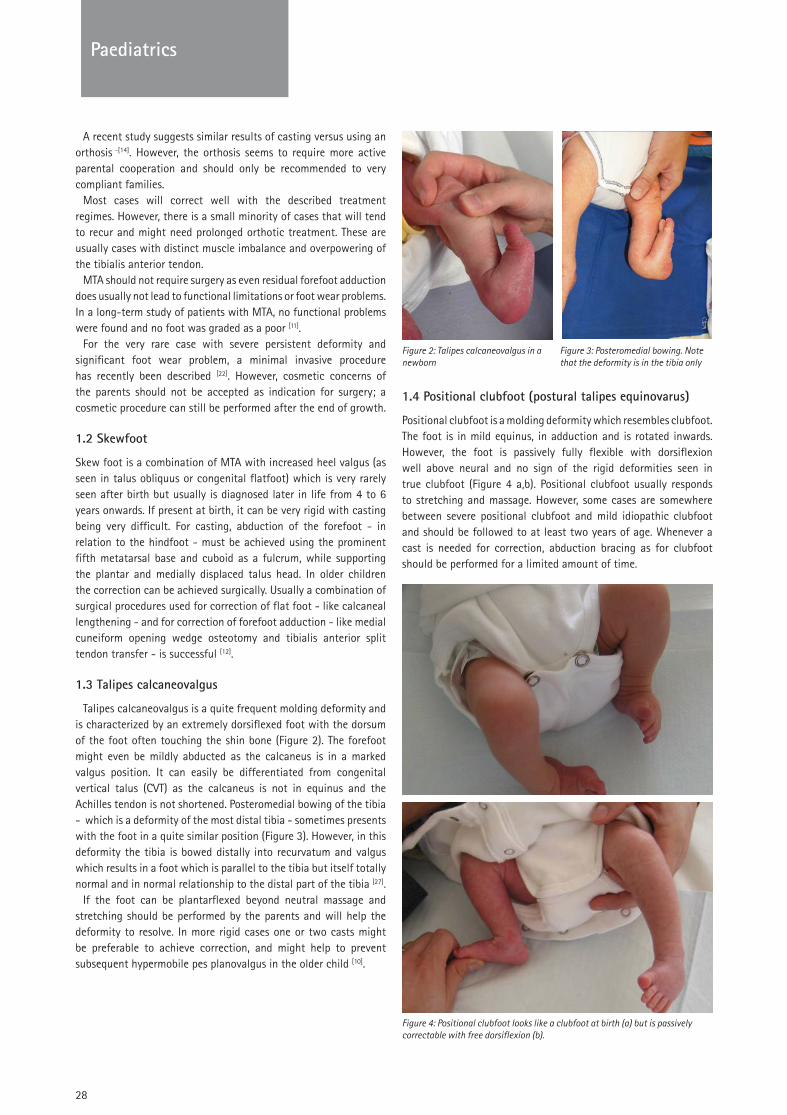
28
Paediatrics
A recent study suggests similar results of casting versus using an orthosis -[14]. However, the orthosis seems to require more active parental cooperation and should only be recommended to very compliant families.
Most cases will correct well with the described treatment regimes. However, there is a small minority of cases that will tend to recur and might need prolonged orthotic treatment. These are usually cases with distinct muscle imbalance and overpowering of the tibialis anterior tendon.
MTA should not require surgery as even residual forefoot adduction does usually not lead to functional limitations or foot wear problems. In a long-term study of patients with MTA, no functional problems were found and no foot was graded as a poor [11].
For the very rare case with severe persistent deformity and significant foot wear problem, a minimal invasive procedure has recently been described [22]. However, cosmetic concerns of the parents should not be accepted as indication for surgery; a cosmetic procedure can still be performed after the end of growth.
1.2 Skewfoot
Skew foot is a combination of MTA with increased heel valgus (as seen in talus obliquus or congenital flatfoot) which is very rarely seen after birth but usually is diagnosed later in life from 4 to 6 years onwards. If present at birth, it can be very rigid with casting being very difficult. For casting, abduction of the forefoot - in relation to the hindfoot - must be achieved using the prominent fifth metatarsal base and cuboid as a fulcrum, while supporting the plantar and medially displaced talus head. In older children the correction can be achieved surgically. Usually a combination of surgical procedures used for correction of flat foot - like calcaneal lengthening - and for correction of forefoot adduction - like medial cuneiform opening wedge osteotomy and tibialis anterior split tendon transfer - is successful [12].
1.3 Talipes calcaneovalgus
Talipes calcaneovalgus is a quite frequent molding deformity and is characterized by an extremely dorsiflexed foot with the dorsum of the foot often touching the shin bone (Figure 2). The forefoot might even be mildly abducted as the calcaneus is in a marked valgus position. It can easily be differentiated from congenital vertical talus (CVT) as the calcaneus is not in equinus and the Achilles tendon is not shortened. Posteromedial bowing of the tibia - which is a deformity of the most distal tibia - sometimes presents with the foot in a quite similar position (Figure 3). However, in this deformity the tibia is bowed distally into recurvatum and valgus which results in a foot which is parallel to the tibia but itself totally normal and in normal relationship to the distal part of the tibia [27].
If the foot can be plantarflexed beyond neutral massage and stretching should be performed by the parents and will help the deformity to resolve. In more rigid cases one or two casts might be preferable to achieve correction, and might help to prevent subsequent hypermobile pes planovalgus in the older child [10].
Figure 2: Talipes calcaneovalgus in a Figure 3: Posteromedial bowing. Note newborn that the deformity is in the tibia only
1.4 Positional clubfoot (postural talipes equinovarus)
Positional clubfoot is a molding deformity which resembles clubfoot. The foot is in mild equinus, in adduction and is rotated inwards. However, the foot is passively fully flexible with dorsiflexion well above neural and no sign of the rigid deformities seen in true clubfoot (Figure 4 a,b). Positional clubfoot usually responds to stretching and massage. However, some cases are somewhere between severe positional clubfoot and mild idiopathic clubfoot and should be followed to at least two years of age. Whenever a cast is needed for correction, abduction bracing as for clubfoot should be performed for a limited amount of time.
Figure 4: Positional clubfoot looks like a clubfoot at birth (a) but is passively correctable with free dorsiflexion (b).
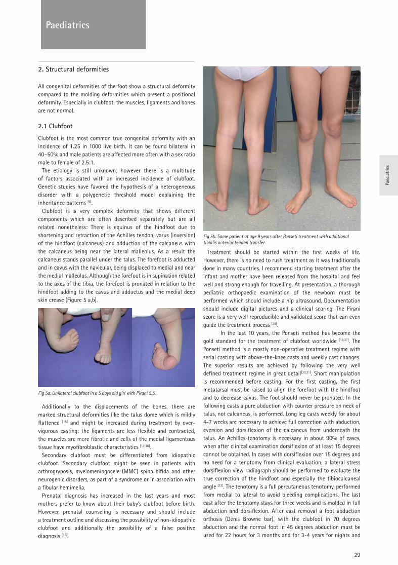
Paed
iatr
ics
29
Paediatrics
2. Structural deformities
All congenital deformities of the foot show a structural deformity compared to the molding deformities which present a positional deformity. Especially in clubfoot, the muscles, ligaments and bones are not normal.
2.1 Clubfoot
Clubfoot is the most common true congenital deformity with an incidence of 1.25 in 1000 live birth. It can be found bilateral in 40–50% and male patients are affected more often with a sex ratio male to female of 2.5:1.
The etiology is still unknown; however there is a multitude of factors associated with an increased incidence of clubfoot. Genetic studies have favored the hypothesis of a heterogeneous disorder with a polygenetic threshold model explaining the inheritance patterns [6].
Clubfoot is a very complex deformity that shows different components which are often described separately but are all related nonetheless: There is equinus of the hindfoot due to shortening and retraction of the Achilles tendon, varus (inversion) of the hindfoot (calcaneus) and adduction of the calcaneus with the calcaneus being near the lateral malleolus. As a result the calcaneus stands parallel under the talus. The forefoot is adducted and in cavus with the navicular, being displaced to medial and near the medial malleolus. Although the forefoot is in supination related to the axes of the tibia, the forefoot is pronated in relation to the hindfoot adding to the cavus and adductus and the medial deep skin crease (Figure 5 a,b).
Fig 5a: Unilateral clubfoot in a 5 days old girl with Pirani 5.5.
Additionally to the displacements of the bones, there are marked structural deformities like the talus dome which is mildly flattened [15] and might be increased during treatment by over- vigorous casting: the ligaments are less flexible and contracted, the muscles are more fibrotic and cells of the medial ligamentous tissue have myofibroblastic characteristics [17,36].
Secondary clubfoot must be differentiated from idiopathic clubfoot. Secondary clubfoot might be seen in patients with arthrogryposis, myelomeningocele (MMC) spina bifida and other neurogenic disorders, as part of a syndrome or in association with a fibular hemimelia.
Prenatal diagnosis has increased in the last years and most mothers prefer to know about their baby’s clubfoot before birth. However, prenatal counseling is necessary and should include a treatment outline and discussing the possibility of non-idiopathic clubfoot and additionally the possibility of a false positive diagnosis [35].
Fig 5b: Same patient at age 9 years after Ponseti treatment with additional tibialis anterior tendon transfer
Treatment should be started within the first weeks of life. However, there is no need to rush treatment as it was traditionally done in many countries. I recommend starting treatment after the infant and mother have been released from the hospital and feel well and strong enough for travelling. At presentation, a thorough pediatric orthopaedic examination of the newborn must be performed which should include a hip ultrasound. Documentation should include digital pictures and a clinical scoring. The Pirani score is a very well reproducible and validated score that can even guide the treatment process [38].
In the last 10 years, the Ponseti method has become the gold standard for the treatment of clubfoot worldwide [18,37]. The Ponseti method is a mostly non-operative treatment regime with serial casting with above-the-knee casts and weekly cast changes. The superior results are achieved by following the very well defined treatment regime in great detail[30,31]. Short manipulation is recommended before casting. For the first casting, the first metatarsal must be raised to align the forefoot with the hindfoot and to decrease cavus. The foot should never be pronated. In the following casts a pure abduction with counter pressure on neck of talus, not calcaneus, is performed. Long leg casts weekly for about 4-7 weeks are necessary to achieve full correction with abduction, eversion and dorsiflexion of the calcaneus from underneath the talus. An Achilles tenotomy is necessary in about 90% of cases, when after clinical examination dorsiflexion of at least 15 degrees cannot be obtained. In cases with dorsiflexion over 15 degrees and no need for a tenotomy from clinical evaluation, a lateral stress dorsiflexion view radiograph should be performed to evaluate the true correction of the hindfoot and especially the tibiocalcaneal angle [33]. The tenotomy is a full percutaneous tenotomy, performed from medial to lateral to avoid bleeding complications. The last cast after the tenotomy stays for three weeks and is molded in full abduction and dorsiflexion. After cast removal a foot abduction orthosis (Denis Browne bar), with the clubfoot in 70 degrees abduction and the normal foot in 45 degrees abduction must be used for 22 hours for 3 months and for 3-4 years for nights and
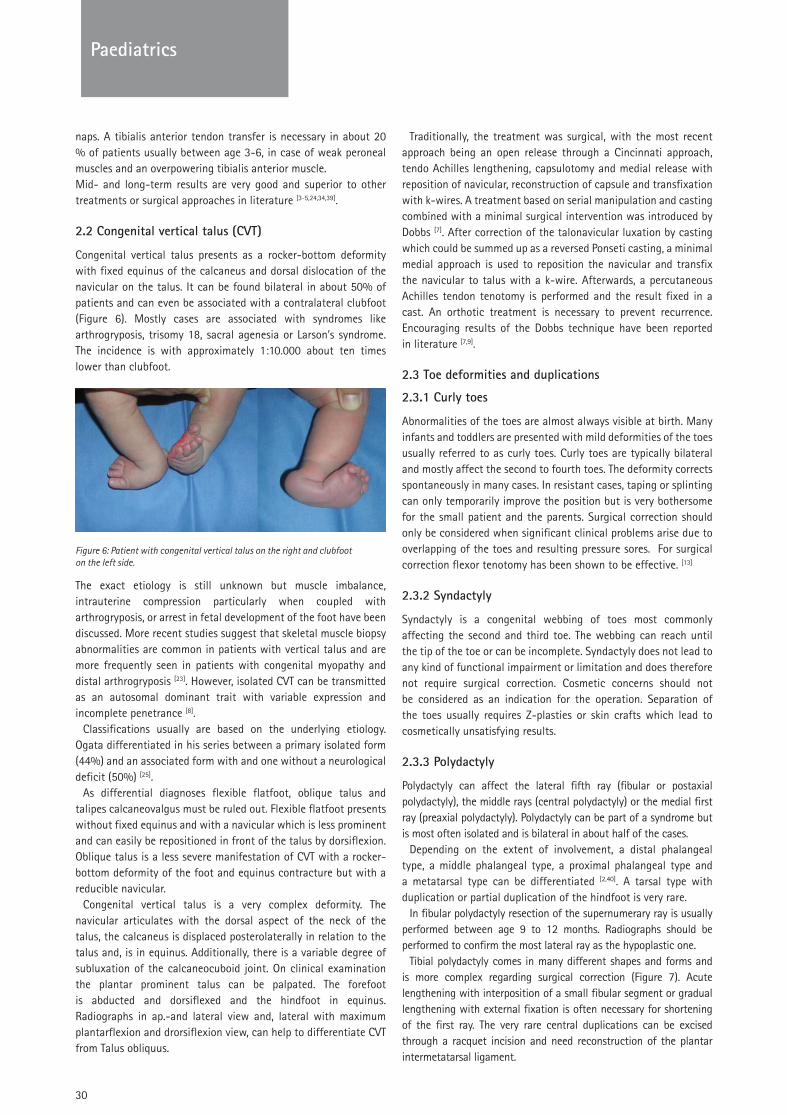
30
Paediatrics
naps. A tibialis anterior tendon transfer is necessary in about 20 % of patients usually between age 3-6, in case of weak peroneal muscles and an overpowering tibialis anterior muscle. Mid- and long-term results are very good and superior to other treatments or surgical approaches in literature [3-5,24,34,39].
2.2 Congenital vertical talus (CVT)
Congenital vertical talus presents as a rocker-bottom deformity with fixed equinus of the calcaneus and dorsal dislocation of the navicular on the talus. It can be found bilateral in about 50% of patients and can even be associated with a contralateral clubfoot (Figure 6). Mostly cases are associated with syndromes like arthrogryposis, trisomy 18, sacral agenesia or Larson’s syndrome. The incidence is with approximately 1:10.000 about ten times lower than clubfoot.
Figure 6: Patient with congenital vertical talus on the right and clubfoot on the left side.
The exact etiology is still unknown but muscle imbalance, intrauterine compression particularly when coupled with arthrogryposis, or arrest in fetal development of the foot have been discussed. More recent studies suggest that skeletal muscle biopsy abnormalities are common in patients with vertical talus and are more frequently seen in patients with congenital myopathy and distal arthrogryposis [23]. However, isolated CVT can be transmitted as an autosomal dominant trait with variable expression and incomplete penetrance [8].
Classifications usually are based on the underlying etiology. Ogata differentiated in his series between a primary isolated form (44%) and an associated form with and one without a neurological deficit (50%) [25].
As differential diagnoses flexible flatfoot, oblique talus and talipes calcaneovalgus must be ruled out. Flexible flatfoot presents without fixed equinus and with a navicular which is less prominent and can easily be repositioned in front of the talus by dorsiflexion. Oblique talus is a less severe manifestation of CVT with a rocker-bottom deformity of the foot and equinus contracture but with a reducible navicular.
Congenital vertical talus is a very complex deformity. The navicular articulates with the dorsal aspect of the neck of the talus, the calcaneus is displaced posterolaterally in relation to the talus and, is in equinus. Additionally, there is a variable degree of subluxation of the calcaneocuboid joint. On clinical examination the plantar prominent talus can be palpated. The forefoot is abducted and dorsiflexed and the hindfoot in equinus. Radiographs in ap.-and lateral view and, lateral with maximum plantarflexion and drorsiflexion view, can help to differentiate CVT from Talus obliquus.
Traditionally, the treatment was surgical, with the most recent approach being an open release through a Cincinnati approach, tendo Achilles lengthening, capsulotomy and medial release with reposition of navicular, reconstruction of capsule and transfixation with k-wires. A treatment based on serial manipulation and casting combined with a minimal surgical intervention was introduced by Dobbs [7]. After correction of the talonavicular luxation by casting which could be summed up as a reversed Ponseti casting, a minimal medial approach is used to reposition the navicular and transfix the navicular to talus with a k-wire. Afterwards, a percutaneous Achilles tendon tenotomy is performed and the result fixed in a cast. An orthotic treatment is necessary to prevent recurrence. Encouraging results of the Dobbs technique have been reported in literature [7,9].
2.3 Toe deformities and duplications
2.3.1 Curly toes
Abnormalities of the toes are almost always visible at birth. Many infants and toddlers are presented with mild deformities of the toes usually referred to as curly toes. Curly toes are typically bilateral and mostly affect the second to fourth toes. The deformity corrects spontaneously in many cases. In resistant cases, taping or splinting can only temporarily improve the position but is very bothersome for the small patient and the parents. Surgical correction should only be considered when significant clinical problems arise due to overlapping of the toes and resulting pressure sores. For surgical correction flexor tenotomy has been shown to be effective. [13]
2.3.2 Syndactyly
Syndactyly is a congenital webbing of toes most commonly affecting the second and third toe. The webbing can reach until the tip of the toe or can be incomplete. Syndactyly does not lead to any kind of functional impairment or limitation and does therefore not require surgical correction. Cosmetic concerns should not be considered as an indication for the operation. Separation of the toes usually requires Z-plasties or skin crafts which lead to cosmetically unsatisfying results.
2.3.3 Polydactyly
Polydactyly can affect the lateral fifth ray (fibular or postaxial polydactyly), the middle rays (central polydactyly) or the medial first ray (preaxial polydactyly). Polydactyly can be part of a syndrome but is most often isolated and is bilateral in about half of the cases.
Depending on the extent of involvement, a distal phalangeal type, a middle phalangeal type, a proximal phalangeal type and a metatarsal type can be differentiated [2,40]. A tarsal type with duplication or partial duplication of the hindfoot is very rare.
In fibular polydactyly resection of the supernumerary ray is usually performed between age 9 to 12 months. Radiographs should be performed to confirm the most lateral ray as the hypoplastic one.
Tibial polydactyly comes in many different shapes and forms and is more complex regarding surgical correction (Figure 7). Acute lengthening with interposition of a small fibular segment or gradual lengthening with external fixation is often necessary for shortening of the first ray. The very rare central duplications can be excised through a racquet incision and need reconstruction of the plantar intermetatarsal ligament.
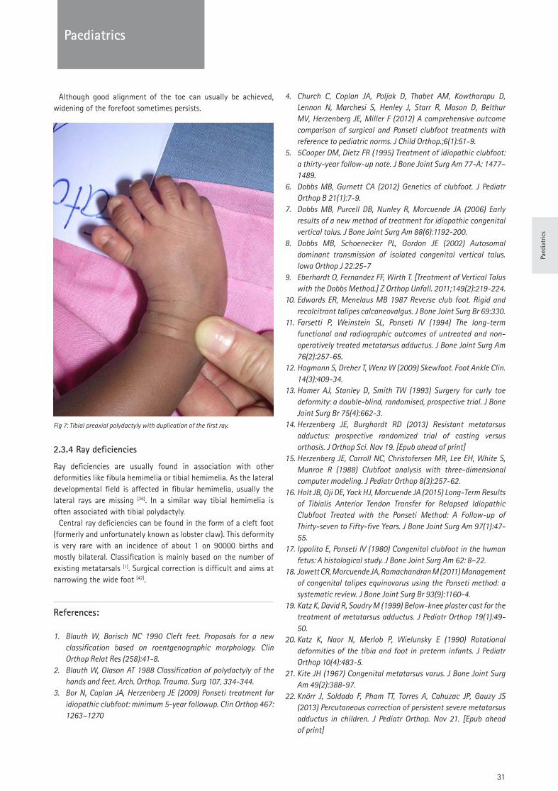
Paed
iatr
ics
31
Paediatrics
Although good alignment of the toe can usually be achieved, widening of the forefoot sometimes persists.
Fig 7: Tibial preaxial polydactyly with duplication of the first ray.
2.3.4 Ray deficiencies
Ray deficiencies are usually found in association with other deformities like fibula hemimelia or tibial hemimelia. As the lateral developmental field is affected in fibular hemimelia, usually the lateral rays are missing [26]. In a similar way tibial hemimelia is often associated with tibial polydactyly.
Central ray deficiencies can be found in the form of a cleft foot (formerly and unfortunately known as lobster claw). This deformity is very rare with an incidence of about 1 on 90000 births and mostly bilateral. Classification is mainly based on the number of existing metatarsals [1]. Surgical correction is difficult and aims at narrowing the wide foot [42].
References:
1. Blauth W, Borisch NC 1990 Cleft feet. Proposals for a new classification based on roentgenographic morphology. Clin Orthop Relat Res (258):41-8.
2. Blauth W, Olason AT 1988 Classification of polydactyly of the hands and feet. Arch. Orthop. Trauma. Surg 107, 334-344.
3. Bor N, Coplan JA, Herzenberg JE (2009) Ponseti treatment for idiopathic clubfoot: minimum 5-year followup. Clin Orthop 467: 1263–1270
4. Church C, Coplan JA, Poljak D, Thabet AM, Kowtharapu D, Lennon N, Marchesi S, Henley J, Starr R, Mason D, Belthur MV, Herzenberg JE, Miller F (2012) A comprehensive outcome comparison of surgical and Ponseti clubfoot treatments with reference to pediatric norms. J Child Orthop.;6(1):51-9.
5. 5Cooper DM, Dietz FR (1995) Treatment of idiopathic clubfoot: a thirty-year follow-up note. J Bone Joint Surg Am 77-A: 1477–1489.
6. Dobbs MB, Gurnett CA (2012) Genetics of clubfoot. J Pediatr Orthop B 21(1):7-9.
7. Dobbs MB, Purcell DB, Nunley R, Morcuende JA (2006) Early results of a new method of treatment for idiopathic congenital vertical talus. J Bone Joint Surg Am 88(6):1192-200.
8. Dobbs MB, Schoenecker PL, Gordon JE (2002) Autosomal dominant transmission of isolated congenital vertical talus. Iowa Orthop J 22:25-7
9. Eberhardt O, Fernandez FF, Wirth T. [Treatment of Vertical Talus with the Dobbs Method.] Z Orthop Unfall. 2011;149(2):219-224.
10. Edwards ER, Menelaus MB 1987 Reverse club foot. Rigid and recalcitrant talipes calcaneovalgus. J Bone Joint Surg Br 69:330.
11. Farsetti P, Weinstein SL, Ponseti IV (1994) The long-term functional and radiographic outcomes of untreated and non-operatively treated metatarsus adductus. J Bone Joint Surg Am 76(2):257-65.
12. Hagmann S, Dreher T, Wenz W (2009) Skewfoot. Foot Ankle Clin. 14(3):409-34.
13. Hamer AJ, Stanley D, Smith TW (1993) Surgery for curly toe deformity: a double-blind, randomised, prospective trial. J Bone Joint Surg Br 75(4):662-3.
14. Herzenberg JE, Burghardt RD (2013) Resistant metatarsus adductus: prospective randomized trial of casting versus orthosis. J Orthop Sci. Nov 19. [Epub ahead of print]
15. Herzenberg JE, Carroll NC, Christofersen MR, Lee EH, White S, Munroe R (1988) Clubfoot analysis with three-dimensional computer modeling. J Pediatr Orthop 8(3):257-62.
16. Holt JB, Oji DE, Yack HJ, Morcuende JA (2015) Long-Term Results of Tibialis Anterior Tendon Transfer for Relapsed Idiopathic Clubfoot Treated with the Ponseti Method: A Follow-up of Thirty-seven to Fifty-five Years. J Bone Joint Surg Am 97(1):47-55.
17. Ippolito E, Ponseti IV (1980) Congenital clubfoot in the human fetus: A histological study. J Bone Joint Surg Am 62: 8–22.
18. Jowett CR, Morcuende JA, Ramachandran M (2011) Management of congenital talipes equinovarus using the Ponseti method: a systematic review. J Bone Joint Surg Br 93(9):1160-4.
19. Katz K, David R, Soudry M (1999) Below-knee plaster cast for the treatment of metatarsus adductus. J Pediatr Orthop 19(1):49-50.
20. Katz K, Naor N, Merlob P, Wielunsky E (1990) Rotational deformities of the tibia and foot in preterm infants. J Pediatr Orthop 10(4):483-5.
21. Kite JH (1967) Congenital metatarsus varus. J Bone Joint Surg Am 49(2):388-97.
22. Knörr J, Soldado F, Pham TT, Torres A, Cahuzac JP, Gauzy JS (2013) Percutaneous correction of persistent severe metatarsus adductus in children. J Pediatr Orthop. Nov 21. [Epub ahead of print]
32
Paediatrics
23. Merrill LJ, Gurnett CA, Connolly AM, Pestronk A, Dobbs MB (2011) Skeletal muscle abnormalities and genetic factors related to vertical talus. Clin Orthop Relat Res 469(4):1167-74.
24. Mindler GT, Kranzl A, Lipkowski CA, Ganger R, Radler C (2014) Results of gait analysis including the Oxford foot model in children with clubfoot treated with the Ponseti method. J Bone Joint Surg Am 96(19):1593-9.
25. Ogata K, Schoenecker PL, Sheridan J (1979) Congenital vertical talus and its familial occurrence: an analysis of 36 patients. Clin Orthop Relat Res (139):128-32.
26. Opitz JM 1985 The developmental field concept. Am J Med Genet 21(1):1-11.
27. Pappas AM (1984) Congenital posteromedial bowing of the tibia and fibula. J Pediatr Orthop 4:525.
28. Paton RW, Choudry Q (2009) Neonatal foot deformities and their relationship to developmental dysplasia of the hip: an 11-year prospective, longitudinal observational study. J Bone Joint Surg Br 91(5):655-8.
29. Perry DC, Tawfiq SM, Roche A, Shariff R, Garg NK, James LA, Sampath J, Bruce CE (2010) The association between clubfoot and developmental dysplasia of the hip. J Bone Joint Surg Br 92(11):1586-8.
30. Ponseti IV (1996) Congenital clubfoot. Fundamentals of treatment. New York: Oxford University Press Inc.
31. Radler C. The Ponseti method for the treatment of congenital club foot: review of the current literature and treatment recommendations. Int Orthop. 2013;37(9):1747-53.
32. Radler C, Egermann M, Riedl K, Ganger R, Grill F (2010) Interobserver reliability of radiographic measurements of contralateral feet of pediatric patients with unilateral clubfoot. J Bone Joint Surg Am 20;92(14):2427-35.
33. Radler C, Manner HM, Suda R, Burghardt R, Herzenberg JE, Ganger R, Grill F (2007) Radiographic Evaluation of Idiopathic Clubfeet Undergoing Ponseti Treatment. J Bone Joint Surg Am 89: 1177-1183.
34. Radler C, Mindler GT, Riedl K, Lipkowski C, Kranzl A (2013) Midterm results of the Ponseti method in the treatment of congenital clubfoot. Int Orthop.;37(9):1827-31.
35. Radler C, Myers AK, Burghardt RD, Arrabal PP, Herzenberg JE, Grill F (2011) Maternal attitudes towards prenatal diagnosis of idiopathic clubfoot. Ultrasound Obstet Gynecol 37(6):658-62.
36. Sano H, Uhthoff HK, Jarvis JG, Mansingh A, Wenckebach GF (1998) Pathogenesis of soft-tissue contracture in club foot. J Bone Joint Surg Br 80(4):641-4.
37. Shabtai L, Specht SC, Herzenberg JE (2014) Worldwide spread of the Ponseti method for clubfoot. World J Orthop 5(5):585-90.
38. Shaheen S, Jaiballa H, Pirani S (2012) Interobserver reliability in Pirani clubfoot severity scoring between a paediatric orthopaedic surgeon and a physiotherapy assistant. J Pediatr Orthop B 21(4):366-8.
39. Smith PA, Kuo KN, Graf AN, Krzak J, Flanagan A, Hassani S, Caudill AK, Dietz FR, Morcuende J, Harris GF (2013) Long-term Results of Comprehensive Clubfoot Release Versus the Ponseti Method: Which Is Better? Clin Orthop Relat Res. Nov 19. [Epub ahead of print]
40. Watanabe H, Fujita S, Oka H (1992) Polydactyly of the foot: an analysis of 265 cases and a morphological classification. Plast Reconstr Surg 89:856-877.
41. Widhe T, Aaro S, Elmstedt E (1988) Foot deformities in the newborn--incidence and prognosis. Acta Orthop Scand 59(2):176-9.
42. Wood VE, Peppers TA, Shook J 1997 Cleft-foot closure: a simplified technique and review of the literature. J Pediatr Orthop 17(4):501-4.
Questions
1. Molding deformities are deformities, a. needing surgical treatment and follow-up. b. connected to the position of the child in utero.c. which are the result of structural changes.d. connected to autosomal dominant diseases.
2. Which of the following deformities is a molding deformity?a. clubfootb. talipes calcaneovalgusc. congenital vertical talusd. preaxial polydactyly
3. In clubfoot treatment using the Ponseti method, the foot abduction brace should be used a. for 3 monthsb. for 2 yearsc. until approximately 4 years of aged. until the foot looks good
4. What is usually not part of clubfoot treatment using the Ponseti method?a. serial castingb. abduction with counter pressure on neck of talusc. a percutaneous Achilles tenotomyd. an ankle foot orthosis (AFO)
5. The incidence of congenital vertical talus is approximatelya. 1:100b. 1:1.000c. 1:10.000d. 1:100.000
Answers1b, 2b, 3c, 4d, 5c
Paed
iatr
ics
33
Paediatrics
Prim. Doz. Dr. Rudolf Ganger,PhD
Paediatric Orthopaedic Department - Orthopaedic Hospital Vienna Speising
Vienna, Austria
Hip Diseases In The Childhood
Hip disorders in the childhood are common in the pediatric population. The initial symptoms may be limping or an abnormal gait. In addition, the child may complain of knee rather than hip pain. Many hip problems in the adult have their origin during growth.This chapter provides an overview of the main orthopaedic hip disorders from birth to adolescence.
1. Developmental Dysplasia Of The Hip (DDH)
Developmental dysplasia of the hip (DDH) occurs in different forms at different ages and includes all grades of instability and morphological abnormalities detected by ultrasound or radiography. The true incidence of DDH is difficult to determine because of disparities in the definition, the type of examinations used to detect DDH and the population being studied. Estimates range from 0.5 to 4 % [1,2].
Understanding the natural history of DDH requires knowledge of the growth and development of the hip joint from birth to skeletal maturity.
Due to differences in the diagnosis and the management of the disease, this entity is divided in two age groups: 1.1. DDH in newborns (0 to 1 year)1.2. DDH after walking age including1.2.1. Neglected cases of hip dislocation1.2.2. Residual dysplasia of the hip1.2.3. Osteonecrosis of the hip after treatment of DDH
1.1 DDH in newborns (0 to 1 year)
Since the introduction of hip sonography and its standardization, results of treatment have improved due to early diagnosis of deformities at birth [3]. Every newborn should be screened for signs of DDH or instability. Clinical examinations include the Barlow and Ortolani techniques and require experienced examiners [4]. Ultrasound according to Graf as a screening method is independent to the examiner’s experience and skills and provides a tool for early detection and early adequate therapy of DDH. Each sonographic type can be assigned to a specific phase of treatment correlated with a specific procedure effective in the given pathoanatomical situation (Table 1). Splinting therapy should be started up to the beginning of the sixth week of life in order not to miss the best time of treatment [5,6]. In cases detected early, conservative treatment can be completed before walking age. The α-angle according to Graf should be at least 60°. A radiograph should be performed in every treated child to exclude osteonecrosis of the hip.
Uncritical splinting which is not adapted to the given sonographic pathoanatomical situation of the hip or noncompliance of the
parents during the splinting phase often leads to surgery with the need for open reduction even when early diagnosed [7,8].
1.2 DDH after walking age
In cases of DDH after walking age, surgery to correct acetabular anatomy is often required. Operative interventions should be performed before the age of five years in order to provide complete anatomical recovery [9-11].
1.2.1 Neglected cases of dislocation after walking age
In countries with general ultrasound screening programs neglected dislocated hips have become rare cases [7,8].
Clinical findings in children with neglected hip dislocation after walking age are limping because of the shortened leg combined with restricted range of motion (restricted abduction and flexion contracture) and abductor muscle insufficiency (Trendelenburg gait). In bilateral cases, compensatory hyperlordosis of the lumbar spine is evident.
The therapeutic management depends on the age of the child and the pathomorphology of the acetabulum and the femur. In unilateral and bilateral cases of hip dislocation, the upper age limit for an open reduction is 8 years [11]. Due to the morphological changes of the hip joint in this age group, open reduction should be performed with pelvic and femoral (varus, derotation and shortening) osteotomies in a single stage procedure (Fig. 1a, 1b). Femoral shortening is necessary to reduce the risk of osteonecrosis following open reduction in older children [10,12].
Table 1: Sonographic-based treatment system according to the given hip types
Stage Findings Therapy Alternative
Reduction IV, III, D Manual reduction
Reduction orthosis
(i.e. Pavlik harness,…)
Retention Reducted IV, III, D
IIc unstable
Fettweis plaster cast in human position
Retention orthosis
(i.e. Pavlik harness,…)
Maturity II a -, IIb, IIc stable
Abduction device according to Mittelmeier-Graf
Maturity orthosis
(i.e. Pavlik harness, Tübinger orthosis,…)
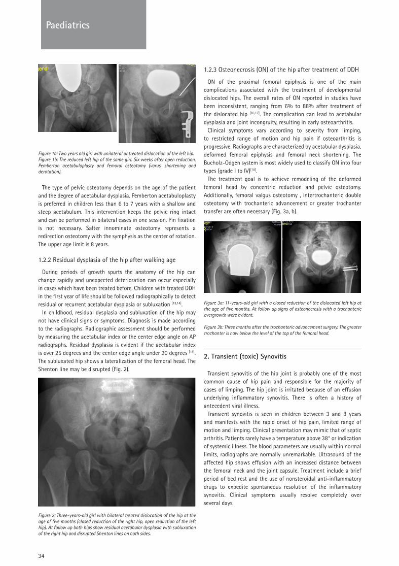
34
Paediatrics
Figure 1a: Two years old girl with unilateral untreated dislocation of the left hip.Figure 1b: The reduced left hip of the same girl. Six weeks after open reduction, Pemberton acetabuloplasty and femoral osteotomy (varus, shortening and derotation).
The type of pelvic osteotomy depends on the age of the patient and the degree of acetabular dysplasia. Pemberton acetabuloplasty is preferred in children less than 6 to 7 years with a shallow and steep acetabulum. This intervention keeps the pelvic ring intact and can be performed in bilateral cases in one session. Pin fixation is not necessary. Salter innominate osteotomy represents a redirection osteotomy with the symphysis as the center of rotation. The upper age limit is 8 years.
1.2.2 Residual dysplasia of the hip after walking age
During periods of growth spurts the anatomy of the hip can change rapidly and unexpected deterioration can occur especially in cases which have been treated before. Children with treated DDH in the first year of life should be followed radiographically to detect residual or recurrent acetabular dysplasia or subluxation [13,14].
In childhood, residual dysplasia and subluxation of the hip may not have clinical signs or symptoms. Diagnosis is made according to the radiographs. Radiographic assessment should be performed by measuring the acetabular index or the center edge angle on AP radiographs. Residual dysplasia is evident if the acetabular index is over 25 degrees and the center edge angle under 20 degrees [15]. The subluxated hip shows a lateralization of the femoral head. The Shenton line may be disrupted (Fig. 2).
Figure 2: Three-years-old girl with bilateral treated dislocation of the hip at the age of five months (closed reduction of the right hip, open reduction of the left hip). At follow up both hips show residual acetabular dysplasia with subluxation of the right hip and disrupted Shenton lines on both sides.
1.2.3 Osteonecrosis (ON) of the hip after treatment of DDH
ON of the proximal femoral epiphysis is one of the main complications associated with the treatment of developmental dislocated hips. The overall rates of ON reported in studies have been inconsistent, ranging from 6% to 88% after treatment of the dislocated hip [16,17]. The complication can lead to acetabular dysplasia and joint incongruity, resulting in early osteoarthritis.
Clinical symptoms vary according to severity from limping, to restricted range of motion and hip pain if osteoarthritis is progressive. Radiographs are characterized by acetabular dysplasia, deformed femoral epiphysis and femoral neck shortening. The Bucholz-Odgen system is most widely used to classify ON into four types (grade I to IV)[18].
The treatment goal is to achieve remodeling of the deformed femoral head by concentric reduction and pelvic osteotomy. Additionally, femoral valgus osteotomy , intertrochanteric double osteotomy with trochanteric advancement or greater trochanter transfer are often necessary (Fig. 3a, b).
Figure 3a: 11-years-old girl with a closed reduction of the dislocated left hip at the age of five months. At follow up signs of osteonecrosis with a trochanteric overgrowth were evident.
Figure 3b: Three months after the trochanteric advancement surgery. The greater trochanter is now below the level of the top of the femoral head.
2. Transient (toxic) Synovitis
Transient synovitis of the hip joint is probably one of the most common cause of hip pain and responsible for the majority of cases of limping. The hip joint is irritated because of an effusion underlying inflammatory synovitis. There is often a history of antecedent viral illness.
Transient synovitis is seen in children between 3 and 8 years and manifests with the rapid onset of hip pain, limited range of motion and limping. Clinical presentation may mimic that of septic arthritis. Patients rarely have a temperature above 38° or indication of systemic illness. The blood parameters are usually within normal limits, radiographs are normally unremarkable. Ultrasound of the affected hip shows effusion with an increased distance between the femoral neck and the joint capsule. Treatment include a brief period of bed rest and the use of nonsteroidal anti-inflammatory drugs to expedite spontaneous resolution of the inflammatory synovitis. Clinical symptoms usually resolve completely over several days.
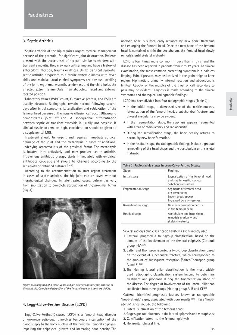
Paed
iatr
ics
35
Paediatrics
3. Septic Arthritis
Septic arthritis of the hip requires urgent medical management because of the potential for significant joint destruction. Patients present with the acute onset of hip pain similar to children with transient synovitis. They may walk with a limp and have a history of antecedent infection, trauma or illness. Unlike transient synovitis, septic arthritis progresses to a febrile systemic illness with fever, chills and malaise. Local clinical symptoms are obvious: swelling of the joint, erythema, warmth, tenderness and the child holds the affected extremity immobile in an abducted, flexed and external rotated position.
Laboratory values (WBC count, C-reactive protein, and ESR) are usually elevated. Radiographs remain normal following several days after initial symptoms. Lateralization and subluxation of the femoral head because of the massive effusion can occur. Ultrasound demonstrates joint effusion. A sonographic differentiation between septic or transient synovitis is usually not possible. If clinical suspicion remains high, consideration should be given to a supplemental MRI.
Treatment should be urgent and requires immediate surgical drainage of the joint and the metaphysis in cases of additional underlying osteomyelitis of the proximal femur. The metaphysis is located intra-articularly and may produce septic arthritis. Intravenous antibiotic therapy starts immediately with empirical antibiotics coverage and should be changed according to the sensitivity of obtained cultures [19,20].
According to the recommendation to start urgent treatment in cases of septic arthritis, the hip joint can be saved without morphological changes. In late-treated cases, deformities vary from subluxation to complete destruction of the proximal femur (Fig. 4).
Figure 4: Radiograph of a three-years-old girl after neonatal septic arthritis of the right hip. Complete destruction of the femoral head and neck are visible.
4. Legg-Calve-Perthes Disease (LCPD)
Legg-Calve-Perthes Diseases (LCPD) is a femoral head disorder of unknown aetiology. It involves temporary interruption of the blood supply to the bony nucleus of the proximal femoral epiphysis, impairing the epiphyseal growth and increasing bone density. The
necrotic bone is subsequently replaced by new bone, flattening and enlarging the femoral head. Once the new bone of the femoral head is contained within the acetabulum, the femoral head slowly remodels until skeletal maturity.
LCPD is four times more common in boys than in girls, and the disease has been reported in patients from 2 to 12 years. At clinical examination, the most common presenting symptom is a painless limping. Pain, if present, may be localized in the groin, thigh or knee region. Hip motion, primarily internal rotation and abduction, is limited. Atrophy of the muscles of the thigh or calf secondary to pain may be evident. Diagnosis is made according to the clinical symptoms and the typical radiographic findings.
LCPD has been divided into four radiographic stages (Table 2):
• In the initial stage, a decreased size of the ossific nucleus, lateralization of the femoral head, a subchondral fracture, and physeal irregularity may be evident.
• In the fragmentation stage, the epiphysis appears fragmented with areas of radiolucency and radiodensity.
• During the reossification stage, the bone density returns to normal by new bone formation.
• In the residual stage, the radiographic findings include a gradual remodeling of the head shape and the acetabulum until skeletal maturity.
Several radiographic classification systems are currently used:1. Catterall proposed a four-group classification, based on the
amount of the involvement of the femoral epiphysis (Catterall group I-IV) [21].
2. Salter and Thompson reported a two-group classification based on the extent of subchondral fracture, which corresponded to the amount of subsequent resorption (Salter-Thomspon group A and B) [56].
3. The Herring lateral pillar classification is the most widely used radiographic classification system helping to determine treatment and prognosis during the fragmentation stage of the disease. The degree of involvement of the lateral pillar can subdivided into three groups (Herring group A, B and C) [23].
Catterall identified prognostic factors, known as radiographic “head-at-risk” signs, associated with poor results [24]. These “head-at-risk” sings include the following:
1. Lateral subluxation of the femoral head;2. Gage sign: radiolucency in the lateral epiphysis and metaphysis;3. Calcification lateral to the femoral epiphysis;4. Horizontal physeal line.
Table 2: Radiographic stages in Legg-Calve-Perthes Disease
Stage Findings
Initial stage Lateralization of the femoral headand smaller ossific nucleusSubchondral fracture
Fragmentation stage Segments of femoral head are demarcatedLucent areas appearIncreased density resolves
Reossification stage New bone formation occurs in the femoral head
Residual stage Acetabulum and head shaperemodels gradually until skeletal maturity
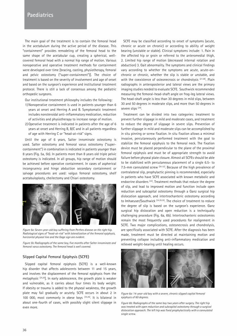
36
Paediatrics
The main goal of the treatment is to contain the femoral head in the acetabulum during the active period of the disease. This “containment” provides remodeling of the femoral head to the same shape of the acetabular cup, creating a spherical, well-covered femoral head with a normal hip range of motion. Various nonoperative and operative treatment methods for containment were developed over time [bracing, casting, physiotherapy, femoral and pelvic osteotomy (“super-containment”)]. The choice of treatment is based on the severity of involvement and age of onset and based on the surgeon’s experience and institutional treatment protocol. There is still a lack of consensus among the pediatric orthopedic surgeons.
Our institutional treatment philosophy includes the following:1) Nonoperative containment is used in patients younger than 6
years at onset and Herring A and B. Symptomatic treatment includes nonsteroidal anti-inflammatory medication, reduction of activities and physiotherapy to increase range of motion.
2) Operative treatment is indicated in patients after the age of 6 years at onset and Herring B, B/C and in all patients regardless of age with Herring C or “head-at-risk” signs.
Until the age of 6 years, Salter innominate osteotomy is used. Salter osteotomy and femoral varus osteotomy (“super-containment”) in combination is indicated in patients younger than 8 years (Fig. 5a, 5b). In patients more than 8 years old triple pelvic osteotomy is indicated. In all groups, hip range of motion should be achieved before operative containment. In cases of aspherical incongruency and hinge abduction secondary containment or salvage procedures are used: valgus femoral osteotomy, shelf acetabuloplasty, cheilectomy and Chiari osteotomy.
Figure 5a: Seven-year-old boy suffering from Perthes disease on the right hip. Radiological signs of “head-at-risk” with lateralization of the femoral epiphysis, horizontal physeal line and the Gage sign are evident.
Figure 5b: Radiographs of the same boy, five months after Salter innominate and femoral varus osteotomy. The femoral head is well covered.
Slipped Capital Femoral Epiphysis (SCFE)
Slipped capital femoral epiphysis (SCFE) is a well-known hip disorder that affects adolescents between 11 and 15 years, and involves the displacement of the femoral epiphysis from the metaphysic [25,26]. In early adolescence, the growth plate is weaker and vulnerable, as it carries about four times its body weight. If obesity or trauma is added to the physeal weakness, the growth plate may fail gradually or acutely. SCFE occurs in about 2 in 100 000, most commonly in obese boys [25,26]. It is bilateral in about one-fourth of cases, with possibly slight silent slippage in even more.
SCFE may be classified according to onset of symptoms (acute, chronic or acute on chronic) or according to ability of weight bearing (unstable or stable). Clinical symptoms include: 1. Pain in the affected hip or groin or referred to the anteromedial thigh, 2. Limited hip range of motion (decreased internal rotation and abduction) 3. Gait abnormality. The symptoms and clinical findings vary according to whether the symptoms are acute, acute-on-chronic or chronic, whether the slip is stable or unstable, and with the coexistence of osteonecrosis or chondrolysis [27,28]. Plain radiographs in anteroposterior and lateral views are the primary imaging studies needed to evaluate SCFE. Southwick recommended measuring the femoral-head-shaft angle on frog-leg lateral views. The head-shaft angle is less than 30 degrees in mild slips, between 30 and 50 degrees in moderate slips, and more than 50 degrees in severe slips [29].
Treatment can be divided into two categories: treatment to prevent further slippage in mild and moderate cases, and treatment to reduce the degree of slippage in severe slips. Prevention of further slippage in mild and moderate slips can be accomplished by in situ pinning or screw fixation. In situ fixation allows a minimal invasive, percutaneously performed treatment with the goal to stabilize the femoral epiphysis to the femoral neck. The fixation device must be placed perpendicular to the plane of the proximal femoral epiphysis and must be of appropriate strength to avoid failure before physeal plate closure. Almost all SCFEs should be able to be stabilized with percutaneous placement of a single 6.5- to 7.5-mm cannulated screw [30-32]. Because of the high prevalence of contralateral slip, prophylactic pinning is recommended, especially in patients who have SCFE associated with known metabolic and endocrine disorders [33]. Treatment methods that reduce the degree of slip, and lead to improved motion and function include open reduction and subcapital osteotomy through a Ganz surgical hip dislocation approach, and intertrochanteric osteotomy according to Imhaeuser/Southwick [34,29,35]. The choice of treatment to reduce the degree of slip is based on the surgeon’s experience. Ganz surgical hip dislocation and open reduction is a techniqually challenging procedure (Fig. 6a, 6b). Intertrochanteric osteotomies remain the most frequently used procedures for realignment in SCFE. Two major complications, osteonecrosis and chondrolysis, are specifically associated with SCFE. After the diagnosis has been made, treatment must be directed at maintaining motion and preventing collapse including anti-inflammatory medication and relieved weight-bearing until healing occurs.
Figure 6a: 14-year-old boy with a severe, chronic slipped capital femoral epiphysis of 60 degrees.
Figure 6b: Radiographs of the same boy two years after surgery. The right hip was treated with open reduction and subcapital osteotomy through a surgical dislocation approach. The left hip was fixed prophylactically with a cannulated single screw.
37
Paed
iatr
ics
Paediatrics
References
1. Toennis D (1984) Die angeborene Hüftdysplasie und Hüftluxation. Springer, Berlin Heidelberg New York
2. Rosendahl K, Markestad T, Lie RT (1994) Ultrasound screening for developmental dysplasia of the hip in the neonate: the effect on treatment rate and prevalence of late cases. Pediatrics 94 (1):47-52
3. Graf R (2006) Hip Sonography. Diagnosis and Treatment of Infant Hip Dysplasia. Springer, Heidelberg
4. Barlow TG (1963) Early Diagnosis and Treatment of Congenital Dislocation of the Hip. Proc R Soc Med 56:804-806
5. Matthiessen HD (1996) Forensic problems in the treatment of hip dysplasias and dislocations. Z Orthop Ihre Grenzgeb 134 (6):Oa10-12
6. Matthiessen HD (1997) Dysplasia and therapy factors in hip developmental disorders. Z Orthop Ihre Grenzgeb 135 (1):Oa12-13
7. Grill F, Muller D (1997) Results of hip ultrasonographic screening in Austria. Orthopade 26 (1):25-32
8. von Kries R, Ihme N, Oberle D, Lorani A, Stark R, Altenhofen L, Niethard FU (2003) Effect of ultrasound screening on the rate of first operative procedures for developmental hip dysplasia in Germany. Lancet 362 (9399):1883-1887.
9. Salter RB (1966) Role of innominate osteotomy in the treatment of congenital dislocation and subluxation of the hip in the older child. J Bone Joint Surg Am 48 (7):1413-1439
10. Galpin RD, Roach JW, Wenger DR, Herring JA, Birch JG (1989) One-stage treatment of congenital dislocation of the hip in older children, including femoral shortening. J Bone Joint Surg Am 71 (5):734-741
11. Lindstrom JR, Ponseti IV, Wenger DR (1979) Acetabular development after reduction in congenital dislocation of the hip. J Bone Joint Surg Am 61 (1):112-118
12. Schoenecker PL, Strecker WB (1984) Congenital dislocation of the hip in children. Comparison of the effects of femoral shortening and of skeletal traction in treatment. J Bone Joint Surg Am 66 (1):21-27
13. Kim HT, Kim JI, Yoo CI (2000) Acetabular development after closed reduction of developmental dislocation of the hip. J Pediatr Orthop 20 (6):701-708
14. Albinana J, Dolan LA, Spratt KF, Morcuende J, Meyer MD, Weinstein SL (2004) Acetabular dysplasia after treatment for developmental dysplasia of the hip. Implications for secondary procedures. J Bone Joint Surg Br 86 (6):876-886
15. Toennis D (1987) Congenital dysplasia and dislocation of the hip in children and adults. Springer, Berlin Heidelberg
16. Pospischill R, Weninger J, Ganger R, Altenhuber J, Grill F (2012) Does open reduction of the developmental dislocated hip increase the risk of osteonecrosis? Clin Orthop Relat Res 470 (1):250-260.
17. Firth GB, Robertson AJ, Schepers A, Fatti L (2010) Developmental dysplasia of the hip: open reduction as a risk factor for substantial osteonecrosis. Clin Orthop Relat Res 468 (9):2485-2494.
18. Bucholz R, Ogden, J. (1978) Patterns of ischemic necrosis of the proximal femur in nonoperatively treated congenital hip diseases. The Hip: Proceedings of the Sixth Open Scientific Meeting of the Hip Society. CV Mosby, St. Louis
19. Morrey BF, Bianco AJ, Jr., Rhodes KH (1975) Septic arthritis in children. Orthop Clin North Am 6 (4):923-934
20. Morrey BF, Peterson HA (1975) Hematogenous pyogenic osteomyelitis in children. Orthop Clin North Am 6 (4):935-951
21. Catterall A, Pringle J, Byers PD, Fulford GE, Kemp HB, Dolman CL, Bell HM, McKibbin B, Ralis Z, Jensen OM, Lauritzen J, Ponseti IV, Ogden J (1982) A review of the morphology of Perthes' disease. J Bone Joint Surg Br 64 (3):269-275
22. Salter RB, Thompson GH (1984) Legg-Calve-Perthes disease. The prognostic significance of the subchondral fracture and a two-group classification of the femoral head involvement. J Bone Joint Surg Am 66 (4):479-489
23. Herring JA, Neustadt JB, Williams JJ, Early JS, Browne RH (1992) The lateral pillar classification of Legg-Calve-Perthes disease. J Pediatr Orthop 12 (2):143-150
24. Catterall A (1971) The natural history of Perthes' disease. J Bone Joint Surg Br 53 (1):37-53
25. Kelsey JL, Keggi KJ, Southwick WO (1970) The incidence and distrubition of slipped capital femoral epiphysis in Connecticut and Southwestern United States. J Bone Joint Surg Am 52 (6):1203-1216
26. Loder RT (1996) The demographics of slipped capital femoral epiphysis. An international multicenter study. Clin Orthop Relat Res (322):8-27
27. Aronson J, Tursky EA (1996) The torsional basis for slipped capital femoral epiphysis. Clin Orthop Relat Res (322):37-42
28. Loder RT, Richards BS, Shapiro PS, Reznick LR, Aronson DD (1993) Acute slipped capital femoral epiphysis: the importance of physeal stability. J Bone Joint Surg Am 75 (8):1134-1140
29. Southwick WO (1967) Osteotomy through the lesser trochanter for slipped capital femoral epiphysis. J Bone Joint Surg Am 49 (5):807-835
30. Goodman WW, Johnson JT, Robertson WW, Jr. (1996) Single screw fixation for acute and acute-on-chronic slipped capital femoral epiphysis. Clin Orthop Relat Res (322):86-90
31. Herman MJ, Dormans JP, Davidson RS, Drummond DS, Gregg JR (1996) Screw fixation of Grade III slipped capital femoral epiphysis. Clin Orthop Relat Res (322):77-85
32. Koval KJ, Lehman WB, Rose D, Koval RP, Grant A, Strongwater A (1989) Treatment of slipped capital femoral epiphysis with a cannulated-screw technique. J Bone Joint Surg Am 71 (9):1370-1377
33. Jerre R, Billing L, Hansson G, Wallin J (1994) The contralateral hip in patients primarily treated for unilateral slipped upper femoral epiphysis. Long-term follow-up of 61 hips. J Bone Joint Surg Br 76 (4):563-567
34. Imhauser G (1954) [Surgical treatment of pathological anteversion of the proximal femur]. Z Orthop Ihre Grenzgeb 85 (3):395-405
35. Leunig M, Slongo T, Kleinschmidt M, Ganz R (2007) Subcapital correction osteotomy in slipped capital femoral epiphysis by means of surgical hip dislocation. Oper Orthop Traumatol 19 (4):389-410.
38
Paediatrics
Questions
1. The appearance of an acute osteomyelitis in childhooda. is mainly induced by a focus of the gastrointestinal areab. is most often caused by Streptoccocusc. is typically located in the diaphyseal area of long bonesd. is most often located in the metaphyseal area of long
bonese. is never penetrating to adjacent joints
2. Which criterion may be cause of an unfavorable result of Perthes disease?
a. Appearance before age 4b. Initial joint effusion c. Type Herring Cd. Reduced abduction during fragmental periode. Conservative treatment for type Catteral II
3. The Bernese periacetabular osteotomy for correction of hip dysplasia is recommended after maturity because of
a. Tendency to overcorrectionb. Involvement of the triradiate cartilagec. Tendency to undercorrectiond. Danger of impingemente. To complicated during growth
4. Which condition CANNOT be found in cause of Femoroacetabular Impingement (FAI)?
a. Lack of acetabular coverageb. Acetabular retroversionc. Protrusion oft he femoral headd. Cross-over signe. Acetabular over-coverage
5. What is the most common age group for Kingella kingae infection?
a. Infants less than 6 months of ageb. Children above 4 yearsc. Children between 6 months and 4 yearsd. Teenagerse. Adults
Answers1d, 2c, 3b, 4a, 5c.

39
Paed
iatr
ics
Paediatrics
40
Reconstruction
Luigi [email protected]
Rocco D’[email protected]
Hip Department, Istituto Ortopedico Galeazzi IRCCS, Milan Italy
Hip Reconstruction
Abstract
Total hip arthroplasty is a successful and cost-effective procedure with more than 1 million replacements performed worldwide per year. To achieve good results and meet patient expectations surgeons must be aware of the key points of this procedure, starting from indication and preoperative evaluation. The radiological workup is discussed in this review, reporting what to look for in a standard X-ray and when to use further imaging techniques. The restoration of hip biomechanics is crucial to obtain good results and preoperative templating helps surgeons for this purpose, anticipating possible intraoperative troubles. Several surgical approaches exist, each one with advantages and disadvantages. The morphology of the proximal femur influences the stem choice, whereas the age holds way over the fixation method. Respecting the principles of press-fit and cemented fixation is of paramount importance to guarantee long-term satisfying results. Basics of reaming and cementing techniques are also reported. Moreover, anatomical landmarks and components orientation have to be taken into account intraoperatively. Available bearing options allow the surgeon to choose the best coupling for each single case, selecting among ceramic or polyethylene inserts and ceramic or metal heads.
1. Introduction
Hip reconstruction aims to give full function recovery of the hip joint. When severely affected the hip joint can become cause of serious discomfort and limitations for the patient, leading to the loss of walking capability.
In case of major intra-articular deformities or incongruences and/or cartilage degeneration the joint replacement is essential so the reconstruction is done by means of artificial materials.This procedure is named total hip replacement (THR) or total hip arthroplasty (THA) and it is one of the most successful procedures of all the surgeries. It has been considered as the operation of the century for its high cost-effectiveness (1). More than 1 million THRs are performed worldwide per year. The clinical outcome is excellent and the implant is functioning over time: survivorship greater than 95% at 10 years is reported by registries data and more than 80% of the prostheses survive at 25 years follow-up (2).
The “happy” or “forgotten” replaced hip is the result of a series of standardized and well conducted steps. These include indication, preoperative evaluation, prevention of complication, preoperative
planning and templating for good implant selection and restoration of biomechanics, careful surgery and finally controlled post-operative phase.
2. Indications
Indications for THR are listed in Table 1. The correct indication is the first step of a successful joint reconstruction, as the optimal clinical results are obtained through a careful patient selection. It should be based on pain, functional impairment, physical examination and not just on radiographic findings. Risks of surgery must be always balanced, so even if THR is a safe procedure pros and cons of each operation must be always evaluated carefully and discussed, offering the best evidence-based available option to each patient (3). Surgeons should always keep in mind that the most relevant factor for satisfaction is meeting patient’s expectations (4), as unrealistic expectations are the main cause of dissatisfaction and health-care providers should help their patients to develop realistic expectations.
Pain, functional impairment such as limp, loss of range of motion, leg length discrepancy can be effectively treated with a THR. Differential diagnosis must always be considered and other sources of pain in the hip region excluded.
Diagnosis is based on clinical and radiological findings (differential diagnosis of hip pain are listed in Table 2).
Table 1. Indications for THR
Primary osteoarthritis
Secondary osteoarthritis• DDH• SCFE• LCP• AVN• Inflammatory disease (RA, SA)• Post-traumatic• Tumours
Femoral head/neck fractures
Table 2. Differential diagnosis of hip pain
Pain of spine origin
Trochanteric syndrome
Tendon problems (Ad muscles, ileopsoas)
Inguinal hernia or other non orthopaedic origins (neurologic, vascular, visceral)
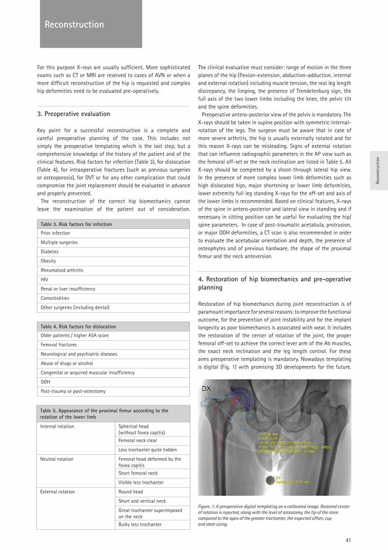
Reco
nstr
uctio
n
41
Reconstruction
For this purpose X-rays are usually sufficient. More sophisticated exams such as CT or MRI are reserved to cases of AVN or when a more difficult reconstruction of the hip is requested and complex hip deformities need to be evaluated pre-operatively.
3. Preoperative evaluation
Key point for a successful reconstruction is a complete and careful preoperative planning of the case. This includes not simply the preoperative templating which is the last step, but a comprehensive knowledge of the history of the patient and of the clinical features. Risk factors for infection (Table 3), for dislocation (Table 4), for intraoperative fractures (such as previous surgeries or osteoporosis), for DVT or for any other complication that could compromise the joint replacement should be evaluated in advance and properly prevented.
The reconstruction of the correct hip biomechanics cannot leave the examination of the patient out of consideration.
The clinical evaluation must consider: range of motion in the three planes of the hip (flexion-extension, abduction-adduction, internal and external rotation) including muscle tension, the real leg length discrepancy, the limping, the presence of Trendelenburg sign, the full axis of the two lower limbs including the knee, the pelvic tilt and the spine deformities.
Preoperative antero-posterior view of the pelvis is mandatory. The X-rays should be taken in supine position with symmetric internal-rotation of the legs. The surgeon must be aware that in case of more severe arthritis, the hip is usually externally rotated and for this reason X-rays can be misleading. Signs of external rotation that can influence radiographic parameters in the AP view such as the femoral off-set or the neck inclination are listed in Table 5. All X-rays should be completed by a shoot-through lateral hip view. In the presence of more complex lower limb deformities such as high dislocated hips, major shortening or lower limb deformities, lower extremity full leg standing X-rays for the off-set and axis of the lower limbs is recommended. Based on clinical features, X-rays of the spine in antero-posterior and lateral view in standing and if necessary in sitting position can be useful for evaluating the hip/spine parameters. In case of post-traumatic acetabula, protrusion, or major DDH deformities, a CT scan is also recommended in order to evaluate the acetabular orientation and depth, the presence of osteophytes and of previous hardware, the shape of the proximal femur and the neck anteversion.
4. Restoration of hip biomechanics and pre-operative planning
Restoration of hip biomechanics during joint reconstruction is of paramount importance for several reasons: to improve the functional outcome, for the prevention of joint instability and for the implant longevity as poor biomechanics is associated with wear. It includes the restoration of the center of rotation of the joint, the proper femoral off-set to achieve the correct lever arm of the Ab muscles, the exact neck inclination and the leg length control. For these aims preoperative templating is mandatory. Nowadays templating is digital (Fig. 1) with promising 3D developments for the future.
Figure. 1: A preoperative digital templating on a calibrated image. Restored center of rotation is reported, along with the level of osteotomy, the tip of the stem compared to the apex of the greater trochanter, the expected offset, cup and stem sizing.
Table 5. Appearance of the proximal femur according to the rotation of the lower limb
Internal rotation Spherical head (without fovea capitis)
Femoral neck clear
Less trochanter quite hidden
Neutral rotation Femoral head deformed by the fovea capitis
Short femoral neck
Visible less trochanter
External rotation Round head
Short and vertical neck
Great trochanter superimposed on the neck
Bulky less trochanter
Table 3. Risk factors for infection
Prior infection
Multiple surgeries
Diabetes
Obesity
Rheumatoid arthritis
HIV
Renal or liver insufficiency
Comorbidities
Other surgeries (including dental)
Table 4. Risk factors for dislocation
Older patients / higher ASA score
Femoral fractures
Neurological and psychiatric diseases
Abuse of drugs or alcohol
Congenital or acquired muscular insufficiency
DDH
Post-trauma or post-osteotomy

42
Reconstruction
It is essential for the selection of the type of implant and for the size of the implant components as well. Information on the level of the neck osteotomy and on the position of the cup is obtained prior to surgery.
Both cups and stems, cemented or not, should be selected according to the bone shape (Dorr’s classification), bone quality and age of the patient. For this choice, femoral templating is a fundamental part of the procedure. The different designs, in particular of the cementless stems, are then evaluated to better fit into the proximal femur.
Implant fixation could be cemented, cementless or hybrid. There are different preferences among the countries and among the different centers, but in each case, the correct principles and techniques must be applied in order to avoid complications. An orthopaedic surgeon should be confident with both cemented and uncemented techniques to be properly applied in each specific case.
5. Principles of fixation and surgical technique
5.1 The Cup
The principles of cementless fixation include reliable mechanical primary stability to allow osseointegration that guarantees a durable biological secondary stability. ‘‘Osseointegration’’ is the attachment of lamellar bone to implants without fibrous tissue. It takes four to twelve weeks and may continue for up to three years (5). Micromotion is not conductive to bony incorporation, if greater than 150 µm, it is incompatible with bone ingrowth, between 40 and 150 µm, it leads to a combination of bone and fibrous tissue formation, and if <20 µm, it results in predominantly bone formation (6). For this purpose a rough surface with adequate grip and biocompatibility is necessary. Titanium has widely demonstrated to have these features and Titanium plasma-spray is the more commonly used for primary routine cases. In the last decade different high porosity implants have been developed. Porous structures have the possibility to balance different properties both with tantalum and titanium: an elastic modulus close to the bone able to prevent stress shielding, an average mechanical strength capable to resist to the applied load, controlled single pore dimensions to stimulate osseointegration, an overall porosity to grant interconnection among pores and both corrosion resistance and biocompatibility. These materials can make a great difference and an improvement in revision surgery and in complex cases with poor bone stock.
Not only the surface matters, but also the shape, the most commonly used nowadays being hemispherical. The primary stability is guaranteed by press-fit insertion. There are two options for improving the press-fit: the cup is squeezed at the equatorial plane (Fig 2A) or the cup is larger than the allocated seat (Fig. 2B). According to this technique, underreaming 1-2 mm is mandatory to achieve a reliable stability (7). For this purpose, it is important to preserve the “sandwich” structure of the acetabulum preserving the bone and reaming only the cartilage and the osteophytes, as a damage of the outer cortex and/or of the lamina quadrilatera can lead to a progressive migration. In case of lack of primary stability, two or more screws can be added in the safe zone
(supero-lateral quadrant).In case of cemented cup, a good surgical technique must be
ensured. Acetabular preparation includes reaming, creation of some holes in the reamed acetabulum for cement inter-digitation, accurate drying and pressurization of the all poly cup during cement polymerization.
Whichever implant or type of fixation is used, cup orientation is a key point of implant success as malposition can lead to both early (dislocation) or late (wear) complications. According to historical Lawinnek’s safe zones (8), cup must be placed at no more than 45° of inclination and between 15° and 25° of anteversion. These parameters are fully valid nowadays and should be strictly followed even if they must be interpreted in more recent findings that include hip/spine relationships and anatomical variations of the pelvis.
The Transverse Acetabular Ligament (TAL) is a simple and easy of use landmark. It is identifiable in the vast majority of cases and can help the surgeon to optimize cup version. TAL should be recognized and followed in all the procedures.
5.2 The stem
The principles of cementless fixation are the same on the femoral side:
• Reliable mechanical primary stability to allow osseointegration
• Durable biological secondary stability• Load transfer to the proximal femur (if possible)• Bone stock preservation
Titanium alloy with a rough surface is the material of choice for achieving these purposes. The size of the pores on the surface should be between 50 µm and 400 µm, and the percentage of voids within the coating should be between 30% and 40% to maintain mechanical strength (10).
The reason for transmitting the forces to the proximal femur is to avoid stress shielding (which means loss of bone quality in the proximal femur due to distal fixation and transmission of forces), cortical hypertrophy at the level of the distal fixation and thigh
Figure. 2A: Cup squeezed at the equatorial plane
Figure. 2B: Cup larger than the allocated seat
Reco
nstr
uctio
n
43
Reconstruction
pain, that was frequently associated in the past with cementless stems with “fit and fill” principles at the distal part of the stem. Whenever possible proximal fixation of the stem should be preferred, according to femoral shape and bone quality.
Cementless designs differ one from the other in terms of geometry and the means of obtaining initial fixation. According to the design, the cementless stems are classified into two main categories: straight stems (which include different types of wedge shape and/or taper geometry) and curved anatomic stems (11). Special designs and modular implants should be considered in complex cases or deformities. Each type of stem has a different surgical technique, that can differ significantly one from the other. Loosening and thigh pain are less prevalent with modern stem designs, as stress shielding is still present, even with newer stem designs. In the last decades the so called “short stems” have been proposed with the aim of bone preserving and transmission of forces to the proximal femur. Even if theoretically promising, there is lack of long-term data compared to traditional well documented implants.
Stem selection is part of the procedure. No cementless stem can be suitable for all the different types of femur. The choice should be based on the bone shape according to Dorr’s classification and to the bone quality, and also on the age of the patient. According to registries data and clinical studies there is quite clear evidence that, at least over the age of 70/75, cemented fixation of the femur has better survival compared to cementless.
Cemented stems are most commonly made of cobalt-chrome and polished. The modern fourth generation cementing technique of the femur includes the use of femoral plug, distal and eventually proximal centralizers, accurate drying of the bone after jet lavage (the latter being controversial), retrograde cement insertion with a long syringe and pressurization after vacuum preparation of the cement. The majority of the studies (including large registry data) are in favour of the use antibiotic-loaded cement. According to registry, hybrid fixation with cementless cup and cemented stem can be considered a good option for the older population. Full cemented implants are good choice for the old patient as well, while in patients younger than 70 the general trend is towards cementless fixation. But as mentioned, age is only one aspect of successful fixation, while bone quality and shape, and the presence of comorbidities are meaningful aspects.
In any case, the orthopaedic surgeon should be confident with both cemented and different types of cementless stems that can harm the greater trochanter in different ways, so requiring different lateral preparation. They must be careful to avoid undersizing of the stem, to avoid varus position (some stems can tolerate it better than others), and to control around 15° of anteversion. Leg length, soft-tissues tension and stability are controlled during surgery by means of less inserted cemented stems or bigger cementless stems, remembering that a less inserted cemented stem increases only the length, while implanting a bigger cementless stem usually increases length and off-set. Using heads with longer necks increases length and off-set as well. Extended off-set stems can be carefully used when the anatomy or the joint stability require this type of solution.
5.3 Approaches
Different approaches are suitable for good hip reconstruction (anterior, antero-lateral, direct lateral, posterior or “mini” posterior,
superior). Some of them got great popularity recently and are quite “fashionable” nowadays. Each approach (as each technique) has its own advantages and disadvantages. What is really important is that Ab muscle function is fully preserved (if damaged, the outcome is poor and there is no effective solution for such a damage), that the rate of complications is acceptable (balanced with supposed advantages) and that a clear view of the landmarks for implant positioning is possible (as this is the key point for long term implant survival). The approach must be also relatively simple, without major damage of the soft-tissues, and suitable for dealing with intraoperative complications in a simple way. Finally, an approach must be suitable for different type of implants in particular for the ones with the best long term results, including cemented components. Current evidence comparing outcomes following anterior versus posterior THR does not demonstrate clear superiority of either approach. Until more rigorous, randomized evidence is available, we recommend choice of surgical approach for THA to be based on patient characteristics, surgeon experience and surgeon and patient preferences.
5.4 Type of bearings
Wear osteolysis is the major cause of late failure. This is the reason why bearing surface selection is so important and the knowledge of tribology as well (12).
Tribology is the science that studies friction, lubrication and wear between two surfaces which are in close contact and move one on the other. The name is derived from the Greek word ‘Τριβος,’ which means rubbing.
Wear is the surface damage with progressive loss of material (debris) due to friction between moving surfaces.
Debris is composed by articles of different material and size shed from the surface of the various parts of the implant due to wear.
Fretting is the relative low amplitude movement (oscillation and sliding) between two mechanically joined parts, under load conditions (between 1 µm and 100 µm). All modular junctions are susceptible to the loading of the body. It provokes wear (debris) and corrosion.
Corrosion is the surface degradation due to electrochemical interactions producing metallic ions and salts, which applies only to metals. Different forms of corrosion have been described (galvanic, fretting, crevice, stress, etc).
Osteolysis is the bone resorption due to the biological response to debris, including osteoclasts activation that can compromise the bone stock around the implant and lead to loosening of the prosthesis in the advanced phase.
Table 6. Main disadvantages for each bearing surface
Bearing surface Disadvantages
Metal-on-polyethelene Wear and osteolysis
Ceramic-on-polyethelene Wear and osteolysis
Metal-on-XLPE Decreased mechanical properties
Ceramic-on-XLPE Decreased mechanical properties
Ceramic-on-ceramic Breakage and squeaking
Metal-on-metalARMD (ALVAL, high ion levels, osteolysis, pseudotumours)
XLPE, highly cross-linked polyethylene; ARMD, adverse reaction to metal debris;ALVAL, aseptic lymphocyte-dominant, vasculitis-associated lesion
44
Reconstruction
Adverse Reaction to Metal Debris (ARMD) or Adverse Local Tissue Reaction (ALTR) include a variety of effects of metal debris including osteolysis and pseudotumours, elevated illness of blood metal ions.
Each bearing surface has its well-known disadvantages (Table 6). On the other hand, there is limited evidence regarding comparative effectiveness of various hip implant bearings. RCTs show similar short- to mid-term survivorship among Ceramic on Ceramic (CoC), Ceramic on Crosslinked Polyethylene (CoXLPE) and Metal on Crosslinked Polyethylene (MoXLPE) in patients younger than 65.
Conventional PE, sterilized by Oxygen free Gamma irradiation or Ethylene Oxide, with correct packaging, can show good clinical long-term results. Metal-on-conventional PE (MoPE) has a higher risk of revision compared with MoXLPE. Clinicians should consider the use of XLPE when using a polyethylene bearing in THA (13). XLPE is going to achieve good medium to long-term results. The improved wear resistance made possible the use of larger heads (32 mm and 36 mm depending on the cup size), improving the joint stability. The cross-linking process, while increasing the wear properties of PE decreases the mechanical ones, making the liners more at risk of fatigue fracture. For this reason a minimum XLPE thickness (5-6 mm) must be preserved as cross-linking decreases the mechanical properties of PE. Recently, XLPE liners with the addition of antioxidants such as Vitamin E were introduced into the market with the aim of reducing the oxidation in vivo. In this way, re-melting is not necessary to avoid the oxidation, and mechanical proprieties are better preserved. As a consequence, the liner can also be thinner and larger heads can be used.
CoPE is at least equal but usually better than MoPE with less wear and lower revision rates. The reason is that ceramic has a higher hardness (5x), it cannot be scratched (less risk of damage during implantation). The surface finishing has only negative roughness rather than deformation. It has better wettability, thus a better lubrication, and it is the material with the highest bio-inertness.
There are increasing concerns on the trunnions with metal heads in MoPE, no clinical reports on ceramic heads. CoXLPE looks like a reasonable bearing surface, the only real problem could be the cost as the risk of breakage is very low with only occasional reports and insignificant in combination with PE.
The third best option of performing bearing could be CoC. Better survival is reported by registries for heads 32 mm or more (36 mm). The metal back must be treated carefully with ceramic liners (that can be at risk in case of shell deformation) in thin press fit cups that depends on bone quality, instruments and surgical technique.Minimum thickness of the metal back is necessary. Moreover, ceramic is very sensitive to malposition. CoC with correct positioning has the lowest wear rate, so it could be indicated for the young patient together with careful surgical technique.
References:
1. Learmonth ID, Young C, Rorabeck C (2007) The “operation of the century”: total hip arthroplasty. Lancet 370(9597):1508-1519
2. Pivec R, Johnson AJ, Mears SC, Mont MA (2012) Hip arthroplasty. Lancet 380(9855):1768-1777
3. Benson M, Boehler N, Szendroi M, Zagra L, Puget J (2014) Ethical standards for orthopaedic surgeons. Bone Joint J 96-B(8):1130-1132
4. Anakwe RE, Jenkins PJ, Moran M (2011) Predicting Dissatisfaction After Total Hip Arthroplasty: A Study of 850 Patients. J Arthroplasty 26(2):209-213
5. Albrektsson T, Branemark PI, Hansson HA, Lindstrom J (1981) Osseointegrated titanium implants. Requirements for ensuring a long-lasting, direct bone-to-implant anchorage in man. Acta Orthop Scand 52 (2):155-170
6. Pilliar RM, Lee JM, Maniatopoulos C (1986) Observations on the effect of movements on bone ingrowth into porous-surfaced implants. Clin Orthop Relat Res 208:108-113
7. Adler E, Stuchin SA, Kummer FJ (1992) Stability of press-fit acetabular cups. J Arthroplasty 7(3):295-301
8. Lewinnek GE, Lewis JL, Tarr R, Compere CL, Zimmerman JR (1978) Dislocations after total hip-replacement arthroplasties. J Bone Joint Surg 60 (2):217-220
9. Abdel MP1, von Roth P, Jennings MT, Hanssen AD, Pagnano MW. (2016) What safe zone? The vast majority of dislocated THAs are within the Lewinnek safe zone for acetabular component position. Clin Orthop Relat Res 474 (2):386–391
10. Albrektsson T, Branemark PI, Hansson HA, Lindstrom J. (1981) Osseointegrated titanium implants. Requirements for ensuring a long-lasting, direct bone-to-implant anchorage in man. Acta Orthop Scand 52 (2):155-170
11. Khanuja HS1, Vakil JJ, Goddard MS, Mont MA (2011) Cementless Femoral Fixation in Total Hip Arthroplasty. J Bone Joint Surg Am 93 (5):500-509
12. Zagra L, Gallazzi E (2018) Bearing surfaces in primary total hip arthroplasty. EFORT Open Rev 3(5) 217-224
13. Paxton et al (2015) Metal-on-conventional polyethylene total hip arthroplasty bearing surfaces have a higher risk of revision than metal-on-highly crosslinked polyethylene: results from a US registry. Clin Orthop Relat Res 473(3):1011-1121
Reco
nstr
uctio
n
45
Reconstruction
Questions
1. Regarding the acetabular component:a. Micromotion at the bone-implant interface is advisable to
enhance biological fixationb. When using hemispherical cup line-to-line reaming (same
size of the real diameter of the cup) is mandatory to achieve press-fit
c. Osteophytes must be preserved to strengthen cup fixationd. Underreaming 1 to 2 millimeters is recommended when
using hemispherical cupe. Neutral version is the target
2. Regarding the stem:a. The surgeon should be confident with a specific stem design
and use it for all femoral shapesb. Fit and fill is the dominant trend for new designsc. Proximal fixation should be preferred, whenever possibled. Extendend off-set stems should be the first choicee. High pain is the most frequent complication of short stems
3. Factors influencing the approach are: a. Surgeon experience, patient characteristics, implant featuresb. Patient choice, body mass index, agec. Surgeon preference, fixation method, genderd. Surgeon should be guided by patient preferencee. Patient characteristics, fixation method, gender
4. Based on available data what is the best choice for a 78-years-old female with osteoporotic bone undergoing THA for primary arthritis: a. Hybrid fixation, using cemented cup and press-fit femoral
componentb. Uncemented THA, using proximally coated stemc. Hybrid fixation, using uncemented cup and cemented
femoral componentd. Uncemented THA, enhancing acetabular fixation with 1 or
2 screwse. Uncemented THA, using fully coated stem
Answers1d, 2c, 3a, 4c
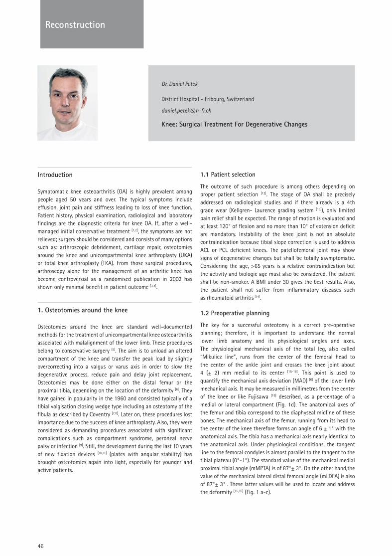
46
Reconstruction
Dr. Daniel Petek
District Hospital - Fribourg, Switzerland
Knee: Surgical Treatment For Degenerative Changes
Introduction
Symptomatic knee osteoarthritis (OA) is highly prevalent among people aged 50 years and over. The typical symptoms include effusion, joint pain and stiffness leading to loss of knee function. Patient history, physical examination, radiological and laboratory findings are the diagnostic criteria for knee OA. If, after a well-managed initial conservative treatment [1,2], the symptoms are not relieved; surgery should be considered and consists of many options such as: arthroscopic debridement, cartilage repair, osteotomies around the knee and unicompartmental knee arthroplasty (UKA) or total knee arthroplasty (TKA). From those surgical procedures, arthroscopy alone for the management of an arthritic knee has become controversial as a randomised publication in 2002 has shown only minimal benefit in patient outcome [3,4].
1. Osteotomies around the knee
Osteotomies around the knee are standard well-documented methods for the treatment of unicompartmental knee osteoarthritis associated with malalignment of the lower limb. These procedures belong to conservative surgery [5]. The aim is to unload an altered compartment of the knee and transfer the peak load by slightly overcorrecting into a valgus or varus axis in order to slow the degenerative process, reduce pain and delay joint replacement. Osteotomies may be done either on the distal femur or the proximal tibia, depending on the location of the deformity [6]. They have gained in popularity in the 1960 and consisted typically of a tibial valgisation closing wedge type including an osteotomy of the fibula as described by Coventry [7,8]. Later on, these procedures lost importance due to the success of knee arthroplasty. Also, they were considered as demanding procedures associated with significant complications such as compartment syndrome, peroneal nerve palsy or infection [9]. Still, the development during the last 10 years of new fixation devices [10,11] (plates with angular stability) has brought osteotomies again into light, especially for younger and active patients.
1.1 Patient selection
The outcome of such procedure is among others depending on proper patient selection [12]. The stage of OA shall be precisely addressed on radiological studies and if there already is a 4th grade wear (Kellgren- Laurence grading system [13]), only limited pain relief shall be expected. The range of motion is evaluated and at least 120° of flexion and no more than 10° of extension deficit are mandatory. Instability of the knee joint is not an absolute contraindication because tibial slope correction is used to address ACL or PCL deficient knees. The patellofemoral joint may show signs of degenerative changes but shall be totally asymptomatic. Considering the age, >65 years is a relative contraindication but the activity and biologic age must also be considered. The patient shall be non-smoker. A BMI under 30 gives the best results. Also, the patient shall not suffer from inflammatory diseases such as rheumatoid arthritis [14].
1.2 Preoperative planning
The key for a successful osteotomy is a correct pre-operative planning; therefore, it is important to understand the normal lower limb anatomy and its physiological angles and axes. The physiological mechanical axis of the total leg, also called “Mikulicz line”, runs from the center of the femoral head to the center of the ankle joint and crosses the knee joint about 4 (± 2) mm medial to its center [15-18]. This point is used to quantify the mechanical axis deviation (MAD) [6] of the lower limb mechanical axis. It may be measured in millimetres from the center of the knee or like Fujisawa [19] described, as a percentage of a medial or lateral compartment (Fig. 1d). The anatomical axes of the femur and tibia correspond to the diaphyseal midline of these bones. The mechanical axis of the femur, running from its head to the center of the knee therefore forms an angle of 6 ± 1° with the anatomical axis. The tibia has a mechanical axis nearly identical to the anatomical axis. Under physiological conditions, the tangent line to the femoral condyles is almost parallel to the tangent to the tibial plateau (0°-1°). The standard value of the mechanical medial proximal tibial angle (mMPTA) is of 87°± 3°. On the other hand,the value of the mechanical lateral distal femoral angle (mLDFA) is also of 87°± 3° . These latter values will be used to locate and address the deformity [15,16] (Fig. 1 a-c).
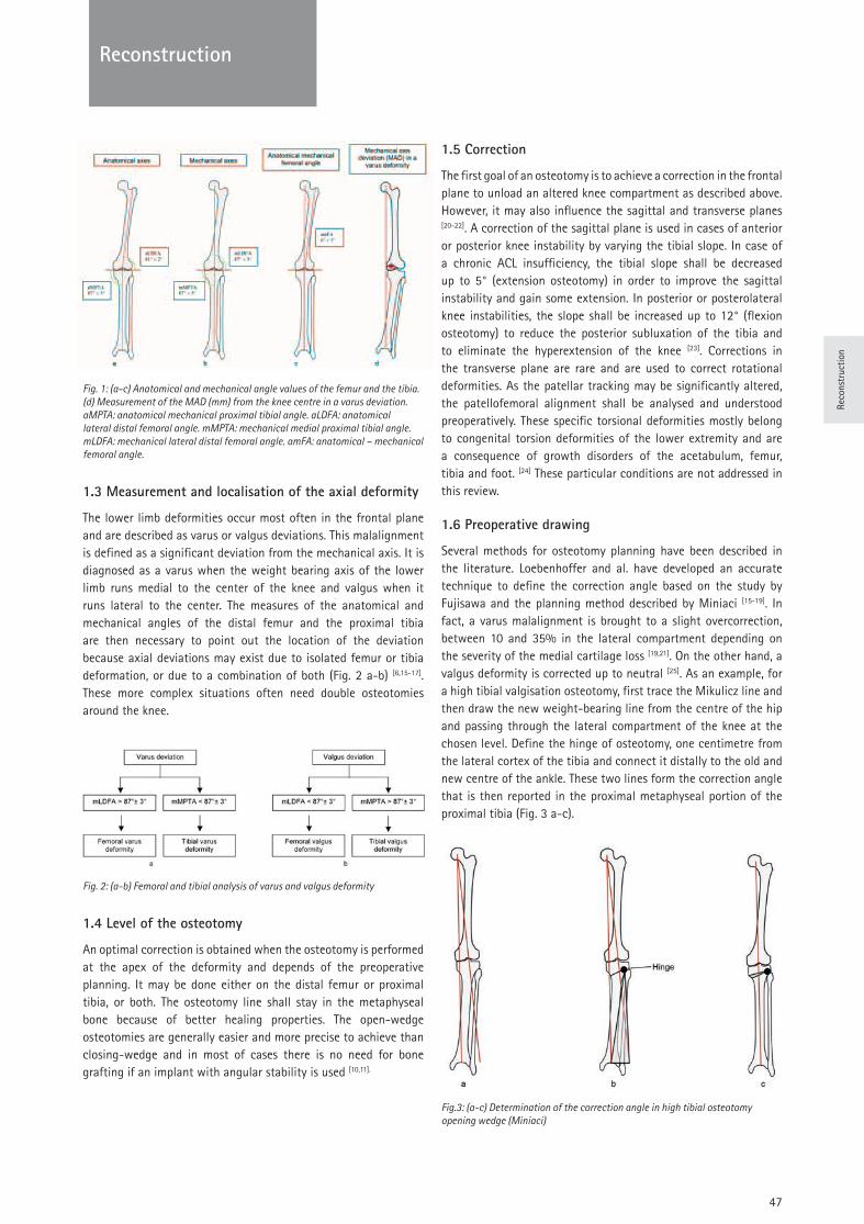
Reco
nstr
uctio
n
47
Reconstruction
Fig. 1: (a-c) Anatomical and mechanical angle values of the femur and the tibia. (d) Measurement of the MAD (mm) from the knee centre in a varus deviation. aMPTA: anatomical mechanical proximal tibial angle. aLDFA: anatomical lateral distal femoral angle. mMPTA: mechanical medial proximal tibial angle. mLDFA: mechanical lateral distal femoral angle. amFA: anatomical – mechanical femoral angle.
1.3 Measurement and localisation of the axial deformity
The lower limb deformities occur most often in the frontal plane and are described as varus or valgus deviations. This malalignment is defined as a significant deviation from the mechanical axis. It is diagnosed as a varus when the weight bearing axis of the lower limb runs medial to the center of the knee and valgus when it runs lateral to the center. The measures of the anatomical and mechanical angles of the distal femur and the proximal tibia are then necessary to point out the location of the deviation because axial deviations may exist due to isolated femur or tibia deformation, or due to a combination of both (Fig. 2 a-b) [6,15-17]. These more complex situations often need double osteotomies around the knee.
Fig. 2: (a-b) Femoral and tibial analysis of varus and valgus deformity
1.4 Level of the osteotomy
An optimal correction is obtained when the osteotomy is performed at the apex of the deformity and depends of the preoperative planning. It may be done either on the distal femur or proximal tibia, or both. The osteotomy line shall stay in the metaphyseal bone because of better healing properties. The open-wedge osteotomies are generally easier and more precise to achieve than closing-wedge and in most of cases there is no need for bone grafting if an implant with angular stability is used [10,11].
1.5 Correction
The first goal of an osteotomy is to achieve a correction in the frontal plane to unload an altered knee compartment as described above. However, it may also influence the sagittal and transverse planes [20-22]. A correction of the sagittal plane is used in cases of anterior or posterior knee instability by varying the tibial slope. In case of a chronic ACL insufficiency, the tibial slope shall be decreased up to 5° (extension osteotomy) in order to improve the sagittal instability and gain some extension. In posterior or posterolateral knee instabilities, the slope shall be increased up to 12° (flexion osteotomy) to reduce the posterior subluxation of the tibia and to eliminate the hyperextension of the knee [23]. Corrections in the transverse plane are rare and are used to correct rotational deformities. As the patellar tracking may be significantly altered, the patellofemoral alignment shall be analysed and understood preoperatively. These specific torsional deformities mostly belong to congenital torsion deformities of the lower extremity and are a consequence of growth disorders of the acetabulum, femur, tibia and foot. [24] These particular conditions are not addressed in this review.
1.6 Preoperative drawing
Several methods for osteotomy planning have been described in the literature. Loebenhoffer and al. have developed an accurate technique to define the correction angle based on the study by Fujisawa and the planning method described by Miniaci [15-19]. In fact, a varus malalignment is brought to a slight overcorrection, between 10 and 35% in the lateral compartment depending on the severity of the medial cartilage loss [19,21]. On the other hand, a valgus deformity is corrected up to neutral [25]. As an example, for a high tibial valgisation osteotomy, first trace the Mikulicz line and then draw the new weight-bearing line from the centre of the hip and passing through the lateral compartment of the knee at the chosen level. Define the hinge of osteotomy, one centimetre from the lateral cortex of the tibia and connect it distally to the old and new centre of the ankle. These two lines form the correction angle that is then reported in the proximal metaphyseal portion of the proximal tibia (Fig. 3 a-c).
Fig.3: (a-c) Determination of the correction angle in high tibial osteotomy opening wedge (Miniaci)
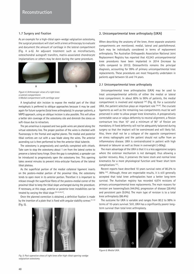
48
Reconstruction
1.7 Surgery and fixation
As an example for a high-tibial open-wedge valgisation osteotomy, the surgical procedure will start with a knee arthroscopy to evaluate and document the amount of cartilage in the lateral compartment (Fig. 4 a-b). An adjuvant treatment such as microfractures, osteochondral autograft transfers, matrix-associated chondrocyte implantations or others may be done during the same procedure.
a b
Figure 4: Arthroscopic views of a right knee. a Lateral compartment. b Medial compartment with cartilage wear
A longitudinal skin incision to expose the medial part of the tibial metaphysis is preferred to oblique approaches because it may be used again for future surgeries (total knee arthroplasty). In younger patients, a MIPO approach, using an oblique incision is also possible. This will allow a better skin coverage of the osteotomy site and diminish the stress on soft-tissue due to retractors.
The pes anserinus is exposed and two guide wires are placed along the virtual osteotomy line. The proper position of the wires is checked with fluoroscopy in the frontal and sagittal planes. The medial and posterior tibial cortices are cut with a saw blade along the wires. The anterior ascending cut is then performed to free the anterior tibial tubercle.
The osteotomy is progressively and carefully completed with chisels. Take care to stop the osteotomy about 1 cm from the lateral cortex to preserve a lateral bony hinge. Once the gap is completed, a spreader can be introduced to progressively open the osteotomy line. This opening takes several minutes to prevent intra-articular fractures of the lateral tibial plateau.
As the superficial portion of the medial collateral ligament inserts on the postero-medial portion of the proximal tibia, the osteotomy tends to open more in its anterior portion. Therefore it is important to release enough the superficial fibres of the postero-medial corner of the proximal tibial to keep the tibial slope unchanged during the procedure. If necessary, at this stage, anterior or posterior knee instabilities can be treated by varying the tibial slope [20-22,26,27].
Once the planned correction is obtained, a definitive fixation is made by the insertion of a plate that is fixed with angular stability screws [11,12] (Fig. 5).
Fig. 5: Post-operative views of right knee after high-tibial opening-wedge valgisation osteotomy
2. Unicompartmental knee arthroplasty (UKA)
When describing the anatomy of the knee, three separate anatomic compartments are mentioned; medial, lateral and patellofemoral. Each may be individually considered in terms of replacement arthroplasty. The Australian Orthopaedic Association National Joint Replacement Registry has reported that 43,543 unicompartmental knee procedures have been implanted in 2014 (increase by 4,6% compared to 2013). Osteoarthritis remains the principal diagnosis, accounting for 99% of primary unicompartmental knee replacements. These procedures are most frequently undertaken in patients aged between 55 and 74 years.
2.1 Unicompartmental knee arthroplasty
Unicompartmental knee arthroplasties (UKA) may be used to treat unicompartmental arthritis of either the medial or lateral knee compartment. In about 80% to 90% of patients, the medial compartment is involved and replaced [30] (Fig. 6). For a successful UKA, the patient selection plays an important role [28,29]. The cruciate ligaments as well as the remaining two compartments must be well preserved in order to allow proper knee kinematics. A preoperatively correctable varus or valgus deformity to neutral alignment, a flexion contracture less than 10° and a minimum of 90° of flexion are mandatory. A fixed deformity will not be adequately balanced during surgery so that the implant will be overstressed and will likely fail. Also, there shall not be a collapse of the opposite compartment on stress radiographs and the patient should not suffer from an inflammatory disease. UKA is contraindicated in patient with high demand or labourer as well as those in overweight (>90kg).
The main advantage of the UKA is that it is a less aggressive surgery where the extensor mechanism is not damaged, thus allowing a quicker recovery. Also, it preserves the bone stock and normal knee kinematics for a more physiological function and fewer short term complications [31].
Recent reports have described 10 years survival rates of 80.2% to 98% [32]. Although, these are respectable results, it is still generally accepted that total knee arthroplasties have a better long-term survival. The Australian registry has recorded 4,874 revisions of primary unicompartmental knee replacements. The main reasons for revision are loosening/lysis (44.0%), progression of disease (28.4%) and persistent pain (9.9%). The main type of revision is to a total knee arthroplasty (86.4%)
The outcome for UKA is variable and ranges from 80.2 to 98% in terms of 10-years survival. Still, UKA has a significantly poorer long-term survival than total knee arthroplasty.
Figure 6: Medial UKA.
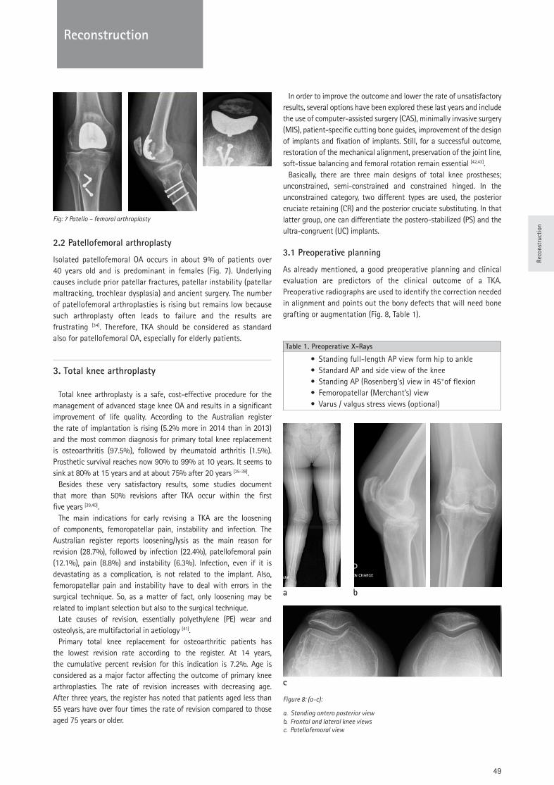
Reco
nstr
uctio
n
49
Reconstruction
Fig: 7 Patello – femoral arthroplasty
2.2 Patellofemoral arthroplasty
Isolated patellofemoral OA occurs in about 9% of patients over 40 years old and is predominant in females (Fig. 7). Underlying causes include prior patellar fractures, patellar instability (patellar maltracking, trochlear dysplasia) and ancient surgery. The number of patellofemoral arthroplasties is rising but remains low because such arthroplasty often leads to failure and the results are frustrating [34]. Therefore, TKA should be considered as standard also for patellofemoral OA, especially for elderly patients.
3. Total knee arthroplasty
Total knee arthroplasty is a safe, cost-effective procedure for the management of advanced stage knee OA and results in a significant improvement of life quality. According to the Australian register the rate of implantation is rising (5.2% more in 2014 than in 2013) and the most common diagnosis for primary total knee replacement is osteoarthritis (97.5%), followed by rheumatoid arthritis (1.5%). Prosthetic survival reaches now 90% to 99% at 10 years. It seems to sink at 80% at 15 years and at about 75% after 20 years [35-39].
Besides these very satisfactory results, some studies document that more than 50% revisions after TKA occur within the first five years [39,40].
The main indications for early revising a TKA are the loosening of components, femoropatellar pain, instability and infection. The Australian register reports loosening/lysis as the main reason for revision (28.7%), followed by infection (22.4%), patellofemoral pain (12.1%), pain (8.8%) and instability (6.3%). Infection, even if it is devastating as a complication, is not related to the implant. Also, femoropatellar pain and instability have to deal with errors in the surgical technique. So, as a matter of fact, only loosening may be related to implant selection but also to the surgical technique.
Late causes of revision, essentially polyethylene (PE) wear and osteolysis, are multifactorial in aetiology [41].
Primary total knee replacement for osteoarthritic patients has the lowest revision rate according to the register. At 14 years, the cumulative percent revision for this indication is 7.2%. Age is considered as a major factor affecting the outcome of primary knee arthroplasties. The rate of revision increases with decreasing age. After three years, the register has noted that patients aged less than 55 years have over four times the rate of revision compared to those aged 75 years or older.
In order to improve the outcome and lower the rate of unsatisfactory results, several options have been explored these last years and include the use of computer-assisted surgery (CAS), minimally invasive surgery (MIS), patient-specific cutting bone guides, improvement of the design of implants and fixation of implants. Still, for a successful outcome, restoration of the mechanical alignment, preservation of the joint line, soft-tissue balancing and femoral rotation remain essential [42,43].
Basically, there are three main designs of total knee prostheses; unconstrained, semi-constrained and constrained hinged. In the unconstrained category, two different types are used, the posterior cruciate retaining (CR) and the posterior cruciate substituting. In that latter group, one can differentiate the postero-stabilized (PS) and the ultra-congruent (UC) implants.
3.1 Preoperative planning
As already mentioned, a good preoperative planning and clinical evaluation are predictors of the clinical outcome of a TKA. Preoperative radiographs are used to identify the correction needed in alignment and points out the bony defects that will need bone grafting or augmentation (Fig. 8, Table 1).
Table 1. Preoperative X-Rays
• Standing full-length AP view form hip to ankle• Standard AP and side view of the knee• Standing AP (Rosenberg’s) view in 45°of flexion• Femoropatellar (Merchant’s) view• Varus / valgus stress views (optional)
a b
c
Figure 8: (a-c):
a. Standing antero posterior view b. Frontal and lateral knee views c. Patellofemoral view
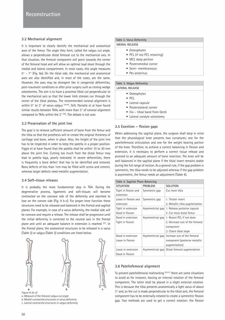
50
Reconstruction
3.2 Mechanical alignment
It is important to clearly identify the mechanical and anatomical axes of the femur. The angle they form, called the valgus cut angle, allows a perpendicular distal femoral cut to the mechanical axis. In that situation, the femoral component will point towards the center of the femoral head and will allow an optimal load share through the medial and lateral compartment. In most cases, this angle measures 5° – 7° (Fig. 9a). On the tibial side, the mechanical and anatomical axes are also identified and, in most of the cases, are the same. However, the axes may be divergent like in congenital deformities, post-traumatic conditions or after prior surgery such as closing wedge osteotomies. The aim is to have a proximal tibial cut perpendicular to the mechanical axis so that the lower limb stresses run through the center of the tibial plateau. The recommended coronal alignment is within 0° to 3° of varus-valgus [44,45]. Still, Parratte et al have found similar results between TKAs with more than 3° of coronal alignment compared to TKAs within the 3° [46]. The debate is not over.
3.3 Preservation of the joint line
The goal is to remove sufficient amount of bone from the femur and the tibia so that the prosthesis will re-create the original thickness of cartilage and bone, when in place. Also, the height of the joint line has to be respected in order to keep the patella in a proper position. Figgie et al have found that the patella shall be within 10 to 30 mm above the joint line. Cutting too much from the distal femur may lead to patella baja, poorly tolerated. In severe deformities, there is frequently a bone defect that has to be identified and restored. Bony defects of less than 1cm may be filled with screw and cement, whereas larger defects need metallic augmentation.
3.4 Soft-tissue releases
It is probably the most fundamental step in TKA. During the degenerative process, ligaments and soft-tissues will become contracted on the concave side of the deformity and starched to lose on the convex side (Fig. 9 b-c). For proper knee function these structures need to be released and balanced in the frontal and sagittal planes. For example, in case of a varus deformity, the medial side will be concave and require a release. The release shall be progressive until the initial deformity is corrected to the neutral axis in the frontal plane and until an adequate balance in extension is reached [47]. In the frontal plane, the anatomical structures to be released in a varus (Table 2) or valgus (Table 3) conditions are listed below.
Figure 9: (a-c): a. Measure of the femoral valgus cut angle b. Medial contracted structures in varus deformity c. Lateral contracted structures in valgus deformity
Table 2. Varus Deformity
MEDIAL RELEASE
• Osteophytes • PCL (if not PCL retaining)• MCL deep portion• Posteromedial corner• Semi- membranosus• Pes anserinus
Table 3. Valgus Deformity
LATERAL RELEASE
• Osteophytes• PCL• Lateral capsule• Posterolateral corner• Ilio – tibial band from Gerdi• Lateral condyle osteotomy
3.5 Exention – flexion gaps
When addressing the sagittal plane, the surgeon shall keep in mind that the physiological knee presents two curvatures; one for the patellofemoral articulation and one for the weight bearing portion of the knee. Therefore, to achieve a correct balancing in flexion and extension, it is necessary to perform a correct tissue release and proceed to an adequate amount of bone resection. The knee will be well balanced in the sagittal plane if the tibial insert remains stable during the full range of motion. As a general rule, if the gap problem is symmetric, the tibia needs to be adjusted whereas if the gap problem is asymmetric, the femur needs an adjustment (Table 4).
Table 4. Sagittal Plane BalancingSITUATION PROBLEM SOLUTIONTight in flexion and extension
Symmetric gap Cut more tibia
Loose in flexion and
extension
Symmetric gap 1. Thicker insert
2. Metallic tibia augmentation
Tight in extension
Good in flexion
Asymmetrical gap 1. Release posterior capsule
2. Cut more distal femur
Good in extension
Tight in flexion
Asymmetrical gap 1. Resect PCL if not done
2. Decrease size of the femoral
component
3. Check tibial slope
Good in extension
Loose in flexion
Asymmetrical gap Increase size of the femoral
component (posterior metallic
augmentation)
Loose in extension
Good in flexion
Asymmetrical gap Distal femoral augmentation
3.6 Patellofemoral alignment
To prevent patellofemoral maltracking [50,51] there are some situations to avoid as for instance, leaving an internal rotation of the femoral component. The latter shall be placed in a slight external rotation. This is because the tibia presents anatomically a light varus of about 3° and, as the cut is made perpendicular to the tibial axis, the femoral component has to be externally rotated to create a symmetric flexion gap. Two methods are used to get a correct rotation: the flexion
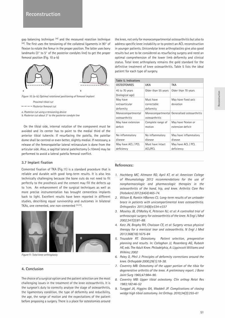
Reco
nstr
uctio
n
51
Reconstruction
gap balancing technique [49] and the measured resection technique [48]. The first uses the tensioning of the collateral ligaments in 90° of flexion to rotate the femur in the proper position. The latter uses bony landmarks (3° to 5° of the posterior condyles line) to get the proper femoral position (Fig. 10 a-b)
Figure 10: (a-b): Optimal rotational positioning of femoral implant
Proximal tibial cut
Posterior femoral cut
a. Posterior cut using a tensioning device b. Posterior cut about 3° to the posterior condyle line
On the tibial side, internal rotation of the component must be avoided and its center has to point to the medial third of the anterior tibial tubercle. If resurfacing the patella, the patellar dome shall be centred or even better, slightly medial. If necessary, a release of the femoropatellar lateral retinaculum is done from the articular side. Also, a sagittal lateral patellectomy (<10mm) may be performed to avoid a lateral patella femoral conflict.
3.7 Implant fixation
Cemented fixation of TKA (Fig. 11) is a standard procedure that is reliable and durable with good long-term results. It is also less technically challenging because the bone cuts do not need to fit perfectly to the prosthesis and the cement may fill the defects up to 1cm. An enhancement of the surgical techniques as well as more precise instrumentation has brought cementless implants back to light. Excellent results have been reported in different studies, describing equal survivorship and outcomes in bilateral TKAs, one cemented, one non-cemented [52,53].
Figure11: Total knee arthroplasty
4. Conclusion
The choice of a surgical option and the patient selection are the most challenging issues in the treatment of the knee osteoarthritis. It is the surgeon’s duty to correctly analyse the stage of osteoarthritis, the ligamentory condition, the type of deformity and reducibility, the age, the range of motion and the expectations of the patient before proposing a surgery. There is a place for osteotomies around
the knee, not only for monocompartmental osteoarthritis but also to address specific knee instability or to protect an ACL reconstruction in younger patients. Unicondylar knee arthroplasties give also good results but are to be considered as resurfacing surgery and need an optimal comprehension of the lower limb deformity and clinical status. Total knee arthroplasty remains the gold standard for the definitive treatment of knee osteoarthritis. Table 5 lists the ideal patient for each type of surgery.
Table 5. IndicationsOSTEOTOMIES UKA TKA
45 to 70 years
(biological age)
Older than 55 years Older than 70 years
May have
extraarticular
deformity
Must have
correctable
deformity
May have fixed axis
deviation
Monocompartmental
osteoarthritis
Monocompartmental
osteoarthritis
Generalised osteoarthritis
May have extension
deficit
Complete range of
motion
May have flexion or
extension deficit
No inflammatory
disease
No inflammatory
disease
May have inflammatory
disease
May have ACL / PCL
deficiency
Must have intact
ACL/PCL
May have ACL / PCL
deficiency
References:
1. Hochberg MC, Altmann RD, April KT, et al: American College of Rheumatology 2012 recommendations for the use of nonpharmacologic and pharmacologic therapies in the osteoarthritis of the hand, hip, and knee. Arthritis Care Res (Hoboken) 2012;64(4):465-74.
2. Wilson B, Rankin HBarnes CL: Long-term results of an unloader brace in patients with unicompartimental knee osteoarthritis. Orthopedics 2011;34(8):e334-e337
3. Moseley JB, O’Malley K, Petersen NJ, et al: A controlled trial of arthroscopic surgery for osteoarthritis of the knee. N Engl J Med 2002;347(2):81-88.
4. Katz JN, Brophy RH, Chaisson CE, et al: Surgery versus physical therapy for a meniscal tear and osteoarthritis. N Engl J Med 2013:368(18):1675-84
5. Trousdale RT. Osteotomy. Patient selection, preoperative planning and results. In: Callaghan JJ, Rosenberg AG, Rubash HE, eds. The Adult Knee. Philadelphia, A: Lippincott Williams and Wilkins; 2002
6. Paley D, Pfeil J: Principles of deformity corrections around the knee. Orthopäde 2000;29(1):18-38.
7. Coventry MB: Osteotomy of the upper portion of the tibia for degenerative arthritis of the knee. A preliminary report. J Bone Joint Surg 1965;47:984–90.
8. Coventry MB: Upper tibial osteotomy. Clin orthop Relat Res 1983;182:46-52
9. Tunggal JA, Higgins GA, Waddell JP. Complications of closing wedge high tibial osteotomy. Int Orthop. 2010;34(2):255-61
52
Reconstruction
10. Lobenhoffer P, Agneskirschner JD. Improvements in surgical technique of valgus high tibial osteotomy. Knee Surg Sports Traumatol Arthrosc. 2003;11(3):132-38
11. Staubli AE, De Simoni C, Babst R et al. TomoFix: a new LCP-concept for open wedge osteotomy of the medial proximal tibia—early results in 92 cases. Injury 2003;34( 2):55–62.
12. Bonnin M, Chambat P. Current status of valgus angle, tibial head closing wedge osteotomy in medial gonarthrosis. Orthopäde 2004;33(2):135-42
13. Kellgren JH, Lawrence JS. Radiological assessment of osteoarthrosis. Ann Rheum Dis 1957;16(4):494-502
14. Lobenhoffer P. Indications for high tibial osteotomy, unicondylar knee arthroplasty, and total knee prosthesis. In: Lobenhoffer P, van Heerwaarden J, Staubli A, eds. Osteotomies Around the Knee.Indications-Planning-Surgical Techniques using Plate Fixators. US-New York,NY:Thieme New York; 2008.
15. Paley D, Herzenberg JE, Tetsworth K, et al. Deformity planning for frontal and saggital plane corrective osteotomies. Orthop Clin North Am 1994;25(3):425-65.
16. Paley D, Tetsworth K. Mechanical deviation of the lower limbs. Preoperative planning of multiapical frontal plane angular and bowing deformities of the femur and tibia. Clin Orthop Relat Res 1992;280:65-71
17. Pape D, Seil R, Adam F, et al. Imaging and preoperative planning of osteotomy of tibial head osteotomy. Orthopäde 2004;33(2):122-34.
18. Miniaci A, Ballmer FT, Ballmer PM, et al. Proximal tibial osteotomy. A new fixation device. Clin Orthop Relat Res 1989;246:250-59.
19. Fujisawa Y, Masuhara K, Shiomi S. The effect of high tibial osteotomy on osteoarthritis of the knee. An arthroscopic study of 54 knee joints. Orthop Clin North Am 1979;10(3):585–608.
20. Noyes FR, Barber-Westin SD, Hewett TE. High tibial osteotomy and ligament reconstruction for varus angulated anteriot cruciate ligament-deficient knees. Am J Sports Med 2000;28(3):282-96.
21. Reichwein F, Nebelung W. High tibial flexion osteotomy for revision of posterior cruciate ligament instability. Unfallchirurg 2007;110(7):597-602.
22. Marti CB, Gautier E, Wachtel AW, et al. Accuracy of frontal and sagittal plane correction in open-wedge high tibial osteotomy. Arthroscopy 2004;20(4):366-72.
23. Giffin JR, Vogrin TM, Zantop T, et al. Effects of increasing tibial slope on the biomechanics of the knee. Am J Sport Med 2004;32(2):376-83.
24. van Heerwaarden RJ, van der Haven, Kooijman M, et al. Derotation osteotomy for correction of congenital rotational lower limb deformities in adolescents and adults. Surg Tech Orthop Traumatol 2003;55-575-A-10:10
25. Johnson F, Leitl S, Waugh W. The distribution of load across the knee. A comparison of static and dynamic measurments. J Bone Joint Surg Br 1980;62(3):346-49
26. Jacobi M, Wahl P, Jakob RP. Basic principles of osteotomies around the knee. In: Lobenhoffer P, van Heerwaarden J, Staubli A, eds. Osteotomies Around the Knee. Indications-Planning-Surgical Techniques using Plate Fixators. US-New York,NY:Thieme New York; 2008
27. Stuart MJ. Tibial antivirus closing wedge osteotomy. Oper Tech Sport Med 2000;8(1):27-31
28. Pennington DW, wienckowski JJ, Lutes WB, et al. Unicompartmental knee arthroplasty in patients sixty years of age or younger. J Bone Joint Surg Am 2003;85:1968-73
29. Parratte S, Argenson JN, Pearce O, et al. Medial unicompartmental knee replacement in the under-50s. J Bone Joint Surg Br 2009;91:351-56
30. Hernborg JS, Nilsson BE. The natural course of untreated osteoarthritis of the knee. Clin Orthop Relat Res 1977;123:130.137
31. Noticewala MS, Geller JA, Lee JH, et al. Unicompartmental knee arthroplasty relieves pain and improves function more than total knee arthroplasty. J Arthroplasty 2012;27(8, suppl):99-105.
32. Heyse TJ, Khefacha A, Peersman G, et al. Survivorship of UKA in the middle-aged. Knee 2012;19(5):585-91
33. Fulkerson JP: Alternatives to patellofemoral arthroplasty. Clin Orthop Relat Res 2005;436:76-80
34. van Jonbergen HP, Werkman DM, Barnaart LF et al. Long-term outcomes of patellofemoral arthroplasty J Arthoplasty 2010;25(7):1066-71
35. Pavone V, Boettner F, Fickert S, Sculo TP: Total condylar knee arthroplasty: Along term follow-up. Clin Orthop Relat Res 2001;388:18-25
36. Rand JA, Ilstrup DM: survivorship analysis of total knee arthroplasty: cumulative rates of survival of 9200 total knee arthroplasties. J Bone Joint Surg Am 1991;73(3):397-409
37. Schai PA, Thornhill TS, Scott RD: Total knee arthroplasty with the PFC system: Results at a minimum of ten years and survivorship analysis. J Bone Joint Surg Br 1998;80(5):850-58
38. Ranawat CS, Flynn WF Jr, Saddler S, et al. Long-term results of the total condylar knee arthroplasty: A 15-years survivorship study. Clin Orthop Relat Res 1993;286:94-102
39. Gioe TJ, Killeen KK, Grimm K, et al. Why are total replacement revised? Analysis of early revision in a community knee implant registery. Clin Orthop Relat Res 2004;428:100-06
40. Sharkey PF, Hozack WJ, Rothman RH, et al. Why are total knee arthroplasties failing today? Clin Orthop Relat Res 2002;404:7-13
41. Lidgren L, Robertsson O: Annual report 2008: The Swedisch Knee Arthroplasty Register. Lund, Sweden, Wallin & Dalholm, 2008
42. Lin WP, Lin J,Horng LC, et al. Quadriceps sparing, minimal-incision total knee arthroplasty: A comparative study. J Arthroplasty 2009;24(7):1024-32
43. Matsumoto T, Muratsu H, KuboS, et al. soft tissue balance measurement in minimal incision surgery compared to conventional total knee arthroplasty. Knee Surg Sports traumatol Arthrosc 2011;19(6):880-86
44. Fang DM, Ritter MA, Davis KE. Coronal alignment in total knee arthroplasty: just how important is it? J Arthroplasty 2009;24:39-43
45. Jeffery RS, Morris RW, Denham RA. Cornal alignment after total knee replacement. J Bone Joint Surg Br 1991;73:709-14
46. Parratte S, Pagnano MW, Trousdale RT, et al. Effect of postoperative mechanical axis alignment on the fifteen-year survival of modern, cemented total knee replacements. J Bone Joint Surg Am. 2010;92:2143-49
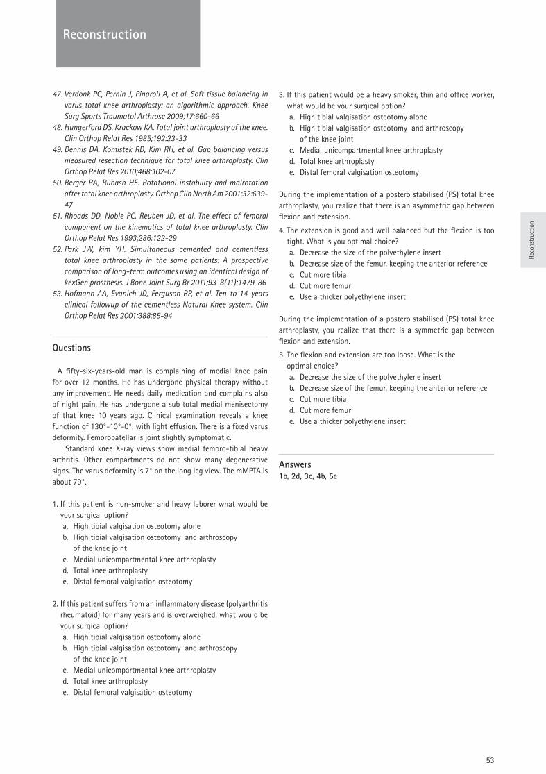
Reco
nstr
uctio
n
53
Reconstruction
47. Verdonk PC, Pernin J, Pinaroli A, et al. Soft tissue balancing in varus total knee arthroplasty: an algorithmic approach. Knee Surg Sports Traumatol Arthrosc 2009;17:660-66
48. Hungerford DS, Krackow KA. Total joint arthroplasty of the knee. Clin Orthop Relat Res 1985;192:23-33
49. Dennis DA, Komistek RD, Kim RH, et al. Gap balancing versus measured resection technique for total knee arthroplasty. Clin Orthop Relat Res 2010;468:102-07
50. Berger RA, Rubash HE. Rotational instability and malrotation after total knee arthroplasty. Orthop Clin North Am 2001;32:639-47
51. Rhoads DD, Noble PC, Reuben JD, et al. The effect of femoral component on the kinematics of total knee arthroplasty. Clin Orthop Relat Res 1993;286:122-29
52. Park JW, kim YH. Simultaneous cemented and cementless total knee arthroplasty in the same patients: A prospective comparison of long-term outcomes using an identical design of kexGen prosthesis. J Bone Joint Surg Br 2011;93-B(11):1479-86
53. Hofmann AA, Evanich JD, Ferguson RP, et al. Ten-to 14-years clinical followup of the cementless Natural Knee system. Clin Orthop Relat Res 2001;388:85-94
Questions
A fifty-six-years-old man is complaining of medial knee pain for over 12 months. He has undergone physical therapy without any improvement. He needs daily medication and complains also of night pain. He has undergone a sub total medial menisectomy of that knee 10 years ago. Clinical examination reveals a knee function of 130°-10°-0°, with light effusion. There is a fixed varus deformity. Femoropatellar is joint slightly symptomatic.
Standard knee X-ray views show medial femoro-tibial heavy arthritis. Other compartments do not show many degenerative signs. The varus deformity is 7° on the long leg view. The mMPTA is about 79°.
1. If this patient is non-smoker and heavy laborer what would be your surgical option?a. High tibial valgisation osteotomy aloneb. High tibial valgisation osteotomy and arthroscopy
of the knee jointc. Medial unicompartmental knee arthroplastyd. Total knee arthroplastye. Distal femoral valgisation osteotomy
2. If this patient suffers from an inflammatory disease (polyarthritis rheumatoid) for many years and is overweighed, what would be your surgical option?a. High tibial valgisation osteotomy aloneb. High tibial valgisation osteotomy and arthroscopy
of the knee jointc. Medial unicompartmental knee arthroplastyd. Total knee arthroplastye. Distal femoral valgisation osteotomy
3. If this patient would be a heavy smoker, thin and office worker, what would be your surgical option?a. High tibial valgisation osteotomy aloneb. High tibial valgisation osteotomy and arthroscopy
of the knee jointc. Medial unicompartmental knee arthroplastyd. Total knee arthroplastye. Distal femoral valgisation osteotomy
During the implementation of a postero stabilised (PS) total knee arthroplasty, you realize that there is an asymmetric gap between flexion and extension.
4. The extension is good and well balanced but the flexion is too tight. What is you optimal choice?a. Decrease the size of the polyethylene insertb. Decrease size of the femur, keeping the anterior referencec. Cut more tibiad. Cut more femure. Use a thicker polyethylene insert
During the implementation of a postero stabilised (PS) total knee arthroplasty, you realize that there is a symmetric gap between flexion and extension.
5. The flexion and extension are too loose. What is the optimal choice?a. Decrease the size of the polyethylene insertb. Decrease size of the femur, keeping the anterior referencec. Cut more tibiad. Cut more femure. Use a thicker polyethylene insert
Answers1b, 2d, 3c, 4b, 5e
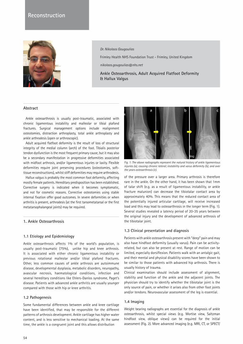
54
Reconstruction
Dr. Nikolaos Gougoulias
Frimley Health NHS Foundation Trust - Frimley, United Kingdom
Ankle Osteoarthrosis, Adult Acquired Flatfoot Deformity & Hallux Valgus
Abstract
Ankle osteoarthrosis is usually post-traumatic, associated with chronic ligamentous instability and malleolar or tibial plafond fractures. Surgical management options include realignment osteotomies, distraction arthroplasty, total ankle arthroplasty and ankle arthrodesis (open or arthroscopic).
Adult acquired flatfoot deformity is the result of loss of structural integrity of the medial column (arch) of the foot. Tibialis posterior tendon dysfunction is the most frequent primary cause, but it may also be a secondary manifestation in progressive deformities associated with midfoot arthrosis, and/or ligamentous injuries or laxity. Flexible deformities require joint preserving procedures (osteotomies, soft-tissue reconstructions), whilst stiff deformities may require arthrodesis.
Hallux valgus is probably the most common foot deformity, affecting mostly female patients. Hereditary predisposition has been established. Corrective surgery is indicated when it becomes symptomatic, and not for cosmetic reasons. Corrective osteotomies using stable internal fixation offer good outcomes. In severe deformities or when arthritis is present, arthrodesis (at the first tarsometatarsal or the first metatarsophalangeal joints) may be required.
1. Ankle Osteoarthrosis
1.1 Etiology and Epidemiology
Ankle osteoarthrosis affects 1% of the world’s population, is usually post-traumatic (75%), unlike hip and knee arthrosis. It is associated with either chronic ligamentous instability or previous rotational malleolar and/or tibial plafond fractures. Other, less common causes of ankle arthrosis are autoimmune disease, developmental dysplasia, metabolic disorders, neuropathy, avascular necrosis, haematological conditions, infection and several hereditary conditions like Ehlers-Danlos syndrome, Paget’s disease. Patients with advanced ankle arthritis are usually younger compared with those with hip or knee arthritis.
1.2 Pathogenesis
Some fundamental differences between ankle and knee cartilage have been identified, that may be responsible for the different patterns of arthrosis development. Ankle cartilage has higher water content, and is less sensitive to mechanical loading. At the same time, the ankle is a congruent joint and this allows distribution
Fig. 1: The above radiographs represent the natural history of ankle ligamentous injuries (a), causing chronic lateral; instability and varus deformity (b), and over the years osteoarthrosis (c).
of the pressure over a larger area. Primary arthrosis is therefore rare in the ankle. On the other hand, it has been shown that 1mm of talar shift (e.g. as a result of ligamentous instability, or ankle fracture malunion) can decrease the tibiotalar contact area by approximately 40%. This means that the reduced contact area of the potentially injured articular cartilage, will receive increased load and this may lead to osteoarthrosis in the longer term (Fig. 1). Several studies revealed a latency period of 20-35 years between the original injury and the development of advanced arthrosis of the tibiotalar joint.
1.3 Clinical presentation and diagnosis
Patients with ankle osteoarthrosis present with “deep” pain and may also have hindfoot deformity (usually varus). Pain can be activity-related, but can also be present at rest. Range of motion can be limited, especially dorsiflexion. Patients walk with an antalgic gait, and their mental and physical disability scores have been shown to be similar to those patients with advanced hip arthrosis. There is usually history of trauma.Clinical examination should include assessment of alignment, stability and function of the ankle and the adjacent joints. The physician should try to identify whether the tibiotalar joint is the only source of pain, or whether it arises also from other foot joints and/or tendons. Neurovascular assessment of the leg is essential.
1.4 Imaging
Weight bearing radiographs are essential for the diagnosis of ankle osteoarthrosis, whilst special views (e.g. Mortise view, Saltzman hindfoot view, oblique views) can be required for the initial assessment (Fig. 2). More advanced imaging (e.g. MRI, CT, or SPECT/
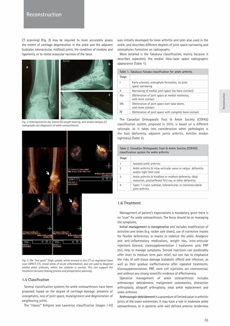
Reco
nstr
uctio
n
55
Reconstruction
CT scanning) (Fig. 3) may be required to more accurately assess the extent of cartilage degeneration in the ankle and the adjacent (subtalar, talonavicular, midfoot) joints, the condition of tendons and ligaments, or to reveal avascular necrosis of the talus.
Fig. 2: Anteroposterior (a), lateral (b) weight bearing, and medial oblique (c) radiographs are diagnostic of ankle osteoarthrosis.
Fig. 3: The “hot spots” (high uptake; white arrows) in this CT co-registered bone scan (SPECT-CT), reveal areas of acute inflammation, and are used to diagnose medial ankle arthrosis, whilst the subtalar is normal. This can support the treatment decision making process and preoperative planning.
1.5 Classification
Several classification systems for ankle osteoarthrosis have been proposed, based on the degree of cartilage damage, presence of osteophytes, loss of joint space, malalignment and degeneration of neighboring joints.
The “classic” Kellgren and Lawrence classification (stages I-IV)
was initially developed for knee arthritis and later also used in the ankle, and describes different degrees of joint space narrowing and osteophytes formation on radiographs.
More detailed is the Takakura classification, mainly because it describes separately the medial tibio-talar space radiographic appearance (Table 1).
The Canadian Orthopaedic Foot & Ankle Society (COFAS) classification system, proposed in 2010, is based on a different rationale, as it takes into consideration other pathologies is the foot (deformity, adjacent joints arthritis, Achilles tendon tightness) (Table 2).
1.6 Treatment
Management of patient’s expectations is mandatory, given there is no “cure” for ankle osteoarthrosis. The focus should be on managing the symptoms.
Initial management is nonoperative and includes modification of activities and shoes (e.g. rocker sole shoes), use of corrective insoles for flexible deformities, or braces to stabilize the ankle. Analgesic and anti-inflammatory medications, weight loss, intra-articular injections (steroid, viscosupplementation / hyaluronic acid, PRP etc), help to manage symptoms. Steroid injections can predictably offer short to medium term pain relief, but one has to emphasize the risks of soft-tissue damage (catabolic effect) and infection, as well as their gradual ineffectiveness after repeated treatments. Viscosupplementation, PRP, stem cell injections are controversial, and without any strong scientific evidence of effectiveness.
Operative management of ankle osteoarthrosis includes arthroscopic debridement, realignment osteotomies, distraction arthroplasty, allograft arthroplasty, total ankle replacement and ankle arthrosis.
Arthroscopic debridement is a procedure of limited value in arthritic joints of the lower extremities. It may have a role in moderate ankle osteoarthrosis, or in patients with well defined anterior tenderness
Table 1. Takakura-Tanaka classification for ankle arthritis
Stage
I Early sclerosis, osteophyte formation, no joint space narrowing
II Narrowing of medial joint space (no bone contact)
IIIa Obliteration of joint space at medial malleolus, with bone contact
IIIb Obliteration of joint space over talar dome, with bone contact
IV Obliteration of joint space with complete bone contact
Table 2. Canadian Orthopaedic Foot & Ankle Society (COFAS) classification system for ankle arthritis
Stage
1 Isolated ankle arthritis
2 Ankle arthritis & intra-articular varus or valgus deformity and/or tight heel cord
3 Ankle arthritis & hindfoot or midfoot deformity, tibial malunion, plantarflexed first ray, or other deformity
4 Types 1-3 plus subtalar, talonavicular, or calcaneocuboid joint arthritis
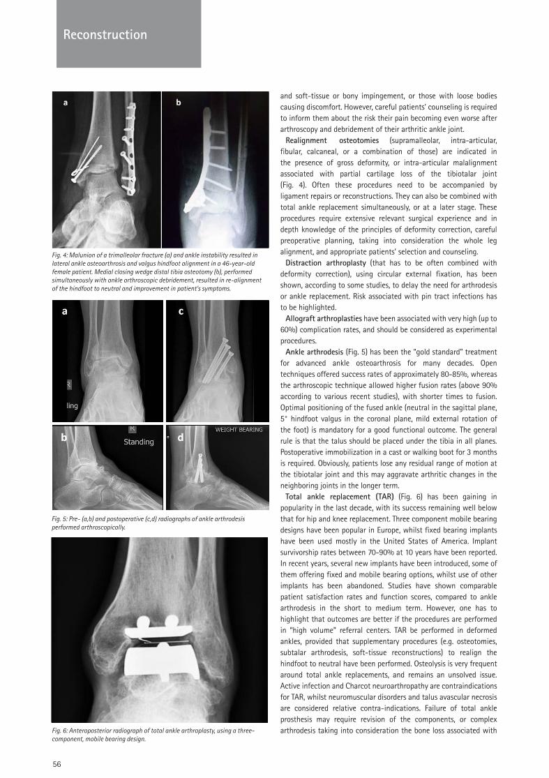
56
Reconstruction
and soft-tissue or bony impingement, or those with loose bodies causing discomfort. However, careful patients’ counseling is required to inform them about the risk their pain becoming even worse after arthroscopy and debridement of their arthritic ankle joint.
Realignment osteotomies (supramalleolar, intra-articular, fibular, calcaneal, or a combination of those) are indicated in the presence of gross deformity, or intra-articular malalignment associated with partial cartilage loss of the tibiotalar joint (Fig. 4). Often these procedures need to be accompanied by ligament repairs or reconstructions. They can also be combined with total ankle replacement simultaneously, or at a later stage. These procedures require extensive relevant surgical experience and in depth knowledge of the principles of deformity correction, careful preoperative planning, taking into consideration the whole leg alignment, and appropriate patients’ selection and counseling.
Distraction arthroplasty (that has to be often combined with deformity correction), using circular external fixation, has been shown, according to some studies, to delay the need for arthrodesis or ankle replacement. Risk associated with pin tract infections has to be highlighted.
Allograft arthroplasties have been associated with very high (up to 60%) complication rates, and should be considered as experimental procedures.
Ankle arthrodesis (Fig. 5) has been the “gold standard” treatment for advanced ankle osteoarthrosis for many decades. Open techniques offered success rates of approximately 80-85%, whereas the arthroscopic technique allowed higher fusion rates (above 90% according to various recent studies), with shorter times to fusion. Optimal positioning of the fused ankle (neutral in the sagittal plane, 5° hindfoot valgus in the coronal plane, mild external rotation of the foot) is mandatory for a good functional outcome. The general rule is that the talus should be placed under the tibia in all planes. Postoperative immobilization in a cast or walking boot for 3 months is required. Obviously, patients lose any residual range of motion at the tibiotalar joint and this may aggravate arthritic changes in the neighboring joints in the longer term.
Total ankle replacement (TAR) (Fig. 6) has been gaining in popularity in the last decade, with its success remaining well below that for hip and knee replacement. Three component mobile bearing designs have been popular in Europe, whilst fixed bearing implants have been used mostly in the United States of America. Implant survivorship rates between 70-90% at 10 years have been reported. In recent years, several new implants have been introduced, some of them offering fixed and mobile bearing options, whilst use of other implants has been abandoned. Studies have shown comparable patient satisfaction rates and function scores, compared to ankle arthrodesis in the short to medium term. However, one has to highlight that outcomes are better if the procedures are performed in “high volume” referral centers. TAR be performed in deformed ankles, provided that supplementary procedures (e.g. osteotomies, subtalar arthrodesis, soft-tissue reconstructions) to realign the hindfoot to neutral have been performed. Osteolysis is very frequent around total ankle replacements, and remains an unsolved issue. Active infection and Charcot neuroarthropathy are contraindications for TAR, whilst neuromuscular disorders and talus avascular necrosis are considered relative contra-indications. Failure of total ankle prosthesis may require revision of the components, or complex arthrodesis taking into consideration the bone loss associated with
Fig. 4: Malunion of a trimalleolar fracture (a) and ankle instability resulted in lateral ankle osteoarthrosis and valgus hindfoot alignment in a 46-year-old female patient. Medial closing wedge distal tibia osteotomy (b), performed simultaneously with ankle arthroscopic debridement, resulted in re-alignment of the hindfoot to neutral and improvement in patient’s symptoms.
Fig. 5: Pre- (a,b) and postoperative (c,d) radiographs of ankle arthrodesis performed arthroscopically.
Fig. 6: Anteroposterior radiograph of total ankle arthroplasty, using a three-component, mobile bearing design.
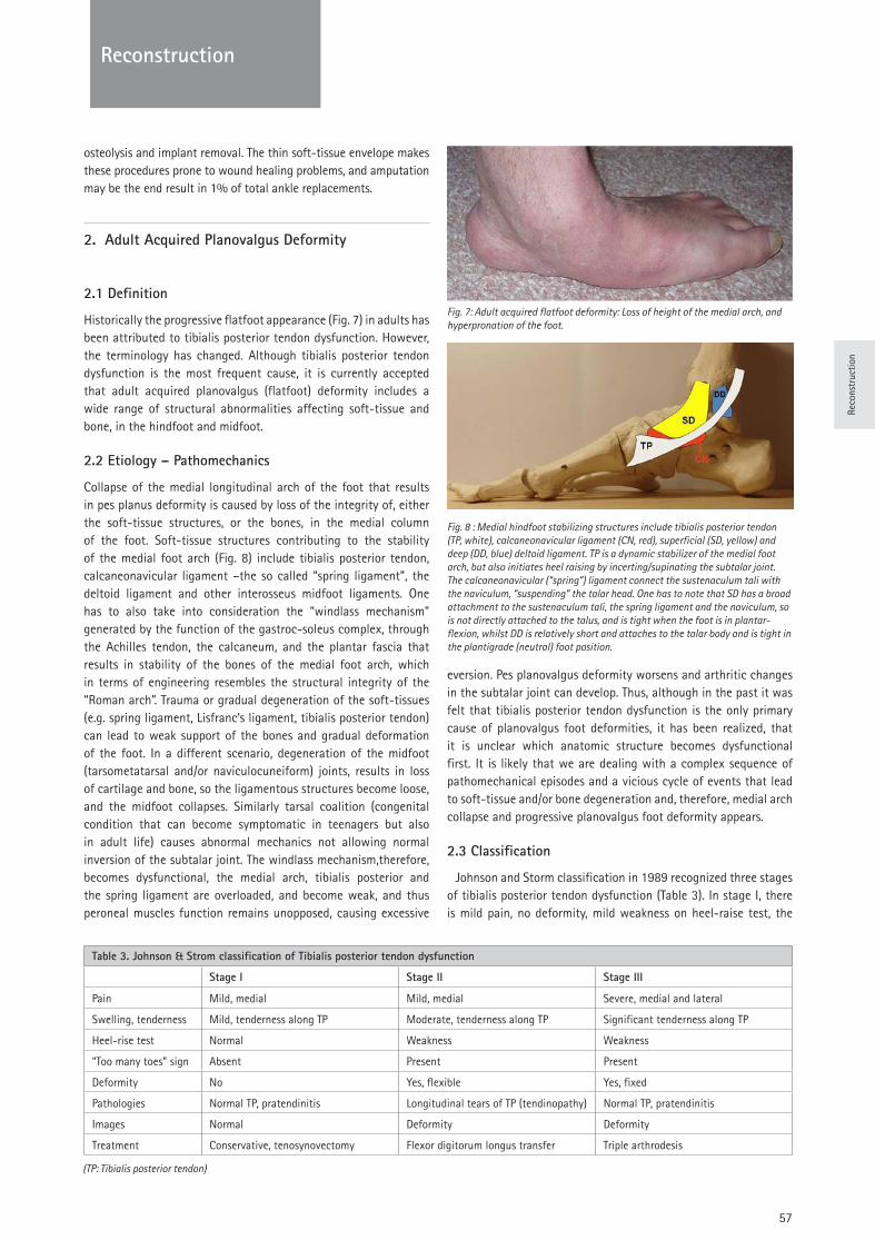
Reco
nstr
uctio
n
57
Reconstruction
osteolysis and implant removal. The thin soft-tissue envelope makes these procedures prone to wound healing problems, and amputation may be the end result in 1% of total ankle replacements.
2. Adult Acquired Planovalgus Deformity
2.1 Definition
Historically the progressive flatfoot appearance (Fig. 7) in adults has been attributed to tibialis posterior tendon dysfunction. However, the terminology has changed. Although tibialis posterior tendon dysfunction is the most frequent cause, it is currently accepted that adult acquired planovalgus (flatfoot) deformity includes a wide range of structural abnormalities affecting soft-tissue and bone, in the hindfoot and midfoot.
2.2 Etiology – Pathomechanics
Collapse of the medial longitudinal arch of the foot that results in pes planus deformity is caused by loss of the integrity of, either the soft-tissue structures, or the bones, in the medial column of the foot. Soft-tissue structures contributing to the stability of the medial foot arch (Fig. 8) include tibialis posterior tendon, calcaneonavicular ligament –the so called “spring ligament”, the deltoid ligament and other interosseus midfoot ligaments. One has to also take into consideration the “windlass mechanism” generated by the function of the gastroc-soleus complex, through the Achilles tendon, the calcaneum, and the plantar fascia that results in stability of the bones of the medial foot arch, which in terms of engineering resembles the structural integrity of the “Roman arch”. Trauma or gradual degeneration of the soft-tissues (e.g. spring ligament, Lisfranc’s ligament, tibialis posterior tendon) can lead to weak support of the bones and gradual deformation of the foot. In a different scenario, degeneration of the midfoot (tarsometatarsal and/or naviculocuneiform) joints, results in loss of cartilage and bone, so the ligamentous structures become loose, and the midfoot collapses. Similarly tarsal coalition (congenital condition that can become symptomatic in teenagers but also in adult life) causes abnormal mechanics not allowing normal inversion of the subtalar joint. The windlass mechanism,therefore, becomes dysfunctional, the medial arch, tibialis posterior and the spring ligament are overloaded, and become weak, and thus peroneal muscles function remains unopposed, causing excessive
eversion. Pes planovalgus deformity worsens and arthritic changes in the subtalar joint can develop. Thus, although in the past it was felt that tibialis posterior tendon dysfunction is the only primary cause of planovalgus foot deformities, it has been realized, that it is unclear which anatomic structure becomes dysfunctional first. It is likely that we are dealing with a complex sequence of pathomechanical episodes and a vicious cycle of events that lead to soft-tissue and/or bone degeneration and, therefore, medial arch collapse and progressive planovalgus foot deformity appears.
2.3 Classification
Johnson and Storm classification in 1989 recognized three stages of tibialis posterior tendon dysfunction (Table 3). In stage I, there is mild pain, no deformity, mild weakness on heel-raise test, the
Fig. 7: Adult acquired flatfoot deformity: Loss of height of the medial arch, and hyperpronation of the foot.
Fig. 8 : Medial hindfoot stabilizing structures include tibialis posterior tendon (TP, white), calcaneonavicular ligament (CN, red), superficial (SD, yellow) and deep (DD, blue) deltoid ligament. TP is a dynamic stabilizer of the medial foot arch, but also initiates heel raising by incerting/supinating the subtalar joint. The calcaneonavicular (“spring”) ligament connect the sustenaculum tali with the naviculum, “suspending” the talar head. One has to note that SD has a broad attachment to the sustenaculum tali, the spring ligament and the naviculum, so is not directly attached to the talus, and is tight when the foot is in plantar-flexion, whilst DD is relatively short and attaches to the talar body and is tight in the plantigrade (neutral) foot position.
Table 3. Johnson & Strom classification of Tibialis posterior tendon dysfunction
Stage I Stage II Stage III
Pain Mild, medial Mild, medial Severe, medial and lateral
Swelling, tenderness Mild, tenderness along TP Moderate, tenderness along TP Significant tenderness along TP
Heel-rise test Normal Weakness Weakness
“Too many toes” sign Absent Present Present
Deformity No Yes, flexible Yes, fixed
Pathologies Normal TP, pratendinitis Longitudinal tears of TP (tendinopathy) Normal TP, pratendinitis
Images Normal Deformity Deformity
Treatment Conservative, tenosynovectomy Flexor digitorum longus transfer Triple arthrodesis
(TP: Tibialis posterior tendon)
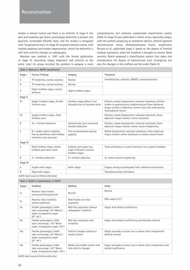
58
Reconstruction
tendon is almost normal and there is no arthritis. In stage II, the pain and weakness get worse, planovalgus deformity is present, but passively correctable (flexible foot), and the tendon is elongated with “longitudinal tears”. In stage III symptoms become worse, with marked weakness and tendon degeneration, whilst the deformity is stiff with arthritic changes on radiographs.
Myerson was credited in 1997 with the formal publication of stage IV, describing valgus alignment and arthritis at the ankle. Later his group revisited the problem to propose a more
comprehensive, but relatively complicated classification system (Table 4). Stage IV was subdivided in subtle versus rigid ankle valgus, with the authors proposing as treatment options, deltoid ligament reconstruction versus tibiotalocalcaneal fusion, respectively. Parsons et al. subdivided stage II, based on the degree of forefoot residual supination, when the hindfoot is brought to neutral. More recently, Raikin proposed a classification system that takes into consideration the degree of talonavicular joint incongruity, but also the changes in the midfoot and the ankle (Table 5).
Table 4. Myerson’s AAFD classification
Stage I Clinical findings Imaging Treatment
A TP tenderness, normal anatomy Normal Immobilisation, orthotics, NSAID’s, tenosynovectomy
B TP tenderness, normal anatomy Normal
CSlight hindfoot valgus, normal anatomy
Slight hindfoot valgus
Stage II
A1 Supple hindfoot valgus, flexible forefoot varus
Hindfoot valgus Meary’s line disrupted Loss of calcaneal pitch
Orthosis, medial displacement calcaneal osteotomy, Achilles tendon or gastrocnemius lengthening and flexor digitorum longus transfer if deformity corrects only with ankle plantar flexionplantar flexion
A2 Supple hindfoot valgus, fixed forefoot varus
Orthosis, medial displacement calcaneal osteotomy, flexor digitorum longus transfer, cotton osteotomy
B A2 + forefoot abduction Talonavicular joint uncovered, forefoot abduction
Orthosis, medial displacement calcaneal osteotomy, flexor digitorum longus transfer, lateral column lengthening
C B + medial column instability, first ray dorsiflexion with hindfoot correction, sinus tarsi pain
First tarsometatarsal plantar gapping
Medial displacement calcaneal osteotomy, flexor digitorum longus transfer, cotton osteotomy or medial column fusion
Stage III
A Rigid hindfoot valgus, lateral hindfoot pain (sinus tarsi)
Subtalar joint space loss, angle of Gissane sclerosis, hindfoot valgus
Triple arthrodesis or custom bracing if not surgical candidate
B A + forefoot abduction A+ forefoot abduction A+ lateral column lengthening
Stage IV
A Supple ankle valgus Ankle valgus Surgery aiming at plantigrade foot+ deltoid reconstruction
B Rigid ankle valgus Tibiotalocalcaneal arthrodesis
Table 5. Raikin’s classification of AAFD
Stage I Hindfoot Midfoot Ankle
IaPosterior tibial tendon tenosynovitistenosynovitis
NeutralNeutral
IbPosterior tibial tendinitis without deformity
Mild flexible mid-foot supination
Mild valgus (<5°)
IIa Flexible planovalgus (<40% talar uncoverage,<30° Meary’s angle, incongruency angle 20°–45°)
Mid-foot supination without radiographic instability
Valgus with deltoid insufficiency
IIb Flexible planovalgus (>40% talar uncoverage, >30° Meary angle, incongruency angle >45°)
Mid-foot supination with instability
Valgus with deltoid insufficiency and tibiotalar arthritis
IIIa Flexible planovalgus (<40% talar uncoverage,<30° Meary’s angle, incongruency angle 20°–45°)
Arthritic changes isolated to medial column
Valgus secondary to bone loss in lateral ankle compartment (deltoid normal)
IIIb Flexible planovalgus (>40% talar uncoverage, >30° Meary angle, incongruency angle >45°)
Medial and middle-column mid-foot arthritic changes
Valgus secondary to bone loss in lateral ankle compartment and deltoid insufficiency
(AAFD: Adult acquired flatfoot deformity)
(AAFD: Adult acquired flatfoot deformity)
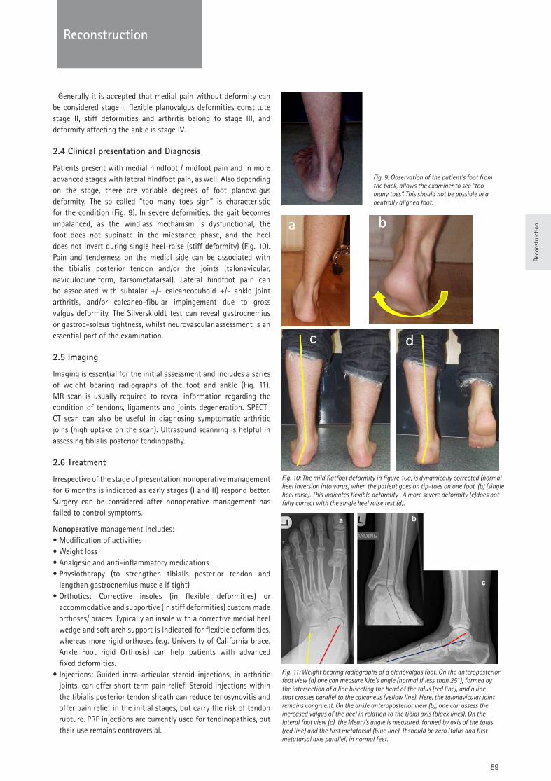
Reco
nstr
uctio
n
59
ReconstructionReconstruction
Generally it is accepted that medial pain without deformity can be considered stage I, flexible planovalgus deformities constitute stage II, stiff deformities and arthritis belong to stage III, and deformity affecting the ankle is stage IV.
2.4 Clinical presentation and Diagnosis
Patients present with medial hindfoot / midfoot pain and in more advanced stages with lateral hindfoot pain, as well. Also depending on the stage, there are variable degrees of foot planovalgus deformity. The so called “too many toes sign” is characteristic for the condition (Fig. 9). In severe deformities, the gait becomes imbalanced, as the windlass mechanism is dysfunctional, the foot does not supinate in the midstance phase, and the heel does not invert during single heel-raise (stiff deformity) (Fig. 10). Pain and tenderness on the medial side can be associated with the tibialis posterior tendon and/or the joints (talonavicular, naviculocuneiform, tarsometatarsal). Lateral hindfoot pain can be associated with subtalar +/- calcaneocuboid +/- ankle joint arthritis, and/or calcaneo-fibular impingement due to gross valgus deformity. The Silverskioldt test can reveal gastrocnemius or gastroc-soleus tightness, whilst neurovascular assessment is an essential part of the examination.
2.5 Imaging
Imaging is essential for the initial assessment and includes a series of weight bearing radiographs of the foot and ankle (Fig. 11). MR scan is usually required to reveal information regarding the condition of tendons, ligaments and joints degeneration. SPECT-CT scan can also be useful in diagnosing symptomatic arthritic joins (high uptake on the scan). Ultrasound scanning is helpful in assessing tibialis posterior tendinopathy.
2.6 Treatment
Irrespective of the stage of presentation, nonoperative management for 6 months is indicated as early stages (I and II) respond better. Surgery can be considered after nonoperative management has failed to control symptoms.
Nonoperative management includes:• Modification of activities• Weight loss• Analgesic and anti-inflammatory medications • Physiotherapy (to strengthen tibialis posterior tendon and
lengthen gastrocnemius muscle if tight) • Orthotics: Corrective insoles (in flexible deformities) or
accommodative and supportive (in stiff deformities) custom made orthoses/ braces. Typically an insole with a corrective medial heel wedge and soft arch support is indicated for flexible deformities, whereas more rigid orthoses (e.g. University of California brace, Ankle Foot rigid Orthosis) can help patients with advanced fixed deformities.
• Injections: Guided intra-articular steroid injections, in arthritic joints, can offer short term pain relief. Steroid injections within the tibialis posterior tendon sheath can reduce tenosynovitis and offer pain relief in the initial stages, but carry the risk of tendon rupture. PRP injections are currently used for tendinopathies, but their use remains controversial.
Fig. 10: The mild flatfoot deformity in figure 10a, is dynamically corrected (normal heel inversion into varus) when the patient goes on tip-toes on one foot (b) (single heel raise). This indicates flexible deformity . A more severe deformity (c)does not fully correct with the single heel raise test (d).
Fig. 9: Observation of the patient’s foot from the back, allows the examiner to see “too many toes”. This should not be possible in a neutrally aligned foot.
Fig. 11: Weight bearing radiographs of a planovalgus foot. On the anteroposterior foot view (a) one can measure Kite’s angle (normal if less than 25°), formed by the intersection of a line bisecting the head of the talus (red line), and a line that crosses parallel to the calcaneus (yellow line). Here, the talonavicular joint remains congruent. On the ankle anteroposterior view (b), one can assess the increased valgus of the heel in relation to the tibial axis (black lines). On the lateral foot view (c), the Meary’s angle is measured, formed by axis of the talus (red line) and the first metatarsal (blue line). It should be zero (talus and first metatarsal axis parallel) in normal feet.
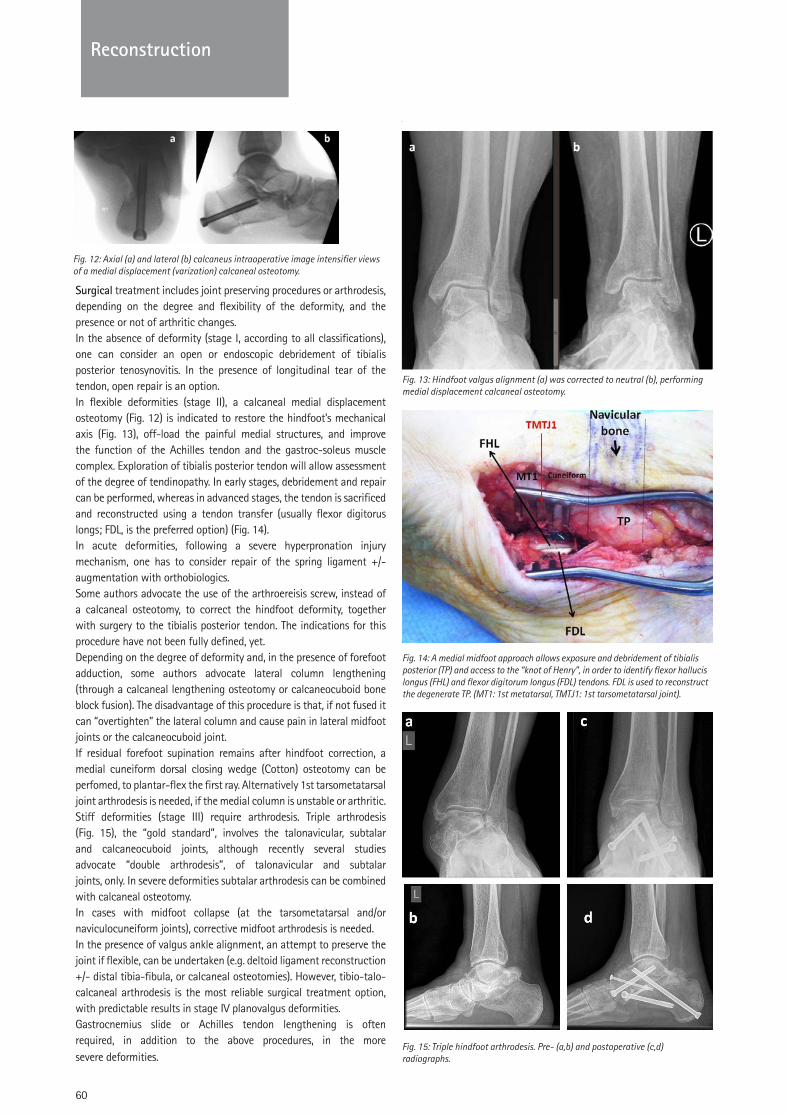
60
SPORTS KNEEReconstruction
Surgical treatment includes joint preserving procedures or arthrodesis, depending on the degree and flexibility of the deformity, and the presence or not of arthritic changes.In the absence of deformity (stage I, according to all classifications), one can consider an open or endoscopic debridement of tibialis posterior tenosynovitis. In the presence of longitudinal tear of the tendon, open repair is an option.In flexible deformities (stage II), a calcaneal medial displacement osteotomy (Fig. 12) is indicated to restore the hindfoot’s mechanical axis (Fig. 13), off-load the painful medial structures, and improve the function of the Achilles tendon and the gastroc-soleus muscle complex. Exploration of tibialis posterior tendon will allow assessment of the degree of tendinopathy. In early stages, debridement and repair can be performed, whereas in advanced stages, the tendon is sacrificed and reconstructed using a tendon transfer (usually flexor digitorus longs; FDL, is the preferred option) (Fig. 14).In acute deformities, following a severe hyperpronation injury mechanism, one has to consider repair of the spring ligament +/- augmentation with orthobiologics.Some authors advocate the use of the arthroereisis screw, instead of a calcaneal osteotomy, to correct the hindfoot deformity, together with surgery to the tibialis posterior tendon. The indications for this procedure have not been fully defined, yet.Depending on the degree of deformity and, in the presence of forefoot adduction, some authors advocate lateral column lengthening (through a calcaneal lengthening osteotomy or calcaneocuboid bone block fusion). The disadvantage of this procedure is that, if not fused it can “overtighten” the lateral column and cause pain in lateral midfoot joints or the calcaneocuboid joint.If residual forefoot supination remains after hindfoot correction, a medial cuneiform dorsal closing wedge (Cotton) osteotomy can be perfomed, to plantar-flex the first ray. Alternatively 1st tarsometatarsal joint arthrodesis is needed, if the medial column is unstable or arthritic.Stiff deformities (stage III) require arthrodesis. Triple arthrodesis (Fig. 15), the “gold standard”, involves the talonavicular, subtalar and calcaneocuboid joints, although recently several studies advocate “double arthrodesis”, of talonavicular and subtalar joints, only. In severe deformities subtalar arthrodesis can be combined with calcaneal osteotomy.In cases with midfoot collapse (at the tarsometatarsal and/or naviculocuneiform joints), corrective midfoot arthrodesis is needed.In the presence of valgus ankle alignment, an attempt to preserve the joint if flexible, can be undertaken (e.g. deltoid ligament reconstruction +/- distal tibia-fibula, or calcaneal osteotomies). However, tibio-talo-calcaneal arthrodesis is the most reliable surgical treatment option, with predictable results in stage IV planovalgus deformities.Gastrocnemius slide or Achilles tendon lengthening is often required, in addition to the above procedures, in the more severe deformities.
Fig. 15: Triple hindfoot arthrodesis. Pre- (a,b) and postoperative (c,d) radiographs.
Fig. 13: Hindfoot valgus alignment (a) was corrected to neutral (b), performing medial displacement calcaneal osteotomy.
Fig. 14: A medial midfoot approach allows exposure and debridement of tibialis posterior (TP) and access to the “knot of Henry”, in order to identify flexor hallucis longus (FHL) and flexor digitorum longus (FDL) tendons. FDL is used to reconstruct the degenerate TP. (MT1: 1st metatarsal, TMTJ1: 1st tarsometatarsal joint).
Fig. 12: Axial (a) and lateral (b) calcaneus intraoperative image intensifier views of a medial displacement (varization) calcaneal osteotomy.
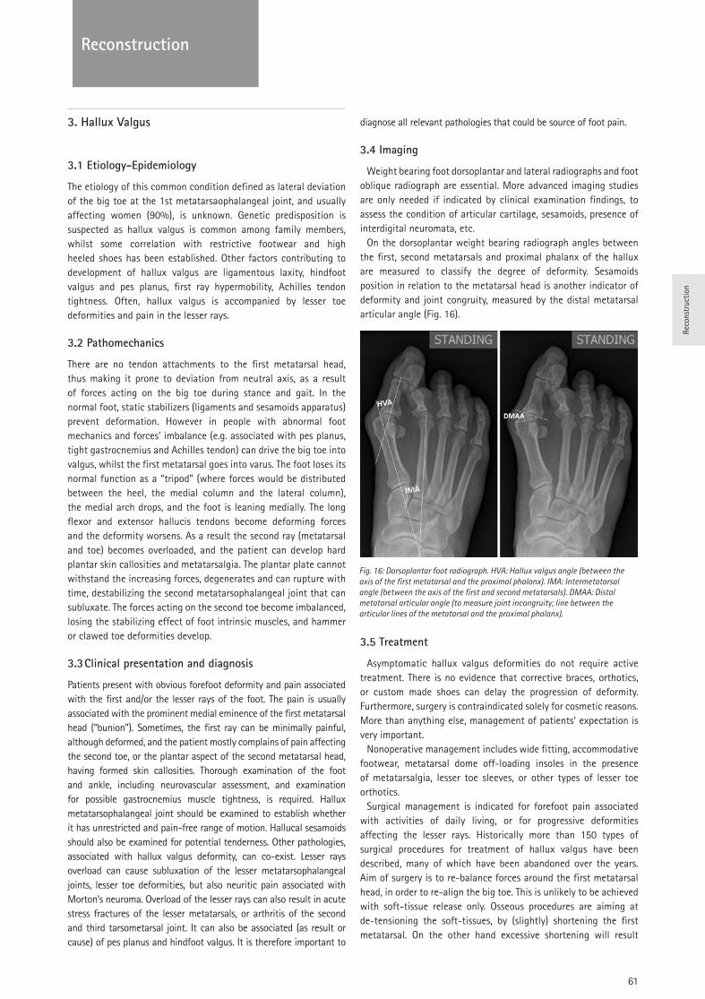
61
SPORTS KNEE
Reco
nstr
uctio
n
Reconstruction
3. Hallux Valgus
3.1 Etiology-Epidemiology
The etiology of this common condition defined as lateral deviation of the big toe at the 1st metatarsaophalangeal joint, and usually affecting women (90%), is unknown. Genetic predisposition is suspected as hallux valgus is common among family members, whilst some correlation with restrictive footwear and high heeled shoes has been established. Other factors contributing to development of hallux valgus are ligamentous laxity, hindfoot valgus and pes planus, first ray hypermobility, Achilles tendon tightness. Often, hallux valgus is accompanied by lesser toe deformities and pain in the lesser rays.
3.2 Pathomechanics
There are no tendon attachments to the first metatarsal head, thus making it prone to deviation from neutral axis, as a result of forces acting on the big toe during stance and gait. In the normal foot, static stabilizers (ligaments and sesamoids apparatus) prevent deformation. However in people with abnormal foot mechanics and forces’ imbalance (e.g. associated with pes planus, tight gastrocnemius and Achilles tendon) can drive the big toe into valgus, whilst the first metatarsal goes into varus. The foot loses its normal function as a “tripod” (where forces would be distributed between the heel, the medial column and the lateral column), the medial arch drops, and the foot is leaning medially. The long flexor and extensor hallucis tendons become deforming forces and the deformity worsens. As a result the second ray (metatarsal and toe) becomes overloaded, and the patient can develop hard plantar skin callosities and metatarsalgia. The plantar plate cannot withstand the increasing forces, degenerates and can rupture with time, destabilizing the second metatarsophalangeal joint that can subluxate. The forces acting on the second toe become imbalanced, losing the stabilizing effect of foot intrinsic muscles, and hammer or clawed toe deformities develop.
3.3 Clinical presentation and diagnosis
Patients present with obvious forefoot deformity and pain associated with the first and/or the lesser rays of the foot. The pain is usually associated with the prominent medial eminence of the first metatarsal head (“bunion”). Sometimes, the first ray can be minimally painful, although deformed, and the patient mostly complains of pain affecting the second toe, or the plantar aspect of the second metatarsal head, having formed skin callosities. Thorough examination of the foot and ankle, including neurovascular assessment, and examination for possible gastrocnemius muscle tightness, is required. Hallux metatarsophalangeal joint should be examined to establish whether it has unrestricted and pain-free range of motion. Hallucal sesamoids should also be examined for potential tenderness. Other pathologies, associated with hallux valgus deformity, can co-exist. Lesser rays overload can cause subluxation of the lesser metatarsophalangeal joints, lesser toe deformities, but also neuritic pain associated with Morton’s neuroma. Overload of the lesser rays can also result in acute stress fractures of the lesser metatarsals, or arthritis of the second and third tarsometarsal joint. It can also be associated (as result or cause) of pes planus and hindfoot valgus. It is therefore important to
diagnose all relevant pathologies that could be source of foot pain.
3.4 Imaging
Weight bearing foot dorsoplantar and lateral radiographs and foot oblique radiograph are essential. More advanced imaging studies are only needed if indicated by clinical examination findings, to assess the condition of articular cartilage, sesamoids, presence of interdigital neuromata, etc.
On the dorsoplantar weight bearing radiograph angles between the first, second metatarsals and proximal phalanx of the hallux are measured to classify the degree of deformity. Sesamoids position in relation to the metatarsal head is another indicator of deformity and joint congruity, measured by the distal metatarsal articular angle (Fig. 16).
3.5 Treatment
Asymptomatic hallux valgus deformities do not require active treatment. There is no evidence that corrective braces, orthotics, or custom made shoes can delay the progression of deformity. Furthermore, surgery is contraindicated solely for cosmetic reasons. More than anything else, management of patients’ expectation is very important.
Nonoperative management includes wide fitting, accommodative footwear, metatarsal dome off-loading insoles in the presence of metatarsalgia, lesser toe sleeves, or other types of lesser toe orthotics.
Surgical management is indicated for forefoot pain associated with activities of daily living, or for progressive deformities affecting the lesser rays. Historically more than 150 types of surgical procedures for treatment of hallux valgus have been described, many of which have been abandoned over the years. Aim of surgery is to re-balance forces around the first metatarsal head, in order to re-align the big toe. This is unlikely to be achieved with soft-tissue release only. Osseous procedures are aiming at de-tensioning the soft-tissues, by (slightly) shortening the first metatarsal. On the other hand excessive shortening will result
Fig. 16: Dorsoplantar foot radiograph. HVA: Hallux valgus angle (between the axis of the first metatarsal and the proximal phalanx). IMA: Intermetatarsal angle (between the axis of the first and second metatarsals). DMAA: Distal metatarsal articular angle (to measure joint incongruity; line between the articular lines of the metatarsal and the proximal phalanx).
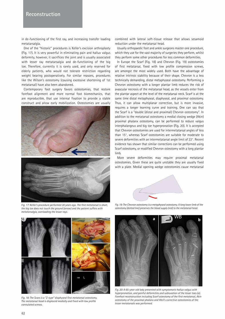
62
Reconstruction
in de-functioning of the first ray, and increasing transfer loading metatarsalgia.
One of the “historic” procedures is Keller’s excision arthroplasty (Fig. 17). It is very powerful in eliminating pain and hallux valgus deformity, however, it sacrifices the joint and is usually associated with lesser ray metatarsalgia and de-functioning of the big toe. Therefore, currently it is rarely used, and only reserved for elderly patients, who would not tolerate restriction regarding weight bearing postoperatively. For similar reasons, procedures like the Wilson’s osteotomy (causing excessive shortening of 1st metatarsal) have also been abandoned.
Contemporary foot surgery favors osteotomies, that restore forefoot alignment and more normal foot biomechanics, that are reproducible, that use internal fixation to provide a stable construct and allow early mobilization. Osteotomies are usually
combined with lateral soft-tissue release that allows sesamoid reduction under the metatarsal head.
Usually orthopaedic foot and ankle surgeons master one procedure, which they use for the vast majority of surgeries they perform, whilst they perform some other procedures for less common deformities.
In Europe the Scarf (Fig. 18) and Chevron (Fig. 19) osteotomies of first metatarsal, fixed with low profile compression screws, are amongst the most widely used. Both have the advantage of relative intrinsic stability because of their shape. Chevron is a less technically demanding, distal metaphyseal osteotomy. Performing a Chevron osteotomy with a longer plantar limb reduces the risk of avascular necrosis of the metatarsal head, as the vessels enter from the plantar aspect at the level of the metatarsal neck. Scarf is at the same time distal metaphyseal, diaphyseal, and proximal osteotomy. Thus, it can allow multiplanar correction, but is more invasive, requires a longer learning curve and training. One can say that the Scarf is a ”double (distal and proximal) Chevron osteotomy". In addition to the metatarsal osteotomy a medial closing wedge (Akin) proximal phalanx osteotomy, can be performed to reduce valgus interphalangeus and big toe hyperpronation (Fig. 20). It is accepted that Chevron osteotomies are used for intermetatarsal angles of less than 15°, whereas Scarf osteotomies are suitable for moderate to severe deformities with an intermetatarsal angle limit of 22°. Recent evidence has shown that similar corrections can be performed using Scarf osteotomy, or modified Chevron osteotomy with a long plantar limb.
More severe deformities may require proximal metatarsal osteotomies. Given these are quite unstable they are usually fixed with a plate. Medial opening wedge osteotomies cause metatarsal
Fig. 19: The Chevron osteotomy is a metaphyseal osteotomy. A long lower limb of the osteotomy (dotted line) preserves the blood supply (red) to the metatarsal head.
Fig. 20: A 65-year-old lady presented with symptomatic hallux valgus with hyperpronation, and painful deformities and subluxation of the lesser toes (a). Forefoot reconstruction including Scarf osteotomy of the first metatarsal, Akin osteotomy of the proximal phalanx and Weil’s corrective osteotomies of the lesser metatarsals was performed.
Fig. 18: The Scars is a “Z-type” diaphyseal first metatarsal osteotomy. The metatarsal head is displaced medially and fixed with low profile cannulated screws.
Fig. 17: Keller’s procedure performed 20 years ago. The first metatarsal is short, the big toe does not touch the ground (arrow) and the patient suffers with metatarsalgia, overloading the lesser rays.
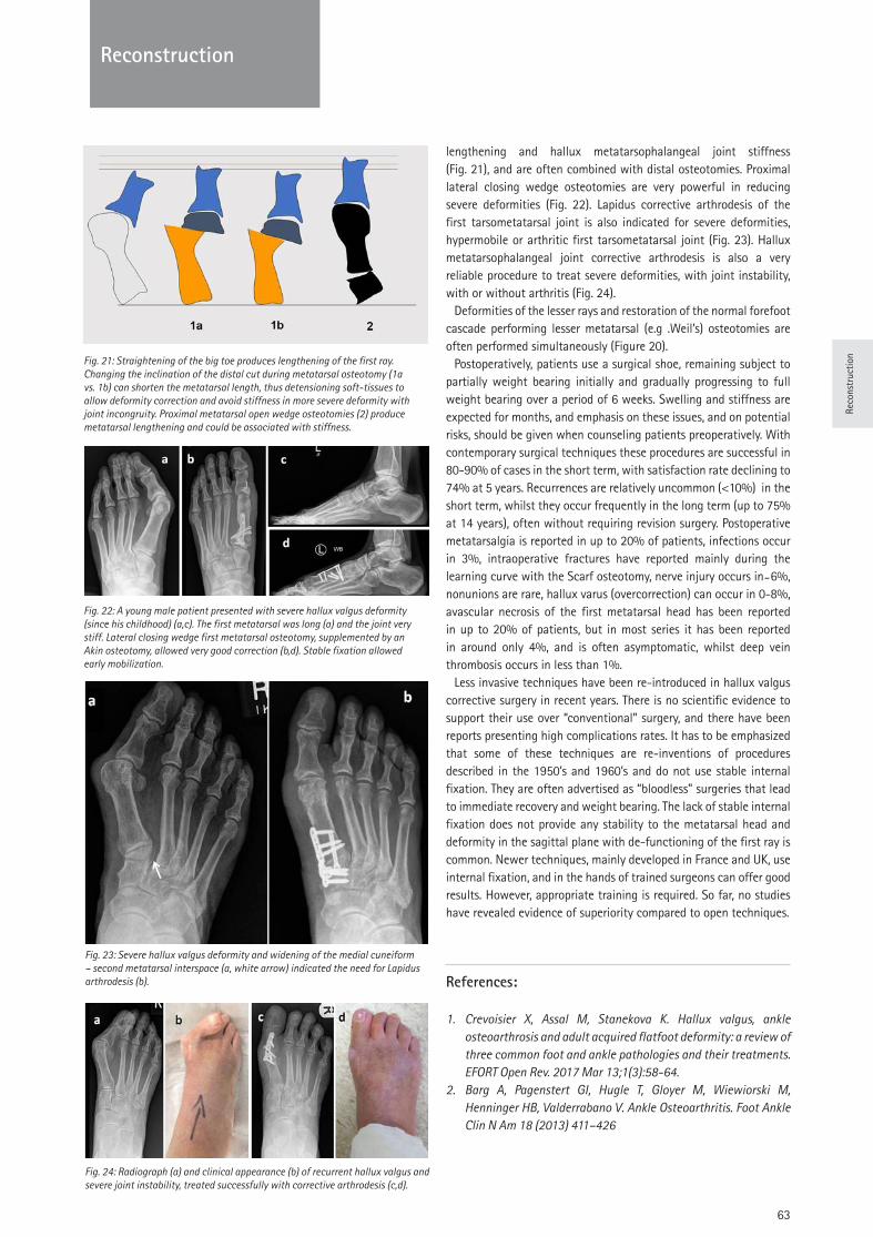
Reco
nstr
uctio
n
63
Reconstruction
lengthening and hallux metatarsophalangeal joint stiffness (Fig. 21), and are often combined with distal osteotomies. Proximal lateral closing wedge osteotomies are very powerful in reducing severe deformities (Fig. 22). Lapidus corrective arthrodesis of the first tarsometatarsal joint is also indicated for severe deformities, hypermobile or arthritic first tarsometatarsal joint (Fig. 23). Hallux metatarsophalangeal joint corrective arthrodesis is also a very reliable procedure to treat severe deformities, with joint instability, with or without arthritis (Fig. 24).
Deformities of the lesser rays and restoration of the normal forefoot cascade performing lesser metatarsal (e.g .Weil’s) osteotomies are often performed simultaneously (Figure 20).
Postoperatively, patients use a surgical shoe, remaining subject to partially weight bearing initially and gradually progressing to full weight bearing over a period of 6 weeks. Swelling and stiffness are expected for months, and emphasis on these issues, and on potential risks, should be given when counseling patients preoperatively. With contemporary surgical techniques these procedures are successful in 80-90% of cases in the short term, with satisfaction rate declining to 74% at 5 years. Recurrences are relatively uncommon (<10%) in the short term, whilst they occur frequently in the long term (up to 75% at 14 years), often without requiring revision surgery. Postoperative metatarsalgia is reported in up to 20% of patients, infections occur in 3%, intraoperative fractures have reported mainly during the learning curve with the Scarf osteotomy, nerve injury occurs in ̴ 6%, nonunions are rare, hallux varus (overcorrection) can occur in 0-8%, avascular necrosis of the first metatarsal head has been reported in up to 20% of patients, but in most series it has been reported in around only 4%, and is often asymptomatic, whilst deep vein thrombosis occurs in less than 1%.
Less invasive techniques have been re-introduced in hallux valgus corrective surgery in recent years. There is no scientific evidence to support their use over “conventional” surgery, and there have been reports presenting high complications rates. It has to be emphasized that some of these techniques are re-inventions of procedures described in the 1950’s and 1960’s and do not use stable internal fixation. They are often advertised as “bloodless” surgeries that lead to immediate recovery and weight bearing. The lack of stable internal fixation does not provide any stability to the metatarsal head and deformity in the sagittal plane with de-functioning of the first ray is common. Newer techniques, mainly developed in France and UK, use internal fixation, and in the hands of trained surgeons can offer good results. However, appropriate training is required. So far, no studies have revealed evidence of superiority compared to open techniques.
References:
1. Crevoisier X, Assal M, Stanekova K. Hallux valgus, ankle osteoarthrosis and adult acquired flatfoot deformity: a review of three common foot and ankle pathologies and their treatments. EFORT Open Rev. 2017 Mar 13;1(3):58-64.
2. Barg A, Pagenstert GI, Hugle T, Gloyer M, Wiewiorski M, Henninger HB, Valderrabano V. Ankle Osteoarthritis. Foot Ankle Clin N Am 18 (2013) 411–426
Fig. 21: Straightening of the big toe produces lengthening of the first ray. Changing the inclination of the distal cut during metatarsal osteotomy (1a vs. 1b) can shorten the metatarsal length, thus detensioning soft-tissues to allow deformity correction and avoid stiffness in more severe deformity with joint incongruity. Proximal metatarsal open wedge osteotomies (2) produce metatarsal lengthening and could be associated with stiffness.
Fig. 22: A young male patient presented with severe hallux valgus deformity (since his childhood) (a,c). The first metatarsal was long (a) and the joint very stiff. Lateral closing wedge first metatarsal osteotomy, supplemented by an Akin osteotomy, allowed very good correction (b,d). Stable fixation allowed early mobilization.
Fig. 23: Severe hallux valgus deformity and widening of the medial cuneiform – second metatarsal interspace (a, white arrow) indicated the need for Lapidus arthrodesis (b).
Fig. 24: Radiograph (a) and clinical appearance (b) of recurrent hallux valgus and severe joint instability, treated successfully with corrective arthrodesis (c,d).
64
Reconstruction
3. Townshend D, Di Silvestro M, Krause F, Penner M, Younger A, Glazebrook M, Wing K. Arthroscopic versus open ankle arthrodesis: a multicenter comparative case series. J Bone Joint Surg Am. 2013 Jan 16;95(2):98-102.
4. Daniels TR, Younger AS, Penner M, Wing K, Dryden PJ, Wong H, Glazebrook M. Intermediate-term results of total ankle replacement and ankle arthrodesis: a COFAS multicenter study. J Bone Joint Surg Am. 2014 Jan 15;96(2):135-42.
5. Deland JT. Adult-acquired flatfoot deformity. J Am Acad Orthop Surg 2008;16:399-406.
6. Abousayed MM, Tartaglione JP, Rosenbaum AJ, Dipreta JA. Classifications in brief: Johnson and Storm classification of adult acquired flatfoot deformity. Clin Orthop Relat Res (2016) 474:588-593.
7. Gougoulias N. Adult acquired flatfoot deformity. Acta Orthopedica et Traumatologica Hellenica 2018;69(2):85-98. (available online at: http://eexot.gr/wp-content/uploads/2018/09/ISSUE_2_2018-1.pdf)
8. Perera AM, Mason L, Stephens MM. The pathogenesis of hallux valgus. J Bone Joint Surg Am. 2011;93:1650-61
9. Robinson AHN, Limbers JP. Modern concepts in the treatment of hallux valgus. J Bone Joint Surg [Br] 2005;87-B:1038-45.
10. Easley ME, Trnka HJ. Current concepts review: hallux valgus part II: operative treatment. Foot Ankle Int 2007;28:748-758.
11. Chong A, Nazarian N, Chandrananth J, Tacey M, Shepherd D, Tran P. Surgery for the correction of hallux valgus: minimum five-year results with a validated patient-reported outcome tool and regression analysis. Bone Joint J. 2015;97-B(2):208-14.
Questions
1. Which one of the following statements is wrong regarding ankle arthrosis.a. It is usually post-traumaticb. Ankle cartilage has reduced resistance to loading compared
with knee cartilagec. Ankle cartilage has better healing capacity than
knee cartilaged. Ankle instability changes tibio-talar pressure distribution
2. Choose the (one) correct statement.a. Total ankle arthroplasty is contraindicated for deformities
of 15°b. Nonunion rate of open ankle arthrodesis is less than 5%c. Failure rate of TAR at 10 yrs is between 10-30%d. Arthroscopy for ankle arthrosis yields 75% good results
3. In stage II adult acquired flatfoot deformity (one correct answer):a. Hindfoot joints are stiffb. Tibialis posterior is usually tendinopathicc. Surgical treatment is always requiredd. Triple arthrodesis is treatment of choice
4. In stage III adult acquired flatfoot deformity (one correct answer):a. The ankle joint is arthriticb. The heel corrects into varus during heel raising c. Rigid orthoses are recommended for nonoperative
managementd. Tibialis posterior tendon is ruptured
5. Which one of the following statements is wrong regarding hallux valgus treatment?a. Recurrence occurs in only 15% of feet, 15 years after surgeryb. Stiffness of hallux MTP joint is possible after surgeryc. Arthrodesis is an option for severe deformity and/or
instabilityd. There is no clear evidence of benefit of percutaneous over
open surgery
Answers1b, 2c, 3b, 4c, 5a
Reco
nstr
uctio
n
65
Reconstruction
Prof. Luc Favard
University Hospital of Tours, CHU Trousseau 37044 Tours Cedex
Degenerative Disorders Of The Shoulder & Elbow
Abstract
The causes of degenerative disorders of the rotator cuff or glenohumeral joint are multifactorial. However, the morphology of the scapula, and more specifically of the acromion and glenoid, seems to play an important role. Indeed, the critical shoulder angle is a predictive factor of cuff tear and has been associated to a risk of osteoarthritis. The same applies to the glenoid version and inclination. Thus, the genesis of the cuff lesions and the development of glenohumeral arthritis are rather associated to the lateral extension of the acromion, than to an extrinsic component.
The natural history of cuff tears is key for the therapeutic decision-making. On average, the lesions begin 15 mm back to the bicipital groove and then gradually extend either backwards, causing a deficit of the external rotators, or forward, leading to the pseudo paralytic shoulder. At the same time, the tendon retracts and the muscle degenerates with atrophy and fatty infiltration, the stage of this latter being an important prognostic factor of the tendon's reparability. The initial medical treatment lasts around six months. If the tendon is repairable, diverse techniques are available for the treatment but none of them has been shown as being superior. Many techniques have also been described for cases where the tendon is not repairable, but again with no evidence of supremacy for any of them. In the presence of a large antero-superior rupture with a pseudo paralytic shoulder, indications for reverse prosthesis0 should be favoured.
Osteoarthritis of the shoulder is secondary to many aetiologies (trauma, instability, rheumatoid arthritis (RA), rotator cuff tear, necrosis, etc.). In primary osteoarthritis, analysis of the glenoid orientation and wear is essential for the choice of treatment. As management of posterior decentering of the humeral head can be difficult, a 3D analysis of the abnormalities and/or virtual surgical implantation with patient-specific shoulder guides are extremely helpful.
Osteoarthritis of the elbow is sometimes primary, often secondary to trauma or RA. Failure of conservative treatment with limitation of the joint's functional range of motion is an indication for surgical management. Joint releases, removal of foreign bodies or synovectomy can be performed under arthroscopy. To override conservative treatment, the surgeon should consider either an arthrodesis (rarely performed nowadays) or a prosthesis. There are several types depending on the constraint. The indication of these prostheses depends on the aetiology and functional requirements of the patient.
I. Degenerative disorders of rotator cuff
The natural history of the rotator cuff disease is tendon deterioration, as shown by the prevalence of tears of these tendons, reported by numerous studies [1, 2, 3]. Due to the significant differences in the population traits and the variability in the design of these studies, the reported prevalence in the general population varies widely depending on the age groups and the type of imaging used (ultrasound, MRI or CT), but also on whether or not the patient is asymptomatic. The thickness of the tear is also determining, complete ruptures ranging from 15% [3] to 50% [1] after the age of 75. Consistent across studies is the finding that a superior age is associated with increased prevalence of rotator cuff pathologies. About 25% of the patients older than 60 years and, half of the patients older than 80 years, developed rotator cuff tear [4].
1.1 Role of the acromion
For long time, the acromion - an extrinsic factor in the subacromial impingement - was the most obvious cause of cuff tear [5, 6]. However, recently, other factors - mainly intrinsic - have been identified as risk factors of rupture such as tendon hypovascularization [7], collagen changes during aging, oxidative stress, or general factors such as hypersolicitation (profession, dominant side), diabetes, tobacco, or genetic traits [8].
The relationship between subacromial impingement and rotator cuff disease in the etiology of rotator cuff injury is a matter of debate, as it is not easy to explain early lesions, interstitial lesions or articular sided lesions. Some groups state that rotator cuff disease is due to primary extrinsic compression [9], whereas others maintain that subacromial impingement is secondary to cuff weakness and humeral migration against the overlying structures [7].
Therefore, acromioplasty, which initially grew exponentially [10], no longer seems appropriate in such a systematic way as literature has shown that it does not significantly change the result, compared to a simple bursectomy [11] or, also, that the results of cuff repairs associated or not with acromioplasty would not differ fundamentally [12, 13]. Finally, only lesions on the bursal side with bursitis and undersurface of the acromion seem to be directly linked to an authentic subacromial conflict, justifying an acromioplasty. It is legitimate to question if apophysis of the scapula has not been wrongly accused in the genesis of rotator cuff tears. In fact, recent studies about the lateral part of the acromion report important information on the relationship between acromion and cuff rupture [14, 15].
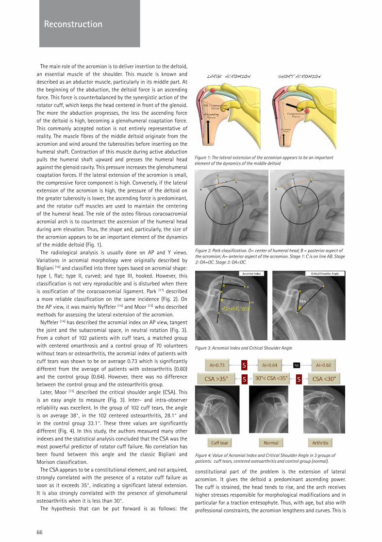
66
SPORTS KNEEReconstruction
The main role of the acromion is to deliver insertion to the deltoid, an essential muscle of the shoulder. This muscle is known and described as an abductor muscle, particularly in its middle part. At the beginning of the abduction, the deltoid force is an ascending force. This force is counterbalanced by the synergistic action of the rotator cuff, which keeps the head centered in front of the glenoid. The more the abduction progresses, the less the ascending force of the deltoid is high, becoming a glenohumeral coaptation force. This commonly accepted notion is not entirely representative of reality. The muscle fibres of the middle deltoid originate from the acromion and wind around the tuberosities before inserting on the humeral shaft. Contraction of this muscle during active abduction pulls the humeral shaft upward and presses the humeral head against the glenoid cavity. This pressure increases the glenohumeral coaptation forces. If the lateral extension of the acromion is small, the compressive force component is high. Conversely, if the lateral extension of the acromion is high, the pressure of the deltoid on the greater tuberosity is lower, the ascending force is predominant, and the rotator cuff muscles are used to maintain the centering of the humeral head. The role of the osteo fibrous coracoacromial acromial arch is to counteract the ascension of the humeral head during arm elevation. Thus, the shape and, particularly, the size of the acromion appears to be an important element of the dynamics of the middle deltoid (Fig. 1).
The radiological analysis is usually done on AP and Y views. Variations in acromial morphology were originally described by Bigliani [16] and classified into three types based on acromial shape: type I, flat; type II, curved; and type III, hooked. However, this classification is not very reproducible and is disturbed when there is ossification of the coracoacromial ligament. Park [17] described a more reliable classification on the same incidence (Fig. 2). On the AP view, it was mainly Nyffeler [14] and Moor [15] who described methods for assessing the lateral extension of the acromion.
Nyffeler [14] has described the acromial index on AP view, tangent the joint and the subacromial space, in neutral rotation (Fig. 3). From a cohort of 102 patients with cuff tears, a matched group with centered omarthrosis and a control group of 70 volunteers without tears or osteoarthritis, the acromial index of patients with cuff tears was shown to be on average 0.73 which is significantly different from the average of patients with osteoarthritis (0.60) and the control group (0.64). However, there was no difference between the control group and the osteoarthritis group.
Later, Moor [15] described the critical shoulder angle (CSA). This is an easy angle to measure (Fig. 3). Inter- and intra-observer reliability was excellent. In the group of 102 cuff tears, the angle is on average 38°, in the 102 centered osteoarthritis, 28.1° and in the control group 33.1°. These three values are significantly different (Fig. 4). In this study, the authors measured many other indexes and the statistical analysis concluded that the CSA was the most powerful predictor of rotator cuff failure. No correlation has been found between this angle and the classic Bigliani and Morison classification.
The CSA appears to be a constitutional element, and not acquired, strongly correlated with the presence of a rotator cuff failure as soon as it exceeds 35°, indicating a significant lateral extension. It is also strongly correlated with the presence of glenohumeral osteoarthritis when it is less than 30°.
The hypothesis that can be put forward is as follows: the
constitutional part of the problem is the extension of lateral acromion. It gives the deltoid a predominant ascending power. The cuff is strained, the head tends to rise, and the arch receives higher stresses responsible for morphological modifications and in particular for a traction entesophyte. Thus, with age, but also with professional constraints, the acromion lengthens and curves. This is
Figure 2: Park classification. O= center of humeral head; B = posterior aspect of the acromion; A= anterior aspect of the acromion. Stage 1: C is on line AB. Stage 2: OA=OC. Stage 3: OA<OC.
Figure 4: Value of Acromial Index and Critical Shoulder Angle in 3 groups of patients: cuff tears, centered osteoarthritis and control group (normal).
Figure 3: Acromial Index and Critical Shoulder Angle
Figure 1: The lateral extension of the acromion appears to be an important element of the dynamics of the middle deltoid
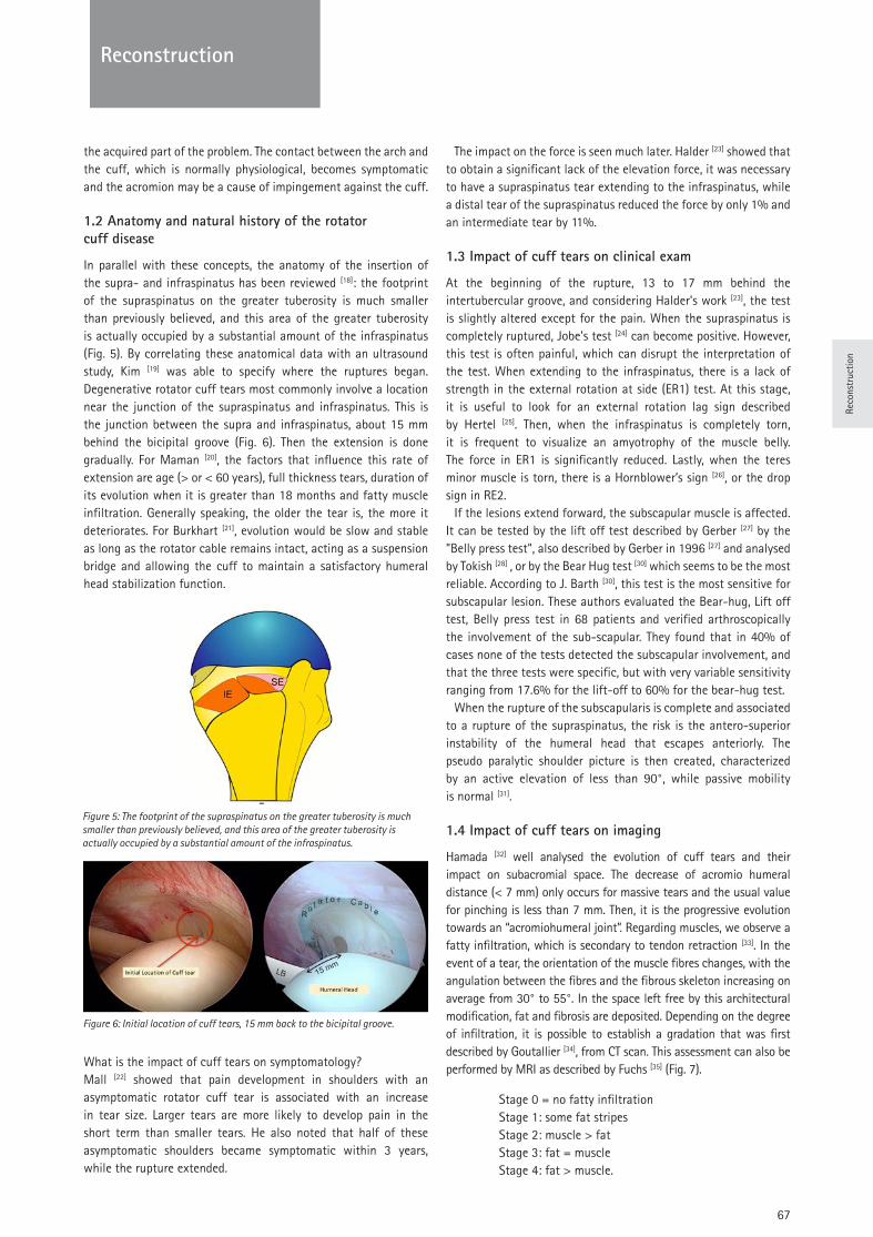
67
SPORTS KNEE
the acquired part of the problem. The contact between the arch and the cuff, which is normally physiological, becomes symptomatic and the acromion may be a cause of impingement against the cuff.
1.2 Anatomy and natural history of the rotator cuff disease
In parallel with these concepts, the anatomy of the insertion of the supra- and infraspinatus has been reviewed [18]: the footprint of the supraspinatus on the greater tuberosity is much smaller than previously believed, and this area of the greater tuberosity is actually occupied by a substantial amount of the infraspinatus (Fig. 5). By correlating these anatomical data with an ultrasound study, Kim [19] was able to specify where the ruptures began. Degenerative rotator cuff tears most commonly involve a location near the junction of the supraspinatus and infraspinatus. This is the junction between the supra and infraspinatus, about 15 mm behind the bicipital groove (Fig. 6). Then the extension is done gradually. For Maman [20], the factors that influence this rate of extension are age (> or < 60 years), full thickness tears, duration of its evolution when it is greater than 18 months and fatty muscle infiltration. Generally speaking, the older the tear is, the more it deteriorates. For Burkhart [21], evolution would be slow and stable as long as the rotator cable remains intact, acting as a suspension bridge and allowing the cuff to maintain a satisfactory humeral head stabilization function.
What is the impact of cuff tears on symptomatology? Mall [22] showed that pain development in shoulders with an asymptomatic rotator cuff tear is associated with an increase in tear size. Larger tears are more likely to develop pain in the short term than smaller tears. He also noted that half of these asymptomatic shoulders became symptomatic within 3 years, while the rupture extended.
The impact on the force is seen much later. Halder [23] showed that to obtain a significant lack of the elevation force, it was necessary to have a supraspinatus tear extending to the infraspinatus, while a distal tear of the supraspinatus reduced the force by only 1% and an intermediate tear by 11%.
1.3 Impact of cuff tears on clinical exam
At the beginning of the rupture, 13 to 17 mm behind the intertubercular groove, and considering Halder's work [23], the test is slightly altered except for the pain. When the supraspinatus is completely ruptured, Jobe's test [24] can become positive. However, this test is often painful, which can disrupt the interpretation of the test. When extending to the infraspinatus, there is a lack of strength in the external rotation at side (ER1) test. At this stage, it is useful to look for an external rotation lag sign described by Hertel [25]. Then, when the infraspinatus is completely torn, it is frequent to visualize an amyotrophy of the muscle belly. The force in ER1 is significantly reduced. Lastly, when the teres minor muscle is torn, there is a Hornblower’s sign [26], or the drop sign in RE2.
If the lesions extend forward, the subscapular muscle is affected. It can be tested by the lift off test described by Gerber [27] by the "Belly press test", also described by Gerber in 1996 [27] and analysed by Tokish [28] , or by the Bear Hug test [30] which seems to be the most reliable. According to J. Barth [30], this test is the most sensitive for subscapular lesion. These authors evaluated the Bear-hug, Lift off test, Belly press test in 68 patients and verified arthroscopically the involvement of the sub-scapular. They found that in 40% of cases none of the tests detected the subscapular involvement, and that the three tests were specific, but with very variable sensitivity ranging from 17.6% for the lift-off to 60% for the bear-hug test.
When the rupture of the subscapularis is complete and associated to a rupture of the supraspinatus, the risk is the antero-superior instability of the humeral head that escapes anteriorly. The pseudo paralytic shoulder picture is then created, characterized by an active elevation of less than 90°, while passive mobility is normal [31].
1.4 Impact of cuff tears on imaging
Hamada [32] well analysed the evolution of cuff tears and their impact on subacromial space. The decrease of acromio humeral distance (< 7 mm) only occurs for massive tears and the usual value for pinching is less than 7 mm. Then, it is the progressive evolution towards an “acromiohumeral joint”. Regarding muscles, we observe a fatty infiltration, which is secondary to tendon retraction [33]. In the event of a tear, the orientation of the muscle fibres changes, with the angulation between the fibres and the fibrous skeleton increasing on average from 30° to 55°. In the space left free by this architectural modification, fat and fibrosis are deposited. Depending on the degree of infiltration, it is possible to establish a gradation that was first described by Goutallier [34], from CT scan. This assessment can also be performed by MRI as described by Fuchs [35] (Fig. 7).
Stage 0 = no fatty infiltration Stage 1: some fat stripes Stage 2: muscle > fat Stage 3: fat = muscle Stage 4: fat > muscle.
Reco
nstr
uctio
n
Reconstruction
Figure 5: The footprint of the supraspinatus on the greater tuberosity is much smaller than previously believed, and this area of the greater tuberosity is actually occupied by a substantial amount of the infraspinatus.
Figure 6: Initial location of cuff tears, 15 mm back to the bicipital groove.
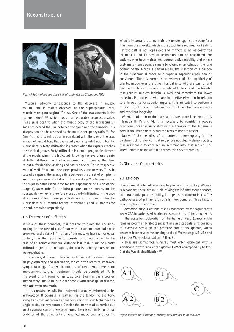
68
SPORTS KNEEReconstruction
Muscular atrophy corresponds to the decrease in muscle volume, and is mainly observed at the supraspinatus level, especially on para-sagittal Y view. One of the assessments is the "tangent sign" [36], which has an unfavourable prognostic value. This sign is positive when the muscle body of the supraspinatus does not exceed the line between the spine and the coracoid. This atrophy can also be assessed by the muscle occupancy ratio [37]. For Kim [38], this fatty infiltration is correlated with the size of the tear. In case of partial tear, there is usually no fatty infiltration. For the supraspinatus, fatty infiltration is greater when the rupture reaches the bicipital groove. Fatty infiltration is a major prognostic element of the repair, when it is indicated. Knowing the evolutionary rate of fatty infiltration and atrophy during cuff tears is therefore essential for decision-making and patient advice. The retrospective work of Melis [39] about 1688 cases provides some answers. Thus, in case of a rupture, the average time between the onset of symptoms and the appearance of a fatty infiltration stage 2 is 54 months for the supraspinatus (same time for the appearance of a sign of the tangent), 56 months for the infraspinatus and 36 months for the subscapular, which is therefore more quickly infiltrated. In the case of a traumatic tear, these periods decrease to 35 months for the supraspinatus, 31 months for the infraspinatus and 31 months for the sub-scapular, respectively.
1.5 Treatment of cuff tears
In view of these concepts, it is possible to guide the decision-making. In the case of a cuff tear with an acromiohumeral space preserved and a fatty infiltration of the muscles less than or equal to two, it is then possible to consider a surgical repair. In the case of an acromio humeral distance less than 7 mm or a fatty infiltration greater than stage 2, the tear is probably massive and non-repairable.
In any case, it is useful to start with medical treatment based on physiotherapy and infiltration, which often leads to improved symptomatology. If after six months of treatment, there is no improvement, surgical treatment should be considered [40]. In the event of a traumatic injury, surgical treatment is indicated immediately. The same is true for people with subscapular disease, who are often traumatic.
If it is a repairable cuff, the treatment is usually performed under arthroscopy. It consists in reattaching the tendon to the bone using trans osseous sutures or anchors, using various techniques as single or double row sutures. Despite the many studies carried out on the comparison of these techniques, there is currently no formal evidence of the superiority of one technique over another [41].
What is important is to maintain the tendon against the bone for a minimum of six weeks, which is the usual time required for healing.
If the cuff is not repairable and if there is no osteoarthritis (Hamada I and II), several techniques can be considered. For patients who have maintained correct active mobility and whose problem is mainly pain, a simple tenotomy or tenodesis of the long portion of the biceps, a partial repair, the insertion of a balloon in the subacromial space or a superior capsular repair can be considered. There is currently no evidence of the superiority of one technique over the other. For patients who are painful and have lost external rotation, it is advisable to consider a transfer that usually involves latissimus dorsi and sometimes the lower trapezius. For patients who have lost active elevation in relation to a large anterior superior rupture, it is indicated to perform a reverse prosthesis with satisfactory results on function recovery and excellent longevity.
When, in addition to the massive rupture, there is osteoarthritis (Hamada III, IV and V), it is necessary to consider a reverse prosthesis, possibly associated with a transfer of the latissimus dorsi if the infra spinatus and the teres minor are absent.
Lastly, if the benefits of an anterior acromioplasty in the treatment of rotator cuff pathology are not clearly demonstrated, it is reasonable to consider an acromioplasty that reduces the lateral margin of the acromion when the CSA exceeds 35°.
2. Shoulder Osteoarthritis
2.1 Etiology
Glenohumeral osteoarthritis may be primary or secondary. When it is secondary, there are multiple etiologies: inflammatory diseases, post-traumatic, post-instability, iatrogenic, osteonecrosis, etc. The pathogenesis of primary arthrosis is more complex. Three factors seem to play a major role:
- Acromion plays a definite role as evidenced by the significantly lower CSA in patients with primary osteoarthritis of the shoulder [15].
- The posterior subluxation of the humeral head (whose origin remains poorly understood) present in some patients is responsible for excessive stress on the posterior part of the glenoid, which becomes biconcave corresponding to the different stages, B1, B2 and B3 of the Walch classification [42] (Fig. 8).
- Dysplasia sometimes humeral, most often glenoidal, with a significant retroversion of the glenoid (>25°) corresponding to type C of the Walch classification [42].
Figure 7: Fatty infiltration stage 4 of infra spinatus on CT scan and MRI.
Figure 8: Walch classification of primary osteoarthritis of the shoulder
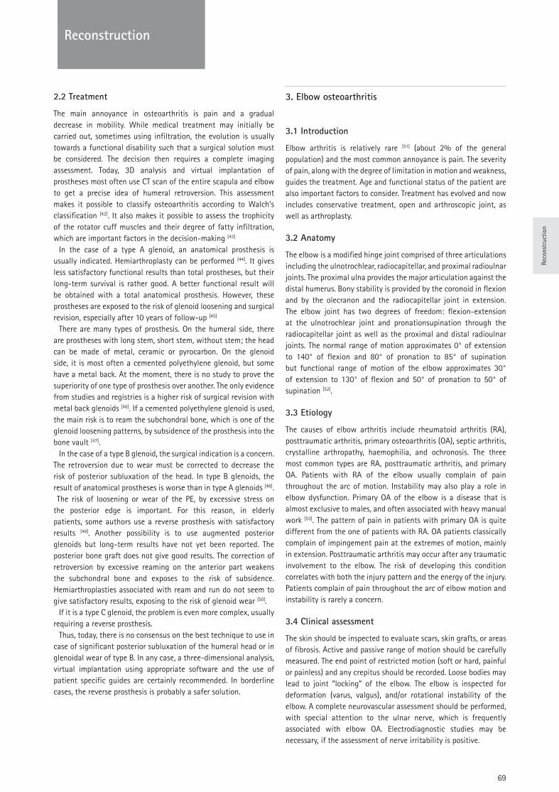
69
INFECTION
Reco
nstr
uctio
n
Reconstruction
2.2 Treatment
The main annoyance in osteoarthritis is pain and a gradual decrease in mobility. While medical treatment may initially be carried out, sometimes using infiltration, the evolution is usually towards a functional disability such that a surgical solution must be considered. The decision then requires a complete imaging assessment. Today, 3D analysis and virtual implantation of prostheses most often use CT scan of the entire scapula and elbow to get a precise idea of humeral retroversion. This assessment makes it possible to classify osteoarthritis according to Walch's classification [42]. It also makes it possible to assess the trophicity of the rotator cuff muscles and their degree of fatty infiltration, which are important factors in the decision-making [43]
In the case of a type A glenoid, an anatomical prosthesis is usually indicated. Hemiarthroplasty can be performed [44]. It gives less satisfactory functional results than total prostheses, but their long-term survival is rather good. A better functional result will be obtained with a total anatomical prosthesis. However, these prostheses are exposed to the risk of glenoid loosening and surgical revision, especially after 10 years of follow-up [45]
There are many types of prosthesis. On the humeral side, there are prostheses with long stem, short stem, without stem; the head can be made of metal, ceramic or pyrocarbon. On the glenoid side, it is most often a cemented polyethylene glenoid, but some have a metal back. At the moment, there is no study to prove the superiority of one type of prosthesis over another. The only evidence from studies and registries is a higher risk of surgical revision with metal back glenoids [46]. If a cemented polyethylene glenoid is used, the main risk is to ream the subchondral bone, which is one of the glenoid loosening patterns, by subsidence of the prosthesis into the bone vault [47].
In the case of a type B glenoid, the surgical indication is a concern. The retroversion due to wear must be corrected to decrease the risk of posterior subluxation of the head. In type B glenoids, the result of anatomical prostheses is worse than in type A glenoids [48]. The risk of loosening or wear of the PE, by excessive stress on the posterior edge is important. For this reason, in elderly patients, some authors use a reverse prosthesis with satisfactory results [49]. Another possibility is to use augmented posterior glenoids but long-term results have not yet been reported. The posterior bone graft does not give good results. The correction of retroversion by excessive reaming on the anterior part weakens the subchondral bone and exposes to the risk of subsidence. Hemiarthroplasties associated with ream and run do not seem to give satisfactory results, exposing to the risk of glenoid wear [50].
If it is a type C glenoid, the problem is even more complex, usually requiring a reverse prosthesis.
Thus, today, there is no consensus on the best technique to use in case of significant posterior subluxation of the humeral head or in glenoidal wear of type B. In any case, a three-dimensional analysis, virtual implantation using appropriate software and the use of patient specific guides are certainly recommended. In borderline cases, the reverse prosthesis is probably a safer solution.
3. Elbow osteoarthritis
3.1 Introduction
Elbow arthritis is relatively rare [51] (about 2% of the general population) and the most common annoyance is pain. The severity of pain, along with the degree of limitation in motion and weakness, guides the treatment. Age and functional status of the patient are also important factors to consider. Treatment has evolved and now includes conservative treatment, open and arthroscopic joint, as well as arthroplasty.
3.2 Anatomy
The elbow is a modified hinge joint comprised of three articulations including the ulnotrochlear, radiocapitellar, and proximal radioulnar joints. The proximal ulna provides the major articulation against the distal humerus. Bony stability is provided by the coronoid in flexion and by the olecranon and the radiocapitellar joint in extension. The elbow joint has two degrees of freedom: flexion-extension at the ulnotrochlear joint and pronationsupination through the radiocapitellar joint as well as the proximal and distal radioulnar joints. The normal range of motion approximates 0° of extension to 140° of flexion and 80° of pronation to 85° of supination but functional range of motion of the elbow approximates 30° of extension to 130° of flexion and 50° of pronation to 50° of supination [52].
3.3 Etiology
The causes of elbow arthritis include rheumatoid arthritis (RA), posttraumatic arthritis, primary osteoarthritis (OA), septic arthritis, crystalline arthropathy, haemophilia, and ochronosis. The three most common types are RA, posttraumatic arthritis, and primary OA. Patients with RA of the elbow usually complain of pain throughout the arc of motion. Instability may also play a role in elbow dysfunction. Primary OA of the elbow is a disease that is almost exclusive to males, and often associated with heavy manual work [53]. The pattern of pain in patients with primary OA is quite different from the one of patients with RA. OA patients classically complain of impingement pain at the extremes of motion, mainly in extension. Posttraumatic arthritis may occur after any traumatic involvement to the elbow. The risk of developing this condition correlates with both the injury pattern and the energy of the injury. Patients complain of pain throughout the arc of elbow motion and instability is rarely a concern.
3.4 Clinical assessment
The skin should be inspected to evaluate scars, skin grafts, or areas of fibrosis. Active and passive range of motion should be carefully measured. The end point of restricted motion (soft or hard, painful or painless) and any crepitus should be recorded. Loose bodies may lead to joint “locking” of the elbow. The elbow is inspected for deformation (varus, valgus), and/or rotational instability of the elbow. A complete neurovascular assessment should be performed, with special attention to the ulnar nerve, which is frequently associated with elbow OA. Electrodiagnostic studies may be necessary, if the assessment of nerve irritability is positive.
70
INFECTION Reconstruction
3.5 Radiographic Evaluation
Anteroposterior, lateral with the elbow flexed at 90° are obtained as a routine and provide adequate information but computed tomography with two- and three-dimensional reconstructions is helpful for surgical planning of joint debridement. Radiographic findings typical for RA include symmetric joint space narrowing, periarticular erosions, and osteopenia. In contrast, some preservation of the ulnohumeral and radiocapitellar joint spaces is common in elbows with primary osteoarthritis. Radiographs reveal osteophyte and loose body formation. Rettig [54] developed a new radiographic classification system with three classes or levels of severity, to predict outcomes after debridement of elbow osteoarthritis.
3.6 Treatment
• Non-operative Treatments
If not medically contraindicated, patients should be prescribed analgesic or anti-inflammatory drugs (NSAIDs). Intra-articular steroid or hyaluronic injections acid can be very effective [55]. Physical therapy is important for the maintenance of elbow range of motion. Patients are advised to use some combination of modalities as activity modification, hinge braces, night splint, etc.
• Operative treatments
Arthrodesis
Nowadays elbow arthrodesis is rarely indicated and most often performed as a salvage operation.
Synovectomy
Open or arthroscopic synovectomy, with or without radial head excision, is an established treatment option for patients with rheumatoid arthritis. The primary goal of synovectomy is to provide pain relief and prevent destruction of the elbow joint [56]. The advantages of arthroscopic synovectomy include improved enhanced visualization, decreased morbidity, and shorter rehabilitation [57]. Conjunction with current biologic disease-modifying antirheumatic drugs is essential to maintain outcomes in patients after arthroscopic synovectomy.
Debridement
This surgery is referred to as the Outerbridge- Kashiwagi (OK) procedure [58]. This surgery is indicated in those patients with primary OA who have continuous pain at the extremes of motion and radiographic evidence of impingement. Arthroscopic debridement and loose body excision is limited to patients with degenerative arthritis and clinical symptoms of impingement pain, limited motion, and intermittent mechanical locking.
Interposition arthroplasty
Arthroplasty by interposition of an Achilles tendon allograft [59]
may be indicated in young, active patients where total elbow prosthesis is contra-indicated, with severe inflammatory arthritis or posttraumatic arthritis, especially those with limited elbow motion. However, elbow instability is the major complication.
Total elbow arthroplasty (TEA)
TEA is indicated for patients older than 65 years of age who have considerable pain, limited motion, and functional deficit and for whom non-operative interventions have failed. There are different prosthesis designs that are generally grouped as unconstrained, semi-constrained, and constrained. Constrained designs lead to a loosening at the cement-bone interface [60]. Non-constrained designs lead to a significant risk of instability. In fact, elbow instability is now considered to be a contraindication for unconstrained TEA. A semi-constrained design attempts to create a solution between constrained and unconstrained TEA. Global survival rate in TEA is between 80% to 90% at ten years with aseptic loosening and infection being the main complications requiring revision [61].
References:
1. Milgrom C, Schaffler M, Gilbert S, van Holsbeeck M. Rotator-cuff changes in asymptomatic adults. The effect of age, hand dominance and gender. J Bone Joint Surg. Br 1995; 77: 296-298.
2. Sher JS, Uribe JW, Posada A, Murphy BJ, Zlatkin MB. Abnormal findings on magnetic resonance images of asymptomatic shoulders. J Bone Joint Surg. Am 1995; 77: 10-15.
3. Moosmayer S, Smith HJ, Tariq R, Larmo A. Prevalence and characteristics of asymptomatic tears of the rotator cuff: an ultrasonographic and clinical study. J Bone Joint Surg. Br 2009; 91: 196-200.
4. Yamamoto A, Takagishi K, Osawa T, et al: Prevalence and risk factors of a rotator cuff tear in the general population. J Shoulder Elbow Surg 2010; 19: 116-120,
5. Neer CS: 2nd Impingement lesions. Clin Orthop Relat Res 173:70-77, 1983
6. Bigliani LU, Ticker JB, Flatow EL, Soslowsky LJ, Mow VC. The relationship of acromial architecture to rotator cuff disease. Clin Sports Med. 1991; 10:823-38.
7. Lohr JF, Uhthoff HK: The microvascular pattern of the supraspinatus tendon. Clin Orthop Relat Res 1990; 254: 35-38.
8. Tashjian, R.Z., Epidemiology, natural history, and indications for treatment of rotator cuff tears. Clin Sports Med, 2012; 31: 589-604.
9. Bigliani LU, Levine WN: Subacromial impingement syndrome. J Bone Joint Surg Am 1997; 12: 1854-1868.
10. Vitale MA, Arons RR, Hurwitz S, Ahmad CS, Levine WN. The rising incidence of acromioplasty. J Bone Joint Surg. Am 2010; 9:1842–1850.
11. Henkus HE, de Witte PB, Nelissen RG, Brand R, van Arkel ER: Bursectomy compared with acromioplasty in the management of subacromial impingement syndrome: A prospective randomised study. J Bone Joint Surg Br 2009; 4: 504-510.
12. Gartsman GM, O’Connor DP: Arthroscopic rotator cuff repair with and without arthroscopic subacromial decompression: A prospective, randomized study of one-year outcomes. J Shoulder Elbow Surg 2004; 4: 424-426.
13. Shin SJ, Oh JH, Chung SW, Song MH. The efficacy of acromioplasty in the arthroscopic repair of small- to medium-sized rotator cuff tears without acromial spur: prospective comparative study. Arthroscopy. 2012; 5: 628-35.
71
INFECTION
Reco
nstr
uctio
n
Reconstruction
14. Nyffeler RW, Werner CM, Sukthankar A, Schmid MR, Gerber C. Association of a large lateral extension of the acromion with rotator cuff tears. J Bone Joint Surg Am 2006; 88: 800-5.
15. Moor BK, Bouaicha S, Rothenfluh DA, Sukthankar A, Gerber C. Is there an association between the individual anatomy of the scapula and the development of rotator cuff tears or osteoarthritis of the glenohumeral joint? A radiological study of the critical shoulder angle. Bone Joint J 2013; 95:9 35-41.
16. Bigliani BU, Morrison ES, April EW: The morphology of the acromion and its relationship to rotator cuff tears. Orthop Trans 1986; 10: 216.
17. Park TS, Park DW, Kim SI, Kweon TH. Roentgenographic assessment of acromial morphology using supraspinatus outlet radiographs. Arthroscopy. 2001; 5: 496-501.
18. Mochizuki T, Sugaya H, Uomizu M, Maeda K, Matsuki K, Sekiya I, Muneta T, Akita, K. Humeral insertion of the supraspinatus and infraspinatus. New anatomical findings regarding the footprint of the rotator cuff. J Bone Joint Surg Am. 2008; 5:962-9.
19. Kim HM, Dahiya N, Teefey SA, Middleton WD, Stobbs G, Steger-May K, Yamaguchi K, Keener JD. Location and initiation of degenerative rotator cuff tears: an analysis of three hundred and sixty shoulders. J Bone Joint Surg. Am 2010; 92: 1088-1096.
20. Maman E, Harris C, White L, Tomlinson G, Shashank M, Boynton E. Outcome of nonoperative treatment of symptomatic rotator cuff tears monitored by magnetic resonance imaging. J Bone Joint Surg. Am 2009; 91: 1898-1906.
21. Burkhart SS, Esch JC, Jolson RS. The rotator crescent and rotator cable: an anatomic description of the shoulder's "suspension bridge". Arthroscopy 1993; 9: 611-616.
22. Mall NA, Kim HM, Keener JD, Steger-May K, Teefey SA, Middleton WD, Stobbs G, Yamaguchi K. Symptomatic progression of asymptomatic rotator cuff tears: a prospective study of clinical and sonographic variables. J Bone Joint Surg. Am 2010; 92: 2623-2633.
23. Halder AM, O'Driscoll SW, Heers G, Mura N, Zobitz ME, An KN, Kreusch-Brinker R. Biomechanical comparison of effects of supraspinatus tendon detachments, tendon defects, and muscle retractions. J Bone Joint Surg. Am 2002; 84: 780-785.
24. Jobe FW, Jobe CM. Painful athletic injuries of the shoulder. Clin Orthop. 1983;117-24.
25. Hertel R, Ballmer FT, Lombert SM, Gerber C. Lag signs in the diagnosis of rotator cuff rupture. J Shoulder Elbow Surg. 1996; 5: 307-13.
26. Walch G, Boulahia A, Calderone S, Robinson AH. The 'dropping' and 'hornblower's' signs in evaluation of rotator-cuff tears. J Bone Joint Surg Br. 1998; 80: 624-8.
27. Gerber C, Krushell RJ. Isolated rupture of the tendon of the subscapularis muscle. Clinical features in 16 cases. J Bone Joint Surg Br. 1991; 73: 389-94.
28. Gerber C, Hersche O, Farron A. Isolated rupture of the subscapularis tendon. J Bone Joint Surg Am. 1996; 78: 1015-23.
29. Tokish JM, Decker MJ, Ellis HB, Torry MR, Hawkins RJ. The belly-press test for the physical examination of the subscapularis muscle: electromyographic validation and comparison to the lift-off test. J Shoulder Elbow Surg. 2003; 12: 427-30.
30. Barth JR, Burkhart SS, De Beer JF. The bear-hug test: a new and sensitive test for diagnosing a subscapularis tear. Arthroscopy. 2006; 22: 1076-84.
31. Collin P, Matsumura N, Lädermann A, Denard PJ, Walch G. Relationship between massive chronic rotator cuff tear pattern and loss of active shoulder range of motion. J Shoulder Elbow Surg. 2014; 8: 1195-202.
32. Hamada K, Fukuda H, Mikasa M, Kobayashi Y. Roentgenographic findings in massive rotator cuff tears. A long-term observation. Clin Orthop. 1990; 92-6.
33. Gerber C, Meyer DC, Frey E, von Rechenberg B, Hoppeler H, Frigg R, et al. Neer Award 2007: Reversion of structural muscle changes caused by chronic rotator cuff tears using continuous musculotendinous traction. An experimental study in sheep. J Shoulder Elbow Surg. 2009; 18: 163-71.
34. Goutallier D, Postel JM, Bernageau J, Lavau L, Voisin MC. Fatty muscle degeneration in cuff ruptures. Pre- and postoperative evaluation by CT scan. Clin Orthop. 1994; 304: 78-83.
35. Fuchs B, Weishaupt D, Zanetti M, Hodler J, Gerber C. Fatty degeneration of the muscles of the rotator cuff: assessment by computed tomography versus magnetic resonance imaging. J Shoulder Elbow Surg. 1999; 8: 599-605.
36. Zanetti M, Gerber C, Hodler J. Quantitative assessment of the muscles of the rotator cuff with magnetic resonance imaging. Investigative radiology. 1998; 33: 163-70.
37. Thomazeau H, Rolland Y, Lucas C, Duval JM, Langlais F. Atrophy of the supraspinatus belly. Assessment by MRI in 55 patients with rotator cuff pathology. Acta Orthop Scan. 1996; 67: 264-8.
38. Kim HM, Dahiya N, Teefey SA, Keener JD, Galatz LM, Yamaguchi K. Relationship of tear size and location to fatty degeneration of the rotator cuff. J Bone Joint Surg Am. 2010; 92: 829-39.
39. Melis B, DeFranco MJ, Chuinard C, Walch G. Natural history of fatty infiltration and atrophy of the supraspinatus muscle in rotator cuff tears. Clin Orthop. 2010; 468: 1498-505.
40. Agout C, Berhouet J, Spiry C, Bonnevialle N, Joudet T, Favard L; French Arthroscopic Society. Functional outcomes after non-operative treatment of irreparable massive rotator cuff tears: Prospective multicenter study in 68 patients. Orthop Traumatol Surg Res. 2018; 104: 189-192.
41. McElvany, M.D., et al., Rotator cuff repair: published evidence on factors associated with repair integrity and clinical outcome. Am J Sports Med, 2015; 2: 491-500.
42. Walch, G., et al., Morphologic study of the glenoid in primary glenohumeral osteoarthritis. J Arthroplasty, 1999; 6: 756-60.
43. Patrick J. Denard, MD, Gilles Walch, MD. Current concepts in the surgical management of primary glenohumeral arthritis with a biconcave glenoid. J Shoulder Elbow Surg. 2013; 22: 1589-1598.
44. Levine WN, Fischer CR, Nguyen D, Flatow EL, Ahmad CS, Bigliani LU: Long-term follow-up of shoulder hemiarthroplasty for glenohumeral osteoarthritis. J Bone Joint Surg 2012; 22: e164.
45. Young A, Walch G, Boileau P, Favard L, Gohlke F, Loew M, et al. A multicenter study of the long-term results of using a flat-back polyethylene glenoid component in shoulder replacement for primary osteoarthritis. J Bone Joint Surg Br 2011; 93: 210-6.
46. Page RS, Pai V, Eng K, Bain G, Graves S, Lorimer M. Cementless versus cemented glenoid components in conventional total shoulder joint arthroplasty: analysis from the Australian Orthopaedic Association National Joint Replacement Registry. J Shoulder Elbow Surg. 2018; 10:1859-1865.
72
Reconstruction
47. Walch G, Young AA, Boileau P, Loew M, Gazielly D, Molé D. Patterns of loosening of polyethylene keeled glenoid components after shoulder arthroplasty for primary osteoarthritis: results of a multicenter study with more than five years of follow-up. J Bone Joint Surg Am. 2012; 2:145-50.
48. Walch G, Moraga C, Young A, Castellanos-Rosas J. Results of anatomic nonconstrained prosthesis in primary osteoarthritis with biconcave glenoid. J Shoulder Elbow Surg 2012; 21: 1526-33.
49. Mizuno N, Raiss P, Denard PJ, Walch G. Reverse total shoulder arthroplasty for primary glenohumeral osteoarthritis with a biconcave glenoid. J Bone Joint Surg Am 2013; 14: 1297-304.
50. Gilmer BB, Comstock BA, Jette JL, Warme WJ, Jackins SE, Matsen FA. The prognosis for improvement in comfort and function after the ream-and-run arthroplasty for glenohumeral arthritis: an analysis of 176 consecutive cases. J Bone Joint Surg 2012; 18 :e102.
51. Ortner, D.J., Description and classification of degenerative bone changes in the distal joint surfaces of the humerus. Am J Phys Anthropol, 1968; 2: 139-55.
52. Morrey BF, Askew LJ, An KN, and Chao EY. A biomechanical study of normal functional elbow motion. J Bone Joint Surg. 1981; 6: 872-7.
53. Stanley, D., Prevalence and etiology of symptomatic elbow osteoarthritis. J Shoulder Elbow Surg. 1994; 6: 386-9.
54. Rettig LA, Hastings H II, Feinberg JR. Primary osteoarthritis of the elbow: lack of radiographic evidence for morphologic predisposition, results of operative debridement at intermediate follow-up, and basis for a new radiographic classification system. J Shoulder Elbow Surg 2008; 17: 97–105.
55. Van Brakel, R.W. and D. Eygendaal, Intra-articular injection of hyaluronic acid is not effective for the treatment of posttraumatic osteoarthritis of the elbow. Arthroscopy, 2006; 11: 1199-1203.
56. Fuerst M, Fink B, Rüther W. Survival analysis and long-term results of elbow synovectomy in rheumatoid arthritis. J Rheumatol 2006; 33: 892-896.
57. Tanaka N, Sakahashi H, Hirose K, Ishima T, Ishii S. Arthroscopic and open synovectomy of the elbow in rheumatoid arthritis. J Bone Joint Surg 2006; 88: 521–525.
58. Kashiwagi D. Intra-articular changes of the osteoarthritic elbow, especially about the fossa olecrani. Japan Orthop Assoc. 1978; 52:1367-72.
59. Larson AN, Morrey BF. Interposition arthroplasty with an Achilles tendon allograft as a salvage procedure for the elbow. J Bone Joint Surg 2008; 90: 2714–2723.
60. Morrey BF, Bryan RS. Complications of total elbow arthroplasty. Clin Orthop Rel Res. 1982;170: 204-12.
61. Fevang, B.T., et al., Results after 562 total elbow replacements: a report from the Norwegian Arthroplasty Register. J Shoulder Elbow Surg, 2009. 3: 449-56.
Questions
1. Which of the following is a risk factor for cuff tear?a. CSA <30°b. CSA > 35°c. Retroversion of the glenoidd. Short lateral extension of the acromione. Acromion bi partita
2 What is the main reason for a pseudo paralytic shoulder?a. Infra spinatus tearb. Infra spinatus and teres minor tearc. Supra, infra spinatus and teres minor teard. Sub scapularis teare. Subscapularis and supra spinatus tear
3. What is the best treatment for a woman 80 y.o., with a primary osteoarthritis and a glenoid B2?a. Hemiarthroplastyb. Anatomical shoulder arthroplasty with bone graftc. Reverse shoulder arthroplastyd. Hemiarthroplasty with ream and run of the glenoide. Gleno humeral arthrodesis
4. Which of following is not possible to do under arthroscopy for elbow arthritisa. Loose bodies removalb. Capsular releasec. Osteophytes resectiond. Interposition arthroplastye. Synovectomy
Answers1b, 2e, 3c, 4d.
Spor
t In
jurie
s
73
Sport Injuries
Dr. Simone Cerciello Casa Di Cura Villa Betania - Rome, ItalyMarrelli Hospital – Crotone, [email protected]
Prof. Philippe NeyretBurjeel Orthopaedics and Sports Medicine Center - Abu Dhabi, [email protected]
Knee Ligaments & Meniscii
Abstract
Mensical and ligament injuries are commonly encountered in orthopaedic practice. Since they may result in functional impairment with mid and long-term consequences, the pathologic anatomy should be carefully evaluated. Medial and lateral meniscii are mainly load transducers. Tears can be either traumatic or degenerative. Since they have reduced healing potential, torn parts meniscii are usually removed trying to preserve the healthy portions in order to avoid late cartilage degeneration. Anterior cruciate ligament (ACL) injuries commonly follow sport traumas. ACL controls anterior tibial translation and pivot. When it is insufficient, knee stability is severely affected and it should be therefore reconstructed. Great care should be taken to reproduce original ligament insertions in order to re-establish its normal biomechanics and ensure knee stability. Posterior cruciate ligament (PCL) is thicker and stronger than ACL. As a consequence, it is usually injured in high-energy traumas such as motor vehicle accidents. In case of minor anatomic damage, residual instability is generally limited and conservative treatment with immobilization and further rehabilitation is still indicated. When major instability is present, PCL reconstruction should be performed as well as the reconstruction of any peripheral ligament damage. Medial collateral ligament (MCL) is the major restraint to valgus stress. It is usually affected in the setting of multi-ligament injuries. Isolated acute lesions are generally treated with immobilization and further rehabilitation. Isolated chronic tears or MCL lesions associated with ACL or PCL ruptures require surgical reinsertion or reconstruction.
1 Meniscii
1.1 Introduction
Meniscal tears are the most common pathology of the knee with a mean annual incidence of 66 per 100000 in Denmark [Hede 1990]. Similarly, around 1 million knee arthroscopies are performed for meniscal problems in the United States of America (USA) annually [Cook 2005], and arthroscopic meniscal debridement is the most common procedure logged during part II board certification [Garrett 2006].
1.2 Anatomy and function
The meniscii are two wedge-shaped semilunar sections of fibrocartilaginous tissue. They are mainly formed of water and type I collagen fibres [Herwig 1984]. The medial meniscus is “C” shaped
and covers around 60% of the medial compartment whereas the lateral meniscus is more “O” covering 80% of the lateral compartment [Kohn 1995]. Both have poor blood supply, which is responsible for the reduced healing potential before and after repair surgery. Supply comes from the periphery via the medial and lateral geniculate arteries, however only the peripheral 10%-25% of the meniscus benefits from a blood supply [Arnoczky 1982]. On the contrary, nerve supply is rich in mechanoreceptors and free nerve endings. The concentration is higher in the peripheral two-thirds, with the peak in the posterior horns of both meniscii [Assimakoupolos 1992]. This suggests that the menisci may have a role in proprioception, stability and protective reflexes. The meniscii play an important role in knee function and biomechanics as load transducers (50% of the body weight in extension and up to 85-90% in flexion [Ahmed 1983]), and contribute to stability and joint lubrication as well. The role in joint stability becomes critical in anterior cruciate ligament (ACL) deficient knees.
1.3 Classification of meniscus injuries
Meniscii lesions can be divided into traumatic/acute and degenerative/chronic. Degenerative and chronic tears have been defined as slowly developing lesions, typically involving a horizontal cleavage of the posterior horn of the medial meniscus in a middle-aged or older person. They are usually asymptomatic and their prevalence increases with age, ranging from 16% in the knees of women aged 50 to 59 years to over 50% in the knees of men aged 70 to 90 years [Englund 2008].
Since they have low/no healing potential, they should be treated non-surgically if possible or with meniscectomy if required. However, since meniscectomy has important consequences on loads distribution it should be carefully evaluated. Total medial meniscectomy decreases tibio-femoral contact area by 75% and increases peak contact pressure by approximately 235% [Baratz 1986].
Acute/traumatic tears are often classified according to their orientation and stability/instability. They can be vertical longitudinal, vertical radial, horizontal, oblique or complex [Binfield 1993]. Longitudinal tears are more common medially, whereas radial tears are more frequently seen laterally [Klimckiewicz 2002]. Traumatic tears can also be defined as ‘stable’ or ‘unstable’ according to their mobility. In stable knees (intact ACL), the rate of post-traumatic meniscal tears is 6% [Maffulli 1993]; the rate is much higher in ACL deficient knees [Gadeyne 2006] and continues to increase along with time.
74
Sport Injuries
1.4 Treatment options
The contribution of the meniscii in joint stability and biomechanics is well established and influences treatment options.
a) Conservative treatment
According to the results of recent systematic reviews and meta-analyses, conservative treatment should be the first option in chronic/degenerative lesions [Elattrache 2014, Thorlund 2015, Bollen 2015]. It consists of rehabilitation and exercise programmes to improve muscle strength, flexibility and proprioception. Yim et al. compared the outcomes of meniscectomy and non-operative strengthening exercises in patients with degenerative horizontal tears of the posterior horn of the medial meniscus. Satisfactory clinical results were found in each group at 2 years FU with no significant difference in terms of pain, function and patient satisfaction [Yim 2013]. Similar outcomes were reported in a recent multicentre randomised controlled study on 351 patients over 45 years of age with a meniscal tear and evidence of osteoarthritis [Katz 2013]. At 12 months FU, no significant differences were found in terms of improvement in functional status and pain between the partial meniscectomy and physical therapy alone. However, there was crossover from the physical therapy group to the surgery group in 35% of patients. Functional outcomes in this cohort of patients at 12 months FU were similar to those patients who had surgery initially. This data confirms, that non-operative treatment is a reasonable first line approach in middle-aged patients with degenerative meniscal tears. Conservative treatment is a reasonable option even in case of traumatic tears; in stable knees, subjective results are good at more than 10 year FU and 85% of patients consider their knee as normal or nearly normal [Chatain 2003]. On the contrary, in ACL-deficient knees, isolated meniscectomy has a dramatic negative impact on knee function with almost 100% osteoarthritis rate at more than 30 years [Neyret 1993]. Clinical outcomes are usually better on the medial side, while they quickly deteriorate on the lateral compartment [Chatain 2003]. The difference is confirmed by the re-arthroscopy rate, which is 14% after lateral meniscectomy and 6% after medial meniscectomy and by the prevalence of joint-line narrowing which is 22% on the medial side and 38% on the lateral side, at more than ten-years FU [Chatain 2003]. The rate of arthritis at 20 years FU is 56% in a specific population after lateral meniscectomies [Hulet 2015]. This is due to the convexity of lateral tibial plateau that mirrors the convexity of the distal femoral condyle. In the absence of a meniscus there is greater tendency to point loading.
b) Partial meniscectomy
Partial meniscectomy is indicated in case of acute tears in the avascular zone of both meniscii with a radial, oblique and flap configuration. In addition, degenerative lesions non-responding to conservative treatment can be addressed with arthroscopic selective meniscectomy. The aim of the procedure is to debride the loose nonviable edges of meniscii in order to keep a stable meniscus. The outcomes are generally very positive in terms of functional recovery and pain relief. The outcomes of partial meniscectomy are superior to those of complete resection as reported by Northmore-Ball et al. [Northmore-Ball 1983]. In a cohort of 219 knees at 4.3 years FU they reported 90% of patients having either good or excellent satisfaction following arthroscopic
partial meniscectomy compared to only 68% in the cohort of open total meniscectomy. Similar data have been reported by Burks et al. who had good or excellent results in 88% of patients [Burks 1997] and by Jaureguito et al. who reported 90% of good or excellent results and 85% of patients resuming pre-injury level of activities at 2 years FU [Jaureguito 1995]. However it is now clear that partial meniscectomy may delay degeneration but not prevent it, and functional outcomes usually decrease along with time. In a study involving 147 athletes, they were reviewed at 4.5 years and then at 14 years [Faunø 1992]. Almost half of patients were asymptomatic at first FU, while only one third was still asymptomatic at last control and 46% had given up or reduced their sporting activity. Conversely the incidence of radiographic changes rose from 40% to 89%. Similar data were reported in a series of 136 patients treated with partial meniscectomy [Jørgensen 1987]. At 8.5 years follow-up there was a re-operation rate of 22.8% and 53% of patients had osteoarthritic radiographic changes compared to only 22% in the unaffected control knee. Radiographic degeneration is usually more common after lateral meniscectomy than medial and the outcomes are generally inferior when some extent of articular cartilage damage pre-exists in surgery.
c) Meniscal repair
Traumatic and acute tears have a great healing potential: they are suitable to repair if the geometry of the lesion allows it. Blood supply is crucial to ensure tissue healing and only tears in the red-red or at least in the red-white zone (up to approximately 5 mm from periphery) have the potential to heal. Therefore, healing stimulating adjuvants such has exogenous fibrin clots have been used to enhance the reparative response in the avascular/partially vascular zones [VanTrommel 1998]. Traditionally, the best indication for meniscal repair is longitudinal tear in a stable knee. ACL deficiency greatly affects meniscal healing potential. Positive results have also been reported in case of radial tears [Boyd 2003]. These tears cause disruption of all the longitudinal fibres and complete loss of meniscus function, if they extend to the peripheral zone. Therefore, radial tears of the red and red-white zones should be repaired in order to restore integrity of the rim [Ra 2013]. Meniscal repair can be performed open or under arthroscopy. Arthroscopic inside-out and outside-in techniques are useful for anterior and middle third tears, which are not easily accessed by an all-inside technique. More recently all-inside arthroscopic meniscal repair techniques have been proposed in lesions of the posterior aspect of the meniscus to avoid the need for accessory incisions. The outcomes of both treatment options are encouraging, however the complication rate is not negligible. In a recent systematic review by Nepple et al, on 13 studies with a minimum of five-year FU, a pooled rate of failure from 20.2% to 24.3% was reported [Nepple 2012]. More nerve symptoms are generally associated with the inside-out repair, while more implant-related complications are associated with the all-inside techniques.
Spor
t In
jurie
s
75
Sport Injuries
2 Anterior Cruciate Ligament
2.1 Introduction
Anterior cruciate ligament (ACL) injuries are common in contact sports (such as soccer, rugby and basketball) and in activities requiring pivoting movements (such as skiing). Reported incidence is more than 250.000 cases per year, representing the most common ligament injury [Frobell 2010]. ACL insufficiency has a dramatic impact on knee function, leading to additional anatomic damages such as meniscal tears, chondral lesions and an increased risk of early onset post-traumatic osteoarthritis (OA) at long-term FU [Levine 2013]. Since the ACL has poor healing capacity, surgery is often indicated.
2.2 Anatomy and function
The ACL is an intra-articular, yet extra-synovial structure formed by two bundles: the antero-medial and the postero-lateral. The AM bundle is anterior and medial to the PL bundle. Both the AM bundle and the PL bundle likely have independent roles in providing stability to the knee. The whole ACL is the major restraint to anterior tibial translation (accounting for about 86% of the total resistance) and to tibial internal and external rotation relative to the femur [Butler 1980].
More than 70% of ACL injuries are the result of non-contact sports (without a direct trauma on the knee joint) involving combined valgus and varus movements associated with tibial internal and external rotation [Quatman 2014]. In addition it has been showed that neuromuscular control deficit has a crucial influence on ACL injury risk [Hewett 2013].
2.3 Treatment options
Conservative treatment is unsatisfactory with residual instability and pain. Similarly, primary repair of ACL is ineffective with persistent symptoms in more than 90% of patients at 5 years FU [Feagin 1976]. When the outcomes of primary repair were compared to those of conservative treatment, they were similar with high rates (40% to 100%) of ACL healing failure [Sandeberg 1987]. According to these evidences surgical reconstruction has emerged as the golden standard in ACL deficient knees. ACL reconstruction is a satisfactory procedure, however, persistent instability has been reported in 10%-30% of patients with a return-to-sport rate of only 60% to 70% after single bundle reconstruction [Prodromos 2008].
a) Femoral tunnel placement
The femoral tunnel placement is crucial to restore normal joint kinematics [Abebe 2011]. The initial trans-tibial technique for femoral tunnel placement was a reproducible option but had the drawback of more vertical tunnel positioning and, tibial and femoral tunnels were dependent each other. Antero-medial technique is more anatomic and has the advantage of independent tunnels, however it is more technically demanding, especially in terms of femoral visualization when the knee is in deep flexion. Out-in technique is a reliable option and allows great freedom when positioning the articular end of the femoral tunnel.
b) Graft choice
Most common autografts are hamstrings (HS), patellar tendon (PT) and quadriceps tendon (QT). Both PT and HS result in a functionally stable knee in more than 95% of surgeries with similar graft failure rate. PT graft has good structural fixation properties, and has the potential for tendon-to-bone healing [Aune 2001]. Drawbacks include anterior knee pain, extensor quadriceps weakness and possible patellar complications [Freedmann 2003]. HS are variable in terms of length and diameter. Although postoperative recovery is usually easier than PT and equally tensioned 4 strands, HS withstands much greater tension strains than a 10-mm PT graft [Tuman 2007], a recent meta-analysis revealed it has higher side-to-side difference than PT at the KT-1000 testing [Freedmann 2003]. QT yields similar strain and functional results as PT, however it is associated with significantly less anterior knee pain and graft-site morbidity compared with PT grafts [DeAngelis 2007]. Allografts have several advantages over autografts including no donor-site morbidity, reduced operative time of graft harvest, availability of larger grafts, superior cosmesis, and the possibility for multiple ligament reconstruction [Prodromos 2007]. Drawbacks include delayed graft incorporation, disease transmission, potential immune reactions, altered mechanical properties caused by sterilization, and increased costs of the allograft [Krych 2008].
c) Double bundle technique
To reproduce the anatomy of the native ACL, some authors have advocated the reconstruction of both bundles. Biomechanical studies have shown clear superiority of this option over single bundle reconstruction [Adachi 2004]; however functional advantages are still controversial and the additional technical difficulties may end up with tunnel malpositioning.
d) Biologically augmented ACL repair
Several options have been investigated to increase ligament integration into the bone tunnels and graft ligamentizations. Platelet-rich-plasma (PRP), which contains several growth factors (including PDGF and TGF-ß1 that increase fibroblast proliferation and collagen production) [Eppley 2004] has been proposed with some success [Sanchez 2010, Silva 2008].
3 Posterior Cruciate Ligament
3.1 Introduction
The posterior cruciate ligament (PCL) is the major restraint to tibial posterior translation. The incidence of PCL injuries varies from 1% to 44 % of all acute knee injuries [Shelbourne 1999] and the discrepancy is mainly related to the mechanism of trauma. It is a thick and strong ligament, therefore its rupture is mainly the consequence of high-energy traumas following motorveichle accidents (around 45% of cases); sports traumas are less common (around 40% of cases) [Schultz 2003]. More precisely, motorcycle accidents account for 28% of the total PCL injuries and soccer injuries account for 25%. Fanelli et al., after arthroscopic evaluation, reported that the incidence of isolated injuries was 7.5%, while in 92.5% of the cases there was a concomitant injury [Fanelli 1993].
76
Sport Injuries
Conversely, Schulz et al. reported that isolated injury was present in 47 % of the cases while associated injuries were present in 53 % of cases. Moreover, it was possible to predict the involvement of other anatomic structures, according to the degree of posterior displacement (posterior displacement between 5–12 mm in case of isolated injury) [Schultz 2003]. More recently, Owesen et al. reported that isolated PCL injuries accounted for about one-third of the total number of PCL injuries [Owesen 2015].
3.2 Anatomy and function
The PCL is the stronger of the two cruciate ligaments and it is usually considered an extra-articular ligament because it is enclosed within its own synovial sheath [Matava 2009]. The ligament is divided into two bundles [Chwaluk 2008]; a large anterolateral bundle (AL) which tightens in flexion and is lax in extension and, a smaller posteromedial bundle (PM) which is conversely tight in extension and lax in flexion [Harner 1995]. The PCL is the major restraint to the posterior displacement of the tibia and accounts for about 95 % of the total restrain [Kannus 1991]. The lateral collateral ligament and the posterolateral corner (PLC), which are the secondary restraints to the posterior translation of the tibia, act in conjunction with the PCL [McAllister 2007]. In addition, the PCL has secondary stabilizing functions, it restraints rotation when the knee is flexed and remains in varus and valgus position when the knee is extended [Malone 2006, Kennedy 2014].
The easiest way to clinically assess a PCL insufficiency is the posterior drawer, which also gives information on the involvement of peripheral structures when it is performed with the foot in internal or external rotation. According to the extent of the posterior drawer it is possible to grade the PCL insufficiency. If the tibia can be displaced posteriorly form zero to 5 mm, it is considered a grade I. In a grade II injury, the tibia is displaced 5 to 10 mm posteriorly, which makes the tibial condyle flush with the femoral condyle. In a grade III injury, the tibia is displaced more than 10 mm posteriorly. This corresponds to a displacement of the tibial condyles posterior to the femoral condyles [Colvin 2009].
3.3 Treatment options
Patients with acute, isolated, grade I and II injuries are usually treated conservatively with splinting, early range of motion and strengthening of the quadriceps muscle with satisfactory outcomes [Harner 1998]. Splinting should be performed in extension, to decrease the tension on the anterolateral bundle and minimize the antagonistic effect on the hamstring muscles. Surgical treatment is recently gaining popularity especially in case of avulsions, grade III injuries and multi-ligament accidents [LaPrade 2015]. Avulsions are usually treated with reinsertion of the bony fragment using a posterior approach, with screws with or without washer when the fragment is large [Colvin 2009]. When it is smaller or comminuted, suture fixation through small drill holes is preferable. In these cases reinsertion should be protected with extension bracing [Cosgarea 2001]. The reconstruction of the PCL can be performed either with single bundle or double bundle. The single bundle option can be performed with a transtibial tunnel technique, where the graft is passed through the tibial tunnel that exits at the level of the PCL footprint and enters a femoral tunnel placed in the position of the anterolateral bundle of the native PCL on the medial femoral condyle [Allen 2002]. Another option is the tibial inlay technique,
which involves the arthroscopic placement of a femoral tunnel and the open creation of a trough at the native footprint of PCL.
Double bundle technique has the theoretical advantages of more anatomic reconstruction, with a superior control on posterior drawer [Harner 2000]. However, clinical studies are still inconclusive [Chhabra 2006]. Several grafts can be used including patellar tendon autografts. Achilles tendon is the most common used autogenous allograft. In any case, the graft should be longer than 40 mm to ensure adequate length for fixation [Veltri 1993].
4 Medial Collateral Ligament
4.1 Introduction
Medial collateral ligament (MCL) is the primary static stabilizer of the medial side of the knee, providing resistance to valgus stress as well as internal and external rotation [Marchant 2009].
MCL injury usually follows a pure valgus stress on the knee as a consequence of contact sports injuries. Anatomic damage to the medial side of the knee has been reported to be 0.24 cases per 1000 inhabitants in the USA and is twice as common in males than females [Wijdicks 2010]. Although isolated MCL injury often occurs, the more severe is its anatomic damage, the more likely it is associated with damage of other structures. The risk of having a concomitant ligament injury is 20% with a grade I MCL injury, 53% with a grade II MCL injury, and 78% with a grade III MCL injury [Fetto 1978]. The most common pattern of combined injury involves the MCL and ACL, representing 7-8% of all ligamentous knee injuries [Shelbourne 2003] and 70% of all multiligamentous knee injuries [Kaeding 2005].
4.2 Anatomy and function
MCL is divided in two: the superficial and the deep parts. The former contributes to the middle layer (layer 2) of the medial side of the knee while the latter is part of the deep layer (layer 3). The MCL is closely correlated with the posterior oblique ligament (POL), which lies in the layer 2 [Warren 1979]. MCL injuries are usually classified according to medial laxity when a valgus force at 30° degrees of flexion is applied to the knee. Grade I sprain is defined as 0-5mm valgus laxity. Patients have tenderness over the MCL but no instability is present; the MCL is stretched and minor tearing is present. Grade II sprains are defined as 6-10mm valgus laxity. Physical examination reveals increased valgus laxity with a firm endpoint and significant partial tear of the ligament is observed. Grade III injury is defined as medial laxity greater than 10mm with no appreciable endpoint, which corresponds to a complete rupture of the ligament [Warren 1979].
4.3 Treatment options
Isolated acute grade I and II injuries are generally treated conservatively with immobilization and physical therapy with satisfactory results and a quick comeback to sports [Chen 2008, Miyamoto 2009]. More controversies exist on the treatment of acute isolated grade III injuries. When the outcomes of surgery and conservative treatment were compared, some authors showed better results in subjective scores and earlier return to play in conservative treatment [McMahon 2007]. Surgery is indicated in case of intra-
Spor
t In
jurie
s
77
Sport Injuries
articular ligaments entrapment, large bony avulsion, or an acute complete tear of the tibial insertion [Marchant 2009, Laprade 2012]. Ineffective conservative treatment of isolated grade III injuries leads to chronic medial laxity, which is an indication for surgery as well [Lind 2009]. The treatment of MCL grade III injuries in the scenario of multi-ligament injury is complex. Timing, order and which ligament should be repaired/reconstructed is debated. In case of combined ACL/PCL and MCL injuries. The ACL/PCL should be repaired first. MCL can be repaired with sutures, reinserted with anchors (for proximal or distal avulsions) or reconstructed with autografts/allografts. However randomized studies comparing rehabilitation and reconstruction of the MCL in association with ACL reconstruction have reported faster restoration of flexion and quadriceps muscle power in the first cohort [Halinen 2009]. At 52 weeks there was no significant difference in outcomes between the two groups. The reconstructed ACL seems to create a favorable environment to the healing of the MCL. Therefore, in case of multi-ligament injury it is reasonable to follow a conservative protocol involving physical therapy for some weeks, in order to let the MCL healing and allow the patient to regain full knee range of motion, and then proceed with ACL reconstruction [Indelicato 1995]
References:
1. Hede A, Jensen DB, Blyme P, Sonne-Holm S. (1990) Epidemiology of meniscal lesions in the knee. 1,215 open operations in Copenhagen 1982-84. Acta Orthop Scand 61:435-437.
2. Cook JL. (2005) The current status of treatment for large meniscal defects. Clin Orthop Relat Res 435:88-95.
3. Garrett WE, Jr., Swiontkowski MF, Weinstein JN, Callaghan J, Rosier RN, Berry DJ, Harrast J, Derosa GP. (2006) American Board of Orthopaedic Surgery Practice of the Orthopaedic Surgeon: Part-II, certification examination case mix. J Bone Joint Surg Am 88(3):660-7.
4. Herwig J, Egner E, Buddecke E. (1984) Chemical changes of human knee joint menisci in various stages of degeneration. Ann Rheum Dis 43:635-640 DOI: 10.1136/ard.43.4.635.
5. Kohn D, Moreno B. (1995) Meniscus insertion anatomy as a basis for meniscus replacement: a morphological cadaveric study. Arthroscopy 11:96-103 DOI: 10.1016/0749-8063(95)900 95-0.
6. Arnoczky SP, Warren RF. (1982) Microvasculature of the human meniscus. Am J Sports Med 10:90-95 DOI: 10.1177/036354658201000205.
7. Assimakopoulos AP, Katonis PG, Agapitos MV, Exarchou EI. (1992) The innervation of the human meniscus. Clin Orthop Relat Res 275:232-6.
8. Ahmed AM, Burke DL. (1983) In-vitro measurement of static pressure distribution in synovial joints—Part I: Tibial surface of the knee. J Biomech Eng 105(3):216-25.
9. Englund M, Guermazi A, Gale D, Hunter DJ, Aliabadi P, Clancy M, Felson DT. (2008) Incidental meniscal findings on knee MRI in middle-aged and elderly persons. N Engl J Med 359:1108-1115.
10. Baratz ME, Fu FH, Mengato R. (1986) Meniscal tears: the effect of meniscectomy and of repair on intraarticular contact areas and stress in the human knee. A preliminary report. Am J Sports Med 14:270-275 DOI: 10.1177/0 36354658601400405.
11. Binfield PM, Maffulli N, King JB. (1993) Patterns of meniscal tears associated with anterior cruciate ligament lesions in athletes. Injury 24:5 57-561 DOI: 10.1016/0020-1383(93)90 038-8.
12. Klimkiewicz JJ, Shaffer B. (2002) Meniscal surgery 2002 update: indications and techniques for resection, repair, regeneration, and replacement. Arthroscopy 18:14-25 DOI: 10.1053/jars.2002.36505.
13. Maffulli N, Binfield PM, King JB, Good CJ. (1993) Acute haemarthrosis of the knee in athletes. A prospective study of 106 cases. J Bone Joint Surg [Br] 75-B:945-949.
14. Gadeyne S, Besse JL, Galand-Desme S, Lerat JL, Moyen B. (2006) Analysis of meniscal lesions accompanying anterior cruciate ligament tears: A retrospective analysis of 156 patients. Rev Chir Orthop Repar Appar Mot 92:448-454.
15. Elattrache N, Lattermann C, Hannon M, Cole B. (2014) New England Journal of Medicine article evaluating the usefulness of meniscectomy is flawed. Arthroscopy 30:542-543.
16. Thorlund JB, Juhl CB, Roos EM, Lohmander LS. (2015) Arthroscopic surgery for degenerative knee: systematic review and meta-analysis of benefits and harms. BMJ 350:h2747.
17. Bollen SR. (2015) Is arthroscopy of the knee completely useless? Meta-analysis a reviewer’s nightmare. Bone Joint J 97-B:1591-1592.
18. Yim JH, Seon JK, Song EK, Choi JI, Kim MC, Lee KB, Seo HY. (2013) A comparative study of meniscectomy and nonoperative treatment for degenerative horizontal tears of the medial meniscus. Am J Sports Med 41:1565-1570 DOI: 10.1177/0363546513488518.
19. Katz JN, Brophy RH, Chaisson CE, de Chaves L, Cole BJ, Dahm DL, Donnell-Fink LA, Guermazi A, Haas AK, Jones MH, Levy BA, Mandl LA, Martin SD, Marx RG, Miniaci A, Matava MJ, Palmisano J, Reinke EK, Richardson BE, Rome BN, Safran-Norton CE, Skoniecki DJ, Solomon DH, Smith MV, Spindler KP, Stuart MJ, Wright J, Wright RW, Losina E. (2013) Surgery versus physical therapy for a meniscal tear and osteoarthritis. N Engl J Med 368: 1675-1684 DOI: 10.1056/NEJMoa1301408.
20. Neyret P, Donell ST, Dejour H. (1993) Results of partial meniscectomy related to he state of the anterior cruciate ligament. Review at 20 to 35 years. J Bone Joint Surg [Br] 75-B:36-40.
21. Chatain F, Adeleine P, Chambat P, Neyret P; Société Française d’Arthroscopie. (2003) A comparative study of medial versus lateral arthroscopic partial meniscectomy on stable knees: 10-year minimum follow-up. Arthroscopy 19:842-849.
22. Hulet C, Menetrey J, Beaufils P, et al; French Arthroscopic Society (SFA). (2015) Clinical and radiographic results of arthroscopic partial lateral meniscectomies in stable knees with a minimum follow up of 20 years. Knee Surg Sports Traumatol Arthrosc 23:225-231.
23. Northmore-Ball MD, Dandy DJ, Jackson RW. (1983) Arthroscopic, open partial, and total meniscectomy. A comparative study. J Bone Joint Surg Br 65:400-404.
24. Burks RT, Metcalf MH, Metcalf RW. (1997) Fifteen-year follow-up of arthroscopic partial meniscectomy. Arthroscopy 13:673-679 DOI: 10.1016/S0749-8063(97)90000-1.
78
Sport Injuries
25. Jaureguito JW, Elliot JS, Lietner T, Dixon LB, Reider B. (1995) The effects of arthroscopic partial lateral meniscectomy in an otherwise normal knee: a retrospective review of functional, clinical, and radiographic results. Arthroscopy 11:29-36 DOI: 10.1016/0749-8063(95)90085-3.
26. Faunø P, Nielsen AB. (1992) Arthroscopic partial meniscectomy: a long-term follow-up. Arthroscopy 8:345-349 DOI: 10.1016/0749-8063(92)90066-K.
27. Jørgensen U, Sonne-Holm S, Lauridsen F, Rosenklint A. (1987) Long-term follow-up of meniscectomy in athletes. A prospective longitudinal study. J Bone Joint Surg Br 69 80-83.
28. vanTrommel MF, Simonian PT, Potter HG, Wickiewicz TL. (1998) Arthroscopic meniscal repair with fibrin clot of complete radial tears of the lateral meniscus in the avascular zone. Arthroscopy 14:360-365 DOI: 10.1016/ S0749-8063(98)70002-7.
29. Boyd KT, Myers PT. (2003) Meniscus preservation; rationale, repair techniques and results. Knee 10(1):1-11.
30. Ra HJ, Ha JK, Jang SH, Lee DW, Kim JG. (2013) Arthroscopic inside-out repair of complete radial tears of the meniscus with a fibrin clot. Knee Surg Sports Traumatol Arthrosc 21:2126-2130.
31. Nepple JJ, Dunn WR, Wright RW. (2012) Meniscal repair outcomes at greater than five years: a systematic literature review and meta-analysis. J Bone Joint Surg Am 94: 222-2227 10.2106/JBJS.K.01584.
32. 32. Frobell RB, Roos EM, Roos HP, Ranstam J, Lohmander LS. (2010) A randomized trial of treatment for acute anterior cruciate ligament tears. N Engl J Med. 363:331–342 doi: 10.1056/NEJMoa0907797
33. Levine JW, Kiapour AM, Quatman CE, Wordeman SC, Goel VK, Hewett TE, Demetropoulos CK. (2013) Clinically relevant injury patterns after an anterior cruciate ligament injury provide insight into injury mechanisms. Am J Sports Med 41:385–395 doi: 10.1177/0363546512465167.
34. Butler DL, Noyes FR, Grood ES. (1980) Ligamentous restraints to anterior-posterior drawer in the human knee. A biomechanical study. J Bone Joint Surg Am 62:259-270.
35. Quatman CE, Kiapour AM, Demetropoulos CK, Kiapour A, Wordeman SC, Levine JW, Goel VK, Hewett TE. (2014) Preferential loading of the ACL compared with the MCL during landing: a novel in sim approach yields the multiplanar mechanism of dynamic valgus during ACL injuries. Am J Sports Med 42:177–186.
36. Hewett TE, Di Stasi SL, Myer GD. (2013) Current concepts for injury prevention in athletes after anterior cruciate ligament reconstruction. Am J Sports Med 41:216–224.
37. Feagin JA Jr, Curl WW. (1976) Isolated tear of the anterior cruciate ligament: 5-year follow- up study. Am J Sports Med 4:95–100.
38. Sandberg R, Balkfors B, Nilsson B, Westlin N. (1987) Operative versus non-operative treatment of recent injuries to the ligaments of the knee: a prospective randomized study. J Bone Joint Surg [Am] 69-A:1120–1126.
39. Prodromos CC, Fu FH, Howell SM, Johnson DH, Lawhorn K. (2008) Controversies in soft-tissue anterior cruciate ligament reconstruction: grafts, bundles, tunnels, fixation, and harvest. J Am Acad Orthop Surg. 16:376–384.
40. Abebe ES, Kim JP, Utturkar GM, Taylor DC, Spritzer CE, Moorman CT 3rd, Garrett WE, DeFrate LE. (2011) The effect of femoral tunnel placement on ACL graft orientation and length during in vivo knee flexion. J Biomechanics. 44:1914–1920 doi: 10.1016/j.jbiomech.2011.04.030.
41. Adachi N, Ochi M, Uchio Ywasa J, Kuriwaka M, Ito Y. (2004) Reconstruction of the anterior cruciate ligament: single- versus double-bundle multistranded hamstring tendons. J Bone Joint Surg Br. 86:515–520.
42. Aune AK, Holm I, Risberg MA, Jensen HK, Steen H. (2001) Four-strand hamstring tendon autograft compared with patellar tendon-bone autograft for anterior cruciate ligament reconstruction: a randomized study with two-year followup. Am J Sports Med. 29:722–728.
43. Freedman KB, D’Amato MJ, Nedeff DD, Kaz A, Bach BR Jr. (2003) Arthroscopic anterior cruciate ligament reconstruction: a metaanalysis comparing patellar tendon and hamstring autografts. Am J Sports Med. 31:2–11.
44. Tuman JM, Diduch DR, Rubino LJ, Baumfeld JA, Nguyen HS, Hart JM. (2007) Predictors for hamstring graft diameter in anterior cruciate ligament reconstruction. Am J Sports Med. 35:1945–1949.
45. DeAngelis JP, Fulkerson JP. (2007) Quadriceps tendon a reliable alternative for reconstruction of the anterior cruciate ligament. Clin Sports Med. 26:587–596.
46. Prodromos C, Joyce B, Shi K. (2007) A meta-analysis of stability of autografts compared to allografts after anterior cruciate ligament reconstruction. Knee Surg Sports Traumatol Arthrosc. 15:851–856.
47. Krych AJ, Jackson JD, Hoskin TL, Dahm DL. (2008) A meta-analysis of patellar tendon autograft versus patellar tendon allograft in anterior cruciate ligament reconstruction. Arthroscopy. 24:292–298.
48. Eppley BL, Woodell JE, Higgins J. (2004) Platelet quantification and growth factor analysis from platelet-rich plasma: implications for wound healing. Plast Reconstr Surg 114:1502–1508.
49. Sanchez M, Anitua E, Azofra J, Prado R, Muruzabal F, Andia I. (2010) Ligamentization of tendon grafts treated with an endogenous preparation rich in growth factors: gross morphology and histology. Arthroscopy 26:470–480.
50. Silva A, Sampaio R. (2009) Anatomic ACL reconstruction: does the platelet-rich plasma accelerate tendon healing? Knee Surg Sports Traumatol Arthrosc 17:676–682.
51. Shelbourne KD, Davis TJ, Patel DV. (1999) The natural history of acute, isolated, nonoperatively treated posterior cruci- ate ligament injuries. A prospective study. Am J Sports Med. 27(3):276–283
52. Schulz MS, Russe K, Weiler A, Eichhorn HJ, Strobel MJ. (2003) Epidemiology of posterior cruciate ligament injuries. Arch Orthop Trauma Surg. 123(4):186–191.
53. Fanelli GC. (1993) Posterior cruciate ligament injuries in trauma patients. Arthroscopy. 9(3):291–294
54. Owesen C, Sivertsen EA, Engebretsen L, Granan LP, Årøen A. (2015) Patients With Isolated PCL Injuries Improve From Surgery as Much as Patients With ACL Injuries After 2 Years. Orthop J Sports Med. 19;3(8):2325967115599539. doi: 10.1177/2325967115599539.
Spor
t In
jurie
s
79
Sport Injuries
55. Matava MJ, Ellis E, Gruber B. (2009) Surgical treatment of posterior cruciate ligament tears:An evolving technique.J Am Acad Orthop Surg. 17(7):435-46.
56. Chwaluk A, Ciszek B. (2008) Anatomy of the posterior cruciate ligament. Ortop Traumatol Rehabil. 10(1):1-11.
57. Harner CD, Xerogeanes JW, Livesay GA, Carlin GJ, Smith BA, Kusayama T, Kashiwaguchi S, Woo SL. (1995) The human posterior cruciate ligament complex: an interdisciplinary study. Ligament morphology and biomechanical evaluation. Am J Sports Med. 23(6):736-45.
58. McAllister DR, Petrigliano FA. (2007) Diagnosis and treatment of posterior cruciate ligament injuries. Current Sports Medicine Reports. 6(5):293-9
59. Kannus P, Bergfeld J, Jarvinen M, Johnson RJ, Pope M, Renstrom P, Yasuda K. (1991) Injuries to the posterior cruciate ligament of the knee. Sports Med. 12(2):110–131
60. Kennedy NI, LaPrade RF, Goldsmith MT, Faucett SC, Rasmussen MT, Coatney GA, Engebretsen L, Wijdicks CA. (2014) Posterior cruciate ligament graft fixation angles, part 1: biomechanical evaluation for anatomic single-bundle reconstruction. Am J Sports Med. 42(10):2338–2345
61. Malone AA, Dowd GS, Saifuddin A. (2006) Injuries of the posterior cruciate ligament and posterolateral corner of the knee. Injury. 37(6):485–501
62. Colvin AC, Meislin RJ. (2009) Posterior cruciate ligament injuries in the athlete: diagnosis and treatment. Bull NYU Hosp Jt Dis. 67(1):45-51
63. Harner CD, Höher J. (1998) Evaluation and treatment of posterior cruciate ligament injuries. Am J Sports Med. 26(3):471-82.
64. LaPrade CM, Civitarese DM, Rasmussen MT, LaPrade RF. (2015) Emerging updates on the posterior cruciate ligament: a review of the current literature. Am J Sports Med. 43(12):3077-92. doi: 10.1177/0363546515572770.
65. Cosgarea AJ, Jay PR. (2001) Posterior cruciate ligament injuries: evaluation and management. J Am Acad Orthop Surg. 9(5):297-307.
66. Veltri DM, Warren RF. (1993) Isolated and Combined Posterior Cruciate Ligament Injuries. J Am Acad Orthop Surg. 1(2):67-75.
67. Allen CR, Kaplan LD, Fluhme DJ, Harner CD. (2002) Posterior cruciate ligament injuries. Curr Opin Rheumatol. 14(2):142-9.
68. Harner CD, Janaushek MA, Kanamori A, Yagi M, Vogrin TM, Woo SL. (2000) Biomechanical analysis of a double-bundle posterior cruciate ligament reconstruction. Am J Sports Med. 28(2):144-51.
69. Chhabra A, Kline AJ, Harner CD. (2006) Single-bundle versus double-bundle posterior cruciate ligament reconstruction: scientific rationale and surgical technique. Instr Course Lect. 55:497-507.
70. Marchant MH Jr Tibor LM, Sekiya JK, Hardaker WT Jr, Garrett WE Jr, Taylor DC. (2009) Management of medial sided knee injuries, part 1: medial collateral ligament. Am J Sports Med. 39(5):1102–1113. doi: 10.1177/0363546510385999
71. Fetto JF, Marshall JL. (1978) Medial collateral ligament injuries of the knee: a r ationale for treatment. Clin Orthop Relat Res. May 132:206-218.
72. Shelbourne KD, Carr DR. (2003) Combined anterior and posterior cruciate and medial collateral ligament injury: nonsurgical and dela yed surgical treatment. Instr Course Lect. 52:413-418.
73. Kaeding CC, Pedroza AD, Parker RD, Spindler KP, McCarty EC, Andrish JT. (2005) Intra-articular findings in the reconstructed multiligament-injured knee. Arthroscopy. Apr 2005;21(4):424-430.
74. Warren LF, Marshall JL. (1976) The supporting structures and layers on the medial side of the knee: an anatomical analysis. J Bone Joint Surg Am. 61(1):56–62
75. Clancy W, Bergfeld, J,O’Connor, GA, Cox JS. (1979) Symposium: functional rehabilitation of isolated medial collateral liament sprains. First, second, and third-degree sprains. Am J Sports Med. 7(3):206-213.
76. Chen L, Kim PD, Ahmad CS, Levine WN. (2008) Medial collateral ligament injuries of the knee: current treatment concepts. Curr Rev Musculoskelet Med. 1(2):108-13. doi: 10.1007/s12178-007-9016-x.
77. Miyamoto RG, Bosco JA, Sherman OH. (2009) Treatment of medial collateral ligament injuries. J Am Acad Orthop Surg. 17(3):152-61.
78. Macmahon P. (2007) Current diagnosis & treatment in sports medicine, in Lange medical book. Lange Medical Books/McGraw Hill Medical Pub, New York, 73–77
79. Laprade RF, Wijdicks CA. (2012) The management of injuries to the medial side of the knee. J Orthop Sports Phys Ther. 42(3):221–233
80. Lind M ,Jakobsen BW, Lund B, Hansen MS, Abdallah O, Christiansen SE. (2009) Anatomical reconstruction of the medial collateral ligament and posteromedial corner of the knee in patients with chronic medial collateral ligament instability. Am J Sports Med. 37(6):1116–1122. doi: 10.1177/0363546509332498.
81. Halinen J, Lindahl J, Hirvensalo E. (2009) Range of motion and quadriceps muscle power after early surgical treatment of acute combined anterior cruciate and grade-III medial collateral ligament injuries. A prospective randomized study. J Bone Joint Surg Am. 91(6):1305-1312.
82. Indelicato PA. (1995) Isolated Medial Collateral Ligament Injuries in the Knee. J Am Acad Orthop Surg. 3(1):9-14.
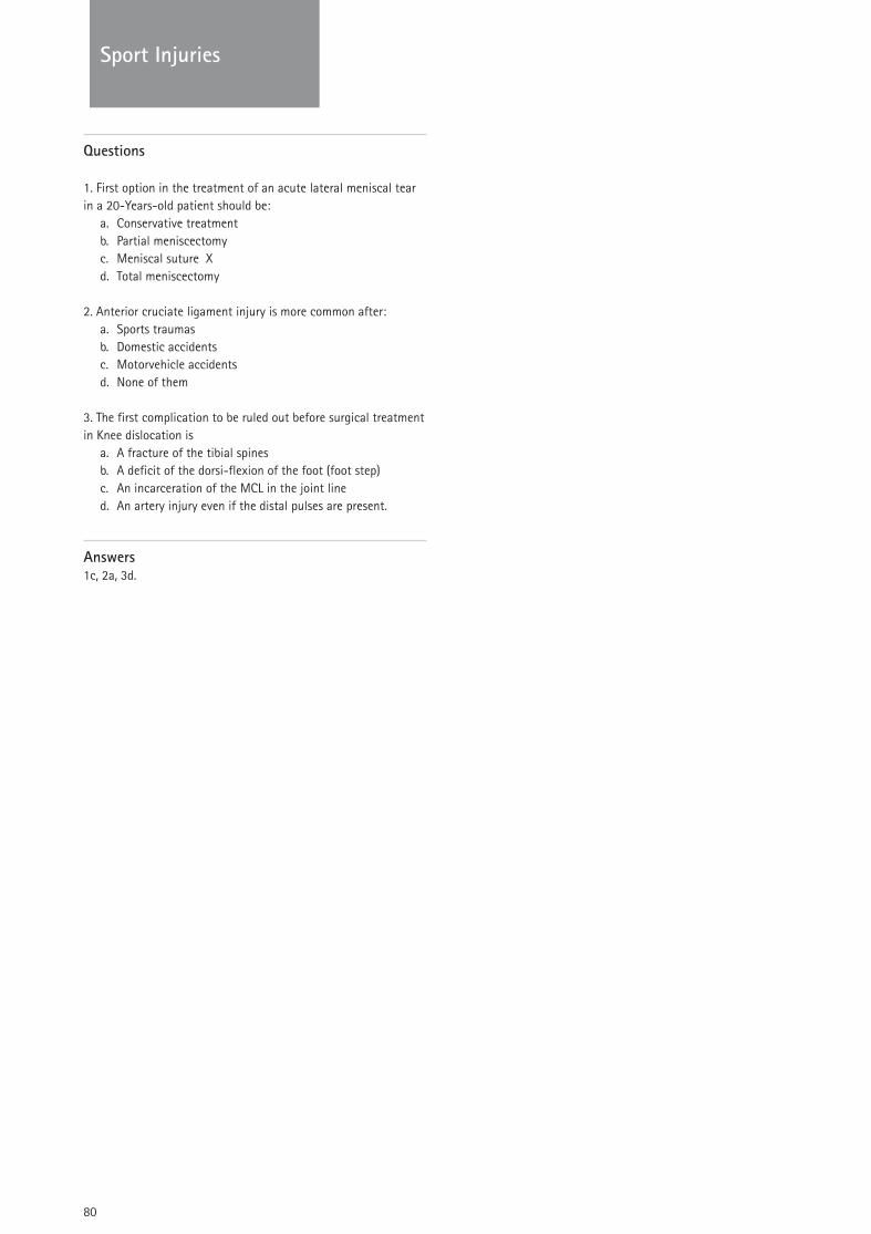
80
TUMOUR Sport Injuries
Questions
1. First option in the treatment of an acute lateral meniscal tear in a 20-Years-old patient should be:
a. Conservative treatment b. Partial meniscectomyc. Meniscal suture Xd. Total meniscectomy
2. Anterior cruciate ligament injury is more common after:a. Sports traumasb. Domestic accidentsc. Motorvehicle accidents d. None of them
3. The first complication to be ruled out before surgical treatment in Knee dislocation is
a. A fracture of the tibial spines b. A deficit of the dorsi-flexion of the foot (foot step)c. An incarceration of the MCL in the joint lined. An artery injury even if the distal pulses are present.
Answers1c, 2a, 3d.
81
TUMOUR
Infe
ctio
n
Musculoskeletal Infections
Dr. Domizio Suva
HUG, University Hospitals of Geneva Geneva, Switzerland
Infections After Total Joint Arthroplasty
1. Introduction
Total hip (THR) and knee (TKR) replacements substantially improve patients' quality of life by reducing pain and increasing mobility. However, despite advances in prevention, these interventions are associated with a risk of infection of 0.5-1.5% for THR and 1-2% for TKR. After revision surgery, the risks of prosthetic joint infection (PJI) are considerably higher, at approximately 10%. Beside a significant morbidity for the patients due to a high number of surgical revisions, the occurrence of an infection triples the cost of a primary arthroplasty. This trend is certainly not going to be reversed in the future, as it is estimated that in 2030, in the US, the number of THR and TKR will increase by 174% and 674%, respectively.
2. Pathophysiology of infection
There are three pathways for contamination: direct inoculation, blood-borne contamination and contamination by contiguity. Direct inoculation is usually held in the operating room, but can also occur later in the presence of a wound dehiscence. Once in contact with the implant, bacteria leave their planktonic form to produce the biofilm, composed of 30% of bacteria and 70% of adhesive and protective matrix. Bacteria in the biofilm are 1,000 to 10,000 times more resistant to antibiotics. From a clinical point of view, the result of the formation of biofilm is that the cure of the infection will require mechanical removal of the biofilm involving, most of the time, a removal of the implant. In summary, in case of any suspicion of infection, it is essential to make the diagnosis as early as possible, in order to avoid biofilm formation and thus preserving the prosthesis.
3. Classification
Prosthetic joint infection is currently classified according to the Coventry classification into early infection, delayed infection and late infection. Early infection is acquired during surgery and is produced by aggressive bacteria such as S.aureus. The delayed presentation is also acquired peri-operatively but produced by less virulent bacteria. Finally, the late infection is of haematogenous origin. The diagnosis of PJI can be very challenging. The last consensus of the Society of Musculoskeletal Infection in the US reported that the diagnosis of PJI requires the presence of one out of two major criteria or three
of the five minor criteria (Table 1). From a microbiological point of view, the most commonly involved germs are Staphylococcus aureus, Coagulase negative staphylococci and Streptococci (Table 2).
4. Clinical presentation
Clinically, there are three presentations of PJI: (1) the acute infection with chills, local inflammation, joint effusion or draining fluid; (2) the low-grade infection, which is oligosymptomatic and is sometimes difficult to differentiate from aseptic loosening, as the patient has chronic pain, perhaps low-grade fever, joint effusion or RX evidence of prosthetic loosening; and (3) finally when the origin of the infection is blood-borne, the clinical picture may initially be dominated by symptoms and signs associated to a systemic infection, such as endocarditis, pneumonia or urosepsis.
Table 1 : Diagnostic criteria according to the Society of Musculoskeletal Infection in the US
MAJOR CRITERIA
- Presence of 2 positive cultures on periprosthetic tissue with an identified germ
- Fistula between the prosthesis and the skin
MINOR CRITERIA
- Elevated CRP and ESR - Elevated WBC in synovial fluid or
positive leukocyte esterase test - Elevated percentage of neutrophils in
the synovial fluid - Positive histological analysis of
periprosthetic tissue - Positive culture of periprosthetic tissue
Table 2 : Germs distribution on prosthesis infection
GERMS RATE
Staphylococcus aureus
Coagulase-negative staphylococci
Streptococcus
Gram-negative bacilli
Enterococcus
Anaerobic bacteria
No germ found
* Propionibacterium acnes (shoulder, spondylodesis)
21-43%
17-39%
7-12%
5-12%
1-8%
2-6%
4-12%
38%
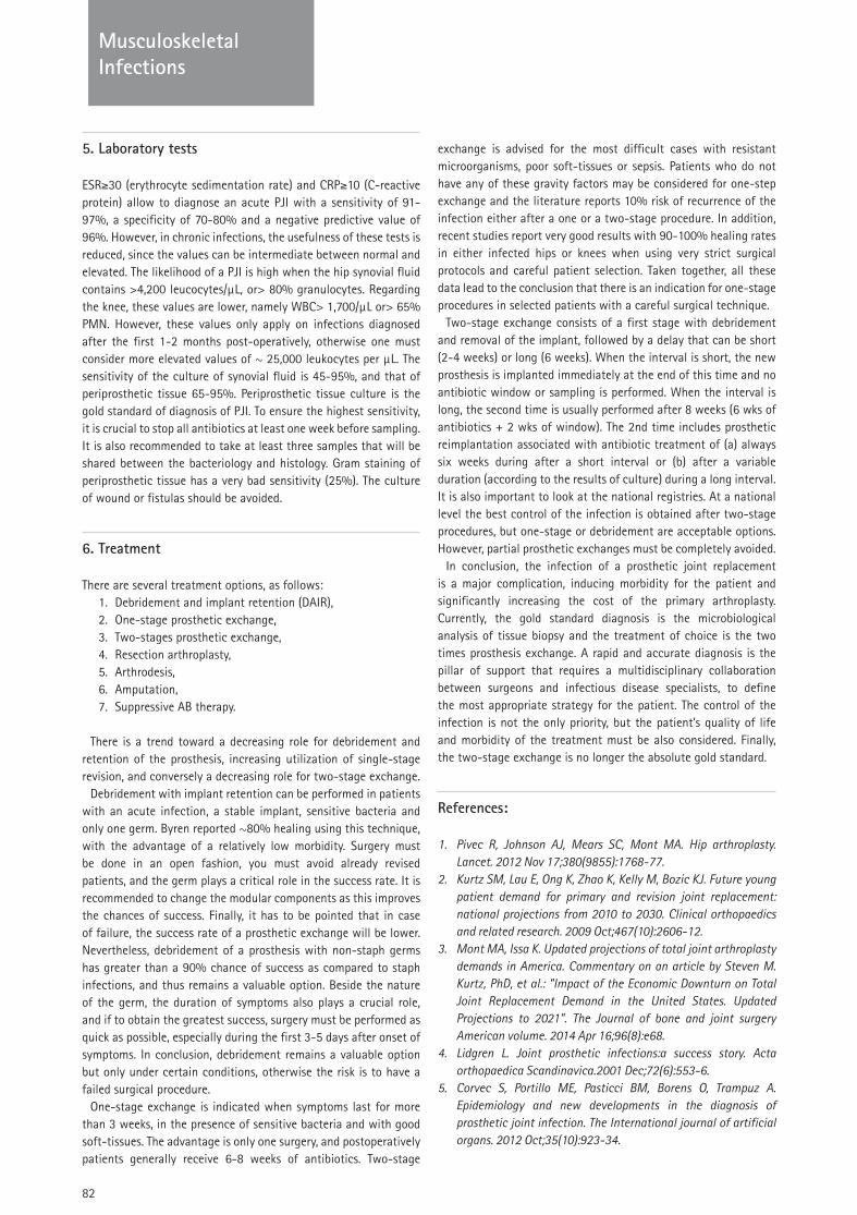
82
Musculoskeletal Infections
5. Laboratory tests
ESR≥30 (erythrocyte sedimentation rate) and CRP≥10 (C-reactive protein) allow to diagnose an acute PJI with a sensitivity of 91-97%, a specificity of 70-80% and a negative predictive value of 96%. However, in chronic infections, the usefulness of these tests is reduced, since the values can be intermediate between normal and elevated. The likelihood of a PJI is high when the hip synovial fluid contains >4,200 leucocytes/µL, or> 80% granulocytes. Regarding the knee, these values are lower, namely WBC> 1,700/µL or> 65% PMN. However, these values only apply on infections diagnosed after the first 1-2 months post-operatively, otherwise one must consider more elevated values of ~ 25,000 leukocytes per µL. The sensitivity of the culture of synovial fluid is 45-95%, and that of periprosthetic tissue 65-95%. Periprosthetic tissue culture is the gold standard of diagnosis of PJI. To ensure the highest sensitivity, it is crucial to stop all antibiotics at least one week before sampling. It is also recommended to take at least three samples that will be shared between the bacteriology and histology. Gram staining of periprosthetic tissue has a very bad sensitivity (25%). The culture of wound or fistulas should be avoided.
6. Treatment
There are several treatment options, as follows: 1. Debridement and implant retention (DAIR), 2. One-stage prosthetic exchange, 3. Two-stages prosthetic exchange, 4. Resection arthroplasty, 5. Arthrodesis, 6. Amputation, 7. Suppressive AB therapy.
There is a trend toward a decreasing role for debridement and retention of the prosthesis, increasing utilization of single-stage revision, and conversely a decreasing role for two-stage exchange.
Debridement with implant retention can be performed in patients with an acute infection, a stable implant, sensitive bacteria and only one germ. Byren reported ~80% healing using this technique, with the advantage of a relatively low morbidity. Surgery must be done in an open fashion, you must avoid already revised patients, and the germ plays a critical role in the success rate. It is recommended to change the modular components as this improves the chances of success. Finally, it has to be pointed that in case of failure, the success rate of a prosthetic exchange will be lower. Nevertheless, debridement of a prosthesis with non-staph germs has greater than a 90% chance of success as compared to staph infections, and thus remains a valuable option. Beside the nature of the germ, the duration of symptoms also plays a crucial role, and if to obtain the greatest success, surgery must be performed as quick as possible, especially during the first 3-5 days after onset of symptoms. In conclusion, debridement remains a valuable option but only under certain conditions, otherwise the risk is to have a failed surgical procedure.
One-stage exchange is indicated when symptoms last for more than 3 weeks, in the presence of sensitive bacteria and with good soft-tissues. The advantage is only one surgery, and postoperatively patients generally receive 6-8 weeks of antibiotics. Two-stage
exchange is advised for the most difficult cases with resistant microorganisms, poor soft-tissues or sepsis. Patients who do not have any of these gravity factors may be considered for one-step exchange and the literature reports 10% risk of recurrence of the infection either after a one or a two-stage procedure. In addition, recent studies report very good results with 90-100% healing rates in either infected hips or knees when using very strict surgical protocols and careful patient selection. Taken together, all these data lead to the conclusion that there is an indication for one-stage procedures in selected patients with a careful surgical technique.
Two-stage exchange consists of a first stage with debridement and removal of the implant, followed by a delay that can be short (2-4 weeks) or long (6 weeks). When the interval is short, the new prosthesis is implanted immediately at the end of this time and no antibiotic window or sampling is performed. When the interval is long, the second time is usually performed after 8 weeks (6 wks of antibiotics + 2 wks of window). The 2nd time includes prosthetic reimplantation associated with antibiotic treatment of (a) always six weeks during after a short interval or (b) after a variable duration (according to the results of culture) during a long interval. It is also important to look at the national registries. At a national level the best control of the infection is obtained after two-stage procedures, but one-stage or debridement are acceptable options. However, partial prosthetic exchanges must be completely avoided.
In conclusion, the infection of a prosthetic joint replacement is a major complication, inducing morbidity for the patient and significantly increasing the cost of the primary arthroplasty. Currently, the gold standard diagnosis is the microbiological analysis of tissue biopsy and the treatment of choice is the two times prosthesis exchange. A rapid and accurate diagnosis is the pillar of support that requires a multidisciplinary collaboration between surgeons and infectious disease specialists, to define the most appropriate strategy for the patient. The control of the infection is not the only priority, but the patient’s quality of life and morbidity of the treatment must be also considered. Finally, the two-stage exchange is no longer the absolute gold standard.
References:
1. Pivec R, Johnson AJ, Mears SC, Mont MA. Hip arthroplasty. Lancet. 2012 Nov 17;380(9855):1768-77.
2. Kurtz SM, Lau E, Ong K, Zhao K, Kelly M, Bozic KJ. Future young patient demand for primary and revision joint replacement: national projections from 2010 to 2030. Clinical orthopaedics and related research. 2009 Oct;467(10):2606-12.
3. Mont MA, Issa K. Updated projections of total joint arthroplasty demands in America. Commentary on an article by Steven M. Kurtz, PhD, et al.: "Impact of the Economic Downturn on Total Joint Replacement Demand in the United States. Updated Projections to 2021". The Journal of bone and joint surgery American volume. 2014 Apr 16;96(8):e68.
4. Lidgren L. Joint prosthetic infections:a success story. Acta orthopaedica Scandinavica.2001 Dec;72(6):553-6.
5. Corvec S, Portillo ME, Pasticci BM, Borens O, Trampuz A. Epidemiology and new developments in the diagnosis of prosthetic joint infection. The International journal of artificial organs. 2012 Oct;35(10):923-34.
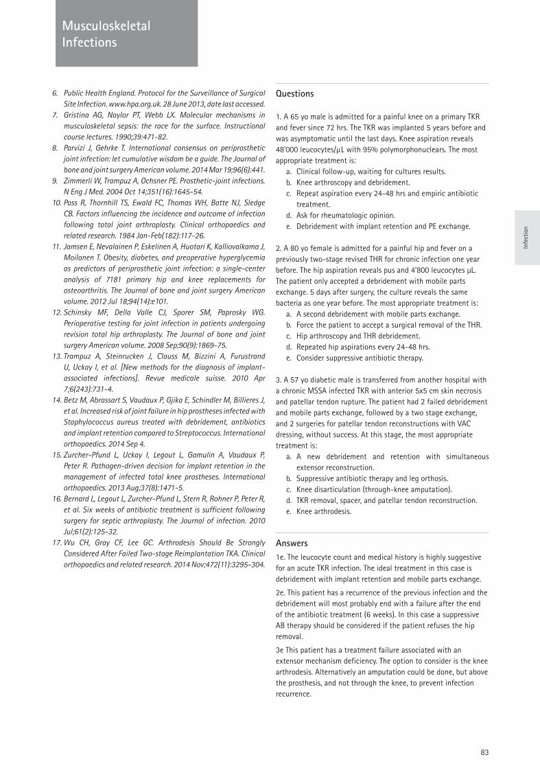
83
Infe
ctio
n
Musculoskeletal Infections
6. Public Health England. Protocol for the Surveillance of Surgical Site Infection. www.hpa.org.uk. 28 June 2013, date last accessed.
7. Gristina AG, Naylor PT, Webb LX. Molecular mechanisms in musculoskeletal sepsis: the race for the surface. Instructional course lectures. 1990;39:471-82.
8. Parvizi J, Gehrke T. International consensus on periprosthetic joint infection: let cumulative wisdom be a guide. The Journal of bone and joint surgery American volume. 2014 Mar 19;96(6):441.
9. Zimmerli W, Trampuz A, Ochsner PE. Prosthetic-joint infections. N Eng J Med. 2004 Oct 14;351(16):1645-54.
10. Poss R, Thornhill TS, Ewald FC, Thomas WH, Batte NJ, Sledge CB. Factors influencing the incidence and outcome of infection following total joint arthroplasty. Clinical orthopaedics and related research. 1984 Jan-Feb(182):117-26.
11. Jamsen E, Nevalainen P, Eskelinen A, Huotari K, Kalliovalkama J, Moilanen T. Obesity, diabetes, and preoperative hyperglycemia as predictors of periprosthetic joint infection: a single-center analysis of 7181 primary hip and knee replacements for osteoarthritis. The Journal of bone and joint surgery American volume. 2012 Jul 18;94(14):e101.
12. Schinsky MF, Della Valle CJ, Sporer SM, Paprosky WG. Perioperative testing for joint infection in patients undergoing revision total hip arthroplasty. The Journal of bone and joint surgery American volume. 2008 Sep;90(9):1869-75.
13. Trampuz A, Steinrucken J, Clauss M, Bizzini A, Furustrand U, Uckay I, et al. [New methods for the diagnosis of implant-associated infections]. Revue medicale suisse. 2010 Apr 7;6(243):731-4.
14. Betz M, Abrassart S, Vaudaux P, Gjika E, Schindler M, Billieres J, et al. Increased risk of joint failure in hip prostheses infected with Staphylococcus aureus treated with debridement, antibiotics and implant retention compared to Streptococcus. International orthopaedics. 2014 Sep 4.
15. Zurcher-Pfund L, Uckay I, Legout L, Gamulin A, Vaudaux P, Peter R. Pathogen-driven decision for implant retention in the management of infected total knee prostheses. International orthopaedics. 2013 Aug;37(8):1471-5.
16. Bernard L, Legout L, Zurcher-Pfund L, Stern R, Rohner P, Peter R, et al. Six weeks of antibiotic treatment is sufficient following surgery for septic arthroplasty. The Journal of infection. 2010 Jul;61(2):125-32.
17. Wu CH, Gray CF, Lee GC. Arthrodesis Should Be Strongly Considered After Failed Two-stage Reimplantation TKA. Clinical orthopaedics and related research. 2014 Nov;472(11):3295-304.
Questions
1. A 65 yo male is admitted for a painful knee on a primary TKR and fever since 72 hrs. The TKR was implanted 5 years before and was asymptomatic until the last days. Knee aspiration reveals 48’000 leucocytes/µL with 95% polymorphonuclears. The most appropriate treatment is:
a. Clinical follow-up, waiting for cultures results.b. Knee arthroscopy and debridement.c. Repeat aspiration every 24-48 hrs and empiric antibiotic
treatment.d. Ask for rheumatologic opinion.e. Debridement with implant retention and PE exchange.
2. A 80 yo female is admitted for a painful hip and fever on a previously two-stage revised THR for chronic infection one year before. The hip aspiration reveals pus and 4’800 leucocytes µL. The patient only accepted a debridement with mobile parts exchange. 5 days after surgery, the culture reveals the same bacteria as one year before. The most appropriate treatment is:
a. A second debridement with mobile parts exchange.b. Force the patient to accept a surgical removal of the THR.c. Hip arthroscopy and THR debridement.d. Repeated hip aspirations every 24-48 hrs.e. Consider suppressive antibiotic therapy.
3. A 57 yo diabetic male is transferred from another hospital with a chronic MSSA infected TKR with anterior 5x5 cm skin necrosis and patellar tendon rupture. The patient had 2 failed debridement and mobile parts exchange, followed by a two stage exchange, and 2 surgeries for patellar tendon reconstructions with VAC dressing, without success. At this stage, the most appropriate treatment is:
a. A new debridement and retention with simultaneous extensor reconstruction.
b. Suppressive antibiotic therapy and leg orthosis.c. Knee disarticulation (through-knee amputation).d. TKR removal, spacer, and patellar tendon reconstruction.e. Knee arthrodesis.
Answers1e. The leucocyte count and medical history is highly suggestive for an acute TKR infection. The ideal treatment in this case is debridement with implant retention and mobile parts exchange.
2e. This patient has a recurrence of the previous infection and the debridement will most probably end with a failure after the end of the antibiotic treatment (6 weeks). In this case a suppressive AB therapy should be considered if the patient refuses the hip removal.
3e This patient has a treatment failure associated with an extensor mechanism deficiency. The option to consider is the knee arthrodesis. Alternatively an amputation could be done, but above the prosthesis, and not through the knee, to prevent infection recurrence.
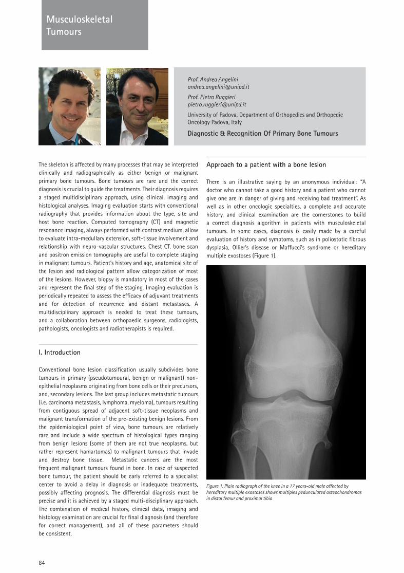
84
TUMOUR Musculoskeletal Tumours
Prof. Andrea Angelini [email protected]
Prof. Pietro Ruggieri [email protected]
University of Padova, Department of Orthopedics and Orthopedic Oncology Padova, Italy
Diagnostic & Recognition Of Primary Bone Tumours
The skeleton is affected by many processes that may be interpreted clinically and radiographically as either benign or malignant primary bone tumours. Bone tumours are rare and the correct diagnosis is crucial to guide the treatments. Their diagnosis requires a staged multidisciplinary approach, using clinical, imaging and histological analyses. Imaging evaluation starts with conventional radiography that provides information about the type, site and host bone reaction. Computed tomography (CT) and magnetic resonance imaging, always performed with contrast medium, allow to evaluate intra-medullary extension, soft-tissue involvement and relationship with neuro-vascular structures. Chest CT, bone scan and positron emission tomography are useful to complete staging in malignant tumours. Patient’s history and age, anatomical site of the lesion and radiological pattern allow categorization of most of the lesions. However, biopsy is mandatory in most of the cases and represent the final step of the staging. Imaging evaluation is periodically repeated to assess the efficacy of adjuvant treatments and for detection of recurrence and distant metastases. A multidisciplinary approach is needed to treat these tumours, and a collaboration between orthopaedic surgeons, radiologists, pathologists, oncologists and radiotherapists is required.
I. Introduction
Conventional bone lesion classification usually subdivides bone tumours in primary (pseudotumoural, benign or malignant) non-epithelial neoplasms originating from bone cells or their precursors, and, secondary lesions. The last group includes metastatic tumours (i.e. carcinoma metastasis, lymphoma, myeloma), tumours resulting from contiguous spread of adjacent soft-tissue neoplasms and malignant transformation of the pre-existing benign lesions. From the epidemiological point of view, bone tumours are relatively rare and include a wide spectrum of histological types ranging from benign lesions (some of them are not true neoplasms, but rather represent hamartomas) to malignant tumours that invade and destroy bone tissue. Metastatic cancers are the most frequent malignant tumours found in bone. In case of suspected bone tumour, the patient should be early referred to a specialist center to avoid a delay in diagnosis or inadequate treatments, possibly affecting prognosis. The differential diagnosis must be precise and it is achieved by a staged multi-disciplinary approach. The combination of medical history, clinical data, imaging and histology examination are crucial for final diagnosis (and therefore for correct management), and all of these parameters should be consistent.
Approach to a patient with a bone lesion
There is an illustrative saying by an anonymous individual: “A doctor who cannot take a good history and a patient who cannot give one are in danger of giving and receiving bad treatment”. As well as in other oncologic specialties, a complete and accurate history, and clinical examination are the cornerstones to build a correct diagnosis algorithm in patients with musculoskeletal tumours. In some cases, diagnosis is easily made by a careful evaluation of history and symptoms, such as in poliostotic fibrous dysplasia, Ollier’s disease or Maffucci’s syndrome or hereditary multiple exostoses (Figure 1).
Figure 1: Plain radiograph of the knee in a 17 years-old male affected by hereditary multiple exostoses shows multiples pedunculated osteochondromas in distal femur and proximal tibia
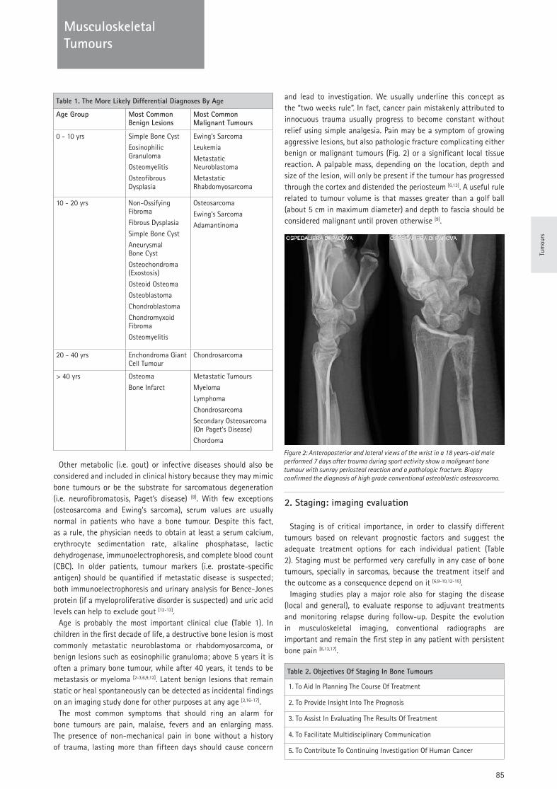
85
TUMOUR
Tum
ours
Musculoskeletal Tumours
Other metabolic (i.e. gout) or infective diseases should also be considered and included in clinical history because they may mimic bone tumours or be the substrate for sarcomatous degeneration (i.e. neurofibromatosis, Paget’s disease) [8]. With few exceptions (osteosarcoma and Ewing’s sarcoma), serum values are usually normal in patients who have a bone tumour. Despite this fact, as a rule, the physician needs to obtain at least a serum calcium, erythrocyte sedimentation rate, alkaline phosphatase, lactic dehydrogenase, immunoelectrophoresis, and complete blood count (CBC). In older patients, tumour markers (i.e. prostate-specific antigen) should be quantified if metastatic disease is suspected; both immunoelectrophoresis and urinary analysis for Bence-Jones protein (if a myeloproliferative disorder is suspected) and uric acid levels can help to exclude gout [12-13].
Age is probably the most important clinical clue (Table 1). In children in the first decade of life, a destructive bone lesion is most commonly metastatic neuroblastoma or rhabdomyosarcoma, or benign lesions such as eosinophilic granuloma; above 5 years it is often a primary bone tumour, while after 40 years, it tends to be metastasis or myeloma [2-3,6,9,12]. Latent benign lesions that remain static or heal spontaneously can be detected as incidental findings on an imaging study done for other purposes at any age [3,16-17].
The most common symptoms that should ring an alarm for bone tumours are pain, malaise, fevers and an enlarging mass. The presence of non-mechanical pain in bone without a history of trauma, lasting more than fifteen days should cause concern
and lead to investigation. We usually underline this concept as the “two weeks rule”. In fact, cancer pain mistakenly attributed to innocuous trauma usually progress to become constant without relief using simple analgesia. Pain may be a symptom of growing aggressive lesions, but also pathologic fracture complicating either benign or malignant tumours (Fig. 2) or a significant local tissue reaction. A palpable mass, depending on the location, depth and size of the lesion, will only be present if the tumour has progressed through the cortex and distended the periosteum [6,13]. A useful rule related to tumour volume is that masses greater than a golf ball (about 5 cm in maximum diameter) and depth to fascia should be considered malignant until proven otherwise [9].
2. Staging: imaging evaluation
Staging is of critical importance, in order to classify different tumours based on relevant prognostic factors and suggest the adequate treatment options for each individual patient (Table 2). Staging must be performed very carefully in any case of bone tumours, specially in sarcomas, because the treatment itself and the outcome as a consequence depend on it [6,9-10,12-15].
Imaging studies play a major role also for staging the disease (local and general), to evaluate response to adjuvant treatments and monitoring relapse during follow-up. Despite the evolution in musculoskeletal imaging, conventional radiographs are important and remain the first step in any patient with persistent bone pain [6,13,17].
Table 1. The More Likely Differential Diagnoses By Age
Age Group Most Common Benign Lesions
Most Common Malignant Tumours
0 - 10 yrs Simple Bone Cyst
Eosinophilic Granuloma
Osteomyelitis
Osteofibrous Dysplasia
Ewing’s Sarcoma
Leukemia
Metastatic Neuroblastoma
Metastatic Rhabdomyosarcoma
10 - 20 yrs Non-Ossifying Fibroma
Fibrous Dysplasia
Simple Bone Cyst
Aneurysmal Bone Cyst
Osteochondroma (Exostosis)
Osteoid Osteoma
Osteoblastoma
Chondroblastoma
Chondromyxoid Fibroma
Osteomyelitis
Osteosarcoma
Ewing’s Sarcoma
Adamantinoma
20 - 40 yrs Enchondroma Giant Cell Tumour
Chondrosarcoma
> 40 yrs Osteoma
Bone Infarct
Metastatic Tumours
Myeloma
Lymphoma
Chondrosarcoma
Secondary Osteosarcoma (On Paget’s Disease)
Chordoma
Table 2. Objectives Of Staging In Bone Tumours
1. To Aid In Planning The Course Of Treatment
2. To Provide Insight Into The Prognosis
3. To Assist In Evaluating The Results Of Treatment
4. To Facilitate Multidisciplinary Communication
5. To Contribute To Continuing Investigation Of Human Cancer
Figure 2: Anteroposterior and lateral views of the wrist in a 18 years-old male performed 7 days after trauma during sport activity show a malignant bone tumour with sunray periosteal reaction and a pathologic fracture. Biopsy confirmed the diagnosis of high grade conventional osteoblastic osteosarcoma.
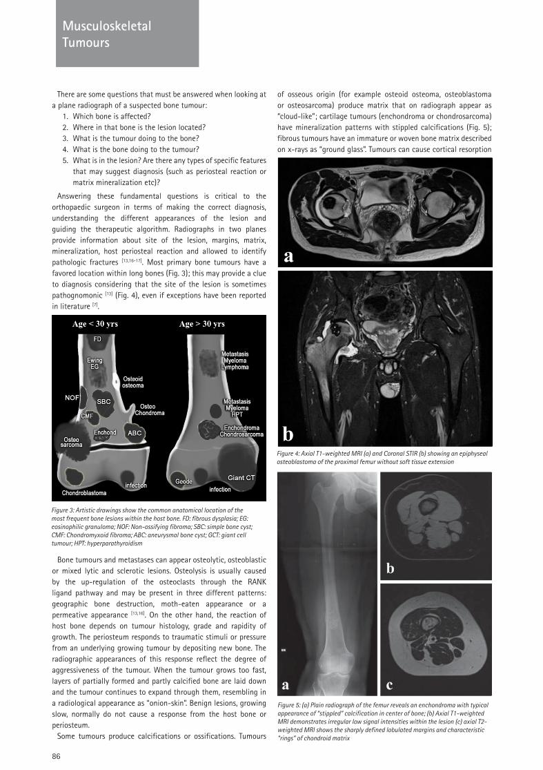
86
Musculoskeletal Tumours
There are some questions that must be answered when looking at a plane radiograph of a suspected bone tumour:
1. Which bone is affected? 2. Where in that bone is the lesion located? 3. What is the tumour doing to the bone?4. What is the bone doing to the tumour?5. What is in the lesion? Are there any types of specific features
that may suggest diagnosis (such as periosteal reaction or matrix mineralization etc)?
Answering these fundamental questions is critical to the orthopaedic surgeon in terms of making the correct diagnosis, understanding the different appearances of the lesion and guiding the therapeutic algorithm. Radiographs in two planes provide information about site of the lesion, margins, matrix, mineralization, host periosteal reaction and allowed to identify pathologic fractures [13,16-17]. Most primary bone tumours have a favored location within long bones (Fig. 3); this may provide a clue to diagnosis considering that the site of the lesion is sometimes pathognomonic [13] (Fig. 4), even if exceptions have been reported in literature [7].
Bone tumours and metastases can appear osteolytic, osteoblastic or mixed lytic and sclerotic lesions. Osteolysis is usually caused by the up-regulation of the osteoclasts through the RANK ligand pathway and may be present in three different patterns: geographic bone destruction, moth-eaten appearance or a permeative appearance [13,16]. On the other hand, the reaction of host bone depends on tumour histology, grade and rapidity of growth. The periosteum responds to traumatic stimuli or pressure from an underlying growing tumour by depositing new bone. The radiographic appearances of this response reflect the degree of aggressiveness of the tumour. When the tumour grows too fast, layers of partially formed and partly calcified bone are laid down and the tumour continues to expand through them, resembling in a radiological appearance as “onion-skin”. Benign lesions, growing slow, normally do not cause a response from the host bone or periosteum.
Some tumours produce calcifications or ossifications. Tumours
of osseous origin (for example osteoid osteoma, osteoblastoma or osteosarcoma) produce matrix that on radiograph appear as “cloud-like”; cartilage tumours (enchondroma or chondrosarcoma) have mineralization patterns with stippled calcifications (Fig. 5); fibrous tumours have an immature or woven bone matrix described on x-rays as “ground glass”. Tumours can cause cortical resorption
Figure 3: Artistic drawings show the common anatomical location of the most frequent bone lesions within the host bone. FD: fibrous dysplasia; EG: eosinophilic granuloma; NOF: Non-ossifying fibroma; SBC: simple bone cyst; CMF: Chondromyxoid fibroma; ABC: aneurysmal bone cyst; GCT: giant cell tumour; HPT: hyperparathyroidism
Figure 4: Axial T1-weighted MRI (a) and Coronal STIR (b) showing an epiphyseal osteoblastoma of the proximal femur without soft tissue extension
Figure 5: (a) Plain radiograph of the femur reveals an enchondroma with typical appearance of “stippled” calcification in center of bone; (b) Axial T1-weighted MRI demonstrates irregular low signal intensities within the lesion (c) axial T2-weighted MRI shows the sharply defined lobulated margins and characteristic “rings” of chondroid matrix
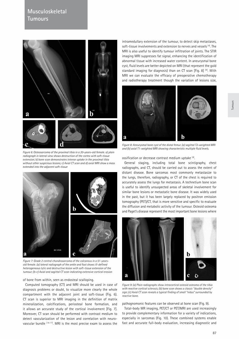
Tum
ours
87
Musculoskeletal Tumours
of bone from within, seen as endosteal scalloping. Computed tomography (CT) and MRI should be used in case of
diagnosis problems or doubt, to visualize more clearly the whole compartment with the adjacent joint and soft-tissue (Fig. 6). CT scan is superior to MRI imaging in the definition of matrix mineralization, calcifications, periosteal bone formation, and it allows an accurate study of the cortical involvement [Fig. 7]. Moreover, CT scan should be performed with contrast medium to detect vascularization of the lesion and correlation with neuro-vascular bundle [16-17]. MRI is the most precise exam to assess the
intramedullary extension of the tumour, to detect skip metastases, soft-tissue involvements and extension to nerves and vessels [4]. The MRI is also useful to identify tumour infiltration of joints. The STIR imaging MRI suppresses fat signal, enhancing the identification of abnormal tissue with increased water content. In aneurysmal bone cyst, fluid levels are better depicted on MRI (that represent the gold standard imaging for diagnosis) than on CT scan [Fig. 8] [4]. With MRI we can evaluate the efficacy of preoperative chemotherapy and radiotherapy treatment though the variation of lesions size,
ossification or decrease contrast medium uptake [4].General staging, including total bone scintigraphy, chest
radiographs, and CT, should be carried out to assess the extent of distant disease. Bone sarcomas most commonly metastasize to the lungs, therefore, radiography, or CT of the chest is required to accurately assess the lungs for metastasis. A technetium bone scan is useful to identify unsuspected areas of skeletal involvement for similar bone lesions or metastatic bone disease. It was widely used in the past, but it has been largely replaced by positron emission tomography (PET)/CT, that is more sensitive and specific to evaluate the diffusion and metabolic activity of the tumour. Osteoid osteoma and Paget’s disease represent the most important bone lesions where
pathognomonic features can be observed at bone scan (Fig. 9). Total-body MR imaging, PET/CT or PET/MRI are used increasingly
to provide complementary information for a variety of indications, especially in sarcomas (Fig. 10). These combined systems enable fast and accurate full-body evaluation, increasing diagnostic and
Figure 6: Osteosarcoma of the proximal tibia in a 20-years-old female. a) plain radiograph in lateral view shows destruction of the cortex with soft-tissue extension; b) bone scan demonstrates intense uptake in the proximal tibia without other suspicious lesions; c) Axial CT scan and d) axial MRI show a mass extended into the adjacent soft-tissue
Figure 9: (a) Plain radiographs show intracortical osteoid osteoma of the tibia with reactive cortical sclerosis; (b) bone scan shows a classic “double density” sign; (c) Axial CT scan reveals a typical finding of small “nidus” surrounded by reactive bone.
Figure 8: Aneurysmal bone cyst of the distal femur. (a) sagittal T2-weighted MRI and (b) axial T1-weighted MRI showing characteristic multiple fluid levels.
Figure 7: Grade 2 central chondrosarcoma of the calcaneus in a 51-years-old female. (a) lateral radiograph of the ankle and foot shows ill-defined heterogeneous lytic and destructive lesion with soft-tissue extension of the tumour; (b-c) Axial and sagittal CT scan indicating extensive cortical erosion
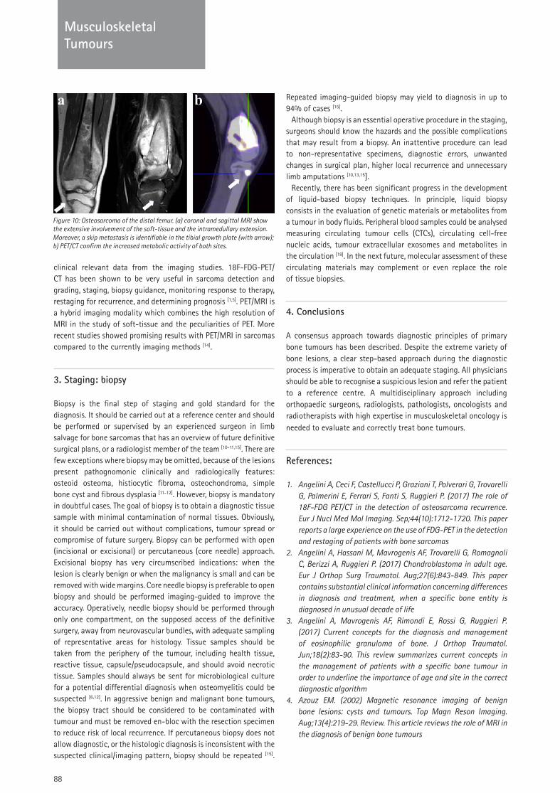
88
Musculoskeletal Tumours
clinical relevant data from the imaging studies. 18F-FDG-PET/CT has been shown to be very useful in sarcoma detection and grading, staging, biopsy guidance, monitoring response to therapy, restaging for recurrence, and determining prognosis [1,5]. PET/MRI is a hybrid imaging modality which combines the high resolution of MRI in the study of soft-tissue and the peculiarities of PET. More recent studies showed promising results with PET/MRI in sarcomas compared to the currently imaging methods [14].
3. Staging: biopsy
Biopsy is the final step of staging and gold standard for the diagnosis. It should be carried out at a reference center and should be performed or supervised by an experienced surgeon in limb salvage for bone sarcomas that has an overview of future definitive surgical plans, or a radiologist member of the team [10-11,15]. There are few exceptions where biopsy may be omitted, because of the lesions present pathognomonic clinically and radiologically features: osteoid osteoma, histiocytic fibroma, osteochondroma, simple bone cyst and fibrous dysplasia [11-12]. However, biopsy is mandatory in doubtful cases. The goal of biopsy is to obtain a diagnostic tissue sample with minimal contamination of normal tissues. Obviously, it should be carried out without complications, tumour spread or compromise of future surgery. Biopsy can be performed with open (incisional or excisional) or percutaneous (core needle) approach. Excisional biopsy has very circumscribed indications: when the lesion is clearly benign or when the malignancy is small and can be removed with wide margins. Core needle biopsy is preferable to open biopsy and should be performed imaging-guided to improve the accuracy. Operatively, needle biopsy should be performed through only one compartment, on the supposed access of the definitive surgery, away from neurovascular bundles, with adequate sampling of representative areas for histology. Tissue samples should be taken from the periphery of the tumour, including health tissue, reactive tissue, capsule/pseudocapsule, and should avoid necrotic tissue. Samples should always be sent for microbiological culture for a potential differential diagnosis when osteomyelitis could be suspected [6,12]. In aggressive benign and malignant bone tumours, the biopsy tract should be considered to be contaminated with tumour and must be removed en-bloc with the resection specimen to reduce risk of local recurrence. If percutaneous biopsy does not allow diagnostic, or the histologic diagnosis is inconsistent with the suspected clinical/imaging pattern, biopsy should be repeated [15].
Repeated imaging-guided biopsy may yield to diagnosis in up to 94% of cases [15].
Although biopsy is an essential operative procedure in the staging, surgeons should know the hazards and the possible complications that may result from a biopsy. An inattentive procedure can lead to non-representative specimens, diagnostic errors, unwanted changes in surgical plan, higher local recurrence and unnecessary limb amputations [10,13,15].
Recently, there has been significant progress in the development of liquid-based biopsy techniques. In principle, liquid biopsy consists in the evaluation of genetic materials or metabolites from a tumour in body fluids. Peripheral blood samples could be analysed measuring circulating tumour cells (CTCs), circulating cell-free nucleic acids, tumour extracellular exosomes and metabolites in the circulation [18]. In the next future, molecular assessment of these circulating materials may complement or even replace the role of tissue biopsies.
4. Conclusions
A consensus approach towards diagnostic principles of primary bone tumours has been described. Despite the extreme variety of bone lesions, a clear step-based approach during the diagnostic process is imperative to obtain an adequate staging. All physicians should be able to recognise a suspicious lesion and refer the patient to a reference centre. A multidisciplinary approach including orthopaedic surgeons, radiologists, pathologists, oncologists and radiotherapists with high expertise in musculoskeletal oncology is needed to evaluate and correctly treat bone tumours.
References:
1. Angelini A, Ceci F, Castellucci P, Graziani T, Polverari G, Trovarelli G, Palmerini E, Ferrari S, Fanti S, Ruggieri P. (2017) The role of 18F-FDG PET/CT in the detection of osteosarcoma recurrence. Eur J Nucl Med Mol Imaging. Sep;44(10):1712-1720. This paper reports a large experience on the use of FDG-PET in the detection and restaging of patients with bone sarcomas
2. Angelini A, Hassani M, Mavrogenis AF, Trovarelli G, Romagnoli C, Berizzi A, Ruggieri P. (2017) Chondroblastoma in adult age. Eur J Orthop Surg Traumatol. Aug;27(6):843-849. This paper contains substantial clinical information concerning differences in diagnosis and treatment, when a specific bone entity is diagnosed in unusual decade of life
3. Angelini A, Mavrogenis AF, Rimondi E, Rossi G, Ruggieri P. (2017) Current concepts for the diagnosis and management of eosinophilic granuloma of bone. J Orthop Traumatol. Jun;18(2):83-90. This review summarizes current concepts in the management of patients with a specific bone tumour in order to underline the importance of age and site in the correct diagnostic algorithm
4. Azouz EM. (2002) Magnetic resonance imaging of benign bone lesions: cysts and tumours. Top Magn Reson Imaging. Aug;13(4):219-29. Review. This article reviews the role of MRI in the diagnosis of benign bone tumours
Figure 10: Osteosarcoma of the distal femur. (a) coronal and sagittal MRI show the extensive involvement of the soft-tissue and the intramedullary extension. Moreover, a skip metastasis is identifiable in the tibial growth plate (with arrow); b) PET/CT confirm the increased metabolic activity of both sites.
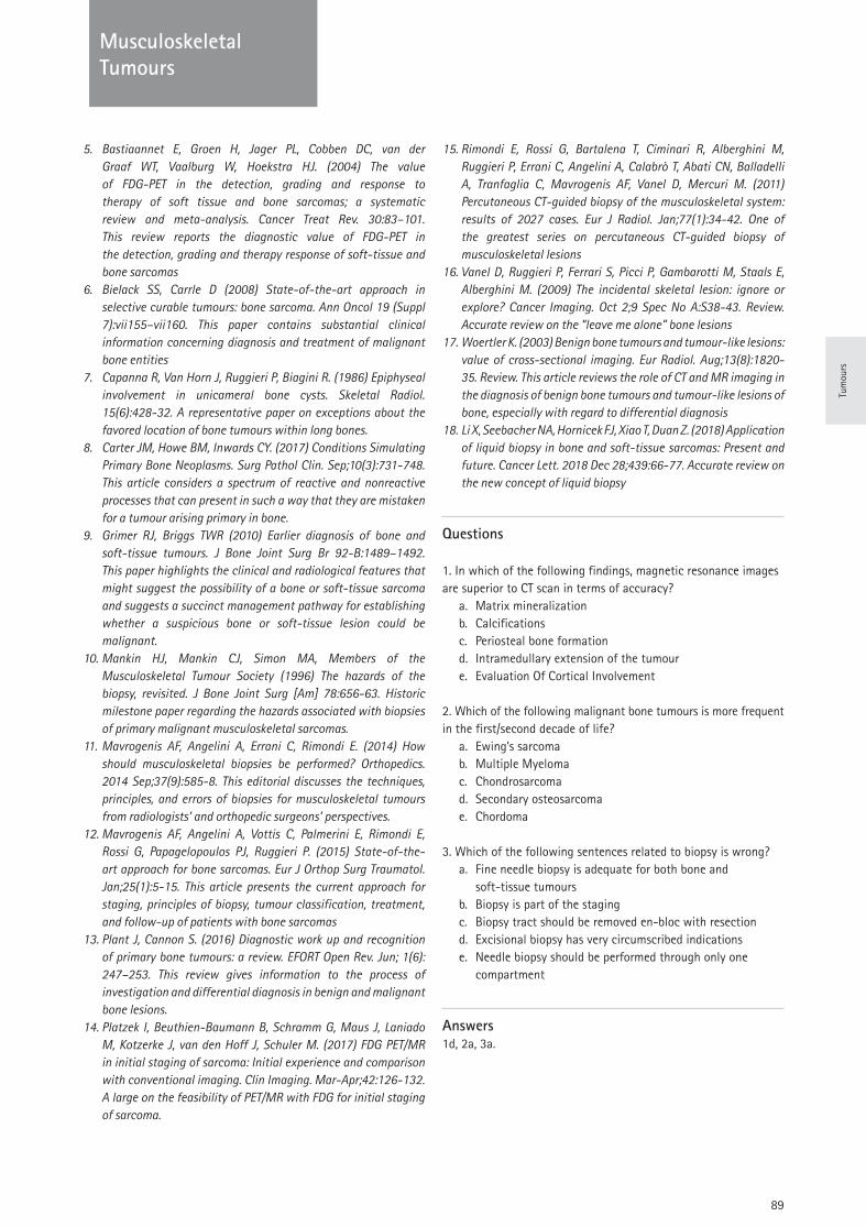
Tum
ours
89
Musculoskeletal Tumours
5. Bastiaannet E, Groen H, Jager PL, Cobben DC, van der Graaf WT, Vaalburg W, Hoekstra HJ. (2004) The value of FDG-PET in the detection, grading and response to therapy of soft tissue and bone sarcomas; a systematic review and meta-analysis. Cancer Treat Rev. 30:83–101.This review reports the diagnostic value of FDG-PET in the detection, grading and therapy response of soft-tissue and bone sarcomas
6. Bielack SS, Carrle D (2008) State-of-the-art approach in selective curable tumours: bone sarcoma. Ann Oncol 19 (Suppl 7):vii155–vii160. This paper contains substantial clinical information concerning diagnosis and treatment of malignant bone entities
7. Capanna R, Van Horn J, Ruggieri P, Biagini R. (1986) Epiphyseal involvement in unicameral bone cysts. Skeletal Radiol. 15(6):428-32. A representative paper on exceptions about the favored location of bone tumours within long bones.
8. Carter JM, Howe BM, Inwards CY. (2017) Conditions Simulating Primary Bone Neoplasms. Surg Pathol Clin. Sep;10(3):731-748. This article considers a spectrum of reactive and nonreactive processes that can present in such a way that they are mistaken for a tumour arising primary in bone.
9. Grimer RJ, Briggs TWR (2010) Earlier diagnosis of bone and soft-tissue tumours. J Bone Joint Surg Br 92-B:1489–1492. This paper highlights the clinical and radiological features that might suggest the possibility of a bone or soft-tissue sarcoma and suggests a succinct management pathway for establishing whether a suspicious bone or soft-tissue lesion could be malignant.
10. Mankin HJ, Mankin CJ, Simon MA, Members of the Musculoskeletal Tumour Society (1996) The hazards of the biopsy, revisited. J Bone Joint Surg [Am] 78:656-63. Historic milestone paper regarding the hazards associated with biopsies of primary malignant musculoskeletal sarcomas.
11. Mavrogenis AF, Angelini A, Errani C, Rimondi E. (2014) How should musculoskeletal biopsies be performed? Orthopedics. 2014 Sep;37(9):585-8. This editorial discusses the techniques, principles, and errors of biopsies for musculoskeletal tumours from radiologists’ and orthopedic surgeons’ perspectives.
12. Mavrogenis AF, Angelini A, Vottis C, Palmerini E, Rimondi E, Rossi G, Papagelopoulos PJ, Ruggieri P. (2015) State-of-the-art approach for bone sarcomas. Eur J Orthop Surg Traumatol. Jan;25(1):5-15. This article presents the current approach for staging, principles of biopsy, tumour classification, treatment, and follow-up of patients with bone sarcomas
13. Plant J, Cannon S. (2016) Diagnostic work up and recognition of primary bone tumours: a review. EFORT Open Rev. Jun; 1(6): 247–253. This review gives information to the process of investigation and differential diagnosis in benign and malignant bone lesions.
14. Platzek I, Beuthien-Baumann B, Schramm G, Maus J, Laniado M, Kotzerke J, van den Hoff J, Schuler M. (2017) FDG PET/MR in initial staging of sarcoma: Initial experience and comparison with conventional imaging. Clin Imaging. Mar-Apr;42:126-132. A large on the feasibility of PET/MR with FDG for initial staging of sarcoma.
15. Rimondi E, Rossi G, Bartalena T, Ciminari R, Alberghini M, Ruggieri P, Errani C, Angelini A, Calabrò T, Abati CN, Balladelli A, Tranfaglia C, Mavrogenis AF, Vanel D, Mercuri M. (2011) Percutaneous CT-guided biopsy of the musculoskeletal system: results of 2027 cases. Eur J Radiol. Jan;77(1):34-42. One of the greatest series on percutaneous CT-guided biopsy of musculoskeletal lesions
16. Vanel D, Ruggieri P, Ferrari S, Picci P, Gambarotti M, Staals E, Alberghini M. (2009) The incidental skeletal lesion: ignore or explore? Cancer Imaging. Oct 2;9 Spec No A:S38-43. Review. Accurate review on the “leave me alone” bone lesions
17. Woertler K. (2003) Benign bone tumours and tumour-like lesions: value of cross-sectional imaging. Eur Radiol. Aug;13(8):1820-35. Review. This article reviews the role of CT and MR imaging in the diagnosis of benign bone tumours and tumour-like lesions of bone, especially with regard to differential diagnosis
18. Li X, Seebacher NA, Hornicek FJ, Xiao T, Duan Z. (2018) Application of liquid biopsy in bone and soft-tissue sarcomas: Present and future. Cancer Lett. 2018 Dec 28;439:66-77. Accurate review on the new concept of liquid biopsy
Questions
1. In which of the following findings, magnetic resonance images are superior to CT scan in terms of accuracy?
a. Matrix mineralizationb. Calcificationsc. Periosteal bone formationd. Intramedullary extension of the tumoure. Evaluation Of Cortical Involvement
2. Which of the following malignant bone tumours is more frequent in the first/second decade of life?
a. Ewing’s sarcomab. Multiple Myelomac. Chondrosarcoma d. Secondary osteosarcoma e. Chordoma
3. Which of the following sentences related to biopsy is wrong?a. Fine needle biopsy is adequate for both bone and
soft-tissue tumoursb. Biopsy is part of the stagingc. Biopsy tract should be removed en-bloc with resectiond. Excisional biopsy has very circumscribed indicationse. Needle biopsy should be performed through only one
compartment
Answers1d, 2a, 3a.
90
TUMOUR Musculoskeletal Tumours
Prof. Miklós Szendröi
Semmelweis University Budapest Budapest, Hungary
Diagnostic Algorithm & Treatment Options In Bone Metastasis
Introduction
Oncology management is becoming an increasingly serious task in orthopaedic and trauma surgery.
In the past, without well established follow-up protocols, metastases were recognized late and mostly palliative treatments, radiotherapy and pain killing were favourized. Nowadays, modern diagnostic tools (PET-CT, whole body MRI, etc.) included into the follow-up protocols allow an early detection of the metastases. Beside improved chemo-, and radiotherapy, new targeted therapy as bisphosphonates and denosumab (antigen against RANKL) reduce skeletal related events. In the surgery, a broad spectrum of tools for reconstruction of defects and osteosynthesis can be used, many of them by minimal invasive techniques. All these advances result in a significant longer survival for metastatic patients even with multiple metastases. Data from the Scandinavian Skeletal Metastasis Registry about the last decade underline this:
- incidence of cancer has increased by 18% - mortality of cancer has increased by 2%
Epidemiology And Characteristics Of Skeletal Metastases
- Cancer - 2nd most frequent causative factor for death
- Every fourth person dies of cancer
- USA: 1,2 million new tumour cases annually - 300.000 metastases in the skeletal system - 20 % will develop a pathological fracture
(Capanna and Campanacci 2001)
- In 65-75 % of bone metastases the primary site is: lung, breast, kidney and prostate
- Imaging: lytic, mixed or sclerotic lesions
- Periosteal reaction is usually absent
- 10-20% are solitary at recognition but multiplication occures in 1-3 yrs
- Pathological fracture in 20% of the cases Risc factors (Mirel, 1989):
- Size (more than 2 cm) - Location (lower limb, proximal femur) - Blastic/lytic - Pain
Metastases are 80-100 times more common in the skeletal system than primary malignant bone tumours. The bones most frequently involved in decreasing sequence are: lumbar, dorsal, cervical spine, ribs, pelvis, proximal femur and tibia, skull, sternum and humerus. Only 1-2% of these secondary tumours affect the short tubular bones of the hand and foot.
1. Imaging
In suspected cases, e.g. when there is local bone pain after history of tumour, an X-ray is taken of the area in question and CT, MRI (occasionally PET-CT) scans are added if necessary. In spine, in the opposite of spondylitis the tumour involves single vertebral bodies, invading the intervertebral space only in a later stage. In the lung tubular bones, the lesion may be central, though it is more often eccentric, involving the cortex. Periosteal reaction is in most cases absent. Bone scan is also extremely important to decide if the process is single or multiple (Fig. 1).
Kidney and lung cancer usually produce lytic metastases in the bones, prostate cancer usually sclerotic ones, breast, thyroid and gastrointestineal cancers have most often mixed sclerotic-lytic metastases.
Clinical Signs, Symptoms:
- deep intermittant pain, independent of movement
- often presents weeks or months before x-ray changes are detected but: there are many reports on painless skeletal metastases in the early stages
- pathologic fracture as first episode in 10-30% of cases
- primary cancer in the case history in cc. 60-70% of cases
- laboratory tests can be useful for recognising the primary site, especially in case of prostate, thyroid cancer or myeloma.
- lytic metastases of kidney or lung cancer fracture frequently, while osteoplastic metastases of prostate cancer rarely break and have a good propensity to heal
- SRE (skeletal related events like fracture, cord compression, radiotherapy for bone metastases) is present in 10 to 20% at diagnosis of lung, breast and prostate cancer (increase up to 50% during the follow-up)
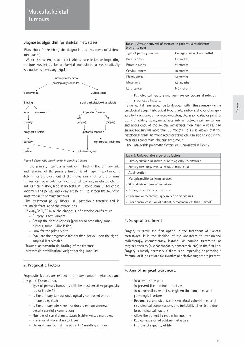
91
TUMOUR
Tum
ours
Musculoskeletal Tumours
Diagnostic algorithm for skeletal metastases
(Flow chart for reaching the diagnosis and treatment of skeletal metastases)
When the patient is admitted with a lytic lesion or impending fracture suspicious for a skeletal metastasis, a systematically evaluation is necessary (Fig.1).
Figure 1: Diagnostic algorithm for impending fracture
If the primary tumour is unknown, finding the primary site and staging of the primary tumour is of major importance. It determines the treatment of the metastasis whether the primary tumour can be oncologically controlled, excised, irradiated etc. or not. Clinical history, laboratory tests, MRI, bone scan, CT for chest, abdomen and pelvis, and x-ray are helpful to screen the four-five most frequent primary tumours.
The treatment policy differs in pathologic fracture and in traumatic fracture of the extremities.
If x-ray/MRI/CT raise the diagnosis of pathological fracture: - Surgery is semi-urgent - Set up the right diagnosis (primary or secondary bone
tumour, tumour-like lesion) - Look for the primary site - Evaluate the prognostic factors then decide upon the right
surgical intervention Trauma: osteosynthesis, healing of the fracture Metastasis: stabilization, weight bearing, mobility
2. Prognostic factors
Prognostic factors are related to primary tumour, metastasis and the patient’s condition.
- Type of primary tumour is still the most sensitive prognostic factor (Table 1)
- Is the primary tumour oncologically controlled or not (inoperable, etc.)?
- Is the primary site known or does it remain unknown despite careful examination?
- Number of skeletal metastases (soliter versus multiplex) - Presence of visceral metastases - General condition of the patient (Karnoffsky’s index)
- Pathological fracture and age have controversial roles as prognostic factors.
Significant differences can certainly occur within these concerning the oncological stage, histological type, grade, radio- and chemotherapy-sensitivity, presence of hormone-receptors, etc. In some studies patients e.g. with solitary kidney metastases (interval between primary tumour and appearence of the skeletal metastases more than 4 years) had an average survival more than 30 months. It is also known, that the histological grade, hormone receptor status etc. can also change in the metastasis concerning the primary tumour.
The unfavorable prognostic factors are summarized in Table 2.
Table 2. Unfavourable prognostic factors
- Primary tumour: unknown, or oncologically uncontrolled
- Primary site: lung, liver, pancreas or melanoma
- Axial location
- Multiple/multiorganic metastases
- Short doubling time of metastases
- Radio-, chemotherapy resistency
- Synchron or metachron appearence of metastases
- Poor general condition of patient, hemoglobin less than 7 mmol/l
3. Surgical treatment
Surgery is rarely the first option in the treatment of skeletal metastases. It is the decision of the oncoteam to recommend radiotherapy, chemotherapy, isotope- or hormon treatment, or targeted therapy (bisphosphonates, denosumab, etc.) in the first line. Surgery is mostly necessary if there is an impending or pathologic fracture, or if indications for curative or ablative surgery are present.
4. Aim of surgical treatment:
- To alleviate the pain - To prevent the imminent fracture - To osteosynthesize and strenghten the bone in case of
pathologic fracture - Decompress and stabilize the vertebral column in case of
neurological complications and instability of vertebra due to pathological fracture
- Allow the patient to regain his mobility - Radical excision of solitary metastases - Improve the quality of life
Known primary tumor
(oncologically controlled)
Solitary met. Multiplex met.
Staging staging (skeletal, extraskeletal)
local extraskeltal impending fracutre
yes no
(biopsy) (biopsy) (biopsy)
prognostic factors patient's condition
surgery non surgical treatment
radical palliative surgery
Fig. 1.: Diagnostic algorythm at impending fracture (Known primary tumor)
In cases where the primary tumour is unknown, finding the primary site and staging the primary tumour is of major importance. Whether the primary tumour can be controlled oncologically, excised, irradiated etc. or not determines the treatment of the metastasis
The treatment policy differs in pathologic fracture and in traumatic fracture of the extremities !!!
If x-ray/MRI/CT raise the diagnosis of pathological fracture:
Surgery is semi-urgent
Table 1. Average survival of metastatic patients with different type of tumour
Type of primary tumour Average survival (in months)
Breast cancer 34 months
Prostate cancer 24 months
Cervical cancer 18 months
Kidney cancer 12 months
Melanoma 3,5 months
Lung cancer 3-6 months
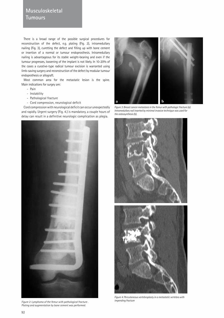
92
TUMOUR Musculoskeletal Tumours
There is a broad range of the possible surgical procedures for reconstruction of the defect, e.g. plating (Fig. 2), intramedullary nailing (Fig. 3), curetting the defect and filling up with bone cement or insertion of a normal or tumour endoprosthesis. Intramedullary nailing is advantageous for its stable weight-bearing and even if the tumour progresses, loosening of the implant is not likely. In 10-20% of the cases a curative-type radical tumour excision is warranted using limb-saving surgery and reconstruction of the defect by modular tumour endoprothesis or allograft.
Most common area for the metastatic lesion is the spine. Main indications for surgey are:
- Pain - Instability - Pathological fracture - Cord compression, neurological deficit
Cord compression with neurological deficit can occur unexpectedly and rapidly. Urgent surgery (Fig. 4.) is mandatory, a couple hours of delay can result in a definitive neurologic complication as plegia.
Figure 2: Lymphoma of the femur with pathological fracture . Plating and augmentation by bone cement was performed.
Figure 3: Breast cancer metastasis in the femur with pathologic fracture (a).Intramedullary nail inserted by minimal invasive technique was used for the osteosynthesis (b).
Figure 4: Percutaneous vertebroplasty in a metastatic vertebra with impending fracture
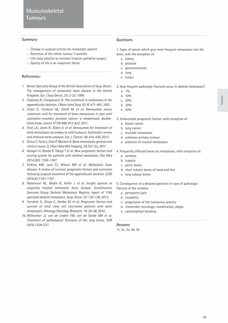
93
TUMOUR
Tum
ours
Musculoskeletal Tumours
Summary
- Change in surgical activity for metastatic patient - Resection of the whole tumour if possible - Life-long solution by minimal invasive palliative surgery - Quality of life is an important factor
References:
1. Breast Specialty Group of the British Association of Surg. Oncol.: The management of metastatic bone disease in the United Kingdom. Eur J Surg Oncol., 25: 3-23, 1999.
2. Capanna R., Campanacci D.: The treatment in metastases in the appendicular skeleton. J Bone Joint Surg. 83-B: 471-481, 2001.
3. Fizazi K., Carducci M., Smith M. et al: Denosumab versus zoledronic acid for treatment of bone metastases in men with castration-resistant prostate cancer: a randomised, double-blind study. Lancet 377(9768): 813-822, 2011.
4. Ford J.A., Jones R., Elders A. et al: Denosumab for treatment of bone metastases secondary to solid tumours: Systematic review and network meta-analysis. Eur. J. Cancer, 49: 416-430, 2013.
5. Greco C, Forte L, Erba P, Mariani G. Bone metastases, general and clinical issues. Q J Nucl Med Mol Imaging. 55:337-52, 2011.
6. Katagiri H, Okada R, Takagi T et al. New prognostic factors and scoring system for patients with skeletal metastasis. Can Med 2014;3(5): 1359-1367.
7. Kirkinis MN, Lyne CJ, Wilson MD et al. Metastatic bone disease: A review of survival, prognostic factors and outcomes following surgical treatment of the appendicular skeleton. EJSO 2016;42:1787-1797.
8. Ratasvuori M., Wedin R., Keller J. et al: Insight opinion to surgically treated metastatic bone disease: Scandinavian Sarcoma Group Skeletal Metastasis Registry report of 1195 operated skeletal metastasis. Surg. Oncol. 22: 132-138, 2013.
9. Szendröi A., Dinya E., Kardos M. et al: Prognostic factors and survival of renal clear cell carcinoma patients with bone metastases. Pahology Oncology Research, 16: 29-38, 2010.
10. Willeumier JJ, van de Linden YM, van de Sande MA et al. Treatment of pathological fractures of the long bones. EOR 2016;1:S28-S37.
Questions
1. Types of cancer which give more frequent metastases into the bone, with the exception of:
a. kidneyb. prostatec. gastrointestinald. lunge. breast
2. How frequent pathologic fractures occur in skeletal metastases?
a. 1%b. 10%c. 20%d. 30%e. 50%
3. Unfavorable prognostic factors, with exception of:a. breast cancerb. lung cancerc. multiple metastasesd. unknown primary tumoure. presence of visceral metastases
4. Frequently affected bones by metastases, with exception of:
a. vertebrab. scapulac. pelvic bonesd. short tubular bones of hand and feete. long tubular bones
5. Consequence of a delayed operation in case of pathologic fracture of the vertebra
a. permanent painb. instabilityc. progression of the tumourous processd. irreversible neurologic complicaiton, plegiae. catastrophoal bleeding
Answers1c, 2c, 3a, 4d, 5d
94
TUMOUR Spine (incl Trauma)
Prof. Haluk Berk
Dokuz Eylul University, School of Medicine Izmir, Turkey
Paediatric Spine Deformities
For normal physiologic functions, the human body requires a spinal column that is coronally and sagittally well aligned. Spine should be able to shift its shape during bodily functions and revert to its original shape after the cessation of the activity. The distortion of the shape of the spine not only creates a cosmetic problem but it also affects the development and the functions of other organs. Spinal deformities may change over time as growing spine almost always carry the risk and potential for progression.
The goal of this work is to provide an overview with up-to-date information on spinal deformities in childhood, including relevant aspects of its initial evaluation, diagnosis, classification, orthopaedic and surgical treatment.
1. Development of spine and thorax
Rapid growth of spine occurs during the first 5 years and prepubertal phases, the rate of growth of the spine decreases considerably in between ages 5 to 10 years. The spine acquires half of its adult size in the first 5 years of life and the rest in the period up until puberty. Thus, knowledge of development of the normal spine is very important in order to understand the nature of the deformity and method of treatment.
Thorax and pulmonary development is somehow different from spine development. Alveolar multiplication occurs until the age of 8, first 3 years being the fastest phase. Thoracic capacity is only at the 30% range of adult capacity by the age of 5 and 50% at age 10 and 100% at age 15. On the other hand, spine achieves its 78% of mature height by age 10. It should be kept in mind that deformities that occur before the age of 5 do not only affect spinal alignment but also disturb the development of the thoracic cage and lungs. It is unavoidable that any intervention that permanently alters spine, and thus thoracic growth, will cause pulmonary problems that have a negative effect on cardiac function as well. While planning to treat spinal deformity, prediction of the child’s remaining growth has utmost importance on the outcome of the treatment. With the current state-of-art in the treatment of immature spine deformities, one has to judge the pros and cons of fusion, growth modulation and growth-friendly techniques. In small children delaying strategies to postpone surgery have become important. It is also very important to estimate the remaining growth of a child near puberty. Calendar age is not a reliable marker in the evaluation of skeletal maturity. Besides the clinical signs of maturation (menarche, Tanner stages) it is necessary to assess child’s maturation by some radiological methods. Risser sign is the most commonly used one, yet it is unreliable if used alone. Risser 0 is a long period that covers Y cartilage closure, peak height velocity and initiation of menses. It
is therefore highly recommended to look for other sites such as hand (Tanner-Whitehouse as popularized by Sanders), olecranon (Sauvegrain method as popularized by Dimeglio). Clinically, peak height growth velocity is the most important indicator.
2. Evaluation of deformity
A crucial part of the evaluation of a patient with spinal deformity is to assure whether the diagnosis is idiopathic or has an underlying pathology. Detailed history taking and physical examination should focus on specific items.
The history should include:• When the spinal deformity was noticed• Rapidity of change, progression rate• Associated pain or discomfort and its patterns• Maturity including onset of menses in females, voice
change in boys, and the onset of rapid growth• Numbness, weakness, difficulty controlling the bowel or
bladder, difficulties with vision, hearing, swallowing or severe headaches
• Other medical conditions• Family history of scoliosis or neuromuscular disorder
A complete musculoskeletal (including neurologic) examination should be performed. Syringomyelia, neurofibromatosis, and Marfan’s syndrome, may look a like idiopathic but the evaluation must also search for evidence of CNS and spinal tumours or underlying neuropathies or myopathies.
Examination should include:• Cutaneous lesions
ᴑ Café au lait spots or axillary freckling (suggestive of neurofibromatosis),
ᴑ Spinal hairy patches, haemangioma or sinuses (suggestive of dysraphism and congenital spine)
• Overall morphology ᴑ Elongated fingers with the “thumb sign” or “wrist sign” (suggestive of Marfan’s syndrome)
ᴑ Short trunk or limbs (suggestive of dwarfism) ᴑ Limb-size discrepancy – potentially associated with neurological causes.
ᴑ Cavus foot, toe clawing, equinous contractures or discrepency between feet
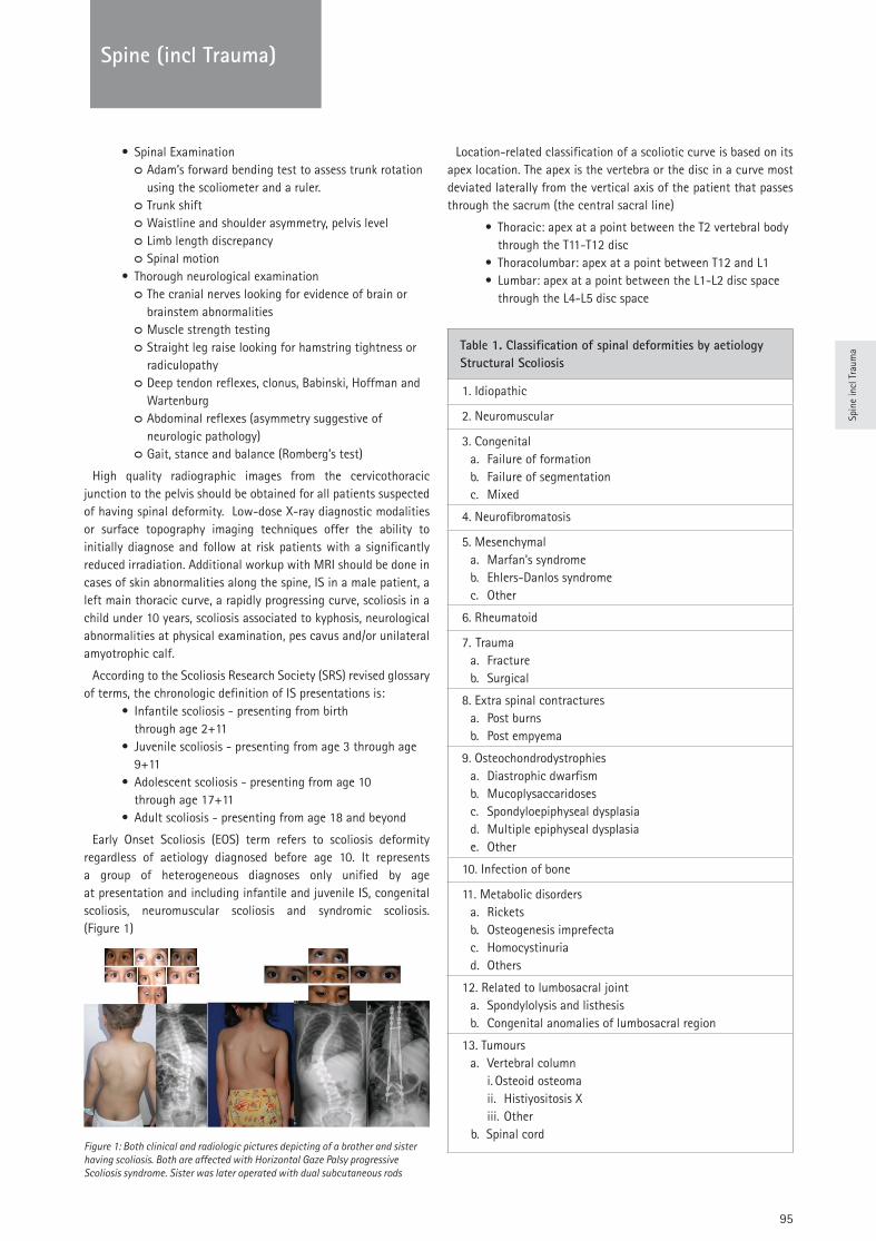
95
TRAUMA
Spin
e in
cl T
raum
a
Spine (incl Trauma)
• Spinal Examination ᴑ Adam’s forward bending test to assess trunk rotation using the scoliometer and a ruler.
ᴑ Trunk shift ᴑ Waistline and shoulder asymmetry, pelvis level ᴑ Limb length discrepancy ᴑ Spinal motion
• Thorough neurological examination ᴑ The cranial nerves looking for evidence of brain or brainstem abnormalities
ᴑ Muscle strength testing ᴑ Straight leg raise looking for hamstring tightness or radiculopathy
ᴑ Deep tendon reflexes, clonus, Babinski, Hoffman and Wartenburg
ᴑ Abdominal reflexes (asymmetry suggestive of neurologic pathology)
ᴑ Gait, stance and balance (Romberg’s test)
High quality radiographic images from the cervicothoracic junction to the pelvis should be obtained for all patients suspected of having spinal deformity. Low-dose X-ray diagnostic modalities or surface topography imaging techniques offer the ability to initially diagnose and follow at risk patients with a significantly reduced irradiation. Additional workup with MRI should be done in cases of skin abnormalities along the spine, IS in a male patient, a left main thoracic curve, a rapidly progressing curve, scoliosis in a child under 10 years, scoliosis associated to kyphosis, neurological abnormalities at physical examination, pes cavus and/or unilateral amyotrophic calf.
According to the Scoliosis Research Society (SRS) revised glossary of terms, the chronologic definition of IS presentations is:
• Infantile scoliosis - presenting from birth through age 2+11
• Juvenile scoliosis - presenting from age 3 through age 9+11
• Adolescent scoliosis - presenting from age 10 through age 17+11
• Adult scoliosis - presenting from age 18 and beyond
Early Onset Scoliosis (EOS) term refers to scoliosis deformity regardless of aetiology diagnosed before age 10. It represents a group of heterogeneous diagnoses only unified by age at presentation and including infantile and juvenile IS, congenital scoliosis, neuromuscular scoliosis and syndromic scoliosis. (Figure 1)
Figure 1: Both clinical and radiologic pictures depicting of a brother and sister having scoliosis. Both are affected with Horizontal Gaze Palsy progressive Scoliosis syndrome. Sister was later operated with dual subcutaneous rods
Location-related classification of a scoliotic curve is based on its apex location. The apex is the vertebra or the disc in a curve most deviated laterally from the vertical axis of the patient that passes through the sacrum (the central sacral line)
• Thoracic: apex at a point between the T2 vertebral body through the T11-T12 disc
• Thoracolumbar: apex at a point between T12 and L1• Lumbar: apex at a point between the L1-L2 disc space
through the L4-L5 disc space
Table 1. Classification of spinal deformities by aetiology Structural Scoliosis
1. Idiopathic
2. Neuromuscular
3. Congenitala. Failure of formationb. Failure of segmentationc. Mixed
4. Neurofibromatosis
5. Mesenchymala. Marfan’s syndromeb. Ehlers-Danlos syndromec. Other
6. Rheumatoid
7. Traumaa. Fractureb. Surgical
8. Extra spinal contracturesa. Post burnsb. Post empyema
9. Osteochondrodystrophiesa. Diastrophic dwarfismb. Mucoplysaccaridosesc. Spondyloepiphyseal dysplasiad. Multiple epiphyseal dysplasiae. Other
10. Infection of bone
11. Metabolic disordersa. Ricketsb. Osteogenesis imprefectac. Homocystinuriad. Others
12. Related to lumbosacral jointa. Spondylolysis and listhesisb. Congenital anomalies of lumbosacral region
13. Tumoursa. Vertebral column
i. Osteoid osteomaii. Histiyositosis Xiii. Other
b. Spinal cord
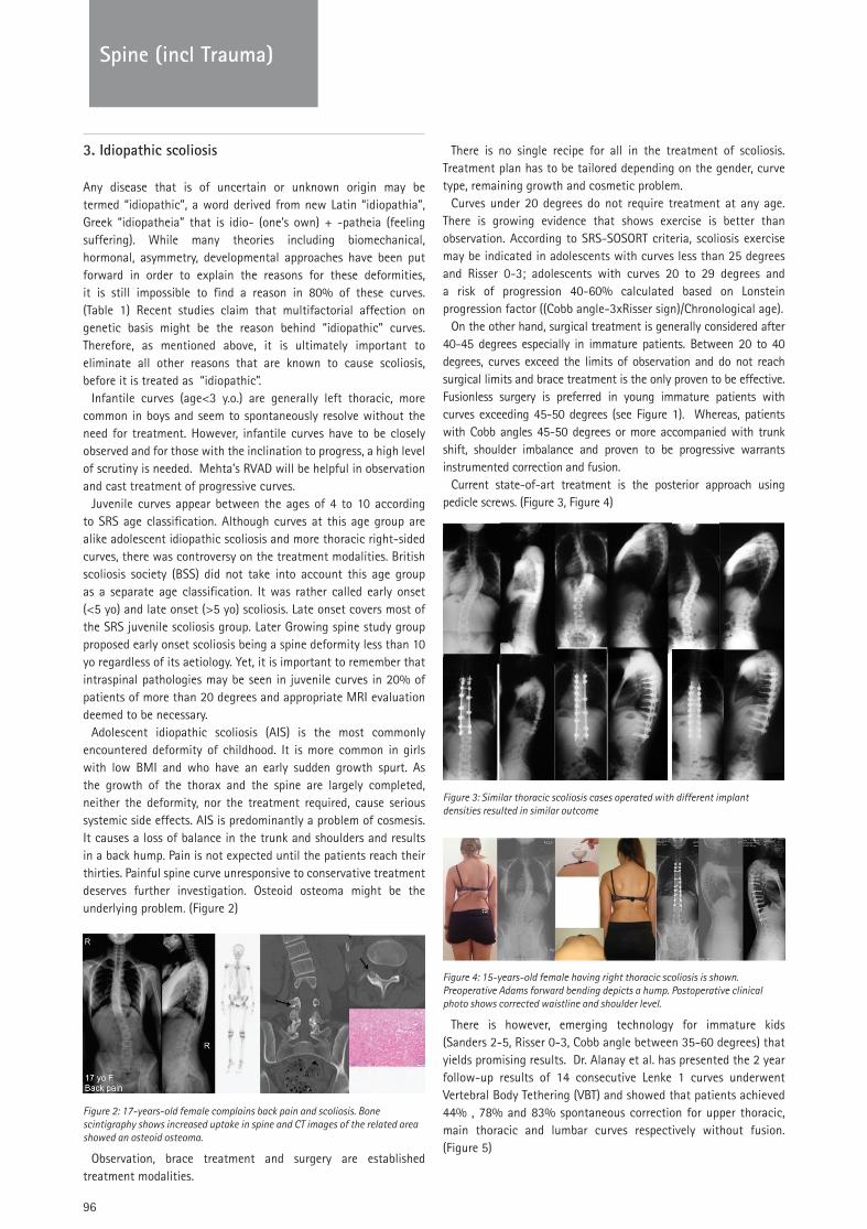
96
TRAUMASpine (incl Trauma)
3. Idiopathic scoliosis
Any disease that is of uncertain or unknown origin may be termed “idiopathic”, a word derived from new Latin “idiopathia”, Greek “idiopatheia” that is idio- (one’s own) + -patheia (feeling suffering). While many theories including biomechanical, hormonal, asymmetry, developmental approaches have been put forward in order to explain the reasons for these deformities, it is still impossible to find a reason in 80% of these curves. (Table 1) Recent studies claim that multifactorial affection on genetic basis might be the reason behind “idiopathic” curves. Therefore, as mentioned above, it is ultimately important to eliminate all other reasons that are known to cause scoliosis, before it is treated as “idiopathic”.
Infantile curves (age<3 y.o.) are generally left thoracic, more common in boys and seem to spontaneously resolve without the need for treatment. However, infantile curves have to be closely observed and for those with the inclination to progress, a high level of scrutiny is needed. Mehta’s RVAD will be helpful in observation and cast treatment of progressive curves.
Juvenile curves appear between the ages of 4 to 10 according to SRS age classification. Although curves at this age group are alike adolescent idiopathic scoliosis and more thoracic right-sided curves, there was controversy on the treatment modalities. British scoliosis society (BSS) did not take into account this age group as a separate age classification. It was rather called early onset (<5 yo) and late onset (>5 yo) scoliosis. Late onset covers most of the SRS juvenile scoliosis group. Later Growing spine study group proposed early onset scoliosis being a spine deformity less than 10 yo regardless of its aetiology. Yet, it is important to remember that intraspinal pathologies may be seen in juvenile curves in 20% of patients of more than 20 degrees and appropriate MRI evaluation deemed to be necessary.
Adolescent idiopathic scoliosis (AIS) is the most commonly encountered deformity of childhood. It is more common in girls with low BMI and who have an early sudden growth spurt. As the growth of the thorax and the spine are largely completed, neither the deformity, nor the treatment required, cause serious systemic side effects. AIS is predominantly a problem of cosmesis. It causes a loss of balance in the trunk and shoulders and results in a back hump. Pain is not expected until the patients reach their thirties. Painful spine curve unresponsive to conservative treatment deserves further investigation. Osteoid osteoma might be the underlying problem. (Figure 2)
Figure 2: 17-years-old female complains back pain and scoliosis. Bone scintigraphy shows increased uptake in spine and CT images of the related area showed an osteoid osteoma.
Observation, brace treatment and surgery are established treatment modalities.
There is no single recipe for all in the treatment of scoliosis. Treatment plan has to be tailored depending on the gender, curve type, remaining growth and cosmetic problem.
Curves under 20 degrees do not require treatment at any age. There is growing evidence that shows exercise is better than observation. According to SRS-SOSORT criteria, scoliosis exercise may be indicated in adolescents with curves less than 25 degrees and Risser 0-3; adolescents with curves 20 to 29 degrees and a risk of progression 40-60% calculated based on Lonstein progression factor ((Cobb angle-3xRisser sign)/Chronological age).
On the other hand, surgical treatment is generally considered after 40-45 degrees especially in immature patients. Between 20 to 40 degrees, curves exceed the limits of observation and do not reach surgical limits and brace treatment is the only proven to be effective. Fusionless surgery is preferred in young immature patients with curves exceeding 45-50 degrees (see Figure 1). Whereas, patients with Cobb angles 45-50 degrees or more accompanied with trunk shift, shoulder imbalance and proven to be progressive warrants instrumented correction and fusion.
Current state-of-art treatment is the posterior approach using pedicle screws. (Figure 3, Figure 4)
Figure 3: Similar thoracic scoliosis cases operated with different implant densities resulted in similar outcome
Figure 4: 15-years-old female having right thoracic scoliosis is shown. Preoperative Adams forward bending depicts a hump. Postoperative clinical photo shows corrected waistline and shoulder level.
There is however, emerging technology for immature kids (Sanders 2-5, Risser 0-3, Cobb angle between 35-60 degrees) that yields promising results. Dr. Alanay et al. has presented the 2 year follow-up results of 14 consecutive Lenke 1 curves underwent Vertebral Body Tethering (VBT) and showed that patients achieved 44% , 78% and 83% spontaneous correction for upper thoracic, main thoracic and lumbar curves respectively without fusion. (Figure 5)
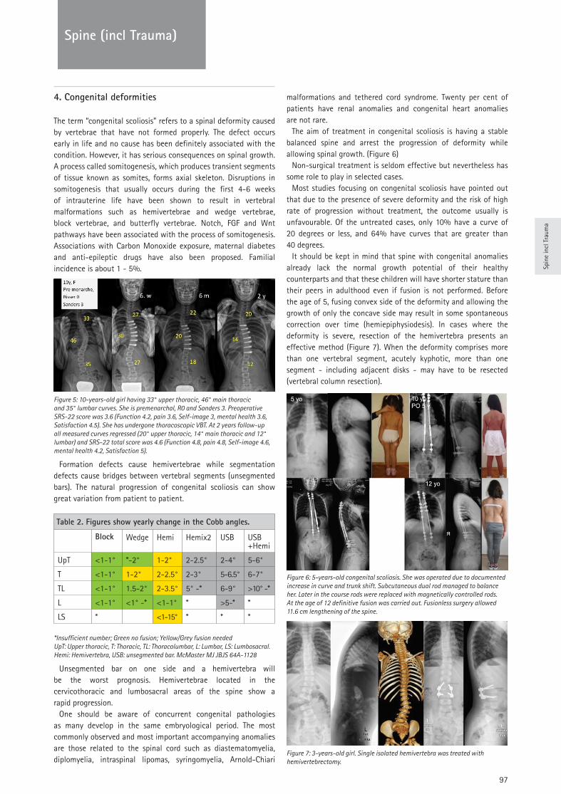
97
TRAUMA
Spin
e in
cl T
raum
a
Spine (incl Trauma)
4. Congenital deformities
The term “congenital scoliosis” refers to a spinal deformity caused by vertebrae that have not formed properly. The defect occurs early in life and no cause has been definitely associated with the condition. However, it has serious consequences on spinal growth. A process called somitogenesis, which produces transient segments of tissue known as somites, forms axial skeleton. Disruptions in somitogenesis that usually occurs during the first 4-6 weeks of intrauterine life have been shown to result in vertebral malformations such as hemivertebrae and wedge vertebrae, block vertebrae, and butterfly vertebrae. Notch, FGF and Wnt pathways have been associated with the process of somitogenesis. Associations with Carbon Monoxide exposure, maternal diabetes and anti-epileptic drugs have also been proposed. Familial incidence is about 1 - 5%.
Figure 5: 10-years-old girl having 33° upper thoracic, 46° main thoracic and 35° lumbar curves. She is premenarchal, R0 and Sanders 3. Preoperative SRS-22 score was 3.6 (Function 4.2, pain 3.6, Self-image 3, mental health 3.6, Satisfaction 4.5). She has undergone thoracoscopic VBT. At 2 years follow-up all measured curves regressed (20° upper thoracic, 14° main thoracic and 12° lumbar) and SRS-22 total score was 4.6 (Function 4.8, pain 4.8, Self-image 4.6, mental health 4.2, Satisfaction 5).
Formation defects cause hemivertebrae while segmentation defects cause bridges between vertebral segments (unsegmented bars). The natural progression of congenital scoliosis can show great variation from patient to patient.
*Insufficient number; Green no fusion; Yellow/Grey fusion needed UpT: Upper thoracic, T: Thoracic, TL: Thoracolumbar, L: Lumbar, LS: Lumbosacral. Hemi: Hemivertebra, USB: unsegmented bar. McMaster MJ JBJS 64A-1128
Unsegmented bar on one side and a hemivertebra will be the worst prognosis. Hemivertebrae located in the cervicothoracic and lumbosacral areas of the spine show a rapid progression.
One should be aware of concurrent congenital pathologies as many develop in the same embryological period. The most commonly observed and most important accompanying anomalies are those related to the spinal cord such as diastematomyelia, diplomyelia, intraspinal lipomas, syringomyelia, Arnold-Chiari
malformations and tethered cord syndrome. Twenty per cent of patients have renal anomalies and congenital heart anomalies are not rare.
The aim of treatment in congenital scoliosis is having a stable balanced spine and arrest the progression of deformity while allowing spinal growth. (Figure 6)
Non-surgical treatment is seldom effective but nevertheless has some role to play in selected cases.
Most studies focusing on congenital scoliosis have pointed out that due to the presence of severe deformity and the risk of high rate of progression without treatment, the outcome usually is unfavourable. Of the untreated cases, only 10% have a curve of 20 degrees or less, and 64% have curves that are greater than 40 degrees.
It should be kept in mind that spine with congenital anomalies already lack the normal growth potential of their healthy counterparts and that these children will have shorter stature than their peers in adulthood even if fusion is not performed. Before the age of 5, fusing convex side of the deformity and allowing the growth of only the concave side may result in some spontaneous correction over time (hemiepiphysiodesis). In cases where the deformity is severe, resection of the hemivertebra presents an effective method (Figure 7). When the deformity comprises more than one vertebral segment, acutely kyphotic, more than one segment - including adjacent disks - may have to be resected (vertebral column resection).
Figure 6: 5-years-old congenital scoliosis. She was operated due to documented increase in curve and trunk shift. Subcutaneous dual rod managed to balance her. Later in the course rods were replaced with magnetically controlled rods. At the age of 12 definitive fusion was carried out. Fusionless surgery allowed 11.6 cm lengthening of the spine.
Figure 7: 3-years-old girl. Single isolated hemivertebra was treated with hemivertebrectomy.
Table 2. Figures show yearly change in the Cobb angles.
Block Wedge Hemi Hemix2 USB USB +Hemi
UpT <1-1° *-2° 1-2° 2-2.5° 2-4° 5-6°
T <1-1° 1-2° 2-2.5° 2-3° 5-6.5° 6-7°
TL <1-1° 1.5-2° 2-3.5° 5° -* 6-9° >10° -*
L <1-1° <1° -* <1-1° * >5-* *
LS * <1-15° * * *
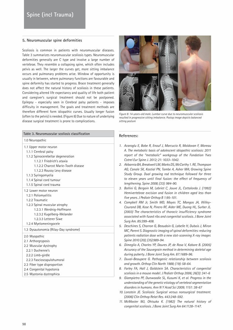
98
TRAUMASpine (incl Trauma)
5. Neuromuscular spine deformities
Scoliosis is common in patients with neuromuscular diseases. Table 3 summarizes neuromuscular scoliosis types. Neuromuscular deformities generally are C type and involve a large number of vertebrae. They resemble a collapsing spine, which often includes pelvis as well. The larger the curves get, more sitting imbalance occurs and pulmonary problems arise. Window of opportunity is usually in between, where pulmonary functions are favourable and spine deformity has started to progress. Brace treatment generally does not affect the natural history of scoliosis in these patients. Considering altered life expectancy and quality of life both patient and caregiver’s surgical treatment should not be postponed. Epilepsy - especially seen in Cerebral palsy patients - imposes difficulty in management. The goals and treatment methods are therefore different form idiopathic curves. Usually longer fusion (often to the pelvis) is needed. (Figure 8) Due to nature of underlying disease surgical treatment is prone to complications.
Figure 8: 14-years-old male. Lumbar curve due to neuromuscular scoliosis resulted in progressive sitting imbalance. Postop image depicts balanced sitting posture
References:
1. Acaroglu E, Bobe R, Enouf J, Marcucio R, Moldovan F, Moreau A. The metabolic basis of adolescent idiopathic scoliosis: 2011 report of the “metabolic” workgroup of the Fondation Yves Cotrel Eur Spine J. 2012; 21: 1033–1042.
2. Akbarnia BA, Breakwell LM, Marks DS, McCarthy 1. RE, Thompson AG, Canale SK, Kostial PN, Tambe A, Asher MA; Growing Spine Study Group. Dual growing rod technique followed for three to eleven years until final fusion: the effect of frequency of lengthening. Spine 2008; (33): 984-90.
3. Bollini G, Bergoin M, Labriet C, Jouve JL, Cottalorda J. (1992) Hemivertebrae excision and fusion in children aged less than five years. J Pediatr Orthop B 1:95-101.
4. Campbell RM Jr, Smith MD, Mayes TC, Mangos JA, Willey-Courand DB, Kose N, Pinero RF, Alder ME, Duong HL, Surber JL. (2003) The characteristics of thoracic insufficiency syndrome associated with fused ribs and congenital scoliosis. J Bone Joint Surg Am. 85:399-408.
5. Deschines S, Charron G, Beaudoin G, Labelle H, Dubois J, Miron MC, Parent S. Diagnostic imaging of spinal deformities: reducing patients radiation dose with a new slot-scanning X-ray imager. Spine 2010 (20); (35):989-94.
6. Dimeglio A, Charles YP, Daures JP, de Rosa V, Kabore B. (2005) Accuracy of the Sauvegrain method in determining skeletal age during puberty. J Bone Joint Surg Am. 87:1689-96.
7. Duval-Beaupere G. Pathogenic relationship between scoliosis and growth. Orthop Clin North 1988; (19): 58-64.
8. Farley FA, Hall J, Goldstein SA. Characteristics of congenital scoliosis in a mouse model. J Pediatr Orthop 2006; 26(3): 341-6
9. Giampietro PF, Dunwoodie SL, Kusumi K, et al. Progress in the understanding of the genetic etiology of vertebral segmentation disorders in humans. Ann N Y Acad Sci 2009; 1151: 38-67
10. Lonstein JE. Scoliosis: Surgical versus nonsurgical treatment. (2006) Clin Orthop Relat Res. 443:248-592.
11. McMaster MJ, Ohtsuka K. (1982) The natural history of congenital scoliosis. J Bone Joint Surg Am 64:1128-1147.
Table 3. Neuromuscular scoliosis classification
1.0 Neuropathic
1.1 Upper motor neuron1.1.1 Cerebral palsy1.1.2 Spinocerebellar degeneration
1.1.2.1 Friedrich’s ataxia1.1.2.2 Charcot Marie-Tooth disease1.1.2.3 Roussy Levy disease
1.1.3 Syringomyelia1.1.4 Spinal cord tumour1.1.5 Spinal cord trauma
1.2 Lower motor neuron1.2.1 Poliomyelitis1.2.2 Traumatic1.2.3 Spinal muscular atrophy
1.2.3.1 Werdnig-Hoffmann1.2.3.2 Kugelberg-Welander1.2.3.3 Letterer Siwe
1.2.4 Myelomeningocele
1.3 Dysautonomia (Rilay-Day syndrome)
2.0 Myopathic2.1 Arthorgryposis2.2 Muscular dystrophy
2.2.1 Duchenne’s2.2.2 Limb-girdle2.2.3 Fascioscapulohumoral
2.3 Fiber type disproportion2.4 Congenital hypotonia2.5 Myotonia dystrophica
99
TRAUMA
Spin
e in
cl T
raum
a
Spine (incl Trauma)
12. Mehta MH Growth as a corrective force in the early treatment of progressive infantile scoliosis. J Bone Joint Surg [2005;87-B:1237-47.
13. Mehta MH. The rib-vertebra angle in the early diagnosis between resolving and pro- gressive infantile scoliosis. J Bone Joint Surg 1972;54-B:230-43
14. Nasca RJ, Stilling FH III, Stell HH. Progression of congenital scoliosis due to hemivertebrae and hemivertebrae with bars. J Bone Joint Surg Am 1975; 57(4): 456-66.]
15. Negrini S, Minozzi S, Bettany-Saltikov J, Zaina F, Chockalingam N, Grivas TB, Kotwicki T, Maruyama T, Romano M, Vasiliadis ES. Braces for idiopathic scoliosis in adolescents. Spine 2010; (35): 1285-93.
16. Negrini S, Minozzi S, Bettany-Saltikov J, Zaina F, Chockalingam N, Grivas TB, Kotwicki T, Maruyama T, Romano M, Vasiliadis ES. Braces for idiopathic scoliosis in adolescents. Cochrane Database Syst Rev. 2010 Janv 20; (1). CD006850.
17. Nelson S, Sanders J. Idiopathic scoliosis 2018 available at http://etext.srs.org/book/ accessed 10.2.2018,
18. Pahys JM, Guille JT. What’s New in Congenital Scoliosis? J Pediatr Orthop. 2018; 38: e173
19. Parent S, Newton PO, Wenger DR. (2005) Adolescent idiopathic scoliosis: Etiology, anatomy, natural history, and bracing. Instr Course Lect 54:529-36.
20. Sanders JO, KhouryJG, Kishan S, Browne RH, Mooney JF, Arnold KD, Sharon McConnell J, Bauma JA, Finegold DA. Predicting Scoliosis Progression from Skeletal Maturity: A Simplified Classification During Adolescence. J Bone Joint Surg -A 2008; 90:540-53.
21. Shahcheraghi GH, Hobbi MH. Patterns and progression in congenital scoliosis. J Pediatr Orthop 1999; 19(6): 766-75
22. Suk SI, Kim JH, Kim WJ, Lee SM, Chung ER, Nah KH. (2002) Posterior vertebral column resection for severe spinal deformities. Spine 27:2374-8.
23. Uzumcugil A, Cil A, Yazici M, Acaroglu E, Alanay A, Aksoy C, Surat A. (2004) Convex growth arrest in the treatment of congenital spinal deformities, revisited. J Pediatr Orthop. 24:658-66.
24. Wang WJ, Yeung HY, Chu WC, Tang NL, Lee KM, Qiu Y, Burwell RG, Cheng JC. Top theories for the etiopathogenesis of adolescent idiopathic scoliosis. J Pediatr Orthop 2011; (31): S14-27.
25. Winter RB, Moe JH, Lonstein JE.(1984) Posterior spinal arthrodesis for congenital scoliosis. J Bone Joint Surg 66A: 1188-1197.
26. Wybier M, Bossard P. Musculoskeletal imaging in progress: The EOS imaging system. Joint Bone Spine
27. Yazici M, Asher MA, Hardacker JW. (2000) Safety and efficacy of Isola-Galveston instrumentation and fusion in the treatment of neuromuscular spinal deformities. J Bone Joint Surg Am 82:524-43.
28. Yazici M, Yilmaz G. (2011) Congenital scoliosis. In: Akbarnia B, Yazici M, Thompson GH (eds) The growing spine. Springer Berlin Heidelberg, pp: 213-28.
Questions
1. Which of the following is true for developing thorax and spine?a. Alveolar multiplication occurs until the age of 5, b. Thoracic capacity is only at the 30% range of adult capacity
by the age of 10 c. Thoracic capacity is only at the 50% at age 10 d. Spine has achieved its 50% of mature height by age 10. e. Deformities that occur before the age of 5 will only affect
spinal alignment .
2. Early Onset Scoliosis by definition refers to:a. Scoliosis deformity regardless of aetiology diagnosed before
age 10 b. Congenital scoliosis diagnosed by the age of 5c. Infantile scoliosis up to age of 3d. Juvenile scoliosis up to age of 9e. Adolescent idiopathic scoliosis at age of 10
3. Which of the following statements regarding congenital scoliosis is true?
a. Disruptions in somitogenesis that usually occurs during the first 4-6 weeks of intrauterine life have been shown to result in vertebral malformations
b. Notch, FGF and Wnt signaling pathways have been associated with the process of somitogenesis.
c. Associations with Carbon Monoxide exposure, maternal diabetes and anti-epileptic drugs have also been proposed.
d. Familial incidence is about 1 - 5%.e. All of the above
Answers1c, 2a, 3e.
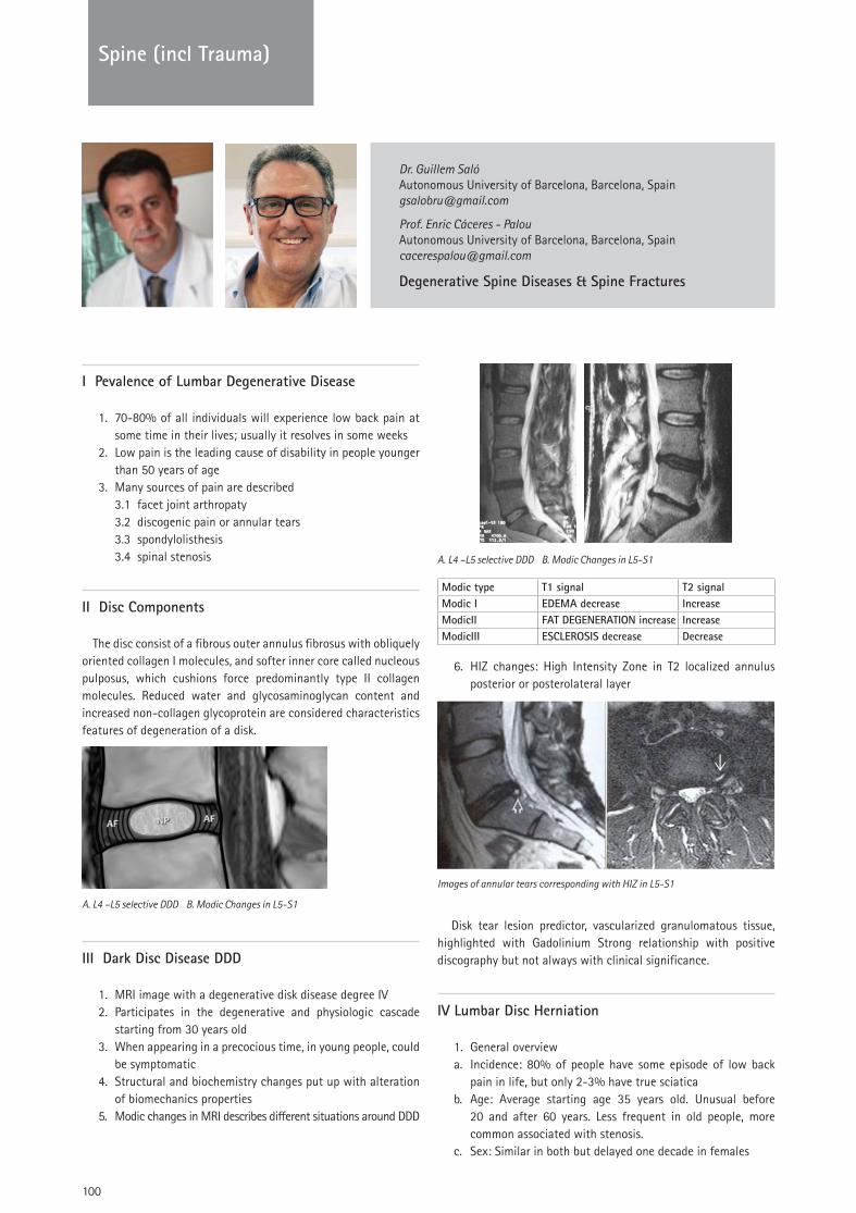
100
Spine (incl Trauma)
Dr. Guillem SalóAutonomous University of Barcelona, Barcelona, [email protected]
Prof. Enric Cáceres - PalouAutonomous University of Barcelona, Barcelona, [email protected]
Degenerative Spine Diseases & Spine Fractures
I Pevalence of Lumbar Degenerative Disease
1. 70-80% of all individuals will experience low back pain at some time in their lives; usually it resolves in some weeks
2. Low pain is the leading cause of disability in people younger than 50 years of age
3. Many sources of pain are described3.1 facet joint arthropaty3.2 discogenic pain or annular tears3.3 spondylolisthesis3.4 spinal stenosis
II Disc Components
The disc consist of a fibrous outer annulus fibrosus with obliquely oriented collagen I molecules, and softer inner core called nucleous pulposus, which cushions force predominantly type II collagen molecules. Reduced water and glycosaminoglycan content and increased non-collagen glycoprotein are considered characteristics features of degeneration of a disk.
A. L4 –L5 selective DDD B. Modic Changes in L5-S1
III Dark Disc Disease DDD
1. MRI image with a degenerative disk disease degree IV2. Participates in the degenerative and physiologic cascade
starting from 30 years old3. When appearing in a precocious time, in young people, could
be symptomatic4. Structural and biochemistry changes put up with alteration
of biomechanics properties5. Modic changes in MRI describes different situations around DDD
A. L4 –L5 selective DDD B. Modic Changes in L5-S1
Modic type T1 signal T2 signalModic I EDEMA decrease IncreaseModicII FAT DEGENERATION increase IncreaseModicIII ESCLEROSIS decrease Decrease
6. HIZ changes: High Intensity Zone in T2 localized annulus posterior or posterolateral layer
Images of annular tears corresponding with HIZ in L5-S1
Disk tear lesion predictor, vascularized granulomatous tissue, highlighted with Gadolinium Strong relationship with positive discography but not always with clinical significance.
IV Lumbar Disc Herniation
1. General overviewa. Incidence: 80% of people have some episode of low back
pain in life, but only 2-3% have true sciaticab. Age: Average starting age 35 years old. Unusual before
20 and after 60 years. Less frequent in old people, more common associated with stenosis.
c. Sex: Similar in both but delayed one decade in females
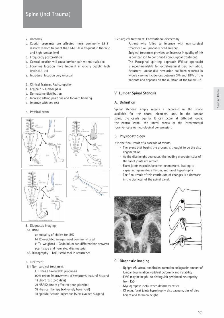
Spin
e in
cl T
raum
a
101
Spine (incl Trauma)
2. Anatomya. Caudal segments are affected more commonly L5-S1
discreetly more frequent than L4-L5 less frequent in thoracic and high lumbar level
b. Frequently posterolateralc. Central location will cause lumbar pain without sciaticad. Foramina location more frequent in elderly people; high
levels (L3-L4) e. Intradural location very unusual
3. Clinical features Radiculopathya. Leg pain > lumbar painb. Dermatome distributionc. Increase sitting positions and forward bendingd. Improve with bed rest
4. Physical exam
5. Diagnostic imaging 5A. RNM a) modality of choice for LHD b) T2-weighted images most commonly used c) T1-weighted + Gadolinium can differentiate between scar tissue and herniated disc material 5B. Discography + TAC useful tool in recurrence
6. Treatment 6.1 Non-surgical treatment: LDH has a favourable prognosis 90% report improvement of symptoms (natural history) 1) Short rest (3-5 days) 2) NSAIDs (more effective than placebo) 3) Physical therapy (extremely beneficial) 4) Epidural steroid injections (50% avoided surgery)
6.2 Surgical treatment: Conventional discectomyPatient who failed to improve with non-surgical treatment will probably need surgery.Surgical treatment provided an increase in quality of life in comparison to continued non-surgical treatment.The Paraspinal splitting approach (Wiltse approach) is recommendable for extraforaminal disc herniation. Recurrent lumbar disc herniation has been reported in widely varying incidences between 3% and 18% of the patients and depends on the duration of the follow-up.
V Lumbar Spinal Stenosis
A. Definition
Spinal stenosis simply means a decrease in the space available for the neural elements, and, in the lumbar spine, the cauda equina. It can occur at different levels: the central canal, the lateral recess or the intervertebral foramen causing neurological compression.
B. Physiopathology
It is the final result of a cascade of events. - The event that begins the process is thought to be the disc
degeneration. - As the disc height decreases, the loading characteristics of
the facet joints are altered. - Facet joints capsules become incompetent, leading to
capsular, ligamentous flavum, and facet hypertrophy. - The final result of this continuum of changes is a decrease
in the diameter of the spinal canal.
C. Diagnostic imaging
- Upright AP, lateral, and flexion-extension radiographs amount of lumbar degeneration, vertebral deformity and instability.
- EMG may be helpful to distinguish peripheral neuropathy from LSS.
- Myelography: useful when deformity exists. - CT scan: facet joints hypertrophy, disc vacuum, size of disc
height and foramen height.
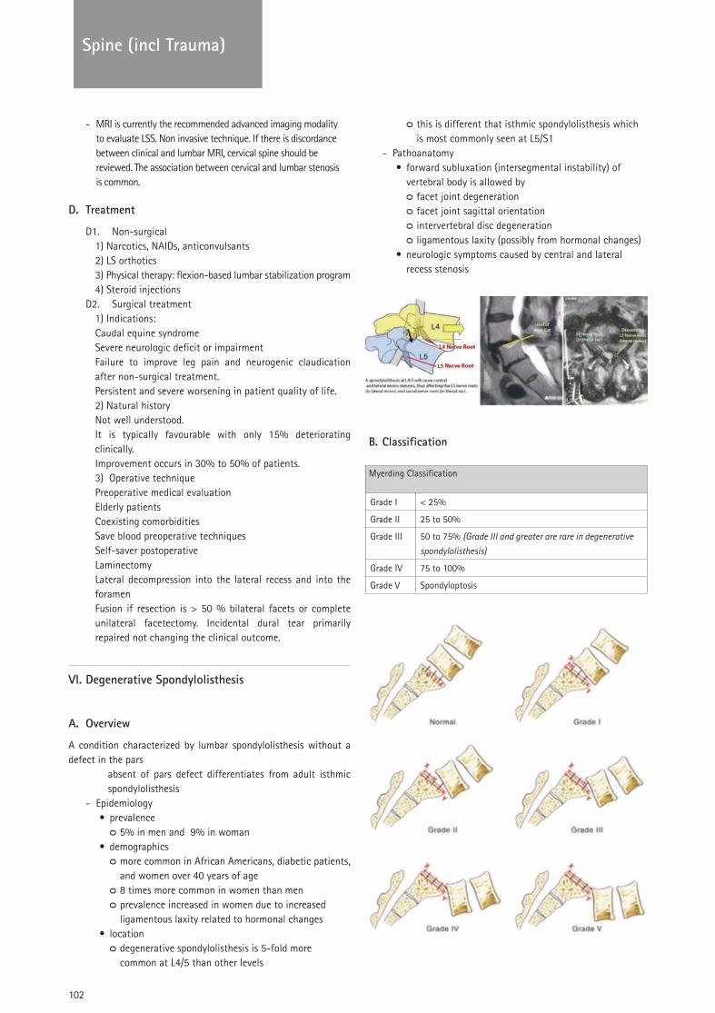
102
TRAUMASpine (incl Trauma)
- MRI is currently the recommended advanced imaging modality to evaluate LSS. Non invasive technique. If there is discordance between clinical and lumbar MRI, cervical spine should be reviewed. The association between cervical and lumbar stenosis is common.
D. Treatment
D1. Non-surgical1) Narcotics, NAIDs, anticonvulsants2) LS orthotics3) Physical therapy: flexion-based lumbar stabilization program4) Steroid injections
D2. Surgical treatment1) Indications:Caudal equine syndromeSevere neurologic deficit or impairmentFailure to improve leg pain and neurogenic claudication after non-surgical treatment. Persistent and severe worsening in patient quality of life.2) Natural historyNot well understood.It is typically favourable with only 15% deteriorating clinically. Improvement occurs in 30% to 50% of patients.3) Operative techniquePreoperative medical evaluationElderly patientsCoexisting comorbiditiesSave blood preoperative techniquesSelf-saver postoperativeLaminectomyLateral decompression into the lateral recess and into the foramenFusion if resection is > 50 % bilateral facets or complete unilateral facetectomy. Incidental dural tear primarily repaired not changing the clinical outcome.
VI. Degenerative Spondylolisthesis
A. Overview
A condition characterized by lumbar spondylolisthesis without a defect in the pars
absent of pars defect differentiates from adult isthmic spondylolisthesis
- Epidemiology• prevalence
ᴑ 5% in men and 9% in woman• demographics
ᴑ more common in African Americans, diabetic patients, and women over 40 years of age
ᴑ 8 times more common in women than men ᴑ prevalence increased in women due to increased ligamentous laxity related to hormonal changes
• location ᴑ degenerative spondylolisthesis is 5-fold more common at L4/5 than other levels
ᴑ this is different that isthmic spondylolisthesis which is most commonly seen at L5/S1
- Pathoanatomy• forward subluxation (intersegmental instability) of
vertebral body is allowed by ᴑ facet joint degeneration ᴑ facet joint sagittal orientation ᴑ intervertebral disc degeneration ᴑ ligamentous laxity (possibly from hormonal changes)
• neurologic symptoms caused by central and lateral recess stenosis
B. Classification
Myerding Classification
Grade I < 25%
Grade II 25 to 50%
Grade III 50 to 75% (Grade III and greater are rare in degenerative spondylolisthesis)
Grade IV 75 to 100%
Grade V Spondyloptosis
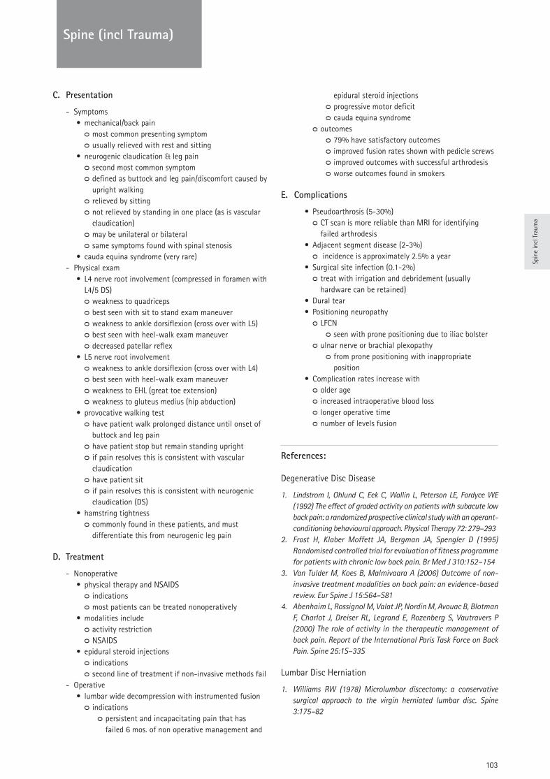
103
TRAUMA
Spin
e in
cl T
raum
a
Spine (incl Trauma)
C. Presentation
- Symptoms• mechanical/back pain
ᴑ most common presenting symptom ᴑ usually relieved with rest and sitting
• neurogenic claudication & leg pain ᴑ second most common symptom ᴑ defined as buttock and leg pain/discomfort caused by upright walking
ᴑ relieved by sitting ᴑ not relieved by standing in one place (as is vascular claudication)
ᴑ may be unilateral or bilateral ᴑ same symptoms found with spinal stenosis
• cauda equina syndrome (very rare) - Physical exam
• L4 nerve root involvement (compressed in foramen with L4/5 DS)
ᴑ weakness to quadriceps ᴑ best seen with sit to stand exam maneuver ᴑ weakness to ankle dorsiflexion (cross over with L5) ᴑ best seen with heel-walk exam maneuver ᴑ decreased patellar reflex
• L5 nerve root involvement ᴑ weakness to ankle dorsiflexion (cross over with L4) ᴑ best seen with heel-walk exam maneuver ᴑ weakness to EHL (great toe extension) ᴑ weakness to gluteus medius (hip abduction)
• provocative walking test ᴑ have patient walk prolonged distance until onset of buttock and leg pain
ᴑ have patient stop but remain standing upright ᴑ if pain resolves this is consistent with vascular claudication
ᴑ have patient sit ᴑ if pain resolves this is consistent with neurogenic claudication (DS)
• hamstring tightness ᴑ commonly found in these patients, and must differentiate this from neurogenic leg pain
D. Treatment
- Nonoperative• physical therapy and NSAIDS
ᴑ indications ᴑ most patients can be treated nonoperatively
• modalities include ᴑ activity restriction ᴑ NSAIDS
• epidural steroid injections ᴑ indications ᴑ second line of treatment if non-invasive methods fail
- Operative• lumbar wide decompression with instrumented fusion
ᴑ indications ᴑ persistent and incapacitating pain that has failed 6 mos. of non operative management and
epidural steroid injections ᴑ progressive motor deficit ᴑ cauda equina syndrome
ᴑ outcomes ᴑ 79% have satisfactory outcomes ᴑ improved fusion rates shown with pedicle screws ᴑ improved outcomes with successful arthrodesis ᴑ worse outcomes found in smokers
E. Complications
• Pseudoarthrosis (5-30%) ᴑ CT scan is more reliable than MRI for identifying failed arthrodesis
• Adjacent segment disease (2-3%) ᴑ incidence is approximately 2.5% a year
• Surgical site infection (0.1-2%) ᴑ treat with irrigation and debridement (usually hardware can be retained)
• Dural tear• Positioning neuropathy
ᴑ LFCN ᴑ seen with prone positioning due to iliac bolster
ᴑ ulnar nerve or brachial plexopathy ᴑ from prone positioning with inappropriate position
• Complication rates increase with ᴑ older age ᴑ increased intraoperative blood loss ᴑ longer operative time ᴑ number of levels fusion
References:
Degenerative Disc Disease
1. Lindstrom I, Ohlund C, Eek C, Wallin L, Peterson LE, Fordyce WE (1992) The effect of graded activity on patients with subacute low back pain: a randomized prospective clinical study with an operant-conditioning behavioural approach. Physical Therapy 72: 279–293
2. Frost H, Klaber Moffett JA, Bergman JA, Spengler D (1995) Randomised controlled trial for evaluation of fitness programme for patients with chronic low back pain. Br Med J 310:152–154
3. Van Tulder M, Koes B, Malmivaara A (2006) Outcome of non-invasive treatment modalities on back pain: an evidence-based review. Eur Spine J 15:S64–S81
4. Abenhaim L, Rossignol M, Valat JP, Nordin M, Avouac B, Blotman F, Charlot J, Dreiser RL, Legrand E, Rozenberg S, Vautravers P (2000) The role of activity in the therapeutic management of back pain. Report of the International Paris Task Force on Back Pain. Spine 25:1S–33S
Lumbar Disc Herniation
1. Williams RW (1978) Microlumbar discectomy: a conservative surgical approach to the virgin herniated lumbar disc. Spine 3:175–82
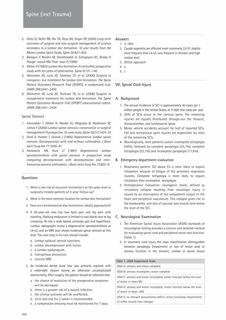
104
TRAUMASpine (incl Trauma)
2. Atlas SJ, Keller RB, Wu YA, Deyo RA, Singer DE (2005) Long-term outcomes of surgical and non-surgical management of sciatica secondary to a lumbar disc herniation: 10 year results from the Maine Lumbar Spine Study. Spine 30:927–935
3. Balague F, Nordin M, Sheikhzadeh A, Echegoyen AC, Brisby H Hooge- woud HM, Fred- man P (1999)
4. Weber H (1983) Lumbar disc herniation. A controlled, prospective study with ten years of observation. Spine 8:131–140
5. Weinstein JN, Lurie JD, Tosteson TD, et al. (2006) Surgical vs nonopera- tive treatment for lumbar disk herniation. The Spine Patient Outcomes Research Trial (SPORT), a randomized trial. JAMA 296:2441–2450
6. Weinstein JN, Lurie JD, Tosteson TD, et al. (2006) Surgical vs nonoperative treatment for lumbar disk herniation. The Spine Patient Outcomes Research Trial (SPORT) observational cohort. JAMA 296:2451–2459
Spinal Stenosis
1. Amundsen T, Weber H, Nordal HJ, Magnaes B, Abdelnoor M, Lilleas F (2000) Lumbar spinal stenosis: conservative or surgical management? A prospective 10-year study. Spine 25(11):1424–35
2. Grob D, Humke T, Dvorak J (1995) Degenerative lumbar spinal stenosis. Decompression with and without arthrodesis. J Bone Joint Surg Am 77:1036–41
3. Herkowitz HN, Kurz LT (1991) Degenerative lumbar spondylolisthesis with spinal stenosis. A prospective study comparing decompression with decompression and inter- transverse process arthrodesis. J Bone Joint Surg Am 73:802–8
Questions
1. What is the risk of recurrent herniation’s at the same level in surgically treated patients at 5-year follow-up?
2. What is the most common location for lumbar disc herniation?
3. How are extraforaminal disc herniations ideally approached?
4. A 65-year-old man has low back pain and leg pain with standing. Walking endurance is limited to two blocks due to leg cramping. He has a wide-based, unsteady gait and hyperflexia. Lumbar radiographs reveal a degenerative spondylolisthesis at L4-L5, and an MRI scan shows moderate spinal stenosis at this level. The next step in his care should include:
a. lumbar epidural steroid injections. b. lumbar decompression with fusion. c. a lumbar epidurogram. d. interspinous distraction. e. cervical MRI.
5. An incidental dental dural tear was primarily repaired with a watertight closure during an otherwise uncomplicated laminectomy. After surgery, the patient should be informed that:
a. the chance of resolution of the preoperative symptoms will be decreased.
b. there is a greater risk of a wound infection. c. the clinical outcome will be unaffected. d. strict bed rest for 2 weeks is recommended. e. a compression dressing must be maintained for 7 days.
Answers 1. 3-18%2. Caudal segments are affected more commonly, L5-S1 slightly
more frequent than L4-L5. Less frequent in thoracic and high lumbar level
3. Wiltse approach4. a5. c
VII. Spinal Cord Injury
A. Background
1. The annual incidence of SCI is approximately 40 cases per 1 million people in the United States, or 11.000 new cases per year.
2. 55% of SCIs occur in the cervical spine. The remaining injuries are equally distributed through-out the thoracic, thoracolumbar, and lumbosacral spine.
3. Motor vehicle accidents account for half of reported SCIs. Fall and recreational sport injuries are responsible for most of the remaining SCIs.
4. Neurologically, most patients sustain incomplete tetraplegia (34%), followed by complete paraplegia (25,1%), complete tetraplegia (22,1%) and incomplete paraplegia (17,5%).
B. Emergency department evaluation
1. Respiratory pattern: SCI above C5 is more likely to require intubation, because of fatigue of the accessory respiratory muscles. Complete tetraplegia is more likely to require intubation than incomplete tetraplegia.
2. Hemodynamic evaluation: neurogenic shock, defined as circulatory collapse resulting from neurologic injury, is caused by an interruption of the sympathetic output to the heart and peripheral vasculature. This collapse gives rise to the bradycardia and loss of vascular and muscle tone below the level of the SCI.
C. Neurological Examination
1. The American Spinal Injury Association (ASIA) standards of neurological testing provides a concise and detailed method for evaluating spinal cord and peripheral nerve root function. (Table 1)
2. In traumatic cord injury the main classification distinguishes between paraplegia (impairment or loss of motor and/ or sensory function in the thoracic, lumbar or sacral neural
Table 1. ASIA Impairment Scale
ASIA A: sensory and motor complete
ASIA B: sensory incomplete, motor complete
ASIA C: sensory and motor incomplete, motor function below the level
of lesion in mean M3
ASIA D: sensory and motor incomplete, motor function below the level
of lesion in mean >M3
ASIA E: no relevant sensor/motor deficit, minor functional impairments
of reflex-muscle tone changes
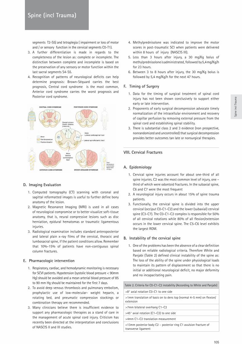
105
TRAUMA
Spin
e in
cl T
raum
a
Spine (incl Trauma)
segments T2-S5) and tetraplegia ( impairment or loss of motor and / or sensory function in the cervical segments C0-T1).
3. A further differentiation is made in regards to the completeness of the lesion as: complete or incomplete. The distinction between complete and incomplete is based on the preservation of any sensory or motor function within the last sacral segments S4-S5.
4. Recognition of patterns of neurological deficits can help determine prognosis: Brown-Séquard carries the best prognosis, Central cord syndrome is the most common, Anterior cord syndrome carries the worst prognosis and Posterior cord syndrome.
D. Imaging Evaluation
1. Computed tomography (CT) scanning with coronal and sagittal reformatted images is useful to further define bony anatomy of the lesion.
2. Magnetic Resonance Imaging (MRI) is used in all cases of neurological compromise or to better visualize soft-tissue anatomy, that is, neural compressive lesions such as disc herniation, epidural hematomas or traumatic ligamentous injuries.
3. Radiological examination includes standard anteroposterior and lateral plain x-ray films of the cervical, thoracic and lumbosacral spine, if the patient conditions allow. Remember that 10%-15% of patients have non-contiguous spinal column fractures.
E. Pharmacologic intervention
1. Respiratory, cardiac, and hemodynamic monitoring is necessary for SCVI patients. Hypotension (systolic blood pressure < 90mm Hg) should be avoided and a mean arterial blood pressure of 85 to 90 mm Hg should be maintained for the first 7 days.
2. To avoid deep venous thrombosis and pulmonary embolism, prophylactic use of low-molecular- weight heparin, a rotating bed, and pneumatic compression stockings or combination therapy are recommended.
3. Many clinicians believe there is insufficient evidence to support any pharmacologic therapies as a stand of care in the management of acute spinal cord injury. Criticism has recently been directed at the interpretation and conclusions of NASCIS II and III studies.
4. Methylprednisolone was indicated to improve the motor scores in post-traumatic SCI when patients were delivered within 8 hours of injury (NASCIS III).
5. Less than 3 hours after injury, a 30 mg/Kg bolus of methylprednisolone is administrated, followed by 5,4 mg/Kg/h for 23 hours.
6. Between 3 to 8 hours after injury, the 30 mg/Kg bolus is followed by 5,4 mg/Kg/h for the next 47 hours.
F. Timing of Surgery
1. Data for the timing of surgical treatment of spinal cord injury has not been shown conclusively to support either early or late intervention.
2. Proponents of early surgical decompression advocate timely normalization of the intracellular environment and recovery of capillar perfusion by removing external pressure from the spinal cord and establishing spinal stability.
3. There is substantial class 2 and 3 evidence (non prospective, nonrandomized and uncontrolled) that surgical decompression provides better outcomes tan late or nonsurgical therapies.
VIII. Cervical Fractures
A. Epidemiology
1. Cervical spine injuries account for about one-third of all spine injuries. C2 was the most common level of injury, one –third of which were odontoid fractures. In the subaxial spine, C6 and C7 were the most frequent
2. A neurological injury occurs in about 15% of spine trauma patients.
3. Functionally, the cervical spine is divided into the upper cervical (occiput C0-C1-C2) and the lower (subaxial) cervical spine (C3-C7). The C0-C1-C2 complex is responsible for 50% of all cervical rotations while 80% of all flexion/extension occurs in the lower cervical spine. The C5-C6 level exhibits the largest ROM.
B. Instability of the cervical spine
1. One of the problems has been the absence of a clear definition based on reliable radiological criteria. Therefore White and Panjabi (Table 2) defined clinical instability of the spine as: The loss of the ability of the spine under physiological loads to maintain its pattern of displacement so that there is no initial or additional neurological deficit, no major deformity and no incapacitating pain.
Table 2. Criteria for C0-C1-C2 instability (According to White and Panjabi)
>8° axial rotation C0–C1 to one side
>1mm translation of basis on to dens top (normal 4–5 mm) on flexion/extension
>7mm bilateral overhang C1–C2
>45° axial rotation (C1–C2) to one side
>4mm C1–C2 translation measurement
<13mm posterior body C2 – posterior ring C1 avulsion fracture of transverse ligament
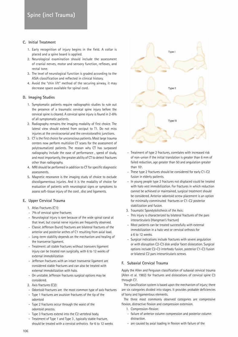
106
TRAUMASpine (incl Trauma)
C. Initial Treatment
1. Early recognition of injury begins in the field. A collar is placed and a spine board is applied.
2. Neurological examination should include the assessment of cranial nerves, motor and sensory function, reflexes, and rectal tone.
3. The level of neurological function is graded according to the ASIA classification and reflected in clinical history.
4. Avoid the “chin lift” method of the securing airway, it may decrease space available for spinal cord.
D. Imaging Studies
1. Symptomatic patients require radiographic studies to rule out the presence of a traumatic cervical spine injury before the cervical spine is cleared. A cervical spine injury is found in 2-6% of all symptomatic patients.
2. Radiography remains the imaging modality of first choice. The lateral view should extend from occiput to T1. Do not miss injuries at the cervicocranial and the cervicotorathic junctions.
3. CT is the first choice for unconscious patients. Most large trauma centers now perform multislice CT scans for the assessment of polytraumatized patients. The reason why CT has surpassed radiography include the ease of performance , speed of study, and most importantly, the greater ability of CT to detect fractures other than radiography.
4. MRI should be performed in addition to CT for specific diagnostic assessments.
5. Magnetic resonance is the imaging study of choice to exclude discoligamentous injuries. And it is the modality of choice for evaluation of patients with neurological signs or symptoms to assess soft-tissue injury of the cord , disc and ligaments.
E. Upper Cervical Trauma
1. Atlas Fractures (C1): - 7% of cervical spine fractures - Neurological injury is rare because of the wide spinal canal at
that level, but cranial nerve injuries are frequently observed. - Classic Jefferson (burst) fractures are bilateral fractures of the
anterior and posterior arches of C1 resulting from axial load. - Long-term stability depends on the mechanism and healing of
the transverse ligament. - Treatment: all stable fractures without transvers ligament
injury can be treated non surgically, with 6 to 12 weeks of external immobilization
- Jefferson fractures with an intact transverse ligament are considered stable fractures and can also be treated with external immobilization with halo.
- On unstable Jefferson fractures surgical options may be considered.
2. Axis fractures (C2): - Odontoid fractures are the most common type of axis fractures - Type 1 fractures are avulsion fractures of the tip of the
odontoid - Type 2 fractures occur through the waist of the
odontoid process. - Type 3 fractures extend into the C2 vertebral body. - Treatment of Type 1 and Type 3 , typically stable fracture,
should be treated with a cervical orthotics for 6 to 12 weeks
- Treatment of type 2 fractures, correlates with increased risk of non-union if the initial translation is greater than 6 mm of failed reduction, age greater than 50 and angulation greater than 10º.
- These type 2 fractures should be considered for early C1-C2 fusion in elderly patients.
- In young people type 2 fractures not displaced could be treated with halo vest immobilization. For fractures in which reduction cannot be achieved or maintained, surgical treatment should be considered. Anterior odontoid screw placement is an option for minimally comminuted fractures or C1-C2 posterior stabilization and fusion.
3. Traumatic Spondylolisthesis of the Axis: - This injury is characterized by bilateral fractures of the pars
interarticularis (Hangman’s fracture) - Most patients can be treated successfully with external
immobilization in a halo vest or cervical orthosis for a 6 to 12 weeks.
- Surgical indications include fractures with severe angulation or with disruption C2-C3 disk and/or facet dislocation. Surgical options include C2-C3 interbody fusion, posterior C1-C3 fusion or bilateral C2 pars interarticularis screws.
F. Subaxial Cervical Trauma
Apply the Allen and Ferguson classification of subaxial cervical trauma (Allen et al. 1982) for fractures and dislocations of cervical spine C3 through C7.
The classification system is based upon the mechanism of injury; there are six categories divided into stages. It provides probable deficiencies of bony and ligamentous elements.
The three most commonly observed categories are compressive flexion, distractive flexion and compression extension.
1. Compression-flexion: - failure of anterior column compression and posterior column
distraction. - are caused by axial loading in flexion with failure of the
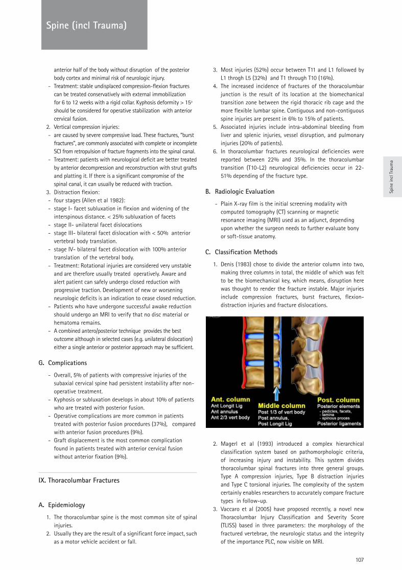
107
TRAUMA
Spin
e in
cl T
raum
a
Spine (incl Trauma)
anterior half of the body without disruption of the posterior body cortex and minimal risk of neurologic injury.
- Treatment: stable undisplaced compression-flexion fractures can be treated conservatively with external immobilization for 6 to 12 weeks with a rigid collar. Kyphosis deformity > 15º should be considered for operative stabilization with anterior cervical fusion.
2. Vertical compression injuries: - are caused by severe compressive load. These fractures, “burst
fractures”, are commonly associated with complete or incomplete SCI from retropulsion of fracture fragments into the spinal canal.
- Treatment: patients with neurological deficit are better treated by anterior decompression and reconstruction with strut grafts and platting it. If there is a significant compromise of the spinal canal, it can usually be reduced with traction.
3. Distraction flexion: - four stages (Allen et al 1982): - stage I- facet subluxation in flexion and widening of the
interspinous distance. < 25% subluxation of facets - stage II- unilateral facet dislocations - stage III- bilateral facet dislocation with < 50% anterior
vertebral body translation. - stage IV- bilateral facet dislocation with 100% anterior
translation of the vertebral body. - Treatment: Rotational injuries are considered very unstable
and are therefore usually treated operatively. Aware and alert patient can safely undergo closed reduction with progressive traction. Development of new or worsening neurologic deficits is an indication to cease closed reduction.
- Patients who have undergone successful awake reduction should undergo an MRI to verify that no disc material or hematoma remains.
- A combined antero/posterior technique provides the best outcome although in selected cases (e.g. unilateral dislocation) either a single anterior or posterior approach may be sufficient.
G. Complications
- Overall, 5% of patients with compressive injuries of the subaxial cervical spine had persistent instability after non-operative treatment.
- Kyphosis or subluxation develops in about 10% of patients who are treated with posterior fusion.
- Operative complications are more common in patients treated with posterior fusion procedures (37%), compared with anterior fusion procedures (9%).
- Graft displacement is the most common complication found in patients treated with anterior cervical fusion without anterior fixation (9%).
IX. Thoracolumbar Fractures
A. Epidemiology
1. The thoracolumbar spine is the most common site of spinal injuries.
2. Usually they are the result of a significant force impact, such as a motor vehicle accident or fall.
3. Most injuries (52%) occur between T11 and L1 followed by L1 throgh L5 (32%) and T1 through T10 (16%).
4. The increased incidence of fractures of the thoracolumbar junction is the result of its location at the biomechanical transition zone between the rigid thoracic rib cage and the more flexible lumbar spine. Contiguous and non-contiguous spine injuries are present in 6% to 15% of patients.
5. Associated injuries include intra-abdominal bleeding from liver and splenic injuries, vessel disruption, and pulmonary injuries (20% of patients).
6. In thoracolumbar fractures neurological deficiencies were reported between 22% and 35%. In the thoracolumbar transition (T10-L2) neurological deficiencies occur in 22-51% depending of the fracture type.
B. Radiologic Evaluation
- Plain X-ray film is the initial screening modality with computed tomography (CT) scanning or magnetic resonance imaging (MRI) used as an adjunct, depending upon whether the surgeon needs to further evaluate bony or soft-tissue anatomy.
C. Classification Methods
1. Denis (1983) chose to divide the anterior column into two, making three columns in total, the middle of which was felt to be the biomechanical key, which means, disruption here was thought to render the fracture instable. Major injuries include compression fractures, burst fractures, flexion-distraction injuries and fracture dislocations.
2. Magerl et al (1993) introduced a complex hierarchical classification system based on pathomorphologic criteria, of increasing injury and instability. This system divides thoracolumbar spinal fractures into three general groups. Type A compression injuries, Type B distraction injuries and Type C torsional injuries. The complexity of the system certainly enables researchers to accurately compare fracture types in follow-up.
3. Vaccaro et al (2005) have proposed recently, a novel new Thoracolumbar Injury Classification and Severity Score (TLISS) based in three parameters: the morphology of the fractured vertebrae, the neurologic status and the integrity of the importance PLC, now visible on MRI.
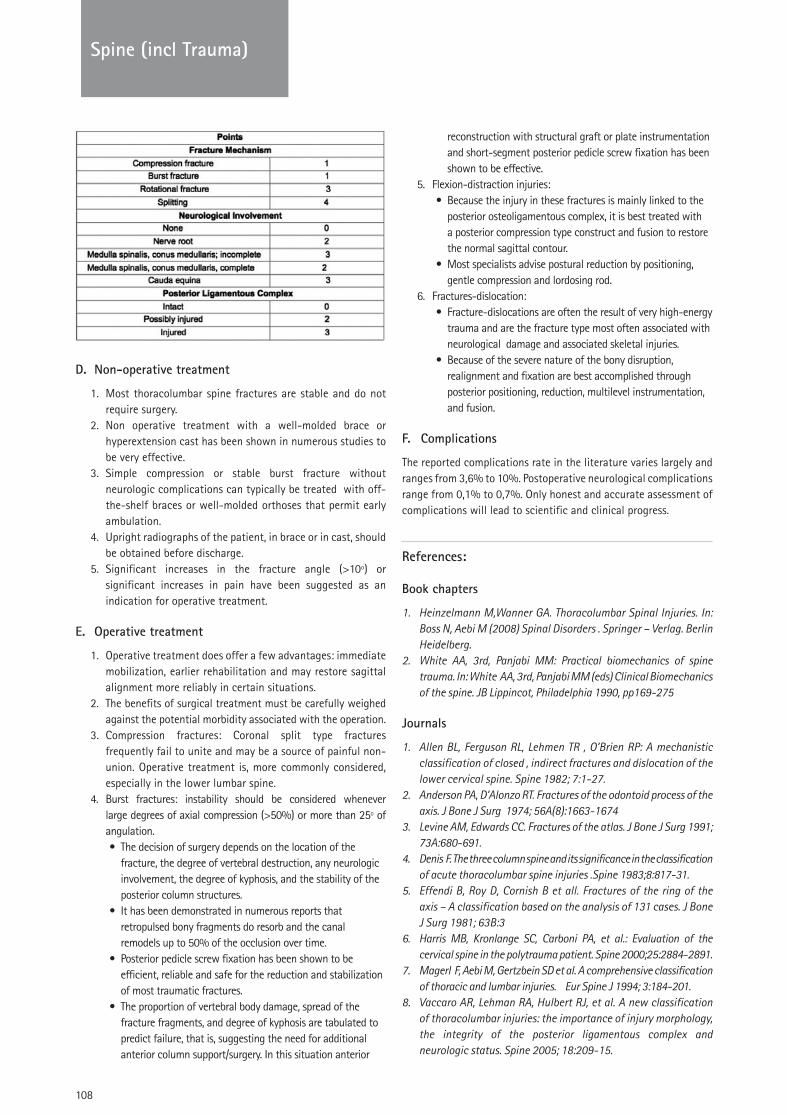
108
Spine (incl Trauma)
D. Non-operative treatment
1. Most thoracolumbar spine fractures are stable and do not require surgery.
2. Non operative treatment with a well-molded brace or hyperextension cast has been shown in numerous studies to be very effective.
3. Simple compression or stable burst fracture without neurologic complications can typically be treated with off-the-shelf braces or well-molded orthoses that permit early ambulation.
4. Upright radiographs of the patient, in brace or in cast, should be obtained before discharge.
5. Significant increases in the fracture angle (>10º) or significant increases in pain have been suggested as an indication for operative treatment.
E. Operative treatment
1. Operative treatment does offer a few advantages: immediate mobilization, earlier rehabilitation and may restore sagittal alignment more reliably in certain situations.
2. The benefits of surgical treatment must be carefully weighed against the potential morbidity associated with the operation.
3. Compression fractures: Coronal split type fractures frequently fail to unite and may be a source of painful non-union. Operative treatment is, more commonly considered, especially in the lower lumbar spine.
4. Burst fractures: instability should be considered whenever large degrees of axial compression (>50%) or more than 25º of angulation.• The decision of surgery depends on the location of the
fracture, the degree of vertebral destruction, any neurologic involvement, the degree of kyphosis, and the stability of the posterior column structures.
• It has been demonstrated in numerous reports that retropulsed bony fragments do resorb and the canal remodels up to 50% of the occlusion over time.
• Posterior pedicle screw fixation has been shown to be efficient, reliable and safe for the reduction and stabilization of most traumatic fractures.
• The proportion of vertebral body damage, spread of the fracture fragments, and degree of kyphosis are tabulated to predict failure, that is, suggesting the need for additional anterior column support/surgery. In this situation anterior
reconstruction with structural graft or plate instrumentation and short-segment posterior pedicle screw fixation has been shown to be effective.
5. Flexion-distraction injuries:• Because the injury in these fractures is mainly linked to the
posterior osteoligamentous complex, it is best treated with a posterior compression type construct and fusion to restore the normal sagittal contour.
• Most specialists advise postural reduction by positioning, gentle compression and lordosing rod.
6. Fractures-dislocation: • Fracture-dislocations are often the result of very high-energy
trauma and are the fracture type most often associated with neurological damage and associated skeletal injuries.
• Because of the severe nature of the bony disruption, realignment and fixation are best accomplished through posterior positioning, reduction, multilevel instrumentation, and fusion.
F. Complications
The reported complications rate in the literature varies largely and ranges from 3,6% to 10%. Postoperative neurological complications range from 0,1% to 0,7%. Only honest and accurate assessment of complications will lead to scientific and clinical progress.
References:
Book chapters
1. Heinzelmann M,Wanner GA. Thoracolumbar Spinal Injuries. In: Boss N, Aebi M (2008) Spinal Disorders . Springer – Verlag. Berlin Heidelberg.
2. White AA, 3rd, Panjabi MM: Practical biomechanics of spine trauma. In: White AA, 3rd, Panjabi MM (eds) Clinical Biomechanics of the spine. JB Lippincot, Philadelphia 1990, pp169-275
Journals
1. Allen BL, Ferguson RL, Lehmen TR , O’Brien RP: A mechanistic classification of closed , indirect fractures and dislocation of the lower cervical spine. Spine 1982; 7:1-27.
2. Anderson PA, D’Alonzo RT. Fractures of the odontoid process of the axis. J Bone J Surg 1974; 56A(8):1663-1674
3. Levine AM, Edwards CC. Fractures of the atlas. J Bone J Surg 1991; 73A:680-691.
4. Denis F. The three column spine and its significance in the classification of acute thoracolumbar spine injuries .Spine 1983;8:817-31.
5. Effendi B, Roy D, Cornish B et all. Fractures of the ring of the axis – A classification based on the analysis of 131 cases. J Bone J Surg 1981; 63B:3
6. Harris MB, Kronlange SC, Carboni PA, et al.: Evaluation of the cervical spine in the polytrauma patient. Spine 2000;25:2884-2891.
7. Magerl F, Aebi M, Gertzbein SD et al. A comprehensive classification of thoracic and lumbar injuries. Eur Spine J 1994; 3:184-201.
8. Vaccaro AR, Lehman RA, Hulbert RJ, et al. A new classification of thoracolumbar injuries: the importance of injury morphology, the integrity of the posterior ligamentous complex and neurologic status. Spine 2005; 18:209-15.
Spin
e in
cl T
raum
a
109
Spine (incl Trauma)
9. Wood K, Butterman G, Mehbod A, Garvey T, Jhanjee R, Sechriest V: Operative compared with nonoperative treatment of a thoracolumbar burts fracture without neurological deficit. J Bone Joint Surg Am 2003;85-A:773-781.
Questions
1. A 44-year-old farmer involved in a rollover accident on his tractor sustained a L1 burst fracture with 20% loss of vertebral body height, 30% canal compromise, and 15º of kyphosis. He remains neurologically intact. The preferred initial course of action should consist of
a. posterior spinal fusion with instrumentation b. a thoracolumbarsacral orthosis (TLSO) extension brace and
early mobilization.c. Bed rest for 6 weeks followed by inmobilization in a castd. Anterior L1 corpectomy and fusion with instrumentation e. Anterior corpectomy followed by posterior fusion with
instrumentation
2. What is the prognosis for ambulation, from best to worst, for patients with an incomplete spinal cord injury? CCS = Central Cord Syndrome. ACS = Anterior Cord Syndrome BSS = Brown-Sequard Syndrome
a. CCS, ACS, BSSb. CCS, BSS, ACS.c. BSS, ACS, CCSd. BSS, CCS, ACSe. ACS, CCS, BSS
3. Which is the most common site of spinal fractures?
4. Which mark be fitting with a incomplete sensory deficit and complete motor deficit in ASIA Impairment Scale?
5. Which classification of thoracolumbar fractures MRI is used to identify PCL injury Posterior Ligament Complex?
Answers1. b2. d3. Thoracolumbar spine 4. ASIA B5. TLICS Thoracolumbar injury classification and severity score
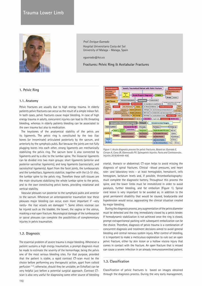
110
Trauma Lower Limb
Prof. Enrique Guerado
Hospital Universitario Costa del Sol University of Malaga - Malaga, Spain
Fractures: Pelvic Ring & Acetabular Fractures
1. Pelvic Ring
1.1. Anatomy
Pelvic fractures are usually due to high energy trauma. In elderly patients pelvic fractures can occur as the result of a simple indoor fall. In both cases, pelvic fractures cause major bleeding. In case of high energy trauma in adults, concurrent injuries can lead to life threating bleeding, whereas in elderly patients bleeding can be associated to the own trauma but also to medication.
The keystones of the anatomical stability of the pelvis are its ligaments. The pelvic ring is constituted by the two iliac bones (or innominate) articulated posteriorly by the sacrum, and anteriorly by the symphysis pubis. But because the joints are not fully plugging bones into each other, strong ligaments are mechanically stabilizing the pelvis ring. The sacrum bone is also connected by ligaments and by a disc to the lumbar spine. The iliosacral ligaments can be divided into two main groups: short ligaments (anterior and posterior sacroiliac ligaments), and long ligaments (sacrosciatic, and sacroischial ligaments). Apart from the facet joints, the lumbosacrals and the lumboiliacs, ligaments stabilize, together with the L5-S1 disc, the lumbar spine to the pelvis ring. Therefore these soft-tissues are the main structures stabilizing the whole lumbar spine to the pelvis, and to the own constituting pelvic bones, providing rotational and vertical stability.
Vascular plexuses run posterior to the symphysis pubis and anterior to the sacrum. Whenever an anteroposterior traumatism tear these plexuses major bleeding can occur, even more important if –very rarely- the iliac vessels are damaged (1). Some others viscerae can be injured such as the bladder, the bowel, the vagina or the uterus, masking a real open fracture. Neurological damage of the lumbosacral or sacral plexuses can complete the possibilities of complementary injuries in pelvis traumatism.
1.2. Diagnosis
The essential problem of severe trauma is major bleeding. Whenever a patient sustains a high energy traumatism, a prompt diagnosis must be made to estimate the severity of the hemorrhage, being the pelvis one of the most serious bleeding sites. For that purpose, provided that the patient is stable, a rapid contrast CT-scan must be the choice before performing any therapeutic action, apart from colloid perfusion (1,2); otherwise, should they be unstable, an EchoFast can be very helpful just before a potential surgical approach. Contrast CT-scan is also very useful for diagnosing some other source of bleeding
cranial, thoracic or abdominal; CT-scan helps to avoid missing the diagnosis of spinal fractures. Clinical –blood pressures, and heart rate- and laboratory tests – at least hemoglobin, hematocrit, cells hemogram, lactatum levels and, if possible, thromboelastography- must complete the diagnostic battery. Throughout this process the spine, and the lower limbs must be immobilized in order to avoid paralysis, further bleeding, and fat embolism (Figure 1). Spinal cord lesion is very important to be avoided as, in addition to the great permanent disability that would be caused, bradycardia and hypotension would occur, aggravating the clinical situation created by major bleeding.
During the diagnosis process, any augmentation of the pelvis diameter must be detected and the ring immediately closed by a pelvis binder. If hemodynamic stabilization is not achieved once the ring is closed, prompt extraperitoneal packing with subsequent embolization can be the choice. Therefore, diagnosis of pelvis trauma is a combination of concurrent diagnosis and treatment decisions aimed to avoid general bleeding and central nervous system injury. After control of bleeding, it is important to make a meticulous exploration to rule out an open pelvic fracture, either by skin lesion or a hollow viscera injury that comes in contact with the fracture. An open fracture that is missed can cause a severe infection in an already immunocommitted patient.
1.3. Classification
Classification of pelvic fractures is based on images obtained through the diagnosis process. During the very early management,
Figure 1: Acute diagnosis process for pelvic fractures. Based on: Guerado E, Cervan A, Cano JR, Giannoudis PV. Spinopelvic Injuries. Facts and Controversies. Injuries 2018;49:449-456.
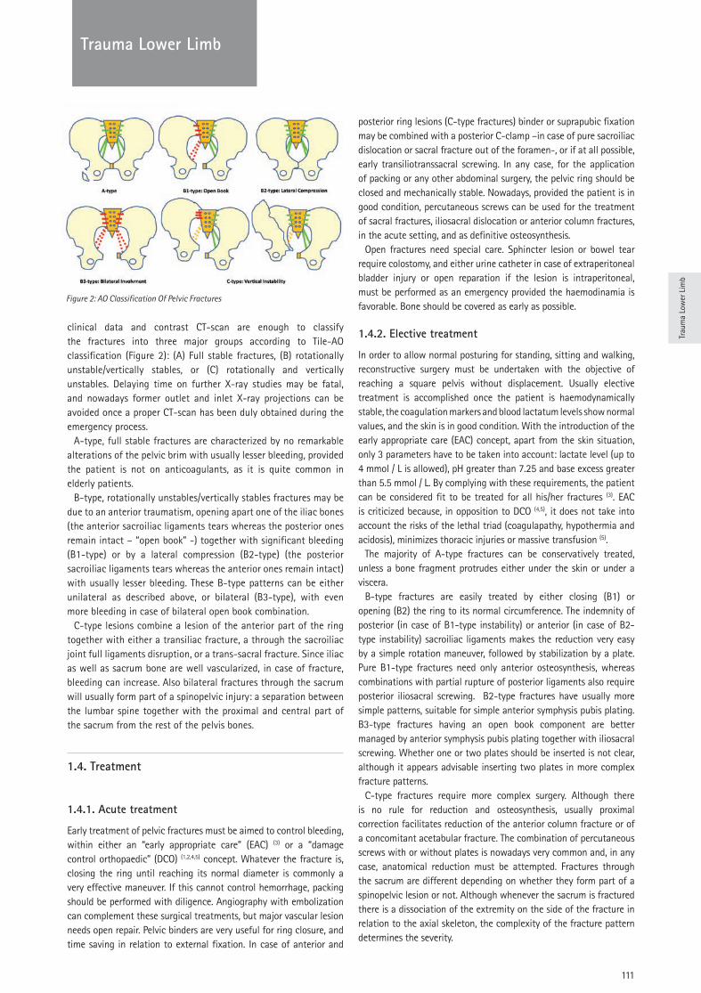
Trau
ma
Low
er L
imb
111
Trauma Lower Limb
clinical data and contrast CT-scan are enough to classify the fractures into three major groups according to Tile-AO classification (Figure 2): (A) Full stable fractures, (B) rotationally unstable/vertically stables, or (C) rotationally and vertically unstables. Delaying time on further X-ray studies may be fatal, and nowadays former outlet and inlet X-ray projections can be avoided once a proper CT-scan has been duly obtained during the emergency process.
A-type, full stable fractures are characterized by no remarkable alterations of the pelvic brim with usually lesser bleeding, provided the patient is not on anticoagulants, as it is quite common in elderly patients.
B-type, rotationally unstables/vertically stables fractures may be due to an anterior traumatism, opening apart one of the iliac bones (the anterior sacroiliac ligaments tears whereas the posterior ones remain intact – “open book” -) together with significant bleeding (B1-type) or by a lateral compression (B2-type) (the posterior sacroiliac ligaments tears whereas the anterior ones remain intact) with usually lesser bleeding. These B-type patterns can be either unilateral as described above, or bilateral (B3-type), with even more bleeding in case of bilateral open book combination.
C-type lesions combine a lesion of the anterior part of the ring together with either a transiliac fracture, a through the sacroiliac joint full ligaments disruption, or a trans-sacral fracture. Since iliac as well as sacrum bone are well vascularized, in case of fracture, bleeding can increase. Also bilateral fractures through the sacrum will usually form part of a spinopelvic injury: a separation between the lumbar spine together with the proximal and central part of the sacrum from the rest of the pelvis bones.
1.4. Treatment
1.4.1. Acute treatment
Early treatment of pelvic fractures must be aimed to control bleeding, within either an “early appropriate care” (EAC) (3) or a “damage control orthopaedic” (DCO) (1,2,4,5) concept. Whatever the fracture is, closing the ring until reaching its normal diameter is commonly a very effective maneuver. If this cannot control hemorrhage, packing should be performed with diligence. Angiography with embolization can complement these surgical treatments, but major vascular lesion needs open repair. Pelvic binders are very useful for ring closure, and time saving in relation to external fixation. In case of anterior and
posterior ring lesions (C-type fractures) binder or suprapubic fixation may be combined with a posterior C-clamp –in case of pure sacroiliac dislocation or sacral fracture out of the foramen-, or if at all possible, early transiliotranssacral screwing. In any case, for the application of packing or any other abdominal surgery, the pelvic ring should be closed and mechanically stable. Nowadays, provided the patient is in good condition, percutaneous screws can be used for the treatment of sacral fractures, iliosacral dislocation or anterior column fractures, in the acute setting, and as definitive osteosynthesis.
Open fractures need special care. Sphincter lesion or bowel tear require colostomy, and either urine catheter in case of extraperitoneal bladder injury or open reparation if the lesion is intraperitoneal, must be performed as an emergency provided the haemodinamia is favorable. Bone should be covered as early as possible.
1.4.2. Elective treatment
In order to allow normal posturing for standing, sitting and walking, reconstructive surgery must be undertaken with the objective of reaching a square pelvis without displacement. Usually elective treatment is accomplished once the patient is haemodynamically stable, the coagulation markers and blood lactatum levels show normal values, and the skin is in good condition. With the introduction of the early appropriate care (EAC) concept, apart from the skin situation, only 3 parameters have to be taken into account: lactate level (up to 4 mmol / L is allowed), pH greater than 7.25 and base excess greater than 5.5 mmol / L. By complying with these requirements, the patient can be considered fit to be treated for all his/her fractures (3). EAC is criticized because, in opposition to DCO (4,5), it does not take into account the risks of the lethal triad (coagulapathy, hypothermia and acidosis), minimizes thoracic injuries or massive transfusion (5).
The majority of A-type fractures can be conservatively treated, unless a bone fragment protrudes either under the skin or under a viscera.
B-type fractures are easily treated by either closing (B1) or opening (B2) the ring to its normal circumference. The indemnity of posterior (in case of B1-type instability) or anterior (in case of B2-type instability) sacroiliac ligaments makes the reduction very easy by a simple rotation maneuver, followed by stabilization by a plate. Pure B1-type fractures need only anterior osteosynthesis, whereas combinations with partial rupture of posterior ligaments also require posterior iliosacral screwing. B2-type fractures have usually more simple patterns, suitable for simple anterior symphysis pubis plating. B3-type fractures having an open book component are better managed by anterior symphysis pubis plating together with iliosacral screwing. Whether one or two plates should be inserted is not clear, although it appears advisable inserting two plates in more complex fracture patterns.
C-type fractures require more complex surgery. Although there is no rule for reduction and osteosynthesis, usually proximal correction facilitates reduction of the anterior column fracture or of a concomitant acetabular fracture. The combination of percutaneous screws with or without plates is nowadays very common and, in any case, anatomical reduction must be attempted. Fractures through the sacrum are different depending on whether they form part of a spinopelvic lesion or not. Although whenever the sacrum is fractured there is a dissociation of the extremity on the side of the fracture in relation to the axial skeleton, the complexity of the fracture pattern determines the severity.
Figure 2: AO Classification Of Pelvic Fractures
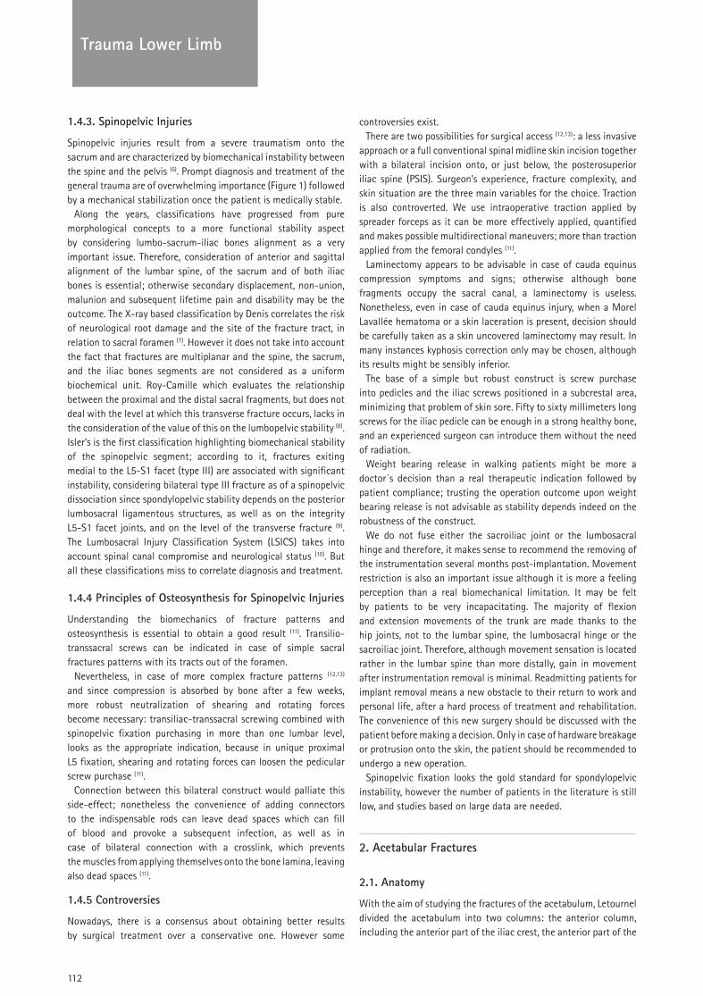
112
TRAUMATrauma Lower Limb
1.4.3. Spinopelvic Injuries
Spinopelvic injuries result from a severe traumatism onto the sacrum and are characterized by biomechanical instability between the spine and the pelvis (6). Prompt diagnosis and treatment of the general trauma are of overwhelming importance (Figure 1) followed by a mechanical stabilization once the patient is medically stable.
Along the years, classifications have progressed from pure morphological concepts to a more functional stability aspect by considering lumbo-sacrum-iliac bones alignment as a very important issue. Therefore, consideration of anterior and sagittal alignment of the lumbar spine, of the sacrum and of both iliac bones is essential; otherwise secondary displacement, non-union, malunion and subsequent lifetime pain and disability may be the outcome. The X-ray based classification by Denis correlates the risk of neurological root damage and the site of the fracture tract, in relation to sacral foramen (7). However it does not take into account the fact that fractures are multiplanar and the spine, the sacrum, and the iliac bones segments are not considered as a uniform biochemical unit. Roy-Camille which evaluates the relationship between the proximal and the distal sacral fragments, but does not deal with the level at which this transverse fracture occurs, lacks in the consideration of the value of this on the lumbopelvic stability (8). Isler’s is the first classification highlighting biomechanical stability of the spinopelvic segment; according to it, fractures exiting medial to the L5-S1 facet (type III) are associated with significant instability, considering bilateral type III fracture as of a spinopelvic dissociation since spondylopelvic stability depends on the posterior lumbosacral ligamentous structures, as well as on the integrity L5-S1 facet joints, and on the level of the transverse fracture (9). The Lumbosacral Injury Classification System (LSICS) takes into account spinal canal compromise and neurological status (10). But all these classifications miss to correlate diagnosis and treatment.
1.4.4 Principles of Osteosynthesis for Spinopelvic Injuries
Understanding the biomechanics of fracture patterns and osteosynthesis is essential to obtain a good result (11). Transilio-transsacral screws can be indicated in case of simple sacral fractures patterns with its tracts out of the foramen.
Nevertheless, in case of more complex fracture patterns (12,13)
and since compression is absorbed by bone after a few weeks, more robust neutralization of shearing and rotating forces become necessary: transiliac-transsacral screwing combined with spinopelvic fixation purchasing in more than one lumbar level, looks as the appropriate indication, because in unique proximal L5 fixation, shearing and rotating forces can loosen the pedicular screw purchase (11).
Connection between this bilateral construct would palliate this side-effect; nonetheless the convenience of adding connectors to the indispensable rods can leave dead spaces which can fill of blood and provoke a subsequent infection, as well as in case of bilateral connection with a crosslink, which prevents the muscles from applying themselves onto the bone lamina, leaving also dead spaces (11).
1.4.5 Controversies
Nowadays, there is a consensus about obtaining better results by surgical treatment over a conservative one. However some
controversies exist.There are two possibilities for surgical access (12,13): a less invasive
approach or a full conventional spinal midline skin incision together with a bilateral incision onto, or just below, the posterosuperior iliac spine (PSIS). Surgeon’s experience, fracture complexity, and skin situation are the three main variables for the choice. Traction is also controverted. We use intraoperative traction applied by spreader forceps as it can be more effectively applied, quantified and makes possible multidirectional maneuvers; more than traction applied from the femoral condyles (11).
Laminectomy appears to be advisable in case of cauda equinus compression symptoms and signs; otherwise although bone fragments occupy the sacral canal, a laminectomy is useless. Nonetheless, even in case of cauda equinus injury, when a Morel Lavallée hematoma or a skin laceration is present, decision should be carefully taken as a skin uncovered laminectomy may result. In many instances kyphosis correction only may be chosen, although its results might be sensibly inferior.
The base of a simple but robust construct is screw purchase into pedicles and the iliac screws positioned in a subcrestal area, minimizing that problem of skin sore. Fifty to sixty millimeters long screws for the iliac pedicle can be enough in a strong healthy bone, and an experienced surgeon can introduce them without the need of radiation.
Weight bearing release in walking patients might be more a doctor´s decision than a real therapeutic indication followed by patient compliance; trusting the operation outcome upon weight bearing release is not advisable as stability depends indeed on the robustness of the construct.
We do not fuse either the sacroiliac joint or the lumbosacral hinge and therefore, it makes sense to recommend the removing of the instrumentation several months post-implantation. Movement restriction is also an important issue although it is more a feeling perception than a real biomechanical limitation. It may be felt by patients to be very incapacitating. The majority of flexion and extension movements of the trunk are made thanks to the hip joints, not to the lumbar spine, the lumbosacral hinge or the sacroiliac joint. Therefore, although movement sensation is located rather in the lumbar spine than more distally, gain in movement after instrumentation removal is minimal. Readmitting patients for implant removal means a new obstacle to their return to work and personal life, after a hard process of treatment and rehabilitation. The convenience of this new surgery should be discussed with the patient before making a decision. Only in case of hardware breakage or protrusion onto the skin, the patient should be recommended to undergo a new operation.
Spinopelvic fixation looks the gold standard for spondylopelvic instability, however the number of patients in the literature is still low, and studies based on large data are needed.
2. Acetabular Fractures
2.1. Anatomy
With the aim of studying the fractures of the acetabulum, Letournel divided the acetabulum into two columns: the anterior column, including the anterior part of the iliac crest, the anterior part of the
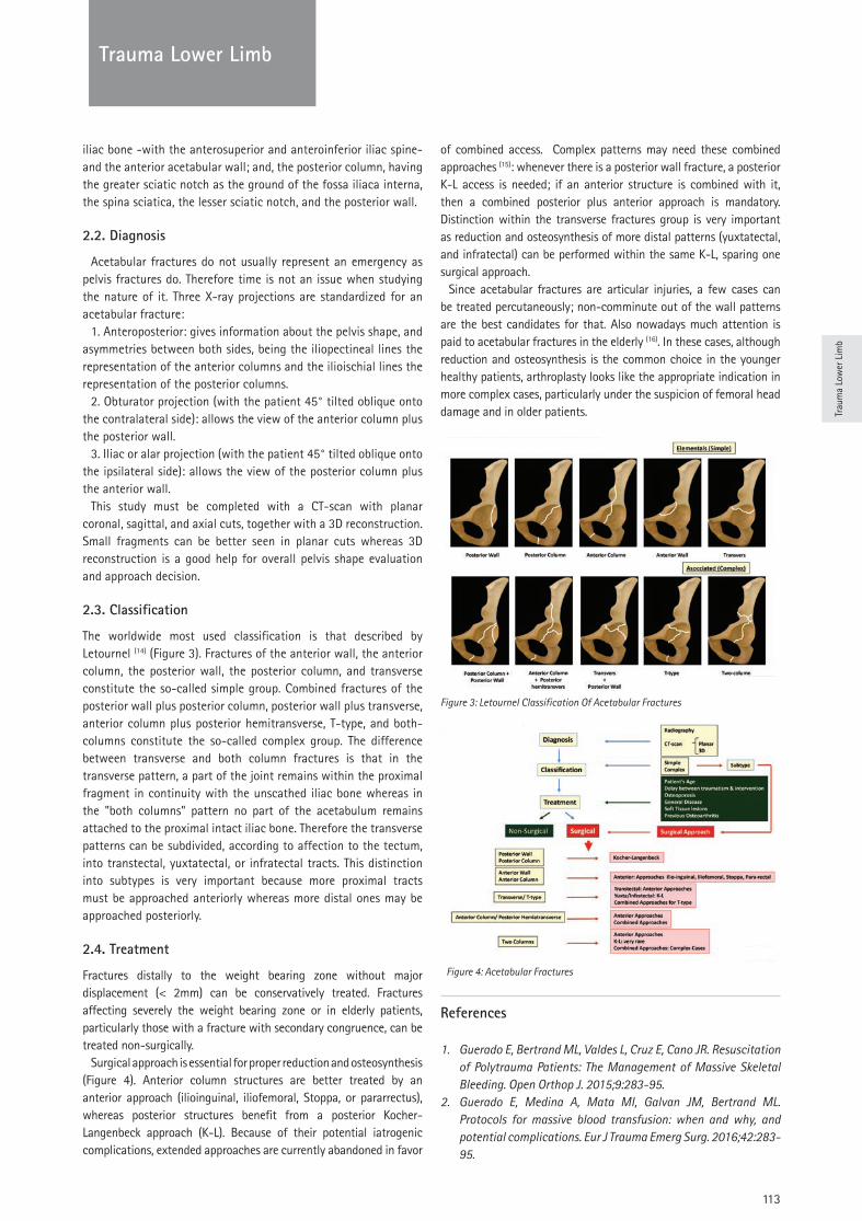
113
Trauma Lower Limb
Trau
ma
Low
er L
imb
iliac bone -with the anterosuperior and anteroinferior iliac spine- and the anterior acetabular wall; and, the posterior column, having the greater sciatic notch as the ground of the fossa iliaca interna, the spina sciatica, the lesser sciatic notch, and the posterior wall.
2.2. Diagnosis
Acetabular fractures do not usually represent an emergency as pelvis fractures do. Therefore time is not an issue when studying the nature of it. Three X-ray projections are standardized for an acetabular fracture:
1. Anteroposterior: gives information about the pelvis shape, and asymmetries between both sides, being the iliopectineal lines the representation of the anterior columns and the ilioischial lines the representation of the posterior columns.
2. Obturator projection (with the patient 45° tilted oblique onto the contralateral side): allows the view of the anterior column plus the posterior wall.
3. Iliac or alar projection (with the patient 45° tilted oblique onto the ipsilateral side): allows the view of the posterior column plus the anterior wall.
This study must be completed with a CT-scan with planar coronal, sagittal, and axial cuts, together with a 3D reconstruction. Small fragments can be better seen in planar cuts whereas 3D reconstruction is a good help for overall pelvis shape evaluation and approach decision.
2.3. Classification
The worldwide most used classification is that described by Letournel (14) (Figure 3). Fractures of the anterior wall, the anterior column, the posterior wall, the posterior column, and transverse constitute the so-called simple group. Combined fractures of the posterior wall plus posterior column, posterior wall plus transverse, anterior column plus posterior hemitransverse, T-type, and both-columns constitute the so-called complex group. The difference between transverse and both column fractures is that in the transverse pattern, a part of the joint remains within the proximal fragment in continuity with the unscathed iliac bone whereas in the "both columns" pattern no part of the acetabulum remains attached to the proximal intact iliac bone. Therefore the transverse patterns can be subdivided, according to affection to the tectum, into transtectal, yuxtatectal, or infratectal tracts. This distinction into subtypes is very important because more proximal tracts must be approached anteriorly whereas more distal ones may be approached posteriorly.
2.4. Treatment
Fractures distally to the weight bearing zone without major displacement (< 2mm) can be conservatively treated. Fractures affecting severely the weight bearing zone or in elderly patients, particularly those with a fracture with secondary congruence, can be treated non-surgically.
Surgical approach is essential for proper reduction and osteosynthesis (Figure 4). Anterior column structures are better treated by an anterior approach (ilioinguinal, iliofemoral, Stoppa, or pararrectus), whereas posterior structures benefit from a posterior Kocher-Langenbeck approach (K-L). Because of their potential iatrogenic complications, extended approaches are currently abandoned in favor
of combined access. Complex patterns may need these combined approaches (15): whenever there is a posterior wall fracture, a posterior K-L access is needed; if an anterior structure is combined with it, then a combined posterior plus anterior approach is mandatory. Distinction within the transverse fractures group is very important as reduction and osteosynthesis of more distal patterns (yuxtatectal, and infratectal) can be performed within the same K-L, sparing one surgical approach.
Since acetabular fractures are articular injuries, a few cases can be treated percutaneously; non-comminute out of the wall patterns are the best candidates for that. Also nowadays much attention is paid to acetabular fractures in the elderly (16). In these cases, although reduction and osteosynthesis is the common choice in the younger healthy patients, arthroplasty looks like the appropriate indication in more complex cases, particularly under the suspicion of femoral head damage and in older patients.
References
1. Guerado E, Bertrand ML, Valdes L, Cruz E, Cano JR. Resuscitation of Polytrauma Patients: The Management of Massive Skeletal Bleeding. Open Orthop J. 2015;9:283-95.
2. Guerado E, Medina A, Mata MI, Galvan JM, Bertrand ML. Protocols for massive blood transfusion: when and why, and potential complications. Eur J Trauma Emerg Surg. 2016;42:283-95.
Figure 3: Letournel Classification Of Acetabular Fractures
Figure 4: Acetabular Fractures
114
Trauma Lower Limb
3. Vallier HA, Wang X, Moore TA, Wilber JH, Como JJ. Timing of orthopaedic surgery in multiple trauma patients: development of a protocol for early appropriate care. J Orthop Trauma. 2013;27:543-51.
4. Guerado E, Bertrand ML, Cano JR, Cerván AM, Galán A. Damage control orthopaedics: State of the art. World J Orthop. 2019;10:1-13.
5. Pape HC, Andruszkow H, Pfeifer R, Hildebrand F, Barkatali BM. Options and hazards of the early appropriate care protocol for trauma patients with major fractures: Towards safe definitive surgery. Injury 2016;47:787-91.
6. Barcello ALL, Rocha VM, Guimaraes JAM. Current Concepts in Spondylopelvic Dissociation. Injury 2017;48:S5-S11.
7. Denis F, Davis S, Comfort T. Sacral fractures: an important problem. Retrospective analysis of 236 cases. Clin Orthop Rel Res. 1988; 227:67-81.
8. Roy-Camille R, Saillant G, Gagna G, Mazel C. Transverse fracture of the upper sacrum. Suicidal jumper's fracture. Spine (Phila Pa 1976). 1985; 10:838-45.
9. Isler B. Lumbosacral lesions associated with pelvic ring injuries. J Orthop Trauma. 1990; 4:1-6.
10. Lehman RA, Jr., Kang DG, Bellabarba C. A new classification for complex lumbosacral injuries. Spine J. 2012; 12:612-28.
11. Guerado E, Cervan AM, Cano JR. Spinopelvic Injuries. Facts and controversies. Injury 2018. 49:449-458. doi: 10.1016/j.injury.2018.03.001.
12. Koning MA, Jehan S, Boszczyk AA, Boszczyk BM. Surgical management of U-shaped sacral fractures: a systematic review of current treatment strategies. Eur Spine J: 2012; 21:829-36.
13. Williams SK, Quinnan SM. Percutaneous Lumbopelvic Fixation for Reduction and Stabilization of Sacral Fractures with Spinopelvic Dissociation Patterns. J Orthop Trauma 2016;30:318-24.
14. Judet R, Judet J, Letournel E. Fractures of the Acetabulum : Classification and Surgical Approaches for Open Reduction. Preliminary Report. J Bone Jt Surg. Am 1964;46:1615-1646.
15. Guerado E, Cano JR, Cruz E. Simultaneous ilioinguinal and Kocher-Langenbeck approaches for the treatment of complex acetabular fractures. Hip Int. 2010;20 Suppl 7:S2-10.
16. Guerado E, Cano JR, Cruz E. Fractures of the acetabulum in elderly patients: an update. Injury. 2012;43:S33-41.
Questions
1. It is true that a non-displaced pelvic fracture in the elderly:a. Always needs to undergo osteosynthesis.b. It needs a high energy traumatism.c. May have a severe bleeding.d. Is infrequent.
2. In emergency, the choice to check the haemodinamical situation is:a. A prompt series of X-rays including the pelvis with a blood
test.b. An EchoFast with a blood test.
c. A prompt CT-scan with a blood test.d. A contrast CT-scan with a blood test.
3. The Early Appropriate Care consist of:a. Treating all fractures in order to stabilize the patient.b. Treating fractures depending on coagulation, lactatum and
ph.c. Treating fractures depending on lactatum, ph, and base excess.d. Treating fractures depending on coagulation, lactatum, ph,
and base excess.
4. A complex bilateral fracture of the sacrum is better treated with:a. A full-threaded transilio-transsacral screw.b. Bilateral partially threaded transilio-transsacral screws.c. a + Spinopelvic instrumentation.d. b + c.
5. The difference between a transverse and a both column fracture is that:
a. In the transverse fracture only the posterior column has a transverse fracture tract.
b. In the transverse fracture only the anterior column has a transverse fracture tract.
c. In the transverse fracture a part of the acetabulum is intact.d. In the both column fracture a part of the acetabulum is
intact.
Answers1c; 2d; 3c; 4c; 5c
Trau
ma
Low
er L
imb
115
Trauma Lower Limb
Assoc. Prof. Dr. Cecilia Rogmark
Skåne University Hospital
Malmö, Sweden
Fractures: Femur, Tibia & Open Fractures
Fractures in the lower extremities occur at all ages. Osteopenia or osteoporosis increase the risk of fracture even after low-energy trauma, and both hip fracture, distal femoral and proximal tibial fractures are considered fragility fractures. The aim of the treatment should be unrestricted mobilization as soon as possible. This is particular important in elderly or otherwise frail patients, who cannot cope with restricted weight bearing. Therefore, fixation should be stable, if possible. The use of hip replacement instead of internal fixation in hip fracture is a good example of immediate stability resulting in better functional outcome.
Always consider the risk of deep vein thrombosis and/or pulmonary embolism, and follow the local guidelines for prophylaxis. Usually antibiotics are given perioperatively to prevent infection, at least in hip fracture cases and other fractures treated with implants. The type to choose depends on the mix of pathogens within the region, resistance pattern, any allergy etc. Generally, antibiotics should be given immediately (<30-60 min) before surgery and should not exceed the 24-hour post-operative period. A single dose is preferred, but if surgery is prolonged or major bleeding occurs, a repeated dose should be given.(1)
1. Hip fractures
Hip fractures are confirmed via conventional X-ray. If ambiguity, MRI will be useful (2).
Surgery should be performed within 24 hours in order to decrease complications and mortality (3).
Care should be given in close cooperation with geriatricians (4).Postoperative rehabilitation, during several months, is necessary
in order to regain function. Multidisciplinary teams may be useful when providing rehabilitation (5).
See NICE Guidelines Hip Fractures, available at: https://www.nice.org.uk/guidance/cg124
2. Femoral neck fracture
2.1. Undisplaced femoral neck fracture
These are usually treated with internal fixation and fixed in situ (6). That is, a valgus impaction is accepted. Currently, a few randomized trials suggest arthroplasty to be considered for undisplaced fractures in elderly patients, due to fewer reoperations and better functional outcome, at least in the short run. More scientific analyses are needed, regarding long-term outcome and cost-effectiveness of the different treatment options.
Non-surgical treatment is not recommended due to the risk of secondary displacement and the need of frequent check-ups. Nevertheless, in case of contraindications for surgery it may be an alternative.
2.2. Displaced femoral neck fracture
Treatment options are internal fixation (after closed or open reduction), total hip arthroplasty (THA) or hemiarthroplasty (7). Choice is guided by the patient’s biological age, activity level, health and supposed remaining life span. For a majority, those with functional and/or psychological limitations, hemiarthroplasty is the most suitable choice. The benefits are: “forgiving” surgery (can be performed by surgeons with less experience, i.e. emergency procedure), shorter surgery time, less bleeding and lower risk of dislocation. In active individuals hemiarthroplasty carries an unacceptable high risk of acetabulum erosion (8). In those cases, THA is a better option. THA in fracture cases puts higher demands on the surgeon, as malpositioned implant parts increase the risk of dislocation and pain. The surgery time is longer, and altogether THA procedures may be demanding to cope with, within an emergency setting. Arthroplasty in fracture patients should be cemented (less risk of periprosthetic fracture) and inserted via direct lateral approach (less risk of dislocation) (9-13). Increased risk of perioperative death in connection with the cementation process can be limited by proper anesthesiologic care and cementing technique (thorough lavage, no excessive pressure etc) (14). In addition, uncemented stems lead to a higher risk of death after the first postoperative week (15, 16). This may be due to their higher risk of periprosthetic fracture.
Internal fixation may be suitable for younger individuals, even though they have the same high risk (30-50%) of pseudarthrosis and avascular necrosis as elderly patients have. But, if healthy, the young patients can better withstand a secondary procedure if needed. And if they heal, they maintain their native hip joint. The age limit for primary prosthesis is controversial. 60, 65 or 70 years are suggested. The important thing is to understand the concept of biological age. For example a 55-year-old with pre-senile dementia and functional limitation may benefit from hemiarthroplasty. Regarding reduction of the fracture, there is no clear evidence that open reduction adds any benefit compared to closed ditto, but studies are very scarce.
2.3. Extracapsular hip fractures
Basicervical fractures are not well studied. Sliding hip screw (SHS), with or without an extra anti-rotational screw, is recommended in most cases.
Stable, or two-fragment trochanteric fractures, should be
116
Trauma Lower Limb
fixed with SHS (17). Using intramedullary nail (IM nail) in these cases brings on unnecessary complications, mainly peri-implantat fractures.
In unstable trochanteric fractures, both SHS and IM nails can be used (18). To understand the ”personality” of the fracture and to obtain a good fracture reduction and implant position, are more important than the implant choice (19). A thin lateral wall with a risk of fragmentation during surgery speaks against the use of a simple SHS. Adding a trochanter stabilization plate may prevent medialization in these cases, or to use an IM nail. Locking plates have no role in proximal femoral fractures (20).
Subtrochanteric fractures can be treated with IM nails or sliding hip screw with biaxial sliding function. Fixed angle plates are not an option nowadays (18).
3. Femoral shaft fractures
Femoral shaft fractures occur mostly due to high energy trauma. An important subgroup, is periprosthetic fractures in the presence of hip and/or knee replacement. With an earlier implant inserted in the femur, preoperative considerations are even more important. Any loosening of a joint prosthesis must be evaluated, and a loose implant should be revised. Use – and understand – the Vancouver classification and always cooperate with an arthroplasty surgeon!
Femoral fractures can lead to great amount of blood loss. In high energy trauma, muscular injury and subsequent necrosis can lead to renal insufficiency.
3.1. Treatment
ER room - temporary stabilization with traction or long posterior splint. In case of severe instability – both generally and/or locally – an immediate external fixation will serve as damage control. This leaves time for other live-saving interventions, and thereafter final internal fixation can take place.
Check vital parameters continuously as blood loss can be substantial, oxygen is often needed. Pain treatment, in femoral blocks may be an option. Monitoring regarding compartment syndrome is important in high energy-trauma cases.
Surgical treatment with intramedullary nailing is the most common method for reamed diaphyseal fractures, and plating (with locking screws) for metaphyseal fractures, as well as periprosthetic fractures.
4. Tibial shaft fractures
4.1. General
Common fracture localization / open fracture quite common due to thin layer of soft-tissue. Higher impact equals higher risk of complications – anamnesis important.
Compartment syndrome – devastating if not diagnosed and treated!
4.2. Treatment
ER room: temporary stabilization with long posterior splint, pain treatment and monitoring regarding compartment syndrome.
Nonoperative treatment is suitable for closed fractures with minimal displacement or stable reduction; in a long leg cast. Encourage ambulation with weight-bearing as tolerated in simple transverse fractures but check with frequent radiographs fracture alignment. Long leg cast may lead to increased joint stiffness and a change to functional bracing or patella tendon bearing lower leg cast at approx. 6 wks is recommended. Compared with surgical fixation, the risk with nonoperative treatment is noted as an increased nonunion and healing time, in particular in displaced fractures. On the other hand, the risk of surgically related infections is absent.
Surgical treatment is nowadays used in most unstable fractures. Methods used are intramedullary nailing, plating and external fixation. In extremely severe cases (see open fractures) even amputation might be necessary. IM nailing with locking screws is the most frequent option, as it leads to fewer non-unions and mal-unions, compared to other methods. It also admits immediate weight-bearing in most cases, leading to a sooner return to activity. The major question is whether tibia should be reamed prior to insertion or not. The evidence is not clear, but suggests some advantage for reaming in closed fractures.
5. Open fractures
Open fractures of the long bones are increasing, now estimated to occur in 30 per 100,000 persons annually.
The injury leads to contamination with pathogens, damaged blood perfusion, and decreased immune defense, which increases the risk of infection (six-folded compared to internal fixation of closed fractures) and healing disturbances. Therefore, treatment should aim at avoiding infection, improve healing and restore function.
5.1. Classification
The typical classification of open fractures is the Gustilo-Anderson system (see below), but other systems are gaining popularity, for example the Ganga Hospital severity score (23).
The Gustilo-Anderson classification:i. Wound <1 cm and minimal/clean soft-tissue injury and
minimal fracture comminutionii. Wound >1 cm and moderate soft-tissue injury/contamination
and moderate fracture comminutioniii. Extensive soft-tissue damage including muscle, skin,
neurovascular structures or Open segmental fracture, irrespective of the wound size or Open fractures over 8 hours olda. Adequate soft-tissue coverage, including
segmental/severely comminuted fracturesb. Extensive loss of soft-tissue, periosteal
stripping, bony exposurec. As B including major arterial injury requiring
repair for limb salvage
5.2. Treatment
Antibiotics intravenously should be given preferably within
Trau
ma
Low
er L
imb
117
Trauma Lower Limb
1 hour postinjury (can be given in pre-hospital setting) – observe local guidelines for type of antibiotic drug. Initial pathogens may be less of a problem than secondary nosocomial infections. Protect the wound! The duration of antibiotics is debated. One recommendation is that antibiotics should be given until primary closure of the wound, or for 72 hours, whichever is sooner (24).
Tetanus prophylaxis should be considered.Immediate simple reposition and temporary fixation; wound
covered in sterile dressings.Do not irrigate open fractures of the long bones, hindfoot or
midfoot in the emergency department before debridement. Initial bacterial culture probably of no use.
5.3. Further treatment
Immediate treatment of highly contaminated open fractures – otherwise time frames as below:
Gustilo III – surgery within 12 hrs; exposure, debridement, low pressure lavage, temporary or final fixation, coverage (primary suture/NWPT (several liters of fluid),/sterile dressing/graft). Assistance by vascular and plastic surgeon might be needed. Soft-tissue grafting can be undertaken as a second procedure, preferably within 72 hrs.
Gustilo II – surgery within 24 hrs; treatment as Gustilo III
Gustilo I – similar to closed fracture.
The historical “6-hour rule” – study on guinea pigs by Friedrich in 1898 – has been questioned in recent papers (21). Debridement can supposedly be performed within the first 12-24 hours without any increased risk of infection. This information is useful when transfer may be needed before surgery.
Amputation – may be primary treatment in certain cases, such as ”a limb is the source of uncontrollable life-threatening bleeding, or a limb is salvageable but attempted preservation would pose an unacceptable risk to the person’s life, or a limb is deemed unsalvageable after orthoplastic assessment” (NICE guidelines) (22).
A recent review paper on open fracture is available in the EFORT Open Reviews (24).
References:
1. Antibiotic prophylaxis in surgery. SIGN publication no.104. Edinburgh2008 [updated 2014; cited 2015. Available from: http://www.sign.ac.uk.
2. Lubovsky O, Liebergall M, Mattan Y, Weil Y, Mosheiff R. Early diagnosis of occult hip fractures MRI versus CT scan. Injury. 2005;36(6):788-92.
3. Simunovic N, Devereaux PJ, Sprague S, Guyatt GH, Schemitsch E, Debeer J, et al. Effect of early surgery after hip fracture on mortality and complications: systematic review and meta-analysis. CMAJ.182(15):1609-16.
4. Grigoryan KV, Javedan H, Rudolph JL. Orthogeriatric care models and outcomes in hip fracture patients: a systematic review and meta-analysis. J Orthop Trauma. 2014;28(3):e49-55.
5. Donohue K, Hoevenaars R, McEachern J, Zeman E, Mehta S. Home-Based Multidisciplinary Rehabilitation following Hip Fracture Surgery: What Is the Evidence? Rehabilitation research and practice. 2013;2013:875968.
6. Handoll HH, Parker MJ. Conservative versus operative treatment for hip fractures in adults. Cochrane Database Syst Rev. 2008(3):CD000337.
7. Parker MJ, Gurusamy KS, Azegami S. Arthroplasties (with and without bone cement) for proximal femoral fractures in adults. Cochrane Database Syst Rev. 2010(6):CD001706.
8. Baker RP, Squires B, Gargan MF, Bannister GC. Total hip arthroplasty and hemiarthroplasty in mobile, independent patients with a displaced intracapsular fracture of the femoral neck. A randomized, controlled trial. J Bone Joint Surg Am. 2006;88(12):2583-9.
9. Enocson A, Hedbeck CJ, Tidermark J, Pettersson H, Ponzer S, Lapidus LJ. Dislocation of total hip replacement in patients with fractures of the femoral neck. Acta orthopaedica. 2009;80(2):184-9.
10. Enocson A, Tidermark J, Tornkvist H, Lapidus LJ. Dislocation of hemiarthroplasty after femoral neck fracture: better outcome after the anterolateral approach in a prospective cohort study on 739 consecutive hips. Acta orthopaedica. 2008;79(2):211-7.
11. Leonardsson O, Garellick G, Karrholm J, Akesson K, Rogmark C. Increased risk of re-operation for bipolar and uncemented hemiarthroplasty. 23,509 procedures after femoral neck fractures from the Swedish Hip Arthroplasty Register 2005-2010. Acta orthopaedica. 83(5), 459-466.
12. Rogmark C, Fenstad AM, Leonardsson O, Engesaeter LB, Karrholm J, Furnes O, et al. Posterior approach uncemented stems increases the risk of reoperation after hemiarthroplasties in elderly hip fracture patients. An analysis of 33,205 procedures in the Norwegian and Swedish national registries. Acta orthopaedica. 85(1), 18-25.
13. Inngul C, Blomfeldt R, Ponzer S, Enocson A. Cemented versus uncemented arthroplasty in patients with a displaced fracture of the femoral neck: a randomised controlled trial. Bone Joint J. 2015;97-B(11):1475-80.
14. Membership of the Working Party, Griffiths R, White SM, Moppett IK, Parker MJ, Chesser TJ, et al. Safety guideline: reducing the risk from cemented hemiarthroplasty for hip fracture 2015: Association of Anaesthetists of Great Britain and Ireland British Orthopaedic Association British Geriatric Society. Anaesthesia. 2015;70(5):623-6.
15. Costa ML, Griffin XL, Pendleton N, Pearson M, Parsons N. Does cementing the femoral component increase the risk of peri-operative mortality for patients having replacement surgery for a fracture of the neck of femur? Data from the National Hip Fracture Database. J Bone Joint Surg Br.93(10):1405-10.
16. Costain DJ, Whitehouse SL, Pratt NL, Graves SE, Ryan P, Crawford RW. Perioperative mortality after hemiarthroplasty related to fixation method. Acta orthopaedica.82(3):275-81.
17. Matre K, Havelin LI, Gjertsen JE, Espehaug B, Fevang JM. Intramedullary nails result in more reoperations than sliding hip screws in two-part intertrochanteric fractures. Clinical orthopaedics and related research. 2013;471(4):1379-86.
18. Parker MJ, Handoll HH. Gamma and other cephalocondylic intramedullary nails versus extramedullary implants for extracapsular hip fractures in adults. Cochrane Database Syst Rev. 2008(3):CD000093.
19. De Bruijn K, den Hartog D, Tuinebreijer W, Roukema G. Reliability of predictors for screw cutout in intertrochanteric hip fractures. J Bone Joint Surg Am. 2012;94(14):1266-72.
118
Trauma Lower Limb
20. Streubel PN, Moustoukas MJ, Obremskey WT. Mechanical failure after locking plate fixation of unstable intertrochanteric femur fractures. J Orthop Trauma. 2013;27(1):22-8.
21. Schenker ML, Yannascoli S, Baldwin KD, Ahn J, Mehta S. Does timing to operative debridement affect infectious complications in open long-bone fractures? A systematic review. J Bone Joint Surg Am. 2012;94(12):1057-64.
22. Fractures (complex): assessment and management NICE guideline [NG37] 2016 https://www.nice.org.uk/guidance/ng37/chapter/recommendations
23. Rajasekaran S. Ganga hospital open injury severity score - A score to prognosticate limb salvage and outcome measures in Type IIIb open tibial fractures. Indian J Orthop 2005;39:4-13.
24. Elniel AR, Giannoudis P. Open fractures of the lower extremity: current management and clinical outcomes. EFORT Open Rev 2018;3 DOI: 10.1302/2058-5241.3.170072
Questions
1. What is correct about undisplaced femoral neck fracture (one alternative)
a. The fracture should always be reduced to an anatomical position
b. Internal fixation is the most common treatmentc. Non-surgical treatment is regarded equally good as surgical
treatment in a majority of patients
2. Which type of hip complication is more common after uncemented femoral stems in fracture related hip arthroplasty, compared to cemented stems?
3. What is correct about antibiotics in emergency treatment of open fractures (one alternative)
a. Antibiotics should be given intravenously within 1 hour from injury
b. Antibiotics should not be given until bacterial cultures shows the type of pathogen
c. Antibiotics should only be given if dirt is seen in the wound
4. Why is it important to have knowledge upon the timeframes within open fractures can be safely operated on?
5. Mention one benefit and one risk with non-surgical treatment of tibial shaft fractures, compared to surgical treatment.
Answers
1. b2. Periprosthetic fracture3. a4. To better organize the care chain, to have the possibility to transfer
a patient to another hospital with proper competence (i.e. vascular surgeons, plastic surgeons, angiography facilities etc)
5. Benefits: absence of surgical complications such as infection, no need for subsequent hard-ware removal, may be less expensive. Risks: increased risk of nonunion and longer healing time
Trau
ma
Low
er L
imb
119
Trauma Lower Limb
Dr. Jose Vicente Andrés Peiró [email protected]
Dr. Jordi Tomás Hernández [email protected]
Trauma Unit. Hospital Vall d’Hebron. Barcelona, Spain
Fractures: Pilon, Ankle, Talus & Calcaneus
1. Pilon Fractures
1.1. Epidemiology
- <1% of lower extremity injuries. - Males about 30 to 40 years old.
1.2. Biomechanics
- Distal end tibia fractures involving the weight-bearing articular surface.
- Rotational mechanism: - Lower energy and soft-tissue damage. - Commonly related to sports. - Spiroid line extending to the ankle joint.
- Axial-loading: - Higher energy and soft-tissue damage; often open. - Motorvehicle accident and fall from height. - Articular and metaphyseal comminution.
1.3. Clinical Evaluation
- Mechanism and comorbidities. - Polytrauma scenario is not uncommon. - Open fractures, large muscular contusion or compartment
syndrome are frequent. - Initial treatment is crucial to ensure the correct evolution
of soft-tissues.
1.4. Radiographic Evaluation
- 3 standard ankle X-Ray views (AP, mortise and lateral). - Complete tibia and foot; check the knee. - Associated fibula fracture implies greater severity. - CT scan after external fixation is mandatory to plan the
surgical reconstruction.
1.5. Classifications
- Rüedi and Allgower classification: the classic. - Type I: non-displaced. - Type II: displaced without important comminution. - Type III: highly comminuted.
- AO/OTA classification: - 43A: extraarticular. Not pilon fractures. - 43B: partial articular fractures. - 43C: complete articular fractures. High degree of
comminution and soft-tissue impairment.
- Trauma Lower Limb
1.6. Treatment
Conservative treatment: - Stable patterns in non-ambulatory patients. Uncommon. - Long leg cast for 6 weeks. - Then, start rehabilitation and apply a brace. - Progress in weight-bearing according to X-Rays.
Operative treatment: - Goals: correct articular reconstruction and stable fixation
leading to anatomical bone-healing and a functional and pain-free ankle. Soft-tissue envelope must be managed carefully.
- Initial care:• · Rotational (low energy) injuries: splint.• · Axial compression (high energy) injuries: staged
treatment with external fixation (“span-scan-plan”).• · Acute fibula fixation is controversial. Surgery is planned
after external fixation and CT scan. A skin bridge of ≥ 6-7cm is needed in between different approaches. Fibula fixation may limit approaching options.
- Open wounds must be debrided, irrigated thoroughly and properly sealed to reduce contamination.
- Definitive fixation is feasible when soft-tissue’s conditions allow it. It usually takes 10 to 15 days.
- The collaboration of plastic surgeons is often needed. - Minimally invasive osteosynthesis with nails or plates is
useful for rotational or barely displaced fractures. - Primary fusion is an option for cases with the most severe
articular destruction. - Circular frames are used in cases of severe bone loss or
soft-tissue damage requiring reconstructive techniques. - The standard treatment for displaced articular fractures
under the right conditions is open reduction and internal fixation with plates and screws:• These are challenging surgeries to perform.• Many approaches are available: anterior, anterolateral,
lateral, posterolateral, posteromedial, medial and anteromedial. The choice of approach must take into consideration fracture pattern and soft-tissue damage.
• The surgical sequence usually goes from posterior (to restore length and axis) to anterior (for fine articular reduction). Sequential prone and supine positions or a floating lateral one can be done.
120
Trauma Lower Limb
• Nowadays, specific implants for many approaches and fragments are available.
• Bone graft or substitutives are usually needed to support articular fragments and treat bone defects.
1.7. Postoperative Management
- Posterior anti-equinus splint until soft-tissue heals (usually two weeks).
- Then, begin range of motion exercises - Progress to full weight-bearing at week 8 to 12, depending
on construct stability and X-Rays. - A walking orthopaedic boot is advised.
1.8. Complications
- Skin breakdown and infection. - Limited range of motion. - Posttraumatic osteoarthritis is frequent. - Sudeck’s syndrome. - Delayed union and nonunion. - Compartment syndrome.
2. Ankle Fractures
2.1. Epidemiology
- Most common articular fracture. - Young men and older women.
2.2. Biomechanics
Low-energy rotational force injures bone and soft-tissue structures: - Medial, lateral and posterior malleoli. - Medial and lateral collateral ligament complexes. - Syndesmotic complex and interosseous membrane.
2.3. Clinical evaluation
- Usually isolated and caused by low-energy twisting. - Comorbidities: diabetes, peripheral vascular or neurological
disease and smoking. - Soft-tissue injury (medial wound, swelling, blisters…) and
neurovascular status - Reduce fracture-dislocations promptly to enhance
soft-tissue healing
2.4. Radiographic evaluation
- 3 standard radiographic views:• AP: talocrural angle 83±4° and tibiofibular overlapping ≥6mm.
• Mortise (15-20° of internal rotation): equal lateral, medial and horizontal clear spaces (≤4mm); and tibiofibular overlapping ≥1mm.
• Lateral: evaluate talar subluxation. Fibula should overlap on posterior third of the tibia.
- CT may be helpful for posterior malleolus evaluation and inframalleolar fractures.
- MRI has a limited role on preoperative planning. - Stressed X-Rays: gravity, manual and weight-bearing.
2.5. Classifications
- Weber and AO/OTA classifications: based on fibular fracture location.• Weber A or AO/OTA 44A: infrasyndesmotic. Usually
stable.• Weber B or AO/OTA 44B: transyndesmotic. Most
common pattern.• Weber C or AO/OTA 44C: suprasyndesmotic. Highly
unstable. Named Maissonneuve fracture if proximal fibula.
- Lauge-Hansen classification: based on mechanism of injury (position of foot-force applied) and its sequence. Helps predicting ligamentous injury.
• Supination-adduction (SAD): ᴑ Stage I (SAD I): infrasyndesmotic fibula fracture or lateral ligament complex tear.
ᴑ SAD II: vertical fracture of tibial malleolus.• Supination-external rotation (SER): most common ankle
fracture (70%). ᴑ SER I: anterior distal tibiofibular ligament disruption or Wagstaffe-Le Fort fracture (fibular avulsion).
ᴑ SER II: short coronal and oblique transyndesmotic fibula fracture.
ᴑ SER III: posterior malleolus or posterior syndesmosis injury.
ᴑ SER IV: transverse medial malleolus or deltoid ligament.
• Pronation-abduction (PAB): ᴑ PAB I: transverse tibial malleolus or deltoid complex injuries.
ᴑ PAB II: anterior distal tibiofibular ligament disruption or Tillaux-Chaput fracture (tibial avulsion).
ᴑ PAB III: slightly comminuted transyndesmotic fibula fracture; impaction or bending.
• Pronation-external rotation (PER): ᴑ PER I: transverse tibial malleolus or deltoid complex injuries.
ᴑ PER II: anterior distal tibiofibular ligament disruption or Tillaux-Chaput fracture (tibial avulsion).
ᴑ PER III: suprasyndesmotic fibula fracture. ᴑ PER IV: posterior malleolus or posterior syndesmosis injury.
• Hyperplantar flexion pattern: ᴑ Not adapted to Lauge-Hansen classification. ᴑ Vertical line on the posteromedial tibia. ᴑ Tibial “spur-sign” on the AP view.
2.6. Treatment
Conservative treatment: - Isolated fibula fractures without deltoid complex
disruption:• Gravity and manual stress test may over-estimate
medial instability.• With weight-bearing X-Rays only 3-4% has medial
instability, needing surgery. - Short leg cast or brace for 4-6w. Weight-bearing as
tolerated.
Trau
ma
Low
er L
imb
121
Trauma Lower Limb
- Consider also for undisplaced fractures in low-demand patients.
Operative treatment: - Goals: to restore proper anatomy and achieve bone and
soft-tissue healing, leading to a functional and pain-free joint.
- Initial management consists on manual reduction and splinting. For highly unstable patterns, consider emergent internal fixation or external fixation.
- Time to definitive fixation depends on soft-tissue status. Injuries with severe soft-tissue compromise or open wounds need external fixation.
- Fibula: restore anatomical length and rotation. Options:• Lag-screw and neutralization plate is the gold standard.• Bridging plate for comminution.• Lag-screw or tension-band-wiring for distal transverse
fracture lines.• Fibula nail is an emerging solution for transyndesmotic
patterns that may reduce soft-tissue-related complications.
- Tibia: anatomical reduction of medial ankle gutter.• Usually two parallel partially-threaded cannulated
screws.• Medial buttress plate for vertical lines.• Tension-band-wiring for the smallest fragments.
- Posterior malleolus: reduce it anatomically if it involves ≥20% of tibial articular surface.• Usually two parallel partially-threaded cannulated
screws either from anterior or posterior (greater compression).
• Posterior buttress plate is needed for larger fragments (Volkmann).
- Syndesmosis: • Check stability after fixation: “hook” or Cotton test
(higher sensitivity).• No consensus on treatment choice: diameter and
number of screws, cortices involved, need for removal, etc.
• Recent increase in the use of suture-buttons, which don’t need to be removed.
- Tibiotalonavicular nail fusion is acceptable for high-risk patients with low functional demand.
2.7. Diabetic Ankle Fracture
- Much higher complications than general population in all treatment modalities.
- Clinical examination may be misleading and injuries misdiagnosed.
- Conservative treatment for stable injuries. Check skin regularly. Prolong immobilization.
- For unstable fractures, use the most resilient construct with least soft-tissue stripping: posterolateral approach, multiple syndesmotic screws, fusion nail, fibula rod, circular frame, etc.
2.8. Postoperative Management
- Anti-equinus posterior splint for two weeks. Then, start range of motion exercises.
- Progress to full weight-bearing at week 4 to 6; depending on fixation and X-Rays findings.
2.9. Complications
- Limited range of motion. - Skin breakdown and infection. - Sudeck’s syndrome. - Loss of reduction and malunion. - Posttraumatic osteoarthritis. - Nonunion is rare. Usually involves medial malleolus. - Tibiofibular synostosis. - Compartment syndrome.
3. Talus Fractures
3.1. Epidemiology
- Infrequent; 0,3% of all fractures. - Second most frequently fractured tarsal bone.
3.2. Biomechanics
- Talus fractures affect predominantly the neck (>50%). - High energy trauma context (fall from height and motor
vehicle accident):• · Hyperdorsiflexion of the foot (“aviator’s fracture”);
causes neck fracture.• · Direct blow on the dorsum of the foot; neck fracture.• · Axial loading between tibia and calcaneus; neck and
body. - They can present severe soft-tissue damage; open wounds
or blisters - Poor blood-supply; trends to avascular necrosis
3.3. Radiographic Evaluation
- 3 standard ankle X-Ray views (AP , mortise and lateral). - AP and lateral foot X-Rays. - Intraoperative Canale’s view to check neck alignment. - CT is always mandatory to asses fracture pattern. - Postoperative MRI could be useful to diagnose avascular
necrosis.
3.4. Classifications
Talar neck fractures are usually classified according to Hawkins' classification that assesses the degree of displacement:
a. Type I: Nondisplaced b. Type II: Dislocation of subtalar jointc. Type III: Dislocation of subtalar and ankle jointsd. Type IV: Dislocation of subtalar, ankle and talonavicular joint
(extruded talus).Talar body fractures are classified by AO/OTA and Sneppen, both referring to the location of the fracture line.
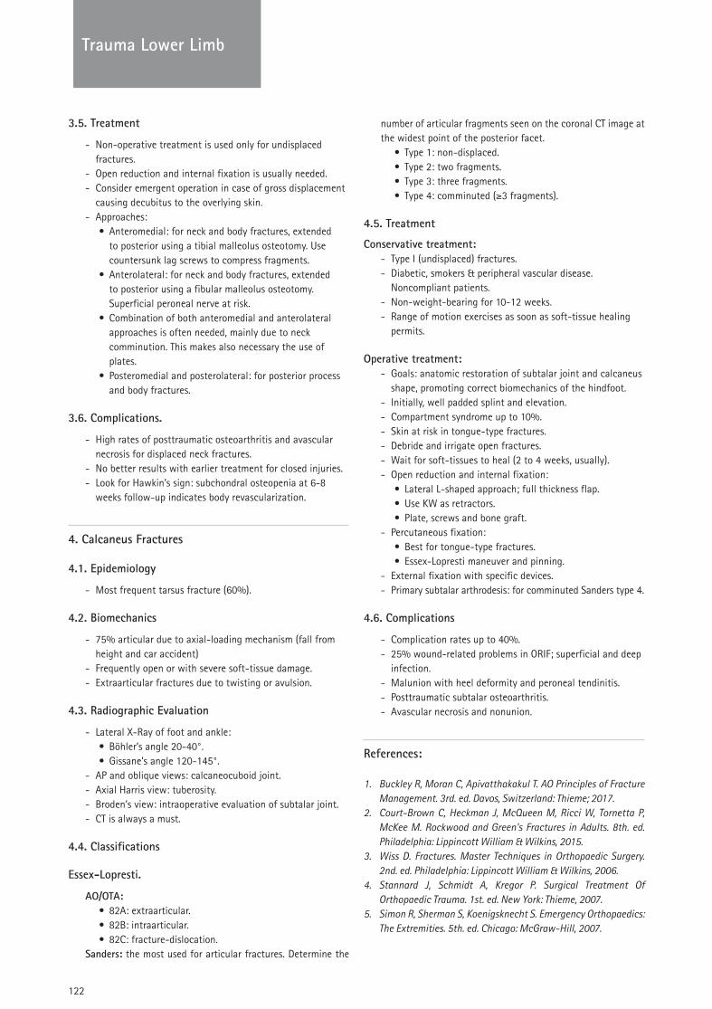
122
Trauma Lower Limb
3.5. Treatment
- Non-operative treatment is used only for undisplaced fractures.
- Open reduction and internal fixation is usually needed. - Consider emergent operation in case of gross displacement
causing decubitus to the overlying skin. - Approaches:
• Anteromedial: for neck and body fractures, extended to posterior using a tibial malleolus osteotomy. Use countersunk lag screws to compress fragments.
• Anterolateral: for neck and body fractures, extended to posterior using a fibular malleolus osteotomy. Superficial peroneal nerve at risk.
• Combination of both anteromedial and anterolateral approaches is often needed, mainly due to neck comminution. This makes also necessary the use of plates.
• Posteromedial and posterolateral: for posterior process and body fractures.
3.6. Complications.
- High rates of posttraumatic osteoarthritis and avascular necrosis for displaced neck fractures.
- No better results with earlier treatment for closed injuries. - Look for Hawkin’s sign: subchondral osteopenia at 6-8
weeks follow-up indicates body revascularization.
4. Calcaneus Fractures
4.1. Epidemiology
- Most frequent tarsus fracture (60%).
4.2. Biomechanics
- 75% articular due to axial-loading mechanism (fall from height and car accident)
- Frequently open or with severe soft-tissue damage. - Extraarticular fractures due to twisting or avulsion.
4.3. Radiographic Evaluation
- Lateral X-Ray of foot and ankle:• Böhler’s angle 20-40°.• Gissane’s angle 120-145°.
- AP and oblique views: calcaneocuboid joint. - Axial Harris view: tuberosity. - Broden’s view: intraoperative evaluation of subtalar joint. - CT is always a must.
4.4. Classifications
Essex-Lopresti.
AO/OTA:• 82A: extraarticular.• 82B: intraarticular.• 82C: fracture-dislocation.
Sanders: the most used for articular fractures. Determine the
number of articular fragments seen on the coronal CT image at the widest point of the posterior facet.
• Type 1: non-displaced.• Type 2: two fragments.• Type 3: three fragments.• Type 4: comminuted (≥3 fragments).
4.5. Treatment
Conservative treatment: - Type I (undisplaced) fractures. - Diabetic, smokers & peripheral vascular disease.
Noncompliant patients. - Non-weight-bearing for 10-12 weeks. - Range of motion exercises as soon as soft-tissue healing
permits.
Operative treatment: - Goals: anatomic restoration of subtalar joint and calcaneus
shape, promoting correct biomechanics of the hindfoot. - Initially, well padded splint and elevation. - Compartment syndrome up to 10%. - Skin at risk in tongue-type fractures. - Debride and irrigate open fractures. - Wait for soft-tissues to heal (2 to 4 weeks, usually). - Open reduction and internal fixation:
• Lateral L-shaped approach; full thickness flap.• Use KW as retractors.• Plate, screws and bone graft.
- Percutaneous fixation:• Best for tongue-type fractures.• Essex-Lopresti maneuver and pinning.
- External fixation with specific devices. - Primary subtalar arthrodesis: for comminuted Sanders type 4.
4.6. Complications
- Complication rates up to 40%. - 25% wound-related problems in ORIF; superficial and deep
infection. - Malunion with heel deformity and peroneal tendinitis. - Posttraumatic subtalar osteoarthritis. - Avascular necrosis and nonunion.
References:
1. Buckley R, Moran C, Apivatthakakul T. AO Principles of Fracture Management. 3rd. ed. Davos, Switzerland: Thieme; 2017.
2. Court-Brown C, Heckman J, McQueen M, Ricci W, Tornetta P, McKee M. Rockwood and Green's Fractures in Adults. 8th. ed. Philadelphia: Lippincott William & Wilkins, 2015.
3. Wiss D. Fractures. Master Techniques in Orthopaedic Surgery. 2nd. ed. Philadelphia: Lippincott William & Wilkins, 2006.
4. Stannard J, Schmidt A, Kregor P. Surgical Treatment Of Orthopaedic Trauma. 1st. ed. New York: Thieme, 2007.
5. Simon R, Sherman S, Koenigsknecht S. Emergency Orthopaedics: The Extremities. 5th. ed. Chicago: McGraw-Hill, 2007.
Trau
ma
Low
er L
imb
123
Trauma Lower Limb
6. Gougolias N, Sakellariou A. 2017. When is a simple fracture of the lateral malleolus not so simple? How to asses stability, wich ones to fix and the role of the deltoide ligament, Bone Joint J. 2017 Jul;99-B(7):851-855.
7. Toth MJ, Yoon RS, Liporace FA, Kowal KJ. What's new in ankle fractures, Injury. 2017 Oct;48(10):2035-2041.
8. Manway JM, Blazek CD, Burns PR. Special considerations in the management of diabetic ankle fractures, Curr Rev Musculoskelet Med. 2018 Sep;11(3):445-455.
9. Tomás-Hernández J. High-energy pilon fractures management: state of the art, EFORT Open Rev. 2017 Mar 13;1(10):354-361.
Questions
1. Which of the following measures is the least appropriate when initially dealing with high-energy pilon fractures?
a. Delta frame.b. Spanning external fixation.c. CT scan.d. Fibula fixation.e. Debridement and irrigation of open wounds.
2. A 65-years-old obese, diabetic and smoker woman arrives at the emergency department with a history of limping after a fall 3 days ago. On the examination, the ankle is swollen and unstable, but there’s no pain. The X-Ray shows a transyndesmotic displaced fibula fracture with great opening of the medial clear on weight-bearing images. Which of the following treatment strategies is incorrect?
a. Percutaneous reduction and fixation of fibula using a rod.b. Posterolateral approach and fibula fixation with plate and
screws.c. Lateral approach and fixation with plate and 4 syndesmotic
screws.d. Lateral approach and fixation with plate and 2 syndesmotic
screws.e. Conservative treatment with long cast for 8 weeks.
3. A 75-years-old lady comes to your emergency department after a twist on the foot when going down stairs. On the clinical examination, you palpate increased pressure on the upper part of the heel and discoloration of the surrounding skin. The lateral X-Ray shows a tongue-type fracture of the calcaneus. Which is the best treatment option?
a. Emergent surgical reduction and fixation.b. External fixation.c. Splinting and delayed surgical treatment.d. Lateral approach when the swelling disappears.e. Primary subtalar fusion.
4. According to the Lauge-Hansen classification of ankle fractures, which of the following description coincides with a pronation-external rotation stage IV fracture?
a. Transyndesmotic fibula fracture and oblique medial malleolus fracture.
b. Suprasyndesmotic comminuted fibula fracture and deltoid complex avulsion.
c. Maissonneuve fracture with posterior syndesmosis injury.d. Dupuytren fracture without posterior syndesmosis injury.e. None of the above.
5. 40 days after a talar neck surgery, the patient comes to your visit. On the control X-Ray you see a radiolucent line immediately under the talar dome. What needs to be done?
a. MRI to rule out avascular necrosis.b. Tibiotalar fusion, as it is a sign of chondral damage.c. It is a poor prognostic sign. The weight-bearing has to be
delayed until the 6th postoperative month.d. Continue normal follow-up.e. Inject cortisone and see the patient in two weeks.
6. Definitive open reduction and internal fixation of a pilon fracture is not indicated in which of the following circumstances?
a. Unbroken blisters around ankle in absence of any skin wrinkling
b. Articular fragments with a gap >3mmc. Articular fragment with a step >1mmd. Varus malalignment at the tibia fracturee. Presence of die-punch central fragments
Answers1d, 2e, 3a, 4c, 5d, 6a.
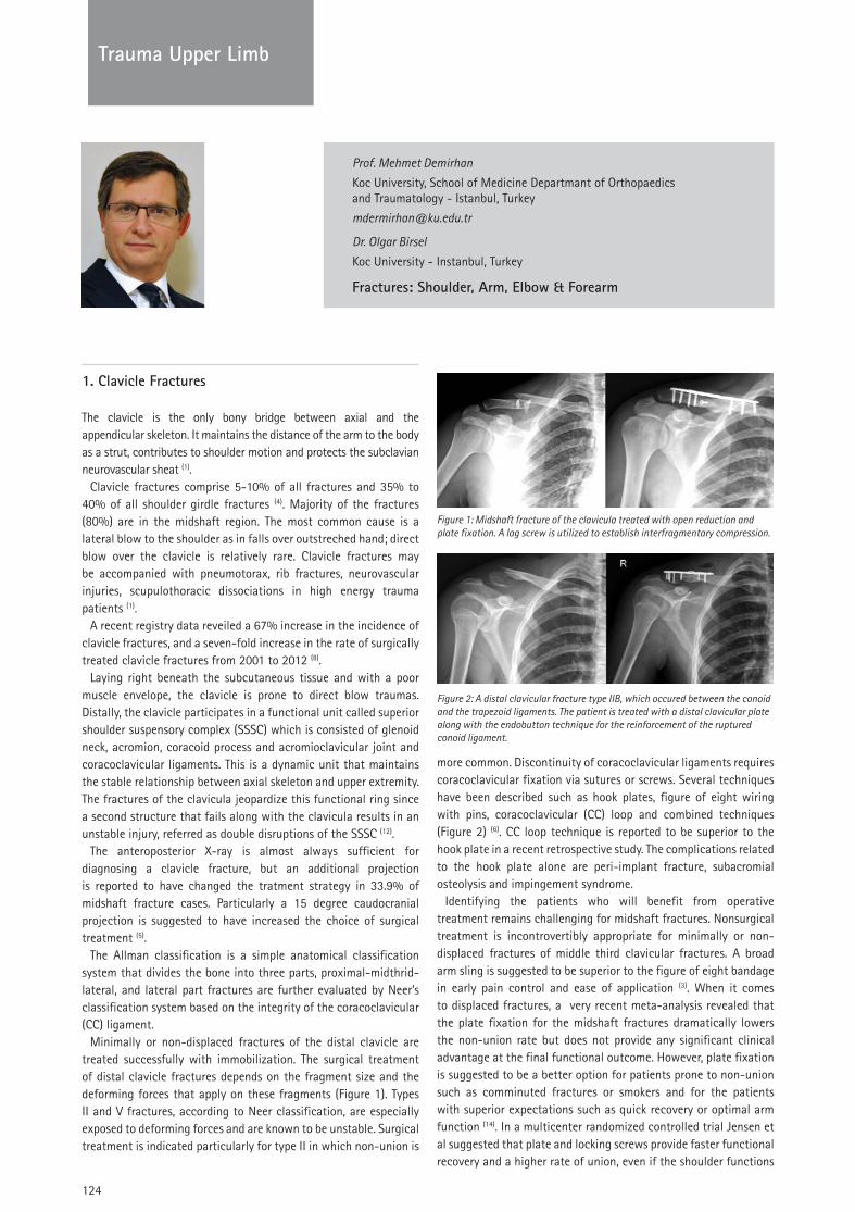
124
TRAUMATrauma Upper Limb
Prof. Mehmet Demirhan
Koc University, School of Medicine Departmant of Orthopaedics and Traumatology - Istanbul, Turkey
Dr. Olgar Birsel
Koc University - Instanbul, Turkey
Fractures: Shoulder, Arm, Elbow & Forearm
1. Clavicle Fractures
The clavicle is the only bony bridge between axial and the appendicular skeleton. It maintains the distance of the arm to the body as a strut, contributes to shoulder motion and protects the subclavian neurovascular sheat (1).
Clavicle fractures comprise 5-10% of all fractures and 35% to 40% of all shoulder girdle fractures (4). Majority of the fractures (80%) are in the midshaft region. The most common cause is a lateral blow to the shoulder as in falls over outstreched hand; direct blow over the clavicle is relatively rare. Clavicle fractures may be accompanied with pneumotorax, rib fractures, neurovascular injuries, scupulothoracic dissociations in high energy trauma patients (1).
A recent registry data reveiled a 67% increase in the incidence of clavicle fractures, and a seven-fold increase in the rate of surgically treated clavicle fractures from 2001 to 2012 (8).
Laying right beneath the subcutaneous tissue and with a poor muscle envelope, the clavicle is prone to direct blow traumas. Distally, the clavicle participates in a functional unit called superior shoulder suspensory complex (SSSC) which is consisted of glenoid neck, acromion, coracoid process and acromioclavicular joint and coracoclavicular ligaments. This is a dynamic unit that maintains the stable relationship between axial skeleton and upper extremity. The fractures of the clavicula jeopardize this functional ring since a second structure that fails along with the clavicula results in an unstable injury, referred as double disruptions of the SSSC (12).
The anteroposterior X-ray is almost always sufficient for diagnosing a clavicle fracture, but an additional projection is reported to have changed the tratment strategy in 33.9% of midshaft fracture cases. Particularly a 15 degree caudocranial projection is suggested to have increased the choice of surgical treatment (5).
The Allman classification is a simple anatomical classification system that divides the bone into three parts, proximal-midthrid-lateral, and lateral part fractures are further evaluated by Neer’s classification system based on the integrity of the coracoclavicular (CC) ligament.
Minimally or non-displaced fractures of the distal clavicle are treated successfully with immobilization. The surgical treatment of distal clavicle fractures depends on the fragment size and the deforming forces that apply on these fragments (Figure 1). Types II and V fractures, according to Neer classification, are especially exposed to deforming forces and are known to be unstable. Surgical treatment is indicated particularly for type II in which non-union is
more common. Discontinuity of coracoclavicular ligaments requires coracoclavicular fixation via sutures or screws. Several techniques have been described such as hook plates, figure of eight wiring with pins, coracoclavicular (CC) loop and combined techniques (Figure 2) (6). CC loop technique is reported to be superior to the hook plate in a recent retrospective study. The complications related to the hook plate alone are peri-implant fracture, subacromial osteolysis and impingement syndrome.
Identifying the patients who will benefit from operative treatment remains challenging for midshaft fractures. Nonsurgical treatment is incontrovertibly appropriate for minimally or non-displaced fractures of middle third clavicular fractures. A broad arm sling is suggested to be superior to the figure of eight bandage in early pain control and ease of application (3). When it comes to displaced fractures, a very recent meta-analysis revealed that the plate fixation for the midshaft fractures dramatically lowers the non-union rate but does not provide any significant clinical advantage at the final functional outcome. However, plate fixation is suggested to be a better option for patients prone to non-union such as comminuted fractures or smokers and for the patients with superior expectations such as quick recovery or optimal arm function (14). In a multicenter randomized controlled trial Jensen et al suggested that plate and locking screws provide faster functional recovery and a higher rate of union, even if the shoulder functions
Figure 1: Midshaft fracture of the clavicula treated with open reduction and plate fixation. A lag screw is utilized to establish interfragmentary compression.
Figure 2: A distal clavicular fracture type IIB, which occured between the conoid and the trapezoid ligaments. The patient is treated with a distal clavicular plate along with the endobutton technique for the reinforcement of the ruptured conoid ligament.
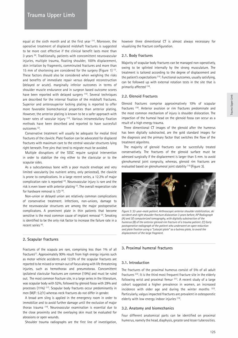
Trau
ma
Uppe
r Lim
b
125
TRAUMATrauma Upper Limb
equal at the sixth month and at the first year (11). Moreover, the operative treatment of displaced midshaft fractures is suggested to be more cost effective if the clinical benefit lasts more than 3 years (9). Traditionally, patients with concomittent neurovascular injuries, multiple trauma, floating shoulder, 100% displacement, skin irritation by fragments, comminuted fractures and more than 15 mm of shortening are considered for the surgery (Figure 1) (1). These factors should also be considered when weighing the risks and benefits of immediate repair versus delayed reconstruction (delayed or acute). marginally inferior outcomes in terms of shoulder muscle endurance and in surgeon based outcome scores have been reported with delayed surgery (10). Several techniques are described for the internal fixation of the midshaft fractures. Superior and anterosuperior locking plating is reported to offer more favorable biomechanical properties than anterior plating. However, the anterior plating is known to be a safer approach with lower rates of vascular injury (13). Various intramedullary fixation methods have been described and reported to have succesfull outcomes (2).
Conservative treatment will usually be adequate for medial third fractures of the clavicle. Plate fixation can be advocated for displaced fractures with maximum care to the central vascular structures lying right beneath. Free pins that tend to migrate must be avoided.
Multiple disruptions of the SSSC require surgical intervention in order to stabilize the ring either to the clavicular or to the scapular sides.
As a subcutaneous bone with a poor muscle envelope and very limited vascularity (no nutrient artery, only periosteal), the clavicle is prone to complications. In a large recent series, a 12.2% of major complication rate is reported (4). Neurovascular injury is rare and the risk is even lower with anterior plating (7). The overall reoperation rate for hardware removal is 1/3 (4).
Non-union or delayed union are relatively common complications of conservative treatment. Infections, non-unions, damage to the neurovascular structures are among the major postoperative complicatons. A prominent plate in thin patients that become sensitive is the most common cause of implant removal (2). Smoking is identified to be the only risk factor to increase the failure rate in a recent series (4).
2. Scapular fractures
Fractures of the scapula are rare, comprising less than 1% of all fractures(1). Approximately 90% result from high energy injuries such as motor vehicle accidents and 12.5% of the scapular fractures are reported to be missed or remain out of focus along with life threatening injuries, such as hemothorax and pneumotorax. Concomittent ipsilateral clavicular fractures are common (19%) and must be ruled out. The most common fracture site, in a large series in the litterature, was scapular body with 52%, followed by glenoid fossa with 29% and processes (11%) (17). Scapular body fractures occur predominantly in men (M/F: 5.2/1) whereas neck fractures do not differ in gender.
A broad arm sling is applied in the emergency room in order to immobilize and to avoid further damage until the exclusion of major thorax trauma (16). Neurovascular examination is essential due to the close proximity and the overlaying skin must be evaluated for abrasions or open wounds.
Shoulder trauma radiographs are the first line of investigation,
however three dimentional CT is almost always necesssary for visualizing the fracture configuration.
2.1. Body fractures
Majority of scapular body fractures can be managed non-operatively, owing to be splinted internally by the strong musculature. The treatment is tailored according to the degree of displacement and the patient’s expectations (15). Functional outcomes, usually satisfying, can be followed up with external rotation tests in the site that is primarily affected (16).
2.2. Glenoid Fractures
Glenoid fractures comprise approximatively 10% of scapular fractures (18). Anterior avulsion or rim fractures predominate and the most common mechanism of injury is shoulder dislocation. The impaction of the humeal head on the glenoid fossa can occur as a result of a high energy trauma.
Three dimentional CT images of the glenoid after the humerus has been digitally substracted, are the gold standard images for the diagnosis and the primary factor that switches the flow of the treatment algoritms.
The majority of glenoid fractures can be succesfully treated conservatively. The fractures of the glenoid surface must be adressed surgically if the displacement is larger than 5 mm. to avoid glenohumeral joint congruity, whereas, glenoid rim fractures are evaluated based on glenohumeral joint stability (15) (Figure 3).
Figure 3: 32-year-male patient. Arthroscopic anterior shoulder stabilization, ski accident and right shoulder fracture dislocation 3 years before; AP Radiograph (A) and 3D computerized tomography, with digitally substraction of the humerus (B) of the anterior glenoid rim fracture of a trauma patient. (C) Early postoperative radiograph of the patient who underwent an open reduction and plate fixation using a “Latarjet plate” as a butress plate, to avoid the displacement of the large fragment.
3. Proximal humeral fractures
3.1. Introduction
The fractures of the proximal humerus consist of 5% of all adult fractures (33). It is the third most frequent fracture site in the elderly following wrist and proximal femur (31). A recent study of a large cohort suggested a higher prevalence in women, an increased incidence with older age and during the winter months (33). Particularly, valgus impacted fractures are prevalent in osteoporotic elderly with low energy indoor injuries (19).
3.2. Anatomy and biomechanics
Four different anatomical parts can be identified on proximal humerus, namely the head, diaphysis, greater and lesser tuberosities.
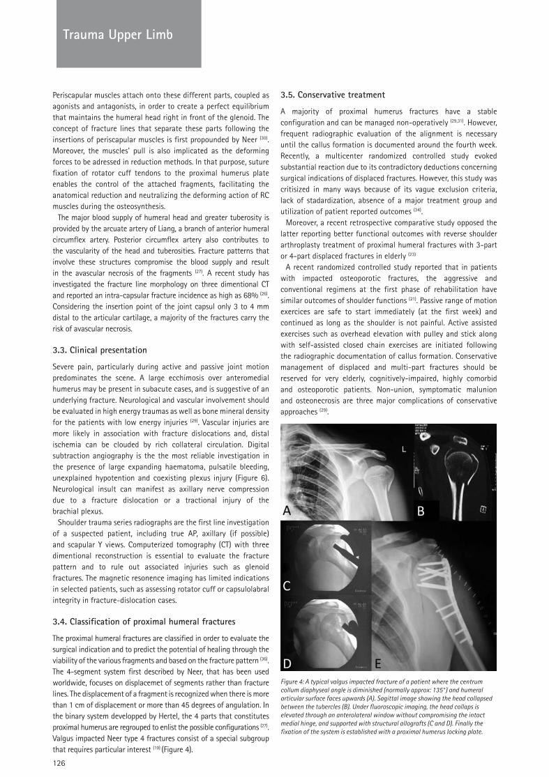
126
TRAUMATrauma Upper Limb
Periscapular muscles attach onto these different parts, coupled as agonists and antagonists, in order to create a perfect equilibrium that maintains the humeral head right in front of the glenoid. The concept of fracture lines that separate these parts following the insertions of periscapular muscles is first propounded by Neer (30). Moreover, the muscles’ pull is also implicated as the deforming forces to be adressed in reduction methods. In that purpose, suture fixation of rotator cuff tendons to the proximal humerus plate enables the control of the attached fragments, facilitating the anatomical reduction and neutralizing the deforming action of RC muscles during the osteosynthesis.
The major blood supply of humeral head and greater tuberosity is provided by the arcuate artery of Liang, a branch of anterior humeral circumflex artery. Posterior circumflex artery also contributes to the vascularity of the head and tuberosities. Fracture patterns that involve these structures compromise the blood supply and result in the avascular necrosis of the fragments (27). A recent study has investigated the fracture line morphology on three dimentional CT and reported an intra-capsular fracture incidence as high as 68% (26). Considering the insertion point of the joint capsul only 3 to 4 mm distal to the articular cartilage, a majority of the fractures carry the risk of avascular necrosis.
3.3. Clinical presentation
Severe pain, particularly during active and passive joint motion predominates the scene. A large ecchimosis over anteromedial humerus may be present in subacute cases, and is suggestive of an underlying fracture. Neurological and vascular involvement should be evaluated in high energy traumas as well as bone mineral density for the patients with low energy injuries (29). Vascular injuries are more likely in association with fracture dislocations and, distal ischemia can be clouded by rich collateral circulation. Digital subtraction angiography is the the most reliable investigation in the presence of large expanding haematoma, pulsatile bleeding, unexplained hypotention and coexisting plexus injury (Figure 6). Neurological insult can manifest as axillary nerve compression due to a fracture dislocation or a tractional injury of the brachial plexus.
Shoulder trauma series radiographs are the first line investigation of a suspected patient, including true AP, axillary (if possible) and scapular Y views. Computerized tomography (CT) with three dimentional reconstruction is essential to evaluate the fracture pattern and to rule out associated injuries such as glenoid fractures. The magnetic resonence imaging has limited indications in selected patients, such as assessing rotator cuff or capsulolabral integrity in fracture-dislocation cases.
3.4. Classification of proximal humeral fractures
The proximal humeral fractures are classified in order to evaluate the surgical indication and to predict the potential of healing through the viability of the various fragments and based on the fracture pattern (36). The 4-segment system first described by Neer, that has been used worldwide, focuses on displacemet of segments rather than fracture lines. The displacement of a fragment is recognized when there is more than 1 cm of displacement or more than 45 degrees of angulation. In the binary system developped by Hertel, the 4 parts that constitutes proximal humerus are regrouped to enlist the possible configurations (27). Valgus impacted Neer type 4 fractures consist of a special subgroup that requires particular interest (19) (Figure 4).
3.5. Conservative treatment
A majority of proximal humerus fractures have a stable configuration and can be managed non-operatively (29,31). However, frequent radiographic evaluation of the alignment is necessary until the callus formation is documented around the fourth week. Recently, a multicenter randomized controlled study evoked substantial reaction due to its contradictory deductions concerning surgical indications of displaced fractures. However, this study was critisized in many ways because of its vague exclusion criteria, lack of stadardization, absence of a major treatment group and utilization of patient reported outcomes (34).
Moreover, a recent retrospective comparative study opposed the latter reporting better functional outcomes with reverse shoulder arthroplasty treatment of proximal humeral fractures with 3-part or 4-part displaced fractures in elderly (23)
A recent randomized controlled study reported that in patients with impacted osteoporotic fractures, the aggressive and conventional regimens at the first phase of rehabilitation have similar outcomes of shoulder functions (21). Passive range of motion exercices are safe to start immediately (at the first week) and continued as long as the shoulder is not painful. Active assisted exercises such as overhead elevation with pulley and stick along with self-assisted closed chain exercises are initiated following the radiographic documentation of callus formation. Conservative management of displaced and multi-part fractures should be reserved for very elderly, cognitively-impaired, highly comorbid and osteoporotic patients. Non-union, symptomatic malunion and osteonecrosis are three major complications of conservative approaches (29).
Figure 4: A typical valgus impacted fracture of a patient where the centrum collum diaphyseal angle is diminished (normally approx: 135°) and humeral articular surface faces upwards (A). Sagittal image showing the head collapsed between the tubercles (B). Under fluoroscopic imaging, the head collaps is elevated through an anterolateral window without compromising the intact medial hinge, and supported with structural allografts (C and D). Finally the fixation of the system is established with a proximal humerus locking plate.
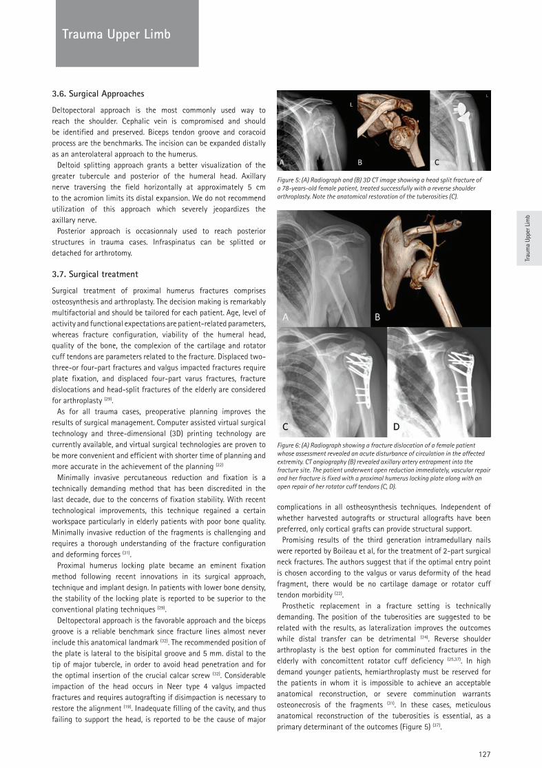
Trau
ma
Uppe
r Lim
b
127
TRAUMATrauma Upper Limb
3.6. Surgical Approaches
Deltopectoral approach is the most commonly used way to reach the shoulder. Cephalic vein is compromised and should be identified and preserved. Biceps tendon groove and coracoid process are the benchmarks. The incision can be expanded distally as an anterolateral approach to the humerus.
Deltoid splitting approach grants a better visualization of the greater tubercule and posterior of the humeral head. Axillary nerve traversing the field horizontally at approximately 5 cm to the acromion limits its distal expansion. We do not recommend utilization of this approach which severely jeopardizes the axillary nerve.
Posterior approach is occasionnaly used to reach posterior structures in trauma cases. Infraspinatus can be splitted or detached for arthrotomy.
3.7. Surgical treatment
Surgical treatment of proximal humerus fractures comprises osteosynthesis and arthroplasty. The decision making is remarkably multifactorial and should be tailored for each patient. Age, level of activity and functional expectations are patient-related parameters, whereas fracture configuration, viability of the humeral head, quality of the bone, the complexion of the cartilage and rotator cuff tendons are parameters related to the fracture. Displaced two-three-or four-part fractures and valgus impacted fractures require plate fixation, and displaced four-part varus fractures, fracture dislocations and head-split fractures of the elderly are considered for arthroplasty (29).
As for all trauma cases, preoperative planning improves the results of surgical management. Computer assisted virtual surgical technology and three-dimensional (3D) printing technology are currently available, and virtual surgical technologies are proven to be more convenient and efficient with shorter time of planning and more accurate in the achievement of the planning (22)
Minimally invasive percutaneous reduction and fixation is a technically demanding method that has been discredited in the last decade, due to the concerns of fixation stability. With recent technological improvements, this technique regained a certain workspace particularly in elderly patients with poor bone quality. Minimally invasive reduction of the fragments is challenging and requires a thorough understanding of the fracture configuration and deforming forces (31).
Proximal humerus locking plate became an eminent fixation method following recent innovations in its surgical approach, technique and implant design. In patients with lower bone density, the stability of the locking plate is reported to be superior to the conventional plating techniques (29).
Deltopectoral approach is the favorable approach and the biceps groove is a reliable benchmark since fracture lines almost never include this anatomical landmark (32). The recommended position of the plate is lateral to the bisipital groove and 5 mm. distal to the tip of major tubercle, in order to avoid head penetration and for the optimal insertion of the crucial calcar screw (32). Considerable impaction of the head occurs in Neer type 4 valgus impacted fractures and requires autografting if disimpaction is necessary to restore the alignment (19). Inadequate filling of the cavity, and thus failing to support the head, is reported to be the cause of major
complications in all ostheosynthesis techniques. Independent of whether harvested autografts or structural allografts have been preferred, only cortical grafts can provide structural support.
Promising results of the third generation intramedullary nails were reported by Boileau et al, for the treatment of 2-part surgical neck fractures. The authors suggest that if the optimal entry point is chosen according to the valgus or varus deformity of the head fragment, there would be no cartilage damage or rotator cuff tendon morbidity (22).
Prosthetic replacement in a fracture setting is technically demanding. The position of the tuberosities are suggested to be related with the results, as lateralization improves the outcomes while distal transfer can be detrimental (24). Reverse shoulder arthroplasty is the best option for comminuted fractures in the elderly with concomittent rotator cuff deficiency (25,37). In high demand younger patients, hemiarthroplasty must be reserved for the patients in whom it is impossible to achieve an acceptable anatomical reconstruction, or severe comminution warrants osteonecrosis of the fragments (31). In these cases, meticulous anatomical reconstruction of the tuberosities is essential, as a primary determinant of the outcomes (Figure 5) (37).
Figure 6: (A) Radiograph showing a fracture dislocation of a female patient whose assessment revealed an acute disturbance of circulation in the affected extremity. CT angiography (B) revealed axillary artery entrapment into the fracture site. The patient underwent open reduction immediately, vascular repair and her fracture is fixed with a proximal humerus locking plate along with an open repair of her rotator cuff tendons (C, D).
Figure 5: (A) Radiograph and (B) 3D CT image showing a head split fracture of a 78-years-old female patient, treated successfully with a reverse shoulder arthroplasty. Note the anatomical restoration of the tuberosities (C).
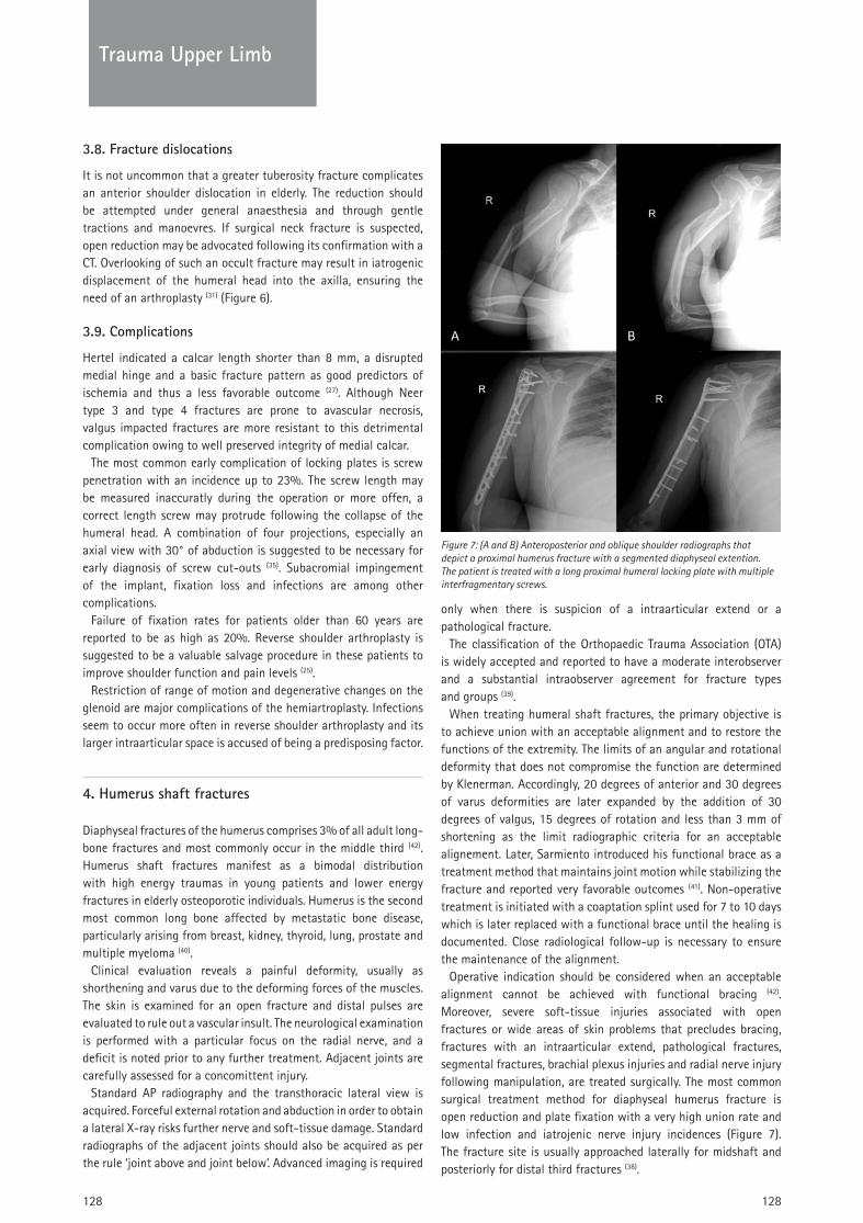
128
TRAUMA
128
Trauma Upper Limb
3.8. Fracture dislocations
It is not uncommon that a greater tuberosity fracture complicates an anterior shoulder dislocation in elderly. The reduction should be attempted under general anaesthesia and through gentle tractions and manoevres. If surgical neck fracture is suspected, open reduction may be advocated following its confirmation with a CT. Overlooking of such an occult fracture may result in iatrogenic displacement of the humeral head into the axilla, ensuring the need of an arthroplasty (31) (Figure 6).
3.9. Complications
Hertel indicated a calcar length shorter than 8 mm, a disrupted medial hinge and a basic fracture pattern as good predictors of ischemia and thus a less favorable outcome (27). Although Neer type 3 and type 4 fractures are prone to avascular necrosis, valgus impacted fractures are more resistant to this detrimental complication owing to well preserved integrity of medial calcar.
The most common early complication of locking plates is screw penetration with an incidence up to 23%. The screw length may be measured inaccuratly during the operation or more offen, a correct length screw may protrude following the collapse of the humeral head. A combination of four projections, especially an axial view with 30° of abduction is suggested to be necessary for early diagnosis of screw cut-outs (35). Subacromial impingement of the implant, fixation loss and infections are among other complications.
Failure of fixation rates for patients older than 60 years are reported to be as high as 20%. Reverse shoulder arthroplasty is suggested to be a valuable salvage procedure in these patients to improve shoulder function and pain levels (25).
Restriction of range of motion and degenerative changes on the glenoid are major complications of the hemiartroplasty. Infections seem to occur more often in reverse shoulder arthroplasty and its larger intraarticular space is accused of being a predisposing factor.
4. Humerus shaft fractures
Diaphyseal fractures of the humerus comprises 3% of all adult long-bone fractures and most commonly occur in the middle third (42). Humerus shaft fractures manifest as a bimodal distribution with high energy traumas in young patients and lower energy fractures in elderly osteoporotic individuals. Humerus is the second most common long bone affected by metastatic bone disease, particularly arising from breast, kidney, thyroid, lung, prostate and multiple myeloma (40).
Clinical evaluation reveals a painful deformity, usually as shorthening and varus due to the deforming forces of the muscles. The skin is examined for an open fracture and distal pulses are evaluated to rule out a vascular insult. The neurological examination is performed with a particular focus on the radial nerve, and a deficit is noted prior to any further treatment. Adjacent joints are carefully assessed for a concomittent injury.
Standard AP radiography and the transthoracic lateral view is acquired. Forceful external rotation and abduction in order to obtain a lateral X-ray risks further nerve and soft-tissue damage. Standard radiographs of the adjacent joints should also be acquired as per the rule ‘joint above and joint below’. Advanced imaging is required
only when there is suspicion of a intraarticular extend or a pathological fracture.
The classification of the Orthopaedic Trauma Association (OTA) is widely accepted and reported to have a moderate interobserver and a substantial intraobserver agreement for fracture types and groups (39).
When treating humeral shaft fractures, the primary objective is to achieve union with an acceptable alignment and to restore the functions of the extremity. The limits of an angular and rotational deformity that does not compromise the function are determined by Klenerman. Accordingly, 20 degrees of anterior and 30 degrees of varus deformities are later expanded by the addition of 30 degrees of valgus, 15 degrees of rotation and less than 3 mm of shortening as the limit radiographic criteria for an acceptable alignement. Later, Sarmiento introduced his functional brace as a treatment method that maintains joint motion while stabilizing the fracture and reported very favorable outcomes (41). Non-operative treatment is initiated with a coaptation splint used for 7 to 10 days which is later replaced with a functional brace until the healing is documented. Close radiological follow-up is necessary to ensure the maintenance of the alignment.
Operative indication should be considered when an acceptable alignment cannot be achieved with functional bracing (42). Moreover, severe soft-tissue injuries associated with open fractures or wide areas of skin problems that precludes bracing, fractures with an intraarticular extend, pathological fractures, segmental fractures, brachial plexus injuries and radial nerve injury following manipulation, are treated surgically. The most common surgical treatment method for diaphyseal humerus fracture is open reduction and plate fixation with a very high union rate and low infection and iatrojenic nerve injury incidences (Figure 7). The fracture site is usually approached laterally for midshaft and posteriorly for distal third fractures (38).
Figure 7: (A and B) Anteroposterior and oblique shoulder radiographs that depict a proximal humerus fracture with a segmented diaphyseal extention. The patient is treated with a long proximal humeral locking plate with multiple interfragmentary screws.
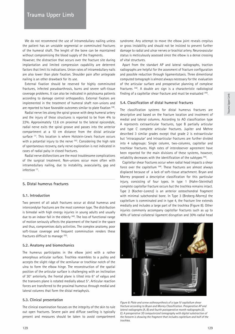
Trau
ma
Uppe
r Lim
b
129
TRAUMA
129
Trauma Upper Limb
We do not recommend the use of intramedullary nailing unless the patient has an unstable segmental or comminuted fractures of the humeral shaft. The lenght of the bone can be maintained without compromising the blood supply of the fragments. However, the distraction that occurs over the fracture site during implantation and limited compression capability are deterrent factors that limit its indications. Union rates of intramedullary nails are also lower than plate fixation. Shoulder pain after antegrade nailing is an other drawback for its use.
External fixation should be reserved for highly comminuted fractures, infected pseudoarthrosis, burns and severe soft-tissue coverage problems. It can also be indicated in polytrauma patients according to damage control orthopaedics. External fixators are implemented in the treatment of humeral shaft non-unions and are reported to have favorable outcomes similar to plate fixation (38).
Radial nerve lies along the spiral groove with deep humeral artery and the injury of these structures is reported to be from 4% to 22%. Approximately 12.6 cm proximal to the lateral epicondyle, radial nerve exits the spiral groove and passes into the anterior compartment at a 10 cm distance from the distal articular surface (1). This location is where Holstein-Lewis fracture occurs with a potantial injury to the nerve (42). Considering the high rate of spontaneous recovery, early nerve exploration is not indicated in cases of radial palsy in closed fractures.
Radial nerve disfonctions are the most troublesome complications of the surgical treatment. Non-unions occur more often with intramedullary nailing, due to instability, avascularity, gap and infection (1).
5. Distal humerus fractures
5.1. Introduction
Two percent of all adult fractures occur at distal humerus and intercondylar fractures are the most common type. The distribution is bimodal with high energy injuries in young adults and usually due to an indoor fall in the elderly (46). The loss of functional range of motion seriously affects the placement of the hand in the space and thus, compromises daily activities. The complex anatomy, poor soft-tissue coverage and frequent comminution renders these fractures difficult to manage (55).
5.2. Anatomy and biomechanics
The humerus participates in the elbow joint with a rather amorphous articular surface. Trochlea resembles to a pulley and accepts the slight ridge of the semilunar or trochlear notch of the ulna to form the elbow hinge. The reconstruction of the spatial position of the articular surface is challenging with an inclination of 30° anteriorly, the frontal plane is tilted into 6° of valgus and the transvers plane is rotated medially about 5°. Articular reaction forces are transferred to the proximal humerus through medial and lateral columns that form the distal metaphysis.
5.3. Clinical presentation
The clinical examination focuses on the integrity of the skin to rule out open fractures. Severe pain and diffuse swelling is typically present and measures should be taken to avoid compartment
syndrome. Any attempt to move the elbow joint reveals crepitus or gross instability and should not be insisted to prevent further damage to radial and ulnar nerves or brachial artery. Neurovascular status is meticulously assessed since the elbow is a dense crossing of vital structures.
Apart from the standart AP and lateral radiographs, traction radiographs are helpful for the assesment of fracture configuration and possible reduction through ligamentotaxis. Three dimentional computed tomograph is almost always necessary for the evaluation of the articular surface and preoperative planning of complexe fractures (46). A double arc sign is a characteristic radiological finding of a capitellar shear fracture and must be evaluated (49).
5.4. Classification of distal humeral fractures
The classification systems for distal humerus fractures are descriptive and based on the fracture location and involment of medial and lateral columns. According to AO classification type A represents extraarticular fractures, type B partially articular and type C complete articular fractures. Jupiter and Mehne described 3 similar grades except that grade 2 is extraarticular but ‘intracapsular’ and intraarticular fractures are further divided into 4 subgroups: Single column, two-columns, capitellar and trochlear fractures. High rates of interobserver agreement have been reported for the main divisions of these systems, however, reliability decreases with the identification of the subtypes (46).
Capitellar shear fractures occur when radial head impacts a shear force over the capitellum (49). These fractures are almost always displaced because of a lack of soft-tissue attachment. Bryan and Morrey proposed a descriptive classification for this particular injury, consisting of four types. In type 1 (Hahn-Steinthal) complete capitellar fracture occurs but the trochlea remains intact. Type 2 (Kocher-Lorenz) is an anterior osteochondral fragment with minimal subchondral bone. In Type 3 (Broberg-Morrey) the capitellum is comminated and in type 4, the fracture line extends medially and includes a large part of the trochlea (Figure 8). Other injuries commonly accompany capitellar fractures such as up to 40% of lateral collateral ligament disruption and 30% radial head
Figure 8: Plate and screw ostheosynthesis of a type IV capitallum shear fracture according to Bryan and Morrey Classification. Preoperative AP and lateral radiographs (A, B) and fourth postoperative month radiographs (D, E). A preoperative 3D computerized tomography with digital substraction of the forearm is showing the fragment that includes capitellum and half of the trochlea.
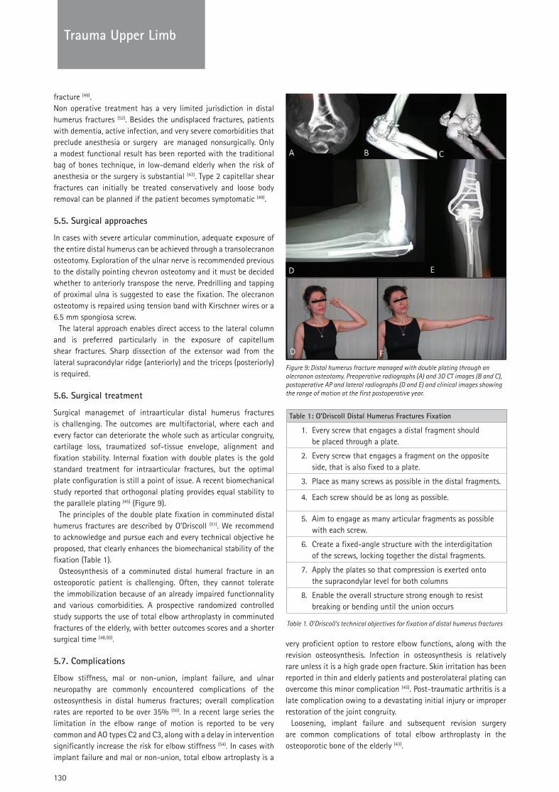
130
TRAUMATrauma Upper Limb
fracture (49). Non operative treatment has a very limited jurisdiction in distal humerus fractures (52). Besides the undisplaced fractures, patients with dementia, active infection, and very severe comorbidities that preclude anesthesia or surgery are managed nonsurgically. Only a modest functional result has been reported with the traditional bag of bones technique, in low-demand elderly when the risk of anesthesia or the surgery is substantial (43). Type 2 capitellar shear fractures can initially be treated conservatively and loose body removal can be planned if the patient becomes symptomatic (49).
5.5. Surgical approaches
In cases with severe articular comminution, adequate exposure of the entire distal humerus can be achieved through a transolecranon osteotomy. Exploration of the ulnar nerve is recommended previous to the distally pointing chevron osteotomy and it must be decided whether to anteriorly transpose the nerve. Predrilling and tapping of proximal ulna is suggested to ease the fixation. The olecranon osteotomy is repaired using tension band with Kirschner wires or a 6.5 mm spongiosa screw.
The lateral approach enables direct access to the lateral column and is preferred particularly in the exposure of capitellum shear fractures. Sharp dissection of the extensor wad from the lateral supracondylar ridge (anteriorly) and the triceps (posteriorly) is required.
5.6. Surgical treatment
Surgical managemet of intraarticular distal humerus fractures is challenging. The outcomes are multifactorial, where each and every factor can deteriorate the whole such as articular congruity, cartilage loss, traumatized sof-tissue envelope, alignment and fixation stability. Internal fixation with double plates is the gold standard treatment for intraarticular fractures, but the optimal plate configuration is still a point of issue. A recent biomechanical study reported that orthogonal plating provides equal stability to the parallele plating (45) (Figure 9).
The principles of the double plate fixation in comminuted distal humerus fractures are described by O’Driscoll (51). We recommend to acknowledge and pursue each and every technical objective he proposed, that clearly enhances the biomechanical stability of the fixation (Table 1).
Osteosynthesis of a comminuted distal humeral fracture in an osteoporotic patient is challenging. Often, they cannot tolerate the immobilization because of an already impaired functionnality and various comorbidities. A prospective randomized controlled study supports the use of total elbow arthroplasty in comminuted fractures of the elderly, with better outcomes scores and a shorter surgical time (48,50).
5.7. Complications
Elbow stiffness, mal or non-union, implant failure, and ulnar neuropathy are commonly encountered complications of the osteosynthesis in distal humerus fractures; overall complication rates are reported to be over 35% (50). In a recent large series the limitation in the elbow range of motion is reported to be very common and AO types C2 and C3, along with a delay in intervention significantly increase the risk for elbow stiffness (54). In cases with implant failure and mal or non-union, total elbow artroplasty is a
very proficient option to restore elbow functions, along with the revision osteosynthesis. Infection in osteosynthesis is relatively rare unless it is a high grade open fracture. Skin irritation has been reported in thin and elderly patients and posterolateral plating can overcome this minor complication (45). Post-traumatic arthritis is a late complication owing to a devastating initial injury or improper restoration of the joint congruity.
Loosening, implant failure and subsequent revision surgery are common complications of total elbow arthroplasty in the osteoporotic bone of the elderly (43).
Table 1: O’Driscoll Distal Humerus Fractures Fixation
1. Every screw that engages a distal fragment should be placed through a plate.
2. Every screw that engages a fragment on the opposite side, that is also fixed to a plate.
3. Place as many screws as possible in the distal fragments.
4. Each screw should be as long as possible.
5. Aim to engage as many articular fragments as possible with each screw.
6. Create a fixed-angle structure with the interdigitation of the screws, locking together the distal fragments.
7. Apply the plates so that compression is exerted onto the supracondylar level for both columns
8. Enable the overall structure strong enough to resist breaking or bending until the union occurs
Figure 9: Distal humerus fracture managed with double plating through an olecranon osteotomy. Preoperative radiographs (A) and 3D CT images (B and C), postoperative AP and lateral radiographs (D and E) and clinical images showing the range of motion at the first postoperative year.
Table 1. O’Driscoll’s technical objectives for fixation of distal humerus fractures
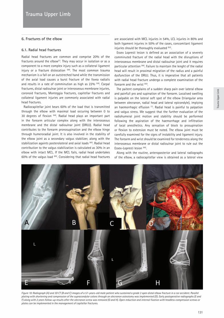
Trau
ma
Uppe
r Lim
b
131
TRAUMATrauma Upper Limb
6. Fractures of the elbow
6.1. Radial head fractures
Radial head fractures are common and comprise 20% of the fractures around the elbow(1). They may occur in isolation or as a component to a more complex injury such as a collateral ligament injury or a fracture dislocation (68). The most common trauma mechanism is a fall on an outstreched hand while the transmission of the axial load causes a burst fracture of the fovea radialis and results in a rate of comminution as high as 22% (59). Carpal fractures, distal radioulnar joint or interosseous membrane injuries, coronoid fractures, Monteggia fractures, capitellar fractures and collateral ligament injuries are commonly associated with radial head fractures.
Radiocapitellar joint bears 60% of the load that is transmitted through the elbow with maximal load occuring between 0 to 30 degrees of flexion (64). Radial head plays an important part in the forearm articular complex along with the interosseous membrane and the distal radioulnar joint (DRUJ). Radial head contributes to the forearm pronosupination and the elbow hinge through humeroradial joint. It is also involved in the stability of the elbow joint as a secondary valgus stabilizer, along with the stabilization againts posterolateral and axial loads (65). Radial head contribution to the valgus stabilization is calculated as 30% in an elbow with intact MCL. If the MCL fails, radial head undertakes 60% of the valgus load (65). Considering that radial head fractures
are associated with MCL injuries in 54%, LCL injuries in 80% and both ligament injuries in 50% of the cases, concomitant ligament injuries should be thoroughly evaluated (62).
Essex Lopresti lesion is defined as an association of a severely comminuted fracture of the radial head with the disruptions of interosseous membrane and distal radioulnar joint and it requires particular attention (66). Failure to maintain the lenght of the radial head will result in proximal migration of the radius and a painful dysfunction of the DRUJ. Thus, it is imperative that all patients with radial head fracture undergo a complete examination of the forearm and the wrist (60).
The paitent complains of a sudden sharp pain over lateral elbow and painfull pro and supination of the forearm. Localised swelling is palpable on the lateral soft spot of the elbow (triangular area between olecranon, radial head and lateral epicondyle), implying an haemorrhagic effusion (1). Radial head is painful to palpation and valgus stress. We suggest that the further evaluation of the radiohumeral joint motion and stability should be performed following the aspiration of the haemorrhage and infiltration of local anesthetics. Any sensation of block to prosupination or flexion to extension must be noted. The elbow joint must be carefully examined for the signs of instability and ligament injury. The forearm and wrist should be examined for tenderness along the interosseous membrane or distal radioulnar joint to rule out the Essex-Lopresti lesion (60).
Along with the routine, anteroposterior and lateral radiographs of the elbow, a radiocapitellar view is obtained as a lateral view
Figure 10: Radiograph (A) and 3D CT (B and C) images of a 57-years-old male patient who sustained a grade 2 open distal elbow fracture in a car accident. Parallel plating with shortening and compression of the supracondylar colons through an olecranon osteotomy was implemented (D). Early postoperative radiographs (E and F) along with 2 years follow-up results after the olecranon screw was removed (G and H). Open reduction and internal fixation with headless compression screws or plates can be implemented in the management of capitellar fractures.
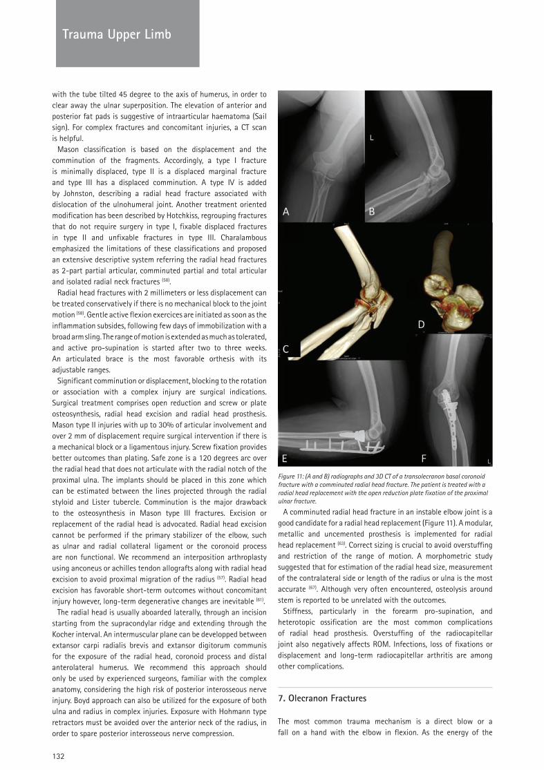
132
TRAUMATrauma Upper Limb
with the tube tilted 45 degree to the axis of humerus, in order to clear away the ulnar superposition. The elevation of anterior and posterior fat pads is suggestive of intraarticular haematoma (Sail sign). For complex fractures and concomitant injuries, a CT scan is helpful.
Mason classification is based on the displacement and the comminution of the fragments. Accordingly, a type I fracture is minimally displaced, type II is a displaced marginal fracture and type III has a displaced comminution. A type IV is added by Johnston, describing a radial head fracture associated with dislocation of the ulnohumeral joint. Another treatment oriented modification has been described by Hotchkiss, regrouping fractures that do not require surgery in type I, fixable displaced fractures in type II and unfixable fractures in type III. Charalambous emphasized the limitations of these classifications and proposed an extensive descriptive system referring the radial head fractures as 2-part partial articular, comminuted partial and total articular and isolated radial neck fractures (58).
Radial head fractures with 2 millimeters or less displacement can be treated conservatively if there is no mechanical block to the joint motion (58). Gentle active flexion exercices are initiated as soon as the inflammation subsides, following few days of immobilization with a broad arm sling. The range of motion is extended as much as tolerated, and active pro-supination is started after two to three weeks. An articulated brace is the most favorable orthesis with its adjustable ranges.
Significant comminution or displacement, blocking to the rotation or association with a complex injury are surgical indications. Surgical treatment comprises open reduction and screw or plate osteosynthesis, radial head excision and radial head prosthesis. Mason type II injuries with up to 30% of articular involvement and over 2 mm of displacement require surgical intervention if there is a mechanical block or a ligamentous injury. Screw fixation provides better outcomes than plating. Safe zone is a 120 degrees arc over the radial head that does not articulate with the radial notch of the proximal ulna. The implants should be placed in this zone which can be estimated between the lines projected through the radial styloid and Lister tubercle. Comminution is the major drawback to the osteosynthesis in Mason type III fractures. Excision or replacement of the radial head is advocated. Radial head excision cannot be performed if the primary stabilizer of the elbow, such as ulnar and radial collateral ligament or the coronoid process are non functional. We recommend an interposition arthroplasty using anconeus or achilles tendon allografts along with radial head excision to avoid proximal migration of the radius (57). Radial head excision has favorable short-term outcomes without concomitant injury however, long-term degenerative changes are inevitable (61).
The radial head is usually aboarded laterally, through an incision starting from the supracondylar ridge and extending through the Kocher interval. An intermuscular plane can be developped between extansor carpi radialis brevis and extansor digitorum communis for the exposure of the radial head, coronoid process and distal anterolateral humerus. We recommend this approach should only be used by experienced surgeons, familiar with the complex anatomy, considering the high risk of posterior interosseous nerve injury. Boyd approach can also be utilized for the exposure of both ulna and radius in complex injuries. Exposure with Hohmann type retractors must be avoided over the anterior neck of the radius, in order to spare posterior interosseous nerve compression.
A comminuted radial head fracture in an instable elbow joint is a good candidate for a radial head replacement (Figure 11). A modular, metallic and uncemented prosthesis is implemented for radial head replacement (63). Correct sizing is crucial to avoid overstuffing and restriction of the range of motion. A morphometric study suggested that for estimation of the radial head size, measurement of the contralateral side or length of the radius or ulna is the most accurate (67). Although very often encountered, osteolysis around stem is reported to be unrelated with the outcomes.
Stiffness, particularly in the forearm pro-supination, and heterotopic ossification are the most common complications of radial head prosthesis. Overstuffing of the radiocapitellar joint also negatively affects ROM. Infections, loss of fixations or displacement and long-term radiocapitellar arthritis are among other complications.
7. Olecranon Fractures
The most common trauma mechanism is a direct blow or a fall on a hand with the elbow in flexion. As the energy of the
Figure 11: (A and B) radiographs and 3D CT of a transolecranon basal coronoid fracture with a comminuted radial head fracture. The patient is treated with a radial head replacement with the open reduction plate fixation of the proximal ulnar fracture.
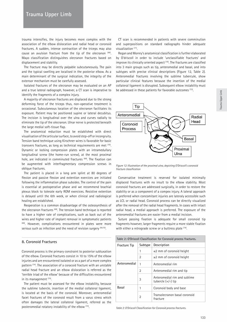
Trau
ma
Uppe
r Lim
b
133
TRAUMATrauma Upper Limb
trauma intensifies, the injury becomes more complex with the association of the elbow dislocation and radial head or coronoid fractures. A sudden, intense contraction of the triceps may also cause an avulsion fracture from the tip of the olecranon (69). Mayo classification distinguishes olecranon fractures based on displacement and stability.
The fracture may be directly palpable subcutaneously. The pain and the typical swelling are localised in the posterior elbow. As a main determinant of the surgical indication, the integrity of the extensor mechanism must be carefully assessed.
Isolated fractures of the olecranon may be evaluated on an AP and a true lateral radiograph, however, a CT scan is imperative to identify the fragments of a complex injury.
A majority of olecranon fractures are displaced due to the strong deforming force of the triceps thus, non-operative treatment is occasional. Subcutaneous location of the olecranon facilitates its exposure. Patient may be positioned supine or lateral decubitus. The incision is longitudinal over the ulna and curves radially to eliminate the tip of the olecranon. Ulnar nerve is protected beneath the large medial soft-tissue flap.
The anatomical reduction must be established with direct visualisation of the articular surface, to avoid step-off or incongruity. Tension band technique using Kirschner wires is favorable for basic transvers fractures, as long as technical requirements are met (70). Dynamic or locking compression plates with an intramedullary longitudinal screw (the home-run screw), at the most proximal hole, are indicated in comminuted fractures (69). The fixation can be augmented with interfragmentary compression screws in oblique fractures.
The patient is placed in a long arm splint at 80 degrees of flexion and passive flexion and extention exercices are initiated following the inflammation phase subsides. The control of the pain is essential at postoperative phase and we recommend brachial plexus block to tolerate early ROM exercices. Resistive extention is delayed until the 6th week, or when clinical and radiological healing are established.
Reoperation is a common disadvantage of the osteosynthesis of the olecranon fractures (69). The tension band technique is reported to have a higher rate of complications, such as back out of the wires and higher rate of implant removal in symptomatic patients (69). However, complications encountered in plates were more serious such as infection and the need of revision surgery (69,70).
8. Coronoid Fractures
Coronoid process is the primary constraint to posterior subluxation of the elbow. Coronoid fractures consist in 10 to 15% of the elbow injuries and are encountered isolated or as a part of a more complex pattern (74). The association of a coracoid fracture with an unstable radial head fracture and an elbow dislocation is referred as the ‘terrible triad of the elbow’ because of the difficulties encountered in its management (75).
The patient must be assessed for the elbow instability, because the sublime tubercle, insertion of the medial collateral ligament, is located at the basis of the coronoid. Moreover, anteromedial facet fractures of the coronoid result from a varus stress which often damages the lateral collateral ligament, referred as the posteromedial rotatory instability of the elbow (73).
CT scan is recommended in patients with severe comminution and superpositions on standard radiographs hinder adequate visualization (71).
Regan and Morrey’s anatomical classification is further elaborated by O’driscoll in order to include ‘unclassifiable fractures’ and improve its clinically oriented aspect (76). The fractures are classified into 3 main groups such as tip, anteromedial and basal, and into subtypes with precise clinical descriptions (Figure 12, Table 2). Anteromedial fractures involving the sublime tubercule, show particular clinical features because the insertion of the medial collateral ligament is disrupted. Subsequent elbow instability must be addressed in these patients for favorable outcomes (72).
Conservative treatment is reserved for isolated minimally displaced fractures with no insult to the elbow stability. Most coronoid fractures are addressed surgically, in order to restore the stability or as a component of a compex injury. A lateral approach is preferred when concomitant injuries are lateraly accessible such as LCL or radial head. Coronoid process can be directly visualized after the removal of the radial head fragments. In cases with intact radial head, a medial approach is preferred. The exposure of the anteromedial fractures are easier from a medial incision.
Suture passing fixation is adequate for small coronoid tip fragments however, larger fragments require a more stable fixation with either a retrograde screw or a buttress plate (74).
Table 2: O’Driscoll Classification for Coronoid process fractures.
Fracture Tip Subtype Description
1 ≤2 mm of coronoid height
2 ≥2 mm of coronoid height
Anteromedial 1 Anteromedial rim
2 Anteromedial rim and tip
3 Anteromedial rim and sublime tubercle (+/-) tip
Basal 1 Coronoid body and base
2 Transolecranon basal coronoid fracture
Figure 12: Illustration of the proximal ulna, depicting O’Driscoll’s coronoid fracture classification
Table 2: O’Driscoll Classification for Coronoid process fractures.
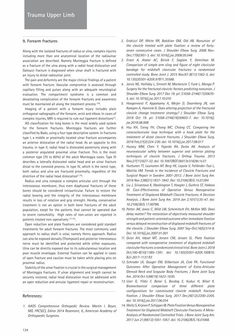
134
TRAUMATrauma Upper Limb
9. Forearm fractures
Along with the isolated fractures of radius or ulna, complex injuries including more than one anatomical location of the radioulnar association are described. Namely Monteggia fracture is defined as a fracture of the ulna along with a radial head dislocation and Galeazzi fracture is diagnosed when ulnar shaft is fractured with an injury to distal radioulnar joint.
The pain and deformity are the major clinical findings of a patient with forearm fracture. Vascular compromise is assessed through capillary filling and pulses along with an adequate neurological evaluation. The compartment syndrome is a common and devastating complication of the forearm fractures and awareness must be maintained all along the treatment process (78).
Imaging of a patient with a forearm injury includes plain orthogonal radiographs of the forearm, wrist and elbow. In cases of complex injuries, MRI is required to rule out ligament distortions(1).
AO classification for long bones is the most widely used system for the forearm fractures. Monteggia fractures are further classified by Bado, using a four type descriptive system. In fractures type I, a middle or proximally located ulnar fracture accompanies an anterior dislocation of the radial head. As an opposite to this trauma, in type II, radial head is dislocated posteriorly along with a posterior angulated proximal ulnar fracture. This is the most common type (70 to 80%) of the adult Monteggia cases. Type III describes a laterally dislocated radial head and an ulnar fracture distal to the coronoid process. In type IV, which is the rarest type, both radius and ulna are fractured proximally, regardless of the direction of the radial head dislocation(1,79).
Radius and ulna compose a complex articular unit through the interosseous membrane, thus even diaphyseal fractures of these bones should be considered intraarticular. Failure to restore the radial bowing and the integrity of the interosseous membrane results in loss of rotation and grip strenght. Hereby, conservative treatment is not an option in both bone fractures of the adult population, exept for the patients that cannot be operated due to severe comorbidity. High rates of non-union are reported in patients treated non-operatively (77,79).
Open reduction and plate fixations are considered gold standart treatments for adult forearm fractures. The most commonly used approach to radius shaft is volar, namely Henry approach. Radius can also be exposed dorsally (Thompson) and posterior interosseous nerve must be identified and protected while either exposures. Ulna can be directly exposed due to its subcutaneous location and poor muscle enveloppe. External fixation can be applied in cases of open fracture and caution must be taken while placing pins on proximal radius.
Stability of the ulnar fixation is crucial in the surgical management of Monteggia fractures. If ulnar alignment and length cannot be securely restored, radial head dislocation must be addressed with an open reduction and annular ligament repair or reconstruction.
References:
1. AAOS Comprehensive Orthopedic Review. Martin I. Boyer, MD, FRCS(C), Editor 2014 Rosemont, IL, American Academy of Orthopaedic Surgeons.
2. Endrizzi DP, White RR, Babikian GM, Old AB. Nonunion of the clavicle treated with plate fixation: a review of forty-seven consecutive cases. J Shoulder Elbow Surg. 2008 Nov-Dec;17(6):951-3. doi: 10.1016/j.jse.2008.05.046
3. Ersen A, Atalar AC, Birisik F, Saglam Y, Demirhan M. Comparison of simple arm sling and figure of eight clavicular bandage for midshaft clavicular fractures: a randomised controlled study. Bone Joint J. 2015 Nov;97-B(11):1562-5. doi: 10.1302/0301-620X.97B11.35588.
4. Jarvis NE, Halliday L, Sinnott M, Mackenzie T, Funk L, Monga P. Surgery for the fractured clavicle: factors predicting nonunion. J Shoulder Elbow Surg. 2017 Dec 19. pii: S1058-2746(17)30670-5. doi: 10.1016/j.jse.2017.10.010
5. Hoogervorst P, Appalsamy A, Meijer D, Doornberg JN, van Kampen A, Hannink G. Does altering projection of the fractured clavicle change treatment strategy? J Shoulder Elbow Surg. 2018 Oct 19. pii: S1058-2746(18)30603-7. doi: 10.1016/j.jse.2018.08.008
6. Hsu KH, Tzeng YH, Chang MC, Chiang CC. Comparing the coracoclavicular loop technique with a hook plate for the treatment of distal clavicle fractures. J Shoulder Elbow Surg. 2018 Feb;27(2):224-230. doi: 10.1016/j.jse.2017.08.017
7. Hussey MM, Chen Y, Fajardo RA, Dutta AK. Analysis of neurovascular safety between superior and anterior plating techniques of clavicle fractures. J Orthop Trauma. 2013 Nov;27(11):627-32. doi: 10.1097/BOT.0b013e31828c1e37.
8. Huttunen TT, Launonen AP, Berg HE, Lepola V, Felländer-Tsai L, Mattila VM. Trends in the Incidence of Clavicle Fractures and Surgical Repair in Sweden: 2001-2012. J Bone Joint Surg Am. 2016 Nov 2;98(21):1837-1842. Doi: 10.2106/JBJS.15.01284
9. Liu J, Srivastava K, Washington T, Hoegler J, Guthrie ST, Hakeos W. Cost-Effectiveness of Operative Versus Nonoperative Treatment of Displaced Midshaft Clavicle Fractures: A Decision Analysis. J Bone Joint Surg Am. 2019 Jan 2;101(1):35-47. doi: 10.2106/JBJS.17.00786.
10. Potter JM, Jones C, Wild LM, Schemitsch EH, McKee MD. Does delay matter? The restoration of objectively measured shoulder strength and patient-oriented outcome after immediate fixation versus delayed reconstruction of displaced midshaft fractures of the clavicle. J Shoulder Elbow Surg. 2007 Sep-Oct;16(5):514-8. Doi: 10.1016/j.jse.2007.01.001
11. Qvist AH, Væsel MT, Jensen CM, Jensen SL. Plate fixation compared with nonoperative treatment of displaced midshaft clavicular fractures: a randomized clinical trial. Bone Joint J. 2018 Oct;100-B(10):1385-1391. doi: 10.1302/0301-620X.100B10.BJJ-2017-1137.R3
12. Schroder LK, Gauger EM, Gilbertson JA, Cole PA. Functional Outcomes After Operative Management of Extra-Articular Glenoid Neck and Scapular Body Fractures. J Bone Joint Surg Am. 2016 Oct 5;98(19):1623-1630.
13. Uzer G, Yildiz F, Batar S, Bozdag E, Kuduz H, Bilsel K. Biomechanical comparison of three different plate configurations for comminuted clavicle midshaft fracture fixation. J Shoulder Elbow Surg. 2017 Dec;26(12):2200-2205. doi: 10.1016/j.jse.2017.06.034
14. Woltz S, Krijnen P, Schipper IB. Plate Fixation Versus Nonoperative Treatment for Displaced Midshaft Clavicular Fractures: A Meta-Analysis of Randomized Controlled Trials. J Bone Joint Surg Am. 2017 Jun 21;99(12):1051-1057. doi: 10.2106/JBJS.16.01068.
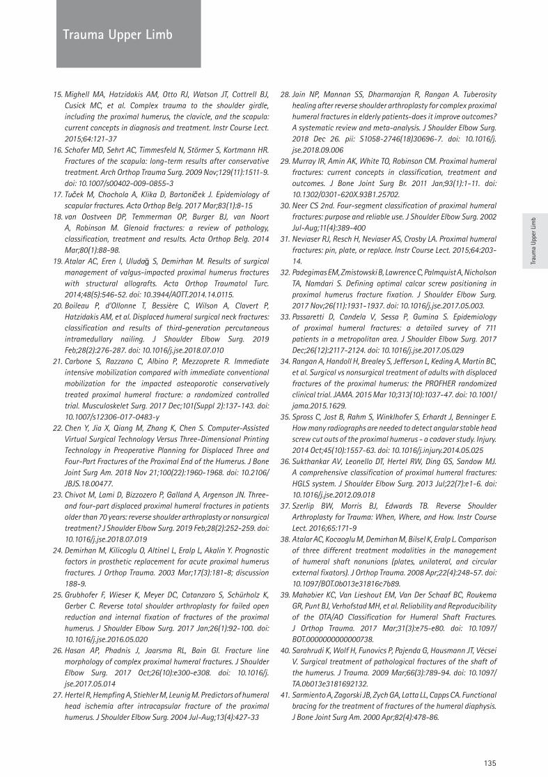
Trau
ma
Uppe
r Lim
b
135
TRAUMATrauma Upper Limb
15. Mighell MA, Hatzidakis AM, Otto RJ, Watson JT, Cottrell BJ, Cusick MC, et al. Complex trauma to the shoulder girdle, including the proximal humerus, the clavicle, and the scapula: current concepts in diagnosis and treatment. Instr Course Lect. 2015;64:121-37
16. Schofer MD, Sehrt AC, Timmesfeld N, Störmer S, Kortmann HR. Fractures of the scapula: long-term results after conservative treatment. Arch Orthop Trauma Surg. 2009 Nov;129(11):1511-9. doi: 10.1007/s00402-009-0855-3
17. Tuček M, Chochola A, Klika D, Bartoníček J. Epidemiology of scapular fractures. Acta Orthop Belg. 2017 Mar;83(1):8-15
18. van Oostveen DP, Temmerman OP, Burger BJ, van Noort A, Robinson M. Glenoid fractures: a review of pathology, classification, treatment and results. Acta Orthop Belg. 2014 Mar;80(1):88-98.
19. Atalar AC, Eren I, Uludağ S, Demirhan M. Results of surgical management of valgus-impacted proximal humerus fractures with structural allografts. Acta Orthop Traumatol Turc. 2014;48(5):546-52. doi: 10.3944/AOTT.2014.14.0115.
20. Boileau P, d'Ollonne T, Bessière C, Wilson A, Clavert P, Hatzidakis AM, et al. Displaced humeral surgical neck fractures: classification and results of third-generation percutaneous intramedullary nailing. J Shoulder Elbow Surg. 2019 Feb;28(2):276-287. doi: 10.1016/j.jse.2018.07.010
21. Carbone S, Razzano C, Albino P, Mezzoprete R. Immediate intensive mobilization compared with immediate conventional mobilization for the impacted osteoporotic conservatively treated proximal humeral fracture: a randomized controlled trial. Musculoskelet Surg. 2017 Dec;101(Suppl 2):137-143. doi: 10.1007/s12306-017-0483-y
22. Chen Y, Jia X, Qiang M, Zhang K, Chen S. Computer-Assisted Virtual Surgical Technology Versus Three-Dimensional Printing Technology in Preoperative Planning for Displaced Three and Four-Part Fractures of the Proximal End of the Humerus. J Bone Joint Surg Am. 2018 Nov 21;100(22):1960-1968. doi: 10.2106/JBJS.18.00477.
23. Chivot M, Lami D, Bizzozero P, Galland A, Argenson JN. Three- and four-part displaced proximal humeral fractures in patients older than 70 years: reverse shoulder arthroplasty or nonsurgical treatment? J Shoulder Elbow Surg. 2019 Feb;28(2):252-259. doi: 10.1016/j.jse.2018.07.019
24. Demirhan M, Kilicoglu O, Altinel L, Eralp L, Akalin Y. Prognostic factors in prosthetic replacement for acute proximal humerus fractures. J Orthop Trauma. 2003 Mar;17(3):181-8; discussion 188-9.
25. Grubhofer F, Wieser K, Meyer DC, Catanzaro S, Schürholz K, Gerber C. Reverse total shoulder arthroplasty for failed open reduction and internal fixation of fractures of the proximal humerus. J Shoulder Elbow Surg. 2017 Jan;26(1):92-100. doi: 10.1016/j.jse.2016.05.020
26. Hasan AP, Phadnis J, Jaarsma RL, Bain GI. Fracture line morphology of complex proximal humeral fractures. J Shoulder Elbow Surg. 2017 Oct;26(10):e300-e308. doi: 10.1016/j.jse.2017.05.014
27. Hertel R, Hempfing A, Stiehler M, Leunig M. Predictors of humeral head ischemia after intracapsular fracture of the proximal humerus. J Shoulder Elbow Surg. 2004 Jul-Aug;13(4):427-33
28. Jain NP, Mannan SS, Dharmarajan R, Rangan A. Tuberosity healing after reverse shoulder arthroplasty for complex proximal humeral fractures in elderly patients-does it improve outcomes? A systematic review and meta-analysis. J Shoulder Elbow Surg. 2018 Dec 26. pii: S1058-2746(18)30696-7. doi: 10.1016/j.jse.2018.09.006
29. Murray IR, Amin AK, White TO, Robinson CM. Proximal humeral fractures: current concepts in classification, treatment and outcomes. J Bone Joint Surg Br. 2011 Jan;93(1):1-11. doi: 10.1302/0301-620X.93B1.25702.
30. Neer CS 2nd. Four-segment classification of proximal humeral fractures: purpose and reliable use. J Shoulder Elbow Surg. 2002 Jul-Aug;11(4):389-400
31. Neviaser RJ, Resch H, Neviaser AS, Crosby LA. Proximal humeral fractures: pin, plate, or replace. Instr Course Lect. 2015;64:203-14.
32. Padegimas EM, Zmistowski B, Lawrence C, Palmquist A, Nicholson TA, Namdari S. Defining optimal calcar screw positioning in proximal humerus fracture fixation. J Shoulder Elbow Surg. 2017 Nov;26(11):1931-1937. doi: 10.1016/j.jse.2017.05.003.
33. Passaretti D, Candela V, Sessa P, Gumina S. Epidemiology of proximal humeral fractures: a detailed survey of 711 patients in a metropolitan area. J Shoulder Elbow Surg. 2017 Dec;26(12):2117-2124. doi: 10.1016/j.jse.2017.05.029
34. Rangan A, Handoll H, Brealey S, Jefferson L, Keding A, Martin BC, et al. Surgical vs nonsurgical treatment of adults with displaced fractures of the proximal humerus: the PROFHER randomized clinical trial. JAMA. 2015 Mar 10;313(10):1037-47. doi: 10.1001/jama.2015.1629.
35. Spross C, Jost B, Rahm S, Winklhofer S, Erhardt J, Benninger E. How many radiographs are needed to detect angular stable head screw cut outs of the proximal humerus - a cadaver study. Injury. 2014 Oct;45(10):1557-63. doi: 10.1016/j.injury.2014.05.025
36. Sukthankar AV, Leonello DT, Hertel RW, Ding GS, Sandow MJ. A comprehensive classification of proximal humeral fractures: HGLS system. J Shoulder Elbow Surg. 2013 Jul;22(7):e1-6. doi: 10.1016/j.jse.2012.09.018
37. Szerlip BW, Morris BJ, Edwards TB. Reverse Shoulder Arthroplasty for Trauma: When, Where, and How. Instr Course Lect. 2016;65:171-9
38. Atalar AC, Kocaoglu M, Demirhan M, Bilsel K, Eralp L. Comparison of three different treatment modalities in the management of humeral shaft nonunions (plates, unilateral, and circular external fixators). J Orthop Trauma. 2008 Apr;22(4):248-57. doi: 10.1097/BOT.0b013e31816c7b89.
39. Mahabier KC, Van Lieshout EM, Van Der Schaaf BC, Roukema GR, Punt BJ, Verhofstad MH, et al. Reliability and Reproducibility of the OTA/AO Classification for Humeral Shaft Fractures. J Orthop Trauma. 2017 Mar;31(3):e75-e80. doi: 10.1097/BOT.0000000000000738.
40. Sarahrudi K, Wolf H, Funovics P, Pajenda G, Hausmann JT, Vécsei V. Surgical treatment of pathological fractures of the shaft of the humerus. J Trauma. 2009 Mar;66(3):789-94. doi: 10.1097/TA.0b013e3181692132.
41. Sarmiento A, Zagorski JB, Zych GA, Latta LL, Capps CA. Functional bracing for the treatment of fractures of the humeral diaphysis. J Bone Joint Surg Am. 2000 Apr;82(4):478-86.
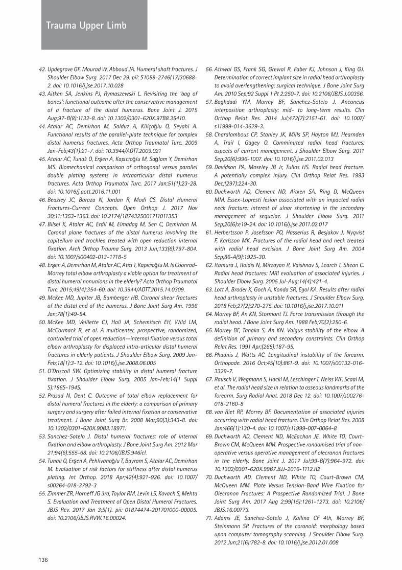
136
TRAUMATrauma Upper Limb
42. Updegrove GF, Mourad W, Abboud JA. Humeral shaft fractures. J Shoulder Elbow Surg. 2017 Dec 29. pii: S1058-2746(17)30688-2. doi: 10.1016/j.jse.2017.10.028
43. Aitken SA, Jenkins PJ, Rymaszewski L. Revisiting the ‘bag of bones’: functional outcome after the conservative management of a fracture of the distal humerus. Bone Joint J. 2015 Aug;97-B(8):1132-8. doi: 10.1302/0301-620X.97B8.35410.
44. Atalar AC, Demirhan M, Salduz A, Kiliçoğlu O, Seyahi A. Functional results of the parallel-plate technique for complex distal humerus fractures. Acta Orthop Traumatol Turc. 2009 Jan-Feb;43(1):21-7. doi: 10.3944/AOTT.2009.021
45. Atalar AC, Tunalı O, Erşen A, Kapıcıoğlu M, Sağlam Y, Demirhan MS. Biomechanical comparison of orthogonal versus parallel double plating systems in intraarticular distal humerus fractures. Acta Orthop Traumatol Turc. 2017 Jan;51(1):23-28. doi: 10.1016/j.aott.2016.11.001
46. Beazley JC, Baraza N, Jordan R, Modi CS. Distal Humeral Fractures-Current Concepts. Open Orthop J. 2017 Nov 30;11:1353-1363. doi: 10.2174/1874325001711011353
47. Bilsel K, Atalar AC, Erdil M, Elmadag M, Sen C, Demirhan M. Coronal plane fractures of the distal humerus involving the capitellum and trochlea treated with open reduction internal fixation. Arch Orthop Trauma Surg. 2013 Jun;133(6):797-804. doi: 10.1007/s00402-013-1718-5
48. Erşen A, Demirhan M, Atalar AC, Atıcı T, Kapıcıoğlu M. Is Coonrad-Morrey total elbow arthroplasty a viable option for treatment of distal humeral nonunions in the elderly? Acta Orthop Traumatol Turc. 2015;49(4):354-60. doi: 10.3944/AOTT.2015.14.0309.
49. McKee MD, Jupiter JB, Bamberger HB. Coronal shear fractures of the distal end of the humerus. J Bone Joint Surg Am. 1996 Jan;78(1):49-54.
50. McKee MD, Veillette CJ, Hall JA, Schemitsch EH, Wild LM, McCormack R, et al. A multicenter, prospective, randomized, controlled trial of open reduction--internal fixation versus total elbow arthroplasty for displaced intra-articular distal humeral fractures in elderly patients. J Shoulder Elbow Surg. 2009 Jan-Feb;18(1):3-12. doi: 10.1016/j.jse.2008.06.005
51. O’Driscoll SW. Optimizing stability in distal humeral fracture fixation. J Shoulder Elbow Surg. 2005 Jan-Feb;14(1 Suppl S):186S-194S.
52. Prasad N, Dent C. Outcome of total elbow replacement for distal humeral fractures in the elderly: a comparison of primary surgery and surgery after failed internal fixation or conservative treatment. J Bone Joint Surg Br. 2008 Mar;90(3):343-8. doi: 10.1302/0301-620X.90B3.18971.
53. Sanchez-Sotelo J. Distal humeral fractures: role of internal fixation and elbow arthroplasty. J Bone Joint Surg Am. 2012 Mar 21;94(6):555-68. doi: 10.2106/JBJS.946icl.
54. Tunalı O, Erşen A, Pehlivanoğlu T, Bayram S, Atalar AC, Demirhan M. Evaluation of risk factors for stiffness after distal humerus plating. Int Orthop. 2018 Apr;42(4):921-926. doi: 10.1007/s00264-018-3792-3
55. Zimmer ZR, Horneff JG 3rd, Taylor RM, Levin LS, Kovach S, Mehta S. Evaluation and Treatment of Open Distal Humeral Fractures. JBJS Rev. 2017 Jan 3;5(1). pii: 01874474-201701000-00005. doi: 10.2106/JBJS.RVW.16.00024.
56. Athwal GS, Frank SG, Grewal R, Faber KJ, Johnson J, King GJ. Determination of correct implant size in radial head arthroplasty to avoid overlengthening: surgical technique. J Bone Joint Surg Am. 2010 Sep;92 Suppl 1 Pt 2:250-7. doi: 10.2106/JBJS.J.00356.
57. Baghdadi YM, Morrey BF, Sanchez-Sotelo J. Anconeus interposition arthroplasty: mid- to long-term results. Clin Orthop Relat Res. 2014 Jul;472(7):2151-61. doi: 10.1007/s11999-014-3629-3.
58. Charalambous CP, Stanley JK, Mills SP, Hayton MJ, Hearnden A, Trail I, Gagey O. Comminuted radial head fractures: aspects of current management. J Shoulder Elbow Surg. 2011 Sep;20(6):996-1007. doi: 10.1016/j.jse.2011.02.013
59. Davidson PA, Moseley JB Jr, Tullos HS. Radial head fracture. A potentially complex injury. Clin Orthop Relat Res. 1993 Dec;(297):224-30.
60. Duckworth AD, Clement ND, Aitken SA, Ring D, McQueen MM. Essex-Lopresti lesion associated with an impacted radial neck fracture: interest of ulnar shortening in the secondary management of sequelae. J Shoulder Elbow Surg. 2011 Sep;20(6):e19-24. doi: 10.1016/j.jse.2011.02.017
61. Herbertsson P, Josefsson PO, Hasserius R, Besjakov J, Nyqvist F, Karlsson MK. Fractures of the radial head and neck treated with radial head excision. J Bone Joint Surg Am. 2004 Sep;86-A(9):1925-30.
62. Itamura J, Roidis N, Mirzayan R, Vaishnav S, Learch T, Shean C. Radial head fractures: MRI evaluation of associated injuries. J Shoulder Elbow Surg. 2005 Jul-Aug;14(4):421-4.
63. Lott A, Broder K, Goch A, Konda SR, Egol KA. Results after radial head arthroplasty in unstable fractures. J Shoulder Elbow Surg. 2018 Feb;27(2):270-275. doi: 10.1016/j.jse.2017.10.011
64. Morrey BF, An KN, Stormont TJ. Force transmission through the radial head. J Bone Joint Surg Am. 1988 Feb;70(2):250-6.
65. Morrey BF, Tanaka S, An KN. Valgus stability of the elbow. A definition of primary and secondary constraints. Clin Orthop Relat Res. 1991 Apr;(265):187-95.
66. Phadnis J, Watts AC. Longitudinal instability of the forearm. Orthopade. 2016 Oct;45(10):861-9. doi: 10.1007/s00132-016-3329-7.
67. Rausch V, Wegmann S, Hackl M, Leschinger T, Neiss WF, Scaal M, et al. The radial head size in relation to osseous landmarks of the forearm. Surg Radiol Anat. 2018 Dec 12. doi: 10.1007/s00276-018-2160-8
68. van Riet RP, Morrey BF. Documentation of associated injuries occurring with radial head fracture. Clin Orthop Relat Res. 2008 Jan;466(1):130-4. doi: 10.1007/s11999-007-0064-8
69. Duckworth AD, Clement ND, McEachan JE, White TO, Court-Brown CM, McQueen MM. Prospective randomised trial of non-operative versus operative management of olecranon fractures in the elderly. Bone Joint J. 2017 Jul;99-B(7):964-972. doi: 10.1302/0301-620X.99B7.BJJ-2016-1112.R2
70. Duckworth AD, Clement ND, White TO, Court-Brown CM, McQueen MM. Plate Versus Tension-Band Wire Fixation for Olecranon Fractures: A Prospective Randomized Trial. J Bone Joint Surg Am. 2017 Aug 2;99(15):1261-1273. doi: 10.2106/JBJS.16.00773.
71. Adams JE, Sanchez-Sotelo J, Kallina CF 4th, Morrey BF, Steinmann SP. Fractures of the coronoid: morphology based upon computer tomography scanning. J Shoulder Elbow Surg. 2012 Jun;21(6):782-8. doi: 10.1016/j.jse.2012.01.008
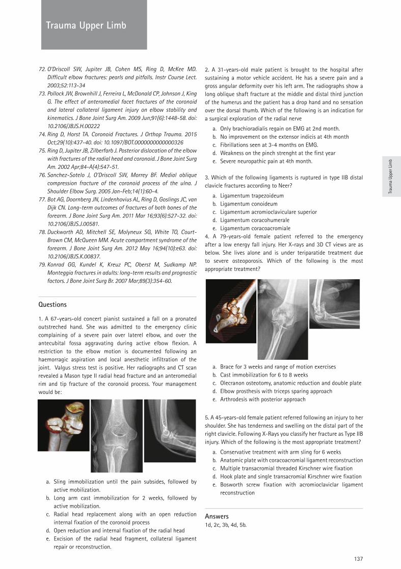
Trau
ma
Uppe
r Lim
b
137
TRAUMATrauma Upper Limb
72. O’Driscoll SW, Jupiter JB, Cohen MS, Ring D, McKee MD. Difficult elbow fractures: pearls and pitfalls. Instr Course Lect. 2003;52:113-34
73. Pollock JW, Brownhill J, Ferreira L, McDonald CP, Johnson J, King G. The effect of anteromedial facet fractures of the coronoid and lateral collateral ligament injury on elbow stability and kinematics. J Bone Joint Surg Am. 2009 Jun;91(6):1448-58. doi: 10.2106/JBJS.H.00222
74. Ring D, Horst TA. Coronoid Fractures. J Orthop Trauma. 2015 Oct;29(10):437-40. doi: 10.1097/BOT.0000000000000326
75. Ring D, Jupiter JB, Zilberfarb J. Posterior dislocation of the elbow with fractures of the radial head and coronoid. J Bone Joint Surg Am. 2002 Apr;84-A(4):547-51.
76. Sanchez-Sotelo J, O’Driscoll SW, Morrey BF. Medial oblique compression fracture of the coronoid process of the ulna. J Shoulder Elbow Surg. 2005 Jan-Feb;14(1):60-4.
77. Bot AG, Doornberg JN, Lindenhovius AL, Ring D, Goslings JC, van Dijk CN. Long-term outcomes of fractures of both bones of the forearm. J Bone Joint Surg Am. 2011 Mar 16;93(6):527-32. doi: 10.2106/JBJS.J.00581.
78. Duckworth AD, Mitchell SE, Molyneux SG, White TO, Court-Brown CM, McQueen MM. Acute compartment syndrome of the forearm. J Bone Joint Surg Am. 2012 May 16;94(10):e63. doi: 10.2106/JBJS.K.00837.
79. Konrad GG, Kundel K, Kreuz PC, Oberst M, Sudkamp NP. Monteggia fractures in adults: long-term results and prognostic factors. J Bone Joint Surg Br. 2007 Mar;89(3):354-60.
Questions
1. A 67-years-old concert pianist sustained a fall on a pronated outstreched hand. She was admitted to the emergency clinic complaining of a severe pain over laterel elbow, and over the antecubital fossa aggravating during active elbow flexion. A restriction to the elbow motion is documented following an haemorragic aspiration and local anesthetic infiltration of the joint. Valgus stress test is positive. Her radiographs and CT scan revealed a Mason type II radial head fracture and an anteromedial rim and tip fracture of the coronoid process. Your management would be:
a. Sling immobilization until the pain subsides, followed by active mobilization.
b. Long arm cast immobilization for 2 weeks, followed by active mobilization.
c. Radial head replacement along with an open reduction internal fixation of the coronoid process
d. Open reduction and internal fixation of the radial heade. Excision of the radial head fragment, collateral ligament
repair or reconstruction.
2. A 31-years-old male patient is brought to the hospital after sustaining a motor vehicle accident. He has a severe pain and a gross angular deformity over his left arm. The radiographs show a long oblique shaft fracture at the middle and distal third junction of the humerus and the patient has a drop hand and no sensation over the dorsal thumb. Which of the following is an indication for a surgical exploration of the radial nerve
a. Only brachioradialis regain on EMG at 2nd month. b. No improvement on the extensor indicis at 4th monthc. Fibrillations seen at 3-4 months on EMG.d. Weakness on the pinch strenght at the first yeare. Severe neuropathic pain at 4th month.
3. Which of the following ligaments is ruptured in type IIB distal clavicle fractures according to Neer?
a. Ligamentum trapezoideumb. Ligamentum conoideumc. Ligamentum acromioclaviculare superiord. Ligamentum coracohumeralee. Ligamentum coracoacromiale
4. A 79-years-old female patient referred to the emergency after a low energy fall injury. Her X-rays and 3D CT views are as below. She lives alone and is under teriparatide treatment due to severe osteoporosis. Which of the following is the most appropriate treatment?
a. Brace for 3 weeks and range of motion exercisesb. Cast immobilization for 6 to 8 weeks c. Olecranon osteotomy, anatomic reduction and double plated. Elbow prosthesis with triceps sparing approache. Arthrodesis with posterior approach
5. A 45-years-old female patient referred following an injury to her shoulder. She has tenderness and swelling on the distal part of the right clavicle. Following X-Rays you classify her fracture as Type IIB injury. Which of the following is the most appropriate treatment?
a. Conservative treatment with arm sling for 6 weeksb. Anatomic plate with coracoacromial ligament reconstructionc. Multiple transacromial threaded Kirschner wire fixationd. Hook plate and single transacromial Kirschner wire fixatione. Bosworth screw fixation with acromioclaviclar ligament
reconstruction
Answers1d, 2c, 3b, 4d, 5b.
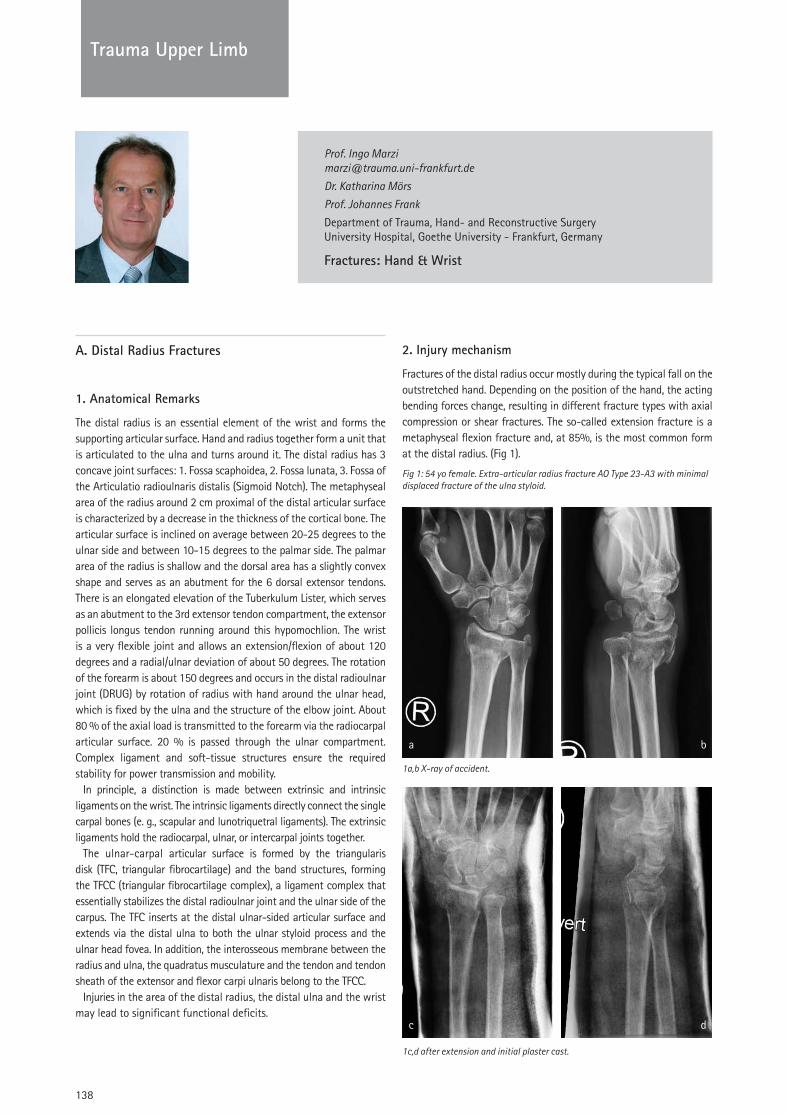
138
TRAUMATrauma Upper Limb
Prof. Ingo Marzi [email protected]
Dr. Katharina Mörs
Prof. Johannes Frank
Department of Trauma, Hand- and Reconstructive Surgery University Hospital, Goethe University - Frankfurt, Germany
Fractures: Hand & Wrist
A. Distal Radius Fractures
1. Anatomical Remarks
The distal radius is an essential element of the wrist and forms the supporting articular surface. Hand and radius together form a unit that is articulated to the ulna and turns around it. The distal radius has 3 concave joint surfaces: 1. Fossa scaphoidea, 2. Fossa lunata, 3. Fossa of the Articulatio radioulnaris distalis (Sigmoid Notch). The metaphyseal area of the radius around 2 cm proximal of the distal articular surface is characterized by a decrease in the thickness of the cortical bone. The articular surface is inclined on average between 20-25 degrees to the ulnar side and between 10-15 degrees to the palmar side. The palmar area of the radius is shallow and the dorsal area has a slightly convex shape and serves as an abutment for the 6 dorsal extensor tendons. There is an elongated elevation of the Tuberkulum Lister, which serves as an abutment to the 3rd extensor tendon compartment, the extensor pollicis longus tendon running around this hypomochlion. The wrist is a very flexible joint and allows an extension/flexion of about 120 degrees and a radial/ulnar deviation of about 50 degrees. The rotation of the forearm is about 150 degrees and occurs in the distal radioulnar joint (DRUG) by rotation of radius with hand around the ulnar head, which is fixed by the ulna and the structure of the elbow joint. About 80 % of the axial load is transmitted to the forearm via the radiocarpal articular surface. 20 % is passed through the ulnar compartment. Complex ligament and soft-tissue structures ensure the required stability for power transmission and mobility.
In principle, a distinction is made between extrinsic and intrinsic ligaments on the wrist. The intrinsic ligaments directly connect the single carpal bones (e. g., scapular and lunotriquetral ligaments). The extrinsic ligaments hold the radiocarpal, ulnar, or intercarpal joints together.
The ulnar-carpal articular surface is formed by the triangularis disk (TFC, triangular fibrocartilage) and the band structures, forming the TFCC (triangular fibrocartilage complex), a ligament complex that essentially stabilizes the distal radioulnar joint and the ulnar side of the carpus. The TFC inserts at the distal ulnar-sided articular surface and extends via the distal ulna to both the ulnar styloid process and the ulnar head fovea. In addition, the interosseous membrane between the radius and ulna, the quadratus musculature and the tendon and tendon sheath of the extensor and flexor carpi ulnaris belong to the TFCC.
Injuries in the area of the distal radius, the distal ulna and the wrist may lead to significant functional deficits.
2. Injury mechanism
Fractures of the distal radius occur mostly during the typical fall on the outstretched hand. Depending on the position of the hand, the acting bending forces change, resulting in different fracture types with axial compression or shear fractures. The so-called extension fracture is a metaphyseal flexion fracture and, at 85%, is the most common form at the distal radius. (Fig 1).
Fig 1: 54 yo female. Extra-articular radius fracture AO Type 23-A3 with minimal displaced fracture of the ulna styloid.
1a,b X-ray of accident.
1c,d after extension and initial plaster cast.
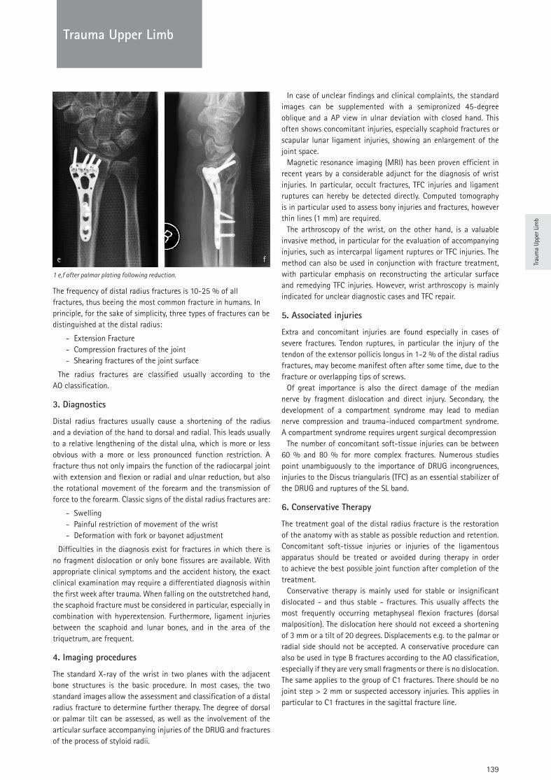
Trau
ma
Uppe
r Lim
b
139
TRAUMATrauma Upper Limb
1 e,f after palmar plating following reduction.
The frequency of distal radius fractures is 10-25 % of all fractures, thus beeing the most common fracture in humans. In principle, for the sake of simplicity, three types of fractures can be distinguished at the distal radius:
- Extension Fracture - Compression fractures of the joint - Shearing fractures of the joint surface
The radius fractures are classified usually according to the AO classification.
3. Diagnostics
Distal radius fractures usually cause a shortening of the radius and a deviation of the hand to dorsal and radial. This leads usually to a relative lengthening of the distal ulna, which is more or less obvious with a more or less pronounced function restriction. A fracture thus not only impairs the function of the radiocarpal joint with extension and flexion or radial and ulnar reduction, but also the rotational movement of the forearm and the transmission of force to the forearm. Classic signs of the distal radius fractures are:
- Swelling - Painful restriction of movement of the wrist - Deformation with fork or bayonet adjustment
Difficulties in the diagnosis exist for fractures in which there is no fragment dislocation or only bone fissures are available. With appropriate clinical symptoms and the accident history, the exact clinical examination may require a differentiated diagnosis within the first week after trauma. When falling on the outstretched hand, the scaphoid fracture must be considered in particular, especially in combination with hyperextension. Furthermore, ligament injuries between the scaphoid and lunar bones, and in the area of the triquetrum, are frequent.
4. Imaging procedures
The standard X-ray of the wrist in two planes with the adjacent bone structures is the basic procedure. In most cases, the two standard images allow the assessment and classification of a distal radius fracture to determine further therapy. The degree of dorsal or palmar tilt can be assessed, as well as the involvement of the articular surface accompanying injuries of the DRUG and fractures of the process of styloid radii.
In case of unclear findings and clinical complaints, the standard images can be supplemented with a semipronized 45-degree oblique and a AP view in ulnar deviation with closed hand. This often shows concomitant injuries, especially scaphoid fractures or scapular lunar ligament injuries, showing an enlargement of the joint space.
Magnetic resonance imaging (MRI) has been proven efficient in recent years by a considerable adjunct for the diagnosis of wrist injuries. In particular, occult fractures, TFC injuries and ligament ruptures can hereby be detected directly. Computed tomography is in particular used to assess bony injuries and fractures, however thin lines (1 mm) are required.
The arthroscopy of the wrist, on the other hand, is a valuable invasive method, in particular for the evaluation of accompanying injuries, such as intercarpal ligament ruptures or TFC injuries. The method can also be used in conjunction with fracture treatment, with particular emphasis on reconstructing the articular surface and remedying TFC injuries. However, wrist arthroscopy is mainly indicated for unclear diagnostic cases and TFC repair.
5. Associated injuries
Extra and concomitant injuries are found especially in cases of severe fractures. Tendon ruptures, in particular the injury of the tendon of the extensor pollicis longus in 1-2 % of the distal radius fractures, may become manifest often after some time, due to the fracture or overlapping tips of screws.
Of great importance is also the direct damage of the median nerve by fragment dislocation and direct injury. Secondary, the development of a compartment syndrome may lead to median nerve compression and trauma-induced compartment syndrome. A compartment syndrome requires urgent surgical decompression
The number of concomitant soft-tissue injuries can be between 60 % and 80 % for more complex fractures. Numerous studies point unambiguously to the importance of DRUG incongruences, injuries to the Discus triangularis (TFC) as an essential stabilizer of the DRUG and ruptures of the SL band.
6. Conservative Therapy
The treatment goal of the distal radius fracture is the restoration of the anatomy with as stable as possible reduction and retention. Concomitant soft-tissue injuries or injuries of the ligamentous apparatus should be treated or avoided during therapy in order to achieve the best possible joint function after completion of the treatment.
Conservative therapy is mainly used for stable or insignificant dislocated - and thus stable - fractures. This usually affects the most frequently occurring metaphyseal flexion fractures (dorsal malposition). The dislocation here should not exceed a shortening of 3 mm or a tilt of 20 degrees. Displacements e.g. to the palmar or radial side should not be accepted. A conservative procedure can also be used in type B fractures according to the AO classification, especially if they are very small fragments or there is no dislocation. The same applies to the group of C1 fractures. There should be no joint step > 2 mm or suspected accessory injuries. This applies in particular to C1 fractures in the sagittal fracture line.
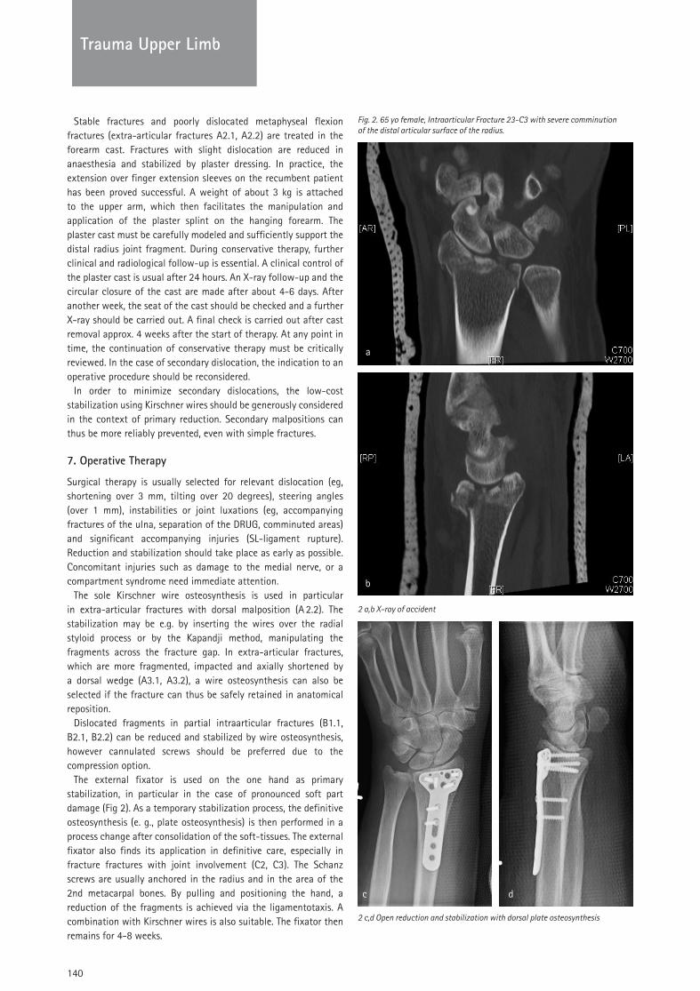
140
TRAUMATrauma Upper Limb
Stable fractures and poorly dislocated metaphyseal flexion fractures (extra-articular fractures A2.1, A2.2) are treated in the forearm cast. Fractures with slight dislocation are reduced in anaesthesia and stabilized by plaster dressing. In practice, the extension over finger extension sleeves on the recumbent patient has been proved successful. A weight of about 3 kg is attached to the upper arm, which then facilitates the manipulation and application of the plaster splint on the hanging forearm. The plaster cast must be carefully modeled and sufficiently support the distal radius joint fragment. During conservative therapy, further clinical and radiological follow-up is essential. A clinical control of the plaster cast is usual after 24 hours. An X-ray follow-up and the circular closure of the cast are made after about 4-6 days. After another week, the seat of the cast should be checked and a further X-ray should be carried out. A final check is carried out after cast removal approx. 4 weeks after the start of therapy. At any point in time, the continuation of conservative therapy must be critically reviewed. In the case of secondary dislocation, the indication to an operative procedure should be reconsidered.
In order to minimize secondary dislocations, the low-cost stabilization using Kirschner wires should be generously considered in the context of primary reduction. Secondary malpositions can thus be more reliably prevented, even with simple fractures.
7. Operative Therapy
Surgical therapy is usually selected for relevant dislocation (eg, shortening over 3 mm, tilting over 20 degrees), steering angles (over 1 mm), instabilities or joint luxations (eg, accompanying fractures of the ulna, separation of the DRUG, comminuted areas) and significant accompanying injuries (SL-ligament rupture). Reduction and stabilization should take place as early as possible. Concomitant injuries such as damage to the medial nerve, or a compartment syndrome need immediate attention.
The sole Kirschner wire osteosynthesis is used in particular in extra-articular fractures with dorsal malposition (A 2.2). The stabilization may be e.g. by inserting the wires over the radial styloid process or by the Kapandji method, manipulating the fragments across the fracture gap. In extra-articular fractures, which are more fragmented, impacted and axially shortened by a dorsal wedge (A3.1, A3.2), a wire osteosynthesis can also be selected if the fracture can thus be safely retained in anatomical reposition.
Dislocated fragments in partial intraarticular fractures (B1.1, B2.1, B2.2) can be reduced and stabilized by wire osteosynthesis, however cannulated screws should be preferred due to the compression option.
The external fixator is used on the one hand as primary stabilization, in particular in the case of pronounced soft part damage (Fig 2). As a temporary stabilization process, the definitive osteosynthesis (e. g., plate osteosynthesis) is then performed in a process change after consolidation of the soft-tissues. The external fixator also finds its application in definitive care, especially in fracture fractures with joint involvement (C2, C3). The Schanz screws are usually anchored in the radius and in the area of the 2nd metacarpal bones. By pulling and positioning the hand, a reduction of the fragments is achieved via the ligamentotaxis. A combination with Kirschner wires is also suitable. The fixator then remains for 4-8 weeks.
Fig. 2. 65 yo female, Intraarticular Fracture 23-C3 with severe comminution of the distal articular surface of the radius.
2 a,b X-ray of accident
2 c,d Open reduction and stabilization with dorsal plate osteosynthesis
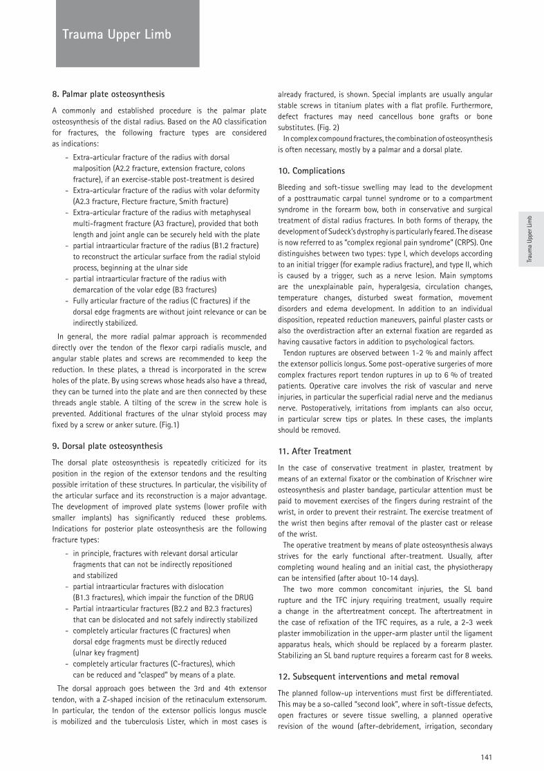
Trau
ma
Uppe
r Lim
b
141
TRAUMATrauma Upper Limb
8. Palmar plate osteosynthesis
A commonly and established procedure is the palmar plate osteosynthesis of the distal radius. Based on the AO classification for fractures, the following fracture types are considered as indications:
- Extra-articular fracture of the radius with dorsal malposition (A2.2 fracture, extension fracture, colons fracture), if an exercise-stable post-treatment is desired
- Extra-articular fracture of the radius with volar deformity (A2.3 fracture, Flecture fracture, Smith fracture)
- Extra-articular fracture of the radius with metaphyseal multi-fragment fracture (A3 fracture), provided that both length and joint angle can be securely held with the plate
- partial intraarticular fracture of the radius (B1.2 fracture) to reconstruct the articular surface from the radial styloid process, beginning at the ulnar side
- partial intraarticular fracture of the radius with demarcation of the volar edge (B3 fractures)
- Fully articular fracture of the radius (C fractures) if the dorsal edge fragments are without joint relevance or can be indirectly stabilized.
In general, the more radial palmar approach is recommended directly over the tendon of the flexor carpi radialis muscle, and angular stable plates and screws are recommended to keep the reduction. In these plates, a thread is incorporated in the screw holes of the plate. By using screws whose heads also have a thread, they can be turned into the plate and are then connected by these threads angle stable. A tilting of the screw in the screw hole is prevented. Additional fractures of the ulnar styloid process may fixed by a screw or anker suture. (Fig.1)
9. Dorsal plate osteosynthesis
The dorsal plate osteosynthesis is repeatedly criticized for its position in the region of the extensor tendons and the resulting possible irritation of these structures. In particular, the visibility of the articular surface and its reconstruction is a major advantage. The development of improved plate systems (lower profile with smaller implants) has significantly reduced these problems. Indications for posterior plate osteosynthesis are the following fracture types:
- in principle, fractures with relevant dorsal articular fragments that can not be indirectly repositioned and stabilized
- partial intraarticular fractures with dislocation (B1.3 fractures), which impair the function of the DRUG
- Partial intraarticular fractures (B2.2 and B2.3 fractures) that can be dislocated and not safely indirectly stabilized
- completely articular fractures (C fractures) when dorsal edge fragments must be directly reduced (ulnar key fragment)
- completely articular fractures (C-fractures), which can be reduced and “clasped” by means of a plate.
The dorsal approach goes between the 3rd and 4th extensor tendon, with a Z-shaped incision of the retinaculum extensorum. In particular, the tendon of the extensor pollicis longus muscle is mobilized and the tuberculosis Lister, which in most cases is
already fractured, is shown. Special implants are usually angular stable screws in titanium plates with a flat profile. Furthermore, defect fractures may need cancellous bone grafts or bone substitutes. (Fig. 2)
In complex compound fractures, the combination of osteosynthesis is often necessary, mostly by a palmar and a dorsal plate.
10. Complications
Bleeding and soft-tissue swelling may lead to the development of a posttraumatic carpal tunnel syndrome or to a compartment syndrome in the forearm bow, both in conservative and surgical treatment of distal radius fractures. In both forms of therapy, the development of Sudeck’s dystrophy is particularly feared. The disease is now referred to as “complex regional pain syndrome” (CRPS). One distinguishes between two types: type I, which develops according to an initial trigger (for example radius fracture), and type II, which is caused by a trigger, such as a nerve lesion. Main symptoms are the unexplainable pain, hyperalgesia, circulation changes, temperature changes, disturbed sweat formation, movement disorders and edema development. In addition to an individual disposition, repeated reduction maneuvers, painful plaster casts or also the overdistraction after an external fixation are regarded as having causative factors in addition to psychological factors.
Tendon ruptures are observed between 1-2 % and mainly affect the extensor pollicis longus. Some post-operative surgeries of more complex fractures report tendon ruptures in up to 6 % of treated patients. Operative care involves the risk of vascular and nerve injuries, in particular the superficial radial nerve and the medianus nerve. Postoperatively, irritations from implants can also occur, in particular screw tips or plates. In these cases, the implants should be removed.
11. After Treatment
In the case of conservative treatment in plaster, treatment by means of an external fixator or the combination of Krischner wire osteosynthesis and plaster bandage, particular attention must be paid to movement exercises of the fingers during restraint of the wrist, in order to prevent their restraint. The exercise treatment of the wrist then begins after removal of the plaster cast or release of the wrist.
The operative treatment by means of plate osteosynthesis always strives for the early functional after-treatment. Usually, after completing wound healing and an initial cast, the physiotherapy can be intensified (after about 10-14 days).
The two more common concomitant injuries, the SL band rupture and the TFC injury requiring treatment, usually require a change in the aftertreatment concept. The aftertreatment in the case of refixation of the TFC requires, as a rule, a 2-3 week plaster immobilization in the upper-arm plaster until the ligament apparatus heals, which should be replaced by a forearm plaster. Stabilizing an SL band rupture requires a forearm cast for 8 weeks.
12. Subsequent interventions and metal removal
The planned follow-up interventions must first be differentiated. This may be a so-called “second look”, where in soft-tissue defects, open fractures or severe tissue swelling, a planned operative revision of the wound (after-debridement, irrigation, secondary
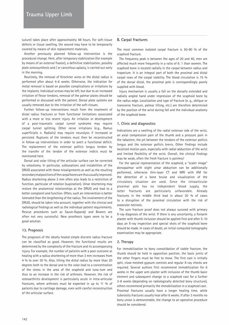
142
TRAUMA
suture) takes place after approximately 48 hours. For soft-tissue defects or tissue swelling, the wound may have to be temporarily covered by means of skin replacement materials.
Another previously planned follow-up intervention is the procedural change. Here, after temporary stabilization (for example by means of an external fixator), a definitive stabilization, possibly plate osteosynthesis and / or cancellous aplasty, is carried out early in the morning.
Routinely, the removal of Kirschner wires at the distal radius is performed after about 4-6 weeks. Otherwise, the indication for metal removal is based on possible complications or irritations by the implants. Individual screws may be left, but due to an increased irritation of flexor tendons, removal of the palmar plates should be performed or discussed with the patient. Dorsal plate systems are usually removed due to the irritation of the soft-tissues.
Further follow-up interventions result from the treatment of distal radius fractures or from functional limitations associated with a more or less severe injury. An irritation or development of a post-traumatic carpal tunnel syndrome may require carpal tunnel splitting. Other nerve irritations (e.g., Ramus superficialis n. Radialis) may require neurolysis if increased or persisted. Ruptures of the tendons must then be reconstructed in follow-up interventions in order to avert a functional deficit. The replacement of the extensor pollicis longus tendon by the transfer of the tendon of the extensor indicis must be mentioned here.
Dorsal and volar tilting of the articular surface can be corrected by osteotomy. In particular, subluxations and instabilities of the DRUG associated with these misalignments as well as the resulting secondary malpositions of the carpal bones are thus usually improved. Radius shortening above 3 mm often also leads to a restriction of function, particular of rotation (supination). Ulnar shortening may restore the anatomical relationships at the DRUG and lead to a better complaint and function. Often, such an intervention is better tolerated than the lengthening of the radius. The involvement of the DRUG, should be taken into account, together with the clinical and radiological findings as well as the individual patient requirements. Rescue procedures such as Sauve-Kapandji and Bowers are often not very successful. New prosthesis types seem to be a good solution.
13. Prognosis
The prognosis of the ideally healed simple discrete radius fracture can be classified as good. However, the functional results are determined by the complexity of the fracture and its accompanying injury. For example, the number of patients with a poor outcome at healing with a radius shortening of more than 3 mm increases from 4 % to over 30 %. Also, tilting the distal radius by more than 20 degrees both to the dorsal and to the volar lead to a concentration of the stress in the area of the scaphoid and luna-tum and thus to an increase in the risk of arthrosis. However, the risk of osteoarthritis development is particularly acute in intra-articular fractures, where arthrosis must be expected in up to 11 % of patients due to cartilage damage, even with careful reconstruction of the articular surface.
B. Carpal fractures
The most common isolated carpal fracture is 50-80 % of the scaphoid fracture.
The frequency peak is between the ages of 20 and 40, men are affected much more frequently in a ratio of 6: 1 than women. The scaphoid bone is located radially in the carpal between radius and trapezium. It is an integral part of both the proximal and distal carpal rows of the carpal stability. The blood circulation is 75 % of the dorsal distal, the proximal pole is correspondingly poorly supplied with blood.
Injury mechanism is usually a fall on the dorsally extended and radially angled hand under impression of the scaphoid bone by the radius edge. Localization and type of fracture (e. g., oblique or transverse fracture, palmar tilting, etc.) are therefore determined by the position of the wrist during fall and the individual anatomy of the scaphoid bone.
1. Clinic and diagnostics
Indications are a swelling of the radial extensor side of the wrist, an axial compression pain of the thumb and a pressure pain in the tabatiere, the pit between the tendons of the extensor pollicis longus and the extensor pollicis brevis. Other findings include localized motion pain, especially with radial abduction of the wrist and limited flexibility of the wrist. Overall, the clinical findings may be weak, often the fresh fracture is painless!
For the special representation of the scaphoid, a “scaler image” dorsopalmar with slight ulnar abduction can be additionally performed, otherwise thin-layer CT and MRI with KM for the detection of a bone bruise and visualization of the circulatory situation are used. Since the intraarticular proximal pole has no independent blood supply, the latter fractures are particularly unfavorable. Already fractures in the middle third lead in about 30 % of cases to a disruption of the proximal circulation with the risk of avascular necrosis.
The sure fracture proof does not always succeed with primary X-ray diagnosis of the wrist. If there is any uncertainty, a forearm plaster with thumb inclusion should be applied first and after 5-10 days an X-ray inspection and special shots of the scaphoid bone should be made. In cases of doubt, an initial computed tomography examination may be appropriate.
2. Therapy
For immobilization to bony consolidation of stable fracture, the thumb should be held in opposition position, the basic joints of the other fingers must be free to move. The first cast is initially split, close-meshed gypsum controls and regular X-ray checks are required. Several authors first recommend immobilization for 6 weeks in the upper arm plaster with inclusion of the thumb basic element and subsequent change to a scaphoid cast for a further 3-8 weeks (depending on radiologically detected bony structure), others recommend primarily the immobilization in a scaphoid cast .Proximal fractures usually have a longer healing time, while tuberosity fractures usually heal after 6 weeks. If after 3 months no bony union is demonstrable, the change to an operative procedure should be considered.
Trauma Upper Limb
Trau
ma
Uppe
r Lim
b
143
TRAUMATrauma Upper Limb
Fig 3. 52 yo male, Severe Luxation of distal radiocarpal joint with severe comminution fracture of the radius 23C3 and oblique fracture of the distal ulna
3a,b accident with luxation of the wrist
3c, initial reduction and stabilization with external fixator
Surgical therapy is indicated for unstable or dislocated scaphoid fractures, which, if already visible on the conventional image, show a pseudarthrosis rate of more than 50 % in a conservative procedure.
For the osteosynthesis of the scaphoid bone, specially developed special screws (Herbert screw) have been proven effective, it is also possible to use miniature screws according to the lag screw principle. The screws can be inserted depending on the type and localization of the fracture from proximal-dorsal or distal-palmar. Cannulated screws allow percutaneous safe stabilization under fluoroscopic control, which has also extended the surgical indication to the possibility of early functional treatment of undisplaced fractures. For fractures with a diastase greater than 1mm or angulation, the indication for open reduction and stabilization is given. Partial spongiosa may also be required from the distal radius or the iliac crest; one dorsal approach should be selected for proximal pole fractures, and one palmar for the other fractures.
Postoperatively, the immobilization takes place in a forearm plaster splint for 4 weeks, after which the wrist is released for the physiotherapeutic treatment. In the case of plate osteosyntheses immobilization for 4 months with subsequent removal of the osteosynthesis material may be necessary to prevent destruction of the cartilage of the radial articular surface. Without therapy, the likelihood of developing scaphoid pseudarthrosis and secondary
wrist arthrosis is very high. In adequately conservative treated fractures good healing rates are shown, the best results are found after complete reduction and good interfragmentary compression with Herbert screw.
Special problems may be fractures of the lunate, os capitatum and os hamatum. In lunate fractures, the risk of post-traumatic avascular bone necrosis is particularly high because of the blood supply. Head fractures tend to cause pseudarthrosis in fractures of the proximal pole for the same reason; fractures of the hamulus ossis hamati, can cause painful nonunion, but also, they can irritate the flexor tendons or the median nerve. A pseudarthrosis can remain symptom-free for years, the diagnosis is often made accidentally in the context of another accident. An operative measure may then be indicated for the prevention of secondary arthrosis of the wrist.
The main cause of the formation of a pseudarthrosis of the scaphoid is primarily the unrecognized or inadequately treated fracture. Failure to consolidate the fracture will result in a instability (DISI). In the further course, arthrosis of the radiocarpal joint and the carpal joints occurs regularly (SNAC wrist: scaphoid nonunion advanced collapse).
Classification of SNAC Wrist: - Stage 1: Osteoarthritis only on the Proc. Styl. radii - Stage 2: Radiocarpal arthrosis - Stage 3: Radiocarpal arthrosis plus arthrosis of the articular
surfaces to capitatum and lunatum - Stage 4: Diffuse osteoarthritis with collapse of the carpal.
In summary, fractures of the wrist need detailed clinical and radiological diagnostics. With persisting pain, secondary X-rays and MRI are recommended. Conservative treatment is possible in mostly undisplaced or minimally displaced fractures, while higher dislocations and unstable ligamentous injuries need differentiated surgical therapy. Conservative therapy and surgical therapy may lead to complications which need to be prevented, detected and repaired.
References:
1. Agnew SP, Huang JI, Ljungquist KL (2015) Predicting a Safe Screw Length for Volar Plate Fixation of Distal Radius Fractures: Lunate Depth as a Marker for Distal Radius Depth. YJHSU. Elsevier Inc; 9999;40:940–4
2. Al-Hadithy N, Hester T, Anakwe R, Hardman J (2015) Systematic review of outcomes following fixed angle intramedullary fixation of distal radius fractures. Int Orthop 39:2381–7
3. Cohen MS, Hotchkiss RN, Kozin SH, Pederson WC, Wolfe SW (2005) Green’s Operative Hand Surgery Online. Churchill Livingstone
4. Diaz-Garcia RJ, Oda T, Shauver MJ, Chung KC (2011) A Systematic Review of Outcomes and Complications of Treating Unstable Distal Radius Fractures in the Elderly. The Journal of Hand Surgery 36:824–835
5. Eichler K, Striebich C, Marzi I, Zangos S, Vogl TJ, Frank J. Surgical treatment of scapholunate ligament injuries. Der Orthopäde. Springer Berlin Heidelberg 2014:1–7
144
TRAUMA
6. Frank J, Pralle H, Lehnert M, Marzi I (2010) Begleitverletzungen distaler Radiusfrakturen. Unfallchirurg 113:796–803
7. Frank J, Sander AL, Lehnert M, Marzi I (2015) Ligamentous injuries of the carpal bones. Trauma und Berufskrankheit, pp 1–8
8. Liu Y, Peng F, Wan Z, et al (2015) Volar locking plate versus external fixation for the treatment of unstable distal radial fractures: a meta-analysis of randomized controlled trials. Journal of Surgical Research. Elsevier Inc 193:324–33
9. Rueger JM, Hartel MJ, Ruecker AH, Hoffmann M (2014) Fractures of the distal radius. Unfallchirurg 117:1025–36
10. Sauerbier M, Bickert B, Tränkle M, Kluge S, Pelzer M, Germann G (2000) Operative Behandlungsmöglichkeiten bei fortgeschrittenem karpalen Kollaps (SNAC/SLAC wrist). Unfallchirurg. Springer-Verlag 103:564–71
11. Suh N, Grewal R (2018) Controversies and best practices for acute scaphoid fracture management. Journal of Hand Surgery (European Volume) 43:4–12
Trau
ma
Uppe
r Lim
b
Trauma Upper Limb
#EOTEP
EFORT Head Office ZA La Pièce 2 1180 Rolle, Switzerland
Phone +41 (0)21 343 4400 Fax +41 (0)21 343 4411
[email protected] www.efort.orgwww.efort.org/linkedin www.efort.org/facebook www.efort.org/instagram www.efort.org/twitter www.efort.org/youtube


