Embed Size (px)
Citation preview
EFFECT OF INHIBITOR AGENTS ADDITION ONCORROSION RESISTANCE PERFORMANCE OF TITANIASOL–GEL COATINGS APPLIED ON 304 STAINLESS STEEL
ALI SHANAGHI*,‡, PAUL K. CHU†,§ and HADI MORADI**Materials Engineering Department, Faculty of Engineering,
Malayer University, Malayer, Iran
†Department of Physics & Materials Science, City University of Hong Kong,Tat Chee Avenue, Kowloon, Hong Kong, P. R. China‡[email protected]; [email protected]
Received 11 April 2016Revised 7 September 2016Accepted 7 September 2016Published 6 October 2016
Hybrid organic–inorganic coatings are deposited on 304 stainless steel substrates by the sol–geltechnique to improve the corrosion resistance. A titania-based nanostructured hybrid sol–gel coatingis impregnated with three di®erent microencapsulated healing agents (inhibitors) including cerium,Benzotriazole (BTA), and 8-Hydroxyquinoline (8H). Field-emission scanning electron microscopy(FE-SEM) and electrochemical impedance spectroscopy (EIS) are performed to investigate the barrierperformance properties. The optimum conditions to achieve corrosion protective coatings for 304stainless steel were determined. The Nyquist plots demonstrate that the activation time of the coatingcontaining 8H as an organic healing agent shows improved behavior when compared to other coatingsincluding cerium and BTA. Cerium as an inorganic healing agent is second and BTA is third andminimum.An increase in the impedance parameters such as resistance and capacitance as a function ofimmersion time is achieved in a 3.5wt.%NaCl solution by using healing agents such asBTA.Actually,over the course of immersion, the barrier performance behavior of the coatings changes and reductionof the impedance observed from the coatings containing Ce and 8H discloses deterioration of theprotection system after immersion for 96 h of immersion in the 3.5%NaCl solution. However, after 96 hof immersion time, the concentration of chloride ions is high and causes increase in defects, microcracks, hole on the surface of hybrid titania nanostructured coating containing Ce and 8H by de-struction of coating, and also hybrid titania nanostructured coating containing BTA; BTA is releasedfrom coating to improve the resistance of passive ¯lm, which is created on the surface.
Keywords: Hybrid ceramic base coating; nanostructure; titania; inhibitor; sol–gel; 304 stainless steel;corrosion resistance.
1. Introduction
Hexavalent chromate is widely used in industrial
application to produce protective coatings.1,2 Owing
to the toxicity of chromium (VI), there has been
increasing focus on the environmental and
health problems caused by chromium (VI) and new
corrosion-resistant coating systems are being ex-
plored.3–9 Coatings can deteriorate due to a number
‡Corresponding author.
Surface Review and Letters, Vol. 24, No. 4 (2017) 1750055 (13 pages)°c World Scienti¯c Publishing CompanyDOI: 10.1142/S0218625X1750055X
1750055-1
of reasons such as the use of incorrect coatings, de-
fective coatings, corrosive environments and exposure
to unexpected outside environments.4 Inspired by a
variety of natural materials, the study in production
of self-healing materials focuses on the preparation of
multifunctional materials that are able to recover
their basic properties including mechanical strength,
conductivity, fracture toughness, and corrosion re-
sistance, after damage has occurred.5 Passive
protective coatings are among the most widespread
approaches for corrosion protection of metallic sub-
strates. These coatings limit corrosion initiation by
restricted ingress of water and corrosive species to the
metal coating interface.6
It is mentioned that most of the research work in
self-healing area is focused on the polymers and poly-
mer composites as these materials are used excessively
in everyday and industrial applications, but the
thickness of coating is more than 50�m.5 However,
self-healing ceramic-based materials with imperative
self-healing e®ects are expected to result in the incor-
poration of a number of merits as well as in resolving
the traditional problems of ceramics and their respec-
tive compositematerials, such as crack, porosity and so
on. In this respect, self-healing coatings may improve
the corrosion protection of metals. Self-healing coat-
ings are engineered to provide superior resistance to
corrosion, especially when the coating is breached or
stressed (mechanically or chemically).10 They also
o®er continuous protection even if the surface is par-
tially damaged. These bene¯cial properties can be
achieved by impregnating corrosion inhibitors into the
coating system because they can provide local protec-
tion in places where the coating is damaged.11 How-
ever, the main purpose of self-healing ceramic-based
coating, in order to make the case when small defects
accrue by cavitation or corrosionmechanism, releasing
the inhibitor and °ow to defects caused to produce
suitable products of corrosion and prevent the growth
of holes or further corrosion. Since this process is done
without human intervention, this coating and its
mechanism are considered as self-healing coatings.
There are several techniques to deposit self-healing
coatings, for instance, physical vapor deposition
(PVD), electrochemical deposition, and the sol–gel
method. Among them, the sol–gel technique is e®ec-
tive in producing adherent and chemically inert oxide
or hybrid ¯lms at a low temperature (< 200�C) whichis required for steel substrates.12–14 In addition, sol–gel
thin ¯lms have been prepared to contain either
inorganic (e.g. cerium) or organic inhibitors (e.g.
Benzotriazole (BTA)).15–17
Austenitic stainless steels are of interest as struc-
tural materials in the chemical and petroleum in-
dustry. Austenitic stainless steels su®ering from
corrosion failure in localized areas increase the pro-
duction and replacement costs.18,19 Cr (VI) com-
pounds are among the most common substances used
for corrosion protection, but these compounds are
highly toxic, and an intense e®ort is being undertaken
to replace them. The sol–gel method is a new alter-
native process to develop coatings in order to improve
the corrosion behavior of the whole systems, such as
titania nanostructured coating prepared using the
sol–gel method and deposited on AISI 304 sub-
strates.20–22 These coatings extend the lifetime of the
substrate, and when an organic or inorganic func-
tionality is attached to the titanium atom, the
thickness of crack-free coatings can be increased. The
preparation of coatings by dipping substrates in sol–
gel solutions is an established method to produce
homogeneous coatings with uniform thickness.20–23 In
this study, hybrid organic–inorganic self-healing
coatings are deposited on austenite stainless steel by
the sol–gel technique and the e®ects on the impreg-
nated inhibitors on the corrosion resistance are eval-
uated by electrochemical techniques.
Passive protective coatings are among the most
widespread approaches for corrosion protection of me-
tallic substrates. Such coatings restrict ingress of water
and corrosive species to the metal coating interface,
limiting corrosion initiation.1,2 Due to the electro-
chemical nature of the corrosion processes, electro-
chemical characterization techniques are well-suited for
evaluation of the coating's protective performance.
2. Experimental Details
304 austenite stainless steel samples were used as the
substrate materials. After polishing to a surface
roughness of approximately Ra ¼ 2�m using Al2O3
slurry, the samples were cleaned with acetone and
then ultrasonically cleaned in ethanol. TiO2 as an
interlayer and sol–gel coating was deposited on the
substrates. The TiO2 sol was prepared from tetra-
n-butyl titanate (TBT), ethanol, ethyl acetoacetate
(EAcAc), poly ethylene glycol (PEG), and distilled
water at room temperature (¯rst solution according
A. Shanaghi, P. K. Chu & H. Moradi
1750055-2
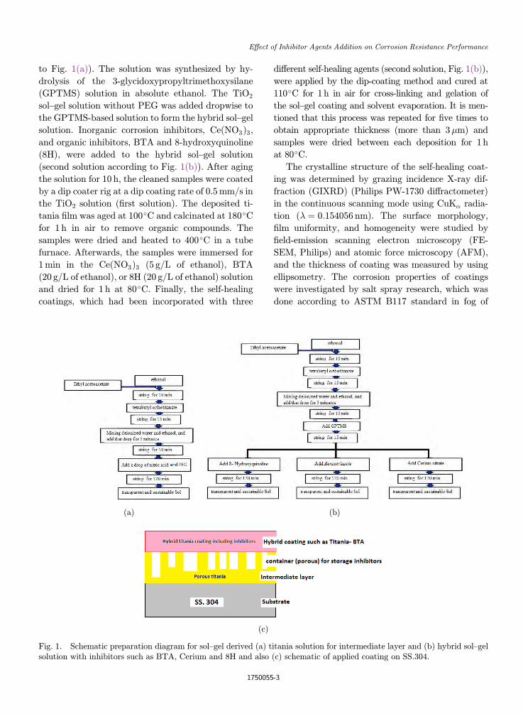
to Fig. 1(a)). The solution was synthesized by hy-
drolysis of the 3-glycidoxypropyltrimethoxysilane
(GPTMS) solution in absolute ethanol. The TiO2
sol–gel solution without PEG was added dropwise to
the GPTMS-based solution to form the hybrid sol–gel
solution. Inorganic corrosion inhibitors, Ce(NO3Þ3,and organic inhibitors, BTA and 8-hydroxyquinoline
(8H), were added to the hybrid sol–gel solution
(second solution according to Fig. 1(b)). After aging
the solution for 10 h, the cleaned samples were coated
by a dip coater rig at a dip coating rate of 0.5mm/s in
the TiO2 solution (¯rst solution). The deposited ti-
tania ¯lm was aged at 100�C and calcinated at 180�Cfor 1 h in air to remove organic compounds. The
samples were dried and heated to 400�C in a tube
furnace. Afterwards, the samples were immersed for
1min in the Ce(NO3Þ3 (5 g/L of ethanol), BTA
(20 g/L of ethanol), or 8H (20 g/L of ethanol) solution
and dried for 1 h at 80�C. Finally, the self-healing
coatings, which had been incorporated with three
di®erent self-healing agents (second solution, Fig. 1(b)),
were applied by the dip-coating method and cured at
110�C for 1 h in air for cross-linking and gelation of
the sol–gel coating and solvent evaporation. It is men-
tioned that this process was repeated for ¯ve times to
obtain appropriate thickness (more than 3�m) and
samples were dried between each deposition for 1h
at 80�C.The crystalline structure of the self-healing coat-
ing was determined by grazing incidence X-ray dif-
fraction (GIXRD) (Philips PW-1730 di®ractometer)
in the continuous scanning mode using CuK� radia-
tion (� ¼ 0:154056 nm). The surface morphology,
¯lm uniformity, and homogeneity were studied by
¯eld-emission scanning electron microscopy (FE-
SEM, Philips) and atomic force microscopy (AFM),
and the thickness of coating was measured by using
ellipsometry. The corrosion properties of coatings
were investigated by salt spray research, which was
done according to ASTM B117 standard in fog of
(a) (b)
(c)
Fig. 1. Schematic preparation diagram for sol–gel derived (a) titania solution for intermediate layer and (b) hybrid sol–gelsolution with inhibitors such as BTA, Cerium and 8H and also (c) schematic of applied coating on SS.304.
E®ect of Inhibitor Agents Addition on Corrosion Resistance Performance
1750055-3
3.5wt.% NaCl solutions at the temperature of 37�Cup to 500 h, and the coatings were cross scratched
following ASTM D1653. These samples were irregu-
larly evaluated at 24 h interval and samples were
taken out when the corrosion occurred. After the test
and appearance of the red rust, the corrosion pro-
ducts were removed, and the samples rinsed with
acetone and dried, then the weight loss of samples
was determined. Also, the corrosion properties of the
coatings were evaluated by electrochemical imped-
ance spectroscopy (EIS) in a 3% NaCl solution in air
at room temperature for up to 96 h. EIS was per-
formed between 0.01Hz and 100 kHz on a frequency
response analyzer (Electrochemical analyzer instru-
ment: CompactStat Ivium Soft 1.805 Release IVIUM
Technologies Netherlands).
3. Results and Discussion
In the sol–gel technique, careful control of the pro-
cessing steps such as hydrolysis and condensation of
metallic alkoxides are needed to obtain the desired
molecular mixture, especially inorganic nanoparticles
in an organic matrix.
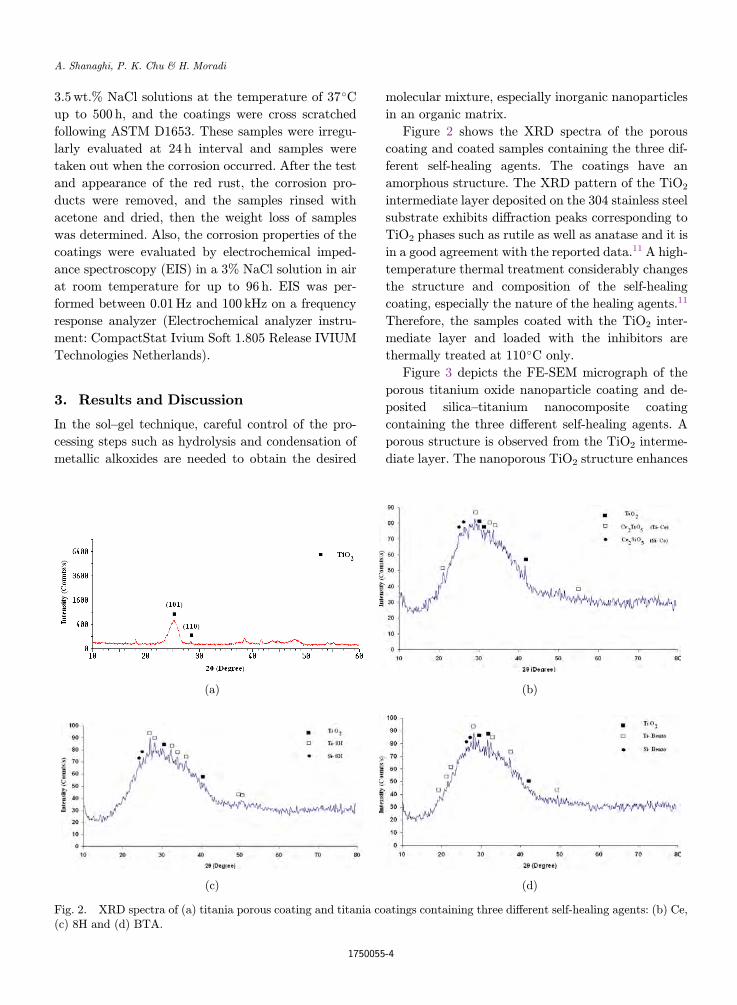
Figure 2 shows the XRD spectra of the porous
coating and coated samples containing the three dif-
ferent self-healing agents. The coatings have an
amorphous structure. The XRD pattern of the TiO2
intermediate layer deposited on the 304 stainless steel
substrate exhibits di®raction peaks corresponding to
TiO2 phases such as rutile as well as anatase and it is
in a good agreement with the reported data.11 A high-
temperature thermal treatment considerably changes
the structure and composition of the self-healing
coating, especially the nature of the healing agents.11
Therefore, the samples coated with the TiO2 inter-
mediate layer and loaded with the inhibitors are
thermally treated at 110�C only.
Figure 3 depicts the FE-SEM micrograph of the
porous titanium oxide nanoparticle coating and de-
posited silica–titanium nanocomposite coating
containing the three di®erent self-healing agents. A
porous structure is observed from the TiO2 interme-
diate layer. The nanoporous TiO2 structure enhances
(a) (b)
(c) (d)
Fig. 2. XRD spectra of (a) titania porous coating and titania coatings containing three di®erent self-healing agents: (b) Ce,(c) 8H and (d) BTA.
A. Shanaghi, P. K. Chu & H. Moradi
1750055-4
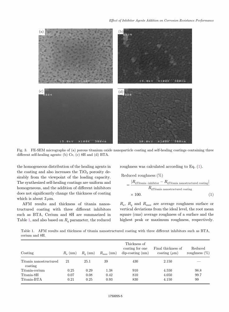
the homogeneous distribution of the healing agents in
the coating and also increases the TiO2 porosity de-
sirably from the viewpoint of the loading capacity.
The synthesized self-healing coatings are uniform and
homogeneous, and the addition of di®erent inhibitors
does not signi¯cantly change the thickness of coating
which is about 3�m.
AFM results and thickness of titania nanos-
tructured coating with three di®erent inhibitors
such as BTA, Cerium and 8H are summarized in
Table 1, and also based on Rq parameter, the reduced
roughness was calculated according to Eq. (1).
Reduced roughness ð%Þ
¼ jRqTitania�inhibitor � RqTitania nanostructured coatingjRqTitania nanostructured coating
� 100: ð1Þ
Ra, Rq and Rmax are average roughness surface or
vertical deviations from the ideal level, the root mean
square (rms) average roughness of a surface and the
highest peak or maximum roughness, respectively.
Table 1. AFM results and thickness of titania nanostructured coating with three di®erent inhibitors such as BTA,cerium and 8H.
Coating Ra (nm) Rq (nm) Rmax (nm)
Thickness ofcoating for onedip-coating (nm)
Final thickness ofcoating (�m)
Reducedroughness (%)
Titania nanostructuredcoating
21 25.1 39 430 2.150 —
Titania-cerium 0.25 0.29 1.38 910 4.550 98.8Titania-8H 0.07 0.08 0.42 810 4.050 99.7Titania-BTA 0.21 0.25 0.93 830 4.150 99
Fig. 3. FE-SEM micrographs of (a) porous titanium oxide nanoparticle coating and self-healing coatings containing threedi®erent self-healing agents: (b) Ce, (c) 8H and (d) BTA.
E®ect of Inhibitor Agents Addition on Corrosion Resistance Performance
1750055-5
According to the data in Table 1, the surface rough-
ness of the coating containing 8H is lower than other
samples. Actually, the mismatch in the coe±cients of
thermal expansion in°uences the S.S 304-ceramics
coating bonding. During the heat treatment of the
titania composite nanostructured coating, generation
of stress between the metal substrate and ceramic
coating as a result of a di®erence in thermal contrac-
tion and expansion decreases the bonding strength of
the coating. Furthermore, mechanical stress is accu-
mulated during subsequent heat treatment of the
coating.5,6 The quantity of surface roughness can
predict a real object interaction with its environment.
The high value of the roughness, prepares the surface
layer to create cracks and destruction layers. Although
the addition of 8H leads to a signi¯cant reduction in
Ra, Rq and Rmax, it is noteworthy that afterwards
addition of BTAand cerium increasesRa, Rq andRmax
and the heterogeneous property of surface.
In fact, releasing 8H from encapsulation to the
cracks led to a smooth layer on the surface and in-
creased homogeneity and uniformity of coating com-
pared to BTA and cerium as an inhibitor. However, it
is noteworthy that repair defects and micro cracks by
releasing inhibitors7,8 increase the homogeneity and
reduce the surface roughness.
It can be supposed that during the formation of
the defects and cracks in the coating, di®usion of the
inhibitors occurs and there is possibly a chemical re-
action at the surface of defects to heal them. It is
noticeable that high concentrations of releasing of
inhibitor, especially for organic inhibitors such as 8H
and BTA, reduce the strength of the network sol–gel
coating and creates delamination and osmotic pres-
sure coating and defect in the network of ceramic
¯lms.10,11 However, organic self-healing agents due to
better reaction with titanium alkoxide, help organic
materials by providing a very homogeneous disper-
sion in the coating.
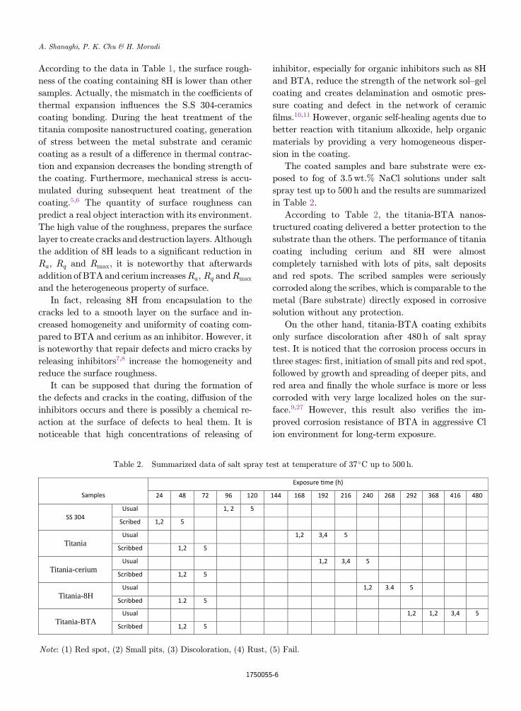
The coated samples and bare substrate were ex-
posed to fog of 3.5wt.% NaCl solutions under salt
spray test up to 500 h and the results are summarized
in Table 2.
According to Table 2, the titania-BTA nanos-
tructured coating delivered a better protection to the
substrate than the others. The performance of titania
coating including cerium and 8H were almost
completely tarnished with lots of pits, salt deposits
and red spots. The scribed samples were seriously
corroded along the scribes, which is comparable to the
metal (Bare substrate) directly exposed in corrosive
solution without any protection.
On the other hand, titania-BTA coating exhibits
only surface discoloration after 480 h of salt spray
test. It is noticed that the corrosion process occurs in
three stages: ¯rst, initiation of small pits and red spot,
followed by growth and spreading of deeper pits, and
red area and ¯nally the whole surface is more or less
corroded with very large localized holes on the sur-
face.9,27 However, this result also veri¯es the im-
proved corrosion resistance of BTA in aggressive Cl
ion environment for long-term exposure.
Table 2. Summarized data of salt spray test at temperature of 37�C up to 500 h.
Samples
Exposure me (h)
24 48 72 96 120 144 168 192 216 240 268 292 368 416 480
SS 304Usual 1, 2 5
Scribed 1,2 5
Titania Usual 1,2 3,4 5
Scribbed 1,2 5
Titania-cerium Usual 1,2 3,4 5
Scribbed 1,2 5
Titania-8HUsual 1,2 3.4 5
Scribbed 1.2 5
Titania-BTAUsual 1,2 1,2 3,4 5
Scribbed 1,2 5
Note: (1) Red spot, (2) Small pits, (3) Discoloration, (4) Rust, (5) Fail.
A. Shanaghi, P. K. Chu & H. Moradi
1750055-6
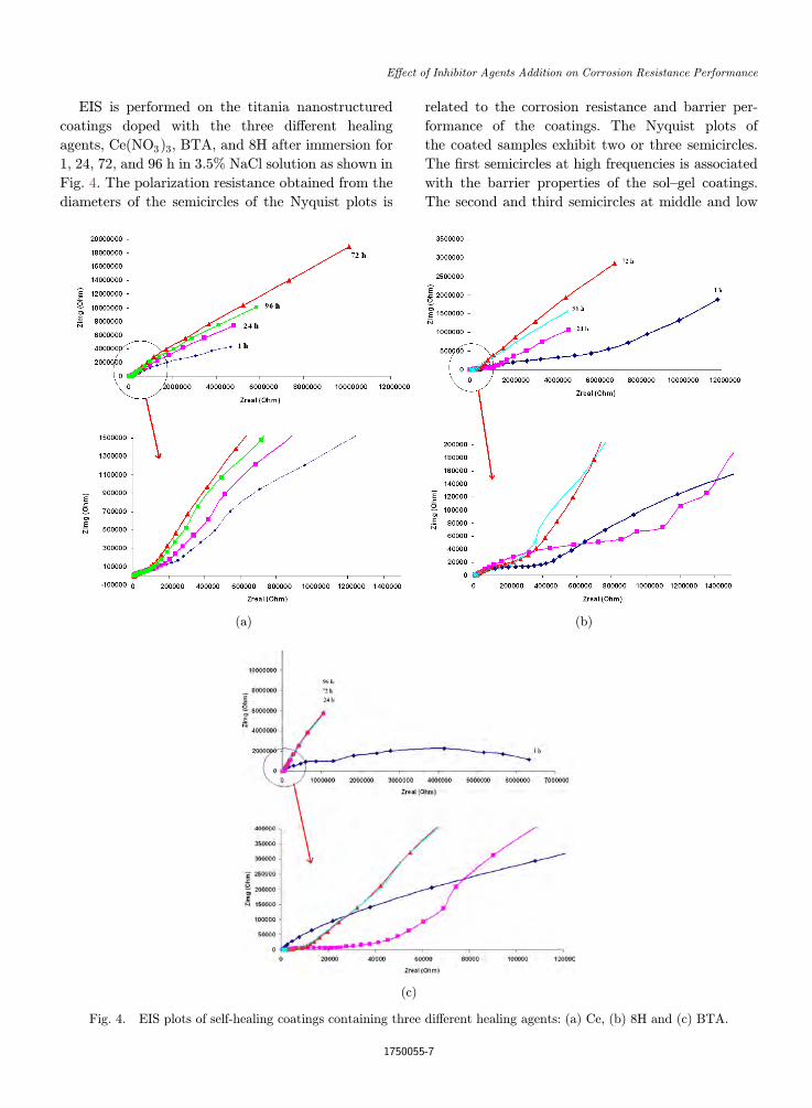
EIS is performed on the titania nanostructured
coatings doped with the three di®erent healing
agents, Ce(NO3Þ3, BTA, and 8H after immersion for
1, 24, 72, and 96 h in 3.5% NaCl solution as shown in
Fig. 4. The polarization resistance obtained from the
diameters of the semicircles of the Nyquist plots is
related to the corrosion resistance and barrier per-
formance of the coatings. The Nyquist plots of
the coated samples exhibit two or three semicircles.
The ¯rst semicircles at high frequencies is associated
with the barrier properties of the sol–gel coatings.
The second and third semicircles at middle and low
(a) (b)
(c)
Fig. 4. EIS plots of self-healing coatings containing three di®erent healing agents: (a) Ce, (b) 8H and (c) BTA.
E®ect of Inhibitor Agents Addition on Corrosion Resistance Performance
1750055-7
frequencies depend on reduction of mass transport in
the solid phase due to commencement of the healing
reaction by releasing inhibitors to defects and growth
of the layer, and also the charge transfer resistance of
the corrosion process. The corrosion resistance of the
titania coating with three di®erent healing agents is
enhanced by increasing the immersion time in 3.5%
NaCl solution. The Nyquist plots demonstrate that
the activation time of the coating containing 8H as an
organic healing agent is enhanced when compared to
other coatings including cerium and BTA. Cerium as
an inorganic healing agent is second and BTA is third
and maximum. Over the course of immersion, the
barrier performance of the coatings changes and re-
duction of the impedance observed from the coatings
containing Ce and 8H discloses deterioration of
the protection system after immersion for 96 h in the
3.5% NaCl solution.
According to Figs. 4(a)–4(c), absorption process at
the electrode surface produces the arc below the hor-
izontal axis, which some researchers have attributed
to the reactants absorbed. Actually after 1 h immer-
sion into 3.5% NaCl solution, chloride ions with high
mobility can penetrate into the holes of coating and
these rings at low frequency re°ect the absorption
reaction on the electrode surface, the initial reactants
are attracted to the surface when the metal coating is
in corrosive environments. Therefore, increasing the
immersion time to 72 h causes the cracks of coating to
be ¯lled by corrosion products, as a result of the re-
action between the solution and the coating, to pre-
vent the transfer times to the metal surface. On the
other hand, results show the system under the control
of the reactants or reaction products from the surface
of the metal is dissolved. This often occurs when a
species within a ¯lm is released to the electrode sur-
face, this situation arises when the covered electrode
reaction product, species or a ¯lm coating solution is
absorbed.9,27 However, after 96 h of immersion time,
the concentration of chloride ions is high and causes
an increase in defects, micro cracks, hole on the sur-
face of hybrid titania nanostructured coating con-
taining Ce and 8H by destruction of coating, and also
hybrid titania nanostructured coating containing
BTA, BTA is released from coating to improve the
resistance of passive ¯lm, which is created on the
surface.
In Fig. 4, increasing diameter of semicircle with
shifting time immersion from1to72h indicates increased
polarization resistance and corrosion resistance. Over
time, more inhibitor molecules have been absorbed on
the surface and therefore more corrosion sites are
blocked. Especially, increased resistance to corrosion
titania-BTA coating for 96 h immersion can be seen
with adsorption inhibitors onmetal surface. Inhibitory
e®ect is dependent on two steps: ¯rst, reach the in-
hibitor interface metal-solution and then inhibit the
absorption inhibitor on the metal surface. Replace-
ment of organic molecules, especially BTA, at the in-
terface instead of water molecules results in most of
them being absorbed on the surface of metals. After
absorption inhibitor molecules on the surface of
metals, this molecules blocked the location of the ac-
tive corrosion. Absorption inhibitor molecules mainly
depend on some physical properties such as chemical
functional groups of themolecule inhibitors, inhibitors
of space, the density of electrons in electron donor
atoms, and the electric charge on the surface of the
metal. According to Fig. 4, the increasing polarization
resistance over 72 h of immersion shows that the pro-
cess of inhibitory molecules adsorbed on S.S 304 sur-
face used during immersion time were not complete
and the thermodynamic equilibrium between mole-
cules adsorbed and molecules in solution was not
achieved. Actually, the inhibitor molecules, such as
cerium and 8H, also tend toward absorption of the
surface and the release is not complete. However, the
release ofBTAconstitutes a thin layer of absorption on
the surface of damaged metal to prevent the in°uence
of corrosive species. Within 48 h, almost all inhibitors
were absorbed on the surface and after 72 h as shown in
the diagrams at the end of a line graph, the ¯lm on the
metal surface is covered and the system under control
of di®usion. But after 96 h, the coating containing ce-
rium and 8H will greatly reduce charge transfer resis-
tance. In fact, these inhibitors, in addition to the
weak corrosion inhibiting properties, can cause local-
ized corrosion and corrosion is severed. It is noticeable
that corrosion behavior of titania-BTA nanos-
tructured coating and charge transfer resistance in-
creased at 96 h immersion due to uptake of inhibitors
on the surface.
However, when su±cient amount of inhibitors
such as cerium, 8H and BTA, is released in the initial
time (1 h), the holes and cracks are ¯lled, and the ¯lm
in the metal/coating interface is produced and ¯nally
leads to improved corrosion resistance. But after 72 h
due to the di®usion of chloride ion and defects on the
A. Shanaghi, P. K. Chu & H. Moradi
1750055-8
surface, corrosion resistance of the coatings contain-
ing cerium and 8H is reduced.
On the other hand, self-healing coatings contain-
ing inhibitors improve the resistance to localized
corrosion due to the formation of the protective layer
which acts as a obstruction to prevent the oxygen
di®usion to the metal surface, but the layer formed is
relatively slim and can be destroyed by di®usion of
corrosive ions and the time required for the destruc-
tion of the protective layer is increased for coating
containing BTA.
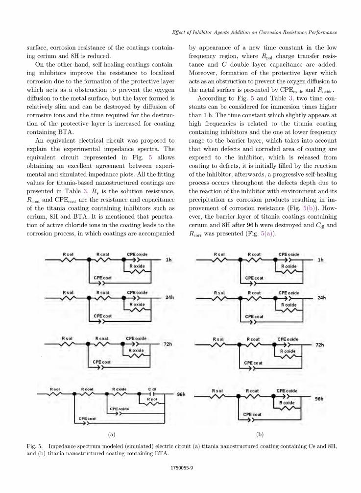
An equivalent electrical circuit was proposed to
explain the experimental impedance spectra. The
equivalent circuit represented in Fig. 5 allows
obtaining an excellent agreement between experi-
mental and simulated impedance plots. All the ¯tting
values for titania-based nanostructured coatings are
presented in Table 3. Rs is the solution resistance,
Rcoat and CPEcoat are the resistance and capacitance
of the titania coating containing inhibitors such as
cerium, 8H and BTA. It is mentioned that penetra-
tion of active chloride ions in the coating leads to the
corrosion process, in which coatings are accompanied
by appearance of a new time constant in the low
frequency region, where Rpol charge transfer resis-
tance and C double layer capacitance are added.
Moreover, formation of the protective layer which
acts as an obstruction to prevent the oxygen di®usion to
the metal surface is presented by CPEoxide and Roxide.
According to Fig. 5 and Table 3, two time con-
stants can be considered for immersion times higher
than 1 h. The time constant which slightly appears at
high frequencies is related to the titania coating
containing inhibitors and the one at lower frequency
range to the barrier layer, which takes into account
that when defects and corroded area of coating are
exposed to the inhibitor, which is released from
coating to defects, it is initially ¯lled by the reaction
of the inhibitor, afterwards, a progressive self-healing
process occurs throughout the defects depth due to
the reaction of the inhibitor with environment and its
precipitation as corrosion products resulting in im-
provement of corrosion resistance (Fig. 5(b)). How-
ever, the barrier layer of titania coatings containing
cerium and 8H after 96 h were destroyed and Cdl and
Rcorr was presented (Fig. 5(a)).
(a) (b)
Fig. 5. Impedance spectrum modeled (simulated) electric circuit (a) titania nanostructured coating containing Ce and 8H,and (b) titania nanostructured coating containing BTA.
E®ect of Inhibitor Agents Addition on Corrosion Resistance Performance
1750055-9
The resistance and capacitance of the titania
coating depend on the type of inhibitor, its crack
ability and amount of absorbed water. In fact, water
uptake into the coating and penetration of active
chloride ions cause its partial destruction. It is men-
tioned that corrosion factors penetrating the titania
coating through cracks and coating were destroyed
locally. The presence of inhibitors improves the cor-
rosion resistance of titania nanostructured coating,
leading to the formation of a signi¯cant amount of
corrosion product by releasing the inhibitor to defects.
As a result, the corrosion resistance is considerably en-
hanced via titania BTA nanostructured coating. Indeed,
with the addition of organic inhibitor, BTA, the healing
of defects occurs than other inhibitors.
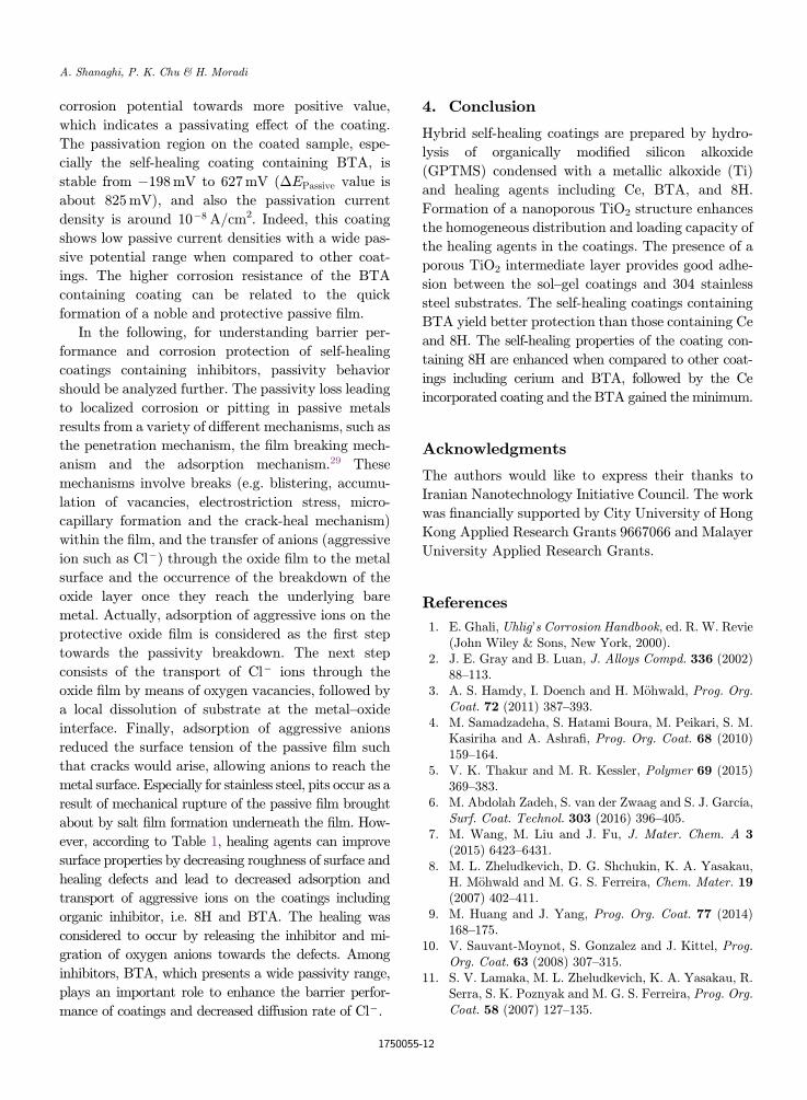
Figure 6 presents the potentiodynamic results
obtained from the coated samples in 3.5% NaCl so-
lution and corrosion parameters, such as passivity
current density (ipassiveÞ corrosion potential (EcorrÞ,corrosion current density (icorrÞ, anodic and cathodic
Tafel slopes (�a, �c), passive potential range
(�EPassiveÞ, polarization resistance (Rp), corrosion
rate (CR) and protection e±ciency which were de-
termined from polarization curves by Tafel extrapo-
lation method and summarized in Table 4. The
application of Tafel's law assumes that the interface
is under kinetic control in spite of the existence of a
protection layer.21
Polarization resistance was derived from Stern–
Geary equation,28 as follows:
RP ¼ �a:�c
2:3icorrð�aþ �cÞ : ð2Þ
The protection e±ciency (%) was calculated accord-
ing to Eq. (3):
Protection efficiency ð%Þ ¼ icorr � ipassiveiCorr
� 100;
ð3Þwhere icorr and ipassive are the corrosion current den-
sities of titania nanostructured coating and coatings
including inhibitors, respectively. It is mentioned that
the passivity current density gives an idea of the rate
of corrosion as in Eq. (3) instead of corrosion current
density of self-healing coatings. The open circuit
potentials of the self-healing coating containing Ce,
8H, and BTA in 3.5% NaCl are �339, �367 and
�241mV vs SCE, respectively. The corrosion poten-
tials of the coatings containing BTA increase
to positive values. Both the anodic and cathodic
branches of the coated samples shift to smaller cur-
rents, i.e. lower rates of the metal corrosion.
According to results, titania nanostructured coatings
containing inhibitors improve the anticorrosive
properties of coating and decrease the corrosion cur-
rent density compared to other coatings and shift the
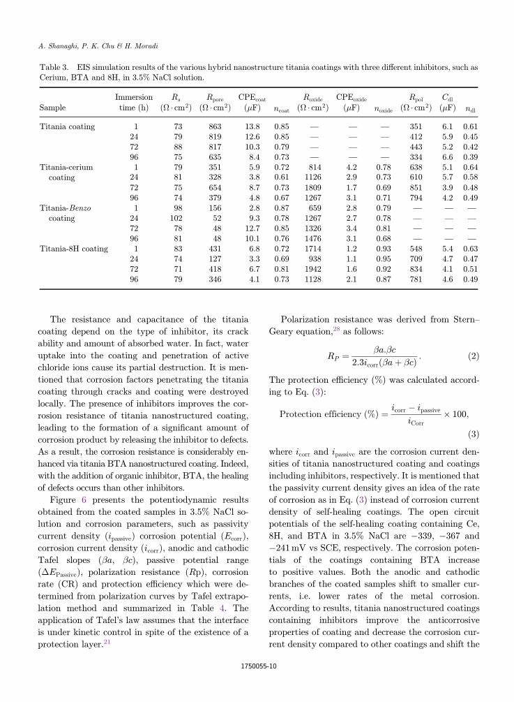
Table 3. EIS simulation results of the various hybrid nanostructure titania coatings with three di®erent inhibitors, such asCerium, BTA and 8H, in 3.5% NaCl solution.
Immersion Rs Rpore CPEcoat
ncoat
Roxide CPEoxide
noxide
Rpol Cdl
ndlSample time (h) (� � cm2Þ (� � cm2Þ (�F) (� � cm2Þ (�F) (� � cm2Þ (�F)
Titania coating 1 73 863 13.8 0.85 — — — 351 6.1 0.6124 79 819 12.6 0.85 — — — 412 5.9 0.4572 88 817 10.3 0.79 — — — 443 5.2 0.4296 75 635 8.4 0.73 — — — 334 6.6 0.39
Titania-ceriumcoating
1 79 351 5.9 0.72 814 4.2 0.78 638 5.1 0.6424 81 328 3.8 0.61 1126 2.9 0.73 610 5.7 0.58
72 75 654 8.7 0.73 1809 1.7 0.69 851 3.9 0.4896 74 379 4.8 0.67 1267 3.1 0.71 794 4.2 0.49
Titania-Benzocoating
1 98 156 2.8 0.87 659 2.8 0.79 — — —
24 102 52 9.3 0.78 1267 2.7 0.78 — — —
72 78 48 12.7 0.85 1326 3.4 0.81 — — —
96 81 48 10.1 0.76 1476 3.1 0.68 — — —
Titania-8H coating 1 83 431 6.8 0.72 1714 1.2 0.93 548 5.4 0.6324 74 127 3.3 0.69 938 1.1 0.95 709 4.7 0.4772 71 418 6.7 0.81 1942 1.6 0.92 834 4.1 0.5196 79 346 4.1 0.73 1128 2.1 0.87 781 4.6 0.49
A. Shanaghi, P. K. Chu & H. Moradi
1750055-10
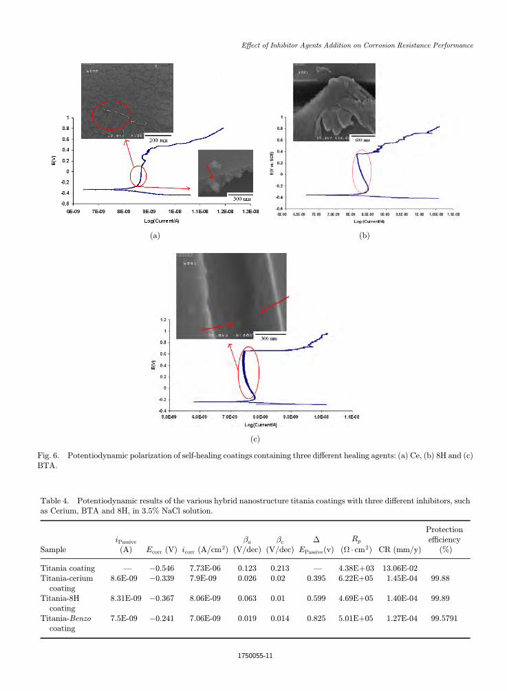
Table 4. Potentiodynamic results of the various hybrid nanostructure titania coatings with three di®erent inhibitors, suchas Cerium, BTA and 8H, in 3.5% NaCl solution.
SampleiPassive(A) Ecorr (V) icorr (A/cm2Þ
�a
(V/dec)�c
(V/dec)�
EPassive(v)
Rp
(� � cm2Þ CR (mm/y)
Protectione±ciency
(%)
Titania coating — �0:546 7.73E-06 0.123 0.213 — 4:38Eþ03 13.06E-02Titania-cerium
coating8.6E-09 �0:339 7.9E-09 0.026 0.02 0.395 6.22Eþ05 1.45E-04 99.88
Titania-8Hcoating
8.31E-09 �0:367 8.06E-09 0.063 0.01 0.599 4.69Eþ05 1.40E-04 99.89
Titania-Benzocoating
7.5E-09 �0:241 7.06E-09 0.019 0.014 0.825 5.01Eþ05 1.27E-04 99.5791
(a) (b)
(c)
Fig. 6. Potentiodynamic polarization of self-healing coatings containing three di®erent healing agents: (a) Ce, (b) 8H and (c)BTA.
E®ect of Inhibitor Agents Addition on Corrosion Resistance Performance
1750055-11
corrosion potential towards more positive value,
which indicates a passivating e®ect of the coating.
The passivation region on the coated sample, espe-
cially the self-healing coating containing BTA, is
stable from �198mV to 627mV (�EPassive value is
about 825mV), and also the passivation current
density is around 10�8 A/cm2. Indeed, this coating
shows low passive current densities with a wide pas-
sive potential range when compared to other coat-
ings. The higher corrosion resistance of the BTA
containing coating can be related to the quick
formation of a noble and protective passive ¯lm.
In the following, for understanding barrier per-
formance and corrosion protection of self-healing
coatings containing inhibitors, passivity behavior
should be analyzed further. The passivity loss leading
to localized corrosion or pitting in passive metals
results from a variety of di®erent mechanisms, such as
the penetration mechanism, the ¯lm breaking mech-
anism and the adsorption mechanism.29 These
mechanisms involve breaks (e.g. blistering, accumu-
lation of vacancies, electrostriction stress, micro-
capillary formation and the crack-heal mechanism)
within the ¯lm, and the transfer of anions (aggressive
ion such as Cl�Þ through the oxide ¯lm to the metal
surface and the occurrence of the breakdown of the
oxide layer once they reach the underlying bare
metal. Actually, adsorption of aggressive ions on the
protective oxide ¯lm is considered as the ¯rst step
towards the passivity breakdown. The next step
consists of the transport of Cl� ions through the
oxide ¯lm by means of oxygen vacancies, followed by
a local dissolution of substrate at the metal–oxide
interface. Finally, adsorption of aggressive anions
reduced the surface tension of the passive ¯lm such
that cracks would arise, allowing anions to reach the
metal surface. Especially for stainless steel, pits occur as a
result of mechanical rupture of the passive ¯lm brought
about by salt ¯lm formation underneath the ¯lm. How-
ever, according to Table 1, healing agents can improve
surface properties by decreasing roughness of surface and
healing defects and lead to decreased adsorption and
transport of aggressive ions on the coatings including
organic inhibitor, i.e. 8H and BTA. The healing was
considered to occur by releasing the inhibitor and mi-
gration of oxygen anions towards the defects. Among
inhibitors, BTA, which presents a wide passivity range,
plays an important role to enhance the barrier perfor-
mance of coatings and decreased di®usion rate of Cl�.
4. Conclusion
Hybrid self-healing coatings are prepared by hydro-
lysis of organically modi¯ed silicon alkoxide
(GPTMS) condensed with a metallic alkoxide (Ti)
and healing agents including Ce, BTA, and 8H.
Formation of a nanoporous TiO2 structure enhances
the homogeneous distribution and loading capacity of
the healing agents in the coatings. The presence of a
porous TiO2 intermediate layer provides good adhe-
sion between the sol–gel coatings and 304 stainless
steel substrates. The self-healing coatings containing
BTA yield better protection than those containing Ce
and 8H. The self-healing properties of the coating con-
taining 8H are enhanced when compared to other coat-
ings including cerium and BTA, followed by the Ce
incorporated coating and the BTA gained the minimum.
Acknowledgments
The authors would like to express their thanks to
Iranian Nanotechnology Initiative Council. The work
was ¯nancially supported by City University of Hong
Kong Applied Research Grants 9667066 and Malayer
University Applied Research Grants.
References
1. E. Ghali, Uhlig's Corrosion Handbook, ed. R. W. Revie(John Wiley & Sons, New York, 2000).
2. J. E. Gray and B. Luan, J. Alloys Compd. 336 (2002)88–113.
3. A. S. Hamdy, I. Doench and H. M€ohwald, Prog. Org.Coat. 72 (2011) 387–393.
4. M. Samadzadeha, S. Hatami Boura, M. Peikari, S. M.Kasiriha and A. Ashra¯, Prog. Org. Coat. 68 (2010)159–164.
5. V. K. Thakur and M. R. Kessler, Polymer 69 (2015)369–383.
6. M. Abdolah Zadeh, S. van der Zwaag and S. J. García,Surf. Coat. Technol. 303 (2016) 396–405.
7. M. Wang, M. Liu and J. Fu, J. Mater. Chem. A 3(2015) 6423–6431.
8. M. L. Zheludkevich, D. G. Shchukin, K. A. Yasakau,H. M€ohwald and M. G. S. Ferreira, Chem. Mater. 19(2007) 402–411.
9. M. Huang and J. Yang, Prog. Org. Coat. 77 (2014)168–175.
10. V. Sauvant-Moynot, S. Gonzalez and J. Kittel, Prog.Org. Coat. 63 (2008) 307–315.
11. S. V. Lamaka, M. L. Zheludkevich, K. A. Yasakau, R.Serra, S. K. Poznyak and M. G. S. Ferreira, Prog. Org.Coat. 58 (2007) 127–135.
A. Shanaghi, P. K. Chu & H. Moradi
1750055-12
12. A. F. Galio, S. V. Lamaka, M. L. Zheludkevich, L. F.P. Dick, I. L. Müller and M. G. S. Ferreira, Surf. Coat.Technol. 204 (2010) 1479–1486.
13. M. L. Zheludkevich, R. Serra, M. F. Montemor, I. M.Miranda Salvado and M. G. S. Ferreira, Surf. Coat.Technol. 200 (2006) 3084–3094.
14. Y.-H. Han, A. Taylor and K. M. Knowles, Surf. Coat.Technol. 202 (2008) 1859–1868.
15. X. Zhong, Q. Li, J. Hu and Y. Luorr, Corros. Sci. 50(2008) 2304.
16. D. G. Shchukin, M. L. Zheludkevich, K. Yasakau,S. Lamaka, M. G. S. Ferreira and H. M€owald, Adv.Mater. 18 (2006) 1672.
17. D. Wang and G. P. Bierwagen, Prog. Org. Coat. 64(2009) 327–338.
18. A. Abou-Elazm, R. Abdel-Karim, I. Elmahallawi andR. Rashad, Corros. Sci. 51 (2009) 203–208.
19. X. Fang, K. Zhang, H. Guo, W. Wang and B. Zhou,Mater. Sci. Eng. A 487 (2008) 7–13.
20. M. Aparicio, A. Jitianu, G. Rodriguez, A. Degnah, K.Al-Marzoki, L. C. Klein and J. Mosa, Electrochim.Acta 202 (2016) 325–332.
21. L. Curkovic, H. O. Curkovic, S. Salopek, M. M. Renjoand S. Šegota, Corros. Sci. 77 (2013) 176–184.
22. A. Marsal, F. Ansart, V. Turq, J. P. Bonino, J. M.Sobrino, Y. M. Chen and J. Garcia, Surf. Coat.Technol. 237 (2013) 234–240.
23. R. Subasri, R. Malathi, A. Jyothirmayi and N. Y.Hebalkar, Ceram. Int. 38 (2012) 5731–5740.
24. N. C. Rosero-Navarroa, L. Paussa, F. Andreatta, Y.Castro, A. Dur�an, M. Aparicio and L. Fedrizzi, Prog.Org. Coat. 69 (2010) 167–174.
25. K. A. Yasakau, M. L. Zheludkevich, O. V. Karavaiand M. G. S. Ferreir, Prog. Org. Coat. 63 (2008)352–361.
26. N. C. Rosero-Navarro, S. A. Pellice, A. Durań and M.Aparicio, Corros. Sci. 50 (2008) 1283–1291.
27. S. Manivannan, S. K. Gopalakrishnan, S. P. KumareshBabu and S. Sundarrajan, Alexandria Eng. J. 55(2016) 663–671.
28. R. B. Figueira, C. J. R. Silva and E. V. Pereira,J. Electrochem. Soc. 162 (2015) C666–C676.
29. J. Soltis, Corros. Sci. 90 (2015) 5–22.
E®ect of Inhibitor Agents Addition on Corrosion Resistance Performance
1750055-13


