Embed Size (px)
Citation preview
Dkk1 Regulates Ventral Midbrain DopaminergicDifferentiation and MorphogenesisDiogo Ribeiro1, Kristina Ellwanger2., Desiree Glagow3., Spyridon Theofilopoulos1, Nina S. Corsini3¤,
Ana Martin-Villalba3, Christof Niehrs2, Ernest Arenas1*
1 Section of Molecular Neurobiology, Department of Medical Biochemistry and Biophysics, Karolinska Institute, Stockholm, Sweden, 2 Division of Molecular Embryology,
DKFZ-ZMBH Alliance, German Cancer Research Center, Heidelberg, Germany, 3 Division of Molecular Neurobiology, German Cancer Research Center, Heidelberg, Germany
Abstract
Dickkopf1 (Dkk1) is a Wnt/b-catenin inhibitor that participates in many processes during embryonic development. One of itsroles during embryogenesis is to induce head formation, since Dkk1-null mice lack head structures anterior to midbrain. TheWnt/b-catenin pathway is also known to regulate different aspects of ventral midbrain (VM) dopaminergic (DA) neurondevelopment and, in vitro, Dkk1-mediated inhibition of the Wnt/b-catenin pathway improves the DA differentiation inmouse embryonic stem cells (mESC). However, the in vivo function of Dkk1 on the development of midbrain DA neuronsremains to be elucidated. Here we examined Dkk1+/2 embryos and found that Dkk1 is required for the differentiation of DAprecursors/neuroblasts into DA neurons at E13.5. This deficit persisted until E17.5, when a defect in the number anddistribution of VM DA neurons was detected. Furthermore, analysis of the few Dkk12/2 embryos that survived until E17.5revealed a more severe loss of midbrain DA neurons and morphogenesis defects. Our results thus show that Dkk1 isrequired for midbrain DA differentiation and morphogenesis.
Citation: Ribeiro D, Ellwanger K, Glagow D, Theofilopoulos S, Corsini NS, et al. (2011) Dkk1 Regulates Ventral Midbrain Dopaminergic Differentiation andMorphogenesis. PLoS ONE 6(2): e15786. doi:10.1371/journal.pone.0015786
Editor: Mike Karl, Center for Regenerative Therapies Dresden, Germany
Received July 28, 2010; Accepted November 24, 2010; Published February 11, 2011
Copyright: � 2011 Ribeiro et al. This is an open-access article distributed under the terms of the Creative Commons Attribution License, which permitsunrestricted use, distribution, and reproduction in any medium, provided the original author and source are credited.
Funding: This work was supported by grants from the Swedish Foundation for Strategic Research (INGVAR and CEDB), Swedish Research Council (VR2008:2811and DBRM), Norwegian Research Council, and Karolinska Institutet to EA, and DR was supported by the Foundation for Science and Technology from thePortuguese Government (SFRH/BD/24585/2005). The funders had no role in study design, data collection and analysis, decision to publish, or preparation of themanuscript.
Competing Interests: The authors have declared that no competing interests exist.
* E-mail: [email protected]
. These authors contributed equally to this work.
¤ Current address: Institute of Molecular Biotechnology of the Austrian Academy of Sciences (IMBA), Vienna, Austria
Introduction
Dickkopf 1 (Dkk1) is a secreted glycoprotein belonging to the
Dkk family, which consists of four members (Dkk-1, -2, -3, -4). All
Dkks share two conserved cystein-rich domains separated by a
linker region [1,2]. Dkk1 is a known Wnt/b-catenin pathway
inhibitor: it binds to Lrp5/6, preventing its interaction with the
Wnt protein and disrupting the Wnt-induced Frizzled-Lrp6
complex formation necessary for signal transduction [3,4,5].
Dkk1 can also block the Wnt/b-catenin pathway by inducing
Lrp6 endocytosis in the presence of Kremen proteins [6]. During
embryonic development dkk1 is first expressed in Xenopus in the
Spemann organizer of the early gastrula, and in mouse in the
anterior visceral endoderm, anterior mesendoderm and foregut
endoderm [1]. Dkk1 has been shown to have a major role in
inducing head formation: injection of dkk1 mRNA in Xenopus
embryos leads to anteriorized embryos with big heads and
enlarged cement glands; together with a dominant-negative
mutant of the BMP2/4 receptor, dkk1 mRNA is also able to
induce secondary axes with complete heads [1]. Loss-of-function
studies further confirm that Dkk1 is essential for head induction:
Xenopus embryos injected with an anti-Dkk1 antibody [1] and Dkk1
knockout mice [7] lack anterior head structures. The head-
inducing activity of Dkk1 is mediated through the inhibition of the
Wnt/b-catenin posteriorizing activity in early gastrula embryos
[8], and in combination with the inhibition of BMP signalling [9].
Besides its role in anterior neural patterning, Dkk1 is involved in
limb formation [10,11], vertebral development [12], bone
formation [13,14] and bilateral eye induction [8]. Dkk1 has also
been described to regulate cell proliferation and programmed cell
death [7,15,16], and to have a role in diseases such as cancer [17]
and Alzheimer’s disease [18]. Consistent with its role in vivo, Dkk1
is able to induce neural differentiation from embryonic stem cells
[19,20,21]. The majority of the Dkk1 effects result from a direct
inhibition of the Wnt/b-catenin pathway, although Dkk1 can
modulate gastrulation movements independently of b-catenin
through activation of the Wnt/Planar Cell Polarity (PCP) pathway
[22], suggesting that Dkk1 is able to modulate two different
branches of Wnt signaling.
The Wnt/b-catenin pathway is involved in several aspects of
neural development [23,24], and has been described to play an
important role in ventral midbrain (VM) dopaminergic (DA)
neuron development: Wnt1, the prototypical ligand of the Wnt/b-
catenin pathway, has been shown to regulate midbrain develop-
ment [25,26], neurogenesis [27], proliferation of DA progenitors
[28,29] and differentiation [30]; the Lrp6 receptor has been
described to be important for the onset of midbrain DA
differentiation and morphogenesis [31], and b-catenin is necessary
PLoS ONE | www.plosone.org 1 February 2011 | Volume 6 | Issue 2 | e15786
for the integrity of the VM neurogenic niche and the progression
from progenitors to DA neurons [32]. However, we also found
increased DA neuronal differentiation in Wnt12/2 and Lrp62/2
mouse embryonic stem cells (mESC) [33]. These effects were
mimicked in wild-type mESC by treatment with Dkk1, indicating
that Dkk1 promotes DA differentiation in vitro. In order to
address whether Dkk1 plays a role in the development of ventral
midbrain DA neurons in vivo we examined Dkk1 deficient mice.
Our results indicate that Dkk1 regulates the distribution and the
number of DA neurons in the developing VM. Interestingly we
found that Dkk1 is required for the differentiation of Nurr1+/TH2
DA precursors (radially migrating neuroblasts) into DA neurons.
Our results thus identify Dkk1 as a new regulator of midbrain DA
neuron development.
Materials and Methods
AnimalsAnimals were housed in specific pathogen free and light,
temperature (21uC) and humidity (50–60% relative humidity)
controlled conditions. Food and water were available ad libitum.
Mice were mated overnight and noon of day of plug was taken as
E0.5. Dkk1+/2 mice [7] were maintained in C57BL/6 congenic
genetic background. Dkk1+/2 embryos were obtained heterozygote
crosses and by heterozygote x wild type crosses. Dkk1+/2 and
Dkk12/2 embryos were compared to wild-type littermates. The
procedures for performing all animal experiments were in
accordance with the principles and guidelines of the ATBW
(officials for animal welfare), Karolinska Institutet as well as with
the German and Swedish law. The permits were reviewed by the
Internal Animal Protection Commission of the German Cancer
Research Center (DKFZ) and the experiments were approved by
the administrative headquarter ‘‘Regierungsprasidium Karlsruhe’’
of the State Baden-Wurttemberg. The approval is based on a
positive vote of an appointed state governmental ethical
commission according to 115 of the German Animal Protection
law (approved license numbers: G-108/05, A-08/05, DKFZ180
and DKFZ206). CD1 mice (Charles River) were housed, bred,
treated and analyzed in accordance with the permit approved by
the Swedish ethical committee ‘‘Stockholms Norra Djurforsoke-
tiska Namnd’’ (ethical approval numbers N154/06 and N145/09).
In situ hybridization and ImmunohistochemistryFor in situ hybridization (ISH), embryos were fixed overnight
before being cryopreserved in 30% sucrose, frozen in OCT and
coronally sectioned (14 mm) onto Superfrost slides. ISH for Dkk1
was performed in fixed tissue as described [28,34] with
digoxigenin-labelled single-stranded RNA probes at 70uC, fol-
lowed by incubation with alkaline phosphatase (AP)- coupled
antibody and nitroblue tetrazolium (NBT) plus 5-bromo-4-chloro-
3-indolyl phosphate (BCIP) (purple) substrates.
For immunohistochemistry (IHC), coronal sections (12–14 mm
thick) were obtained after adjusting the angle. Sections were pre-
incubated for 1 hour in blocking solution (PBS, 0.25% Triton-X
100 and 5% normal goat serum) followed by incubation at 4uCovernight with one or more of the following primary antibodies
diluted in blocking solution: rabbit anti-TH (1:300, PelFreeze),
mouse anti-TH (1:500, ImmunoStar), mouse anti- bIII tubulin
(Tuj1;1:1000, Promega), rabbit anti-Lmx1a (1:500, gift from M.
German), rabbit anti-Nurr1 (1:250, Santa Cruz Biotech.), rabbit
anti Ki67 (1:500, Neomarkers), mouse anti-Islet1 (1:100, Devel-
opmental Studies Hybridoma Bank), rabbit anti-Pitx3 (1:500, gift
from M. Smidt), rabbit anti-Wnt1 (1:500, Abcam), rabbit anti-
cleaved Caspase 3 (1:100, Cell Signaling), mouse anti-Brn3a
(1:500, Millipore). After washing, slides were incubated for 1–
2 hours at room temperature with the appropriate secondary
antibodies: biotinylated (1:400, Jackson Laboratories) or fluor-
ophore conjugated (1:700, AlexaFluor 555 and 488). TO-PRO1-
iodide nuclear stain (1mM, 1:200, Invitrogen) was performed for
visualization of cells. Biotinylated secondary antibodies were
visualized with the Vector Laboratories ABC immunoperoxidase
kit, using 3-39 diaminobenzidine tetrahydrochloride (DAB
0.5 mg/ml); endogenous peroxidase activity was quenched for
20 minutes with 5% H2O2 prior to pre-incubation with secondary
antibody. When necessary, antigen retrieval was performed prior
to incubation with primary antibody with a target retrieval
solution (Dako). ISH and IHC photos were acquired with a Zeiss
Axioplan2 microscope and collected with a Hamamatsu camera
C4742-95 with the Openlab and Photoshop software. Confocal
pictures were taken with a Zeiss LSM 5 EXCITER microscope.
VM primary culturesVentral midbrains of E10.5 CD1 mice were dissected out in ice-
cold PBS supplemented with 0.2% glucose, mechanically dissoci-
ated in serum-free N2 medium through flame-narrowed Pasteur
pipettes and plated at a final density of 150,000 cells/cm2 in poly-
D-lysine-coated plates. Cultures were treated with recombinant
mouse Wnt3a (100ng/ml, R&D) and the equivalent volume of
0.1% bovine serum albumin (BSA) as a control. Treatment of
cultures was initiated at the time of plating and cultures were
incubated for 6 hours in N2 medium at 37uC in 5%CO2. Cells
were lysed and total RNA was extracted using the RNeasy Mini
Kit (Qiagen), 1 mg was treated with RQ1 RNase-free DNase
(Promega, Madison, WI) and reverse transcribed using Super-
Script II Reverse Transcriptase (Invitrogen) and random primers
(Invitrogen) (RT+ reaction). Parallel reactions without reverse
transcriptase enzyme were done as a control (RT2 reaction) and
Sybr green real-time quantitative PCR assays were performed as
previously described [35]. Expression levels were obtained by
normalization with the value of the housekeeping gene encoding
18S rRNA (Ambion, Austin, USA) obtained for every sample in
parallel assays. The primers sequence for Dkk1 were as follows:
Forward-TCAATTCCAACGCGATCAAGA; Reverse- GGCT-
GGTAGTTGTCAAGAGTCTGG.
Cell Counts and Statistical AnalysesCell counts were performed in every fifth 14 mm coronal
midbrain section through the entire DA domain (SN and VTA,
from rostral to caudal). Graphics show the average number of cells
(somas or nuclei) stained with antibodies against TH, Lmx1a,
Nurr1, Pitx3, Islet1, Brn3a, Caspase3 or Ki67 in the serial sections
and the entire DA domain, for every embryo analyzed. Depending
on the embryonic stage and staining, 3–8 Dkk1+/2embryos were
counted per condition (see figure legends). Nuclear markers were
counted using ImageJ. All the measurements were performed in
coronal sections through the midbrain and the distances were
measured in pixels using ImageJ. The ventral tegmental (VTA)
height was determined by drawing a vertical line between the most
dorsal and most ventral cells in the VTA. Results in text and the
graphs are presented as mean 6 standard error of the mean (s.e.m)
for each genotype. Cell numbers were compared with a Student’s
t-test using GraphPad. (*) p,0.05, (**) p,0.01, (***) p,0.001.
Results
Dkk1 is expressed in the developing ventral midbrainDkk1 is dynamically expressed in several areas of the developing
central nervous system, including the mesencephalon [36,37], but
Dkk1 and Ventral Midbrain Development
PLoS ONE | www.plosone.org 2 February 2011 | Volume 6 | Issue 2 | e15786
its expression in relation to DA neurons has not been examined. In
order to characterize the spatial and temporal pattern of
expression of Dkk1 during the DA neurogenic period, we
performed in situ hybridization in E9.5–13.5 mouse embryos.
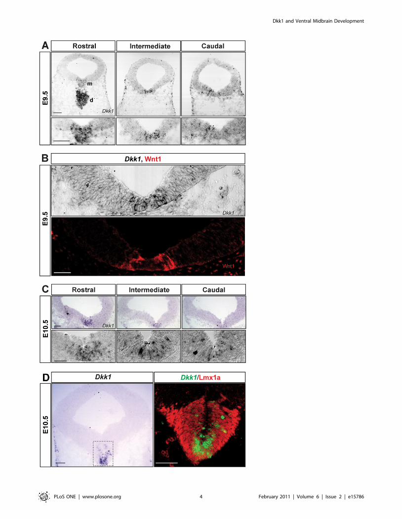
Dkk1 was detected at significant levels in the VM at E9.5 and
E10.5, but it was undetectable at E11.5–13.5. At E9.5, Dkk1 was
highly expressed in the ventral diencephalon and was expressed in
a salt and pepper pattern in the VM, where it followed a rostro-
caudal gradient with higher expression levels rostrally than
caudally (Figure 1A). Interestingly, cells expressing Dkk1 were
found both in the lateral part of the floorplate, which expresses
Wnt1, and in the medial part of the floorplate (Figure 1B). At
E10.5, Dkk1 mRNA was detected in the three layers of the
developing VM (ventricular, intermediate and marginal zones,
Figure 1C). At this stage, the midbrain expression of Dkk1 was
restricted to the medial part of the floorplate, where it was highly
expressed in cells adjacent to the midline (Figure 1C). In order to
confirm the midbrain DA identity of the Dkk1-expressing cells at
E10.5, we analyzed the expression of Dkk1 relative to that of
Lmx1a, a transcription factor expressed in the entire DA lineage
[38]. Immunohistochemistry for Lmx1a on Dkk1-probed sections
revealed that only a medial subpopulation of Lmx1a+ cells
expressed Dkk1 (Figure 1C). Shortly after, at E10.75, Dkk1 was
only weakly expressed rostrally and no expression was detected in
intermediate and caudal levels (data not shown). Dkk1 expression
was not detectable after E10.75. The position of Dkk1+ cells and
the timing of Dkk1 expression suggested that Dkk1 may work as a
regulator of DA neurogenesis and/or DA differentiation. We
therefore examine these two processes in Dkk1 mutant mice.
Midbrain DA neuron development is not affected inDkk1+/2 mice at E11.5
To address the function of Dkk1 in midbrain development we
examined Dkk1+/2 and Dkk12/2 embryos. However, since Dkk12/2
mice showed a deletion of brain structures including the midbrain, we
focused our analysis on Dkk1+/2 embryos. At E11.5, when DA
neurogenesis starts, we did not observe any significant difference in
the numbers of TH+ cells (TH- tyrosine hydroxylase, the rate limiting
enzyme in dopamine synthesis and a DA neuron marker) in
heterozygous embryos compared with wild-type littermate controls
(Figure 2A and 2B). Similarly, when we examined the expression of
the early neuronal marker Tuj1, no differences were found
(Figure 2A). To further examine whether other aspects of midbrain
DA development were affected in the Dkk1+/2 embryos we
performed immunohistochemistry for Lmx1a and again found no
differences between Dkk1+/2 and Wt embryos in the number or
distribution of progenitor cells at E11.5 (Figure 2C and D). These
results indicated that the initial aspects of DA neuron development
were not affected by the loss of one allele of Dkk1.
Dopaminergic differentiation is impaired in Dkk1+/2
mutantsTo determine whether DA neuron development was affected at later
stages we performed immunohistochemistry for TH in Dkk1+/2 E13.5
embryos, the time at which DA neurogenesis peaks in the VM.
Interestingly we found a significant reduction (40%) in the number of
DA neurons in the Dkk1+/2 embryos compared to Wt (Figure 3A and
3B). In order to exclude the possibility of a general developmental delay
we measured the crown-rump length in Wt and Dkk1+/2 embryos and
found no differences (Figure S1B). To determine if the observed
phenotype was due to a general neurogenic defect we analyzed the
expression of Tuj1 and saw no decrease between Dkk1+/2 and Wt
embryos (Figure S1A). Moreover, analysis of the expression of Islet1,
which labels oculomotor neurons, and Brn3a, which labels the red
nucleus neurons in the VM, revealed no difference between Wt and
Dkk1+/2 embryos at E13.5 (Figure S2), suggesting that the observed
phenotype was specific. To determine whether the decrease in the
number of TH+ cells was due to increased cell death we performed
immunohistochemistry for active Caspase 3, which labels cells in
apoptosis. However, very few Caspase3+ cells were detected in Wt and
Dkk1+/2 embryos, and no significant difference in the number of
apoptotic cells was detected (Figure S3A). We next sought to determine
which stage of DA differentiation was being affected by the absence of
one Dkk1 allele. The expression of Ki67 (a cell cycle marker) in the
ventricular zone of the VM was not altered at E13.5, indicating that the
DA progenitor pool was not affected (Figure S3B). Analysis of the
expression of Nurr1, a marker of dopaminergic precursors (radially
migrating neuroblasts) and neurons did not show any difference
between Dkk1+/2 and Wt embryos at E13.5 (Figure 3C and D). These
results indicated that a normal number of postmitotic DA precursors
are generated. We then examined the TH/Nurr1 ratio to measure the
proportion of Nurr1+ cells that become TH+ cells. Interestingly, we
found a decrease in the differentiation of DA precursors into TH+
neurons in Dkk1+/2 embryos compared with Wt littermates (Figure 3E).
Moreover, when the number of cells expressing the DA-specific
transcription factor Pitx3 was examined, we found a small albeit
significant reduction in the number of Pitx3+ cells at E13.5 (Figure 3F
and G), confirming that Dkk1 indeed regulates the DA differentiation of
precursors into DA neurons.
The DA differentiation defect persists at later stages inDkk1+/2 mutants
We next examined if the impairment in DA differentiation
observed at E13.5 persisted at later embryonic stages and we
analyzed the expression of TH in E17.5 embryos. Interestingly, we
found a 30% decrease in the number of TH+ neurons in Dkk1+/2
mice in comparison with Wt littermate controls (Figure 4A and
4B), indicating that the differentiation deficit does not recover
during late embryonic stages. At E17.5, we also observed an
abnormal distribution of TH+ DA neurons at different anterior-
posterior levels in the VM (Figure 4A). To evaluate the extent of
this phenotype we measured the height of the DA domain at the
VTA level and found a significant decrease in Dkk1+/2 embryos
(Figure 4C and D). Interestingly, this phenotype was only evident
in Dkk1+/2 mice at E17.5. However, we also found that 20% of
the E13.5 Dkk1+/2 embryos showed exencephaly (not shown).
Thus, our results indicate that in addition to a defect in DA
differentiation, an alteration in VM cell distribution and
morphogenesis also takes place in Dkk1+/2 mice.
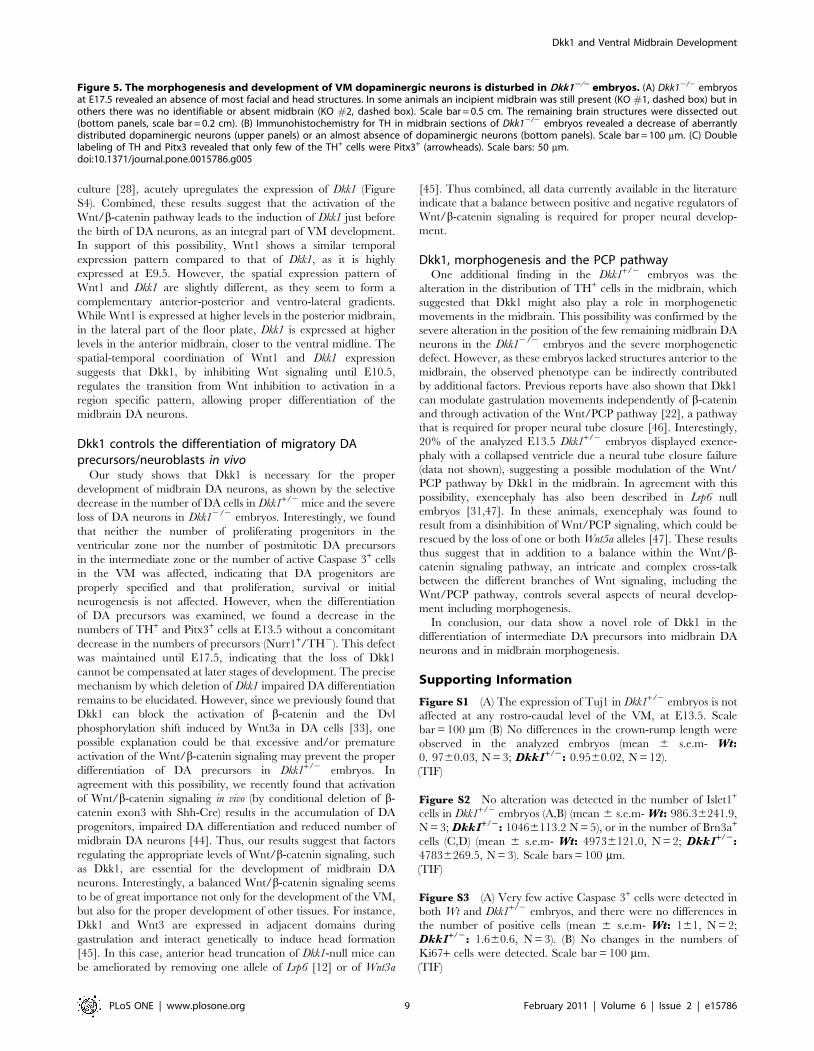
VM morphogenesis and DA neuron development isseverely disturbed in Dkk12/2 mice
In order to further characterize the function of Dkk1 in
midbrain DA neuron development we analyzed the few E17.5
Dkk12/2 mice that we obtained. As expected, these embryos
showed a strong phenotype which included a deletion of the most
anterior head structures [7]. The level of midbrain deletion varied
in the animals examined from embryos in which it was possible to
identify a rudimentary midbrain, (Figure 5A, KO#1) to embryos
with no identifiable or absent midbrain (Figure 5A, KO#2). In
these embryos, immunohistochemistry for TH revealed either a
severe decrease in the number of TH+ cells or an almost complete
absence of DA neurons (Figure 5B). In the few Dkk12/2 mice in
which TH+ cells were present, they were not found in their typical
position and were very abnormally distributed forming clusters
(Figure 5B). These findings indicated that Dkk1, directly or
Dkk1 and Ventral Midbrain Development
PLoS ONE | www.plosone.org 3 February 2011 | Volume 6 | Issue 2 | e15786
Dkk1 and Ventral Midbrain Development
PLoS ONE | www.plosone.org 4 February 2011 | Volume 6 | Issue 2 | e15786
indirectly, is required for proper VM morphogenesis and cell
distribution. Moreover, the TH+ cells found in some of the Dkk12/2
mice displayed an atypical morphology with very few or no
projections (Figure 5C). Interestingly, only a few of these TH+ cells
were found to express the midbrain specific transcription factor,
Pitx3 (Figure 5C, arrowheads). These results thus indicate that while
some midbrain tissue can still be formed in the absence of Dkk1,
Dkk1 is required for DA differentiation and midbrain morphogen-
esis.
Discussion
The development of midbrain DA neurons proceeds in a tightly
regulated fashion by several signaling pathways and transcription
factors. Understanding the mechanisms responsible for the birth,
differentiation and maintenance of these cells is of great interest to
improve the generation and yield of DA neurons for cell-
replacement therapies in Parkinson’s disease. We previously
showed that Wnt signaling is important for different aspects of
midbrain DA neuron development [28,30,31,39], and that Dkk1-
mediated inhibition of the Wnt/b-catenin pathway improves the
DA differentiation in mESC [33]. This led us to investigate
whether Dkk1 plays a role in the development of midbrain DA
neurons in vivo. Analysis of Dkk1+/2 embryos showed that Dkk1 is
required for the differentiation of DA precursors into DA neurons.
This defect was to some extent specific as Islet1+ motor neurons
and Brn3a+ red nuclei neurons were not affected. Furthermore,
analysis of the few Dkk12/2 embryos that survived until E17.5
revealed a decrease or a near absence of midbrain DA neurons
and a severe defect in midbrain morphogenesis.
Dkk1 is expressed in a very precise temporal and spatialpattern during VM development
The expression of Dkk1 was detected in the VM at E9.5 and its
expression started to decrease at E10.5, just before the onset of DA
neurogenesis. Interestingly, b-catenin expression and transcrip-
tional activity, as assessed in the TOPGAL reporter mice, has been
found to follow a similar spatial-temporal expression pattern [28].
Importantly, it has been described that Dkk1 is a direct
transcriptional target of b-catenin [40,41,42,43] as part of a
negative feedback loop. In agreement with this possibility, we
hereby report that treatment of primary E10.5 VM cultures with
Wnt3a, a known activator of Wnt/b-catenin signaling in this
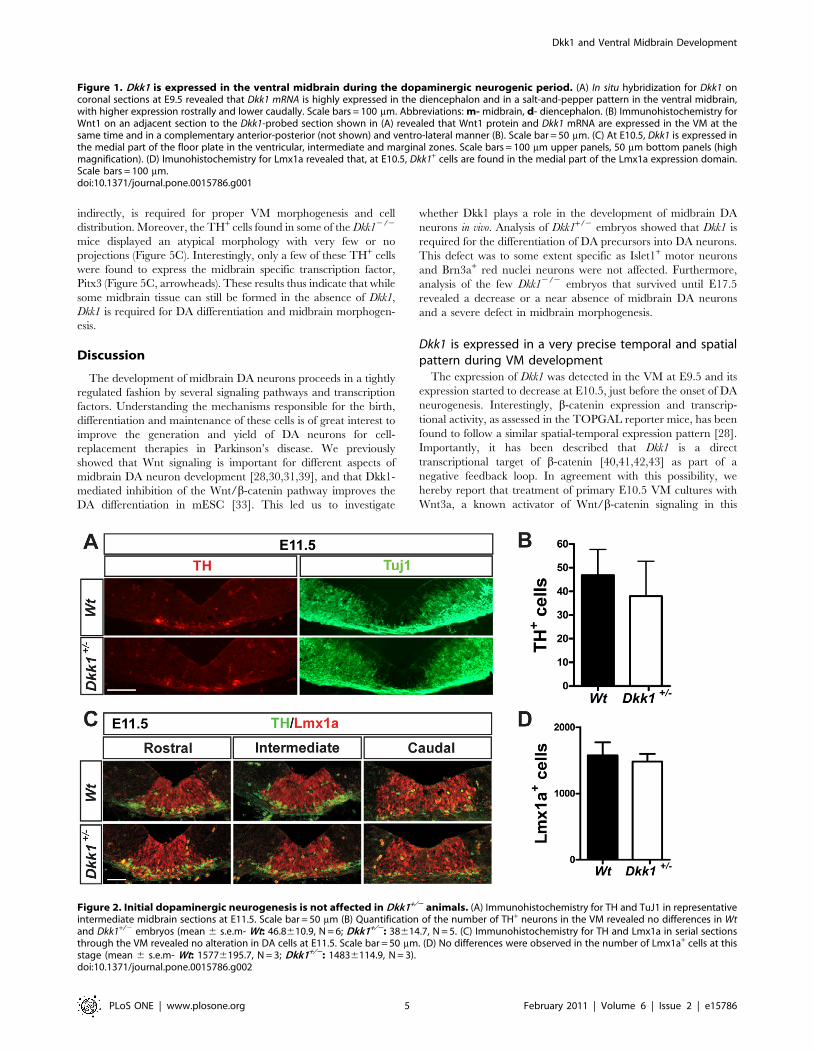
Figure 2. Initial dopaminergic neurogenesis is not affected in Dkk1+/2 animals. (A) Immunohistochemistry for TH and TuJ1 in representativeintermediate midbrain sections at E11.5. Scale bar = 50 mm (B) Quantification of the number of TH+ neurons in the VM revealed no differences in Wtand Dkk1+/2 embryos (mean 6 s.e.m- Wt: 46.8610.9, N = 6; Dkk1+/2: 38614.7, N = 5. (C) Immunohistochemistry for TH and Lmx1a in serial sectionsthrough the VM revealed no alteration in DA cells at E11.5. Scale bar = 50 mm. (D) No differences were observed in the number of Lmx1a+ cells at thisstage (mean 6 s.e.m- Wt: 15776195.7, N = 3; Dkk1+/2: 14836114.9, N = 3).doi:10.1371/journal.pone.0015786.g002
Figure 1. Dkk1 is expressed in the ventral midbrain during the dopaminergic neurogenic period. (A) In situ hybridization for Dkk1 oncoronal sections at E9.5 revealed that Dkk1 mRNA is highly expressed in the diencephalon and in a salt-and-pepper pattern in the ventral midbrain,with higher expression rostrally and lower caudally. Scale bars = 100 mm. Abbreviations: m- midbrain, d- diencephalon. (B) Immunohistochemistry forWnt1 on an adjacent section to the Dkk1-probed section shown in (A) revealed that Wnt1 protein and Dkk1 mRNA are expressed in the VM at thesame time and in a complementary anterior-posterior (not shown) and ventro-lateral manner (B). Scale bar = 50 mm. (C) At E10.5, Dkk1 is expressed inthe medial part of the floor plate in the ventricular, intermediate and marginal zones. Scale bars = 100 mm upper panels, 50 mm bottom panels (highmagnification). (D) Imunohistochemistry for Lmx1a revealed that, at E10.5, Dkk1+ cells are found in the medial part of the Lmx1a expression domain.Scale bars = 100 mm.doi:10.1371/journal.pone.0015786.g001
Dkk1 and Ventral Midbrain Development
PLoS ONE | www.plosone.org 5 February 2011 | Volume 6 | Issue 2 | e15786
Dkk1 and Ventral Midbrain Development
PLoS ONE | www.plosone.org 6 February 2011 | Volume 6 | Issue 2 | e15786
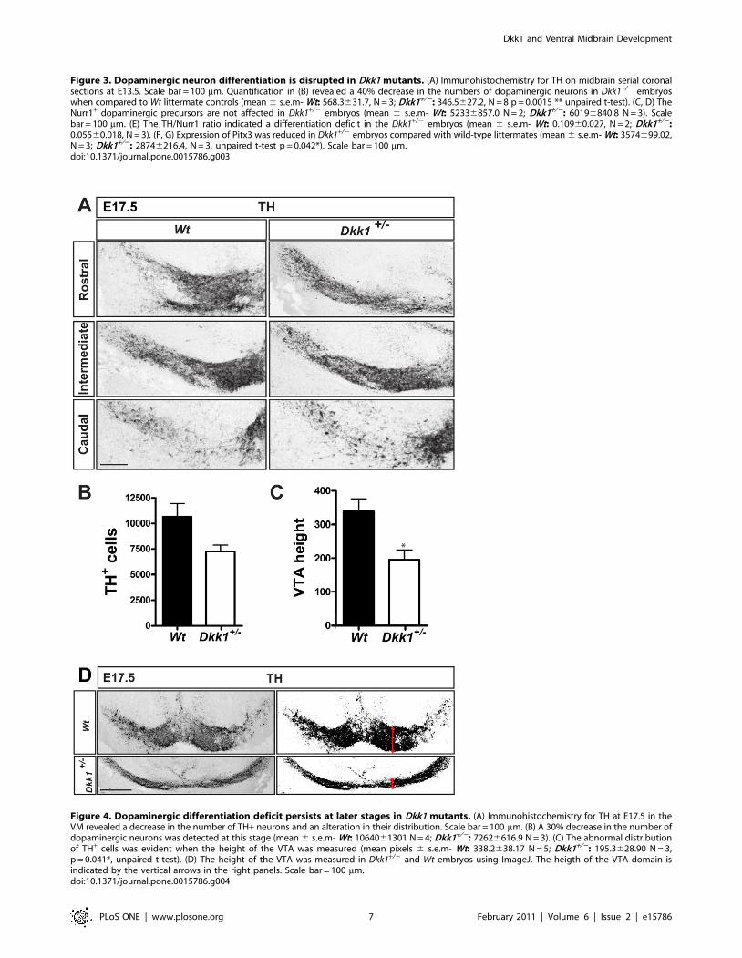
Figure 4. Dopaminergic differentiation deficit persists at later stages in Dkk1 mutants. (A) Immunohistochemistry for TH at E17.5 in theVM revealed a decrease in the number of TH+ neurons and an alteration in their distribution. Scale bar = 100 mm. (B) A 30% decrease in the number ofdopaminergic neurons was detected at this stage (mean 6 s.e.m- Wt: 1064061301 N = 4; Dkk1+/2: 72626616.9 N = 3). (C) The abnormal distributionof TH+ cells was evident when the height of the VTA was measured (mean pixels 6 s.e.m- Wt: 338.2638.17 N = 5; Dkk1+/2: 195.3628.90 N = 3,p = 0.041*, unpaired t-test). (D) The height of the VTA was measured in Dkk1+/2 and Wt embryos using ImageJ. The heigth of the VTA domain isindicated by the vertical arrows in the right panels. Scale bar = 100 mm.doi:10.1371/journal.pone.0015786.g004
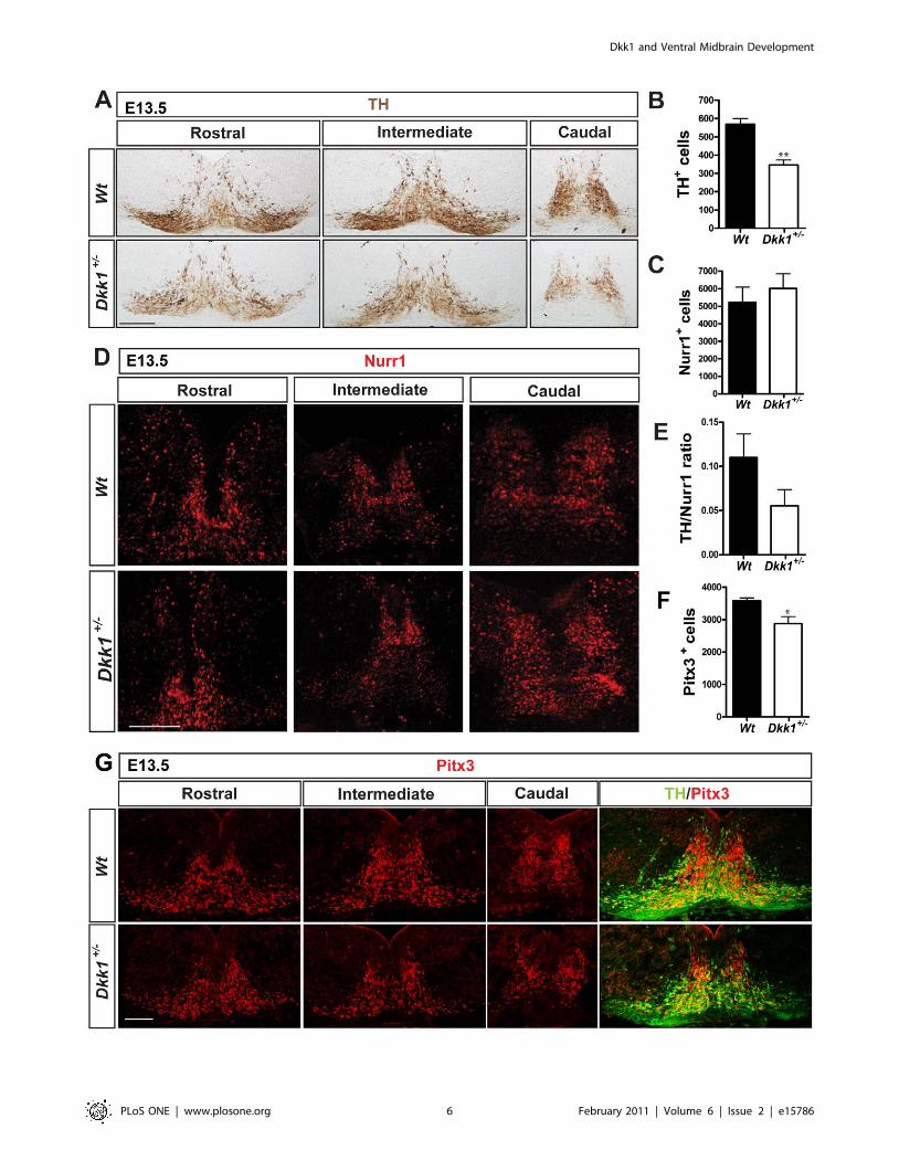
Figure 3. Dopaminergic neuron differentiation is disrupted in Dkk1 mutants. (A) Immunohistochemistry for TH on midbrain serial coronalsections at E13.5. Scale bar = 100 mm. Quantification in (B) revealed a 40% decrease in the numbers of dopaminergic neurons in Dkk1+/2 embryoswhen compared to Wt littermate controls (mean 6 s.e.m- Wt: 568.3631.7, N = 3; Dkk1+/2: 346.5627.2, N = 8 p = 0.0015 ** unpaired t-test). (C, D) TheNurr1+ dopaminergic precursors are not affected in Dkk1+/2 embryos (mean 6 s.e.m- Wt: 52336857.0 N = 2; Dkk1+/2: 60196840.8 N = 3). Scalebar = 100 mm. (E) The TH/Nurr1 ratio indicated a differentiation deficit in the Dkk1+/2 embryos (mean 6 s.e.m- Wt: 0.10960.027, N = 2; Dkk1+/2:0.05560.018, N = 3). (F, G) Expression of Pitx3 was reduced in Dkk1+/2 embryos compared with wild-type littermates (mean 6 s.e.m- Wt: 3574699.02,N = 3; Dkk1+/2: 28746216.4, N = 3, unpaired t-test p = 0.042*). Scale bar = 100 mm.doi:10.1371/journal.pone.0015786.g003
Dkk1 and Ventral Midbrain Development
PLoS ONE | www.plosone.org 7 February 2011 | Volume 6 | Issue 2 | e15786
Dkk1 and Ventral Midbrain Development
PLoS ONE | www.plosone.org 8 February 2011 | Volume 6 | Issue 2 | e15786
culture [28], acutely upregulates the expression of Dkk1 (Figure
S4). Combined, these results suggest that the activation of the
Wnt/b-catenin pathway leads to the induction of Dkk1 just before
the birth of DA neurons, as an integral part of VM development.
In support of this possibility, Wnt1 shows a similar temporal
expression pattern compared to that of Dkk1, as it is highly
expressed at E9.5. However, the spatial expression pattern of
Wnt1 and Dkk1 are slightly different, as they seem to form a
complementary anterior-posterior and ventro-lateral gradients.
While Wnt1 is expressed at higher levels in the posterior midbrain,
in the lateral part of the floor plate, Dkk1 is expressed at higher
levels in the anterior midbrain, closer to the ventral midline. The
spatial-temporal coordination of Wnt1 and Dkk1 expression
suggests that Dkk1, by inhibiting Wnt signaling until E10.5,
regulates the transition from Wnt inhibition to activation in a
region specific pattern, allowing proper differentiation of the
midbrain DA neurons.
Dkk1 controls the differentiation of migratory DAprecursors/neuroblasts in vivo
Our study shows that Dkk1 is necessary for the proper
development of midbrain DA neurons, as shown by the selective
decrease in the number of DA cells in Dkk1+/2 mice and the severe
loss of DA neurons in Dkk12/2 embryos. Interestingly, we found
that neither the number of proliferating progenitors in the
ventricular zone nor the number of postmitotic DA precursors
in the intermediate zone or the number of active Caspase 3+ cells
in the VM was affected, indicating that DA progenitors are
properly specified and that proliferation, survival or initial
neurogenesis is not affected. However, when the differentiation
of DA precursors was examined, we found a decrease in the
numbers of TH+ and Pitx3+ cells at E13.5 without a concomitant
decrease in the numbers of precursors (Nurr1+/TH2). This defect
was maintained until E17.5, indicating that the loss of Dkk1
cannot be compensated at later stages of development. The precise
mechanism by which deletion of Dkk1 impaired DA differentiation
remains to be elucidated. However, since we previously found that
Dkk1 can block the activation of b-catenin and the Dvl
phosphorylation shift induced by Wnt3a in DA cells [33], one
possible explanation could be that excessive and/or premature
activation of the Wnt/b-catenin signaling may prevent the proper
differentiation of DA precursors in Dkk1+/2 embryos. In
agreement with this possibility, we recently found that activation
of Wnt/b-catenin signaling in vivo (by conditional deletion of b-
catenin exon3 with Shh-Cre) results in the accumulation of DA
progenitors, impaired DA differentiation and reduced number of
midbrain DA neurons [44]. Thus, our results suggest that factors
regulating the appropriate levels of Wnt/b-catenin signaling, such
as Dkk1, are essential for the development of midbrain DA
neurons. Interestingly, a balanced Wnt/b-catenin signaling seems
to be of great importance not only for the development of the VM,
but also for the proper development of other tissues. For instance,
Dkk1 and Wnt3 are expressed in adjacent domains during
gastrulation and interact genetically to induce head formation
[45]. In this case, anterior head truncation of Dkk1-null mice can
be ameliorated by removing one allele of Lrp6 [12] or of Wnt3a
[45]. Thus combined, all data currently available in the literature
indicate that a balance between positive and negative regulators of
Wnt/b-catenin signaling is required for proper neural develop-
ment.
Dkk1, morphogenesis and the PCP pathwayOne additional finding in the Dkk1+/2 embryos was the
alteration in the distribution of TH+ cells in the midbrain, which
suggested that Dkk1 might also play a role in morphogenetic
movements in the midbrain. This possibility was confirmed by the
severe alteration in the position of the few remaining midbrain DA
neurons in the Dkk12/2 embryos and the severe morphogenetic
defect. However, as these embryos lacked structures anterior to the
midbrain, the observed phenotype can be indirectly contributed
by additional factors. Previous reports have also shown that Dkk1
can modulate gastrulation movements independently of b-catenin
and through activation of the Wnt/PCP pathway [22], a pathway
that is required for proper neural tube closure [46]. Interestingly,
20% of the analyzed E13.5 Dkk1+/2 embryos displayed exence-
phaly with a collapsed ventricle due a neural tube closure failure
(data not shown), suggesting a possible modulation of the Wnt/
PCP pathway by Dkk1 in the midbrain. In agreement with this
possibility, exencephaly has also been described in Lrp6 null
embryos [31,47]. In these animals, exencephaly was found to
result from a disinhibition of Wnt/PCP signaling, which could be
rescued by the loss of one or both Wnt5a alleles [47]. These results
thus suggest that in addition to a balance within the Wnt/b-
catenin signaling pathway, an intricate and complex cross-talk
between the different branches of Wnt signaling, including the
Wnt/PCP pathway, controls several aspects of neural develop-
ment including morphogenesis.
In conclusion, our data show a novel role of Dkk1 in the
differentiation of intermediate DA precursors into midbrain DA
neurons and in midbrain morphogenesis.
Supporting Information
Figure S1 (A) The expression of Tuj1 in Dkk1+/2 embryos is not
affected at any rostro-caudal level of the VM, at E13.5. Scale
bar = 100 mm (B) No differences in the crown-rump length were
observed in the analyzed embryos (mean 6 s.e.m- Wt:0. 9760.03, N = 3; Dkk1+/2: 0.9560.02, N = 12).
(TIF)
Figure S2 No alteration was detected in the number of Islet1+
cells in Dkk1+/2 embryos (A,B) (mean 6 s.e.m- Wt: 986.36241.9,
N = 3; Dkk1+/2: 10466113.2 N = 5), or in the number of Brn3a+
cells (C,D) (mean 6 s.e.m- Wt: 49736121.0, N = 2; Dkk1+/2:47836269.5, N = 3). Scale bars = 100 mm.
(TIF)
Figure S3 (A) Very few active Caspase 3+ cells were detected in
both Wt and Dkk1+/2 embryos, and there were no differences in
the number of positive cells (mean 6 s.e.m- Wt: 161, N = 2;
Dkk1+/2: 1.660.6, N = 3). (B) No changes in the numbers of
Ki67+ cells were detected. Scale bar = 100 mm.
(TIF)
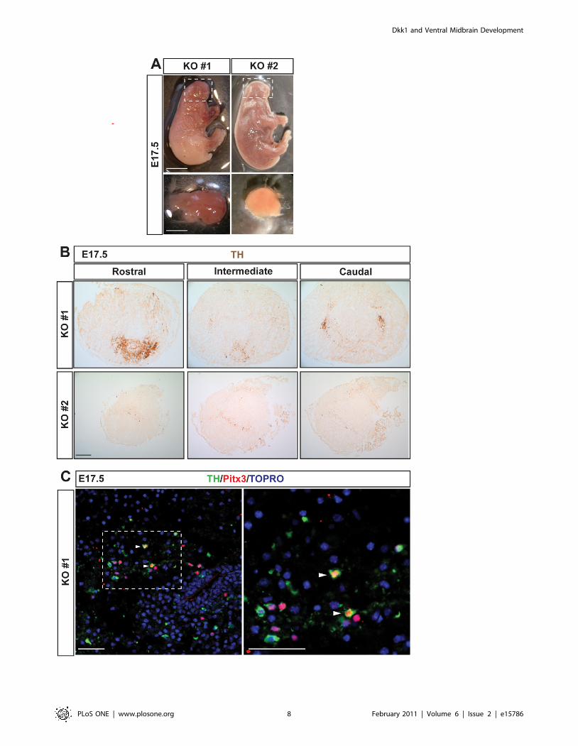
Figure 5. The morphogenesis and development of VM dopaminergic neurons is disturbed in Dkk12/2 embryos. (A) Dkk12/2 embryosat E17.5 revealed an absence of most facial and head structures. In some animals an incipient midbrain was still present (KO #1, dashed box) but inothers there was no identifiable or absent midbrain (KO #2, dashed box). Scale bar = 0.5 cm. The remaining brain structures were dissected out(bottom panels, scale bar = 0.2 cm). (B) Immunohistochemistry for TH in midbrain sections of Dkk12/2 embryos revealed a decrease of aberrantlydistributed dopaminergic neurons (upper panels) or an almost absence of dopaminergic neurons (bottom panels). Scale bar = 100 mm. (C) Doublelabeling of TH and Pitx3 revealed that only few of the TH+ cells were Pitx3+ (arrowheads). Scale bars: 50 mm.doi:10.1371/journal.pone.0015786.g005
Dkk1 and Ventral Midbrain Development
PLoS ONE | www.plosone.org 9 February 2011 | Volume 6 | Issue 2 | e15786
Figure S4 Dkk1 expression was upregulated in mouse E10.5 VM
primary cultures treated with Wnt3a for 6 hours (mean 6 s.e.m-
BSA: 0.32260.008; Wnt3a: 0.66760.008 N = 3, p = 0.002
** paired t-test).
(TIF)
Acknowledgments
We would like to thank Johnny Soderlund and Lottie Jansson-Sjostrand for
technical assistance, Dr. Emma R. Andersson for the critical reading of the
manuscript and all the members of the Arenas lab for fruitful discussions.
Author Contributions
Conceived and designed the experiments: DR CN AMV EA. Performed
the experiments: DR KE DG ST NSC. Analyzed the data: DR EA.
Contributed reagents/materials/analysis tools: KE DG NSC AMV CN.
Wrote the paper: DR EA. Fundraising: EA. Manuscript writing: DR EA.
Final approval of manuscript: DR KE DG ST NSC AMV CN EA.
References
1. Glinka A, Wu W, Delius H, Monaghan AP, Blumenstock C, et al. (1998)
Dickkopf-1 is a member of a new family of secreted proteins and functions in
head induction. Nature 391: 357–362.
2. Krupnik VE, Sharp JD, Jiang C, Robison K, Chickering TW, et al. (1999)
Functional and structural diversity of the human Dickkopf gene family. Gene
238: 301–313.
3. Bafico A, Liu GZ, Yaniv A, Gazit A, Aaronson SA (2001) Novel mechanism of
Wnt signalling inhibition mediated by Dickkopf-1 interaction with Lrp6/Arrow.
Nature Cell Biology 3: 683–686.
4. Mao BY, Wu W, Li Y, Hoppe D, Stannek P, et al. (2001) LDL-receptor-related
protein 6 is a receptor for Dickkopf proteins. Nature 411: 321–325.
5. Semenov MV, Tamai K, Brott BK, Kuhl M, Sokol S, et al. (2001) Head inducer
Dickkopf-1 is a ligand for Wnt coreceptor Lrp6. Current Biology 11: 951–961.
6. Mao BY, Wu W, Davidson G, Marhold J, Li MF, et al. (2002) Kremen proteins
are Dickkopf receptors that regulate Wnt/beta-catenin signalling. Nature 417:
664–667.
7. Mukhopadhyay M, Shtrom S, Rodriguez-Esteban C, Chen L, Tsukui T, et al.
(2001) Dickkopf1 is required for embryonic head induction and limb
morphogenesis in the mouse. Developmental Cell 1: 423–434.
8. Kazanskaya O, Glinka A, Niehrs C (2000) The role of Xenopus dickkopf1 in
prechordal plate specification and neural patterning. Development 127:
4981–4992.
9. Barrantes ID, Davidson G, Grone HJ, Westphal H, Niehrs C (2003) Dkk1 and
noggin cooperate in mammalian head induction. Genes & Development 17:
2239–2244.
10. Adamska M, MacDonald BT, Sarmast ZH, Oliver ER, Meisler MH (2004) En1
and Wnt7a interact with Dkk1 during limb development in the mouse.
Developmental Biology 272: 134–144.
11. Grotewold L, Theil T, Ruther U (1999) Expression pattern of Dkk-1 during
mouse limb development. Mechanisms of Development 89: 151–153.
12. MacDonald BT, Adamska M, Meisler MH (2004) Hypomorphic expression of
Dkk1 in the doubleridge mouse: dose dependence and compensatory
interactions with Lrp6. Development 131: 2543–2552.
13. Li J, Sarosi I, Cattley RC, Pretorius J, Asuncion F, et al. (2006) Dkk1-mediated
inhibition of Wnt signaling in bone results in osteopenia. Bone 39: 754–766.
14. Morvan F, Boulukos K, Clement-Lacroix P, Roman SR, Suc-Royer I, et al.
(2006) Deletion of a single allele of the Dkk1 gene leads to an increase in bone
formation and bone mass. Journal of Bone and Mineral Research 21: 934–945.
15. Gregory CA, Singh H, Perry AS, Prockop DJ (2003) The Wnt signaling inhibitor
dickkopf-1 is required for reentry into the cell cycle of human adult stem cells
from bone marrow. Journal of Biological Chemistry 278: 28067–28078.
16. Wang J, Shou J, Chen XB (2000) Dickkopf-1, an inhibitor of the Wnt signaling
pathway, is induced by p53. Oncogene 19: 1843–1848.
17. Tian E, Zhan FH, Walker R, Rasmussen E, Ma YP, et al. (2003) The role of the
Wnt-signaling antagonist DKK1 in the development of osteolytic lesions in
multiple myeloma. New England Journal of Medicine 349: 2483–2494.
18. Caricasole A, Copani A, Caraci F, Aronica E, Rozemuller AJ, et al. (2004)
Induction of Dickkopf-1, a negative modulator of the Wnt pathway, is associated
with neuronal degeneration in Alzheimer’s brain. Journal of Neuroscience 24:
6021–6027.
19. Kong XB, Zhang C (2009) Dickkopf (Dkk) 1 promotes the differentiation of
mouse embryonic stem cells toward neuroectoderm. In Vitro Cellular &
Developmental Biology-Animal 45: 185–193.
20. Verani R, Cappuccio I, Spinsanti P, Gradini R, Caruso A, et al. (2007)
Expression of the Wnt inhibitor Dickkopf-1 is required for the induction of
neural markers in mouse embryonic stem cells differentiating in response to
retinoic acid. Journal of Neurochemistry 100: 242–250.
21. Watanabe K, Kamiya D, Nishiyama A, Katayama T, Nozaki S, et al. (2005)
Directed differentiation of telencephalic precursors from embryonic stem cells.
Nature Neuroscience 8: 288–296.
22. Caneparo L, Huang YL, Staudt N, Tada M, Ahrendt R, et al. (2007) Dickkopf-1
regulates gastrulation movements by coordinated modulation of Wnt/beta
catenin and Wnt/PCP activities, through interaction with the Dally-like
homolog Knypek. Genes & Development 21: 465–480.
23. Ciani L, Salinas PC (2005) WNTs in the vertebrate nervous system: Frompatterning to neuronal connectivity. Nature Reviews Neuroscience 6:
351–U317.
24. Gaulden J, Reiter JF (2008) Neur-ons and neur-offs: regulators of neural
induction in vertebrate embryos and embryonic stem cells. Human MolecularGenetics 17: R60–R66.
25. McMahon AP, Bradley A (1990) The WNT-1 (INT-1) protooncogene isrequired for development of a large region of the mouse brain. Cell 62:
1073–1085.
26. Thomas KR, Capecchi MR (1990) Targeted disruption of the murine INT-1protooncogene resulting in severe abnormalities in midbrain and cerebellar
development. Nature 346: 847–850.
27. Joksimovic M, Yun BA, Kittappa R, Anderegg AM, Wchang W, et al. (2009)
Wnt antagonism of Shh facilitates midbrain floor plate neurogenesis. NatureNeuroscience 12: 125–131.
28. Castelo-Branco GA, Wagner J, Rodriguez FJ, Kele J, Sousa K, et al. (2003)Differential regulation of midbrain dopaminergic neuron development by Wnt-
1, Wnt-3a, and Wnt-5a. Proceedings of the National Academy of Sciences of the
United States of America 100: 12747–12752.
29. Panhuysen M, Weisenhorn DMV, Blanquet V, Brodski C, Heinzmann U, et al.
(2004) Effects of Wnt1 signaling on proliferation in the developing mid-/hindbrain region. Molecular and Cellular Neuroscience 26: 101–111.
30. Prakash N, Brodski C, Naserke T, Puelles E, Gogoi R, et al. (2006) A Wnt1-regulated genetic network controls the identity and fate of midbrain-
dopaminergic progenitors in vivo. Development 133: 89–98.
31. Castelo-Branco G, Andersson ER, Minina E, Sousa KM, Ribeiro D, et al. (2010)
Delayed Dopaminergic Neuron Differentiation in Lrp6 Mutant Mice.
Developmental Dynamics 239: 211–221.
32. Tang MZ, Miyamoto Y, Huang EJ (2009) Multiple roles of beta-catenin in
controlling the neurogenic niche for midbrain dopamine neurons. Development136: 2027–2038.
33. Cajanek L, Ribeiro D, Liste I, Parish CL, Bryja V, et al. (2009) Wnt/beta-Catenin Signaling Blockade Promotes Neuronal Induction and Dopaminergic
Differentiation in Embryonic Stem Cells. Stem Cells 27: 2917–2927.
34. Conlon RA, Herrmann BG (1993) Detection of messenger-RNA by in situ
hybridization to postimplantation embryo whole mounts. Guide to Techniquesin Mouse Development. San Diego: Academic Press Inc. pp 373–383.
35. Rawal N, Castelo-Branco G, Sousa KM, Kele J, Kobayashi K, et al. (2006)
Dynamic temporal and cell type-specific expression of Wnt signalingcomponents in the developing midbrain. Experimental Cell Research 312:
1626–1636.
36. Lieven O, Knobloch J, Ruther U (2010) The regulation of Dkk1 expression
during embryonic development. Dev Biol.
37. Monaghan AP, Kioschis P, Wu W, Zuniga A, Bock D, et al. (1999) Dickkopf
genes are co-ordinately expressed in mesodermal lineages. Mechanisms ofDevelopment 87: 45–56.
38. Andersson E, Tryggvason U, Deng QL, Friling S, Alekseenko Z, et al. (2006)Identification of intrinsic determinants of midbrain dopamine neurons. Cell 124:
393–405.
39. Andersson ER, Prakash N, Cajanek L, Minina E, Bryja V, et al. (2008) Wnt5aRegulates Ventral Midbrain Morphogenesis and the Development of A9–A10
Dopaminergic Cells In Vivo. Plos One 3.
40. Chamorro MN, Schwartz DR, Vonica A, Brivanlou AH, Cho KR, et al. (2005)
FGF-20 and DKK1 are transcriptional targets of beta-catenin and FGF-20 isimplicated in cancer and development. Embo Journal 24: 73–84.
41. Gonzalez-Sancho JM, Aguilera O, Garcia JM, Pendas-Franco N, Pena C, et al.(2005) The Wnt antagonist DICKKOPF-1 gene is a downstream target of beta-
catenin/TCF and is downregulated in human colon cancer. Oncogene 24:
1098–1103.
42. Niida A, Hiroko T, Kasai M, Furukawa Y, Nakamura Y, et al. (2004) DKK1, a
negative regulator of Wnt signaling, is a target of the beta-catenin/TCFpathway. Oncogene 23: 8520–8526.
43. Shinya M, Eschbach C, Clark M, Lehrach H, Furutani-Seiki M (2000) ZebrafishDkk1, induced by the pre-MBT Wnt signaling, is secreted from the prechordal
plate and patterns the anterior neural plate. Mechanisms of Development 98:3–17.
Dkk1 and Ventral Midbrain Development
PLoS ONE | www.plosone.org 10 February 2011 | Volume 6 | Issue 2 | e15786

44. Tang M, Villaescusa J, Luo S, Guitarte C, Lei S, et al. (2010) Interactions of
Wnt/beta-catenin signaling and sonic hedgehog regulate the neurogenesis ofventral midbrain dopamine neurons. J Neurosci 30: 9280–9291.
45. Lewis SL, Khoo PL, De Young RA, Steiner K, Wilcock C, et al. (2008) Dkk1
and Wnt3 interact to control head morphogenesis in the mouse. Development135: 1791–1801.
46. Wallingford JB, Harland RM (2002) Neural tube closure requires Dishevelled-
dependent convergent extension of the midline. Development 129: 5815–5825.
47. Bryja V, Andersson ER, Schambony A, Esner M, Bryjova L, et al. (2009) The
Extracellular Domain of Lrp5/6 Inhibits Noncanonical Wnt Signaling In Vivo.
Molecular Biology of the Cell 20: 924–936.
Dkk1 and Ventral Midbrain Development
PLoS ONE | www.plosone.org 11 February 2011 | Volume 6 | Issue 2 | e15786


