Embed Size (px)
Citation preview
RESEARCH Open Access
Disagreement in cardiac outputmeasurements between fourth-generationFloTrac and critical care ultrasonography inpatients with circulatory shock: aprospective observational studyThomas Kaufmann1* , Ramon P. Clement1, Bart Hiemstra1,2 , Jaap Jan Vos1 , Thomas W. L. Scheeren1 ,Frederik Keus2, Iwan C. C. van der Horst2 and SICS Study Group
Abstract
Background: Cardiac output measurements may inform diagnosis and provide guidance of therapeuticinterventions in patients with hemodynamic instability. The FloTrac™ algorithm uses uncalibrated arterial pressurewaveform analysis to estimate cardiac output. Recently, a new version of the algorithm has been developed. Theaim was to assess the agreement between FloTrac™ and routinely performed cardiac output measurementsobtained by critical care ultrasonography in patients with circulatory shock.
Methods: A prospective observational study was performed in a tertiary hospital from June 2016 to January 2017.Adult critically ill patients with circulatory shock were eligible for inclusion. Cardiac output was measuredsimultaneously using FloTrac™ with a fourth-generation algorithm (COAP) and critical care ultrasonography (COCCUS).The strength of linear correlation of both methods was determined by the Pearson coefficient. Bland-Altman plotand four-quadrant plot were used to track agreement and trending ability.
Result: Eighty-nine paired cardiac output measurements were performed in 17 patients during their first 24 h ofadmittance. COAP and COCCUS had strong positive linear correlation (r2 = 0.60, p < 0.001). Bias of COAP and COCCUS
was 0.2 L min−1 (95% CI − 0.2 to 0.6) with limits of agreement of − 3.6 L min−1 (95% CI − 4.3 to − 2.9) to 4.0 L min−1
(95% CI 3.3 to 4.7). The percentage error was 65.6% (95% CI 53.2 to 77.3). Concordance rate was 64.4%.
Conclusions: In critically ill patients with circulatory shock, there was disagreement and clinically unacceptabletrending ability between values of cardiac output obtained by uncalibrated arterial pressure waveform analysis andcritical care ultrasonography.
Trial registration: Clinicaltrials.gov, NCT02912624, registered on September 23, 2016
Keywords: Cardiac output, Critical care ultrasonography, Intensive care, Critically ill, Shock, Monitoring, Pulsecontour analysis
© The Author(s). 2019 Open Access This article is distributed under the terms of the Creative Commons Attribution 4.0International License (http://creativecommons.org/licenses/by/4.0/), which permits unrestricted use, distribution, andreproduction in any medium, provided you give appropriate credit to the original author(s) and the source, provide a link tothe Creative Commons license, and indicate if changes were made. The Creative Commons Public Domain Dedication waiver(http://creativecommons.org/publicdomain/zero/1.0/) applies to the data made available in this article, unless otherwise stated.
* Correspondence: [email protected] of Anesthesiology, University Medical Center Groningen,University of Groningen, P.O. Box 30.001, 9700 RB Groningen, TheNetherlandsFull list of author information is available at the end of the article
Kaufmann et al. Journal of Intensive Care (2019) 7:21 https://doi.org/10.1186/s40560-019-0373-5

BackgroundCritically ill patients with circulatory shock have in-creased risks of multi-organ failure, long-term morbidity,and mortality [1]. Advanced hemodynamic monitoringin these patients may inform diagnosis and simultan-eously guide management by providing insight into car-diac function, cardiac preload, and afterload [2]. Severalmethods for measuring cardiac output (CO) exist, vary-ing from invasive (e.g. thermodilution by pulmonaryartery catheter (PAC)) to minimally invasive (e.g. pulsecontour analysis by FloTrac™ (Edwards Lifesciences,Irvine, USA)) or even non-invasive (e.g. transthoracicDoppler ultrasound by critical care ultrasonography(CCUS)). These methods all have their own merits, dis-advantages and requirements [3].One type of pulse contour analysis is the uncalibrated
arterial pressure waveform analysis method to estimateCO (APCO). Reliability of APCO is questioned in pa-tients with hemodynamic instability, and this occurs fre-quently in patients admitted to the ICU [4]. Therefore,CO measurements obtained by APCO should be inter-preted with caution in critically ill patients with circula-tory shock [5, 6].The FloTrac™ system using the APCO method calcu-
lates CO based on the principle that aortic pulse pres-sure is proportional to stroke volume (SV) and inverselyrelated to aortic compliance using a proprietary algo-rithm. FloTrac™ has been widely studied in more than70 validation studies as of yet, mostly showing adequateperformance in normo- and hypodynamic conditions,but not in patients with large changes in vascular tonewhich typically occur in patients with circulatory shock[7]. However, these studies vary by the statisticalmethods and versions of the algorithm used. Recently,the fourth-generation algorithm was developed to im-prove performance.Evaluation of the trending ability rather than the
agreement of absolute values of CO monitoring devicesis increasingly considered in validation studies for as-sessment of potential clinical usefulness [8]. In additionto one single CO measurement for diagnosing circula-tory shock, repeated measurements of CO informing thetrending ability could be informative for monitoring andguidance of supportive treatments of patients with circu-latory shock.The aim of our study was to compare both agreements
and trending ability for APCO measurements of CO(COAP) with CO routinely measured by CCUS (COCCUS)in critically ill patients with circulatory shock. CCUSwas chosen as the reference standard since it is the pre-ferred method for diagnosis, but not for monitoring, ofcirculatory shock in critically ill patients and is widelyavailable [2, 9]. Importantly, it should be noted thatCCUS is not a gold standard reference technique for
method comparison studies aiming to evaluate the valid-ity of CO monitors [10].
MethodsThis study was a substudy of the Simple IntensiveCare Studies-I (SICS-I), which was a single-centre,prospective, observational cohort study in which allconsecutive acutely admitted adult patients expectedto stay beyond 24 h were included (NCT02912624)[16, 17]. The STROBE guidelines for reporting obser-vational studies were used (Additional file 1) [11].The checklist for CO monitor method comparisonstudies was used [10]. The local institutional reviewboard (Medisch Ethische Toetsingscommissie, Univer-sity Medical Center Groningen) approved the study(M15.168207 and M16.193856). Written informedconsent was obtained from all patients.
Selection criteriaIn this substudy, all consecutive acutely admitted adult pa-tients with suspected circulatory shock and expected tostay beyond 48 h were included from June 2016 to January2017. The circulatory shock was defined as the require-ment of any dose of vasopressor to maintain a mean arter-ial pressure (MAP) of 60mmHg or if the MAP remainedbelow 70mmHg despite fluid resuscitation (defined by atleast 1000mL of crystalloids). In addition, at least oneother sign of organ or tissue hypoperfusion had to bepresent: altered state of mind (Alert-Voice-Pain-Unre-sponsive scale) [12], mottled skin (Mottling score ≥ 1[13]), decreased urine output (≤ 0.3mL kg−1 h−1) or in-creased serum lactate level (≥ 2mmol L−1). Exclusion cri-teria were inability to obtain sufficient quality CCUSimages; no arterial line; atrial fibrillation; and aortic valveor mitral valve diseases known to impair the arterial wave-form. We included this group of patients because COmeasurements are indicated to identify the type of shock,select necessary therapeutic interventions and evaluate pa-tient’s response to therapy [2].
ObjectivesThe primary objective was to evaluate COAP measure-ments in terms of the agreement and trending abilityagainst COCCUS as reference technique in patients withcirculatory shock.
Definitions and biasPatient characteristics including clinical, hemodynamicand laboratory variables as well as Acute Physiology AndChronic Health Evaluation (APACHE) IV and SimplifiedAcute Physiology Score (SAPS) II values were recorded
Kaufmann et al. Journal of Intensive Care (2019) 7:21 Page 2 of 8
[14, 15]. Measurements were performed following proto-colized definitions and procedures [16, 17].In short, COCCUS was measured by transthoracic
echocardiography using the Vivid-S6 system (GeneralElectric, Horton, Norway) with cardiac probe M3S orM4S, and with default cardiac imaging setting. The para-sternal long axis was used to measure the left ventricularoutflow tract diameter. In the apical five-chamber view,a pulse wave Doppler signal in the left ventricular out-flow tract was used to measure the velocity time integral.COCCUS was calculated using an established formula[18]. CCUS was performed after ICU admission within6 h and repeated once every 24 h after admission pro-vided there was no interference with clinical care. Re-searchers were trained in performing CCUS byexperienced cardiologist-intensivists.The FloTrac™ sensor was connected to an indwelling
radial artery catheter and an EV1000™ monitor (version4.00; Edwards Lifesciences, Irvine, USA), which continu-ously displayed COAP values. The value of COAP dis-played on the EV1000™ monitor was registeredsimultaneously (i.e. ‘beat-to-beat’) with each COCCUS
measurement.All measurements, including CCUS findings, were
kept blind for the caregivers. Quality of CCUS imagesand COCCUS measurements were validated by an
independent specialized core laboratory (GroningenImage Core Lab) blinded for the COAP measurements
Statistical analysisNo formal sample size calculation was performed dueto lack of data on COAP variation in patients withcirculatory shock. Therefore, this study has an ex-ploratory nature.Data were presented as means with standard devia-
tions or medians with interquartile ranges depending ondistributions. Normality of data was checked using theShapiro-Wilk test. Dichotomous and categorical datawere presented in proportions.Correlations were assessed by scatter plot, and the
strength of linear correlation was determined by calcu-lating a Pearson (r) coefficient. Bland-Altman analyses ofrepeated measurements in each patient were performedand means (bias) and SD of the differences, 95% limitsof agreement (LOA) (=mean difference ± 1.96 × SD ofthe difference) as well as the percentage error of COAP
versus COCCUS were calculated [19, 20]. To evaluate thetrending ability of COAP versus COCCUS a four-quadrantplot was used and the concordance rate was calculatedusing an exclusion zone of 0.5 Lmin−1 [21]. For statis-tical analysis, we used STATA version 15.0 (StataCorp,College Station, USA).
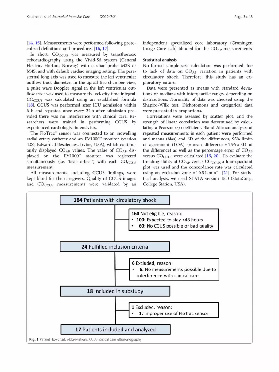
Fig. 1 Patient flowchart. Abbreviations: CCUS, critical care ultrasonography
Kaufmann et al. Journal of Intensive Care (2019) 7:21 Page 3 of 8
ResultsParticipantsDuring the study period, 184 patients were diagnosedwith circulatory shock, but only 24 patients appeared eli-gible for this study. One hundred patients who had cir-culatory shock were not included as they were expectedto stay for less than 48 h, and 60 patients with circula-tory shock were not included because CCUS was notpossible or image quality was insufficient to performmeasurements. Six patients had to be excluded becausestudy procedures interfered with clinical care, leaving 18patients to be included. One patient was excluded after-wards for invalid COAP measurements due to improperuse of a FloTrac™ sensor. Thus, 17 patients were in-cluded in the final analyses (Fig. 1).
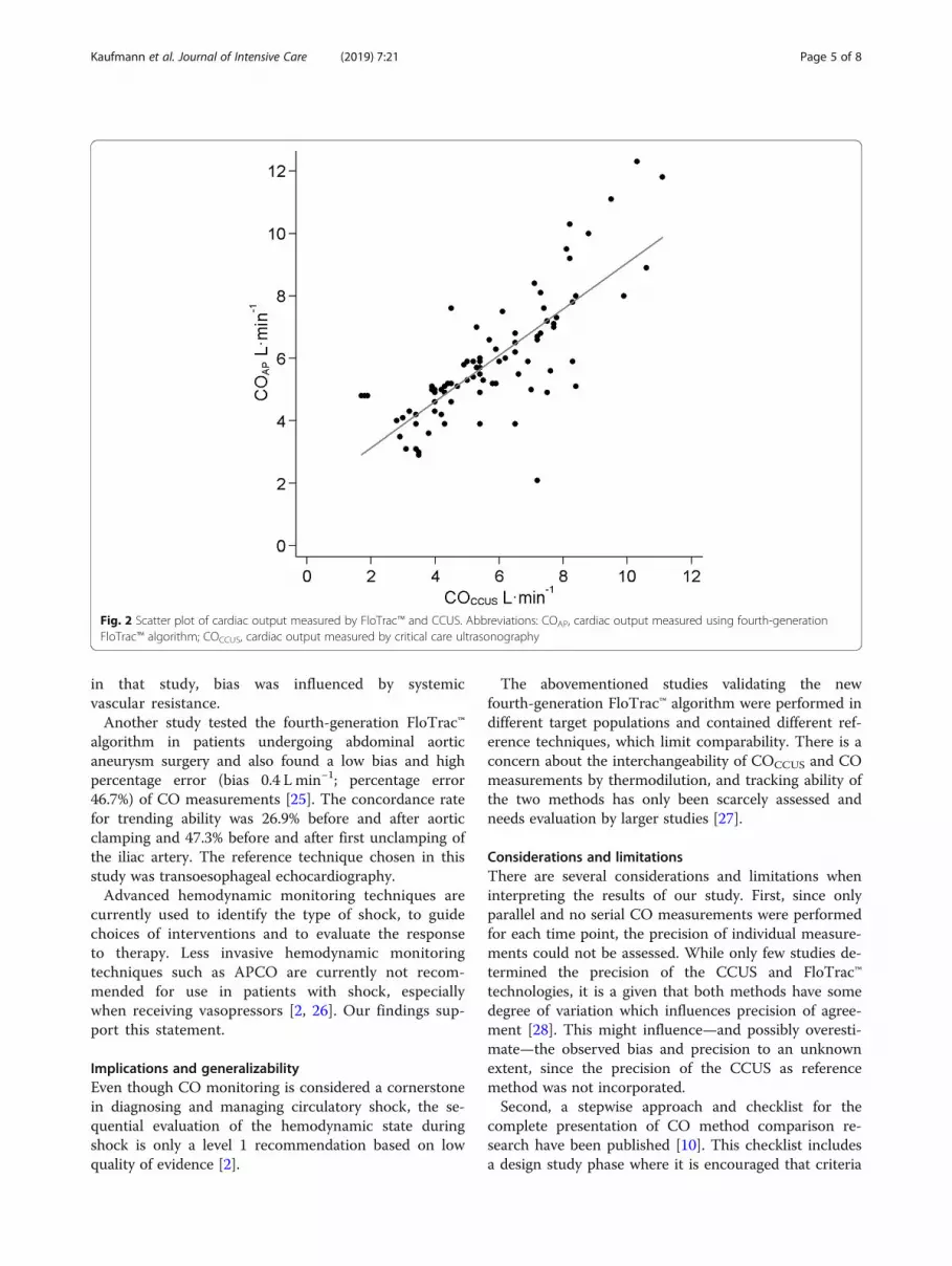
Bias, precision and correlationThe characteristics of the 17 included patients areshown in Table 1 (and Additional file 2: Table S1). Themean COAP and COCCUS for 89 paired measurementswere 5.9 ± 1.9 Lmin−1 and 5.7 ± 2.0 L min−1, respectively(p = 0.24). A significant correlation was observed for allCO measurements (r2 = 0.60, p < 0.001) (Fig. 2). Bias was0.2 L min−1 (95% CI − 0.2 to 0.6), with LOA of − 3.6 Lmin−1 (95% CI − 4.3 to − 2.9) to 4.0 L min−1 (95% CI 3.3to 4.7) (Fig. 3). Plotting a regression line in theBland-Altman plot gave no arguments for proportionalbias (line not shown). The overall percentage error was65.6% (95% CI 53.2 to 77.3). Individual cardiac outputmeasurements for each patient are provided in Add-itional file 3: Table S2.
Trending abilityFor assessment of trending ability 72 paired measure-ments were analysed. Trending of measurements wasevaluated using a four-quadrant plot (Fig. 4). Forty-fivepaired measurements showed a clinically relevantchange, which was defined as larger than 0.5 Lmin−1.The concordance rate was 64.4%.
DiscussionIn this prospective observational study, agreement andtrending ability of COAP was compared with COCCUS incritically ill patients with circulatory shock. COAP
showed a low bias of 0.2 Lmin−1 but a large percentageerror of 65.6% when compared with COCCUS, indicatingdisagreement [20]. Trending ability was poor with a con-cordance rate of 64.4%. The new FloTrac™ algorithmshould not be used for diagnosis or guidance of treat-ment in critically ill patients with circulatory shock.
InterpretationThere are no data on the reliability of CO measure-ments with the fourth-generation FloTrac™ software
algorithm in critically ill patients with shock as of yet.The main concern with the previous version(s) of theAPCO algorithm was the lack of reliability in trackingCO changes after hemodynamic interventions or inpatients with sepsis [7, 22].The low bias and the high percentage error of CO
measurements are in accordance with results from an-other study, which tested the fourth-generation algorithm for tracking CO measurements after administra-tion of phenylephrine to increase vasomotor tone in pa-tients prior to cardiac surgery (bias − 0.7 Lmin−1;percentage error 55.4%) [23]. Concordance rate fortrending ability was 87% which was higher than in ourstudy. In that study, the chosen reference technique formeasuring CO was thermodilution.In a more recent study in patients undergoing car-
diac surgery, the new FloTrac™ algorithm also showedlack of agreement and trending ability (bias − 0.4 Lmin−1; percentage error 37.1%; concordance rate 76%)[24]. The reference technique was thermodilution and
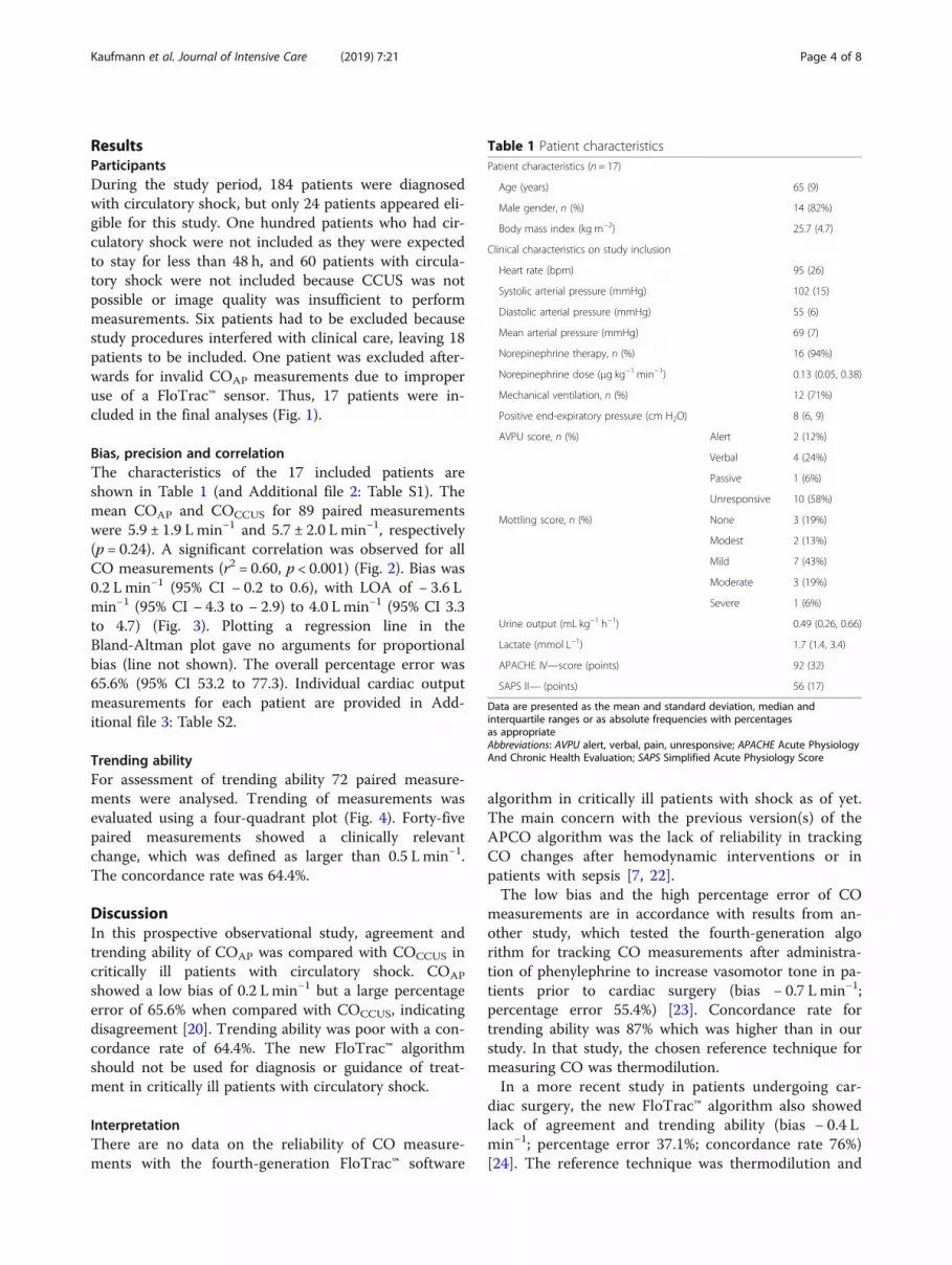
Table 1 Patient characteristicsPatient characteristics (n = 17)
Age (years) 65 (9)
Male gender, n (%) 14 (82%)
Body mass index (kg m−2) 25.7 (4.7)
Clinical characteristics on study inclusion
Heart rate (bpm) 95 (26)
Systolic arterial pressure (mmHg) 102 (15)
Diastolic arterial pressure (mmHg) 55 (6)
Mean arterial pressure (mmHg) 69 (7)
Norepinephrine therapy, n (%) 16 (94%)
Norepinephrine dose (μg kg−1 min−1) 0.13 (0.05, 0.38)
Mechanical ventilation, n (%) 12 (71%)
Positive end-expiratory pressure (cm H2O) 8 (6, 9)
AVPU score, n (%) Alert 2 (12%)
Verbal 4 (24%)
Passive 1 (6%)
Unresponsive 10 (58%)
Mottling score, n (%) None 3 (19%)
Modest 2 (13%)
Mild 7 (43%)
Moderate 3 (19%)
Severe 1 (6%)
Urine output (mL kg−1 h−1) 0.49 (0.26, 0.66)
Lactate (mmol L−1) 1.7 (1.4, 3.4)
APACHE IV—score (points) 92 (32)
SAPS II— (points) 56 (17)
Data are presented as the mean and standard deviation, median andinterquartile ranges or as absolute frequencies with percentagesas appropriateAbbreviations: AVPU alert, verbal, pain, unresponsive; APACHE Acute PhysiologyAnd Chronic Health Evaluation; SAPS Simplified Acute Physiology Score
Kaufmann et al. Journal of Intensive Care (2019) 7:21 Page 4 of 8
in that study, bias was influenced by systemicvascular resistance.Another study tested the fourth-generation FloTrac™
algorithm in patients undergoing abdominal aorticaneurysm surgery and also found a low bias and highpercentage error (bias 0.4 L min−1; percentage error46.7%) of CO measurements [25]. The concordance ratefor trending ability was 26.9% before and after aorticclamping and 47.3% before and after first unclamping ofthe iliac artery. The reference technique chosen in thisstudy was transoesophageal echocardiography.Advanced hemodynamic monitoring techniques are
currently used to identify the type of shock, to guidechoices of interventions and to evaluate the responseto therapy. Less invasive hemodynamic monitoringtechniques such as APCO are currently not recom-mended for use in patients with shock, especiallywhen receiving vasopressors [2, 26]. Our findings sup-port this statement.
Implications and generalizabilityEven though CO monitoring is considered a cornerstonein diagnosing and managing circulatory shock, the se-quential evaluation of the hemodynamic state duringshock is only a level 1 recommendation based on lowquality of evidence [2].
The abovementioned studies validating the newfourth-generation FloTrac™ algorithm were performed indifferent target populations and contained different ref-erence techniques, which limit comparability. There is aconcern about the interchangeability of COCCUS and COmeasurements by thermodilution, and tracking ability ofthe two methods has only been scarcely assessed andneeds evaluation by larger studies [27].
Considerations and limitationsThere are several considerations and limitations wheninterpreting the results of our study. First, since onlyparallel and no serial CO measurements were performedfor each time point, the precision of individual measure-ments could not be assessed. While only few studies de-termined the precision of the CCUS and FloTrac™technologies, it is a given that both methods have somedegree of variation which influences precision of agree-ment [28]. This might influence—and possibly overesti-mate—the observed bias and precision to an unknownextent, since the precision of the CCUS as referencemethod was not incorporated.Second, a stepwise approach and checklist for the
complete presentation of CO method comparison re-search have been published [10]. This checklist includesa design study phase where it is encouraged that criteria
Fig. 2 Scatter plot of cardiac output measured by FloTrac™ and CCUS. Abbreviations: COAP, cardiac output measured using fourth-generationFloTrac™ algorithm; COCCUS, cardiac output measured by critical care ultrasonography
Kaufmann et al. Journal of Intensive Care (2019) 7:21 Page 5 of 8
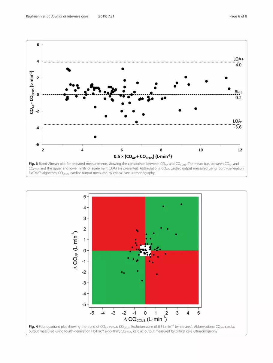
Fig. 4 Four-quadrant plot showing the trend of COAP versus COCCUS. Exclusion zone of 0.5 L min−1 (white area). Abbreviations: COAP, cardiacoutput measured using fourth-generation FloTrac™ algorithm; COCCUS, cardiac output measured by critical care ultrasonography
Fig. 3 Bland-Altman plot for repeated measurements showing the comparison between COAP and COCCUS. The mean bias between COAP andCOCCUS and the upper and lower limits of agreement (LOA) are presented. Abbreviations: COAP, cardiac output measured using fourth-generationFloTrac™ algorithm; COCCUS, cardiac output measured by critical care ultrasonography
Kaufmann et al. Journal of Intensive Care (2019) 7:21 Page 6 of 8
for acceptable bias and LOA or percentage error are de-fined, and a sample size calculation should be performedprior to the conduct of method comparison studies. Inour study, we defined clinically acceptable limits basedon available literature, but we did not specify a samplesize in advance. The current study could serve as a pilotfor a further validation study.Third, during the study period, we included only 17
patients. Patients with circulatory shock were eligibleonly if they were expected to stay for longer than 48 hand if it was possible to perform CCUS. We chose thisdefinition to ensure that a complete picture of shocktreatment could be presented which allowed for the bestcomparison between the two methods. Last, CCUS wasused as a reference technique in our study despite pul-monary or transpulmonary thermodilution being thegold standard for CO method comparison studies [10].Therefore, we cannot prove direct superiority of eithermethod. In order to do this, a comparison with a ther-modilution method will have to be performed. We choseCCUS as reference because it is currently the first-lineevaluation modality in patients with circulatory shockand also because it is widely available and used in theICU for diagnostic purposes [2, 29]. However, images re-quired to make COCCUS measurements are unobtainablein up to 20% of patients [30].FloTrac™ measurements of CO are still not recom-
mended in critically ill patients [5, 6], and further clinicalstudies comparing minimally invasive techniques for COestimation with a reference technique are needed forfurther validation of these techniques and also for ex-tending applicability to other types of patients, who wereinitially not the target population.
ConclusionsIn critically ill patients with circulatory shock, there wasdisagreement and clinically unacceptable trending abilitybetween values of cardiac output obtained by uncali-brated arterial pressure waveform analysis and criticalcare ultrasonography.
Additional files
Additional file 1: STROBE Statement-Checklist of items that should beincluded in reports of cohort studies. Checklist of items that should beincluded in reports of cohort studies according to the STROBE statement.(DOCX 42 kb)
Additional file 2: Table S1. Detailed patient characteristics. Tableshowing extended patient characteristics to further describe the studypopulation. (DOCX 15 kb)
Additional file 3: Table S2. Cardiac output measurements with criticalcare ultrasonography and FloTrac. Overview of each cardiac outputmeasurement performed with critical care ultrasonography and FloTrac.(DOCX 24 kb)
AbbreviationsAPACHE: Acute Physiology And Chronic Health Evaluation; APCO: ArterialPressure waveform analysis method to estimate Cardiac Output;CCUS: Critical care ultrasonography; CO: Cardiac output; COAP: Cardiacoutput measured using fourth-generation FloTrac™ algorithm;COCCUS: Cardiac output measured using critical care ultrasonography;ICU: Intensive care unit; LOA: Limits of agreement; MAP: Mean arterialpressure; PAC: Pulmonary artery catheter; SAPS: Simple Acute PhysiologyScore; SICS: Simple Intensive Care Studies; STROBE: STrengthening theReporting of OBservational studies in Epidemiology; SV: Stroke volume
AcknowledgementsThe SICS Study Group members include the following: project leaders: GeertKoster; Frederik Keus; Iwan CC van der Horst. Research coordinator: WillemDieperink. Researchers who conducted patient inclusions: Roos Bleijendaal;Yasmin F. Cawale; Ramon P. Clement; Devon Dijkhuizen; Ruben J Eck; BartHiemstra; Anja Haker; Casper D.H. Hilbink; Thomas Kaufmann; MartieneKlasen, Manon Klaver; Laura J. Schokking; Victor W. Sikkens; Madelon Vos;Justin Woerlee; Renske Wiersema.
FundingNo external funding was obtained for this study. All FloTrac™ sensors werepaid for with departmental funding.
Availability of data and materialsThe datasets generated during and/or analysed during the current study areavailable from the corresponding author on reasonable request.
Authors’ contributionsTK and RPC included the patients, drafted the manuscript and conductedthe analyses. BH developed the protocol and implemented the study. JJVand TWLS critically reviewed the manuscript. IvdH and EK created the idea ofthe study. All authors critically reviewed the manuscript and agreed with thefinal version and findings. All authors read and approved the finalmanuscript.
Ethics approval and consent to participateThe local institutional review board (Medisch Ethische Toetsingscommissie,University Medical Center Groningen) approved the study (M15.168207 andM16.193856). Written informed consent was obtained from all patients.
Consent for publicationNot applicable.
Competing interestsTWLS received honoraria from Edwards Lifesciences (Irvine, California, USA)for consulting and for giving lectures. The other authors declare that theyhave no competing interests.
Publisher’s NoteSpringer Nature remains neutral with regard to jurisdictional claims inpublished maps and institutional affiliations.
Author details1Department of Anesthesiology, University Medical Center Groningen,University of Groningen, P.O. Box 30.001, 9700 RB Groningen, TheNetherlands. 2Department of Critical Care, University Medical CenterGroningen, University of Groningen, Groningen, The Netherlands.
Received: 17 January 2019 Accepted: 14 March 2019
References1. Sakr Y, Reinhart K, Vincent J-L, Sprung CL, Moreno R, Ranieri VM, et al.
Does dopamine administration in shock influence outcome? Results ofthe sepsis occurrence in acutely ill patients (SOAP) study. Crit CareMed. 2006;34:589–97.
2. Cecconi M, De Backer D, Antonelli M, Beale R, Bakker J, Hofer C, et al.Consensus on circulatory shock and hemodynamic monitoring. Task forceof the European Society of Intensive Care Medicine. Intensive Care Med.2014;40:1795–815.
Kaufmann et al. Journal of Intensive Care (2019) 7:21 Page 7 of 8
3. Clement RP, Vos JJ, Scheeren TWL. Minimally invasive cardiac output technologiesin the ICU: putting it all together. Curr Opin Crit Care. 2017;23:302–9.
4. Saugel B, Wagner JY, Scheeren TWL. Cardiac output monitoring: lessinvasiveness, less accuracy? J Clin Monit Comput. 2016;30(6):753–5.
5. De Backer D, Bakker J, Cecconi M, Hajjar L, Liu DW, Lobo S, et al. Alternatives tothe Swan–Ganz catheter. Intensive Care Med. 2018;44:730–41.
6. Saugel B, Vincent J-L. Cardiac output monitoring: how to choose the optimalmethod for the individual patient. Curr Opin Crit Care. 2018;24(3):165–72.
7. Slagt C, Malagon I, Groeneveld ABJ. Systematic review of uncalibratedarterial pressure waveform analysis to determine cardiac output and strokevolume variation. Br J Anaesth. 2014;112(4):626–37.
8. Critchley LA, Lee A, Ho AMH. A critical review of the ability of continuouscardiac output monitors to measure trends in cardiac output. Anesth Analg.2010;111:1180–92.
9. Koster G, Van Der Horst ICC. Critical care ultrasonography in circulatoryshock. Curr Opin Crit Care. 2017;23:326–33.
10. Montenij LJ, Buhre WF, Jansen JR, Kruitwagen CL, De Waal EE. Methodologyof method comparison studies evaluating the validity of cardiac outputmonitors: a stepwise approach and checklist. Br J Anaesth. 2016;116:750–8.
11. von Elm E, Altman DG, Egger M, Pocock SJ, Gøtzsche PC, VandenbrouckeJP. The Strengthening the Reporting of Observational Studies inEpidemiology (STROBE) statement: guidelines for reporting observationalstudies. Lancet. 2007;370:1453–7.
12. American College of Surgeons Committee on Trauma. Advanced lifesupport course for physicians. Chicago: American College ofSurgeons; 1993.
13. Ait-Oufella H, Lemoinne S, Boelle PY, Galbois A, Baudel JL, Lemant J, et al. Mottlingscore predicts survival in septic shock. Intensive Care Med. 2011;37:801–7.
14. Zimmerman JE, Kramer AA, McNair DS, Malila FM. Acute Physiology andChronic Health Evaluation (APACHE) IV: hospital mortality assessment fortoday’s critically ill patients. Crit Care Med. 2006;34:1297–310.
15. Gall JR, Lemeshow S, Saulnier F. A new simplified acute physiology score(SAPS II) based on a European/north American multicenter study. JAMA.1993;270:2957–63.
16. Hiemstra B, Eck RJ, Koster G, Wetterslev J, Perner A, Pettilä V, et al. Clinicalexamination, critical care ultrasonography and outcomes in the critically ill:cohort profile of the simple intensive care studies-I. BMJ Open. 2017;7:8–11.
17. Hiemstra B, Koster G, Wiersema R, Hummel YM, van der Harst P, Snieder H,et al. The diagnostic accuracy of clinical examination for estimating cardiacindex in critically ill patients: the Simple Intensive Care Studies-I. IntensiveCare Medicine 2019;45(2):190–200.
18. Lewis JF, Kuo LC, Nelson JG, Limacher MC, Quinones MA. Pulsed Dopplerechocardiographic determination of stroke volume and cardiac output:clinical validation of two new methods using the apical window.Circulation. 1984;70:425–31.
19. Bland JM, Altman DG. Agreement between methods of measurement withmultiple observations per individual. J Biopharm Stat. 2007;17:571–82.
20. Critchley LAH, Critchley JAJH. A meta-analysis of studies using bias andprecision statistics to compare cardiac output measurement techniques. JClin Monit Comput. 1999;15:85–91.
21. Saugel B, Grothe O, Wagner JY. Tracking changes in cardiac output:statistical considerations on the 4-quadrant plot and the polar plotmethodology. Anesth Analg. 2015;121:514–24.
22. Monnet X, Vaquer S, Anguel N, Jozwiak M, Cipriani F, Richard C, et al.Comparison of pulse contour analysis by Pulsioflex and Vigileo to measureand track changes of cardiac output in critically ill patients. Br J Anaesth.2015;114:235–43.
23. Suehiro K, Tanaka K, Mikawa M, Uchihara Y, Matsuyama T, Matsuura T,Funao T, Yamada T, Mori T, Nishikawa K. Improved performance of thefourth-generation FloTrac/Vigileo system for tracking cardiac outputchanges. J Cardiothorac Vasc Anesth. 2015;29(3):656–62.
24. Kusaka Y, Ohchi F, Minami T. Evaluation of the fourth-generation FloTrac/Vigileo system in comparison with the intermittent bolus Thermodilutionmethod in patients undergoing cardiac surgery. J Cardiothorac Vasc Anesth.2018;000:4–11.
25. Maeda T, Hattori K, Sumiyoshi M, Kanazawa H, Ohnishi Y. Accuracy andtrending ability of the fourth-generation FloTrac/Vigileo system™ in patientsundergoing abdominal aortic aneurysm surgery. J Anesth. 2018;32(3):387–93.
26. Teboul JL, Saugel B, Cecconi M, De Backer D, Hofer CK, Monnet X, et al. Lessinvasive hemodynamic monitoring in critically ill patients. Intensive CareMed. 2016;42:1350–9.
27. Wetterslev M, Møller-Sørensen H, Johansen RR, Perner A. Systematicreview of cardiac output measurements by echocardiography vs.thermodilution: the techniques are not interchangeable. Intensive CareMed. 2016;42:1223–33.
28. Hapfelmeier A, Cecconi M, Saugel B. Cardiac output method comparisonstudies: the relation of the precision of agreement and the precision ofmethod. J Clin Monit Comput. 2016;30(2):149–55.
29. Saugel B, Reese PC, Wagner JY, Buerke M, Huber W, Kluge S, et al. Advancedhemodynamic monitoring in intensive care medicine. Med Klin IntensivmedNotfmed. 2017;113:192–201.
30. Jensen MB, Sloth E, Larsen KM, Schmidt MB. Transthoracic echocardiography forcardiopulmonary monitoring in intensive care. Eur J Anaesthesiol. 2004;21:700–7.
Kaufmann et al. Journal of Intensive Care (2019) 7:21 Page 8 of 8


