Embed Size (px)
Citation preview
Proc. Natl. Acad. Sci. USAVol. 93, pp. 15108–15113, December 1996Biophysics
Direct interaction of flagellin termini essential for polymorphicability of flagellar filament
YUKO MIMORI-KIYOSUE*, FERENC VONDERVISZT, ICHIRO YAMASHITA, YOSHINORI FUJIYOSHI, AND KEIICHI NAMBA†
International Institute for Advanced Research, Matsushita Electric Industrial Co., Ltd., 3-4 Hikaridai, Seika 619-02, Japan
Communicated by Donald L. D. Caspar, Florida State University, Tallahassee, FL, October 14, 1996 (received for review April 29, 1996)
ABSTRACT We report the structures of f lagellar fila-ments reconstituted from various f lagellins with small ter-minal truncations. Flagellins from Salmonella typhimuriumstrains SJW1103 (wild type), SJW1660, and SJW1655 wereused, which form a left-handed supercoil, the L- and R-typestraight forms, respectively. Structure analyses were done byelectron cryomicroscopy and helical image reconstructionwith a help of x-ray fiber diffraction for determining precisehelical symmetries. Truncation of either terminal region,irrespective of the original f lagellin species, results in astraight filament having a helical symmetry distinct eitherfrom the L- or R-type. This filament structure is namedLt-type. Although the local subunit packing is similar in allthree types, a close comparison shows that the Lt-type packingis almost identical to the R-type but distinct from the L-type,which demonstrates the strong two-state preference of thesubunit interactions. The structure clearly suggests that bothtermini are located in the inner tube of the concentricdouble-tubular structure of the filament core, and theirproper interaction is responsible for the correct folding offairly large terminal regions that form the inner tube. Thedouble tubular structure appears to be essential for thepolymorphic ability of f lagellar filaments, which is requiredfor the swimming–tumbling of bacterial taxis.
Molecular events behind the swimming–tumbling pattern ofbacterial motility involve polymorphic transitions of normallyleft-handed supercoiled flagellar filaments into a right-handedform (1) upon quick reversal of motor rotation that drives thefilament (2–4). The polymorphic mechanism of the filamenthas been modeled assuming two-state subunit conformations(5–7), but not been fully understood because of the limitedresolution of available structural data that show only the outershape of subunits and distribution of axially aligned bundles ofa-helices in the core (8–10).The supercoiled forms of bacterial f lagellar filaments are
thought to be constructed from a mixture of two distinctsubunit conformations of flagellin arranged in a regularmanner, while all the subunits are chemically identical (5).There are two types of straight filaments with distinct helicalsymmetries, one called the L-type, in which the 11 protofila-ments are tilted to the left, and the other called the R-type,with the protofilaments tilted to the right (11). Flagellinmutants from SJW1660 (the L-type) and SJW1655 (the R-type) can be copolymerized to form different types of super-coiled filaments, depending on the mixing ratio (12). There-fore, the two conformations of the flagellin mutants arethought to be representing the two that coexist in the super-coiled filaments.Flagellin from Salmonella typhimurium is composed of 494
amino acids (13). About 65 residues of the NH2 terminal and44 of the COOH terminal are disordered in monomeric form(14) but well folded in the filament (15), which indicate their
roles in assembly regulation mechanism to suppress sponta-neous polymerization of flagellin under physiological condi-tions. Although there is no direct evidence, indirect evidencehas suggested that these terminal regions contribute to thecore part of the filament, which occupies about half of thefilament diameter of 230 Å and has a concentric double tubularstructure (9, 10).The role of the disordered terminal regions in filament
formation, stability, and polymorphism has been studied byusing proteolytic fragments of flagellin with terminal deletionsof different sizes (16). Polymerization could be induced evenwhen most of the disordered terminal regions were removed,although the filaments were much less stable. The mostdramatic effect observed was that truncation of only a fewresidues from wild-type flagellin resulted in straight filamentsupon polymerization and loss of polymorphic ability.Taking advantage of the straight form of the filaments
reconstituted from terminally truncated flagellins, we carriedout structure analysis of these filaments to find out the locationof the termini and their roles in the polymorphic ability of theintact filaments. The simplest interpretation of the results isthat both termini are located in the inner tube of the concentricdouble tubular structure of the filament core, and their directinteraction is responsible for the proper folding of fairly largeterminal regions that form the inner tube. Strong preferenceof the subunit interactions in the outer tube to be in one of thetwo states is also demonstrated, and its implication for thepolymorphic mechanism is discussed.
MATERIALS AND METHODS
Preparation of Filaments of Flagellins with TerminalTruncations. Flagellins were isolated from a wild-typestrain, SJW1103, and two mutant strains, SJW1660 andSJW1655, and purified as described (14). Fragments off lagellins with small terminal truncations were prepared bylimited proteolysis of monomeric f lagellins and purified byionic exchange column chromatography (16). The F-(1–486)fragments, which lack the 8 COOH terminal residues, wereprepared from all three types of f lagellin, while the F-(20–494) fragment, which is deprived of the NH2 terminal 19residues, was made from SJW1655 f lagellin. Filaments werereconstituted by adding appropriate concentrations (0.2–0.5M) of (NH4)2SO4 to solutions containing the truncatedf lagellins, as described (16).Electron Cryomicroscopy and Helical Image Reconstruc-
tion. The experimental procedure of electron cryomicroscopy,including the preparation of frozen hydrated samples, is fullydescribed in ref. 9. Electron micrographs were taken at amagnification of 350,000 with a JEOL model JEM-3000SFF(300 kV) electron microscope equipped with a cryo-stagecooled to 4 K with liquid helium (17) and a field emissionelectron source (18). The defocus levels were within the range
The publication costs of this article were defrayed in part by page chargepayment. This article must therefore be hereby marked ‘‘advertisement’’ inaccordance with 18 U.S.C. §1734 solely to indicate this fact.
*Present address: ERATO, Cell Axis Project, 17 Minamimachi,Nakadoji, Shimogyo, Kyoto 600 Japan.†To whom reprint requests should be addressed.
15108
from 2 to 5 mm. The helical image reconstruction procedurefully described in ref. 9 was used in the exactly same way,including correction for the contrast transfer function. Thedata out to 12-Å resolution were included for calculating thedensity maps and radial density distribution.X-Ray Fiber Diffraction.Well-oriented filament sols were
prepared as described (19). Filament sols prepared by cen-trifugation at 10,000 3 g for 15 h were made into a liquidcrystalline state in the solution containing 30 mM NaCl and50 mM glycine-acetate (pH 5.0). After further centrifugationof the sols in capillary tubes at 20003 g for 20 h, the sols wereplaced in a superconducting magnet with a 13.5 Teslamagnetic field for 2 to 3 days. X-ray fiber diffraction patternswere recorded as described (19), using a rotating anode x-raygenerator (Rigaku, Tokyo), a Ni-coated double mirror optics(Rigaku), and Imaging Plate (Fuji). The layer-line spacingsand intensity distributions were obtained by a data process-ing procedure using the two-dimensional angular deconvo-lution method, as partially described in ref. 19, and thesedata were used only to determine the helical symmetries ofthe filaments.
RESULTS AND DISCUSSION
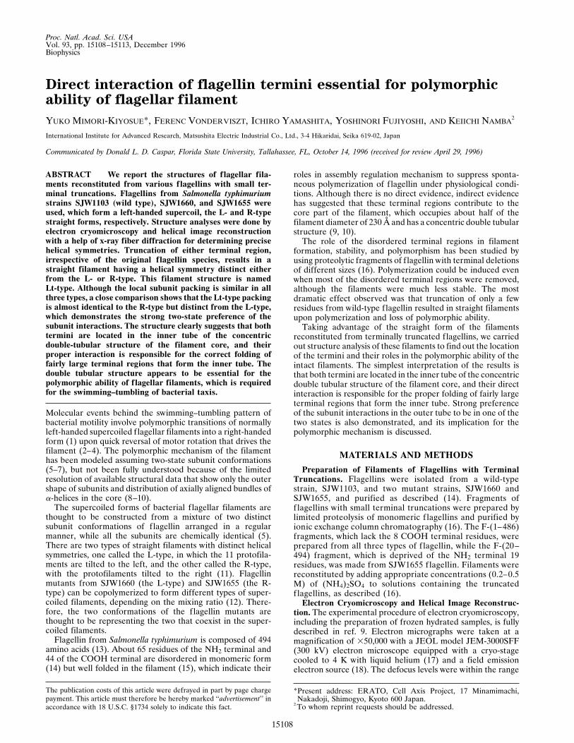
Helical Symmetry of the Filaments of Truncated Flagellins.Studying the structures of filaments of flagellins with theCOOH terminal eight residues truncated [F-(1–486)] fromstrains SJW1103 (wild type), SJW1660 (L-type), and SJW1655(R-type) by electron cryomicroscopy, it turned out that thesymmetries of these filaments were different either from the L-or R-type, but all were the same irrespective of the originalf lagellin. Truncation of 19 residues from the NH2 terminal ofSJW1655 flagellin [F-(20–494)] also resulted in the samesymmetry. Fourier transforms of filament images demonstrateit clearly, as shown in Fig. 1. The somewhat poor quality of theFourier transforms in Fig. 1 C–F is caused by significantlylower mechanical stability of the filaments of truncated flagel-lins compared with the intact filaments, for which the Fouriertransforms are shown in Fig. 1 A and B. Flagellins with largertruncations also yielded filaments of the same symmetry (datanot shown).In Fig. 1 C–F, the diffraction patterns indicate that the
longitudinal protofilaments are tilted to the left in contrast tothe R-type (Fig. 1B), but the tilt angle is larger than that of the
FIG. 1. Computed Fourier transforms and x-ray diffraction pattern of various reconstituted filaments. (A–F) Computed Fourier transforms ofelectron cryomicrographs of frozen hydrated filaments: (A) SJW1660 (the L-type), (B) SJW1655 (the R-type), (C) F-(1–486) of SJW1103 (wildtype), (D) F-(1–486) of SJW1660, (E) F-(1–486) of SJW1655; and (F) F-(20–494) of SJW1655. (G) X-ray fiber diffraction pattern from an orientedfilament sol of SJW1655 F-(1–486). For all the figures, the data out to about 20-Å resolution are shown. The order of Fourier–Bessel component(or helical start number), n, of each layer line is indicated on the right side. In the Fourier transforms of the L- and R-type (A and B), relativelayer-line intensities and the order of Fourier–Bessel component are strongly correlated because the local subunit packing is similar to each other,and this makes it easy to identify the order. Therefore, the difference in helical symmetry between the two, such as the tilt of the 11-start helix,can be roughly identified from the axial positions of the major layer lines. If the strong layer line, n 5 25, is above the relatively weaker layer line,n5 6, the 11-stranded protofilaments are tilted to the left, or vice versa. The larger the distance between the two layer lines, the larger the tilt angle(see also Fig. 2 B and C). This relation is also valid in the Fourier transforms of the filaments of truncated flagellins (C–F), except that some ofthe Fourier–Bessel components have slightly different orders: e.g., n5 1 of the L- and R-type is 3; n5 25 is24; n5 6 is 7; and so on. This indicatesthat the local packing and orientation of the subunits are more or less preserved also in the filaments of truncated flagellins.
Biophysics: Mimori-Kiyosue et al. Proc. Natl. Acad. Sci. USA 93 (1996) 15109
L-type (Fig. 1A). Although the overall layer-line pattern issimilar to that of the L-type, there is a small but clear shift ofthe radial peak positions of the layer-line intensity distribu-tions. We carefully checked the peak positions and phaseresiduals between the near and far sides to find out the orderof Fourier–Bessel component (or helical start number) of eachlayer line. More accurate radial peak positions were obtainedfrom the x-ray fiber diffraction pattern shown in Fig. 1G. Theyare listed in Table 1, from which the Bessel orders of the majorlayer lines were unambiguously determined. The filamentradius is the same as those of the L- and R-type, and the radialpeak position of the layer line labeled 211 is unchanged,showing that this filament also has 11 protofilaments. How-ever, the other Bessel orders are changed: from25 to24, from6 to 7, and from 1 to 3. The layer-line spacings were measuredfrom the x-ray diffraction pattern and the helical symmetry wasdetermined by building the reciprocal lattice using these Besselorders. The helical lattice parameters are listed in Table 2. Wecall this the ‘‘Lt’’ type symmetry, where ‘‘L’’ stands for theleft-handed 11-start helix and ‘‘t’’ for truncated flagellin.Comparison of Three-Dimensional Structures. Three-
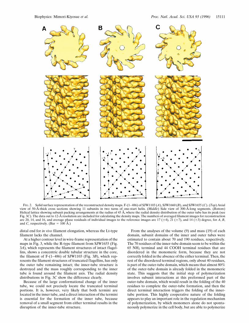
dimensional structures of the Lt-type filaments as well as theL-type were deduced by helical image reconstruction, exactlyin the same way as described for the structure analysis of theR-type filament by Mimori et al. (9). Helical image recon-struction of all four filaments of truncated flagellins (threewith COOH terminal truncation and one with NH2 terminaltruncation) produced almost identical density maps except forsmall differences around the filament axis. The reconstructedimages in a solid surface representation are shown in Fig. 2,which compares the Lt-, L-, and R-type filaments. In Fig. 2, thefilament structure of F-(1–486) of SJW1103 represents thoseof the four Lt-type filaments.In the structures of all three filaments, there are no obvious
differences in the shapes of outer projections and short verticalcolumn densities that are directly attached to the denselypacked core, while the subunit orientations seem to be slightlydifferent according to the different tilt angles of the proto-filaments. The local subunit packing relations are also similarto one another.Because the two conformations of the mutant flagellin
subunits of the L- and R-type filaments shown in Fig. 2 B andC, respectively, are thought to represent the two that coexistin the supercoiled filaments, the small difference betweenthem indicates that the conformational change involved inpolymorphism is a rather small one. The close packing ofaxially aligned a-helices in the filament core (8–10) and thenature of the local lattice conversion (see Fig. 4B) suggest thatthe probable conformational change involved in the polymor-phic transition would be a distinct mutual shift of thesea-helices by about 2.5 Å along the protofilament, and the
subunit rotation within a cylindrical surface plane assumed inthe polymorphic model by Calladine (6, 7) would be ratherunlikely. In any case, these two lattices are interconvertible toeach other by a continuous twisting deformation of the cylin-drical surface lattice around the axis, except for the small butdistinct difference in the intersubunit distance along theprotofilament.However, the Lt-type symmetry could only be produced by
the following operation involving a discontinuous transforma-tion on either the L- or R-type: first open up the tubularstructure at an interface of any two protofilaments, thendeform the tube so that the protofilament on the right at theinterface slides down by one subunit relative to the one on theleft, and close the tube. Similar relations within a group oflattices have been found in the polyhead tubes of bacterio-phages (20) and the carbon nanotubes (21). This transforma-tion may be called a reconstructive transformation as opposedto a displacive one between the L- and R-type filaments, in thesimilar way as discussed by Moody (22) for the contraction ofbacteriophage tail sheath.Structural Difference in the Inner Core. The difference
between the subunit structures of intact and truncated flagel-lins can be found in the axial views (Fig. 2 Top). The L- andR-type filaments have the central channel with a diameter ofabout 30 Å, which is necessary for subunit transport to the
Table 1. Radial positions of the first peaks of the layer-lines
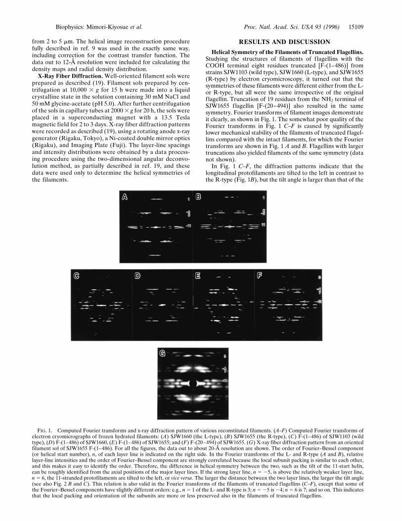
Type G211yG11 G7yG6 G24yG25 G3yG1Lt 0.0195 (0.0195) 0.0135 (0.0134) 0.0090 (0.0091) 0.0075 (0.0082)Parity Odd Odd Even OddR 0.0195 0.0120 0.0110 0.0035
Layer-lines are specified by Gn, where n indicates the Bessel order. Radial peak positions weremeasured on x-ray fiber diffraction patterns and are listed in Å21 unit. The errors are 60.0003 Å21 atmost. The structurally corresponding Fourier–Bessel components are listed in the same column for theLt- and R-type and they are indicated in the top row as Lt-typeyR-type. Parity indicates the phasedifference between the near and far side of the layer line and it is even when the difference is close to08 and odd when close to 1808. Since the helical symmetry of the R-type is well established, the radial peakpositions of the Lt-type layer lines are calculated from those of the R-type layer lines, taking into accountthe Bessel order of each layer line, and listed in parenthesis. The close match between the observed andcalculated radial positions and phase parities confirms that the deduced symmetry of the Lt-type is correct.Only for G3, the difference between the observed and calculated radial positions is relatively large,probably because of the significant difference in the subunit orientations with respect to the filament axisbetween the Lt- and R-type structures, but the peak position calculated for G2 is 0.060, which is twicefurther than that for G3.
Table 2. Helical symmetries and cylindrical surfacelattice parameters
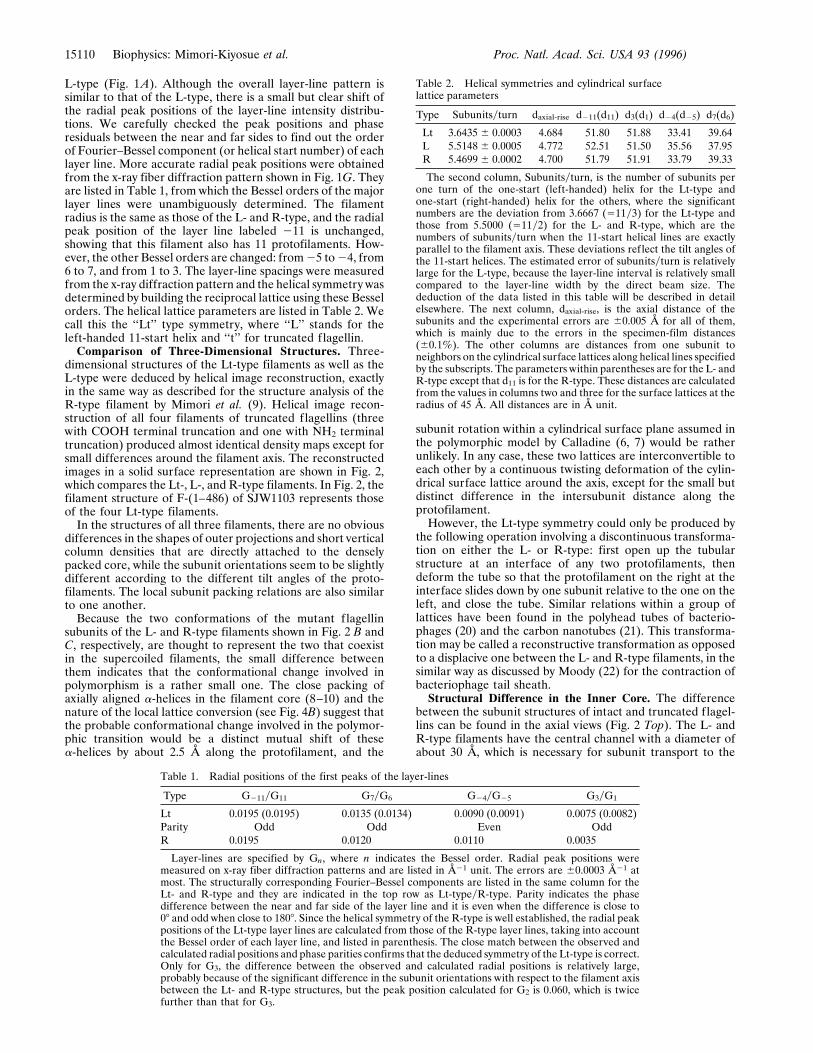
Type Subunitsyturn daxial-rise d211(d11) d3(d1) d24(d25) d7(d6)
Lt 3.6435 6 0.0003 4.684 51.80 51.88 33.41 39.64L 5.5148 6 0.0005 4.772 52.51 51.50 35.56 37.95R 5.4699 6 0.0002 4.700 51.79 51.91 33.79 39.33
The second column, Subunitsyturn, is the number of subunits perone turn of the one-start (left-handed) helix for the Lt-type andone-start (right-handed) helix for the others, where the significantnumbers are the deviation from 3.6667 (511y3) for the Lt-type andthose from 5.5000 (511y2) for the L- and R-type, which are thenumbers of subunitsyturn when the 11-start helical lines are exactlyparallel to the filament axis. These deviations reflect the tilt angles ofthe 11-start helices. The estimated error of subunitsyturn is relativelylarge for the L-type, because the layer-line interval is relatively smallcompared to the layer-line width by the direct beam size. Thededuction of the data listed in this table will be described in detailelsewhere. The next column, daxial-rise, is the axial distance of thesubunits and the experimental errors are 60.005 Å for all of them,which is mainly due to the errors in the specimen-film distances(60.1%). The other columns are distances from one subunit toneighbors on the cylindrical surface lattices along helical lines specifiedby the subscripts. The parameters within parentheses are for the L- andR-type except that d11 is for the R-type. These distances are calculatedfrom the values in columns two and three for the surface lattices at theradius of 45 Å. All distances are in Å unit.
15110 Biophysics: Mimori-Kiyosue et al. Proc. Natl. Acad. Sci. USA 93 (1996)
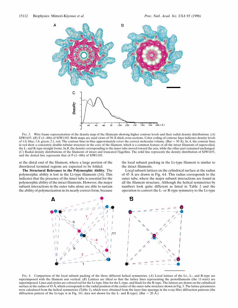
distal end for in vivo filament elongation, whereas the Lt-typefilament lacks the channel.At a higher contour level in wire-frame representation of the
maps in Fig. 3, while the R-type filament from SJW1655 (Fig.3A), which represents the filament structures of intact f lagel-lins, shows a concentric double tubular structure in the core,the filament of F-(1–486) of SJW1103 (Fig. 3B), which rep-resents the filament structures of truncated flagellins, has onlythe outer tube remaining intact; the inner-tube structure isdestroyed and the mass roughly corresponding to the innertube is found around the filament axis. The radial densitydistributions in Fig. 3C show the difference clearly.Because of the large conformational change of the inner
tube, we could not precisely locate the truncated terminalportions. It is, however, very likely that both termini arelocated in the inner tube, and a direct interaction of the terminiis essential for the formation of the inner tube, becauseremoval of a small segment from either terminal results in thedisruption of the inner-tube structure.
From the analyses of the volume (9) and mass (19) of eachdomain, subunit domains of the inner and outer tubes wereestimated to contain about 70 and 190 residues, respectively.The 70 residues of the inner-tube domain seem to be within the65 NH2 terminal and 44 COOH terminal residues that aredisordered in the monomeric form, because they are notcorrectly folded in the absence of the either terminal. Then, therest of the disordered terminal regions, only about 40 residues,is part of the outer-tube domain, which means that almost 80%of the outer-tube domain is already folded in the monomericstate. This suggests that the initial step of polymerizationinvolves subunit interactions at this preformed part of theouter-tube domain, which would result in the folding of the 40residues to complete the outer-tube formation, and then thedirect terminal interaction triggers the folding of the inner-tube portion. This highly cooperative nature of the foldingappears to play an important role in the regulation mechanismof polymerization, by which monomers alone do not sponta-neously polymerize in the cell body, but are able to polymerize
FIG. 2. Solid surface representation of the reconstructed density maps. F-(1–486) of SJW1103 (A), SJW1660 (B), and SJW1655 (C). (Top) Axialview of 50-Å-thick cross sections showing 11 subunits in two turns of one-start helix. (Middle) Side view of 300-Å-long segments. (Bottom)Helical lattice showing subunit packing arrangements at the radius of 45 Å, where the radial density distribution of the outer tube has its peak (seeFig. 3C). The data out to 12-Å resolution are included for calculating the density maps. The numbers of averaged filament images for reconstructionare 20, 14, and 16, and average phase residuals of individual images to the reference images are 17 (64), 21 (67), and 14 (63) degree, for A, B,and C, respectively. (Bar 5 100 Å.)
Biophysics: Mimori-Kiyosue et al. Proc. Natl. Acad. Sci. USA 93 (1996) 15111
at the distal end of the filament, where a large portion of thedisordered terminal regions are expected to be folded.The Structural Relevance to the Polymorphic Ability. The
polymorphic ability is lost in the Lt-type filaments (16). Thisindicates that the presence of the inner tube is essential for thepolymorphic ability of the intact filaments. However, themajorsubunit interactions in the outer tube alone are able to sustainthe ability of polymerization in its nearly correct form, because
the local subunit packing in the Lt-type filament is similar tothe intact filaments.Local subunit lattices on the cylindrical surface at the radius
of 45 Å are drawn in Fig. 4A. This radius corresponds to theouter tube, where the major subunit interactions are found inall the filament structure. Although the helical symmetries bynumbers look quite different as listed in Table 2 and theoperation to convert the L- or R-type symmetry to the Lt-type
FIG. 3. Wire frame representation of the density map of the filaments showing higher contour levels and their radial density distributions. (A)SJW1655. (B) F-(1–486) of SJW1103. Both maps are axial views of 50-Å-thick cross sections. Color coding of contour lines indicates density levelsof 1.0, blue; 1.8, green; 2.1, red. The contour lines in blue approximately cover the correct molecular volume. (Bar 5 50 Å). In A, the contour linesin red show a concentric double-tubular structure in the core of the filament, which is a common feature of all the intact filaments of supercoiled,the L- and R-type straight forms. In B, the density corresponding to the inner tube moved toward the axis, while the other part remained unchanged.(C) Radial density distributions of the filaments of intact and truncated flagellins. The solid line represents the density distribution of SJW1655,and the dotted line represents that of F-(1–486) of SJW1103.
FIG. 4. Comparison of the local subunit packing of the three different helical symmetries. (A) Local lattices of the Lt-, L-, and R-type aresuperimposed with the filament axis vertical. (B) Lattices are tilted so that the lattice lines representing the protofilaments (the 11-start) aresuperimposed. Lines and circles are colored red for the Lt-type, blue for the L-type, and black for the R-type. The lattices are drawn on the cylindricalsurface at the radius of 45 Å, which corresponds to the radial position of the center of the outer-tube structure shown in Fig. 3. The lattice parameterswere calculated from the helical symmetries (Table 1), which were obtained from the layer-line spacings in the x-ray fiber diffraction patterns (thediffraction pattern of the Lt-type is in Fig. 1G; data not shown for the L- and R-type). (Bar 5 20 Å.)
15112 Biophysics: Mimori-Kiyosue et al. Proc. Natl. Acad. Sci. USA 93 (1996)
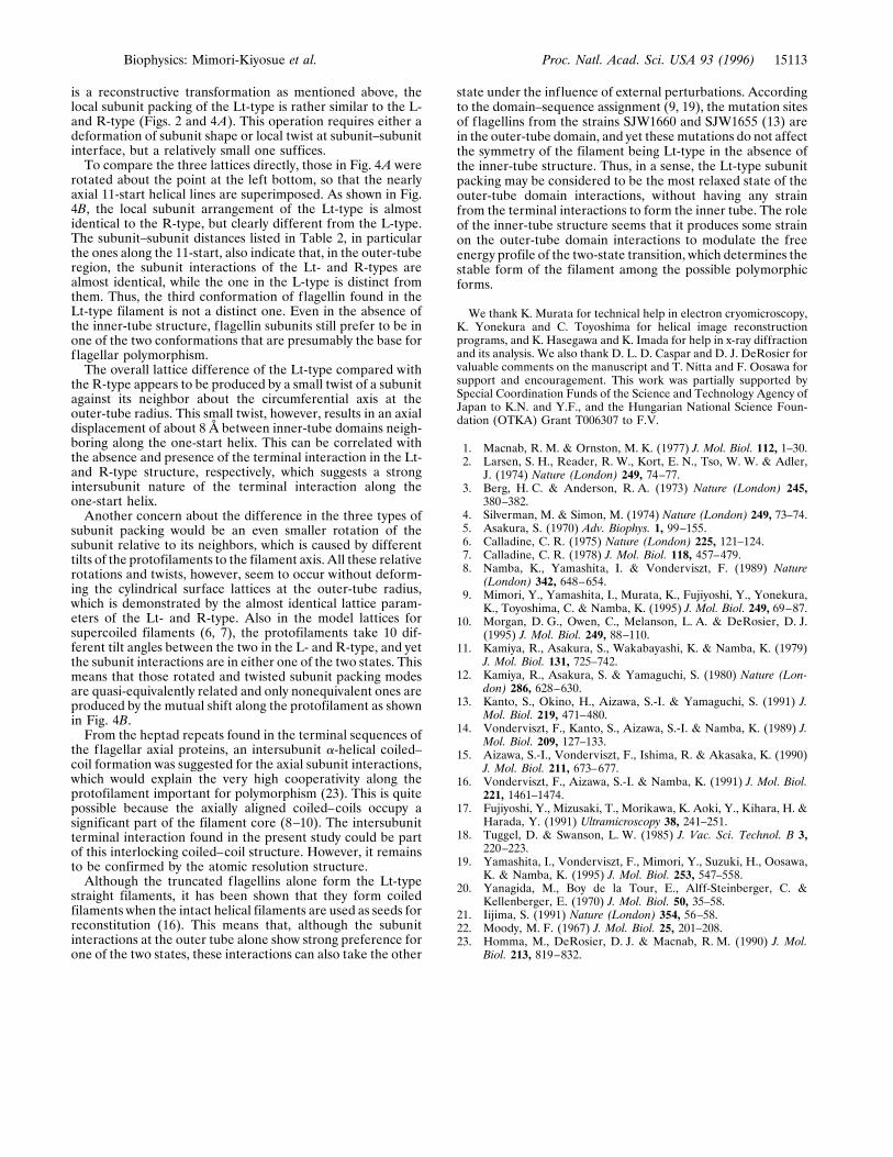
is a reconstructive transformation as mentioned above, thelocal subunit packing of the Lt-type is rather similar to the L-and R-type (Figs. 2 and 4A). This operation requires either adeformation of subunit shape or local twist at subunit–subunitinterface, but a relatively small one suffices.To compare the three lattices directly, those in Fig. 4A were
rotated about the point at the left bottom, so that the nearlyaxial 11-start helical lines are superimposed. As shown in Fig.4B, the local subunit arrangement of the Lt-type is almostidentical to the R-type, but clearly different from the L-type.The subunit–subunit distances listed in Table 2, in particularthe ones along the 11-start, also indicate that, in the outer-tuberegion, the subunit interactions of the Lt- and R-types arealmost identical, while the one in the L-type is distinct fromthem. Thus, the third conformation of flagellin found in theLt-type filament is not a distinct one. Even in the absence ofthe inner-tube structure, f lagellin subunits still prefer to be inone of the two conformations that are presumably the base forflagellar polymorphism.The overall lattice difference of the Lt-type compared with
the R-type appears to be produced by a small twist of a subunitagainst its neighbor about the circumferential axis at theouter-tube radius. This small twist, however, results in an axialdisplacement of about 8 Å between inner-tube domains neigh-boring along the one-start helix. This can be correlated withthe absence and presence of the terminal interaction in the Lt-and R-type structure, respectively, which suggests a strongintersubunit nature of the terminal interaction along theone-start helix.Another concern about the difference in the three types of
subunit packing would be an even smaller rotation of thesubunit relative to its neighbors, which is caused by differenttilts of the protofilaments to the filament axis. All these relativerotations and twists, however, seem to occur without deform-ing the cylindrical surface lattices at the outer-tube radius,which is demonstrated by the almost identical lattice param-eters of the Lt- and R-type. Also in the model lattices forsupercoiled filaments (6, 7), the protofilaments take 10 dif-ferent tilt angles between the two in the L- and R-type, and yetthe subunit interactions are in either one of the two states. Thismeans that those rotated and twisted subunit packing modesare quasi-equivalently related and only nonequivalent ones areproduced by the mutual shift along the protofilament as shownin Fig. 4B.From the heptad repeats found in the terminal sequences of
the flagellar axial proteins, an intersubunit a-helical coiled–coil formation was suggested for the axial subunit interactions,which would explain the very high cooperativity along theprotofilament important for polymorphism (23). This is quitepossible because the axially aligned coiled–coils occupy asignificant part of the filament core (8–10). The intersubunitterminal interaction found in the present study could be partof this interlocking coiled–coil structure. However, it remainsto be confirmed by the atomic resolution structure.Although the truncated flagellins alone form the Lt-type
straight filaments, it has been shown that they form coiledfilaments when the intact helical filaments are used as seeds forreconstitution (16). This means that, although the subunitinteractions at the outer tube alone show strong preference forone of the two states, these interactions can also take the other
state under the influence of external perturbations. Accordingto the domain–sequence assignment (9, 19), the mutation sitesof flagellins from the strains SJW1660 and SJW1655 (13) arein the outer-tube domain, and yet these mutations do not affectthe symmetry of the filament being Lt-type in the absence ofthe inner-tube structure. Thus, in a sense, the Lt-type subunitpacking may be considered to be the most relaxed state of theouter-tube domain interactions, without having any strainfrom the terminal interactions to form the inner tube. The roleof the inner-tube structure seems that it produces some strainon the outer-tube domain interactions to modulate the freeenergy profile of the two-state transition, which determines thestable form of the filament among the possible polymorphicforms.
We thank K. Murata for technical help in electron cryomicroscopy,K. Yonekura and C. Toyoshima for helical image reconstructionprograms, and K. Hasegawa and K. Imada for help in x-ray diffractionand its analysis. We also thank D. L. D. Caspar and D. J. DeRosier forvaluable comments on the manuscript and T. Nitta and F. Oosawa forsupport and encouragement. This work was partially supported bySpecial Coordination Funds of the Science and Technology Agency ofJapan to K.N. and Y.F., and the Hungarian National Science Foun-dation (OTKA) Grant T006307 to F.V.
1. Macnab, R. M. & Ornston, M. K. (1977) J. Mol. Biol. 112, 1–30.2. Larsen, S. H., Reader, R. W., Kort, E. N., Tso, W. W. & Adler,
J. (1974) Nature (London) 249, 74–77.3. Berg, H. C. & Anderson, R. A. (1973) Nature (London) 245,
380–382.4. Silverman, M. & Simon, M. (1974) Nature (London) 249, 73–74.5. Asakura, S. (1970) Adv. Biophys. 1, 99–155.6. Calladine, C. R. (1975) Nature (London) 225, 121–124.7. Calladine, C. R. (1978) J. Mol. Biol. 118, 457–479.8. Namba, K., Yamashita, I. & Vonderviszt, F. (1989) Nature
(London) 342, 648–654.9. Mimori, Y., Yamashita, I., Murata, K., Fujiyoshi, Y., Yonekura,
K., Toyoshima, C. & Namba, K. (1995) J. Mol. Biol. 249, 69–87.10. Morgan, D. G., Owen, C., Melanson, L. A. & DeRosier, D. J.
(1995) J. Mol. Biol. 249, 88–110.11. Kamiya, R., Asakura, S., Wakabayashi, K. & Namba, K. (1979)
J. Mol. Biol. 131, 725–742.12. Kamiya, R., Asakura, S. & Yamaguchi, S. (1980) Nature (Lon-
don) 286, 628–630.13. Kanto, S., Okino, H., Aizawa, S.-I. & Yamaguchi, S. (1991) J.
Mol. Biol. 219, 471–480.14. Vonderviszt, F., Kanto, S., Aizawa, S.-I. & Namba, K. (1989) J.
Mol. Biol. 209, 127–133.15. Aizawa, S.-I., Vonderviszt, F., Ishima, R. & Akasaka, K. (1990)
J. Mol. Biol. 211, 673–677.16. Vonderviszt, F., Aizawa, S.-I. & Namba, K. (1991) J. Mol. Biol.
221, 1461–1474.17. Fujiyoshi, Y., Mizusaki, T., Morikawa, K. Aoki, Y., Kihara, H. &
Harada, Y. (1991) Ultramicroscopy 38, 241–251.18. Tuggel, D. & Swanson, L. W. (1985) J. Vac. Sci. Technol. B 3,
220–223.19. Yamashita, I., Vonderviszt, F., Mimori, Y., Suzuki, H., Oosawa,
K. & Namba, K. (1995) J. Mol. Biol. 253, 547–558.20. Yanagida, M., Boy de la Tour, E., Alff-Steinberger, C. &
Kellenberger, E. (1970) J. Mol. Biol. 50, 35–58.21. Iijima, S. (1991) Nature (London) 354, 56–58.22. Moody, M. F. (1967) J. Mol. Biol. 25, 201–208.23. Homma, M., DeRosier, D. J. & Macnab, R. M. (1990) J. Mol.
Biol. 213, 819–832.
Biophysics: Mimori-Kiyosue et al. Proc. Natl. Acad. Sci. USA 93 (1996) 15113


