Embed Size (px)
Citation preview
Depletion of TDP 43 overrides the need for exonicand intronic splicing enhancers in the humanapoA-II genePablo Arrisi Mercado1, Youhna M. Ayala1, Maurizio Romano1,2,
Emanuele Buratti1 and Francisco E. Baralle1,*
1International Centre for Genetic Engineering and Biotechnology, Padriciano 99, I-34012 Trieste, Italy and2Department of Physiology and Pathology, University of Trieste, Via A. Fleming 22, 34127 Trieste, Italy
Received August 2, 2005; Revised September 19, 2005; Accepted September 27, 2005
ABSTRACT
Exon 3 of the human apolipoprotein A-II (apoA-II) geneis efficiently included in the mRNA although itsacceptor site is significantly weak because of a pecu-liar (GU)16 tract instead of a canonical polypyrimidinetract within the intron 2/exon 3 junction. Our previousstudies demonstrated that the SR proteins ASF/SF2and SC35 bind specifically an exonic splicingenhancer (ESE) within exon 3 and promote exon 3splicing. In the present study, we show that the ESEis necessary only in the proper context. In addition,we have characterized two novel sequences in theflanking introns that modulateapoA-II exon 3 splicing.There is a G-rich element in intron 2 that interactswith hnRNPH1 and inhibits exon 3 splicing. Thesecond is a purine rich region in intron 3 that bindsSRp40 and SRp55 and promotes exon 3 inclusion inmRNA. We have also found that the (GU) repeats inthe apoA-II context bind the splicing factor TDP-43and interfere with exon 3 definition. Significantly,blocking of TDP-43 expression by small interferingRNA overrides the need for all the other cis-actingelements making exon 3 inclusion constitutive evenin the presence of disrupted exonic and intronicenhancers. Altogether, our results suggest thatexonic and intronic enhancers have evolved to bal-ance the negative effects of the two silencers locatedin intron 2 and hence rescue the constitutive exon 3inclusion in apoA-II mRNA.
INTRODUCTION
The pre mRNA splicing process leading to the synthesis ofmature mRNA takes place in the spliceosome, a complex of
small ribonucleoprotein particles (snRNPs) and splicingfactors and is finely modulated (1,2). Cis-acting elementsthat influence splice site selection include the 50 and 30 splicesites, the branch point sequence and the polypyrimidinetract. Beyond the above mentioned canonical cis-acting ele-ments, accessory sequences, such as exonic splicing enhancers(ESEs), exonic splicing silencers (ESSs), intronic splicingenhancers (ISEs) and intronic splicing silencers (ISSs) arealso important for the definition of an exon (3,4).
Recently, we have demonstrated the existence of an ESEwithin the third exon of the apolipoprotein A-II (apoA-II) genethat specifically binds the SR proteins ASF/SF2 and SC35.The mutagenesis of this element prevented the binding ofthese trans-acting elements and caused in vivo skipping ofexon 3 (5). Moreover, overexpression of ASF/SF2 and SC35improved exon 3 inclusion, further supporting the positiveinvolvement of these factors in exon 3 definition. Mechanis-tically, the recruitment of ASF/SF2 and SC35 might be actingas part of a scaffold to allow the recognition of the 30 splicesite, whose polypyrimidine tract is weak because of the pecu-liar (GU)16 stretch adjacent to the intron/exon junction, whichprevious functional studies have indicated as a controller ofexon 3 splicing (5). In this paper we show that the (GU)16
stretch in the apoA-II context also binds the TDP-43 protein,a splicing factor implicated in the CFTR exon 9 skippingthrough its interaction with the (GU) repeats located withinthe 30 splice site of CFTR intron 8 (6). Thus, we have revisitedthe function of the (GU) tract in apoA-II intron 2 by demon-stration that its interaction with TDP-43 has an inhibitoryrole for the splicing of apoA-II exon 3, similarly to what wasobserved for the splicing of CFTR exon 9. Moreover, we havecharacterized a novel negative cis-acting element withinapoA-II IVS2 and a positive cis-acting element in IVS3involved in the control of the apoA-II exon 3 splicing. Inter-estingly, RNA interference against TDP-43 abolishes theeffects of both exonic and intronic enhancer disruption. Thisresult seems to indicate that ESEs and ISEs in the human
*To whom correspondence should be addressed. Tel: +39 040 3757337; Fax: +39 040 3757361; Email: [email protected]
� The Author 2005. Published by Oxford University Press. All rights reserved.
The online version of this article has been published under an open access model. Users are entitled to use, reproduce, disseminate, or display the open accessversion of this article for non-commercial purposes provided that: the original authorship is properly and fully attributed; the Journal and Oxford University Pressare attributed as the original place of publication with the correct citation details given; if an article is subsequently reproduced or disseminated not in its entirety butonly in part or as a derivative work this must be clearly indicated. For commercial re-use, please contact [email protected]
6000–6010 Nucleic Acids Research, 2005, Vol. 33, No. 18doi:10.1093/nar/gki897
Published online October 27, 2005
apoAII gene have evolved to counteract the negative effect ofTDP43 binding to the (GU) repeats.
MATERIALS AND METHODS
Constructs
The wild-type and A97T sets of the pcDNApoIVS2 andpcDNApoIVS3 constructs were generated throughout over-lap PCR method using apoA-II gene and apoA-II cDNA astemplates. All pApo constructs carrying deletions or pointmutations were generated by site-directed mutagenesis usingstandard PCR conditions. The identity of all constructswas checked by a CEQ2000 sequencer (Beckman Coulter,Fullerton, CA) according to the manufacturer’s instructions.Constructs PTBApo-wt and PTBApo-A97T as well asPTBApo-wt-ISEmut and PTBApo-A97T-ISEmut were gener-ated as follows: the apoA-II exon 3 plus portions of bothflanking introns 2 (174 nt) and 3 (116 nt) were PCR-amplified from pApo-wt and pApo-A97T, respectively, andfilled in with DNA polymerase (Klenow). The fragment wasthen inserted in a modified version of the a-globin–fibronectinEDB minigene in which this alternatively spliced exon hadbeen removed to generate a unique Nde I site which wassubsequently filled in with DNA polymerase I (Klenow) treat-ment, making it suitable for the insertion of the exon andits flanking introns under study. Constructs carrying partialdeletions of either intron 2 or intron 3 were prepared startingfrom templates pApo-wt and pApo-A97T. The deletions wereperformed by overlapping PCRs products obtained usingsuitable primers. Generation of constructs mg(GU)16 andmgD(GU) for in vitro experiments was carried out amplifyingfrom pApo-wt and D(gu) plasmids (5) with the followingcouple of primers: SXhoI (50-cccgctcgagcactgttaccaacatga-agct-30) and HindIII/SacII AS (50-ttttccgcggaagcttctctgcgaa-ttctaagcctaatctgccctaa-30). Whereas generation of construct(GU)16-Ex3 was performed with the primers: ApoAII-TG/Exon3–50 (XhoI) (50-cccgctcgagggacccagctgaaaagag-30) andApoAII-Exon3–30 (SacII) (50-ttttccgcggaagcttttggcctcggcct-gaa-30).
Transfections
The DNA used for transient transfections was prepared withJetStar purification kit (Genomed, GmbH, Lohne, Germany)following the manufacturer’s instructions. Then, 3 mg of DNAconstruct was transfected in 3 · 105 human hepatocarcinomaHep3B cells using Qiagen Effectene transfection reagents.Total RNA was extracted using RNAwiz reagent (Ambion,Austin, TX) and retro-transcribed with poly(dT) primer. Toamplify only the messenger derived from the transfections,PCRs were carried out with primers Ex1–1221 S (50-accaag-gacagagacgctggct-30) and Not/Cla rev (50-tctggacactgcggccg-catcg-30) for constructs derived from pApo gene system andwith primers a (50-caacttcactcctaagccactg-30) and Bra (50-gggtcaccaggaagttggttaaatca-30) for constructs derived fromthe a-globin–fibronectin EDB minigene (PTB). The condi-tions used for the PCRs were the following: 94�C for 5 minfor the initial denaturation, 94�C for 30 min, 60�C for 1 min,72�C for 1.5 min for 35 cycles, and 72�C for 7 min for the finalextension. The PCRs were optimized to be in the exponential
phase of amplification. For the SR protein overexpressionexperiments, 2 mg of the constructs pApo-wt, pApo-ISE3m,pApoA97T or pApoA97T-ISE3m was cotransfected with2 mg of SRp40 and SRp55 coding sequences cloned intopCG vector (a kind gift of Dr J. Caceres). The results of allthe transfections are the representative of at least three inde-pendent experiments. In order to quantify the proportion ofalternative splicing, the relative amount of exon 3 skippingwas quantified by optical densitometry.
RNA electrophoretic mobility shift assay (EMSA)and UV-crosslinking of protein and RNA
In order to generate the two different versions of humanapoA-II intron 3 ISE RNAs, EcoRI–SalI double digestionwas carried out on the construct pApo-wt and pApo-wt-ISEmut and subsequently cloned in pBluescript (SK) plasmiddigested with the same enzymes. Once linearized with EcoRI,the constructs were in vitro transcribed in the presence of[a-32 P]UTP for 60 min at 37�C and treated with DNase forthe same period of time. For EMSA, the [a-32P]UTP-labeledRNA probes (4–6 fmol) were incubated with 5.2 mM HEPES,pH 7.9, 1 mM MgCl2, 0.8 mM magnesium acetate, 0.52 mMDTT, 3.8% glycerol, 0,75 mM ATP, 1 mM GTP, 0.5 mg/mlheparin and 30 mg of HeLa nuclear extract (4C Biotech,Seneffe, Belgium) in a final volume of 20 ml for 20 minat room temperature. Following the addition of 5 ml of50% (v/v) glycerol and tracking dye, the complexes wereresolved on a 4% polyacrylamide gel (ratio of 19:1acrylamide/bisacrylamide) in 75 mM Tris–glycine buffer(75 mM Tris and 75 mM glycine) at 15–25 mA for 3–4 hat 4�C. The gels were dried and exposed to X-OMAT ARfilms for 1–3 h. The UV-crosslinking assay was performedby incubation of the [a-32P]UTP-labeled RNA probes (1 ·106 c.p.m./incubation) with 20 mg of HeLa nuclear extractin 20 ml of final volume at 30�C for 15 min. Final bindingconditions were 20 mM HEPES, pH 7.9, 72 mM KCl, 1.5 mMMgCl2, 0.78 mM magnesium acetate, 0.52 mM DTT, 3.8%glycerol, 0.75 mM ATP, 1 mM GTP and heparin at a 5 mg/mlfinal concentration as a nonspecific competitor. In the com-petition experiments increasing amounts of cold RNA (themolar ratios of cold/labeled RNA were 3 and 6) was alsoadded as a competitor 5 min before addition of the labeledRNAs. The samples were then transferred in the wells of anHLA plate (Intermed, Nunc, Roskilde, Denmark) and irradi-ated with UV light on ice (800 000 kJ for 5 min). UnboundRNA was then digested with 30 mg of RNase A and 6 U ofRNase T1 (Sigma) by incubation at 37�C for 30 min. Thesamples were then analyzed by 10% SDS–PAGE followedby autoradiography.
Crosslinking of RNA to adipicdehydrazide-agarose beads
Two synthetic RNA oligos IVS2-G123w, auaccuuuuuggggag-ggggcagagagc, and IVS2n-G123m, auaccuuuuugcggagcg-cgcagagagc (Dharmacon, Chicago, IL), were generated andcrosslinked to adipic dehydrazide–agarose beads. Afterwardsthey were separately incubated with HeLa nuclear extract withthe addition of heparin to a final concentration of 5 mg/ml.Proteins were separated on a 10% SDS–PAGE gel and
Nucleic Acids Research, 2005, Vol. 33, No. 18 6001
visualized by Coomassie brilliant blue staining or electro-blotted onto a nitrocellulose membrane and probed with arabbit polyclonal anti-hnRNPH antiserum. Immunoblottingswere detected using the ECL chemiluminescence kit (Pierce,Rockford, IL).
Immunoprecipitation of SR proteins andTDP-43 following UV-crosslinking
The UV-crosslinking of labeled RNAs with commercialHeLa nuclear extract was performed as described above.After the 30 min incubation with RNase at 37�C, 150 ml ofimmunoprecipitation (IP) buffer (20 mM Tris, pH 8.0,300 mM NaCl, 1 mM EDTA and 0.25% Nonidet P-40) wasadded to each sample together with 1 mg of monoclonalantibodies mouse anti-SR 1H4 (Zymed Laboratories Inc.,San Francisco, CA) and incubated for 2 h at 4�C on a rotatorwheel. Afterward, 30 ml of protein A7G-Plus-agarose (SantaCruz Biotechnology, Santa Cruz, CA) was added to eachsample and incubated overnight at 4�C. The beads were sub-jected to four washing cycles with 1.5 ml of IP buffer andloaded on a 10% SDS–PAGE. The gels were run at a constantrate of 30 mA for 3.5 h, dried and exposed for 4–6 days with aBiomax Screen (Kodak, Rochester, NY). The [a-32P]UTP-labeled RNA probes mg(GU)16, mgD(GU) and (GU)16-Ex3used for UV-cross linking/IP were treated as describedabove, with the only difference that 1 mg of polyclonal anti-serum against TDP-43 was added. After a 2 h incubation, 30 mlof Protein A/G-Plus Agarose (Santa Cruz Biotechnologies)was added to each sample and the mixture was incubatedat 4�C overnight. The beads were then subjected to fourwashing cycles with 1.5 ml of IP buffer and loaded onto anSDS–PAGE. Gels were run at a constant 30 mA for �3.5 h,dried, and then exposed for 4–6 days with a BioMax Screen(Kodak). Three independent IP assays were carried out foreach antibody.
RNA interference against TDP-43
Hep3B cells (2 · 105) were seeded 24 h before the firsttransfection with the small interfering RNA (siRNA) oligo-nucleotide specific for TDP-43 (Y. M. Ayala, personalcommunication). Oligofectamine (3 ml) (Invitrogen BV,Leek, The Netherlands) was combined with 13 ml of Optimemmedium (Invitrogen) and incubated for 7 min at room tem-perature (RT) (reaction 1). Meanwhile, in a different tube, 2.5ml of the TDP-43 siRNA was mixed with 180 ml of Optimem(reaction 2). After the incubation, both reactions were mixedand left for 20 min at RT. Afterwards, the mix was added to thecells and maintained in the incubator at 37�C and 6% of CO2
for 48 h. The cells were then washed with phosphate-bufferedsaline (PBS) and the transfection procedure was repeated withthe difference that cells were maintained for only 24 h. Cellswere washed with PBS and transfected with 0.5 mg of eachminigene using Qiagen Effectene transfection reagents. RNAextraction and RT–PCR analysis were performed using pri-mers complementary to sequences in the flanking fibronectinexonic sequences. Cell lysates were collected and analyzed forhnRNP H1 endogenous protein expression by western analysisusing anti-H1 rabbit serum.
RESULTS
An intronic splicing enhancer is presentwithin apoA-II intron 3
Our previous studies have evidenced that (i) the 30 splice siteof IVS2 is weak because of the (GU) tract adjacent to theacceptor site and (ii) a positive splicing cis-acting element(ESEwt) is placed within exon 3 and its alteration by pointmutations (ESE-A97T) causes exon 3 skipping (5). In general,the interplay among different exonic and/or intronic sequencesand the trans-acting factors can influence the recruitment ofthe constitutive splicing factors U2AF65 and U1 to the 30 and50 splice site, respectively. We were interested to identify otherapoA-II exon 3 splicing control elements eventually placedwithin its intronic flanking regions and to test these potentialcis-acting elements in an heterologous system. For this pur-pose, the wild-type and A97T ApoA-II exon 3 (133 nt) with itsflanking intronic regions (174 nt of intron 2 and 116 nt ofintron 3) were cloned into the third intron of the pTB vector,which is an a-globin/fibronectin reporter minigene systemused to study alternative splicing of several exons (7,8).(Figure 1A). The splicing profile from pTB minigenes over-lapped those obtained with the pApo constructs (Figure 1B).In fact, pTBApo-wt and pTBApo-A97T constructs yielded5 and 85% of exon 3 exclusion (Figure 1B, lanes 3 and 4),so mimicking substantially the splicing ratio from the corre-sponding variants in apoAII gene context (Figure 1B, lanes 1and 2). These results indicate that the 423 nt genomic fragmentof apoAII gene contains all the features important for effici-ent exon 3 inclusion in apoAII mRNA. Therefore, the pTBconstructs can be used to the following mapping of potentialintronic cis-acting elements that influence exon 3 splicing.
Initially, in order to verify the eventual presence of otherapoA-II exon 3 splicing control elements, we screened itsintronic flanking regions looking for possible cis-acting ele-ments whose sequence might resemble previously describedmodulators of splicing. In order to look for other splicingcontrol elements in IVS3, we created a deletion of 97 nt inintron 3 (from nt 1925 to 2022 in the apoA-II gene; GenBankaccession number X04898.1) so obtaining the constructpApo-wt-D1925–2022 (Figure 1C). This 97 nt-deletion causeda 40% skipping of the exon 3 (Figure 1E, lane 3). This obser-vation supports the existence of one or more elements withinintron 3 that promote the inclusion of the apoA-II exon 3. Wenoticed the presence of three G runs in intron 3 just 10 bpdownstream of the exon 3 donor splice site, namely I3G1 toI3G3, and a polypurine-rich sequence between nt 1935 and1960 (Figure 1C). As these elements are close to the splicesites of exon 3, we have explored first the possible effects of Gruns on the control of the apoA-II exon 3 splicing. To this aim,we first created mutants where the third, the first two andall the three G runs were disrupted by point mutations inintron 3. However, the transient transfection of this mutantdid not show significant changes in the apoA-II exon 3splicing pattern in comparison with the pApo-wt construct(Figure 1D). Thus, it is reasonable to exclude a role in thesplicing of exon 3 for the G runs within intron 3. Conversely, thedeletion of the purine-rich tract caused 25% apoA-II exon 3skipping (Figure 1E, lane 4). In order to confirm the enhancingactivity within nt 1941–1967, the continuity of the purine
6002 Nucleic Acids Research, 2005, Vol. 33, No. 18
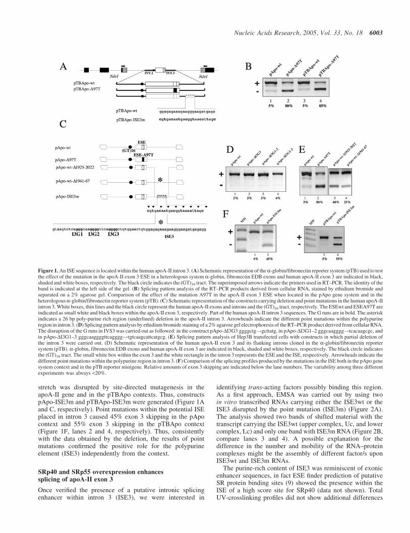
stretch was disrupted by site-directed mutagenesis in theapoA-II gene and in the pTBApo contexts. Thus, constructspApo-ISE3m and pTBApo-ISE3m were generated (Figure 1Aand C, respectively). Point mutations within the potential ISEplaced in intron 3 caused 45% exon 3 skipping in the pApocontext and 55% exon 3 skipping in the pTBApo context(Figure 1F, lanes 2 and 4, respectively). Thus, consistentlywith the data obtained by the deletion, the results of pointmutations confirmed the positive role for the polypurineelement (ISE3) independently from the context.
SRp40 and SRp55 overexpression enhancessplicing of apoA-II exon 3
Once verified the presence of a putative intronic splicingenhancer within intron 3 (ISE3), we were interested in
identifying trans-acting factors possibly binding this region.As a first approach, EMSA was carried out by using twoin vitro transcribed RNAs carrying either the ISE3wt or theISE3 disrupted by the point mutation (ISE3m) (Figure 2A).The analysis showed two bands of shifted material with thetranscript carrying the ISE3wt (upper complex, Uc, and lowercomplex, Lc) and only one band with ISE3m RNA (Figure 2B,compare lanes 3 and 4). A possible explanation for thedifference in the number and mobility of the RNA–proteincomplexes might be the assembly of different factor/s uponISE3wt and ISE3m RNAs.
The purine-rich content of ISE3 was reminiscent of exonicenhancer sequences, in fact ESE finder prediction of putativeSR protein binding sites (9) showed the presence within theISE of a high score site for SRp40 (data not shown). TotalUV-crosslinking profiles did not show additional differences
Figure 1. An ISE sequence is located within the human apoA-II intron 3. (A) Schematic representation of thea-globin/fibronectin reporter system (pTB) used to testthe effect of the mutation in the apoA-II exon 3 ESE in a heterologous system a-globin, fibronectin EDB exons and human apoA-II exon 3 are indicated in black,shaded and white boxes, respectively. The black circle indicates the (GT)16 tract. The superimposed arrows indicate the primers used in RT–PCR. The identity of theband is indicated at the left side of the gel. (B) Splicing pattern analysis of the RT–PCR products derived from cellular RNA, stained by ethidium bromide andseparated on a 2% agarose gel. Comparison of the effect of the mutation A97T in the apoA-II exon 3 ESE when located in the pApo gene system and in theheterologousa-globin/fibronectin reporter system (pTB). (C) Schematic representation of the constructs carrying deletion and point mutations in the human apoA-IIintron 3. White boxes, thin lines and the black circle represent the human apoA-II exons and introns and the (GT)16 tract, respectively. The ESEwt and ESEA97T areindicated as small white and black boxes within the apoA-II exon 3, respectively. Part of the human apoA-II intron 3 sequences. The G runs are in bold. The asteriskindicates a 26 bp poly-purine rich region (underlined) deletion in the apoA-II intron 3. Arrowheads indicate the different point mutations within the polypurineregion in intron 3. (D) Splicing pattern analysis by ethidium bromide staining of a 2% agarose gel electrophoresis of the RT–PCR product derived from cellular RNA.The disruption of the G runs in IVS3 was carried out as followed: in the construct pApo-DI3G3 ggggctg!gcttatg, in pApo-DI3G1–2 gggcaagggg!tcacaagcgc, andin pApo-DI3G1–3 gggcaaggggttcagggg!tgtcaagcattcatgcg. (E) Splicing pattern analysis of Hep3B transfected cells with constructs in which partial deletion ofthe intron 3 were carried out. (D) Schematic representation of the human apoA-II exon 3 and its flanking introns cloned in the a-globin/fibronectin reportersystem (pTB). a-globin, fibronectin EDB exons and human apoA-II exon 3 are indicated in black, shaded and white boxes, respectively. The black circle indicatesthe (GT)16 tract. The small white box within the exon 3 and the white rectangle in the intron 3 represents the ESE and the ISE, respectively. Arrowheads indicate thedifferent point mutations within the polypurine region in intron 3. (F) Comparison of the splicing profiles produced by the mutations in the ISE both in the pApo genesystem context and in the pTB reporter minigene. Relative amounts of exon 3 skipping are indicated below the lane numbers. The variability among three differentexperiments was always <20%.
Nucleic Acids Research, 2005, Vol. 33, No. 18 6003
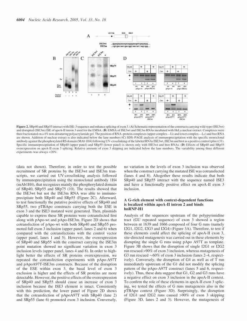
(data not shown). Therefore, in order to test the possiblerecruitment of SR proteins by the ISE3wt and ISE3m tran-scripts, we carried out UV-crosslinking analysis followedby immunoprecipitation using the monoclonal antibody 1H4(mAb1H4), that recognizes mainly the phosphorylated domainof SRp40, SRp55 and SRp75 (10). The results showed thatthe ISE3wt but not the ISE3m RNA was able to immuno-precipitate both SRp40 and SRp55 (Figure 2C). Afterward,to test functionally the putative positive effects of SRp40 andSRp55, two pTBApo constructs carrying both the ESE inexon 3 and the ISE3 mutated were generated. Then, plasmidscapable to express these SR proteins were cotransfected firstalong with pApo-wt and pApo-ISE3m. Figure 2D shows thatcotransfection of pApo-wt with both SRp40 and SRp55 pro-moted full exon 3 inclusion (upper panel, lanes 2 and 6) whencompared with the cotransfections with the control vector(upper panel, lanes 1 and 5). However, the overexpressionof SRp40 and SRp55 with the construct carrying the ISE3mpoint mutation showed no significant variation in exon 3inclusion levels (upper panel, lanes 4 and 8). In order to high-light better the effects of SR proteins overexpression, werepeated the cotransfection experiments with pApo-A97Tand pApoA97T-ISE3m constructs. Because of the disruptionof the ESE within exon 3, the basal level of exon 3exclusion is higher and the effects of SR proteins are moredetectable. However, the positive effects of the overexpressionof SRp40 and SRp55 should cause an increase of exon 3inclusion because the ISE3 element is intact. Consistentlywith this prediction, the lower panel of Figure 2D showsthat the cotransfection of pApoA97T with SRp40 (lane 2)and SRp55 (lane 6) promoted exon 3 inclusion. Conversely,
no variation in the levels of exon 3 inclusion was observedwhen the construct carrying the mutated ISE was cotransfected(lanes 4 and 8). Altogether these results indicate that bothSRp40 and SRp55 interact with the sequence named ISE3and have a functionally positive effect on apoA-II exon 3inclusion.
A G-rich element with context-dependent functionsis localized within apoA-II intron 2 and bindshnRNP-H1
Analysis of the sequences upstream of the polypyrimidinetract (GU repeated sequence) of exon 3 showed a regionbetween nt 1639 and 1666 composed of four G runs (namelyI2G1, I2G2, I2G3 and I2G4) (Figure 3A). Therefore, to test ifthese elements could affect the splicing of apoA-II exon 3,site-directed mutagenesis was carried out in these elements bydisrupting the single G runs using pApo A97T as template.Figure 3B shows that the disruption of single I2G1 or I2G2run rescued >90% of exon 3 inclusion, whereas mutagenesis ofG3 run rescued �60% of exon 3 inclusion (lanes 2–4, respect-ively). Conversely, the disruption of G4 as well as of T runimmediately upstream of the G1 did not modify the splicingpattern of the pApo-A97T construct (lanes 5 and 6, respect-ively). Thus, these data suggest that G1, G2 and G3 runs havea negative effect on exon 3 inclusion in the apoA-II context.To confirm the role of these elements in apoA-II exon 3 splic-ing, we tested the effects of G runs mutagenesis also in thepTBApo context (Figure 3D). Surprisingly, the disruptionof I2G1 and I2G2 runs caused >90% of exon 3 skipping(Figure 3D, lanes 2 and 3). However, the mutagenesis of
Figure 2. SRp40 and SRp55 interact with ISE-3 sequence and enhance splicing of exon 3. (A) Schematic representation of the constructs carrying wild-type (ISE3wt)and disrupted (ISE3m) ISE of apoA-II intron 3 used for the EMSA. (B) EMSA of ISE3wt and ISE3m RNAs incubated with HeLa nuclear extract. Complexes werethen fractionated on a 4% non-denaturing polyacrylamide gel. The position of RNA–protein complexes (upper complex—Uc and lower complex—Lc) and free RNAare shown. Addition of nuclear extract is also indicated below the lane numbers (C) SDS–PAGE analysis of immunoprecipitation with the specific monoclonalantibody against the phosphorylated RS domain (MAb 1H4) following UV-crosslinking of the labeled RNAs ISE3wt, ISE3m and htot as a positive control (plus) (31).Specific immunoprecipitation of SRp40 (upper panel) and SRp55 (lower panel) is shown only with ISE3wt and htot RNAs. (D) Effects of SRp40 and SRp55overexpression on apoA-II exon 3 splicing. Relative amounts of exon 3 skipping are indicated below the lane numbers. The variability among three differentexperiments was always <20%.
6004 Nucleic Acids Research, 2005, Vol. 33, No. 18
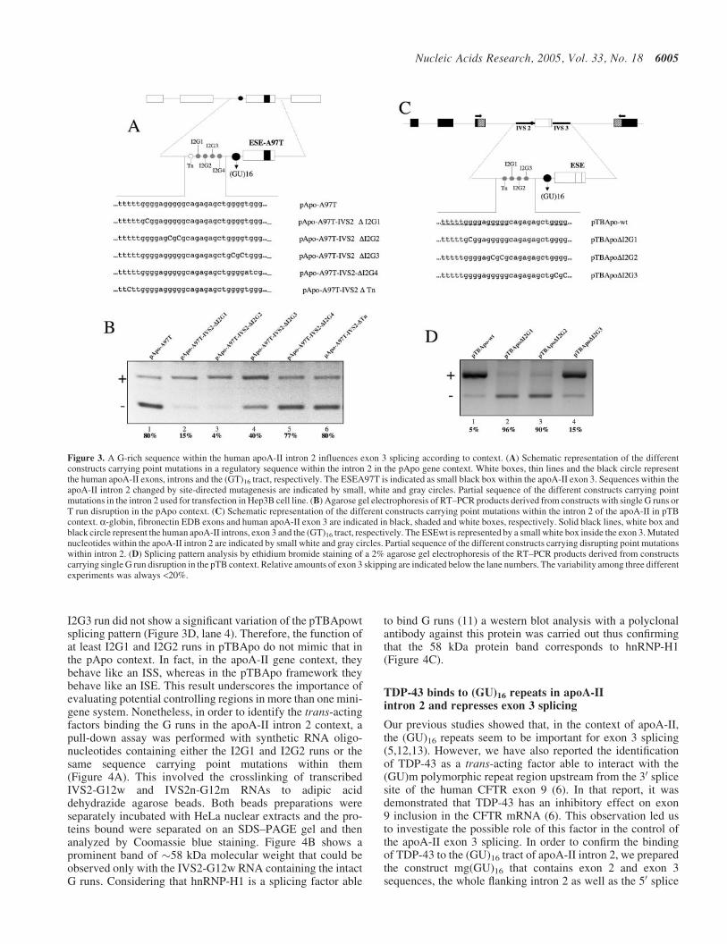
I2G3 run did not show a significant variation of the pTBApowtsplicing pattern (Figure 3D, lane 4). Therefore, the function ofat least I2G1 and I2G2 runs in pTBApo do not mimic that inthe pApo context. In fact, in the apoA-II gene context, theybehave like an ISS, whereas in the pTBApo framework theybehave like an ISE. This result underscores the importance ofevaluating potential controlling regions in more than one mini-gene system. Nonetheless, in order to identify the trans-actingfactors binding the G runs in the apoA-II intron 2 context, apull-down assay was performed with synthetic RNA oligo-nucleotides containing either the I2G1 and I2G2 runs or thesame sequence carrying point mutations within them(Figure 4A). This involved the crosslinking of transcribedIVS2-G12w and IVS2n-G12m RNAs to adipic aciddehydrazide agarose beads. Both beads preparations wereseparately incubated with HeLa nuclear extracts and the pro-teins bound were separated on an SDS–PAGE gel and thenanalyzed by Coomassie blue staining. Figure 4B shows aprominent band of �58 kDa molecular weight that could beobserved only with the IVS2-G12w RNA containing the intactG runs. Considering that hnRNP-H1 is a splicing factor able
to bind G runs (11) a western blot analysis with a polyclonalantibody against this protein was carried out thus confirmingthat the 58 kDa protein band corresponds to hnRNP-H1(Figure 4C).
TDP-43 binds to (GU)16 repeats in apoA-IIintron 2 and represses exon 3 splicing
Our previous studies showed that, in the context of apoA-II,the (GU)16 repeats seem to be important for exon 3 splicing(5,12,13). However, we have also reported the identificationof TDP-43 as a trans-acting factor able to interact with the(GU)m polymorphic repeat region upstream from the 30 splicesite of the human CFTR exon 9 (6). In that report, it wasdemonstrated that TDP-43 has an inhibitory effect on exon9 inclusion in the CFTR mRNA (6). This observation led usto investigate the possible role of this factor in the control ofthe apoA-II exon 3 splicing. In order to confirm the bindingof TDP-43 to the (GU)16 tract of apoA-II intron 2, we preparedthe construct mg(GU)16 that contains exon 2 and exon 3sequences, the whole flanking intron 2 as well as the 50 splice
Figure 3. A G-rich sequence within the human apoA-II intron 2 influences exon 3 splicing according to context. (A) Schematic representation of the differentconstructs carrying point mutations in a regulatory sequence within the intron 2 in the pApo gene context. White boxes, thin lines and the black circle representthe human apoA-II exons, introns and the (GT)16 tract, respectively. The ESEA97T is indicated as small black box within the apoA-II exon 3. Sequences within theapoA-II intron 2 changed by site-directed mutagenesis are indicated by small, white and gray circles. Partial sequence of the different constructs carrying pointmutations in the intron 2 used for transfection in Hep3B cell line. (B) Agarose gel electrophoresis of RT–PCR products derived from constructs with single G runs orT run disruption in the pApo context. (C) Schematic representation of the different constructs carrying point mutations within the intron 2 of the apoA-II in pTBcontext. a-globin, fibronectin EDB exons and human apoA-II exon 3 are indicated in black, shaded and white boxes, respectively. Solid black lines, white box andblack circle represent the human apoA-II introns, exon 3 and the (GT)16 tract, respectively. The ESEwt is represented by a small white box inside the exon 3. Mutatednucleotides within the apoA-II intron 2 are indicated by small white and gray circles. Partial sequence of the different constructs carrying disrupting point mutationswithin intron 2. (D) Splicing pattern analysis by ethidium bromide staining of a 2% agarose gel electrophoresis of the RT–PCR products derived from constructscarrying single G run disruption in the pTB context. Relative amounts of exon 3 skipping are indicated below the lane numbers. The variability among three differentexperiments was always <20%.
Nucleic Acids Research, 2005, Vol. 33, No. 18 6005
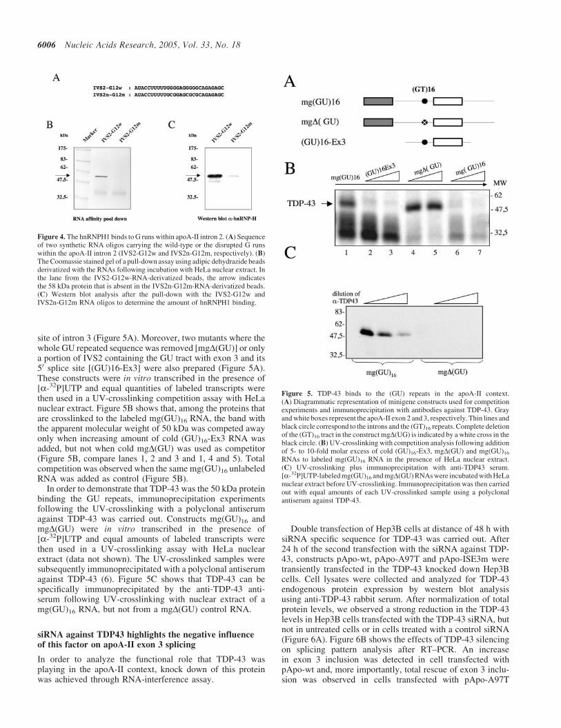
site of intron 3 (Figure 5A). Moreover, two mutants where thewhole GU repeated sequence was removed [mgD(GU)] or onlya portion of IVS2 containing the GU tract with exon 3 and its50 splice site [(GU)16-Ex3] were also prepared (Figure 5A).These constructs were in vitro transcribed in the presence of[a-32P]UTP and equal quantities of labeled transcripts werethen used in a UV-crosslinking competition assay with HeLanuclear extract. Figure 5B shows that, among the proteins thatare crosslinked to the labeled mg(GU)16 RNA, the band withthe apparent molecular weight of 50 kDa was competed awayonly when increasing amount of cold (GU)16-Ex3 RNA wasadded, but not when cold mgD(GU) was used as competitor(Figure 5B, compare lanes 1, 2 and 3 and 1, 4 and 5). Totalcompetition was observed when the same mg(GU)16 unlabeledRNA was added as control (Figure 5B).
In order to demonstrate that TDP-43 was the 50 kDa proteinbinding the GU repeats, immunoprecipitation experimentsfollowing the UV-crosslinking with a polyclonal antiserumagainst TDP-43 was carried out. Constructs mg(GU)16 andmgD(GU) were in vitro transcribed in the presence of[a-32P]UTP and equal amounts of labeled transcripts werethen used in a UV-crosslinking assay with HeLa nuclearextract (data not shown). The UV-crosslinked samples weresubsequently immunoprecipitated with a polyclonal antiserumagainst TDP-43 (6). Figure 5C shows that TDP-43 can bespecifically immunoprecipitated by the anti-TDP-43 anti-serum following UV-crosslinking with nuclear extract of amg(GU)16 RNA, but not from a mgD(GU) control RNA.
siRNA against TDP43 highlights the negative influenceof this factor on apoA-II exon 3 splicing
In order to analyze the functional role that TDP-43 wasplaying in the apoA-II context, knock down of this proteinwas achieved through RNA-interference assay.
Double transfection of Hep3B cells at distance of 48 h withsiRNA specific sequence for TDP-43 was carried out. After24 h of the second transfection with the siRNA against TDP-43, constructs pApo-wt, pApo-A97T and pApo-ISE3m weretransiently transfected in the TDP-43 knocked down Hep3Bcells. Cell lysates were collected and analyzed for TDP-43endogenous protein expression by western blot analysisusing anti-TDP-43 rabbit serum. After normalization of totalprotein levels, we observed a strong reduction in the TDP-43levels in Hep3B cells transfected with the TDP-43 siRNA, butnot in untreated cells or in cells treated with a control siRNA(Figure 6A). Figure 6B shows the effects of TDP-43 silencingon splicing pattern analysis after RT–PCR. An increasein exon 3 inclusion was detected in cell transfected withpApo-wt and, more importantly, total rescue of exon 3 inclu-sion was observed in cells transfected with pApo-A97T
Figure 5. TDP-43 binds to the (GU) repeats in the apoA-II context.(A) Diagrammatic representation of minigene constructs used for competitionexperiments and immunoprecipitation with antibodies against TDP-43. Grayand white boxes represent the apoA-II exon 2 and 3, respectively. Thin lines andblack circle correspond to the introns and the (GT)16 repeats. Complete deletionof the (GT)16 tract in the construct mgD(UG) is indicated by a white cross in theblack circle. (B) UV-crosslinking with competition analysis following additionof 5- to 10-fold molar excess of cold (GU)16-Ex3, mgD(GU) and mg(GU)16
RNAs to labeled mg(GU)16 RNA in the presence of HeLa nuclear extract.(C) UV-crosslinking plus immunoprecipitation with anti-TDP43 serum.[a-32P]UTP-labeled mg(GU)16 and mgD(GU) RNAs were incubated with HeLanuclear extract before UV-crosslinking. Immunoprecipitation was then carriedout with equal amounts of each UV-crosslinked sample using a polyclonalantiserum against TDP-43.
Figure 4. The hnRNPH1 binds to G runs within apoA-II intron 2. (A) Sequenceof two synthetic RNA oligos carrying the wild-type or the disrupted G runswithin the apoA-II intron 2 (IVS2-G12w and IVS2n-G12m, respectively). (B)The Coomassie stained gel of a pull-down assay using adipic dehydrazide beadsderivatized with the RNAs following incubation with HeLa nuclear extract. Inthe lane from the IVS2-G12w-RNA-derivatized beads, the arrow indicatesthe 58 kDa protein that is absent in the IVS2n-G12m-RNA-derivatized beads.(C) Western blot analysis after the pull-down with the IVS2-G12w andIVS2n-G12m RNA oligos to determine the amount of hnRNPH1 binding.
6006 Nucleic Acids Research, 2005, Vol. 33, No. 18
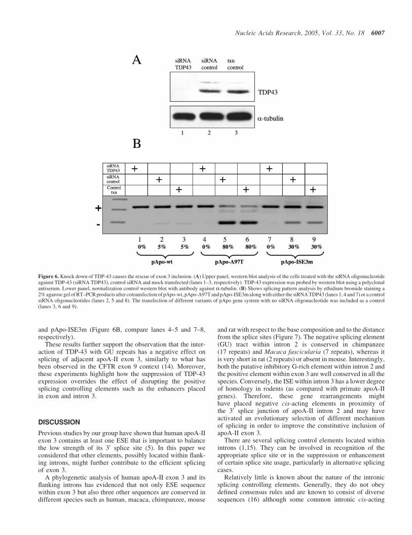
and pApo-ISE3m (Figure 6B, compare lanes 4–5 and 7–8,respectively).
These results further support the observation that the inter-action of TDP-43 with GU repeats has a negative effect onsplicing of adjacent apoA-II exon 3, similarly to what hasbeen observed in the CFTR exon 9 context (14). Moreover,these experiments highlight how the suppression of TDP-43expression overrides the effect of disrupting the positivesplicing controlling elements such as the enhancers placedin exon and intron 3.
DISCUSSION
Previous studies by our group have shown that human apoA-IIexon 3 contains at least one ESE that is important to balancethe low strength of its 30 splice site (5). In this paper weconsidered that other elements, possibly located within flank-ing introns, might further contribute to the efficient splicingof exon 3.
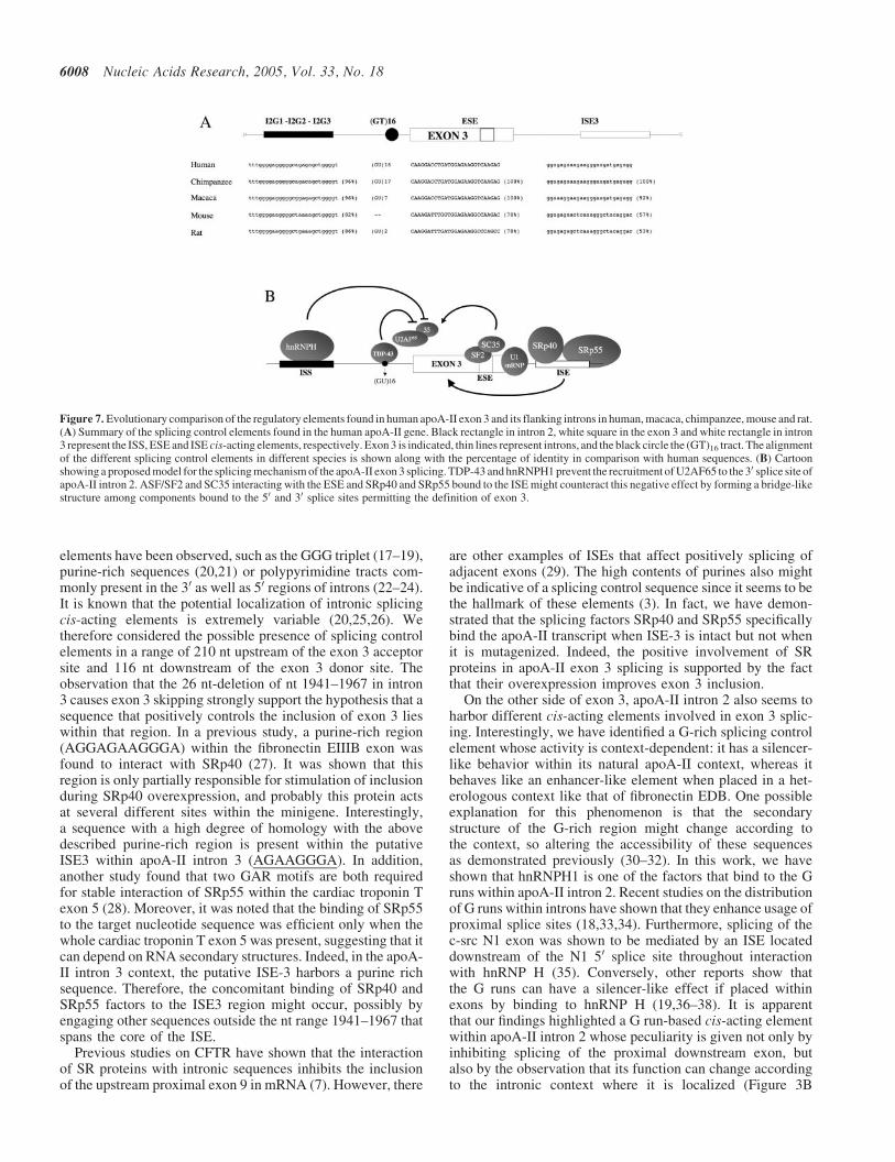
A phylogenetic analysis of human apoA-II exon 3 and itsflanking introns has evidenced that not only ESE sequencewithin exon 3 but also three other sequences are conserved indifferent species such as human, macaca, chimpanzee, mouse
and rat with respect to the base composition and to the distancefrom the splice sites (Figure 7). The negative splicing element(GU) tract within intron 2 is conserved in chimpanzee(17 repeats) and Macaca fascicularia (7 repeats), whereas itis very short in rat (2 repeats) or absent in mouse. Interestingly,both the putative inhibitory G-rich element within intron 2 andthe positive element within exon 3 are well conserved in all thespecies. Conversely, the ISE within intron 3 has a lower degreeof homology in rodents (as compared with primate apoA-IIgenes). Therefore, these gene rearrangements mighthave placed negative cis-acting elements in proximity ofthe 30 splice junction of apoA-II intron 2 and may haveactivated an evolutionary selection of different mechanismof splicing in order to improve the constitutive inclusion ofapoA-II exon 3.
There are several splicing control elements located withinintrons (1,15). They can be involved in recognition of theappropriate splice site or in the suppression or enhancementof certain splice site usage, particularly in alternative splicingcases.
Relatively little is known about the nature of the intronicsplicing controlling elements. Generally, they do not obeydefined consensus rules and are known to consist of diversesequences (16) although some common intronic cis-acting
Figure 6. Knock down of TDP-43 causes the rescue of exon 3 inclusion. (A) Upper panel, western blot analysis of the cells treated with the siRNA oligonucleotideagainst TDP-43 (siRNA TDP43), control siRNA and mock transfected (lanes 1–3, respectively). TDP-43 expression was probed by western blot using a polyclonalantiserum. Lower panel, normalization control western blot with antibody against a-tubulin. (B) Shows splicing pattern analysis by ethidium bromide staining a2% agarose gel of RT–PCR products after cotransfection of pApo-wt, pApo-A97T and pApo-ISE3m along with either the siRNA TDP43 (lanes 1, 4 and 7) or a controlsiRNA oligonucleotides (lanes 2, 5 and 8). The transfection of different variants of pApo gene system with no siRNA oligonucleotide was included as a control(lanes 3, 6 and 9).
Nucleic Acids Research, 2005, Vol. 33, No. 18 6007
elements have been observed, such as the GGG triplet (17–19),purine-rich sequences (20,21) or polypyrimidine tracts com-monly present in the 30 as well as 50 regions of introns (22–24).It is known that the potential localization of intronic splicingcis-acting elements is extremely variable (20,25,26). Wetherefore considered the possible presence of splicing controlelements in a range of 210 nt upstream of the exon 3 acceptorsite and 116 nt downstream of the exon 3 donor site. Theobservation that the 26 nt-deletion of nt 1941–1967 in intron3 causes exon 3 skipping strongly support the hypothesis that asequence that positively controls the inclusion of exon 3 lieswithin that region. In a previous study, a purine-rich region(AGGAGAAGGGA) within the fibronectin EIIIB exon wasfound to interact with SRp40 (27). It was shown that thisregion is only partially responsible for stimulation of inclusionduring SRp40 overexpression, and probably this protein actsat several different sites within the minigene. Interestingly,a sequence with a high degree of homology with the abovedescribed purine-rich region is present within the putativeISE3 within apoA-II intron 3 (AGAAGGGA). In addition,another study found that two GAR motifs are both requiredfor stable interaction of SRp55 within the cardiac troponin Texon 5 (28). Moreover, it was noted that the binding of SRp55to the target nucleotide sequence was efficient only when thewhole cardiac troponin T exon 5 was present, suggesting that itcan depend on RNA secondary structures. Indeed, in the apoA-II intron 3 context, the putative ISE-3 harbors a purine richsequence. Therefore, the concomitant binding of SRp40 andSRp55 factors to the ISE3 region might occur, possibly byengaging other sequences outside the nt range 1941–1967 thatspans the core of the ISE.
Previous studies on CFTR have shown that the interactionof SR proteins with intronic sequences inhibits the inclusionof the upstream proximal exon 9 in mRNA (7). However, there
are other examples of ISEs that affect positively splicing ofadjacent exons (29). The high contents of purines also mightbe indicative of a splicing control sequence since it seems to bethe hallmark of these elements (3). In fact, we have demon-strated that the splicing factors SRp40 and SRp55 specificallybind the apoA-II transcript when ISE-3 is intact but not whenit is mutagenized. Indeed, the positive involvement of SRproteins in apoA-II exon 3 splicing is supported by the factthat their overexpression improves exon 3 inclusion.
On the other side of exon 3, apoA-II intron 2 also seems toharbor different cis-acting elements involved in exon 3 splic-ing. Interestingly, we have identified a G-rich splicing controlelement whose activity is context-dependent: it has a silencer-like behavior within its natural apoA-II context, whereas itbehaves like an enhancer-like element when placed in a het-erologous context like that of fibronectin EDB. One possibleexplanation for this phenomenon is that the secondarystructure of the G-rich region might change according tothe context, so altering the accessibility of these sequencesas demonstrated previously (30–32). In this work, we haveshown that hnRNPH1 is one of the factors that bind to the Gruns within apoA-II intron 2. Recent studies on the distributionof G runs within introns have shown that they enhance usage ofproximal splice sites (18,33,34). Furthermore, splicing of thec-src N1 exon was shown to be mediated by an ISE locateddownstream of the N1 50 splice site throughout interactionwith hnRNP H (35). Conversely, other reports show thatthe G runs can have a silencer-like effect if placed withinexons by binding to hnRNP H (19,36–38). It is apparentthat our findings highlighted a G run-based cis-acting elementwithin apoA-II intron 2 whose peculiarity is given not only byinhibiting splicing of the proximal downstream exon, butalso by the observation that its function can change accordingto the intronic context where it is localized (Figure 3B
Figure 7. Evolutionary comparison of the regulatory elements found in human apoA-II exon 3 and its flanking introns in human, macaca, chimpanzee, mouse and rat.(A) Summary of the splicing control elements found in the human apoA-II gene. Black rectangle in intron 2, white square in the exon 3 and white rectangle in intron3 represent the ISS, ESE and ISE cis-acting elements, respectively. Exon 3 is indicated, thin lines represent introns, and the black circle the (GT)16 tract. The alignmentof the different splicing control elements in different species is shown along with the percentage of identity in comparison with human sequences. (B) Cartoonshowing a proposed model for the splicing mechanism of the apoA-II exon 3 splicing. TDP-43 and hnRNPH1 prevent the recruitment of U2AF65 to the 30 splice site ofapoA-II intron 2. ASF/SF2 and SC35 interacting with the ESE and SRp40 and SRp55 bound to the ISE might counteract this negative effect by forming a bridge-likestructure among components bound to the 50 and 30 splice sites permitting the definition of exon 3.
6008 Nucleic Acids Research, 2005, Vol. 33, No. 18
and D). In addition to a possible role of RNA structure, thiscontext-dependent effect may also depend on interactions withthe other splicing regulatory elements that are specific for theapoA-II gene. In addition, the enhancer/silencer activity mightbe affected by splicing events preceding the definition ofapoA-II exon 3 or by the architecture of the transcriptionalmachinery (both constructs have different promoters) asdemonstrated in other splicing models (39,40). Future studieswill be aimed at characterizing the mechanisms of splicingcontrol by G runs within IVS 2 and the role of hnRNP H1.
The other cis-acting element found close to the 30 splice siteof intron 2 consists in the (GU)16 repeats that were previouslyshown to be functional as polypyrimidine tract (13). However,it is important the context where it is located: in fact, thereplacement of the EDA exon polypyrimidine tract with theapoA-II (GT)16 tract resulted in 95% of EDA exon exclusion(5). Furthermore, previous experiments on the apoA-II modelshowed that either the deletion of (GU)16 tract or its replace-ment by (CA)16 promoted exon 3 skipping (5,12). In principle,it could not be excluded that the deletion might cause exon3 exclusion through alteration of branch point 30 splice sitespacing and that the replacement of (GU)16 by (CA)16 mightinterfere with the 30 splice site definition because of the bind-ing to another factor, such as hnRNP L that was recentlydescribed as able to bind to (CA) repeats in the eNOS gene(41). We now have confirmed that the apoA-II (GU)16 repeatsare bound specifically by TDP-43 protein, similarly to whathappen in the CFTR intron 8 context (6). Considering thatTDP-43 has a detrimental role for CFTR exon 9 splicing, wewondered if this factor might have a similar effect also in theapoA-II intron 2 context and that this effect was counteractedby the positive elements mapped above. Indeed, siRNAexperiments to knock down TDP-43 in Hep3B cells haveshown its inhibitory role also for apoA-II exon 3 splicing.This finding overlaps what was observed with CFTR exon 9,suggesting a general negative role of TDP-43 factors recruitedby (GU) repeats placed at the 30 splice site. Moreover, theobservation that the (GU) tract do not bind to U2AF65 (13)suggest that different mechanisms for 30 splice site recognitioncan exist. Altogether these observations further suggest thatthe 30 splice site definition of the apoA-II exon 3 might besupported by other strong cis-acting elements that can contrastthe negative effects of TDP-43 binding in proximity of the30 splice site of exon 3. The question arises if all thesemodulatory elements have evolved to counteract the negativeinfluence of the weak 30 splice site or have additional role inapoA-II splicing. The negative effect of TDP-43 on exon 3splicing is remarkably more evident when we transfected theconstructs pApo-A97T and pApo-ISE3m in cells where theexpression of that protein was blocked by siRNA: in fact,TDP-43 silencing was able to fully rescue the effects ofboth exonic and intronic enhancer mutations. This findingsuggests that apoA-II exon 3 definition can tolerate the dis-ruption of splicing enhancers on condition that the inhibitoryeffects of TDP-43 is relieved. It follows that exon 3 splicingresults from the coordinate action of positive cis-acting ele-ments that balance the negative influence on apoA-II exon 3splicing derived from the elements located at the 30 end ofintron 2. A large body of evidences indicates that U2AF35 isrequired for constitutive splicing and also works as a mediatorof enhancer-dependent splicing (42). In vitro protein–RNA
interaction studies with pre-mRNAs containing either aconstitutive or regulated splicing enhancer have shown thatU2AF35 directly mediates interactions between U2AF65 andproteins bound to the enhancers (43). On the basis of our data,in the apoA-II intron 2 context, without enhancers, U2AF65should not work efficiently owing to the weakness of thepolypyrimidine tract. We are temped to speculate that theinteraction of TDP-43 with (GU) repeats and of hnRNPH1to the G runs within intron 2 might interfere the functionof U2AF65. Therefore, both the exonic and the intronicenhancers might have a pivotal role in definition of exon 3.However, the binding of ASF/SF2 and SC35 to the enhancerwithin exon 3 as well as the interaction of SRp40 and SRp55to the sequence within intron 3 might be crucial to recruit theconstitutive splicing factors U2AF35 and U2AF65 essential todefine the 30 splice site of intron 2. In this way, a bridge-likestructure among components bound to the 50 and 30 splicesites would be formed to define exon 3 independently of thepolypyrimidine tract pathway.
ACKNOWLEDGEMENTS
This work was supported by grants from Telethon OnlusFoundation—Italy (no. GGP02453) and F.I.R.B. (no.RBNE01W9PM) to F.E.B. Funding to pay the Open Accesspublication charges for this article was provided by ICGEB.
Conflict of interest statement. None declared.
REFERENCES
1. Smith,C.W. and Valcarcel,J. (2000) Alternative pre-mRNA splicing:the logic of combinatorial control. Trends Biochem. Sci.,25, 381–388.
2. Black,D.L. (2003) Mechanisms of alternative pre-messenger RNAsplicing. Annu. Rev. Biochem., 72, 291–336.
3. Blencowe,B.J. (2000) Exonic splicing enhancers: mechanism of action,diversity and role in human genetic diseases. Trends Biochem. Sci.,25, 106–110.
4. Hovhannisyan,R.H. and Carstens,R.P. (2005) A novel intronic ciselement, ISE/ISS-3, regulates rat fibroblast growth factor receptor 2splicing through activation of an upstream exon and repression of adownstream exon containing a noncanonical branch point sequence.Mol. Cell. Biol., 25, 250–263.
5. Arrisi-Mercado,P., Romano,M., Muro,A.F. and Baralle,F.E. (2004)An exonic splicing enhancer offsets the atypical GU-rich 30 splicesite of human apolipoprotein A-II exon 3. J. Biol. Chem., 279,39331–39339.
6. Buratti,E., Dork,T., Zuccato,E., Pagani,F., Romano,M. and Baralle,F.E.(2001) Nuclear factor TDP-43 and SR proteins promote in vitro andin vivo CFTR exon 9 skipping. EMBO J., 20, 1774–1784.
7. Pagani,F., Buratti,E., Stuani,C., Romano,M., Zuccato,E., Niksic,M.,Giglio,L., Faraguna,D. and Baralle,F.E. (2000) Splicing factors induceCFTR exon 9 skipping through a non-evolutionary conserved intronicelement. J. Biol. Chem., 275, 21041–21047.
8. Buratti,E., Dork,T., Zuccato,E., Pagani,F., Romano,M. and Baralle,F.E.(2001) Nuclear factor TDP-43 and SR proteins promote in vitro andin vivo CFTR exon 9 skipping. EMBO J., 20, 1774–1784.
9. Cartegni,L., Wang,J., Zhu,Z., Zhang,M.Q. and Krainer,A.R. (2003)ESEfinder: a web resource to identify exonic splicing enhancers.Nucleic Acids Res., 31, 3568–3571.
10. Neugebauer,K.M. and Roth,M.B. (1997) Distribution of pre-mRNAsplicing factors at sites of RNA polymerase ImI transcription.Genes Dev., 11, 1148–1159.
11. Matunis,M.J., Xing,J. and Dreyfuss,G. (1994) The hnRNP F protein:unique primary structure, nucleic acid-binding properties, and subcellularlocalization. Nucleic Acids Res., 22, 1059–1067.
Nucleic Acids Research, 2005, Vol. 33, No. 18 6009
12. Shelley,C.S. and Baralle,F.E. (1987) Deletion analysis of a unique 30
splice site indicates that alternating guanine and thymine residuesrepresent an efficient splicing signal. Nucleic Acids Res., 15, 3787–3799.
13. Coolidge,C.J., Seely,R.J. and Patton,J.G. (1997) Functional analysis ofthe polypyrimidine tract in pre-mRNA splicing. Nucleic Acids Res.,25, 888–896.
14. Buratti,E. and Baralle,F.E. (2001) Characterization and functionalimplications of the RNA binding properties of nuclear factor TDP-43,a novel splicing regulator of CFTR exon 9. J. Biol. Chem., 276,36337–36343.
15. Ladd,A.N. and Cooper,T.A. (2002) Finding signals that regulatealternative splicing in the post-genomic era. Genome Biol., 3,reviews0008.
16. Fairbrother,W.G. and Chasin,L.A. (2000) Human genomic sequences thatinhibit splicing. Mol. Cell. Biol., 20, 6816–6825.
17. Carlo,T., Sterner,D.A. and Berget,S.M. (1996) An intron splicingenhancer containing a G-rich repeat facilitates inclusion of a vertebratemicro-exon. RNA, 2, 342–353.
18. McCullough,A.J. and Berget,S.M. (1997) G triplets located throughout aclass of small vertebrate introns enforce intron borders and regulate splicesite selection. Mol. Cell. Biol., 17, 4562–4571.
19. Romano,M., Marcucci,R., Buratti,E., Ayala,Y.M., Sebastio,G. andBaralle,F.E. (2002) Regulation of 30 splice site selection in the 844ins68polymorphism of the cystathionine b-synthase gene. J. Biol. Chem.,12, 12.
20. McCullough,A.J. and Schuler,M.A. (1997) Intronic and exonic sequencesmodulate 50 splice site selection in plant nuclei. Nucleic Acids Res.,25, 1071–1077.
21. Hastings,M.L., Wilson,C.M. and Munroe,S.H. (2001) A purine-richintronic element enhances alternative splicing of thyroid hormonereceptor mRNA. RNA, 7, 859–874.
22. Pagani,F., Buratti,E., Stuani,C., Bendix,R., Dork,T. and Baralle,F.E.(2002) A new type of mutation causes a splicing defect in ATM.Nature Genet., 30, 426–429.
23. Zuccato,E., Buratti,E., Stuani,C., Baralle,F.E. and Pagani,F. (2004)An intronic polypyrimidine-rich element downstream of the donor sitemodulates cystic fibrosis transmembrane conductance regulator exon 9alternative splicing. J. Biol. Chem., 279, 16980–16988.
24. Hastings,M.L. and Krainer,A.R. (2001) Functions of SR proteins in theU12-dependent AT-AC pre-mRNA splicing pathway. RNA, 7, 471–482.
25. Zheng,Z.M., Quintero,J., Reid,E.S., Gocke,C. and Baker,C.C. (2000)Optimization of a weak 30 splice site counteracts the function of a bovinepapillomavirus type 1 exonic splicing suppressor in vitro and in vivo.J. Virol., 74, 5902–5910.
26. Rowen,L., Young,J., Birditt,B., Kaur,A., Madan,A., Philipps,D.L.,Qin,S., Minx,P., Wilson,R.K., Hood,L. et al. (2002) Analysis of thehuman neurexin genes: alternative splicing and the generation of proteindiversity. Genomics, 79, 587–597.
27. Du,K., Peng,Y., Greenbaum,L.E., Haber,B.A. and Taub,R. (1997)HRS/SRp40-mediated inclusion of the fibronectin EIIIB exon, a possiblecause of increased EIIIB expression in proliferating liver. Mol. Cell.Biol., 17, 4096–4104.
28. Nagel,R.J., Lancaster,A.M. and Zahler,A.M. (1998) Specific binding ofan exonic splicing enhancer by the pre-mRNA splicing factor SRp55.RNA, 4, 11–23.
29. Gallego,M.E., Gattoni,R., Stevenin,J., Marie,J. and Expert-Bezancon,A.(1997) The SR splicing factors ASF/SF2 and SC35 have antagonisticeffects on intronic enhancer-dependent splicing of the beta-tropomyosinalternative exon 6A. EMBO J., 16, 1772–1784.
30. Buratti,E. and Baralle,F.E. (2004) Influence of RNA secondary structureon the pre-mRNA splicing process. Mol. Cell. Biol., 24, 10505–10514.
31. Buratti,E., Muro,A.F., Giombi,M., Gherbassi,D., Iaconcig,A. andBaralle,F.E. (2004) RNA folding affects the recruitment of SR proteins bymouse and human polypurinic enhancer elements in the fibronectin EDAexon. Mol. Cell. Biol., 24, 1387–1400.
32. Muro,A.F., Caputi,M., Pariyarath,R., Pagani,F., Buratti,E. andBaralle,F.E. (1999) Regulation of fibronectin EDA exon alternativesplicing: possible role of RNA secondary structure for enhancer display.Mol. Cell. Biol., 19, 2657–2671.
33. Sirand-Pugnet,P., Durosay,P., Brody,E. and Marie,J. (1995) An intronic(A/U)GGG repeat enhances the splicing of an alternative intron of thechicken beta-tropomyosin pre-mRNA. Nucleic Acids Res., 23,3501–3507.
34. McCullough,A.J. and Berget,S.M. (2000) An intronic splicing enhancerbinds U1 snRNPs to enhance splicing and select 50 splice sites. Mol. Cell.Biol., 20, 9225–9235.
35. Chou,M.Y., Rooke,N., Turck,C.W. and Black,D.L. (1999) hnRNP H is acomponent of a splicing enhancer complex that activates a c-srcalternative exon in neuronal cells. Mol. Cell. Biol., 19, 69–77.
36. Chen,C.D., Kobayashi,R. and Helfman,D.M. (1999) Binding of hnRNP Hto an exonic splicing silencer is involved in the regulation of alternativesplicing of the rat beta-tropomyosin gene. Genes Dev., 13, 593–606.
37. Jacquenet,S., Mereau,A., Bilodeau,P.S., Damier,L., Stoltzfus,C.M. andBranlant,C. (2001) A second exon splicing silencer within humanimmunodeficiency virus type 1 tat exon 2 represses splicing of Tat mRNAand binds protein hnRNP H. J. Biol. Chem., 276, 40464–40475.
38. Pagani,F., Buratti,E., Stuani,C. and Baralle,F.E. (2003) Missense,nonsense, and neutral mutations define juxtaposed regulatory elements ofsplicing in cystic fibrosis transmembrane regulator exon 9. J. Biol. Chem.,278, 26580–26588.
39. Cramer,P., Pesce,C.G., Baralle,F.E. and Kornblihtt,A.R. (1997)Functional association between promoter structure and transcriptalternative splicing. Proc. Natl Acad. Sci. USA, 94, 11456–11460.
40. Pagani,F., Stuani,C., Zuccato,E., Kornblihtt,A.R. and Baralle,F.E. (2003)Promoter architecture modulates CFTR exon 9 skipping. J. Biol. Chem.,278, 1511–1517.
41. Hui,J., Stangl,K., Lane,W.S. and Bindereif,A. (2003) HnRNP Lstimulates splicing of the eNOS gene by binding to variable-length CArepeats. Nature Struct. Biol., 10, 33–37.
42. Graveley,B.R., Hertel,K.J. and Maniatis,T. (2001) The role of U2AF35and U2AF65 in enhancer-dependent splicing. RNA, 7, 806–818.
43. Zuo,P. and Maniatis,T. (1996) The splicing factor U2AF35mediates critical protein-protein interactions in constitutive andenhancer-dependent splicing. Genes Dev., 10, 1356–1368.
6010 Nucleic Acids Research, 2005, Vol. 33, No. 18