Embed Size (px)
Citation preview

Universidade de Lisboa
Faculdade de Farmácia
Preparation and characterization of time-
dependent drug delivery system by tablet press-
coating
Catarina Martins Bento Oliveira
Mestrado Integrado em Ciências Farmacêuticas
2019

Universidade de Lisboa
Faculdade de Farmácia
Preparation and characterization of time-
dependent drug delivery system by tablet press-
coating
Catarina Martins Bento Oliveira
Monografia do Mestrado Integrado em Ciências Farmacêuticas
apresentada à Universidade de Lisboa através da Faculdade de Farmácia
Orientador: Doutor Rebaz Ali
Co-orientador: Doutor João F. Pinto, Professor associado
2019

2
3
Abstract
Time-dependent delivery systems are designed to offer a fast or prolonged release of
the drug after a programmed time, called lag time. These systems have many applications,
either as chronotherapeutical formulations or to obtain drug delivery into the colon. The goal
of this study was to obtain press-coated time-dependent tablets containing prednisone in the
core. Two main formulations were prepared, both comprised of rupturable materials, one
with ethyl cellulose and another containing a mixture of glyceryl behenate, a hydrophobic
lipid, and dicalcium phosphate. The results indicated that drug release from the optimized
press-coated formulations was characterized by a distinct lag time followed by burst drug
release. The presence of a superdisintegrant in the core was crucial to develop the adequate
pressure to rupture the coat, especially with the glyceryl behenate/dicalcium phosphate
formulation. Dicalcium phosphate revealed to be helpful in decreasing the size of the tablets,
without changing their mass, therefore offering the possibility of increased intake, and,
consequently, increased compliance. Dicalcium phosphate exhibited an impact on the
variability of lag times between different media, causing lower lag times in acidic pH.
However, if the percentage of dicalcium phosphate was kept at 20%, formulations pH
independent were obtain. Glyceryl behenate had a negative impact on lag time, while a
soluble excipient, PVP K30, had a positive impact. The lag time could be controlled by
varying the ratio glyceryl behenate:PVP K30. With the optimized formulation of glyceryl
behenate, water uptake was high, especially in HCl (35.93 ± 1.92%). With the ethyl cellulose
formulations, not only did the presence of a soluble excipient have a positive impact on lag
time, but also the presence of a swellable excipient in the coat. It was also proven that the
presence of a good binder was crucial when using EC of a bigger diameter, to prevent
immediate release. The water uptake of this formulation remained relatively low, 4.82 ±
0.20% in HCl and 4.05 ± 0.07 % in phosphate buffer. Further, different diameter tablets were
prepared, keeping the same coat:core mass ratio, to understand if the formulations were
capable of undergoing higher drug loading. Lag time remain similar for both formulations.
Keywords: Time-dependent delivery systems; Press-coating; Rupturable coat; Lag time.
4
Resumo
Os sistemas de libertação tempo-dependente são desenhados para oferecer uma rápida
libertação ou uma libertação prolongada do fármaco após um tempo programado,
denominado de lag time. Estes sistemas apresentam diversas aplicações, por exemplo, como
formulações cronoterapêuticas ou para obtenção de libertação de um fármaco no cólon. Este
trabalho teve como objetivo o desenvolvimento de comprimidos revestidos por compressão
com libertação dependente do tempo, contendo prednisona no núcleo. Foram preparadas duas
formulações, ambas contendo materiais que libertam o fármaco após rutura do revestimento.
Uma das formulações era constituida por etil celulose enquanto que a outra era constituida
por uma mistura de behenato de glicerilo e fosfato dicálcico. Ambas as formulações
otimizadas exibiram um perfil de libertação caracterizado por um distinto lag time, seguido
de libertação imediata do fármaco. A presença de um superdisintegrante no núcleo mostrou-
se ser essencial para o desenvolvimento da pressão necessária para romper o revestimento,
sobretudo com a formulação de behenato de glicerilo/fosfato dicálcico. A presença de fosfato
dicálcico permitiu a redução da espessura dos comprimidos, sendo possível obter
comprimidos menores sem redução da sua massa. O fosfato dicálcio demonstrou ter um
impacto na variabilidade de lag times obtidos em diferentes pH, provocando lag times mais
curtos em pH acídico. No entanto, quando a percentagem foi mantida nos 20%, foi possivel
obter formulações com um comportamento independente do pH. O behenato de glicerilo teve
um impacto negativo no lag time, enquanto que um excipiente solúvel, PVP K30, teve um
impacto positivo. Assim, o lag time pode ser controlado variando o ratio behenato de
glicerilo:PVP K30. A absorção de água foi elevada com esta formulação, sendo mais
significativa com o pH ácido (35.93 ± 1.92%). Com a formulação de etil celulose, a presença
de um excipiente solúvel no revestimento teve também um impacto positivo no lag time. Para
além disso, a presença de um excipiente com alguma capacidade de intumescimento no
revestimento levou a lag times mais curtos. Foi também provado que a presença de um bom
aglutinante no revestimento é essencial, de forma a prevenir libertação imediata do fármaco.
A absorção de água foi relativamente baixa com esta formulação, 4.82 ± 0.20% em HCl e
4.05 ± 0.07 % em tampão fosfato. Adicionalmetne, comprimidos com vários diâmetros foram
preparados. Respeitado o ratio de massa núcleo:revestimento, o lag time manteve-se
semelhante. Assim, é possível a preparação de comprimidos maiores, de forma a aumentar a
capacidade de loading de fármaco.
5
Palavras-chave: Sistemas de libertação tempo-dependente; revestimento por
compressão; libertação por rutura; lag time.

6

7
Abbreviations
Ac-Di-Sol – Croscarmellose sodium
DCP – Dicalcium phosphate
DR – Delayed release
EC – Ethyl cellulose
ER – Extended release
GB – Glyceryl behenate
GI – Gastrointestinal
HEC – Hydroxyethyl cellulose
HPC – Hydroxypropyl cellulose
HPMC – Hydroxypropyl methyl cellulose
HPMCAS – Hypromellose acetate succinate
IBD – Inflammatory bowel disease
IR – Immediate release
OSDRC – One-step dry-coated
PEO – Polyethylene oxide
RA – Rheumatoid arthritis
SDL – Spray-dried lactose
SD – Standard deviation
8
Contents
1. Introduction ................................................................................................................... 11
1.1. Modified Release Dosage forms............................................................................ 11
1.1.1. Time-dependent delivery systems .................................................................. 12
1.1.1.1. Delivery systems with rupturable coating layers .................................... 14
1.1.1.2. Delivery systems with swellable/erodible coating layers ............................ 15
1.2. Coating technology ................................................................................................ 15
1.2.1. Press-coating .................................................................................................. 17
1.2.1.1. Factors affecting performance and drug release of press-coated delivery
systems….. ................................................................................................................ 19
1.3. Chronopharmaceuticals ......................................................................................... 21
1.3.1. Chronopharmaceutical dosage forms on the market ...................................... 22
2. Aim of the project ......................................................................................................... 29
3. Materials and methods .................................................................................................. 30
3.1. Materials ................................................................................................................ 30
3.2. Methods ................................................................................................................. 30
3.2.1. Preparation of core tablets of prednisone ....................................................... 30
3.2.2. Preparation of press-coated tablets ................................................................. 31
3.2.3. In vitro release studies .................................................................................... 32
3.2.4. Water uptake and dry mass loss measurement ............................................... 32
4. Results and discussion .................................................................................................. 34
4.1. Physical characterization of the tablets ..................................................................... 34
4.2. In vitro release studies from GB coated tablets ......................................................... 35
4.3. In vitro release studies of EC coated tablets .............................................................. 45
4.3.1. Study of different sized tablets, containing the same core:coat ratio ............. 48
4.4. Characterization of a commercially available formulation (Lodotra) ................... 49
4.4.1. Physical characterization ................................................................................ 49
9
4.4.2. Release profile of the drug ............................................................................. 50
4.5. Comparison of different formulations ................................................................... 52
4.5.1. Water uptake and dry mass loss ..................................................................... 52
5. Conclusion .................................................................................................................... 53
6. Future work ...................................................................................................................... 55
References ............................................................................................................................ 57
ANNEX I .............................................................................................................................. 62
Index of figures
Figure 1 – Modified release dosage forms. .......................................................................... 12
Figure 2 – Outline of the performance of coated delivery systems for oral time-dependent
release on exposure to aqueous fluids .................................................................................. 14
Figure 3 – Classification of solventless techniques to obtain coated dosage forms ............ 17
Figure 4 – Manufacturing process of press-coating ............................................................. 18
Figure 5 - Pharmacokinetics of modified-release prednisone and conventional prednisone 28
Figure 6 – Time Dependent Delivery System ...................................................................... 34
Figure 7 – Release profiles of TDDS 3-TDDS 6, containing inner core 1. ......................... 36
Figure 8 – TDDS 4 after release study ................................................................................. 36
Figure 9 – Release profiles of TDDS 7, TDDS 8, TDDS 9, containing inner core 1. ......... 37
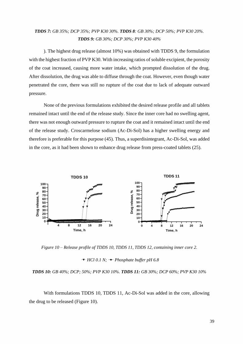
Figure 10 – Release profile of TDDS 10, TDDS 11, TDDS 12, containing inner core 2. ... 38
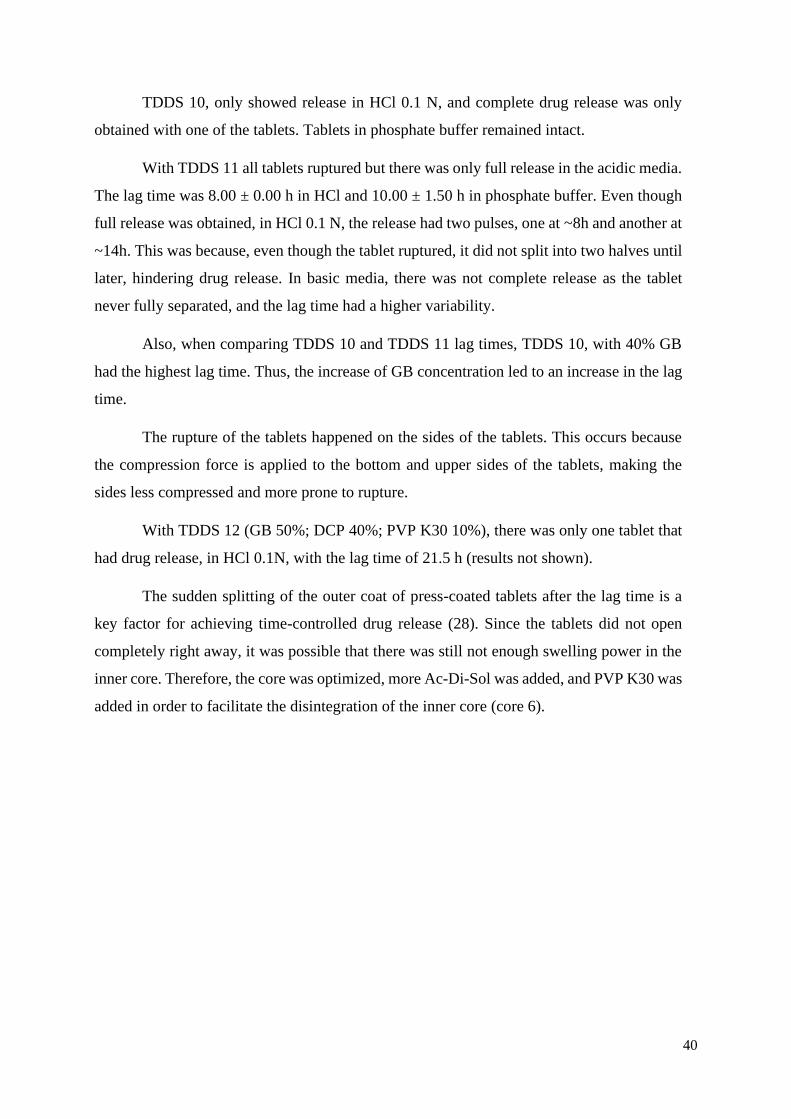
Figure 11 – Release profile of TDDS 13 and TDDS 14, containing inner core 6. .............. 39
Figure 12 – Release profile of TDDS 15 and TDDS 16, containing inner core 6. .............. 40
Figure 13 – Release profile of TDDS 17 and TDDS 18, containing inner core 6. .............. 41
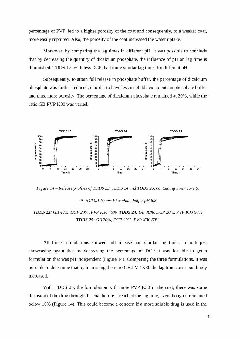
Figure 14 – Release profiles of TDDS 23, TDDS 24 and TDDS 25, containing inner core 6.
.............................................................................................................................................. 43
Figure 15 – Release mechanism of TDDS 24. ..................................................................... 44
Figure 16 – Comparison of the release profile of TDDS 24, with different diameters and drug
loading. ................................................................................................................................. 44
Figure 17 – Release profile of TDDS 1, containing inner core 1. ........................................ 45
Figure 18 – Release profile of TDDS 19 and TDDS 20, containing inner core 6. .............. 46
Figure 19 – Release profile of TDDS 21 and TDDS 22, containing inner core 6. .............. 47
10
Figure 20 – Release profile of TDDS 26, containing inner core 6. ...................................... 48
Figure 21 – Comparison of the release profile of TDDS 21, with different diameters and drug
loading. ................................................................................................................................. 49
Figure 22 – Release profile of a commercially available formulation, Lodotra. ................. 51
Figure 23 – Release mechanism of Lodotra. ........................................................................ 51
Figure 24 – Water uptake in 0.1 N HCl and phosphate buffer, pH 6.8. ............................... 52
Figure 25 – Dry mass loss in 0.1 N HCl and phosphate buffer, pH 6.8. .............................. 53
Index of tables
Table 1 – Diseases that require time dependent delivery systems. ...................................... 23
Table 2 – Marketed technologies of Chronopharmaceutical dosage forms ......................... 24
Table 3 – Composition of core tablets (%) .......................................................................... 30
Table 4 – Composition of the coat of press-coated tablets formulated with glyceryl behenate
(%) ........................................................................................................................................ 31
Table 5 – Composition of the coat of press-coated tablets formulated with ethyl cellulose (%)
.............................................................................................................................................. 32
Table 6 – Physical characteristic of 9 mm press-coated tablets. .......................................... 34
Table 7 – Physical characteristics of 11 mm press-coated tablets ....................................... 35
Table 8 – Excipients in the formulation of Lodotra ............................................................. 49
Table 9 – Physical characterization of Lodotra (5 mg) ........................................................ 50
11
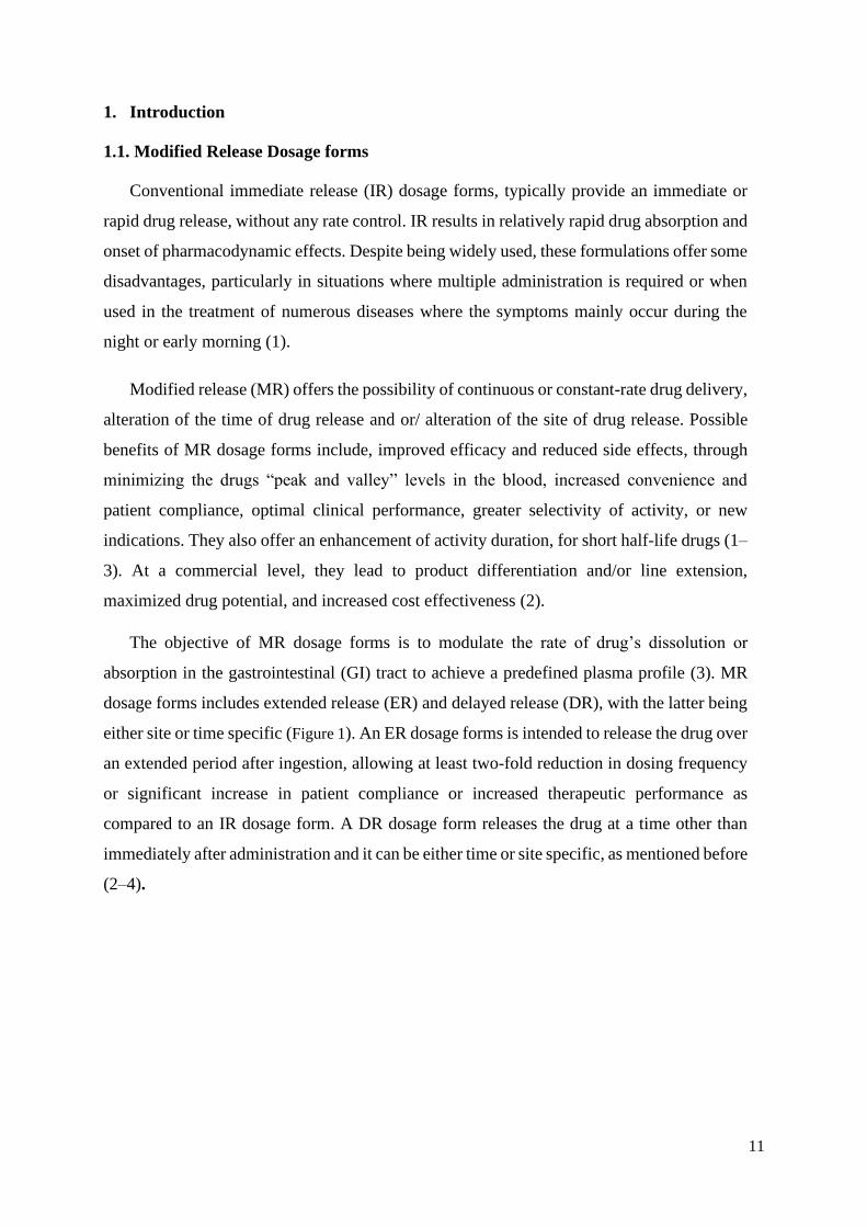
1. Introduction
1.1. Modified Release Dosage forms
Conventional immediate release (IR) dosage forms, typically provide an immediate or
rapid drug release, without any rate control. IR results in relatively rapid drug absorption and
onset of pharmacodynamic effects. Despite being widely used, these formulations offer some
disadvantages, particularly in situations where multiple administration is required or when
used in the treatment of numerous diseases where the symptoms mainly occur during the
night or early morning (1).
Modified release (MR) offers the possibility of continuous or constant-rate drug delivery,
alteration of the time of drug release and or/ alteration of the site of drug release. Possible
benefits of MR dosage forms include, improved efficacy and reduced side effects, through
minimizing the drugs “peak and valley” levels in the blood, increased convenience and
patient compliance, optimal clinical performance, greater selectivity of activity, or new
indications. They also offer an enhancement of activity duration, for short half-life drugs (1–
3). At a commercial level, they lead to product differentiation and/or line extension,
maximized drug potential, and increased cost effectiveness (2).
The objective of MR dosage forms is to modulate the rate of drug’s dissolution or
absorption in the gastrointestinal (GI) tract to achieve a predefined plasma profile (3). MR
dosage forms includes extended release (ER) and delayed release (DR), with the latter being
either site or time specific (Figure 1). An ER dosage forms is intended to release the drug over
an extended period after ingestion, allowing at least two-fold reduction in dosing frequency
or significant increase in patient compliance or increased therapeutic performance as
compared to an IR dosage form. A DR dosage form releases the drug at a time other than
immediately after administration and it can be either time or site specific, as mentioned before
(2–4).

12
Figure 1 – Modified release dosage forms.
1.1.1. Time-dependent delivery systems
Time-dependent delivery systems (TDDS) are DR dosage forms designed to offer a
fast and complete or extended release of drug after a programmed time, called lag phase or
lag time (5–7). The release process may be triggered by external signals (e.g. chemical,
thermal, electric and magnetic stimuli) or, it can be regulated by inherent mechanisms, that
are expected to perform consistently, independent of major physiological variables, such as,
pH, ionic strength or temperature (5,6).
TDDS reduces dosing frequency and, unlike sustained release delivery systems,
provide a timely pharmacological effect, enabling a reduction in side effects associated with
a prolonged, and at times unnecessary, exposure to a drug (8). The great potential of such
formulations is their suitability for providing the patient with the correct dosage, at the correct
time, thus allowing a reduction in dosing, cost and frequency. This time-dependent approach
is particularly important in pathologies with predominant night or early morning symptoms
(bronchial asthma, rheumatoid arthritis, etc.). In this case, TDDS could provide a therapeutic
effect, without having to interrupt the normal sleep pattern of patients, which could lead to
reduced compliance (5).
Another interesting application of TDDS is the delivery into the large bowel, with a
time dependent approach that relies on the small intestinal transit time, practically
13
independent of the characteristics of the dosage form, as well as of the fasted and fed state of
the subject (5,6).
Despite moderate drug absorption properties, the colon represents an interesting site of
absorption for drugs that may cause irritation or be degraded in the upper GI tract, as well as
for nonabsorbable molecules that are supposed to act in the gut lumen (9,10). The latter fact
could be particularly interesting for colon delivery in the treatment of inflammatory bowel
disease (IBD) and chemoprevention of colorectal adenocarcinoma (5,6). Currently, the
dosage forms most commonly used in the treatment of IBD rely on the different pHs of the
GI tract. However, the pH of the GI tract is highly variably in these patients and most of the
drug is released in the upper small intestine after gastric emptying (11).
For time dependent colon delivery, an entering coating is usually employed in order to
overcome the highly variable stomach emptying time (5,6). Moreover, peptide and protein
drugs, which are known to be more prone to enzymatic degradation in the small bowel, may
have their oral bioavailability increased (12).
Besides, time-dependent dosage forms, can prevent the occurrence of detrimental drug-
drug interactions, without the need of changing the administration schedule of combined
medication, which could lead, once more, to increased compliance (5,14).
Time controlled systems can consist or single-unit or multiple unit systems. Single unit
systems consist mainly of capsule-shaped and advanced osmotic devices (9). Multiple unit
systems decrease the unit-to-unit variability, when compared to single unit. On the other
hand, however, low drug loading, incomplete drug release, proportionally higher need for
excipients, lack of manufacturing reproducibility and efficacy, a large number of process
variables, multiple formulation steps, higher cost of production, and need of advanced
technology are some of the disadvantages (9,10).
It is widely known that the oral route presents itself as the preferred one, mostly
because of the cost-effectiveness and usually high compliance of patients (5). Therefore, oral
time-dependent release dosage forms have been developed and according to the coating
agents employed, the release mechanism may involve erosion, rupture or diffusion (the coat
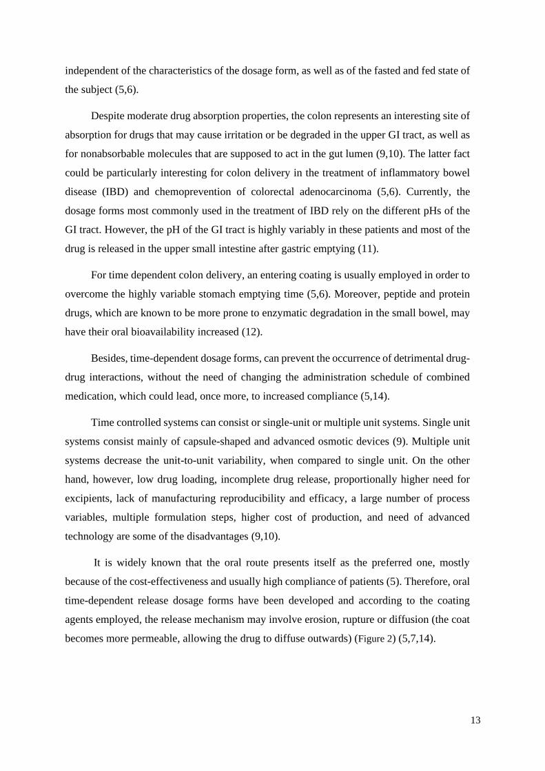
becomes more permeable, allowing the drug to diffuse outwards) (Figure 2) (5,7,14).
14
Figure 2 – Outline of the performance of coated delivery systems for oral time-dependent release
on exposure to aqueous fluids. Adapted from (14).
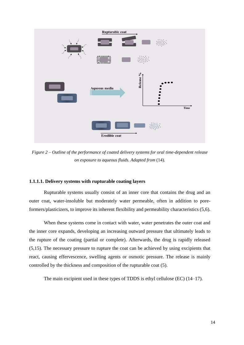
1.1.1.1. Delivery systems with rupturable coating layers
Rupturable systems usually consist of an inner core that contains the drug and an
outer coat, water-insoluble but moderately water permeable, often in addition to pore-
formers/plasticizers, to improve its inherent flexibility and permeability characteristics (5,6).
When these systems come in contact with water, water penetrates the outer coat and
the inner core expands, developing an increasing outward pressure that ultimately leads to
the rupture of the coating (partial or complete). Afterwards, the drug is rapidly released
(5,15). The necessary pressure to rupture the coat can be achieved by using excipients that
react, causing effervescence, swelling agents or osmotic pressure. The release is mainly
controlled by the thickness and composition of the rupturable coat (5).
The main excipient used in these types of TDDS is ethyl cellulose (EC) (14–17).

15
1.1.1.2. Delivery systems with swellable/erodible coating layers
Erodible release systems are mostly based on hydrophilic polymers that form the
coating. These, in turn, may swell, erode and/or dissolve when in contact with aqueous fluids,
due to the glassy-rubbery polymer transition, which results in an appropriate lag time before
drug release occurs (14). Lag time is mostly dependent on the appropriate polymer
particularly, molecular mass of the polymer, and coating level (10).
Hydrophilic cellulose ethers, hydroxyethyl cellulose (HEC), hydroxypropyl cellulose
(HPC) and hydroxypropyl methyl cellulose (HPMC) are employed as the main components
of the coating as they have shown to have an adequate swelling behaviour, an established
safety profile, ease of handling, availability in different grades and reasonable costs as well
as pH independence, due to the non-ionic nature of their polymers (5,6,14,18).
1.2. Coating technology
Pharmaceutical coating is used for various reasons, such as achieving superior
organoleptic and aesthetic characteristics, providing physical and chemical protection
(protection from moisture, light and/or air; protection from gastric acid or gastric enzymes;
enhanced mechanical strength) or to attain modified drug release profile, either by altering
the site, the time of release and/or the release rate (19,20).
Sugar coating was the first modern pharmaceutical coating and was mainly used in order
to improve the palatability of bitter medicines. However, this technique had long processing
times (up to 5 days), a requirement for high level of expertise and difficulties involving the
standardizing of the procedure. Also, the risk of bacterial and mold growth was high, there
were restrictions in tablet shape and lack of automation. This led to the introduction of film
coating, that, consequently, led to a significant reduction in the processing time (18).
Film coating, carried out by a fluid bed or rotating pan equipment, involves spraying the
coating onto the substrate cores that can be powder, granules, pellets, tablets and capsules.
The coating materials are solubilized or suspended in an organic and/or aqueous vehicle (19).
Film coating offers many advantages, for instance, good reproducibility of the process,
ability of being applied to different dosage forms, process automation, increased process
control and improved batch-to-batch uniformity of the product (18,19).
16
However, organic solvents, despite offering shorter processing times and
straightforward film formation, carry many disadvantages. The toxicity of the residual
solvent in the coating, the high cost of organic solvents and its recycling, the safety hazards
to operators as well as strict environmental regulation has led to a shift to the use of water as
a solvent (18).
The use of water as a solvent eliminates many of the disadvantages of using organic
solvents in solvent-based coating techniques. Despite, heat is necessary for evaporating the
water present in the coating and, because of the relatively high latent heat of vaporization of
water, slow drying rate of the coating becomes an issue, causing longer processing times. In
addition, drugs can be sensitive to residual moisture in the film and this can cause long-term
stability problems and may ultimately change the permeability of the core to the drug and
alter the performance of the coating layer. Another problem stemming from using water as
the solvent is that the control of microbial presence becomes an problem, especially when
cellulose polymers are used as the coating material (18).
Thus, the limitations of film coating include mostly problems related with the use of
solvents and their removal. Consequently, the elimination of solvents from the coating
process can present a significant advancement (18).
Solventless coating techniques allow for a reduction in costs, by eliminating the slow
and expensive processes of solvent treatment. Furthermore, it can significantly reduce
processing time as it eliminates the slow drying and evaporation steps. Also, techniques
where there is no heating source can provide an alternative method to coat heat-sensitive
drugs. Solvent free techniques may offer an alternative for preparation of microcapsules
containing antigens or proteins, which can be of much importance due to the current trend
towards biopharmaceutical molecules (18,21).
Techniques that would be classified into solvent free can be further divided according
to the physical state of the coat-forming agents when applied onto the surface (Figure 3). In
liquid-based techniques, melts or liquid precursors applied onto the surface are consolidated
either by cooling (hot-melt coating) or by UV-initiated polymerization (coating by
photocuring), to attain a continuous layer. In solid-based techniques, the coating may be
directly applied by compression (press-coating), or it can be layered and simultaneously
consolidated by heating (powder coating). Furthermore, in powder coating, the process can

17
be optimized, especially as regards to the initial deposition phases, by spraying liquid aids
and/or through particle charging (19).
Figure 3 – Classification of solventless techniques to obtain coated dosage forms. Adapted from
(19).
1.2.1. Press-coating
Press-coating, also known as compression coating or dry coating or double
compression was one of the first solvent-free coating techniques (18,19). Generally, it
consists of an inner core surrounded by an outer coat. Conventionally, the inner core is
compressed first. Then, a tableting machine is pre-filled with a certain amount of coating
material, the inner core is placed on the centre of the powder bed and the remaining coating
mixture is added on top. Finally, all of the contents are compressed in order to obtain an outer
layer of defined thickness (Figure 4) (1,19).

18
Figure 4 – Manufacturing process of press-coating. (I) – Prefilling the die with about half of the
coating materials; (II) – Placing the tablet on top of the powder bed; (III) – Centring of the tablet;
(IV) – Filling the die with the rest of the coating material; (V) – Compression; (VI) – Ejection of the
tablet. Adapted from (1).
It offers several advantages, like the possibility of separation incompatible drugs in the
core and the coat within the same dosage form. Additionally, it is possible to formulate a
dosage form that releases two active substance in different parts of the GI tract. Moreover, it
offers protection to hygroscopic, light sensitive, oxygen labile and acid-labile drugs (18,21).
The manufacturing of dry coated tablets using this method is high cost because of the
requirement of preparing the core tablets beforehand. The process is relatively slow when
compared to other solventless techniques, the coatings are thick and may not be suited for
immediate release (26). Also, the requirement for core tablet supply system leads to problems
such as double-core, non-core, off-centre and inlay (22).
One of the major problems associated with this technique is the centring of the inner
core. When the core is not centred correctly, there may be variation in the release profile of
the inner core (1,18,19). To solve this problem Ozeki et al. developed the one-step dry-coated
tablet manufacturing method (OSDRC system). This system does not require the preparation
of the core tablet beforehand, as the whole system is prepared in a single process (22). The
manufacturing method was executed by using upper and lower punches, which had a double
structure, a centre punch and an outer punch surrounding the centre punch. The three-step
process involves the formation of the first outer layer (lower), the core and the upper outer
coat layer, followed by a compression in every step (18,22).
The OSDRC system eliminates the necessity of a supply system and therefore
eliminates the problems that stem from such. Also, this system provides thinner coatings
compared to conventional dry coating (22).

19
1.2.1.1. Factors affecting performance and drug release of press-coated delivery
systems
Press-coated tablets consist of two layers, an inner core and an outer coating. The
coating may have different rate-controlling materials in order to achieve time or site-specific
drug delivery and/or attain extended release. The drug release behaviour is controlled by
different variables that may be present in the inner core and outer coat. These factor include,
the solubility of the drug, the ratio core:coat, the composition of both inner core and coat, the
compression force and also the location of the inner core (21).
The solubility and permeability are important variables to think of when formulating
as they affect the absorption of the drug. Therefore, the solubility of the drug present in the
inner core is an important factor to monitor (21). It has been shown that higher solubility
drugs offer shorter lag times (23). Rujivipat and Bodmeier prepared HPMC compression-
coated tablets containing different solubilities drugs in the inner core (24). It was shown that
depending on their solubility the drugs were released either by diffusion and/or erosion of
the gelled HPMC coat. Carbamazepine, the least water-soluble drug was released completely
after a lag time, after erosion of the HPMC coat. The release of the other drugs, more water-
soluble, happened by diffusion through the gel prior to erosion, showcasing a sigmoidal
release profile.
Other components of the inner core can also affect the lag time. Lin et al showed that
diluents with different solubilities affect the lag time (23). More soluble diluents, such as
spray-dried lactose, facilitate the dissolution, shortening both disintegration and lag times.
Also, it was shown that the presence of an osmotic agent, sodium chloride, for instance,
generates a higher internal osmotic pressure and distinctly decreases the lag time.
Additionally, the presence of a superdisintegrant results in the bursting effect of the tablets,
caused by the swelling, and enhances the drug release from press-coated tablets (25).
The ratio core:coat has also been shown to influence lag time. Due to faster
erosion/rupture of the press coat, a higher core:coat ratio leads to a shorter lag time (26).
Likewise, the constitution of the coat effects the release profile. The coating material
is of extreme importance as it affects variables such as mechanical strength, release profile
and stability (18). For instance, the presence of water insoluble/rupturable (EC), erodible
20
(low molecular weight HPMC, HPC, PEO), gellable or swellable (high molecular weight
HPMC, gums), pH-dependent soluble (HPMCAS, Eudragit copolymers) polymers or a
mixture of these can modulate the drug release (1). The compressibility is also highly
dependent of the coating material, making its selection a central step (18).
The choice between a rupturable or swellable/erodible material also affects the release
of the drug from the inner core. An erodible coating does not modify the release profile of
the drug present in the core; a swelling coat, however, may delay the release of the drug and
alter the release performance of the inner core (27). Because of this, when an extended release
is required after lag time, often a swellable coat is employed. When a burst effect is required,
an erodible or rupturable coat seems to be the best choice.
Several studies have demonstrated the effect of hydrophilic excipients present in the
inner core on the lag time (28,29). Lin et al. studied the effect of several direct-compressible
excipients, spray-dried lactose (SDL) and a polymer with hydrophilic properties (HPMC)
(28). It was shown that the lag time was dependent on type of excipient present in the coat,
with SDL providing a shorter lag time. The different physico-chemical properties of HPMC
and SDL can explain the different lag time. The quick dissolution of SDL provides a more
porous structure for medium penetration, while a more viscous gel layer of HPMC swollen
on the whole tablet might delay the penetration of the medium and cause prolongation of the
lag time. A different study used as hydrophilic excipients HPMC (E5), HPC (EF and SSL),
povidone (K30), copovidone, polyethylene glycol (4000), lactose and mannitol. With
increasing concentration of the excipients, there was a reduction in the lag phase before
release, with the freely water soluble diluents having a bigger influence (lactose and
mannitol) (29).
The size of the polymer particle can also influence lag time. Various lag times were
obtained by using different EC particle sizes, with smaller particles providing longer lag
times. The finner the article, the less residual porosity remains, providing a more torturous
path for medium penetration. Therefore, by choosing the size of the particle, one can
modulate the lag time (30).
21
1.3. Chronopharmaceuticals
Chronopharmaceuticals comprises the fundamentals of chronobiology and
pharmaceutics, with chronobiology being the study of biological rhythm and its mechanism
(1,9). Chronobiology assumes that all organisms, when it comes to bioprocesses and
functions, exhibit predictable variability over time (21). The biological rhythm is controlled
by a number of factors, internal or external, such as food intake, metabolism, appetite,
digestion, hormonal changes, etc (31).
It has been shown that many diseases follow the circadian rhythm. In these, medications
pharmacodynamics and pharmacokinetics are influenced by the chronopharmacological
phenomena (9,29,31). For instance, some disorders provoke either night-time or early
morning symptoms. The traditional way of treating these, is to deliver a higher dose of drug
in the form of either an IR or ER drug formulation before going to bed, in order to maintain
therapeutic concentrations until the next morning. This, however, leads to increased side
effects and also subjects the body to a metabolic load even when not necessary (29).
Therefore, it makes sense for a drug to be administrated at the correct time, in order to
achieve concentration peaks at times where the symptoms are present or exacerbated. Hence,
chronopharmaceuticals may improve efficacy and minimize side-effects, by releasing a drug
at a rhythm that matches the biological needs. Many studies have showed exactly that the
timing of administration can increase the efficacy and diminish toxicity of many drugs (31).
For instance, a study of 26 patients with rheumatoid arthritis showed that the administration
of low dose prednisolone at 2 a.m., instead of 7.30 a.m., resulted in an improvement of
morning symptoms (32). However, a long-term therapeutic regimen based on waking up
patients is not only impractical but could also affect the circadian rhythm itself.
Chronotherapeuticals, involves not only new medicines but the improved application of
established ones in a different and more biologically manner (33). The new chronotropic
technology may be developed by synchronizing the drug concentrations to rhythms in disease
activity. With chronopharmaceutical dosage forms, the drug is released at a desired time to
match the biological needs, which results in improved efficacy and patient-compliance
(1,31). By not exposing the patient to unnecessary doses of drug, a reduction of side-effects
can be achieved too (9).
Not only it can be achieved by using TDDS to match the peaks in the disease activity but
also, in certain instances, it can be achieved by unequal morning and evening dosing
22
schedules of sustained release 12h medication systems, or application of a special tablet and
capsule formulations dosed at designed times to proportion medications over the 24h in
synchrony with the biological rhythm (33).
For the development of chronotropic drug delivery system, an extensive knowledge of
the pathology is required, therefore, these system may be used in diseases having enough
scientific background in order to justify their need as compared to a conventional drug
delivery systems (1). These include asthma, arthritis, duodenal ulcer, cancer, cardiovascular
diseases, diabetes, hypercholesterolemia, neurological disorders etc. which have a well
stablished circadian rhythm (
23
Table 1) (34).
A drawback, however, is that, although drug release can be controlled, drug absorption
cannot. Therefore, drugs with variable absorption in the GI tract, are not good candidates for
chronopharmaceutical drug delivery systems (31).
1.3.1. Chronopharmaceutical dosage forms on the market
A few technologies have been developed as chronopharmaceutical dosage forms to fulfil
unmet medical needs in the treatment of various diseases. OROS technology, Geoclock®
Technology, TimerX® technology, Pulsicap™, DIFFUCAPS®, CEFORM® technology and
CODAS® technology are example of marketed technologies that have the
chronopharmaceutical concept as a basis for their development (Table 2).
24
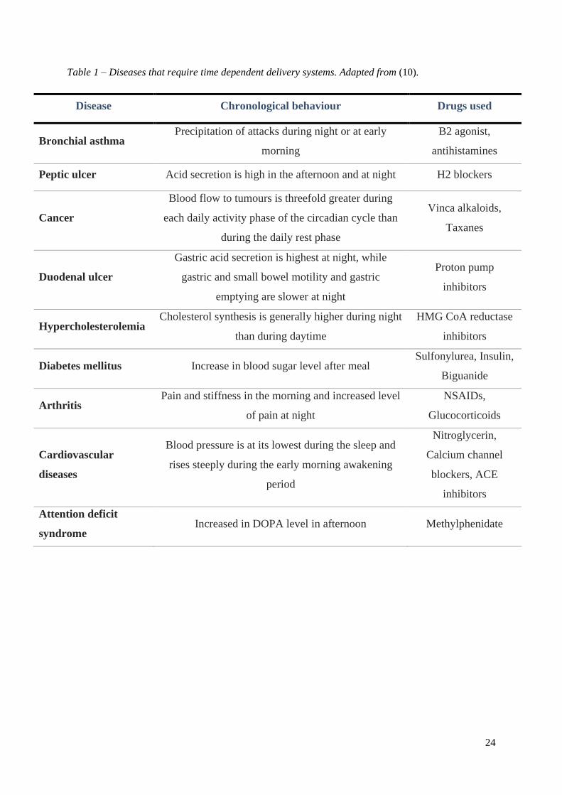
Table 1 – Diseases that require time dependent delivery systems. Adapted from (10).
Disease Chronological behaviour Drugs used
Bronchial asthma Precipitation of attacks during night or at early
morning
B2 agonist,
antihistamines
Peptic ulcer Acid secretion is high in the afternoon and at night H2 blockers
Cancer
Blood flow to tumours is threefold greater during
each daily activity phase of the circadian cycle than
during the daily rest phase
Vinca alkaloids,
Taxanes
Duodenal ulcer
Gastric acid secretion is highest at night, while
gastric and small bowel motility and gastric
emptying are slower at night
Proton pump
inhibitors
Hypercholesterolemia Cholesterol synthesis is generally higher during night
than during daytime
HMG CoA reductase
inhibitors
Diabetes mellitus Increase in blood sugar level after meal Sulfonylurea, Insulin,
Biguanide
Arthritis Pain and stiffness in the morning and increased level
of pain at night
NSAIDs,
Glucocorticoids
Cardiovascular
diseases
Blood pressure is at its lowest during the sleep and
rises steeply during the early morning awakening
period
Nitroglycerin,
Calcium channel
blockers, ACE
inhibitors
Attention deficit
syndrome Increased in DOPA level in afternoon Methylphenidate

25
Table 2 – Marketed technologies of Chronopharmaceutical dosage forms.
Drug (registered trademark®) Technology Drug release mechanism References
Verapamil HCl (Covera-HS)
Nifedipine (Procardia XL)
Doxazosin mesylate (Cardura XL)
Oxybutynin HCl (Ditropan XL)
Glipzide (Glucotrol XL)
Paliperidone (Invega)
OROS®
technology
Osmotic Regulation.
Comprised of a bilayer or trilayer tablet core, one push layer and one or more drug layers. The push layer
contains an osmotic agent and swellable polymers. A semipermeable membrane surrounds the core
(drilled with a delivery orifice). The active pharmaceutical is pushed away through the channel due to
pump effect of the osmotic agent. Usually designed for extended release.
(1,10,34)
Prednisone (Lodotra) Geoclock®
technology
Time-dependent delivery
Consists of an active drug core inside an outer layer consisting of a hydrophobic material. The drug is
released after rupturing of the outer core.
(10,35)
Oxybutynin HCl (cystine CR)
Oxymorphone (Opana ER)
TIMERx®
Swelling, diffusion, erosion.
(34)
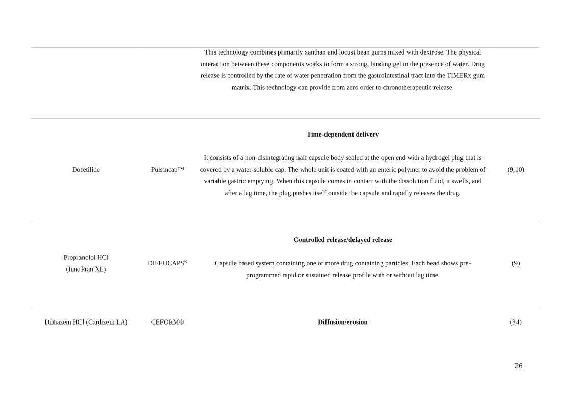
26
This technology combines primarily xanthan and locust bean gums mixed with dextrose. The physical
interaction between these components works to form a strong, binding gel in the presence of water. Drug
release is controlled by the rate of water penetration from the gastrointestinal tract into the TIMERx gum
matrix. This technology can provide from zero order to chronotherapeutic release.
Dofetilide Pulsincap™
Time-dependent delivery
It consists of a non-disintegrating half capsule body sealed at the open end with a hydrogel plug that is
covered by a water-soluble cap. The whole unit is coated with an enteric polymer to avoid the problem of
variable gastric emptying. When this capsule comes in contact with the dissolution fluid, it swells, and
after a lag time, the plug pushes itself outside the capsule and rapidly releases the drug.
(9,10)
Propranolol HCl
(InnoPran XL) DIFFUCAPS®
Controlled release/delayed release
Capsule based system containing one or more drug containing particles. Each bead shows pre-
programmed rapid or sustained release profile with or without lag time.
(9)
Diltiazem HCl (Cardizem LA) CEFORM®
Diffusion/erosion
(34)

27
Microspheres that may be coated for controlled release with an enteric coating or may be combined into a
fast/slow release combination.
Verapamil HCl (Verelan) CODAS®
Multiparticulate pH dependent system/a delayed onset of drug release.
A non-entering coating is applied in order to delay the release of the drug up to 5 h. Release controlling
coat consists of a mixture of both water-soluble and water insoluble polymers. After water soluble
polymers get dissolved, pores are formed, and the drug diffuses through the pores.
(1,34)

28
Geoclock® technology, developed by SkyePharma, consists of a new oral drug
delivery system in the form of a press-coated tablet. Geoclock® tablets are provided with an
active drug core, surrounded by an outer layer entailing of a mixture of hydrophobic wax and
brittle material in order to obtain a lag time pH independent. This technology allows for the
delivery of immediate and slow release active cores. It not only offers application in
controlled release but also improved release of colonic drug delivery as well as for multiple
pulse drug delivery (10).
Lodotra, developed by this same company, makes use of this technology. This dosage
form consists of an inner core containing the drug, prednisone, and an inert, non-soluble and
non-swellable coating. The coat consists of a mixture of a hydrophobic lipid, glyceryl
behenate, a mostly non soluble diluent, dicalcium phosphate dihydrated, and a pore former,
povidone K 29/32. The core is mainly comprised of lactose as a diluent and it also has a
superdisintegrant, croscarmellose sodium (36).
The coating prevents release of the drug over an extended period of time, so that no
absorption occurs for around 4 h. After this lag time, the drug is rapidly released and after 2
h more than 80% of the drug should be released. This tablet is produced using a press-coating
technology where a previously compressed core tablet is compressed using a multilayer tablet
press-coat to form a press-coated tablet (10,36).
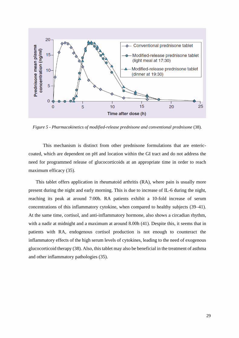
This dosage form has been designed to achieve maximum plasma levels 6 h after
administration (Figure 5). This enables the patient to swallow the tablet at around 22.00h,
with the dose of prednisone not being released until after 4 h, which is regarded as the optimal
timing to relieve the early morning symptoms of stiffness and pain (10,37).
29
Figure 5 - Pharmacokinetics of modified-release prednisone and conventional prednisone (38).
This mechanism is distinct from other prednisone formulations that are enteric-
coated, which are dependent on pH and location within the GI tract and do not address the
need for programmed release of glucocorticoids at an appropriate time in order to reach
maximum efficacy (35).
This tablet offers application in rheumatoid arthritis (RA), where pain is usually more
present during the night and early morning. This is due to increase of IL-6 during the night,
reaching its peak at around 7:00h. RA patients exhibit a 10-fold increase of serum
concentrations of this inflammatory cytokine, when compared to healthy subjects (39–41).
At the same time, cortisol, and anti-inflammatory hormone, also shows a circadian rhythm,
with a nadir at midnight and a maximum at around 8.00h (41). Despite this, it seems that in
patients with RA, endogenous cortisol production is not enough to counteract the
inflammatory effects of the high serum levels of cytokines, leading to the need of exogenous
glucocorticoid therapy (38). Also, this tablet may also be beneficial in the treatment of asthma
and other inflammatory pathologies (35).

30
2. Aim of the project
The aim of this project was to develop a time-dependent drug delivery system and study
the effect of formulation variables on lag time. Tablets were prepared by press-coating, with
a rupturable coat, in order to modulate the release profile. The desired release profile was
characterized by a lag time, followed by immediate and complete release of the drug.
Two main formulations were prepared, one with ethyl cellulose and another with a
mixture of glyceryl behenate and dicalcium phosphate, as the water-insoluble excipients of
the coat. The core tablet was formulated using prednisone, a borderline BCS Class I
compound, used in the management of diseases such as rheumatoid arthritis or bronchial
asthma.
Physical characterization was performed for each formulation, more particularly, the
mass, height and thickness of the tablets was assessed.
The ratio of excipients of the coat was altered in order to study their effect on lag time.
Specifically, the effect of the ratio soluble/insoluble excipient was studied, for both
formulations. Also, the influence of the presence of an excipient with solubility dependent
on pH, on lag time, was evaluated. To determine the lag time and release profile, release
studies were performed for each formulation; to determine if the formulation had a behaviour
pH independent, these studies were performed in different pH.
One of the main concerns of press-coated tablets is low drug loading; with larger tablets,
a higher drug loading could be achieved. Thus, tablets with different diameter were prepared
using the optimized formulations, in order to establish if by keeping the same core:coat mass
ratio the release profiles stayed similar.
Since water uptake is one the most important steps that influence drug release from tablets
with a rupturable coat, the model formulations were compared with a commercially available
formulation, with a similar release profile, in terms of water uptake and dry mass loss.
31
3. Materials and methods
3.1. Materials
The materials used were: Prednisone (Prednisone, micronized), HPMC K4M (MethocelTM
K4M DC2 Premium), Ethyl cellulose 10 cP (EthocelTM Std 10 cP Premium), Microcrystalline
cellulose (Avicel® PH105), Direct compressible lactose (Flowlac® 100),
Polyvinylpyrrolidone K 30 (Kollidon® 30), Glyceryl dibehenate (Compritol® 888 ATO),
Dicalcium phosphate anhydrous (DI-CAFOS® A 60), Colloidal silicon dioxide (Aerosil®
200), Magnesium stearate (Ligamed® MF-2-V), Croscarmellose sodium (Ac-Di-Sol ®).
3.2. Methods
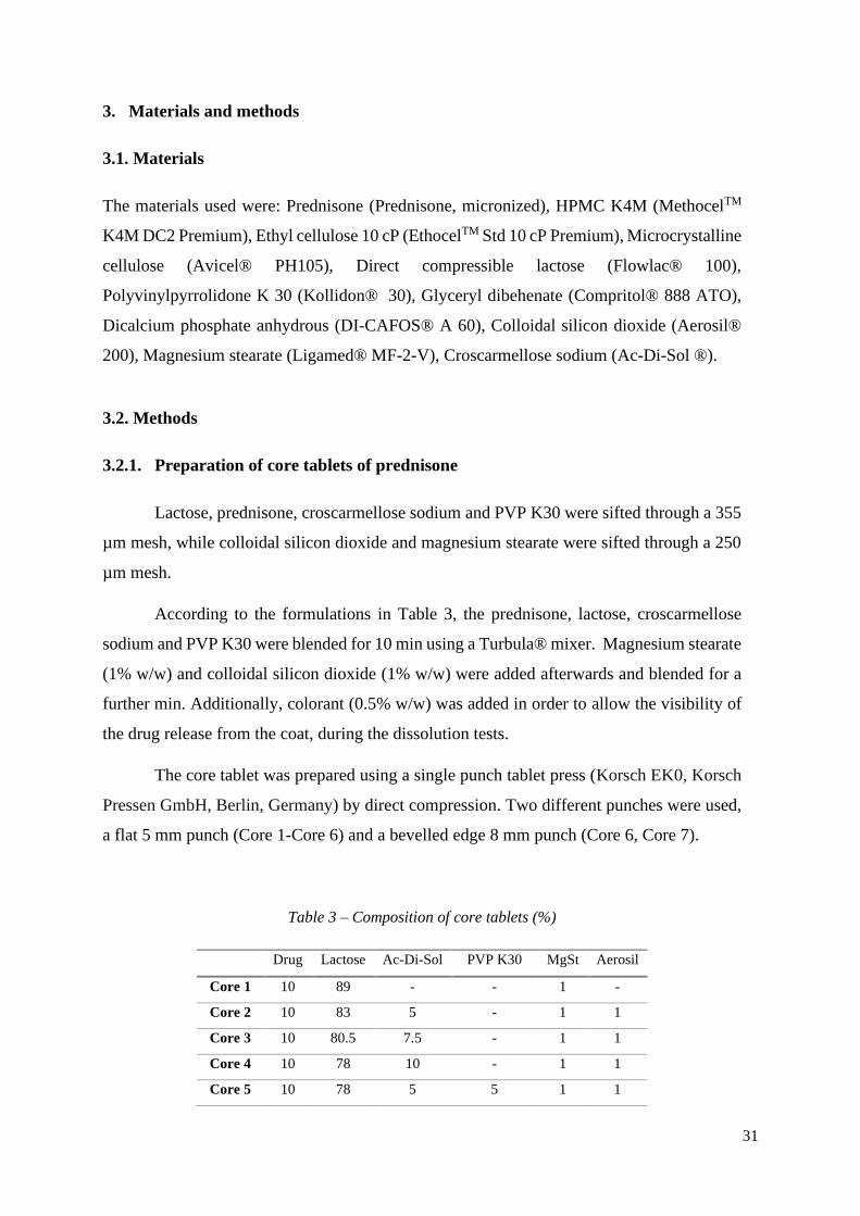
3.2.1. Preparation of core tablets of prednisone
Lactose, prednisone, croscarmellose sodium and PVP K30 were sifted through a 355
µm mesh, while colloidal silicon dioxide and magnesium stearate were sifted through a 250
µm mesh.
According to the formulations in Table 3, the prednisone, lactose, croscarmellose
sodium and PVP K30 were blended for 10 min using a Turbula® mixer. Magnesium stearate
(1% w/w) and colloidal silicon dioxide (1% w/w) were added afterwards and blended for a
further min. Additionally, colorant (0.5% w/w) was added in order to allow the visibility of
the drug release from the coat, during the dissolution tests.
The core tablet was prepared using a single punch tablet press (Korsch EK0, Korsch
Pressen GmbH, Berlin, Germany) by direct compression. Two different punches were used,
a flat 5 mm punch (Core 1-Core 6) and a bevelled edge 8 mm punch (Core 6, Core 7).
Table 3 – Composition of core tablets (%)
Drug Lactose Ac-Di-Sol PVP K30 MgSt Aerosil
Core 1 10 89 - - 1 -
Core 2 10 83 5 - 1 1
Core 3 10 80.5 7.5 - 1 1
Core 4 10 78 10 - 1 1
Core 5 10 78 5 5 1 1
32
Core 6 10 73 10 5 1 1
Core 7 4.5 78.5 10 5 1 1
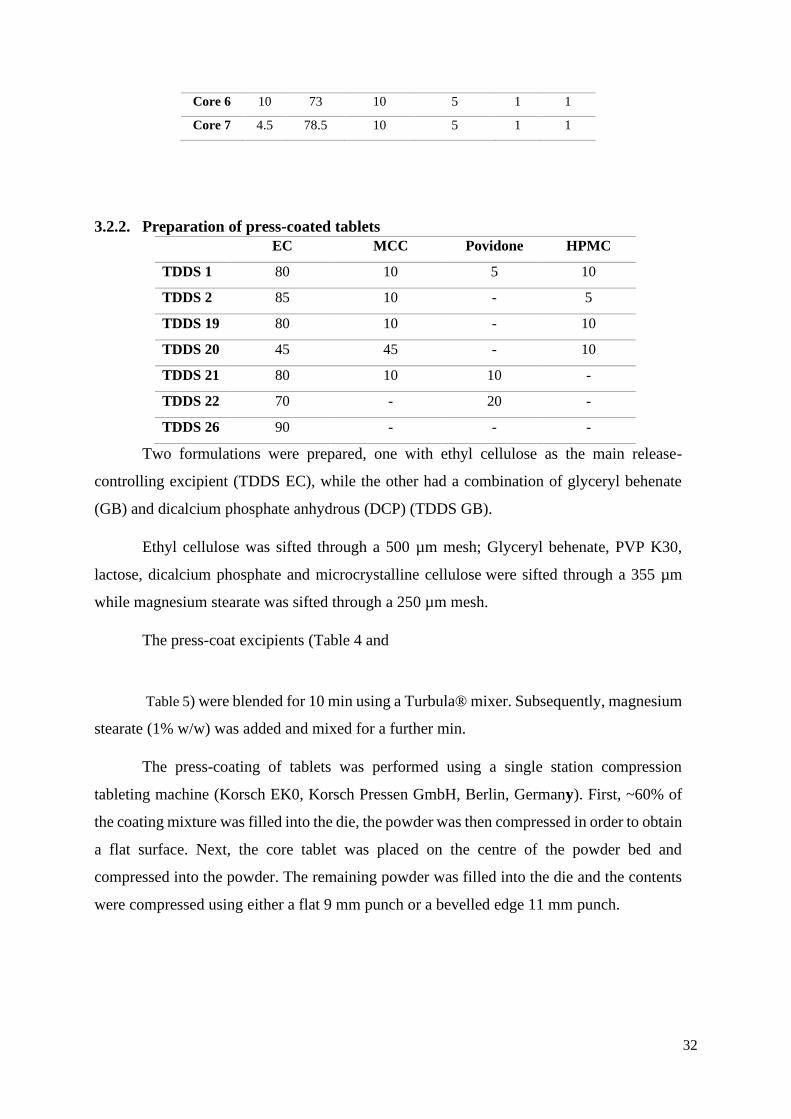
3.2.2. Preparation of press-coated tablets
Two formulations were prepared, one with ethyl cellulose as the main release-
controlling excipient (TDDS EC), while the other had a combination of glyceryl behenate
(GB) and dicalcium phosphate anhydrous (DCP) (TDDS GB).
Ethyl cellulose was sifted through a 500 µm mesh; Glyceryl behenate, PVP K30,
lactose, dicalcium phosphate and microcrystalline cellulose were sifted through a 355 µm
while magnesium stearate was sifted through a 250 µm mesh.
The press-coat excipients (Table 4 and
Table 5) were blended for 10 min using a Turbula® mixer. Subsequently, magnesium
stearate (1% w/w) was added and mixed for a further min.
The press-coating of tablets was performed using a single station compression
tableting machine (Korsch EK0, Korsch Pressen GmbH, Berlin, Germany). First, ~60% of
the coating mixture was filled into the die, the powder was then compressed in order to obtain
a flat surface. Next, the core tablet was placed on the centre of the powder bed and
compressed into the powder. The remaining powder was filled into the die and the contents
were compressed using either a flat 9 mm punch or a bevelled edge 11 mm punch.
EC MCC Povidone HPMC
TDDS 1 80 10 5 10
TDDS 2 85 10 - 5
TDDS 19 80 10 - 10
TDDS 20 45 45 - 10
TDDS 21 80 10 10 -
TDDS 22 70 - 20 -
TDDS 26 90 - - -
33
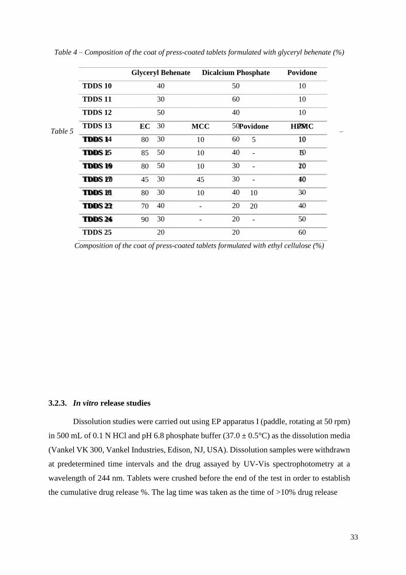
Table 4 – Composition of the coat of press-coated tablets formulated with glyceryl behenate (%)
Table 5 –
Composition of the coat of press-coated tablets formulated with ethyl cellulose (%)
3.2.3. In vitro release studies
Dissolution studies were carried out using EP apparatus I (paddle, rotating at 50 rpm)
in 500 mL of 0.1 N HCl and pH 6.8 phosphate buffer (37.0 ± 0.5°C) as the dissolution media
(Vankel VK 300, Vankel Industries, Edison, NJ, USA). Dissolution samples were withdrawn
at predetermined time intervals and the drug assayed by UV-Vis spectrophotometry at a
wavelength of 244 nm. Tablets were crushed before the end of the test in order to establish
the cumulative drug release %. The lag time was taken as the time of >10% drug release
Glyceryl Behenate Dicalcium Phosphate Povidone
TDDS 10 40 50 10
TDDS 11 30 60 10
TDDS 12 50 40 10
TDDS 13 30 50 20
TDDS 14 30 60 10
TDDS 15 50 40 10
TDDS 16 50 30 20
TDDS 17 30 30 40
TDDS 18 30 40 30
TDDS 23 40 20 40
TDDS 24 30 20 50
TDDS 25 20 20 60
EC MCC Povidone HPMC
TDDS 1 80 10 5 10
TDDS 2 85 10 - 5
TDDS 19 80 10 - 10
TDDS 20 45 45 - 10
TDDS 21 80 10 10 -
TDDS 22 70 - 20 -
TDDS 26 90 - - -
34
3.2.4. Water uptake and dry mass loss measurement
Tablets were placed separately into a container filled with 40 mL 0.1 N HCl and
phosphate buffer pH 6.8 (n=2). Afterwards, the containers were placed in a horizontal shaker
(37ºC, 80 rpm; Gesellschaft fuer Labortechnik, Burgwedel, Germany). Samples were
withdrawn at 1h, 2h and 3h and accurately massed (wet mass (t)). Subsequently they were
dried to constant mass at 60ºC (dry mass (t)). The water content and dry mass loss at the time
t was calculated using the following equations:
𝑊𝑎𝑡𝑒𝑟 𝑐𝑜𝑛𝑡𝑒𝑛𝑡 (%) (𝑡) = 𝑤𝑒𝑡 𝑚𝑎𝑠𝑠 (𝑡) − 𝑑𝑟𝑦 𝑚𝑎𝑠𝑠 (𝑡)
𝑤𝑒𝑡 𝑚𝑎𝑠𝑠 (𝑡)× 100
𝐷𝑟𝑦 𝑚𝑎𝑠𝑠 𝑙𝑜𝑠𝑠 (%) (𝑡) = 𝑑𝑟𝑦 𝑚𝑎𝑠𝑠 (0) − 𝑑𝑟𝑦 𝑚𝑎𝑠𝑠 (𝑡)
𝑑𝑟𝑦 𝑚𝑎𝑠𝑠 (0)× 100
(1)
(2)

35
4. Results and discussion
4.1. Physical characterization of the tablets
Figure 6 – Time Dependent Delivery System (TDDS).
Tablets were composed of an inner core, in blue, surrounded by a coat, white (Figure
6). The inner core contained the drug, while the coat had no active substance. Both
formulations were composed of an inner core with a diameter of 5 mm, surrounded by a coat
that had a thickness of 2 mm. The total diameter was 9 mm for both formulations. The height
was 6 mm for the formulation with EC and 5 mm for the GB formulation. The mass of the
inner core was 50 ± 3 mg, and the total mass was 400 ± 21 mg for both formulations (Table
6).
Table 6 – Physical characteristic of 9 mm press-coated tablets.
When comparing both formulations, the main difference resided in the height of the
tablet, with TDDS GB having a smaller height in the coat, therefore affecting the tablet’s
total height. This can be explained because of the presence of DCP in the TDDS GB
formulation. This powder has a high density, which allows for a significant reduction of tablet
size, without changing its mass (42). This reduction is particularly visible when DI-CAFOS
Core Coat Total
Mass
(mg)
Height
(mm)
Diameter
(mm)
Mass (mg) Height
(mm)
Thickness
(mm)
Mass (mg) Height
(mm)
Diameter
(mm)
TDDS EC 50 ± 3 2 5 350 ± 17,5 2 2 400 ± 20.5 6 9
TDDS GB 50 ± 3 2 5 350 ± 17,5 1,5 2 400 ± 20.5 5 9
36
A60 is used, due to its smaller and more spherical particles, when compared to DI-CAFOS
A150, as previously shown (43).
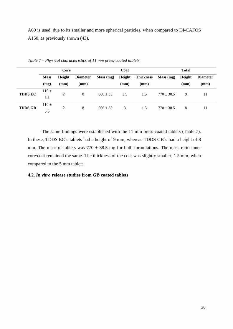
Table 7 – Physical characteristics of 11 mm press-coated tablets
The same findings were established with the 11 mm press-coated tablets (Table 7).
In these, TDDS EC’s tablets had a height of 9 mm, whereas TDDS GB’s had a height of 8
mm. The mass of tablets was 770 ± 38.5 mg for both formulations. The mass ratio inner
core:coat remained the same. The thickness of the coat was slightly smaller, 1.5 mm, when
compared to the 5 mm tablets.
4.2. In vitro release studies from GB coated tablets
Core Coat Total
Mass
(mg)
Height
(mm)
Diameter
(mm)
Mass (mg) Height
(mm)
Thickness
(mm)
Mass (mg) Height
(mm)
Diameter
(mm)
TDDS EC 110 ±
5.5 2 8 660 ± 33 3.5 1.5 770 ± 38.5 9 11
TDDS GB 110 ±
5.5 2 8 660 ± 33 3 1.5 770 ± 38.5 8 11
37
0 4 8 12 16 20 24
0
10
20
30
40
50
60
70
80
90
100
TDDS 3
Time, h
Dru
g r
ele
ase, %
0 4 8 12 16 20 24
0
10
20
30
40
50
60
70
80
90
100
TDDS 4
Time, h
Dru
g r
ele
ase, %
0 4 8 12 16 20 24
0
10
20
30
40
50
60
70
80
90
100
TDDS 5
Time, h
Dru
g r
ele
ase, %
4 8 12 16 20 24
0
10
20
30
40
50
60
70
80
90
100
TDDS 6
Time, h
Dru
g r
ele
ase, %
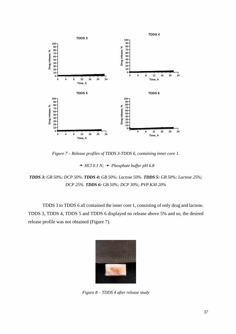
Figure 7 – Release profiles of TDDS 3-TDDS 6, containing inner core 1.
HCl 0.1 N; Phosphate buffer pH 6.8
TDDS 3: GB 50%; DCP 50%. TDDS 4: GB 50%; Lactose 50%. TDDS 5: GB 50%; Lactose 25%;
DCP 25%. TDDS 6: GB 50%; DCP 30%; PVP K30 20%
TDDS 3 to TDDS 6 all contained the inner core 1, consisting of only drug and lactose.
TDDS 3, TDDS 4, TDDS 5 and TDDS 6 displayed no release above 5% and so, the desired
release profile was not obtained (Figure 7).
Figure 8 – TDDS 4 after release study

38
Two different soluble excipients were used in TDDS 3 to TDDS 6. Lactose, due to
its water soluble and hydrophilic nature, rapidly dissolves and therefore decreases the
tortuosity and/ or increases the porosity of the coat. It has previously been used to perform
as an hydrophilic excipient and has led to a quick rupture of the press-coated tablet (28).
Because of this, initially, with TDDS 4 and TDDS 5, lactose was used in the coat to act as a
channelling agent. However, there was diffusion of the colorant through the coat (Figure 8)
also suggesting the diffusion of the drug. Subsequently, PVP K30 was chosen to be the pore
former excipient. For this, a small molecular mass was selected, with a higher dissolution
rate (44).
0 4 8 12 16 20 24
0
10
20
30
40
50
60
70
80
90
100
TDDS 7
Time, h
Dru
g r
ele
ase, %
0 4 8 12 16 20 24
0
10
20
30
40
50
60
70
80
90
100
TDDS 8
Time, h
Dru
g r
ele
ase, %
0 4 8 12 16 20 24
0
10
20
30
40
50
60
70
80
90
100
TDDS 9
Time, h
Dru
g r
ele
ase, %
Figure 9 – Release profiles of TDDS 7, TDDS 8, TDDS 9, containing inner core 1.
HCl 0.1 N; Phosphate buffer pH 6.8
TDDS 7: GB 35%; DCP 35%; PVP K30 30%. TDDS 8: GB 30%; DCP 50%; PVP K30 20%.
TDDS 9: GB 30%; DCP 30%; PVP K30 40%
With PVP K30 as the hydrophilic excipient, diffusion of the colorant was not visible.
There was still no sudden release of the drug after a lag time, but as the concentration of the
pore former increased, there was more release of the drug through diffusion with TDDS 7
(~8.38% drug release), TDDS 8 (~4.80% drug release) and TDDS 9 (~9.17% drug release)
(
Figure 9 – Release profiles of TDDS 7, TDDS 8, TDDS 9, containing inner core 1.
HCl 0.1 N; Phosphate buffer pH 6.8
39
TDDS 7: GB 35%; DCP 35%; PVP K30 30%. TDDS 8: GB 30%; DCP 50%; PVP K30 20%.
TDDS 9: GB 30%; DCP 30%; PVP K30 40%
). The highest drug release (almost 10%) was obtained with TDDS 9, the formulation
with the highest fraction of PVP K30. With increasing ratios of soluble excipient, the porosity
of the coat increased, causing more water intake, which prompted dissolution of the drug.
After dissolution, the drug was able to diffuse through the coat. However, even though water
penetrated the core, there was still no rupture of the coat due to lack of adequate outward
pressure.
None of the previous formulations exhibited the desired release profile and all tablets
remained intact until the end of the release study. Since the inner core had no swelling agent,
there was not enough outward pressure to rupture the coat and it remained intact until the end
of the release study. Croscarmelose sodium (Ac-Di-Sol) has a higher swelling energy and
therefore is preferable for this purpose (45). Thus, a superdisintegrant, Ac-Di-Sol, was added
in the core, as it had been shown to enhance drug release from press-coated tablets (25).
4 8 12 16 20 24
0
10
20
30
40
50
60
70
80
90
100
TDDS 10
Time, h
Dru
g r
ele
ase, %
0 4 8 12 16 20 24
0
10
20
30
40
50
60
70
80
90
100
TDDS 11
Time, h
Dru
g r
ele
ase, %
Figure 10 – Release profile of TDDS 10, TDDS 11, TDDS 12, containing inner core 2.
HCl 0.1 N; Phosphate buffer pH 6.8
TDDS 10: GB 40%; DCP; 50%; PVP K30 10%. TDDS 11: GB 30%; DCP 60%; PVP K30 10%
With formulations TDDS 10, TDDS 11, Ac-Di-Sol was added in the core, allowing
the drug to be released (Figure 10).
40
TDDS 10, only showed release in HCl 0.1 N, and complete drug release was only
obtained with one of the tablets. Tablets in phosphate buffer remained intact.
With TDDS 11 all tablets ruptured but there was only full release in the acidic media.
The lag time was 8.00 ± 0.00 h in HCl and 10.00 ± 1.50 h in phosphate buffer. Even though
full release was obtained, in HCl 0.1 N, the release had two pulses, one at ~8h and another at
~14h. This was because, even though the tablet ruptured, it did not split into two halves until
later, hindering drug release. In basic media, there was not complete release as the tablet
never fully separated, and the lag time had a higher variability.
Also, when comparing TDDS 10 and TDDS 11 lag times, TDDS 10, with 40% GB
had the highest lag time. Thus, the increase of GB concentration led to an increase in the lag
time.
The rupture of the tablets happened on the sides of the tablets. This occurs because
the compression force is applied to the bottom and upper sides of the tablets, making the
sides less compressed and more prone to rupture.
With TDDS 12 (GB 50%; DCP 40%; PVP K30 10%), there was only one tablet that
had drug release, in HCl 0.1N, with the lag time of 21.5 h (results not shown).
The sudden splitting of the outer coat of press-coated tablets after the lag time is a
key factor for achieving time-controlled drug release (28). Since the tablets did not open
completely right away, it was possible that there was still not enough swelling power in the
inner core. Therefore, the core was optimized, more Ac-Di-Sol was added, and PVP K30 was
added in order to facilitate the disintegration of the inner core (core 6).
41
4 8 12 16 20
0102030405060708090
100
TDDS 13
Time, h
Dru
g r
ele
ase, %
4 8 12 16 20 24
0102030405060708090
100
TDDS14
Time, h
Dru
g r
ele
ase, %
Figure 11 – Release profile of TDDS 13 and TDDS 14, containing inner core 6.
HCl 0.1 N; Phosphate buffer pH 6.8
TDDS 13: GB 30%; DCP 50%; PVP K30 20%. TDDS 14: GB 30%; DCP 60%; PVP K30 10%
TDDS 13 allowed the complete release of the drug in HCl, but in phosphate buffer
there was only one tablet that ruptured (Figure 11).
With TDDS 14 there was full release of the drug in HCl 0.1 N, with a lag time of 7.83
± 0.62 h. In phosphate buffer, all tablets had immediate drug release but there was only
~100% release with one of the tablets. The SD was higher in phosphate buffer with a lag time
of 13.17 ± 3.00 h. (Figure 11).
When comparing the lag time in HCl with the lag time in phosphate buffer, the lag
time in phosphate buffer was higher. Generally, calcium salts are insoluble in aqueous media
at neutral or alkaline pH. However, they are soluble in diluted acids, such as HCl 0.1 N (46).
Therefore, in acidic media, DCP dissolved and formed more pores, causing more water to go
into the core while weakening the coat and the rupture happened more quickly.
In order to reduce the lag time variability between different media, the ratio GB:DCP
was increased in order to reduce the influence of dicalcium phosphate and achieve a pH
independent formulation.

42
0 4 8 12 16 20 24
0
10
20
30
40
50
60
70
80
90
100
TDDS 15
Time, h
Dru
g r
ele
ase, %
0 4 8 12 16 20 24
0
10
20
30
40
50
60
70
80
90
100
TDDS 16
Time, h
Dru
g r
ele
ase, %
Figure 12 – Release profile of TDDS 15 and TDDS 16, containing inner core 6.
HCl 0.1 N; Phosphate buffer pH 6.8
TDDS 15: GB 50%; DCP 40%; PVP K30 10%. TDDS 16: GB 50%, DCP 30%, PVP K30 20%
TDDS 15 did not show to be a good formulation, as the lag time was very high and
there was high variability between different media as well as within the same media (Figure
12).
TDDS 16 also showed variability, within the same pH, as well as between different
media Figure 12. The lag time was 7.00 ± 1.08 h, in HCl 0.1 N. The lag time in phosphate
buffer was higher and showed more variability.
The formulations with a higher percentage of GB, saw the lag time increase,
particularly TDDS 15 that had less PVP K30, thus, less soluble excipient in the coat. The
presence of a high percentage of GB, an insoluble hydrophobic lipid, made for a less porous
coat and prevented the penetration of media, with a negative influence on lag time, such as
shown previously (47). The next action was to increase the ratio PVP K30:GB in order to
increase the soluble excipient and decrease the insoluble lipid, to achieve a shorter and less
variable lag time.

43
4 8 12 16 20 24
0
10
20
30
40
50
60
70
80
90
100
TDDS 17
Time, h
Dru
g r
ele
ase, %
0 4 8 12 16 20 24
0
10
20
30
40
50
60
70
80
90
100
TDDS 18
Time, h
Dru
g r
ele
ase, %
Figure 13 – Release profile of TDDS 17 and TDDS 18, containing inner core 6.
HCl 0.1 N; Phosphate buffer pH 6.8
TDDS 17: GB 30%; DCP 30%; PVP K30 40%. TDDS 18: GB 30%; DCP 40%; PVP K30 30%
Comparing TDDS 17 and TDDS 18 with the previous formulation, TDDS 16, the lag
times were shorter and less variable. Reducing the percentage of GB and increasing the
percentage of PVP K30, led to shorter lag times caused by a higher porosity, leading to more
water uptake.
TDDS 17 had similar lag times in different media, 5.67 ± 0.24 h in HCl and 6.50 ±
0.41 h in phosphate buffer (Figure 13). The release in HCl was complete, after 1h the drug
release was >80% in all vessels. However, in phosphate buffer, the release was not complete,
after 20h the release was ~80%. This happened because even though the tablets ruptured,
they did not separate into two halves.
TDDS 18 had, again, similar lag times in different media, 5.50 ± 0.00 h in HCl and
6.83 ± 0.62 h in phosphate buffer (Figure 13). With this formulation, it took 2h to reach a
>80% release in HCl. Still, in phosphate buffer the tablets did not separate into two halves.
Comparing both formulations, it was possible to understand that by increasing the
percentage of PVP K30, the pore former, the lag time decreased; TDDS 17 had a lag time of
6.08 ± 0.45 h, while TDDS 18 had a lag time of 6.67 ± 0.85 h (Figure 13). Again, the higher
44
percentage of PVP, led to a higher porosity of the coat and consequently, to a weaker coat,
more easily ruptured. Also, the porosity of the coat increased the water uptake.
Moreover, by comparing the lag times in different pH, it was possible to conclude
that by decreasing the quantity of dicalcium phosphate, the influence of pH on lag time is
diminished. TDDS 17, with less DCP, had more similar lag times for different pH.
Subsequently, to attain full release in phosphate buffer, the percentage of dicalcium
phosphate was further reduced, in order to have less insoluble excipients in phosphate buffer
and thus, more porosity. The percentage of dicalcium phosphate remained at 20%, while the
ratio GB:PVP K30 was varied.
0 4 8 12 16 20 24
0
10
20
30
40
50
60
70
80
90
100
TDDS 23
Time, h
Dru
g r
ele
ase, %
0 4 8 12 16 20 24
0
10
20
30
40
50
60
70
80
90
100
TDDS 24
Time, h
Dru
g r
ele
ase, %
0 4 8 12 16 20 24
0
10
20
30
40
50
60
70
80
90
100
TDDS 25
Time, h
Dru
g r
ele
ase, %
Figure 14 – Release profiles of TDDS 23, TDDS 24 and TDDS 25, containing inner core 6.
HCl 0.1 N; Phosphate buffer pH 6.8
TDDS 23: GB 40%, DCP 20%, PVP K30 40%. TDDS 24: GB 30%, DCP 20%, PVP K30 50%
TDDS 25: GB 20%, DCP 20%, PVP K30 60%
All three formulations showed full release and similar lag times in both pH,
showcasing again that by decreasing the percentage of DCP it was feasible to get a
formulation that was pH independent (Figure 14). Comparing the three formulations, it was
possible to determine that by increasing the ratio GB:PVP K30 the lag time correspondingly
increased.
With TDDS 25, the formulation with more PVP K30 in the coat, there was some
diffusion of the drug through the coat before it reached the lag time, even though it remained
below 10% (Figure 14). This could become a concern if a more soluble drug is used in the
45
core. In this instance, there might happen a higher release of the drug before the rupturing of
the tablet, due to a higher dissolution rate of the drug.
All formulation had >80% drug release, 2h after lag time was over in both pH.
Therefore, the desired release profile was obtained.
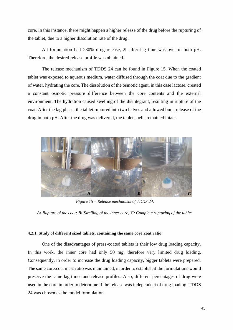
The release mechanism of TDDS 24 can be found in Figure 15. When the coated
tablet was exposed to aqueous medium, water diffused through the coat due to the gradient
of water, hydrating the core. The dissolution of the osmotic agent, in this case lactose, created
a constant osmotic pressure difference between the core contents and the external
environment. The hydration caused swelling of the disintegrant, resulting in rupture of the
coat. After the lag phase, the tablet ruptured into two halves and allowed burst release of the
drug in both pH. After the drug was delivered, the tablet shells remained intact.
4.2.1. Study of different sized tablets, containing the same core:coat ratio
One of the disadvantages of press-coated tablets is their low drug loading capacity.
In this work, the inner core had only 50 mg, therefore very limited drug loading.
Consequently, in order to increase the drug loading capacity, bigger tablets were prepared.
The same core:coat mass ratio was maintained, in order to establish if the formulations would
preserve the same lag times and release profiles. Also, different percentages of drug were
used in the core in order to determine if the release was independent of drug loading. TDDS
24 was chosen as the model formulation.
Figure 15 – Release mechanism of TDDS 24.
A: Rupture of the coat; B: Swelling of the inner core; C: Complete rupturing of the tablet.
A B C

46
0 4 8 12 16 20 24
0
10
20
30
40
50
60
70
80
90
100
TDDS 24 9 mm 10% drug
Time, h
Dru
g r
ele
ase, %
4 8 12 16 20 24
0
10
20
30
40
50
60
70
80
90
100
TDDS 24 11 mm 4.5% drug
Time, h
Dru
g r
ele
ase, %
0 4 8 12 16 20 24
0
10
20
30
40
50
60
70
80
90
100
TDDS 24 11 mm 10% drug
Time, h
Dru
g r
ele
ase, %
Figure 16 – Comparison of the release profile of TDDS 24, with different diameters and drug
loading.
HCl 0.1 N; Phosphate buffer pH 6.8
With the increase of diameter, the lag time did not suffer many changes (Figure 16).
The behaviour was identical, they all had a release profile characterized by a distinct lag time
and released >80% of the drug after 2h, maximum. TDDS 24’s lag time was 5.12 ± 0.37 h;
TDDS 24 11 mm, 4.5% drug was 4.5 ± 0.58 h and TDDS 24, 11 mm, 10% drug was 4.33 ±
0.55 h. The lag time decreased to some extent with the 11 mm tablets. The thickness of the
coat is one of the parameters that most affect the lag time. The 11 mm tablets were less thick,
the thickness was only 1.5 mm, whereas the 9 mm tablets had a thickness of 2 mm. Thus, the
decrease of thickness may explain the decrease of the lag time as it led to a quicker medium
penetration into the core (10,17). The variability between pH remained low with both
formulations. Drug loading had no effect on lag time.
4.3. In vitro release studies of EC coated tablets
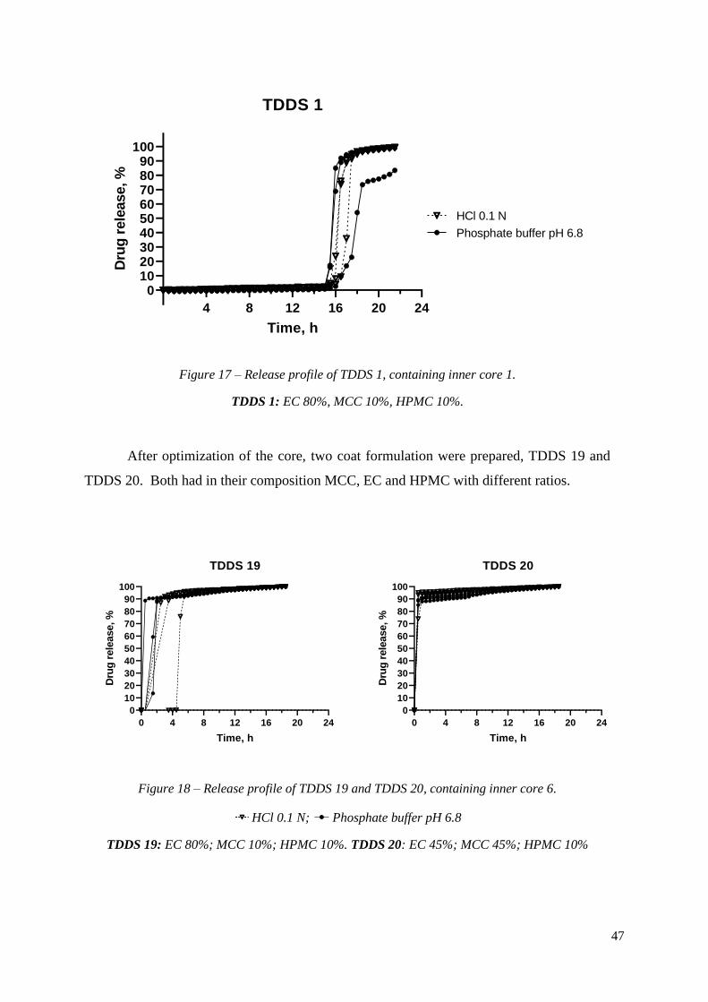
TDDS 1, formulated with EC, had a profile characterized by a lag time of 16.17 ±
0.62 h, followed by immediate release of the drug (Figure 17). The release was pH
independent, as it was evident by comparing lag times between different media (16.33 ± 0.47
h in HCl and 16.00 ± 0.71 h in pH 6.8).
47
4 8 12 16 20 24
0102030405060708090
100
TDDS 1
Time, h
Dru
g r
ele
ase, %
Phosphate buffer pH 6.8
HCl 0.1 N
Figure 17 – Release profile of TDDS 1, containing inner core 1.
TDDS 1: EC 80%, MCC 10%, HPMC 10%.
After optimization of the core, two coat formulation were prepared, TDDS 19 and
TDDS 20. Both had in their composition MCC, EC and HPMC with different ratios.
0 4 8 12 16 20 24
0
10
20
30
40
50
60
70
80
90
100
TDDS 19
Time, h
Dru
g r
ele
ase, %
0 4 8 12 16 20 24
0
10
20
30
40
50
60
70
80
90
100
TDDS 20
Time, h
Dru
g r
ele
ase, %
Figure 18 – Release profile of TDDS 19 and TDDS 20, containing inner core 6.
HCl 0.1 N; Phosphate buffer pH 6.8
TDDS 19: EC 80%; MCC 10%; HPMC 10%. TDDS 20: EC 45%; MCC 45%; HPMC 10%
48
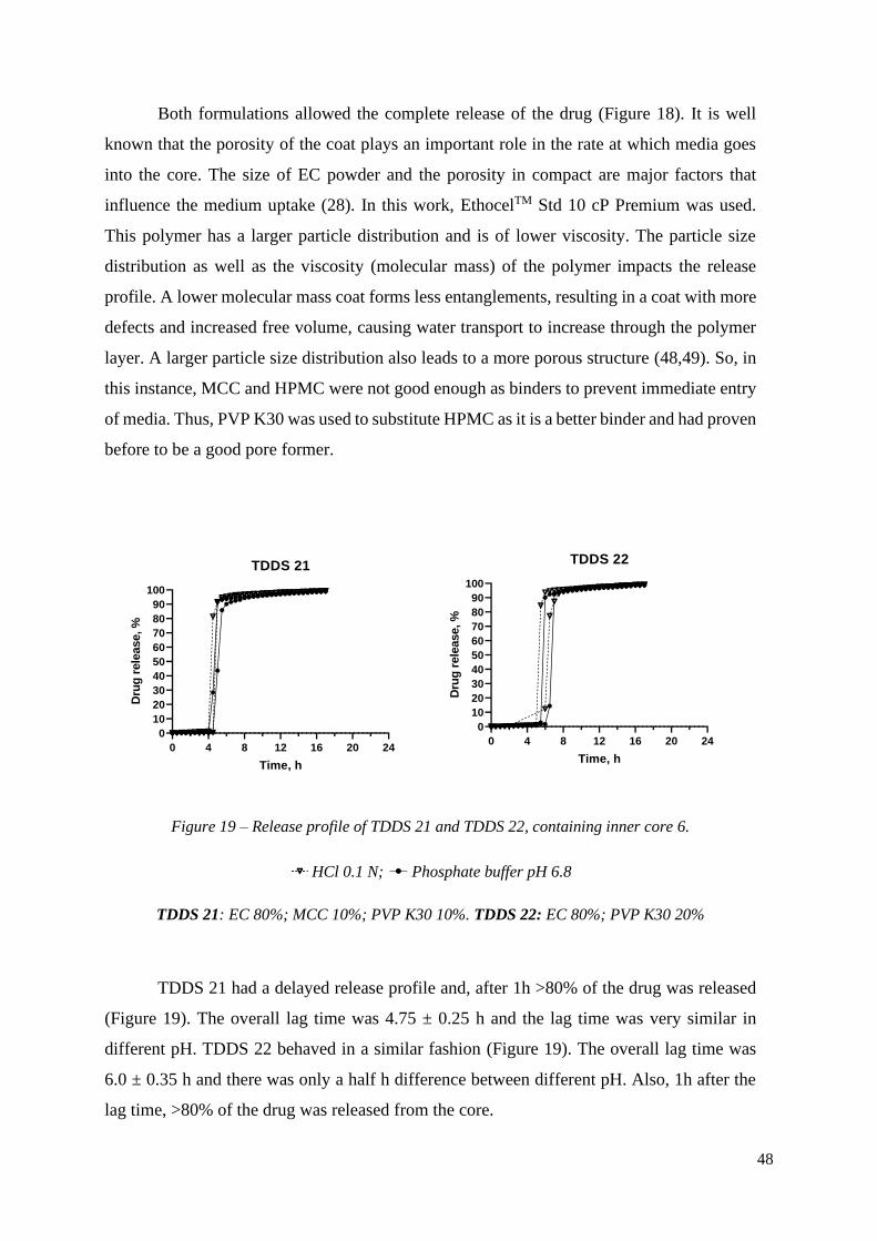
Both formulations allowed the complete release of the drug (Figure 18). It is well
known that the porosity of the coat plays an important role in the rate at which media goes
into the core. The size of EC powder and the porosity in compact are major factors that
influence the medium uptake (28). In this work, EthocelTM Std 10 cP Premium was used.
This polymer has a larger particle distribution and is of lower viscosity. The particle size
distribution as well as the viscosity (molecular mass) of the polymer impacts the release
profile. A lower molecular mass coat forms less entanglements, resulting in a coat with more
defects and increased free volume, causing water transport to increase through the polymer
layer. A larger particle size distribution also leads to a more porous structure (48,49). So, in
this instance, MCC and HPMC were not good enough as binders to prevent immediate entry
of media. Thus, PVP K30 was used to substitute HPMC as it is a better binder and had proven
before to be a good pore former.
0 4 8 12 16 20 24
0
10
20
30
40
50
60
70
80
90
100
TDDS 21
Time, h
Dru
g r
ele
ase, %
0 4 8 12 16 20 24
0
10
20
30
40
50
60
70
80
90
100
TDDS 22
Time, h
Dru
g r
ele
ase, %
Figure 19 – Release profile of TDDS 21 and TDDS 22, containing inner core 6.
HCl 0.1 N; Phosphate buffer pH 6.8
TDDS 21: EC 80%; MCC 10%; PVP K30 10%. TDDS 22: EC 80%; PVP K30 20%
TDDS 21 had a delayed release profile and, after 1h >80% of the drug was released
(Figure 19). The overall lag time was 4.75 ± 0.25 h and the lag time was very similar in
different pH. TDDS 22 behaved in a similar fashion (Figure 19). The overall lag time was
6.0 ± 0.35 h and there was only a half h difference between different pH. Also, 1h after the
lag time, >80% of the drug was released from the core.
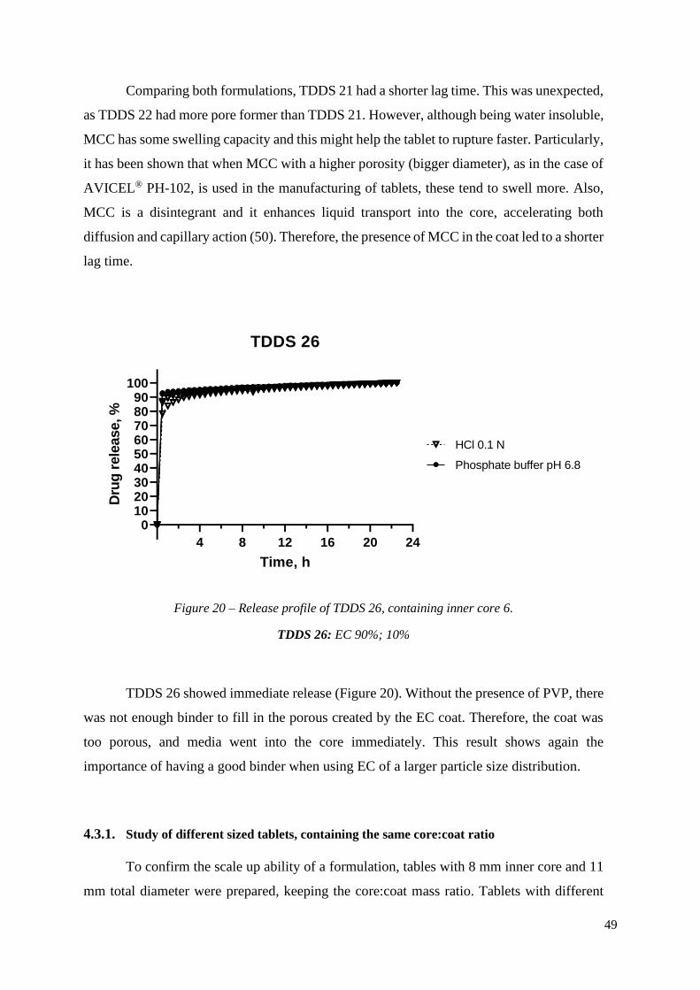
49
Comparing both formulations, TDDS 21 had a shorter lag time. This was unexpected,
as TDDS 22 had more pore former than TDDS 21. However, although being water insoluble,
MCC has some swelling capacity and this might help the tablet to rupture faster. Particularly,
it has been shown that when MCC with a higher porosity (bigger diameter), as in the case of
AVICEL® PH-102, is used in the manufacturing of tablets, these tend to swell more. Also,
MCC is a disintegrant and it enhances liquid transport into the core, accelerating both
diffusion and capillary action (50). Therefore, the presence of MCC in the coat led to a shorter
lag time.
4 8 12 16 20 24
0102030405060708090
100
TDDS 26
Time, h
Dru
g r
ele
ase, %
HCl 0.1 N
Phosphate buffer pH 6.8
Figure 20 – Release profile of TDDS 26, containing inner core 6.
TDDS 26: EC 90%; 10%
TDDS 26 showed immediate release (Figure 20). Without the presence of PVP, there
was not enough binder to fill in the porous created by the EC coat. Therefore, the coat was
too porous, and media went into the core immediately. This result shows again the
importance of having a good binder when using EC of a larger particle size distribution.
4.3.1. Study of different sized tablets, containing the same core:coat ratio
To confirm the scale up ability of a formulation, tables with 8 mm inner core and 11
mm total diameter were prepared, keeping the core:coat mass ratio. Tablets with different

50
ratios of drug were prepared to determine if the drug loading influenced the release profile.
TDDS 21 was chosen as the model formulation.
0 4 8 12 16 20 24
0
10
20
30
40
50
60
70
80
90
100
TDDS 21 9 mm 10% drug
Time, h
Dru
g r
ele
ase, %
4 8 12 16 20 24
0102030405060708090
100
TDDS 21 11 mm 4.5 % drug
Time, h
Dru
g r
ele
ase, %
4 8 12 16 20 24
0102030405060708090
100
TDDS 21 11 mm 10% drug
Time, h
Dru
g r
ele
ase, %
Figure 21 – Comparison of the release profile of TDDS 21, with different diameters and drug
loading.
HCl 0.1 N; Phosphate buffer pH 6.8
TDDS 21, 9 mm, had a lag time of 4.75 ± 0.25 h and in 1h after lag time, >80% of the
drug was released (Figure 21). Similarly, the formulations with the bigger diameter, had 80%
of drug release, 1h after lag time was over. They had slightly different lag time when
compared with TDDS 21 5 mm; TDDS 21 11 mm, 4.5% drug, had a lag time of 4.25 ± 0.38
h and TDDS 21 11 mm, 10% drug, had a lag time of 4.25 ± 0.25 h. All tablets had a pH
independent behaviour. Similarly to the TDDS 24 formulation (Figure 16), the decrease of
lag time might have been due to the reduced thickness of the coat in the sides. The thickness
is one of the parameters that most affect lag time in rupturable systems as it controls medium
permeability (10,17). With reduced thickness, medium penetrated the core more easily and
the ruptures happened faster. Again, the concentration of the drug had no effect on lag time.
4.4. Characterization of a commercially available formulation (Lodotra)
4.4.1. Physical characterization
Lodotra is a commercially available formulation that is designed to release the drug,
present in the core, after a lag time of approximately 4h. The mechanism is meant to rely on
time, and be independent of other variables, such as pH.
51
Table 8 – Excipients in the formulation of Lodotra
Inner Core Coat
Colloidal anhydrous sílica Colloidal anhydrous silica
Croscarmellose sodium Dicalcium phosphate dihydrated
Lactose, monohydrated Glyceryl behenate
Povidone K 29/32 Povidone K 29/32
Magnesium stearate Magnesium stearate
Red iron oxide Yellow iron oxide
Lodotra is mainly comprised of a combination of a hydrophobic wax, glyceryl
behenate, a non-fatty hydrophobic filler, dibasic calcium phosphate and a binder, PVP K
29/32. The core is constituted by the drug, lactose, croscarmellose sodium and PVP K2 9/32
(Table 8).
Table 9 – Physical characterization of Lodotra (5 mg)
Core Coat Total
Mass
(mg)
Height
(mm)
Diameter
(mm)
Mass
(mg)
Height
(mm)
Thickness
(mm)
Mass
(mg)
Height
(mm)
Diameter
(mm)
Lodotra 60 2 5 350 1,5 2 410 5 9
Lodotra had a total mass of ~410 mg, 60 mg of core and 350 mg of coat (Table 9). The
inner core diameter was 5 mm, while the total diameter was 9 mm, making a coat with 2 mm
of thickness. The height of the tablet was 5 mm. Thus, it had similar features to the tablets
developed in this research, particularly the 5mm/9mm tablets, and so made a comparison
possible.
4.4.2. Release profile of the drug

52
4 8 12 16 20 24
0102030405060708090
100
Release profile Lodotra 5 mg
Time, h
Dru
g r
ele
ase, %
HCl 0.1 N
Phosphate buffer pH 6.8
Figure 22 – Release profile of a commercially available formulation, Lodotra.
Lodotra, 5 mg, had a lag time of 4.75 ± 0.25 h, followed by immediate release of the
drug. The lag time was independent on pH. In HCl, after 30 min, >80% of the drug was
release; in phosphate buffer, it took 2h to reach the same drug release (Figure 22).
The release mechanism relies on a rupturable coat (Figure 23 The tablet does not release
any drug until the lag time is over. When lag time is over, the tablets ruptures due to the
Figure 23 – Release mechanism of Lodotra.
A: Rupturing of the coat; B: Media goes into the core; C: Swelling of the inner core; D: Complete
opening of the tablet.
D C
B A

53
increase of pressure in the core. Afterwards, more water goes into the core, making the inner
core swell even more. Finally, the tablet opens completely, and the drug is fully released.
Hence, the mechanism is similar to the formulations developed in this work.
4.5. Comparison of different formulations
4.5.1. Water uptake and dry mass loss
Generally, the process of drug release takes place in three steps, water goes into the
core, the core swells and the coat ruptures, making water uptake an important parameter to
determine. Thus, gravimetric studies on hydration and dry mass loss were performed in order
to determine the rate and the extent of water uptake in three formulations, TDDS 21, TDDS
24 and Lodotra (Figure 24).
0 1 2 3 4
0
10
20
30
40
0.1 N HCl
Time, h
Me
dia
up
take, %
TDDS 21
TDDS 24
Lodotra
0 1 2 3 4
0
10
20
30
40
Phosphate buffer, pH 6.8
Time, h
Me
dia
up
take, %
TDDS 21
TDDS 24
Lodotra
Figure 24 – Water uptake in 0.1 N HCl and phosphate buffer, pH 6.8.
TDDS 21: EC 80%; MCC 10%; PVP K30 10%. TDDS 24: GB 30%; DCP 20%; PVP K30 50%.
Lodotra had a water uptake of 17.25 ± 0.27% in HCl, after 3 h. In phosphate buffer,
the water uptake was lower, 3.41 ± 0.10%, after the same time. This can be explained by the
presence of DCP in the coat. As explained before, DCP is generally insoluble but it is soluble
in diluted acids. Consequently, in HCl, when in contact with the medium, it formed more
micro-cavities and triggered the osmotic effect which promoted the swelling and
consequently, rupture of the coat.
Similarly, TDDS 24 showed different water uptake in different media, also because
of DCP. TDDS 24, after 3 h, had 35.93 ± 1.92% water uptake in HCl and 24.50 ± 0.19% in

54
phosphate buffer. Even though the high water uptake was not a problem with the current
formulations as there was no drug release >10% before the lag time, the high water uptake
might be of concern if a more soluble drug is used in the core; in that case, there might be a
higher diffusion of the drug, prior to the rupture of the coat. Even in phosphate buffer, where
DCP is insoluble, the water uptake was high due to the high percentage of soluble excipient,
PVP, in the coat.
On the other hand, TDDS 21 had comparable water uptake in different media. After
3 h, it remained low, 4.82 ± 0.20% in HCl and 4.05 ± 0.07 % in phosphate buffer. The water
uptake only increased ~2% between 1 h and 3 h.
Comparing formulations, TDDS 24, due to its higher content of DCP, had a higher
uptake than Lodotra. TDDS 21, had a similar behaviour to Lodotra.
0 1 2 3 4
0
10
20
30
40
0.1 N HCl
Time, h
Dry
mass lo
ss, %
TDDS 21
TDDS 24
Lodotra
0 1 2 3 4
0
10
20
30
40
Phosphate buffer, pH 6.8
Time, h
Dry
mass lo
ss, %
TDDS 21
TDDS 24
Lodotra
Figure 25 – Dry mass loss in 0.1 N HCl and phosphate buffer, pH 6.8.
TDDS 21: EC 80%; MCC 10%; PVP K30 10%. TDDS 24: GB 30%; DCP 20%; PVP K30 50%.
Dry mass results showed resemblance with water uptake. TDDS 21 and Lodotra
showed very little dry mass loss. TDDS 24 was the formulation that showed the highest dry
mass loss, since DCP and PVP K30 were present in a high fraction and so, dissolved
themselves in the media. The dry mass loss was higher in HCl due to the presence of DCP,
again. Due to its solubility in HCl, it is more easily dissolved and released from the coat.
5. Conclusion

55
Press-coating was used to develop the delivery systems in this work. It allowed to
obtain tablets containing an inner core, surrounded by a rupturable coat, which was able to
modulate the release of the drug from the core by modifying the excipients in the coat.
All optimized formulations (TDDS 21, TDDS 22, TDDS 23, TDDS 24, TDDS 25)
exhibited the desired release profile, a lag time followed by a sudden and complete release
of the drug.
Looking at the release profiles obtain with the GB formulations many assumptions can
be taken. Firstly, the ratio of DCP played an important role in obtaining a formulation pH
independent, owing to its higher solubility in diluted acids. It was shown that by decreasing
the percentage of DCP, the lag time between different pH became less variable. Therefore,
to achieve a pH independent system, DCP should not be present in a high fraction. Also, by
varying the ratio GB:PVP K30, the lag time could be modified; the higher the ratio, the higher
the lag time.
An issue with the GB formulations was the possibility of drug diffusion prior to
rupturing of the tablet. As the percentage of soluble excipient, PVP K30, increased, so did
the diffusion of the drug prior to rupturing. Additionally, water uptake was very high,
stressing again this issue. In this case, the release never reached levels above 5%. But, if a
more soluble drug was used in the core, there might be more diffusion and the desired profile
might not be reached.
The EC formulations showed pH independence from the beginning. With these
formulations, as the EC powder had a large size distribution, the presence of a good binder
was pivotal. Without a binder, media went into the core to fast and immediate release was
obtained. As for excipients, not only was the lag time influenced by the ratio EC:soluble
excipient, but it was also influenced by the presence of swellable materials in the coat, which
supported the rupture of the coat.
Both developed formulations showed that it was possible to increase the size of the
tablet, and maintain the same release profile, as long as the same core:coat mass ratio was
respected. This is crucial as one of the disadvantages of press-coated tablets is low drug
loading. By increasing the size of the whole system, and hence the size of the inner core, a
higher drug loading is possible. Also, drug concentration showed no effect on lag time.
56
One of the differences between the EC and GB formulations was the height of the tablet.
The presence of DCP in the GB tablet, a high-density powder, led to a smaller tablet. This
finding is important because dosage forms of a smaller size increase the comfort of intake
and thereby enhance the compliance, particularly in paediatric and geriatric populations.
The presence of a swellable agent in the core is of extreme importance to promote the
rupture of the coat, as was shown with formulations TDDS 3-TDDS 9.
One of the factors that most affect the variability of the lag time is the centring of the
tablet. Since, in this case, the centring was done manually, this was a source of errors.
Therefore, the high variability within batches was probably due to the lack of experience of
the operator.
6. Future work
Considering the obtained results and other described in the literature, it is important to
perform further studies.
Firstly, it is important to evaluate the influence the core:coat mass ratio, on lag time.
Moreover, the study of other manufacturing variables should be performed, particularly, the
effect of the hardness and compression force, of both the core and coat, on lag time.
As mentioned before, diffusion of the drug prior to lag time, is one of the concerns of
these delivery systems. Therefore, the influence of drug solubility on the release profile,
should be assessed. Specifically, release studies with a more soluble drug along with a less
soluble drug ought to be executed.
One of the applications of TDDS is delivery into the large bowel. To achieve this, an
enteric coating could be applied onto the press-coated tablet, to overcome the variable
stomach emptying time. This approach should be tested and release studies with a continuous
pH should be performed. Furthermore, it would be important to evaluate the stability of
peptides and proteins, under compression, as these have application in the treatment of such
diseases.
The process of manufacturing the tablets was very time-consuming and showed large
variability. Therefore, if a fully automatic press-coating machine was used, coupled with the

57
OSDRC system, the time of manufacturing can be reduced, as well as the variability
decreased. This method of manufacturing should be tested to consider its impact on lag time.

58
References
1. Lin SY, Kawashima Y. Current status and approaches to developing press-coated
chronodelivery drug systems. J Control Release. 2012;157(3):331–53.
2. Qiu, Y;Lee PI. Rational Design of Oral Modified-Release Drug Delivery Systems. In:
Developing Solid Oral Dosage Forms. Elsevier Inc.; 2017. p. 519–54.
3. Qiu, Y.; Zhou D. Understanding design and development of modified release solid
oral dosage forms. J Valid Technol. 2011;17(2):23–32.
4. Wang R, Conner DP, Li B V. Bioavailability and Bioequivalence Aspects of Oral
Modified-Release Drug Products. AAPS J. 2017;19(2):360–6.
5. Maroni A, Zema L, Curto MD Del, Loreti G, Gazzaniga A. Oral pulsatile delivery:
Rationale and chronopharmaceutical formulations. Int J Pharm. 2010;398(1–2):1–8.
6. Maroni A, Zema L, Cerea M, Sangalli ME. Oral pulsatile drug delivery systems.
Expert Opin Drug Deliv. 2005;2(5):855–71.
7. Sungthongjeen S, Puttipipatkhachorn S, Paeratakul O, Dashevsky A, Bodmeier R.
Development of pulsatile release tablets with swelling and rupturable layers. J Control
Release. 2004;95(2):147–59.
8. Wang H, Cheng L, Wen H, Li C, Li Y, Zhang X, et al. A time-adjustable pulsatile
release system for ketoprofen: In vitro and in vivo investigation in a pharmacokinetic
study and an IVIVC evaluation. Eur J Pharm Biopharm. 2017;119:192–200.
9. Gandhi BR, Mundada AS, Gandhi PP. Chronopharmaceutics: As a clinically relevant
drug delivery system. Drug Deliv. 2011;18(1):1–18.
10. Jain D, Raturi R, Jain V, Bansal P, Singh R. Recent technologies in pulsatile drug
delivery systems. Biomatter. 2011;1(1):57–65.
11. Fukui E, Miyamura N, Uemura K, Kobayashi M. Preparation of enteric coated timed-
release press-coated tablets and evaluation of their function by in vitro and in vivo
tests for colon targeting. Int J Pharm. 2000;204(1–2):7–15.
12. Gazzaniga A, Maroni A, Sangalli ME, Zema L. Time-controlled oral delivery systems
for colon targeting. Expert Opin Drug Deliv. 2006;3(5):583–97.
13. Saigal N, Baboota S, Ahuja A, Ali J. Multiple-pulse drug delivery systems: setting a

59
new paradigm for infectious disease therapy. Expert Opin Drug Deliv. 2009;6(4):441–
52.
14. Maroni A, Zema L, Loreti G, Palugan L, Gazzaniga A. Film coatings for oral pulsatile
release. Int J Pharm. 2013;457(2):362–71.
15. Dashevsky A, Bussemer T, Mohamad A, Bodmeier R. Process and Formulation
Variables Affecting the Performance of a Rupturable Capsule-Based Drug Delivery
System with Pulsatile Drug Release. Drug Dev Ind Pharm. 2004;30(2):171–9.
16. Sungthongjeen S, Puttipipatkhachorn S, Paeratakul O, Dashevsky A, Bodmeier R.
Development of pulsatile release tablets with swelling and rupturable layers. J Control
Release. 2004 Mar;95(2):147–59.
17. Lin H, Lin S, Lin Y, Ho H, Lo Y, Sheu M. Release characteristics and in vitro – in
vivo correlation of pulsatile pattern for a pulsatile drug delivery system activated by
membrane rupture via osmotic pressure and swelling. Eur J Pharm Biopharm.
2008;70:289–301.
18. Bose S, Bogner RH. Solventless pharmaceutical coating processes: A review. Pharm
Dev Technol. 2007;12(2):115–31.
19. Foppoli AA, Maroni A, Cerea M, Zema L, Gazzaniga A. Dry coating of solid dosage
forms: an overview of processes and applications. Drug Dev Ind Pharm.
2017;43(12):1919–31.
20. Luo Y, Zhu J, Ma Y, Zhang H. Dry coating, a novel coating technology for solid
pharmaceutical dosage forms. Int J Pharm. 2008;358(1–2):16–22.
21. Lin SY, Kawashima Y. Current status and approaches to developing press-coated
chronodelivery drug systems. J Control Release. 2012;157(3):331–53.
22. Ozeki Y, Watanabe Y, Inoue S, Danjo K. Comparison of the compression
characteristics between new one-step dry-coated tablets (OSDRC) and dry-coated
tablets (DC). Int J Pharm. 2003;259(1–2):69–77.
23. Lin SY, Lin KH, Li MJ. Influence of excipients, drugs, and osmotic agent in the inner
core on the time-controlled disintegration of compression-coated ethylcellulose
tablets. Vol. 91, Journal of Pharmaceutical Sciences. 2002. p. 2040–6.
60
24. Rujivipat S, Bodmeier R. Modified release from hydroxypropyl methylcellulose
compression-coated tablets. Int J Pharm. 2010;402(1–2):72–7.
25. Nunthanid J, Luangtana-anan M, Sriamornsak P, Limmatvapirat S, Huanbutta K,
Puttipipatkhachorn S. Use of spray-dried chitosan acetate and ethylcellulose as
compression coats for colonic drug delivery: Effect of swelling on triggering in vitro
drug release. Eur J Pharm Biopharm. 2009;71(2):356–61.
26. Rujivipat S, Bodmeier R. Improved drug delivery to the lower intestinal tract with
tablets compression-coated with enteric/nonenteric polymer powder blends. Eur J
Pharm Biopharm. 2010;76(3):486–92.
27. Conte U, Maggi L, Torre ML, Giunchedi P, Manna A La. Press-coated tablets for
time-programmed release of drugs. Biomaterials. 1993;14(13):1017–23.
28. Lin S-Y, Li M-J, Lin K-H. Hydrophilic excipients modulate the time lag of time-
controlled disintegrating press-coated tablets. AAPS PharmSciTech. 2005;5(4):25–9.
29. Patadia R, Vora C, Mittal K, Mashru R. Investigating critical effects of variegated
lubricants, glidants and hydrophilic additives on lag time of press coated ethylcellulose
tablets. Pharm Dev Technol. 2016;21(3):302–10.
30. Lin SY, Lin KH, Li MJ. Micronized ethylcellulose used for designing a directly
compressed time-controlled disintegration tablet. J Control Release. 2001;70(3):321–
8.
31. K. Philip A, Philip B. Chronopharmaceuticals: Hype or Future of Pharmaceutics. Curr
Pharm Des. 2011;17(15):1512–6.
32. L.L. C, D.S. J, L.P. H, R.H. S, M.G. P, J.R. K. Alleviation of morning joint stiffness
by low-dose prednisone in rheumatoid arthritis is associated with circadian changes in
IL-6 and cortisol. Int J Clin Rheumtol. 2011;6(2):241–9.
33. Smolensky MH, Peppas NA. Chronobiology, drug delivery, and chronotherapeutics.
Adv Drug Deliv Rev. 2007;59(9–10):828–51.
34. Mandal AS, Biswas N, Karim KM, Guha A, Chatterjee S, Behera M, et al. Drug
delivery system based on chronobiology-A review. J Control Release.
2010;147(3):314–25.

61
35. Buttgereit F, Gibofsky A. Delayed-release prednisone -- a new approach to an old
therapy. Expert Opin Pharmacother. 2013;14(8):1097–106.
36. Schaeffler A. DELAYED-RELEASE GLUCOCORTICOID TREATMENT OF
RHEUMATOID DISEASE. Vol. 32. United States of America: United States Patent;
US 9,504,699 B2, 2016.
37. Arvidson NG, Gudbjörnsson B, Larsson A, Hällgren R. The timing of glucocorticoid
administration in rheumatoid arthritis. Ann Rheum Dis. 1997;56(1):27–31.
38. Alten R. Chronotherapy with modified-release prednisone in patients with rheumatoid
arthritis. Expert Rev Clin Immunol. 2012;8(2):123–33.
39. Clarke L, Jessop DS, Hunt LP, Kirwan JR, Perry MG, Straub RH. Effect of novel
therapeutic glucocorticoids on circadian rhythms of hormones and cytokines in
rheumatoid arthritis. Ann N Y Acad Sci. 2010;1193(1):127–33.
40. Arvidson NG, Gudbjornsson B, Elfman L, Ryden AC, Totterman TH, Hallgren R.
Circadian rhythm of serum interleukin-6 in rheumatoid arthritis. Ann Rheum Dis.
1994;53(8):521–4.
41. Straub RH, Cutolo M. Circadian rhythms in rheumatoid arthritis: Implications for
pathophysiology and therapeutic management. Arthritis Rheum. 2007;56(2):399–408.
42. Carstensen JT, Ertell C. Physical and chemical properties of calcium phosphates for
solid state pharmaceutical formulations. Drug Dev Ind Pharm. 1990;16(7):1121–33.
43. Scholler M, Emrich A, Silke J. Ingredients for Direct Compression of tablets: Two
types of Dicalcium phosphate anhydrous for controlling the size of tablets. In 2013. p.
44. Eyjolfsson R. Kollidon: Polyvinylpyrrolidone excipients for the pharmaceutical
industry. 9th ed. BASF; 2008. 17–119 p.
45. Mohamad A, Dashevsky A. pH-independent pulsatile drug delivery system based on
hard gelatin capsules and coated with aqueous dispersion Aquacoat® ECD. Eur J
Pharm Biopharm. 2006;64(2):173–9.
46. Zakowiecki D, Lachmann M. Beyond just a filler – application of calcium phosphates
in direct compression formulations [Internet]. 2019 [cited 2019 Mar 20]. Available
from: https://www.expresspharma.in/cphi-p-mec-india/beyond-just-a-filler-

62
application-of-calcium-phosphates-in-direct-compression-formulations/
47. Patil S, Shahiwala A. Chronomodulated press-coated pulsatile therapeutic system for
aceclofenac: optimization of factors influencing drug release and lag time.
ChronoPhysiology Ther. 2011;1.
48. ETHOCELTM Ethylcellulose A Technical Review. Dow Pharma and Food; 2016.
49. ETHOCEL PREMIUM POLYMERS FOR PHARMACEUTICAL APPLICATIONS.
Dow Pharma and Food; 1998.
50. Yassin S, Goodwin DJ, Anderson A, Sibik J, Wilson DI, Gladden LF, et al. The
Disintegration Process in Microcrystalline Cellulose Based Tablets, Part 1: Influence
of Temperature, Porosity and Superdisintegrants. J Pharm Sci. 2015;104(10):3440–
50.
63
ANNEX I
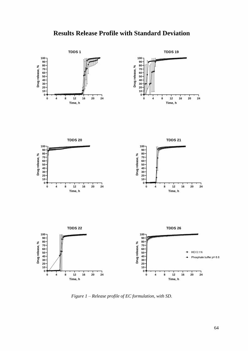
64
Results Release Profile with Standard Deviation
0 4 8 12 16 20 24
0
10
20
30
40
50
60
70
80
90
100
TDDS 1
Time, h
Dru
g r
ele
ase, %
0 4 8 12 16 20 24
0
10
20
30
40
50
60
70
80
90
100
TDDS 19
Time, h
Dru
g r
ele
ase, %
0 4 8 12 16 20 24
0
10
20
30
40
50
60
70
80
90
100
TDDS 20
Time, h
Dru
g r
ele
ase, %
0 4 8 12 16 20 24
0
10
20
30
40
50
60
70
80
90
100
TDDS 21
Time, h
Dru
g r
ele
ase, %
0 4 8 12 16 20 24
0
10
20
30
40
50
60
70
80
90
100
TDDS 22
Time, h
Dru
g r
ele
ase, %
0 4 8 12 16 20 24
0
10
20
30
40
50
60
70
80
90
100
TDDS 26
Time, h
Dru
g r
ele
ase, %
Figure 1 – Release profile of EC formulation, with SD.
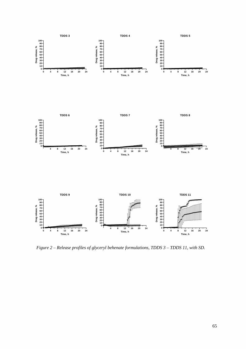
65
0 4 8 12 16 20 24
0
10
20
30
40
50
60
70
80
90
100
TDDS 3
Time, h
Dru
g r
ele
ase, %
0 4 8 12 16 20 24
0
10
20
30
40
50
60
70
80
90
100
TDDS 4
Time, h
Dru
g r
ele
ase, %
0 4 8 12 16 20 24
0
10
20
30
40
50
60
70
80
90
100
TDDS 5
Time, h
Dru
g r
ele
ase, %
4 8 12 16 20 24
0
10
20
30
40
50
60
70
80
90
100
TDDS 6
Time, h
Dru
g r
ele
ase, %
0 4 8 12 16 20 24
0
10
20
30
40
50
60
70
80
90
100
TDDS 7
Time, h
Dru
g r
ele
ase, %
4 8 12 16 20 24
0
10
20
30
40
50
60
70
80
90
100
TDDS 8
Time, h
Dru
g r
ele
ase, %
0 4 8 12 16 20 24
0
10
20
30
40
50
60
70
80
90
100
TDDS 9
Time, h
Dru
g r
ele
ase, %
4 8 12 16 20 24
0
10
20
30
40
50
60
70
80
90
100
TDDS 10
Time, h
Dru
g r
ele
ase, %
0 4 8 12 16 20 24
0
10
20
30
40
50
60
70
80
90
100
TDDS 11
Time, h
Dru
g r
ele
ase, %
Figure 2 – Release profiles of glyceryl behenate formulations, TDDS 3 – TDDS 11, with SD.

66
0 4 8 12 16 20 24
0
10
20
30
40
50
60
70
80
90
100
TDDS 13
Time, h
Dru
g r
ele
ase, %
4 8 12 16 20 24
0
10
20
30
40
50
60
70
80
90
100
TDDS 14
Time, h
Dru
g r
ele
ase, %
0 4 8 12 16 20 24
0
10
20
30
40
50
60
70
80
90
100
TDDS 15
Time, h
Dru
g r
ele
ase, %
0 4 8 12 16 20 24
0
10
20
30
40
50
60
70
80
90
100
TDDS 16
Time, h
Dru
g r
ele
ase, %
0 4 8 12 16 20 24
0
10
20
30
40
50
60
70
80
90
100
TDDS 17
Time, h
Dru
g r
ele
ase, %
0 4 8 12 16 20 24
0
10
20
30
40
50
60
70
80
90
100
TDDS 18
Time, h
Dru
g r
ele
ase, %
0 4 8 12 16 20 24
0
10
20
30
40
50
60
70
80
90
100
TDDS 23
Time, h
Dru
g r
ele
ase, %
0 4 8 12 16 20 24
0
10
20
30
40
50
60
70
80
90
100
TDDS 24
Time, h
Dru
g r
ele
ase, %
0 4 8 12 16 20 24
0
10
20
30
40
50
60
70
80
90
100
TDDS 25
Time, h
Dru
g r
ele
ase, %
Figure 3 – Release profiles of glyceryl behenate formulations, TDDS 13 – TDDS 18 and TDDS 23
– TDDS 25, with SD.


