Embed Size (px)
Citation preview
This article appeared in a journal published by Elsevier. The attachedcopy is furnished to the author for internal non-commercial researchand education use, including for instruction at the authors institution
and sharing with colleagues.
Other uses, including reproduction and distribution, or selling orlicensing copies, or posting to personal, institutional or third party
websites are prohibited.
In most cases authors are permitted to post their version of thearticle (e.g. in Word or Tex form) to their personal website orinstitutional repository. Authors requiring further information
regarding Elsevier’s archiving and manuscript policies areencouraged to visit:
http://www.elsevier.com/copyright

Author's personal copy
International Journal of Pharmaceutics 394 (2010) 122–142
Contents lists available at ScienceDirect
International Journal of Pharmaceutics
journa l homepage: www.e lsev ier .com/ locate / i jpharm
Review
Dendrimer toxicity: Let’s meet the challenge
Keerti Jain, Prashant Kesharwani, Umesh Gupta, N.K. Jain ∗
Pharmaceutics Research Laboratory, Department of Pharmaceutical Sciences, Dr. H. S. Gour University Sagar (M.P.) 470003 India
a r t i c l e i n f o
Article history:Received 8 January 2010Received in revised form 19 April 2010Accepted 19 April 2010Available online 28 April 2010
Keywords:DendrimersCationic toxicityBiocompatibilitySurface engineering
a b s t r a c t
Dendrimers are well-defined, versatile polymeric architecture with properties resembling biomolecules.Dendritic polymers emerged as outstanding carrier in modern medicine system because of its derivatis-able branched architecture and flexibility in modifying it in numerous ways. Dendritic scaffold has beenfound to be suitable carrier for a variety of drugs including anticancer, anti-viral, anti-bacterial, antitu-bercular etc., with capacity to improve solubility and bioavailability of poorly soluble drugs. In spite ofextensive applicability in pharmaceutical field, the use of dendrimers in biological system is constrainedbecause of inherent toxicity associated with them. This toxicity is attributed to the interaction of surfacecationic charge of dendrimers with negatively charged biological membranes in vivo. Interaction of den-drimers with biological membranes results in membrane disruption via nanohole formation, membranethinning and erosion. Dendrimer toxicity in biological system is generally characterized by hemolytictoxicity, cytotoxicity and hematological toxicity. To minimize this toxicity two strategies have been uti-lized; first, designing and synthesis of biocompatible dendrimers; and second, masking of peripheralcharge of dendrimers by surface engineering. Biocompatible dendrimers can be synthesized by employ-ing biodegradable core and branching units or utilizing intermediates of various metabolic pathways.Dendrimer biocompatibility has been evaluated in vitro and in vivo for efficient presentation of biologicalperformance. Surface engineering masks the cationic charge of dendrimer surface either by neutralizationof charge, for example PEGylation, acetylation, carbohydrate and peptide conjugation; or by introducingnegative charge such as half generation dendrimers. Neutral and negatively charged dendrimers do notinteract with biological environment and hence are compatible for clinical applications as elucidated byvarious studies examined in this review. Chemical modification of the surface is an important strategy toovercome the toxicity problems associated with the dendrimers. The present review emphasizes on theapproaches available to overcome the cationic toxicity inherently associated with the dendrimers.
© 2010 Elsevier B.V. All rights reserved.
Contents
1. Introduction . . . . . . . . . . . . . . . . . . . . . . . . . . . . . . . . . . . . . . . . . . . . . . . . . . . . . . . . . . . . . . . . . . . . . . . . . . . . . . . . . . . . . . . . . . . . . . . . . . . . . . . . . . . . . . . . . . . . . . . . . . . . . . . . . . . . . . . . . . 1232. Dendrimers toxicity . . . . . . . . . . . . . . . . . . . . . . . . . . . . . . . . . . . . . . . . . . . . . . . . . . . . . . . . . . . . . . . . . . . . . . . . . . . . . . . . . . . . . . . . . . . . . . . . . . . . . . . . . . . . . . . . . . . . . . . . . . . . . . . . . . 124
2.1. Cytotoxicity . . . . . . . . . . . . . . . . . . . . . . . . . . . . . . . . . . . . . . . . . . . . . . . . . . . . . . . . . . . . . . . . . . . . . . . . . . . . . . . . . . . . . . . . . . . . . . . . . . . . . . . . . . . . . . . . . . . . . . . . . . . . . . . . . . . 1252.2. Hemolytic toxicity . . . . . . . . . . . . . . . . . . . . . . . . . . . . . . . . . . . . . . . . . . . . . . . . . . . . . . . . . . . . . . . . . . . . . . . . . . . . . . . . . . . . . . . . . . . . . . . . . . . . . . . . . . . . . . . . . . . . . . . . . . . . 1262.3. Haematological toxicity . . . . . . . . . . . . . . . . . . . . . . . . . . . . . . . . . . . . . . . . . . . . . . . . . . . . . . . . . . . . . . . . . . . . . . . . . . . . . . . . . . . . . . . . . . . . . . . . . . . . . . . . . . . . . . . . . . . . . . 1262.4. Immunogenicity . . . . . . . . . . . . . . . . . . . . . . . . . . . . . . . . . . . . . . . . . . . . . . . . . . . . . . . . . . . . . . . . . . . . . . . . . . . . . . . . . . . . . . . . . . . . . . . . . . . . . . . . . . . . . . . . . . . . . . . . . . . . . . 1262.5. In vivo toxicity . . . . . . . . . . . . . . . . . . . . . . . . . . . . . . . . . . . . . . . . . . . . . . . . . . . . . . . . . . . . . . . . . . . . . . . . . . . . . . . . . . . . . . . . . . . . . . . . . . . . . . . . . . . . . . . . . . . . . . . . . . . . . . . . 127
3. Membrane interaction . . . . . . . . . . . . . . . . . . . . . . . . . . . . . . . . . . . . . . . . . . . . . . . . . . . . . . . . . . . . . . . . . . . . . . . . . . . . . . . . . . . . . . . . . . . . . . . . . . . . . . . . . . . . . . . . . . . . . . . . . . . . . . . 1273.1. Interaction with lipid bilayers . . . . . . . . . . . . . . . . . . . . . . . . . . . . . . . . . . . . . . . . . . . . . . . . . . . . . . . . . . . . . . . . . . . . . . . . . . . . . . . . . . . . . . . . . . . . . . . . . . . . . . . . . . . . . . . . 1273.2. Interaction with cell membrane . . . . . . . . . . . . . . . . . . . . . . . . . . . . . . . . . . . . . . . . . . . . . . . . . . . . . . . . . . . . . . . . . . . . . . . . . . . . . . . . . . . . . . . . . . . . . . . . . . . . . . . . . . . . . 129
4. Solutions for toxicity issues . . . . . . . . . . . . . . . . . . . . . . . . . . . . . . . . . . . . . . . . . . . . . . . . . . . . . . . . . . . . . . . . . . . . . . . . . . . . . . . . . . . . . . . . . . . . . . . . . . . . . . . . . . . . . . . . . . . . . . . . . . 1294.1. Biodegradable/biocompatible dendrimers . . . . . . . . . . . . . . . . . . . . . . . . . . . . . . . . . . . . . . . . . . . . . . . . . . . . . . . . . . . . . . . . . . . . . . . . . . . . . . . . . . . . . . . . . . . . . . . . . . . 129
4.1.1. Polyether dendrimers . . . . . . . . . . . . . . . . . . . . . . . . . . . . . . . . . . . . . . . . . . . . . . . . . . . . . . . . . . . . . . . . . . . . . . . . . . . . . . . . . . . . . . . . . . . . . . . . . . . . . . . . . . . . . . 1294.1.2. Polyester dendritic system . . . . . . . . . . . . . . . . . . . . . . . . . . . . . . . . . . . . . . . . . . . . . . . . . . . . . . . . . . . . . . . . . . . . . . . . . . . . . . . . . . . . . . . . . . . . . . . . . . . . . . . . . 1304.1.3. Polyether imine dendrimers . . . . . . . . . . . . . . . . . . . . . . . . . . . . . . . . . . . . . . . . . . . . . . . . . . . . . . . . . . . . . . . . . . . . . . . . . . . . . . . . . . . . . . . . . . . . . . . . . . . . . . . . 132
∗ Corresponding author. Tel.: +91 7582 264712; fax: +91 7582 264712.E-mail address: [email protected] (N.K. Jain).
0378-5173/$ – see front matter © 2010 Elsevier B.V. All rights reserved.doi:10.1016/j.ijpharm.2010.04.027
Author's personal copy
K. Jain et al. / International Journal of Pharmaceutics 394 (2010) 122–142 123
4.1.4. Polyether–copolyester (PEPE) dendrimers . . . . . . . . . . . . . . . . . . . . . . . . . . . . . . . . . . . . . . . . . . . . . . . . . . . . . . . . . . . . . . . . . . . . . . . . . . . . . . . . . . . . . . . . . 1324.1.5. Phosphate dendrimers. . . . . . . . . . . . . . . . . . . . . . . . . . . . . . . . . . . . . . . . . . . . . . . . . . . . . . . . . . . . . . . . . . . . . . . . . . . . . . . . . . . . . . . . . . . . . . . . . . . . . . . . . . . . . . 1324.1.6. Citric acid dendrimers . . . . . . . . . . . . . . . . . . . . . . . . . . . . . . . . . . . . . . . . . . . . . . . . . . . . . . . . . . . . . . . . . . . . . . . . . . . . . . . . . . . . . . . . . . . . . . . . . . . . . . . . . . . . . . 1334.1.7. Melamine dendrimers . . . . . . . . . . . . . . . . . . . . . . . . . . . . . . . . . . . . . . . . . . . . . . . . . . . . . . . . . . . . . . . . . . . . . . . . . . . . . . . . . . . . . . . . . . . . . . . . . . . . . . . . . . . . . . 1334.1.8. Peptide dendrimers . . . . . . . . . . . . . . . . . . . . . . . . . . . . . . . . . . . . . . . . . . . . . . . . . . . . . . . . . . . . . . . . . . . . . . . . . . . . . . . . . . . . . . . . . . . . . . . . . . . . . . . . . . . . . . . . . 1334.1.9. Triazine dendrimers . . . . . . . . . . . . . . . . . . . . . . . . . . . . . . . . . . . . . . . . . . . . . . . . . . . . . . . . . . . . . . . . . . . . . . . . . . . . . . . . . . . . . . . . . . . . . . . . . . . . . . . . . . . . . . . . 133
4.2. Surface engineered dendrimers . . . . . . . . . . . . . . . . . . . . . . . . . . . . . . . . . . . . . . . . . . . . . . . . . . . . . . . . . . . . . . . . . . . . . . . . . . . . . . . . . . . . . . . . . . . . . . . . . . . . . . . . . . . . . . 1344.2.1. PEGylation . . . . . . . . . . . . . . . . . . . . . . . . . . . . . . . . . . . . . . . . . . . . . . . . . . . . . . . . . . . . . . . . . . . . . . . . . . . . . . . . . . . . . . . . . . . . . . . . . . . . . . . . . . . . . . . . . . . . . . . . . . 1344.2.2. Carbohydrate engineered dendrimers . . . . . . . . . . . . . . . . . . . . . . . . . . . . . . . . . . . . . . . . . . . . . . . . . . . . . . . . . . . . . . . . . . . . . . . . . . . . . . . . . . . . . . . . . . . . . 1344.2.3. Acetylation . . . . . . . . . . . . . . . . . . . . . . . . . . . . . . . . . . . . . . . . . . . . . . . . . . . . . . . . . . . . . . . . . . . . . . . . . . . . . . . . . . . . . . . . . . . . . . . . . . . . . . . . . . . . . . . . . . . . . . . . . . 1364.2.4. Half generation- or anionic dendrimers . . . . . . . . . . . . . . . . . . . . . . . . . . . . . . . . . . . . . . . . . . . . . . . . . . . . . . . . . . . . . . . . . . . . . . . . . . . . . . . . . . . . . . . . . . . . 1364.2.5. Amino acid or peptide conjugated dendrimers . . . . . . . . . . . . . . . . . . . . . . . . . . . . . . . . . . . . . . . . . . . . . . . . . . . . . . . . . . . . . . . . . . . . . . . . . . . . . . . . . . . . 1364.2.6. Drug and DNA/gene conjugated dendrimers . . . . . . . . . . . . . . . . . . . . . . . . . . . . . . . . . . . . . . . . . . . . . . . . . . . . . . . . . . . . . . . . . . . . . . . . . . . . . . . . . . . . . . . 1374.2.7. Antibody functionalized dendrimers . . . . . . . . . . . . . . . . . . . . . . . . . . . . . . . . . . . . . . . . . . . . . . . . . . . . . . . . . . . . . . . . . . . . . . . . . . . . . . . . . . . . . . . . . . . . . . . 1384.2.8. Tuftsin conjugated dendrimers . . . . . . . . . . . . . . . . . . . . . . . . . . . . . . . . . . . . . . . . . . . . . . . . . . . . . . . . . . . . . . . . . . . . . . . . . . . . . . . . . . . . . . . . . . . . . . . . . . . . . 1384.2.9. Folate-conjugated dendrimers . . . . . . . . . . . . . . . . . . . . . . . . . . . . . . . . . . . . . . . . . . . . . . . . . . . . . . . . . . . . . . . . . . . . . . . . . . . . . . . . . . . . . . . . . . . . . . . . . . . . . 1394.2.10. Miscellaneous . . . . . . . . . . . . . . . . . . . . . . . . . . . . . . . . . . . . . . . . . . . . . . . . . . . . . . . . . . . . . . . . . . . . . . . . . . . . . . . . . . . . . . . . . . . . . . . . . . . . . . . . . . . . . . . . . . . . . 139
5. Conclusion and future prospects . . . . . . . . . . . . . . . . . . . . . . . . . . . . . . . . . . . . . . . . . . . . . . . . . . . . . . . . . . . . . . . . . . . . . . . . . . . . . . . . . . . . . . . . . . . . . . . . . . . . . . . . . . . . . . . . . . . . 139Acknowledgements . . . . . . . . . . . . . . . . . . . . . . . . . . . . . . . . . . . . . . . . . . . . . . . . . . . . . . . . . . . . . . . . . . . . . . . . . . . . . . . . . . . . . . . . . . . . . . . . . . . . . . . . . . . . . . . . . . . . . . . . . . . . . . . . . . 140References . . . . . . . . . . . . . . . . . . . . . . . . . . . . . . . . . . . . . . . . . . . . . . . . . . . . . . . . . . . . . . . . . . . . . . . . . . . . . . . . . . . . . . . . . . . . . . . . . . . . . . . . . . . . . . . . . . . . . . . . . . . . . . . . . . . . . . . . . . . 140
1. Introduction
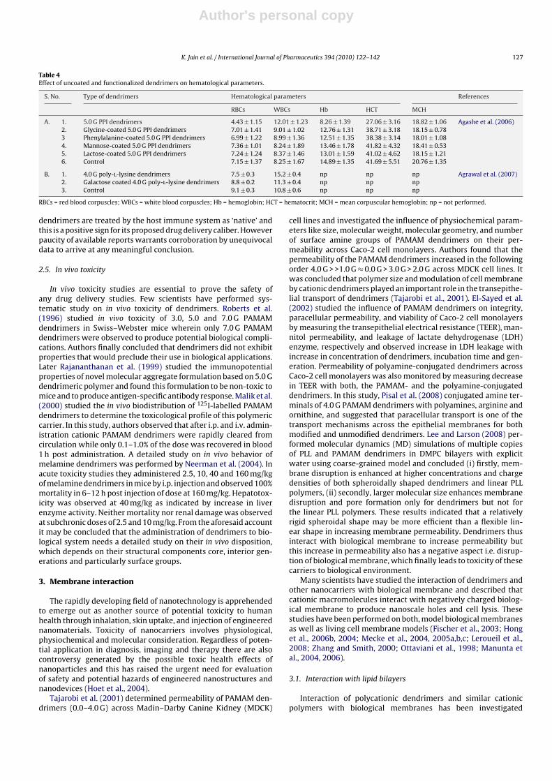
Currently there is a growing interest in the use of polymers fordelivery of therapeutic compounds. Polymer therapeutics consti-tutes an important class of drug delivery vehicles (Pignatello etal., 2009) such as nanoparticles, microspheres, polymeric micellesand dendrimers, which have proved their potential in drug andgene/DNA delivery. Dendrimers are emerging as promising drugdelivery modules because of their well-defined nanoscale poly-meric scaffold with low polydispersity index and controlled surfacefunctionalities. These carriers share the features of defined com-positions, high molecular mass and highly branched architecturewith various names such as ‘cascade molecules’ or ‘arborols’. Theseare the synthetic macromolecules with a tree-like structure firstintroduced by Tomalia et al. (1985) and Newkome et al. (1985).The term dendrimer arises from a Greek word ‘dendron’ mean-ing ‘tree/branch’, due to its resemblance with a tree and ‘meros’meaning part. Dendrimers are defined as globular, monodispersemacromolecules with novel three-dimensional polymeric architec-tures in which all bonds emerge radially from a central focal pointor core with repeat units that each contributes a branching pointin dendrimer (Dykes et al., 2001; Frechet, 2002).
Dendrimers posses three distinguishable architectural compo-nents i.e. an interior core, interior layer (generations) composed ofrepeating units radially attached to the interior core, and exterior(terminal functionality) attached to outermost interior generation(Fig. 1). The higher generation dendrimers, due to their globularstructure, occupy a smaller hydrodynamic volume compared to
Fig. 1. General structure of dendrimers.
the corresponding linear polymers. The dendritic structure is char-acterized by layers between each generation (Boas and Heegaard,2003; Jain and Gupta, 2008). The unique properties of dendrimersmake them attractive carriers for drug and gene/DNA delivery(Zinselmeyer et al., 2002). Dendrimers have made an importantcontribution in fields of biomedicine, chemistry and biological sci-ence, material science, engineering and electronics.
Dendrimers have proved their excellence as carrier for drugsand the bioactives because of their three-dimensional architecture,low polydispersity and high functionality. These enhance solubil-ity (Jain and Gupta, 2008; Yiyun et al., 2005) and bioavailabilityof drugs (Emanuele et al., 2004; Chauhan et al., 2003) as well asoffer the potential advantages of modifying the release of drug(Gajbhiye et al., 2009) including site-specific targeting (Bhadraet al., 2005). Dendrimers enhance solubility and bioavailability ofpoorly water-soluble drug by their container properties owing totheir hydrophobic core and hydrophilic periphery (Jain and Gupta,2008; Yiyun et al., 2005; Lim and Simanek, 2008). The possiblemechanisms for dendrimer mediated solubility enhancement arehydrophobic interaction between drug and hydrophobic interior ofdendrimer, hydrogen bonding and ionic interactions (Aulenta et al.,2003). Dendrimer mediated solubilization is affected by generationsize, pH, core, temperature, polymeric architecture and functionalgroups (Gupta et al., 2007). Dendrimers may prolong the residencetime of drug, increase the stability of bioactives, and protect it frombiological environment. Methotrexate–dendrimer complex pre-pared in our laboratory was found to achieve pH-dependent releaseand significant targeting in the brain and bone marrow. This com-plex was also stable at physiological pH (Khopade and Jain, 1997).In addition to this dendrimers has also been used in gene therapy tointroduce genetic material into target cells. Virus was the earliestgene delivery vector but its use is associated with high risk sincethey can induce strong immunogenic response in patient (Robbinsaand Ghivizzania, 1998). To circumvent this risk associated withvirus many non-viral vectors were developed by scientists (Luoand Saltzman, 2000). Many polycationic polymers have been usedin gene delivery including albumin, chitosan, poly-l-lysine (PLL),cationic liposomes, diethylaminoethyl dextran, polyethylenimineand dendrimers. Dendrimers such as polypropyleneimine (PPI) andpolyamido amine (PAMAM) (Figs. 2 and 3), with positive charge ontheir surface, have the ability for condensation of DNA followedby transfection (Dufes et al., 2005; Omidi et al., 2005). Dendrimersform a stable complex with DNA that can penetrate cell membranedue to its positive charge to release DNA inside cells. They can be

Author's personal copy
124 K. Jain et al. / International Journal of Pharmaceutics 394 (2010) 122–142
Fig. 2. PPI dendrimer.
surface modified to enhance transfection efficiency and specificityto target cells (Li et al., 2007). Dendrimers offer a better opportunityfor anticancer drug delivery. Various nanocarriers have been usedfor the delivery of anticancer agents (Amiji, 2006) but dendrimerhas proved itself a resourceful moiety in the therapy of malignantgrowths because of its vital applicability in the delivery of anti-neoplastic drug (Wu et al., 1994), contrast agents (Konda et al.,2001; Backer et al., 2005) and in gene therapy, including its pivotalrole in neutron capture therapy (Shukla et al., 2003; Nishiyama etal., 2009), photodynamic therapy (Hu et al., 2008), and photother-mal therapy (Agrawal et al., 2008). Apart from these, dendrimers arealso useful as catalyst (Nlate et al., 2006) and organic light-emittingdiodes (Brauge et al., 2006). Nlate et al. (2006) synthesized 9- and27-armed tetrakis (diperoxotungsto) phosphate-cored dendrimersto be used as a recoverable and reusable catalyst with hydrogen per-oxide in the oxidation of alkenes, sulfides and alcohols. Phosphorusdendrimers bearing fluorescent end groups such as naphthalene,anthracene and pyrene were also synthesized for the embellish-ment of organic light-emitting diodes (Brauge et al., 2006).
2. Dendrimers toxicity
Unarguably dendrimers have brought the tremendous advancesin the field of drug delivery. A great optimism is expected regardingtheir potential in biomedical field but because of nanometric sizei.e. 1–100 nm they may interact effectively and specifically withthe components of cell such as plasma membranes, cell organelles(endosomes, mitochondria, nucleus) and proteins such as enzymeetc., as all these cellular components are themselves nanometerin size range (Lee et al., 2008; Choi et al., 2006). Most types of
nanoparticles developed by scientists are non-selective. Nanoscalesize range of nanoparticles made them to interact with or perme-ate through plasma membrane of biological system. These aspectsof nanocarriers are of substantial interest in cell transfection strat-egy and are being developed for in vivo gene delivery (Lee et al.,2008; Choi et al., 2006; Wang et al., 2007, 2009). But there isanother aspect of non-selective uptake of these nanoparticles i.e.their potential to cause cytotoxicity. In the following section wewill focus on the cytotoxic behavior of dendrimers, the mechanismby which these cationic macromolecules induce cytotoxicity andpossible strategies to alleviate dendrimer toxicity.
Regardless of the extensive pharmaceutical and biomedicalapplications of dendrimers, toxicity associated due to terminal-NH2 groups and multiple cationic charge, limits their candidaturesfor clinical applications (Malik et al., 2000; Wilbur et al., 1998;Brazeau et al., 1998). Dendrimers like PPI, PAMAM and PLL exertsignificant in vitro cytotoxicity due to their surface cationic groups(Agashe et al., 2006; Kolhatkar et al., 2007). Evidence regard-ing dendrimer safety is conflicting (Roberts et al., 1996). Thereare reports of concentration and generation-dependent toxicity offree amine groups present at their periphery (Chen et al., 2004).Chen et al. (2004) reported the cytotoxicity of cationic melaminedendrimers having surface groups like amine, guanidine, car-boxylate, sulphonate or phosphonate and concluded that cationicdendrimers were much more cytotoxic than anionic or PEGylateddendrimers. Not only dendrimers but also cationic macromoleculesin general cause destabilization of the cell membrane and result incell lysis (Rittner et al., 2002; Hong et al., 2006a; Fischer et al., 2003).This is an important finding and a possible tip to scientists workingin the relevant areas of research.

Author's personal copy
K. Jain et al. / International Journal of Pharmaceutics 394 (2010) 122–142 125
Fig. 3. PAMAM dendrimer.
2.1. Cytotoxicity
Jevprasesphant et al. (2003) investigated cytotoxicity of PAMAMdendrimers in Caco-2 cell lines and observed a significant cyto-toxicity with these dendrimers. Malik et al. (2000) also reportedthe cytotoxicity of PPI and PAMAM dendrimers in three cancercell lines B16F10, CCRF and HepG2. The study was a breakthroughin this direction. Agashe et al. (2006) studied the cytotoxicity ofplain 5.0 G PPI, amino acid protected and carbohydrate-coatedPPI dendrimers in HepG2 and COS-7 cell lines and observed theeffect of terminal functional groups, concentration and incuba-tion time on cytotoxicity. For 5.0 G PPI cell viability was studiedusing different concentration of dendrimers over incubation timesof 24, 48 and 72 h (Table 1). Cell viability was found to decrease
with increase in concentration and incubation time. The cytotox-icity was found to be concentration- as well as time-dependentfor 5.0 G PPI and attributed to the presence of free primaryamine groups in 5.0 G PPI and the positive charge associatedwith them. COS-7 cell lines showed higher cell viability as com-pared with HepG2 at all concentration and incubation timepoints.
Stasko et al. (2007) evaluated cytotoxicity and membrane dis-ruption by PPI dendrimers, PEG conjugated PPI dendrimers and PPIdendrimers with peripheral neutral acetamide groups, on culturedhuman umbilical vein endothelial cells (HUVEC) and found thatthe plain PPI dendrimers demonstrated drastic time-dependentchanges in the plasma membrane permeability and prominentcytotoxicity (Table 2).
Table 1Cytotoxicity of 5.0 G PPI dendrimer against HepG2 and COS-7 cell lines (Agashe et al., 2006).
Conc. of 5.0 G PPI dendrimer (mg/ml) Cell lines Time of incubation (h) Cell viability (%)
0.001 HepG2 24 66.80.001 COS-7 24 71.50.001 HepG2 72 16.80.001 COS-7 72 23.11.0 HepG2 24 7.61.0 COS-7 24 11.71.0 HepG2 72 1.71.0 COS-7 72 2.3

Author's personal copy
126 K. Jain et al. / International Journal of Pharmaceutics 394 (2010) 122–142
Table 2Cytotoxicity studies on PPI and PAMAM dendrimers.
Type of dendrimers Cell lines Observations References
5.0 G PPI dendrimers a. Hep G2 cellsb. COS-7 cells
a. Cell viability 66.8% at 0.001 mg/ml and7.6% at 1 mg/ml conc. after 24 hb. Cell viability 71.5% at 0.001 mg/ml and11.7% at 1 mg/ml conc. after 24 h
Agashe et al. (2006)
Cultured human umbilical veinendothelial cells (HUVEC)
Drastic time-dependent changes in theplasma membrane permeability andprominent cytotoxicity
Stasko et al. (2007)
Cationic or whole generation PAMAMdendrimersa. 2.0 Gb. 3.0 G, 4.0 G
Caco-2 cells a. Cytotoxic at concentration above 700 �Mb. Cytotoxic at all concentrations examinedby investigator
Jevprasesphant et al. (2003)
2.2. Hemolytic toxicity
The free cationic terminal groups of dendrimers interact withRBCs and this polycationic nature of dendrimers leads to hemolysis(Table 3) (Bhadra et al., 2003; Asthana et al., 2005). The hemolytictoxicity of dendrimers was investigated by mixing dendrimers andRBC suspension followed by incubation for definite time intervalsat 37 ◦C, centrifugation at 3000 rpm for 15 min followed by anal-ysis of the supernatant at 540 nm spectrophotometrically againstblank (in normal saline), and percentage hemolysis was calculatedagainst absorbance factor of 100% hemolytic sample in distilledwater (Agashe et al., 2006; Agrawal et al., 2007).
Bhadra et al. (2003) developed 4.0 G PAMAM dendrimers fordelivery of an anticancer drug 5-FU and found its haemolytic tox-icity to be ∼15.3–17.3%. Later Asthana et al. (2005) also evaluatedthese dendrimers for hemolytic toxicity and observed up to 18%hemolytic toxicity, close to the value reported by Bhadra et al.(2003). Malik et al. (2000) reported 3.0 G PPI and PAMAM den-drimers to induce hemolysis above a concentration of 1 mg/ml.Bhadra et al. (2005) observed marked hemolysis with 4.0 and 5.0 GPPI dendrimers. The authors found 35.7% hemolysis with 4.0 GPPI dendrimers and 49.2% hemolysis with 5.0 G PPI dendrimers.Agashe et al. (2006) also investigated the hemolytic toxicity of5.0 G PPI dendrimers. They observed 34.2 ± 0.2%, 51.6 ± 0.3% and86.2 ± 0.6% hemolysis with 5.0 G PPI dendrimers at concentrationof 1 mg/ml after incubation for 1, 2 and 4 h, respectively. PLL den-drimers were also found to induce hemolysis in vitro (Agrawal etal., 2007). Agrawal et al. (2007) found 14.1 ± 1.02% hemolysis with4.0 G PLL dendrimers. Available reports suggest that higher gener-ation dendrimers may have greater hemolytic toxicity, which maybe ascribed to the greater overall cationic charge.
2.3. Haematological toxicity
Dendrimers induce hemolysis owing to interaction with RBCsand they also influence hematological parameters attributable topolycationic nature of the uncoated dendrimers. The effect of PPIdendrimers on different blood parameters including white bloodcorpuscles (WBCs), red blood corpuscles (RBCs), haemoglobin (Hb),
haematocrit (HCT) and mean corpuscular haemoglobin (MCH)was determined using Erma particle counter by Agashe et al.(2006). Authors observed a significant decrease in RBC count, asubstantial increase in WBC count, decrease in Hb content andMCH value, and a considerable difference in HCT value betweencontrol and PPI dendrimers. RBCs count, Hb, HCT and MCH val-ues were found to decrease drastically to 4.43 ± 1.15, 8.26 ± 1.39,27.06 ± 3.16 and 18.82 ± 1.06 from normal values of 7.15 ± 1.37,14.89 ± 1.35, 41.69 ± 5.51 and 20.76 ± 1.35, respectively with 5.0 Gplain PPI dendrimers (Table 4).
Bhadra et al. (2005) compared the effect of primaquine phos-phate loaded dendrimers on various haematological parameters inmale albino rats (Sprague–Dawley strain) and found RBC count todecrease below normal values by 1.1 ± 0.05 × 10⁶ RBCs/�l and WBCcount to increase by 2.7 ± 0.3 × 10³ �l−1 cells as compared to nor-mal values by uncoated PPI dendrimers. Later Agrawal et al. (2007)also observed a significant increase in WBCs count and decrease inRBCs count, with 4.0 G PLL dendrimers (Table 4). The RBCs countwas found to decrease up to 7.5 ± 0.3 × 10⁶ �l−1 and WBCs countwas found to increase up to 15.2 ± 0.4 × 10⁶ �l−1 with 4.0 G PLLdendrimers. From these studies we may conclude that the cationicdendrimers might exhibit significant impairment in hematologicalparameters i.e. a considerable increase in WBCs count, decrease inRBCs count, Hb, HCT and MCH values and this underlines the needfor a strategy to render them more biocompatible.
2.4. Immunogenicity
Some scientists investigated the immunogenicity of dendrimersand concluded that dendrimers shows no or only weak immuno-genicity (Roberts et al., 1996; Agashe et al., 2006; Rajananthananet al., 1999). Roberts et al. (1996) inspected the immunogenicityof PAMAM dendrimers by immunoprecipitation and Ouchterlonydouble-diffusion assay but observed no signs of immunogenicitywith the dose range of 0.1–0.0001 �M. Agashe et al. (2006) inves-tigated the immunogenicity of 5.0 G PPI dendrimers in Balb/C miceusing ELISA for monitoring antibody titre and reported that den-drimers was unable to provoke any detectable humoral immuneresponse under the experimental conditions. This indicates that
Table 3Hemolytic toxicity of some dendrimers.
Type of dendrimers Percent hemolysis References
5.0 G PPI dendrimers 86.2 ± 0.6% (at 1 mg/ml conc. after incubation of 4 h) Agashe et al. (2006)49.2% Bhadra et al. (2005)
4.0 G PAMAM dendrimer 18% Asthana et al. (2005)∼15.3–17.3% Bhadra et al. (2003)
3.0 G PAMAM dendrimers Hemolytic above a concentration of 1 mg/ml Malik et al. (2000)3.0 G PPI dendrimers Hemolytic above a concentration of 1 mg/ml Malik et al. (2000)4.0 G Poly-l-lysine Dendrimers 14.1 ± 1.02% Agrawal et al. (2007)4.0 G PPI dendrimers 35.7% Bhadra et al. (2005)
Author's personal copy
K. Jain et al. / International Journal of Pharmaceutics 394 (2010) 122–142 127
Table 4Effect of uncoated and functionalized dendrimers on hematological parameters.
S. No. Type of dendrimers Hematological parameters References
RBCs WBCs Hb HCT MCH
A. 1. 5.0 G PPI dendrimers 4.43 ± 1.15 12.01 ± 1.23 8.26 ± 1.39 27.06 ± 3.16 18.82 ± 1.06 Agashe et al. (2006)2. Glycine-coated 5.0 G PPI dendrimers 7.01 ± 1.41 9.01 ± 1.02 12.76 ± 1.31 38.71 ± 3.18 18.15 ± 0.783 Phenylalanine-coated 5.0 G PPI dendrimers 6.99 ± 1.22 8.99 ± 1.36 12.51 ± 1.35 38.38 ± 3.14 18.01 ± 1.084. Mannose-coated 5.0 G PPI dendrimers 7.36 ± 1.01 8.24 ± 1.89 13.46 ± 1.78 41.82 ± 4.32 18.41 ± 0.535. Lactose-coated 5.0 G PPI dendrimers 7.24 ± 1.24 8.37 ± 1.46 13.01 ± 1.59 41.02 ± 4.62 18.15 ± 1.216. Control 7.15 ± 1.37 8.25 ± 1.67 14.89 ± 1.35 41.69 ± 5.51 20.76 ± 1.35
B. 1. 4.0 G poly-l-lysine dendrimers 7.5 ± 0.3 15.2 ± 0.4 np np np Agrawal et al. (2007)2. Galactose coated 4.0 G poly-l-lysine dendrimers 8.8 ± 0.2 11.3 ± 0.4 np np np3. Control 9.1 ± 0.3 10.8 ± 0.6 np np np
RBCs = red blood corpuscles; WBCs = white blood corpuscles; Hb = hemoglobin; HCT = hematocrit; MCH = mean corpuscular hemoglobin; np = not performed.
dendrimers are treated by the host immune system as ‘native’ andthis is a positive sign for its proposed drug delivery caliber. Howeverpaucity of available reports warrants corroboration by unequivocaldata to arrive at any meaningful conclusion.
2.5. In vivo toxicity
In vivo toxicity studies are essential to prove the safety ofany drug delivery studies. Few scientists have performed sys-tematic study on in vivo toxicity of dendrimers. Roberts et al.(1996) studied in vivo toxicity of 3.0, 5.0 and 7.0 G PAMAMdendrimers in Swiss–Webster mice wherein only 7.0 G PAMAMdendrimers were observed to produce potential biological compli-cations. Authors finally concluded that dendrimers did not exhibitproperties that would preclude their use in biological applications.Later Rajananthanan et al. (1999) studied the immunopotentialproperties of novel molecular aggregate formulation based on 5.0 Gdendrimeric polymer and found this formulation to be non-toxic tomice and to produce antigen-specific antibody response. Malik et al.(2000) studied the in vivo biodistribution of 125I-labelled PAMAMdendrimers to determine the toxicological profile of this polymericcarrier. In this study, authors observed that after i.p. and i.v. admin-istration cationic PAMAM dendrimers were rapidly cleared fromcirculation while only 0.1–1.0% of the dose was recovered in blood1 h post administration. A detailed study on in vivo behavior ofmelamine dendrimers was performed by Neerman et al. (2004). Inacute toxicity studies they administered 2.5, 10, 40 and 160 mg/kgof melamine dendrimers in mice by i.p. injection and observed 100%mortality in 6–12 h post injection of dose at 160 mg/kg. Hepatotox-icity was observed at 40 mg/kg as indicated by increase in liverenzyme activity. Neither mortality nor renal damage was observedat subchronic doses of 2.5 and 10 mg/kg. From the aforesaid accountit may be concluded that the administration of dendrimers to bio-logical system needs a detailed study on their in vivo disposition,which depends on their structural components core, interior gen-erations and particularly surface groups.
3. Membrane interaction
The rapidly developing field of nanotechnology is apprehendedto emerge out as another source of potential toxicity to humanhealth through inhalation, skin uptake, and injection of engineerednanomaterials. Toxicity of nanocarriers involves physiological,physiochemical and molecular consideration. Regardless of poten-tial application in diagnosis, imaging and therapy there are alsocontroversy generated by the possible toxic health effects ofnanoparticles and this has raised the urgent need for evaluationof safety and potential hazards of engineered nanostructures andnanodevices (Hoet et al., 2004).
Tajarobi et al. (2001) determined permeability of PAMAM den-drimers (0.0–4.0 G) across Madin–Darby Canine Kidney (MDCK)
cell lines and investigated the influence of physiochemical param-eters like size, molecular weight, molecular geometry, and numberof surface amine groups of PAMAM dendrimers on their per-meability across Caco-2 cell monolayers. Authors found that thepermeability of the PAMAM dendrimers increased in the followingorder 4.0 G > >1.0 G ≈ 0.0 G > 3.0 G > 2.0 G across MDCK cell lines. Itwas concluded that polymer size and modulation of cell membraneby cationic dendrimers played an important role in the transepithe-lial transport of dendrimers (Tajarobi et al., 2001). El-Sayed et al.(2002) studied the influence of PAMAM dendrimers on integrity,paracellular permeability, and viability of Caco-2 cell monolayersby measuring the transepithelial electrical resistance (TEER), man-nitol permeability, and leakage of lactate dehydrogenase (LDH)enzyme, respectively and observed increase in LDH leakage withincrease in concentration of dendrimers, incubation time and gen-eration. Permeability of polyamine-conjugated dendrimers acrossCaco-2 cell monolayers was also monitored by measuring decreasein TEER with both, the PAMAM- and the polyamine-conjugateddendrimers. In this study, Pisal et al. (2008) conjugated amine ter-minals of 4.0 G PAMAM dendrimers with polyamines, arginine andornithine, and suggested that paracellular transport is one of thetransport mechanisms across the epithelial membranes for bothmodified and unmodified dendrimers. Lee and Larson (2008) per-formed molecular dynamics (MD) simulations of multiple copiesof PLL and PAMAM dendrimers in DMPC bilayers with explicitwater using coarse-grained model and concluded (i) firstly, mem-brane disruption is enhanced at higher concentrations and chargedensities of both spheroidally shaped dendrimers and linear PLLpolymers, (ii) secondly, larger molecular size enhances membranedisruption and pore formation only for dendrimers but not forthe linear PLL polymers. These results indicated that a relativelyrigid spheroidal shape may be more efficient than a flexible lin-ear shape in increasing membrane permeability. Dendrimers thusinteract with biological membrane to increase permeability butthis increase in permeability also has a negative aspect i.e. disrup-tion of biological membrane, which finally leads to toxicity of thesecarriers to biological environment.
Many scientists have studied the interaction of dendrimers andother nanocarriers with biological membrane and described thatcationic macromolecules interact with negatively charged biolog-ical membrane to produce nanoscale holes and cell lysis. Thesestudies have been performed on both, model biological membranesas well as living cell membrane models (Fischer et al., 2003; Honget al., 2006b, 2004; Mecke et al., 2004, 2005a,b,c; Leroueil et al.,2008; Zhang and Smith, 2000; Ottaviani et al., 1998; Manunta etal., 2004, 2006).
3.1. Interaction with lipid bilayers
Interaction of polycationic dendrimers and similar cationicpolymers with biological membranes has been investigated
Author's personal copy
128 K. Jain et al. / International Journal of Pharmaceutics 394 (2010) 122–142
employing an assortment of analytical techniques (Hong et al.,2006b, 2004; Mecke et al., 2004, 2005a; Leroueil et al., 2008; Zhangand Smith, 2000). Interaction with these drug carriers increases thepermeability and decreases the integrity of biological membrane.This leads to the leakage of cytosolic proteins such as lactate dehy-drogenase (LDH) and luciferase (Luc.) etc, and finally membranedisruption and cell lysis (Mecke et al., 2005b,c).
Ottaviani et al. (1998) investigated the interaction of PAMAMdendrimers with liposomes formed of 1,2-dimyristoyl-sn-glycero-3-phosphocholine (DMPC) and suggested that dendrimers couldbe exploited as vital carrier for gene/DNA and other biomolecules.Authors investigated the interaction between dendrimers and lipo-somes by negative staining transmission electron microscopy,dynamic light scattering (DLS), and electron paramagnetic reso-nance (EPR). In the analysis of EPR spectra authors found that attemperatures below the critical temperature transition of DMPCfrom liquid-crystal phase to the gel (or solid) phase, the vesi-cle structure changed and became more rigid and organizedwhereas it is generally assumed that the area of the phos-phatidylcholine head group remains almost unchanged upontransition. Based on this fact the authors assumed that predom-inantly dipolar and electrostatic interactions occurred betweendendrimer and liposome surface groups. From the changes inparameter obtained from electron spin-echo envelope modulation(ESEEM) and continuous wave-electron paramagnetic resonancethe authors concluded that dendrimer–vesicle interactions arestronger for higher generations than for lower generations. Finallythey suggested that the dendrimers interacted with the mem-brane surface, but did not permanently perturb the membraneproperties (enhanced biocompatibility); measurements at lowtemperatures indicated that interaction was more effective forprotonated dendrimers at larger sizes (higher generations), andtherefore they have more potential as carriers of biomolecules toapproach the cell membrane. In continuation Zhang and Smith(2000) evaluated disruption properties of 4.0–7.0 G PAMAM den-drimers and found that 6.0 and 7.0 G dendrimers were effectiveat inducing leaky fusion of anionic, large unilamellar vesicles.
Authors interpreted the enhanced membrane disruption in termsof a membrane bending model, which is based on the hypothe-sis that a rigid, polycationic dendrimer sphere uses electrostaticforces to bend a malleable, anionic membrane and inducesbilayer packing stresses. This bending model is biomimetic sincethe protein-induced membrane bending is currently thought tobe an important factor in the fusion mechanism of influenzavirus.
Later Hong et al. (2004) and Mecke et al. (2004) exploredthe mechanism by which cationic PAMAM dendrimer inducesmembrane damage. Authors used DMPC lipid bilayers for probingthe mechanism of cationic PAMAM dendrimer-induced mem-brane damage. Similar studies were also performed with somenanoparticles to investigate the interaction of other polycationicpolymers including poly-l-lysine (PLL), polyethyleneimine (PEI)and diethylaminoethyl-dextran (DEAE-DEX), which are establishedas imperative carrier for gene and drug delivery (Hong et al., 2006b).Authors observed that 7.0 G PAMAM dendrimers instigated thenanoscale holes of diameter between 15 and 40 nm in lipid bilayerswhereas 5.0 G PAMAM dendrimers did not induce nanoscale holeformation but expanded the size of pre-existing defects in the bilay-ers. It was hypothesized that these holes are upshots of electrostaticinteractions between dendrimers and supported lipid bilayers andthe formation of dendrimer-nucleated lipid vesicles (Mecke et al.,2004).
Leroueil et al. (2008) employed atomic force microscopy todetermine the common mechanism of interaction of nanoparticleswith lipids and physical disruption of lipid membranes. Authorsperformed the experiments with variety of nanoparticles includ-ing cell penetrating peptide (MSI-78), protein (TAT), dendrimers(PAMAM and pentanol-core PAMAM dendrons), polycationic poly-mers (polyethyleneimine and diethylaminoethyl-dextran), andwith inorganic particles (Au–NH2, SiO2–NH2) on supported lipidbilayers. They found that these polycationic nanoparticles inter-acted with biological membranes and induce cell disruption byformation of nanoholes, membrane thinning, and/or membraneerosion mainly.
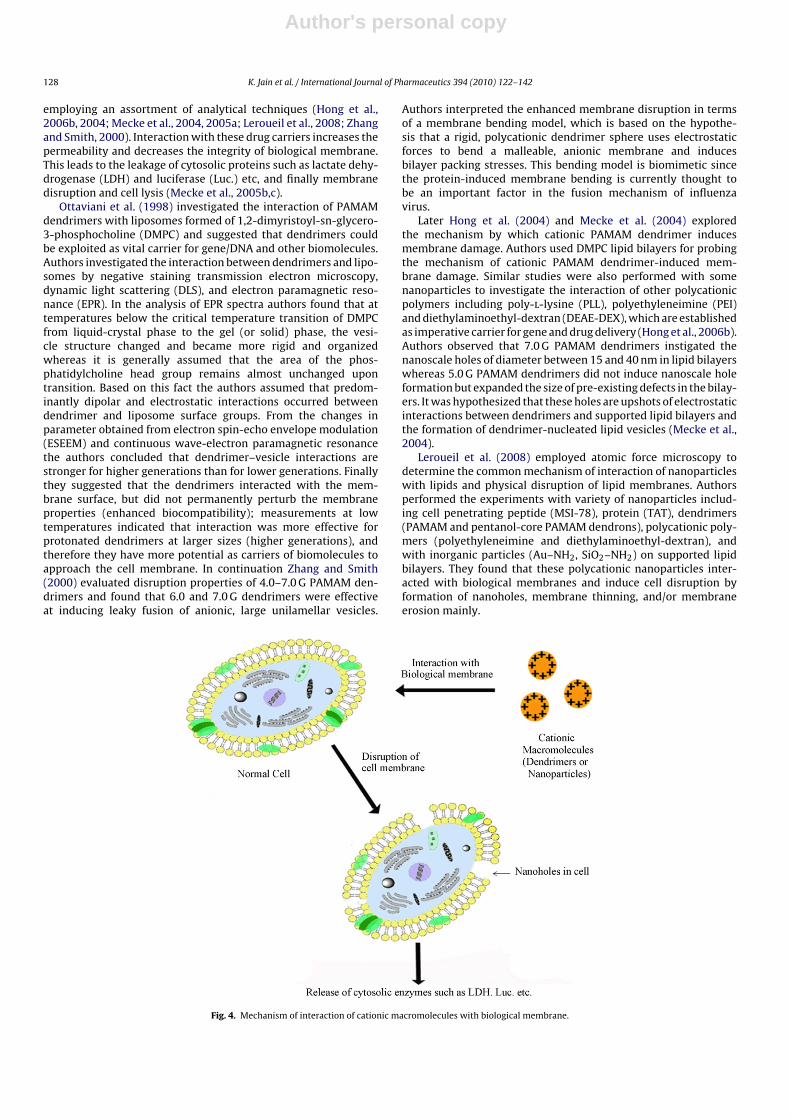
Fig. 4. Mechanism of interaction of cationic macromolecules with biological membrane.
Author's personal copy
K. Jain et al. / International Journal of Pharmaceutics 394 (2010) 122–142 129
Conclusively, dendrimers with surface cationic charges inter-act with negatively charged biological membrane because of theirpositive charge. This interaction leads to membrane disruption byinducing formation of nanoholes or causing membrane thinning orerosion that is responsible for the toxicity of dendrimers. Ultimatelythis leads to leakage of cytosolic enzymes, and cell lysis (Fig. 4).
3.2. Interaction with cell membrane
Available literatures widely suggest a concluding trend thatcationic macromolecules show a considerable cytotoxicity. Fischeret al. (2003) performed a comparative in vitro cytotoxicity studyof different cationic gene delivery systems on L929 mouse fibrob-lasts cells using MTT assay and measuring the release of thecytosolic enzyme lactate dehydrogenase (LDH). The study was per-formed on following cationic macromolecules: poly(ethylenimine)(PEI), poly(l-lysine), poly(diallyl-dimethyl-ammonium chloride),diethylaminoethyl-dextran, poly(vinyl pyridinium bromide), star-burst dendrimer, cationized albumin and native albumin. Authorsfound the following ranking of polymers with regard tocytotoxicity with native albumin being the least toxic fol-lowed by cationized albumin: starburst dendrimer, poly(vinylpyridinium bromide), diethylaminoethyl-dextran, poly(diallyl-dimethyl-ammonium chloride); PEI and PLL being the mostcytotoxic. They also studied the nature of cell death induced bythese carriers and assumed that cell lysis was not caused by apop-tosis (necrotic cell reaction) since it was found that cell nucleiretained its size and chromatin was homogeneously distributedbut cell membranes lost their integrity very rapidly at an earlystage. Manunta et al. (2004) investigated the uptake procedure ofdendrimer–DNA complex (dendriplexes) in endothelial cell line.Authors also assessed whether the internalization of dendriplexestook place randomly on the cell surface or at preferential sitessuch as membrane rafts. They observed that membrane rafts wereimportant for the internalization of non-viral vectors in genetherapy; dendrimer conducts gene delivery via cholesterol depen-dent pathway (Manunta et al., 2004). Later Manunta et al. (2006)investigated the internalization and transfection properties of den-driplexes using HepG2 and HeLa cell lines that express few caveolaeand observed that dendrimer–DNA complex may use differentinternalization pathways in different cells but enhanced in the pres-ence of caveolin into HeLa, HepG2 and endothelial cells.
Hong et al. (2006b, 2004) also examined the disruption eventson living cells in vitro using KB and Rat2 cell membranes throughatomic force microscopy (AFM), enzyme assays, flow cell cytom-etry and fluorescence microscopy. They found a dose-dependentrelease of the cytoplasmic proteins lactate dehydrogenase (LDH)and luciferase (Luc), which indicated that interaction of dendrimerswith membrane resulted in decreased integrity of cell membraneand lead to induction of permeability with subsequent internal-ization of dendrimers. Up to a concentration of 500 nm neither5.0 G amine- nor acetamide-terminated PAMAM dendrimers werecytotoxic. These researchers also realized that the induction ofpermeability by dendrimers was not permanent and leaking ofcytoplasmic proteins returned to normal levels upon removal ofdendrimers. 5.0 G dendrimers modified with acetamide do notinternalize to the cells and hence internalization is attributable tointeraction between cationic macromolecules and cell membranefollowed by increase in permeability, which is pivotal for the deliv-ery of biomolecules inside the cell.
4. Solutions for toxicity issues
There is a great need to devise methods to circumvent thecytotoxicity and hemolytic toxicity associated with these promis-
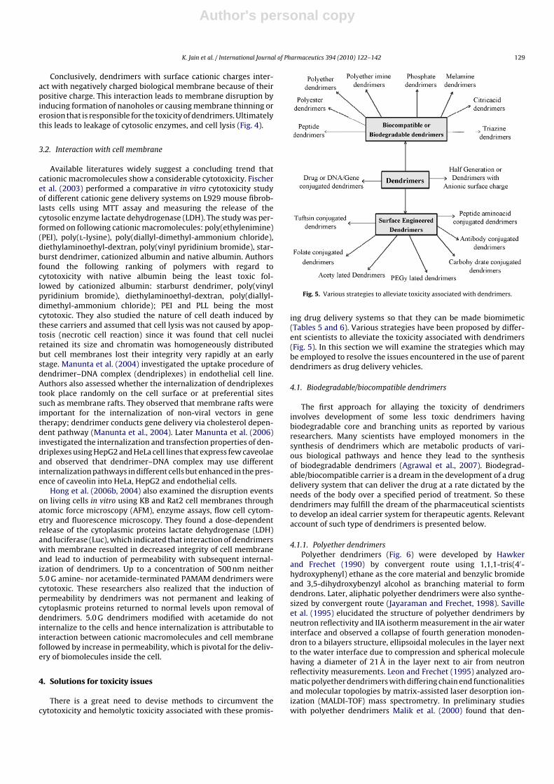
Fig. 5. Various strategies to alleviate toxicity associated with dendrimers.
ing drug delivery systems so that they can be made biomimetic(Tables 5 and 6). Various strategies have been proposed by differ-ent scientists to alleviate the toxicity associated with dendrimers(Fig. 5). In this section we will examine the strategies which maybe employed to resolve the issues encountered in the use of parentdendrimers as drug delivery vehicles.
4.1. Biodegradable/biocompatible dendrimers
The first approach for allaying the toxicity of dendrimersinvolves development of some less toxic dendrimers havingbiodegradable core and branching units as reported by variousresearchers. Many scientists have employed monomers in thesynthesis of dendrimers which are metabolic products of vari-ous biological pathways and hence they lead to the synthesisof biodegradable dendrimers (Agrawal et al., 2007). Biodegrad-able/biocompatible carrier is a dream in the development of a drugdelivery system that can deliver the drug at a rate dictated by theneeds of the body over a specified period of treatment. So thesedendrimers may fulfill the dream of the pharmaceutical scientiststo develop an ideal carrier system for therapeutic agents. Relevantaccount of such type of dendrimers is presented below.
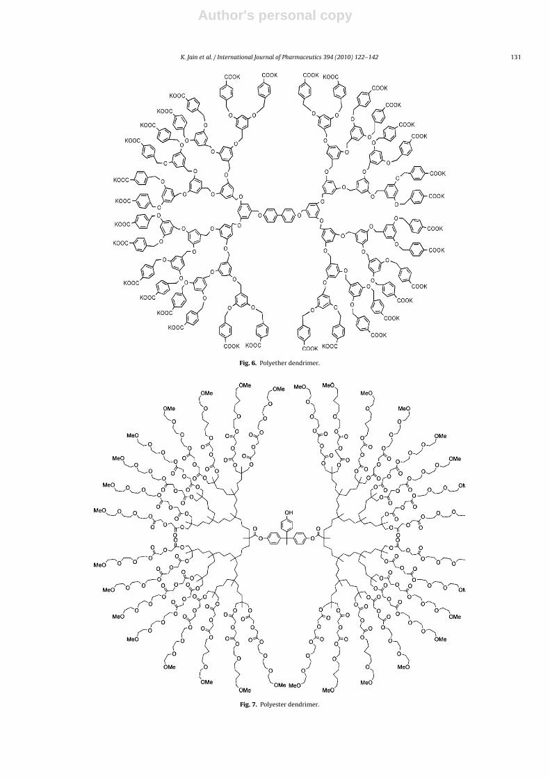
4.1.1. Polyether dendrimersPolyether dendrimers (Fig. 6) were developed by Hawker
and Frechet (1990) by convergent route using 1,1,1-tris(4′-hydroxyphenyl) ethane as the core material and benzylic bromideand 3,5-dihydroxybenzyl alcohol as branching material to formdendrons. Later, aliphatic polyether dendrimers were also synthe-sized by convergent route (Jayaraman and Frechet, 1998). Savilleet al. (1995) elucidated the structure of polyether dendrimers byneutron reflectivity and IIA isotherm measurement in the air waterinterface and observed a collapse of fourth generation monoden-dron to a bilayers structure, ellipsoidal molecules in the layer nextto the water interface due to compression and spherical moleculehaving a diameter of 21 Å in the layer next to air from neutronreflectivity measurements. Leon and Frechet (1995) analyzed aro-matic polyether dendrimers with differing chain end functionalitiesand molecular topologies by matrix-assisted laser desorption ion-ization (MALDI-TOF) mass spectrometry. In preliminary studieswith polyether dendrimers Malik et al. (2000) found that den-
Author's personal copy
130 K. Jain et al. / International Journal of Pharmaceutics 394 (2010) 122–142
Table 5Cytotoxicity studies on biocompatible and surface engineered dendrimers.
Type of dendrimers Cell lines Results References
Polyether imine dendrimers a. Human breast cancer T47D Cell survival >98% up to 100 mg/ml ofdendrimer solution
Krishna et al. (2005)
b. African green monkey CV-1
SN-38 complexed G4- PAMAM dendrimers Caco-2 cells No appreciable toxicity (cell viability>90% after incubation for 2 h at 0.1 �Mconc.)
Kolhatkar et al. (2008)
Glycine-coated 5.0 G PPI dendrimers a. Hep G2 cells a. Cell viability 95.37% at 1 mg/ml conc.after 24 h
Agashe et al. (2006)
b. COS-7 cells b. Cell viability 96.1% at 1 mg/ml conc.after 24 h
Phenylalanine-coated 5.0 G PPI dendrimers a. Hep G2 cells a. Cell viability 96.2% at 1 mg/ml conc.after 24 h
Agashe et al. (2006)
b. COS-7 cells b. Cell viability 93.0% at 1 mg/ml conc.after 24 h
Mannose-coated 5.0 G PPI dendrimers a. Hep G2 cells a. Cell viability 95.2% at 1 mg/ml conc.after 24 h
Agashe et al. (2006)
b. COS-7 cells b. Cell viability 98.1% at 1 mg/ml conc.after 24 h
Lactose-coated 5.0 G PPI dendrimers a. Hep G2 cells a. Cell viability 94.7% at 1 mg/ml conc.after 24 h
Agashe et al. (2006)
b. COS-7 cells b. Cell viability 97.71% at 1 mg/ml conc.after 24 h
5.0 G PPI dendrimers with peripheral neutralacetamide groups
Cultured human umbilical veinendothelial cells (HUVEC)
Decrease in cytotoxicity was observed Stasko et al. (2007)
Carboxylic acid-terminated PPI dendrimers B16F10 cells Not cytotoxic up to concentration of1 mg/ml after 24 h
Malik et al. (2000)
PEGylated PPI dendrimers Cultured human umbilical veinendothelial cells (HUVEC)
Elimination or reduction of acutecytotoxicity of plain PPI dendrimers
Stasko et al. (2007)
Carboxylic acid-terminated PAMAMdendrimers (Half generation)
B16F10 cells Not cytotoxic up to concentration of1 mg/ml after 24 h
Malik et al. (2000)
Anionic or half generation PAMAMdendrimers (2.5 G, 3.5 G)
Caco-2 cells No cytotoxicity up to 1 Mmconcentration.
Jevprasesphant et al. (2003)
Lauroyl- and PEGylated PAMAM dendrimers Caco-2 cells Seven fold reduction in cytotoxicitycompared to unmodified dendrimers
Jevprasesphant et al. (2003)
drimers with carboxylate and malonate surface groups were notlytic at 1 h but after incubation for 24 h they were lytic (Malik et al.,2000).
4.1.2. Polyester dendritic systemBo et al. (1997) furnished a method for the rapid synthe-
sis of polyester dendrimers using methyl 3,5-dihydroxybenzoateand 3,5-dibenzyloxybenzoic acid as monomers (Fig. 7). Ihre et al.(2001, 2002) designed and synthesized dendritic polyester sys-tem to improve tumor targeting and therapeutic efficacy due tothe EPR effect observed in tumor tissue. These systems werebased on the monomer unit 2,2-bis(hydroxymethyl)propanoicacid (Ihre et al., 2001, 2002). Galie et al. (2006) employed thesedendrimers for boron neutron capture therapy containing carbo-rane. Hirayama et al. (2005) synthesized new aliphatic polyesterdendrimers from benzyl acetoacetate and t-butyl acrylate by ace-toacetic acid ester synthesis using divergent growth method. Goh
et al. (2002) proposed a new approach for the controlled synthe-sis of multicomponent dendrimers wherein they synthesized threeoligonucleotide–dendron conjugates using solid phase techniquesand hybridized to create a second generation polyester dendrimerwith DNA as a core and bearing two types of peripheral functionalgroups. Another example of polyester dendrimers is polyglycerolsuccinic acid dendrimers (Luman et al., 2003), synthesized byconvergent synthesis from biocompatible or biodegradable build-ing blocks including succinic acid, glycerol and PEG. Hirayamaet al. (2005) synthesized new aliphatic polyester dendrimersfrom benzyl acetoacetate and t-butyl acrylate by acetoaceticacid ester synthesis using divergent growth method. Gillies etal. (2005) investigated the biological performance of these den-drimers and found that the polyester dendrimer–poly(ethyleneoxide) hybrids were non-toxic to cells and high levels of tumoraccumulation were found in mice bearing subcutaneous B16F10tumor.
Table 6Hemolytic toxicity profile of surface modified dendrimers.
Type of dendrimers % Hemolysis References
Glycine-coated 5.0 G PPI dendrimers 4.9 ± 0.2% (at 1 mg/ml conc. after incubation of 4 h) Agashe et al. (2006)Phenylalanine-coated 5.0 G PPI dendrimers 3.3 ± 0.4% (at 1 mg/ml conc. after incubation of 4 h) Agashe et al. (2006)Mannose-coated 5.0 G PPI dendrimers 2.9 ± 0.6% (at 1 mg/ml conc. after incubation of 4 h) Agashe et al. (2006)Lactose-coated 5.0 G PPI dendrimers 2.2 ± 0.3% (at 1 mg/ml conc. after incubation of 4 h) Agashe et al. (2006)Half generations of carboxylic acid-terminated PAMAM dendrimers Negligible hemolytic toxicity Bhadra et al. (2003)PEGylated PAMAM dendrimers <5% Bhadra et al. (2003)Carboxylic acid-terminated PAMAM dendrimers (Half generation) Not hemolytic up to concentration of 2 mg/ml Malik et al. (2000)Carboxylic acid-terminated PPI dendrimers Not hemolytic up to concentration of 2 mg/ml Malik et al. (2000)Oligoethyleneoxide-terminated carbosilane dendrimers Not hemolytic up to concentration of 2 mg/ml Malik et al. (2000)Galactose coated 4.0 G poly-l-lysine dendrimers 7.3 ± 2.8% Agrawal et al. (2007)Galactose coated 4.0 G PPI dendrimers 7.1% Bhadra et al. (2005)Galactose coated 5.0 G PPI dendrimers 10% Bhadra et al. (2005)
Author's personal copy
K. Jain et al. / International Journal of Pharmaceutics 394 (2010) 122–142 131
Fig. 6. Polyether dendrimer.
Fig. 7. Polyester dendrimer.

Author's personal copy
132 K. Jain et al. / International Journal of Pharmaceutics 394 (2010) 122–142
4.1.3. Polyether imine dendrimersKrishna et al. (2005) performed the synthesis and biological
evaluation of 3-amino-propan-1-ol based polyether imine den-drimers. They synthesized this dendrimer with nitrogen core usinga new monomer 3-(bis-(3-hydroxypropyl)amino)propan-1-ol. Theauthors studied the in vitro cytotoxicity of these dendrimers withpropyl ether-imine and carboxylic acid groups at their peripheriesand carried out these studies by performing MTT assays on twodifferent cell lines, human breast cancer T47D and African greenmonkey kidney CV-1. The cell survival rate was more than 98% upto 100 mg/ml of the dendrimer solutions in both the cell lines. TheMTT assay of polyether imine dendrimers revealed no measurablecytotoxicity on both the cell lines.
4.1.4. Polyether–copolyester (PEPE) dendrimersCarnahan and Grinstaff (2001) synthesized and character-
ized polyether–ester dendrimers from glycerol and lactic acid.Dhanikula and Hildgen (2007a) synthesized polyether–copolyesterdendrimers from PEG and characterized their surface by AFMand X-ray photoelectron spectroscopy (XPS). They utilized thesedendrimers for encapsulation and release of methotrexate andexploited methotrexate loaded dendrimers for the treatmentof gliomas. Authors found enhanced efficacy and intratumoral
transport capability with fine capacity to encapsulate methotrex-ate in dendrimeric architecture (Dhanikula and Hildgen, 2007b;Dhanikula et al., 2008). They evaluated the cytotoxicity of thesePEPE dendrimers on RAW 264.7 cell lines using LDH leakage. Thesedendrimers were considered to be biocompatible and no death wasreported up to concentration of 250 �g/ml (Dhanikula and Hildgen,2007b). In addition to this, IC50 of methotrexate was reduced afterloading into PEPE dendrimers. These dendrimers were also foundto show enhanced permeability across blood brain barrier (BBB).Methotrexate loaded dendrimers were found to be effective againstU87MG and U 343 MGa cells and methotrexate resistance cells.Dhanikula et al. (2008) concluded that these dendrimers may actas potential delivery systems for the treatment of gliomas.
4.1.5. Phosphate dendrimersDomanski et al. (2004) investigated haemotoxicity and cytotox-
icity of water-insoluble, 5.0 G thiophosphate (Fig. 8) dendrimersincluding their influence on human red blood cells structure, mem-brane integrity and on the growth of nucleated cells represented byChinese hamster ovary (CHO-K1) cell line. Authors found increasein erythrocyte membrane stability and thermal durability buttrigger in echinocytosis upon interaction of dendrimer with ery-throcyte membrane proteins.
Fig. 8. Thiophosphate dendrimer.

Author's personal copy
K. Jain et al. / International Journal of Pharmaceutics 394 (2010) 122–142 133
4.1.6. Citric acid dendrimersNamaji and Adeli (2005) synthesized triblock dendrimers of
citric acid–polyethylene glycol–citric acid for drug delivery. Theframework of dendrimers comprised of biocompatible materi-als. Authors solubilized some hydrophobic drug molecules like5-amino salicylic acid (5-ASA), pyridine, mephenamic acid anddiclofenac in aqueous solution. These drug–dendrimers complexeswere found to be stable at room temperature for more than 10months.
4.1.7. Melamine dendrimersMelamine dendrimers were synthesized by both divergent and
convergent growth method (Zhang and Simanek, 2000; Zhang etal., 2001). Multivalent dendrimers of melamine may be obtainedby thiol-disulfide exchange (Umali and Simanek, 2003; Zhang etal., 2003a). These dendrimers were reported to reduce hepatotoxi-city of anticancer drugs, methotrexate and 6-mercaptopurine withincreased solubility by dendrimers (Neerman et al., 2008). Thesewere investigated as drug delivery candidates and for in vitro andin vivo toxicity (Lim and Simanek, 2008; Chen et al., 2004; Neermanet al., 2004). Neerman et al. (2004) found that melamine dendrimerin doses up to 10 mg/kg showed no hepatic toxicity at subchronicdoses although doses up to 40 mg/kg showed liver necrosis in mice.
4.1.8. Peptide dendrimersThis class of dendrimers comprises of different types of amino
acids such as lysine, arginine etc and these dendrimers were foundto exhibit promising vaccine and drug carrier properties (Agrawal
et al., 2007; Wegmann et al., 2008; Cubillos et al., 2008; Tamand Spetzler, 2001). Shao and Tam (1995) developed peptide den-drimers using a new tactic with oxime, hydrazone, thiazolidinelinkages using uprotected peptides as building blocks and selectiveligation between an aldehyde and a weak base.
Agrawal et al. (2007) synthesized 4.0 G poly-l-lysine dendrimersusing PEG 1000 as core and l-lysine amino acid as branching unitfollowing repetitive protection with di-tertiary butyl pyrocarbon-ate (di-BOC) and deprotection step. They found a considerablereduction in hemolytic toxicity in comparison with PPI dendrimersof similar generation.
4.1.9. Triazine dendrimersSimanek and coworkers made the major contribution in this
regard. They assessed the propensity of triazine dendrimers as adrug delivery vehicle (Zhang et al., 2003b) and found these den-drimers a suitable solubilizing agent for hydrophobic molecules;carrier for delivery of drug molecule and apparently non-toxic inpreliminary animal models. Triazine dendrimers was found to pro-duce no toxic effect on kidneys and liver in single doses deliveredintraperitoneally up to 10 mg/kg in preliminary toxicological stud-ies in mice. Later, Chouai and Simanek (2008) reported a divergentand iterative synthesis method for 2.0 G triazine dendrimers (Fig. 9)for a kilogram scale using common laboratory equipment fromtrichlorotriazine. Lai et al. (2008) prepared star-shaped mesogensbased on triazine and piperazine units. The biodistribution of tri-azine dendrimers is affected by number of PEG chains attachedto the dendrimers while increase in molecular mass results in
Fig. 9. Triazine dendrimer.
Author's personal copy
134 K. Jain et al. / International Journal of Pharmaceutics 394 (2010) 122–142
Fig. 10. Drug conjugated PEGylated dendrimer.
enhanced elimination half lives. Tumor uptake took place for alltype of PEGylated dendrimers with multiple PEG groups of 0.6, 2,and 5 kDa (Lim et al., 2008). Recently, our group has developedtriazine dendrimers using triazine trichloride as core. The den-drimers were developed using utilizing differential reactivity ofthe cyanuric chloride (triazine trichloride) upto third generationby divergent method using diethanolamine as branching unit. Thestudies related to drug delivery aspect of theswe dendrimers are inprogress.
4.2. Surface engineered dendrimers
Surface engineering appears to be one of the best strategiesfor abatement of dendrimer toxicity (Tables 4–6). The presence ofmultiple surface sites also makes possible the attachment of moi-eties of various functionalities to the surface through covalent ornon-covalent bonding. Surface modification of dendrimers leads toprotection of surface amine groups and reduces the inherent cyto-toxicity of dendrimers (Roberts et al., 1996; Jevprasesphant et al.,2003; Luo et al., 2002). Apart from reduction in inherent toxicity ofdendrimers, functionalization also imparts some other propertiesbeneficial for their use as drug delivery system including improve-ment in drug encapsulation efficiency, improved biodistributionand pharmacokinetic properties, increase in solubility, targeting tospecific site, better transfection efficiency, sustained and controlleddrug release, improvement in stability profile, and improved ther-apeutic potential as anti-viral, anti-bacterial activity (Gajbhiye etal., 2009; Bhadra et al., 2005, 2003; Konda et al., 2001; Agrawal etal., 2009).
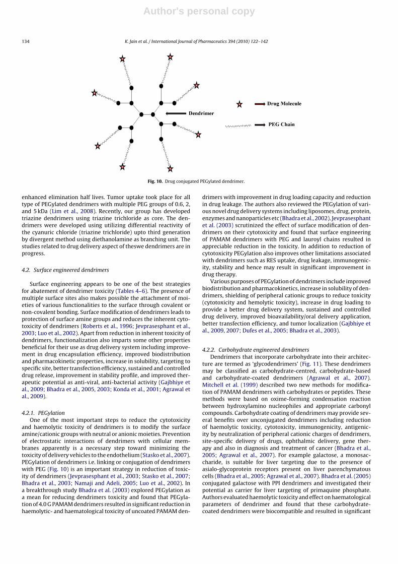
4.2.1. PEGylationOne of the most important steps to reduce the cytotoxicity
and haemolytic toxicity of dendrimers is to modify the surfaceamine/cationic groups with neutral or anionic moieties. Preventionof electrostatic interactions of dendrimers with cellular mem-branes apparently is a necessary step toward minimizing thetoxicity of delivery vehicles to the endothelium (Stasko et al., 2007).PEGylation of dendrimers i.e. linking or conjugation of dendrimerswith PEG (Fig. 10) is an important strategy in reduction of toxic-ity of dendrimers (Jevprasesphant et al., 2003; Stasko et al., 2007;Bhadra et al., 2003; Namaji and Adeli, 2005; Luo et al., 2002). Ina breakthrough study Bhadra et al. (2003) explored PEGylation asa mean for reducing dendrimers toxicity and found that PEGyla-tion of 4.0 G PAMAM dendrimers resulted in significant reduction inhaemolytic- and haematological toxicity of uncoated PAMAM den-
drimers with improvement in drug loading capacity and reductionin drug leakage. The authors also reviewed the PEGylation of vari-ous novel drug delivery systems including liposomes, drug, protein,enzymes and nanoparticles etc (Bhadra et al., 2002). Jevprasesphantet al. (2003) scrutinized the effect of surface modification of den-drimers on their cytotoxicity and found that surface engineeringof PAMAM dendrimers with PEG and lauroyl chains resulted inappreciable reduction in the toxicity. In addition to reduction ofcytotoxicity PEGylation also improves other limitations associatedwith dendrimers such as RES uptake, drug leakage, immunogenic-ity, stability and hence may result in significant improvement indrug therapy.
Various purposes of PEGylation of dendrimers include improvedbiodistribution and pharmacokinetics, increase in solubility of den-drimers, shielding of peripheral cationic groups to reduce toxicity(cytotoxicity and hemolytic toxicity), increase in drug loading toprovide a better drug delivery system, sustained and controlleddrug delivery, improved bioavailability/oral delivery application,better transfection efficiency, and tumor localization (Gajbhiye etal., 2009, 2007; Dufes et al., 2005; Bhadra et al., 2003).
4.2.2. Carbohydrate engineered dendrimersDendrimers that incorporate carbohydrate into their architec-
ture are termed as ‘glycodendrimers’ (Fig. 11). These dendrimersmay be classified as carbohydrate-centred, carbohydrate-basedand carbohydrate-coated dendrimers (Agrawal et al., 2007).Mitchell et al. (1999) described two new methods for modifica-tion of PAMAM dendrimers with carbohydrates or peptides. Thesemethods were based on oxime-forming condensation reactionbetween hydroxylamino nucleophiles and appropriate carbonylcompounds. Carbohydrate coating of dendrimers may provide sev-eral benefits over unconjugated dendrimers including reductionof haemolytic toxicity, cytotoxicity, immunogenicity, antigenic-ity by neutralization of peripheral cationic charges of dendrimers,site-specific delivery of drugs, ophthalmic delivery, gene ther-apy and also in diagnosis and treatment of cancer (Bhadra et al.,2005; Agrawal et al., 2007). For example galactose, a monosac-charide, is suitable for liver targeting due to the presence ofasialo-glycoprotein receptors present on liver parenchymatouscells (Bhadra et al., 2005; Agrawal et al., 2007). Bhadra et al. (2005)conjugated galactose with PPI dendrimers and investigated theirpotential as carrier for liver targeting of primaquine phosphate.Authors evaluated haemolytic toxicity and effect on haematologicalparameters of dendrimer and found that these carbohydrate-coated dendrimers were biocompatible and resulted in significant

Author's personal copy
K. Jain et al. / International Journal of Pharmaceutics 394 (2010) 122–142 135
Fig. 11. Carbohydrate conjugated dendrimer.
reduction of haemolytic toxicity compared to uncoated den-drimers. Authors detected 35.7%, 49.2%. 10% and 7.1% hemolysiswith 4.0 G, 5.0 G uncoated and 4.0 G, 5.0 G galactose coated PPIdendrimers, respectively, at concentration of 5 mg/ml after incu-bation for 1 h with RBC suspension at 37 ◦C. Similar results wereobtained for the effect of plain and protected dendrimers on haema-
tological parameters. A relatively higher stimulation of WBCs countand reduction in RBCs count by uncoated dendrimer comparedwith coated PPI dendrimers was obtained. Agashe et al. (2006)conjugated lactose and mannose to the 5.0 G PPI dendrimers andcompared the toxicity profile of these dendrimers with that ofthe parent dendrimer. A substantial reduction in cytotoxicity and

Author's personal copy
136 K. Jain et al. / International Journal of Pharmaceutics 394 (2010) 122–142
haemolytic toxicity was observed with sugar conjugated (lactoseand mannose) dendrimers and statistically insignificant differencein haematological parameters was witnessed compared to the con-trol. Thus, these carbohydrate-coated dendrimers could emerge asnewer biocompatible carriers for drug delivery.
Polyether–copolyester dendrimers were conjugated with d-glucosamine as carrier for methotrexate in treatment of gliomas.They were evaluated for their efficacy against U87 MG and U 343MGa cells, permeability across in vitro BBB model and distributioninto avascular human glioma tumor spheroids. These glucosylateddendrimers were found effective against gliomas, more perme-able across BBB and able to kill even methotrexate resistant cells,including reduction of IC50 of methotrexate. In distribution stud-ies with glioma tumor glucosylated dendrimers were found to bedistributed throughout the avascular tumor spheroids within 6 h,while nonglucosylated dendrimers could do so in 12 h (Dhanikula etal., 2008). Agrawal et al. (2007) synthesized galactose coated 4.0 Gpoly-l-lysine dendrimers and reported 5.2 times reduction in thehemolytic toxicity compared to 4.0 G PPI dendrimers coupled witha negligible effect on hematological parameters.
4.2.3. AcetylationAs discussed above, neutralization of the amine surface charge of
dendrimer is essential to preclude toxicity and nonspecific uptakeof the drug conjugates and the modification of surface amino groupsof dendrimers with acetyl groups has resulted in the reduction oftoxicity of dendrimers (Stasko et al., 2007). Plain PPI dendrimersdemonstrated prominent cytotoxicity in HUVEC, but when Staskoet al. (2007) prepared fluorescently labeled derivatives of 5.0 GPPI dendrimers with acetylation and evaluated their cytotoxicityand membrane disruption in HUVEC cells, they found a signifi-cant reduction in the acute cytotoxicity of cationic primary aminecontaining dendrimers.
Acetylation of dendrimers is also a route to specifically func-tionalize the primary surface amines (Zhuo et al., 1999). Zhuo etal. synthesized a series of dendritic polymers with a core of 1, 4, 7,10-tetraazacyclododecane and Michael addition of methyl acrylatefollowed by amidation with ethylenediamine. They then acylatedthe peripheral amine groups by acetic anhydride, which leads toincreased solubility of dendrimers in water. The acetylated den-drimers were then reacted with 1-bromoacetyl-5-fluorouracil toform dendrimer-5-FU conjugates to form a carrier for the controlledrelease of antitumor drugs (Zhuo et al., 1999). Recently Waite et al.(2009) studied the acetylated 5.0 G PAMAM dendrimers for cellulardelivery of siRNA. PAMAM dendrimers were acetylated with aceticanhydride and complexed with siRNA, the cytotoxicity of complexwas evaluated using U87 malignant glioma cells and increase inacetylation resulted in reduced polymer cytotoxicity to U87 cellswith enhanced dissociation of dendrimer/siRNA complexes andthus promoted the release of siRNA from complexes.
Majoros et al. (2002) have defined the exact percentage ofacetylated end groups of the dendrimer with several techniquesincluding potentiometric titration, GPC, 1H NMR, and 13C NMRand investigated the precise stoichiometry required for the acety-lation of surface amines of 5.0 G PAMAM dendrimer, the natureof the acetylation reaction used and an analysis of well-defineddendrimer conjugates for use in the biomedical field.
Kolhatkar et al. (2007) acetylated the amine groups of PAMAMdendrimers and investigated the effect of this modification oncytotoxicity, permeability and cellular uptake in Caco-2 cell mono-layers. Authors found a 10-fold decrease in cytotoxicity withincrease in number of surface acetyl groups while maintaining per-meability across plasma membrane.
Kukowska-Latallo et al. (2005) conjugated the acetylatedPAMAM dendrimers with folic acid as a targeting agent and thencoupled these conjugates to methotrexate or tritium and either flu-
orescein or 6-carboxytetramethylrhodamine. They injected theseconjugates in mice bearing human KB tumors that overexpressthe folic acid receptor and found a 10-fold higher efficacy withrespect to free methotrexate. No gross toxicity was observed upto 99 days and also no morphologic abnormalities were observedon the histopathological examination of the liver, spleen, kidney,lung and heart.
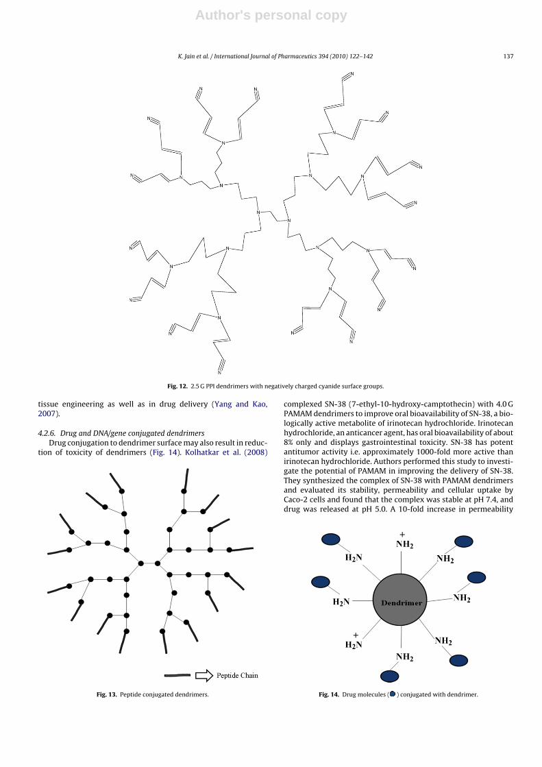
4.2.4. Half generation- or anionic dendrimersDendrimers like PAMAM and PPI have positively charged surface
groups after full generation synthesis, but their half generationshave carboxylic acid surface groups or cyanide surface groups,respectively (Fig. 12). We have already discussed that the toxicityof dendrimers and other cationic molecules is due to their surfacecationic groups and hence half generation dendrimers, which hasnegatively charged carboxylic or cyanide surface groups, showsnegligible toxicity (haemolytic toxicity or cytotoxicity) (Malik etal., 2000; Roberts et al., 1996; Bhadra et al., 2003). Malik et al.(2000) reported that carboxylic acid-terminated PAMAM den-drimers were not haemolytic up to concentration of 2 mg/ml,whereas amine-terminated full generation PAMAM were found tobe lytic above a concentration of 1 mg/ml. Bhadra et al. (2003)also reported similar results, and found that half generations ofcarboxylic acid-terminated PAMAM dendrimers showed negligi-ble hemolytic toxicity while full generation PAMAM dendrimersshowed a haemolysis of ∼15.3–17.3%. Jevprasesphant et al. (2003)also studied the cytotoxicity of whole generation (cationic) and halfgeneration (anionic) PAMAM dendrimers in Caco-2 cells and founda higher toxicity for whole generation dendrimers compared to halfgeneration (Jevprasesphant et al., 2003). Thus half generation den-drimers are lesser or non-toxic as compared to full generations.These reports hold valid for PAMAM dendrimers and may be appli-cable to other dendrimer types as well.
4.2.5. Amino acid or peptide conjugated dendrimersAmino acid such as phenyl alanine and glycine conjugated
dendrimers show a significant reduction in toxicities associ-ated with dendrimers (Agashe et al., 2006). Conjugation of theseamino acids to 5.0 G PPI dendrimers revealed a substantial reduc-tion in haemolytic toxicity; also these amino acid functionalizeddendrimers did not demonstrate any concentration- and time-dependent cytotoxicity. Many techniques have been employed fordecoration of dendrimers with amino acids and peptides at theirsurface. Kono et al. (2005) tailored PAMAM dendrimers with pheny-lalanine or leucine residues at their periphery to attain efficientgene transfection. Authors prepared phenylalanine- or leucine-grafted PAMAM dendrimers by reaction of amine-terminated 4.0 GPAMAM dendrimers with butyl pyrocarbonate protected phenylalanine (Boc-Phe) or butyl pyrocarbonate protected leucine (Boc-Leu) using N-hydroxy succinimide (NHS) and N,N′-dicyclohexylcarbodiimide (DCC). They found higher transfection efficiency inaddition to lower cytotoxicity with phenylalanine-modified den-drimers in contrast with some commonly applied transfectionagents.
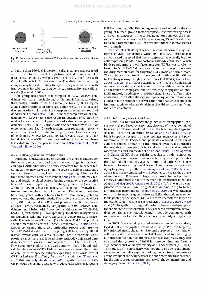
Arginine conjugated dendrimers demonstrated higher transfec-tion efficiency and suitability for gene delivery (Choi et al., 2004;Okuda et al., 2004). Dendrimers have also been modified withsome peptides (Fig. 13). Yang and Kao (2007) conjugated star-burst anionic 3.5 G and cationic 4.0 G PAMAM dendrimers witharginine-glycine-aspartate (RGD) peptides. They monitored cel-lular internalization of dendrimers by utilizing FITC-conjugatedPAMAM dendrimers in adherent fibroblasts and reported thatanionic 3.5 G-based dendritic RGD clusters showed no negativeeffect on fibroblast viability as well as for cationic 4.0 G-based den-dritic RGD clusters at lower concentrations. According to authorsthese nanoscale dendritic RGD clusters hold great potential for
Author's personal copy
K. Jain et al. / International Journal of Pharmaceutics 394 (2010) 122–142 137
Fig. 12. 2.5 G PPI dendrimers with negatively charged cyanide surface groups.
tissue engineering as well as in drug delivery (Yang and Kao,2007).
4.2.6. Drug and DNA/gene conjugated dendrimersDrug conjugation to dendrimer surface may also result in reduc-
tion of toxicity of dendrimers (Fig. 14). Kolhatkar et al. (2008)
Fig. 13. Peptide conjugated dendrimers.
complexed SN-38 (7-ethyl-10-hydroxy-camptothecin) with 4.0 GPAMAM dendrimers to improve oral bioavailability of SN-38, a bio-logically active metabolite of irinotecan hydrochloride. Irinotecanhydrochloride, an anticancer agent, has oral bioavailability of about8% only and displays gastrointestinal toxicity. SN-38 has potentantitumor activity i.e. approximately 1000-fold more active thanirinotecan hydrochloride. Authors performed this study to investi-gate the potential of PAMAM in improving the delivery of SN-38.They synthesized the complex of SN-38 with PAMAM dendrimersand evaluated its stability, permeability and cellular uptake byCaco-2 cells and found that the complex was stable at pH 7.4, anddrug was released at pH 5.0. A 10-fold increase in permeability
Fig. 14. Drug molecules ( ) conjugated with dendrimer.
Author's personal copy
138 K. Jain et al. / International Journal of Pharmaceutics 394 (2010) 122–142
Fig. 15. Formation of complex between DNA and dendrimer via interaction of pos-itive and negative charge.
and more than 100-fold increase in cellular uptake was observedwith respect to free SN-38. In cytotoxicity studies with complexno appreciable toxicity was observed after incubation for 2 h withCaco-2 cells at 0.1 �M concentration. Therefore dendrimer drugcomplex may be used to reduce the cytotoxicity of dendrimers withimprovement in stability, drug delivery, permeability and cellularuptake (Lee et al., 2006).
Our group has shown that complex of 4.0 G PAMAM den-drimer with water-insoluble and acidic anti-inflammatory drug,flurbiprofen, results in lesser haemolytic toxicity at an equiv-alent concentration than the plain dendrimers. This is becausedrug molecules could protect the peripheral free amino groups ofdendrimers (Asthana et al., 2005). Similarly complexation of den-drimers with DNA or gene also results in reduction of cytotoxicityof dendrimers because of protection of cationic charge of den-drimers (Li et al., 2007). Complexation of dendrimer with DNA oroligonucleotides (Fig. 15) leads to significant reduction in toxicityof dendrimers and this is due to the protection of cationic chargeof dendrimers by negatively charged DNA. Many researchers havefound dendrimer–DNA or dendrimer–oligonucleotide complexesless cytotoxic than the parent dendrimers (Brazeau et al., 1998;Yoo and Juliano, 2000).
4.2.7. Antibody functionalized dendrimersAntibody conjugated delivery systems are a novel strategy for
the delivery of cytotoxic and other therapeutic agents to specificcell types. Antibodies may be a very important tool in cancer tar-geting as they may serve as a homing device to deliver therapeuticagents to tumor site, may lead to specific targeting of tumor cellsthat overexpresses certain antigens (Chang et al., 1999), may tar-get and attack the blood vessels feeding a tumor or the connectivetissues (stroma) supporting it i.e. antiangiogenic effect (Wu et al.,2006), or they may block or neutralize the action of growth fac-tors required for the growth of tumor cells. Dendrimers have alsobeen conjugated with antibodies to form immunoconjugates toserve various therapeutic needs. Two different antibodies 60bcaand J591 that bound to CD14 and prostate specific membraneantigen (PSMA), respectively conjugated to 5.0 G PAMAM den-drimers and labeled with fluorescein isothiocyanate (G5-FI-60B,G5-FI-PA) for targeting CD14-expressing HL-60 human myeloblas-tic leukemia cells and PSMA expressing LNCaP prostate cancercells. The antibodies 60bca and J591 binds to CD14 and prostatespecific membrane antigen (PSMA), respectively. Thomas et al.(2004) conjugated these two antibodies (60bca and J591) to5.0 G PAMAM dendrimers for targeting CD14-expressing HL-60human myeloblastic leukemia cells and PSMA expressing LNCaPprostate cancer cells and labeled these antibody conjugated den-drimers with fluorescein isothiocyanate (G5-FI-60B, G5-FI-PA).Flow cytometry, confocal microscopy and two-photon-based opti-cal fiber fluorescence (TPOFF) detection system showed the uptakeof conjugates to specific antigen expressing cells while controlG5-FI lacked specific affinity for any of the cell lines (Thomas etal., 2004). Similarly Shukla et al. (2006) synthesized anti-HER2-G5-PAMAM dendrimers tagged with AlexaFluor (AF) for targeting
HER2-expressing cells. This conjugate was synthesized for the tar-geting of human growth factor receptor-2 overexpressing breastand ovarian cancer cells. The conjugate not only showed the bind-ing and internalization into HER2-expressing MCA-207 cell linesbut also it targeted the HER2-expressing tumors in in vivo studieswith animals.
Patri et al. (2004) synthesized immunodendrimers by tai-loring PAMAM dendrimers with J591 anti-PSMA monoclonalantibody and observed that these conjugates specifically bind tocells expressing PSMA. A monoclonal antibody cetuximab, whichbinds to epidermal growth factor receptor (EGFR), was covalentlylinked to 5.0 G PAMAM dendrimers via its Fc region contain-ing drug methotrexate for targeting EGFR-positive brain tumors.The conjugate was found to be cytotoxic with specific affinityto EGFR-expressing rat glioma cell lines F98 (EGFR) (Wu et al.,2006). Wangler et al. (2008) examined the impact of conjugationon immunoreactivity of derivatized antibody with respect to sizeand number of conjugates and for this they conjugated an anti-EGFR antibody hMAb425 with PAMAM dendrimers of different sizecontaining up to 128 chelating agents per conjugation site and con-cluded that the number of derivatization sites had crucial effect onimmunoreactivity whereas dendrimer size did not have significantinfluence on activity.
4.2.8. Tuftsin conjugated dendrimersTuftsin is a natural macrophage activator tetrapeptide (Thr-
Lys-Pro-Arg) produced by enzymatic cleavage of the Fc-domain ofheavy chain of immunoglobulin G as the free peptide fragment(Najjar, 1987), first identified by Najjar and Nishioka (1970). Itbinds to specific receptors on macrophages, monocytes and poly-morphonuclear leukocytes and possesses a broad spectrum ofactivities related primarily to the immune system. It stimulatesthe migration, phagocytic, bactericidal and tumoricidal activity ofmacrophages and leukocytes (Fridkin and Najjar, 1989; Agrawaland Gupta, 2000). Since tuftsin specifically binds monocytes,macrophages and polymorphonuclear leukocytes and potentiatestheir natural killer activity against tumors and pathogens; it maybe used to increase drug specificity and lower systemic drug toxic-ity by targeting drug to these cells (Gupta and Haq, 2005; Bai et al.,2008). It has been conjugated with liposomes to increase the uptakeof amphotericin B by macrophages to improve chemotherapeuticefficacy of amphotericin B for treatment of leishmanial infections(Gupta and Haq, 2005; Agrawal et al., 2002). Tuftsin was also con-jugated with an anti-viral drug Azidothymidine (AZT) to targetHIV-infected macrophages (Fridkin et al., 2005). It was attachedwith an anticancer drug methotrexate (MTX) through an enzyme-labile pentapeptide spacer (GFLGC) to form chemotactic targetingmoiety for targeting cancer chemotherapy (Bai et al., 2008). Mezoet al. (2006) synthesized oligotuftsin-based branched oligopeptidefor chemotactic drug targeting. They prepared tetratuftsin deriva-tives containing chemotactic formyl tripeptide conjugated withmethotrexate and studied their chemotactic activity and cytotoxi-city.
In 2008 Dutta et al. prepared efavirenz, an anti-viral drug,loaded tuftsin conjugated PPI dendrimers (TuPPI) for targetingHIV-infected macrophages in vitro and observed a much highercellular uptake of efavirenz from TuPPI compared to free drug byHIV-infected fresh human mononuclear cells (Mo/Mac). They alsoevaluated the cytotoxity of TuPPI in these cell lines and found asignificant reduction in cytotoxicity of PPI dendrimers (p < 0.001).This reduction in cytotoxicity was thought to be due to the shield-ing effect of the bulky peptide masking the cytotoxic free primaryamino groups at the periphery of PPI dendrimers and thus prevent-ing the amino groups from interacting with the cell membrane anddamaging them (Dutta et al., 2008).

Author's personal copy
K. Jain et al. / International Journal of Pharmaceutics 394 (2010) 122–142 139
4.2.9. Folate-conjugated dendrimersThe folic acid receptor is overexpressed by many types of
tumor cells such as lung, renal, ovarian, endometrial, colorectal,breast, testis, myelocytic blood cells, neuroendocrine carcinomasand brain metastases (Garin-Chesa et al., 1993; Wang and Low,1998). Folate-anchored carriers enter the cell by receptor medi-ated endocytosis (RME), bypassing cellular barriers, and allow theattached moiety to enter cancer cells of tumor xenografts (Calicetiet al., 2003). Folate mediated targeting has not been used not onlyin drug delivery but also for the delivery of gene, imaging agentand diagnostic agents (Wang and Low, 1998). This phenomenahas also been utilized in dendrimer mediated cancer therapy andfolate-conjugated dendrimers were developed to target tumor cells(Singh et al., 2008; Quintana et al., 2002). Kono et al. (1999) syn-thesized folate-anchored polyether dendritic systems as a carrierwith potential tumor cell specificity. They prepared this carrier sys-tem by introducing hydrazide groups to ester-terminated polyetherdendrimers by reaction with hydrazine followed by conjugation offolate residues to the hydrazide chain ends of the dendrimers bydirect condensation with folic acid. These conjugates were solublein aqueous medium above pH 7.4 (Kono et al., 1999). PAMAM den-drimers were conjugated with FITC and then with folate throughEDC linkage. 5.0 G PAMAM dendrimer was dissolved in DMSO andsodium phosphate buffer under nitrogen for conjugation to FITC.The mixture was stirred in the dark for ∼24 h, then washed andultrafiltered in Centricon tubes. Furthermore, Quintana et al. (2002)conjugated folate to the dendrimer through an EDC linkage. Ananticancer drug (methotrexate) was incorporated into dendrimersaimed at effective delivery to the cancer tissues with reducedtoxicity. To gain access to the tumor surface, via the circulatorysystem, the drug needs to be hydrophilic; and to penetrate thecell membrane, the lipid solubility must be high. The simultaneousoccurrence of both properties in the drug is difficult and, as dis-cussed previously, folate-conjugated dendrimers enter the cell byRME, bypasses the cellular barriers, allowing hydrophilic drugs toenter cancer cells of tumor xenografts (Quintana et al., 2002). Later,Choi et al. (2005) also utilized this method to conjugate folic acidor FITC to partially acetylated PAMAM dendrimers and finally theyattached this system covalently to oligonucleotides. In in vitro stud-ies this DNA-linked dendrimer clusters indicated specific bindingto KB cells expressing the folate receptor and also showed the inter-nalization of the dendrimer cluster. Singh et al. (2008) conjugatedPAMAM dendrimers with folic acid and folic acid–PEG conjugatesand evaluated their efficacy and safety in tumor-bearing mice andfound these folate-PEG-dendrimer to be significantly safe and effec-tive in tumor targeting including reduction in hemolytic toxicity,sustained release and higher accumulation in tumor area.
4.2.10. MiscellaneousBaker and co-workers synthesized PAMAM dendrimer based
multifunctional nanodevice for delivery of anticancer agent. Onto these multifunctional dendrimers authors combined the cancerimaging and targeted delivery by attaching different ligands; acetylgroup to neutralize primary amine groups, FITC for cancer imaging,folic acid as targeting moiety and methotrexate, an anticancer agent(Majoros et al., 2005).
Miao et al. (2007) incorporated biodegradable poly(�-caprolactone)s onto the PAMAM dendrimers. The authorssynthesized a series of star poly(�-caprolactone)s (PCL) withdendritic cores via ring-opening polymerization of �-caprolactoneinitiated by PAMAM dendrimers. The in vitro degradation of thesedendrimers is influenced by the content of PAMAM dendrimersand caprolactone. Thus a highly biodegradable carrier may beobtained by incorparing caprolactone with dendrimers.
Li et al. (2007) synthesized amphiphilic dendritic poly(l-lysine)-b-poly(l-lactide)-b-dendritic poly(l-lysine) dendrimers bearing
two two-generation of poly(l-lysine) dendrons and a centralhydrophobic biodegradable poly(l-lactide). These dendrimers wasdesigned, synthesized and applied as a biodegradable gene car-rier. Their cytotoxicity was appraised by conducting MTT assay inhuman hepatocellular carcinoma cell line (SMMC-7721) and a verylow cytotoxicity was observed (Li et al., 2007).
5. Conclusion and future prospects
The past decade witnessed a large explosion in the field of drugdelivery. A number of delivery devices were explored to controldrug disposition in the biological environment in order to obtainmaximum therapeutic benefit with minimum toxicity. Along withthese delivery devices dendrimer has emerged as promising drugdelivery device in modern medicine system. Their well-definedarchitecture made them useful in biological systems. Dendrimerswith their large molecular size and multivalent surfaces provide anexcellent platform for the attachment of drug, gene, targeting moi-eties and homing devices, imaging agents etc., as well as they canbe hydrophobically modified to mimic inverse micelles and havepresented unrestrained ‘guest host’ properties.
Although dendrimers has made tremendous progress in thedelivery of therapeutic agents yet their posbile potential is con-strained because of inherent toxicity problems associated withthem. Dendrimers and other cationic macromolecular delivery sys-tem interact nonspecifically with negatively charged biologicalmembrane. This non-selective interaction of cationic delivery sys-tem causes membrane disruption through formation of nanoholes,membrane thinning and membrane erosion followed by leakageof cytosolic enzymes and cell death. Consequently it results in dif-ferent toxicities of dendrimers including cytotoxicity, haemolytictoxicity and haematological toxicity. Currently, there is great needto devise strategies to overcome difficulties encountered in drugdelivery using a parent dendrimer as the carrier.
A plethora of research is going on to resolve the issues relatingto safety, toxicity and efficacy of dendrimers in order to formulatesome strategies for reduction of toxicity associated with den-drimers. Design and development of new dendrimers includingbiodegradable and/or biocompatible and surface engineered den-drimers may alleviate this problem successfully and may lead to adelivery system with a potential therapeutic efficacy vis a vis mini-mum biological toxicity. The major emphasis is on the developmentof biomimetic dendrimers, which will enhance the biocompatibil-ity of dendrimers. A detailed study of these nanostructures wouldhelp in the evolution of newer concepts that could be intimatelyinvolved in sustaining the life processes. Various dendrimers hav-ing framework of biocompatible materials have been developedin the last decade such as citric acid dendrimers composed of cit-ric acid, and polyethylene glycol, phosphate dendrimers, lysinedendrimers etc. Some other biodegradable dendrimers includingpolyester dendrimers, polyether dendrimers, triazine dendrimershave also been developed to reduce the toxicity of dendrimers.
Next to the development of biocompatible dendrimers, anotherrewarding strategy for the development of less toxic dendrimeris surface engineering. In case of dendrimers surface engineeringis a method of modifying the surface of dendrimers with variousfunctional groups to mask the cationic charge of dendrimers. It isthe method by which cytotoxic dendrimers could be converted intobiocompatible dendrimers. This has been supported by various sur-face engineering strategies such as PEGylation, acetylation, folateconjugation, carbohydrate, peptide, drug, gene and tuftsin con-jugated, anionic and antibody functionalized dendrimers. Besidesreduction of toxicity surface engineering may also accomplish someother benefits in the field of drug delivery including drug encapsu-lation, solubilization, protection of biomolecules, gene transfection,modified release, targeting with improved stability profile, biodis-
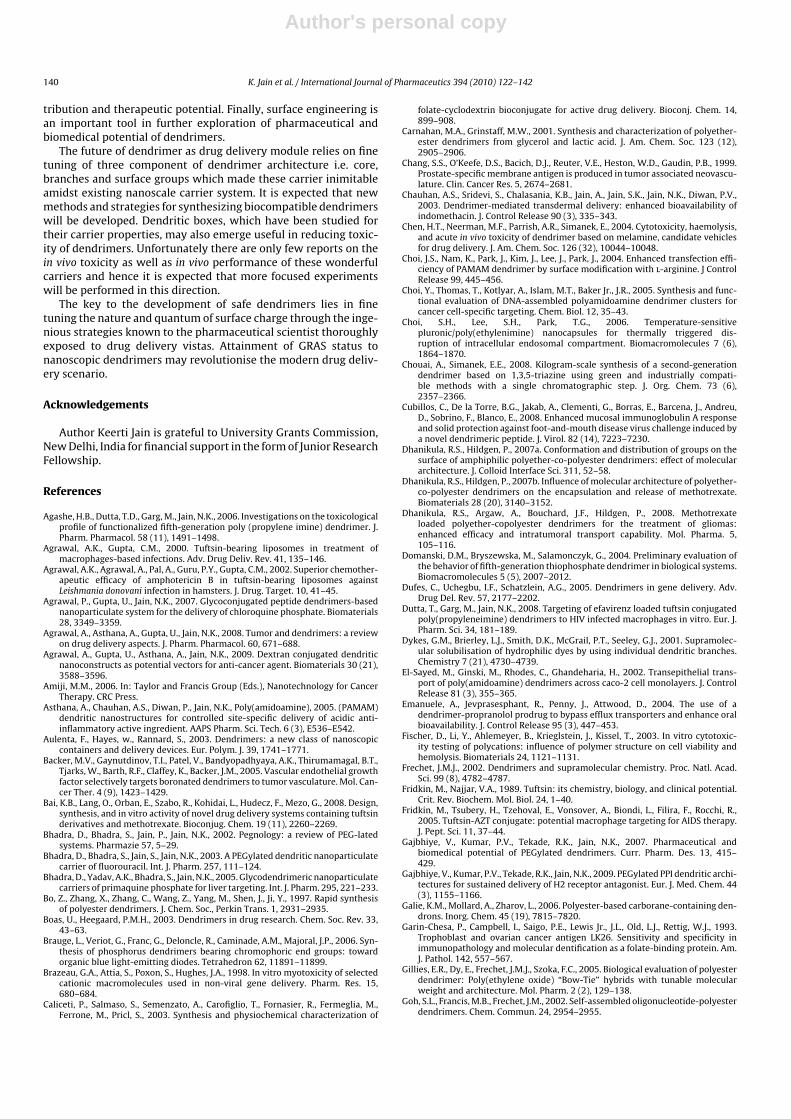
Author's personal copy
140 K. Jain et al. / International Journal of Pharmaceutics 394 (2010) 122–142
tribution and therapeutic potential. Finally, surface engineering isan important tool in further exploration of pharmaceutical andbiomedical potential of dendrimers.
The future of dendrimer as drug delivery module relies on finetuning of three component of dendrimer architecture i.e. core,branches and surface groups which made these carrier inimitableamidst existing nanoscale carrier system. It is expected that newmethods and strategies for synthesizing biocompatible dendrimerswill be developed. Dendritic boxes, which have been studied fortheir carrier properties, may also emerge useful in reducing toxic-ity of dendrimers. Unfortunately there are only few reports on thein vivo toxicity as well as in vivo performance of these wonderfulcarriers and hence it is expected that more focused experimentswill be performed in this direction.
The key to the development of safe dendrimers lies in finetuning the nature and quantum of surface charge through the inge-nious strategies known to the pharmaceutical scientist thoroughlyexposed to drug delivery vistas. Attainment of GRAS status tonanoscopic dendrimers may revolutionise the modern drug deliv-ery scenario.
Acknowledgements
Author Keerti Jain is grateful to University Grants Commission,New Delhi, India for financial support in the form of Junior ResearchFellowship.
References
Agashe, H.B., Dutta, T.D., Garg, M., Jain, N.K., 2006. Investigations on the toxicologicalprofile of functionalized fifth-generation poly (propylene imine) dendrimer. J.Pharm. Pharmacol. 58 (11), 1491–1498.
Agrawal, A.K., Gupta, C.M., 2000. Tuftsin-bearing liposomes in treatment ofmacrophages-based infections. Adv. Drug Deliv. Rev. 41, 135–146.
Agrawal, A.K., Agrawal, A., Pal, A., Guru, P.Y., Gupta, C.M., 2002. Superior chemother-apeutic efficacy of amphotericin B in tuftsin-bearing liposomes againstLeishmania donovani infection in hamsters. J. Drug. Target. 10, 41–45.
Agrawal, P., Gupta, U., Jain, N.K., 2007. Glycoconjugated peptide dendrimers-basednanoparticulate system for the delivery of chloroquine phosphate. Biomaterials28, 3349–3359.
Agrawal, A., Asthana, A., Gupta, U., Jain, N.K., 2008. Tumor and dendrimers: a reviewon drug delivery aspects. J. Pharm. Pharmacol. 60, 671–688.
Agrawal, A., Gupta, U., Asthana, A., Jain, N.K., 2009. Dextran conjugated dendriticnanoconstructs as potential vectors for anti-cancer agent. Biomaterials 30 (21),3588–3596.
Amiji, M.M., 2006. In: Taylor and Francis Group (Eds.), Nanotechnology for CancerTherapy. CRC Press.
Asthana, A., Chauhan, A.S., Diwan, P., Jain, N.K., Poly(amidoamine), 2005. (PAMAM)dendritic nanostructures for controlled site-specific delivery of acidic anti-inflammatory active ingredient. AAPS Pharm. Sci. Tech. 6 (3), E536–E542.
Aulenta, F., Hayes, w., Rannard, S., 2003. Dendrimers: a new class of nanoscopiccontainers and delivery devices. Eur. Polym. J. 39, 1741–1771.
Backer, M.V., Gaynutdinov, T.I., Patel, V., Bandyopadhyaya, A.K., Thirumamagal, B.T.,Tjarks, W., Barth, R.F., Claffey, K., Backer, J.M., 2005. Vascular endothelial growthfactor selectively targets boronated dendrimers to tumor vasculature. Mol. Can-cer Ther. 4 (9), 1423–1429.
Bai, K.B., Lang, O., Orban, E., Szabo, R., Kohidai, L., Hudecz, F., Mezo, G., 2008. Design,synthesis, and in vitro activity of novel drug delivery systems containing tuftsinderivatives and methotrexate. Bioconjug. Chem. 19 (11), 2260–2269.
Bhadra, D., Bhadra, S., Jain, P., Jain, N.K., 2002. Pegnology: a review of PEG-latedsystems. Pharmazie 57, 5–29.
Bhadra, D., Bhadra, S., Jain, S., Jain, N.K., 2003. A PEGylated dendritic nanoparticulatecarrier of fluorouracil. Int. J. Pharm. 257, 111–124.
Bhadra, D., Yadav, A.K., Bhadra, S., Jain, N.K., 2005. Glycodendrimeric nanoparticulatecarriers of primaquine phosphate for liver targeting. Int. J. Pharm. 295, 221–233.
Bo, Z., Zhang, X., Zhang, C., Wang, Z., Yang, M., Shen, J., Ji, Y., 1997. Rapid synthesisof polyester dendrimers. J. Chem. Soc., Perkin Trans. 1, 2931–2935.
Boas, U., Heegaard, P.M.H., 2003. Dendrimers in drug research. Chem. Soc. Rev. 33,43–63.
Brauge, L., Veriot, G., Franc, G., Deloncle, R., Caminade, A.M., Majoral, J.P., 2006. Syn-thesis of phosphorus dendrimers bearing chromophoric end groups: towardorganic blue light-emitting diodes. Tetrahedron 62, 11891–11899.
Brazeau, G.A., Attia, S., Poxon, S., Hughes, J.A., 1998. In vitro myotoxicity of selectedcationic macromolecules used in non-viral gene delivery. Pharm. Res. 15,680–684.
Caliceti, P., Salmaso, S., Semenzato, A., Carofiglio, T., Fornasier, R., Fermeglia, M.,Ferrone, M., Pricl, S., 2003. Synthesis and physiochemical characterization of
folate-cyclodextrin bioconjugate for active drug delivery. Bioconj. Chem. 14,899–908.
Carnahan, M.A., Grinstaff, M.W., 2001. Synthesis and characterization of polyether-ester dendrimers from glycerol and lactic acid. J. Am. Chem. Soc. 123 (12),2905–2906.
Chang, S.S., O’Keefe, D.S., Bacich, D.J., Reuter, V.E., Heston, W.D., Gaudin, P.B., 1999.Prostate-specific membrane antigen is produced in tumor associated neovascu-lature. Clin. Cancer Res. 5, 2674–2681.
Chauhan, A.S., Sridevi, S., Chalasania, K.B., Jain, A., Jain, S.K., Jain, N.K., Diwan, P.V.,2003. Dendrimer-mediated transdermal delivery: enhanced bioavailability ofindomethacin. J. Control Release 90 (3), 335–343.
Chen, H.T., Neerman, M.F., Parrish, A.R., Simanek, E., 2004. Cytotoxicity, haemolysis,and acute in vivo toxicity of dendrimer based on melamine, candidate vehiclesfor drug delivery. J. Am. Chem. Soc. 126 (32), 10044–10048.
Choi, J.S., Nam, K., Park, J., Kim, J., Lee, J., Park, J., 2004. Enhanced transfection effi-ciency of PAMAM dendrimer by surface modification with l-arginine. J ControlRelease 99, 445–456.
Choi, Y., Thomas, T., Kotlyar, A., Islam, M.T., Baker Jr., J.R., 2005. Synthesis and func-tional evaluation of DNA-assembled polyamidoamine dendrimer clusters forcancer cell-specific targeting. Chem. Biol. 12, 35–43.
Choi, S.H., Lee, S.H., Park, T.G., 2006. Temperature-sensitivepluronic/poly(ethylenimine) nanocapsules for thermally triggered dis-ruption of intracellular endosomal compartment. Biomacromolecules 7 (6),1864–1870.
Chouai, A., Simanek, E.E., 2008. Kilogram-scale synthesis of a second-generationdendrimer based on 1,3,5-triazine using green and industrially compati-ble methods with a single chromatographic step. J. Org. Chem. 73 (6),2357–2366.
Cubillos, C., De la Torre, B.G., Jakab, A., Clementi, G., Borras, E., Barcena, J., Andreu,D., Sobrino, F., Blanco, E., 2008. Enhanced mucosal immunoglobulin A responseand solid protection against foot-and-mouth disease virus challenge induced bya novel dendrimeric peptide. J. Virol. 82 (14), 7223–7230.
Dhanikula, R.S., Hildgen, P., 2007a. Conformation and distribution of groups on thesurface of amphiphilic polyether-co-polyester dendrimers: effect of moleculararchitecture. J. Colloid Interface Sci. 311, 52–58.
Dhanikula, R.S., Hildgen, P., 2007b. Influence of molecular architecture of polyether-co-polyester dendrimers on the encapsulation and release of methotrexate.Biomaterials 28 (20), 3140–3152.
Dhanikula, R.S., Argaw, A., Bouchard, J.F., Hildgen, P., 2008. Methotrexateloaded polyether-copolyester dendrimers for the treatment of gliomas:enhanced efficacy and intratumoral transport capability. Mol. Pharma. 5,105–116.
Domanski, D.M., Bryszewska, M., Salamonczyk, G., 2004. Preliminary evaluation ofthe behavior of fifth-generation thiophosphate dendrimer in biological systems.Biomacromolecules 5 (5), 2007–2012.
Dufes, C., Uchegbu, I.F., Schatzlein, A.G., 2005. Dendrimers in gene delivery. Adv.Drug Del. Rev. 57, 2177–2202.
Dutta, T., Garg, M., Jain, N.K., 2008. Targeting of efavirenz loaded tuftsin conjugatedpoly(propyleneimine) dendrimers to HIV infected macrophages in vitro. Eur. J.Pharm. Sci. 34, 181–189.
Dykes, G.M., Brierley, L.J., Smith, D.K., McGrail, P.T., Seeley, G.J., 2001. Supramolec-ular solubilisation of hydrophilic dyes by using individual dendritic branches.Chemistry 7 (21), 4730–4739.
El-Sayed, M., Ginski, M., Rhodes, C., Ghandeharia, H., 2002. Transepithelial trans-port of poly(amidoamine) dendrimers across caco-2 cell monolayers. J. ControlRelease 81 (3), 355–365.
Emanuele, A., Jevprasesphant, R., Penny, J., Attwood, D., 2004. The use of adendrimer-propranolol prodrug to bypass efflux transporters and enhance oralbioavailability. J. Control Release 95 (3), 447–453.
Fischer, D., Li, Y., Ahlemeyer, B., Krieglstein, J., Kissel, T., 2003. In vitro cytotoxic-ity testing of polycations: influence of polymer structure on cell viability andhemolysis. Biomaterials 24, 1121–1131.
Frechet, J.M.J., 2002. Dendrimers and supramolecular chemistry. Proc. Natl. Acad.Sci. 99 (8), 4782–4787.
Fridkin, M., Najjar, V.A., 1989. Tuftsin: its chemistry, biology, and clinical potential.Crit. Rev. Biochem. Mol. Biol. 24, 1–40.
Fridkin, M., Tsubery, H., Tzehoval, E., Vonsover, A., Biondi, L., Filira, F., Rocchi, R.,2005. Tuftsin-AZT conjugate: potential macrophage targeting for AIDS therapy.J. Pept. Sci. 11, 37–44.
Gajbhiye, V., Kumar, P.V., Tekade, R.K., Jain, N.K., 2007. Pharmaceutical andbiomedical potential of PEGylated dendrimers. Curr. Pharm. Des. 13, 415–429.
Gajbhiye, V., Kumar, P.V., Tekade, R.K., Jain, N.K., 2009. PEGylated PPI dendritic archi-tectures for sustained delivery of H2 receptor antagonist. Eur. J. Med. Chem. 44(3), 1155–1166.
Galie, K.M., Mollard, A., Zharov, L., 2006. Polyester-based carborane-containing den-drons. Inorg. Chem. 45 (19), 7815–7820.
Garin-Chesa, P., Campbell, I., Saigo, P.E., Lewis Jr., J.L., Old, L.J., Rettig, W.J., 1993.Trophoblast and ovarian cancer antigen LK26. Sensitivity and specificity inimmunopathology and molecular dentification as a folate-binding protein. Am.J. Pathol. 142, 557–567.
Gillies, E.R., Dy, E., Frechet, J.M.J., Szoka, F.C., 2005. Biological evaluation of polyesterdendrimer: Poly(ethylene oxide) “Bow-Tie” hybrids with tunable molecularweight and architecture. Mol. Pharm. 2 (2), 129–138.
Goh, S.L., Francis, M.B., Frechet, J.M., 2002. Self-assembled oligonucleotide-polyesterdendrimers. Chem. Commun. 24, 2954–2955.
Author's personal copy
K. Jain et al. / International Journal of Pharmaceutics 394 (2010) 122–142 141
Gupta, C.M., Haq, W., 2005. Tuftsin-bearing liposomes as antibiotic carriers in treat-ment of macrophage infections. Methods Enzymol. 391, 291–304.
Gupta, U., Agashe, H.B., Jain, N.K., 2007. Polypropylene imine dendrimer mediatedsolubility enhancement: effect of pH and functional groups of hydrophobes. J.Pharm. Pharm. Sci. 10 (3), 358–367.
Hawker, C.J., Frechet, J.M.J., 1990. Preparation of polymers with controlled moleculararchitecture. A new convergent approach to dendritic macromolecules. J. Am.Chem. Soc. 112 (21), 7638–7647.
Hirayama, Y., Nakamura, T., Uehara, S., Sakamoto, Y., Yamaguchi, K., Sei, Y., Iwa-mura, M., 2005. Synthesis and characterization of polyester dendrimers fromacetoacetate and acrylate. Org. Lett. 7 (40), 525–528.
Hoet, P.H.M., Nemmar, A., Nemery, B., 2004. Health impact of nanomaterials? Nat.Biotechnol. 22 (19), 104–119.
Hong, S., Bielinska, A.U., Mecke, A., Keszler, B., Beals, J.L., Shi, X., Balogh, L., Orr, B.G.,Baker Jr., J.R., Banaszak, Holl, M.M., 2004. Interaction of poly(amidoamine) den-drimers with supported lipid bilayers and cells: hole formation and the relationto transport. Bioconj. Chem. 15 (4), 774–782.
Hong, S., Hessler, J.A., Holl, M.M.B., Leroueil, P., Mecke, A., Orr, B.G., 2006a. Physi-cal interaction of nanoparticles with biological membranes: the observation ofnanoscale hole formation. J. Chem. Health Saf. 13 (3), 16–20.
Hong, S., Leroueil, P.R., Janus, E.K., Peters, J.L., Kober, M.M., Islam, M.T., Orr, B.G., BakerJr., J.R., Holl, M.M.B., 2006b. Interaction of polycationic polymers with supportedlipid bilayers and cells: nanoscale hole formation and enhanced membrane per-meability. Bioconj. Chem. 17 (3), 728–734.
Hu, K.W., Huang, C.C., Hwu, J.R., Su, W.C., Shieh, D.B., Yeh, C.S., 2008. A new pho-tothermal therapeutic agent: core-free nanostructured Au x Ag1-x dendrites.Chemistry 14 (10), 2956–2964.
Ihre, H., Padilla De Jesus, O.L., Frechet, J.M., 2001. Fast and convenient divergentsynthesis of aliphatic ester dendrimers by anhydride coupling. J. Am. Chem. Soc.123 (25), 5908–5917.
Ihre, H.R., Padilla De Jesus, O.L., Szoka, F.C., Frechet, J.M.J., 2002. Polyester dendriticsystems for drug delivery applications: design, synthesis, and characterization.Bioconj. Chem. 13 (3), 443–452.
Jain, N.K., Gupta, U., 2008. Application of dendrimer-drug complexation in theenhancement of drug solubility and bioavailability. Expert Opin. Drug Metabol.Toxicol. 4 (8), 1035–1052.
Jayaraman, M., Frechet, J.M.J., 1998. A convergent route to novel aliphatic polyetherdendrimers. J. Am. Chem. Soc. 120 (49), 12996–12997.
Jevprasesphant, R., Penny, J., Jalal, R., Attwood, D., McKeown, N., D’Emanuele, A.,2003. The influence of surface modification on the cytotoxicity of PAMAM den-drimers. Int. J. Pharm. 252, 263–266.
Khopade, A.J., Jain, N.K., 1997. Dendrimer delivery of methotrexate, Abstract book.In: Symposium on Particulate Systems, from Formulation to Production, Istan-bul, 6–7 oct.
Kolhatkar, R.B., Kitchens, K.M., Swaan, P.W., Ghandehari, H., 2007. Surface acety-lation of polyamidoamine (PAMAM) dendrimers decreases cytotoxicity whilemaintaining membrane permeability. Bioconj. Chem. 18 (6), 2054–2060.
Kolhatkar, R.B., Swaan, P., Ghandehari, H., 2008. Potential oral delivery of 7-ethyl-10-hydroxy-camptothecin (SN-38) using poly(amidoamine) dendrimers. Pharm.Res. 25 (7), 1723–1729.
Konda, S.D., Aref, M., Wang, S., Brechbiel, M., Wiener, E.C., 2001. Specific target-ing of folate dendrimer MRI contrast agents to the high affinity folate receptorexpressed in overian tumor xenografts. Magn. Reson. Mater Phys. Biol. Med. 12,104–113.
Kono, K., Liu, M., Frechet, J.M., 1999. Design of dendritic macromolecules containingfolate or methotrexate residues. Bioconj. Chem. 10 (6), 1115–1121.
Kono, K., Akiyama, H., Takahashi, T., Takagishi, T., Harada, A., 2005. Transfec-tion activity of polyamidoamine dendrimers having hydrophobic amino acidresidues in the periphery. Bioconj. Chem. 16, 208–214.
Krishna, T.R., Jain, S., Tatu, U.S., Jayaraman, N., 2005. Synthesis and biological evalua-tion of 3-amino-propan-1-ol based poly(ether imine) dendrimers. Tetrahedron61 (17), 4281–4288.
Kukowska-Latallo, J.F., Candido, K.A., Cao, Z., Nigavekar, S.S., Majoros, I.J., Thomas,T.P., Balogh, L.P., Khan, M.K., Baker, J.R., 2005. Nanoparticle targeting of anti-cancer drug improves therapeutic response in animal model of human epithelialcancer. Cancer Res. 65 (12), 5317–5324.
Lai, L.L., Lee, C.H., Wang, L.Y., Cheng, K.L., Hsu, H.F., 2008. Star-shaped mesogensof triazine-based dendrons and dendrimers as unconventional columnar liquidcrystals. J. Org. Chem. 73 (2), 485–490.
Lee, H., Larson, R.G., 2008. Lipid bilayer curvature and pore formation induced bycharged linear polymers and dendrimers: the effect of molecular shape. J. Phys.Chem. B. 112 (39), 12279–12285.
Lee, C.C., Gillies, E.R., Fox, M.E., Guillaudeu, S.J., Frechet, J.M., Dy, E.E., Szoka,F.C., 2006. A singal dose of doxorubicin-functionalized bow-tie dendrimercures mice bearing C-26 colon carcinomas. Proc. Natl. Acad. Sci. 103 (45),16649–16654.
Lee, S.H., Choi, S.H., Kim, S.H., Park, T.G., 2008. Thermally sensitive cationic poly-mer nanocapsules for specific cytosolic delivery and efficient gene silencingof siRNA: swelling induced physical disruption of endosome by cold shock. J.Control Release 125, 25–32.
Leon, J.W., Frechet, J.M.J., 1995. Analysis of aromatic polyether dendrimers anddendrimer-linear block copolymers by matrix-assisted laser desorption ioniza-tion mass spectrometry. J. Polymer Bull. 35 (4), 449–455.
Leroueil, P.R., Berry, S.A., Duthie, K., Han, G., Rotello, V.M., McNerny, D.Q., Baker Jr.,J.R., Orr, B.G., Holl, M.M., 2008. Wide varieties of cationic nanoparticles inducedefects in supported lipid bilayers. Nano Lett. 8 (2), 420–424.
Li, Y., Cui, L., Li, Q., Jia, L., Xu, Y., Fang, Q., Cao, A., 2007. Novel symmetric amphiphilicdendritic poly(l-lysine)-b-poly(l-lactide)-b-dendritic poly(l-lysine) with highplasmid DNA binding affinity as a biodegradable gene carrier. Biomacro-molecules 8 (5), 1409–1416.
Lim, J., Simanek, E.E., 2008. Synthesis of water-soluble dendrimers based onmelamine bearing 16 paclitaxel groups. Org. Lett. 10 (2), 201–204.
Lim, J., Guo, Y., Rostollan, C.L., Stanfield, J., Hsieh, J.T., Sun, X., Simanek, E.E., 2008. Therole of the size and number of polyethylene glycol chains in the biodistributionand tumor localization of triazine dendrimers. Mol. Pharm. 5 (4), 540–547.
Luman, N.R., Smeds, K.A., Grinstaff, M.W., 2003. The convergent synthesis ofpoly(glycerol-succinic acid) dendritic macromolecules. Chemistry 9 (22),5618–5626.
Luo, D., Saltzman, W.M., 2000. Synthetic DNA delivery systems. Nat. Biotech. 18,33–37.
Luo, D., Haverstick, K., Belcheva, N., Han, E., Saltzman, M., 2002. Poly(ethyleneglycol)-conjugated PAMAM dendrimer for biocompatible, high-efficiency DNAdelivery. Macromolecules 35, 3456–3462.
Majoros, I.R., Keszler, B., Woehler, S., Bull, T., Baker, J.R., 2002. Acetylation ofpoly(amidoamine) dendrimers. Macromolecules 36, 5526–5529.
Majoros, I.J., Thomas, T.P., Mehta, C.B., Baker Jr., J.R., 2005. Poly (amidoamine)dendrimer-based multifunctional engineered nanodevice for cancer therapy. J.Med. Chem. 48 (19), 5892–5899.
Malik, N., Wiwattanapatapee, R., Klopsch, R., Lorenz, K., Frey, H., Weener, J.W., Meijer,E.W., Paulus, W., Duncan, R., 2000. Dendrimers: relationship between structureand biocompatibility in vitro, and preliminary studies on the biodistribution of125 I-labelled polyamidoamine dendrimers in vivo. J. Control Release 65 (1–2),133–148.
Manunta, M., Tan, P.H., Sagoo, P., Kashefi, K., George, A.J.T., 2004. Gene delivery bydendrimers operates via a cholesterol dependent pathway. Nucleic Acid Res. 32(9), 2730–2739.
Manunta, M., Nichols, B.J., Tana, P.H., Sagooa, P., Harpera, J., Georgea, A.J.T., 2006.Gene delivery by dendrimers operates via different pathways in different cells,but is enhanced by the presence of caveolin. J. Immunol. Methods 314, 134–146.
Mecke, A., Uppuluri, S., Sassanella, T.J., Lee, D.K., Ramamoorthy, A., Baker, J.R., Orr,B.G., Banaszak Holl, M.M., 2004. Direct observation of lipid bilayer disruption bypoly(amidoamine) dendrimers. Chem. Phys. Lipids 132, 3–14.
Mecke, A., Lee, D.K., Ramamoorthy, A., Orr, B.G., Holl, M.M.B., 2005a. Membranethinning due to antimicrobial peptide binding: an atomic force microscopy studyof MSI-78 in lipid bilayers. Biophys. J. 89 (6), 4043–4050.
Mecke, A., Majoros, I.J., Patri, A.K., Baker, J.R., Holl, M.M.B., 2005b. Lipid bilayer dis-ruption by polycationic polymers: the roles of size and chemical functionalgroup. Langmuir 21, 10348–10354.
Mecke, A., Lee, D.K., Ramamoorthy, A., Orr, B.G., Holl, M.M.B., 2005c. Synthetic andnatural polymer nanoparticles interact selectively with fluid-phase domains ofDMPC lipid bilayers. Langmuir 21 (19), 8588–8590.
Mezo, G., Lang, O., Jakab, A., Bai, K.B., Szabo, I., Schlosser, G., Lang, J., Kohidai, L.,Hudecz, F., 2006. Synthesis of oligotuftsin-based branched oligopeptide conju-gates for chemotactic drug targeting. J. Pept. Sci. 12 (5), 328–336.
Miao, Z.M., Cheng, S.X., Zhang, X.Z., Wang, Q.R., Zhuo, R.X., 2007. Degradation anddrug release property of star poly(epsilon-caprolactone)s with dendritic cores.J. Biomed. Mater. Res. B. Appl. Biomater. 81, 40–49.
Mitchell, J.P., Roberts, K.D., Langley, J., Koentgen, F., Lambert, J.N., 1999. A directmethod for the formation of peptide and carbohydrate dendrimers. Bioorg. Med.Chem. Lett. 9, 2785–2788.
Najjar, V.A., 1987. Biological effects of tuftsin and its analogs. Drug Future 12,147–160.
Najjar, V.A., Nishioka, K., 1970. “Tuftsin”: a natural phagocytosis stimulating peptide.Nature 228 (5272), 672–673.
Namaji, H., Adeli, M., 2005. Dendrimers of citric acid and poly (ethylene glycol) asthe new drug-delivery agents. Biomaterials 26 (10), 1175–1183.
Neerman, M.F., Zhang, W., Parrish, A.R., Simanek, E.E., 2004. In vitro and in vivo eval-uation of a melamine dendrimer as a vehicle for drug delivery. Int. J. Pharm. 281,129–132.
Neerman, M.F., Chen, H.T., Parrish, A.R., Simanek, E.E., 2008. Reduction of drugtoxicity using dendrimers based on melamine. Mol. Pharm. 1 (5), 390–393.
Newkome, G.R., Yao, Z.Q., Baker, G.R., Gupta, V.K., 1985. Micelles. Part 1. Cascademolecules. A new approach to micelles. J. Org. Chem. 50, 2003–2004.
Nishiyama, N., Morimoto, Y., Jang, W.D., Kataoka, K., 2009. Design and developmentof dendrimer photosensitizer-incorporated polymeric micelles for enhancedphotodynamic therapy. Adv. Drug Del. Rev. 61 (4), 327–338.
Nlate, S., Plault, L., Astruc, D., 2006. Synthesis of 9- and 27-armedtetrakis(diperoxotungsto) phosphate-cored dendrimers and their use asrecoverable and reusable catalysts in the oxidation of alkenes, sulfides, andalcohols with hydrogen peroxide. Chemistry 12 (3), 903–914.
Okuda, T., Sugiyama, A., Niidome, T., Aoyagi, H., 2004. Characters of dendriticpoly(l-lysine) analogues with the terminal lysines replaced with arginines andhistidines as gene carriers in vitro. Biomaterials 25 (3), 537–544.
Omidi, Y., Hollins, A.J., Droyton, R.M., Akhtar, S., 2005. Polypropyleneimine den-drimer induced gene expression changes the effects of complexation with DNA,dendrimer generation and cell type. J. Drug Target. 13, 431–443.
Ottaviani, M.F., Matteini, P., Brustolon, M., Turro, N.J., Jockusch, S., Tomalia, D.A.,1998. Characterization of starburst dendrimers and vesicle solutions and theirinteractions by CW- and pulsed-EPR, TEM and dynamic light scattering. J. Phys.Chem. B 102 (31), 6029–6039.

Author's personal copy
142 K. Jain et al. / International Journal of Pharmaceutics 394 (2010) 122–142
Patri, A.K., Myc, A., Beals, J., Thomas, T.P., Bander, N.H., Baker Jr., J.R., 2004. Synthesisand in vitro testing of J591 antibody-dendrimer conjugates for targeted prostatecancer therapy. Bioconj. Chem. 15 (6), 1174–1181.
Pignatello, R., Musumeci, T., Impallomeni, G., Carnemolla, G.M., Puglisi, G., Bal-listreri, A., 2009. Poly(3-hydroxybutyrate-co-epsilon-caprolactone) copolymersand poly(3-hydroxybutyrate-co-3-hydroxyvalerate-co-epsilon-caprolactone)terpolymers as novel materials for colloidal drug delivery systems. Eur. J.Pharm. Sci. 37 (3–4), 451–462.
Pisal, D.S., Yellepeddi, K.V., Kumar, A., Kaushik, R.S., Hildreth, M.B., Guan,X., Palakurthi, S., 2008. Permeability of surface-modified polyamidoamine(PAMAM) dendrimers across caco-2 cell monolayers. Int. J. Pharma. 350 (1–2),113–121.
Quintana, A., Raczka, E., Piehler, L., Lee, I., Myc, A., Majoros, I., Patri, A.K., Thomas, T.,Mule, J., 2002. Design and function of a dendrimer based therapeutic nanodevicetargeted to tumor cells through the folate receptor. Pharm. Res. 19, 1310–1316.
Rajananthanan, P., Attard, G.S., Sheikh, N.A., Morrow, W.J., 1999. Evaluation of novelaggregate structures as adjuvants: composition, toxicity studies and humoralresponses. Vaccine 17, 715–730.
Rittner, K., Benavente, A., Bompard, S., Heitz, F., Divita, G., Brasseur, R., Jacobs,E., 2002. New basic membrane-destabilizing peptides for plasmid-based genedelivery in vitro and in vivo. Mol. Ther. 5, 104–114.
Robbinsa, P.D., Ghivizzania, S.C., 1998. Viral vectors for gene therapy. Pharmacol.Ther. 80, 35–47.
Roberts, J.C., Bhalgat, M.K., Zera, R.T., 1996. Preliminary biological evaluation ofpolyaminoamine (PAMAM) starburst dendrimers. J. Biomed. Mater. Res. 30,53–65.
Saville, P.M., Reynolds, P.A., White, J.M., Hawker, C.J., Frechet, J.M.J., Wooley, K.L.,Penfold, J., Webster, J.R.P., 1995. Neutron reflectivity and structure of polyetherdendrimers as langmuir films. J. Phys. Chem. 99 (20), 8283–8289.
Shao, J., Tam, J.P., 1995. Unprotected peptides as building blocks for the synthesisof peptide dendrimers with oxime, hydrazone, and thiazolidine linkages. J. Am.Chem. Soc. 117 (14), 3893–3899.
Shukla, S., Wu, G., Chatterjee, M., Yang, W., Sekido, M., Dio, L.A., Muller, R., Sudimack,J.J., Lee, R.J., Barth, R.F., Tjarks, W., 2003. Synthesis and biological evaluation offolate receptor-targeted boronated PAMAM dendrimers as potential agents forneutron capture therapy. Bioconj. Chem. 14, 158–167.
Shukla, R., Thomas, T.P., Peters, J.L., Desai, A.M., Kukowska-Latallo, J., Patri, A.K.,Kotlyar, A., Baker Jr., J.R., 2006. HER2 specific tumor targeting with dendrimerconjugated anti-HER2 mAb. Bioconj. Chem. 17 (5), 1109–1115.
Singh, P., Gupta, U., Asthana, A., Jain, N.K., 2008. Folate and folate-PEG-PAMAMdendrimers: synthesis, characterization, and targeted anticancer drug deliverypotential in tumor bearing mice. Bioconj. Chem. 19 (11), 2239–2252.
Stasko, N.A., Johnson, C.B., Schoenfisch, M.H., Johnson, T.A., Holmuhamedov, E.L.,2007. Cytotoxicity of polypropylenimine dendrimer conjugates on culturedendothelial cells. Biomacromolecules 8 (12), 3853–3859.
Tajarobi, F., El-Sayed, M., Rege, B.D., Polli, J.E., Ghandehari, H., 2001. Transport of polyamidoamine dendrimers across Madin–Darby canine kidney cells. Int. J. Pharm.215, 263–267.
Tam, J.P., Spetzler, J.C., 2001. Synthesis and application of peptide dendrimers asprotein mimetics. Curr. Protoc. Immunol., Chapter 9, Unit 9.6.
Thomas, T.P., Patri, A.K., Myc, A., Myaing, M.T., Ye, J.Y., Norris, T.B., Baker Jr., J.R., 2004.In-vitro targeting of synthesized antibody-conjugated dendrimer nanoparticles.Biomacromolecules 5 (6), 2269–2274.
Tomalia, D.A., Baker, H., Dewald, J., Hall, M., Kallos, G., Martin, S., Roeck, J., Ryder, J.,Smith, P., 1985. A new class of polymers: starburst-dendritic macromolecules.Polym. J. 17, 117–132.
Umali, A.P., Simanek, E.E., 2003. Thiol-disulfide exchange yields multivalent den-drimers of melamine. Org. Lett. 5 (8), 1245–1247.
Waite, C.L., Sparks, S.M., Uhrich, K.E., Roth, C.M., 2009. Acetylation of PAMAM den-drimers for cellular delivery of siRNA. BMC Biotechnol. 23, 9–38.
Wang, S., Low, P.S., 1998. Folate-mediated targeting of antineoplastic drugs, imagingagents, and nucleic acids to cancer cells. J. Control Release 53, 39–48.
Wang, X.L., Ramusovic, S., Nguyen, T., Lu, Z.R., 2007. Novel polymerizable surfactantswith pH-sensitive amphiphilicity and cell membrane disruption for efficientsiRNA delivery. Bioconj. Chem. 18 (6), 2169–2177.
Wang, X.L., Xu, R., Lu, Z.R., 2009. A peptide-targeted delivery system with pH-sensitive amphiphilic cell membrane disruption for efficient receptor-mediatedsiRNA delivery. J. Control Release 134 (3), 207–213.
Wangler, C., Moldenhauer, G., Eisenhut, M., Haberkorn, U., Mier, W., 2008. Antibody-dendrimer conjugates: the number, not the size of the dendrimers, determinesthe immunoreactivity. Bioconj. Chem. 19 (4), 813–820.
Wegmann, K.W., Wagner, C.R., Whitham, R.H., Hinrichs, D.J., 2008. Synthetic pep-tide dendrimers block the development and expression of experimental allergicencephalomyelitis. J. Immunol. 181 (5), 3301–3309.
Wilbur, D., Pathare, P., Hamlin, D., Bhular, K., Vessela, R., 1998. Biotin reagents forantibody pretargeting: Synthesis, radioiodination, and evaluation of biotiny-lated starburst dendrimers. Bioconj. Chem. 9, 813–825.
Wu, C., Brechbiel, M.W., Kozak, R.W., Gansow, O.A., 1994. Metal-chelate-dendrimer-antibody constructs for use in radioimmunotherapy and imaging. Bioorg. Med.Chem. Lett. 4, 449–454.
Wu, G., Barth, R.F., Yang, W., Kawabata, S., Zhang, L., Green-Church, K., 2006. Tar-geted delivery of methotrexate to epidermal growth factor receptor-positivebrain tumors by means of cetuximab (IMC-C225) dendrimer bioconjugates. Mol.Cancer Ther. 5, 52–59.
Yang, H., Kao, W.J., 2007. Synthesis and characterization of nanoscale dendritic RGDclusters for potential applications in tissue engineering and drug delivery. Int. J.Nanomed. 2, 89–99.
Yiyun, C., Tongwen, X., Rongqiang, F., 2005. Polyamidoamine dendrimers usedas solubility enhancers of ketoprofen. Eur. J. Med. Chem. 40 (12), 1390–1393.
Yoo, H., Juliano, R., 2000. Enhanced delivery of antisense oligonucleotides withfluorophore-conjugated PAMAM dendrimers. Nucleic Acids Res. 28, 4225–4231.
Zhang, W., Simanek, E.E., 2000. Dendrimers based on melamine. Divergent andorthogonal, convergent syntheses of a G3 dendrimer. Org. Lett. 2 (6), 843–845.
Zhang, Z.Y., Smith, B.D., 2000. High-generation polycationic dendrimers are unusu-ally effective at disrupting anionic vesicles: Membrane bending model. Bioconj.Chem. 11 (6), 805–814.
Zhang, W., Nowlan 3rd, D.T., Thomson, L.M., Lackowski, W.M., Simanek, E.E., 2001.Orthogonal, convergent syntheses of dendrimers based on melamine with oneor two unique surface sites for manipulation. J. Am. Chem. Soc. 123 (37),8914–8922.
Zhang, W., Tichy, S.E., Perez, L.M., Maria, G.C., Lindahl, P.A., Simanek, E.E., 2003a. Eval-uation of multivalent dendrimers based on melamine: kinetics of thiol-disulfideexchange depends on the structure of the dendrimer. J. Am. Chem. Soc. 125 (17),5086–5094.
Zhang, W., Jiang, J., Qin, C., Perez, L.M., Parrish, A.R., Safe, S.H., Simanek, E.E., 2003b.Triazine dendrimers for drug delivery: evaluation of solubilization properties,activity in cell culture, and in vivo toxicity of a candidate vehicle. Supramol.Chem. 15, 607–616.
Zhuo, R.X., Du, B., Lu, Z.R., 1999. In vitro release of 5-fluorouracil with cyclic coredendritic polymer. J. Control Release 57 (3), 249–257.
Zinselmeyer, B., Mackay, S., Schatzlein, A., Uchegbu, I., 2002. The lower-generationpolypropyleneimine dendrimers are effective gene transfer agents. Pharm. Res.19 (7), 960–967.