Embed Size (px)
Citation preview
Demographic, Clinical, and MicrobialAspects of Chronic and AggressivePeriodontitis in Colombia:A Multicenter StudyGloria Ines Lafaurie,* Adolfo Contreras,† Alexandra Baron,* Javier Botero,† Isabel Mayorga-Fayad,*Adriana Jaramillo,† Astrid Giraldo,‡ Ferney Gonzalez,§ Sergio Mantilla,i Alejandro Botero,¶
Luz Helena Archila,i Antonio Dıaz,# Tatiana Chacon,** Diana Marcela Castillo,*Marisol Betancourt,† Maria Del Rosario Aya,* and Roger Arce†
Background: The microbial profile of periodontal disease variesamong different human populations. This study evaluated the demo-graphic, clinical, and microbiologic aspects of periodontitis in a multi-geographic sample in Colombia.
Methods: Three hundred twenty-five patients with chronic peri-odontitis (CP), 158 patients with aggressive periodontitis (AgP), and137 healthy-gingivitis controls from five regions of the country were stud-ied. Clinical, microbial, and sociodemographic data were collected.Microbiologic identification was performed using polymerase chain reac-tion 16S rRNA gene on pooled subgingival samples, and the presence ofGram-negative enteric rods was evaluated by culture. Bivariate and mul-tivariate logistic regression analyses were conducted.
Results: Porphyromonas gingivalis occurred in 71.5% of individualswith periodontitis, Tannerella forsythensis occurred in 58.5%, Campylo-bacter rectus occurred in 57.5%, Actinobacillus actinomycetemcomitansoccurred in 23.6%, and enteric rods occurred in 34.5%. P. gingivalis wasmore common in CP and AgP than controls. A. actinomycetemcomitanswas increased in AgP compared to controls and patients with CP. T. forsy-thensis, C. rectus, and Eikenella corrodens had a low presence in the WestPacific and Central regions, and enteric rods were increased in the Centralregion (P <0.05). Other sociodemographic factors were not associatedwith these microorganisms.
Conclusions: Geographic regions do not influence the microbiota, butthe microbiota may vary by geographic region. P. gingivalis, T. forsythen-sis, and C. rectus are the most prevalent periodontophatic microorganismsin Colombia. A. actinomycetemcomitans was more common in AgP, and alarge percentage of the population studied had enteric rods in the subgin-gival plaque. J Periodontol 2007;78:629-639.
KEY WORDS
Actinobacillus actinomycetemcomitans; chronic periodontitis;Gram-negative rods; periodontitis; Porphyromonas gingivalis.
The infectious nature ofperiodontal disease hasbeen recognized since
the 1960s.1,2 Later, it was con-firmed by microbiologic stud-ies of periodontal diseaseworldwide; a set of anaerobic,Gram-negative, and microaero-philic microorganisms was in-creased significantly in thesubgingival plaque of patientswith periodontitis.
Some of these identifiedmicroorganisms are Porp-hyromonas gingivalis,3-12
Actinobacillus actinomyce-temcomitans,3-12 Tannerellaforsythensis6,8,10,12, Eikenellacorrodens,7,12 Campylobac-ter rectus,6,12 Micromonasmicros,6,12 Treponema denti-cola,12 and Prevotella inter-media.3-12 The majority ofstudies on periodontal micro-flora in different populationsfound a common pattern inthe bacterial component in pa-tients with periodontitis. How-ever, there are differences inthe reported prevalences ofthe main microorganisms as-sociated with chronic and
* Basic Oral Research Unit Institute, El Bosque University, Bogota, Colombia.† Periodontal Medicine Group, Valle University, Cali, Colombia.‡ School of Dentistry, Health Science Institute, Medellın, Colombia.§ School of Dentistry, Metropolitana University, Barranquilla, Colombia.i School of Dentistry, Santo Tomas University, Bucaramanga, Colombia.¶ School of Dentistry, Antioquia University, Medellın, Colombia.# School of Dentistry, Cartagena University, Cartagena, Colombia.** School of Dentistry, Autonoma de Manizales University, Manizales, Colombia.
doi: 10.1902/jop.2007.060187
J Periodontol • April 2007
629
aggressive periodontitis among various popula-tions.8,13-16 Importantdifferencesalsoare observed re-garding the colonization of specific microorganisms insome geographic areas, such as the high colonizationof A. actinomycetemcomitans in Chinese and Koreanpopulations17-19 and the presence of enteric rods inpa-tients with periodontitis in developing countries.8,20-22
These differences may have an important influence onperiodontal therapy and may be associated with ethnicaspects, dietary habits, use of over-the-counter antimi-crobials, development level, and sanitation conditions.
Colombia is a multicultural country with very largegeographic, climatic, cultural, and socioeconomicdifferences. These variations warrant the need to eval-uate the epidemiological behavior of periodontal dis-ease, taking into account the various geographicregions that constitute the territory. The NationalStudy on Oral Health (ENSAB III)23 indicated that;50% of the Colombian population >35 years ofage had loss of attachment, and generalized peri-odontal disease was present in 30% of these adults.In terms of severity, 14% of the population close to35 years of age had moderate to severe loss of attach-ment; this increased to 40% at 60 years of age. Thisstudy demonstrated that there were significant differ-ences in the prevalence and severity of periodontaldisease among regions and suggested the need foran evaluation of the factors associated with these dif-ferences.
Few studies have been able to evaluate large pop-ulation samples and the influence of sociodemo-graphic factors on the prevalence of the mostimportant microorganisms associated with periodon-titis. Alpagot et al.24 and Sirinian et al.25 comparedthese variables among ethnic groups in the UnitedStates, a country with a very high immigration rate;however, there are no reports from countries withlow immigration rates that allow for the establishmentof differences between regions, cultures, and socio-economic levels and their effects on the compositionof the periodontal microflora. The purpose of thisstudy was to report on the clinical, microbial, and so-ciodemographic aspects of patients with chronic andaggressive periodontal disease in Colombia.
MATERIALS AND METHODS
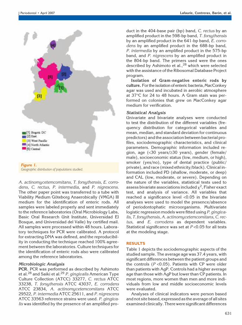
Study PopulationSix hundred and twenty patients were evaluated: 325patients with chronic periodontitis (CP), 158 patientswith aggressive periodontitis (AgP), and 137 healthycontrols. These patients attended the clinics of theschools of dentistry at the El Bosque University(Bogota DC), Santo Tomas University (Bucara-manga: East region), Valle University (Cali: WestPacific region), Metropolitana University (Barranquilla:North Atlantic region), Cartagena University (Carta-
gena: North Atlantic region), Antioquia University(Medellın: Central region), Health Science Institute(Medellın: Central region), and Autonoma de ManizalesUniversity (Manizales: Central region) and were seen inthe private practices of periodontists who participated inthe study between July 2003 and November 2005.
Exclusion criteria included diabetes, periodontaltherapy during the last year, and the use of antimicro-bials or non-steroidal anti-inflammatory drugs in the 6months before clinical examination and sample col-lection. This study was approved by the institutionalreview board of each university, and all participantssigned a written consent form.
Clinical EvaluationMedical history and clinical and radiographic exami-nation were conducted for each patient. A full peri-odontal examination was carried out by periodontistsfrom the different universities. The diagnoses of CPand AgP were made based on criteria defined atthe workshop sponsored by the American Academyof Periodontology (AAP) in 1999.26 Individuals withvarying degrees of gingival inflammation but withoutperiodontal pockets were used as controls. The fol-lowing clinical parameters were recorded: probingdepth (PD), clinical attachment level (CAL), and per-centage of sites with bleeding on probing (BOP). Amarked probe†† was used in all instances. All clinicalresearchers underwent a calibration session on the di-agnosis criteria examining 15 patients, including clin-ical history, radiographs, laboratory examinations,clinical photographs, and a later discussion. Calibra-tion exercises yielded an agreement ‡91% for PD and‡82% for CAL.
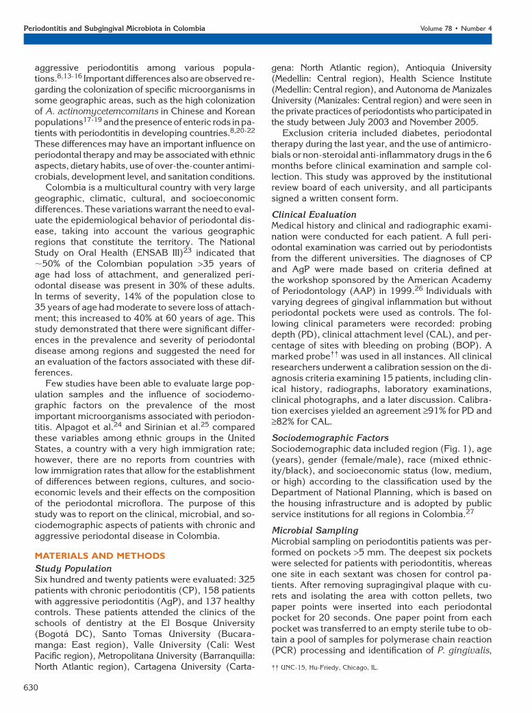
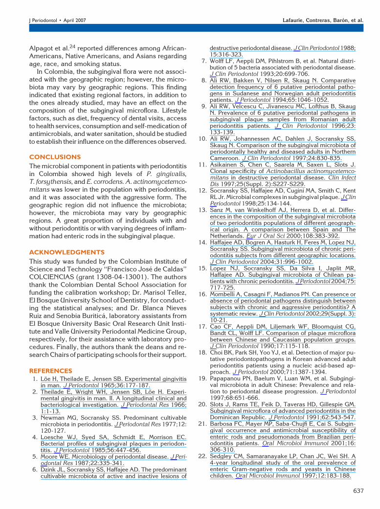
Sociodemographic FactorsSociodemographic data included region (Fig. 1), age(years), gender (female/male), race (mixed ethnic-ity/black), and socioeconomic status (low, medium,or high) according to the classification used by theDepartment of National Planning, which is based onthe housing infrastructure and is adopted by publicservice institutions for all regions in Colombia.27
Microbial SamplingMicrobial sampling on periodontitis patients was per-formed on pockets >5 mm. The deepest six pocketswere selected for patients with periodontitis, whereasone site in each sextant was chosen for control pa-tients. After removing supragingival plaque with cu-rets and isolating the area with cotton pellets, twopaper points were inserted into each periodontalpocket for 20 seconds. One paper point from eachpocket was transferred to an empty sterile tube to ob-tain a pool of samples for polymerase chain reaction(PCR) processing and identification of P. gingivalis,
†† UNC-15, Hu-Friedy, Chicago, IL.
Periodontitis and Subgingival Microbiota in Colombia Volume 78 • Number 4
630
A. actinomycetemcomitans, T. forsythensis, E. corro-dens, C. rectus, P. intermedia, and P. nigrescens.The other paper point was transferred to a tube withViability Medium Goteborg Anaerobically (VMGA) IIImedium for the identification of enteric rods. Allsamples were labeled properly and sent immediatelyto the reference laboratories (Oral Microbiology Labs,Basic Oral Research Unit Institute, Universidad ElBosque, and Universidad del Valle) by certified mail.All samples were processed within 48 hours. Labora-tory techniques for PCR were calibrated. A protocolfor extracting DNA was defined, and the reproducibil-ity in conducting the technique reached 100% agree-ment between the laboratories. Culture techniques forthe identification of enteric rods also were calibratedamong the reference laboratories.
Microbiologic AnalysisPCR. PCR was performed as described by Ashimotoet al.28 and Saiki et al.29 P. gingivalis American TypeCulture Collection (ATCC) 33277, C. rectus ATCC33238, T. forsythensis ATCC 43037, E. corrodensATCC 23834, A. actinomycetemcomitans ATCC29522, P. intermedia ATCC 25611, and P. nigrescensATCC 33563 reference strains were used. P. gingiva-lis was identified by the presence of an amplified pro-
duct in the 404-base pair (bp) band, C. rectus by anamplified product in the 598-bp band, T. forsythensisby an amplified product in the 641-bp band, E. corro-dens by an amplified product in the 688-bp band,P. intermedia by an amplified product in the 575-bpband, and P. nigrescens by an amplified product inthe 804-bp band. The primers used were the onesdescribed by Ashimoto et al.,28 which were selectedwith the assistance of the Ribosomal Database Projectprogram.
Isolation of Gram-negative enteric rods byculture. For the isolationofentericbacteria,MacConkeyagar was used and incubated in aerobic atmosphereat 37�C for 24 to 48 hours. A Gram stain was per-formed on colonies that grew on MacConkey agarmedium for verification.
Statistical AnalysisUnivariate and bivariate analyses were conductedto test the distribution of the different variables (fre-quency distribution for categorical variables andmean, median, and standard deviation for continuouspredictors) and the association between bacterial pro-files, sociodemographic characteristics, and clinicalparameters. Demographic information included re-gion, age (<30 years/‡30 years), gender (female/male), socioeconomic status (low, medium, or high),smoker (yes/no), type of dental practice (public/private), and race (mixed ethnicity/black). Clinical in-formation included PD (shallow, moderate, or deep)and CAL (low, moderate, or severe). Depending onthe nature of the variables, statistical tests used toassess bivariate associations included x2, Fisher exacttest, and analysis of variance. All variables thatreached a significance level <0.05 in the bivariateanalyses were used to model the presence/absenceof periodontophatic microorganisms. Multivariatelogistic regression models were fitted using P. gingiva-lis, T. forsythensis, A. actinomycetemcomitans, C. rec-tus, and E. corrodens as dependent variables.Statistical significance was set at P <0.05 for all testsat the modeling stage.
RESULTS
Table 1 depicts the sociodemographic aspects of thestudied sample. The average age was 37.4 years, withsignificant differences between the patient groups andthe controls (P <0.05). Patients with CP were olderthan patients with AgP. Controls had a higher averageage than those with AgP but lower than CP patients. Inmost regions, more women than men and more indi-viduals from low and middle socioeconomic levelswere evaluated.
Analyses of clinical indicators were person basedand not sitebased, expressedas the averageofall sitesexaminedclinically.There were significantdifferences
Figure 1.Geographic distribution of populations studied.
J Periodontol • April 2007 Lafaurie, Contreras, Baron, et al.
631
in all clinical parameters between patients with peri-odontitis and controls (P <0.0001). There were no dif-ferences between the periodontitis groups, except forPD of sites selected for microbiologic evaluation, andBOP,whichwasmore severe inAgP(P <0.05).The fre-quency of smokers was similar for AgP and controlgroups, and there was a higher frequency of smokersamong CP patients, which was significantly differentfrom the control group (P <0.05) (Table 2).
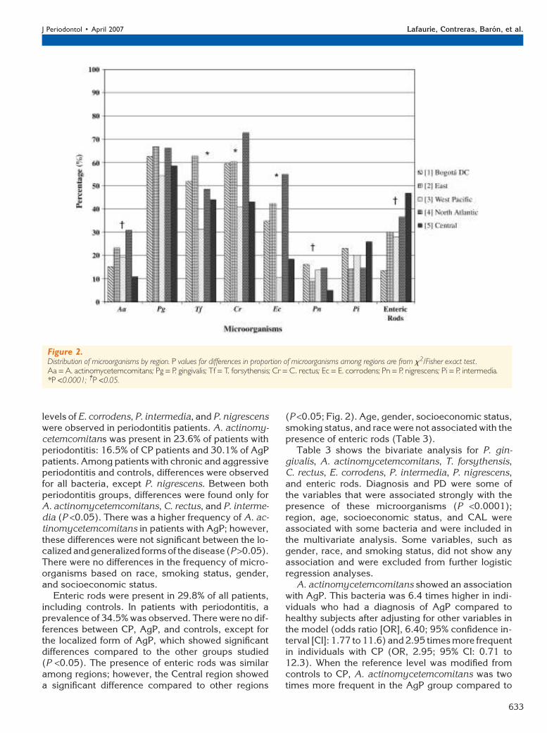
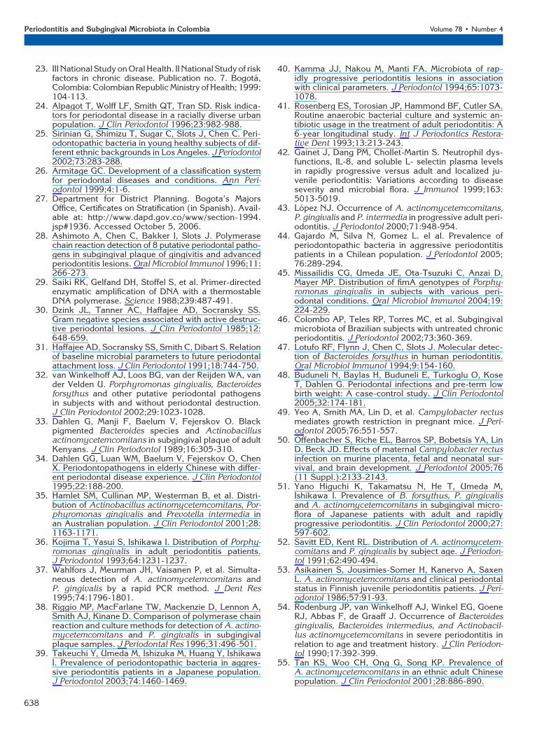
The distribution of bacteria among different regionsin all subjects is shown in Figure 2. With the exceptionof P. gingivalis and P. intermedia, there were signifi-cant differences in all of the bacteria among regions.
The biggest differences were observed for T. forsyth-ensis, C. rectus, and E. corrodens (P <0.0001). A acti-nomycetemcomitans and P. nigrescens also showeddifferences among regions (P <0.05).
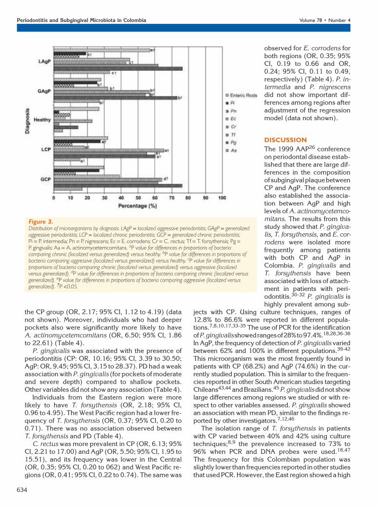
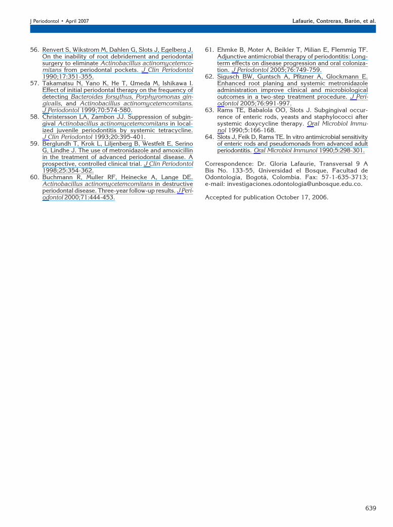
Figure 3 shows the percentage distribution of mi-croorganisms studied among the different diagnoses.P. gingivalis was the most common (71.5%) bacteriain patients with periodontitis. Specifically, it was foundin 68.2% of CP patients, 74.6% of AgP patients, and14.5% of controls. T. forsythensis was found in58.5% of CP patients, 58.1% of AgP patients, and only3.1% of controls. C. rectus was present in 57.5% of pa-tients with periodontitis and 22.7% of controls; lower
Table 1.
Demographic Characteristics by Clinical Diagnosis
Variable CP AgP Healthy/Gingivitis Total
Subjects (N) 325 158 137 620
Age (years, mean – SD) 45.6 – 10.6* 28.0 – 6.8* 28.9 – 10.3 37.4 – 12.9
Gender (%) F: 53.5 F: 62.0 F: 61.3 F: 57.4M: 46.5 M: 38.0 M: 38.7 M: 42.6
Socioeconomic status (%)† 1: 33.9 1: 44.3 1: 40.9 1: 38.12: 55.7 2: 48.1 2: 36.3 2: 49.63: 10.4* 3: 7.6‡ 3: 22.8 3: 12.3
Race (%) M: 95.4 M: 96.2 M: 99.3 M: 96.5B: 4.6‡ B: 3.8 B: 0.7 B: 3.6
Type of practice (%) Pu: 81.2 Pu: 88.0 Pu: 75.4 Pu: 81.7Pr: 18.8 Pr: 12.0‡ Pr: 24.6 Pr : 18.3
M = mixed; B = black; Pu = public; Pr = private.P values derived from t or x
2/Fisher exact tests. Unless otherwise noted, comparisons had no statistical significance.* Comparison versus healthy (P <0.0001).† 1 = low; 2 = middle; 3 = high.‡ Comparison versus healthy (P <0.05).
Table 2.
Clinical Indicators and Smoking Status by Diagnosis
Variable CP AgP Healthy/Gingivitis Average
Subjects (N) 325 158 137 620 (total N)
PD (mm) (mean – SD) 3.9 – 1.1* 3.8 – 0.9* 1.8 – 0.4 3.4 –1.3
CAL (mm) (mean – SD) 4.2 –1.6* 4.0 – 1.4* 1.0 – 1.2 3.4 – 1.9
PD sites sampled (mean – SD) 7.5 – 1.1*† 7.9 – 1.5*† 2.1 – 0.6 6.3 – 2.7
BOP (% positive) 53.50*† 59.30*† 20.00 48.00
Smoking status (% positive) 18.70‡ 14.70 9.20 15.60
P values derived from t or x2/Fisher exact tests. Unless otherwise noted, comparisons had no statistical significance.
* Comparison versus healthy (P <0.0001).† Comparison chronic versus aggressive (P <0.05).‡ Comparison versus healthy (P <0.05).
Periodontitis and Subgingival Microbiota in Colombia Volume 78 • Number 4
632
levels of E. corrodens, P. intermedia, and P. nigrescenswere observed in periodontitis patients. A. actinomy-cetemcomitans was present in 23.6% of patients withperiodontitis: 16.5% of CP patients and 30.1% of AgPpatients. Among patients with chronic and aggressiveperiodontitis and controls, differences were observedfor all bacteria, except P. nigrescens. Between bothperiodontitis groups, differences were found only forA. actinomycetemcomitans, C. rectus, and P. interme-dia (P <0.05). There was a higher frequency of A. ac-tinomycetemcomitans in patients with AgP; however,these differences were not significant between the lo-calized and generalized forms of the disease (P >0.05).There were no differences in the frequency of micro-organisms based on race, smoking status, gender,and socioeconomic status.
Enteric rods were present in 29.8% of all patients,including controls. In patients with periodontitis, aprevalence of 34.5% was observed. There were no dif-ferences between CP, AgP, and controls, except forthe localized form of AgP, which showed significantdifferences compared to the other groups studied(P <0.05). The presence of enteric rods was similaramong regions; however, the Central region showeda significant difference compared to other regions
(P <0.05; Fig. 2). Age, gender, socioeconomic status,smoking status, and race were not associated with thepresence of enteric rods (Table 3).
Table 3 shows the bivariate analysis for P. gin-givalis, A. actinomycetemcomitans, T. forsythensis,C. rectus, E. corrodens, P. intermedia, P. nigrescens,and enteric rods. Diagnosis and PD were some ofthe variables that were associated strongly with thepresence of these microorganisms (P <0.0001);region, age, socioeconomic status, and CAL wereassociated with some bacteria and were included inthe multivariate analysis. Some variables, such asgender, race, and smoking status, did not show anyassociation and were excluded from further logisticregression analyses.
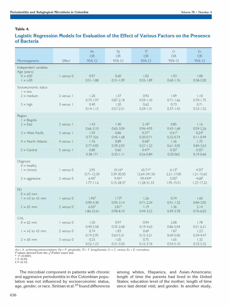
A. actinomycetemcomitans showed an associationwith AgP. This bacteria was 6.4 times higher in indi-viduals who had a diagnosis of AgP compared tohealthy subjects after adjusting for other variables inthe model (odds ratio [OR], 6.40; 95% confidence in-terval [CI]: 1.77 to 11.6) and 2.95 times more frequentin individuals with CP (OR, 2.95; 95% CI: 0.71 to12.3). When the reference level was modified fromcontrols to CP, A. actinomycetemcomitans was twotimes more frequent in the AgP group compared to
Figure 2.Distribution of microorganisms by region. P values for differences in proportion of microorganisms among regions are from x2/Fisher exact test.Aa = A. actinomycetemcomitans; Pg = P. gingivalis; Tf = T. forsythensis; Cr = C. rectus; Ec = E. corrodens; Pn = P. nigrescens; Pi = P. intermedia.*P <0.0001; †P <0.05.
J Periodontol • April 2007 Lafaurie, Contreras, Baron, et al.
633
the CP group (OR, 2.17; 95% CI, 1.12 to 4.19) (datanot shown). Moreover, individuals who had deeperpockets also were significantly more likely to haveA. actinomycetemcomitans (OR, 6.50; 95% CI, 1.86to 22.61) (Table 4).
P. gingivalis was associated with the presence ofperiodontitis (CP: OR, 10.16; 95% CI, 3.39 to 30.50;AgP: OR, 9.45; 95% CI, 3.15 to 28.37). PD had a weakassociation with P. gingivalis (for pockets of moderateand severe depth) compared to shallow pockets.Other variables did not show any association (Table 4).
Individuals from the Eastern region were morelikely to have T. forsythensis (OR, 2.18; 95% CI,0.96 to 4.95). The West Pacific region had a lower fre-quency of T. forsythensis (OR, 0.37; 95% CI, 0.20 to0.71). There was no association observed betweenT. forsythensis and PD (Table 4).
C. rectus was more prevalent in CP (OR, 6.13; 95%CI, 2.21 to 17.00) and AgP (OR, 5.50; 95% CI, 1.95 to15.51), and its frequency was lower in the Central(OR, 0.35; 95% CI, 0.20 to 062) and West Pacific re-gions (OR, 0.41; 95% CI, 0.22 to 0.74). The same was
observed for E. corrodens forboth regions (OR, 0.35; 95%CI, 0.19 to 0.66 and OR,0.24; 95% CI, 0.11 to 0.49,respectively) (Table 4). P. in-termedia and P. nigrescensdid not show important dif-ferences among regions afteradjustment of the regressionmodel (data not shown).
DISCUSSION
The 1999 AAP26 conferenceon periodontal disease estab-lished that there are large dif-ferences in the compositionofsubgingivalplaquebetweenCP and AgP. The conferencealso established the associa-tion between AgP and highlevels of A. actinomycetemco-mitans. The results from thisstudy showed that P. gingiva-lis, T. forsythensis, and E. cor-rodens were isolated morefrequently among patientswith both CP and AgP inColombia. P. gingivalis andT. forsythensis have beenassociated with loss of attach-ment in patients with peri-odontitis.30-32 P. gingivalis ishighly prevalent among sub-
jects with CP. Using culture techniques, ranges of12.8% to 86.6% were reported in different popula-tions.7,8,10,17,33-35 The use of PCR for the identificationofP.gingivalisshowedrangesof28%to97.4%.18,28,36-38
In AgP, the frequency of detection of P. gingivalis variedbetween 62% and 100% in different populations.39-42
This microorganism was the most frequently found inpatients with CP (68.2%) and AgP (74.6%) in the cur-rently studied population. This is similar to the frequen-cies reported in other South American studies targetingChileans43,44 andBrazilians.45 P.gingivalisdidnot showlarge differences among regions we studied or with re-spect to other variables assessed. P. gingivalis showedan association with mean PD, similar to the findings re-ported by other investigators.7,12,46
The isolation range of T. forsythensis in patientswith CP varied between 40% and 42% using culturetechniques;8,9 the prevalence increased to 73% to96% when PCR and DNA probes were used.18,47
The frequency for this Colombian population wasslightly lower than frequencies reported inother studiesthat used PCR. However, the East region showed a high
Figure 3.Distribution of microorganisms by diagnosis. LAgP = localized aggressive periodontitis; GAgP = generalizedaggressive periodontitis; LCP = localized chronic periodontitis; GCP = generalized chronic periodontitis;Pi = P. intermedia; Pn = P. nigrescens; Ec = E. corrodens; Cr = C. rectus; Tf = T. forsythensis; Pg =P. gingivalis; Aa = A. actinomycetemcomitans. aP value for differences in proportions of bacteriacomparing chronic (localized versus generalized) versus healthy. bP value for differences in proportions ofbacteria comparing aggressive (localized versus generalized) versus healthy. cP value for differences inproportions of bacteria comparing chronic (localized versus generalized) versus aggressive (localizedversus generalized). dP value for differences in proportions of bacteria comparing chronic (localized versusgeneralized). eP value for differences in proportions of bacteria comparing aggressive (localized versusgeneralized). †P <0.05.
Periodontitis and Subgingival Microbiota in Colombia Volume 78 • Number 4
634
frequency of this microorganism compared to other re-gions. This microorganism was highly variable amongregions and did not show any association with PD. Fu-ture studies should beconducted toestablishmore pre-cisely the microorganism’s behavior in Colombianpatients with periodontitis.
C. rectus showed a high frequency in the populationwith periodontitis and among controls. Lately, the im-portance of chronic infections associated with C. rec-tus has been of interest. Recently, Buduneli et al.48
reported that C. rectus may have a role in increasingthe risk for preterm low birth weight. Yeo et al.49 foundthat remote subcutaneous maternal C. rectus infec-tion increased fetal growth restriction in a mousemodel. Offenbacher et al.50 also found that maternalC. rectus infection induced placental inflammation aswell asconcomitant increases in fetal brain interferon-gin a mouse model.
In the present study, A. actinomycetemcomitanswas observed two times more frequently in AgPthan in CP. The levels of A. actinomycetemcomitansreported in periodontitis vary among populations. InAgP, A. actinomycetemcomitans varied between3% and 53% among Europeans and Americans,40-42
Japanese,39,51 and Chileans.43,44 Isolates of A. acti-nomycetemcomitans in CP varied between 8% and57% in Americans and Europeans7,28,37,38,52-54 andreached 28.5% to 40% in African countries.8,10,33
The highest frequencies were reported in Chineseand Koreans, with a range between 63% and
83%.18,34,55 This study demonstrated that the pres-ence of A. actinomycetemcomitans varied between16.5% for CP and 30.1% for AgP. These values arewithin the range of North American, European, andother South American populations,44 but are lowerthan among Asians and Africans.
Sociodemographic variables did not influence thepresence of A. actinomycetemcomitans. A significantassociation was observed between this microorgan-ism, AgP, and PD, whereas a weaker associationwas observed with CP. A systematic review conductedby Mombelli et al.16 showed that the presence orabsence of A. actinomycetemcomitans did not dis-criminate between patients with AgP and CP. A. acti-nomycetemcomitans has shown a low response toscaling and root planing.56,57 Several antimicrobialprotocols administered with periodontal therapy havebeen implemented for the elimination of the periodon-tal pockets associated with A. actinomycetemcomi-tans;58-62 therefore, it was important to study itspresence in this population.
A great percentage of the population showed thepresence of enteric rods in subgingival plaque, includ-ing the population without periodontitis. Similar fre-quencies were reported among Brazilians.21,46 Highlevels of resistance to antimicrobials used in peri-odontal treatment have been reported for entericrods.21,63,64 Further studies are required in order toclarify the effect of enteric rods on clinical parametersand response to periodontal treatment.
Table 3.
Bivariate Analysis of Demographic and Clinical Factors
Variable Aa Pg Tf Ent Rod Cr Ec Pn Pi
Socioeconomic status 0.035† 0.077 0.183 0.36 0.51 0.42 0.5 0.28
Type of practice 0.447 0.128 0.01† 0.35 0.49 0.08 0.86 0.7
Region 0.004† 0.309 0.0002* <0.0001* <0.0001* <0.0001* 0.03† 0.17
Diagnosis <0.0001* <0.0001* <0.0001* 0.7 <0.0001* <0.0001* 0.87 <0.0001*
Gender 0.081 0.312 0.347 0.41 0.97 0.05* 0.49 0.77
Age 0.385 0.002† <0.0001* 0.26 0.001† 0.06 0.46 0.01
Race 0.248 0.1 0.741 0.84 0.49 0.11 0.1 0.02
Smoker 0.518 0.109 0.103 0.74 0.75 0.1 0.11 0.006†
PD <0.0001* <0.0001* <0.0001* 0.94 <0.0001* <0.0001* 0.72 0.008†
PD sites sampled 0.001† <0.0001* <0.0001* 0.29 <0.0001* <0.0001* 0.56 <0.0001*
CAL 0.0006† <0.0001* <0.0001* 0.13 <0.0001* <0.0001* 0.93 <0.0001*
Aa = A. actinomycetemcomitans; Pg = P. gingivalis; Tf = T. forsythensis; Ent Rod = enteric rods; Cr = C. rectus; Ec = E. corrodens; Pi = P. intermedia; Pn = P.nigrescens.All variables are categorized based on levels used for logistic regression models. P values derived from the x
2/Fisher exact test.* P <0.0001.† P <0.05.
J Periodontol • April 2007 Lafaurie, Contreras, Baron, et al.
635
The microbial component in patients with chronicand aggressive periodontitis in this Colombian popu-lation was not influenced by socioeconomic status,age, gender, or race. Sirinian et al.25 found differences
among whites, Hispanics, and Asian-Americans;length of time the parents had lived in the UnitedStates; education level of the mother; length of timesince last dental visit; and gender. In another study,
Table 4.
Logistic Regression Models for Evaluation of the Effect of Various Factors on the Presenceof Bacteria
Microorganisms Effect
Aa
OR
95% CI
Pg
OR
95% CI
Tf
OR
95% CI
Cr
OR
95% CI
Ec
OR
95% CI
Independent variablesAge (years)
0 = £30 1 versus 0 0.97 0.60 1.02 1.03 1.081 = >30 0.51-1.88 0.31-1.09 0.55-1.89 0.60-1.76 0.58-2.00
Socioeconomic status1 = low2 = medium 2 versus 1 1.20 1.37 0.92 1.09 1.10
0.73-1.97 0.87-2.18 0.59-1.43 0.71-1.66 0.70-1.753 = high 3 versus 1 0.40 1.20 0.62 0.73 0.71
0.14-1.15 0.57-2.51 0.29-1.31 0.37-1.45 0.33-1.52
Region1 = Bogota2 = East 2 versus 1 1.43 1.40 2.18‡ 0.85 1.16
0.66-3.10 0.65-3.04 0.96-4.95 0.43-1.68 0.59-2.263 = West Pacific 3 versus 1 1.59 0.86 0.37† 0.41† 0.24†
0.77-326 0.45-1.68 0.20-0.71 0.22-0.74 0.11-0.494 = North Atlantic 4 versus 1 1.76 0.89 0.58‡ 1.36 1.75
0.77-4.05 0.39-2.03 0.27-1.23 0.61-3.02 0.84-3.635 = Central 5 versus 1 0.80 0.60 0.47* 0.35† 0.35†
0.38-171 0.33-1.11 0.26-0.84 0.20-062 0.19-0.66
Diagnosis0 = healthy1 = chronic 1 versus 0 2.95 10.16* 65.71* 6.13† 4.35†
0.71-12.30 3.39-30.50 12.64-341.50 2.21-17.00 1.21-15.652 = aggressive 2 versus 0 6.40† 9.45* 59.434* 5.50† 4.68†
1.77-11.6 3.15-28.37 11.28-31.33 1.95-15.51 1.27-17.22
PD0 = £3 mm1 = >3 to <5 mm 1 versus 0 1.96‡ 1.73‡ 1.26 0.74 1.60
0.90-4.30 0.95-3.14 0.71-2.24 0.41-1.32 0.84-3.052 = ‡5 mm 2 versus 0 6.50† 2.81‡ 1.19 1.36 2.14
1.86-22.61 0.98-8.10 0.44-3.22 0.49-3.78 0.76-6.02
CAL0 = £2 mm 1 versus 0 1.50 0.97 0.94 2.08 1.78
0.40-5.58 0.35-2.68 0.19-4.63 0.86-5.04 0.51-6.211 = >2 to <5 mm 2 versus 0 0.74 1.83 0.69 1.87 1.23
0.19-2.95 0.63-5.31 0.15-3.21 0.69-5.06 0.34-4.472 = ‡5 mm 3 versus 0 0.25 1.05 0.75 1.65 1.32
0.52-1.23 0.31-3.50 0.15-3.76 0.54-5.10 0.33-5.32
Aa = A. actinomycetemcomitans; Pg = P. gingivalis; Tf = T. forsythensis; Cr = C. rectus; Ec = E. corrodens.P values derived from the x
2/Fisher exact test.* P <0.0001.† P <0.05.‡ P <0.10.
Periodontitis and Subgingival Microbiota in Colombia Volume 78 • Number 4
636
Alpagot et al.24 reported differences among African-Americans, Native Americans, and Asians regardingage, race, and smoking status.
In Colombia, the subgingival flora were not associ-ated with the geographic region; however, the micro-biota may vary by geographic regions. This findingindicated that existing regional factors, in addition tothe ones already studied, may have an effect on thecomposition of the subgingival microflora. Lifestylefactors, such as diet, frequency of dental visits, accessto health services, consumption and self-medication ofantimicrobials, and water sanitation, should be studiedto establish their influence on the differences observed.
CONCLUSIONS
The microbial component in patients with periodontitisin Colombia showed high levels of P. gingivalis,T. forsythensis, and E. corrodens. A. actinomycetemco-mitans was lower in the population with periodontitis,and it was associated with the aggressive form. Thegeographic region did not influence the microbiota;however, the microbiota may vary by geographicregions. A great proportion of individuals with andwithout periodontitis or with varying degrees of inflam-mation had enteric rods in the subgingival plaque.
ACKNOWLEDGMENTS
This study was funded by the Colombian Institute ofScience and Technology ‘‘Francisco Jose de Caldas’’COLCIENCIAS (grant 1308-04-13001). The authorsthank the Colombian Dental School Association forfunding the calibration workshop; Dr. Marisol Tellez,El Bosque University School of Dentistry, for conduct-ing the statistical analyses; and Dr. Blanca NievesRuiz and Senobia Buritica, laboratory assistants fromEl Bosque University Basic Oral Research Unit Insti-tute and Valle University Periodontal Medicine Group,respectively, for their assistance with laboratory pro-cedures. Finally, the authors thank the deans and re-search Chairs of participating schools for their support.
REFERENCES1. Loe H, Theilade E, Jensen SB. Experimental gingivitis
in man. J Periodontol 1965;36:177-187.2. Theilade E, Wright WH, Jensen SB, Loe H. Experi-
mental gingivitis in man. II. A longitudinal clinical andbacteriological investigation. J Periodontal Res 1966;1:1-13.
3. Newman MG, Socransky SS. Predominant cultivablemicrobiota in periodontitis. J Periodontal Res 1977;12:120-127.
4. Loesche WJ, Syed SA, Schmidt E, Morrison EC.Bacterial profiles of subgingival plaques in periodon-titis. J Periodontol 1985;56:447-456.
5. Moore WE. Microbiology of periodontal disease. J Peri-odontal Res 1987;22:335-341.
6. Dzink JL, Socransky SS, Haffajee AD. The predominantcultivable microbiota of active and inactive lesions of
destructive periodontal disease. J Clin Periodontol 1988;15:316-323.
7. Wolff LF, Aeppli DM, Pihlstrom B, et al. Natural distri-bution of 5 bacteria associated with periodontal disease.J Clin Periodontol 1993;20:699-706.
8. Ali RW, Bakken V, Nilsen R, Skaug N. Comparativedetection frequency of 6 putative periodontal patho-gens in Sudanese and Norwegian adult periodontitispatients. J Periodontol 1994;65:1046-1052.
9. Ali RW, Velcescu C, Jivanescu MC, Lofthus B, SkaugN. Prevalence of 6 putative periodontal pathogens insubgingival plaque samples from Romanian adultperiodontitis patients. J Clin Periodontol 1996;23:133-139.
10. Ali RW, Johannessen AC, Dahlen J, Socransky SS,Skaug N. Comparison of the subgingival microbiota ofperiodontally healthy and diseased adults in NorthernCameroon. J Clin Periodontol 1997;24:830-835.
11. Asikainen S, Chen C, Saarela M, Saxen L, Slots J.Clonal specificity of Actinobacillus actinomycetemco-mitans in destructive periodontal disease. Clin InfectDis 1997;25(Suppl. 2):S227-S229.
12. Socransky SS, Haffajee AD, Cugini MA, Smith C, KentRL Jr. Microbial complexes in subgingival plaque. J ClinPeriodontol 1998;25:134-144.
13. Sanz M, van Winkelhoff AJ, Herrera D, et al. Differ-ences in the composition of the subgingival microbiotaof two periodontitis populations of different geograph-ical origin. A comparison between Spain and TheNetherlands. Eur J Oral Sci 2000;108:383-392.
14. Haffajee AD, Bogren A, Hasturk H, Feres M, Lopez NJ,Socransky SS. Subgingival microbiota of chronic peri-odontitis subjects from different geographic locations.J Clin Periodontol 2004;31:996-1002.
15. Lopez NJ, Socransky SS, Da Silva I, Japlit MR,Haffajee AD. Subgingival microbiota of Chilean pa-tients with chronic periodontitis. J Periodontol 2004;75:717-725.
16. Mombelli A, Casagni F, Madianos PN. Can presence orabsence of periodontal pathogens distinguish betweensubjects with chronic and aggressive periodontitis? Asystematic review. J Clin Periodontol 2002;29(Suppl. 3):10-21.
17. Cao CF, Aeppli DM, Liljemark WF, Bloomquist CG,Bandt CL, Wolff LF. Comparison of plaque microflorabetween Chinese and Caucasian population groups.J Clin Periodontol 1990;17:115-118.
18. Choi BK, Park SH, Yoo YJ, et al. Detection of major pu-tative periodontopathogens in Korean advanced adultperiodontitis patients using a nucleic acid-based ap-proach. J Periodontol 2000;71:1387-1394.
19. Papapanou PN, Baelum V, Luan WM, et al. Subgingi-val microbiota in adult Chinese: Prevalence and rela-tion to periodontal disease progression. J Periodontol1997;68:651-666.
20. Slots J, Rams TE, Feik D, Taveras HD, Gillespie GM.Subgingival microflora of advanced periodontitis in theDominican Republic. J Periodontol 1991;62:543-547.
21. Barbosa FC, Mayer MP, Saba-Chujfi E, Cai S. Subgin-gival occurrence and antimicrobial susceptibility ofenteric rods and pseudomonads from Brazilian peri-odontitis patients. Oral Microbiol Immunol 2001;16:306-310.
22. Sedgley CM, Samaranayake LP, Chan JC, Wei SH. A4-year longitudinal study of the oral prevalence ofenteric Gram-negative rods and yeasts in Chinesechildren. Oral Microbiol Immunol 1997;12:183-188.
J Periodontol • April 2007 Lafaurie, Contreras, Baron, et al.
637
23. III National Study onOral Health. IINational Study of riskfactors in chronic disease. Publication no. 7. Bogota,Colombia: Colombian Republic Ministry of Health; 1999:104-113.
24. Alpagot T, Wolff LF, Smith QT, Tran SD. Risk indica-tors for periodontal disease in a racially diverse urbanpopulation. J Clin Periodontol 1996;23:982-988.
25. Sirinian G, Shimizu T, Sugar C, Slots J, Chen C. Peri-odontopathic bacteria in young healthy subjects of dif-ferent ethnic backgrounds in Los Angeles. J Periodontol2002;73:283-288.
26. Armitage GC. Development of a classification systemfor periodontal diseases and conditions. Ann Peri-odontol 1999;4:1-6.
27. Department for District Planning. Bogota’s MajorsOffice, Certificates on Stratification (in Spanish). Avail-able at: http://www.dapd.gov.co/www/section-1994.jsp#1936. Accessed October 5, 2006.
28. Ashimoto A, Chen C, Bakker I, Slots J. Polymerasechain reaction detection of 8 putative periodontal patho-gens in subgingival plaque of gingivitis and advancedperiodontitis lesions. Oral Microbiol Immunol 1996;11:266-273.
29. Saiki RK, Gelfand DH, Stoffel S, et al. Primer-directedenzymatic amplification of DNA with a thermostableDNA polymerase. Science 1988;239:487-491.
30. Dzink JL, Tanner AC, Haffajee AD, Socransky SS.Gram negative species associated with active destruc-tive periodontal lesions. J Clin Periodontol 1985;12:648-659.
31. Haffajee AD, Socransky SS, Smith C, Dibart S. Relationof baseline microbial parameters to future periodontalattachment loss. J Clin Periodontol 1991;18:744-750.
32. van Winkelhoff AJ, Loos BG, van der Reijden WA, vander Velden U. Porphyromonas gingivalis, Bacteroidesforsythus and other putative periodontal pathogensin subjects with and without periodontal destruction.J Clin Periodontol 2002;29:1023-1028.
33. Dahlen G, Manji F, Baelum V, Fejerskov O. Blackpigmented Bacteroides species and Actinobacillusactinomycetemcomitans in subgingival plaque of adultKenyans. J Clin Periodontol 1989;16:305-310.
34. Dahlen GG, Luan WM, Baelum V, Fejerskov O, ChenX. Periodontopathogens in elderly Chinese with differ-ent periodontal disease experience. J Clin Periodontol1995;22:188-200.
35. Hamlet SM, Cullinan MP, Westerman B, et al. Distri-bution of Actinobacillus actinomycetemcomitans, Por-phyromonas gingivalis and Prevotella intermedia inan Australian population. J Clin Periodontol 2001;28:1163-1171.
36. Kojima T, Yasui S, Ishikawa I. Distribution of Porphy-romonas gingivalis in adult periodontitis patients.J Periodontol 1993;64:1231-1237.
37. Wahlfors J, Meurman JH, Vaisanen P, et al. Simulta-neous detection of A. actinomycetemcomitans andP. gingivalis by a rapid PCR method. J Dent Res1995;74:1796-1801.
38. Riggio MP, MacFarlane TW, Mackenzie D, Lennon A,Smith AJ, Kinane D. Comparison of polymerase chainreaction and culture methods for detection of A. actino-mycetemcomitans and P. gingivalis in subgingivalplaque samples. J Periodontal Res 1996;31:496-501.
39. Takeuchi Y, Umeda M, Ishizuka M, Huang Y, IshikawaI. Prevalence of periodontopathic bacteria in aggres-sive periodontitis patients in a Japanese population.J Periodontol 2003;74:1460-1469.
40. Kamma JJ, Nakou M, Manti FA. Microbiota of rap-idly progressive periodontitis lesions in associationwith clinical parameters. J Periodontol 1994;65:1073-1078.
41. Rosenberg ES, Torosian JP, Hammond BF, Cutler SA.Routine anaerobic bacterial culture and systemic an-tibiotic usage in the treatment of adult periodontitis: A6-year longitudinal study. Int J Periodontics Restora-tive Dent 1993;13:213-243.
42. Gainet J, Dang PM, Chollet-Martin S. Neutrophil dys-functions, IL-8, and soluble L- selectin plasma levelsin rapidly progressive versus adult and localized ju-venile periodontitis: Variations according to diseaseseverity and microbial flora. J Immunol 1999;163:5013-5019.
43. Lopez NJ. Occurrence of A. actinomycetemcomitans,P. gingivalis and P. intermedia in progressive adult peri-odontitis. J Periodontol 2000;71:948-954.
44. Gajardo M, Silva N, Gomez L. el al. Prevalence ofperiodontopathic bacteria in aggressive periodontitispatients in a Chilean population. J Periodontol 2005;76:289-294.
45. Missailidis CG, Umeda JE, Ota-Tsuzuki C, Anzai D,Mayer MP. Distribution of fimA genotypes of Porphy-romonas gingivalis in subjects with various peri-odontal conditions. Oral Microbiol Immunol 2004;19:224-229.
46. Colombo AP, Teles RP, Torres MC, et al. Subgingivalmicrobiota of Brazilian subjects with untreated chronicperiodontitis. J Periodontol 2002;73:360-369.
47. Lotufo RF, Flynn J, Chen C, Slots J. Molecular detec-tion of Bacteroides forsythus in human periodontitis.Oral Microbiol Immunol 1994;9:154-160.
48. Buduneli N, Baylas H, Buduneli E, Turkoglu O, KoseT, Dahlen G. Periodontal infections and pre-term lowbirth weight: A case-control study. J Clin Periodontol2005;32:174-181.
49. Yeo A, Smith MA, Lin D, et al. Campylobacter rectusmediates growth restriction in pregnant mice. J Peri-odontol 2005;76:551-557.
50. Offenbacher S, Riche EL, Barros SP, Bobetsis YA, LinD, Beck JD. Effects of maternal Campylobacter rectusinfection on murine placenta, fetal and neonatal sur-vival, and brain development. J Periodontol 2005;76(11 Suppl.):2133-2143.
51. Yano Higuchi K, Takamatsu N, He T, Umeda M,Ishikawa I. Prevalence of B. forsythus, P. gingivalisand A. actinomycetemcomitans in subgingival micro-flora of Japanese patients with adult and rapidlyprogressive periodontitis. J Clin Periodontol 2000;27:597-602.
52. Savitt ED, Kent RL. Distribution of A. actinomycetem-comitans and P. gingivalis by subject age. J Periodon-tol 1991;62:490-494.
53. Asikainen S, Jousimies-Somer H, Kanervo A, SaxenL. A. actinomycetemcomitans and clinical periodontalstatus in Finnish juvenile periodontitis patients. J Peri-odontol 1986;57:91-93.
54. Rodenburg JP, van Winkelhoff AJ, Winkel EG, GoeneRJ, Abbas F, de Graaff J. Occurrence of Bacteroidesgingivalis, Bacteroides intermedius, and Actinobacil-lus actinomycetemcomitans in severe periodontitis inrelation to age and treatment history. J Clin Periodon-tol 1990;17:392-399.
55. Tan KS, Woo CH, Ong G, Song KP. Prevalence ofA. actinomycetemcomitans in an ethnic adult Chinesepopulation. J Clin Periodontol 2001;28:886-890.
Periodontitis and Subgingival Microbiota in Colombia Volume 78 • Number 4
638
56. Renvert S, Wikstrom M, Dahlen G, Slots J, Egelberg J.On the inability of root debridement and periodontalsurgery to eliminate Actinobacillus actinomycetemco-mitans from periodontal pockets. J Clin Periodontol1990;17:351-355.
57. Takamatsu N, Yano K, He T, Umeda M, Ishikawa I.Effect of initial periodontal therapy on the frequency ofdetecting Bacteroides forsythus, Porphyromonas gin-givalis, and Actinobacillus actinomycetemcomitans.J Periodontol 1999;70:574-580.
58. Christersson LA, Zambon JJ. Suppression of subgin-gival Actinobacillus actinomycetemcomitans in local-ized juvenile periodontitis by systemic tetracycline.J Clin Periodontol 1993;20:395-401.
59. Berglundh T, Krok L, Liljenberg B, Westfelt E, SerinoG, Lindhe J. The use of metronidazole and amoxicillinin the treatment of advanced periodontal disease. Aprospective, controlled clinical trial. J Clin Periodontol1998;25:354-362.
60. Buchmann R, Muller RF, Heinecke A, Lange DE.Actinobacillus actinomycetemcomitans in destructiveperiodontal disease. Three-year follow-up results. J Peri-odontol 2000;71:444-453.
61. Ehmke B, Moter A, Beikler T, Milian E, Flemmig TF.Adjunctive antimicrobial therapy of periodontitis: Long-term effects on disease progression and oral coloniza-tion. J Periodontol 2005;76:749-759.
62. Sigusch BW, Guntsch A, Pfitzner A, Glockmann E.Enhanced root planing and systemic metronidazoleadministration improve clinical and microbiologicaloutcomes in a two-step treatment procedure. J Peri-odontol 2005;76:991-997.
63. Rams TE, Babalola OO, Slots J. Subgingival occur-rence of enteric rods, yeasts and staphylococci aftersystemic doxycycline therapy. Oral Microbiol Immu-nol 1990;5:166-168.
64. Slots J, Feik D, Rams TE. In vitro antimicrobial sensitivityof enteric rods and pseudomonads from advanced adultperiodontitis. Oral Microbiol Immunol 1990;5:298-301.
Correspondence: Dr. Gloria Lafaurie, Transversal 9 ABis No. 133-55, Universidad el Bosque, Facultad deOdontologıa, Bogota, Colombia. Fax: 57-1-635-3713;e-mail: [email protected].
Accepted for publication October 17, 2006.
J Periodontol • April 2007 Lafaurie, Contreras, Baron, et al.
639


