Embed Size (px)
Citation preview
Stroke is available at www.ahajournals.org/journal/str
Stroke
Stroke. 2022;53:00–00. DOI: 10.1161/STROKEAHA.121.037541 March 2022 1
Correspondence to: Shadi Yaghi, MD, FAHA, Department of Neurology, Brown Medical School, 593 Eddy Street APC 5, Providence, RI 02903. Email [email protected] article was sent to Theresa A. Jones, Guest Editor, for review by expert referees, editorial decision, and final disposition.Supplemental Material is available at https://www.ahajournals.org/doi/suppl/10.1161/STROKEAHA.121.037541.For Sources of Funding and Disclosures, see page xxx.
© 2022 American Heart Association, Inc.
ORIGINAL CONTRIBUTION
Direct Oral Anticoagulants Versus Warfarin in the Treatment of Cerebral Venous Thrombosis (ACTION-CVT): A Multicenter International StudyShadi Yaghi , MD; Liqi Shu , MD; Ekaterina Bakradze , MD; Setareh Salehi Omran , MD; James A. Giles , MD; Jordan Y. Amar, MD; Nils Henninger , MD, PhD, Dr med; Marwa Elnazeir, MD; Ava L. Liberman , MD; Khadean Moncrieffe , BS; Jenny Lu , BS; Richa Sharma , MD; Yee Cheng, MD; Adeel S. Zubair, MD; Alexis N. Simpkins , MD, PhD, MSCR; Grace T. Li , MD; Justin Chi Kung , BS; Dezaray Perez , BS; Mirjam Heldner , MD; Adrian Scutelnic , MD; David Seiffge , MD; Bernhard Siepen, MD; Aaron Rothstein , MD; Ossama Khazaal , MD; David Do , MD; Sami Al Kasab , MD; Line Abdul Rahman , MD; Eva A. Mistry , MD; Deborah Kerrigan , MD; Hayden Lafever, BS; Thanh N. Nguyen , MD; Piers Klein ; Hugo Aparicio , MD; Jennifer Frontera , MD; Lindsey Kuohn , BS; Shashank Agarwal , MD; Christoph Stretz , MD; Narendra Kala, MD; Sleiman El Jamal , MD; Alison Chang, BS; Shawna Cutting , MD; Han Xiao , MPH; Adam de Havenon , MD; Varsha Muddasani , MD; Teddy Wu , MD; Duncan Wilson, MD; Amre Nouh, MD; Syed Daniyal Asad , MD; Abid Qureshi , MD; Justin Moore , MD; Pooja Khatri , MD; Yasmin Aziz , MD; Bryce Casteigne , MD; Muhib Khan , MD; Yao Cheng, MD; Brian Mac Grory , MD; Martin Weiss, MD; Dylan Ryan , MD; Maria Cristina Vedovati, MD; Maurizio Paciaroni , MD; James E. Siegler , MD; Scott Kamen , BS; Siyuan Yu , BS; Christopher R. Leon Guerrero , MD; Eugenie Atallah , MD; Gian Marco De Marchis , MD, MSc; Alex Brehm , MD; Tolga Dittrich , MD; Marios Psychogios , MD; Ronald Alvarado-Dyer , MD; Tareq Kass-Hout, MD; Shyam Prabhakaran , MD; Tristan Honda , BS; David S. Liebeskind , MD; Karen Furie MD
BACKGROUND: A small randomized controlled trial suggested that dabigatran may be as effective as warfarin in the treatment of cerebral venous thrombosis (CVT). We aimed to compare direct oral anticoagulants (DOACs) to warfarin in a real-world CVT cohort.
METHODS: This multicenter international retrospective study (United States, Europe, New Zealand) included consecutive patients with CVT treated with oral anticoagulation from January 2015 to December 2020. We abstracted demographics and CVT risk factors, hypercoagulable labs, baseline imaging data, and clinical and radiological outcomes from medical records. We used adjusted inverse probability of treatment weighted Cox-regression models to compare recurrent cerebral or systemic venous thrombosis, death, and major hemorrhage in patients treated with warfarin versus DOACs. We performed adjusted inverse probability of treatment weighted logistic regression to compare recanalization rates on follow-up imaging across the 2 treatments groups.
RESULTS: Among 1025 CVT patients across 27 centers, 845 patients met our inclusion criteria. Mean age was 44.8 years, 64.7% were women; 33.0% received DOAC only, 51.8% received warfarin only, and 15.1% received both treatments at different times. During a median follow-up of 345 (interquartile range, 140–720) days, there were 5.68 recurrent venous thrombosis, 3.77 major hemorrhages, and 1.84 deaths per 100 patient-years. Among 525 patients who met recanalization analysis inclusion criteria, 36.6% had complete, 48.2% had partial, and 15.2% had no recanalization. When compared with warfarin, DOAC treatment was associated with similar risk of recurrent venous thrombosis (aHR, 0.94 [95% CI, 0.51–1.73]; P=0.84), death (aHR, 0.78 [95% CI, 0.22–2.76]; P=0.70), and rate of partial/complete recanalization (aOR, 0.92 [95% CI, 0.48–1.73]; P=0.79), but a lower risk of major hemorrhage (aHR, 0.35 [95% CI, 0.15–0.82]; P=0.02).
Dow
nloaded from http://ahajournals.org by on February 21, 2022
ORIG
INAL
CON
TRIB
UTIO
NYaghi et al DOACs Versus Warfarin in CVT
2 March 2022 Stroke. 2022;53:00–00. DOI: 10.1161/STROKEAHA.121.037541
CONCLUSIONS: In patients with CVT, treatment with DOACs was associated with similar clinical and radiographic outcomes and favorable safety profile when compared with warfarin treatment. Our findings need confirmation by large prospective or randomized studies.
GRAPHIC ABSTRACT: A graphic abstract is available for this article.
Key Words: anticoagulants ◼ contraindications ◼ dabigatran ◼ hemorrhage ◼ venous thrombosis
Cerebral venous thrombosis (CVT) is an uncommon cause of stroke, usually affecting younger patients.1 In the absence of contraindications, parenteral fol-
lowed by oral anticoagulation is the recommended treat-ment.2 Randomized trials3–6 and guidelines7–9 indicate that direct oral anticoagulants (DOACs) are a viable and preferred alternative to warfarin for the treatment of patients with venous thromboembolism (VTE). Although the favorable safety and efficacy of DOACs in VTE treat-ment is frequently extrapolated to patients with CVT, lim-ited data exist to support this approach.
See related article, p XXX
The RESPECT-CVT trial (A Clinical Trial Comparing Efficacy and Safety of Dabigatran Etexilate With War-farin in Patients With Cerebral Venous and Dural Sinus Thrombosis), which randomized 120 patients with CVT to dabigatran versus warfarin, showed no significant dif-ferences in efficacy and safety outcomes between the 2 treatment groups.10 However, this study was underpow-ered to show differences in safety or efficacy between the 2 groups due to the low rates of VTE recurrence and hemorrhagic complications.10 Therefore, it remains uncertain whether DOACs are as safe and effective as warfarin in patients with CVT. A better understanding
of this issue is important since the pathomechanism(s) underlying CVT and VTE and their subsequent risks may differ.11,12
Large randomized controlled trials are challenging to conduct given the low incidence of CVT. Observational, real-world data may help answer whether treatment with DOACs versus warfarin is associated with similar effi-cacy and safety outcomes. In this study, we sought to compare the safety and efficacy of DOACs to warfarin in patients with CVT by collecting observational real-world data from large stroke centers worldwide.
METHODSInstitutional Review Board approval was obtained from each participating center to perform the study. Informed Consent was waived by Institutional Board Review. De-identified data are available upon reasonable request to the corresponding author.
Patient PopulationThe ACTION-CVT (Anticoagulation in the Treatment of Cerebral Venous Thrombosis) study is a multicenter, inter-national (United States, Italy, Switzerland, New Zealand; Figure S1) retrospective observational study that included consecutive adult patients with CVT confirmed on imaging admitted to each of the participating centers over a period of 6 years (January 1, 2015–December 31, 2020). Patients with CVT at each institution were initially identified using ICD-9 (325.0, 437.6, and 671.5) and ICD-10 codes (I67.6) with acceptable sensitivity and specificity.13,14 This was fol-lowed by review of medical records and imaging studies to confirm the diagnosis of CVT.
We excluded patients who were not treated with oral anti-coagulation as well as patients in whom a specific anticoagu-lation strategy (DOAC or warfarin) would typically be used or preferred such as in the setting of antiphospholipid antibody syndrome associated CVT (warfarin is the drug of choice),15 active cancer (DOACs may be preferred over warfarin),16 and pregnancy (oral anticoagulation particularly with warfarin is contraindicated).17 For the recanalization analysis, we excluded patients who underwent endovascular treatment and those who were on both treatments (DOACs and warfarin) prior to completion of follow-up imaging.
Study VariablesThe following variables were collected through medical record review:
Nonstandard Abbreviations and Acronyms
ACTION-CVT Anticoagulation in the Treatment of Cerebral Venous Thrombosis
CVT cerebral venous thrombosisDOAC direct oral anticoagulantsHR hazard ratioIPTW inverse probability of treatment
weightedOR odds ratioRESPECT-CVT A Clinical Trial Comparing Efficacy
and Safety of Dabigatran Etexilate With Warfarin in Patients With Cerebral Venous and Dural Sinus Thrombosis
VTE venous thromboembolism
Dow
nloaded from http://ahajournals.org by on February 21, 2022
ORIGINAL CONTRIBUTIONYaghi et al DOACs Versus Warfarin in CVT
Stroke. 2022;53:00–00. DOI: 10.1161/STROKEAHA.121.037541 March 2022 3
1. Demographic factors: Age, sex, race (White, Black, or other), and ethnicity (Hispanic versus non-Hispanic).
2. CVT risk factors: Body mass index closest to the time of diagnosis, history of prior VTE, head trauma, lumbar punc-ture, mastoiditis/sinusitis within 3 months of CVT diagno-sis, smoking history, oral contraceptive use, delivery within 12 weeks of CVT diagnosis, and family history of venous thrombosis.
3. Clinical variables: Days from first symptoms to diagnosis, duration of oral anticoagulation, days from initiation of anticoagulation to follow-up imaging, and clinical symp-toms at presentation (headache, focal deficit, seizure, coma, etc.).
4. Imaging variables: Brain imaging findings (venous infarct, cerebral edema, or brain hemorrhage) and CVT location (deep venous sinus involvement versus no deep venous sinus involvement).
5. Laboratory variables closest to time of diagnosis: Platelet count, one or more antiphospholipid antibodies present at the time of diagnosis but not meeting criteria for antiphos-pholipid antibody syndrome at the time of evaluation, fac-tor V Leiden mutation, and prothrombin gene mutation.
6. In-hospital treatments: Parenteral anticoagulation (low molecular weight heparin or intravenous heparin), endo-vascular treatment, and neurosurgical treatment (extra-ventricular drain or craniotomy).
Oral Anticoagulant TypeThe primary predictor in this study was oral anticoagulant type (DOAC versus warfarin). Medical records were reviewed for compliance with anticoagulant therapy and international nor-malized ratio checks for patients prescribed warfarin. The times of oral anticoagulation initiation and cessation were recorded. In patients who remained on anticoagulation at the time of data abstraction, the last day of anticoagulation was considered as the last day of follow-up.
OutcomesStudy outcomes were abstracted by individual sites through review of all available medical records including outside hos-pital records at the time of data abstraction. The outcomes were (1) primary outcome: Recurrent venous thrombosis (VTE or CVT) during follow-up. Recurrent CVT included de novo CVT as well as extension of previous CVT occurring while on oral anticoagulation therapy. The inclusion of CVT extension is in line with the recurrent VTE definition in clini-cal trials4,5 and is an important outcome to capture in CVT patients as CVT progression can lead to worsening venous infarction, cerebral edema, hemorrhage, or increased intra-cranial pressure. (2) Imaging outcomes: Recanalization sta-tus on last venous imaging study abstracted from radiology reports (no recanalization, partial recanalization, or complete recanalization). Complete recanalization was defined as full recanalization of the thrombosed vein or sinus without any residual thrombus. Partial recanalization was defined as improved opacification or flow in the affected cerebral sinus or vein, but with residual thrombus present on follow-up imaging. No recanalization was defined as no change or worsening in opacification or flow in the affected cerebral sinus or vein from baseline imaging. (3) Safety outcomes:
Major hemorrhage defined as new or worsening intracranial hemorrhage or major extracranial hemorrhage defined as systemic hemorrhage with at least 2 g/dL drop in hemoglo-bin level or requiring blood transfusion occurring while on oral anticoagulation therapy. Symptomatic intracranial hem-orrhage was defined as any new or worsening intracranial hemorrhage on a follow-up brain imaging study causing new or worsening neurological signs or symptoms. (4) Any death during follow-up. All clinical outcomes were included if they occurred while on oral anticoagulation. Radiographic outcomes were included if the follow-up imaging study was done after initiation of oral anticoagulation.
Analytical PlanData verification was conducted by Drs Yaghi and Shu to ensure data integrity and consistency. Several queries were sent to participating sites regarding predictors, outcomes, and other variables. Missing data was not imputed. We used t test, χ2 test, Fisher exact test, or Wilcoxon rank-sum test (Mann-Whitney 2-sample statistic) as appropriate to com-pare co-variates between those with versus those without 90-day follow-up.
For univariate analyses, patients were divided into 2 groups based on the oral anticoagulant used: strictly warfarin versus strictly DOAC use. Analysis of clinical outcomes used the full data set, and endpoints occurring on oral anticoagulation counted against the drug in use at the time. Radiographic outcomes were only considered in patients using strictly one anticoagulant (DOAC or warfarin) prior to the follow-up venous imaging study. Between-group comparisons were done by t test, χ2 test, Fisher exact test or Wilcoxon rank-sum test (Mann-Whitney 2-sample statistic) as appropriate.
Unweighted and inverse probability of treatment weighted (IPTW) unadjusted and adjusted Cox-regression analyses with cluster frailty were used to test associations between oral anticoagulant type (DOAC versus warfarin) and clinical efficacy and safety outcomes. Patients who switched oral anticoagulant types were considered as crossovers from one arm to the other and analyzed using an as-treated approach. For each arm, the start of follow-up was considered as the time the oral anticoagulant was initiated and subjects were censored if they died, were lost to follow-up, or discontin-ued the oral anticoagulant prior to the outcome of inter-est. For the radiological outcome, we used unadjusted and adjusted binary logistic regression analyses to test associa-tions between anticoagulant type and recanalization status. For the recurrent venous thrombosis outcome, we adjusted for variables associated with recurrent venous thrombosis in prior studies including age, sex, history of VTE,18,19 as well as the presence of one or more positive antiphospholipid anti-bodies that may portend an increased risk based on patho-physiological considerations. For the major hemorrhage and death outcome, we adjusted for predictors of intracranial and extracranial hemorrhage, age, sex, intracranial hemorrhage at baseline20,21 as well as deep CVT location, which is a predictor of poor functional outcome and mortality in CVT. The recana-lization status outcome was adjusted for age, sex, intracranial hemorrhage at baseline, duration of anticoagulation therapy prior to follow-up imaging, and deep CVT location. These variables have either been shown to be directly or indirectly
Dow
nloaded from http://ahajournals.org by on February 21, 2022
ORIG
INAL
CON
TRIB
UTIO
NYaghi et al DOACs Versus Warfarin in CVT
4 March 2022 Stroke. 2022;53:00–00. DOI: 10.1161/STROKEAHA.121.037541
associated with recanalization status in prior studies,22 or are surrogate markers of thrombus burden and thus theoretically would be associated with lower likelihood of recanalization. Additional analyses were performed adding variables that dif-fered between the 2 treatment groups (strictly DOACs versus strictly warfarin) to the models. Fully adjusted models included the prespecified variables above as well as variables that sig-nificantly differed between the 2 groups on univariate analy-sis, and these were used for IPTW weighting and propensity score matching.
Sensitivity analyses included propensity score matching (with and without replacement, caliper 0.05) to test the asso-ciations between anticoagulant type and study outcomes given differences between treatment groups at baseline. For the major hemorrhage outcome, a sensitivity analysis was performed excluding asymptomatic intracranial hemor-rhages from the major hemorrhage outcome. Furthermore, as the recurrent CVT outcome included both CVT extension and de novo CVT, we attempted to investigate the effect of DOACs versus warfarin on each of the 2. Since our study could not differentiate between these 2 outcomes and since in the clinical setting, extension of a previous CVT is likely to occur in the early timeframe after diagnosis, we used time of recurrent CVT as a surrogate marker to identify CVT exten-sion cases. We used Cox regression analyses to compare the effect on DOAC versus warfarin on recurrent CVT occur-ring at 0 to 90 days and >90 days from diagnosis, which rep-resents the typical time frame for recanalization assessment. We also performed the same analyses using the 2-week time cutoff23 as well as the 4-week time cutoff. Additional sensitivity analyses were done for the IPTW models, exclud-ing biologically plausible co-variates that lack supportive data to include described above. For major hemorrhage and recanalization, we excluded the co-variates baseline hemor-rhage and deep venous involvement from the models; for recurrent VTE, we excluded the co-variate positive antiphos-pholipid antibodies from the models. We also ran separate
IPTW models excluding patients with COVID-19 and those without 90-day follow-up.
For Cox regression models, proportionality was assessed using Schoenfeld residuals and parametric survival model was used when proportionality was not met. One year Kaplan-Meier survival curves of recurrent venous thrombosis, major hemor-rhage, and death were plotted using unadjusted models with start of follow-up at the time of oral anticoagulation initiation and censoring at either an event of interest, death, loss of fol-low-up, or discontinuation/switch of oral anticoagulant therapy. Data were analyzed using Stata (version 15.1), and a P<0.05 was considered statistically significant.
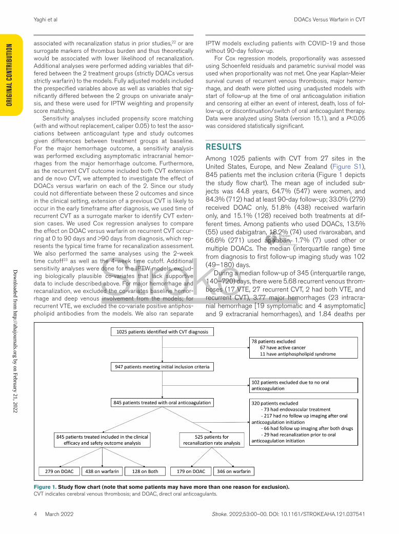
RESULTSAmong 1025 patients with CVT from 27 sites in the United States, Europe, and New Zealand (Figure S1), 845 patients met the inclusion criteria (Figure 1 depicts the study flow chart). The mean age of included sub-jects was 44.8 years, 64.7% (547) were women, and 84.3% (712) had at least 90-day follow-up; 33.0% (279) received DOAC only, 51.8% (438) received warfarin only, and 15.1% (128) received both treatments at dif-ferent times. Among patients who used DOACs, 13.5% (55) used dabigatran, 18.2% (74) used rivaroxaban, and 66.6% (271) used apixaban, 1.7% (7) used other or multiple DOACs. The median (interquartile range) time from diagnosis to first follow-up imaging study was 102 (49–180) days.
During a median follow-up of 345 (interquartile range, 140–720) days, there were 5.68 recurrent venous throm-boses (17 VTE, 27 recurrent CVT, 2 had both VTE, and recurrent CVT), 3.77 major hemorrhages (23 intracra-nial hemorrhage [19 symptomatic and 4 asymptomatic] and 9 extracranial hemorrhages), and 1.84 deaths per
Figure 1. Study flow chart (note that some patients may have more than one reason for exclusion).CVT indicates cerebral venous thrombosis; and DOAC, direct oral anticoagulants.
Dow
nloaded from http://ahajournals.org by on February 21, 2022
ORIGINAL CONTRIBUTIONYaghi et al DOACs Versus Warfarin in CVT
Stroke. 2022;53:00–00. DOI: 10.1161/STROKEAHA.121.037541 March 2022 5
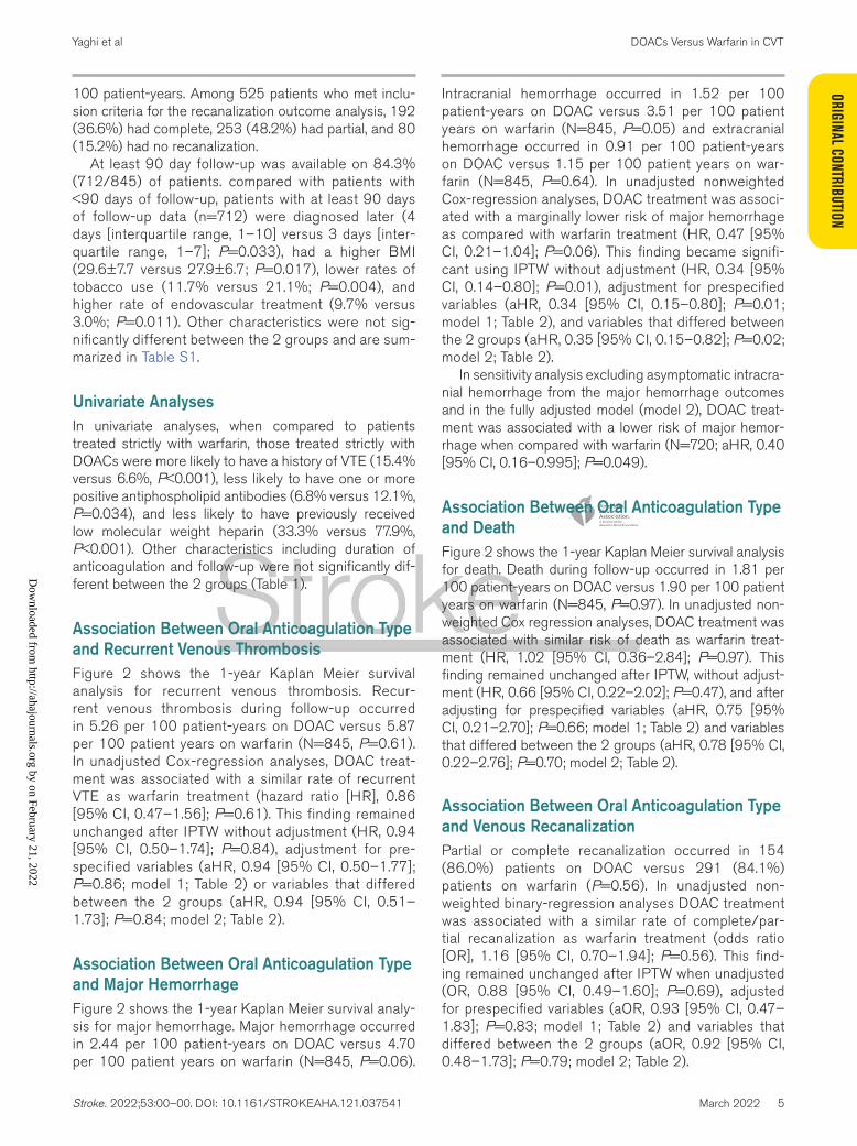
100 patient-years. Among 525 patients who met inclu-sion criteria for the recanalization outcome analysis, 192 (36.6%) had complete, 253 (48.2%) had partial, and 80 (15.2%) had no recanalization.
At least 90 day follow-up was available on 84.3% (712/845) of patients. compared with patients with <90 days of follow-up, patients with at least 90 days of follow-up data (n=712) were diagnosed later (4 days [interquartile range, 1–10] versus 3 days [inter-quartile range, 1–7]; P=0.033), had a higher BMI (29.6±7.7 versus 27.9±6.7; P=0.017), lower rates of tobacco use (11.7% versus 21.1%; P=0.004), and higher rate of endovascular treatment (9.7% versus 3.0%; P=0.011). Other characteristics were not sig-nificantly different between the 2 groups and are sum-marized in Table S1.
Univariate AnalysesIn univariate analyses, when compared to patients treated strictly with warfarin, those treated strictly with DOACs were more likely to have a history of VTE (15.4% versus 6.6%, P<0.001), less likely to have one or more positive antiphospholipid antibodies (6.8% versus 12.1%, P=0.034), and less likely to have previously received low molecular weight heparin (33.3% versus 77.9%, P<0.001). Other characteristics including duration of anticoagulation and follow-up were not significantly dif-ferent between the 2 groups (Table 1).
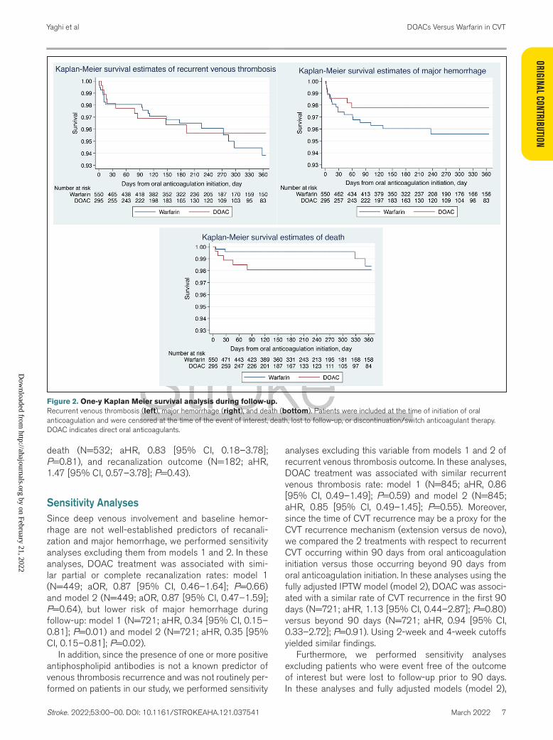
Association Between Oral Anticoagulation Type and Recurrent Venous ThrombosisFigure 2 shows the 1-year Kaplan Meier survival analysis for recurrent venous thrombosis. Recur-rent venous thrombosis during follow-up occurred in 5.26 per 100 patient-years on DOAC versus 5.87 per 100 patient years on warfarin (N=845, P=0.61). In unadjusted Cox-regression analyses, DOAC treat-ment was associated with a similar rate of recurrent VTE as warfarin treatment (hazard ratio [HR], 0.86 [95% CI, 0.47–1.56]; P=0.61). This finding remained unchanged after IPTW without adjustment (HR, 0.94 [95% CI, 0.50–1.74]; P=0.84), adjustment for pre-specified variables (aHR, 0.94 [95% CI, 0.50–1.77]; P=0.86; model 1; Table 2) or variables that differed between the 2 groups (aHR, 0.94 [95% CI, 0.51–1.73]; P=0.84; model 2; Table 2).
Association Between Oral Anticoagulation Type and Major HemorrhageFigure 2 shows the 1-year Kaplan Meier survival analy-sis for major hemorrhage. Major hemorrhage occurred in 2.44 per 100 patient-years on DOAC versus 4.70 per 100 patient years on warfarin (N=845, P=0.06).
Intracranial hemorrhage occurred in 1.52 per 100 patient-years on DOAC versus 3.51 per 100 patient years on warfarin (N=845, P=0.05) and extracranial hemorrhage occurred in 0.91 per 100 patient-years on DOAC versus 1.15 per 100 patient years on war-farin (N=845, P=0.64). In unadjusted nonweighted Cox-regression analyses, DOAC treatment was associ-ated with a marginally lower risk of major hemorrhage as compared with warfarin treatment (HR, 0.47 [95% CI, 0.21–1.04]; P=0.06). This finding became signifi-cant using IPTW without adjustment (HR, 0.34 [95% CI, 0.14–0.80]; P=0.01), adjustment for prespecified variables (aHR, 0.34 [95% CI, 0.15–0.80]; P=0.01; model 1; Table 2), and variables that differed between the 2 groups (aHR, 0.35 [95% CI, 0.15–0.82]; P=0.02; model 2; Table 2).
In sensitivity analysis excluding asymptomatic intracra-nial hemorrhage from the major hemorrhage outcomes and in the fully adjusted model (model 2), DOAC treat-ment was associated with a lower risk of major hemor-rhage when compared with warfarin (N=720; aHR, 0.40 [95% CI, 0.16–0.995]; P=0.049).
Association Between Oral Anticoagulation Type and DeathFigure 2 shows the 1-year Kaplan Meier survival analysis for death. Death during follow-up occurred in 1.81 per 100 patient-years on DOAC versus 1.90 per 100 patient years on warfarin (N=845, P=0.97). In unadjusted non-weighted Cox regression analyses, DOAC treatment was associated with similar risk of death as warfarin treat-ment (HR, 1.02 [95% CI, 0.36–2.84]; P=0.97). This finding remained unchanged after IPTW, without adjust-ment (HR, 0.66 [95% CI, 0.22–2.02]; P=0.47), and after adjusting for prespecified variables (aHR, 0.75 [95% CI, 0.21–2.70]; P=0.66; model 1; Table 2) and variables that differed between the 2 groups (aHR, 0.78 [95% CI, 0.22–2.76]; P=0.70; model 2; Table 2).
Association Between Oral Anticoagulation Type and Venous RecanalizationPartial or complete recanalization occurred in 154 (86.0%) patients on DOAC versus 291 (84.1%) patients on warfarin (P=0.56). In unadjusted non-weighted binary-regression analyses DOAC treatment was associated with a similar rate of complete/par-tial recanalization as warfarin treatment (odds ratio [OR], 1.16 [95% CI, 0.70–1.94]; P=0.56). This find-ing remained unchanged after IPTW when unadjusted (OR, 0.88 [95% CI, 0.49–1.60]; P=0.69), adjusted for prespecified variables (aOR, 0.93 [95% CI, 0.47–1.83]; P=0.83; model 1; Table 2) and variables that differed between the 2 groups (aOR, 0.92 [95% CI, 0.48–1.73]; P=0.79; model 2; Table 2).
Dow
nloaded from http://ahajournals.org by on February 21, 2022
ORIG
INAL
CON
TRIB
UTIO
NYaghi et al DOACs Versus Warfarin in CVT
6 March 2022 Stroke. 2022;53:00–00. DOI: 10.1161/STROKEAHA.121.037541
Propensity Score MatchingPropensity score matching with replacement included 721 patients for primary outcome analysis, 720 patients for safety outcome and for death analyses, and 448 patients for recanalization outcome. In these analy-ses, DOAC treatment was associated with a similar risk of recurrent venous thrombosis (aHR, 0.95 [95% CI, 0.48–1.87]; P=0.88), death (aHR, 0.97 [95% CI,
0.31–2.99]; P=0.95), partial/complete recanalization (aOR, 0.59 [95% CI, 0.26–1.32]; P=0.20), and nonsig-nificantly lower risk of major hemorrhage (aHR, 0.42 [95% CI, 0.16–1.06]; P=0.07) as compared with warfa-rin treatment (Table 2). Using propensity score match-ing without replacement, the findings were similar for all outcomes: recurrent venous thrombosis (N=534; aHR, 0.72 [95% CI, 0.32–1.58]; P=0.41), major hemorrhage (N=532; aHR, 0.34 [95% CI, 0.12–0.95]; P=0.04),
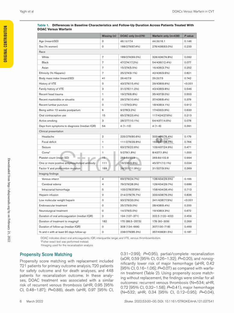
Table 1. Differences in Baseline Characteristics and Follow-Up Duration Across Patients Treated With DOAC Versus Warfarin
Missing (n) DOAC only (n=279) Warfarin only (n=438) P value
Age (mean±SD) 0 46.1±17.4 44.3±16.1 0.146
Sex (% women) 0 188/279(67.4%) 276/438(63.0%) 0.233
Race
White 7 189/274(69.0%) 326/436(74.8%) 0.092
Black 7 47/274(17.2%) 54/436(12.4%) 0.077
Asian 7 15/274(5.5%) 16/436(3.7%) 0.252
Ethnicity (% Hispanic) 7 25/274(9.1%) 42/436(9.6%) 0.821
Body mass index (mean±SD) 41 29.4±7.9 29.2±7.5 0.742
History of VTE 0 43/279(15.4%) 29/438(6.6%) <0.001
Family history of VTE 3 31/276(11.2%) 43/438(9.8%) 0.546
Recent head trauma 1 19/279(6.8%) 35/437(8.0%) 0.553
Recent mastoiditis or sinusitis 0 29/279(10.4%) 37/438(8.4%) 0.379
Recent lumbar puncture 0 11/279(3.9%) 18/438(4.1%) 0.912
Being within 12 weeks postpartum 6 9/278(3.2%) 17/433(3.9%) 0.633
Oral contraceptive use 15 65/278(23.4%) 117/424(27.6%) 0.213
Active smoking 3 28/277(10.1%) 64/437(14.6%) 0.078
Days from symptoms to diagnosis (median IQR) 54 4 (1–10) 4 (1–9) 0.391
Clinical presentation
Headache 2 225/279(80.6%) 333/436(76.4%) 0.179
Focal deficit 1 111/279(39.8%) 169/437(38.7%) 0.766
Seizure 1 63/279(22.6%) 109/437(24.9%) 0.471
Coma* 1 5/279(1.8%) 8/437(1.8%) 1.000
Platelet count (mean SD) 15 269.8±93.9 269.8±102.8 0.994
One or more positive antiphospholipid antibody 111 16/235(6.8%) 45/371(12.1%) 0.034
Factor V and prothrombin mutation 189 24/201(11.9%) 31/327(9.5%) 0.369
Imaging findings
Venous infarct 4 69/279(24.7%) 128/434(29.5%) 0.165
Cerebral edema 4 79/279(28.3%) 129/434(29.7%) 0.686
Intracranial hemorrhage 5 105/278(37.8%) 158/434(36.4%) 0.713
Heparin infusion 0 214/279(76.7%) 333/438(76.0%) 0.836
Low molecular weight heparin 0 93/279(33.3%) 341/438(77.9%) <0.001
Endovascular treatment 0 25/279(9.0%) 28/438(6.4%) 0.200
Neurosurgical treatment 0 14/279(5.0%) 19/438(4.3%) 0.672
Duration of oral anticoagulation (median IQR) 0 194 (107–371) 202.5 (120–402) 0.459
Duration of treatment to imaging† 182 170 (88.5–257.5) 178 (90–309) 0.269
Duration of follow-up (median IQR) 0 308 (134–666) 307(130–718) 0.469
% and n with at least 90 days follow-up 0 238/279(85.3%) 357/438(81.5%) 0.187
DOAC indicates direct oral anticoagulants; IQR, interquartile range; and VTE, venous thromboembolism. *Fisher exact test was performed instead.†Imaging used for the recanalization analysis
Dow
nloaded from http://ahajournals.org by on February 21, 2022
ORIGINAL CONTRIBUTIONYaghi et al DOACs Versus Warfarin in CVT
Stroke. 2022;53:00–00. DOI: 10.1161/STROKEAHA.121.037541 March 2022 7
death (N=532; aHR, 0.83 [95% CI, 0.18–3.78]; P=0.81), and recanalization outcome (N=182; aHR, 1.47 [95% CI, 0.57–3.78]; P=0.43).
Sensitivity AnalysesSince deep venous involvement and baseline hemor-rhage are not well-established predictors of recanali-zation and major hemorrhage, we performed sensitivity analyses excluding them from models 1 and 2. In these analyses, DOAC treatment was associated with simi-lar partial or complete recanalization rates: model 1 (N=449; aOR, 0.87 [95% CI, 0.46–1.64]; P=0.66) and model 2 (N=449; aOR, 0.87 [95% CI, 0.47–1.59]; P=0.64), but lower risk of major hemorrhage during follow-up: model 1 (N=721; aHR, 0.34 [95% CI, 0.15–0.81]; P=0.01) and model 2 (N=721; aHR, 0.35 [95% CI, 0.15–0.81]; P=0.02).
In addition, since the presence of one or more positive antiphospholipid antibodies is not a known predictor of venous thrombosis recurrence and was not routinely per-formed on patients in our study, we performed sensitivity
analyses excluding this variable from models 1 and 2 of recurrent venous thrombosis outcome. In these analyses, DOAC treatment was associated with similar recurrent venous thrombosis rate: model 1 (N=845; aHR, 0.86 [95% CI, 0.49–1.49]; P=0.59) and model 2 (N=845; aHR, 0.85 [95% CI, 0.49–1.45]; P=0.55). Moreover, since the time of CVT recurrence may be a proxy for the CVT recurrence mechanism (extension versus de novo), we compared the 2 treatments with respect to recurrent CVT occurring within 90 days from oral anticoagulation initiation versus those occurring beyond 90 days from oral anticoagulation initiation. In these analyses using the fully adjusted IPTW model (model 2), DOAC was associ-ated with a similar rate of CVT recurrence in the first 90 days (N=721; aHR, 1.13 [95% CI, 0.44–2.87]; P=0.80) versus beyond 90 days (N=721; aHR, 0.94 [95% CI, 0.33–2.72]; P=0.91). Using 2-week and 4-week cutoffs yielded similar findings.
Furthermore, we performed sensitivity analyses excluding patients who were event free of the outcome of interest but were lost to follow-up prior to 90 days. In these analyses and fully adjusted models (model 2),
Figure 2. One-y Kaplan Meier survival analysis during follow-up. Recurrent venous thrombosis (left), major hemorrhage (right), and death (bottom). Patients were included at the time of initiation of oral anticoagulation and were censored at the time of the event of interest, death, lost to follow-up, or discontinuation/switch anticoagulant therapy. DOAC indicates direct oral anticoagulants.
Dow
nloaded from http://ahajournals.org by on February 21, 2022
ORIG
INAL
CON
TRIB
UTIO
NYaghi et al DOACs Versus Warfarin in CVT
8 March 2022 Stroke. 2022;53:00–00. DOI: 10.1161/STROKEAHA.121.037541
DOAC treatment was associated with a similar risk of recurrent venous thrombosis (N=623; aHR, 0.91 [95% CI, 0.50–1.67]; P=0.76), death (N=620; aHR, 0.69 [95% CI, 0.23–2.04]; P=0.50), and partial/complete recanali-zation (N=419; aOR, 0.91 [95% CI, 0.46–1.79]; P=0.79) but a lower risk of major hemorrhage (N=622; aHR, 0.33 [95% CI, 0.14–0.78]; P=0.01).
Finally, since part of the study was conducted during the COVID-19 pandemic, our study included patients with CVT in the setting of COVID-19 (n=6 patients). When such patients were excluded in sensitivity analy-ses, the findings remained unchanged.
DISCUSSIONThis large, multicenter, international, retrospective, observational study found that, in a real-world cohort of patients diagnosed with CVT, DOAC treatment was asso-ciated with a similar risk of VTE recurrence, death, and CVT recanalization rates but a lower risk of major hem-orrhage, as compared with warfarin treatment. These findings are consistent with other studies showing simi-lar efficacy but improved safety with DOACs compared with warfarin.24–26 Our findings are concordant with the RESPECT-CVT trial as well as systematic reviews and meta-analyses of small observational studies that sug-gested comparable outcomes with DOACs versus war-farin in patients with CVT.10,27–29 Importantly, in contrast to previous studies,10,27–29 we observed a reduced risk of major hemorrhage with DOACs compared with warfarin.
The goals of anticoagulation in patients with CVT are to reduce the risk of recurrent venous thrombo-sis, CVT extension, death, and achieve cerebral venous recanalization. Consistent with prior studies of VTE3–6 or
CVT,10,27–29 we observed a comparable risk of recurrent venous thrombosis and death in patients treated with warfarin versus DOACs. A further important goal in the management of patients with CVT is to promote recana-lization, as it has been shown that lack of recanalization is associated with long-term morbidity including chronic headaches.30 Arguably, recanalization aids in renormal-izing elevated intracranial pressure and thus attenuates the risk of vision loss and chronic papilledema, as well as the development of a dural arterio-venous fistula due to persistent elevation in venous pressure. Similar to others,10,27–29,31 we found similar recanalization rates in patients treated with DOAC versus warfarin treatment. It is also important to note that the recurrent CVT outcome in our study included progression of thrombosis while on oral anticoagulation. This is a clinically important outcome because thrombosis progression may lead to neurologi-cal deterioration, increased intracranial pressure, venous infarction, and promote intracerebral hemorrhage. This rate was captured in dabigatran treated patients in the RESPECT-CVT trial and was 1.7%,10 which may possibly explain the difference between our recurrent CVT rate and that in prior observational studies.
The favorable safety profile of DOACs over warfarin in our study is consistent with data from non-CVT patient populations. For example, in patients with atrial fibrilla-tion, apixaban has comparable risk of extracranial hem-orrhage but lower risk of overall major and intracranial hemorrhage when compared with warfarin.25 Rivaroxa-ban and dabigatran had similar rates of major and clini-cally relevant bleeding when compared with warfarin but lower risk of intracranial hemorrhage.24,26 Furthermore, in patients with VTE, apixaban has been shown to have sim-ilar efficacy in recurrence VTE risk reduction but reduced
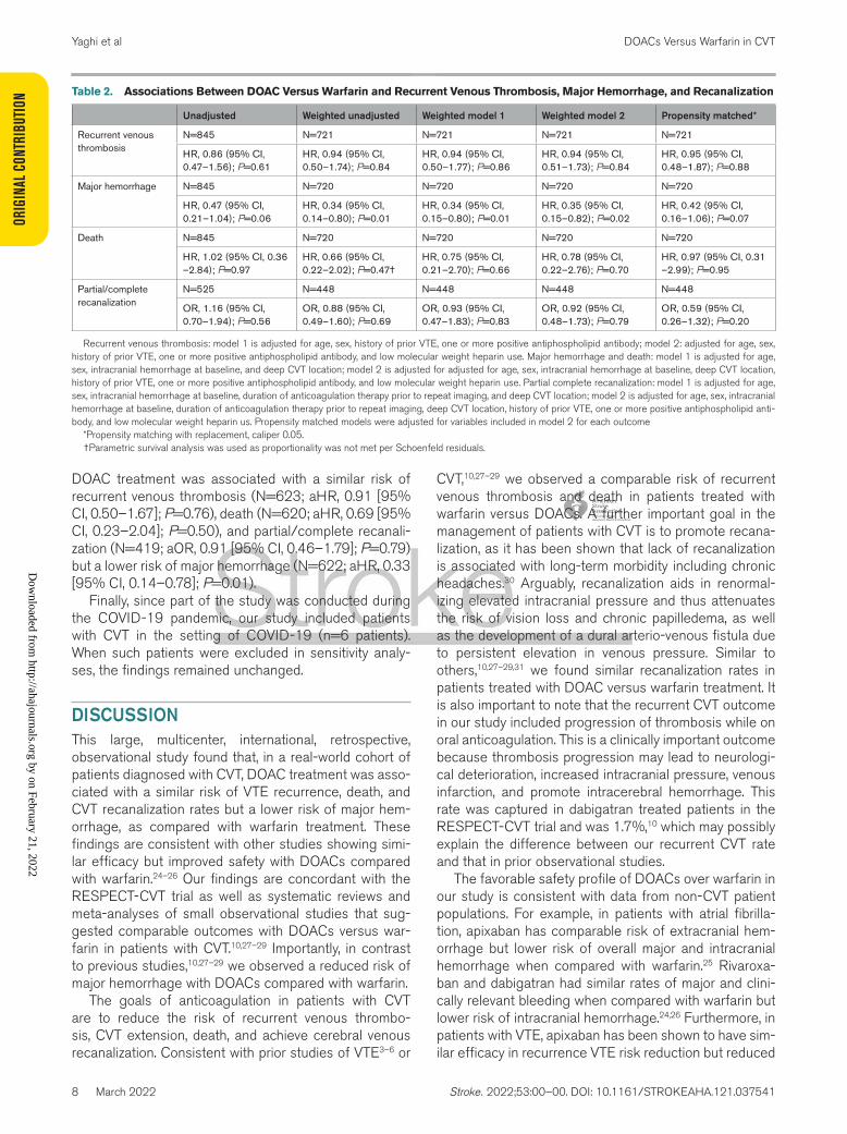
Table 2. Associations Between DOAC Versus Warfarin and Recurrent Venous Thrombosis, Major Hemorrhage, and Recanalization
Unadjusted Weighted unadjusted Weighted model 1 Weighted model 2 Propensity matched*
Recurrent venous thrombosis
N=845 N=721 N=721 N=721 N=721
HR, 0.86 (95% CI, 0.47–1.56); P=0.61
HR, 0.94 (95% CI, 0.50–1.74); P=0.84
HR, 0.94 (95% CI, 0.50–1.77); P=0.86
HR, 0.94 (95% CI, 0.51–1.73); P=0.84
HR, 0.95 (95% CI, 0.48–1.87); P=0.88
Major hemorrhage N=845 N=720 N=720 N=720 N=720
HR, 0.47 (95% CI, 0.21–1.04); P=0.06
HR, 0.34 (95% CI, 0.14–0.80); P=0.01
HR, 0.34 (95% CI, 0.15–0.80); P=0.01
HR, 0.35 (95% CI, 0.15–0.82); P=0.02
HR, 0.42 (95% CI, 0.16–1.06); P=0.07
Death N=845 N=720 N=720 N=720 N=720
HR, 1.02 (95% CI, 0.36 –2.84); P=0.97
HR, 0.66 (95% CI, 0.22–2.02); P=0.47†
HR, 0.75 (95% CI, 0.21–2.70); P=0.66
HR, 0.78 (95% CI, 0.22–2.76); P=0.70
HR, 0.97 (95% CI, 0.31 –2.99); P=0.95
Partial/complete recanalization
N=525 N=448 N=448 N=448 N=448
OR, 1.16 (95% CI, 0.70–1.94); P=0.56
OR, 0.88 (95% CI, 0.49–1.60); P=0.69
OR, 0.93 (95% CI, 0.47–1.83); P=0.83
OR, 0.92 (95% CI, 0.48–1.73); P=0.79
OR, 0.59 (95% CI, 0.26–1.32); P=0.20
Recurrent venous thrombosis: model 1 is adjusted for age, sex, history of prior VTE, one or more positive antiphospholipid antibody; model 2: adjusted for age, sex, history of prior VTE, one or more positive antiphospholipid antibody, and low molecular weight heparin use. Major hemorrhage and death: model 1 is adjusted for age, sex, intracranial hemorrhage at baseline, and deep CVT location; model 2 is adjusted for adjusted for age, sex, intracranial hemorrhage at baseline, deep CVT location, history of prior VTE, one or more positive antiphospholipid antibody, and low molecular weight heparin use. Partial complete recanalization: model 1 is adjusted for age, sex, intracranial hemorrhage at baseline, duration of anticoagulation therapy prior to repeat imaging, and deep CVT location; model 2 is adjusted for age, sex, intracranial hemorrhage at baseline, duration of anticoagulation therapy prior to repeat imaging, deep CVT location, history of prior VTE, one or more positive antiphospholipid anti-body, and low molecular weight heparin us. Propensity matched models were adjusted for variables included in model 2 for each outcome
*Propensity matching with replacement, caliper 0.05.†Parametric survival analysis was used as proportionality was not met per Schoenfeld residuals.
Dow
nloaded from http://ahajournals.org by on February 21, 2022
ORIGINAL CONTRIBUTIONYaghi et al DOACs Versus Warfarin in CVT
Stroke. 2022;53:00–00. DOI: 10.1161/STROKEAHA.121.037541 March 2022 9
risk of hemorrhagic complications when compared with warfarin.4 Additionally, a posthoc analysis of the RE-COVER and RE-COVER II trials (Efficacy and Safety of Dabigatran compared With Warfarin for 6 Month Treat-ment of Acute Symptomatic Venous Thromboembolism) showed similar risk of recurrent VTE or VTE-related mor-tality but reduced major hemorrhage in patients treated with dabigatran when compared with warfarin.32 Inter-estingly, the improved safety of dabigatran over warfarin was only seen in younger patients, which more closely resembles the age group studied in our population.32 An important challenge to consider about intracranial hem-orrhage in the setting of CVT is that new or worsening hemorrhage may have been linked to progression of thrombosis and thus may be more linked to treatment efficacy as opposed to safety.
Our study supports current evidence that DOACs rep-resent a reasonable alternative to warfarin in patients with CVT. Although DOACs do not require blood level monitoring, they generally are more expensive than war-farin. Thus, further studies are needed to test whether the improved safety profile of DOACs is cost-effective. Several studies suggest that DOACs offer a cost-effec-tive alternative for patients with atrial fibrillation and those with VTE,33,34 however, cost analyses are lacking for patients with CVT. Nevertheless, it is expected that once patent protection for DOACs expires more afford-able generic drugs will become available.
Our study has several limitations inherent to its retro-spective, observational design, and noncentral and non-blinded determination and adjudication of clinical and imaging outcomes. While we compared baseline char-acteristics between the 2 groups and included propen-sity score adjustment and matching, we cannot exclude the possibility of residual treatment-by-indication bias. Second, 15.7% of patients were lost to follow-up within 90-days. However, the baseline characteristics were overall similar between the patients with versus without at least 90-day follow-up, assuaging concerns about major attrition bias. Third, the low event rate of recur-rent VTE in our study, which is consistent with other studies,10,19,35 may have left us underpowered to show a difference between the 2 groups and precluded us from conducting detailed subgroup analyses. For exam-ple, CVT is a heterogenous disease for which it remains uncertain whether one treatment strategy may be better than the other in certain sub-populations. Fourth, recur-rent CVT in our study included both de novo CVT as well as CVT extension. This may at least partially explain the higher recurrent CVT rates in our studies compared with others. While both are important outcomes to cap-ture, we are unable to distinguish between the two with certainty in our study and thus it remains unknown if one treatment is superior to the other with respect to each of these outcomes. Fifth, we did not have data on international normalized ratio levels in warfarin treated
patients and thus some recurrence of venous throm-bosis and major hemorrhage events may have been in the setting of subtherapeutic or supratherapeutic anti-coagulation. This, however, reflects a true real-world experience in patients treated with warfarin. Sixth, the rate of asymptomatic hemorrhage in our study was likely low due to ascertainment bias since asymptom-atic patients do not typically undergo follow-up brain imaging. Finally, the timing of follow-up imaging was heterogeneous among patients, limiting our recanaliza-tion status analysis.
CONCLUSIONSOur findings provide real-world data supporting the use of DOACs as a reasonable alternative to warfarin treat-ment in patients with CVT. Given the study limitations, our findings should be interpreted with caution and require confirmation by large prospective observational studies such as the DOAC-CVT study (Direct Oral Anticoagu-lants in the Treatment of Cerebral Venous Thrombosis; https://clinicaltrials.gov; NCT04660747) and the ongo-ing randomized SECRET trial (Study of Rivaroxaban for Cerebral Venous Thrombosis; https://clinicaltrials.gov; NCT03178864).
ARTICLE INFORMATIONReceived October 4, 2021; final revision received January 7, 2022; accepted January 12, 2022.
Presented in part at the International Stroke Conference, New Orleans, LA, and virtual, February 9–11, 2022.
AffiliationsDepartment of Neurology, Brown University, Providence, RI (S.Y., L.S., C.S., N.K., S.E.J., A.C., S.C., K.F.). Department of Neurology, University of Alabama at Birming-ham (E.B.). Department of Neurology, University of Colorado School of Medicine, Aurora (S.S.O.). Department of Neurology, Washington University, Saint Louis, MO (J.A.G., J.Y.A.). Department of Neurology (N.H., M.E.) and Department of Psy-chiatry (N.H.), University of Massachusetts, Worcester. Department of Neurology, Weill Cornell Medical Center, NY (A.L.L.). Department of Neurology, Montefiore Medical Center, NY (K.M., J.L.). Department of Neurology, Yale University, New Haven, CT (R.S., Y.C., A.S.Z., A.d.H.). Department of Neurology, University of Flor-ida, Gainesville (A.N.S., G.T.L., J.C.K., D.P.). Department of Neurology, Inselspital Universitätsspital, Bern, Switzerland (M.H., A.S., D.S., B.S.). Department of Neurol-ogy, University of Pennsylvania, Philadelphia, PA (A.R., O.K., D.D.). Department of Neurology (S.A.K., L.A.R.) and Department of Neurosurgery (S.A.K.), Medical University of South Carolina, Charleston. Department of Neurology and Reha-bilitation Medicine, University of Cincinnati (E.A.M., P.K., Y.A., B.C.). Department of Neurology, Vanderbilt University, Nashville, TN (D.K., H.L.). Department of Neurol-ogy, Boston University School of Medicine, MA (T.N.N., P.K., H.A.). Department of Neurology, New York University, NY (J.F., L.K., S.A.). Department of Biosta-tistics, University of California Santa Barbara (H.X.). Department of Neurology, University of Utah, Salt Lake City (V.M.). Department of Neurology, Christchurch hospital, New Zealand (T.W., D.W.). Department of Neurology, Hartford Hospital, CT (A.N., S.D.A.). Department of Neurology, University of Kansas, Kansas City (A.Q., J.M.). Department of Neurology, Spectrum Health, Michigan State Univer-sity, Grand Rapids (M.K., Y.C.). Department of Neurology, Duke University, Dur-ham, NC (B.M.G., M.W., D.R.). Department of Medicine and Surgery, University of Perugia, Italy (M.C.V.). Neurology – Stroke Unit, IRCCS MultiMedica, Milano, Italy (M.P.). Department of Neurology, Cooper University, Camden, NJ (J.E.S., S.K., S.Y.). Department of Neurology, George Washington University, District of Columbia (C.R.L.G., E.A.). Department of Neurology, University Hospital Basel and University of Basel, Switzerland (G.M.D.M., T.D.). Department of interventional and diagnos-tic Neuroradiology, Clinic of Radiology and Nuclear Medicine, University Hospital
Dow
nloaded from http://ahajournals.org by on February 21, 2022
ORIG
INAL
CON
TRIB
UTIO
NYaghi et al DOACs Versus Warfarin in CVT
10 March 2022 Stroke. 2022;53:00–00. DOI: 10.1161/STROKEAHA.121.037541
Basel and University of Basel, Switzerland (A.B., M.P.). Department of Neurology, University of Chicago, IL (R.A.-D., T.K.-H., S.P.). Department of Neurology, Univer-sity of California at Los Angeles (T.H., D.S.L.).
Sources of FundingThis work has been supported partially by Italian Ministry of Heatlh Ricerca Cor-rente—IRCCS MultiMedica.
DisclosuresDr Giles reports grants from Preston M. Green Charitable Foundation and grants from American Heart Association. Dr Henninger reports employment by University of Massachusetts Medical School. Dr Li reports grants from National Institute of Health. Dr Siepen reports grants from Banger-Rhyner Foundation/Swiss Academy of Medical Sciences. Dr Nguyen reports compensation from Medtronic for other services. Dr Aparicio reports grants from American Acad-emy of Neurology; grants from Alzheimer’s Association; and employment by Boston University. Dr de Havenon reports compensation from Integra for con-sultant services; grants from Regeneron Pharmaceuticals, Inc; stock options in Certus; and grants from AMAG Pharmaceuticals, Inc. Dr Nouh reports stock options in openwater and compensation from Genentech for other services. Dr Assad reports employment by Massachusetts General Hospital. Dr Khatri reports compensation from Bayer for consultant services. Dr Paciaroni reports compensation from PFIZER CANADA INC for other services; compensation from Bristol-Myers Squibb for other services; compensation from Bayer for oth-er services; compensation from Boehringer Ingelheim for other services; and compensation from SANOFI-AVENTIS US LLC for other services. Dr Siegler reports compensation from AstraZeneca for other services and compensation from Ceribell for consultant services. Dr Marchis reports travel support from Medtronic; travel support from Pfizer; and compensation from Bayer for consul-tant services. Dr Prabhakaran reports compensation from AbbVie for consultant services; compensation from Wolters Klewer Health, Inc for consultant services; and compensation from National Institute of Health for other services. Dr Liebe-skind reports compensation from Genentech for consultant services; compen-sation from Cerenovus for consultant services; compensation from Medtronic for consultant services; and compensation from Stryker for consultant services. Dr Furie reports compensation from Janssen Biotech for consultant services.Dr Khan reports research support from National Institute of Neurological Diseases and Stroke (NINDS). The other authors report no conflicts.
Supplemental MaterialSTROBE ChecklistFigure S1Table S1
REFERENCES 1. Bousser MG, Ferro JM. Cerebral venous thrombosis: an update. Lancet
Neurol. 2007;6:162–170. doi: 10.1016/S1474-4422(07)70029-7 2. Saposnik G, Barinagarrementeria F, Brown RD Jr, Bushnell CD, Cucchiara
B, Cushman M, deVeber G, Ferro JM, Tsai FY; American Heart Asso-ciation Stroke Council and the Council on Epidemiology and Prevention. Diagnosis and management of cerebral venous thrombosis: a statement for healthcare professionals from the American Heart Association/Ameri-can Stroke Association. Stroke. 2011;42:1158–1192. doi: 10.1161/STR. 0b013e31820a8364
3. Büller HR, Décousus H, Grosso MA, Mercuri M, Middeldorp S, Prins MH, Raskob GE, Schellong SM, Schwocho L, Segers A, et al. Edoxaban versus warfarin for the treatment of symptomatic venous thromboembolism. N Engl J Med. 2013;369:1406–1415. doi: 10.1056/NEJMoa1306638
4. Agnelli G, Buller HR, Cohen A, Curto M, Gallus AS, Johnson M, Masiukiewicz U, Pak R, Thompson J, Raskob GE, et al; AMPLIFY Investigators. Oral apixa-ban for the treatment of acute venous thromboembolism. N Engl J Med. 2013;369:799–808. doi: 10.1056/NEJMoa1302507
5. Bauersachs R, Berkowitz SD, Brenner B, Buller HR, Decousus H, Gallus AS, Lensing AW, Misselwitz F, Prins MH, Raskob GE, et al. Oral riva-roxaban for symptomatic venous thromboembolism. N Engl J Med. 2010;363:2499–2510
6. Schulman S, Kearon C, Kakkar AK, Mismetti P, Schellong S, Eriksson H, Baanstra D, Schnee J, Goldhaber SZ; RE-COVER Study Group. Dabigatran versus warfarin in the treatment of acute venous thromboembolism. N Engl J Med. 2009;361:2342–2352. doi: 10.1056/NEJMoa0906598
7. Kearon C, Akl EA, Ornelas J, Blaivas A, Jimenez D, Bounameaux H, Huisman M, King CS, Morris TA, Sood N, et al. Antithrombotic therapy for VTE disease:
CHEST Guideline and Expert Panel Report. Chest. 2016;149:315–352. doi: 10.1016/j.chest.2015.11.026
8. Osteresch R, Fach A, Hambrecht R, Wienbergen H. [ESC guidelines 2019 on diagnostics and management of acute pulmonary embolism]. Herz. 2019;44:696–700. doi: 10.1007/s00059-019-04863-5
9. Ortel TL, Neumann I, Ageno W, Beyth R, Clark NP, Cuker A, Hutten BA, Jaff MR, Manja V, Schulman S, et al. American Society of Hematology 2020 guidelines for management of venous thromboembolism: treatment of deep vein thrombosis and pulmonary embolism. Blood Adv. 2020;4:4693–4738. doi: 10.1182/bloodadvances.2020001830
10. Ferro JM, Coutinho JM, Dentali F, Kobayashi A, Alasheev A, Canhão P, Karpov D, Nagel S, Posthuma L, Roriz JM, et al; RE-SPECT CVT Study Group. Safety and efficacy of dabigatran etexilate vs dose-adjusted warfa-rin in patients with cerebral venous thrombosis: a randomized clinical trial. JAMA Neurol. 2019;76:1457–1465. doi: 10.1001/jamaneurol.2019.2764
11. Silvis SM, Middeldorp S, Zuurbier SM, Cannegieter SC, Coutinho JM. Risk factors for cerebral venous thrombosis. Semin Thromb Hemost. 2016;42:622–631. doi: 10.1055/s-0036-1584132
12. Liberman AL, Merkler AE, Gialdini G, Messé SR, Lerario MP, Murthy SB, Kamel H, Navi BB. Risk of pulmonary embolism after cerebral venous throm-bosis. Stroke. 2017;48:563–567. doi: 10.1161/STROKEAHA.116.016316
13. Liberman AL, Kamel H, Mullen MT, Messé SR. International classification of diseases, Ninth Revision (ICD-9) diagnosis codes can identify cerebral venous thrombosis in hospitalized adults. Neurohospitalist. 2016;6:147–150. doi: 10.1177/1941874416648198
14. Handley JD, Emsley HC. Validation of ICD-10 codes shows intracranial venous thrombosis incidence to be higher than previously reported. Health Inf Manag. 2020;49:58–61. doi: 10.1177/1833358318819105
15. Sayar Z, Moll R, Isenberg D, Cohen H. Thrombotic antiphospholipid syn-drome: A practical guide to diagnosis and management. Thromb Res. 2021;198:213–221. doi: 10.1016/j.thromres.2020.10.010
16. Lyman GH, Carrier M, Ay C, Di Nisio M, Hicks LK, Khorana AA, Leavitt AD, Lee AYY, Macbeth F, Morgan RL, et al. American Society of Hematology 2021 guidelines for management of venous thromboembolism: prevention and treatment in patients with cancer. Blood Adv. 2021;5:927–974. doi: 10.1182/bloodadvances.2020003442
17. Bates SM, Greer IA, Hirsh J, Ginsberg JS. Use of antithrombotic agents during pregnancy: the Seventh ACCP Conference on Antithrombotic and Thrombolytic Therapy. Chest. 2004;126(Suppl 3):627S–644S. doi: 10.1378/chest.126.3_suppl.627S
18. Miranda B, Ferro JM, Canhão P, Stam J, Bousser MG, Barinagarrementeria F, Scoditti U; ISCVT Investigators. Venous thromboembolic events after cerebral vein thrombosis. Stroke. 2010;41:1901–1906. doi: 10.1161/ STROKEAHA.110.581223
19. Palazzo P, Agius P, Ingrand P, Ciron J, Lamy M, Berthomet A, Cantagrel P, Neau JP. Venous thrombotic recurrence after cerebral venous throm-bosis: a long-term follow-up study. Stroke. 2017;48:321–326. doi: 10.1161/STROKEAHA.116.015294
20. Girot M, Ferro JM, Canhão P, Stam J, Bousser MG, Barinagarrementeria F, Leys D; ISCVT Investigators. Predictors of outcome in patients with cerebral venous thrombosis and intracerebral hemorrhage. Stroke. 2007;38:337–342. doi: 10.1161/01.STR.0000254579.16319.35
21. Klok FA, Kooiman J, Huisman MV, Konstantinides S, Lankeit M. Predict-ing anticoagulant-related bleeding in patients with venous thromboem-bolism: a clinically oriented review. Eur Respir J. 2015;45:201–210. doi: 10.1183/09031936.00040714
22. Aguiar de Sousa D, Lucas Neto L, Canhão P, Ferro JM. Recanalization in cerebral venous thrombosis. Stroke. 2018;49:1828–1835.
23. Aguiar de Sousa D, Lucas Neto L, Arauz A, Sousa AL, Gabriel D, Correia M, Gil-Gouveia R, Penas S, Carvalho Dias M, Correia MA, et al. Early recanaliza-tion in patients with cerebral venous thrombosis treated with anticoagula-tion. Stroke. 2020;51:1174–1181.
24. Patel MR, Mahaffey KW, Garg J, Pan G, Singer DE, Hacke W, Breithardt G, Halperin JL, Hankey GJ, Piccini JP, et al; ROCKET AF Investigators. Rivaroxaban versus warfarin in nonvalvular atrial fibrillation. N Engl J Med. 2011;365:883–891. doi: 10.1056/NEJMoa1009638
25. Granger CB, Alexander JH, McMurray JJ, Lopes RD, Hylek EM, Hanna M, Al-Khalidi HR, Ansell J, Atar D, Avezum A, et al; ARISTOTLE Committees and Investigators. Apixaban versus warfarin in patients with atrial fibrillation. N Engl J Med. 2011;365:981–992. doi: 10.1056/NEJMoa1107039
26. Connolly SJ, Ezekowitz MD, Yusuf S, Eikelboom J, Oldgren J, Parekh A, Pogue J, Reilly PA, Themeles E, Varrone J, et al; RE-LY Steering Committee and Investigators. Dabigatran versus warfarin in patients with atrial fibrillation. N Engl J Med. 2009;361:1139–1151. doi: 10.1056/NEJMoa0905561
Dow
nloaded from http://ahajournals.org by on February 21, 2022
ORIGINAL CONTRIBUTIONYaghi et al DOACs Versus Warfarin in CVT
Stroke. 2022;53:00–00. DOI: 10.1161/STROKEAHA.121.037541 March 2022 11
27. Bose G, Graveline J, Yogendrakumar V, Shorr R, Fergusson DA, Le Gal G, Coutinho J, Mendonça M, Viana-Baptista M, Nagel S, et al. Direct oral anticoagulants in treatment of cerebral venous thrombosis: a systematic review. BMJ Open. 2021;11:e040212. doi: 10.1136/ bmjopen-2020-040212
28. Nepal G, Kharel S, Bhagat R, Ka Shing Y, Ariel Coghlan M, Poudyal P, Ojha R, Sunder Shrestha G. Safety and efficacy of Direct Oral Anticoagu-lants in cerebral venous thrombosis: a meta-analysis. Acta Neurol Scand. 2022;145:10–23. doi: 10.1111/ane.13506
29. Lee GKH, Chen VH, Tan CH, Leow AST, Kong WY, Sia CH, Chew NWS, Tu TM, Chan BPL, Yeo LLL, et al. Comparing the efficacy and safety of direct oral anticoagulants with vitamin K antagonist in cere-bral venous thrombosis. J Thromb Thrombolysis. 2020;50:724–731. doi: 10.1007/s11239-020-02106-7
30. Ji K, Zhou C, Wu L, Li W, Jia M, Chu M, Wu D, Hou C, Duan J, Meng R, et al. Risk factors for severe residual headache in cerebral venous thrombosis. Stroke. 2021;52:531–536. doi: 10.1161/STROKEAHA.120.029820
31. Cheung YW, Middeldorp S, Prins MH, Pap AF, Lensing AW, Ten Cate-Hoek AJ, Villalta S, Milan M, Beyer-Westendorf J, Verhamme P, et al; Einstein PTS Investigators Group. Post-thrombotic syndrome in patients treated with rivaroxaban or enoxaparin/vitamin K antagonists for acute deep-vein
thrombosis. A post-hoc analysis. Thromb Haemost. 2016;116:733–738. doi: 10.1160/TH16-01-0041
32. Schulman S, Kakkar AK, Goldhaber SZ, Schellong S, Eriksson H, Mismetti P, Christiansen AV, Friedman J, Le Maulf F, Peter N, et al; RE-COVER II Trial Investigators. Treatment of acute venous thrombo-embolism with dabigatran or warfarin and pooled analysis. Circulation. 2014;129:764–772. doi: 10.1161/CIRCULATIONAHA.113.004450
33. Al Mukdad M, Al-Badriyeh D, Elewa HF. Cost-effectiveness evaluations among the direct oral anticoagulants for the prevention and treatment of venous thromboembolism: systematic review. Clin Appl Thromb Hemost. 2019;25:1076029619849103. doi: 10.1177/1076029619849103
34. Lorenzoni V, Pirri S, Turchetti G. Cost-effectiveness of direct non-vitamin K oral anticoagulants versus vitamin K antagonists for the management of patients with non-valvular atrial fibrillation based on available “Real-World” evidence: the Italian National Health System Perspective. Clin Drug Investig. 2021;41:255–267. doi: 10.1007/s40261-021-01002-z
35. Dentali F, Poli D, Scoditti U, Di Minno MN, De Stefano V, Stefano VD, Siragusa S, Kostal M, Palareti G, Sartori MT, et al; CErebral VEin Throm-bosis International Study Investigators. Long-term outcomes of patients with cerebral vein thrombosis: a multicenter study. J Thromb Haemost. 2012;10:1297–1302. doi: 10.1111/j.1538-7836.2012.04774.x
Dow
nloaded from http://ahajournals.org by on February 21, 2022


