Embed Size (px)
Citation preview
CRYSTALLOGRAPHY NEWSBritish Crystallographic Association
No.78 September 2001
BCA Spring Meeting 2002
Dorothy Hodgkin - RSC Landmark
Fibre Diffraction
Crystallography and Antiquities
2001 Walter Hälg Prize
Book ReviewsQuarterly
ISSN 1467-2790
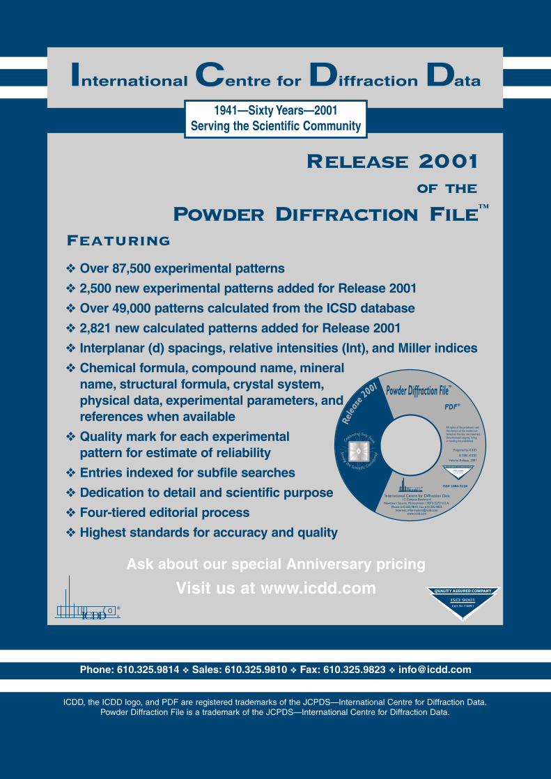
Phone: 610.325.9814 ❖ Sales: 610.325.9810 ❖ Fax: 610.325.9823 ❖ [email protected]
IInternational CCentre for DDiffraction DData
1941—Sixty Years—2001Serving the Scientific Community
Release 2001of the
Powder Diffraction File™
Featuring
❖ Over 87,500 experimental patterns
❖ 2,500 new experimental patterns added for Release 2001
❖ Over 49,000 patterns calculated from the ICSD database
❖ 2,821 new calculated patterns added for Release 2001
❖ Interplanar (d) spacings, relative intensities (Int), and Miller indices
❖ Chemical formula, compound name, mineralname, structural formula, crystal system,physical data, experimental parameters, andreferences when available
❖ Quality mark for each experimental pattern for estimate of reliability
❖ Entries indexed for subfile searches
❖ Dedication to detail and scientific purpose
❖ Four-tiered editorial process
❖ Highest standards for accuracy and quality
Ask about our special Anniversary pricing
Visit us at www.icdd.com
ICDD, the ICDD logo, and PDF are registered trademarks of the JCPDS—International Centre for Diffraction Data.Powder Diffraction File is a trademark of the JCPDS—International Centre for Diffraction Data.
A new creative forcein X-ray Diffraction
Oxford Diffractionis a new limited company ownedin joint venture by OxfordInstruments and Kuma Diffraction.
Created in 2001, OxfordDiffraction combinestechnology, knowledge andresources from both companiesto generate a new creative forcein X-ray diffraction with skills inCCD technology, X-ray optics,cryogenics, precision mechanicsand software.
Our products include:
Xcalibur™ automated 4-circlekappa X-ray diffractometerfeaturing
• Low noise Sapphire CCD
• CrysAlis software (small molecules, incommensurates,twins etc)
Helijet - see above
KM6 - six circle synchrotron diffractometer
United Kingdom & Rest of WorldTel: +44 (0)1235 532132
GermanyTel: +49 (0)221 500 7137
PolandTel: +48 71 354 52 23
www.oxford-diffraction.comE-mail: [email protected]
Helijet – helium jet for cryocrystallography
• Base temperature <15 K• 2 litres per hour helium
consumption at 15 K• Uniform temperature
distribution• No crystal shielding
A new creative forcein X-ray Diffraction
A new limited company owned in joint venture by Oxford Instruments and Kuma Diffraction
NEW
See usat the DGK,BCA, ACA
and ECM20 shows
to deliver solutionsin X-ray structure determination
working in partnership with you
The Microsource® can bring outstandingbenefits to people working in the areas of
• Macromolecular crystallography
• Protein crystallography
• Combinatorial chemistry
• Medicinal and pharmaceutical chemistry
• High temperatures and high pressures
• Real time monitoring
• Microdiffraction
Contact us today for further information.
The Microsource® X-ray generator is a revolutionary microfocusX-ray source, setting the standard for high brightness applications.
Bede plc comprises:bede scientific instruments ltdmicrosource divisionbede scientific incorporatedreflex sro
Bede Scientific Instruments LtdBowburn South Ind. Est. Bowburn Co Durham DH6 5AD UK
T +44 (0)191 377 2476 F +44 (0)191 377 9952E [email protected] W www.bede.co.uk
contact Graham Fraser
Bede Scientific Incorporated 14 Inverness Drive EastSuite H-100 Englewood CO 80112 USA
T +1 (303)790 8647 F +1 (303)790 8648E [email protected] W www.bede.com
contact Keith Bowen
division
Contents September 2001
1
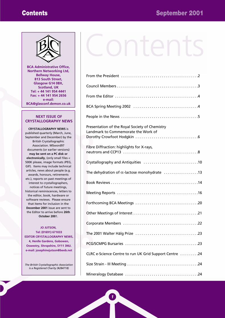
From the President . . . . . . . . . . . . . . . . . . . . . . . . . . . . . . . . . . . . . .2
Council Members . . . . . . . . . . . . . . . . . . . . . . . . . . . . . . . . . . . . . . . .3
From the Editor . . . . . . . . . . . . . . . . . . . . . . . . . . . . . . . . . . . . . . . . .4
BCA Spring Meeting 2002 . . . . . . . . . . . . . . . . . . . . . . . . . . . . . . . .4
People in the News . . . . . . . . . . . . . . . . . . . . . . . . . . . . . . . . . . . . . .5
Presentation of the Royal Society of ChemistryLandmark to Commemorate the Work ofDorothy Crowfoot Hodgkin . . . . . . . . . . . . . . . . . . . . . . . . . . . . . . .6
Fibre Diffraction: highlights for X-rays,neutrons and CCP13 . . . . . . . . . . . . . . . . . . . . . . . . . . . . . . . . . . . . .8
Crystallography and Antiquities . . . . . . . . . . . . . . . . . . . . . . . . . . .10
The dehydration of α-lactose monohydrate . . . . . . . . . . . . . . . . .13
Book Reviews . . . . . . . . . . . . . . . . . . . . . . . . . . . . . . . . . . . . . . . . . . .14
Meeting Reports . . . . . . . . . . . . . . . . . . . . . . . . . . . . . . . . . . . . . . . .16
Forthcoming BCA Meetings . . . . . . . . . . . . . . . . . . . . . . . . . . . . . . .20
Other Meetings of Interest . . . . . . . . . . . . . . . . . . . . . . . . . . . . . . . .21
Corporate Members . . . . . . . . . . . . . . . . . . . . . . . . . . . . . . . . . . . . .22
The 2001 Walter Hälg Prize . . . . . . . . . . . . . . . . . . . . . . . . . . . . . . .23
PCG/SCMPG Bursaries . . . . . . . . . . . . . . . . . . . . . . . . . . . . . . . . . . . .23
CLRC e-Science Centre to run UK Grid Support Centre . . . . . . . . .24
Size Strain - III Meeting . . . . . . . . . . . . . . . . . . . . . . . . . . . . . . . . . . .24
Mineralogy Database . . . . . . . . . . . . . . . . . . . . . . . . . . . . . . . . . . . .24
BCA Administrative Office,Northern Networking Ltd,
Bellway House,813 South Street,Glasgow G14 0BX,
Scotland, UKTel: + 44 141 954 4441Fax: + 44 141 954 2656
e-mail:[email protected]
NEXT ISSUE OFCRYSTALLOGRAPHY NEWS
CRYSTALLOGRAPHY NEWS ispublished quarterly (March, June,September and December) by the
British CrystallographicAssociation. MSword97
documents (or earlier versions)may be sent on a PC disk or
electronically, (only small files <500K please, image formats JPEG,GIF). Items may include technicalarticles, news about people (e.g.
awards, honours, retirementsetc.), reports on past meetings of
interest to crystallographers,notices of future meetings,
historical reminiscences, letters tothe editor, book, hardware or
software reviews. Please ensurethat items for inclusion in the
December 2001 issue are sent tothe Editor to arrive before 26th
October 2001.
JO JUTSON,
Tel: (01691) 671033
EDITOR CRYSTALLOGRAPHY NEWS,
4, Henlle Gardens, Gobowen,
Oswestry, Shropshire, SY11 3NU.
e-mail: [email protected]
The British Crystallographic Associationis a Registered Charity (#284718)
Contents
The conference season is upon us,and even in non-crystallographicmeetings, crystallography iseverywhere to be seen. I was at aGordon Conference on 3-DElectron Microscopy in RhodeIsland in June ( I was one of justthree crystallographers!), and wasamazed at the work in which lowresolution particles whose shapewas determined by single particleelectron microscopy studies werefilled with structures taken fromthe protein data bank thus givinga model of macromolecularassemblies at an atomicresolution. Studies of musclecontraction, for example, at thislevel are simply astonishing, andthe microscopists have their sightsset on whole cells and organellesat a similar resolution.
Other developments proceedapace: powder diffraction cannow relatively routinely solvestructures ten times the size ofthose 5 years ago, especiallyorganic molecules even usinglaboratory data; the flood ofbiological macromoleculestructures continues unabated(More than 14000 new structureswere deposited in the proteindata bank last year.); datacollection hardware continues toimprove and computing hasbecome a minor cost.Crystallographic softwarebecomes increasinglysophisticated and easier for non-experts to use (although this hasobvious dangers). Diamond, thenew synchrotron, is on the way,and plans are afoot for a newneutron spallation source; thesewill keep the UK in the forefrontof crystallographic technology forthe next decade. Crystallographyhas always been an exciting
science, but it seems to me thatthe excitement is increasing andnot in any way diminishing.Despite our many reservationsabout the way in whichgovernment and others handleand manage science, it is a greattime to be a crystallographer.
Our own spring meeting has toreflect all this, and the organisersof our next meeting inNottingham have done a splendidjob: the plenary session is devotedto structure solution and phasingas applied to all systems fromsmall molecules tomacromolecular assemblies usingboth single crystals and powders.There are sessions on DNArecombination and repair,polymorphism, drugs and disease,Reitveld refinement, detectors,thin films, crystallisation,education and the formation of anew special interest groupdevoted to the Diamond facility.Many of these sessions will takethe form of workshops. Everyattempt has been made to keepcosts to a minimum, and theNottingham campus is adelightful place. How can anyoneresist?
I hope to see many of you at theKrakov ECM Meeting at the endof August. ECM conferences alsoreflect the scope and diversity of21st century crystallography aswell as the BCA Spring Meetings.
Chris GilmoreAugust 2001
From the President September 2001
President’s Remarks
2
Cover pictures left to right:
X-ray diffraction image of normal breasttissue.
2Fo-Fc map calculated from cellulose II.
Detail of Middle Kingdom coffin from ElBersheh.
Chinese underglaze-blue porcelain dish,mid-14th century, Yuan Dynasty.
Dorothy Hodgkin plaque.

Council Members 2001 - 2002
3
President (2003) Prof. Chris J.Gilmore Department of Chemistry University of Glasgow GLASGOW G12 8QQ tel: 0141 330 3716 [email protected]
Vice President (2004)Prof. Paul Fewster Philips Analytical Research CentreCross Oak Lane Redhill RH1 5HA tel: 01293 815714 [email protected]
Secretary (2004) Dr. Christine Cardin University of Reading Dept of Chemistry Whiteknights Reading RG6 6AD tel: 0118 9318215 [email protected]
Treasurer (2003) Mr. David J.Taylor 35 Birchley Road, Billinge WIGAN Lancs WN5 7QJ tel: 01744 893108 [email protected]
Ordinary membersDr Margaret J. Adams (2003) Lab. of Molecular Biophysics Rex Richards BuildingSouth Parks Road OXFORD OX1 3QU tel: 01865 275391 [email protected]
Dr Jeremy K. Cockcroft (2003) (WebMaster)School of Crystallography Birkbeck CollegeMalet Street LONDON WC1E 7HX, tel: 020 7631 6849 [email protected]
Prof. Paul R. Raithby (2003) Department of Chemistry, University of Bath, Claverton DownBATH BA2 7AYtel: (01225) 323183, [email protected]
GROUP REPRESENTATIVES
Biological Structures Dr. A. Hadfield (2002), Department of Biochemistry, University of BristolBRISTOL BS8 1TD, tel: 0117 928 7436, [email protected]
Chemical Crystallography Dr Harry Powell (2002) MRC Lab. of Molecular Biology MRC Centre Hills RoadCAMBRIDGE CB2 2QHtel: 01223 402423 [email protected]
Industrial Prof. C.S.Frampton Roche Discovery Welwyn 40 Broadwater Road Welwyn Garden CityHertfordshire HL7 3AY, tel: 01707 366272/[email protected]
Physical Crystallography Dr C.C.Wilson, Rutherford Appleton Laboratory, ISIS Facility, CHILTON, Oxon OX11 0QXtel: 01235 82 1900 ext 5137 or 0123544 5137, [email protected]
Co-opted MembersProf. P.Barnes, Department of Crystallography, Birkbeck College, Malet Street, LONDON, WC1E 7HXtel: 020 7631 6817 [email protected]
Prof. M.M.Woolfson FRS (2002) University of York Department of Physics Heslington York, Y01 5DDtel: 01904 432 238 [email protected]
Ex-officio Members
Editor "Crystallography News"Dr. Josephine Jutson 4 Henlle Gardens Gobowen, Oswestry Shropshire SY11 3NUtel: 01691 671 033 [email protected]
Education Officer Mrs K.M.Crennell 'Fortran Friends' P.O.Box 64 Didcot Oxon OX11 0TH, tel: 01235 834 [email protected]
GROUP CHAIRMEN
Biological Structures GroupDr. Richard PauptitProtein Structure LabAstraZeneca, Mereside, Alderley Park,Macclesfield SK10 4TG, tel: 01625-516135 [email protected]
Chemical Crystallography GroupProf. Paul R. Raithby Department of Chemistry University of Bath Claverton DownBATH BA2 7AYtel: (01225) 323183 [email protected]
Industrial GroupProf. C.S.Frampton Roche Discovery Welwyn, 40 Broadwater Road, Welwyn Garden City,Hertfordshire HL7 3AY, tel: 01707 366272/366031 [email protected]
Physical Crystallography GroupProfessor J.L. FinneyDepartment of Physics & Astronomy University College London,tel: 0171 380 [email protected]
Full committee details on the BCA website -http://bca.cryst.bbk.ac.uk/BCA/
BCA Council Members 2001 - 2002
Have you ever wondered whatgoes on behind the scenes atthe British Museum? In thisissue Andrew Middletondescribes how XRD plays apart in determining how,when and where antiquitieswere made. In contrast thearticle on Fibre Diffraction byTrevor Forsyth, ILL coversapplications such as adiagnostic method for theinvestigation of cancertumours in breast tissue andthe study of hydrogen
bonding in cellulose. Also inthis issue there is a report onthe Presentation of the RoyalSociety of Chemistry Landmarkto Commemorate the Work ofDorothy Hodgkin.
Jo JutsonApril 2001
From the Editor BCA Spring Meeting 2002
4
The third issue
The next BCA Spring Meeting willbe held on the University Parkcampus of the University ofNottingham. An exciting scientificprogramme, characterised by alarge number of joint sessionsbetween the BCA groups, hasbeen drawn up by theirrepresentatives. This includes aPlenary Session on new methodsof structure solution and phasing,along with other sessions on DNA
recombination and repair;polymorphism and structuralchanges; proteins, drugs anddisease; new laboratory sourcesand detectors; thin films, andworkshops on topics such asmacromolecular crystallisation,Rietveld refinement, theCRYSTALS program … and muchmore. So whether you want tohear the latest results, find outabout new techniques andinstrumentation, hone your skillsin the workshops, or simply catchup with old friends, we are sureyou will be able to enjoy doing allof these.
The University Park campus hasextensive, attractive green spaceand even boasts a boating lake.On-site cultural attractionsinclude the Arts Centre and thebrand new D.H. LawrencePavilion. Accommodation in hallsof residence is available within afew minutes walk of the lecturetheatres and exhibition halls, allof which are situated in the PopeBuilding. The social programmecomprises a mixer reception on
Monday evening, a sponsoredwine reception/poster session onTuesday evening and theconference dinner on Wednesday.
We think we have a greatprogramme of scientific sessions,and a superb venue for the SpringMeeting in 2002. Full details ofthe Meeting will appear in theDecember issue ofCrystallography News: theMeeting should be particularlyattractive to students, who willqualify for free registration. Inaddition to the city itself,Nottingham is also an excellentbase for visiting a wide range ofattractions, from the scenery ofthe Peak District to historic citieslike Lincoln. We look forward towelcoming you here next March.
Sandy Blake (email: [email protected])
Claire Wilson (email: [email protected])
Local organisers
BCA SpringMeeting 2002The University ofNottingham, Monday 25th- Thursday 28th March2002
People in the News September 2001
5
The European CrystallographicAssociation has awarded thesecond European CrystallographyPrize to Professor Jochen R.Schneider of the HASYLAB atDESY, Hamburg, Germany.Professor Schneider is recognisedfor his pioneering work on theapplication of gamma-rayspectroscopy and his high energysynchrotron radiation studies, aswell as his more recentinvolvement in the developmentof the free electron laser.
Professor Schneider was born inBurgstädt, Saxony, Germany andstudied Physics in Hamburg afteran education as electricalengineer. He did his PhD underthe direction of Professor H.Maier-Leibnitz at the InstituteLaue-Langevin in Grenoble,France. His work on gamma-raydiffractometry and Comptonscattering was performed at theHahn-Meitner-Institut in Berlin,the synchrotron radiation work atDESY-HASYLAB in Hamburg,where he is now heavily involvedin the development of free-electron lasers driven by linearaccelerators. Professor Schneideris presently Head of HASYLAB andDirector of Research forSynchrotron Radiation and Free-Electron Lasers at DESY.
OBE for Professor Julia Higgins
Congratulations to Professor JuliaHiggins OBE FRS, who becameDame Commander of the BritishEmpire in the Queen's BirthdayHonours list. Julia Higgins isProfessor of Polymer Science inImperial College. She also chairsthe Athena project on women inscience, engineering andtechnology (SET) in HigherEducation in the UK, along withan impressive list of otherresponsibilities both to the SETcommunity and for women in SET.
Fellows of the RoyalSociety
Congratulations to Dr. AndrewLeslie and Professor GeorgeSheldrick who have beenappointed Fellows of the RoyalSociety.
Dr Leslie is Senior Scientist at theMRC Laboratory of MolecularBiology, Cambridge. He hasdetermined the atomic detail of anumber of biologically importantstructures and most recentlysolved the structure of hepatitis Bvirus protein.
Professor Sheldrick is Professor ofStructural Chemsitry at theUniversity of Gottingen andDirector of the Institute furAnorganische Chemie, Gottingen,Germany. He has been a majorcontributor to the field ofchemical X-ray crystallography forthe past three decades anddeveloped the SHELX computerprograms, for structuredetermination and refinement.
New ICDD ExecutiveDirector
The International Centre forDiffraction Data (ICDD) haveannounced that Dr. Tim Fawcetthas joined the ICDD as its newExecutive Director. Tim is a longtime ICDD member, ICDD Fellow,and served on the Board ofDirectors from 1988-1990. Hebrings to the ICDD outstandingexperience in management andR&D for product development, aswell as an exceptionalbackground in X-ray diffraction.
The J.D. Hanawalt Award
Mr. Raymond P. Goehner, and Dr.Joseph R. Michael, both of SandiaNational Laboratories,Albuquerque, New Mexico, U.S.A.have been selected to receive the2001 J.D. Hanawalt Award forexcellence in the field of X-raypowder diffraction. The J.D.Hanawalt Award is presentedevery three years for important,recent contributions to the fieldof X-ray powder diffraction andphase identification publishedwithin the last five years. Theaward consists of acommemorative plaque, anhonorarium, and travel funds toattend the meeting at which theaward and lecture will bepresented.
Retirement - Ian Langford
Dr Ian Langford, Reader inPowder Diffraction, University ofBirmingham, has retired afterworking for almost 40 years with
People in the News
Second EuropeanCrystallography PrizeAwarded to ProfessorJochen R. Schneider
Powder Diffraction, 21 of themwith Arthur (Prof. A.J.C.) Wilson - 5in the Viriamu Jones Laboratory,Dept of Physics, UniviversityCollege, Cardiff and then in thePhysics Dept (now School ofPhysics & Astronomy) atBirmingham. Ian is a leadinginternational expert in highresolution X-ray powderdiffraction applied to manymaterials of environmental,technological and industrialinterest and is the author of manypapers on Powder Diffraction. Hewas a founder member of the BCAand was involved at the outset inestablishing the Industrial Group.Ian served on the IG Committee, asan academic representative, from1983 to 1989 and was the Group'sfirst Secretary/Treasurer (1983/87).He was also a member of the BCACouncil from 1985 to 1992, as theAssociation's Treasurer from 1988to 1992.
Presentation of theRoyal Society ofChemistryLandmark toCommemorate theWork of DorothyCrowfoot Hodgkin On 14 May 2001 the LectureRoom of the University NaturalHistory Museum, Oxford was fullof people eagerly anticipating thepresentation of the second UKRoyal Society of Chemistrylandmark. The proceedings werechaired by Professor GrahamRichards, Chairman of theDepartment of Chemistry in theUniversity of Oxford. The Vice-Chancellor of the University, thehistorian Dr.C.R.Lucas welcomedus to the dedication. He remindedus that Dorothy was the firstBritish Woman to win a Nobelprize in Chemistry in 1964. TheUniversity takes pride in herachievements which havecontributed to the success of theChemistry Department in Oxford,the largest in the country. He wassure that Dorothy would havebeen proud of the fact that some40% of today's students arewomen.
One of Dorothy's students,Professor Sir Tom Blundell, FRS,then spoke on "Structural Biologyand Crystallography today: theinfluence of Dorothy Hodgkin oncurrent developments".(Reported later in this article) Inintroducing the speaker, GrahamRichards reminisced that he and
Tom had been undergraduatestogether at Brasenose College,and now Tom was Dorothy'ssecond most famous student. (themost famous being MargaretThatcher (nee Roberts) who laterabandoned chemistry for law andbecame Prime Minister).
Professor Tony Ledwith of theRoyal Society of Chemistry thenpresented a memorial plaque toProfessor Richards who received iton behalf of the University. SinceOxford weather is unpredictable,a virtual unveiling was projectedon the screen. We saw crimsonvelvet curtains slowly open toreveal the plaque which was fixedto the wall on the archway of themain entrance to the InorganicChemistry Laboratory in SouthParks Road. The inscription reads:
National HistoricChemical Landmark
The work of Dorothy CrowfootHodgkin at the University ofOxford."In this building from 1956-1994and at other times in the OxfordScience Area, Professor DorothyCrowfoot Hodgkin (1910-1994)OM, FRS, Nobel Laureate, ledpioneering work on the structuresof antibiotics, vitamins andproteins, including penicillin,Vitamin B12 and insulin using X-ray diffraction techniques. Manymethods for solving crystalstructures were developed takingadvantage of digital computersfrom the very earliest days. Thework provided a basis for much ofpresent day molecular structuredriven molecular biology andmedicinal chemistry."14 May 2001
Royal Scoiety of Chemistry Landmark September 2001
6
AcknowledgementsBCA Sponsors
The BritishCrystallographic
Association is grateful toBirkbeck College,
University of London,who host and manage
the server for ourWebsite.
Royal Scoiety of Chemistry Landmark September 2001
7
"Structural Biology andCrystallography today:the influence of DorothyHodgkin on currentdevelopments"
Professor Sir Tom Blundell FRS (Sir William Dunn Professor ofBiochemistry at the University ofCambridge)
Professor Blundell began bydescribing Dorothy Hodgkin'searly work in Cambridge with herPh.D supervisor, J.D.Bernal,determining the structure ofprotein crystals using X-rays. In1934 she took high quality X-rayphotographs of the crystallineprotein pepsin, having realisedthat the crystals had to be kept intheir mother liquor if they were
to retain their order. The study ofproteins in water has beenimmensely important over theyears. The final paragraph of thepaper in Nature which she andBernal published demonstratesremarkable foresight.
"At this stage such ideas aremerely speculative, but now thata crystalline protein has beenmade to give X-Ray photographs,it is clear that we have the meansof checking them by examiningthe structures of all crystallineproteins, arriving at far moredetailed conclusions aboutprotein structure than previousphysical or chemical methodshave been able to give."
Pepsin proved to be a member ofa large family of proteolyticenzymes called the asparticproteinases which have beenextensively studied since then.They have about 320 amino acidsand 2 motifs of Asp-Thr-Cly at theactive site. Charles Bunn was oneof the first to work on renninwhich was also found to be anaspartic proteinase containingthis same motif; it is involved inthe inhibition of angiotensin IIsynthesis, and may be a possibletarget for better drugs to reducehigh blood pressure. Pepsin hasan extended deep active site anddrug companies began to thinkabout discovering drugs to fit it.In the 1980s chemistry dominateddrug design, X-ray crystallographywas traditionally used to modelthe 3D structures. Knowledge ofthe structure allows the design ofstructure based inhibitors butmore recently it has provedcheaper to generate anti-hypertensive drugs usinggenomics.
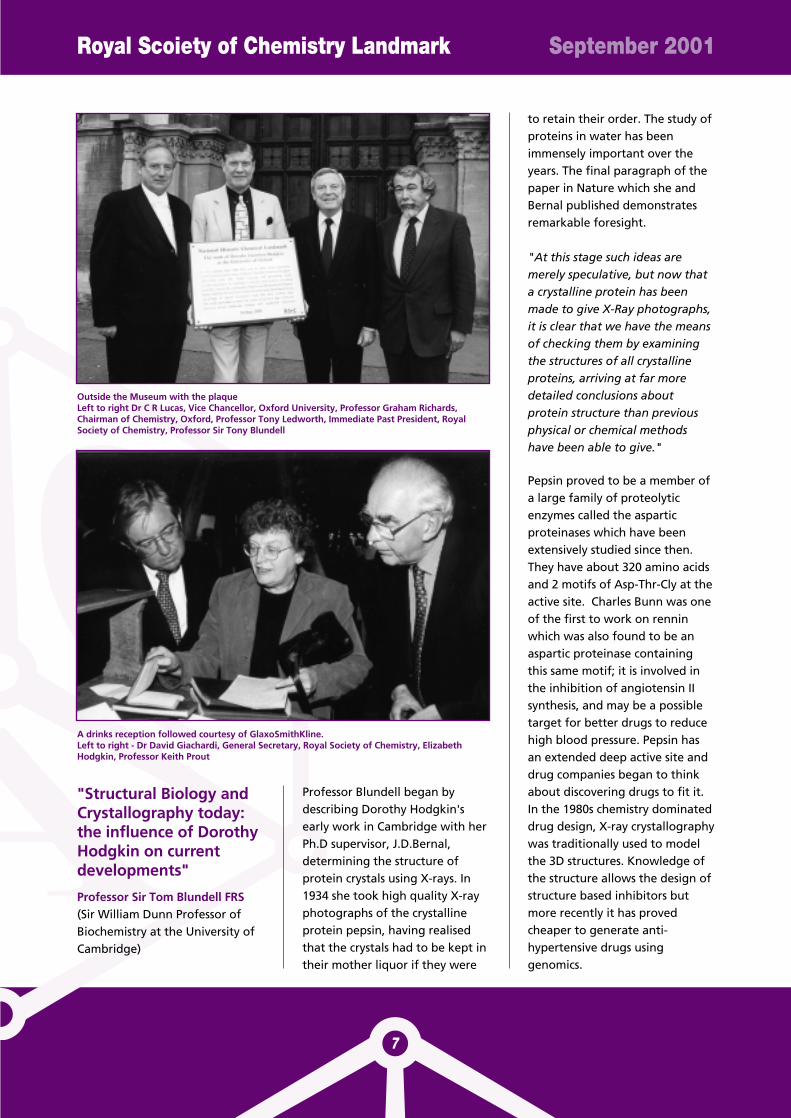
Outside the Museum with the plaqueLeft to right Dr C R Lucas, Vice Chancellor, Oxford University, Professor Graham Richards,Chairman of Chemistry, Oxford, Professor Tony Ledworth, Immediate Past President, RoyalSociety of Chemistry, Professor Sir Tony Blundell
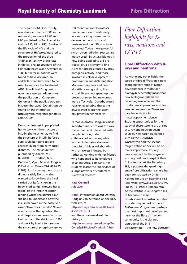
A drinks reception followed courtesy of GlaxoSmithKline. Left to right - Dr David Giachardi, General Secretary, Royal Society of Chemistry, ElizabethHodgkin, Professor Keith Prout
Royal Scoiety of Chemistry Landmark
8
Fibre Diffraction
The pepsin motif, Asp-Thr-Cly,was also identified in 1985 in theretroviral genomes of RSV andHIV, published by Toh H et al. inNature 315, 691 (1985). Studies ofthe life cycle of HIV and thestructure of HIV proteinase led tothe production of the drug'Indinavar', an HIV proteinaseinhibitor. The 3D structure of theHIV proteinases was discovered in1989 but later mutations werefound to have occurred, sococktails of inhibitors had to beused to improve the treatment ofAIDS. Pre-clinical Drug designnow has a new paradigm sincethe publication of CompleteGenomes in the public databasesin December 2000. (Details can befound on the Internet athttp://igweb.integratedgenomics.com/GOLD/)
Dorothy's interest in people ledher to work on the structure ofinsulin, she felt she had to findthe structure of insulin before acure could be found to savechildren dying from early onsetdiabetes. This structure waspublished by Adams, M.J.,Blundell, T.L, Dodson, G.G,Dodson,E, Vijau, M, and HodgkinD.C et al. in Nature 224, 491-495(1969). Just knowing the structuredid not satisfy Dorothy, shewanted to know how the insulincarried out its function in thebody. Fred Sanger showed her amodel of the insulin receptorbinding which she admired butshe had to understand how theinsulin behaved in the body. Sheasked 'How does it work?' No onecould answer that question thenand despite more recent work byHubbard and Hendrickson in 1995and work by Louise Johnson onthe structure of phosphorylase we
still cannot answer Dorothy'ssimple question. Traditionally,laboratory X-rays were used todetermine the structure ofproteins and their 3D structuresmodelled. Today more powerfulsynchrotron radiation sources aremostly used. Structural biology isnow being applied to aid pre-clinical drug discovery to findcures for diseases caused by straymitogenic activity, and thoseinvolved in cell development,proliferation and differentiation.Modern computers and newalgorithms using a drug likevirtual library now speed up theprocess of screening new drugsmore effectively. Dorothy wouldhave enjoyed using these; shealways tried to use the latestequipment in her research.
Perhaps Dorothy Hodgkin's mostimportant influence was the wayshe worked and interacted withpeople. Although shecollaborated with many whoworked in industry, she neverthought of this as collaboratingwith a faceless industry, butrather as working with her friendswho happened to be employedby an industrial company. Herstudents learnt the importance ofa large network of contacts tosuccessful research.
Kate CrennellJuly 2001
Note: Information about DorothyHodgkin can be found on the BCAweb site, athttp://bca.cryst.bbk.ac.uk/BCA/obits/CVS/DCH.htmland there is an excellent lifehistory at:http://www.engr.psu.edu/wep/EngCompSp98/Aclausi/HodgkinD.html
Fibre Diffraction:highlights for X-rays, neutrons andCCP13
Fibre Diffraction with X-rays and neutrons
As with many other fields, thescope of fibre diffraction is nowchanging very rapidly. Majordevelopments in molecularbiology/biochemistry mean thatnew biological systems arebecoming available and thattotally new approaches exist forsample preparation. There areparallels with this inmaterial/polymer science.Exciting opportunities for thestudy of these systems are arisingat X-ray and neutron beamsources. New facilities plannedsuch as the DIAMONDsynchrotron and the secondtarget station at ISIS will be ofmajor importance. Equallyimportant will be the upgrade ofexisting facilities to exploit theirfull potential. At the DaresburySRS, a purpose designed high-angle fibre diffraction camera hasbeen constructed by Dr. R.Keyhoe for use on beamline 14.1(see http:// www.dl.ac.uk/ SRS/ PX/line14/ 14_1/fibre_camera.html).At the Institut Laue Langevin (ILL)in Grenoble a majorrefurbishment of instrumentationin under way as part of the ILLMillennium Programme: perhapsthe most important developmenthere for the fibre diffractioncommunity is the plannedupgrade of the D19diffractometer – the new detector
Fibre Diffraction September 2001
9
on this instrument will give a gainin detecting solid angle ofapproximately 25 and open upentirely new possibilities for fibrediffraction (as well as singlecrystal) work (see http://www.ill.fr/ YellowBook/ D19/help/ dev/ development.html).
Fibre diffraction has made criticalcontributions to ourunderstanding of biological andsynthetic polymer systems in thepast. The following highlightsfrom recent X-ray and neutronfibre diffraction work illustratethe importance of the techniquefor the future.
X-ray fibre diffraction –new insight to breasttumour tissue from theDaresbury SRS
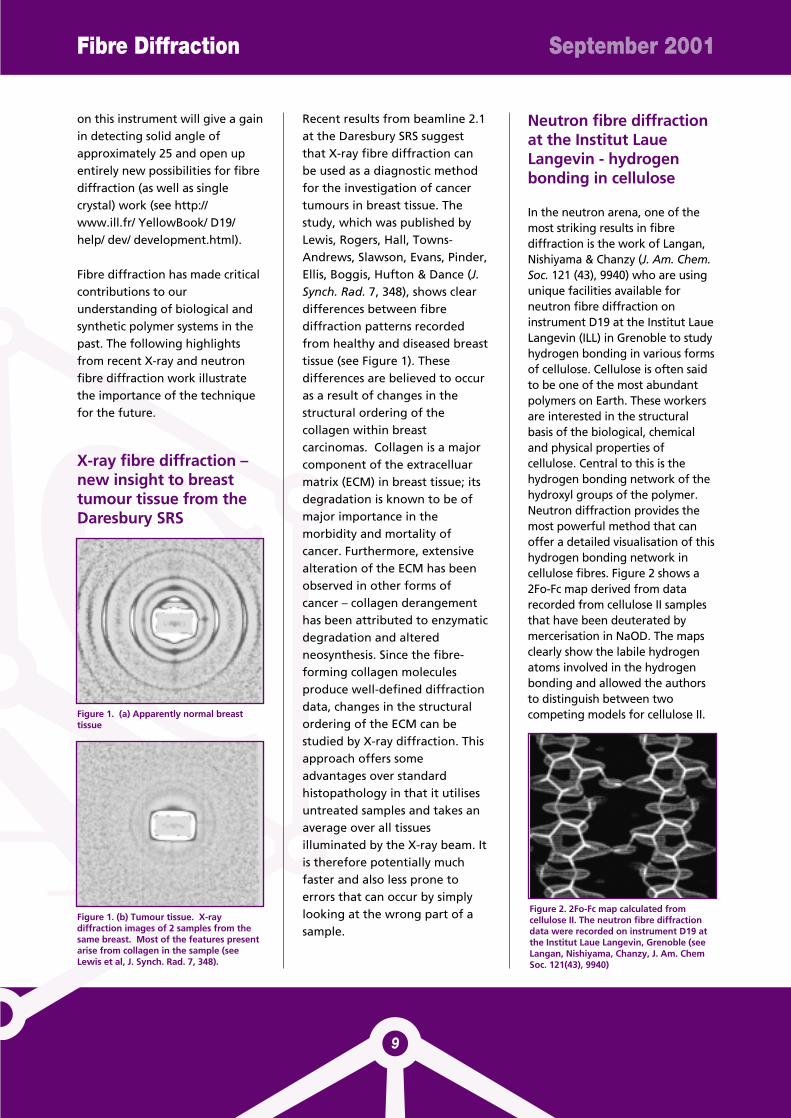
Recent results from beamline 2.1at the Daresbury SRS suggestthat X-ray fibre diffraction canbe used as a diagnostic methodfor the investigation of cancertumours in breast tissue. Thestudy, which was published byLewis, Rogers, Hall, Towns-Andrews, Slawson, Evans, Pinder,Ellis, Boggis, Hufton & Dance (J.Synch. Rad. 7, 348), shows cleardifferences between fibrediffraction patterns recordedfrom healthy and diseased breasttissue (see Figure 1). Thesedifferences are believed to occuras a result of changes in thestructural ordering of thecollagen within breastcarcinomas. Collagen is a majorcomponent of the extracelluarmatrix (ECM) in breast tissue; itsdegradation is known to be ofmajor importance in themorbidity and mortality ofcancer. Furthermore, extensivealteration of the ECM has beenobserved in other forms ofcancer – collagen derangementhas been attributed to enzymaticdegradation and alteredneosynthesis. Since the fibre-forming collagen moleculesproduce well-defined diffractiondata, changes in the structuralordering of the ECM can bestudied by X-ray diffraction. Thisapproach offers someadvantages over standardhistopathology in that it utilisesuntreated samples and takes anaverage over all tissuesilluminated by the X-ray beam. Itis therefore potentially muchfaster and also less prone toerrors that can occur by simplylooking at the wrong part of asample.
Neutron fibre diffractionat the Institut LaueLangevin - hydrogenbonding in cellulose
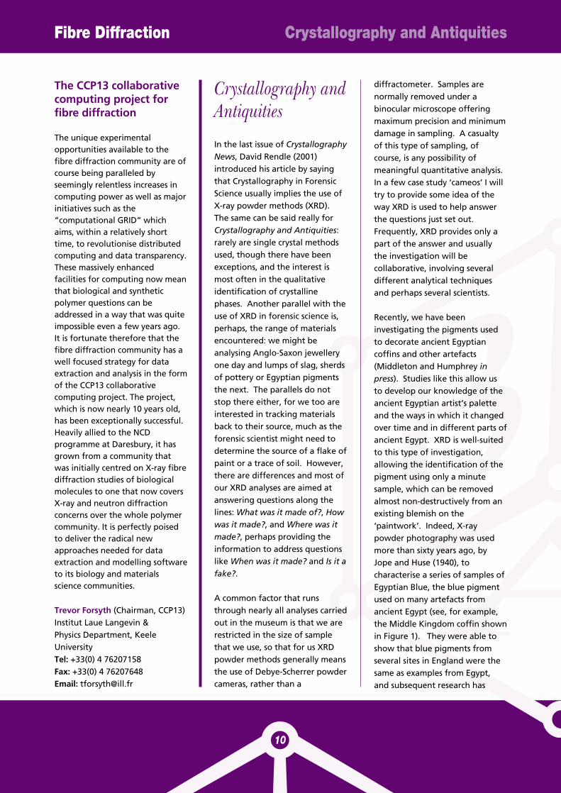
In the neutron arena, one of themost striking results in fibrediffraction is the work of Langan,Nishiyama & Chanzy (J. Am. Chem.Soc. 121 (43), 9940) who are usingunique facilities available forneutron fibre diffraction oninstrument D19 at the Institut LaueLangevin (ILL) in Grenoble to studyhydrogen bonding in various formsof cellulose. Cellulose is often saidto be one of the most abundantpolymers on Earth. These workersare interested in the structuralbasis of the biological, chemicaland physical properties ofcellulose. Central to this is thehydrogen bonding network of thehydroxyl groups of the polymer.Neutron diffraction provides themost powerful method that canoffer a detailed visualisation of thishydrogen bonding network incellulose fibres. Figure 2 shows a2Fo-Fc map derived from datarecorded from cellulose II samplesthat have been deuterated bymercerisation in NaOD. The mapsclearly show the labile hydrogenatoms involved in the hydrogenbonding and allowed the authorsto distinguish between twocompeting models for cellulose II.Figure 1. (a) Apparently normal breast
tissue
Figure 1. (b) Tumour tissue. X-raydiffraction images of 2 samples from thesame breast. Most of the features presentarise from collagen in the sample (seeLewis et al, J. Synch. Rad. 7, 348).
Figure 2. 2Fo-Fc map calculated fromcellulose II. The neutron fibre diffractiondata were recorded on instrument D19 atthe Institut Laue Langevin, Grenoble (seeLangan, Nishiyama, Chanzy, J. Am. ChemSoc. 121(43), 9940)
The CCP13 collaborativecomputing project forfibre diffraction
The unique experimentalopportunities available to thefibre diffraction community are ofcourse being paralleled byseemingly relentless increases incomputing power as well as majorinitiatives such as the“computational GRID” whichaims, within a relatively shorttime, to revolutionise distributedcomputing and data transparency.These massively enhancedfacilities for computing now meanthat biological and syntheticpolymer questions can beaddressed in a way that was quiteimpossible even a few years ago. It is fortunate therefore that thefibre diffraction community has awell focused strategy for dataextraction and analysis in the formof the CCP13 collaborativecomputing project. The project,which is now nearly 10 years old,has been exceptionally successful.Heavily allied to the NCDprogramme at Daresbury, it hasgrown from a community thatwas initially centred on X-ray fibrediffraction studies of biologicalmolecules to one that now coversX-ray and neutron diffractionconcerns over the whole polymercommunity. It is perfectly poisedto deliver the radical newapproaches needed for dataextraction and modelling softwareto its biology and materialsscience communities.
Trevor Forsyth (Chairman, CCP13)Institut Laue Langevin &Physics Department, KeeleUniversityTel: +33(0) 4 76207158Fax: +33(0) 4 76207648Email: [email protected]
Crystallography andAntiquities
In the last issue of CrystallographyNews, David Rendle (2001)introduced his article by sayingthat Crystallography in ForensicScience usually implies the use ofX-ray powder methods (XRD).The same can be said really forCrystallography and Antiquities:rarely are single crystal methodsused, though there have beenexceptions, and the interest ismost often in the qualitativeidentification of crystallinephases. Another parallel with theuse of XRD in forensic science is,perhaps, the range of materialsencountered: we might beanalysing Anglo-Saxon jewelleryone day and lumps of slag, sherdsof pottery or Egyptian pigmentsthe next. The parallels do notstop there either, for we too areinterested in tracking materialsback to their source, much as theforensic scientist might need todetermine the source of a flake ofpaint or a trace of soil. However,there are differences and most ofour XRD analyses are aimed atanswering questions along thelines: What was it made of?, Howwas it made?, and Where was itmade?, perhaps providing theinformation to address questionslike When was it made? and Is it afake?.
A common factor that runsthrough nearly all analyses carriedout in the museum is that we arerestricted in the size of samplethat we use, so that for us XRDpowder methods generally meansthe use of Debye-Scherrer powdercameras, rather than a
diffractometer. Samples arenormally removed under abinocular microscope offeringmaximum precision and minimumdamage in sampling. A casualtyof this type of sampling, ofcourse, is any possibility ofmeaningful quantitative analysis.In a few case study ‘cameos’ I willtry to provide some idea of theway XRD is used to help answerthe questions just set out.Frequently, XRD provides only apart of the answer and usuallythe investigation will becollaborative, involving severaldifferent analytical techniquesand perhaps several scientists.
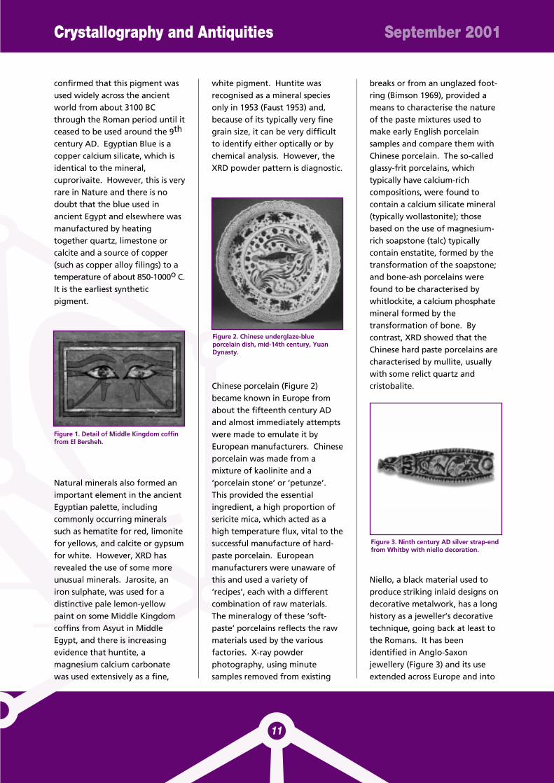
Recently, we have beeninvestigating the pigments usedto decorate ancient Egyptiancoffins and other artefacts(Middleton and Humphrey inpress). Studies like this allow usto develop our knowledge of theancient Egyptian artist’s paletteand the ways in which it changedover time and in different parts ofancient Egypt. XRD is well-suitedto this type of investigation,allowing the identification of thepigment using only a minutesample, which can be removedalmost non-destructively from anexisting blemish on the‘paintwork’. Indeed, X-raypowder photography was usedmore than sixty years ago, byJope and Huse (1940), tocharacterise a series of samples ofEgyptian Blue, the blue pigmentused on many artefacts fromancient Egypt (see, for example,the Middle Kingdom coffin shownin Figure 1). They were able toshow that blue pigments fromseveral sites in England were thesame as examples from Egypt,and subsequent research has
Fibre Diffraction Crystallography and Antiquities
10
Crystallography and Antiquities September 2001
11
confirmed that this pigment wasused widely across the ancientworld from about 3100 BCthrough the Roman period until itceased to be used around the 9th
century AD. Egyptian Blue is acopper calcium silicate, which isidentical to the mineral,cuprorivaite. However, this is veryrare in Nature and there is nodoubt that the blue used inancient Egypt and elsewhere wasmanufactured by heatingtogether quartz, limestone orcalcite and a source of copper(such as copper alloy filings) to atemperature of about 850-1000o C.It is the earliest syntheticpigment.
Natural minerals also formed animportant element in the ancientEgyptian palette, includingcommonly occurring mineralssuch as hematite for red, limonitefor yellows, and calcite or gypsumfor white. However, XRD hasrevealed the use of some moreunusual minerals. Jarosite, aniron sulphate, was used for adistinctive pale lemon-yellowpaint on some Middle Kingdomcoffins from Asyut in MiddleEgypt, and there is increasingevidence that huntite, amagnesium calcium carbonatewas used extensively as a fine,
white pigment. Huntite wasrecognised as a mineral speciesonly in 1953 (Faust 1953) and,because of its typically very finegrain size, it can be very difficultto identify either optically or bychemical analysis. However, theXRD powder pattern is diagnostic.
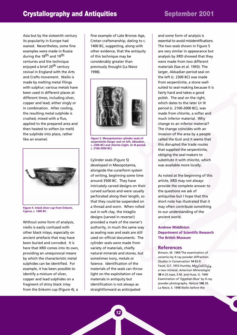
Chinese porcelain (Figure 2)became known in Europe fromabout the fifteenth century ADand almost immediately attemptswere made to emulate it byEuropean manufacturers. Chineseporcelain was made from amixture of kaolinite and a‘porcelain stone’ or ‘petunze’.This provided the essentialingredient, a high proportion ofsericite mica, which acted as ahigh temperature flux, vital to thesuccessful manufacture of hard-paste porcelain. Europeanmanufacturers were unaware ofthis and used a variety of‘recipes’, each with a differentcombination of raw materials.The mineralogy of these ‘soft-paste’ porcelains reflects the rawmaterials used by the variousfactories. X-ray powderphotography, using minutesamples removed from existing
breaks or from an unglazed foot-ring (Bimson 1969), provided ameans to characterise the natureof the paste mixtures used tomake early English porcelainsamples and compare them withChinese porcelain. The so-calledglassy-frit porcelains, whichtypically have calcium-richcompositions, were found tocontain a calcium silicate mineral(typically wollastonite); thosebased on the use of magnesium-rich soapstone (talc) typicallycontain enstatite, formed by thetransformation of the soapstone;and bone-ash porcelains werefound to be characterised bywhitlockite, a calcium phosphatemineral formed by thetransformation of bone. Bycontrast, XRD showed that theChinese hard paste porcelains arecharacterised by mullite, usuallywith some relict quartz andcristobalite.
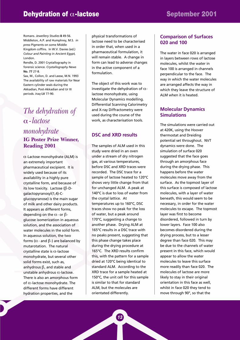
Niello, a black material used toproduce striking inlaid designs ondecorative metalwork, has a longhistory as a jeweller’s decorativetechnique, going back at least tothe Romans. It has beenidentified in Anglo-Saxonjewellery (Figure 3) and its useextended across Europe and into
Figure 1. Detail of Middle Kingdom coffinfrom El Bersheh.
Figure 2. Chinese underglaze-blueporcelain dish, mid-14th century, YuanDynasty.
Figure 3. Ninth century AD silver strap-endfrom Whitby with niello decoration.
Asia but by the sixteenth centuryits popularity in Europe hadwaned. Nevertheless, some fineexamples were made in Russiaduring the 18th and 19th
centuries and the techniqueenjoyed a brief 20th centuryrevival in England with the Artsand Crafts movement. Niello ismade by melting metal filingswith sulphur; various metals havebeen used in different places atdifferent times, including silver,copper and lead, either singly orin combination. After cooling,the resulting metal sulphide iscrushed, mixed with a flux,applied to the prepared area andthen heated to soften (or melt)the sulphide into place, ratherlike an enamel.
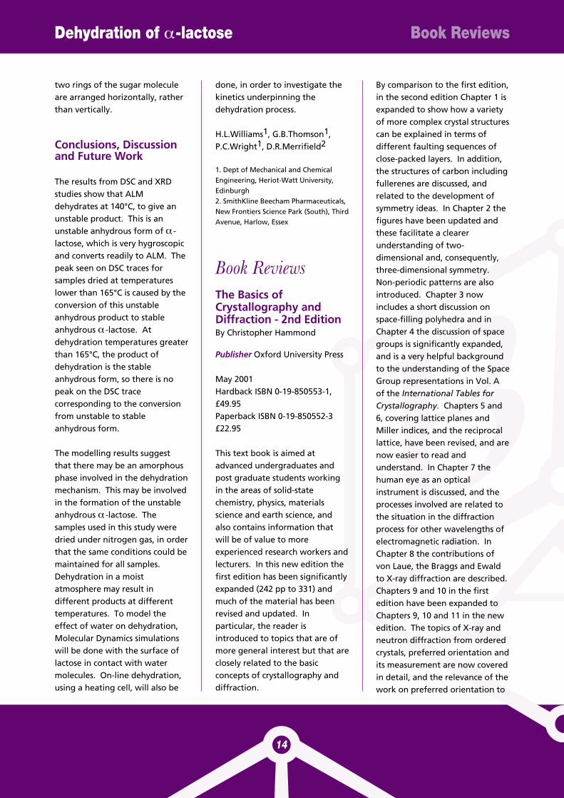
Without some form of analysis,niello is easily confused withother black inlays, especially onancient artefacts that may havebeen buried and corroded. It ishere that XRD comes into its own,providing an unequivocal meansby which the characteristic metalsulphides can be identified. Forexample, it has been possible toidentify a mixture of silver,copper and lead sulphides on afragment of shiny black inlayfrom the Enkomi cup (Figure 4), a
fine example of Late Bronze Age,Cretan craftsmanship, dating to c.1400 BC, suggesting, along withother evidence, that the antiquityof this technique may beconsiderably greater thanpreviously thought (La Niece1998).
Cylinder seals (Figure 5)developed in Mesopotamia,alongside the cuneiform systemof writing, beginning some timearound 3500 BC. They haveintricately carved designs on theircurved surfaces and were usuallyperforated along their length, sothat they could be suspended ona thread and worn. When rolledout in soft clay, the intagliodesigns (carved in reverse!)provided a mark of the owner’sauthority, in much the same wayas sealing wax and seals are stillused on official documents. Thecylinder seals were made fromvariety of materials, chieflynatural minerals and stones, butsometimes ivory, metals orfaience. Identification of thematerials of the seals can throwlight on the exploitation of rawmaterials in antiquity butidentification is not always asstraightforward as anticipated
and some form of analysis isessential to avoid misidentifications.The two seals shown in Figure 5are very similar in appearance butanalysis by XRD showed that theywere made from two differentmaterials (Sax et al. 1993). Thelarger, Akkadian period seal onthe left (c. 2300 BC) was madefrom serpentinite, a stone well-suited to seal-making because it isfairly hard and takes a goodpolish. The seal on the right,which dates to the later Ur IIIperiod (c. 2100-2000 BC), wasmade from chlorite, a softer andmuch inferior material. Whychange to an inferior material?The change coincides with aninvasion of the area by a peoplecalled the Guti and it seems thatthis disrupted the trade routesthat supplied the serpentinite,obliging the seal-makers tosubstitute it with chlorite, whichwas available more locally.
As noted at the beginning of thisarticle, XRD may not alwaysprovide the complete answer tothe questions we ask ofantiquities but I hope that thisshort note has illustrated that itmay often contribute somethingto our understanding of theancient world.
Andrew MiddletonDepartment of Scientific ResearchThe British Museum
ReferencesBimson, M. 1969 The examination ofceramics by X-ray powder diffraction.Studies in Conservation 14 83-9.Faust, G.F. 1953 Huntite, Mg3Ca(CO3)4,a new mineral. American Mineralogist38 4-23.Jope, E.M. and Huse, G. 1940Examination of ‘Egyptian Blue’ by X-raypowder photography. Nature 146 26.La Niece, S. 1998 Niello before the
Crystallography and Antiquities September 2001
12
Figure 4. Inlaid silver cup from Enkomi,Cyprus, c. 1400 BC.
Figure 5. Mesopotamian cylinder seals ofserpentinite (larger seal on left, Akkadian,c. 2300 BC) and chlorite (right, Ur III period,c. 2100-2000 BC).
Dehydration of α-lactose September 2001
13
Romans. Jewellery Studies 8 49-56.Middleton, A.P. and Humphrey, M.S. inpress Pigments on some MiddleKingdom coffins. In W.V. Davies (ed.)Colour and Painting in Ancient Egypt,London.Rendle, D. 2001 Crystallography inforensic science. Crystallography NewsNo. 77 27-8.Sax, M., Collon, D. and Leese, M.N. 1993The availability of raw materials for NearEastern cylinder seals during theAkkadian, Post-Akkadian and Ur IIIperiods. Iraq LV 77-90.
The dehydration of α - lactosemonohydrateIG Poster Prize Winner,Reading 2001
α-Lactose monohydrate (ALM) isan extremely importantpharmaceutical excipient. It iswidely used because of itsavailability in a highly purecrystalline form, and because ofits low toxicity. Lactose (β-D-galactopyranosyl(1,4)-C-glucopyranose) is the main sugarof milk and other dairy products.It appears as different forms,depending on the α - or β-glucose isomerization in aqueoussolution, and the association ofwater molecules in the solid form.In aqueous solution, the twoforms (α - and β-) are balanced bymutarotation. The naturalcrystalline state is α-lactosemonohydrate, but several othersolid forms exist, such as,anhydrous β, and stable andunstable anhydrous α-lactose.There is also an amorphous formof α -lactose monohydrate. Thedifferent forms have differenthydration properties, and the
physical transformations oflactose need to be characterisedin order that, when used in apharmaceutical formulation, itwill remain stable. A change inform can lead to adverse changesin the active component of aformulation.
The object of this work was toinvestigate the dehydration of α-lactose monohydrate, usingMolecular Dynamics modelling.Differential Scanning Calorimetryand X-ray Diffractometry wereused during the course of thework, as characterisation tools.
DSC and XRD results
The samples of ALM used in thisstudy were dried in an ovenunder a stream of dry nitrogengas, at various temperatures,before DSC and XRD traces wererecorded. The DSC trace for asample of lactose heated to 120°Cshows very little change from thatfor unchanged ALM. A peak at140°C is due to loss of water fromthe crystal lattice. Attemperatures up to 160°C, DSCtraces show no peak for the lossof water, but a peak around170°C, suggesting a change toanother phase. Drying ALM at165°C results in a DSC trace withno peaks present, suggesting thatthis phase change takes placeduring the drying procedure at165°C. The XRD results confirmthis, with the pattern for a sampledried at 120°C being identical tostandard ALM. According to theXRD trace for a sample heated at150°C, the unit cell for this sampleis similar to that for standardALM, but the molecules areorientated differently.
Comparison of Surfaces020 and 100
The water in face 020 is arrangedin layers between rows of lactosemolecules, whilst the water inface 100 is arranged in channelsperpendicular to the face. Theway in which the water moleculesare arranged affects the way inwhich they leave the structure ofALM when it is heated.
Molecular DynamicsSimulations
The simulations were carried outat 420K, using the Hooverthermostat and Dreidingpotential set throughout. NVTdynamics were done. Thesimulation of surface 020suggested that the face goesthrough an amorphous faceduring the drying phase. Thishappens before the watermolecules move away from thesurface. As the topmost layer ofthis surface is composed of lactosemolecules, with a layer of waterbeneath, this would seem to benecessary, in order for the watermolecules to escape. The topmostlayer was first to becomedisordered, followed in turn bylower layers. Face 100 alsobecomes disordered during thedrying process, but to a lesserdegree than face 020. This maybe due to the channels of waterpresent in this face, which wouldappear to allow the watermolecules to leave this surfacemore readily than face 020. Themolecules of lactose are morelikely to stay in their originalorientation in this face as well,whilst in face 020 they tend tomove through 90°, so that the
two rings of the sugar moleculeare arranged horizontally, ratherthan vertically.
Conclusions, Discussionand Future Work
The results from DSC and XRDstudies show that ALMdehydrates at 140°C, to give anunstable product. This is anunstable anhydrous form of α -lactose, which is very hygroscopicand converts readily to ALM. Thepeak seen on DSC traces forsamples dried at temperatureslower than 165°C is caused by theconversion of this unstableanhydrous product to stableanhydrous α -lactose. Atdehydration temperatures greaterthan 165°C, the product ofdehydration is the stableanhydrous form, so there is nopeak on the DSC tracecorresponding to the conversionfrom unstable to stableanhydrous form.
The modelling results suggestthat there may be an amorphousphase involved in the dehydrationmechanism. This may be involvedin the formation of the unstableanhydrous α -lactose. Thesamples used in this study weredried under nitrogen gas, in orderthat the same conditions could bemaintained for all samples.Dehydration in a moistatmosphere may result indifferent products at differenttemperatures. To model theeffect of water on dehydration,Molecular Dynamics simulationswill be done with the surface oflactose in contact with watermolecules. On-line dehydration,using a heating cell, will also be
done, in order to investigate thekinetics underpinning thedehydration process.
H.L.Williams1, G.B.Thomson1,P.C.Wright1, D.R.Merrifield2
1. Dept of Mechanical and ChemicalEngineering, Heriot-Watt University,Edinburgh2. SmithKline Beecham Pharmaceuticals,New Frontiers Science Park (South), ThirdAvenue, Harlow, Essex
Book ReviewsThe Basics ofCrystallography andDiffraction - 2nd EditionBy Christopher Hammond
Publisher Oxford University Press
May 2001 Hardback ISBN 0-19-850553-1,£49.95Paperback ISBN 0-19-850552-3£22.95
This text book is aimed atadvanced undergraduates andpost graduate students workingin the areas of solid-statechemistry, physics, materialsscience and earth science, andalso contains information thatwill be of value to moreexperienced research workers andlecturers. In this new edition thefirst edition has been significantlyexpanded (242 pp to 331) andmuch of the material has beenrevised and updated. Inparticular, the reader isintroduced to topics that are ofmore general interest but that areclosely related to the basicconcepts of crystallography anddiffraction.
By comparison to the first edition,in the second edition Chapter 1 isexpanded to show how a varietyof more complex crystal structurescan be explained in terms ofdifferent faulting sequences ofclose-packed layers. In addition,the structures of carbon includingfullerenes are discussed, andrelated to the development ofsymmetry ideas. In Chapter 2 thefigures have been updated andthese facilitate a clearerunderstanding of two-dimensional and, consequently,three-dimensional symmetry.Non-periodic patterns are alsointroduced. Chapter 3 nowincludes a short discussion onspace-filling polyhedra and inChapter 4 the discussion of spacegroups is significantly expanded,and is a very helpful backgroundto the understanding of the SpaceGroup representations in Vol. Aof the International Tables forCrystallography. Chapters 5 and6, covering lattice planes andMiller indices, and the reciprocallattice, have been revised, and arenow easier to read andunderstand. In Chapter 7 thehuman eye as an opticalinstrument is discussed, and theprocesses involved are related tothe situation in the diffractionprocess for other wavelengths ofelectromagnetic radiation. InChapter 8 the contributions ofvon Laue, the Braggs and Ewaldto X-ray diffraction are described.Chapters 9 and 10 in the firstedition have been expanded toChapters 9, 10 and 11 in the newedition. The topics of X-ray andneutron diffraction from orderedcrystals, preferred orientation andits measurement are now coveredin detail, and the relevance of thework on preferred orientation to
Dehydration of α-lactose Book Reviews
14
Book Reviews September 2001
15
materials and earth sciencesemphasised. Chapter 12 containsan excellent discussion andexplanation of stereographicprojections, and the usefulness ofthe method explained. The bookis completed with a series ofuseful appendices includingdetails of crystallographic modelbuilders, crystallographicsoftware, biographical notes onfamous crystallographers andother scientists working in thearea of diffraction, usefulcrystallographic relationships,vectors and complex numbers,and systematic absences. Thereare useful teaching exercises atthe end of each chapter withquite detailed answers at the endof the book.
Overall, the second editionrepresents a significantimprovement on the already highquality first edition. The materialis clearly laid out and the subjectis developed logically. Thefundamental importance ofsymmetry is made at an earlystage. While the subject matter iscorrectly described as ‘basic’ thetext can be read at several levelsand there are aspects that will beof interest to undergraduatestudents and others that arebetter suited to researchers withgreater experience and expertise.Throughout, the text is veryreadable, and the level ofmathematics is appropriate to thesubject matter covered. Theadditional material of preferredorientation fibrous materials isvery interesting and useful, and isthe discussion of electrondiffraction and its applicationsgiven in Chapter 11.
I strongly recommend this book
to students and lecturers workingin the area of crystallography anddiffraction. The textbook is onein a series sponsored by theInternational Union ofCrystallography and published byOxford University Press.
Paul RaithbyUniversity of BathJuly 2001
Nobel Prize Women inScience. Their Lives,Struggles andMomentous Discoveriesby Sharon Bertsch McGrayne
Publisher Carol Publishing Group1998 2nd Edition UK price £15
ISBN 0-9702256-0-1 450 pages,paperback with line diagrams andphotographs
This is a fascinating book for allthose interested in the history ofscience, especially those whowonder why some peoplebecome great scientists and yetothers with apparently similarabilities leave science altogether.This book describes the lives of 15women who have eitherthemselves won a Nobel prize orplayed a crucial role in a Nobel-prize winning project; it tries toanswer the question: why haveonly 10 of recipients of the NobelPrize in the sciences been womenwhen there have been over 300men since the Prizes were firstawarded in 1901?
The first chapter, 'A Passion forDiscovery' analyses the qualitiesthese women had in commonwhich enabled them to overcomethe many obstacles in their way.
First, they adored science, theyhad many other activities but theywere all passionately determinedand in love with their work.
They had sympathetic parents andinfluential relatives; most of themwere from professional oracademic families; for examplewe learn that Dorothy Hodgkin'suncle asked a friend for advice asto how Dorothy could continue inscience after her first degree. Thefriend replied she should try tobecome a student of J.D.Bernal.
Religious values stressingeducation were critical; somewere Quakers and half wereJewish.
Behind many of these successfulwomen stood a man; the Braggsand J.D.Bernal encouraged ageneration of English womencrystallographers, includingDorothy Hodgkin. Others werenot so fortunate, on page 7 weread 'Unfortunately for JocelynBell Burnell's career her thesisadviser failed to become hermentor and she received little orno career counselling.'
Good luck and timing were vitallyimportant; pioneers like MarieCurie and Lise Meitner came ofage just as European Universitiesopened their doors to women;eight of the fifteen women wereborn within fifteen years of eachother, eleven within a singlegeneration between 1896 and1921. Some, like Gertrude Elion,(prize winner in 1988) hadworked for years withoutmanaging to get a 'scientific' job,but it was not until late in theSecond World War that theshortage of industrial chemists
enabled her to get a research postwith Burroughs Wellcome. Thereshe worked on purines, studyingnot only their structures but alsotheir biochemical reactions,searching for better drugs fordiseases such as leukaemia andothers to prevent rejection intransplanted kidneys. The book has 3 sections, 'Firstgeneration pioneers', includingMarie Curie and Lise Meitner,'Second generation' whichincludes both Dorothy Hodgkinand Rosalind Franklin, and 'TheNew Generation', Jocelyn BellBurnell, an astronomer whodiscovered quasars as a graduatestudent, and Christiane Nüsslein-Volhard, a developmentalbiologist who gained the Nobelprize for physiology and medicinein 1995.
Each woman's life is described ina separate chapter usinginformation from primary andsecondary sources and interviewswith colleagues, students, family,friends and experts in the field.Bibliographic references for thesources are collected into asection of 'Notes' at the end andthere is a comprehensive index.All scientific explanations arenontechnical and illustrated withsimple diagrams or photographs,including some I had not seenbefore for the crystallographers.
I enjoyed this book; I learnednew facts on the careers of somepeople I thought I knew all aboutand as a physicist I was surprisedto find I understood the morebiological and medical chapters.
Kate CrennellJuly 2001
Meeting Reports
Eighth Intensive Coursein X-ray StructureAnalysis
30th March – 7th April 2001,Trevelyan College, Durham
The Eighth Course was the thirdone to be held in Durham, thecourses having begun in AstonUniversity, Birmingham, in 1987.This course generally followed thesuccessful format of itspredecessors, but it has evolved toreflect changes which haveoccurred in structure analysis, aprocess which will continue. DavidWatkin, the director of the firstfive Courses, made a welcomereturn in the role of lecturer,replacing Bob Gould who haddirected the previous two Coursesin Durham.
The Course is based on lecturesgiven by a group of four lecturers,interspersed with tutorials in smallgroups of eight students: this timewe had nine groups, a total of 72students. All lecturers and tutorsare present for the duration of theCourse, and so are available forinformal questions or discussions:it is common to see tutors sacrificeat least part of their coffee breaksto help students with particularqueries. The Course began on thefirst evening with a gentlereminder by David Watkin aboutessential mathematics in the so-called “Matrix Mixer”, and thenran for a further seven days withjust an afternoon off forrecuperation. Students werewarned in advance that the Coursewould be intensive and it lived up
to this promise.
Students came from a wide range– of places, fields of study andbackground – and had a similarrange of experience of singlecrystal structure determination.The immense skill, effort andcommitment by the group tutorsin engendering co-operation andcollaborative working is anessential part of the Course, andone which is very muchappreciated by the students.
The Course covered all aspects ofstructure determination, includingfundamental concepts ofsymmetry, diffraction, Fouriersynthesis and direct methods andtheir application to solving andinterpreting structures. Thepractical side also included asurvey of crystal growthtechniques, data collection,structure solution, refinement,interpretation and presentation.The Course finished with lecturesand a practical session ondatabases given by staff of theCambridge Crystallographic DataCentre.
The evenings were given over notonly to the Matrix Mixer, but alsoto sessions on crystallisation, acrystallographic bar quiz, anexpert panel and studentpresentations. The evening wherethe students were mandated towork together to produce grouppresentations was particularlyentertaining and informative. Forlight relief one evening featured aCeilidh marked by greatenthusiasm: if occasionally thedances went chaotically wrongthat was all part of the fun. TheCourse Dinner occupied the finalevening, where those responsible
Book Reviews Meeting Reports
16
Meeting Reports September 2001
17
for making the Course a success –sponsors, organisers, lecturers,tutors, students and local staff –were thanked for theircontributions.
The Course was clearly highlyappreciated by the students, whoprovided valuable feedback whichwill inform the content andstructure of the next Course in2003. There are particularchallenges in the wide range ofprevious experience ofmathematics and crystallography.It is important to provideadequate support in these areaswithout detracting from the moreadvanced material, and this is oneof several aspects of the Coursewhich will be developed over thenext two years. A book based onthe Course, written by one pastand three current lecturers andentitled “Crystal Structure Analysis– Principles and Practice” is due tobe published by OUP in January2002. The Ninth Course in 2003will have five lecturers, with SimonParsons changing role from tutorto lecturer.The venue was again a highlysuccessful element of the Course.The lecture facilities at TrevelyanCollege have been upgraded sincethe previous Course, and the closeproximity to accommodation,catering and social amenitiesmeans that the different aspectsof the Course could be easilyintegrated.
The number of scientists involvedin crystallographic structuredetermination continues toexpand: it is an essential techniquebut one where they must acquireexpertise quickly and alongsideother methods. The IntensiveCourse is a vital resource, offering
the opportunity of concentratedstudy and learning support whichis not available elsewhere. There ismuch use of crystallographic blackboxes within computer programs,which invite the user to press abutton or follow a script. In manycases this does work reasonablywell but in difficult cases even thebest black boxes may eventuallysay “Consult an experiencedcrystallographer”. Only byunderstanding how these boxeswork can difficult problems besolved, and developing thisunderstanding is the primeobjective of the Course.
Sandy BlakeUniversity of NottinghamCourse Director 2001
BCA Industrial GroupPharmaceutical SpecialInterest Group Meeting
7th June 2001, GlaxoSmithKline
Paul Higginson of Pfizer openedthe meeting with the presentation"Automated Solutions to HighThroughput Crystal Screening".Paul’s presentation highlightedthe cost that could occur in thepharmaceutical industry shouldthe wrong solid form be takeninto development. He highlightedthe benefits of automatedscreening, not just to theresearcher who is relieved of themonotony of repeatedexperiments, but also to thecompany who can reduce the riskof an unwanted form appearinglate in development. Pauldemonstrated the use ofautomated screening, and theinstrumentation involved, with asuccessful case study. He
concluded by emphasising that ahigh quantity of data needs highquality data management toproduce usable knowledge.
“Prediction, Morphology andMechanical Properties ofParacetamol” was presented bySally Price of UCL. Sally’spresentation demonstrated howdifficult polymorph prediction canbe, but also highlighted some ofthe ways that may makepolymorph prediction morefeasible in the future. Shehighlighted how the use of globalminima in lattice energy wouldalways be necessary, but pointedout the high number of minimathat normally occur within afeasible range. She then went onto explain how you couldeliminate unlikely structures basedon their morphologies. Forinstance, crystal structures whoseelastic constants mean they wouldbe too compressible to be realisticare unlikely to form. In addition,crystal structures whose slowestgrowing face is too slow are alsounlikely to form. Sally concludedby suggesting that involvingnucleation kinetics may be theway forward in this area.
Our hostess Clare Anderton gavethe final talk of the morningentitled “On-Line Monitoring ofSolid-State Form duringCrystallisations by RamanSpectroscopy”. Clare explainedwhat Raman Spectroscopy is andhow it works. She thenhighlighted some of theadvantages over methods such asXRPD, especially for onlinemonitoring. These included: highthroughput, real timeinvestigation, multiplexing withmore than one probe, portability
of the instrument and thesensitivity of Raman to solidsforming in solution. Clare thentook us through two case studiesto show just how effective themethod can be. The first casestudy showed how the crystallineform could be monitored from themid-point to the end of acrystallisation procedure. Thesecond case study showed how theform of a product varied duringthe manufacturing procedure,previously unseen by off-linemeasurements. Both case studiesallowing quantification of formsto as little as 5-10% in situ.
The first presentation of theafternoon was by Dr. Arjen vanLangevelde of Crystallics on “High-Throughput XRPD in PolymorphDiscovery”. Crystallics is a companywhich specialises in theinvestigation of solid formpharmaceutical materials, inparticular polymorphidentification, characterisation andproduction. Polymorphidentification is achieved throughan automated high-throughputcrystallisation screen. Hereconcentration, solventcomposition, temperature, coolinggradients and ripening time canbe individually controlled toproduce a wide range ofcrystallisation condition thusmaximising the chances ofdiscovering polymorphs. Morethan 1000 different crystallisationexperiments can be undertaken atone time, yet each crystallisationtakes place in 20ml wells,minimising the amount of materialrequired. The resultant solids arescreened for crystallinity by anautomated X-Ray PowderDiffraction (XRPD) system, whileproprietary software has been
developed for the interpretationof the XRPD patterns to allow forfingerprint phase analysis throughpeak search and cross correlation.Once identified, Crystallics cancharacterise these polymorphs,off-line, by thermal analysis (DSC,TGA) and by crystal structuredetermination, typically usingSingle Crystal X-ray Diffraction. Ifnecessary, a ‘MultiMax’ reactor canbe used to optimise crystal growthconditions to produce suitablequality single crystals. If only small,poorly diffracting crystals areavailable the crystal structure canbe determined from high-resolution XRPD data. Ab initiostructure prediction is also offeredas a means of structuredetermination. Finally, oncepolymorphs have been identifiedand characterised, thecrystallisation conditions tooptimise the production a specificpolymorph on scale up can beprecisely determined using the‘MultiMax’ reactor in batches upto 50 ml. This system is able toautomatically determine the meta-stable zone through solutionturbidity measurements. (Furtherinformation can be found athttp://www.crystallics.nl/)
Dr. Alastair Florence of theUniversity of Strathclydecontinued the session with hispresentation entitled ‘DASHtasticAdventures with the Bruker-D8’.Alastair related his recent successin using the DASH program tosolve molecular crystal structuresfrom Powder X-ray diffractiondata collected on the Bruker-D8diffractometer. The diffractometerin his laboratory is equipped witha primary monochromator toprovide Cu Kα1 radiation and aPosition Sensitive Detector (PSD)
which enables the accuratelocation of reflection positionsalong with good angularresolution and superior countingstatistics. This specificationproduces data with a quality thatmaximises the chances of solvingthe structure. It is vital that theeffects of preferred orientation onintensities are removed from themeasured patterns and this isachieved by collecting data using arotating capillary geometry. Toreduce absorption effectsborosilicate capillaries are used,while data is improved still furtherby using a 1mm slit which reducesthe background at no cost to themeasured intensities. Data analysiswith DASH begins with unit celland space group determination.The peak positions of the first 20reflections are located using DASHand passed to an input file for athird party indexing program suchas DICVOL-91. After indexing, acareful process of unit cell andspace group verification takesplaces by visual comparison ofpredicted ‘tick marks’ vs observedpeak positions. The next stage is toperform a Pawley fit of the rawdata which seeks to model thepattern background, zero point,intensities, peak widths and alsorefine the unit cell parameters.Typically data is collected to 60° 2θbut the high angle data is oftenomitted from the fit due to a highdegree of peak overlap and poorsignal-to-noise in this region. Afterdata processing, the business ofsolving the structure can takeplace and this was described usingChlorpropamide as an example.Initially a trial structure wasgenerated by placing theasymmetric unit at random withinthe unit cell. Comparison of thesimulated XRD pattern with the
Meeting Reports September 2001
18
Meeting Reports September 2001
19
experimental one clearlydemonstrated that the structurewas incorrect. A SimulatedAnnealing algorithm was thenused to move the molecules aboutthe cell and vary torsion angleswithin the molecules to generatemore trial structures and minimisethe difference between simulatedand experimental patterns.300,000 structures were tested tofind the one that gave anexcellent fit across the wholepattern at which point thestructure was considered to besolved. The process took just 5minutes!
The third presentation of theafternoon was by Detlef Beckersof Phillips Analytical and wasentitled “Temperature andHumidity Controlled X-RayDiffraction Analysis on 4-Epi-Oxytetracycline”. The ability tomonitor structural changes of amaterial as a function oftemperature and relative humidityis of great importance topharmaceutical industry. Anunexpected phase transition athigh temperature and highhumidity during storage ortransport can turn a powerfulmedicine into a useless powder.Phillips Analytical have developeda stage for the X’Pert ProDiffraction System where samplechamber temperature andhumidity can be independentlycontrolled. Humidity levels of 5%to 95% can be achieved betweenroom temperature and 50°C. Anexample of the use of the systemwas described by reference tomeasurements on 4-epi-oxytetracycline (OTC). OTC is abasic compound for theproduction of various types ofantibiotics, however it is unstable
under ambient conditions and cantransform to 4-epi-OTC. The levelof the 4-epi-OTC impurity must becontrolled to be less than 1%during production. HPLC is used tomonitor the 4-epi-OTC level butthe system must first be calibratedagainst standards of accuratelyknown 4-epi-OTC content. In orderto reproducibly generate thesestandard samples, theenvironmental conditions thatgovern the formation of 4-epi-OTCmust be understood. Using theX’Pert Pro system, three crystallinephases were found to beassociated with the formation of4-epi-OTC and by recording PXRDpatterns as a function oftemperature (5°C steps) and RH (5% steps) it was possible toconstruct a phase diagram relatingall three of the phases. Using thisinformation the conditionsrequired to generate samples ofprecisely known 4-epi-OTC contentcould be determined.
Chris Weston of Bruker AXSconcluded the session with hispresentation on “X-Ray RapidScreening System forCombinatorial Chemistry”.Combinatorial chemistry refers totechniques to fabricate, test andstore the resulting data for amaterial library containing tens,hundreds or even thousandsdifferent materials or compounds.Combinatorial investigationsrequire rapid screening techniquesto test and evaluate variations ofcomposition, structure andproperty within a material library.X-ray diffraction is one of the mostsuitable screening techniques forsolids since it is fast and non-destructive and abundantinformation can be revealed fromthe diffraction pattern. A two-
dimensional X-ray diffractionsystem designed for rapidscreening, D8 Discover GADDS forCombinatorial Screening, has beendeveloped for this purpose. Thesystem consists of a θ-θ verticalgoniometer on which are housedX-ray tube and optics, a two-dimensional X-ray detector, andan XYZ sample stage pluslaser/video for sample alignmentand monitoring. This geometryallows the combinatorial library ofsamples to be mounted with ease.The two-dimensional multiwiredetector can collect a large area ofa diffraction pattern with highspeed, high sensitivity, low noiseand derives intensities byintegrating around the powderrings thus limiting the effects ofpreferred orientation. Thelaser/video system ensures thateach sample is aligned accuratelyon the instrument center.Different areas of a sample can beprobed by the system since the X-ray beam can be collimated from1000 to 50 µm. Once all the datahas been collected the GADDSsoftware can be used to performthe user specified screen. Thiscould be phase identification(qualitative and quantitative) ormeasurements such as degree ofcrystallinity, particle size, textureor stress. To achieve this a wideselection of screening parameterscan be extracted from the patternssuch as integrated intensity,maximum intensity, peak width,peak 2θ position, crystallinity andvarious stress components. Thescreening results can be displayedin colour coded map, 3D surfaceplot, or pass/fail map with userdefined criteria.
Brett Cooper, Merck Sharpe &Dohme and Neil Feeder, Pfizer
Forthcoming BCA Meetings
Industrial Group:Autumn Meeting -Crystallography inIndustryThursday 1st November 2001,10am- 4pm Pilkington, Lathom, Lancashire
Welcome and IntroductionJack Brettle, Head of ScienceSupport, Pilkington
Crystallography and X-rayReflectometry at Pilkington Mark Farnworth, Pilkington
GADD Sir! 21st Century Tools ofthe Trade - Applications of XRPDin the Characterization ofPharmaceuticalsAnne Kavanagh, AstraZeneca,Macclesfield
XRD Studies on the ThermalStability of ElectrodepositedNanocrystalline NickelMatthias Abraham, University ofOxford.
Microstructure and Performanceof Materials.Keith Rogers, Cranfield University
Presentation of an IndustrialGroup Award to Ian Langford
Award Lecture - Zinc OxideIan Langford, Honorary SeniorResearch Fellow, BirminghamUniversity
Applications of XRD in theImaging IndustryDavid Beveridge, ILFORD ImagingUK Ltd
Glass Content of GroundGranulated Blast Furnace SlagIan Slipper, University ofGreenwich
Standards for Line Profile AnalysisSteve Norval, ICI plc.
Optional visit to the PilkingtonExhibition Area at 9.30 or duringlunchThe Pilkington Exhibition area iswell worth a visit. The 9:30 optionwill suit those people stayingovernight in local hotels. Theexhibition covers glassmanufacture and the innovativeuse of glass in a diverse range ofproducts. A registration form isincluded with this newsletter. Formore information contact JudithShackleton (e-mail:[email protected])
Chemical CrystallographyGroup: Autumn MeetingMesomolecularcrystallographyWednesday November 14 - 2001,10.35am - 4.15pm, Aston University
The meeting will focus onmethods and experiences ofstructure solution, refinementand results for large smallmolecules. Invited speakersinclude;
Dr Andrew Burrows (University ofBath)'Co-ordination and HydrogenBond Interplay in SupramolecularNetwork Formation'
Professor Martin Schroder(University of Nottingham) 'Construction of FrameworkPolymers: Catenates, Helicatesand Porous Materials'
Dr Jonathan Steed (King'sCollege, London)'Crystal Frustration - and how toAvoid It'
Professor Richard Winpenny(University of Manchester)'Studies of High Nuclearity 3d-Metal Cages with UnusualMagnetic Properties'
A registration form is includedwith this newsletter. Full detailsincluding a registration form arealso on the CCG pages of the BCAwebsite. The local organiser isCarl Schwalbe (e-mail:[email protected]).Parking on site is limited, and theuse of public transport or car-sharing is encouraged.Participants who need car parkingspace should contact thelocal organizer directly.
Offers of short presentations(particularly by post-graduatesand post-doctoral workers) at themeeting should be sent to thescientific session organiser,
Professor Paul R. Raithby,Department ofChemistry, University of Bath,Bath BA2 7AY, Tel: (01225)826444 Fax: (01225) 826231 E-mail: [email protected]
PCG WorkshopIntroduction to thePrinciples and Practice ofRietveld Refinement
IMPORTANT - CHANGE OF DATEand VENUEPlease note that this Workshopwill now be held as a Satellitemeeting to the BCA SpringMeeting 2002, at the University ofNottingham.
Forthcoming BCA Meetings September 2001
20
Forthcoming BCA Meetings September 2001
21
The workshop will take placeimmediately prior to the BCAmeeting, on Sunday 24 andMonday 25 March, 2002. TheWorkshop will conclude at thestart of the main BCA meeting atlunchtime on Monday 25 March.Full details are still being finalisedand will appear on the WorkshopWebPage:http://www.isis.rl.ac.uk/Crystallography/RietveldWorkshop.htm
With this meeting the PCG willbegin a series of tutorialworkshops on powder diffractionprofile refinement methods. Thistechnique, much used and a vitalcomponent of much of physicalcrystallography, is very powerfulbut if improperly used can lead toproblems both in the refinementprocess itself and in the resultingstructural models. The aim of theseworkshops is to provide a generalintroduction to the method and itsapplications. They are aimed bothat those new to the technique,particularly research students andpost-docs, and those who feel theneed for a refresher.
The first of these workshops will beheld on 24 & 25 March, 2002, atthe University of Nottingham, as aSatellite meeting to the BCA 2002Spring Meeting. It will introducethe basics of profile refinementusing the Rietveld method. Theworkshop will include introductorylectures, demonstrations andhands-on examples. Topics willinclude:
• principles of Rietveldrefinement, includingminimisation;
• crystal structure refinement andwhat it achieves, including theuse of constraints and restraints;
• data collection strategies,including angle- and energy-dispersive techniques with bothX-ray and neutrons;
• basic refinement strategies -how to give yourself the bestchance to get the right result;
• an introduction to some of thesoftware suites available.
Speakers will include Bill David(ISIS/UCL), Jeremy Cockcroft(Birkbeck) and Kevin Knight (ISIS).Further details will be announcedon the meeting Web site as theydevelop.
Contact the organiser Chick Wilson([email protected]) for moredetails.
BCA PhysicalCrystallography Group and the IoP StructuralCondensed Matter PhysicsGroup
Autumn MeetingApplications of high pressure instructural studies.Hosted and supported by theDaresbury Laboratory.Daresbury Laboratory,5 December 2001
This one-day meeting will coverthe wide-ranging applications ofhigh-pressure techniques in thestudy of crystal structure. Themeeting will include scientificpresentations, accounts of thelatest technical advances inpressure techniques for both X-rayand neutron diffraction and alsospectroscopy, and the opportunityto view some of the advancedhigh-pressure kit currently beingmade available in laboratories andcentral facilities. Among the topics
covered will be: high-pressuresingle-crystal diffraction in thelaboratory using point and areadetectors; developments in high-pressure single-crystal and powderneutron diffraction; state-of-the-art high-pressure powder-diffraction at synchrotron sources;high-pressure EXAFS; andopportunities in high-pressurespectroscopy.
Speakers will include:• Andy Jephcoat (Oxford) -
Raman Studies of BondingUnder Pressure
• Dave Keen (ISIS) - High-PressureSingle-Crystal NeutronDiffraction
• Bill Marshall (ISIS) - NeutronPowder-Diffraction at High-Pressure
• Mohamed Mezouar (ESRF) -High-Pressure Powder-Diffraction Using SynchrotronRadiation
• Simon Parsons (Edinburgh) -Uses of Area Detectors in Single-Crystal Diffraction at HighPressure
Registration will be free and localaccommodation will be available ifrequired. The deadline for receiptof registrations will be 16November, but please registerearlier if you requireaccommodation.
For more information on themeeting contact the organiser:Dave Allan (The University ofEdinburgh; [email protected]) orthe PCG Secretary Chick Wilson(C.C. [email protected]). Full detailsand an on-line registration formare available on the meetingwebsite:http://www.isis.rl.ac.uk/crystallography/HighPressureMeeting.htm

Other Meetings ofInterestIf you have news of any meetingsto add to list please send them tothe BCA Web [email protected] orto the Editor,[email protected]
October 18 2001Frontiers in Crystal Structure Analysis,(Annual Meeting of the Swiss Societyfor Crystallography), Yverdon,Switzerland, 18th October 2001[Details via IUCr website]
October 22-26, 2001International Symposium on CrystalChemistry of Coordination, Organicand Supramolecular Compounds.Moldova, 22-26 Oct 2001. [websitehttp://phys.asm.md/ISCrChem]
October 25-27, 2001Pittsburgh Diffraction Conference,Cincinnati, Ohio, USA, 25th - 27thOctober 2001[websitehttp://www.che.uc.edu/Chemistry/PDC/Pdc.htm]
March 4-7, 200210th Annual Meeting of the GermanSociety for Crystallography (DeutscheGesellschaft fuer Kristallographie /DGK) Kiel, Germany [Details from IUCrwebsite]
March 23-28, 20029th International Conference on theCrystallization of BiologicalMacromolecules, Jena, Germany,[websitehttp://www.conventus.de/iccbm9/]
March 25 - 28, 2002 BCA Annual Meeting, NottinghamUniversity (Full details in Decemberissue)
August 6 - 15, 2002 XIX Congress of the InternationalUnion of Crystallography, Geneva,Switzerland [email: [email protected]]
September 2001
22
Corporate Membership
BCA Corporate MembershipThe BCA values its close ties with commercial companies involved
with crystallography. To enhance these contacts, the BCA is pleasedto announce that they are now offering Corporate Membership.
Corporate Membership is available on an annual basis running from1 January to 31 December and includes the following benefits:
• Up to 10 free BCA memberships for your employees.
• A 10% discount on exhibition stands at the annual SpringMeeting.
• Free insert in the annual Spring Meeting delegate bag.
• Two free full registrations to the annual Spring Meeting.
• Ten complimentary copies of the quarterly BCA Newsletter.
• Corporate Members will be listed in every BCA Newsletter andon the BCA Web Site with links to your corporate site.
The cost of this membership is £600.00 per annum
To apply for Corporate Membership, or if you have any enquiries,please contact:
BCA Administrative OfficeNorthern Networking Ltd
Bellway House813 South Street
GlasgowG14 0BX
Phone 0141 954 4441Fax 0141 954 2656
e-mail [email protected]
Astex Technology
Bede Scientific Instruments Ltd
Bruker/Nonius
Cambridge Crystallographic Data Centre
Hampton Research
International Centre forDiffraction Data
Molecular Structure Corporation
Oxford Cryosystems
Philips Analytical
Corporate Members
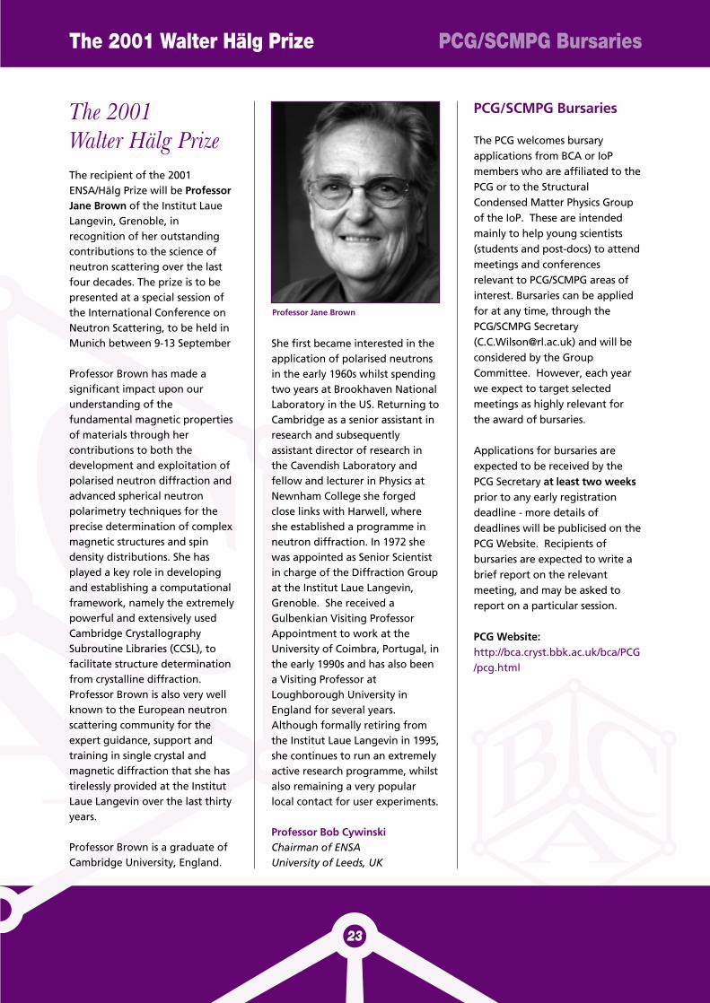
The 2001 Walter Hälg Prize PCG/SCMPG Bursaries
23
The 2001Walter Hälg PrizeThe recipient of the 2001ENSA/Hälg Prize will be ProfessorJane Brown of the Institut LaueLangevin, Grenoble, inrecognition of her outstandingcontributions to the science ofneutron scattering over the lastfour decades. The prize is to bepresented at a special session ofthe International Conference onNeutron Scattering, to be held inMunich between 9-13 September
Professor Brown has made asignificant impact upon ourunderstanding of thefundamental magnetic propertiesof materials through hercontributions to both thedevelopment and exploitation ofpolarised neutron diffraction andadvanced spherical neutronpolarimetry techniques for theprecise determination of complexmagnetic structures and spindensity distributions. She hasplayed a key role in developingand establishing a computationalframework, namely the extremelypowerful and extensively usedCambridge CrystallographySubroutine Libraries (CCSL), tofacilitate structure determinationfrom crystalline diffraction.Professor Brown is also very wellknown to the European neutronscattering community for theexpert guidance, support andtraining in single crystal andmagnetic diffraction that she hastirelessly provided at the InstitutLaue Langevin over the last thirtyyears.
Professor Brown is a graduate ofCambridge University, England.
She first became interested in theapplication of polarised neutronsin the early 1960s whilst spendingtwo years at Brookhaven NationalLaboratory in the US. Returning toCambridge as a senior assistant inresearch and subsequentlyassistant director of research inthe Cavendish Laboratory andfellow and lecturer in Physics atNewnham College she forgedclose links with Harwell, whereshe established a programme inneutron diffraction. In 1972 shewas appointed as Senior Scientistin charge of the Diffraction Groupat the Institut Laue Langevin,Grenoble. She received aGulbenkian Visiting ProfessorAppointment to work at theUniversity of Coimbra, Portugal, inthe early 1990s and has also beena Visiting Professor atLoughborough University inEngland for several years.Although formally retiring fromthe Institut Laue Langevin in 1995,she continues to run an extremelyactive research programme, whilstalso remaining a very popularlocal contact for user experiments.
Professor Bob CywinskiChairman of ENSAUniversity of Leeds, UK
PCG/SCMPG Bursaries
The PCG welcomes bursaryapplications from BCA or IoPmembers who are affiliated to thePCG or to the StructuralCondensed Matter Physics Groupof the IoP. These are intendedmainly to help young scientists(students and post-docs) to attendmeetings and conferencesrelevant to PCG/SCMPG areas ofinterest. Bursaries can be appliedfor at any time, through thePCG/SCMPG Secretary([email protected]) and will beconsidered by the GroupCommittee. However, each yearwe expect to target selectedmeetings as highly relevant forthe award of bursaries.
Applications for bursaries areexpected to be received by thePCG Secretary at least two weeksprior to any early registrationdeadline - more details ofdeadlines will be publicised on thePCG Website. Recipients ofbursaries are expected to write abrief report on the relevantmeeting, and may be asked toreport on a particular session.
PCG Website:http://bca.cryst.bbk.ac.uk/bca/PCG/pcg.html
Professor Jane Brown
CLRC e-Science Centre torun UK Grid Support Centre
The CLRC e-Science Centre hasbeen invited by Tony Hey, theDirector of the e-Science CoreProgramme, to set up and managethe Grid Support Centre for theUK on behalf of the programme.The CLRC e-Science Centre has alsoreceived funding from OST todevelop a Grid infrastructure forits own experimental, computingand data facilities in support of itsuser community. CLRC has invited Edinburgh andManchester Universities toparticipate in the Support Centreto ensure that the Centre hasaccess to the best experiencecurrently available in the UK onGrid middleware and services.
When it is operational, the Centrewill provide a range of services to
researchers in the UK who wish touse the Grid in their work. TheCentre will also be producing areport consolidating theexperience of all 3 partners withthe current generation of Gridtools.
For more information see websitehttp://www.e-science.clrc.ac.uk
Size Strain – IIIAnalysis of microstructure andresidual stress by diffractionmethods
December 2-5, 2001, TRENTO - ITALYInformation, on-line registration,on-line hotel reservation,instructions for authors, abstractsubmission, programme andupdates are available onlythrough the conference web-site:
http://bragg.ing.unitn.it/sizestrain
Mineralogy Database
This mineral database websitecontains more than 5,000 webpages of mineral data. It includesselected mineral definitions andmineral pictures. There are 4,205individual mineral speciesdescriptions with links. Mineraldata on individual species arelinked to a mineral table bycrystallography, X-Ray powderdiffraction, chemical composition,physical and optical properties,Dana's new classification, Strunzclassification, and alphabeticallistings of mineral species. Links toother sources of mineral dataavailable on the WWW are alsoincluded. Website address:http://webmineral.com
CLRC e-Science Centre Mineralogy Database
ANNOUNCEMENT
New Products for Protein Crystal Growth
Clear Strategy™ Screens I & IITwo new crystal growth screens offering rational planning of experiments, full control of pH, cryoprotection, and more.
Developed by the York Structural Biology Laboratory
Additive Screen24 popular crystal growth additives in a box and ready to use.
XRL™ PlateA raised lid Linbro style plate manufactured in the UK. No transatlantic shipping costs. Also available pregreased or use a Grease Machine
CrystalClene™ SlipsThe plastic coverslip guaranteed clean and RNase free. Now available round or square
Gelled-Surface™ CrystallizationAn improved approach to containerless crystallisation for optimisation
3D Structure Screen™A two step crystal growth trial. Separate nucleation from crystal growth for fewer, larger crystals
Saradakis & Chayen (Protein Science (2000), 9:755-757
Suppliers of kits and reagents for biomolecular structure research
61 - 63 Dudley Street, LUTON,BEDS., U.K. LU2 0NP
Tel & Fax:+44 (0)1582 482245,email: [email protected],
www.m-d-l.demon.co.uk
See the web site for details of these and all our products.www.m-d-l.demon.co.uk
Philips Analyticalintroduces a new, extremelyfast X-ray detector forpowder diffractometry: theX’Celerator. With thisdetector, hours of datacollection change intominutes, without anycompromise on dataquality.
� Ultrafast data collectionthrough RTMS (RealTime Multiple Strip)detection technology
� No-compromiseresolution for powdersand otherpolycrystalline materials
� Compact PreFIXmodule
� Maintenance-freedetection system
For more information:Philips AnalyticalPhilips HouseCambridge Business ParkCowley RoadCambridge CB4 0HATel. : 01223 468888Fax : 01223 468884
email:[email protected]
Internet:www.analytical.philips.com
New !
You’re always looking for the best.
That’s why we join to work on
your future needs in single crystal
diffraction. Now Bruker AXS and Nonius
have formed the new Bruker AXS business
unit named BRUKER NONIUS.
The focus of BRUKER NONIUS is to
accelerate the development of innovative
technologies for X-ray crystallography,
while strengthening worldwide service and
support.
Cutting-edge technologies, highly
experienced and motivated employees,
and unique customer dedication –
BRUKER NONIUS combines and
enhances the best elements of two
leading X-ray companies.
There are now three
BRUKER NONIUS centres of
competence: Delft, Madison, and
Karlsruhe
U.K.:
BRUKER AXS LTD.
Meadowside,
Mountbatten Way,
CONGLETON,
Cheshire
CW12 1DN U.K.
Tel: (+44) (1260) 296900
Fax: (+44) (1260) 296909
www.bruker-axs.co.uk
North America:
BRUKER AXS INC.
5465 East Cheryl Parkway
MADISON, WI 53711
USA
Tel. (+1) (608) 276-3000
Fax (+1) (608) 276-3006
www.bruker-axs.com
The Netherlands:
BRUKER NONIUS B.V.
P.O. Box 811
2600 AV DELFT
THE NETHERLANDS
Tel. (+31) (15) 2698300
Fax (+31) (15) 2627401
www.nonius.nl
AIM
009
77
NOW YOUR SINGLE CRYSTAL WORLD IS IN GOOD HANDS!
BRUKER AXS AND NONIUS – A WINNING COMBINATION
BRUKER ADVANCED X-RAY SOLUTIONS
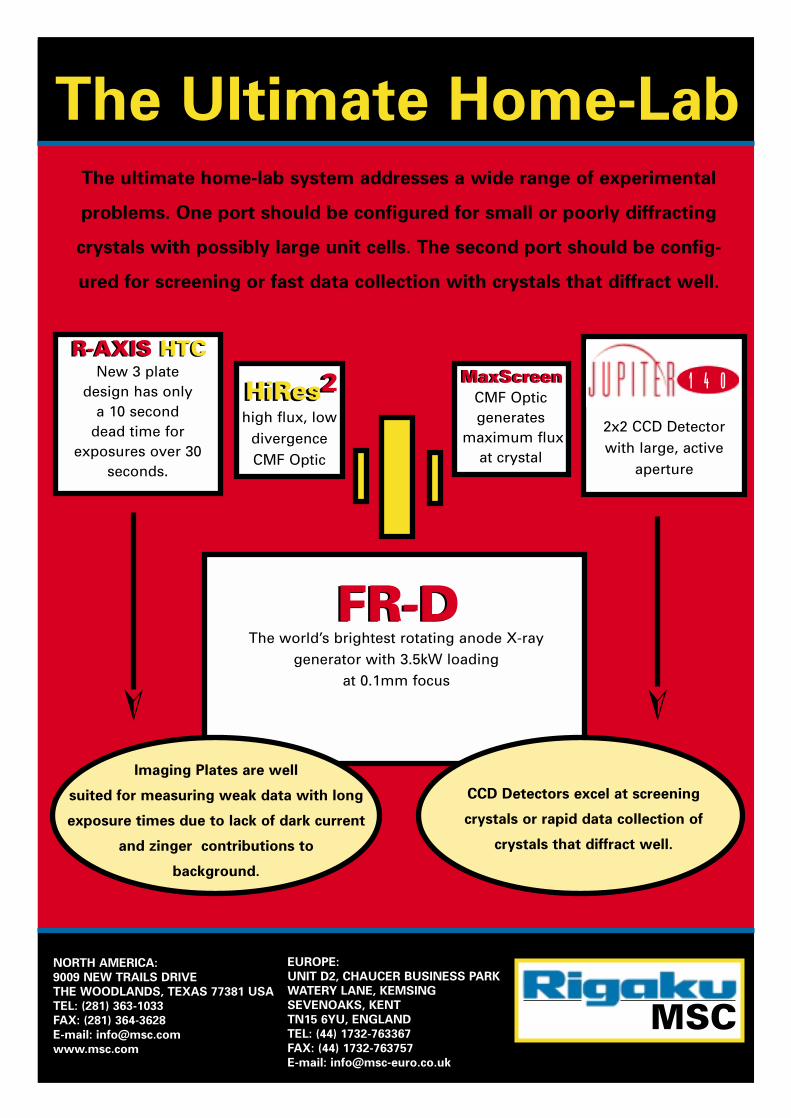
The ultimate home-lab system addresses a wide range of experimental
problems. One port should be configured for small or poorly diffracting
crystals with possibly large unit cells. The second port should be config-
ured for screening or fast data collection with crystals that diffract well.
EUROPE:
UNIT D2, CHAUCER BUSINESS PARK
WATERY LANE, KEMSING
SEVENOAKS, KENT
TN15 6YU, ENGLAND
TEL: (44) 1732-763367
FAX: (44) 1732-763757
E-mail: [email protected]
NORTH AMERICA:
9009 NEW TRAILS DRIVE
THE WOODLANDS, TEXAS 77381 USA
TEL: (281) 363-1033
FAX: (281) 364-3628
E-mail: [email protected]
www.msc.com
➢➢
R-AXIS HTCNew 3 plate
design has only a 10 second
dead time for exposures over 30
seconds.
HiRes2high flux, low
divergence CMF Optic
2x2 CCD Detector with large, active
aperture
MaxScreenCMF Opticgenerates
maximum fluxat crystal
FR-DThe world’s brightest rotating anode X-ray
generator with 3.5kW loading at 0.1mm focus
The Ultimate Home-Lab
Imaging Plates are well
suited for measuring weak data with long
exposure times due to lack of dark current
and zinger contributions to
background.
CCD Detectors excel at screening
crystals or rapid data collection of
crystals that diffract well.
HiRes2R-AXIS HTC
FR-D
MaxScreen 1 4 0
MSC
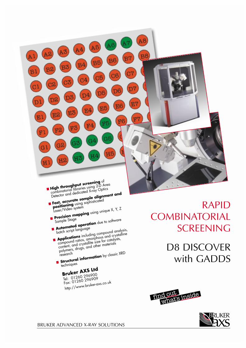
RAPIDCOMBINATATA ORIAL
SCREENING
D8 DISCOVERwith GADDS
BRUKER ADVANCEDVANCED V X-RAYAYA SOLUTIONS

Collecting x-ray data from frozen crystals
can be a tricky business.
Mounting samples and freezing them
needs careful attention to many variables all
of which have to be just right to guarantee
success.
When it comes to the Oxford Cryostream
Cooler, we like to think just that little bit
Cooler thinking...
harder on your behalf to make sure
everything goes to plan. With all of our new
technologies and innovative ideas, we pride
ourselves in our Cooler Thinking.
OxfordCryosystems
Head Office3 Blenheim Office Park Lower Road . Long HanboroughOxford . OX8 8LN . UKT. +44 (0)1993 883488 F +44 (0)1993 883988E. [email protected]
w w w . O x f o r d C r y o s y s t e m s . c o m / 7 0 0 s e r i e s /
North American Office220 Wood Road . Braintree
MA 02184-2403 . USAT. 1-866-OXCRYO-8
781-843-5900F. 781-843-5945