Embed Size (px)
Citation preview

Cross-reactivity of mAbs to human CD antigens
with cells from cattle
Paul Sopp a,*, Dirk Werling b, Cynthia Baldwin c
a Institute for Animal Health, Compton RG20 7NN, UKb The Royal Veterinary College, London, UK
c Department of Veterinary & Animal Sciences, University of Massachusetts, Amherst, MA 01003, USA
Abstract
A panel of 377 commercially available mAbs were submitted to the animal homologue section of the 8th International Workshop
on Human Leukocyte Differentiation Antigens (HLDA8, Adelaide, Australia) for cross-reactivity studies on different animal
species. In this study we describe the results of testing the mAbs on cattle cells by flow cytometry and Western blot. Eight
commercial suppliers participated, providing mAbs to a total of 144 CD antigens plus controls. Fifty-two mAbs were identified as
potentially staining cattle cells in the first round screen. In the second phase, 38 mAbs were confirmed as staining cattle cells. This
included some that may recognise polymorphic determinants and others with atypical distribution patterns compared to humans.
mAb to human CD9, CD11a, CD14, CD18, CD21, CD23, CD29, CD44, CD45R, CD47, CD49d and CD172a cross-reacted with
bovine cells and mAb to CD22, CD88, CD119 and CD163 stained CD antigens that have not previously been identified in cattle.
# 2007 Elsevier B.V. All rights reserved.
Keywords: Cattle; CD; Cross-reactivity
www.elsevier.com/locate/vetimm
Veterinary Immunology and Immunopathology 119 (2007) 106–114
1. Introduction
The testing of mAbs for cross-species reactivity has
proven useful for identifying reagents to previously
unrecognised antigens in several different species.
Three International Workshops on Ruminant Antigens
(Howard et al., 1991; Howard and Naessens, 1993;
Naessens and Hopkins, 1996) have revealed, as expected
due to their close evolutionary relationship, a high degree
of cross-species reactivity of mAbs to cattle, sheep and
goat antigens. Studies of mAbs to ruminant antigens on
cells from non-ruminants also revealed several cross-
reactions with canine, swine and human cells (Schuberth
et al., 1996; Vilmos et al., 1996).
* Corresponding author. Tel.: +44 1635 577292;
fax: +44 1635 577263.
E-mail address: [email protected] (P. Sopp).
0165-2427/$ – see front matter # 2007 Elsevier B.V. All rights reserved.
doi:10.1016/j.vetimm.2007.06.014
To date the most comprehensive studies of cross-
reactivities of human anti-CD mAb for bovine molecules
have involved the testing of mAbs submitted to the
International Workshops on Human Leukocyte Differ-
entiation Antigens (HLDA) (Sopp and Howard, 1997;
Sopp et al., 2001). In these, large panels of antibodies,
representative of most of the available ‘anti-CD’ antigens
mAbs, were screened on bovine cells and about 10%
were found to cross-react. In many cases the potentially
useful cross-reactive mAbs were not easily available and
consequently the reagents have not been fully char-
acterised. Those previously recognized are in Table 1. To
combat this problem a panel of 377 commercially
available mAbs (including controls) was organised by
representatives of the Veterinary Immunology Commit-
tee of the International Union of Immunological
Societies (VIC-IUIS) for testing on a wide range of
animal species. This panel included the widest possible

P. Sopp et al. / Veterinary Immunology and Immunopathology 119 (2007) 106–114 107
Table 1
Summary of previous studies of human CD mAbs on cattle cells
Typical No. of mAbs identified Atypical No. of mAbs identified
HLDA 5 HLDA 6 HLDA 5 HLDA 6
CD11a 1 CDw17 1
CD14 9 2 CD19 1
CD18 2 2 CD35 1
CD21 8 3 CD37 1
CD23 1 CD49c 1
CD27 1 2 CD50 1
CD29 7 2 CD54 1
CD39 1 CD55 1
CD44 3 CD56 1
CD47 2 CD66 3 3
CD49a 2 1 CD71 1
CD49b 3 CD74 1
CD49d 3 CD81 1
CD49e 2 1 CD86 3
CD51/CD61 5 CD88 2
CD62L 3 CD102 1
CD62P 1 CD105 1
CD63 3 CD117 5
CDw78 1 CD120b 1
CD98 1 CD122 1
CD100 1 2 CD165 1
Typical: staining patterns consistent on cattle cells compared to human cells; Atypical: different cellular distribution in cattle compared to humans;
HLDA 5: summary of results from 5th International Human Leucocyte Differentiation Antigen Workshop (Sopp and Howard, 1997); HLDA 6:
summary of results from 6th International Human Leucocyte Differentiation Antigen Workshop (Sopp et al., 2001).
range of CD mAbs available from the donors. In this
study we describe the testing of the mAbs on cattle cells
where the aim was to identify, as far as possible,
commercially available mAbs that react with the cattle
homologue of the human antigen.
2. Materials and methods
2.1. Monoclonal antibodies
Thefull list of mAbs and their suppliers are listed in the
chapter ‘‘Summary of the Animal homologue section of
HLDA8’’ of this volume. All mAbs were used at a
concentration specified by the suppliers and diluted in
buffer (PBS supplemented with 1% (w/v) BSA and 0.1%
(w/v) NaN3). Most mAbs were of murine origin, purified
and unconjugated but a few were directly conjugated with
FITC or R-PE. A small minority were rat Ig. The anti-
bovine mAbs CC42 (CD2, Davis and Splitter, 1991), CC-
G33 (CD14, Sopp et al., 1996), CC21 (CD21, Naessens
et al., 1990) and CC15 (WC1, Morrison and Davis, 1991)
were used as biotin conjugates in two colour flow
cytometry (FCM). These mAbs were used to identify the
main sub-populations in peripheral blood (PB): CD2 for
ab-T cells and some gd-T cells, CD14 for monocytes,
CD21 for B cells and WC1 for CD2� gd-T cells.
2.2. Preparation of cells
Peripheral blood samples were prepared by density
gradient centrifugation using standard techniques to
yield mononuclear cells (PBMC) or by red blood cell
lysis using ammonium chloride buffer to give a
leucocyte suspension (WBC) (Sopp et al., 2001) and
resuspended in buffer. Cells from two animals were
assayed at both laboratory sites (Institute for Animal
Health and the University of Massachusetts) in both the
first and second round studies.
2.3. Staining cells for FCM
The first round studies involved single colour FCM
and the second phase included one and two colour FCM.
Standard indirect staining techniques using fluorochrome
conjugates were employed (Sopp et al., 2001) except for
the directly labelled mAbs. Goat F(ab0)2 anti-mouse Ig
FITC and goat anti-rat FITC (Southern Biotech,
Birmingham, AL, USA) were used as secondary reagents
where appropriate for the HLDA8 mAb panel and strep-
tavidin–phycoerythrin conjugate (Southern Biotech) was
used to detect the biotinylated mAbs. The cells were
assayed using a FACSCalibur (Becton Dickinson, San
Jose, CA).

P. Sopp et al. / Veterinary Immunology and Immunopathology 119 (2007) 106–114108
2.4. Analysis of FCM data
Data were analysed using CellQuest (Becton
Dickinson) or FCS Express (De Novo Software,
Ontario, Canada). Mononuclear cells and polymorpho-
nuclear cells were distinguished by gating using low
angle light scatter (forward scatter, FSC) and orthogo-
nal light scatter (side scatter, SSC) and each cell type
was analysed separately. Selection of mAbs that stained
bovine cells in the first round was determined by
comparison with control mAbs. After background
(control mAb) subtraction, any mAbs that stained cells
at �1% of total gated cells were assessed and the
staining patterns examined. All of these mAbs were
included in the second phase unless the observed
staining patterns and proportion of stained cells were in
total discordance with the published human data at
which point it was concluded to be an erroneous
reactivity. In the second round, one colour FCM was
repeated on the restricted panel of mAbs and more
detailed analyses were performed by two colour FCM.
In this, the two-parameter plots of data obtained with
mAb against humans molecules and those mAb known
to be reactive with bovine markers were studied in detail
for consistency of their pattern of reactivity with those
reported for human cells.
2.5. Western blot analysis
Whole cell lysates from bovine or human PBMC were
prepared as described (Werling et al., 2004). Briefly, cells
were harvested, counted, and washed once with ice cold
PBS (300 � g, 10 min, 4 8C), transferred to an Eppen-
dorf tube and pelleted (2000 � g, 10 min, 4 8C). Cell
pellets (8 � 107) were lysed in 500 ml of lysis buffer,
Mammalian Protein Extraction Reagent (M-PERM,
Pierce, Oxnard, CA), supplemented with 50 mM sodium
fluoride, 1 mM sodium vanadate, 0.5 mM PMSF,
10 mg ml�1 aprotinin, 10 mg ml�1 leupeptin (all reagents
from Roche Diagnostics, Rotkreuz, Switzerland). The
cellular extract was sonicated for 15 s on ice, allowed to
sit for 20 min, and then centrifuged at 15,000 � g for
10 min. The supernatant was removed and boiled for
3 min with 5� Laemmli buffer. Twenty microliters of
each sample was loaded onto a 10% SDS-PAGE gel, and
run at 100 V for 1.5 h in a MiniProtean chamber (BioRad,
Reinach, Switzerland). Cell proteins were blotted onto
nitrocellulose (ECL; Amersham, Arlington Heights, IL)
at 40 V for 4 h. The nitrocellulose was blocked with 5%
milk powder in PBS with 0.05% Tween 20 (TPBS)
overnight, washed, and incubated for 2 h at room
temperature with the primary antibody and appropriate
controls all diluted 1:200 in TPBS. Blots were washed
five times with TPBS and incubated for 1 h with
horseradish peroxidase-conjugated anti-rat IgG or anti-
mouse IgG antibody, respectively (Amersham, diluted
1:3000 in TPBS). Immunoreactive bands were developed
using a chemiluminescent substrate (ECL; Amersham).
Antibody specificity was verified by comparing the size
of positive bands with published molecular weights
under denaturing conditions.
3. Results and discussion
3.1. FCM—first round screening
Fifty-two mAbs from the first phase studies were
selected for further analysis in the second round
(Table 2). Many of the mAbs gave distinctive staining
and were good candidates for cross-reactive mAb.
However for others, where the expected target popula-
tion was small, there was significant doubt whether the
staining was genuinely against the bovine homologue
and not just unusually high non-specific background
staining. For some mAb PB cells were not ideal targets
and it is possible that a few genuinely cross-reactive
mAb may have been missed. As an example, CD34
positive cells are rare in peripheral blood except in
neonates so we cannot exclude the possibility that one
or more of the CD34 mAb were cross-reactive. With the
limited amount of mAb supplied to the study it was not
possible to test a wide range of targets. Another
potential problem that was not easy to overcome was the
possibility that mAbs were not used at their optimal
concentration on bovine cells. However, it is fully
expected that the optimal dilution for use on human
cells, determined by the supplier and used here, would
be suitable in most cases.
3.2. Second round analyses
The aims of the second round included: (1) repeating
the results of the first round to eliminate spurious data;
(2) to look more critically at the staining patterns, in
both one and two colour FCM and compare these with
published data of human CD antigens; (3) to attempt to
identify the MW of the antigen recognized by Western
blotting and compare this with the human molecule.
3.3. FCM—second round studies
Twenty-six mAbs (indicated by +) in Table 2 reacted
with bovine cells where the staining pattern was
consistent with recognising the bovine homologue of

P. Sopp et al. / Veterinary Immunology and Immunopathology 119 (2007) 106–114 109
Table 2
Results and interpretation of second round FCM assays
CD Clone Supplier Percentage of indicated sub-population
stained by mAb
Site assessment Overall
assessment
%CD2 %WC1 %CD21 %CD14 IAHa UMASSb
CD1a B17.20.9 Coulter 3.4 6.6 39.5 40.4 + � P
CD9 RH1A VMRD 7.9 60.1 19.1 89.5 + + +
CD9 LT86A VMRD 12.7 66.2 43.8 90.3 + + +
CD9 MM2/57 Serotec 8.6 61.2 15.9 78.8 + + +
CD10 SN5c Serotec 2.6 2.9 8.0 13.7 +/? �/? A
CD10 ALB1 Coulter NT NT NT NT NT + P
CD11a HUH73A VMRD 74.9 69.6 54.6 98.7 + + +
CD14 CAM36A VMRD 2.4 2.5 9.3 92.4 + + +
CD14 TUK4 Dako 1.4 2.8 7.6 92.0 + + +
CD14 M5E2 BD
Pharmingen
1.2 3.5 7.6 87.9 + + +
CD16 3G8 Coulter 0.9 2.5 3.8 4.1 � � �CD18 BAQ30A VMRD 67.0 85.1 78.0 99.5 + + +
CD18 H20A VMRD 50.0 52.0 63.7 99.4 + + +
CD18 7E4 Coulter 36.8 41.9 66.3 98.2 + + +
CD18 MHM23 Dako 88.9 87.7 79.8 99.6 + + +
CD21 LB21 Serotec 3.6 6.4 91.1 5.4 + + +
CD21 BL13 Coulter 3.1 7.2 87.9 5.2 + � P
CD22 Mc64-12 Serotec 4.0 4.9 43.9 9.3 + + +
CD23 9P.25 Coulter 2.7 5.8 49.2 20.7 + NT +
CD25 B1.49.9 Coulter 2.7 6.3 23.1 41.2 +/? � ?
CD27 LT27 Serotec 4.3 6.6 38.3 22.9 +/? +/? ?
CD28 YTH913.12 Serotec 1.4 9.2 8.0 8.9 � � �CD29 3S3 Serotec 10.7 9.9 13.2 36.0 � + P
CD29 K20 Dako 33.2 16.1 21.5 72.9 + �/? +
CD32 AT10 Serotec 9.5 7.3 11.2 33.5 +/? � A
CD32 2 E1 Coulter 3.5 5.7 42.5 49.2 + � P
CD44 BAG40A VMRD 92.2 91.4 57.9 98.7 + + +
CD44 LT41A VMRD 83.4 96.3 40.9 95.5 + + +
CD45 DH16A VMRD 55.8 30.0 19.8 19.9 + + +
CD47 HUH69A VMRD 99.8 98.8 99.8 99.9 + + +
CD47 HUH71A VMRD 99.9 99.9 100.0 99.9 + + +
CD49d HP2/1 Coulter 59.9 58.8 76.9 95.9 + �/? +
CD49d P4G9 Dako 38.3 31.8 37.6 61.3 + �/? +
CD49d 9F10 BD
Pharmingen
44.4 40.5 58.3 89.2 + + +
CD51/61 23C6 Serotec 1.1 2.3 4.0 3.5 � � ?
CD59 MUC93A VMRD 1.0 2.5 4.6 3.3 � � P
CD61 PM6/13 Serotec 2.1 3.5 6.3 5.5 � �/+ ?
CD61 Y2/51 Dako 2.2 4.9 5.8 4.1 � � ?
CD62L FMC46 Dako 52.8 90.0 48.2 91.6 + � P
CD 62P AK-6 Dako 1.6 3.7 7.1 5.6 � � ?
CD69 TP1.55.3 Coulter 2.3 4.6 10.6 4.0 ? � �CD88 S5/1 Serotec 29.9 15.5 65.1 80.5 + + A
CD88 W17/1 Serotec 47.0 74.5 22.9 76.0 + NT A
CD119 BB1E2 Serotec 23.1 6.4 82.8 78.5 + �/? +
CD120a H398 Serotec 1.3 2.5 4.6 3.9 � � �CD163 Ber-MAC3 Dako 1.5 3.7 6.7 78.2 + +/? +
CD165 AD2-13H12 Serotec 3.6 4.9 38.7 16.6 ? � A
CD166 3A6 Serotec 4.4 3.5 6.7 25.8 ? �/? ?
CD172a DH59B VMRD 2.6 4.8 30.4 97.6 + + +
CD200 MRC-OX-104 Serotec 3.7 3.5 12.2 2.8 � + A/P
CD236R Ret40f Dako 1.3 3.4 6.0 3.2 � � �CD247 G3 Serotec 1.8 2.5 5.5 1.9 � � �G1 control Dako Dako 1.2 4.2 5.7 2.4

P. Sopp et al. / Veterinary Immunology and Immunopathology 119 (2007) 106–114110
Table 2 (Continued )
CD Clone Supplier Percentage of indicated sub-population
stained by mAb
Site assessment Overall
assessment
%CD2 %WC1 %CD21 %CD14 IAHa UMASSb
IgM control Dako Dako 1.9 4.0 6.2 3.3
G2a control Dako Dako 1.6 3.5 5.6 2.0
G2b control Dako Dako 1.9 3.4 6.1 2.9 Background controls
(+) Stained cattle cells; (�) no reaction; (A) atypical staining pattern compared to humans; (P) may recognise a polymorphic determinant; (?)
insufficient evidence to make a conclusion.a Data generated at the Institute for Animal Health.b University of Massachusetts.
Table 3
Results of Western blot assays of human and cattle cell lysates
CD Clone MW (kDa) of band
Human Cattle
CD9 RH1A 30–45 30–45
CD9 LT86A ? –
CD10 SN5c – –
CD11a HUH73A 97–220 –
CD22 Mc64-12 97–220 –
CD23 9P.25 45–50 –
CD27 LT27 – –
CD44 BAG40A 66–97 –
CD44 LT41A 66–97 –
CD49d HP2/1 97–220 –
CD69 TP1.55.3 20–45 –
CD88 S5/1 30–45 30–45
CD88 W17/1 30–45 –
CD119 BB1E2 50 (?) –
CD163 Ber-MAC3 45–66 (?) –
CD247 G3 – –
(?) Uncertain results/faint bands, – = no band detected.
the human CD antigen. This included mAbs to CD9
(although strong staining of PMN is atypical), CD11a,
CD14, CD18, CD21, CD22, CD23, CD29, CD44,
CD45R, CD47, CD49d, CD119, CD163 and CD172a;
examples of staining patterns are shown in Fig. 1.
Within this group were mAbs Tuk4 and M5E2 (CD14),
BL13 (CD21), 9P25 (CD23), K20 (CD29) and 9F10
(CD49d)—the cross-reactivity of these has been
previously reported (Tuk4 and M5E2 (CD14), BL13
(CD21), K20 (CD29), 9F10 (CD49d): Sopp and
Howard, 1997; 9P25 (CD23): Sopp et al., 2001).
With some mAbs the staining patterns of bovine cells
were inconsistent with those reported in humans
(marked ‘A’ in Table 2) and it is not possible to
conclude that they recognise bovine homologues.
However the staining patterns suggested that genuine
reactions and not non-specific binding were seen and
thus these mAb should be re-examined, possibly on a
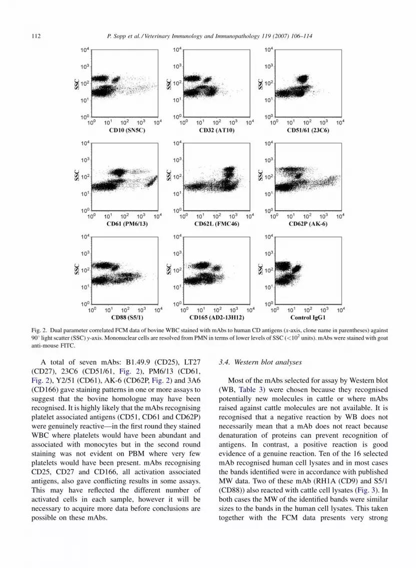
broader range of targets. CD10 mAbs generally stained a
small proportion of PBM but failed to significantly stain
PMN (Fig. 2) in contrast to reports in humans where
PMN are recognised. CD32 mAbs also failed to stain
PMN (Fig. 2) this is different to reports in humans and
also dissimilar to results with mAbs raised against
bovine CD32 (Zhang et al., 1994). CD88 mAbs (W17/1
and S5/1) strongly stained a sub-population of PBM
(Fig. 2) but once again did not stain PMN, this
observation was also reported by Sopp and Howard
(1997). S5/1 recognised a protein by Western blot of the
expected size (approximately 30–45 kDa) adding to the
evidence that the cattle homologue of CD88 was
identified (Table 3). mAb AD2-13H12 which recognises
human CD165 and is reported to react with thymocytes
and thymic epithelial cells weakly stained most bovine
PB (Fig. 2). In summary, most of the mAbs that
recognised bovine cells with an atypical staining pattern
compared to humans did so because of their non-
reactivity with PMN, this indicated that either bovine
PMN are significantly different in their surface marker
expression compared to human PMN or alternatively the
bovine cells are altered for surface marker expression
during or as a result of the preparation method.
An unexpected result of this study was the apparent
abundance of mAbs that may recognise polymorphic
epitopes of antigens—this was characterised by staining
of bovine cells in one or more animals at one laboratory
but not the second laboratory. Polymorphisms in cattle
have been reported for CD4 and CD5 (Morrison et al.,
1991) but are relatively rare. It is unlikely that technical
errors occurred but it will be necessary to repeat the
results on a diverse range of cattle types before solid
conclusions can be made. The staining pattern for each
of the mAbs; B17.20.9 (CD1a), ALB1 (CD10), BL13
(CD21), 2E1 (CD32), MUC93A (CD59), FMC46
(CD62L) and MRC-OX-104 (CD200) marked ‘P’ in
Table 2 were distinctive and with the exception of
CD10, CD32 (see above) and CD200 were consistent
with the mAb recognising the bovine homologue of the
human antigen.

P. Sopp et al. / Veterinary Immunology and Immunopathology 119 (2007) 106–114 111
Fig. 1. Two colour immunofluorescence staining of bovine PBMC. mAbs to human CD antigens (indicated on x-axis with clone names in
parentheses) and mAbs to characterised bovine antigens (indicated on y-axis). mAbs to bovine antigens were biotinylated and detected using
streptavidin–phycoerythrin; mAbs to human antigens were stained with goat anti-mouse FITC. Fluorescence was assayed by FCM.
P. Sopp et al. / Veterinary Immunology and Immunopathology 119 (2007) 106–114112
Fig. 2. Dual parameter correlated FCM data of bovine WBC stained with mAbs to human CD antigens (x-axis, clone name in parentheses) against
908 light scatter (SSC) y-axis. Mononuclear cells are resolved from PMN in terms of lower levels of SSC (<102 units). mAbs were stained with goat
anti-mouse FITC.
A total of seven mAbs: B1.49.9 (CD25), LT27
(CD27), 23C6 (CD51/61, Fig. 2), PM6/13 (CD61,
Fig. 2), Y2/51 (CD61), AK-6 (CD62P, Fig. 2) and 3A6
(CD166) gave staining patterns in one or more assays to
suggest that the bovine homologue may have been
recognised. It is highly likely that the mAbs recognising
platelet associated antigens (CD51, CD61 and CD62P)
were genuinely reactive—in the first round they stained
WBC where platelets would have been abundant and
associated with monocytes but in the second round
staining was not evident on PBM where very few
platelets would have been present. mAbs recognising
CD25, CD27 and CD166, all activation associated
antigens, also gave conflicting results in some assays.
This may have reflected the different number of
activated cells in each sample, however it will be
necessary to acquire more data before conclusions are
possible on these mAbs.
3.4. Western blot analyses
Most of the mAbs selected for assay by Western blot
(WB, Table 3) were chosen because they recognised
potentially new molecules in cattle or where mAbs
raised against cattle molecules are not available. It is
recognised that a negative reaction by WB does not
necessarily mean that a mAb does not react because
denaturation of proteins can prevent recognition of
antigens. In contrast, a positive reaction is good
evidence of a genuine reaction. Ten of the 16 selected
mAb recognised human cell lysates and in most cases
the bands identified were in accordance with published
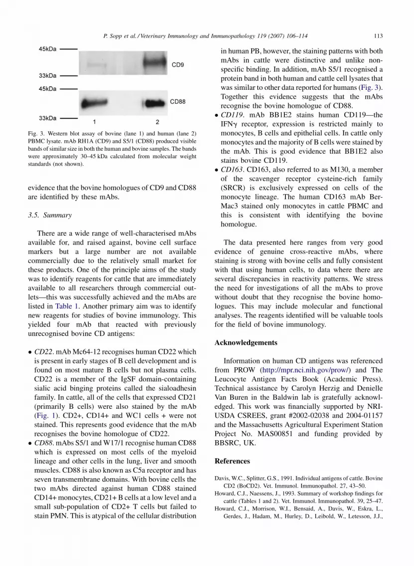
MW data. Two of these mAb (RH1A (CD9) and S5/1
(CD88)) also reacted with cattle cell lysates (Fig. 3). In
both cases the MW of the identified bands were similar
sizes to the bands in the human cell lysates. This taken
together with the FCM data presents very strong
P. Sopp et al. / Veterinary Immunology and Immunopathology 119 (2007) 106–114 113
Fig. 3. Western blot assay of bovine (lane 1) and human (lane 2)
PBMC lysate. mAb RH1A (CD9) and S5/1 (CD88) produced visible
bands of similar size in both the human and bovine samples. The bands
were approximately 30–45 kDa calculated from molecular weight
standards (not shown).
evidence that the bovine homologues of CD9 and CD88
are identified by these mAbs.
3.5. Summary
There are a wide range of well-characterised mAbs
available for, and raised against, bovine cell surface
markers but a large number are not available
commercially due to the relatively small market for
these products. One of the principle aims of the study
was to identify reagents for cattle that are immediately
available to all researchers through commercial out-
lets—this was successfully achieved and the mAbs are
listed in Table 1. Another primary aim was to identify
new reagents for studies of bovine immunology. This
yielded four mAb that reacted with previously
unrecognised bovine CD antigens:
� CD22. mAb Mc64-12 recognises human CD22 which
is present in early stages of B cell development and is
found on most mature B cells but not plasma cells.
CD22 is a member of the IgSF domain-containing
sialic acid binging proteins called the sialoadhesin
family. In cattle, all of the cells that expressed CD21
(primarily B cells) were also stained by the mAb
(Fig. 1). CD2+, CD14+ and WC1 cells + were not
stained. This represents good evidence that the mAb
recognises the bovine homologue of CD22.
� CD88. mAbs S5/1 and W17/1 recognise human CD88
which is expressed on most cells of the myeloid
lineage and other cells in the lung, liver and smooth
muscles. CD88 is also known as C5a receptor and has
seven transmembrane domains. With bovine cells the
two mAbs directed against human CD88 stained
CD14+ monocytes, CD21+ B cells at a low level and a
small sub-population of CD2+ T cells but failed to
stain PMN. This is atypical of the cellular distribution
in human PB, however, the staining patterns with both
mAbs in cattle were distinctive and unlike non-
specific binding. In addition, mAb S5/1 recognised a
protein band in both human and cattle cell lysates that
was similar to other data reported for humans (Fig. 3).
Together this evidence suggests that the mAbs
recognise the bovine homologue of CD88.
� CD119. mAb BB1E2 stains human CD119—the
IFNg receptor, expression is restricted mainly to
monocytes, B cells and epithelial cells. In cattle only
monocytes and the majority of B cells were stained by
the mAb. This is good evidence that BB1E2 also
stains bovine CD119.
� CD163. CD163, also referred to as M130, a member
of the scavenger receptor cysteine-rich family
(SRCR) is exclusively expressed on cells of the
monocyte lineage. The human CD163 mAb Ber-
Mac3 stained only monocytes in cattle PBMC and
this is consistent with identifying the bovine
homologue.
The data presented here ranges from very good
evidence of genuine cross-reactive mAbs, where
staining is strong with bovine cells and fully consistent
with that using human cells, to data where there are
several discrepancies in reactivity patterns. We stress
the need for investigations of all the mAbs to prove
without doubt that they recognise the bovine homo-
logues. This may include molecular and functional
analyses. The reagents identified will be valuable tools
for the field of bovine immunology.
Acknowledgements
Information on human CD antigens was referenced
from PROW (http://mpr.nci.nih.gov/prow/) and The
Leucocyte Antigen Facts Book (Academic Press).
Technical assistance by Carolyn Herzig and Denielle
Van Buren in the Baldwin lab is gratefully acknowl-
edged. This work was financially supported by NRI-
USDA CSREES, grant #2002-02038 and 2004-01157
and the Massachusetts Agricultural Experiment Station
Project No. MAS00851 and funding provided by
BBSRC, UK.
References
Davis, W.C., Splitter, G.S., 1991. Individual antigens of cattle. Bovine
CD2 (BoCD2). Vet. Immunol. Immunopathol. 27, 43–50.
Howard, C.J., Naessens, J., 1993. Summary of workshop findings for
cattle (Tables 1 and 2). Vet. Immunol. Immunopathol. 39, 25–47.
Howard, C.J., Morrison, W.I., Bensaid, A., Davis, W., Eskra, L.,
Gerdes, J., Hadam, M., Hurley, D., Leibold, W., Letesson, J.J.,

P. Sopp et al. / Veterinary Immunology and Immunopathology 119 (2007) 106–114114
et al., 1991. Summary of workshop findings for leukocyte antigens
of cattle. Vet. Immunol. Immunopathol. 27, 21–27.
Morrison, W.I., Davis, W.C., 1991. Individual antigens of cattle.
Differentiation antigens expressed predominantly on CD4�CD8� T lymphocytes (WC1, WC2). Vet. Immunol. Immuno-
pathol. 27, 71–76.
Morrison, W.I., Howard, C.J., Hinson, C.A., 1991. Polymorphism of
the CD4 and CD5 differentiation antigens in cattle. Vet. Immunol.
Immunopathol. 27, 235–238.
Naessens, J., Hopkins, J., 1996. Introduction and summary of work-
shop findings. Vet. Immunol. Immunopathol. 52, 213–235.
Naessens, J., Newson, J., McHugh, N., Howard, C.J., Parsons, K.,
Jones, B., 1990. Characterization of a bovine leucocyte differ-
entiation antigen of 145,000 MW restricted to B lymphocytes.
Immunology 69, 525–530.
Schuberth, H.J., Rabe, H.U., Lange, A., Leibold, W., 1996. Reactivity
of monoclonal antibodies with bovine blood mononuclear cells
activated by mitogens and superantigens. Vet. Immunol. Immu-
nopathol. 52, 313–321.
Sopp, P., Howard, C.J., 1997. Cross-reactivity of monoclonal anti-
bodies to defined human leucocyte differentiation antigens with
bovine cells. Vet. Immunol. Immunopathol. 56, 11–25.
Sopp, P., Kwong, L.S., Howard, C.J., 1996. Identification of bovine
CD14. Vet. Immunol. Immunopathol. 52, 323–328.
Sopp, P., Kwong, L.S., Howard, C.J., 2001. Cross-reactivity with
bovine cells of monoclonal antibodies submitted to the 6th Inter-
national Workshop on Human Leukocyte Differentiation Anti-
gens. Vet. Immunol. Immunopathol. 78, 197–206.
Vilmos, P., Kurucz, E., Ocsovszki, I., Keresztes, G., Ando, I., 1996.
Phylogenetically conserved epitopes of leukocyte antigens. Vet.
Immunol. Immunopathol. 52, 415–426.
Werling, D., Hope, J.C., Howard, C.J., Jungi, T.W., 2004. Differential
production of cytokines, reactive oxygen and nitrogen by bovine
macrophages and dendritic cells stimulated with Toll-like receptor
agonists. Immunology 111, 41–52.
Zhang, G., Young, J.R., Tregaskes, C.R., Howard, C.J., 1994. Cattle Fc
gamma RII: molecular cloning and ligand specificity. Immuno-
genetics 39, 423–427.


