Embed Size (px)
Citation preview
Covalent heme attachment to the protein in human hemeoxygenase-1 with selenocysteine replacing the His25 proximaliron ligand
Yongying Jiang, Michael J. Trnka, Katalin F. Medzihradszky, Hugues Ouellet, YongqiangWang, and Paul R. Ortiz de Montellano*Department of Pharmaceutical Chemistry, University of California, 600 16th Street, San Francisco,California 94158-2517
AbstractTo characterize heme oxygenase with a selenocysteine (SeCys) as the proximal iron ligand, we haveexpressed truncated human heme oxygenase-1 (hHO-1) His25Cys, in which Cys-25 is the onlycysteine, in the Escherichia coli cysteine auxotroph strain BL21(DE3)cys. Selenocysteineincorporation into the protein was demonstrated by both intact protein mass measurement and massspectrometric identification of the selenocysteine-containing tryptic peptide. One selenocysteine wasincorporated into approximately 95% of the expressed protein. Formation of an adduct with Ellman'sreagent (DTNB) indicated that the selenocysteine in the expressed protein was in the reduced state.The heme-His25SeCys hHO-1 complex could be prepared by either (a) supplementing theoverexpression medium with heme, or (b) reconstituting the purified apoprotein with heme. Underreducing conditions in the presence of imidazole, a covalent bond is formed by addition of theselenocysteine residue to one of the heme vinyl groups. No covalent bond is formed when the hemeis replaced by mesoheme, in which the vinyls are replaced by ethyl groups. These results, togetherwith our earlier demonstration that external selenolate ligands can transfer an electron to the iron(Jiang, Y., Ortiz de Montellano, P.R., Inorg. Chem., 47, 3480-3482 (2008)), indicate that a selenylradical is formed in the hHO1 His25SeCys mutant that adds to a heme vinyl group.
Keywordsheme oxygenase; selenocysteine; heme covalent binding; selenium-iron ligation; electronic spectra;mass spectrometry
1. IntroductionHuman heme oxygenase-1 (hHO-1) catalyzes the NADPH- and cytochrome P450 reductase(CPR)-dependent conversion of heme to biliverdin, a multistep process in which the enzymefirst oxidizes heme to α-meso-hydroxyheme, then via loss of CO to verdoheme, and finally toiron biliverdin (1,2). Reduction of the iron biliverdin to the ferrous state by electron transferfrom CPR results in product dissociation from the enzyme (3). The three products of thereaction are consequently CO, ferrous iron, and biliverdin. Furthermore, this multisteptransformation is unusual in that the enzyme utilizes heme as both a prosthetic group and asubstrate. The first of the three oxidative steps, the conversion of heme to α-meso-hydroxyheme, superficially resembles a cytochrome P450-catalyzed monooxygenation, but
*Corresponding author: Dr. Paul Ortiz de Montellano, University of California, Genentech Hall GH-N572D, 600 16th Street, SanFrancisco, CA 94143-2280, TEL: (415) 476-2903, FAX: (415) 502-4728, [email protected].
NIH Public AccessAuthor ManuscriptJ Inorg Biochem. Author manuscript; available in PMC 2010 March 1.
Published in final edited form as:J Inorg Biochem. 2009 March ; 103(3): 316–325. doi:10.1016/j.jinorgbio.2008.11.002.
NIH
-PA Author Manuscript
NIH
-PA Author Manuscript
NIH
-PA Author Manuscript
the hydroxylation is mediated by a different mechanism. The ferric hydroperoxide [Fe(III)-OOH] intermediate, which in a cytochrome P450 (P450) reaction is a precursor of the ferryl[formally Fe(V)=O] hydroxylating species (4), is actually responsible for the porphyrinhydroxylation in heme oxygenase (5,6). Hydrogen bonding networks and the nature of theproximal ligand to the heme iron atom appear to be important factors in channeling the ferrichydroperoxide intermediate into the appropriate reaction pathway (2,7,8).
A cysteine thiolate is invariably the proximal iron ligand in all P450 enzymes, whereas theproximal ligand in all the heme oxygenases is a histidine. In hHO-1, His-25 has been identifiedas the proximal iron ligand by site-directed mutagenesis (9), resonance Raman spectroscopy(9), and X-ray crystallography (10). In an earlier effort to evaluate the role of the proximalligand on heme oxygenase catalysis, we used site specific mutagenesis to replace His-25 inhHO-1 by a cysteine (11). Coordination of the thiolate to the iron in the ferric state of theresulting H25C mutant was confirmed by resonance Raman spectroscopy combined withlabeling of the iron with 54Fe, but reduction of the iron resulted in protonation, dissociation,or replacement of the thiolate ligand, as indicated by the observation of a ferrous-CO absorptionmaximum at 412 nm rather than at the 450 nm expected for thiolate ligation. This hHO-1 H25Cmutant was devoid of heme oxygenase activity. Although it still accepts electrons from CPR,its normal redox partner, it uses them in an uncoupled manner to reduce O2 to H2O2.
The atomic radius of the selenocysteine selenium atom (1.17 Å) is only slightly larger thanthat of the sulfur in cysteine (1.04 Å), but the selenocysteine pKa is ∼5.2 whereas that ofcysteine is ∼8.3 (12,13). Thus, at pH 7, selenocysteine is ∼98% ionized whereas less than 5%of cysteine is ionized. Replacement of the cysteine by a selenocysteine in the hHO-1 H25Cmutant might therefore favor coordination of the fully ionized selenocysteine ligand to the iron,whereas coordination of the thiolate rather than protonated thiol to the iron depends on whetherthe protein environment can lower its pKa value. Coordination of either a thiolate or selenolateto the ferrous heme iron should give the spectroscopic signature of a cytochrome P450 enzyme– i.e., a Soret maximum for the ferrous-CO complex at ∼450 nm. In view of the fact that hHO-1normally accepts electrons from CPR and binds and activates oxygen (1,2), the protein mightpreferentially produce a ferryl species and thus acquire a P450-like monooxygenase activity.
Insertion of selenocysteine into a protein sequence through genetic code manipulations is acomplex cotranslational process not easily adapted to recombinant protein expression.Selenocysteine, the 21st amino acid in the genetic code, is encoded by a predefined UGA codonthat normally functions as a stop codon (13,14). Decoding of UGA as selenocysteine requiresthe assistance of a species-specific selenocysteine insertion sequence (SECIS) element in themRNA and the involvement of several proteins dedicated to selenocysteine incorporation(15). Both the species specificity of the SECIS element and the complexity of thecotranslational process greatly constrain recombinant selenoprotein synthesis. An alternativeapproach is to use a cysteine auxotrophic strain of Escherichia coli that makes no cysteine,enabling the incorporation of exogenously provided selenocysteine into the protein (16). Thisapproach, which exploits the fact that selenocysteine competes with cysteine in theaminoacylation reaction of cysteinyl-tRNA ligase, has been successfully utilized in theexpression of a number of selenoproteins in which selenocysteine incorporation ranged from70% to over 91% (16-19). In contrast to the insertion of selenocysteine through genetic codemanipulations, the cysteine auxotroph approach lends itself to recombinant protein expression.However, seleno-hemoprotein expression in the cysteine auxotroph is subject to the followinglimitations: (1) the availability of appropriate cell strains; (2) inhibition of cell basal expressionunder the selenocysteine incorporation protocol; and (3) non-specific replacement of allcysteine residues in the protein. The approach is therefore most appropriate for well-expressedproteins with one or two cysteine residues. The hHO-1 H25C mutant satisfies these conditions,as Cys-25 is the only cysteine residue in a well-expressed protein.
Jiang et al. Page 2
J Inorg Biochem. Author manuscript; available in PMC 2010 March 1.
NIH
-PA Author Manuscript
NIH
-PA Author Manuscript
NIH
-PA Author Manuscript
Here we report expression of the selenocysteine-substituted hHO-1 H25C mutant in an E.coli cysteine auxotroph and its characterization. The selenocysteine in this protein was foundunder reducing conditions to form a covalent bond with a vinyl group of the heme, presumablyvia a process that involves generation of the selenyl radical by electron transfer to the hemeiron atom and addition of the resulting selenyl radical to a heme vinyl group.
2. Materials and methods2.1. Chemicals
Amino acids and L-selenocystine were purchased from Sigma-Aldrich. Tri(2-carboxyethyl)phosphine (TCEP) was purchased from Molecular Probes (Eugene, OR). Mesoheme was fromFrontier Scientific, Inc. (Logan, Utah). Unless specified, all other reagents and biochemicalswere purchased from Fisher Scientific. Water was purified with a Milli-Q purification system(Millipore).
2.2. Gene constructionThe gene sequence coding for a 6×His-tag was attached to the hHO-1 H25C gene C-terminusby PCR. The PCR was carried out using a pBAce/hHO-1/H25C plasmid as a template and theprimers 5′-CAGGAAACAGGATCCATCGATGCTTAGGAGGTCATATGGAGCGTCCGCAACCCGAC-3′ and 5′-CTGCAGGTCGACAAGCTTTTAATGGTGATGGTGATGGTGAGCCTGGGAGCGGGTGTTG-3′. The underlined letters in the downstream primer encode a C-terminal 6×His tag. The PCR product was purified by 1% (w/v) agarose gel electrophoresis,digested directly using Nde I and Hind III (New England Biolabs), and ligated into a similarlycut pET21a(+) vector (Invitrogen) to give a new plasmid. The resulting product wastransformed into Escherichia coli DH5α cells for ampicillin screening and the resultingconstruct was sequenced.
2.3. Cell strain and mediaThe cysteine auxotrophic E. coli host cell BL21(DE3) selB∷kan cys51E (referred to as BL21(DE3)cys) was kindly provided by Dr. Marie-Paule Strub (National Institute of Health,Bethesda, MD) (16,18). The cysteine biosynthetic pathway in this cell type is blocked by amutation in the cysE gene encoding for a serine O-acetyltransferase (16). Competent cells wereprepared by a modified RbCl2 method. The cell growth medium (1 L) was prepared accordingto the literature with slight modifications (18). A solution containing NaOAc (1 g), NH4Cl (2g), K2HPO4 (10 g), sodium succinate (2.7 g), glycerol (8 mL), and Milli-Q water (800 mL)was autoclaved and cooled to room temperature. The amino acid (−Cys) solution (200 mL)contained Ala, Arg, Gln, Glu, Gly, Ser (400 mg ea.), Asp, Met (250 mg ea.), Asn, His, Ile, Leu,Lys, Pro, Thr, Tyr (100 mg, ea.), and Phe, Trp, Val (50 mg ea.). The amino acid solution wasautoclaved separately and then added to the buffer solution. The medium was thensupplemented with the following filter sterilized solutions: Cys (50 mg), MgSO4 (2 mM),CaCl2 (0.1 mM), biotin (0.5 mg), nicotinamide (100 mg), thiamine (50 mg), and a trace elementsolution (1 mL) containing H3BO3 (40 μM), CoCl2 (3 μM), CuCl2 (0.1 μM), MnCl2 (8 μM),ZnCl2 (1 μM), and FeCl3 (10 μM). The cell expression medium (1 L) was prepared in the samemanner except that cysteine was not added and MgSO4 was replaced by Mg(OAc)2 (3 mM).The cell wash buffer (1 L) contained NaOAc (1 g), K2HPO4 (10 g), and Milli-Q water (1 L).The solution was autoclaved and pre-chilled to 4 °C before use.
Jiang et al. Page 3
J Inorg Biochem. Author manuscript; available in PMC 2010 March 1.
NIH
-PA Author Manuscript
NIH
-PA Author Manuscript
NIH
-PA Author Manuscript
2.4. Expression of the heme-bound hHO-1 H25C and H25SeCys proteinsCell expression was carried out on a 1 L scale in a 2.8 L-Fernbach flask. The BL21(DE3)cyscompetent cells were transformed with the pET21a/hHO-1/H25C gene and a single colony wasselected for overnight growth in LB medium (50 mL) containing ampicillin (100 mg/L),kanamycin (50 mg/L), and Cys (50 mg/L) at 37 °C with shaking at 220 rpm. The overnightculture was centrifuged at 5,000 × g for 5 min at 4 °C and the pellet was resuspended in thecell growth media (1 L) supplemented with Cys (50 mg/L). The culture was grown at 37 °Cwith shaking at 220 rpm. At OD600 nm of ∼1.5 (after about 5 h), isopropyl β-D-1-thiogalactopyranoside (IPTG) (1 mM) was added, followed by chloramphenicol (10 μg/mL)10 min later. After 5 min, the cells were cooled on ice and sedimented in sterilized 1 L-bottlesby centrifugation at 5,000 × g for 15 min at 4 °C. The cell pellet was washed twice with pre-chilled cell wash buffer (1 L) and resuspended in the expression medium (1 L) supplementedwith L-selenocystine (100 mg) for the hHO-1 H25SeCys mutant or Cys (50 mg) for the H25Cmutant, rifampicin (400 mg), and hemin (15 mg). The expression was induced by adding IPTG(0.5 mM) and continued at 37 °C with shaking at 220 rpm for 12-14 h. The cells were thenharvested, washed twice with ice-cooled phosphate buffer (20 mM, pH 7.5, 1 mM EDTA), andstored at −80 °C. The wet cell pellet weighed 4-5 g.
2.5. Purification of the heme-bound hHO-1 H25C and H25SeCysThe frozen cell pellet was thawed on ice and resuspended in 10 mL of buffer A (50 mM sodiumphosphate buffer, pH 7.4, 0.5 M NaCl, 10% (v/v) glycerol, 20 mM imidazole, and 2 mMphenylmethylsulphonyl fluoride (PMSF) containing 5 mM β-mercaptoethanol. After additionof lysozyme (1 mg/mL), the cell suspension was stirred for 30 min at 4 °C. The cells weredisrupted by sonication using a Branson sonicator (3 × 2-min bursts at 50% power, with 2 mincooling on ice between each burst). Cell debris was removed by centrifugation at 100,000 ×g for 1 h at 4 °C. The supernatant solution was loaded onto a 5-mL HisTrap HP column (GE)equilibrated with buffer A. The column was washed with buffer A and the protein was elutedwith buffer B (same as buffer A except 200 mM imidazole). Fractions of deep brownish colorwere collected and dialyzed at 4 °C against buffer C (50 mM sodium phosphate buffer, pH 7.4,10% (v/v) glycerol, and 0.1 mM EDTA). The protein solution was concentrated to >200 μMusing an Amicon Ultra 10,000 Da centrifugal filter (Millipore). A fraction of the protein wasfurther purified by fast protein liquid chromatograpy (FPLC) (Amersham) using a SuperdexHR75 size-exclusion column (isocratic gradient of 50 mM phosphate buffer, pH 7.5, 150 mMNaCl, and 0.1 mM EDTA). The fractions were analyzed by sodium dodecyl sulfatepolyacrylamide gel electrophoresis (SDS-PAGE), and those showing hHO-1 H25SeCys (orH25C) band were pooled and concentrated as described above.
For purification under anaerobic conditions, buffers A, B, and C were prepared in the gloveboxaccording to the description in section 2.10. After cell disruption and ultracentrifugation, theprotein solution was transferred into the glovebox and the purification proceeded as describedabove except that the FPLC purification was not done.
2.6. Expression and purification of the hHO-1 H25SeCys apoproteinThe hHO-1 H25SeCys apoprotein was expressed in the same manner as described above forthe heme-bound hHO-1 H25SeCys with the following modifications: (a) Both the cell growthand expression media were stirred with Chelex-100 resin (10 g/L) for 2 h to remove the tracesof iron in the media (20) and the resin was removed by passage through a 0.2 μm membrane(Millipore) prior to autoclaving; (b) FeCl3 was not added to the media; and (c) hemin was notadded in the expression medium.
The apoprotein was purified in the same manner as described above for the heme-bound hHO-1H25SeCys with the following modifications: (a) Ellman's reagent (10 mM) was added to the
Jiang et al. Page 4
J Inorg Biochem. Author manuscript; available in PMC 2010 March 1.
NIH
-PA Author Manuscript
NIH
-PA Author Manuscript
NIH
-PA Author Manuscript
cell lysis buffer; (b) The dialysis buffer was 10 mM potassium phosphate buffer (pH 7.4)containing 0.1 mM ethylenediaminetetraacetic acid (EDTA); and (c) After dialysis, the proteinsolution was loaded onto a QA-52 column (Whatman) equilibrated with 10 mM potassiumphosphate buffer (pH 7.4) containing 0.1 mM EDTA. The protein was eluted with a lineargradient of 0 - 100 mM KCl in the same buffer. The fractions were analyzed by SDS-PAGEand those showing the hHO-1 H25SeCys band were pooled and concentrated as describedabove. The purified protein was the hHO-1 H25SeCys-TNB adduct in which the selenocysteinewas modified by addition of TNB (5-thio-2-nitrobenzoic acid).
The following experiments were conducted in the glovebox. Typically, a solution of the hHO-1H25SeCys-TNB adduct (0.3 nmol) in 2.5 mL of 10 mM potassium phosphate buffer (pH 7.4)was treated with tris-(2-carboxyethyl)phosphine (TCEP, 10 mmol) at room temperature for 30min. The solution was then loaded onto a PD-10 column equilibrated with 10 mM potassiumphosphate buffer (pH 7.4). The protein was eluted with 3.5 mL of the same buffer and theyellow TNB fraction was eluted with another 5 mL of buffer. The amount of TNB was estimatedby measuring the optical absorbance at 410 nm (ε410 nm = 0.19 mM-1cm-1).
2.7. Reconstitution of the hHO-1 H25SeCys apoprotein with heme or mesohemeReconstitution and purification of the hHO-1 H25SeCys heme complex followed previouslydescribed protocols (21,22). Briefly, the hHO-1 H25SeCys apoprotein was incubated withheme or mesoheme (1.5 equiv) in 10 mM phosphate buffer (pH 7.4) at room temperature for3 h. The protein solution was then loaded onto a hydroxyapatite column (Biorad). After thecolumn was washed with 10 mM phosphate buffer (pH 7.4), the protein was eluted with a lineargradient of 10 - 100 mM phosphate buffer (pH 7.4).
2.8. Expression, purification, and heme reconstitution of the hHO-1 H25C apoproteinExpression and purification of the hHO-1 H25C apoprotein from E. coli DH5α cells followeda published protocol except that the cell incubation temperature was changed from 30 °C to37 °C (11,21). Expression of the hHO-1 H25C apoprotein from the BL21(DE3)cys cells wascarried out in the cell growth medium (+Cys) as described in section 2.3 and cells were inducedby adding IPTG (0.5 mM) at OD600 of ∼1.0, followed by expression at 37 °C for 12-14 h. Theprotein was purified according to the same procedure as described in section 2.6 except thatEllman's reagent was not added to the cell lysis buffer. Reconstitution of the hHO-1 H25Capoprotein with heme and the following purification of the complex were done by the sameprocedure as described in section 2.7.
2.9. Optical absorption spectroscopyOptical spectra were recorded on a CARY UV-visible scanning spectrophotometer (Varian)in 100 mM potassium phosphate buffer (pH 7.4) at 23 °C. The hHO-1 H25C concentrationwas estimated by the Bradford method or from the extinction coefficient (140 mM-1cm-1) ofthe Soret absorption maximum of the wild-type heme-hHO-1 complex (21). Formation of theferrous CO complex was achieved by bubbling CO gas (Airgas, CA) into the ferric enzymesolution for approximately 30 s through a septum-sealed cuvette prior to the injection of 1 mMsodium dithionite using a gas-tight syringe (Hamilton, Reno, NV). The reduced pyridinehemochromogen assay was carried out according to the method in the literature (23).
2.10. Anaerobic techniquesAnaerobic experiments were done in a glovebox (Unilab, Mbraun Inc., Stratham, NH). Thebuffer was boiled for 5 min, cooled to <50 °C while bubbling argon through the solution, andthen further cooled to room temperature with stirring in the glovebox overnight before use.The oxygen level in the glovebox was monitored by an oxygen sensor and was less than 2 ppm.
Jiang et al. Page 5
J Inorg Biochem. Author manuscript; available in PMC 2010 March 1.
NIH
-PA Author Manuscript
NIH
-PA Author Manuscript
NIH
-PA Author Manuscript
UV-visible spectra were recorded on a UV-visible scanning spectrophotometer (Agilent 8453)in the glovebox.
2.11. Covalent heme binding in the hHO-1 H25SeCys heme complexCovalent heme attachment was observed when the HisTrap column-purified heme-boundhHO-1 H25SeCys was incubated with β-mercaptoethanol (100 mM) under anaerobicconditions. The buffer was 50 mM sodium phosphate buffer, pH 7.4, 0.5 M NaCl, 10% (v/v)glycerol, 200 mM imidazole.
Formation of the covalent heme adduct of the reconstituted hHO-1 H25SeCys heme complexwas examined in the glovebox. Equal samples of the hHO-1 H25SeCys heme (or mesoheme)complex (∼10 μM) in 100 mM phosphate buffer (pH 7.4) containing 5 mM DTT (dithiothreitol)were incubated in parallel with each of the following reagents: (a) water; (b) sodium dithionite(1 mM); (c) imidazole (100 mM); and (d) sodium dithionite (1 mM) and imidazole (100 mM).The parallel samples were incubated at room temperature for from 4-14 h and analyzed byHPLC at different time intervals to quantitate covalent heme adduct formation.
2.12. HPLC analysis of covalent hemeHPLC analysis was performed with an Applied Biosystems Poros R2 C18 column (4.6 × 150mm, 10 μm) fitted with a guard column. Solvent A was water containing 0.1% trifluoroaceticacid, and solvent B was acetonitrile containing 0.1% trifluoroacetic acid. The gradient programconsisted of linear segments with 30% B (0-3 min), from 30% to 50% B (3-8 min), 50% B(8-9.5 min), from 50% to 95% B (9.5-13 min), 95% B (13-18 min), from 95% to 30% B (18-20min), and 30% B (20-22 min) at a flow rate of 1.5 mL/min. The eluent was monitored at 280and 400 nm.
2.13. Mass SpectrometryFor peptide analysis in solution digestion was carried out with trypsin at 37 °C for 4 h. Priorto the digestion the protein was denatured in 25% acetonitrile, at 56 °C. The peptides werefractionated on a reversed phase nanocolumn (C12, 75 μm × 15 mm). The HPLC system wasa Dionex Ultimate with Famos autosampler. The flow rate was ∼ 300 nL/min. Solvent A was0.1% formic acid in water and solvent B was 0.1% formic acid in acetonitrile. The column wasequilibrated with 5% solvent B. A linear gradient was then developed up to ∼35% B in 30 min.The eluting peptides were analyzed by LC/MS/MS on a QqTOF mass spectrometer (QSTARXL, Applied Biosystems) in a data-dependent fashion: 1 sec surveys were followed by 3 secCID experiments on computer-selected multiply charged ions. ProteinProspector v4.25.2 wasused for data analysis.
Analysis of the selenocysteine-containing peptide-heme adducts by MALDI-TOF-TOF MS(matrix-assisted laser desorption ionization-time-of-flight tandem mass spectrometry) wascarried out as follows. A 1 μL aliquot of the peptide/matrix mixture was spotted onto a MALDItarget plate using the drying droplet method. The 4700 mass analyzer (Applied Biosystems)was calibrated in the range of 500-4500 Da with a peptide mass calibration kit (Sigma), usedaccording to the manufacturer's instructions. Data were collected in the same mass range usingan average of at least 100 laser shots. Spectra were analyzed using Data Explorer software(Applied Biosystems).
For intact protein mass measurements, the samples were diluted from the nominalconcentration to ∼5 μM by addition of 0.1% formic acid in water. A 5 pmol aliquot of theprotein was loaded onto a Phenomenex Jupiter C4 column for 10 min at a flow rate ofapproximately 0.5 μL/min at a mobile phase concentration of 5% B (mobile phase A = 0.1%formic acid in H2O, mobile phase B = 0.1% formic acid in acetonitrile). The organic content
Jiang et al. Page 6
J Inorg Biochem. Author manuscript; available in PMC 2010 March 1.
NIH
-PA Author Manuscript
NIH
-PA Author Manuscript
NIH
-PA Author Manuscript
was then raised to 65% B over 3 min and held at 65% B for an additional 12 min. The eluentfrom the column was directed into a QqTOF (quadrupole-orthogonal acceleration-time-of-flight) hybrid tandem mass spectrometer (QSTAR-XL, Applied Biosystems/MDS Sciex). MSspectra were acquired from m/z 500-1700. Charge state envelopes were deconvoluted usingthe Bayesian Protein Reconstruct algorithm within the BioAnalyst 2.0 suite of programs(Applied Biosystems/MDS Sciex).
3. Results3.1. Expression and purification of the hHO-1 H25SeCys mutant
The hHO-1 H25SeCys mutant was expressed using the pET21a/hHO-1 H25C genetransformed BL21(DE3)cys cysteine auxotrophic strain of E. coli. The expression processincluded cell growth in a cysteine-containing medium, cysteine wash-out, and subsequentoverexpression of the desired protein in cells suspended in a selenocysteine-containing medium(16-19). The selenocysteine added to the cell expression medium was in the oxidized (CysSe-SeCys) state, as the reduced form is toxic to the cell and is not stable in air. The ability of thecell redox system to reduce the diselenide bond was recently confirmed (24). The hHO-1H25SeCys mutant was expressed either as a heme complex or as the apoprotein by adding orremoving hemin and the iron source in the culture media, respectively.
Heme-bound hHO-1 H25SeCys and the corresponding apoprotein were purified by twodifferent protocols. The heme-bound H25SeCys was purified by nickel column affinitychromatography followed by FPLC on a size-exclusion column. The complex was found to besensitive to air during the purification process. When the purification was conducted in theabsence of reducing agents, the optical purity ratio (Reinheitzahl or Rz, defined as ASoret/A280 nm) for the complex was found to be 0.5, nearly 4-fold lower than that of the heme-boundhHO-1 H25C mutant (Rz = ∼1.8). The low Rz value indicated that most of the protein did notcontain heme. When the purification was conducted in the presence of β-mercaptoethanol, theRz for the protein was initially ∼1.3, but this value gradually decreased under aerobicconditions. The low or decreasing Rz value for the protein presumably reflects degradation ofthe heme, presumably by hydrogen peroxide that is generated by oxidation of reducing agentsin the buffer. These results indicate that the heme-bound hHO-1 H25SeCys is sensitive tooxidative conditions. This is supported by the observation that the Rz of the protein was stablewith time when the purification was conducted in the glovebox under anaerobic conditions(Fig. 1). The protein yield, ∼20 mg per liter of culture, was comparable to that of the wild-typehHO-1 (∼35 mg/L) and significantly higher than that of the H25C mutant (2-3 mg/L) whenexpressed in DH5α cells using a pBAce vector (11,21).
The hHO-1 H25SeCys apoprotein was purified with Ellman's reagent (5,5′-dithiobis(2-nitrobenzoate, DTNB) in the cell lysis buffer. Ellman's reagent was added to protect theselenocysteine residue in the protein from being oxidized by converting it to a selenenyl sulfidethat could be cleaved subsequently by reduction. The same strategy was used previously in thepurification of selenoGADPH (17). The hHO-1 H25SeCys-TNB adduct was purified by nickelcolumn affinity chromatography followed by passage through a QA-52 anion exchangecolumn. The purified protein had an absorption maximum at 340 nm attributable to theselenocysteine-TNB adduct (Fig. 2). Formation of the hHO-1 H25SeCys-TNB adduct,confirmed by mass spectrometry (see below), indicated that the selenocysteine residue in theexpressed protein was in the reduced state. The purified apoprotein yield (∼12 mg per liter ofculture) was lower than that of the heme-bound hHO-1 H25SeCys, possibly as a result of theinclusion of an additional purification step and/or the effect of iron-depletion in the media.
Jiang et al. Page 7
J Inorg Biochem. Author manuscript; available in PMC 2010 March 1.
NIH
-PA Author Manuscript
NIH
-PA Author Manuscript
NIH
-PA Author Manuscript
3.2. Reconstituted hHO-1 H25SeCys heme complexThe reconstituted hHO-1 H25SeCys heme complex was obtained by reducing the hHO-1H25SeCys-TNB adduct followed by reconstitution of the protein with heme. Reductivecleavage of the TNB adduct was achieved by anaerobically incubating the hHO-1 H25SeCys-TNB protein with TCEP in the glovebox. The completeness of the reaction was indicated bya shift of the absorption maximum from 340 nm to 325 nm (Fig. 2). The reaction was completewithin minutes. The apoprotein and TNB in the buffer were then separated on a size-exclusionPd-10 column, providing a protein fraction with an absorption maximum at 280 nm and afraction of yellowish TNB with an absorption maximum at 410 nm. The identity of the elutedTNB was confirmed by comparing its UV-visible spectrum and HPLC retention time withthose of an authentic standard. The molar ratio between the protein and TNB was ∼1:1,suggesting that the selenocysteine residue in the hHO-1 H25SeCys was quantitatively modifiedby TNB. Reconstitution of the hHO-1 H25SeCys apoprotein with heme followed a previousprotocol except that the reconstitution was conducted anaerobically in the glovebox (22,24).The reconstituted hHO-1 H25SeCys heme complex after hydroxyapatite column purificationhad an Rz of 1.8, the same as that of the reconstituted hHO-1 H25C heme complex.
3.3. Purity of the hHO-1 H25SeCys protein by SDS-PAGEThe purity of the hHO-1 H25SeCys protein was examined by SDS-PAGE. Interestingly, incontrast to the hHO-1 H25C mutant, the SDS-PAGE gel of the hHO-1 H25SeCys proteinshowed not only a monomer band of ∼31 KDa, but also a dimer band of ∼62 KDa despite thefact that the samples loaded onto the gel had been boiled with the reducing agent DTT (Fig.3). The monomer and dimer bands were of roughly the same intensity. This result was observedfor both the heme-bound H25SeCys protein and the H25SeCys apoprotein. Under the sameconditions, the hHO-1 H25C mutant exhibited only a monomer band. Replacing the DTT withTCEP in the sample loading buffer gave the same result. Trypsin digestion and LC/MS analysisof both the hHO-1 H25SeCys monomer and dimer bands revealed that both bands containeda selenocysteine-containing peptide (see below). However, the monomer band contained boththe corresponding cysteine and selenocysteine- peptides, whereas the dimer band onlycontained the selenocysteine-peptide. FPLC of the hHO-1 H25SeCys protein on a Superdex-75size-exclusion column gave a single protein peak with the same retention time as the H25Cprotein, indicating that the H25SeCys protein was entirely monomeric (data not shown). Theseresults indicate that the H25SeCys dimer observed by SDS-PAGE was generated as the proteinwas denatured and was due to oxidative formation of the difficultly reducible Se-Se bondbetween the selenocysteine residues in two denatured protein molecules.
3.4. Mass spectrometric analysis of the hHO-1 H25SeCys proteinSelenocysteine incorporation into the hHO-1 H25SeCys protein was confirmed by massspectrometric analysis of both the intact protein and its peptide digests. When hHO-1H25SeCys in buffer was digested with trypsin in the absence of a reducing agent, theselenocysteine-containing peptide (EVSeCysTQAENAEFMR) was found as a dimer with themethionine also oxidized to a sulfoxide. As shown in Fig. 4, the mass spectrum of the identifiedpeptide matches perfectly both the calculated quadruple-charged mass, m/z 796.05 (the mostabundant isotopic ion, not the monoisotopic ion) and the theoretical distribution based onnatural isotopic abundance of chemical elements. The identity of this peptide was also verifiedby CID analysis (data not shown). The corresponding cysteine-containing peptide was notfound in the digest mixture. Observation of the selenocysteine-peptide as a dimer is consistentwith the observation of a dimer band on SDS-PAGE (Fig. 3). Both the hHO-1 H25SeCysmonomer and dimer bands on the SDS-PAGE gel were cut from another gel, reduced, andalkylated with iodoacetamide, and were digested for LC/MS analysis. Other than the expectedalkylated selenocysteine-peptide, the same peptide was also found with the selenocysteine
Jiang et al. Page 8
J Inorg Biochem. Author manuscript; available in PMC 2010 March 1.
NIH
-PA Author Manuscript
NIH
-PA Author Manuscript
NIH
-PA Author Manuscript
residue converted into a dehydroalanine. The formation of these peptides can be attributed toselenium elimination from the selenocysteine residue during the sample preparation protocol(25). While both cysteine and selenocysteine-containing peptides were found in the monomerband, only the selenocysteine-containing peptide was found in the dimer band. The ratiobetween the cysteine and selenocysteine-containing peptides in the monomer band could notbe determined from the mass spectrometric data.
Selenocysteine incorporation into hHO-1 H25SeCys was confirmed and the incorporationyield was estimated by electrospray mass spectrometry (ESIMS) of the hHO-1 H25SeCys TNBadduct. The deconvoluted molecular mass of the hHO-1 H25SeCys-TNB was 31,336 ± 3 Da(Fig. 5), in complete agreement with that predicted for the adduct (31,336). The hHO-1 H25C-TNB adduct was not detected, as shown by the mass spectrometric raw data (Fig. 5 inert),which indicates that the SeCys containing mutant accounted for >95% of the total hHO-1protein.
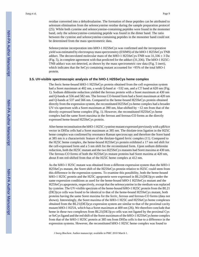
3.5. UV-visible spectroscopic analysis of the hHO-1 H25SeCys heme complexThe ferric heme-bound hHO-1 H25SeCys protein obtained from the cell expression systemhad a Soret maximum at 402 nm, a weak Q-band at ∼532 nm, and a CT band at 620 nm (Fig.1). Sodium dithionite reduction yielded the ferrous protein with a Soret maximum at 430 nmand Q-bands at 556 and 582 nm. The ferrous CO-bound form had a Soret maximum at 420 nmand Q-bands at 537 and 566 nm. Compared to the heme-bound H25SeCys protein obtaineddirectly from the expression system, the reconstituted H25SeCys-heme complex had a broaderUV-vis spectrum with a Soret maximum at 390 nm, blue-shifted by ∼12 nm from that of thedirectly expressed heme complex (Fig. 1). However, the reconstituted H25SeCys hemecomplex had the same Soret maxima in the ferrous and ferrous-CO forms as the directlyexpressed heme-bound H25SeCys protein.
After heme reconstitution the hHO-1 H25C cysteine mutant expressed previously with a pBAcevector in DH5α cells had a Soret maximum at 385 nm. The thiolate-iron ligation in the H25Cheme complex was confirmed by resonance Raman spectroscopy and therefore the Soret bandat 385 nm is a characteristic feature of the thiolate-ligated ferric complex (11). Compared tothe H25C heme complex, the heme-bound H25SeCys protein exhibited a 17 nm red shift forthe cell-expressed form and a 5 nm shift for the reconstituted form. Upon sodium dithionitereduction, both the H25C mutant and the two H25SeCys mutants had Soret maxima at 430 nm.The ferrous-CO forms of both the H25SeCys mutant proteins had Soret maxima at 420 nm,about 8 nm red-shifted from that of the H25C heme complex at 412 nm.
As the hHO-1 H25C mutant was obtained from a different expression system than the hHO-1H25SeCys mutant, the Soret shift of the H25SeCys protein relative to H25C could stem fromthis difference in the expression systems. To examine this possibility, both the heme-boundhHO-1 H25C protein and the H25C apoprotein were expressed in BL21(DE3)cys under thesame expression conditions as used for the heme-bound hHO-1 H25SeCys mutant and theH25SeCys apoprotein, respectively, except that the selenocysteine in the medium was replacedby cysteine. The UV-visible spectrum of the heme-bound hHO-1 H25C protein from the BL21(DE3)cys cells was found to be identical to that of the heme-bound H25SeCys mutant, bothproteins having the same Soret maxima for the ferric, ferrous and ferrous-CO forms (data notshown). Interestingly, the Soret maxima of the hHO-1 H25C and H25SeCys heme complexesobtained from the BL21(DE3)cys expression system are similar to that of the proximal cavitymutant hHO-1 H25A, which has a Soret maximum at 400 nm (26). We therefore conclude thatheme in these two complexes from BL21(DE3)cys cells was not ligated by the proximal Cysor SeCys ligand and the red shift of the Soret maximum of the hHO-1 H25SeCys heme complexfrom that of the hHO-1 H25C protein at 385 nm from DH5α cells is due to a difference in theexpression systems. However, the reconstituted hHO-1 H25C heme complex was found to
Jiang et al. Page 9
J Inorg Biochem. Author manuscript; available in PMC 2010 March 1.
NIH
-PA Author Manuscript
NIH
-PA Author Manuscript
NIH
-PA Author Manuscript
contain two fractions of proteins that were partially separated by chromatography on ahydroxyapatite column, one with a Soret maximum at 385 nm and the other at 410 nm (Datanot shown). The presence of two fractions of proteins was also previously observed for otherreconstituted hHO-1 His132 mutants (27). The former had the same Soret maxima as thereconstituted hHO-1 H25C heme complex from the DH5α cells. Therefore, the ∼5 nm red shiftof the reconstituted hHO-1 H25SeCys heme complex from the H25C 385 nm species was notrelated to the expression system but could be a result of the replacement of Cys with SeCys atthe protein active site. However, this small UV absorption change was found negligible whenthe UV-vis spectra of the two proteins were superimposed on each other, particularly becausethe absorption bands of both proteins around the Soret maxima were very broad.
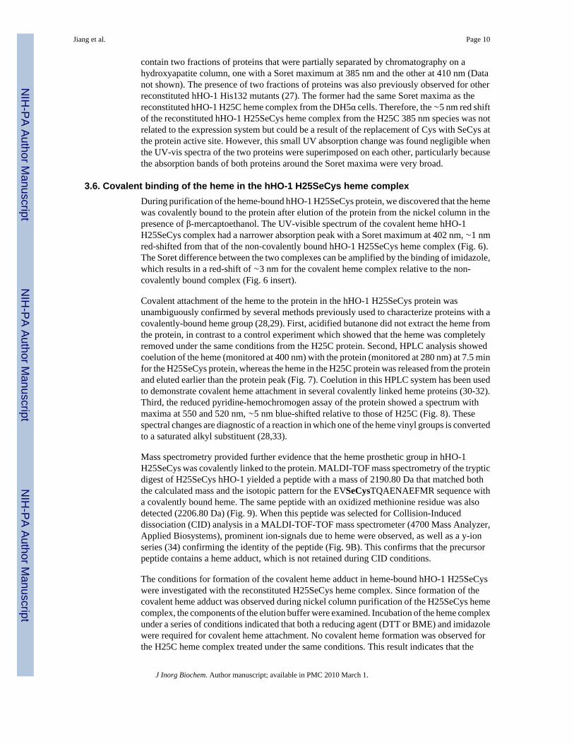
3.6. Covalent binding of the heme in the hHO-1 H25SeCys heme complexDuring purification of the heme-bound hHO-1 H25SeCys protein, we discovered that the hemewas covalently bound to the protein after elution of the protein from the nickel column in thepresence of β-mercaptoethanol. The UV-visible spectrum of the covalent heme hHO-1H25SeCys complex had a narrower absorption peak with a Soret maximum at 402 nm, ∼1 nmred-shifted from that of the non-covalently bound hHO-1 H25SeCys heme complex (Fig. 6).The Soret difference between the two complexes can be amplified by the binding of imidazole,which results in a red-shift of ∼3 nm for the covalent heme complex relative to the non-covalently bound complex (Fig. 6 insert).
Covalent attachment of the heme to the protein in the hHO-1 H25SeCys protein wasunambiguously confirmed by several methods previously used to characterize proteins with acovalently-bound heme group (28,29). First, acidified butanone did not extract the heme fromthe protein, in contrast to a control experiment which showed that the heme was completelyremoved under the same conditions from the H25C protein. Second, HPLC analysis showedcoelution of the heme (monitored at 400 nm) with the protein (monitored at 280 nm) at 7.5 minfor the H25SeCys protein, whereas the heme in the H25C protein was released from the proteinand eluted earlier than the protein peak (Fig. 7). Coelution in this HPLC system has been usedto demonstrate covalent heme attachment in several covalently linked heme proteins (30-32).Third, the reduced pyridine-hemochromogen assay of the protein showed a spectrum withmaxima at 550 and 520 nm, ∼5 nm blue-shifted relative to those of H25C (Fig. 8). Thesespectral changes are diagnostic of a reaction in which one of the heme vinyl groups is convertedto a saturated alkyl substituent (28,33).
Mass spectrometry provided further evidence that the heme prosthetic group in hHO-1H25SeCys was covalently linked to the protein. MALDI-TOF mass spectrometry of the trypticdigest of H25SeCys hHO-1 yielded a peptide with a mass of 2190.80 Da that matched boththe calculated mass and the isotopic pattern for the EVSeCysTQAENAEFMR sequence witha covalently bound heme. The same peptide with an oxidized methionine residue was alsodetected (2206.80 Da) (Fig. 9). When this peptide was selected for Collision-Induceddissociation (CID) analysis in a MALDI-TOF-TOF mass spectrometer (4700 Mass Analyzer,Applied Biosystems), prominent ion-signals due to heme were observed, as well as a y-ionseries (34) confirming the identity of the peptide (Fig. 9B). This confirms that the precursorpeptide contains a heme adduct, which is not retained during CID conditions.
The conditions for formation of the covalent heme adduct in heme-bound hHO-1 H25SeCyswere investigated with the reconstituted H25SeCys heme complex. Since formation of thecovalent heme adduct was observed during nickel column purification of the H25SeCys hemecomplex, the components of the elution buffer were examined. Incubation of the heme complexunder a series of conditions indicated that both a reducing agent (DTT or BME) and imidazolewere required for covalent heme attachment. No covalent heme formation was observed forthe H25C heme complex treated under the same conditions. This result indicates that the
Jiang et al. Page 10
J Inorg Biochem. Author manuscript; available in PMC 2010 March 1.
NIH
-PA Author Manuscript
NIH
-PA Author Manuscript
NIH
-PA Author Manuscript
selenocysteine is responsible for the formation of the covalent heme adduct in the heme-boundhHO-1 H25SeCys. Interestingly, no covalent heme attachment was observed when the proteinwas incubated with H2O2. This is consistent with previous observations that reducingconditions were required for covalent heme attachment in other proteins (28,35). To providefurther information on the site of covalent attachment to the heme, the hHO-1 H25SeCysapoprotein was reconstituted with iron (III) mesoporphyrin (mesoheme) in which ethyl groupsreplace the 2- and 4-vinyl groups of normal heme. Based on the HPLC analysis and the pyridinehemochromogen assay (Data not shown), the mesoheme-bound H25SeCys did not form acovalent heme product under conditions that gave the normal covalent heme adduct. This result,together with the shift in the absorbance maximum of the pyridine hemochromogen spectrum,indicates that heme in the covalent heme hHO-1 H25SeCys complex is attached to the proteinthrough one of its vinyl groups.
4. DiscussionWe have investigated the incorporation of selenocysteine as the proximal ligand in the hHO-1H25C mutant, a protein in which Cys-25 is the only cysteine residue, in order to (a) clarify therole of the proximal ligand in this enzyme system, and (b) characterize the interactions of aselenolate ligand with a heme iron atom. While selenoproteins are widely distributed in nature,a hemeprotein with a selenocysteine as the proximal ligand has not been reported in the refereedliterature, although such a protein was cursorily reported in an abstract in 2006 (36).Selenocysteine is unique as a potential iron ligand because of (a) its lower pKa, which meansthe thiolate form is the dominant one at physiological pH, (b) its greater electron donatingproperties, and (c) a size similar to that of cysteine itself.
To prepare the selenocysteine-ligated hHO-1 protein, we expressed the hHO-1 H25SeCysmutant in an E. coli cysteine auxotroph in the presence of selenocysteine. This cysteineauxotroph expression system was previously shown to allow the efficient replacement of acysteine by a selenocysteine (16-19). Mass spectrometric analysis showed >95% incorporationof the selenocysteine into the protein, which was obtained in a yield comparable to that of thecysteine-containing protein. This extent of selenocysteine incorporation is on the high endrelative to the previously reported 73% to over 91% incorporation into other proteins (16-19).A high level of selenocysteine incorporation was consistent with our identification of theselenocysteine-, but not cysteine-containing peptide in the mixture generated by trypsindigestion of the hHO-1 H25SeCys protein.
The observation that the hHO-1 H25SeCys protein migrated as both a monomer and a dimeron SDS-PAGE may stem from the presence of only one such residue in the protein, as thisprecludes the formation of internal diseleno or seleno-sulfide crosslinks. The dimer, however,is an artifact that results from denaturation of the protein when the sample is prepared for SDS-PAGE or mass spectrometric analysis, as FPLC indicates that the intact protein is entirelymonomeric. The dimer is observed in the SDS-PAGE gel because the intermolecular Se-Sebond formed when the protein is denatured is not readily reduced by the β-mercaptoethanol inthe electrophoresis mixture. LC/MS analysis of the tryptic peptides from the dimer bandunambiguously confirms that the dimer derives entirely from the selenocysteine-containingprotein.
Although the incorporation of selenocysteine in a properly folded protein is clearly establishedby our results, it is unclear whether the selenocysteine is stably coordinated to the heme ironin the hHO-1 H25SeCys mutant. The heme-bound hHO-1 H25SeCys and H25C mutantsproduced by the cysteine auxotroph system supplemented with selenocysteine or cysteine,respectively, have nearly identical UV-vis spectra in the ferric, ferrous, and ferrous-CO states.These spectra differ, however, from the spectra obtained earlier by expression of the hHO-1
Jiang et al. Page 11
J Inorg Biochem. Author manuscript; available in PMC 2010 March 1.
NIH
-PA Author Manuscript
NIH
-PA Author Manuscript
NIH
-PA Author Manuscript
H25C mutant using a pBAce vector in DH5α cells and more closely resemble those of theproximal cavity hHO-1 H25A mutant (26). It is clear that the selenocysteine does not coordinateto the heme iron as a selenolate in the ferrous state, as a Soret maximum in the range of 450nm would be expected from such coordination. The absence of this spectrum and its similarityto that of the H25A mutant, together with the fact that the ligand should be present as aselenolate due to its low pKa, clearly suggests that the selenocysteine is not coordinated to theiron in the ferrous state in the isolated protein. The coordination state in the ferric enzyme ismore difficult to establish. Although the Soret maxima of the reconstituted hHO-1 H25SeCysand H25C heme complexes showed a small ∼5 nm difference, the spectra of the two proteinswere nearly identical when superimposed on each other, particularly because their absorptionbands were very broad. Both mutants also have nearly identical UV-vis spectra in the ferrousand ferrous-CO states, indicating the absence of a selenolate ligand in the reconstituted hHO-1H25SeCys heme complex in the ferrous state. In previous work we demonstrated that thecysteine sulfur did coordinate to the ferric heme iron in the H25C mutant, but dissociated inthe ferrous state. This may also be true of the selenocysteine ligand, but the spectra of the ferriccomplexes are not sufficiently differentiated to unambiguously assert this.
We recently reported formation of a complex of the proximal hHO-1 H25A cavity mutant withan external benzeneselenolate anion as the iron ligand (37). Interestingly, in this complex, theselenium transfers an electron to the heme iron atom, as demonstrated by partial formation ofa ferrous-CO complex in the absence of other reducing agents. If the more electron donatingbenzylselenolate is introduced as the ligand, the conversion to the ferrous-CO complex isquantitative. In the presence of oxygen, the iron undergoes autoxidation, a reaction that oxidizesand consumes the external selenolate ligand, and the iron eventually reverts to the ferric state.Similar electron transfer to the iron does not occur when thiophenol is the externally providedligand. Thus, the higher electron donating properties of the selenolate ligand result not only incoordination but also electron transfer from the selenium to the iron.
In the present situation, where the selenium ligand was from a selenocysteine residue in theprotein sequence, electron transfer also apparently occurs (Scheme 1). However, in theH25SeCys protein, the selenyl radical that is formed adds to a vinyl group of the heme, resultingin covalent attachment of the heme to the protein. The addition clearly occurs at the vinyl group,as no covalent attachment is observed when the protein is reconstituted with mesoheme (ethylinstead of vinyl substituents) rather than heme. Based on the pyridine hemochromogenspectroscopic shift and the molecular mass of the tryptic peptide with the covalently boundheme, the vinyl group gives rise to a saturated bond linking the heme to the selenium, implyingthat a hydrogen atom is also incorporated into the product. Similar treatment of the normalH25C protein with a cysteine ligand does not result in covalent binding of the heme,demonstrating that this process is specifically a property of the selenocysteine ligand.
We have previously reported that reaction of ferrous hHO-1 with CBrCl3 produces a CCl3radical that adds to one of the heme vinyl groups (30). The resulting radical adjacent to theporphyrin ring is oxidized by electron transfer to the ferric iron, giving a cation that is trappedby the proximal histidine ligand to give a His25-heme link. This finding shows that (a) theheme vinyl groups in hHO-1 can react with radical agents, and (b) the heme in the hHO-1active site has a significant ability to move about, including to positions that place the vinylgroup close to the proximal iron ligand. Covalent heme binding involving an adjacent Cys orMet mutation has been observed in other heme proteins (28,29,35). The ability of the selenolatemoiety to transfer an electron to the heme iron, with consequent modification of the heme, mayexplain the failure to observe selenolate coordination to the heme iron in the ferrous state andtherefore of a 450 nm-like absorption maximum for its CO complex.
Jiang et al. Page 12
J Inorg Biochem. Author manuscript; available in PMC 2010 March 1.
NIH
-PA Author Manuscript
NIH
-PA Author Manuscript
NIH
-PA Author Manuscript
The requirement for imidazole in formation of the heme-protein covalent bond may be due toa need for coordination of a ligand at the sixth iron site, possibly to prevent coordination ofoxygen leading to autooxidation of the iron. However, we cannot exclude alternative roles,such as a donor of the hydrogen that quenches the radical expected from radical addition ofthe selenocysteine radical to the vinyl group.
In summary, it is clear from the present results that a hemoprotein selenocysteine iron ligandis unstable towards electron transfer to the heme iron atom, a process that can result in covalentheme binding of the heme to the protein via addition of a selenocysteine radical to a heme vinylgroup. Interestingly, in work in progress we have substituted a selenocysteine for the normalcysteine in a P450 enzyme, have observed the expected Soret maximum in the 450 nm range,and have not observed the internal electron transfer seen with hHO-1 H25SeCys (unpublishedresults). Factors that control the redox potential balance in a P450 versus hHO-1 thus intervenein modulating the specific properties of the proximal ligand and its interaction with the ironatom.
AcknowledgmentsWe thank Marie-Paule Strub for the kind gift of the cysteine auxotroph E. coli strain. This work was supported byNational Institutes of Health grants DK30297 (POM) and GM25515 (POM). The mass spectrometric analyses werecarried out in the National Bio-Organic Biomedical Mass Spectrometry Resource at UCSF (A.L. Burlingame, Director)supported by the National Institutes of Health Biomedical Research Technology Program through grants RR014606,RR001614, RR015804, and RR019934.
References1. Ortiz de Montellano, PR.; Auclair, K. Porphyrin Handbook. Kadish, K.; Smith, K.; Guilard, R., editors.
Vol. 12. Academic Press; New York: 2003. p. 183-210.2. Unno M, Matsui T, Ikeda-Saito M. Nat Prod Rep 2007;24:553–570. [PubMed: 17534530]3. Liu Y, Ortiz de Montellano PR. J Biol Chem 2000;275:5297–5307. [PubMed: 10681502]4. Ortiz de Montellano, PR.; De Voss, JJ. Cytochrome P450: Structure, Mechanism, and Biochemistry.
Vol. 3rd. Ortiz de Montellano, PR., editor. Kluwer Academic/Plenum Publishers; New York: 2005.p. 183-245.
5. Wilks A, Torpey J, Ortiz de Montellano PR. J Biol Chem 1994;269:29553–29556. [PubMed: 7961940]6. Davydov RM, Yoshida T, Ikeda-Saito M, Hoffman BM, Amer J. Chem Soc 1999;121:10656–10657.7. Li Y, Syvitiski R, Auclair K, Wilks A, Ortiz de Montellano PR, La Mar GN. J Biol Chem
2002;277:33018–33031. [PubMed: 12070167]8. Syvitski RT, Li Y, Auclair K, Ortiz de Montellano PR, La Mar GN. J Am Chem Soc 2002;124:14296–
14297. [PubMed: 12452690]9. Sun J, Loehr TM, Wilks A, Ortiz de Montellano PR. Biochemistry 1994;33:13734–13740. [PubMed:
7947784]10. Schuller DJ, Wilks A, Ortiz de Montellano PR, Poulos T. Nature Struct Biol 1999;6:860–867.
[PubMed: 10467099]11. Liu Y, Moënne-Loccoz P, Hildebrand D, Wilks A, Loehr TM, Mauk AG, Ortiz de Montellano PR.
Biochemistry 1999;38:3733–3743. [PubMed: 10090762]12. Jacob C, Giles GI, Giles NM, Sies H. Angew Chem Int Ed 2003;42:4742–4758.13. Johansson L, Gafvelin G, Arnér ESJ. Biochim Biophys Acta 2005;1726:1–13. [PubMed: 15967579]14. Atkins JF, Gesteland RF. Nature 2000;407:463–465. [PubMed: 11028985]15. Jiang Z, Arner ES, Mu Y, Johansson L, Shi J, Zhao S, Liu S, Wang R, Zhang T, Yan G, Liu J, Shen
J, Luo G. Biochem Biophys Res Commun 2004;321:94–101. [PubMed: 15358220]16. Müller S, Senn H, Gsell B, Vetter W, Baron C, Böck A. Biochemistry 1994;33:3404–3412. [PubMed:
8136378]17. Boschi-Muller S, Muller S, Van Dorsselaeer A, Böck A, Branlant G. FEBS Lett 1998;20:241–245.
[PubMed: 9845330]
Jiang et al. Page 13
J Inorg Biochem. Author manuscript; available in PMC 2010 March 1.
NIH
-PA Author Manuscript
NIH
-PA Author Manuscript
NIH
-PA Author Manuscript
18. Strub MP, Hoh F, Sanchez JF, Strub JM, Böck A, Aumelas A, Dumas C. Structure 2003;11:1359–1367. [PubMed: 14604526]
19. Sanchez JF, Hoh F, Strub MP, Aumelas A, Dumas C. Structure 2002;10:1363–1370. [PubMed:12377122]
20. Ghiladi RA, Knudsen GM, Medzihradsky KF, Ortiz de Montellano PR. J Biol Chem 2005;280:22651–22663. [PubMed: 15840564]
21. Wilks A, Ortiz de Montellano PR. J Biol Chem 1993;268:22357–22362. [PubMed: 8226746]22. Yoshida T, Kikuchi G. J Biol Chem 1978;253:4224–4229. [PubMed: 96115]23. Antonini, M.; Brunori, E. Hemoglobin and Myoglobin and their Reactions with Ligands. North
Holland Publishers; Amsterdam: 1971.24. Beld J, Woycechowsky KJ, Hilvert D. Biochemistry 2007;46:5382–5390. [PubMed: 17419591]25. Ma S, Caprioli RM, Hill KE, Burk RF. J Am Soc Mass Spectrom 2003;14:593–600. [PubMed:
12781460]26. Wilks A, Sun J, Loehr TM, Ortiz de Montellano PR. J Am Chem Soc 1995;117:2925–2926.27. Matera KM, Zhou H, Migita CT, Hobert SE, Ishikawa K, Katakura K, Maeshima H, Yoshida T,
Ikeda-Saito M. Biochemistry 1997;36:4909–4915. [PubMed: 9125512]28. Metcalfe CL, Daltrop O, Ferguson SJ, Raven EL. Biochem J 2007;408:355–361. [PubMed:
17714075]29. Metcalfe CL, Ott M, Patel N, Singh K, Mistry SC, Goff HM, Raven EL. J Am Chem Soc
2004;126:16242–16248. [PubMed: 15584761]30. Wilks A, Medzihradszky KF, Ortiz de Montellano PR. Biochemistry 1998;37:2889–2896. [PubMed:
9485440]31. Colas C, Ortiz de Montellano PR. J Biol Chem 2004;279:24131–24140. [PubMed: 15039425]32. Hoch U, Ortiz de Montellano PR. J Biol Chem 2001;276:11339–11346. [PubMed: 11139583]33. Daltrop O, Allen JWA, Willis AC, Ferguson SJ. Proc Natl Acad Sci USA 2002;99:7872–7876.
[PubMed: 12060734]34. Biemann K. Methods in Enzymology 1990;193:886–887. [PubMed: 2074849]35. Barker PD, Ferrer JC, Mylrajan M, Loehr TM, Feng R, Konishi Y, Funk WD, MacGillivray RTA,
Mauk AG. Proc Natl Acad Sci USA 1993;99:6542–6546. [PubMed: 8341666]36. Gromov, I.; Garcia-Rubio, I.; Aldag, C.; Schweiger, A.; Hilvert, D. 6th European Federation of EPR
Groups Meeting Abstract, OT3; 2006.37. Jiang Y, Ortiz de Montellano PR. Inorg Chem 2008;47:3480–3482. [PubMed: 18376820]
5. AbbreviationshHO-1
human heme oxygenase lacking the membrane binding sequence
P450 cytochrome P450
CPR cytochrome P450 reductase
SeCys selenocysteine
TCEP tri(2-carboxyethyl)phospine
IPTG isopropyl β-D-1-thiogalactopyranoside
FPLC
Jiang et al. Page 14
J Inorg Biochem. Author manuscript; available in PMC 2010 March 1.
NIH
-PA Author Manuscript
NIH
-PA Author Manuscript
NIH
-PA Author Manuscript
fast protein liquid chromatography
SDS-PAGE sodium dodecyl sulfate polyacrylamide electrophoresis
TNB 5-thio-2-nitrobenzoate
Jiang et al. Page 15
J Inorg Biochem. Author manuscript; available in PMC 2010 March 1.
NIH
-PA Author Manuscript
NIH
-PA Author Manuscript
NIH
-PA Author Manuscript
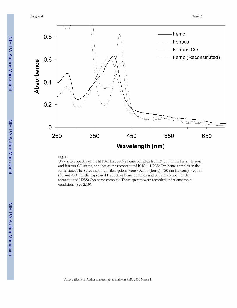
Fig. 1.UV-visible spectra of the hHO-1 H25SeCys heme complex from E. coli in the ferric, ferrous,and ferrous-CO states, and that of the reconstituted hHO-1 H25SeCys heme complex in theferric state. The Soret maximum absorptions were 402 nm (ferric), 430 nm (ferrous), 420 nm(ferrous-CO) for the expressed H25SeCys heme complex and 390 nm (ferric) for thereconstituted H25SeCys heme complex. These spectra were recorded under anaerobicconditions (See 2.10).
Jiang et al. Page 16
J Inorg Biochem. Author manuscript; available in PMC 2010 March 1.
NIH
-PA Author Manuscript
NIH
-PA Author Manuscript
NIH
-PA Author Manuscript
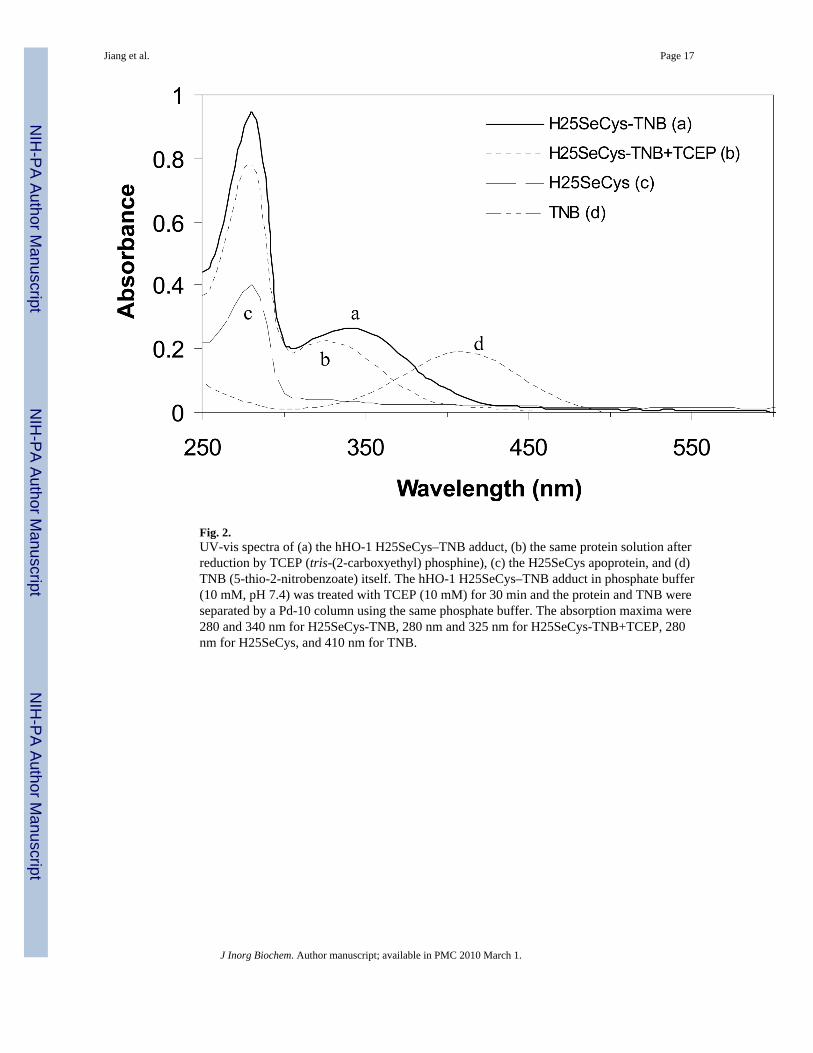
Fig. 2.UV-vis spectra of (a) the hHO-1 H25SeCys–TNB adduct, (b) the same protein solution afterreduction by TCEP (tris-(2-carboxyethyl) phosphine), (c) the H25SeCys apoprotein, and (d)TNB (5-thio-2-nitrobenzoate) itself. The hHO-1 H25SeCys–TNB adduct in phosphate buffer(10 mM, pH 7.4) was treated with TCEP (10 mM) for 30 min and the protein and TNB wereseparated by a Pd-10 column using the same phosphate buffer. The absorption maxima were280 and 340 nm for H25SeCys-TNB, 280 nm and 325 nm for H25SeCys-TNB+TCEP, 280nm for H25SeCys, and 410 nm for TNB.
Jiang et al. Page 17
J Inorg Biochem. Author manuscript; available in PMC 2010 March 1.
NIH
-PA Author Manuscript
NIH
-PA Author Manuscript
NIH
-PA Author Manuscript
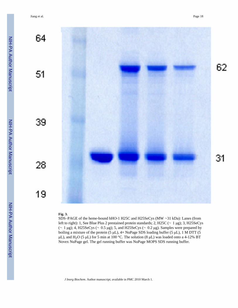
Fig. 3.SDS–PAGE of the heme-bound hHO-1 H25C and H25SeCys (MW ∼31 kDa): Lanes (fromleft to right): 1, See Blue Plus 2 prestained protein standards; 2, H25C (∼ 1 μg); 3, H25SeCys(∼ 1 μg); 4, H25SeCys (∼ 0.5 μg); 5, and H25SeCys (∼ 0.2 μg). Samples were prepared byboiling a mixture of the protein (5 μL), 4× NuPage SDS loading buffer (5 μL), 1 M DTT (5μL), and H2O (5 μL) for 5 min at 100 °C. The solution (8 μL) was loaded onto a 4-12% BTNovex NuPage gel. The gel running buffer was NuPage MOPS SDS running buffer.
Jiang et al. Page 18
J Inorg Biochem. Author manuscript; available in PMC 2010 March 1.
NIH
-PA Author Manuscript
NIH
-PA Author Manuscript
NIH
-PA Author Manuscript
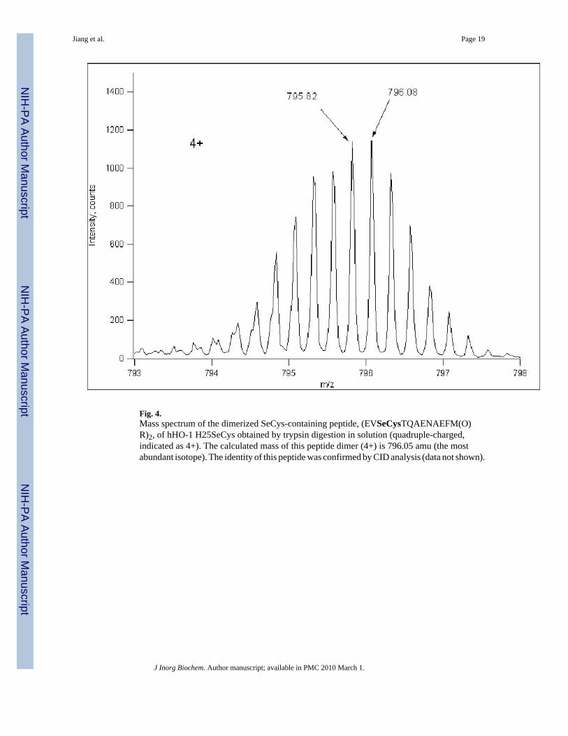
Fig. 4.Mass spectrum of the dimerized SeCys-containing peptide, (EVSeCysTQAENAEFM(O)R)2, of hHO-1 H25SeCys obtained by trypsin digestion in solution (quadruple-charged,indicated as 4+). The calculated mass of this peptide dimer (4+) is 796.05 amu (the mostabundant isotope). The identity of this peptide was confirmed by CID analysis (data not shown).
Jiang et al. Page 19
J Inorg Biochem. Author manuscript; available in PMC 2010 March 1.
NIH
-PA Author Manuscript
NIH
-PA Author Manuscript
NIH
-PA Author Manuscript
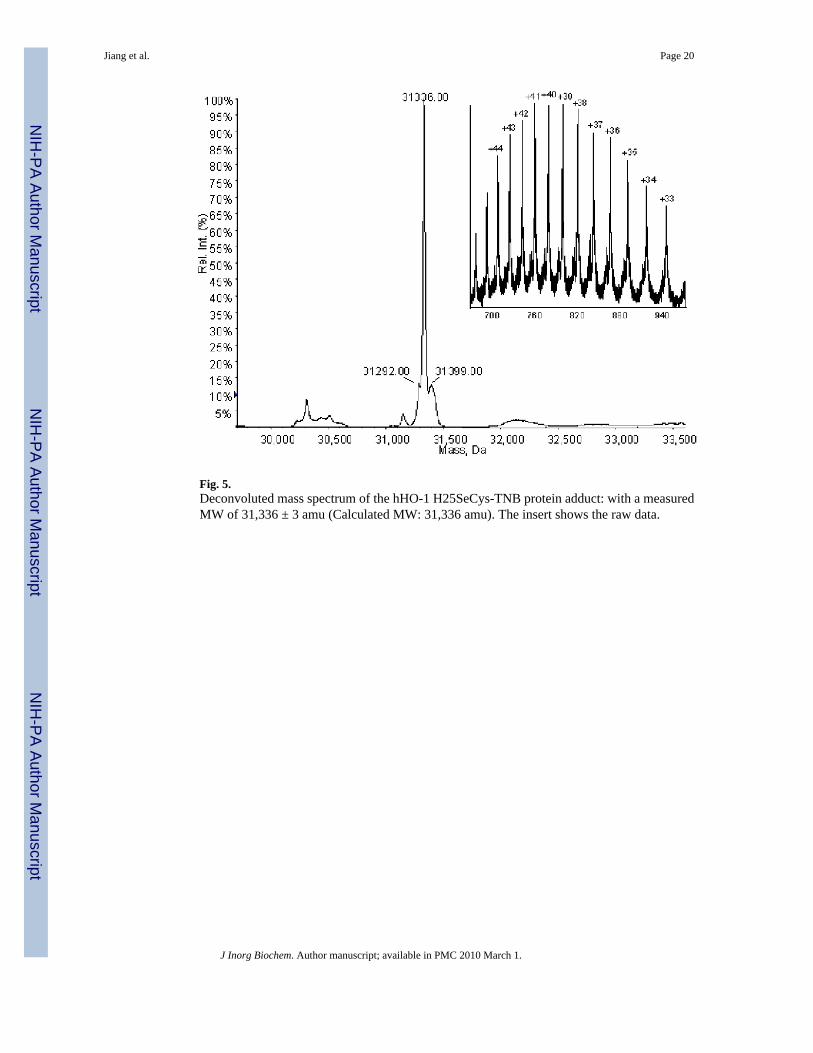
Fig. 5.Deconvoluted mass spectrum of the hHO-1 H25SeCys-TNB protein adduct: with a measuredMW of 31,336 ± 3 amu (Calculated MW: 31,336 amu). The insert shows the raw data.
Jiang et al. Page 20
J Inorg Biochem. Author manuscript; available in PMC 2010 March 1.
NIH
-PA Author Manuscript
NIH
-PA Author Manuscript
NIH
-PA Author Manuscript
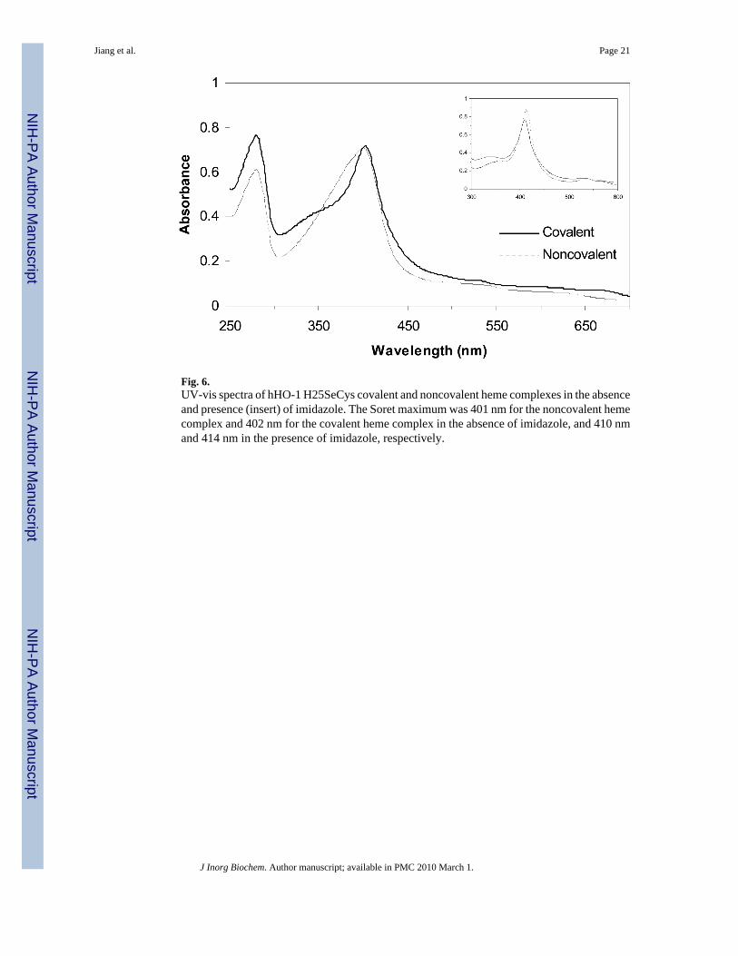
Fig. 6.UV-vis spectra of hHO-1 H25SeCys covalent and noncovalent heme complexes in the absenceand presence (insert) of imidazole. The Soret maximum was 401 nm for the noncovalent hemecomplex and 402 nm for the covalent heme complex in the absence of imidazole, and 410 nmand 414 nm in the presence of imidazole, respectively.
Jiang et al. Page 21
J Inorg Biochem. Author manuscript; available in PMC 2010 March 1.
NIH
-PA Author Manuscript
NIH
-PA Author Manuscript
NIH
-PA Author Manuscript
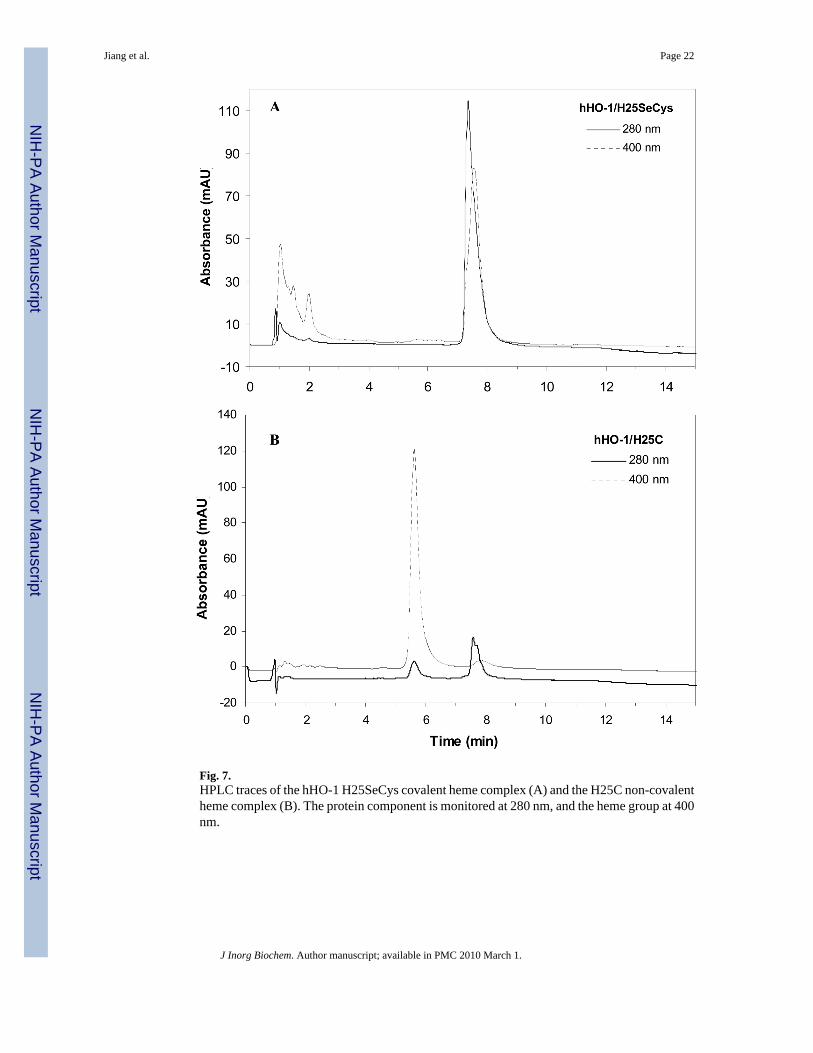
Fig. 7.HPLC traces of the hHO-1 H25SeCys covalent heme complex (A) and the H25C non-covalentheme complex (B). The protein component is monitored at 280 nm, and the heme group at 400nm.
Jiang et al. Page 22
J Inorg Biochem. Author manuscript; available in PMC 2010 March 1.
NIH
-PA Author Manuscript
NIH
-PA Author Manuscript
NIH
-PA Author Manuscript
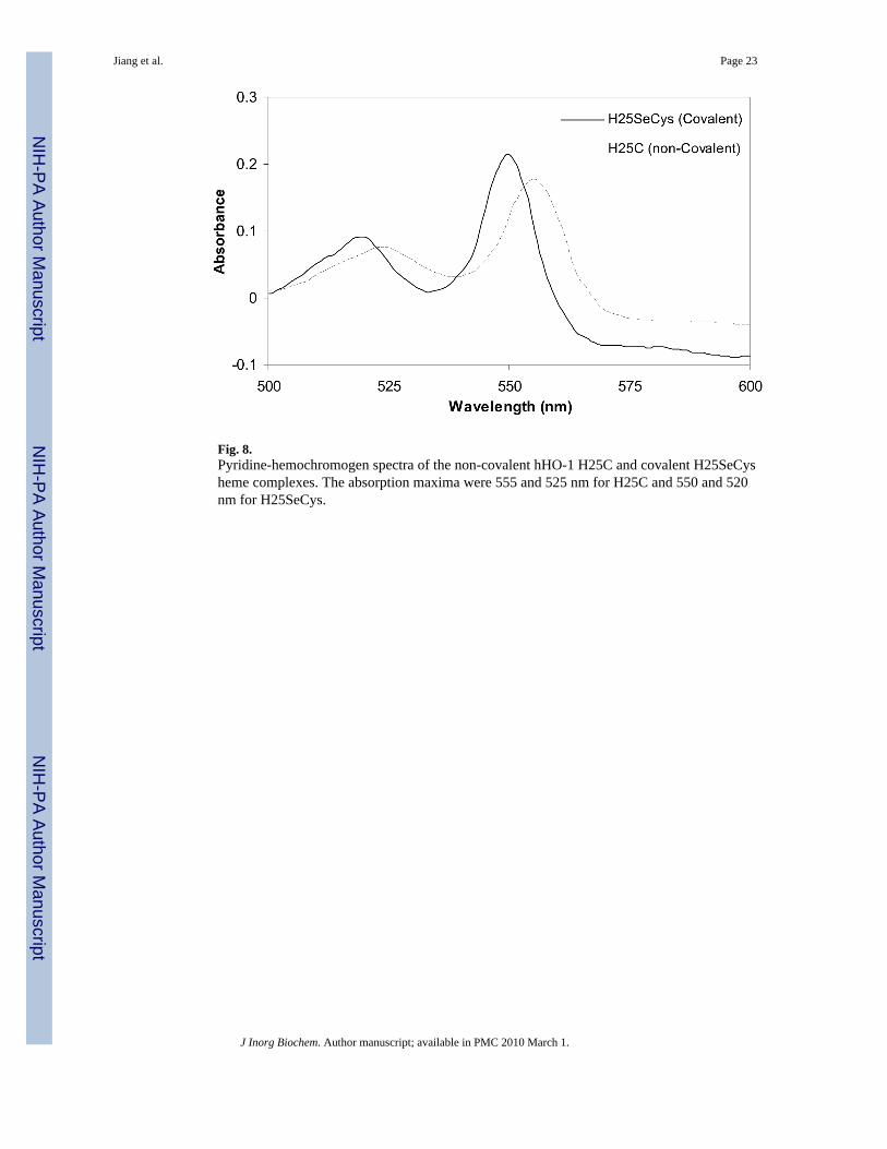
Fig. 8.Pyridine-hemochromogen spectra of the non-covalent hHO-1 H25C and covalent H25SeCysheme complexes. The absorption maxima were 555 and 525 nm for H25C and 550 and 520nm for H25SeCys.
Jiang et al. Page 23
J Inorg Biochem. Author manuscript; available in PMC 2010 March 1.
NIH
-PA Author Manuscript
NIH
-PA Author Manuscript
NIH
-PA Author Manuscript
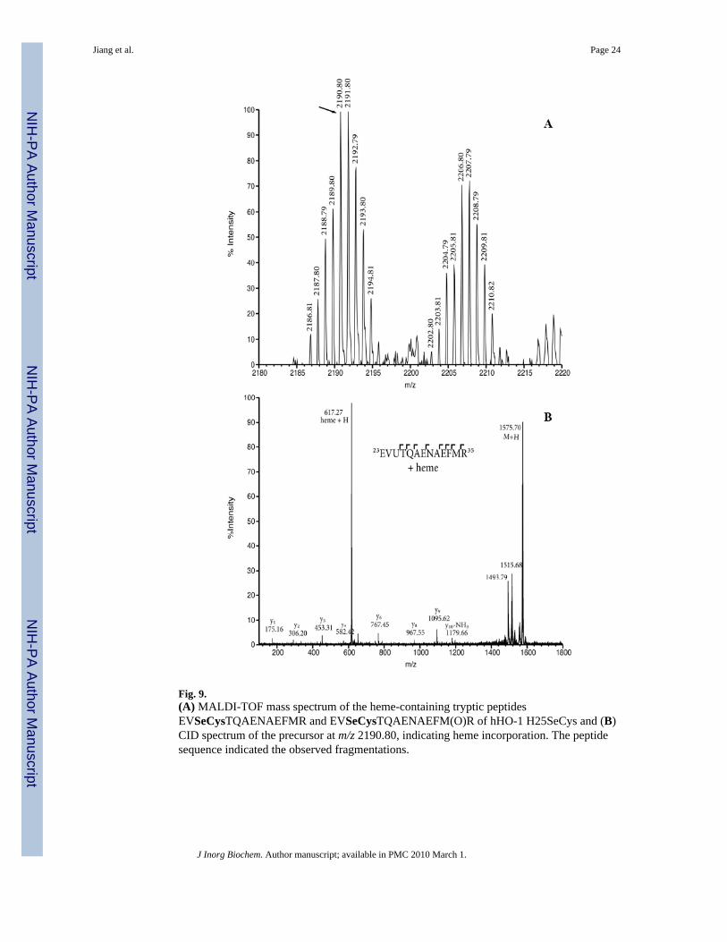
Fig. 9.(A) MALDI-TOF mass spectrum of the heme-containing tryptic peptidesEVSeCysTQAENAEFMR and EVSeCysTQAENAEFM(O)R of hHO-1 H25SeCys and (B)CID spectrum of the precursor at m/z 2190.80, indicating heme incorporation. The peptidesequence indicated the observed fragmentations.
Jiang et al. Page 24
J Inorg Biochem. Author manuscript; available in PMC 2010 March 1.
NIH
-PA Author Manuscript
NIH
-PA Author Manuscript
NIH
-PA Author Manuscript
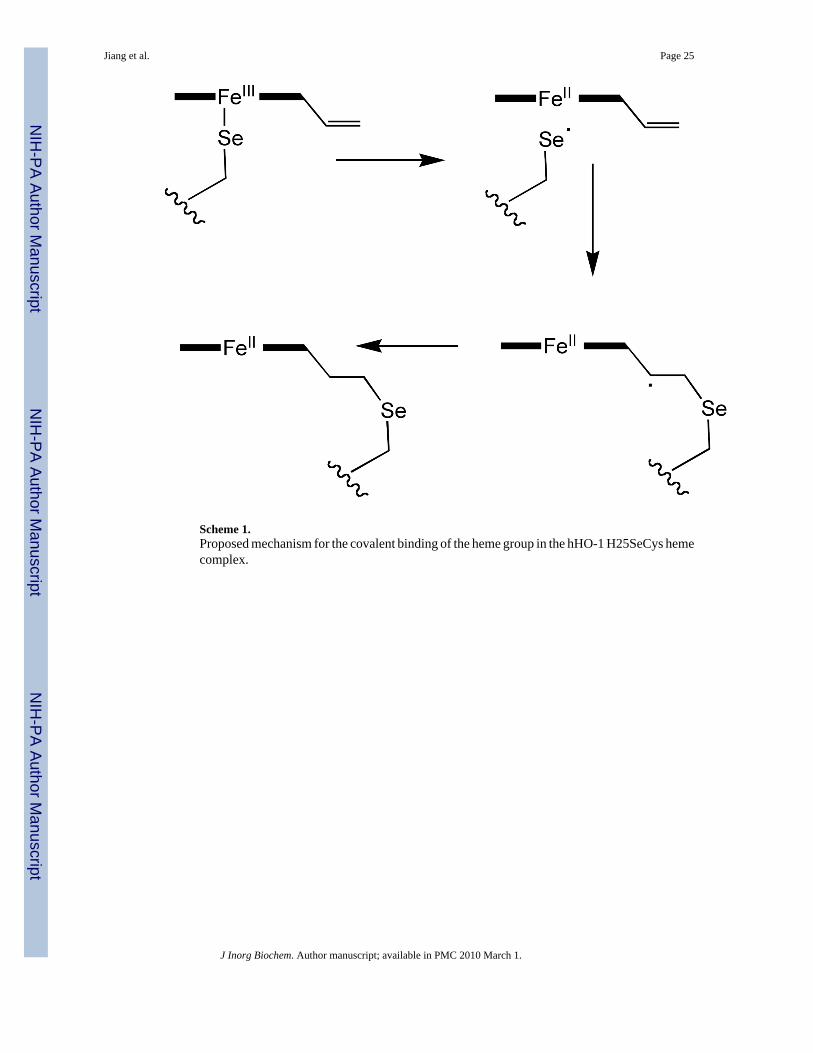
Scheme 1.Proposed mechanism for the covalent binding of the heme group in the hHO-1 H25SeCys hemecomplex.
Jiang et al. Page 25
J Inorg Biochem. Author manuscript; available in PMC 2010 March 1.
NIH
-PA Author Manuscript
NIH
-PA Author Manuscript
NIH
-PA Author Manuscript


