Embed Size (px)
Citation preview

Downloaded from UvA-DARE, the Institutional Repository of the University of Amsterdam (UvA)http://dare.uva.nl/document/17664
File ID 17664Filename Deutekom.pdf
SOURCE, OR PART OF THE FOLLOWING SOURCE:Type DissertationTitle Faecal incontinence : impact, treatment and diagnostic work-upAuthor M. DeutekomFaculty Faculty of MedicineYear 2005Pages 184
FULL BIBLIOGRAPHIC DETAILS: http://dare.uva.nl/record/165322
Copyrights It is not permitted to download or to forward/distribute the text or part of it without the consent of the copyright holder(usually the author), other then for strictly personal, individual use. UvA-DARE is a service provided by the Library of the University of Amsterdam (http://dare.uva.nl)

Faecal incontinence
Impact, treatment and diagnostic work-up

© 2005 Marije Baart de la Faille-Deutekom, Amsterdam
Faecal incontinence: Impact, treatment and diagnostic work-up
PhD thesis, University of Amsterdam – with references – with summary in Dutch
ISBN: 90-9019724-9
Cover by Job Baart de la Faille
Layout by Marije Baart de la Faille-Deutekom
Printed by Buijten & Schipperheijn, Amsterdam
All rights reserved. No part of this publication may be reproduced, stored or transmitted
in any way or by any means, without prior permission of the author.
Studies published in this thesis were supported by grant 945-01-013 of the Netherlands
Organization for Health Research and Development.
The printing of this thesis is financially supported by:
Stichting tot bevordering van de Klinische Epidemiologie, University of Amsterdam,
Paul Hartmann B.V., Medeco B.V., and the Dutch Cochrane Centre
Faecal incontinence
Impact, treatment and diagnostic work-up
ACADEMISCH PROEFSCHRIFT
ter verkrijging van de graad van doctor aan de Universiteit van Amsterdam op gezag van de Rector Magnificus
prof. mr. P.F. van der Heijden ten overstaan van een door het college voor promoties ingestelde
commissie, in het openbaar te verdedigen in de Aula der Universiteit
op donderdag 13 oktober 2005, te 12.00 uur
door
Marije Deutekom
geboren te Amsterdam

Promotiecommissie Promotores: Prof. dr. P.M.M. Bossuyt Prof. dr. J. Stoker Overige leden: Prof. dr. Y. van der Graaf Prof. dr. D.J. Gouma Prof. dr. B.A. van Hout Dr. P. Fockens Dr. W.G. van Gemert Dr. T.G. Wiersma Faculteit der Geneeskunde

Voor mijn lieve ouders,

Contents Chapter 1 Introduction 9 Chapter 2 Impact of faecal incontinence on 15 health domains Colorectal Disease, 2005, May: 7(3): 263-269 Chapter 3 Costs of outpatients with faecal 27 incontinence Scandinavian Journal of Gastroenterology, 2005, May: 40(5): 552-558 Chapter 4 Selecting an outcome measure for 41 evaluating treatment in faecal incontinence Diseases of the Colon and Rectum, 2005, in press Chapter 5 Anal plugs for the containment of faecal 55
incontinence Cochrane Database Systematic Reviews, 2005, July: (3): CD005086 Chapter 6 Electrical stimulation and pelvic floor 71 muscle training with biofeedback in patients with faecal incontinence Diseases of the Colon and Rectum, Revised version submitted Chapter 7 Ability of tests to predict outcome of pelvic 89
floor rehabilitation in patients with faecal incontinence Submitted Chapter 8 Patients' perception of tests in the 105 assessment of faecal incontinence British Journal of Radiology, 2005, in press

Chapter 9 Clinical presentation of faecal incontinence 121 and anorectal function, what is the relationship? Submitted Chapter 10 Relation between external anal sphincter atrophy at 137 endoanal MRI and clinical, functional, and anatomical characteristics in patients with faecal incontinence Diseases of the Colon and Rectum, Revised version submitted Chapter 11 Discussion 155 Chapter 12 Summary in Dutch 167 List of publications 173 Dankwoord 177 Curriculum Vitae 183


Chapter 1
Introduction

Chapter 1 Introduction
10
Introduction
Background I encountered in the past years difficulty to convince people around me of the need for studying faecal incontinence. Very few people knew that the involuntary loose of stool is a quite common condition. A recent systematic review of the literature has shown that about 11 to 15% of the population encounters some sort of problems in maintaining faecal continence.1 This prevalence is far from known in the general population. Possible reasons are the embarrassment of patients and doctors in addressing this problem. Only a third of symptomatic patients in the USA discuss their faecal incontinence complaints with their physicians.2
The sad thing about this embarrassment is that faecal incontinence is doomed to remain a taboo as patients remain reluctant to admit they have problems with their continence. The decreased quality of life of patients with faecal incontinence 3-7 is in part due to this taboo but is primarily aggravated by the barriers caused by the disorder itself. The unfamiliarity of most people with this disorder can isolate and depress patients. Patients with faecal incontinence can be afraid to leave their homes, as they need to be in the neighbourhood of a toilet, which could lead to social isolation.
A common misconception is that faecal incontinence only affects old and/or institutionalized people. Although the prevalence of faecal incontinence rises as age increases, faecal incontinence also affects younger people. Especially among the younger age groups there is an over presentation of women. Obstetric factors are a likely explanation why women clearly outnumber men amongst younger patients with faecal incontinence. It has been shown that the prevalence of defects of the anal sphincter complex after vaginal delivery in primiparous women is almost 27%.8 Vaginal delivery is also a risk factor for neurogenic damage of the anorectal unit. Other possible causes for faecal incontinence can be anatomical (e.g. anorectal surgery), neurological (e.g. diabetes mellitus, stroke, or spina bifida), functional (e.g. inflammatory bowel disease, proctitis, or psychiatric disorders), or congenital (e.g. imperforate anus).
Anorectal tests Faecal continence relies on a number of factors, including intact pelvic floor muscles, adequate pelvic floor innervation, adequate rectal distensibility (compliance), and stool consistency. Impairment of one or more of these factors can result in incontinence.9,10 Faecal incontinence is often due to multiple pathogenic mechanisms and is rarely attributable to a single factor.11,12

Chapter 1
11
Anorectal tests can be used to explore the underlying cause of faecal incontinence. The muscles that are believed to be most important in keeping continence are the internal anal sphincter (IAS) and the external anal sphincter (EAS). Tests have been developed to investigate the anatomy and the function of these muscles. Endoanal MRI and endoanal sonography investigate the anatomy (presence of defects and/or atrophy) of the IAS and EAS. Defecography allows an evaluation of the movements of the rectum, presence or absence of rectoceles, enteroceles and intussusceptions.
Functional characteristics of the anorectal complex are examined with various tests. Anorectal manometry evaluates the muscular contraction and relaxation of the anal sphincters by the measurement of pressures in the anal canal. With rectal and anal mucosal sensitivity measurement the threshold sensation of respectively rectum and anus can be determined. Pudendal nerve terminal motor latency testing assesses the innervation of the anorectal unit. With rectal capacity measurement the reservoir capacity of the rectum can be determined.
Treatment The initial treatment of faecal incontinence should focus on dietary changes (e.g. avoidance of foods that might lead to thin stool), anti-diarrhoea medication, or bowel habit training. When conservative treatment does not diminish complaints several other treatments options are available.
Pelvic floor rehabilitation (electrical stimulation and/or pelvic floor muscle training with biofeedback) is most commonly used as first therapy after failure of conservative treatment, as pelvic floor rehabilitation treatment is simple, inexpensive and mostly without unfavourable physical effects. When anatomical defects of the sphincter muscles have been depicted, a surgical treatment is an option. Unfortunately some patients will remain faecal incontinent after these forms of treatment. Therefore, innovative methods like dynamic graciloplasty, artificial bowel implantation and sacral-nerve neuromodulation have been developed in recent years.
With none of the treatments there exists consensus regarding the success rate nor which patients are likely to benefit from treatment. At this stage it remains a challenge to guide treatment in the individual patient.
Aim and outline of this thesis
In an ideal world it would be possible to select the optimal treatment for every patient. Selection of treatment could be made by using all available information,

Chapter 1 Introduction
12
from medical history, physical examination and additional tests. The efficiency of the health care system would markedly improve when patients would only be subjected to tests that diminish diagnostic uncertainty. Furthermore, only treatments with a high chance for success should be offered to patients. This efficiency requires a thorough evaluation of test characteristics and treatment results. Tests without any value of information or treatments without any chance on success could be omitted from the management pathway for an individual patient or, potentially, in all patients.
Although we are aware that this ideal world does not exist, the aim of the work summarized in the present thesis was to collect the evidence to build an optimal testing and treatment path for patients with faecal incontinence.
The first two chapters report on an evaluation of the effects of faecal incontinence on patients’ lives. Chapter 2 addresses patients’ health status. The relation between severity of incontinence and impact on general health domains was evaluated and the health status of patients with faecal incontinence was compared with that of the general population. Chapter 3 evaluates the effects in terms of costs. Costs associated with health care resources, out-of-pocket expenses, and production losses in paid and unpaid work were calculated taking a societal perspective.
In the evaluation of treatment a large variation exists in the outcome measures that are used to evaluate the effects of treatment. In Chapter 4 different outcome measures are evaluated and compared with patient perception of relief.
Patients with faecal incontinence can also use containment products. Chapter 5 contains a systematic review of studies on the effectiveness of anal plugs for containing faecal incontinence.
After failure of conservative treatment the most common treatment option is pelvic floor rehabilitation. Chapter 6 describes a study of the effects of electrical stimulation and pelvic floor muscle training with biofeedback in patients with faecal incontinence. Chapter 7 presents an analysis of the value of tests in predicting the success of treatment with pelvic floor rehabilitation.
The remaining chapters address different ways to evaluate tests. The study reported in Chapter 8 focuses on the perceived burden of anorectal tests. Chapter 9 presents an analysis of the relation between the clinical presentation of faecal incontinence and test information. In the study in Chapter 10 findings of imaging tests were compared with patient’s medical history and anorectal functional test variables.

Chapter 1
13
References
1. Macmillan AK, Merrie AE, Marshall RJ, Parry BR. The prevalence of fecal incontinence in community-dwelling adults: a systematic review of the literature. Dis Colon Rectum 2004;47:1341-1349.
2. Johanson JF, Lafferty J. Epidemiology of fecal incontinence: the silent affliction. American Journal of Gastroenterology 1996;91:33-36.
3. Damon H, Dumas P, Mion F. Impact of anal incontinence and chronic constipation on quality of life. Gastroenterol Clin Biol 2004;28:16-20.
4. Perry S, Shaw C, McGrother C, Matthews RJ, Assassa RP, Dallosso H, Williams K, Brittain KR, Azam U, Clarke M, Jagger C, Mayne C, Castleden CM. Prevalence of faecal incontinence in adults aged 40 years or more living in the community. Gut 2002;50:480-484.
5. Edwards NI, Jones D. The prevalence of faecal incontinence in older people living at home. Age Ageing 2001;30:503-507.
6. Sailer M, Bussen D, Debus ES, Fuchs KH, Thiede A. Quality of life in patients with benign anorectal disorders. Br J Surg 1998;85:1716-1719.
7. MacLennan AH, Taylor AW, Wilson DH, Wilson D. The prevalence of pelvic floor disorders and their relationship to gender, age, parity and mode of delivery. BJOG 2000;107:1460-1470.
8. Oberwalder M, Connor J, Wexner SD. Meta-analysis to determine the incidence of obstetric anal sphincter damage. Br J Surg 2003;90:1333-1337.
9. Fernandez-Fraga X, Azpiroz F, Malagelada JR. Significance of pelvic floor muscles in anal incontinence. Gastroenterology 2002;123:1441-1450.
10. Soffer EE, Hull T. Fecal incontinence: a practical approach to evaluation and treatment. Am J Gastroenterol 2000;95:1873-1880.
11. Azpiroz F, Enck P, Whitehead WE. Anorectal functional testing: review of collective experience. Am J Gastroenterol 2002;97:232-240.
12. Rao SS, Patel RS. How useful are manometric tests of anorectal function in the management of defecation disorders? Am J Gastroenterol 1997;92:469-475.


Chapter 2
Impact of faecal incontinence severity on health domains
Marije Deutekom1, Maaike P. Terra2, Annette C. Dobben2, Marcel G.W. Dijkgraaf1, Cor G.M.I. Baeten3, Jaap Stoker2, Patrick M.M Bossuyt1
1Department of Clinical Epidemiology and Biostatistics, 2Department of Radiology from the Academic Medical Centre, Amsterdam, The Netherlands, 3Department of Surgery, University Hospital Maastricht, Maastricht, The Netherlands
Colorectal Diseases, 2005 May: 7(3):263-9

Chapter 2 Impact of faecal incontinence on health domains
16
Abstract
Background Faecal incontinence is a problem that can have a major impact on the quality of life of those affected. Our aim was to relate the severity of faecal incontinence to the impact on several general health domains. Methods Patients from a prospective diagnostic cohort study, performed in 16 medical centres in the Netherlands, were invited to the study. The severity of incontinence was determined with the Vaizey score, which ranges from 0 (continent) to 24 (totally incontinent). Based on their Vaizey score, patients were assigned to one of five severity categories. All patients completed the EuroQol-5D instrument, which evaluates the existence of problems on five health domains: mobility, self-care, usual activities, pain/discomfort, and anxiety/ depression. Results Data from 259 consecutive patients (25 male) could be analyzed. Their mean age was 59 years (SD ± 12). The mean duration of faecal incontinence was 8.1 years (SD ± 8). The proportion of patients reporting problems rose significantly with increasing severity of faecal incontinence in the domains of usual activities (ranging from 36% in the least severe group to 71% in the most severe group (p<0.001)), pain/discomfort (ranging from 35% to 60%; p=0.025), and anxiety/depression (ranging from 23% to 49%; p=0.037). No significant trends could be observed in the domains of mobility and self-care. Conclusion There exists a significant relation between severity of incontinence and frequency of reported problems in the domains of usual activities, pain/discomfort and anxiety/depression.

Chapter 2
17
Introduction
Faecal incontinence is defined as recurrent uncontrolled passage of faecal material at an inappropriate time or in an inappropriate place more than twice a month.1 The reported prevalence values range from 1.4% in the general population2 to 46% in institutionalized elderly.3,4 It is possible that the real prevalence is even higher than reported as faecal incontinence is associated with high social stigma and people do not easily seek help for this disorder out of embarrassment.5-7 Childbearing injuries (sphincter and/or pudendal nerve damage) and prior anorectal surgery (sphincter trauma) are the main causes of faecal incontinence.8-10
Several studies have demonstrated that faecal incontinence affects quality of life, by comparing the quality of life of patients with faecal incontinence with that of a control group.2,11-14 Quality of life of patients with faecal incontinence was shown to increase after a clinically successful treatment.15-22 Yet, most of the information available on faecal incontinence is derived from a few studies that are methodologically rather weak and from clinical experience with patients with this disorder.23
Some studies have relied on a disease specific quality of life, whereas others used a generic measure. Both methods for measuring quality of life have their disadvantages and advantages.24 Disease specific questionnaires may be more sensitive to change, while generic questionnaires enable us to make comparisons between the target population and other populations.
In practice the severity of faecal incontinence is often assessed using a clinical grading scale. Various such grading scales have been proposed, differing in specificity and length, although most scales include questions about the type and the frequency of the incontinent episodes.19,25-28 Some clinical grading scales have been validated with a subjective clinical score.27 The scale developed by Vaizey et al.27 had the highest correlation with a subjective clinical score. Moreover, the Vaizey incontinence score was shown to be superior to other scores with respect to reproducibility and sensitivity to change.27
So far, it is unclear to what extent the severity of faecal incontinence is associated with quality of life. Although Rockwood et al.26 showed a high correlation between opinions on severity of faecal incontinence of surgeons and patients, it has been hypothesized that clinical scales do not reflect the total personal impact of faecal incontinence on a patient’s life.29
The primary goal of the present study was therefore to evaluate the relation between severity of incontinence and impact on general health domains. We also

Chapter 2 Impact of faecal incontinence on health domains
18
investigated how the health status of patients with faecal incontinence related to values of the general population.
Methods
Subjects The study was conducted as part of a large diagnostic cohort study. Between December 2001 and April 2004 consenting eligible patients with faecal incontinence visiting one of 16 participating Dutch hospitals were consecutively included. Inclusion criteria for the cohort study were the existence of faecal incontinence complaints for six months or more, a Vaizey incontinence score of at least 12 (see below), and failure of conservative treatment, based on diet recommendations and/or antidiarrhetics. Excluded were patients with an age below 18, patients diagnosed less than two years ago with an anorectal tumor and patients with a previous ileoanal or coloanal anastomosis. As the clinical cohort study investigated the treatment effect of pelvic floor rehabilitation, patients with chronic diarrhoea (always fluid stools, three or more times a day), overflow incontinence, proctitis, soiling (minor loss of liquid substance accompanied by irritation of rectum) and rectal prolapse were also excluded from participation. The medical ethics committees of all participating hospitals approved the study.
Clinical grading scale After patients had given informed consent, data were collected concerning demographics and medical history at baseline during an interview with a clinician. In this interview patients were asked about the duration of their faecal incontinence complaints. Faecal incontinence severity was assessed with the incontinence scale developed by Vaizey.27 This scale consists of three items about the type (gas, fluid, solid) and frequency of incontinence (all scored from zero to four) and four additional items addressing social invalidation (zero to four), the need to wear a pad or plug (zero or two), the use of constipating medication (zero or two) and the presence of urge incontinence (zero or four). The total score on the Vaizey scale ranges from 0 (complete continence) to 24 (complete incontinence).
Experts taught us that the interpretation of specialists varied on the items ‘loss of liquid stool and ‘loss of gas’. Therefore, we decided for consistency that a patient’s frequency of incontinence for gas is equal to or exceeds his frequency of liquid stool. Furthermore we assumed that a patient’s frequency of incontinence for liquid stool is at least the same as the frequency of solid stool.
Chapter 2
19
Health status At the end of their first visit to the hospital, when baseline data had been collected, patients were handed out a health-status questionnaire. Health-status was assessed by means of the EQ-5D instrument, developed by the EuroQol Group.30 This questionnaire addresses 5 domains (mobility, self-care, usual activities, pain/discomfort, anxiety/ depression). The domains each comprise 3 levels: no problems, some/ moderate problems and extreme problems/unable to. The responses on the five dimensions can be transformed into a summary utility measure, expressing the valuation of the health state. This utility can range from –0.594 (worst imaginable health state) to 1 (optimal health).31
Analysis Patients were assigned to one of five incontinence severity subgroups based on the quintiles of the Vaizey incontinence score, rounded to the closest integer (12-15; 16-17; 18; 19-20; 21-24). Analysis of variance (age and duration of incontinence) and chi-square statistics (gender and presence or absence of urinary incontinence) were used to test for differences between severity subgroups. We used the linear association chi-square statistic to test for significant differences in the percentage of patients reporting problems across severity subgroups.
In order to make a comparison between patients with faecal incontinence and the general population, an analysis of responses on the EQ-5D of the complete patient group was performed. Data from the general population were derived from a study measuring the health of a representative sample of the population of the Netherlands by means of the EQ-5D. In 1991 a postal survey on a random selection of 1400 households based on postal area coded on the right bank of the River Maas (districts with over 20% immigrants were excluded) was conducted.32 Results are available from 857 residents. As age has a confounding effect on health status we stratified by age in establishing reference values.33 Furthermore, we balanced the number of men and women in the reference population to reflect the gender distribution in the patient population. With the binominal test we tested for differences in the percentage of faecal incontinent patients reporting problems (moderate or extreme) and the corresponding proportion in the reference population. P-values below 0.05 were considered to indicate statistical significance.
Results
Data were available of 259 patients, of which 234 were female (90%). Their mean age was 59.1 years (SD ± 12) with a mean duration of incontinence of 8.1 years

Chapter 2 Impact of faecal incontinence on health domains
20
(SD ± 8). The mean Vaizey incontinence score was 18.3 (SD ± 3). Table 1 shows the responses of all patients on the five health domains as measured by the EQ-5D.
Table 1 Impact of faecal incontinence on 5 health domains
Domain No problems Moderate problems Extreme problems
Mobility 179 (69%) 78 (30%) 2 (1%)
Self care 236 (94%) 12 (5%) 2 (1%)
Usual activities 132 (51%) 117 (45%) 9 (4%)
Pain/discomfort 126 (49%) 111 (44%) 18 (7%)
Anxiety/depression 166 (64%) 88 (34%) 4 (2%)
Patients in the five score categories (≤15; 16-17; 18; 19-20; ≥21) showed no significant differences for age, duration of incontinence, proportion of females or presence of urinary incontinence (Table 2).
Table 2 Characteristics of patients in the five severity sub categories
Vaizey score n Age (yrs) Duration (yrs) Females Urine incontinence
≤ 15 52 60.2 9.4 94% 49%
16-17 53 58.9 8.2 85% 60%
18 42 58.0 8.9 83% 54%
19-20 63 58.1 6.8 94% 56%
≥ 21 49 60.2 7.7 94% 57%
Figures 1a-e show the association between severity of faecal incontinence and impact on health for five domains: mobility (a), self-care (b), usual activities (c), pain/discomfort (d) and anxiety/depression (e). The health domain ‘usual activities’ was highly negatively affected by the severity of incontinence. The percentage of patients reporting problems (moderate or extreme) on this dimension was 36% in the least severe incontinence group compared to 71% in the most severe group (p<0.001). A similar trend could be observed for the domains pain/discomfort and anxiety/depression in which the percentage of patients reporting any problems ranged from 35% and 23%, respectively, in the least severe group to 60% (p=0.025) and 49% (p=0.037) in the most severe group. No significant trends could be observed in the domains of mobility and self-care.

Chapter 2
21
Figure 1 a-e Percentage or patients reporting problems on the 5 health domains, stratified by severity of faecal incontinence Percentage of patients reporting moderate problems (black) and severe problems (dark gray)
Figure 2 Utility values stratified by severity of faecal incontinence Utility (possible range –0.594 (worst imaginable health state) – 1 (optimal health) values (mean ± 95% CI) in the severity categories. Significant (p=0.012) relation between utility value and severity.
Figure 2 shows how the observed trends in three domains lead to a significant relation between incontinence severity and total utility values (p=0.012).

Chapter 2 Impact of faecal incontinence on health domains
22
To investigate the impact of faecal incontinence on health domains we compared the frequency of reported problems with that of a Dutch reference population. Data were divided into five age categories ranging from ‘30-39 years to 70-79 years’. Figures 3a-e show the percentages of problems in the five healthdomains, stratified by age: mobility (a), self-care (b), usual activities (c), pain/discomfort (d) and anxiety/depression (e).
In all domains, except self-care, patients with faecal incontinence reported more problems than members of the same age in a reference population. These differences reached significance for mobility (all age categories), for usual activities and anxiety/ depression (all age categories except 30-39 years) and for pain/ discomfort (age categories: 40-49 and 50-59 years).
Figure 3 a-e Impact of faecal incontinence on 5 health domains, stratified by age Differences between faecal incontinent patients (moderate problems: black; severe problems: dark gray) and reference population (moderate problems: white; severe problems: light gray) * p<0.0, ** p<0.001.
Discussion
The results of this study show that the severity of incontinence, as measured with the clinical Vaizey incontinence score, was significantly associated with the percentage of reported problems. Patients with higher Vaizey incontinence scores, reflecting more severe faecal incontinence, more often reported problems in the domains usual activities, pain/discomfort and anxiety/

Chapter 2
23
depression. Moreover, in all domains, except self-care, patients with faecal incontinence reported more problems than members of the same age in a reference population.
A number of potential limitations of this study should be taken into account. Due to the exclusion criteria some groups of patients with faecal incontinence were not included. The patients in our study were seeking medical attention with complaints existing at least 6 months, and 8 years on average. Therefore our results cannot be unconditionally generalized to all patients with faecal incontinence. It is possible that persons with faecal incontinence who do not seek medical assistance will report fewer health problems than the patients in this study. This has been seen in a study involving urinary incontinence.34 In that study the consultation rate increased with increasing severity, impact and duration of symptoms, frequency and amount of leakage, and age.
Ninety percent of the participants in our study were female. This imbalance between men and women is not due to a form of selection bias but is inherent to the disorder of faecal incontinence.35 The small number of men in this study, a direct result of this imbalance, prevents us from make meaningful comparisons between males and females.
We used the EQ-5d as a generic measure of health status. Generic questionnaires contain non-specific items and scoring systems applicable to widely varying states of health, but they have been said to lack sensitivity in women with non-life-threatening conditions such as urinary incontinence.36 The results of this study show that at baseline the EQ-5d is sensitive enough to differentiate between different severity groups.
Many different continence scores have been developed to assess the severity of incontinence but no measure has received universal uptake.29 In this study the Vaizey incontinence score has been used.27 This score exists of quantitative measurements in terms of stool type and frequency25,27 and items concerning impact on daily life, pad use, and urge incontinence. Although scores for all separate items are derived from patient reports, clinicians decided on what kind of items needed to be incorporated in the scores. Previous research29 has questioned the value of clinical scales, as they are developed by clinicians and focus heavily on quantitative measurements whereas patients tend to put more emphasis on social limitations caused by the incontinence. The results of this study do show a relation between the clinicians’ incontinence score and patients’ perception, as measured by the EQ-5d. We feel that the quantitative measure-ments are a good reflection of the consequences of faecal incontinence on the health of patients.

Chapter 2 Impact of faecal incontinence on health domains
24
This study can generate acceptance to clinical incontinence scores in general. The score developed by Wexner7 contains items that are similar to those in the Vaizey score but has no items concerning urge incontinence and medication. The scores in this scale range from 0 (no incontinence) to 20 (severe incontinence). Our analyses revealed significant associations for usual activities and anxiety/depression with severity as measured by the Wexner score (data not shown). Pain/discomfort nearly reached significance when using the Wexner score and while mobility did not reach significance in the Vaizey analysis, it did reach significance in the Wexner analysis.
We are aware that one of the items of the Vaizey incontinence score itself addresses the impact of incontinence on daily living. The inclusion of this item could have led to an artificially inflated association between the total Vaizey incontinence score and the health domains. A separate analysis, in which this single item was left out, showed identical, significant associations between the severity scores, daily activities and anxiety/depression (data not shown).
We decided to score the frequency items of gas liquid and solid stool of the Vaizey incontinence score in a consistent way, to achieve uniformity amongst specialists. It is possible that these scoring rules have led to higher average Vaizey incontinence scores than reported elsewhere. However, this decision has no effect on the interpretation of the results that a higher Vaizey score is related to a higher frequency of problem reporting.
Previous studies have shown that the quality of life of patients with faecal incontinence is worse than that of a control group.2,12-14,22,37 According to Rothbarth et al.38 faecal incontinence is a physically and psychologically distressing handicap that has major impact on quality of life. In many patients it leads to isolation, alienation from friends and family, fear, and loss of social independence and self-esteem. This could explain the higher proportion of problems in mobility, usual activities, pain/discomfort and anxiety/depression. However, faecal incontinence is a symptom ranging from infrequent to daily loss of solid stool and thus may affect patients differentially.
To our knowledge this study is the first with a large patient cohort to examine the relation between the clinical severity of faecal incontinence and impact on health domains. Perry et al.2 did show with data of a cross-sectional postal survey that major incontinence (defined as: soiling of underwear, outer clothing, furnishing, or bedding several times a month or more often) had a larger effect on quality of life than minor incontinence (staining of underwear several times a month or more often). In the study from Perry et al. quality of life was not assessed by means of a validated questionnaire, but by asking a single question. In the study of Rothbarth et al.38 severity of incontinence was

Chapter 2
25
measured with the Wexner score. The results of the Rothbarth study showed that when the Wexner score is 9 points or higher, faecal incontinence significantly affects quality of life (n=32).
The results of our study are comparable with the results of a large study on urinary incontinence.39 In that study urinary incontinence was found to have a negative psychosocial impact that increased with severity. In conclusion, we have shown a significant association of severity of incontinence with impact on the domains of usual activities, pain/discomfort and anxiety/depression. Patients with faecal incontinence reported more problems in all domains, except self-care, than members of the same age in a reference population.
These associations support the use of the Vaizey incontinence score in clinical practice and in evaluation research. The severity of incontinence as measured by this score also reflects the impact of faecal incontinence on general well-being. It has yet to be demonstrated to what extent effective therapy will lead to an improvement in the clinical severity grading and health status.
References
1. Thomas TM, Egan M, Walgrove A, Meade TW. The prevalence of faecal and double incontinence. Com Med 1984; 6(3):216-20.
2. Perry S, Shaw C, McGrother C et al. Prevalence of faecal incontinence in adults aged 40 years or more living in the community. Gut 2002; 50(4):480-4.
3. Borrie MJ, Davidson HA. Incontinence in institutions: costs and contributing factors. CMAJ 1992; 147(3):322-8. 4. Johanson JF, Irizarry F, Doughty A. Risk factors for fecal incontinence in a nursing home population. Journal of
Clinical Gastroenterology 1997; 24(3):156-60. 5. Mavrantonis C, Wexner SD. A clinical approach to fecal incontinence. Journal of Clinical Gastroenterology
1998; 27(2):108-21. 6. Johanson JF, Lafferty J. Epidemiology of fecal incontinence: the silent affliction. American Journal of
Gastroenterology 1996; 91(1):33-6. 7. Jorge JM, Wexner SD. Etiology and management of fecal incontinence. Dis Colon Rectum 1993; 36(1):77-97. 8. Kamm MA. Obstetric damage and faecal incontinence. Lancet 1994; 344(8924):730-3. 9. Snooks SJ, Henry MM, Swash M. Faecal incontinence due to external anal sphincter division in childbirth is
associated with damage to the innervation of the pelvic floor musculature: a double pathology. Br J Obstet Gynaecol 1985; 92(8):824-8.
10. Toglia MR. Pathophysiology of anorectal dysfunction. Obstet Gynecol Clin North Am 1998; 25(4):771-81, vi. 11. Bai Y, Chen H, Hao J, Huang Y, Wang W. Long-term outcome and quality of life after the Swenson procedure
for Hirschsprung's disease. J Pediatr Surg 2002; 37(4):639-42. 12. Edwards NI, Jones D. The prevalence of faecal incontinence in older people living at home. Age Ageing 2001;
30(6):503-7. 13. Sailer M, Bussen D, Debus ES, Fuchs KH, Thiede A. Quality of life in patients with benign anorectal disorders. Br
J Surg 1998; 85(12):1716-9. 14. MacLennan AH, Taylor AW, Wilson DH, Wilson D. The prevalence of pelvic floor disorders and their relationship
to gender, age, parity and mode of delivery. BJOG 2000; 107(12):1460-70. 15. Baeten CG, Geerdes BP, Adang EM et al. Anal dynamic graciloplasty in the treatment of intractable fecal
incontinence. N Engl J Med 1995; 332(24):1600-5.

Chapter 2 Impact of faecal incontinence on health domains
26
16. Baeten CG, Bailey HR, Bakka A et al. Safety and efficacy of dynamic graciloplasty for fecal incontinence: report of a prospective, multicenter trial. Dynamic Graciloplasty Therapy Study Group. Dis Colon Rectum 2000; 43(6):743-51.
17. Takahashi T, Garcia-Osogobio S, Valdovinos MA et al. Radio-frequency energy delivery to the anal canal for the treatment of fecal incontinence. Dis Colon Rectum 2002; 45(7):915-22.
18. Rockwood TH, Church JM, Fleshman JW et al. Fecal Incontinence Quality of Life Scale: quality of life instrument for patients with fecal incontinence. Dis Colon Rectum 2000; 43(1):9-16.
19. Hull TL, Floruta C, Piedmonte M. Preliminary results of an outcome tool used for evaluation of surgical treatment for fecal incontinence. Dis Colon Rectum 2001; 44(6):799-805.
20. Lehur PA, Zerbib F, Neunlist M, Glemain P, Bruley D, V. Comparison of quality of life and anorectal function after artificial sphincter implantation. Dis Colon Rectum 2002; 45(4):508-13.
21. Kenefick NJ, Vaizey CJ, Cohen RC, Nicholls RJ, Kamm MA. Medium-term results of permanent sacral nerve stimulation for faecal incontinence. Br J Surg 2002; 89(7):896-901.
22. Wexner SD, Baeten C, Bailey R et al. Long-term efficacy of dynamic graciloplasty for fecal incontinence. Dis Colon Rectum 2002; 45(6):809-18.
23. Miner PB. Economic and personal impact of fecal and urinary incontinence. Gastroenterology 2004; 126(1):S8-S13.
24. Rockwood TH. Incontinence severity and QOL scales for fecal incontinence. Gastroenterology 2004; 126 (suppl):s104-s113.
25. Pescatori M, Anastasio G, Bottini C, Mentasti A. New grading and scoring for anal incontinence. Evaluation of 335 patients. Dis Colon Rectum 1992; 35(5):482-7.
26. Rockwood TH, Church JM, Fleshman JW et al. Patient and surgeon ranking of the severity of symptoms associated with fecal incontinence: the fecal incontinence severity index. Dis Colon Rectum 1999; 42(12):1525-32.
27. Vaizey CJ, Carapeti E, Cahill JA, Kamm MA. Prospective comparison of faecal incontinence grading systems. Gut 1999; 44(1):77-80.
28. Shelton AA, Madoff RD. Defining anal incontinence: Establishing a uniform continence scale. Seminars in Colon & Rectal surgery 1997; 8(2):54-60.
29. Byrne CM, Pager CK, Rex J, Roberts R, Solomon MJ. Assessment of quality of life in the treatment of patients with neuropathic fecal incontinence. Dis Colon Rectum 2002; 45(11):1431-6.
30. Williams A. Euroqol - A New Facility for the Measurement of Health-Related Quality-Of-Life. Health Policy 1990; 16(3):199-208.
31. Dolan P. Modeling valuations for EuroQol health states. Med Care 1997; 35(11):1095-108. 32. Essink-Bot ML, Stouthard ME, Bonsel GJ. Generalizability of valuations on health states collected with the
EuroQolc-questionnaire. Health Econ 1993; 2(3):237-46. 33. Kind P, Dolan P, Gudex C, Williams A. Variations in population health status: results from a United Kingdom
national questionnaire survey. BMJ 1998; 316(7133):736-41. 34. Hannestad YS, Rortveit G, Hunskaar S. Help-seeking and associated factors in female urinary incontinence. The
Norwegian EPINCONT Study. Epidemiology of Incontinence in the County of Nord-Trondelag. Scand J Prim Health Care 2002; 20(2):102-7.
35. Henry MM. Pathogenesis and management of fecal incontinence in the adult. Gastroenterol Clin North Am 1987; 16(1):35-45.
36. Kelleher C. Quality of life and urinary incontinence. Baillieres Best Pract Res Clin Obstet Gynaecol 2000; 14(2):363-79.
37. Bai Y, Chen H, Hao J, Huang Y, Wang W. Long-term outcome and quality of life after the Swenson procedure for Hirschsprung's disease. J Pediatr Surg 2002; 37(4):639-42.
38. Rothbarth J, Bemelman WA, Meijerink WJ et al. What is the impact of fecal incontinence on quality of life? Dis Colon Rectum 2001; 44(1):67-71.
39. Hunskaar S, Sandvik H. One hundred and fifty men with urinary incontinence. III. Psychosocial consequences. Scand J Prim Health Care 1993; 11(3):193-6.

Chapter 3
Costs of outpatients with faecal incontinence
Marije Deutekom1, Annette C. Dobben2, Marcel G.W. Dijkgraaf1, Maaike P. Terra2, Jaap Stoker2, Patrick M.M Bossuyt1
1Department of Clinical Epidemiology and Biostatistics, 2Department of Radiology from the Academic Medical Centre, Amsterdam, The Netherlands, 3Department of Surgery, University Hospital Maastricht, Maastricht, The Netherlands
Scandinavian Journal of Gastroenterology, 2005 May: 40(5):552-8

Chapter 3 Costs of outpatients with faecal incontinence
28
Abstract
Background Faecal incontinence is a problem with a high prevalence. Patients generally suffer from their problems for many years. It has been shown that quality of life is negatively affected but health economic data for faecal incontinence are limited. Aim was to to estimate costs associated with faecal incontinence in a large outpatient study group, taking a societal perspective. Methods Based on questionnaire data we calculated the costs of health care resources, out-of-pocket expenses and costs associated with production losses in paid and unpaid work. Results Data were available of 253 patients, of which 228 (90%) were female and 209 (83%) were treated in an academic medical centre. Their mean age was 59 years (SD ± 13) with a mean duration of incontinence of 8.5 years (SD ± 8.3). Total costs were estimated on € 2,169 per faecal incontinent patient per year. More than half of the total costs were the result of production losses in paid and unpaid work. Costs of healthcare visits accounted for almost a fifth of total costs. Costs associated with protective material (partially reimbursable and not reimbursable) formed only one tenth of total costs, while incontinence medication was only responsible for 5% of total costs. Conclusion More than half of total costs of faecal incontinence are made up of indirect non-medical costs. The costs associated with the use of incontinence material and other personal expenses are limited.

Chapter 3
29
Introduction
Faecal incontinence is defined as recurrent uncontrolled passage of faecal material at an inappropriate time or in an inappropriate place more than twice a month.1 The causes for faecal incontinence are diverse. In most cases a combination of factors leads to incontinence complaints. Frequently cited causes are childbearing injuries and prior anorectal surgery.2,3 It is generally agreed that the impact of faecal incontinence is large and diverse.
The effects of faecal incontinence can be observed in terms of impaired quality of life compared to control groups4-7 and in societal costs.8 The costs are said to be the result of the use of incontinence material, medical care, absence from work, and entrance in nursing homes.8 Data about the magnitude of these costs are lacking.8 The aim of our study was to estimate the annual per patient costs of faecal incontinence in a large outpatient study group.
For a sound economic analysis it is now customary to adopt a societal perspective. This means that all relevant costs, regardless of who bears the costs, are taken into account.9 Hence we studied the direct medical costs of health care resources used as well as the direct and indirect non-medical costs of out-of-pocket expenses and production losses.9-11
Methods
Subjects The study was conducted as part of a large diagnostic cohort study, designed to identify tests that can predict treatment success with either pelvic floor rehabilitation or anterior anal sphincter repair. Between December 2001 and April 2004 consenting eligible patients with faecal incontinence were consecutively included. The medical ethics committees of all 16 participating hospitals approved the study.
Inclusion criteria were faecal incontinence complaints existing for six months or more, a Vaizey incontinence score of at least 12, and failure of conservative treatment, based on diet recommendations and/or antidiarrhetics. Excluded were patients under the age of 18, patients diagnosed less than two years ago with an anorectal tumor, patients with a previous ileoanal or coloanal anastomosis, patients with chronic diarrhea (always fluid stools, three or more times a day), overflow incontinence, proctitis, soiling and rectal prolapse.

Chapter 3 Costs of outpatients with faecal incontinence
30
Data sources At study entry data on medical history, clinical characteristics, severity of incontinence, and medication use were collected in a case report form. The severity of faecal incontinence was assessed using the incontinence scale developed by Vaizey.12 This scale contains items about the type (gas, fluid, solid) and frequency of incontinence. In addition the scale addresses social invalidation, the need to wear a pad or plug, the use of constipating medication and the presence of urge incontinence. The total score on the Vaizey scale ranges from 0 (complete continence) to 24 (complete incontinence).
Patients were also requested to complete at home a questionnaire. This questionnaire focused on the effects of faecal incontinence on the use of health care resources, personal expenses, and work status. Data collected at baseline, thus before patients received any treatment, were analyzed.
Costs We decided to use the societal perspective, looking at all significant costs related to the illness or intervention, regardless of who experiences the costs.13 These costs include not only medical and other resources, but also the time of patients and unpaid caregivers. In accordance with the Dutch guidelines for pharmacoeconomic research a distinction was made between direct and indirect costs. A further separation was made between costs within and costs outside the healthcare sector.14 This leads to a total of four cost categories.
Direct medical: Direct medical costs have a direct relationship with the disease treated. These incorporate costs subscribed to costs for prevention, diagnostics, therapeutics, revalidation and care. Indirect medical: Treatment of disease can lead to increased longevity. In the years gained, costs can arise due to the treatment of other, non-related diseases. Indirect medical costs are defined as the costs of these possible treatments. In general the inclusion of these costs is under debate. As no treatment of faecal incontinence is expected to increase longevity, it is not necessary to incorporate this cost category. Direct non-medical: Costs associated with travel and time are called direct non-medical costs. For instance, a visit to the hospital is not only associated with medical costs of the consultation but will also incur costs for the patient. The patient needs to travel to and from the hospital (transport costs) and he/she (and family) needs to free up time for the visit. Costs for resources that are associated with the illness but for which patients have to pay themselves (out-of-pocket expenses) are also included.

Chapter 3
31
Indirect non-medical: Indirect non-medical costs are costs associated with production losses. Due to their illness patients may be less productive during paid or unpaid work, more frequently absent from work and/or need more help in and around the house. Direct medical: use of health care resources Patients were asked about the number and type of medical consultations, related to their incontinence, during the previous two weeks. As a consequence of their incontinence patients may depend on absorbent and protective materials. Hence patients were also asked to estimate their average daily use of incontinence material. Resource use has been recoded into two separate categories. Disposable diapers, disposable diaper pants and washable diapers are reported in a category ‘diapers’. The use of anal tampons, bed covering under layers, and faeces bags is grouped in a category ‘various materials’. Data on the use of prescribed medication concerning faecal incontinence were categorized into laxative or anti-diarrhoea medication.
Unit costs of consultations were taken from a manual for health care cost studies in the Netherlands.15 When necessary, prices were adjusted to the type of hospital (university or other). Market prices were used for absorbent and protective materials. Unit costs of medication use were derived from the Dutch Pharmacotherapeutic Electronic Guide 2003 (http://www.fk.cvz.nl/). All used prices can be found in appendix 1.
Direct non-medical: out-of-pocket expenses Travel costs were determined for visits to the hospital and visits to health care providers in a patient’s own neighbourhood. Patients were asked for their usual mode of transport (per car, public transport, taxi or otherwise) to the various health care providers. The travel costs per visit were derived from the indicated mode of transport, the standard kilometre prices per mode of transport (see appendix), the distance to the different healthcare providers (appendix), and the parking fees (appendix), in the case of travel by car. Total transport costs were calculated by multiplying the costs per visit with the average visits per year per patient. Patients had the opportunity to indicate other incontinence-related costs (in monthly expenditures) for items, divided into skincare products, cleaning material, special food, clothing, and various.
Indirect non-medical: production losses of paid and unpaid work Patients were asked to what extent their faecal incontinence interfered with working life. In case of a paid occupation we distinguished production losses following sick leave, from losses following decreased productivity during worked

Chapter 3 Costs of outpatients with faecal incontinence
32
hours. Patients who had indicated having a paid occupation were asked about their working hours and days per week. These data were used to estimate the average duration of a single working day. In conjunction, mean salary data were used to calculate an average income per working hour.
To measure the impact of faecal incontinence at work, patients were questioned about the number of days in the last two weeks they had been absent from work as a consequence of their health problems. The average costs of sick leave were calculated as the product of the reported average net income per working hour, and the average number of sick-leave hours. In accordance with the friction cost method16 we assumed that patients with a sick leave exceeding 123 consecutive days15 could be replaced by another member of the workforce and generated no further costs.
We further assessed the impact of faecal incontinence on work by paying attention to productivity losses due to health problems without absence from work. One method to calculate these costs is to assess the number of days per week patients were less efficient due to health problems. We elicited an efficiency score on a scale from 1 (maximal inefficient) to 10 (as efficient as normal). The costs were then calculated by assuming that the percentual efficiency loss per hour can be reflected as a loss in net working time. These lost working hours were transformed to costs by multiplying them with the average net hourly wage.
The impact of faecal incontinence on unpaid work was assessed. Patients were asked how much time others spent on domestic tasks they would otherwise have performed themselves. Patients were also asked to state the number of hours during the last two weeks that they received such help. Help could be provided by family members, other unpaid persons, alpha-help, or other paid persons. The unit costs per hour were derived from the literature.17 Total costs The total costs are presented as the product of the observed volume of resources used and the unit costs. All costs are expressed as costs per patient per year. We used price indexing when appropriate (2% per year) to reflect the year 2004.
Results
Data were available on 253 patients, of which 228 (90%) were female and 209 (83%) were treated in an academic medical centre. Their mean age was 59 years (SD ± 13) with a mean duration of incontinence of 8 years (SD ± 8). Their mean incontinence score on the Vaizey scale was 18 (SD ± 3).
Chapter 3
33
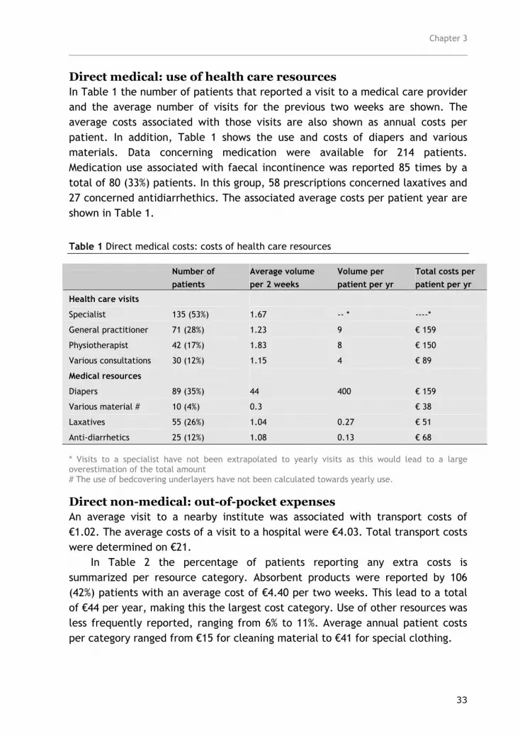
Direct medical: use of health care resources In Table 1 the number of patients that reported a visit to a medical care provider and the average number of visits for the previous two weeks are shown. The average costs associated with those visits are also shown as annual costs per patient. In addition, Table 1 shows the use and costs of diapers and various materials. Data concerning medication were available for 214 patients. Medication use associated with faecal incontinence was reported 85 times by a total of 80 (33%) patients. In this group, 58 prescriptions concerned laxatives and 27 concerned antidiarrhethics. The associated average costs per patient year are shown in Table 1.
Table 1 Direct medical costs: costs of health care resources
* Visits to a specialist have not been extrapolated to yearly visits as this would lead to a large overestimation of the total amount # The use of bedcovering underlayers have not been calculated towards yearly use.
Direct non-medical: out-of-pocket expenses An average visit to a nearby institute was associated with transport costs of €1.02. The average costs of a visit to a hospital were €4.03. Total transport costs were determined on €21.
In Table 2 the percentage of patients reporting any extra costs is summarized per resource category. Absorbent products were reported by 106 (42%) patients with an average cost of €4.40 per two weeks. This lead to a total of €44 per year, making this the largest cost category. Use of other resources was less frequently reported, ranging from 6% to 11%. Average annual patient costs per category ranged from €15 for cleaning material to €41 for special clothing.
Number of patients
Average volume per 2 weeks
Volume per patient per yr
Total costs per patient per yr
Health care visits
Specialist 135 (53%) 1.67 -- * ----*
General practitioner 71 (28%) 1.23 9 € 159
Physiotherapist 42 (17%) 1.83 8 € 150
Various consultations 30 (12%) 1.15 4 € 89
Medical resources
Diapers 89 (35%) 44 400 € 159
Various material # 10 (4%) 0.3 € 38
Laxatives 55 (26%) 1.04 0.27 € 51
Anti-diarrhetics 25 (12%) 1.08 0.13 € 68

Chapter 3 Costs of outpatients with faecal incontinence
34
Table 2 Direct non-medical costs: out-of-pocket expenses
Patients Average costs per 2 weeks
Total costs per patient per yr
Absorbent material 106 (42%) € 4.4 €44
Skincare products 29 (11%) € 5.4 € 16
Special clothing 26 (10%) € 15.6 € 41
Cleaning material 22 (9%) € 6.9 € 15
Food 16 (6%) € 22.3 € 36
Various 25 (10%) € 6.4 € 16
Indirect non-medical: production losses of paid and unpaid work Seventy-six patients (30%) had a paid occupation. The mean age of the working patients was 48 years (±9.5) and 87% was female. Six patients reported a sick leave longer than the friction period. These were excluded from further analysis. The remaining 70 patients with a paid occupation, worked on average 25 hours a week and reported a net income of €10.5 per hour.
Only 4 patients reported absence from work due to their illness. The mean number of hours of absence was 21.4 per two weeks. Efficiency loss was reported by 36 (51%) of all working patients for an average of 17.2 hours a week and a mean efficiency score of 7.8. In Table 3 the costs associated with absence and lost productivity per patient per year are shown. Most patients were capable of performing unpaid homecare activities, although some occasionally with problems. For 62 (25%) patients paid or unpaid helpers performed some household tasks. Costs associated with help in housekeeping are summarized in Table 3.
Table 3 Indirect non-medical costs: costs associated with production losses in paid and unpaid work
Patients Hours per 2 weeks Total costs/ patient/ yr
Paid work
Absenteeism 4 (1.5%) 21.4 € 92
Lost productivity 36 (14%) 35.2 (efficiency: 7.8) € 301
Unpaid work
Help from unpaid workers 39 (15%) 12.4 € 402
Help from paid workers 33 (13%) 5 € 322
Total costs In table 4 the costs of all categories are summarized and total costs per year per patient have been calculated. Total costs were estimated at € 2,169 per faecal

Chapter 3
35
incontinent patient per year. More than half of these costs were indirect non-medical costs (€ 1,118). In this category there were clearly more costs associated with help needed by unpaid activities than by impact on paid activities. Medical costs were estimated at €714 in total. The smallest cost category was the direct non-medical costs. On average patients spent € 337 per year on transport and other incontinence related resources.
Table 4 Total costs
Direct medical cost
Healthcare visits € 398
Medical resources € 316
Total direct medical € 714
Direct non-medical cost
Transport costs € 21
Resources € 316
Total direct non-medical € 337
Indirect non-medical costs
Paid work: absence of work € 92
Paid work: lost productivity € 301
Unpaid work € 724
Total indirect non-medical € 1,118
TOTAL € 2,169
Discussion
This study is the first to collect data concerning all relevant cost categories in outpatients with faecal incontinence.
A number of potential limitations of this study should be taken into account. Due to the exclusion criteria of the diagnostic cohort study some groups of patients with faecal incontinence were not studied. All patients in our study were seeking medical attention with complaints existing for at least 6 months and 8 years on average. Therefore our results cannot be unconditionally generalized to all patients with faecal incontinence. It is likely that the direct medical costs will be lower for subjects with faecal incontinence who do not seek medical help, as they will have fewer medical consultations. Although we emphasized in the questionnaires that patients were invited to report only those resources that were associated with their faecal incontinence complaints, we cannot fully assure the validity of all responses. Underreporting or incorrect attribution cannot entirely be excluded.

Chapter 3 Costs of outpatients with faecal incontinence
36
We observed a high frequency of visits to specialists in the previous two weeks. In comparison, the number of visits to a specialist as reported by the central database of Statistics Netherlands is ten times lower than in this study (http://statline.cbs.nl/StatWeb/start.asp?LA=nl&DM=SLNL&lp=Search/Search).
We believe that this large deviation in frequency from the general population is not due to faecal incontinence in itself but to the design of this study. The data for this cost study were collected as part of a larger cohort study of the diagnosis and treatment of faecal incontinence. In this study patients were seen by various specialists before being tested. We therefore deemed it inappropriate to extrapolate the observed frequency of specialist visits in the previous two weeks to yearly visits.
We were surprised by the limited use of incontinence material. Partially reimbursable incontinence products were used by only 37% of patients. A possible explanation for this finding is that many patients feel embarrassed by their disorder. This could lead to fear in discussing their problems and a lack of knowledge concerning suitable incontinence products. We did not check whether patients were aware of the existence of partially reimbursable incontinence products, or whether they had tried them before but were dissatisfied. In contrast, 42% of patients use non-reimbursable absorbent products, such as sanitary napkins. As almost half of the patients (48%) were also to some degree incontinent for urine, it is possible that absorbent material was used for both forms of incontinence.
Total costs associated with protective material (partially reimbursable and not reimbursable) were estimated at €241 per patient per year. These results are comparable with the results of Mellgren et al.17 In their study, published in 1999, the costs associated with protective material were estimated at $222.
We used available market prices to calculate the costs for the protective material. These prices are known to be fluctuating but these fluctuations are unlikely to have a significant impact on the total costs of the protective material, as prices and volumes are low.
The average costs associated with other personal expenses were limited. Only a small number of patients (6 to 11%) reported the buying of skincare products, cleaning products, special clothing, food, or other items. At the start of the study the Dutch health care reimbursed incontinence medication. We therefore decided extract medication data from the clinical report forms. We found that 37% of patients used some form of medication to control their incontinence. Most patients reported using anti-diarrhetics only when necessary. It is therefore likely that total medication costs are over-

Chapter 3
37
estimated as they have been calculated as if medication was used on a daily basis.
We found that indirect non-medical costs made up the largest part of the total costs related to faecal incontinence (€ 1118). The majority of patients had no paid occupation, which can be understood by looking at the average age (almost 60 years) and the preponderance of women (90%). It is therefore not surprising that most costs were made in order to make up for lost production during unpaid work; 28% of patients indicated that other people were taking over some of their home duties. Ten patients reported some absence from work due to health problems (13%), which is comparable with the population average (approximately 8%).
Most previous studies on faecal incontinence have focused on a narrow part of the problem and their results cannot be extrapolated to the whole population with the disorder8 Borrie et al.18 calculated the costs of incontinence in nursing homes and estimated this on $9,771 per patient per year. The total costs in the Borrie study were mainly associated with nursing time. The study of Mellgren et al.17 calculated the treatment costs of faecal incontinence after childbirth. They calculated total costs to be $17,116. In the Mellgren study indirect costs had not been calculated and some other cost items were not considered. The higher costs in the Mellgren study compared with ours probably related to their inclusion of treatment costs while we focused on ‘chronic’ faecal incontinent patients. Adang et al.19 calculated the costs of faecal incontinence but without incorporating costs associated with medical care. They estimated these costs to be $793 per patient per year, substantially lower than our results. As the paper of Adang et al.19 does not explicitly state how costs have been calculated it is difficult to interpret this difference.
Malouf et al.20 state in their review that an economic evaluation should not only include the immediate and long-term health and social care costs, but also personal, travel and indirect costs. To our opinion this study is the first to try to calculate all costs in outpatients with chronic faecal incontinence.
As this is a cross-sectional study, we do not know if and how costs develop over time. Further studies should elucidate whether costs are higher in the initial phases of the faecal incontinence complaints, or if costs increase as problems persist. We also do not know yet to what extent partial relief from incontinence problems will decrease costs.
Costs and personal impact need not to go hand in hand. Patients for whom costs are limited may experience high levels of disability and impaired social life. In all, our results show a relatively high number of patients seeking medical assistance but a surprisingly limited use of appropriate incontinence material,

Chapter 3 Costs of outpatients with faecal incontinence
38
possibly due to patients’ embarrassment. New strategies are needed for increasing access to available resources. Advances in medical research directed toward decreasing the number of patients with faecal incontinence and limiting the number of incontinent episodes will no doubt improve the quality of life for these patients while decreasing the economic impact of incontinence.
References
1. Thomas TM, Egan M, Walgrove A, Meade TW. The prevalence of faecal and double incontinence. Com Med 1984; 6(3):216-220.
2. Kamm MA. Obstetric damage and faecal incontinence. Lancet 1994; 344(8924):730-733. 3. Toglia MR. Pathophysiology of anorectal dysfunction. Obstet Gynecol Clin North Am 1998; 25(4):771-81, vi. 4. Perry S, Shaw C, McGrother C, Matthews RJ, Assassa RP, Dallosso H et al. Prevalence of faecal incontinence in
adults aged 40 years or more living in the community. Gut 2002; 50(4):480-484. 5. Edwards NI, Jones D. The prevalence of faecal incontinence in older people living at home. Age Ageing 2001;
30(6):503-507. 6. Sailer M, Bussen D, Debus ES, Fuchs KH, Thiede A. Quality of life in patients with benign anorectal disorders. Br
J Surg 1998; 85(12):1716-1719. 7. MacLennan AH, Taylor AW, Wilson DH, Wilson D. The prevalence of pelvic floor disorders and their relationship
to gender, age, parity and mode of delivery. BJOG 2000; 107(12):1460-1470. 8. Miner PB. Economic and personal impact of faecal and urinary incontinence. Gastroenterology 2004; 126(1):S8-
S13. 9. van Hout BA. Whom and how to treat: weighing the costs and effects. Scand J Gastroenterol Suppl
2003;(239):3-10.Drummond MF, O'Brien BJ, Stoddard GL, Torrance GW. Methods for economic evaluation of health care progtammes. 2 ed. Oxford: Oxford university press, 1997.
10. Gold DM, Halligan S, Kmiot WA, Bartram CI. Intraobserver and interobserver agreement in anal endosonography. Br J Surg 1999; 86(3):371-375.
11. Vaizey CJ, Carapeti E, Cahill JA, Kamm MA. Prospective comparison of faecal incontinence grading systems. Gut 1999; 44(1):77-80.
12. Gold MR, Siegel JE, Russell LB, Weinstein MC. Cost-effectiveness in health and medicine. Oxford: Oxford University Press, 1996.
13. Koopmanschap MA, Rutten FF. Berekening van kosten van zorg; vaak onderschat in economische evaluaties. Tijdschrift voor gezondheidswetenschappen 1998; 76:83-88.
14. Oostenbrink JB, Koopmanschap MA, Rutten FF. Standardisation of costs: the Dutch Manual for Costing in economic evaluations. Pharmacoeconomics 2002; 20(7):443-454.
15. Koopmanschap MA, Rutten FF, van Ineveld BM, van Roijen L. The friction cost method for measuring indirect costs of disease. J Health Econ 1995; 14(2):171-189.
16. Mellgren A, Jensen LL, Zetterstrom JP, Wong WD, Hofmeister JH, Lowry AC. Long-term cost of faecal incontinence secondary to obstetric injuries. Dis Colon Rectum 1999; 42(7):857-865.
17. Borrie MJ, Davidson HA. Incontinence in institutions: costs and contributing factors. CMAJ 1992; 147(3):322-328.
18. Adang EM, Engel GL, Rutten FF, Geerdes BP, Baeten CG. Cost-effectiveness of dynamic graciloplasty in patients with faecal incontinence. Dis Colon Rectum 1998; 41(6):725-733.
19. Malouf AJ, Chambers MG, Kamm MA. Clinical and economic evaluation of surgical treatments for faecal incontinence. Br J Surg 2001; 88(8):1029-1036.

Chapter 3
39
Appendix 1
Source Price 2004 (in euros)
Type of consultation
General practioner Oostenbrink, 2002 17.69
Specialist in an academic hospital Oostenbrink, 2002 72.83
Specialist in a general hospital Oostenbrink, 2002 41.62
Physiotherapist Oostenbrink, 2002 18.73
Dietician NVD 1999-2000 23.33
Company physician AMR 1999-2000 23.55
Social worker AMR 1999-2000 17.98
Visiting nurse Oostenbrink, 2002 33.04
Absorbent and protective material
Disposable diapers Coloplast 0.35
Disposable diaper pants Coloplast 0.64
Washable diapers and diaper pants Coloplast 21
Anal tampons Coloplast 6.7
Medication
Laxatives This study & http://www.fk.cvz.nl/ 187.4 (±62)
Anti-diarrhetics This study & http://www.fk.cvz.nl/ 519.4 (±137)
Transport costs
Average distance to hospital RIVM 7.0 km
Av. distance to local health care provider RIVM 1.8 km
Car and public transport per kilometre Oostenbrink, 2002 0.125
Taxi per kilometre Oostenbrink, 2002 1.40
Parking fee Oostenbrink, 2002 1.20
Help in house
Unpaid helper Oostenbrink, 2002 8.32
Paid helper Oostenbrink, 1999 19.05


Chapter 4
Selecting an outcome measure for evaluating treatment in faecal incontinence
Marije Deutekom1, Maaike P. Terra2, Annette C. Dobben2, Marcel G.W. Dijkgraaf1, Richelle J.F. Felt-Bersma3, Jaap Stoker2, Patrick M.M. Bossuyt1
1Department of Clinical Epidemiology and Biostatistics, 2Department of Radiology from the Academic Medical Centre, Amsterdam, The Netherlands, 3Department of Gastroenterology from the VU Medical Centre, Amsterdam, the Netherlands
Diseases of the Colon and Rectum, 2005, in press

Chapter 4 Selecting an outcome measure for evaluating treatment in faecal incontinence
42
Abstract
Background Various outcome measures exist to evaluate treatment in faecal incontinence, including descriptive, severity (faecal incontinence scoring systems), and impact (quality of life questionnaires) and diagnostic measures. We studied associations between changes after treatment for a number of outcome measures and compared them to patients’ subjective perception of relief. Methods We analyzed of 66 patients (92% female; mean 62 years) data (Vaizey score, Wexner score, two impact scales, utility, resting pressure and maximal incre-mental squeeze pressure) at baseline and after pelvic floor rehabilitation. In a standardized interview by phone we asked patients to compare their situation before and after treatment. Correlations between changes in outcome measures were calculated. These changes were compared with patients’ subjective perception. Results There was a high correlation between the changes in the Vaizey and Wexner score (r=.94, p<.01). Changes in Vaizey and Wexner correlated moderately with changes in maximum incremental squeeze pressure (r=-.29, -.30, both p<.05). Changes in utility and resting pressure were not correlated with changes in any of the other measurements (all r’s between -.086 and .18). Average severity scores (Vaizey and Wexner) were 1 point lower for patients who rated their situation as worse or equal (62%), 4 points for patients who reported their situation to be better (21%), and 9 points lower in patients who to rated their situation much better (17%) (p<0.05). Conclusion Severity measures are best related to patients’ subjective perception of relief.

Chapter 4
43
Introduction
Faecal incontinence is defined as “the involuntary loss of flatus, liquid or solid stool that is a social or hygienic problem”.1 The reported prevalence ranges from 1.4% in the general population, where it has been defined as soiling of underwear, outer clothing, furnishing, or bedding several times a month or more often2 to 46% in institutionalized elderly people, with incontinence defined as at least one incontinent episode per week.3
Treatment aims to resolve the incontinence complaints by trying to recover the impaired continence mechanisms. A wide range of treatments is available, ranging from conservative therapy, such as dietary recommendations, anti-diarrhoea medication or pelvic floor rehabilitation, to surgical treatment by external anal sphincter repair, graciloplasty, artificial sphincter implantation or sacral nerve stimulation.4,5
Before a treatment is introduced in practice, it should be thoroughly evaluated. Similarly, existing treatment strategies should also be critically evaluated. Determining the effectiveness of treatment requires an evaluation of treatment outcome. Many outcome measures are available. They can be divided into three broad categories: descriptive measures, severity measures, and impact measures.6
Descriptive measures include a list of items that relate to various aspects of incontinence. Such measures are frequently used as outcome measure in popu-lation-based studies aiming to determine the incidence of incontinence.7,8 As no single score is calculated, these measures are difficult to use in research studies.6
Severity measures (faecal incontinence scoring systems) can be differentiated in grading scales and summary scores. Grading scales contain various categories of incontinence in an ordinal fashion ranging from complete continence to complete incontinence. The number of grades in these grading scales, described in literature, ranges from three to five.6 In the past these scales have been used to assess treatment outcomes.9 Nowadays summary scores that recognize that incontinence is more complex than assumed by the ordinal categories are more often used as primary outcome.10-17 These summary scores contain items concerning the frequency and type of stool loss 18,19 and sometimes extra items regarding alteration in lifestyle and pad and/or medication use.4,20
Other outcome measures address the impact of faecal incontinence on a patient’s quality of life. This impact is measured by using questionnaires, while attention can focus on the impact on general health related quality of life, or on disease specific quality of life.

Chapter 4 Selecting an outcome measure for evaluating treatment in faecal incontinence
44
Such impact measures are seldom used as primary outcome, however they are often taken into account as secondary outcome. 11,13,14,17,21-23
Although the use of subjective perception is increasing, diagnostic measures, such as tests to document the functional and anatomic characteristics of the rectum and anal sphincter complex, are still often used as secondary outcome measures9,11,16,17,21,23-28 and sometimes even as primary outcome.10,29,30 These tests can help to determine the causes and may guide treatment planning, but doubts exists as to whether these measures can be used to study treatment outcome of faecal incontinence.6,31
Unfortunately there exists no consensus about the optimal outcome measure in the assessment of treatment in faecal incontinence. Baxter and colleagues6 have argued that faecal incontinence is a symptom and, as such, the subjective perception of the patient must be the foundation of any evaluation of incontinence. Many of the existing outcome measures have been insufficiently validated. It has been suggested that research must focus on evaluating the reliability and validity of the existing outcome measures and that comparisons between various measures would enrich our knowledge concerning the usefulness of these measures.6
The primary aim of this study was to compare in a series of patients with faecal incontinence, changes in various outcome measures before and after treatment.
The variety in used outcome measure is paralleled by a lack of consensus regarding the outcome measure that best reflects patients’ perception. The secondary aim was to compare changes in outcome measure with patients’ subjective perception of relief.
Methods
Subjects The data for this study were collected as part of a large clinical cohort study of the value of diagnostic tests in patients with faecal incontinence. Between December 2001 and April 2004 consenting eligible patients with faecal incontinence visiting one of 16 participating Dutch hospitals were consecutively included. Details of this study will be reported elsewhere.
Inclusion criteria for the cohort study were the existence of faecal incontinence complaints for six months or more, a Vaizey incontinence score20 of 12 or more, and failure of conservative treatment, based on dietary recommendations and/or anti-diarrhoea medication.

Chapter 4
45
Excluded were patients with an age below 18, patients diagnosed less than two years ago with an anorectal tumour and patients with a previous ileoanal or coloanal anastomosis. Patients with chronic diarrhoea (always fluid stools, three or more times a day), overflow incontinence, proctitis, soiling (defined as leakage of faecal material out of the anus after normal defecation leading to perineal eczema), and rectal prolapse were also excluded from participation. The medical ethics committees of all participating hospitals approved the study and signed informed consent was obtained from all participating patients.
Measurements After receiving signed informed consent of patients, data concerning medical history were collected by physicians through a patient interview. These comprised the possible underlying causes for faecal incontinence, duration of faecal incontinence complaints, type and frequency of faecal incontinence and severity of faecal incontinence (Vaizey score).20
The Vaizey scale consists of three items about the type (gas, fluid, solid) and frequency of incontinence (all scored from zero to four) and four additional items addressing alteration in lifestyle (zero to four), the need to wear a pad or plug (zero or two), the use of constipating medication (zero or two) and the lack of ability to defer defecation for 15 minutes (zero or four). The total score on the Vaizey scale ranges from 0 (complete continence) to 24 (complete incon-tinence).20 The Wexner score also contains three items about the type and frequency of incontinence (scored from zero to four), as well as items on pad usage and lifestyle alteration (both also scored from zero to four).4,20 The total score on this scale ranges from 0 (complete continence) to 20 (complete incontinence) and was calculated was during data-analysis from the case record form.
At the end of their first visit patients were handed questionnaires, which they were requested to complete at home. Health status was assessed by means of the EQ-5D instrument, developed by the EuroQol Group.32 This questionnaire addresses five domains (mobility, self-care, daily activities, pain/discomfort, anxiety/depression). The domains each comprise three levels: no problems, some/ moderate problems and extreme problems/unable to. The responses on the five dimensions can be transformed into a summary utility value, expressing a choice-based valuation of the health state. This utility value can range from –0.594 (worst imaginable health state) to 1 (optimal health).33
Disease specific functioning was measured with a Dutch validated quality of life questionnaire originally developed for Hirschsprung’s disease/ Anorectal malformation (HAQL).34 The four response categories, ranging from ‘never’ to ‘very often’, were recoded and linearly transformed to a 0 to 100 scale, with

Chapter 4 Selecting an outcome measure for evaluating treatment in faecal incontinence
46
higher scores indicating a higher level of functioning. The 42 items of the questionnaire were combined to form three subscales: constipation, faecal incontinence and disease-specific psychosocial functioning. In this study we used only two subscales: faecal incontinence and disease-specific psychosocial functioning.
Anal manometry was performed using a microballoon, a water perfused catheter with or without sleeve, or a solid-state transducer. The solid-state method or water-perfusion method without sleeve was performed by means of a pull-through technique. The catheter (Konigsberg Instrument Inc., Pasadena, CA; Medtronic, Skolvunde, Denmark; Dentsleeve Pty Ltd, Parkside, Australia) was inserted in the anal canal and the (mean) maximal resting pressure (mmHg) was measured. Subsequently, the (mean) maximal squeeze pressure (mmHg) was determined by asking patients to squeeze three times during 10 seconds with one-minute intervals. An average maximal squeeze pressure (mmHg) was calculated.
All patients underwent nine sessions of pelvic floor rehabilitation comprising electrical stimulation and/or different elements of biofeedback. The treatment program and targets were customized per patient. All measurements and assessments performed at baseline were repeated approximately three months after treatment.
In March 2004 one of the researchers (MT) identified 79 patients who had recently finished treatment. These patients were contacted by phone and invited to provide additional information in a standardized interview. After approval, patients were retrospectively asked to compare their present situation, after treatment, with their level of incontinence before treatment. Patients were asked to score their current situation on a scale of 1 (much worse), 2 (worse), 3 (no change), 4 (better), or 5 (much better). The interview was executed by a trained interviewer (MD) who was blinded for all outcome measures and treat-ment details.
Analysis Paired t-tests were used to investigate the significance of changes in the continuous and multi-categorical outcome measures after treatment. We used parametric Pearson correlation coefficients to compare the changes from baseline after treatment in the various outcome measures.
To investigate the relation between change in outcome measure and patients perception of relief we used analysis of variance, with subjective perception as grouping variable and the outcome measures as test variables. For post-hoc analyses we used Bonferonni corrections.

Chapter 4
47
For all analyses, p-values below 0.05 were considered to indicate statistical significance. The interpretation of the magnitude of the observed correlations was based on Cohen: significant correlations below 0.3 were considered low, between 0.3 and 0.5 moderate, and 0.5 or above high.35
Results
The interviews took place in March and April 2004. We were able to obtain interview data from 68 patients (86%); for two of these patients clinical data were absent. We analyzed all data of the remaining 66 patients, of which 61 were female (92%). Their mean age was 62 years (SD ± 11) with a mean duration of incontinence of 7 years (SD ± 7). The median duration between last treatment session and outcome assessment was 1.9 months (interquartile range: .9 to 3.3). Baseline values of outcome measures are shown in Table 1.
Table 1 Baseline measurements
Relation between changes in outcome measures after treatment After treatment the average Vaizey score decreased from 18 to 15 (p<0.001). The average Wexner score decreased from 15 to 12 (p<0.001). The resting pressure was significantly higher after treatment (from 51 mmHG to 56 mmHG, p=.046). No difference was present in the incremental squeeze pressure. None of the impact measures (‘faecal incontinence’, ‘disease-specific psychosocial func-tioning’ and utility) showed a significant change.
Table 2 shows the correlations between the changes in the respective outcome measures. There was a high correlation between the changes in the two
Outcome measure Score Range
Severity measures
Vaizey score 18 (3) 0 (complete continence) to 24 (complete incontinence)
Wexner score 15 (3) 0 (complete continence) to 20 (complete incontinence)
Impact measures
Utility value .81 (.19) -0.594 (worst) to 1 (optimal)
Faecal incontinence 76 (15) 0 (worst) to 100 (optimal)
Disease specific psychosocial functioning 66 (26) 0 (worst) to 100 (optimal)
Diagnostic measures
Maximum incremental squeeze pressure (mmHg) 36 (32) not applicable
Resting pressure (mmHg) 52 (24) not applicable

Chapter 4 Selecting an outcome measure for evaluating treatment in faecal incontinence
48
severity measures (Vaizey and Wexner) (p<0.01). The Wexner score correlated weakly with the impact measure ‘disease-specific psychosocial functioning’ (p=0.046). There existed a moderate correlation between the changes in severity measures and changes in maximum incremental squeeze pressure (p<0.05). No correlation was apparent between changes in maximum incremental squeeze pressure and resting pressure. The impact measure ‘disease-specific psychosocial functioning’ was highly correlated with the impact measure ‘faecal incontinence’ (p<0.01). Changes in utility and resting pressure were not correlated with changes in any of the other measurements.
Table 2 Correlation between changes in outcome measures from baseline after treatment
Pearson’s r are shown for all combinations of measures. * Correlation is significant at the 0.05 level (2-tailed). **Correlation is significant at the 0.01 level (2-tailed).
Relation between outcome measures with patients’ perception The mean time between the end of treatment and the interview was 2 months. In the interviews 11 patients (16%) rated their situation after treatment much better than before, 14 (21%) said it was better, 37 (56%) said it was equal, 3 (5%) that it was worse and 1 (2%) that it was much worse.
Based on these responses we made three subgroups: worse or equal (n=41); better (n=14); and much better (n=11). There existed no difference between the three groups in the average changes of maximal incremental squeeze pressure (p=.95), resting pressure (p=.60) and utility values (p=.216). Significant differences could be observed in Vaizey score (p<.001), Wexner score (p<.001)
Severity Impact Diagnostic
Vaizey score
Wexner score
Utility
Faecal incontinence
Psychosoc functioning
Max. incr. squeeze
Vaizey score
Severity
Wexner score
.94 **
Utility -.099 -.033
Faecal Incontinence
-.10 -.20 .17
Impact Psychosocial
functioning -.25 -.26* .22 .52**
Max. incr. squeeze
-.29* -.30* -.17 .099 .22
Diagnostic
Resting pressure
-.028 .048 .18 -.017 -.044 -.086

Chapter 4
49
and in the impact measures ‘faecal incontinence’ (p=.011) and ‘disease specific psychosocial functioning’ (p=.022).
Post-hoc tests showed that all three groups were significantly different from each other with respect to change in Vaizey score (p<0.05). The group that perceived their situation much better had a decrease in Vaizey of 9 points, the group that perceived their situation better a decrease of 4 points and the group that rated their situation worse or equal an average decrease of 1 point (Figure 1a).
Figure 1a-d Change in outcome measures stratified by subjective perception Data are shown as mean ± 95% confidence interval * Significant difference (p<0.05) between much better # Significant difference (p<0.05) between better

Chapter 4 Selecting an outcome measure for evaluating treatment in faecal incontinence
50
The change in Wexner score was significantly larger for the patients that rated their situation much better (-9 points) than for the other two groups (worse or equal: -1 point; better: -4 points) (Figure 1b). The differences in impact scores ‘faecal incontinence’ and ‘disease specific psychosocial functioning’ reached only significance between the ‘much better’ group (+9 and +13 points respectively) and the group that perceived their situation equal or worse (-2 and –1 points respectively) (Figure 1c, 1d).
Discussion
This study in patients treated with pelvic floor rehabilitation compared changes in severity measures (Vaizey and Wexner score), anal manometry (resting pressure and maximal incremental squeeze pressure) and impact measures (utility, faecal incontinence, and disease specific functioning). Changes in anal resting pressure were found to be unrelated to any other change or patients’ perception of relief. Changes in maximal incremental squeeze pressure were weakly correlated with changes in severity measures but not with patients’ perception of relief. Change in severity measures and change in impact measures were well related to patients’ perception of relief.
A number of potential limitations of this study should be taken into account. The group of patients in this study does not represent the full spectrum of faecal incontinence complaints. Due to the exclusion criteria some groups were not included. All patients in our study were seeking medical attention with complaints existing at least 6 months, and 7 years on average. Ninety-two percent of the participants in our study were female, an imbalance that is not due to a form of selection bias but is inherent to the disorder of faecal incontinence.36 The percentage of patients reporting relief was modest in our study, limiting the power to detect differences between patients.
In our study, we compared change in outcome measures with patient’s perception of relief. We are aware that the validation of the latter measure is problematic, due to the subjective part of the question. An alternative and possibly more objective measure would have been the use of diary report to assess improvement. From diary reports we would be able to see whether or not actual incontinent events truly decrease, but compliance in the use of diaries can be low. A study by Stone and colleagues described an actual compliance rate of 11% with paper diaries opposed to the 95% reported.37 The use of electronic diaries had been shown to increase compliance markedly. We doubt whether the acceptance of electronic diaries in our study group of patients with faecal

Chapter 4
51
incontinence (predominantly females and around 60 years) would have been high.
In the absence of a true golden standard to assess the severity of incontinence, we cannot claim that the observed relation in our study between patients’ perception and severity measure is truly caused by agreement between patients and clinicians on the factors that underlie the severity of incontinence. One mechanism that could have biased this relation is a ‘placebo’ effect. Patients might (want to) believe that they have improved, report their situation to be better and coinciding report less episodes of unwanted stool loss. In measuring the outcome of treatment, we had to make a number of choices. We decided to use two manometric tests as diagnostic measures. We believed that imaging tests would have been less useful, as our treatment consisted of pelvic floor rehabilitation.
Many different severity measures scores have been developed to assess the severity of incontinence, including grading scales and summary scores. We believe that summary scores recognize that incontinence is more complex than assumed by the grading scales. Grading scales can be further differentiated into those with only items concerning the frequency and type of stool loss18,19 and those with extra items regarding alteration in lifestyle and pad and/or medication use.4,20 We selected two of the most extensive scores for our study.
Experts taught us that the interpretation of specialists varied on the items ‘loss of liquid stool’ and ‘loss of gas’. For consistency across centres, we decided that a patient’s frequency of incontinence for gas equals or exceeds her or his frequency of liquid stool. Furthermore we assumed that a patient’s frequency of incontinence for liquid stool is at least the same as the frequency of solid stool. It is possible that these scoring rules have led to higher average incontinence scores than reported elsewhere. However, this decision did not affect the changes between subgroups in the Vaizey and Wexner score.
It has been suggested that these severity scores might be subject to recall bias. Naliboff38 stated that a single retrospective rating of ‘usual’ symptom intensity may be a good reflection of the average symptom intensity (at least for chronic pain). In a study about constipation it has been shown that the results regarding the presence of constipation are highly compatible between self-reported constipation and diary-based constipation.39 We expect that the frequency of reported symptoms as used in determining the severity of incontinence does reflect the actual frequency of symptoms.
As impact measure we used utilities and a disease specific functioning questionnaire, originally designed for patients with anorectal malformation and Hirschsprung’s disease.34 This questionnaire results in three impact subscale
Chapter 4 Selecting an outcome measure for evaluating treatment in faecal incontinence
52
scores: constipation, faecal incontinence, and disease-specific psychosocial functioning. In 2000 Rockwood and colleagues and Reilly and colleagues40,41 reported on the development of health-related quality of life scales to address issues related specifically to faecal incontinence. Unfortunately a validated Dutch version of one of these questionnaires was absent at the initiation of our study.
To some extent Hirschsprung patients suffer from faecal incontinence complaints similar to those of the patients in our study. We decided to leave out the subscale ‘constipation’ in our study, as this complaint is of less importance in our patient group. The significant difference in the impact measures ‘faecal incontinence’ and ‘disease specific psychosocial functioning’ between patients who rated their situation much better and patients who rated their situation worse or unchanged can be interpreted as an initial validation for the use of this scale in our group of patients.
Our results show no relation between changes in diagnostic measures and relief, as experienced by patients. This supports the suggestion that diagnostic measures do not measure the severity of faecal incontinence itself nor are an apt outcome measure.6,31
How then should treatments of faecal incontinence be evaluated? Baxter has argued that the severity and impact of incontinence should be considered in the evaluation of faecal incontinence.6 In our study utilities appeared to be too insensitive as a patient-oriented outcome measure. This corresponds with earlier suggestions that generic questionnaires, containing non-specific items and scoring systems applicable to widely varying states of health, lack sensitivity in women with non-life-threatening conditions such as urinary incontinence.42 Both impact scales of the disease specific functioning questionnaire were associated with patients’ perception of relief. However patients who reported their situation as better did not differ in these scales from patients who reported their situation much better.
Our results show a strong association between the clinicians’ incontinence score and patients’ subjective feeling concerning relief. We found significant differences in Vaizey and Wexner score between patients who rated their situation worse or equal, patients who reported their situation better, who reported their situation much better. This finding is in contrast with previous research43 that has questioned the value of clinical scales, as they are developed by clinicians and focus heavily on quantitative measurements whereas patients tend to put more emphasis on social limitations caused by the incontinence.
As far as we know, our study is the first to compare changes in a series of outcome measures and subjective perception. We have shown that severity

Chapter 4
53
measures (Vaizey score and Wexner score) have a clear relation with patient’s subjective feeling concerning relief and therefore believe that severity measures should be included to evaluate outcome in faecal incontinence trials. We hope that this study inspires others to explore criteria for treatment success in faecal incontinence trials.
References
1. Abrams, P, Cardozo, L, Khoury, S, Wein, AJ. Incontinence: Proceedings of the Second International Consultation on Incontinence. Health Publication Limited, Plymbridge Distributors, Ltd, 2nd Edition, 2002.
2. Thomas TM, Egan M, Walgrove A, Meade TW. The prevalence of faecal and double incontinence. Com Med. 1984;6:216-20.
3. Perry S, Shaw C, McGrother C, Matthews RJ, Assassa RP, Dallosso H et al. Prevalence of faecal incontinence in adults aged 40 years or more living in the community. Gut 2002;50:480-4.
4. Jorge JM, Wexner SD. Etiology and management of faecal incontinence. Dis Colon Rectum 1993;36:77-97. 5. Matzel KE, Bittorf B, Stadelmaier U, Hohenberger W. Sacral nerve stimulation in the treatment of faecal
incontinence. Chirurg 2003;74:26-32. 6. Baxter NN, Rothenberger DA, Lowry AC. Measuring faecal incontinence. Dis Colon Rectum 2003;46:1591-605. 7. Macmillan AK, Merrie AE, Marshall RJ, Parry BR. The prevalence of faecal incontinence in community-dwelling
adults: a systematic review of the literature. Dis Colon Rectum 2004;47:1341-9. 8. Soderstrom U, Hoelcke M, Alenius L, Soderling AC, Hjern A. Urinary and faecal incontinence: a population-
based study. Acta Paediatr. 2004;93:386-9. 9. van Tets WF, Kuijpers JHC. Pelvic floor procedures produce no consistent changes in anatomy or physiology.
Dis Colon Rectum 1998;41:365-9. 10. Casal E, San Ildefonso A, Carracedo R, Facal C, Sanchez JA. Artificial bowel sphincter in severe anal
incontinence. Colorectal Dis 2004;6:180-4. 11. Mahony RT, Malone PA, Nalty J, Behan M, O'Connell PR, O'Herlihy C. Randomized clinical trial of intra-anal
electromyographic biofeedback physiotherapy with intra-anal electromyographic biofeedback augmented with electrical stimulation of the anal sphincter in the early treatment of postpartum faecal incontinence. Am J Obstet Gynecol 2004;191:885-90.
12. Rasmussen OO, Buntzen S, Sorensen M, Laurberg S, Christiansen J. Sacral nerve stimulation in faecal incontinence. Dis Colon Rectum 2004;47:1158-63.
13. Nichols CM, Gill EJ, Nguyen T, Barber MD, Hurt WG. Anal sphincter injury in women with pelvic floor disorders. Obstet Gynecol 2004;104:690-6.
14. O'Brien PE, Dixon JB, Skinner S, Laurie C, Khera A, Fonda D. A prospective, randomized, controlled clinical trial of placement of the artificial bowel sphincter (Acticon Neosphincter) for the control of faecal incontinence. Dis Colon Rectum 2004;47:1852-60.
15. Norderval S, Nsubuga D, Bjelke C, Frasunek J, Myklebust I, Vonen B. Anal incontinence after obstetric sphincter tears: incidence in a Norwegian county. Acta Obstet Gynecol Scand 2004;83:989-94.
16. Osterberg A, Edebol Eeg-Olofsson K, Hallden M, Graf W. Randomized clinical trial comparing conservative and surgical treatment of neurogenic faecal incontinence. Br J Surg 2004;91:1131-7.
17. Beddy P, Neary P, Eguare EI, et al. Electromyographic biofeedback can improve subjective and objective measures of faecal incontinence in the short term. J Gastrointest Surg 2004;8:64-72.
18. Rockwood TH, Church JM, Fleshman JW, et al. Patient and surgeon ranking of the severity of symptoms associated with faecal incontinence: the faecal incontinence severity index. Dis Colon Rectum 1999;42:1525-32.
19. Pescatori M, Anastasio G, Bottini C, Mentasti A. New grading and scoring for anal incontinence. Evaluation of 335 patients. Dis Colon Rectum 1992;35:482-7.
20. Vaizey CJ, Carapeti E, Cahill JA, Kamm MA. Prospective comparison of faecal incontinence grading systems. Gut 1999;44:77-80.
21. Jarrett ME, Varma JS, Duthie GS, Nicholls RJ, Kamm MA. Sacral nerve stimulation for faecal incontinence in the UK. Br J Surg. 2004;91:755-61.

Chapter 4 Selecting an outcome measure for evaluating treatment in faecal incontinence
54
22. Matzel KE, Kamm MA, Stosser M, et al. Sacral spinal nerve stimulation for faecal incontinence: multicentre study. Lancet 2004;363:1270-6.
23. Solomon MJ, Pager CK, Rex J, Roberts R, Manning J. Randomized, controlled trial of biofeedback with anal manometry, transanal ultrasound, or pelvic floor retraining with digital guidance alone in the treatment of mild to moderate faecal incontinence. Dis Colon Rectum 2003;46:703-10.
24. Ozturk R, Niazi S, Stessman M, Rao SS. Long-term outcome and objective changes of anorectal function after biofeedback therapy for faecal incontinence. Aliment Pharmacol Ther 2004;20:667-74.
25. Fynes M, Marshall K, Cassidy M, et al. A prospective, randomized study comparing the effect of augmented biofeedback with sensory biofeedback alone on faecal incontinence after obstetric trauma. Dis Colon Rectum 1999;42:760-1.
26. Kusunoki M, Shoji Y, Ikeuchi H, Yamagata K, Yamamura T, Utsunomiya J. Usefulness of valproate sodium for treatment of incontinence after ileoanal anastomosis. Surgery 1990;107:311-5.
27. Sun WM, Read NW, Verlinden M. Effects of loperamide oxide on gastrointestinal transit time and anorectal function in patients with chronic diarrhoea and faecal incontinence. Scand J Gastroenterol 1997;32:34-8.
28. Deen Ki, Oya M, Ortiz J, Keighley MR. Randomized trial comparing three forms of pelvic floor repair for neuropathic faecal incontinence. Br J Surg 1993;80:794-8.
29. Carapeti EA, Kamm MA, Phillips RK. Randomized controlled trial of topical phenylephrine in the treatment of faecal incontinence. Br J Surg 2000;87:38-42.
30. Cheetham MJ, Kamm MA, Phillips RK. Topical phenylephrine increases anal canal resting pressure in patients with faecal incontinence. Gut 2001;48:356-9.
31. Shelton AA, Madoff RD. Defining anal incontinence: Establishing a uniform continence scale. Seminars in Colon & Rectal surgery 1997;8:54-60.
32. Williams A. Euroqol - A New Facility for the Measurement of Health-Related Quality-Of-Life. Health Policy 1990;16:199-208.
33. Dolan P. Modeling valuations for EuroQol health states. Med.Care 1997;35:1095-108. 34. Hanneman MJ, Sprangers MA, De Mik EL, et al. Quality of life in patients with anorectal malformation or
Hirschsprung's disease: development of a disease-specific questionnaire. Dis Colon Rectum 2001;44:1650-60. 35. Cohen J. Statistical power analysis for the bahavioural sciences. New Jersey: Lawrence Erlbaum Associates,
1988. 36. Henry MM. Pathogenesis and management of faecal incontinence in the adult. Gastroenterol.Clin North Am
1987;16:35-45. 37. Stone AA, Shiffman S, Schwartz JE, Broderick JE, Hufford MR. Patient non-compliance with paper diaries. BMJ
2002;324:1193-4. 38. Naliboff BD. Choosing outcome variables: global assessment and diaries. Gastroenterology 2004;126:S129-S134. 39. Pamuk ON, Pamuk GE, Celik AF. Revalidation of description of constipation in terms of recall bias and visual
scale analog questionnaire. J Gastroenterol Hepatol. 2003;18:1417-22. 40. Reilly WT, Talley NJ, Pemberton JH, Zinsmeister AR. Validation of a questionnaire to assess faecal
incontinence and associated risk factors: Faecal Incontinence Questionnaire. Dis Colon Rectum 2000;43:146-53. 41. Rockwood TH, Church JM, Fleshman JW, et al. Faecal Incontinence Quality of Life Scale: quality of life
instrument for patients with faecal incontinence. Dis Colon Rectum 2000;43:9-16. 42. Kelleher C. Quality of life and urinary incontinence. Baillieres Best Pract Res Clin Obstet Gynaecol
2000;14:363-79. 43. Byrne CM, Pager CK, Rex J, Roberts R, Solomon MJ. Assessment of quality of life in the treatment of patients
with neuropathic faecal incontinence. Dis Colon Rectum 2002;45:1431-6.

Chapter 5
Anal plugs for the containment of faecal incontinence
Marije Deutekom1 and Annette C. Dobben2
1Department of Clinical Epidemiology and Biostatistics, 2Department of Radiology from the Academic Medical Centre, Amsterdam
Cochrane Database of Systematic Reviews, 2005, Jul 20:(3):CD005086

Chapter 5 Anal plugs for the containment of faecal incontinence
56
Abstract
Background Faecal incontinence is a distressing disorder with high social stigma. Not all people with faecal incontinence can be cured with conservative or surgical treatment and they may need to rely on containment products, such as anal plugs. Objective was to assess the performance of different types of anal plugs for containment of faecal incontinence.
Methods Search strategy We searched the Cochrane Incontinence Group Specialised Register (searched 22 November 2004), MEDLINE (January 1966 to November 2004), CINAHL (January 1982 to November Week 3 2004), EMBASE (January 1996 to 2004 Week 47), INVERT (Dutch nursing database) (January 1993 to November 2004) and Web of Science (January 1988 to November 2004). Reference lists of identified trials were searched and plugs manufacturers were contacted for trials. No language or other limitations were imposed. Selection criteria Types of studies: This review was limited to randomised and quasi-randomised controlled trials (including crossovers) of anal plug use for the management of faecal incontinence. Types of participants: Children and adults with faecal incontinence. Types of interventions: Any type of anal plug. Comparison interventions might include no treatment, conservative (physical) treatments, nutritional interventions, surgery, pads and other types or sizes of plugs. Data collection & analysis Two reviewers independently assessed methodological quality and extracted data from the included trials. Authors of all included trials were contacted for clarification concerning methodological issues.
Results Four studies with a total of 136 participants were included. Two studies compared the use of plugs versus no plugs, one study compared two sizes of the same brand of plug, and one study compared two brands of plugs. In all included studies there was considerable dropout (in total 48 (35%) dropped out before the end of the study) for varying reasons. Data presented are thus subject to potential bias. 'Pseudo-continence' was, however, achieved by some of those who continued to use plugs, at least in the short-term. In a comparison of two
Chapter 5
57
different types of plug, plug loss was less often reported and overall satisfaction was greater during use of polyurethane plugs than polyvinyl-alcohol plugs. Conclusions The available data were limited and incomplete, and not all pre-specified outcomes could be evaluated. Consequently, only tentative conclusions are possible. The available data suggest that anal plugs can be difficult to tolerate. However, if they are tolerated they can be helpful in preventing incontinence. Plugs could then be useful in a selected group of people either as a substitute for other forms of management or as an adjuvant treatment option. Plugs come in different designs and sizes; the review showed that the selection of the type of plug can impact on its performance.

Chapter 5 Anal plugs for the containment of faecal incontinence
58
Introduction
Faecal incontinence is defined as the involuntary passage of faecal material through the anal canal.1 The reported prevalence values range from 1.4% in the general population (defined as soiling of underwear, outer clothing, furnishing, or bedding several times a month or more often)2 to 46% in institutionalised elderly people (defined as at least one incontinent episode per week).3 It is possible that the real prevalence is even higher than reported as faecal incontinence is associated with high social stigma and people are reluctant to seek help for this disorder because of embarrassment. 4,5
The causes for faecal incontinence are diverse. In most cases a combination of factors leads to incontinence. Frequently cited causes are injuries during childbirth and prior anorectal surgery.6,7 But many other causes have been described, including loose stool, intestinal hurry, and neurological disease or injury.
Treatments available range from conservative therapy, such as dietary recommendations and anti-diarrhoeal medication, to surgical treatment by either sphincter repair, dynamic graciloplasty, artificial anal sphincter implantation, or sacral nerve stimulation.4,8 Nowadays, the most common treatments are pelvic floor muscle training - with or without biofeedback - and anterior anal sphincter repair.6 The reported success rates with these forms of treatment vary, but it is recognized that none of the treatments will resolve the incontinence problems in all patients.
Where incontinence persists despite active treatment there may be no option other than containment. Brazzelli et al.9 have reviewed the use of pads for the containment of anal and urinary incontinence. Problems when using pads for faecal incontinence are that the odour from the anal leakage is difficult to control and extensive use of pads can result in skin condition problems. A possible way to avoid these problems is the use of an anal plug (sometimes called 'anal tampon'): a device specially developed for containing faecal incontinence.
Different types of anal plugs are known, all aiming to block the loss of stool. They were first used in patients suffering from faecal incontinence due to major neurological problems, such as caused by spina bifida.10 Nowadays, plugs are also sometimes used by patients with faecal incontinence who do not have an underlying neurological condition.
At this point it is unclear how effective anal plugs are in controlling stool loss in patients with faecal incontinence (with or without neurological impairments) and whether some types of anal plugs are more effective than

Chapter 5
59
others. This review aims to bring together in a systematic way the best available evidence to address these issues.
The objectives of this review were to assess the performance of different types of anal plugs for containment of faecal incontinence. The following comparisons were considered: (1) anal plugs versus no plugs, (2) one type of anal plug versus another; and (3) anal plugs versus any other treatment.
Methods
Criteria for considering studies for this review Types of studies This review was limited to randomised and quasi-randomised controlled trials (including crossovers) of anal plug use for the management of faecal incontinence. Types of participants All patients (children and adults) with faecal incontinence. Types of interventions Studies investigating the relative performance of anal plugs. Potential comparison interventions include no treatment, conservative (physical) treatments, nutritional interventions, surgery, pads, and other types or sizes of plugs. Types of outcome measures 1. Patient symptoms
• frequency of incontinence of stool or flatus (diary or self-report)
• degree of incontinence (e.g. stool weight)
• incontinence score
• episodes of anal urgency
2. Physical measures • achievement of pseudo-continence (continence only while wearing a plug)
• wearing time and frequency of use
• leakage rate
• odour control
3. Patient satisfaction • satisfaction with incontinence controlling capacity
• tolerability of plug (including persistence in using the plug)
• comfort of plug in use
• comfort of plug removal/ease of removal

Chapter 5 Anal plugs for the containment of faecal incontinence
60
• feeling of cleanness
4. Health status measures • impact of incontinence on health status, social life, and quality of life
5. Costs 6. Other outcomes
• non pre-specified outcomes later judged important when performing the review
Search strategy for identification of studies We formulated a comprehensive and exhaustive search strategy in an attempt to identify all relevant studies regardless of language or publication status (published, unpublished, in press, and in progress).
This review has drawn on the search strategy developed for the Cochrane Incontinence Group. Relevant trials were identified from the Group's specialised register of trials, which is described under the Cochrane Incontinence Group's details in The Cochrane Library. The register contains trials identified from MEDLINE, CINAHL, The Cochrane Central Register of Controlled Trials (CENTRAL), and hand searching of journals and conference proceedings.
For this review the authors performed the following additional searches. All searches were carried out on 26 November 2004. The following electronic bibliographic databases were searched: MEDLINE (January 1966 to November 2004); CINAHL (January 1982 to November Week 3 2004); EMBASE (January 1996 to 2004 Week 47); INVERT (Dutch nursing database - Index van de Nederlandstalige Verpleegkundige Tijdschriftliteratuur) (January 1993 to November 2004); and Web of Science (January 1988 to November 2004).
The following search terms were used: 1.tampon*
2.plug*
3.incontinen*
4.stool*
5.faec*
6.fecal incontinence/ [mesh]
7.flatus [mesh] OR anal [mesh]
8.anus OR anal
9.(1 or 2) AND (3 or 4 or 5 or 6 or 7 or 8) * = truncation symbol.
Additionally all reference lists of identified trials were searched. We contacted two manufacturers that marketed plugs to ask for details of unpublished or ongoing trials. We did not impose any language or other limitations on the searches.

Chapter 5
61
Methods of the review Study selection Two reviewers assessed the title and abstract of references identified by the search strategy. The full reports of all potentially eligible randomised and quasi-randomised controlled trials were then obtained for further assessment of eligibility. Any disagreements were resolved by discussion. Studies were only included if they were randomised or quasi-randomised trials. Methodological quality assessment The quality of eligible trials was assessed independently by the two reviewers using a pre-defined quality assessment form (see details under the Incontinence Group in The Cochrane Library). Reviewers were not blind to author, institution or journal. Disagreements between reviewers were resolved by discussion. Studies were not excluded from the review on the basis of methodological quality. Data abstraction Relevant data regarding inclusion criteria (study design, participants, interventions and outcomes), quality criteria (randomisation and blinding), and results were extracted independently by the two reviewers using a data abstraction form adapted from the form designed by the Dutch Cochrane Centre. In cases where insufficient data were reported authors were contacted for further information (such as method of randomisation, statistical methods). Data analysis Data were analysed using the MetaView statistical software in Review Manager (RevMan 4.2.5). For dichotomous variables, relative risks and 95% confidence intervals (CI) were derived for each outcome. It was not possible to combine data from the included studies as outcomes and type of comparisons varied. We instead present a qualitative synthesis of the results of the primary studies. Description of studies The search strategy identified 13 potentially eligible studies. When full citations were obtained nine studies could not be included: seven were patient series, one was a case study, and one study was excluded as we understood from the author that this paper did not report a randomised trial. Thus in total, four studies met our inclusion criteria.10-13 Two of these studies were derived from the specialised trials register of the incontinence group.11,12 One was derived by the additional searches performed by one of the authors.10 The final trial was obtained by contacting an anal plug manufacturer.13

Chapter 5 Anal plugs for the containment of faecal incontinence
62
The reports of two of the included trials had not been published at the time of finishing the review.12,13 We received permission from the authors to use their data in our review. The total number of participants across the trials was 136. For a detailed description of individual studies please refer to the Table 1. Design: Three studies used a randomised crossover designs10,11,13 and one was a parallel group randomised controlled trial.12 Sample size: Sample sizes were 1613, 3410, 3811, and 4812. Diagnosis: All studies included patients with faecal incontinence. One study included patients who were partially continent or incontinent following imperforate anus repair.11 One study included children who had faecal incontinence due to a high type imperforate anus and children with spina bifida.13 One study included children (greater than 4 years) and young adults (16-45 years) who were incontinent due to congenital or acquired neurogenic disorders12, and one included adult outpatients after failure of previous treatment.10 Location/setting: One trial was carried out in Scotland and participants were identified primarily by hospital specialists from Paediatric Surgery or Gastroenterology in Aberdeen, Inverness and Glasgow.12 One trial was carried out in Germany in a hospital for Paediatric Surgery11, one in Belgium at the departments of Paediatrics and Urology in an academic medical centre13, and one in England in a specialist colorectal hospital where patients received an individual instruction with a nurse specialist.10 Interventions: The four identified trials made the following comparisons: 1. anal plug versus no plug12,13; 2-I. one type of anal plug versus another: comparison of two sizes of the same type of plug (poly-urethane anal plug)10; 2-II. one type of anal plug versus another: comparison of two different types of plugs (poly-urethane anal plug versus polyvinyl-alcohol plug).11 Length of treatment: Three trials lasted between four and six weeks.10,11,13 One trial lasted one year.12 Outcomes: Common reported outcomes were frequency of incontinent episodes (effectiveness of treatment), satisfaction and tolerance.

Chap
ter
5
63
Tabl
e 1
Char
acte
rist
ics
of in
clud
ed s
tudi
es
Stud
y M
etho
ds
Part
icip
ants
In
terv
enti
ons
Out
com
es
Not
es
Bond
20
05
Rand
omis
ed
cont
rolle
d tr
ial (
2:1)
48 p
atie
nts
(28
child
ren;
age
>4
year
s an
d 20
you
ng
adul
ts;
age
16-4
5 ye
ars)
-Pol
y-ur
etha
ne a
nal p
lug
(Con
veen
, Co
lopl
ast)
(t
wo
size
s)
- no
inte
rven
tion
Func
tion
al S
tatu
s II-
R -S
F-36
-a
pat
ient
Gen
erat
ed In
dex
of Q
ualit
y of
Lif
e
-a C
arer
Gen
erat
ed In
dex
of Q
ualit
y of
Lif
e
-the
Dar
tmou
th C
OO
P Ch
arts
-
a co
ndit
ion-
spec
ific
mea
sure
dev
elop
ed f
or
the
rese
arch
-
qual
itat
ive
data
-
heal
th s
ervi
ce u
tilis
atio
n da
ta
- co
sts
data
-
eval
uati
on o
f ed
ucat
ion
pack
age'
32 (
16 c
hild
ren
and
15 a
dult
s)
allo
cate
d pl
ugs;
16
(11
child
ren
and
5 ad
ults
) al
loca
ted
to n
o pl
ugs.
6
part
icip
ants
did
not
com
plet
e th
e tr
ial (
5 in
plu
g gr
oup
and
1 in
co
ntro
l gro
up).
Nor
ton
20
01
Rand
omis
ed
cros
s-ov
er
tria
l
Adul
t ou
tpat
ient
s (n
=34)
att
endi
ng a
sp
ecia
list
colo
rect
al h
ospi
tal
afte
r fa
ilure
of
prev
ious
tre
atm
ent
-Pol
y-ur
etha
ne a
nal p
lug
(Con
veen
, Co
lopl
ast)
37
mm
dia
met
er w
hen
open
-P
oly-
uret
hane
ana
l plu
g (C
onve
en,
Colo
plas
t) 4
5 m
m d
iam
eter
whe
n op
en
- Co
mfo
rt o
f in
sert
ing
plug
-
Com
fort
of
plug
in u
se
- Co
mfo
rt o
f ta
king
plu
g ou
t -
Capa
city
of
cont
rolli
ng f
aeca
l lea
kage
-
Pref
eren
ce
- Pa
tien
t ch
arac
teri
stic
s w
hich
pre
dict
whe
n th
e pl
ug w
ill h
elp
the
mos
t
Of
the
34 p
atie
nts
offe
red
the
plug
4 r
efus
ed a
s th
ey d
islik
ed
the
idea
, 2
faile
d to
att
end,
8
drop
ped
out
afte
r tr
ying
fir
st
plug
, be
caus
e of
dis
com
fort
and
9
drop
ped
out
afte
r tr
ying
one
si
ze o
f pl
ug r
efus
ing
to t
ry t
he
seco
nd s
ize.
11
pati
ents
co
mpl
eted
the
pro
toco
l.
Pfro
mm
er
2000
Rand
omis
ed
cros
s-ov
er
tria
l
38 p
arti
ally
co
ntin
ent
or
inco
ntin
ent
pati
ents
fol
low
ing
impe
rfor
ate
anus
re
pair
(ag
e >
6-15
)
-Pol
y-ur
etha
ne a
nal p
lug
(Con
veen
, Co
lopl
ast)
(s
ize
clos
ed/o
pen
diam
eter
of
14.5
/38m
m
or 1
5.5/
45m
m)
- EF
F-EF
F po
lyvi
nyl-
alco
hol p
lug
(Med
. SS
E-Sy
stem
) (d
iam
eter
s ra
ngin
g fr
om 1
5mm
to
38m
m.
Use
d si
ze
depe
nden
t on
ana
l can
al
diam
eter
)
- St
ool c
onsi
sten
ce
- Aw
aren
ess
of r
eple
tion
-
Effe
ctiv
enes
s of
tre
atm
ent
- Fe
elin
g of
sec
urit
y -
Loss
of
plug
-
Inco
nven
ienc
e -
Ove
rall
sati
sfac
tion
Dro
p-ou
t:
-2 p
atie
nts
liked
fir
st t
este
d pr
oduc
t -6
pat
ient
s fo
und
the
smal
lest
av
aila
ble
size
of
the
prod
ucts
te
sted
fir
st t
oo b
ig
- 2
pati
ents
rep
orte
d di
scom
fort
-
1 pa
tien
t co
nsta
ntly
lost
one
of
the
prod
ucts
-4
pat
ient
s fa
iled
to c
ompl
ete
the
prot
ocol
for
non
-plu
g re
late
d re
ason
s
Van
Win
ckel
20
05
Rand
omis
ed
cros
s-ov
er
tria
l
7 pa
tien
ts (
4 to
12
yrs;
3 g
irls
) w
ith
high
typ
e of
im
porf
erat
e an
us
and
9 pa
tien
ts w
ith
spin
a bi
fida
(6
to
13 y
rs,
2 gi
rls)
- Po
ly-u
reth
ane
anal
pl
ug (
Conv
een,
Co
lopl
ast)
(si
ze 1
2 or
13
mm
; de
pend
ing
on
pref
eren
ce).
-
no in
terv
enti
on
-Num
ber
of s
tool
s -
Num
ber
of s
oilin
g ep
isod
es
- N
umbe
r of
dia
pers
or
pads
use
d -
Num
ber
of p
lugs
-
Sati
sfac
tion
-2 (
pati
ents
wit
h im
porf
erat
e an
us)
drop
ped-
out
due
to
disc
omfo
rt a
nd p
ain
-2 (
pati
ents
wit
h sp
ina
bifi
da)
drop
ped
out
beca
use
of lo
sing
th
e pl
ug

Chapter 5 Anal plugs for the containment of faecal incontinence
64
Methodological quality of included studies Potential for selection bias at trial entry In all crossover trials the order of the intervention was randomised.10,11,13 In none of these studies were details provided concerning the methods used for randomisation and concealment. In the parallel group randomised controlled trial the participants were randomly allocated to the intervention or control group.12 Randomisation was performed using pre-determined codes. Potential for bias at time of treatment or outcome assessment As the studies included in this review investigated anal plugs it is difficult to blind patients and staff to intervention. In two studies the use of plugs was compared to a control intervention in which patients did not receive any treatment.12,13 Blinding was impossible. In the remaining two randomised crossover studies two types or two sizes of plugs were compared.10,11 Both studies did not report any blinding. Potential for bias in trial analysis In three studies the number and reasons for patient dropouts were clearly described.10,11,13 In one study10 23 of the 34 (68%) patients did not start or dropped out. Reasons why patients dropped out were: they disliked the idea and did not start the study (n=4), they failed to attend the clinic (n=2), they dropped out because of discomfort after trying the first plug (n=8); and dropped out after trying one size of plug refusing to try the second one (n=9).In one study11 15 of the 38 patients (39%) dropped out before the end of the study. Two patients liked the first tested product and ended participation, six patients found the smallest available size of the products, tested first, too big, two patients reported discomfort, one patient constantly lost one of the products and four patients failed to complete the protocol for non-plug related reasons. In one study13 4 of the 16 patients dropped out (25%). Reported reasons for this were discomfort and pain (n=2) and losing the plug (n=2). In one study12 6 patients dropped out from the 48 included, but did not report any reasons for this. All those who dropped out were in the intervention group.
In only one of the studies was an intention to treat analysis performed.13 In none of the trials was there a description whether data-analysis was performed blindly.
Results
Three randomised crossover trials and one randomised controlled trial were included in this review. As the reported outcome measures varied amongst trials, a quantitative synthesis of the results was not feasible. In the summary tables 'N'

Chapter 5
65
denotes the total number of patients and 'n' denotes the number of patients who had the outcome. Unfortunately the data from the randomised crossover studies were not presented in a form suitable for inclusion in the formal analysis. Comparison 1: anal plugs versus no plugs Two of the included studies compared the use of anal plugs with standard treatment.12,13 In both studies patients were allowed to choose their preferred size of plug. In both trials a choice could be made between small or larger Coloplast plugs. Patient symptoms Pseudo continence was reported in six out of 16 patients in the treatment period in the crossover study, and in none of the 16 during the control period. Patients achieving pseudo continence were reported to show greater satisfaction with treatment during plug use (no further data provided by the author) than when not using a plug.13 Three of the 16 patients (two with anal atresia and one with spina bifida) continued using the plug after the study. Neither stool frequency nor stool consistency was affected by use of the plug (no further data provided by the author). In the parallel group trial, clinically derived condition-specific measures (such as protection, rash/skin problems, and unpleasant odour) tended to favour the plugs group for all patients (adults and children)12, although no difference was statistically significant, confidence intervals were wide, and drop-out rates were considerable. Patient satisfaction In the randomised crossover trial13 four patients did not complete the treatment period due to discomfort and pain (n=2; anal atresia) and losing of the plug (n=2; spina bifida). The plug was thus not tolerated in four out of 16 patients. All patients tolerated the control period. Health status measures Bond and colleagues12 reported data for adults on changes in general health (3/14 versus 0/5; RR: 2.63; 95% 0.16 to 43.63), bodily pain (6/15 versus 3/5; RR: 0.67; 95% 0.26 to 1.72) and various measures of well being (derived from SF-36). Confidence intervals were all wide, reflecting the small numbers studied. Costs Little or no evidence was obtained that the plug led to significant reductions in the overall costs of care.12

Chapter 5 Anal plugs for the containment of faecal incontinence
66
Comparison 2-I: one type of anal plug versus another (comparison of two sizes) One study10 compared two sizes of a plug in a randomised crossover design. Due to the high dropout in this study and the incomplete data, no results concerning the comparison are available.
Comparison 2-II: one type of anal plug versus another (comparison of two types) One study11 compared two types of plugs in a randomised crossover design: Polyurethane anal plug (Conveen, Coloplast) (PU plug) versus EFF-EFF polyvinyl-alcohol plug (Med. SSE-System) (PVA plug). Patient symptoms The absence of soiling episodes was reported in 15 (65%) patients when using the PU plug and by 14 (60%) patients when using the PVA plug. Patient satisfaction Feelings of security were reported by 16 patients (69%) while using the PU plug and by 10 patients (43%) when using the PVA plug (Other Data Table 02.02). Loss of plug was reported by 7 patients (30%) with the PU plug and by 15 patient 65% with the PVA plug. Inconvenience was reported by 9 patients (39%) when using the PU plug and by 16 (69%) patients when using the PVA plug. Overall satisfaction, defined as patients' opinion that the plug was good to very good, was reported more often for the PU plug (n=17) than for the PVA plug (n=8). 14 patients preferred the PU plug, 5 patients preferred the PVA plug, and 4 patients reported no preference. No data were available for the other pre-specified outcomes. Comparison 3: anal plug versus any other treatment No eligible trials were found.
Discussion
This review of anal plugs for the containment of faecal incontinence was limited by the small quantity of eligible studies and participants and the fact that combining data was either impossible or inappropriate. Only one parallel group randomised controlled trial that compared the use of plugs with no intervention could be included.12 The other three included trials were randomised crossover studies. These also reported dropouts and are further limited by not allowing longer-term acceptability rates to be assessed. One such trial compared plug use

Chapter 5
67
versus no intervention.13 The other two studies compared two types of plugs. Two sizes of the same plug were investigated in one study10 and two brands of plugs were compared in the other study.11 Reported outcome measures varied. Two studies reported patient symptoms11,13; one study reported physical measures12; two studies reported patient satisfaction11,13, and one study reported the outcome of health status measures and costs.12 There are other variables that may influence the successful wearing of anal plugs, such as leakage rate, skin problems, odour, wearing time, frequency of use, age, social environment and patient characteristics. Unfortunately there were insufficient data in the included studies to take these factors in consideration in our review.
Participant groups also varied between the studies. The participants in two studies11,13 suffered from faecal incontinence due to congenital diseases. These patients are a minority in the total population of patients with faecal incontinence.
Due to the diversity in comparisons, outcome measures and type of participants we were not able to perform a quantitative synthesis of the data but described the data per comparison. This does not allow us to state firm and precise conclusions and emphasizes the need for further research.
The methodological quality of the four included trials was generally poor. Inclusion and exclusion criteria were given in two studies12,13, and one study described only inclusion criteria.11 However, none of the studies provided outcomes related to severity and / or frequency of faecal incontinence.
Concealment of allocation was performed in only one study.12 Due to the nature of the intervention of the studies it appeared to be impossible to blind the patients or outcome assessors. Only one trial reported that the researcher who was responsible for the inclusion of patients was securely blinded to the randomisation process.12
Incompleteness of follow up occurred in most trials, caused by selective withdrawal of patients, to a large extent related to intolerance or dissatisfaction with the intervention. An intention to treat analysis could be performed in only one trial.13 In the study comparing two sizes of anal plugs the high rate of dropout meant that it was not possible to extract data.10 However, it did not appear that there was a difference in dropout rates between studies or patient groups. Most trials studied small patient groups, limiting the power to detect differences between groups.
One trial excluded data from patients who reported that they did not have difficulties with these particular outcomes before or after the intervention.12 Thus, Bond only reported comparisons between the intervention group and

Chapter 5 Anal plugs for the containment of faecal incontinence
68
control group when the outcome troubled the patient and it was reported as the same, improved or worse. The results presented in this review included data from all the patients. Consequently, our results differ from those published by the author.
Reviewers' conclusions
Implications for practice This review has focussed on the performance of anal plugs for containment of faecal incontinence. The available data were limited and incomplete and not all pre-specified outcomes could be evaluated. Consequently, we can only draw tentative conclusions.
The available data suggest that anal plugs can be difficult to tolerate. For those who do persist, the limited evidence available suggests that plugs may be helpful in alleviating the problems caused by incontinence. In a minority of people with faecal incontinence, plugs could be useful either as a substitute for other forms of management or as an adjuvant treatment option. Plugs come in different designs and sizes; the review showed that the selection of the type of plug can impact on its performance. Specifically, a polyurethane plug performed better than a polyvinyl-alcohol plug in one trial.
Implications for research This review has illustrated how difficult it is to undertake rigorous evaluations of devices for incontinence. We were only able to extract data from three trials, of which two trials presented small patients groups. Crossover designs are attractive for chronic conditions like intractable faecal incontinence, but the trials reviewed showed high dropout rates (to the extent that in one trial no useful information was generated), and anyway do not allow longer-term performance to be evaluated. Whether or not people persist with using a plug is likely to be a good indication of its value. However, the single parallel group randomised trial that could potentially address this issue illustrated the difficulties of using this design: recruitment proved difficult, and there were more dropouts in the group allocated plugs. This could, therefore, be a scenario where high quality obser-vational studies could provide useful information. The strength of a randomised controlled trial, however, is that it allows a more reliable assessment of what would happen without the use of plugs; this would clearly be preferable if problems with compliance and retention in the study could be resolved.
There are lessons to be learnt for the design and conduct of future trials. Better reporting of study methods and detailed descriptions of interventions is a necessity. Future studies should describe their procedure for blinding, especially

Chapter 5
69
in studies where it is quite difficult, if not impossible, to maintain true blinding. Patients will be aware of wearing a plug, and this may affect outcome. While tolerance is a key point in the willingness to use an anal plug, the reasons for patient withdrawal and dropout must be specified and the long-term effects need to be evaluated. Both intention-to-treat analysis and per-protocol analysis should be performed. Sensitivity analyses should explore sensible assumptions about those participants with missing data, for example due to dropout.
When evaluating different plugs, investigators should take into account costs and quality of life, as well as other, specific advantages and disadvantages of plug use. To avoid bias from socially desired answers from participants, advantages and disadvantages of plug use should be elicited by an independent researcher.
References
1. Soffer EE, Hull T. Fecal incontinence: a practical approach to evaluation and treatment. Am J Gastroenterol 2000;95:1873-1880.
2. Perry S, Shaw C, McGrother C, Matthews RJ, Assassa RP, Dallosso H, Williams K, Brittain KR, Azam U, Clarke M, Jagger C, Mayne C, Castleden CM. Prevalence of faecal incontinence in adults aged 40 years or more living in the community. Gut 2002;50:480-484.
3. Borrie MJ, Davidson HA. Incontinence in institutions: costs and contributing factors. CMAJ 1992;147:322-328. 4. Jorge JM, Wexner SD. Etiology and management of fecal incontinence. Dis Colon Rectum 1993;36:77-97. 5. Mavrantonis C, Wexner SD. A clinical approach to fecal incontinence. Journal of Clinical Gastroenterology
1998;27:108-121. 6. Kamm MA. Faecal incontinence. BMJ 1998;316:528-532. 7. Toglia MR. Pathophysiology of anorectal dysfunction. Obstet Gynecol Clin North Am 1998;25:771-81, vi. 8. Matzel KE, Bittorf B, Stadelmaier U, Hohenberger W. Sacral nerve stimulation in the treatment of faecal
incontinence. Chirurg 2003;74:26-32. 9. Brazzelli M, Shirran E, Vale L. Absorbent products for containing urinary and/or fecal incontinence in adults. J
Wound Ostomy Continence Nurs 2002;29:45-54. 10. Norton C, Kamm MA. Anal plug for faecal incontinence. Colorectal Disease 2001;. 3. 11. Pfrommer W, Holschneider AM, Loffler N, Schauff B, Ure BM. A new polyurethane anal plug in the treatment of
incontinence after anal atresia repair. Eur J Pediatr Surg 2000;10:186-190. 12. Bond C. The anal plug: an evaluation of a novel management option for faecal incontinence. Final report to
Chief Scientist Office, Scottish Executive Health Department, Edinburgh 2005. 13. Van Winckel M. Clinical evaluation of a new anal medical device to achieve faecal continence in spina bifida
and anal atresia patients. 2005.


Chapter 6
Electrical stimulation and pelvic floor muscle training with biofeedback in patients with faecal incontinence
Maaike P. Terra1, Annette C. Dobben1, Berry Berghmans2, Marije Deutekom3, Cor G.M.I. Baeten4, Lucas W.M. Janssen5, Guy E.E. Boeckxstaens6, Alexander.F. Engel7, Richelle J.F. Felt-Bersma8, J.Frederik W. Slors9, Michael F. Gerhards10, A.Bart Bijnen 11, Ellen Everhardt12, W.Ruud Schouten13, Patrick M.M. Bossuyt 3, Jaap Stoker1
Departments of Radiology1, Clinical Epidemiology and Biostatistics3, Gastroenterology6, and Surgery9, Academic Medical Centre, Amsterdam. Departments of Urology2 and Surgery4, University Hospital Maastricht. Department of Surgery5, University Medical Centre Utrecht. Department of Surgery7, Zaans Medical Centre, Zaandam. Department of Gastroenterology8, VU Medical Centre, Amsterdam. Department of Surgery10, Onze Lieve Vrouwe Gasthuis, Amsterdam. Department of Surgery11, Medical Centre Alkmaar. Department of Gynecology12, Medical Spectrum Twente, Enschede. Department of Surgery13, Erasmus Medical Centre, Rotterdam, the Netherlands.
Revised version submitted to Diseases of the Colon and Rectum

Chapter 6 Electrical stimulation and pelvic floor muscle training with biofeedback in patients with faecal incontinence
72
Abstract
Background Pelvic floor rehabilitation (PFR) is an appealing treatment for patients with faecal incontinence but reported results vary. We wanted to assess the outcome of PFR in a large series of consecutive patients with faecal incontinence due to different aetiologies.
Methods 281 patients (252 women) were included. Data on medical history, anal manometry, rectal capacity measurement and endoanal sonography were collected. Subgroups of patients were defined by anal sphincter complex integrity, and nature and possible underlying causes of faecal incontinence. Subsequently patients were referred for PFR, comprising nine sessions of electrical stimulation and pelvic floor muscle training with biofeedback. PFR outcome was documented with Vaizey score, anal manometry and rectal capacity measurement findings.
Results Vaizey score improved from baseline in 143 of 239 patients (60%), remained unchanged in 56 (23%) and deteriorated in 40 patients (17%). Mean Vaizey score reduced with 3.2 points (p<0.001). A Vaizey score reduction of ≥50% was observed in 32 patients (13%). Mean squeeze pressure (+5.1mmHg; p=0.04) and maximal tolerated volume (+11ml; p=0.01) improved from baseline. Resting pressure (p=0.22), sensory threshold (p=0.52) and urge sensation (p=0.06) remained unchanged. Subgroup analyses did not show substantial differences in effects of PFR between subgroups.
Conclusions PFR leads overall to a modest improvement in severity of faecal incontinence, squeeze pressure and maximal tolerated volume. Only in a few patients a substantial improvement of the baseline Vaizey score was observed. Further studies are needed to identify those patients that are most likely to benefit from PFR.

Chapter 6
73
Introduction
Faecal incontinence is a common disorder1 and can be defined as the involuntary loss of flatus, liquid or solid stool that is a social or hygienic problem.2 Faecal incontinence is primarily caused by anal sphincter defects, neuropathy, reduced rectal capacity and compliance, reduced faecal consistency, or a combination of these causes.3
Pelvic floor rehabilitation, pelvic floor muscle training combined with biofeedback4,5 and electrical stimulation6, is a commonly used therapy and several studies have evaluated its outcome in patients with faecal incontinence. Interpretation and comparison of reported results are difficult, as these studies varied in patient selection, sample size, methodology, biofeedback and electrical stimulation techniques used, as well as in outcome measures, criteria for success and length of follow-up period.3,5-7 The reported success rates after pelvic floor muscle training with biofeedback vary from 0% to 100%.8 Studies evaluating the outcome of electrical stimulation in isolation or in combination with pelvic floor muscle training with biofeedback are rare but have shown promising results.9-12
As pelvic floor rehabilitation is simple, inexpensive and mostly without unfavourable physical side effects, it is an appealing conservative treatment option in patients with faecal incontinence.3,13 We wanted to assess the outcome of a standardized pelvic floor rehabilitation program, comprising nine sessions of electrical stimulation and pelvic floor muscle training with biofeedback, in a large population of consecutive patients with faecal incontinence due to different aetiologies. In addition, we wanted to compare the outcome across a number of clinical subgroups.
Methods
Patients This prospective cohort study was performed between December 2001 and March 2005 in 16 medical centres in the Netherlands. The Medical Ethics Committees of all centres had approved the study.
We invited consecutive patients with faecal incontinence. Inclusion criteria were faecal incontinence complaints for at least six months, a Vaizey score of at least 1214 and failure of conservative treatment, including dietary measures and medication. Patients under 18, patients diagnosed with an anorectal tumour, chronic diarrhoea (always fluid stools, three or more times a week), overflow incontinence, proctitis, soiling, previous ileoanal or coloanal anastomosis and/or rectal prolapse were excluded, as were patients who had received pelvic floor

Chapter 6 Electrical stimulation and pelvic floor muscle training with biofeedback in patients with faecal incontinence
74
rehabilitation in the previous six months or who were considered to be cognitively unable to undergo pelvic floor rehabilitation. Diagnostic tests Eligible patients who signed informed consent were included. Information on medical history, including duration, nature, severity and possible underlying causes for faecal incontinence was then obtained by physicians.
Nature of faecal incontinence was classified as either passive incontinence (defecation loss without the patient’s knowledge), urge incontinence (lack to defer defecation for 15 minutes), or a combination of passive and urge incontinence.15,16 Severity of faecal incontinence was assessed with the grading system of Vaizey14, ranging from zero (complete continence) to 24 (complete incontinence). Possible underlying causes for faecal incontinence were divided in two main groups: anatomical disorders versus functional disorders.
Anal manometry took place according to the solid-state or water perfused technique without or with sleeve.17 The solid-state method or water-perfusion method without sleeve was performed by means of a pull-through technique. The catheter (Konigsberg Instrument Inc., Pasadena, CA; Medtronic, Skolvunde, Denmark; Dentsleeve Pty Ltd, Parkside, Australia) was inserted in the anal canal and the (mean) maximal resting pressure (normal range 35 to 70 mmHg) was measured. Subsequently, the (mean) maximal squeeze pressure (normal range 140 to 220 mmHg) was determined by asking patients to squeeze three times during 10 seconds with one-minute intervals. An average maximal squeeze pressure (mmHg) was calculated. Further, the mean squeeze increment pressure (maximal squeeze pressure minus resting pressure; mmHg) was calculated.
Rectal capacity measurement was performed by using a balloon catheter or a barostat.17 The minimal rectal sensation perceived (sensory threshold; normal range 10 to 30 ml), the volume associated with the initial urge to defecate (urge sensation; normal range 60 to 150 ml) and the volume at which the patient experienced discomfort and an intense desire to defecate (the maximal tolerated volume; normal range 150 to 250 ml) were determined.17
Endoanal sonography was performed with an ultrasound scanner with a radial endoscopic probe (7.5 or 10 MHz transducer).17 The integrity of the anal sphincter complex was assessed. A defect of the internal or external anal sphincter was defined by a discontinuity of the muscle ring (anatomic defect) and/ or characterized by loss of the normal architecture, with an area of amorphous texture that usually has low reflectiveness (functional defect, scar tissue). A defect was noted when the axially extent comprised at least one hour (i.e. 30°) of the circumference of the sphincteric ring and the longitudinally

Chapter 6
75
extent included at least the proximal, middle, distal, or a combination of different levels of the anal canal.
Anal manometry, rectal capacity measurement and endoanal sonography were performed according to a standard procedure, by physicians or technicians with at least 10 years experience in performing and evaluating the specific tests. In two hospitals the tests were performed by residents with approximately 1 year experience in performing and evaluating the tests, supervised by a physician with at least 10 years experience with anal manometry, rectal capacity measurement and endoanal sonography.
Pelvic floor rehabilitation After diagnostic testing, patients were referred for pelvic floor rehabilitation. Nationwide 71 specialized pelvic physiotherapists, trained and instructed in a uniform way, contributed to this study. Pelvic floor rehabilitation was administered according to a standardized protocol, which had been developed by clinicians and physiotherapists specialized in the field of pelvic floor disorders. Patients underwent weekly sessions of minimal 35 minutes for 9 weeks. Details about the training program and at home exercises were explained to the patient.
Physiotherapists were aware of details about medical history, but blinded for findings at anal manometry, rectal capacity measurement and endoanal sonography. During the sessions physiotherapists obtained data by performing digital rectal examination, rectal balloon training and electromyography. Treatment targets and program were formulated based on these data. Each subsequent session, included rectal balloon training, electrical stimulation and/or electromyographic feedback (Figure 1). Digital rectal examination External anal sphincter and puborectal muscle strength was assessed according to the Oxford score18,19, ranging from 0 (no muscle contraction) to 5 (strong contraction). Endurance of sub-maximal strength and exhaustion of these muscles were also determined.

Chapter 6 Electrical stimulation and pelvic floor muscle training with biofeedback in patients with faecal incontinence
76
Figure 1 Patient flow in the study PFR= pelvic floor rehabilitation, EAS= external anal sphincter; PM= puborectal muscle, ES= electrical stimulation, EMG= electromyographic, RBT= rectal balloon training. The Oxford score reflects the strength of the puborectal muscles and external anal sphincter muscle. The Oxford score ranges from 0 to 5 with 0 indicating no muscle contraction, 1 vibrating contraction, 2 weak contraction, 3 average contraction, 4 good contraction and 5 strong contraction.
Electrical stimulation Electrical stimulation was offered only to patients with a poorly functioning external anal sphincter and/or puborectal muscle (Oxford score <3) and performed at 50 Hz with biphasic pulse duration of 200 microseconds using an intra-anal electrode (NEEN, Enraf-Nonius NV, Delft, The Netherlands) and a muscle stimulator (Myomed 932, Enraf-Nonius NV, Delft, The Netherlands). Electrical stimulation was given during 20 minutes in 13-second cycles, comprising five seconds activity and eight seconds rest. Electrical stimulation was offered till patients were able to develop independently enough (Oxford score ≥ 3) external anal sphincter and puborectal muscle strength. Biofeedback Rectal balloon training was offered to all patients. A rectal balloon (International Medical Products BV, Zutphen, The Netherlands) attached to a 60 ml syringe was introduced in the rectum and slowly inflated with air. Sensory threshold, urge sensation, and maximal tolerated volume were assessed.17
Patients with an insensitive rectum were taught to perceive smaller volumes of distension until a normal level of sensory threshold was reached or further
Start PFR n=248
Per session evaluation Oxford score PM and EAS and formulation treatment
Drop out before PFR
n=33
Both PM and EAS < 3
EAS < 3 and PM ≥ 3 OR EAS ≥ 3 and PM < 3
Both EAS and PM ≥ 3
ES + RBT EMG-biofeedback + RBT
Included n=281

Chapter 6
77
improvement could not be expected. In patients with a hypersensitive rectum stepwise relaxation and suppression of urgency feeling in response to rectal filling were learned till a normal level of urge sensation was reached or the urge did not diminish with time or was uncomfortable. Patients with weakness of the pelvic floor muscles learned to contract these muscles immediately and strongly in response to rectal filling. The physiotherapist gave auditory feedback to the patient.
Electromyographic feedback was offered to all patients with an average functioning external anal sphincter and/or puborectal muscle (Oxford score ≥ 3) and performed with the same device and electrode as used for electrical stimulation. Activity of the external anal sphincter and puborectal muscle in response to rectal filling, during rest, maximal contraction and after maximal contraction, were registered and presented to the patient.
Contraction capacities including duration, relaxation, timing and coordination of the pelvic floor muscles were trained. Auditory feedback was given to stimulate correct performance of contractions. Patients were instructed and encouraged to perform a home maintenance program consisting of the above-mentioned contraction exercises.
Any protocol violations or study-related adverse events were noted. Three months after pelvic floor rehabilitation patients were referred back to the physician, who obtained data from medical history, anal manometry and rectal capacity measurement to evaluate the effects of pelvic floor rehabilitation. Statistical analysis To assess outcome of pelvic floor rehabilitation differences in Vaizey score, anal manometry findings and rectal capacity measurement findings between baseline and after therapy were calculated and tested for significance using t-test statistics for paired data. A reduction in Vaizey score <50% was defined as a slight improvement and a reduction of ≥50% as a substantial improvement. Pearson correlation coefficient was computed to assess the association between change in Vaizey score after therapy and baseline Vaizey score.
We also evaluated the results in clinical subgroups, defined by anal sphincter complex integrity at endoanal sonography, and by nature and possible underlying causes of faecal incontinence. Analysis of variance was used to study the existence of significant differences in mean change of outcome measure between clinical subgroups. Whenever significant differences between means were found Bonferroni corrections were used to determine which means differed.
T-test for paired data and chi-square statistics were used to test for significant differences between pelvic floor rehabilitation data obtained at the

Chapter 6 Electrical stimulation and pelvic floor muscle training with biofeedback in patients with faecal incontinence
78
initial and final session. For all statistical tests p-values below 0.05 were considered to represent statistically significance.
Results
Patients We included 281 patients (252 women). Their mean age was 59 years (SD ± 13). The median duration of faecal incontinence was five years (range 0.5 to 57 years). The mean Vaizey score was 18 (SD ± 3.1). Table 1 shows the possible underlying causes for faecal incontinence. Mean resting and squeeze pressure were 48 mmHg (SD ± 22) and 87 mmHg (SD ± 39), respectively. Mean sensory threshold, urge sensation and maximal tolerated volume were 50 ml (SD ± 33), 93 ml (SD ± 49), and 156 ml (SD ± 68), respectively. Available details about the respective subgroups are shown in Table 2.
Table 1 Possible underlying causes of faecal incontinence in 29 males and 252 females.
Medical history Number of patients (percentages)
Anatomical congenital Spina bifida 1 (0.4%)
Complicated vaginal delivery 208 (74%)
Colorectal/ Anorectal surgery 78 (28%)
Gynaecological surgery 110 (39%)
Anatomical Anatomical traumatic
Urological surgery 49 (17%)
Cerebral disorders 9 (3.2%)
Spinal cord disorders / surgery 26 (9.2%) Neurological
Miscellaneous 6 (2.1%)
Diabetic Mellitus 26 (9.2%) Metabolic
Thyreoid disorders 15 (5.3%)
Crohn's disease 2 (0.7%) Inflammatory bowel diseases Diverticulitis 7 (2.4%)
Functional
Systemic diseases Connective tissue disease 13 (4.6%)

Chap
ter
6
79
Tabl
e 2
Mea
n ch
ange
of
Vaiz
ey s
core
, fi
ndin
gs f
rom
ana
l man
omet
ry a
nd f
indi
ngs
from
rec
tal c
apac
ity
mea
sure
men
t af
ter
pelv
ic f
loor
re
habi
litat
ion
in c
linic
al s
ubgr
oups
of
pati
ents
* Si
gnif
ican
t di
ffer
ence
in m
ean
chan
ge o
f re
stin
g pr
essu
re b
etw
een
pati
ents
wit
h pa
ssiv
e an
d th
ose
wit
h co
mbi
ned
inco
ntin
ence
M
ean
chan
ge=
data
aft
er t
hera
py m
inus
dat
a be
fore
the
rapy
; IA
S= i
nter
nal a
nal s
phin
cter
; EA
S= e
xter
nal a
nal s
phin
cter
; PM
= pu
bore
ctal
mus
cle;
com
bine
d in
cont
inen
ce=
pass
ive
and
urge
inco
ntin
ence
.Vaiz
ey s
core
Re
stin
g pr
essu
re
Sque
eze
pres
sure
Se
nsor
y th
resh
old
Urg
e se
nsat
ion
Max
imal
to
lera
ted
volu
me
Base
line
data
Mea
n ch
ange
p
Mea
n ch
ange
p
Mea
n ch
ange
p
Mea
n ch
ange
p
Mea
n ch
ange
p
Mea
n ch
ange
p
No
defe
ct (
N=9
4)
-3.4
3.
6 4.
9 4.
1 6.
8 18
Solit
ary
defe
ct IA
S (N
=10)
-2
.9
-8.9
-6
.4
-4.8
-3
.1
17
Solit
ary
defe
ct E
AS (
N=7
9)
-2.6
1.
8 7.
1 -2
0.
14
0.12
Endoanal sonography
Com
bine
d de
fect
IAS
and
EAS
(N=6
7)
-3.3
0.84
-0.8
0.22
4
0.77
2.4
0.72
12
0.52
12
0.4
Pass
ive
inco
ntin
ence
(N
=9)
-4.4
18
7.
6 -3
2 -1
3 1.
7
Urg
e in
cont
inen
ce (
N=1
08)
-3.9
3.
5 7.
5 -1
5.
8 17
Nature incontinence
Com
bine
d in
cont
inen
ce (
N=1
54)
-2.6
0.15
-0.6
0.03
*
4.3
0.82
3.8
0.05
7
0.57
7.8
0.57
Non
e (N
=19)
-4
.5
-2.9
-9
.6
24
24
-1.2
Onl
y an
atom
ical
(N
=194
) -3
.1
2.7
6.7
7.9
7.9
14
Onl
y fu
ncti
onal
(N
=12)
-1
.4
3.1
3.3
-18
-18
4.6
Possible underlying causes
Anat
omic
al a
nd F
unct
iona
l (N
=56)
-3
.7
0.41
-0.9
0.32
4.9
0.47
-0.3
0.25
-0.3
0.72
8.1
0.58

Chapter 6 Electrical stimulation and pelvic floor muscle training with biofeedback in patients with faecal incontinence
80
Pelvic floor rehabilitation Figure 1 shows the flow of patients in the study. Of 281 patients, 248 (88%) patients started with pelvic floor rehabilitation. Thirty-three patients (12%) did not start the pelvic floor rehabilitation program for different reasons, including concomitant disease (n=9), lack of time or motivation (n=9), spontaneous improvement of faecal incontinence (n=2), or unknown reasons (n=13).
The mean number of weeks between inclusion and first treatment session was 13 (SD ± 10). Mean number sessions with electrical stimulation, rectal balloon training, and electromyographic feedback was 1.8 (SD ± 2.9), 7.2 (SD ± 1.4), and 6.9 (SD ± 1.8), respectively.
Mean duration between first and final treatment session was 10 (SD ± 5) weeks. Details of diagnostic pelvic floor rehabilitation data are shown in Table 3.
Table 3 Mean change in diagnostic pelvic floor rehabilitation data
Diagnostic pelvic floor rehabilitation data First session mean (SD)
Final session mean (SD)
Mean change p-value
Oxford score PM 2.9 (±1) 3.6 (±1) 0.7 <0.001
Oxford score EAS 2.5 (±1) 3.1 (±1.1) 0.6 <0.001
Endurance submaximal PM strength (≤ 30 seconds) 13 (±10) 19 (±10) 6.4 <0.001
Exhaustion PM (0-5 maximal contractions of 1 sec) 3.8 (±1.5) 4.8 (±1.7) 1 <0.001
Endurance submaximal EAS strength (≤ 30 seconds) 11 (±9.2) 15 (±11) 3.9 <0.001
Digital rectal examination
Exhaustion EAS (0-5 maximal contractions of 1 sec) 3.5 (±1.7) 4.2 (±1.6) 0.7 <0.001
Sensory threshold (ml) 31 (±22) 34 (±28) 1.8 0.35
Urge sensation (ml) 81 (±51) 91 (±37) 12 <0.001 Rectal balloon training
Maximal tolerated volume (ml) 173 (±72) 213 (±57) 44 <0.001
Resting tone PM and EAS before activity (μV) 6.9 (±5.1) 5.7 (±3.8) -1.2 <0.001
Activity PM and EAS at maximal contraction (μV) 28 (±17) 34 (±19) 5.8 <0.001
Electro-myography
Resting tone PM and EAS after maximal contraction (μV) 6 (±4.6) 4.7 (±3.9) -1.2 <0.001
Mean change= data after therapy minus data before therapy; PM= puborectal muscle; EAS= external anal sphincter.
In some patients one or more planned training sessions with electrical stimulation, rectal balloon training and/or electromyographic feedback had to be cancelled for patient specific (n=24), physiotherapist specific (lack of time n=12, other reasons n=82), or technical specific (n=8) reasons. In 35 patients electrical

Chapter 6
81
stimulation, rectal balloon training and/or electromyographic feedback sessions did not take place according the protocol. In 13 patients the rectal balloon was damaged and in 8 patients another intra-anal electrode than prescribed by protocol was used during one or more sessions. Adverse events comprised pain (n=3), rectal bleeding (n=4) and latex allergy (n=1). Outcome of pelvic floor rehabilitation Mean number of weeks between the last treatment session and evaluation of outcome was 11 (SD ± 10). Three patients dropped out after therapy (lack of time or motivation (n=2), concomitant disease (n=1)). Vaizey scores at baseline and after therapy were available both in 239 (85%) patients. Figure 2 shows the changes from baseline. Figure 2 Vaizey score at baseline and after PFR The line reflects no change of the Vaizey score compared to baseline. The blocks under this line represent the patients in which the Vaizey score improved. In the majority of patients the Vaizey score decreased after therapy.
The Vaizey score improved from baseline in 143 patients (60%), remained unchanged in 56 (23%) and deteriorated in 40 others (17%). Vaizey score improvement was slight in 111 patients (47%) and substantial in 32 patients (13%). Mean Vaizey score after pelvic floor rehabilitation was 15 (SD ± 5.4), an average change of -3.2 points (95% CI: -2.6 to -3.9; p<0.001). Change in Vaizey score was not strongly associated with baseline Vaizey score (r=0.2; p=0.001).
Baseline and follow-up anal manometry findings were available in 217 patients (77%). Mean resting pressure increased from baseline in 114 patients (53%), remained unchanged in 11 (5%) and decreased in 92 patients (42%). Mean
Vaizey score at baseline
242220181614121086420
Vaiz
ey s
core
afte
r phy
siot
hera
py
24
22
20
18
16
14
12
10
8
6
4
2
0

Chapter 6 Electrical stimulation and pelvic floor muscle training with biofeedback in patients with faecal incontinence
82
squeeze pressure increased from baseline in 121 patients (56%), remained unchanged in 3 (1%) and decreased in 93 patients (43%). Mean resting and squeeze pressure after therapy were 50 mmHg (SD ± 24) and 91 mmHg (SD ± 44), respectively, a mean increase of 1.6 mmHg (95% CI -1 to 4.1; p=0.22) and of 5.1 mmHg (95% CI 0.3 to 10; p=0.04).
Baseline and follow-up sensory threshold, urge sensation and maximal tolerated volume were available in respectively 204 (73%), 207 (74%) and 207 (74%) patients. After pelvic floor rehabilitation mean sensory threshold increased in 93 patients (45%), remained unchanged in 22 (11%) and decreased in 89 patients (44%). Mean urge sensation increased from baseline in 111 patients (54%), remained unchanged in 19 (9%) and decreased in 77 others (37%). Mean maximal tolerated volume increased from baseline in 112 patients (54%), remained unchanged in 17 (8%) and decreased in 78 patients (38%). Mean sensory threshold, urge sensation and maximal tolerated volume after treatment were 53 ml (SD ± 37), 102 ml (SD ± 53), and 170 ml (SD ± 68), respectively, a mean increase of 1.6 ml (95% CI: -3.4 to 6.7; p=0.52), 6 ml (95% CI: -0.3 to 12; p=0.06), and 11 ml (95% CI: 2.7 to 20; p=0.01).
Significant differences in change from baseline in Vaizey score and findings from anal manometry and rectal capacity measurement in any of the subgroups are demonstrated in Table 2.
Discussion
This study demonstrates that a pelvic floor rehabilitation program comprising electrical stimulation and different elements of pelvic floor muscle training with biofeedback leads to a slight improvement from baseline in the severity of faecal incontinence in a majority of patients with faecal incontinence. Only a few of the studied patients demonstrated a substantial improvement after treatment. The significant improvement from baseline in mean Vaizey score was modest and accompanied by a significant but small increase in average squeeze pressure and maximal tolerated volume, and without a significant change in mean resting pressure, sensory threshold, and urge sensation. Subgroup analyses showed that the changes in Vaizey score, squeeze pressure, sensory threshold, urge sensation and maximal tolerated volume were irrespective of anal sphincter complex integrity and the nature and possible underlying causes of faecal incontinence. A significant difference in change of resting pressure was only found between patients with passive incontinence and those with combined incontinence.
A number of potential limitations of this study should be taken into account. The observed changes in Vaizey score, findings from anal manometry, and rectal
Chapter 6
83
capacity measurement findings after therapy in our study cannot exclusively be attributed to the pelvic floor rehabilitation program, as this study was not designed as a randomized clinical trial. The effects of patient expectations, placebo response and natural course cannot be completely excluded.20 We performed a prospective cohort study as we wanted to evaluate the effects of a pelvic floor rehabilitation program in a large patient group of patients with faecal incontinence due to mixed aetiologies, and to compare the results in clinical subgroups. Due to the design of this study changes in outcome cannot be ascribed to specific elements of the pelvic rehabilitation program used in this study.
In the literature, several outcome measurements have been used to evaluate the success of pelvic floor rehabilitation. We decided to use the Vaizey score as our primary outcome measure, as in clinical practice this score is a widely used summary score containing important incontinence-specific items.14 A previous study demonstrated that this scoring system is reproducible and correlates well with physician’s clinical impression14,21. Studies of our group have shown that higher Vaizey scores are associated with more reported problems in general health domains22 and that changes in Vaizey score reflect patient’s subjective perception of relief.23 The latter study revealed that after therapy the mean Vaizey score was one point lower for patients who rated their situation as worse or equal, 4 points for patients who reported their situation to be better, and 9 points lower in patients who rated their situation much better. The Wexner incontinence score, another widely used incontinence score, is also reproducible and correlates well with the physician’s clinical impression.14,21 We found similar as for the Vaizey incontinence score a significant average improvement compared to baseline for the Wexner incontinence score in the total patient group and no significant difference in change of Wexner score between subgroups (data not shown). As Bharucha24 has emphasized the importance of using objective outcome measures in therapeutic trials, we incorporated anal manometry and rectal capacity measurement as additional outcome measures.
We are aware of the susceptibility of the Vaizey score to recall bias yet Naliboff25 reported that a single retrospective rating of ‘usual’ symptom intensity may be a good reflection of the average symptom intensity for chronic pain. Pamuk and authors26 have shown in a constipation study that the results concerning the presence of constipation were highly compatible between self-reported constipation and diary-based constipation. We expect the frequency of reported symptoms in the Vaizey score to reflect the actual frequency of symptoms. Diaries may be less affected by recall bias, as they can capture experiences close to the time of occurrence. Unfortunately, a recent study has

Chapter 6 Electrical stimulation and pelvic floor muscle training with biofeedback in patients with faecal incontinence
84
demonstrated that the compliance rate of paper diaries is low and that hoarding is a frequently encountered problem in using paper diaries.27 Although the use of electronic diaries had been shown to increase compliance, we doubt whether the acceptance of electronic diaries in our study would have been high given the demographic characteristics of our group of patients. Self-assessment questionnaires may be preferable over questionnaires by an interviewer (e.g. Vaizey score) for assessing the level of complaints as they reduce the likelihood of socially desirable or biased answers.28 Bakx and colleagues28 have developed a specific colorectal functional outcome (COREFO) questionnaire, but this questionnaire was not available at the time this study was initiated.
The treatment program in this study was customized on a per patient basis using a patient’s Oxford score to guide therapy. We are aware of the subjective component in the Oxford score, but a previous study showed good agreement between digital assessment of pelvic floor muscle strength and vaginal perineometry.18 Patients participating in this study live wide-spread over the Netherlands. To diminish patient discomfort and to increase compliance, a large number of physiotherapists were selected to participate in our study. The high number of involved physiotherapists may have increased the variability in the Oxford score.
There is no consensus in the literature about the optimal number and duration of pelvic floor rehabilitation sessions.3 We limited therapy to nine sessions as most patients in the Netherlands have reimbursement for this maximum number only. It is unknown whether this limitation prevented some patients from obtaining adequate improvement.
Perfect treatment requires both a cooperative patient and a skilled and motivated physiotherapist.29,30 Physiotherapists participating in this study were experienced in performing pelvic floor rehabilitation and uniformly trained and instructed. Minor protocol violations were observed which may have decreased somewhat the homogeneity of the collected data.
Comparisons between our findings and those from other studies are difficult, as previous studies varied in patient selection, design, outcome measures, as well as in the biofeedback or electrical stimulation techniques used.3,5-7 Other studies have also reported a significant improvement in severity of faecal incontinence,9,20,31 a significant improvement in squeeze pressure10,30-34 and no significant change in resting pressure.32,34 In contrast to previous studies we did not observe a significant change in sensory threshold and urge sensation.34,35 Several authors emphasized the importance of patient selection and reported that pelvic floor rehabilitation is less effective in patients with neurogenic faecal incontinence36 and passive incontinence,12 but we could not find a strong basis

Chapter 6
85
for these arguments. Norton and colleagues found better results in patients with an intact anal sphincter complex.12 We found as earlier studies37,38 that effects were not significantly different between clinical subgroups based on anal sphincter complex integrity.
Pelvic floor rehabilitation aims to strengthen the voluntary muscles and to enhance anorectal sensory perception and compliance. In addition to an increase of squeeze pressure, one expects an improvement to discriminate and respond to smaller rectal volumes in patients with a hyposensitive rectum, and a tolerance of lager volumes and urge resistance in patients with a hypersensitive rectum. We observed a significant increase in squeeze pressure and maximal tolerated volume but the effects were small and might be of no clinical relevance. Furthermore no significant changes in sensory threshold and urge sensation were observed. The improvement of the majority of the diagnostic pelvic floor rehabilitation data was also limited. The fact that we were unable to demonstrate a substantial improvement of trained parameters could be explained by the complex underlying pathophysiology in the participating patients, since the mean Vaizey score was relatively high, indicating moderate to severe faecal incontinence complaints. Another explanation could be that our study group existed of patients with a wide spectrum of predisposing causes. The limited number of pelvic floor rehabilitation session may also have contributed. Since physiological data did not improve substantially in our study it is puzzling and unclear what exactly caused symptom improvement. Due to the complex and multi-factorial pathogenesis of faecal incontinence we can only hypothesize which factors contributed to improvement.
In conclusion, this study demonstrated that a pelvic floor muscle rehabilitation program can provide relief, with a substantial improvement of faecal incontinence complaints in a minority of patients only. The observed changes in Vaizey score, squeeze pressure, and maximal tolerated volume after treatment were significant but small. As subgroup analyses were unable to identify patients most likely to benefit from pelvic floor rehabilitation, evaluating predictors of response to select those patients is a topic for future research.
References
1. Macmillan AK, Merrie AE, Marshall RJ, Parry BR. The prevalence of fecal incontinence in community-dwelling adults: a systematic review of the literature. Dis Colon Rectum 2004;47:1341-1349.
2. Incontinence: Proceedings of the Second International Consultation on Incontinence. Plymouth: Health Publication Lt, 2001.
3. Madoff RD, Parker SC, Varma MG, Lowry AC. Faecal incontinence in adults. Lancet 2004;364:621-632.

Chapter 6 Electrical stimulation and pelvic floor muscle training with biofeedback in patients with faecal incontinence
86
4. Engel BT, Nikoomanesh P, Schuster MM. Operant conditioning of rectosphincteric responses in the treatment of fecal incontinence. N Engl J Med 1974; 290:646-649.
5. Norton C. Behavioral management of fecal incontinence in adults. Gastroenterology 2004;126:S64-S70. 6. Hosker G, Norton C, Brazzelli M. Electrical stimulation for faecal incontinence in adults. Cochrane Database
Syst Rev 2000;CD001310. 7. Norton C, Hosker G, Brazzelli M. Biofeedback and/or sphincter exercises for the treatment of faecal
incontinence in adults. Cochrane Database Syst Rev 2000;CD002111. 8. Norton C, Kamm MA. Anal sphincter biofeedback and pelvic floor exercises for faecal incontinence in adults--a
systematic review. Aliment Pharmacol Ther 2001;15:1147-1154. 9. Beddy P, Neary P, Eguare EI, McCollum R, Crosbie J, Conlon KC et al. Electromyographic biofeedback can
improve subjective and objective measures of fecal incontinence in the short term. J Gastrointest Surg 2004;8:64-72.
10. Fynes MM, Marshall K, Cassidy M, Behan M, Walsh D, O'Connell PR et al. A prospective, randomized study comparing the effect of augmented biofeedback with sensory biofeedback alone on fecal incontinence after obstetric trauma. Dis Colon Rectum 1999;42:753-758.
11. Lorenz EP, Wondzinski A. Results of conservative and surgical therapy of anal incontinence. 1974 to 1992 patient sample. Zentralbl Chir 1996; 121:669-675.
12. Norton C, Kamm MA. Outcome of biofeedback for faecal incontinence. Br J Surg 1999;86:1159-1163. 13. Jorge JM, Habr-Gama A, Wexner SD. Biofeedback therapy in the colon and rectal practice. Appl Psychophysiol
Biofeedback 2003;28:47-61. 14. Vaizey CJ, Carapeti E, Cahill JA, Kamm MA. Prospective comparison of faecal incontinence grading systems.
Gut 1999;44:77-80. 15. Rao SS. Pathophysiology of adult fecal incontinence. Gastroenterology 2004;126:S14-S22. 16. Soffer EE, Hull T. Fecal incontinence: a practical approach to evaluation and treatment. Am J Gastroenterol
2000;95:1873-1880. 17. Diamant NE, Kamm MA, Wald A, Whitehead WE. AGA technical review on anorectal testing techniques.
Gastroenterology 1999; 116(3):735-760. 18. Isherwood PJ, Rane A. Comparative assessment of pelvic floor strength using a perineometer and digital
examination. BJOG 2000;107:1007-1011. 19. Laycock J, Jerwood D. Pelvic floor muscle assessment: The PERFECT scheme. Physiotherapy 2001;87:631-642. 20. Norton C, Chelvanayagam S, Wilson-Barnett J, Redfern S, Kamm MA. Randomized Controlled Trial of
Biofeedback for Fecal Incontinence. Gastroenterology 2003;125:1320-1329. 21. Baxter NN, Rothenberger DA, Lowry AC. Measuring fecal incontinence. Dis Colon Rectum 2003;46:1591-1605. 22. Deutekom M, Terra MP, Dobben AC, Dijkgraaf MG, Baeten CG, Stoker J et al. Impact of faecal incontinence
severity on health domains. Colorectal Dis 2005;7:263-269. 23. Deutekom M, Terra MP, Dobben AC, Dijkgraaf MG, Felt-Bersma RJF, Stoker J et al. Selecting an outcome
measure for evaluating treatment in fecal incontinence. Dis Colon Rectum; provisionally accepted for publication.
24. Bharucha AE. Outcome measures for fecal incontinence: anorectal structure and function. Gastroenterology 2004;126:S90-S98.
25. Naliboff BD. Choosing outcome variables: global assessment and diaries. Gastroenterology 2004;126:S129-S134. 26. Pamuk ON, Pamuk GE, Celik AF. Revalidation of description of constipation in terms of recall bias and visual
scale analog questionnaire. J Gastroenterol Hepatol 2003;18:1417-1422. 27. Stone AA, Shiffman S, Schwartz JE, Broderick JE, Hufford MR. Patient non-compliance with paper diaries. BMJ
2002;324:1193-1194. 28. Bakx R, Sprangers MA, Oort FJ, van Tets WF, Bemelman WA, Slors JF et al. Development and validation of a
colorectal functional outcome questionnaire. Int J Colorectal Dis 2005;20:126-136. 29. Ryn AK, Morren GL, Hallbook O, Sjodahl R. Long-term results of electromyographic biofeedback training for
fecal incontinence. Dis Colon Rectum 2000;43:1262-1266. 30. Keck JO, Staniunas RJ, Coller JA, Barrett RC, Oster ME, Schoetz DJ, Jr. et al. Biofeedback training is useful in
fecal incontinence but disappointing in constipation. Dis Colon Rectum 1994;37:1271-1276. 31. Mahony RT, Malone PA, Nalty J, Behan M, O'Connell PR, O'Herlihy C. Randomized clinical trial of intra-anal
electromyographic biofeedback physiotherapy with intra-anal electromyographic biofeedback augmented with electrical stimulation of the anal sphincter in the early treatment of postpartum fecal incontinence. Am J Obstet Gynecol 2004;191:885-890.
Chapter 6
87
32. Chiarioni G, Bassotti G, Stegagnini S, Vantini I, Whitehead WE. Sensory retraining is key to biofeedback therapy for formed stool fecal incontinence. Am J Gastroenterol 2002;97:109-117.
33. Patankar SK, Ferrara A, Larach SW, Williamson PR, Perozo SE, Levy JR et al. Electromyographic assessment of biofeedback training for fecal incontinence and chronic constipation. Dis Colon Rectum 1997;40:907-911.
34. Rao SS, Welcher KD, Happel J. Can biofeedback therapy improve anorectal function in fecal incontinence? Am J Gastroenterol 1996;91:2360-2366.
35. Glia A, Gylin M, Akerlund JE, Lindfors U, Lindberg G. Biofeedback training in patients with fecal incontinence. Dis Colon Rectum 1998;41:359-364.
36. van Tets WF, Kuijpers JH, Bleijenberg G. Biofeedback treatment is ineffective in neurogenic fecal incontinence. Dis Colon Rectum 1996;39:992-994.
37. Leroi AM, Dorival MP, Lecouturier MF, Saiter C, Welter ML, Touchais JY et al. Pudendal neuropathy and severity of incontinence but not presence of an anal sphincter defect may determine the response to biofeedback therapy in fecal incontinence. Dis Colon Rectum 1999;42:762-769.
38. Rieger NA, Wattchow DA, Sarre RG, Cooper SJ, Rich CA, Saccone GT et al. Prospective trial of pelvic floor retraining in patients with fecal incontinence. Dis Colon Rectum 1997;40:821-826.


Chapter 7
The ability of tests to predict the outcome of pelvic floor rehabilitation in patients with faecal incontinence
Maaike P. Terra1, Marije Deutekom2, Annette C. Dobben1, Cor G.M.I. Baeten3, Lucas W.M. Janssen4, Guy E.E. Boeckxstaens5, Alexander F. Engel6, Richelle J.F. Felt-Bersma7, J.Frederik W. Slors8, Michael F. Gerhards9, A.Bart Bijnen10, Ellen Everhardt11, W. Ruud Schouten12, Berry Berghmans13, Patrick M.M. Bossuyt2, Jaap Stoker1
Departments of Radiology1, Clinical Epidemiology and Biostatistics2, Gastroenterology5, and Surgery8, Academic Medical Centre, Amsterdam. Departments of Surgery3 and Urology13, University Hospital Maastricht. Department of Surgery4, University Medical Centre Utrecht, Utrecht. Department of Surgery6, Zaans Medical Centre, Zaandam. Department of Gastroenterology7, VU Medical Centre, Amsterdam. Department of Surgery9, Onze Lieve Vrouwe Gasthuis, Amsterdam. Department of Surgery10, Medical Centre Alkmaar, Alkmaar. Department of Gynecology11, Medical Spectrum Twente, Enschede. Department of Surgery12, Erasmus Medical Centre, Rotterdam, the Netherlands.
Submitted

Chapter 7 The ability of tests to predict the outcome of pelvic floor rehabilitation in patients with faecal incontinence
90
Abstract
Background Pelvic floor rehabilitation does not provide the same degree of relief in all patients with faecal incontinence. The purpose of this study was to determine the ability of tests to predict the outcome of pelvic floor rehabilitation in a large population of patients with faecal incontinence due to different aetiologies.
Methods Prospective cohort study in 16 medical centres, including 287 consecutive faecal incontinent patients. After medical history, patients underwent a standardized series of tests, including physical examination, anal manometry, pudendal nerve terminal motor latency testing, anal sensitivity testing, rectal capacity measurement, defecography, endoanal sonography, and endoanal MRI. Subsequently, patients were referred for pelvic floor rehabilitation. Outcome of pelvic floor rehabilitation was quantified by the Vaizey incontinence score. Linear regression analyses were used to identify candidate predictor variables and to construct a multivariable prediction model for the post-treatment Vaizey score.
Results After pelvic floor rehabilitation the average baseline Vaizey score (18; SD ± 3) was reduced with 3.2 points (p<0.001). In addition to the baseline Vaizey score, six elements from medical history were significantly associated with the post-treatment Vaizey score: duration of incontinence, passive incontinence, thin stool consistency, solid stool consistency, sensation of incomplete evacuation, and primary repair of a rupture after vaginal delivery at childbed, (R2:0.20). The predictive value was significantly but marginally improved by adding the following test results: perineal and/or perianal scar tissue (physical examination), maximal squeeze pressure (anal manometry) and internal anal sphincter atrophy (endoanal MRI) (R2:0.23; p=0.02).
Conclusions Additional tests have a limited role in predicting success of pelvic floor rehabilitation in patients with faecal incontinence.
Chapter 7
91
Introduction
Faecal incontinence is a common1,2 disabling condition that affects the lifestyle of patients.3 Continence is a multi-factorial mechanism, requiring an intact chain of anatomical structures and physiological mechanisms.4 Faecal incontinence is primarily caused by anal sphincter defects, neuropathy, reduced rectal capacity and compliance, or a combination of these factors.4
There is a wide variety of treatment options available for patients with faecal incontinence ranging from conservative therapy (medication, dietary advisement, pelvic floor rehabilitation (PFR)) to surgical intervention.4 Biofeedback and electrical stimulation are both PFR techniques commonly used in patients with faecal incontinence. The outcome of these treatment modalities alone or in combination has been extensively evaluated, leading to a wide range of reported success rates.5-8 Some of this variability can be explained by between study differences in patient selection, methodology, biofeedback and/or electrical stimulation techniques used, outcome measurements, criteria for success and duration of follow-up.4,9-11 A recent study evaluating PFR (pelvic floor muscle training with biofeedback and electrical stimulation) in a large population with faecal incontinence due to different aetiologies, demonstrated that PFR provides a slight relief of faecal incontinence complaints in the majority of patients and a major relief in a minority only. Identification of factors predictive of the response to PFR would be helpful in selecting patients for PFR and counselling patients on the likely outcome of PFR.12,13 To select patients who may benefit from PFR an accurate evaluation of the underlying pathophysiology and an understanding of the likely cause of faecal incontinence are crucial.12,13
Additional to medical history, several tests can be used to assess patients with faecal incontinence, including physical examination, anorectal functional tests and imaging techniques.4,14,15 Up till now, there is no consensus regarding which tests should be performed in patients with faecal incontinence and what their utility is in selecting patients for PFR.12,13,16
The purpose of this study was to prospectively determine the value of tests, in isolation and in combination, to predict the outcome of PFR in a large series of patients with faecal incontinence due to different aetiologies.
Methods
Patients This prospective study was performed between December 2001 and April 2005 in 16 medical centres in the Netherlands. The Medical Ethics Committees of all

Chapter 7 The ability of tests to predict the outcome of pelvic floor rehabilitation in patients with faecal incontinence
92
hospitals approved the study. Consecutive patients with faecal incontinence were invited. Inclusion criteria were faecal incontinence complaints for at least six months, a Vaizey incontinence score of at least 1217 and failure of conservative treatment (including diet measurements and medication). Patients under eighteen, patients diagnosed with an anorectal tumour, patients with chronic diarrhoea (always fluid stools, three or more times a week), overflow incontinence, proctitis, soiling (leakage of faecal material out of the anus after normal defecation often leading to perineal eczema), previous ileoanal or coloanal anastomosis and rectal prolapse were excluded, as were patients who had received PFR in the previous six months. Patients who were considered to be unable to undergo PFR because of limited comprehension or intellectual capacity were also excluded.
Eligible patients were asked for signed informed consent. Participating patients underwent medical history and a standardized series of tests, consisting of physical examination, a set of anorectal functional tests and imaging techniques. As not all participating centres were well equipped to perform each of the anorectal functional tests and/or imaging techniques, patients from these centres were referred to one of the other participating centres to undergo the specific examinations. After testing all patients were referred for a standardized PFR program. The outcome of PFR will be reported elsewhere. This study focuses on the predictive value of medical history and additional tests.
Medical history Medical history was obtained by physicians and included duration, nature, and degree of faecal incontinence, as well as bowel habits and likely underlying causes for faecal incontinence. All participating physicians used the same structured forms to obtain information from medical history. The nature of incontinence was divided in passive incontinence (defecation loss without the patient’s knowledge) and urge incontinence (lack to defer defecation for 15 minutes).13,18 Degree of faecal incontinence was assessed according to the grading system of Vaizey.17 This grading system contains several quantitative and qualitative incontinence-specific items and the total score ranges from zero (complete continence) to 24 (complete incontinence). Bowel habits comprised frequency of defecation (≤ 7/ week or >7/week), stool consistency (thin, soft mushy, solid, firm, varying) and sensation of incomplete evacuation (<1/week or ≥1/week). The likely underlying causes for faecal incontinence were divided in relevant subgroups reflecting the whole spectrum of causes of faecal incontinence (Table 1).

Chapter 7
93
Additional tests All tests were performed by specialized physicians or technicians according to a standard procedure that had been established during joined meetings of the research group members of all participating hospitals.
Physical examination Physical examination comprised inspection of the perineum and perianal area for presence of scar tissue and digital rectal examination.15 Digital rectal examination assessed the resting pressure and squeeze pressure (inadequate or adequate) of the anal sphincter complex, as well as the presence of an anal sphincter defect.
Anorectal functional tests Anal manometry took place according to the solid-state or water perfused technique without or with sleeve.15 The solid-state method or water-perfusion method without sleeve was performed by means of a pull-through technique. The catheter (Konigsberg Instrument Inc., Pasadena, CA; Medtronic, Skolvunde, Denmark; Dentsleeve Pty Ltd, Parkside, Australia) was inserted in the anal canal and the (mean) maximal resting pressure (mmHg) was measured. Subsequently, the (mean) maximal squeeze pressure (mmHg) was determined by asking patients to squeeze three times during 10 seconds with one-minute intervals. An average maximal squeeze pressure (mmHg) was calculated. Further, the mean squeeze increment pressure (maximal squeeze pressure minus resting pressure; mmHg) was calculated.
Pudendal nerve terminal motor latency testing assessed at the right and the left side the pudendal nerve terminal motor latency using a St. Mark’s Hospital electrode (Dantec; Skovlunde, Denmark).19 The pudendal nerve was stimulated on each side and the time needed for the external anal sphincter to contract after stimulation was measured. Latencies longer than 2.2 milliseconds were classified as pathologic.
Anal sensitivity testing was performed with a stimulation electrode (Dantec Keypoint, Skovlunde, Denmark) mounted on a Foley Ch 12 catheter.15 The anal sensation was measured by positioning the electrode into the mid-anal canal and gradually increasing the current (up to a maximum of 20 mAmp), until patients reported some sensation. To determine the threshold for anal sensation (mAmp) the lowest of three following measurements was used.
The capacity measurement of the rectum was performed by using a balloon attached on a Foley Ch 14 catheter.15 The balloon catheter was introduced in the rectum and inflated with air with slow increments of 50 ml. The minimal rectal sensation perceived (sensory threshold), the volume associated with the initial

Chapter 7 The ability of tests to predict the outcome of pelvic floor rehabilitation in patients with faecal incontinence
94
urge to defecate (urge sensation) and the volume at which the patient experienced discomfort or pain and an intense desire to defecate (the maximal tolerated volume) were determined.
Imaging techniques Defecography was performed with contrast medium in rectum, small bowel and in females the vagina.20,21 The dynamics of defecation were evaluated and the presence of intussusception, anterior rectocele, enterocele, sigmoidocele, or peritoneocele was assessed.
Endoanal sonography was performed with an ultrasound scanner (Bruel and Kjaer, Gentfofte, Denmark; Multiview Aloka, Tokyo, Japan) with radial endoscopic probe and a 7.5 or 10 MHz transducer.22,23 The endoscopic probe was introduced into the anus to the level of the anorectal verge and slowly withdrawn. The presence of an internal and/or external anal sphincter defect was assessed.
Endoanal MR imaging was performed at a 1 T or 1.5 T MR unit (System 1: Philips Gyroscan ACS-NT, Philips Medical Systems, Best, the Netherlands; System 2: General Electric Horizon Echospeed, General Electric, Milwaukee, Ill) with a dedicated endoanal coil.22-24 The endoanal coil was inserted in the anal canal and the presence of defects of the external and internal anal sphincter was assessed, as was the presence of internal and external anal sphincter atrophy.
Pelvic floor rehabilitation Pelvic floor rehabilitation was administered by specialized pelvic physiotherapists according to a standardized protocol, which was compounded by clinicians and physiotherapists specialized in the field of pelvic floor disorders. Participating physiotherapists were uniformly trained and instructed to perform the treatment protocol adequately. Patients underwent weekly 35 minutes sessions for 9 weeks. Physiotherapeutic treatment comprised electrical stimulation and different elements of biofeedback (rectal balloon training and electromyographic biofeedback). Treatment was customized per patient. An extensive explanation and description of the pelvic floor rehabilitation program and specific treatment targets will be reported elsewhere. Outcome of pelvic floor rehabilitation was assessed three months after completing the pelvic floor rehabilitation treatment, using the Vaizey incontinence score.
Data analysis This study aimed to identify elements from patient’s medical history, physical examination, anorectal functional tests, and imaging tests that could predict the Vaizey score after treatment. First, the assumption of linearity between the
Chapter 7
95
continuous variables and the change in Vaizey score was studied, using visual inspection and spline functions. If necessary, the continuous variables were transformed to better approach linearity. Then linear regression analyses of the post-treatment Vaizey score were used to identify candidate predictor variables, using the baseline Vaizey score as a covariate in all of the models. Since the aim of this analysis is prediction, a liberal p-value (p<0.2) was chosen to select candidate predictors.25
Subsequently, multivariable linear regression analysis with a stepwise back-wards selection procedure was used to construct prediction models for the post-treatment Vaizey score. The initial prediction model (model 1) included elements from medical history only. Subsequently, to calculate the added value of elements derived from tests, above the variables identified from medical history, different models were built. Separate models calculated the added value of the candidate predictors derived from physical examination (model 2), anorectal functional tests (model 3), and imaging techniques (model 4), each time using a stepwise backwards selection procedure (p<0.2).
A final model (model 5) was built combining both the predictors from medical history and the predictors from all additional tests. The total proportion explained variance (R2) explained by this final model was examined. The R2 takes value in the 0 to 1 range, with values closer to 1 indicating a better fit.
Results
Patients In total 287 patients were included. Thirty-seven patients (13%) dropped out before or during pelvic floor rehabilitation, and baseline and follow-up Vaizey score were available for 250 patients (87%). Their mean age was 59 years (SD ± 13); 228 (91%) were female and 22 (9%) were male. The median duration of faecal incontinence was 5 years (interquartile range 2 to 10). The mean Vaizey incontinence score at baseline was 18 (SD ± 3). Mean Vaizey score after pelvic floor rehabilitation was 15 (SD ± 5), an average reduction of 3.2 points (95% CI: -2.6 to -3.9; p<0.001).
Candidate predictors Baseline characteristics obtained from medical history, physical examination, anorectal functional tests and imaging techniques are summarized in Table 1. None of the variables appeared to have a non-linear relation with the post-treatment Vaizey score.
The results from the regression analyses to identify candidate predictor variables are shown in Table 1. Higher baseline Vaizey scores were significantly

Chapter 7 The ability of tests to predict the outcome of pelvic floor rehabilitation in patients with faecal incontinence
96
associated with higher post-treatment Vaizey scores, i.e. a worse outcome of pelvic floor rehabilitation (p<0.001). The following elements from medical history were also significantly associated (p<0.2) with worse treatment outcome: presence of passive incontinence, thin stool consistency, sensation of incomplete evacuation, primary repair of a rupture after vaginal delivery at childbed, and any gynecological risk factor. Female gender, a longer duration of incontinence, and the presence of solid stool consistency were significantly associated with a better treatment outcome (Table 1).
Inadequate maximal squeeze pressure, perineal and/or perianal scar tissue and presence of an anal sphincter defect were the only candidate predictors identified from physical examination (Table 1). All these variables were negatively associated with treatment outcome.
Of the anorectal functional tests results, only a higher resting pressure and maximal squeeze pressure at anal manometry were positive predictive of outcome after pelvic floor rehabilitation (Table 1). A defect of the external anal sphincter and atrophy of the internal anal sphincter depicted at endoanal MRI, were found to have a significantly negative association with treatment outcome (Table 1). Data obtained at defecography or endoanal sonography were not associated with the post-treatment Vaizey score.
Table 1 Baseline characteristics and results of univariate regression analyses
ß p-value
Medical history
Vaizey score at baseline * 18 (±3) +0.61 .00 *
Gender (female) * 228 (91%) -1.30 .16 *
Age (years) 59 (±13) -0.03 .23
Duration of faecal incontinence (years) * 8 (±9) -0.05 .19 *
Presence of urge incontinence 241 (96%) +1.67 .35
Presence of passive incontinence * 145 (58%) +1.54 .02 *
Frequency defecation (<7 times/week) 189 (76%) +0.44 .57
Sensation of incomplete evacuation (≥1/week) * 142 (57%) +1.11 .10 *
Thin stool consistency * 9 (4%) +4.31 .01 *
Soft mushy stool consistency 79 (32%) +0.16 .82
Solid stool consistency * 57 (23%) -1.20 .12 *
Firm stool consistency 9 (4%) +1.55 .37
Varying stool consistency 90 (36%) -0.21 .75
Rupture after vaginal delivery repaired at childbed * 84 (34%) +1.75 .01 *
Rupture after vaginal delivery repaired at operating room 31 (12%) -0.38 .70
Any obstetric risk factor 190 (76%) -2.58 .74

Chapter 7
97
Any ano-and colorectal risk factor 69 (28%) +0.48 .52
Any gynecological risk factor (e.g. hysterectomy) * 99 (40%) +1.03 .12 *
Any urological risk factor (e.g. Burch operation) 47 (19%) +0.77 .36
Any neurological risk factor 32 (13%) -0.54 .58
Any metabolic risk factor 33 (13%) +0.44 .65
Any faecal consistency risk factor (e.g. diverticulitis) 9 (4%) +1.53 .39
Physical examination
Squeeze pressure (inadequate) * 208 (88%) +1.34 .19 *
Resting pressure (inadequate) 166 (70%) +0.03 .97
Perineal and/or perianal scar tissue * 138 (59%) +1.50 .03 *
Defect anal sphincter complex * 80 (34%) +1.05 .14 *
Anorectal functional tests
Resting pressure (mmHg) * 49 (±23) -0.03 .04 *
Maximal squeeze pressure (mmHg) * 87 (±40) -0.02 .006 *
Difference anal-rectal pressure, coughing (mmHg) 20 (±38) +0.00 .78
Difference anal-rectal pressure, straining (mmHg) 8 (±31) +0.01 .54
Sensory threshold (ml) 49 (±33) +0.01 .34
Urge sensation (ml) 92 (±49) +0.00 .46
Maximal tolerable volume (ml) 156 (±68) -0.00 .95
Pathological pudendal nerve latency right side 83 (38%) +0.06 .93
Pathological pudendal nerve latency left side 85 (39%) -0.39 .57
Threshold anal sensation (mAmp) 7.6 (±6) +0.01 .88
Defecography
Presence of anterior rectocele 52 (27%) -0.30 .71
Presence of entero-, sigmo-, of peritoneocele 39 (21%) +0.21 .82
Presence of intussusception 74 (39%) +0.68 .35
Endoanal sonography
Presence of EAS defect 136 (58%) +0.32 .63
Presence of IAS defect 68 (29%) +0.00 .99
Endoanal MRI
Presence of EAS defect * 88 (46%) +1.1 .14 *
Presence of IAS defect 71 (37%) +0.46 .57
Presence of EAS atrophy 127 (66%) -0.06 .95
Presence of IAS atrophy * 34 (18%) +1.39 .16 *
* indicates p-value below 0.2, EAS =external anal sphincter, and IAS= internal anal sphincter.

Chapter 7 The ability of tests to predict the outcome of pelvic floor rehabilitation in patients with faecal incontinence
98
Multivariable analyses of response to treatment After identifying predictor variables we investigated their pattern of missingness. Data were complete for almost 96% datapoints, and we were able to complete the dataset using multiple imputation based on correlations.
Table 2 Results of the multivariable regression analyses
Model 1 Model 2 Model 3 Model 4 Model 5
ß p ß p ß p ß p ß p
Physical examination
Squeeze pressure(inadequate) *
Defect anal sphincter complex *
Perineal and/or perianal scar tissue
+0.94 .15 +0.80 .22
Anal manometry
Resting pressure (mmHg) *
Maximal squeeze pressure (mmHg)
-0.19 .03 -0.02 .02
Endoanal MRI
Presence of EAS defect *
Presence of IAS atrophy +1.16 .08 +1.59 .06
Medical history
Vaizey score at baseline +0.51 .00 +0.51 .00 +0.49 .00 +0.53 .00 +0.51 .00
Gender *
Duration of incontinence -0.06 .14 -0.06 .11 -0.06 .11 -0.06 .08 -0.06 .07
Presence of passive incontinence
+1.5 .02 +.94 .15 +1.14 .09 +1.55 .02 +1.16 .08
Sensation of incomplete evacuation (≥1/week)
+0.83 .19 +0.78 .22 +0.96 .13 +0.80 .21 +0.88 .16
Thin stool consistency +3.43 .04 +3.26 .06 +3.73 .03 +3.18 .06 +3.25 .06
Solid stool consistency -0.99 .19 -1.06 .16 -0.78 .31 -1.09 .15 -0.94 .22
Rupture after vaginal delivery repaired at childbed
+2.07 .00 +1.85 .01 +1.73 .01 +2.16 .00 +1.63 .02
Any gynecological risk factors *
Constant +4.2 +3.8 +6.5 +3.7 +5.6
R2 of model .20 .21 .22 .21 .23
Significance of change# .15 .03 .16 .02
# indicates compared to model 1 (only clinical parameters),* indicates in multivariable analysis p-value above 0.2

Chapter 7
99
The initial multivariable model included all candidate predictors identified at medical history. In the backward elimination procedure two variables had a p-value above 0.2 and were therefore removed from the model: gender and any gynaecological risk factor. The remaining model (model 1 at Table 2) had a total R2 of 0.20.
In the second multivariable analysis we added the candidate predictors from physical examination to model 1 and applied a backwards elimination strategy. In addition to the variables from medical history, only perineal and/or perianal scar tissue was significantly associated with the Vaizey score after therapy; inadequate squeeze pressure and presence of an anal sphincter defect were not. Adding perineal and/or perianal scar tissue marginally increased the total R2 to 0.21 (model 2 in Table 2; p=0.15).
In a similar way, we looked at the candidate predictors from anal manometry. Only maximal squeeze pressure was significantly associated with the post-treatment Vaizey score in this multivariable model and increased the R2 to 0.22 (model 3 in Table 2; p=0.03). Of the candidate predictors from endoanal MRI, only internal anal sphincter atrophy remained in model 4. This model resulted in a R2 of 0.21 (model 4 at Table 2; p=0.16).
The final multivariable model (model 5) contained, in addition to the Vaizey score at baseline, six patient characteristics (passive incontinence, thin stool consistency, solid stool consistency, duration of incontinence, sensation of incomplete evacuation and primary repair of a rupture after vaginal delivery at childbed) and three test variables (perineal and/or perianal scar tissue, maximal squeeze pressure and internal anal sphincter atrophy).
A higher Vaizey score at baseline, passive incontinence, thin stool consistency, sensation of incomplete evacuation, primary repair of a rupture after vaginal delivery at childbed, perineal and/or perianal scar tissue, and atrophy of the internal anal sphincter were associated with poor response, whereas a longer duration of faecal incontinence, solid stool consistency and a higher maximal squeeze pressure were related with better response. This complete model resulted in a R2 of 0.23 (model 5 in Table 3; p=0.02).
Figure 1 shows the association between the predicted Vaizey score based on the complete model (model 5) and the observed post-treatment Vaizey score. On basis of the final model it is impossible to accurately determine the post-treatment score of an individual patient.

Chapter 7 The ability of tests to predict the outcome of pelvic floor rehabilitation in patients with faecal incontinence
100
Figure 1 Association between the predicted post-treatment Vaizey score based on medical history and additional test and the observed post-treatment Vaizey score.
Discussion
This study demonstrates that tests have a limited role in predicting outcome of PFR in patients with faecal incontinence due to mixed aetiologies. We found a number of elements from medical history to be associated with the post-treatment Vaizey score, including duration of faecal incontinence, passive incontinence, thin or solid stool consistency, sensation of incomplete evacuation, primary repair of a rupture after vaginal delivery at childbed, and baseline Vaizey score. Adding test parameters from physical examination (perineal and/or perianal scar tissue), anal manometry (maximal squeeze pressure) and endoanal MRI (internal anal sphincter atrophy) marginally improved the predictive value to outcome, but overall no accurate assessment of the post-treatment Vaizey score in an individual patient was possible.
A number of potential limitations should be taken into account. Some patients groups were excluded for this study as in these patients a specific disorder was held responsible for the faecal incontinence complaints. These patients needed treatment for that disorder and would not a priori be eligible for PFR. Consequently, the study population of this study does not represent the full spectrum of faecal incontinence complaints and, therefore, the observed results cannot be unconditionally generalized to all patients with faecal incontinence. The majority of participating patients was female (91%), an imbalance that is not due to a form of selection bias but is inherent to the aetiology of faecal

Chapter 7
101
incontinence, as obstetric damage of the anal sphincter complex proved to be a major cause of faecal incontinence.26 To assess the outcome of PFR we used the Vaizey score, as this score is a widely used summary score containing important incontinence-specific items like frequency and type of faecal incontinence, alteration in life style and pad and/or medication use. The Vaizey score has proved to be reproducible and previous studies have demonstrated an association between this scoring system, physicians’ clinical impression and patients’ subjective perception of relief.17,27,28
The additional tests, although performed according to standard procedures, were performed by different specialized physicians or technicians, and the equipment used was not identical for all tests. This goes hand in hand with the multicentre design of our study, reflecting daily clinical practice. Due to the design of our study the observed changes in Vaizey score after treatment cannot exclusively be attributed to the PFR program, as this study was not randomized with a parallel control group receiving no treatment. The cohort study design was selected as we wanted to evaluate the value of tests in predicting the outcome of PFR in a large group of patients with faecal incontinence due to mixed aetiologies, as worldwide this was, till now, not well established.12,13,16
In contrast to other studies29,30 this study found that symptom severity and duration of faecal incontinence could predict outcome after PFR to some extent. The observation that a longer duration was related to better outcome is not completely clear, but the fact that patients with a higher Vaizey score, indicating more severe faecal incontinence, were less likely to respond to PFR could be explained by the fact that in these patients the underlying pathophysiology will be more extensive than in patients with a lower score. We did not find an association between outcome and age or gender, while another study had reported that patients under age 55 had a negative response to treatment.31 The fact that thin stool consistency was related to poor outcome and solid stool consistency to good response, confirms the importance of stool consistency, additionally to normal anorectal function, in maintaining continence.13,14 Previous studies have reported that PFR was less effective in patients with neurogenic faecal incontinence32 and more effective in patients with faecal incontinence due to anal surgery or trauma30, but we found only in patients with a primary repair of a rupture at childbed after vaginal delivery and scar tissue of the perineum and/or perianal area a worse outcome of PFR. The sensation of incomplete rectal evacuation might for instance be related to the presence of an anterior rectocele or irritable bowel syndrome, but neither information from defecography nor from rectal capacity measurement was

Chapter 7 The ability of tests to predict the outcome of pelvic floor rehabilitation in patients with faecal incontinence
102
related to outcome. The latter is in contrast with other studies, which reported improved outcomes in patients with the ability to sense rectal distension.33,34
Unlike previous studies29,34,35 this study showed that baseline maximal squeeze pressure was related to outcome. PFR aims to reinforce the external anal sphincter and its effects may be more pronounced in patients with a reasonable pre-treatment maximal squeeze pressure, reflecting external anal sphincter function.
The unfavourable outcome of PFR in patients with internal anal sphincter atrophy might be explained by the fact that the internal anal sphincter is the main factor responsible for maintaining continence at rest, and its function is not trained by PFR. Potentially the same explanation holds for the worse outcome in patients with passive incontinence, which is thought to be related to internal anal sphincter dysfunction.36 Norton et al. found better results after PFR in patients with an intact anal sphincter complex8 but we demonstrated, as did earlier studies37,29 that the presence of an internal and/or external anal sphincter defect was not important for predicting outcome following PFR, just like the presence of external anal sphincter atrophy.
Diagnostic tests are used to gain information about the underlying pathophysiology of faecal incontinence.4,14,15 Faecal incontinence is a multifactorial disorder and results of different tests should be combined to achieve a clear impression about the aetiology. Substantial variation exists between institutions and clinicians in the interpretation of test results and their management consequences.16 Although this study has shown that some elements from medical history and additional test variables were predictive to response after PFR, the overall predictive value of the multivariable model was limited. On basis of additional tests to assess faecal incontinence patients can not be informed on the likely outcome of PFR. This suggests that additional tests are not strictly essential before referring patients for PFR and that these tests might be more helpful in selecting patients for surgical treatment options.
References
1. Perry S, Shaw C, McGrother C et al. Prevalence of faecal incontinence in adults aged 40 years or more living in the community. Gut 2002;50(4):480-4.
2. Thomas TM, Egan M, Walgrove A et al. The prevalence of faecal and double incontinence. Community Med 1984;6(3):216-20.
3. Deutekom M, Terra MP, Dobben AC et al. Impact of faecal incontinence severity on health domains. Colorectal Dis 2005;7(3):263-9.
4. Madoff RD, Parker SC, Varma MG et al. Faecal incontinence in adults. Lancet 2004;364(9434):621-32. 5. Beddy P, Neary P, Eguare EI et al. Electromyographic biofeedback can improve subjective and objective
measures of fecal incontinence in the short term. J Gastrointest Surg 2004;8(1):64-72. 6. Fynes MM, Marshall K, Cassidy M et al. A prospective, randomized study comparing the effect of augmented
biofeedback with sensory biofeedback alone on fecal incontinence after obstetric trauma. Dis Colon Rectum 1999;42(6):753-8.

Chapter 7
103
7. Lorenz EP, Wondzinski A. [Results of conservative and surgical therapy of anal incontinence. 1974 to 1992 patient sample]. Zentralbl Chir 1996;121(8):669-75.
8. Norton C, Kamm MA. Outcome of biofeedback for faecal incontinence. Br J Surg 1999;86(9):1159-63. 9. Hosker G, Norton C, Brazzelli M. Electrical stimulation for faecal incontinence in adults. Cochrane Database
Syst Rev 2000;(2):CD001310. 10. Norton C, Hosker G, Brazzelli M. Biofeedback and/or sphincter exercises for the treatment of faecal
incontinence in adults. Cochrane Database Syst Rev 2000;(2):CD002111. 11. Norton C. Behavioral management of fecal incontinence in adults. Gastroenterology 2004;126(1 Suppl 1):S64-
S70. 12. Prather CM. Physiologic variables that predict the outcome of treatment for fecal incontinence.
Gastroenterology 2004;126(1 Suppl 1):S135-S140. 13. Rao SS. Pathophysiology of adult fecal incontinence. Gastroenterology 2004;126(1 Suppl 1):S14-S22. 14. Bharucha AE. Fecal incontinence. Gastroenterology 2003;124(6):1672-85. 15. Diamant NE, Kamm MA, Wald A et al. AGA technical review on anorectal testing techniques. Gastroenterology
1999;116(3):735-60. 16. Whitehead WE, Wald A, Norton NJ. Priorities for treatment research from different professional perspectives.
Gastroenterology 2004;126(1 Suppl 1):S180-S185. 17. Vaizey CJ, Carapeti E, Cahill JA et al. Prospective comparison of faecal incontinence grading systems. Gut
1999;44(1):77-80. 18. Soffer EE, Hull T. Fecal incontinence: a practical approach to evaluation and treatment. Am J Gastroenterol
2000;95(8):1873-80. 19. Kiff ES, Swash M. Slowed conduction in the pudendal nerves in idiopathic (neurogenic) faecal incontinence. Br
J Surg 1984;71(8):614-6. 20. Jorge JM, Habr-Gama A, Wexner SD. Clinical applications and techniques of cinedefecography. Am J Surg
2001;182(1):93-101. 21. Wiersma TG, Mulder CJ, Reeders JW et al. Dynamic rectal examination (defecography). Baillieres Clin
Gastroenterol 1994;8(4):729-41. 22. Stoker J, Halligan S, Bartram CI. Pelvic floor imaging. Radiology 2001;218(3):621-41. 23. Stoker J, Bartram CI, Halligan S. Imaging of the posterior pelvic floor. Eur Radiol 2002;12(4):779-88. 24. Rociu E, Stoker J, Zwamborn AW et al. Endoanal MR imaging of the anal sphincter in fecal incontinence.
Radiographics 1999;19:S171-S177. 25. Spiegelhalter DJ. Probabilistic prediction in patient management and clinical trials. Stat Med 1986;5(5):421-33. 26. Henry MM. Pathogenesis and management of fecal incontinence in the adult. Gastroenterol Clin North Am
1987;16(1):35-45. 27. Deutekom M, Terra MP, Dobben AC et al. Selecting an outcome measure for evaluating treatment in fecal
incontinence. Dis Colon Rectum, in press. 28. Baxter NN, Rothenberger DA, Lowry AC. Measuring fecal incontinence. Dis Colon Rectum 2003;46(12):1591-605. 29. Rieger NA, Wattchow DA, Sarre RG et al. Prospective trial of pelvic floor retraining in patients with fecal
incontinence. Dis Colon Rectum 1997;40(7):821-6. 30. Cerulli MA, Nikoomanesh P, Schuster MM. Progress in biofeedback conditioning for fecal incontinence.
Gastroenterology 1979;76(4):742-6. 31. Fernandez-Fraga X, Azpiroz F, Aparici A et al. Predictors of response to biofeedback treatment in anal
incontinence. Dis Colon Rectum 2003;46(9):1218-25. 32. van Tets WF, Kuijpers JH, Bleijenberg G. Biofeedback treatment is ineffective in neurogenic fecal
incontinence. Dis Colon Rectum 1996;39(9):992-4. 33. Chiarioni G, Bassotti G, Stegagnini S et al. Sensory retraining is key to biofeedback therapy for formed stool
fecal incontinence. Am J Gastroenterol 2002;97(1):109-17. 34. Wald A. Biofeedback therapy for fecal incontinence. Ann Intern Med 1981;95(2):146-9. 35. Sangwan YP, Coller JA, Barrett RC et al. Can manometric parameters predict response to biofeedback therapy
in fecal incontinence? Dis Colon Rectum 1995;38(10):1021-5. 36. Vaizey CJ, Kamm MA, Bartram CI. Primary degeneration of the internal anal sphincter as a cause of passive
faecal incontinence. Lancet 1997;349(9052):612-5. 37. Leroi AM, Dorival MP, Lecouturier MF et al. Pudendal neuropathy and severity of incontinence but not presence
of an anal sphincter defect may determine the response to biofeedback therapy in fecal incontinence. Dis Colon Rectum 1999;42(6):762-9.


Chapter 8
Patients’ perception of tests in the assessment of faecal incontinence
Marije Deutekom1, Maaike P. Terra2, Marcel G.W. Dijkgraaf1, Annette C. Dobben2, Jaap Stoker2, Guy E. Boeckxstaens3, Patrick M.M Bossuyt1
1Department of Clinical Epidemiology and Biostatistics, 2Department of Radiology and 3Department of Gastroenterology from the Academic Medical Centre, Amsterdam, the Netherlands
British Journal of Radiology, 2005, in press

Chapter 8 Patients’ perception of tests in the assessment of faecal incontinence
106
Abstract
Background An evaluation of patient perception of endoanal MRI compared to defecography and anorectal functional testing in the workup of patients with faecal incontinence.
Methods Consenting consecutive patients underwent a standard testing protocol consisting of endoanal MRI, defecography and anorectal function combination. Patient experience was evaluated with a self-administered questionnaire, addressing anxiety, embarrassment, pain, and discomfort, each measured on a 1 (none) to 5 (extreme) point-scale. Patients were also asked to rank the three tests from least to most inconvenient. Statistical analysis was performed with non-parametric tests.
Results Data from 211 patients (23 men; mean age 59 (SD±12)) were available. MRI had the lowest average score for embarrassment and discomfort (1.6) and defecography the highest (1.9, and 2.0, respectively) (p<0.0001, tested with general linear model for related samples). The average pain score was lowest for MRI (1.4) and highest for the anorectal function combination (1.7) (p<0.0001). Level of anxiety was highest for MRI (1.6 versus 1.4; p=0.03). MRI was scored as least inconvenient by 69% of patients.
Conclusion Endoanal MRI was scored as least inconvenient. However, the differences in patient burden between the three diagnostic tests were small and absolute values were low for all tests. Patient perception will not be a key feature in determining an optimal diagnostic strategy in faecal incontinence.

Chapter 8
107
Introduction
Faecal incontinence is defined as recurrent uncontrolled passage of faecal material at an inappropriate time or in an inappropriate place more than twice a month.1 The reported prevalence values range from 1.4% in the general population2 to 46% in institutionalized elderly.3,4 It is possible that the real prevalence is even higher than reported as faecal incontinence is associated with high social stigma and people do not easily seek help for this disorder out of embarrassment.5,6 Childbearing injuries (sphincter and/or pudendal nerve damage) and prior anorectal surgery (sphincter trauma) are the main causes of faecal incontinence.7,8
Diagnostic tools are used to determine the exact cause of the faecal incontinence complaints and aim to guide future therapy. In the evaluation of faecal incontinence clinicians can use a large variety of diagnostic tools, including anorectal function tests and anorectal imaging techniques after medical history and physical examination.
Treatment guidance appears to be problematic as there exists debate in the value of diagnostic tests with respect to treatment outcomes.9 It has been shown that the existence of sphincter atrophy has a negative predictive value on the success of sphincter repair.10 About ten years ago endoluminal MRI of the rectum and the anus was introduced.11 It appeared that, although endoanal MRI was comparable to endoanal ultrasound for identifying defects of the sphincter, only endoanal MRI could reveal thinning of the external sphincter reflecting muscle atrophy.12
Its ability to identify sphincter defects as well as external sphincter atrophy makes endoanal MRI a likely candidate for a diagnostic strategy to guide treatment decisions in patients with faecal incontinence. Yet an optimal diagnostic strategy should also try to minimize patient burden, as extensive testing may be taxing to patients.
It has been shown in a review that anxiety-related reactions occur in approximately 4% to 30% of patients undergoing MRI, ranging from apprehension to severe reactions that interfere with the performance of the test.13 These findings were from studies using non-invasive MRI techniques. It could be hypothesized that the use of an endoluminal coil could be even more bothersome for patients but there exists no data to either refute or confirm this hypothesis.
We designed a study to evaluate and compare the patient burden of diagnostic tests used in the work-up of patients with faecal incontinence as part of a clinical cohort study aiming to identify prognostic factors for treatment success by pelvic floor rehabilitation.

Chapter 8 Patients’ perception of tests in the assessment of faecal incontinence
108
We studied the perceived burden of endoanal MRI, defecography and an anorectal function test combination, consisting of anorectal manometry, pudendal nerve terminal motor latency, rectal capacity measurement, anal and rectal sensitivity measurement and endoanal sonography.
Methods
The clinical cohort study had started in December 2001. By February 2004 240 consenting consecutive patients with faecal incontinence visiting one of 16 participating (blinded for review) medical centres were included in the cohort study. The medical ethics committees of all participating hospitals approved the study.
Patients were identified by surgeons, gastroenterologists and a gynaecologist participating in the large diagnostic cohort study. Patients were referred to these physicians by general practitioners or by physicians who were not participating in the cohort study. After receiving signed informed consent of patients, data concerning medical history were collected by physicians. All participating physicians used the same structured forms for medical history. Patients were questioned about the duration of their faecal incontinence complaints. The severity of faecal incontinence was assessed by means of an incontinence scale developed by Vaizey.14 This scale contains items about the type (gas, fluid, solid) and frequency of incontinence and additional items addressing social invalidation, the need to wear a pad or plug, the use of constipating medication and the presence of urge incontinence. The total score on the Vaizey scale ranges from 0 (complete continence) to 24 (complete incontinence).
Inclusion criteria for the cohort study were the existence of faecal incontinence complaints for six months or more, a Vaizey incontinence score of at least 12, and failure of conservative treatment, based on diet recommend-dations and/or antidiarrhetics. Excluded were patients with an age below 18, patients diagnosed less than two years ago with an anorectal tumor and patients with a previous ileoanal or coloanal anastomosis. As the clinical cohort study investigated the treatment effect of pelvic floor rehabilitation, patients with chronic diarrhoea (always fluid stools, three or more times a day), overflow incontinence, proctitis, soiling (leakage of faecal material out of the anus after normal defecation leading to perineal eczema), and rectal prolapse were also excluded from participation.
Some patient categories were excluded from one or more tests. Defecography was not performed in females younger than 45 years without

Chapter 8
109
sterilization, except on indication based on clinical symptoms (e.g. lower abdominal pain and/or false urge to defaecate) and clinical findings (e.g. symptoms of prolapse) because of the cumulative radiation doses of somatic and genetic effects. Before the MRI examination patients were questioned about claustrophobia and had to complete a questionnaire comprising exclusion criteria for a MRI examination, like a pacemaker, claustrophobia and other contra-indications. Patients with a pacemaker were excluded from the MRI examination, a pacemaker being an absolute contraindication for MRI.
Only the data of patients who experienced all three test sessions were analyzed in this study.
Diagnostic tests Patients underwent three diagnostic sessions: one with endoanal MRI, a second with defecography, and a third one with a combination of anorectal function tests, consisting of anorectal manometry, pudendal nerve terminal motor latency, rectal capacity measurement, anal and rectal sensitivity measurement, and endoanal sonograpy. None of the patients received sedation for any of the tests. The decision to evaluate the burden of the latter anorectal function test combination was made because these tests are usually performed in a single testing session and we expected that patients would find it difficult to differentiate the tests. Logistic considerations prevented us from randomizing the order of the tests.
All diagnostic tests were performed according to a standard procedure that had been established during joined meetings of the research group members of all participating hospitals. Not all centres were equipped to perform all tests therefore not all patients could be tested at a single site. All patients received prior to testing standard written information concerning the tests.
MRI Endoanal MRI visualizes the muscles of the pelvic floor. Endoanal imaging was performed with 1 T or 1.5 T MR (System 1: General Electric Horizon Echospeed ; General Electric, Milwaukee, Ill; System 2: Philips Gyroscan ACS-NT; Philips Medical Systems, Best, the Netherlands) clinical closed bore units and a dedicated endoanal coil with a diameter of 18 mm. All patients were asked to fast four hours prior to the MR examinations to minimize artifacts from bowel peristalsis. In all hospitals, except one, the patients were injected intramuscularly with an antiperistaltic drug to reduce bowel motion before start of imaging. No intravenous contrast medium was used. The endoanal coil was covered with a condom and after lubricant inserted in the anal canal with the patient in a left lateral position. After positioning of the endoanal coil the

Chapter 8 Patients’ perception of tests in the assessment of faecal incontinence
110
patients were turned in supine position and moved into the magnet. The patient was instructed not to squeeze to prevent artifacts of movement. The scan period took on average twenty minutes. As this test was performed as part of a larger study, patients were also studied with a phased array coil in the same session after removal of the endoanal coil. No intravenous contrast medium was used and no dynamic sequences were performed with external phased array coil MRI. The burden expressed by the patients for MRI was the burden for the combination of endoanal and phased-array MRI. The total duration for this combination was around 40 minutes.
Defecography Defecography allows an evaluation of the movements of the rectum, insufficiency of the sphincter, presence or absence of rectoceles, enteroceles and intussusceptions. Patients were instructed to drink contrast medium diluted water before the examination. The test started with the patient in left decubital position. Through an injection pistol barium paste (200 – 300 ml bariumsulphate prepared by the hospital pharmacy or Evacu-Paste (E-Z-EM® Inc., Westbury, NY, USA) was injected manually in the rectum. In female patients amidotrizoideacid 50% gel was also injected via a syringe in the vagina. The perineum was located with amidotrizoideacid 50% gel solution or located by a catheter with leadmark. Subsequently, the entire x-ray table was tilted upright 90º and the patient was seated on a radiolucent specially developed defecography chair. Defecography took approximately 15 minutes (room time). After the test was performed the patient was instructed to drink extra to eliminate the contrast.
Anorectal function tests combination All tests were performed in left lateral position and flexed hips to 90 degrees. Anal manometry evaluates the muscular contraction and relaxation of the anal sphincters by the measurement of pressures in the anal canal. Anal manometry took place according to the solid-state or water perfused technique without or with sleeve. The catheter (Konigsberg Instrument Inc., Pasadena, CA; Medtronic, Skolvunde, Denmark; Dentsleeve Pty Ltd, Parkside, Australia) was introduced and stabilized in the anal sphincter complex. After positioning of the catheter the basal sphincter pressure, maximum squeeze pressure and rectal anal inhibitory reflex were measured.
Pudendal nerve terminal motor latency determines the integrity of the pudendal nerve. The finger with a glove-mounted St. Mark’s Hospital electrode (Dantec; Skovlunde, Denmark) was inserted in the rectum. The pudendal nerve was electrically stimulated (supra maximum stimulus of 0.05 msec) on each side near the ischial spine.

Chapter 8
111
With rectal and anal sensitivity measurement the threshold sensation of respectively rectum and anus was determined. The stimulation electrode (Dantec Keypoint, Skovlunde, Denmark) was mounted on a catheter and introduced in the rectum. A constant current was increased gradually to a maximum of 20 mAmp. The same procedure was performed in the anus to determine the threshold sensation of the anus.
The capacity measurement of the rectum was performed by introducing a single use urinary catheter (female, 14 Ch) with a latex balloon tied to the end, covered with a lubricant and connected to a 50 ml syringe, in the rectum. The balloon catheter was inflated with air with slow increments of 50 ml until the maximum tolerable volume was reached. The minimal rectal sensation perceived (sensory threshold), the volume associated with the initial urge to defecate (urge sensation) and the volume at which the patient experienced discomfort and an intense desire to defecate (the maximal tolerated volume) were determined.
Endoanal sonography was performed with an ultrasound scanner (Bruel and Kjaer, Gentfofte, Denmark; Multiview Aloka, Tokyo, Japan) with radial endoscopic probe and a 7.5-MHz transducer. The probe was covered with a condom and after application of a lubricant, introduced in the anal canal with the patient in left lateral or prone position. The probe was slightly withdrawn so all the different levels of the anal sphincter complex could be visualized. The total duration of the anorectal function test combination was between 30 and 55 minutes. Test questionnaire The self-administered questionnaire was handed out by a physician before the first test was performed. Patients were requested to take the questionnaire home and to complete the questionnaire after their last test. One researcher (MD) collected all completed questionnaires and contacted patients when no questionnaires were returned. When necessary extra questionnaires were mailed. The questionnaire consisted of three modules. First, a standard formatted Likert scoring module was used with four items concerning pain, embarrassment, discomfort, and anxiety. The first three items have previously been used in a study of the acceptance of computer tomographic colonoscopy by patients.15 Based on literature data we added anxiety as the fourth item.13;16,17 Responses were scored on a five-point scale with 1 indicating ‘none’ and 5 indicating ‘extreme’. By adding the item scores, an overall burden score was determined. Second, a comparative assessment module was used, forcing patients to rank the different tests from least to most inconvenient. Finally, a behavioural intent module was used by asking patients whether or not they, if opportune, would

Chapter 8 Patients’ perception of tests in the assessment of faecal incontinence
112
recommend each test to friends or relatives. The different modules were collated into one comprehensive questionnaire. Statistical analyses The general linear model for related samples was used to compare the burden of the different tests. When a statistical difference was found paired t-tests were used as post-hoc tests. Subgroup analyses were performed based on age, duration of faecal incontinence (using a median split) and gender. Unpaired t-tests were used to test for differences between groups of patients with respect to sum burden scores. We also investigated with unpaired t-tests if burden values were different depending on whether patients received their tests in a single centre or in multiple centres. We analyzed whether test order and the time lag between date of last test and date of completed questionnaire affected experienced burden, using Pearson correlation coefficients.
Additional analyses were performed to study the association between the subjective ranking of a test and the burden variables. For each of the three tests, patients were categorized according to position of that test in their inconvenience ranking. We used analysis of variance to examine differences in the amount of burden between the different patient groups. P-values below 0.05 were considered to represent a statistically significant difference.
Results
Patient characteristics From the 270 handed out questionnaires 240 questionnaires were returned during the study period (response rate: 89%) . Data were missing for one or more tests in 29 patients for various reasons: last test occurred after completion of the questionnaire (n=15), contra indication (n=8), claustrophobia (n=3) or unknown (n=3). For 211 (23 male; 188 female) patients all test data were available and could be analyzed. These patients had a mean age of 59.2 (SD ± 12.2) years, duration of incontinence 8.5 (SD ± 8.4) years and Vaizey incontinence score of 18.0 (SD ± 3.1).
Order and timing of the tests Information on the date of testing were available in 157 patients (74%). The mean duration between the first and last test was 62 days (SD ± 92). Many tests were performed on the same day. The mean time between last test and completion of the questionnaire was 27 days (SD ± 50). As the exact testing times were absent from a number of patient records, the exact test order could be derived for 108 patients (Table 1).

Chapter 8
113
Table 1 Order of testing
Test order Frequency
Anorectal function combination, defecography, MRI 18 (17%)
Anorectal function combination, MRI, defecography 14 (13%)
Defecography, anorectal function combination, MRI 32 (30%)
MRI, defecography, anorectal function combination 32 (30%)
Other 12 (11%)
Test burden The reported burden of testing was low for all three tests, with average burden scores in the 1 to 2 range on all four items (Figure 1).
Figure 1 Burden scores of the three tests in faecal incontinence with respect to pain, embarrassment, discomfort anxiety, and sum burden *Difference between 3 tests (p< 0.05) ** Difference between 3 tests (p< 0.001) Values indicate mean and 95% confidence interval; n=211. Post-hoc tests showed significantly lower burden scores for MRI compared to the combination of anorectal function tests (pain and sum burden) and defecography (embarrassment, discomfort and sum burden). Significant between test differences were noted for embarrassment, pain, discomfort as well as for the total burden sum score. For embarrassment, discomfort and total burden MRI had the lowest average score (1.56, 1.62, and

Chapter 8 Patients’ perception of tests in the assessment of faecal incontinence
114
6.16 respectively) and defecography the highest (1.92, 2.00, and 6.85 respectively) (all p<0.001). MRI scored also lowest regarding pain (1.38), whereas the highest pain score was observed for the anorectal function combination (1.73) (all p<0.001). The level of anxiety between tests also reached significance (p=0.013) with higher values for MRI (1.6) compared to for defecography (1.4) and anorectal function tests (1.4).
Younger patients (below 59 years) had a significantly higher total burden sum score for MRI (6.6 versus 5.7), defecography (7.4 versus 6.3) and anorectal function tests (7.1 versus 6.1) than older patients (Table 2). No differences with respect to the total burden sum scores of the three tests were observed between subgroups characterized by gender or duration of incontinence. Whether patients received their tests in a single centres (63%) or in multiple centres (37%) did not influence experienced burden.
Table 2 Subgroup analyses on total burden
Despite these low average scores a group of patients (24%, n=51) reported, at least on one item (anxiety, embarrassment, pain, or discomfort) of a test, a high burden score (4 or 5). One or more items of MRI were given a high burden score by 12 % (n=25) of all patients; this percentage was 16% (n=33) for defecography and 12% (n=26) for anorectal function test combination.
Patients reporting a high burden score for at least one of the items of MRI and anorectal function combination, were significantly younger then patients who did not report a high burden score (55 years versus 60 years (p=0.045) and 54 years versus 60 years (p=0.015), respectively). There were no other significant associations between medical history and the group of patients that gave a high burden score on at least one item of a test.
In a subset of 137 patients we analysed the effect of time lag between last test and completed questionnaire. No relation was observed between total
MRI Total burden
Anorectal function Total burden
Defecography Total burden
Gender Male 6.1 7.0 6.4
Female 6.1 p=0.68 6.6 p=0.73 6.9 p=0.28
Age* < 59 6.6 7.0 7.4
> 59 5.5 p<0.01 6.3 p=0.01 6.3 p=0.02
Duration of incontinence
< 5 years 6.2 6.7 6.9
> 5 years 6.0 p=0.67 6.5 p=0.57 6.9 p=0.92
Site Single 6.0 6.9 6.9
Multiple 6.2 p=0.71 6.2 p=0.16 6.9 p=0.94

Chapter 8
115
burden of any of the tests with time-lag (MRI (r= -0.13; p=0.14); anorectal function tests (r=-0.07; p=0.44); defecography (r=-0.002; p=0.98)).
Order of testing was present in 108 patients. Analysis showed that test order did not influence the amount of experienced burden (MRI (p=0.36); anorectal function tests (p=0.83); defecography (p=0.63)).
Only a small number of patients would not recommend one of the tests to a friend or relative: 7 for MRI (3.3%), 12 for defecography (5.6 %), and 6 for the anorectal function test combination (2.8%). Reasons for not advising MRI were possibly anxious reactions (n=4), fear of loss of stool (n=1), headache (n=1) and unknown (n=1).
Defecography was not advised for various reasons: the dislike of the ingestion of the contrast medium before defecography (n=3), experienced pain (n=5), unclearness about use of results (n=2), anxiety (n=1) and the lack of privacy (n=1). Reasons for not recommending the anorectal function test combination were pain (n=5) and long duration (n=1).
Not all patients responded to the ranking question, therefore analyses were done on the 174 respondents (82%). On the ranking question MRI scored best, with 120 (69%) patients scoring MRI as least inconvenient (figure 2).
Figure 2 Inconvenience ranking of three tests in faecal incontinence Proportion of patients reporting the test to be most inconvenient (black), least inconvenient (white) or in between (light grey).
Further analysis revealed an association for all three tests between the position in the ranking question (from least to most inconvenient) and the reported burden (figure 3). Higher rankings (more inconvenient) corresponded with a significant higher burden sum score for that test (MRI: p<0.001, anorectal function combination: p=0.03, and defecography: p<0.001)

Chapter 8 Patients’ perception of tests in the assessment of faecal incontinence
116
Figure 3 Burden sum scores and inconvenience ranking Values indicate mean and 95% confidence interval.
Discussion
This study investigated and compared the burden of endoanal MRI to defecography and the anorectal function test combination. Although endoanal MRI was associated with the highest level of anxiety, MRI was found to have the lowest average scores for pain, embarrassment, discomfort and total burden. MRI did also well on the ranking question with almost 70% of all patients scoring MRI as least inconvenient.
Despite the significant differences in burden between tests, we should note that the differences were small and that absolute levels of burden were low for all tests. There did exist a group of patients (24%) that reported a high score on at least one aspect of a test. These patients were on the average younger, but it appeared to be impossible to identify on the basis of medical history this group of patients. Only a small percentage of patients would not recommend one of the tests to a friend or relative.
It could be hypothesized that larger differences in perceived burden exist across subgroups. We therefore performed a series of subgroup analyses, defined by gender, age, and duration of incontinence. Younger patients (below 59 years) reported a significantly higher total burden sum score for all three tests. This finding could possibly be explained by a diminished pelvic floor sensory enervation in the older patient population or to less anxiety or embarrassment related to the procedure itself. Gender, duration of faecal incontinence complaints, order of tests, single or multiple site testing and location played no significant role in the amount of perceived burden of the tests.
Least inconvenient
Middle inconvenient
Most inconvenient

Chapter 8
117
A number of potential limitations of this study should be taken into account. The obtained results were derived from data of patients voluntarily seeking help. It is possible that there exists a group of patients who do not request medical care, as they are less willing to undergo diagnostic testing, probably having higher burden scores if ever tested. As patients seeking medical attention may suffer more severely from their complaints they could downgrade the burden of the testing in comparison to the burden of their illness.
The observed imbalance between men and women in our study is not due to a form of selection bias but is inherent to the disorder of faecal incontinence.18 We were forced to combine five tests into an anorectal function combination, as these tests are usually performed in a single testing session. When designing the study we learned that patients found it difficult to differentiate the tests during the testing sequence. We anticipated that in between measurement might interfere with the experience of the testing sequence and decided to rely on a post-hoc assessment of the overall burden of the combination of tests. Unfortunately, this prevents us from making separate statements on endosonography. As endosonography and endoanal MRI produce comparable information, it would be interesting to compare the burden of these two imaging modalities.
Although some have questioned the role of defecography in the diagnostic work up of faecal incontinence, our research group had decided to include this diagnostic modality within the cohort study. Some authors have underscored the importance of the role of defecography for accurately diagnosing intussusceptions and anterior rectoceles19 or for determining the aetiology of outlet obstruction symptoms in patients with combined faecal incontinence.20 In a suggested work-up of faecal incontinent patients by Felt21 defecography was one of the components of the diagnostic procedures. MR-defecography is primarily employed in patients with prolapse or constipation, while the role of MR-defecography in incontinent patients is unclear. For this reason MR-defecography was not part of this diagnostic cohort study evaluating current practice.
Another possible limitation is the non-random test order. The order in which tests were offered to patients varied considerably, but the results of our analysis showed that test order did not significantly affect experienced burden.
It has been shown that past experience with testing can influence the perception of patients of a test22-24. It is unusual for patients to undergo repeated testing, so we expect very few of our patients to have undergone one or more of these tests previously. The low prevalence and the lack of data prevent us from exploring explicitly any bias due to prior experience. We believe that in our

Chapter 8 Patients’ perception of tests in the assessment of faecal incontinence
118
questionnaire study total bias is kept to a minimum. Non-response bias is negligible as we achieved a response rate of 89%. The questionnaires were self-administered so there exists no potential for interviewer bias. Response bias was minimized by assuring anonymity of the patient. Further, the questionnaires were handed out by a physician after the tests were performed, but patients were requested to complete the questionnaire at home. Finally due to the subject of the questionnaire we do not expect that patients were likely to respond in a sociable desirable manner.
We have tried to standardize the information given to patients by handing out a written information sheet prior to testing. However we cannot claim that all patients received exactly the same oral information by their specialists. To our knowledge this is the first study to investigate the patient burden of endoanal MRI, defecography and anorectal function test combination. The burden of MRI has been studied before, mostly with respect to patient anxiety. In a review by Melendez et al.13 it has been shown that anxiety-related reactions occur in approximately 4% to 30% of patients undergoing MRI. In this study three patients did not undergo an MRI because of claustrophobia. None of the other patients became anxious up to a level that the test could not be executed, and none of the examinations had to be discarded because of motion artefacts, also associated with high patient anxiety in the past.17,25-27
The percentage of patients reporting high anxiety levels was low in comparison to other studies. One possible reason for this could be that anxiety was measured after the test had been performed. Various studies have reported lower anxiety levels post MRI compared to pre MRI.16,25,27 Another possible explanation for the lower number of patients with anxiety reactions could lie in the fact that patients suffered from their incontinence for a long duration and were not afraid that MRI would reveal a certain malignant disease. Studies have shown that test anxiety could result from insecurity about what the test would reveal.16,28 Although the amount of anxiety of MRI in this study was low in comparison to earlier studies, the observed values were slightly higher than those for defecography and the anorectal function combination
MRI scored better with respect to other variables than defecography and the anorectal function combination. Small but significantly lower scores for MRI were seen for pain, embarrassment and discomfort. Total burden sum score was also significantly lower for MRI. Because all tests were performed as part of a larger study designed to evaluate the diagnostic performance of these tests, patients received phased-array MRI as well as endoanal MRI in a single session. The present study addressed the burden of that total MRI session. It can be expected

Chapter 8
119
that the burden for a diagnostic session with endoanal MRI only would be even somewhat smaller, because of the shorter duration of this single procedure. Overall, MRI was preferred more often than defecography and functional testing, with 120 (69%) patients scoring MRI as least inconvenient. For every test we observed a significant relationship between the given inconvenience rank and the burden sum score. Patients ranking a test as least inconvenient reported significantly less burden than patients who ranked this test as most inconvenient. We feel confident in concluding that the burden sum score, based on the combination of embarrassment, pain, anxiety and discomfort, is a reflection of relative inconvenience. The observed relation between the burden values and the ranking question supports the construct validity of this short and apprehensive questionnaire.
In summary, in this study, set up to investigate the burden of diagnostic tests used in the assessment of faecal incontinence, we found significant differences between tests, with MRI scoring significantly better than defecography and the anorectal function combination. As the differences were small and the average burden values were low for all tests, we find it safe to say that the role of burden of testing in the search for an optimal strategy in faecal incontinence will be limited. The preferred diagnostic pathway will most likely be based on maximizing diagnostic accuracy at acceptable costs. Efforts to collect more information on test accuracy and costs are underway.
References
1. Thomas TM, Egan M, Walgrove A, Meade TW. The prevalence of faecal and double incontinence. Com Med 1984;6:216-220
2. Perry S, Shaw C, McGrother C, Matthews RJ, Assassa RP, Dallosso H et al. Prevalence of faecal incontinence in adults aged 40 years or more living in the community. Gut 2002;50:480-484
3. Borrie MJ, Davidson HA. Incontinence in institutions: costs and contributing factors. CMAJ 1992;147:322-328 4. Johanson JF, Irizarry F, Doughty A. Risk factors for fecal incontinence in a nursing home population. J Clin
Gastroenterol 1997;24:156-160 5. Mavrantonis C, Wexner SD. A clinical approach to fecal incontinence. J Clin Gastroenterol 1998;27:108-121 6. Jorge JM, Wexner SD. Etiology and management of fecal incontinence. Dis Colon Rectum 1993;36:77-97 7. Kamm MA. Obstetric damage and faecal incontinence. Lancet 1994;344:730-733 8. Toglia MR. Pathophysiology of anorectal dysfunction. Obstet Gynecol Clin North Am 1998;25:771-81, vi 9. Prather CM. Physiologic variables that predict the outcome of treatment for fecal incontinence.
Gastroenterology 2004;126 (suppl):s135-s14 10. Briel JW, Stoker J, Rociu E, Lameris JS, Hop WCJ, Schouten WR. External anal sphincter atrophy on endoanal
magnetic resonance imaging adversely affects continence after sphincteroplasty. Br J Surg 1999;86:1322-1327 11. Stoker J, Rociu E. Endoluminal MR imaging of anorectal diseases. J Magn Reson Imaging 1999;9:631-634 12. Rociu E, Stoker J, Eijkemans MJ, Schouten WR, Lameris JS. Fecal incontinence: endoanal US versus endoanal
MR imaging. Radiology 1999;212:453-458 13. Melendez JC, McCrank E. Anxiety-related reactions associated with magnetic resonance imaging examinations.
JAMA 1993;270:745-747 14. Vaizey CJ, Carapeti E, Cahill JA, Kamm MA. Prospective comparison of faecal incontinence grading systems.
Gut 1999;44:77-80

Chapter 8 Patients’ perception of tests in the assessment of faecal incontinence
120
15. Van Gelder RE, Birnie E, Florie J, Schutter MP, Bartelsman JF, Snel P, et al. A comparison of patient preference of CT colonography and colonoscopy: a five-week follow-up study. Radiology 2004;233 (2):328-337
16. Katz RC, Wilson L, Frazer N. Anxiety and its determinants in patients undergoing Magnetic Resonance Imaging. J Behav Ther Exp Psych 1994;25:131-134
17. MacKenzie R, Sims C, Owens RG, Dixon AK. Patients' perceptions of magnetic resonance imaging. Clin Radiol 1995;50:137-143
18. Henry MM. Pathogenesis and management of fecal incontinence in the adult. Gastroenterol Clin North Am 1987;16:35-45
19. Jorge JM, Habr-Gama A, Wexner SD. Clinical applications and techniques of cinedefecography. Am J Surg 2001;182:93-101
20. Wiersma TG, Mulder CJ, Reeders JW. Dynamic rectal examination: its significant clinical value. Endoscopy 1997;29:462-471
21. Felt-Bersma RJF, Cuesta MA. Fecal incontinence 1994 - which test and which outcome. Neth J Med 1994;44:182-188
22. Mueller PR, Biswal S, Halpern EF, Kaufman JA, Lee MJ. Interventional radiologic procedures: patient anxiety, perception of pain, understanding of procedure, and satisfaction with medication--a prospective study. Radiology 2000;215:684-688
23. Murphy KJ, Brunberg JA. Adult claustrophobia, anxiety and sedation in MRI. Magn Reson Imaging 1997;15:51-54 24. Thorp D, Owens RG, Whitehouse G, Dewey ME. Subjective experiences of magnetic resonance imaging. Clin
Radiol 1990;41:276-278 25. Dantendorfer K, Amering M, Bankier A, Helbich T, Prayer D, Youssefzadeh S, et al. A study of the effects of
patient anxiety, perceptions and equipment on motion artifacts in magnetic resonance imaging. Magn Reson Imaging 1997;15:301-306
26. Sarji SA, Abdullah BJ, Kumar G, Tan AH, Narayanan P. Failed magnetic resonance imaging examinations due to claustrophobia. Australas Radiol 1998;42:293-295
27. Quirk ME, Letendre AJ, Ciottone RA, Lingley JF. Anxiety in patients undergoing MR imaging. Radiology 1989;170:463-466
28. Gebbensleben B, Rohde H. Anxiety before gastrointestinal endoscopy--a significant problem? Dtsch Med Wochenschr 1990;115:1539-1544

Chapter 9
Clinical presentation of faecal incontinence and anorectal function, what is the relationship?
Marije Deutekom1, Annette C. Dobben2, Maaike P. Terra2, Alexander F. Engel3, Jaap Stoker 2, Patrick M.M. Bossuyt1, Guy E.E. Boeckxstaens 4
1Department of Clinical Epidemiology and Biostatistics, 2Department of Radiology, 4Department of Gastroenterology from the Academic Medical Centre, Amsterdam, 3Department of Surgery, Zaans Medical Centre, Zaandam, the Netherlands
Submitted

Chapter 9 Clinical presentation of faecal incontinence and anorectal function, what is the relationship?
122
Abstract
Background Faecal incontinence is classified into various types: passive, urge and combined. The clinical presentation of faecal incontinence has been thought to be related with the underlying physiological or anatomical abnormality.
Methods We explored associations between the frequency of urge and passive incontinence with anatomical and functional anorectal characteristics in a consecutive series of 284 faecal incontinent patients (91% female, mean age: 59 (SD ± 13) years) with a mean Vaizey incontinence score of 18 (SD ± 3))
Results Urge incontinence was reported as ‘daily’ by 56%, ‘often’ by 27%, and ‘sometimes’ by 8%. No relation was observed between the frequency of urge incontinence, and manometric data, anal mucosal sensitivity testing, or external or internal anal sphincter defects. A significant but weak relation (rs= -0.13; p=0.03) was observed between the maximal tolerable volume and the frequency of urge incontinence. Passive incontinence was reported as ‘daily’ by 13%, ‘often’ by 29%, and ‘sometimes’ by 14% of all patients. Resting pressure (rs= -0.28; p<0.01) and maximal squeeze pressure (rs= -0.27; p<0.01) were both associated with frequency of passive incontinence. No relationship could be detected between clinical presentation and rectal sensation, anal mucosal sensitivity, or internal or external anal sphincter defects.
Conclusion The hypothesized associations between urge and passive incontinence and functional and anatomical impairment of the anorectum are less clear-cut than previously assumed. The clinical presentation of faecal incontinence can not serve as a substitute for anorectal testing.

Chapter 9
123
Introduction Normal anorectal function is dependent on a complex interaction between anal sphincter function and rectal sensory function.1 An impairment in one, or a combination, of these factors may lead to faecal incontinence. The underlying physiological or anatomical abnormality has been thought to be related with the clinical presentation of faecal incontinence. Faecal incontinence has been classified clinically into three specific types: urge incontinence, the unwanted loss of stool despite active attempts to inhibit defecation, passive incontinence, defined as unwanted loss of stool without the patient awareness, or a combination of urge and passive incontinence.2
The anal sphincter consists of two muscular components: the internal anal sphincter and the external anal sphincter. The internal sphincter contributes about 85% of the total pressure in the anal canal at rest and is chiefly responsible for anal continence at rest.3 The external anal sphincter is recruited to increase anal canal pressure when continence is threatened by raised intra-rectal or intra-abdominal pressure.2 Impairments of the internal sphincter can result in decreased resting pressures. This abnormality has been associated with the existence of passive incontinence.4,5 Dysfunction of the external anal sphincter leading to decreased squeeze pressures has been linked to urge incontinence.4-6,7
Rectal sensory function is commonly quantified by recording the threshold volumes required to elicit a range of rectal sensations (first sensation, urge sensation and maximal tolerable volume).8 Alterations of afferent pathways can result in hyposensitivity (elevated sensory thresholds)9 which has been observed in patients with idiopathic faecal incontinence.10,11 Decreased sensory thresholds of the rectum (hypersensitivity) may be responsible for the heightened perception of rectal filling and act as an independent trigger of faecal incontinence.12 Rectal hypersensitivity has been observed in 44% of patients with faecal incontinence 7 and is said to be related with urgency.7,13,14
Previous studies have analysed differences between patients with isolated urge incontinence, patients with isolated passive incontinence, and patients with a combination of both. In these studies no attempt was made to investigate associations with the frequency of incontinent episodes. It is probable that patients with a more severe type of incontinence, reflected in a higher frequency of incontinence episodes, may have more severe impairment in anorectal function than patients with a less severe form of that specific type of incontinence.
A manifest association between the type and frequency of incontinence and anatomical and functional characteristics of the anorectal region would facilitate

Chapter 9 Clinical presentation of faecal incontinence and anorectal function, what is the relationship?
124
the work-up of patients with incontinence. A reported frequency of urge and passive incontinent episodes could help to clarify the impaired continence mechanism, guiding decisions about additional testing in evaluating patients with faecal incontinence.
This study evaluated the associations between the frequency of clinical symptoms and anatomic and functional characteristics of the anorectum in a large group of patients with severe faecal incontinence due to different aetiologies.
Methods
Patients The data for this study were collected in a large clinical cohort study on the outcome of standardized pelvic floor physiotherapy in patients with faecal incontinence. Between December 2001 and April 2004 consecutive eligible patients with faecal incontinence visiting one of 16 participating Dutch hospitals were invited to the study. The medical ethics committees of all participating hospitals had approved the cohort study. Details of this cohort study will be reported elsewhere.
Inclusion criteria for the cohort study were the existence of faecal incontinence complaints for six months or more, a Vaizey incontinence score15 of 12 or more, and failure of conservative treatment, based on dietary recommendations and/or anti-diarrhoeal medication. Excluded were patients with an age below 18, patients diagnosed less than two years ago with an anorectal tumor and patients with a previous ileoanal or coloanal anastomosis. Patients with chronic diarrhoea (always fluid stools, three or more times a day), overflow incontinence, proctitis, soiling (defined as leakage of faecal material out of the anus after normal defecation often leading to perineal eczema), rectal prolapse and patients who had received physiotherapy of the pelvic floor in the previous six months, were also excluded from participation.
Clinical information Eligible patients were asked for informed consent. In consenting patients, data on medical history were collected by the participating physicians. These included details of stool consistency, duration of faecal incontinence complaints, nature and severity of faecal incontinence, and risk factors for faecal incontinence. Risk factors for faecal incontinence in the medical history were classified in two main groups: anatomical risk factors versus functional risk factors.
The frequency of passive incontinence (unwanted loss of stool without the patient awareness) was scored on a 5 point rating scale, anchored as 1 ‘never’, 2

Chapter 9
125
‘seldom’ (<1/month), 3 ‘sometimes’ (<1/week and ≥1/month), 4 ‘regularly’ (<1/day and ≥1/week) and 5 ‘daily’ (≥1/day). The same 5 point rating scale was used to score the frequency of urge incontinence (unwanted loss of stool despite active attempts to inhibit defecation).2
The severity of faecal incontinence was assessed with the grading system of Vaizey.15 This scale consists of three items about the type (gas, fluid, solid) and frequency of incontinence (all scored from zero to four) and four additional items addressing alteration in lifestyle (zero to four), the need to wear a pad or plug (zero or two), the use of constipating medication (zero or two) and the lack of ability to defer defecation for 15 minutes (zero or four). The total score on the Vaizey scale ranges from 0 (complete continence) to 24 (complete incontinence.15,16
Anorectal functional tests All additional tests were performed by specialized physicians or technicians, according to standardized procedures that had been established during joined meetings of the research group members of all participating hospitals. The tests were performed in only seven medical centres out of all participating hospitals, as not all centres were well equipped to perform the tests. All tests were performed in left lateral position and flexed hips to 90 degrees. Anorectal manometry Anal manometry took place according to the solid-state or water perfused technique with or without sleeve, depending on preference or experience of the hospital. The solid-state method or water-perfusion method without sleeve was performed by means of a pull-through technique. The catheter (Konigsberg Instrument Inc., Pasadena, CA; Medtronic, Skolvunde, Denmark; Dentsleeve Pty Ltd, Parkside, Australia) was inserted in the anal canal and the (mean) maximal resting pressure (mmHg) was measured. Subsequently, the (mean) maximal squeeze pressure (mmHg) was determined by asking patients to squeeze three times during 10 seconds with one-minute intervals. An average maximal squeeze pressure (mmHg) was calculated. Further, the mean squeeze increment pressure (maximal squeeze pressure minus resting pressure; mmHg) was calculated. Rectal sensation The sensation of the rectum was measured by introducing a single use urinary catheter (female, 14 Ch) with a latex balloon tied to the end, covered with a lubricant and connected to a 50 ml syringe, in the rectum. The balloon catheter was inflated with air with slow increments of 50 ml until the maximum tolerable volume was reached. The minimal rectal sensation perceived (sensory threshold),

Chapter 9 Clinical presentation of faecal incontinence and anorectal function, what is the relationship?
126
the volume associated with the initial urge to defecate (urge sensation) and the volume at which the patient experienced discomfort and an intense desire to defecate (the maximal tolerated volume) were determined.
Only in one centre rectal capacity measurement was done with a barostat procedure. A compliant polyethylene barostat bag was fastened to one side of a polyvinyl catheter. The catheter was linked to a strain gauge and a computer-controlled air injection system (G&J Electronics Inc, Ontario, Canada). With the patient in left lateral position, the bag was inserted into the rectum, 10 cm from the anal canal. Before each measurement, approximately 100 cc of air, was injected and aspirated from the bag to unfold it. Next, the bag was inflated with air to selected pressure-plateau’s (range 0-60mm Hg). Subjects were instructed to report when they experienced first sensation, urge and maximum tolerable volume.
Anal mucosal sensitivity With anal mucosal sensitivity measurement the threshold sensation of the anus was determined. A ring electrode (Dantec Keypoint, Skovlunde, Denmark) was positioned into the mid-anal canal to measure the anal mucosal sensitivity. This stimulation electrode mounted on a Foley urine catheter was applied and the current was increased gradually (up to a maximum of 20 mAmp), until patients reported some sensation. To determine the threshold the lowest of three consecutive sensations was used. Endoanal sonography Endoanal sonography evaluates the integrity of the anal sphincter complex. Endoanal sonography was performed with an ultrasound scanner (Bruel and Kjaer, Gentfofte, Denmark, Multiview Aloka, Tokyo, Japan) with radial endoscopic probe and a 7.5-MHz transducer. The endoscopic probe was introduced into the anus to the level of the anorectal verge and slowly withdrawn. The presence of an internal and/or external anal sphincter defect was assessed.
Analysis Associations between the frequency of urge incontinence with patient characteristics (age, gender, presence of risk factors, duration of incontinence, and Vaizey score), and functional and anatomical characteristics of the anorectum (resting pressure, maximal incremental squeeze pressure, sensation measurements, anal mucosal sensibility, internal anal sphincter defects, and external anal sphincter defects) were analysed with a Spearman rank correlations for continuous variables and Χ2 for trend statistics for categorical

Chapter 9
127
measures. Associations between the frequency of passive incontinence and the same set of variables were tested identically.
Conditional associations with functional and anatomical characteristics of the anorectum adjusted for the other variables were evaluated with a multivariable linear regression analysis with a stepwise backwards selection procedure. P-values below 0.05 were considered to indicate statistical significance.
Interactions between functional and anatomical variables were tested by creating two-way interaction terms (n=36) between these variables in a multivariable linear regression model. To correct for multiple comparisons p-values were adjusted according to the method of Benjamini and Hochberg.17
Results
Patient characteristics Data of 284 patients (257 females; 91%) were available. The reported frequencies of urge and passive incontinence are shown in Table 1. Table 1 Frequencies of urge and passive incontinence
Most patients reported a combination of urge and passive incontinence (n=161; 57%). Isolated urge incontinence was reported by 112 patients (39%) and exclusively passive by 9 patients (3%). Two patients reported ‘never’ for both types of incontinence. The mean age of the included patients was 59 (SD ± 13) years, with a mean duration of incontinence of 8 years (SD ± 9) and a mean Vaizey incontinence score of 18 (SD ± 3).
There existed a significant positive correlation (rs= +.43) between the frequency of urge incontinence and the Vaizey score (Table 2). Patients with a higher frequency of incontinent episodes had a higher Vaizey score. The Vaizey score in the group without urge incontinence was 15 and in the group with daily urge incontinence 19. Also a significant positive, but weak, relation was observed between the frequency of passive incontinence with the Vaizey score (rs= +.16) and with age (rs= +.13). No relation was present between the frequency of urge incontinence with age (rs= -.01). Further no significant relations were observed
Never Seldom Sometimes Often Daily
Urge incontinence 11 (4%) 15 (5%) 22 (8%) 76 (27%) 160 (56%)
Passive incontinence 114 (40%) 11 (4%) 40 (14%) 82 (29%) 37 (13%)

Chapter 9 Clinical presentation of faecal incontinence and anorectal function, what is the relationship?
128
between the frequency of passive or urge incontinence with percentage females, or duration of complaints.
Table 2 Patient characteristics and medical history stratified by frequency of urge and passive incontinence
*p<0.05 Continuous variables are shown as mean (± SD)
At least one anatomical risk factor (e.g. complicated vaginal delivery, colorectal anorectal surgery, or gynaecological surgery) was reported by 240 patients (85%) and at least one functional risk factor (e.g. neurological disorder or diabetic mellitus) was reported by 77 patients (27%). A total of 176 patients reported only an anatomical risk factor in their medical history (62%), 13 patients (5%) reported only a functional risk factor, 64 patients (23%) an anatomical and functional risk factor, and 31 patients (11%) reported no anatomical or functional risk factor.
Never Seldom Some- times Often Daily rs p
Age Urge 66 (±10) 56 (± 56) 63 (±12) 57(±14) 59 (± 12) -.01 .84
Passive* 57 (±12) 56 (± 8) 60 (±11) 62(±13 ) 59 (± 13) .13 .03*
Gender (female) Urge 10 (91%) 15 (100%) 18 (82%) 70 (92%) 144 (90%) -- .72
Passive 102 (90%) 11 (100%) 34 (85%) 76 (93%) 34 (92%) -- .59
Duration of complaints (yrs) Urge 4 (± 3) 7 (±7) 10 (± 9) 9 (± 10) 8 (± 8) .00 .96
Passive 8 (± 9) 9 (± 6) 11 (± 12) 6 (± 6) 9 (± 10) -.03 .64
Vaizey score Urge* 15 (± 2) 16 (± 2) 16 (± 3) 18 (± 3) 19 (± 3) .43 <.01*
Passive* 18 (± 3) 18 (± 3) 18 (± 3) 18 (± 3) 20 (± 3) .16 .01*
Only anatomical risk factor Urge 9 (82%) 8 (53%) 13 (59%) 52 (68%) 94 (59%) -- .33
Passive 67 (59%) 6 (55%) 27 (68%) 51 (62%) 25 (68%) -- .32
Only functional risk factor Urge 1 (9%) 1 (7%) 0 (0%) 5 (7%) 6 (4%) -- .52
Passive 7 (6%) 1 (9%) 0 (0%) 4 (5%) 1 (3%) -- .34
Combination of risk factors Urge* 0 (0%) 1 (7%) 6 (27%) 14 (18%) 43 (27%) -- .02*
Passive 26 (23%) 3 (27%) 9 (23%) 19 (23%) 7 (19%) -- .76
No risk factor Urge 1 (9%) 5 (33%) 3 (14%) 5 (7%) 17 (11%) -- .20
Passive 14 (12%) 1 (9%) 4 (10%) 8 (10%) 4 (11%) -- .63

Chapter 9
129
Patients with a high frequency of urge incontinence more often reported a combination of anatomical and functional risk factors in their medical history (p=0.02). A combination of risk factors was reported by none of the patients who had reported never to experience urge incontinence versus 27% of the patients with daily urge incontinence. No significant associations were found between the frequency of passive incontinence and risk factors in medical history (table 2).
Functional and anatomical characteristics Not all test results were present in all patients. Percentage missing ranged from 3% (manometry) to 11% (anal mucosal sensitivity). Manometry The mean resting pressure was 49 mmHg (± 23) in the complete patient group. Resting pressure was not related with the frequency of urge incontinence (rs =.05) (Figure 1).The frequency of passive incontinent episodes was significantly associated with resting pressure (rs =-.29). The average resting pressure was highest (56 mmHg) in the group without passive incontinence episodes and lowest (38 mmHg) in the group that reported daily passive incontinence (Figure 1).
Figure 1 Relation between frequency of urge and passive incontinence and resting pressure ■ Urge incontinence
○Passive incontinence Data denotes mean (±95% Confidence Interval)
never seldom sometimes often daily
Frequency
0
10
20
30
40
50
60
70
Res
ting
pres
sure
(mm
Hg)

Chapter 9 Clinical presentation of faecal incontinence and anorectal function, what is the relationship?
130
The mean maximal squeeze pressure was 87 mmHg (±39). The maximal squeeze pressure was not related with the frequency of urge incontinence (rs = -.02) (Figure 2). A significant association was observed between maximal squeeze pressure and frequency of passive incontinence (rs = -.27) (Figure 2). Maximal squeeze pressure was 100 mmHg in the group without passive incontinence and 71 mmHg in the group with daily passive incontinence.
Figure 2 Relation between frequency of urge and passive incontinence and maximal squeeze pressure ■ Urge incontinence
○Passive incontinence Data denotes mean (±95% Confidence Interval)
Anal mucosal sensitivity The mean anal mucosal sensitivity was 7.6 mA (±5.5). There was no significant relation observed between anal mucosal sensitivity with either the frequency of urge incontinence (rs = -.02) or the frequency of passive incontinence (rs = -.08) (table 3).
never seldom sometimes often daily
Frequency
0
20
40
60
80
100
120
Max
imal
squ
eeze
pre
ssur
e (m
mH
g)

Chapter 9
131
Table 3 Functional and anatomical characteristics stratified by frequency of urge and passive incontinence
*p<0.05 Continuous variables are shown as mean (± SD) Rectal sensation Mean first sensation was 50 ml (±35), mean urge sensation 93 ml (±50) and the mean maximal tolerable volume was 158 ml (±75). For the frequency of passive incontinence no significant associations were observed with first sensation (rs= -.01), urge sensation (rs= .02), and maximal tolerable volume (rs=-.02) (Figure 3). First sensation (rs =.-.01), and urge sensation (rs =-.01) were also not significantly related with the frequency of urge incontinence. There did exist a significant but
Never Seldom Some times Often Daily rs p
Resting pressure (mmHg) Urge 38 (± 20) 52 (± 27) 46 (± 14) 48 (± 21) 50 (± 24) .05 .40
Passive 56 (± 24) 47 (± 22) 47 (± 17) 44 (± 19) 38 (± 24) -.29 <.01*
Maximal squeeze pressure (mmHg) Urge 89 (± 32) 83 (± 40) 98 (± 38) 85 (± 40) 87 (± 39) -.02 .77
Passive 100 (± 39) 71 (± 27) 79 (± 38) 82 (± 38) 71 (± 34) -.27 <.01*
First sensation (ml) Urge 58 (± 56) 56 (± 35) 45 (± 34) 52 (± 39) 48 (± 31) -.01 .87
Passive 48 (± 33) 52 (± 29) 56 (± 40) 51 (± 36) 43 (± 31) -.01 .89
Urge sensation (ml) Urge 112 (± 67) 105 (± 62) 84 (± 41) 90 (± 50) 93 (± 50) -.01 .87
Passive 90 (± 48) 108 (± 68) 93 (± 46) 96 (± 54) 90 (± 52) .02 .80
Maximal tolerable volume (ml) Urge 221 (± 79) 173 (± 73) 166 (± 71) 156 (± 83) 152 (± 71) -.13 .03*
Passive 152 (± 59) 212 (±163) 157 (± 78) 160 (± 74) 154 (± 76) -.02 .77
Anal mucosal sensitivity (mA) Urge 10 (± 6) 7 (± 4) 5 (± 3) 8 (± 6 ) 8 (± 6) -.02 .81
Passive 7 (± 5) 9 (± 5) 7 (± 4) 8 (± 6) 9 (± 7) .08 .19
Isolated defect EAS Urge 3 (27%) 4 (27%) 8(42%) 29(40%) 43(29%) -- .62
Passive 33(31%) 3 (27%) 14(39%) 37(35%) 10(28%) -- .84
Isolated defect IAS Urge 0 (0%) 0(0%) 3 (16%) 2(3%) 7(5%) -- .79
Passive 5 (5%) 0(0%) 1 (3%) 4(5%) 2(6%) -- .80
Combined defect EAS, IAS Urge 2(18%) 4(27%) 4(21%) 21 (28%) 37 (25%) -- .81
Passive 28 (26%) 2(18%) 9(25%) 16(20%) 13(36%) -- .81

Chapter 9 Clinical presentation of faecal incontinence and anorectal function, what is the relationship?
132
weak negative relation (rs = -.13; p=0.03) (Figure 3) between maximal tolerated volume and frequency of urge incontinence. The maximal tolerated volume in the group without urge incontinence was 221 ml and in the group with daily urge incontinence 152 ml.
Figure 3 Relation between frequency of urge and passive incontinence and maximal tolerable volume ■ Urge incontinence ○ Passive incontinence Data denotes mean (±95% Confidence Interval)
Endoanal sonography An isolated defect of the external anal sphincter was detected in 87 (31%) patients, an isolated defect of the internal anal sphincter in 12 (4%) patients, and a combined defect in 68 (25%) patients. No significant relations between any of the anatomical findings were either related with the frequency of urge of passive incontinence (Table 2). Multivariable analysis With multivariable regression analysis joint associations with functional and anatomical characteristics and their interactions were tested. For urge incontinence, in the backward elimination procedure of the multivariable model, containing all functional and anatomical variables, only maximal tolerated volume remained in the model (p= 0.017). For passive incontinence only resting pressure (p=0.025) and maximal squeeze pressure (p=0.043) remained in the model. In both models (urge and passive incontinence) none of the interaction terms reached statistical significance (all adjusted p-values 0.06 or above).
never seldom sometimes often dailyFrequency
0
100
200
300
400
Max
imal
tole
rabl
e vo
lum
e

Chapter 9
133
Discussion
In several studies the hypothesis has been put forward that the clinical presentation of faecal incontinence may be related to specific physiological and/or anatomical abnormalities.4-7,13,14 A clear relation could facilitate the work-up of patients with faecal incontinence. In that case, the clinical presentation could clarify the nature of the underlying impaired continence mechanism, potentially making additional tests of lesser importance. It was expected that patients with a severe type of incontinence would show more impairment in anorectal function than patients with a less severe form of that type of incontinence. In order to investigate this hypothesis, the present study related the frequency of urge and passive incontinent episodes to functional and anatomical characteristics of the anorectum in a large cohort of patients with faecal incontinence.
Only weak relations were detected between 1. the frequency of urge incontinence with maximal tolerable volume and 2. between the frequency of passive incontinence with resting pressure and maximal squeeze pressure. Hence, in contrast to previous reports, our findings suggest that the clinical presentation of patients with faecal incontinence is not helpful in identifying the underlying pathophysiological mechanism and thus can not serve as a substitute for anorectal function testing, further underscoring the complexity of this disorder.
When faecal contents reach the rectosigmoid area resulting in the sensation of desire to defaecate, mainly voluntary increase of the external anal sphincter activity will result in delay of defaecation. When patients are unable to generate adequate maximal squeeze pressures, this will inevitably lead to incontinence, in particular urge incontinence. Previous studies indeed provided evidence to support the role of impaired external sphincter function in patients with predominant urge incontinence.4,6,7 Patients with urge incontinence had lower maximal squeeze pressures4,6 and a defect of the external anal sphincter could be depicted more often compared to patients with passive incontinence 4. Similarly, Chan and co-workers reported that 88% of all patients with urgency had external anal sphincter dysfunction 7.
The present study did however not find an association between the frequency of urge incontinence and dysfunction of the external anal sphincter. Patients with more frequent urge incontinent episodes were not more likely to have an external sphincter defect nor a lower maximal squeeze pressure compared to patients with less frequent episodes. The present study did however find a weak but significant association between the frequency of urge

Chapter 9 Clinical presentation of faecal incontinence and anorectal function, what is the relationship?
134
incontinence and maximal tolerable volume; patients frequently reporting urge incontinence had a lower maximal tolerable volume pointing to rectal hypersensitivity or impaired rectal reservoir capacity. These findings are in line with previous studies describing that rectal hypersensitivity is related with urge incontinence.7:13 Bharucha and colleagues13 reported that reduced rectal capacity is useful in discriminating patients with faecal incontinence from controls, thereby underscribing the importance of hypersensitivity. However, Chan7 concluded that external anal sphincter dysfunction per se is the most important pathophysiologic factor in urge faecal incontinence irrespective of rectal sensitivity. Our results however point to a more profound effect of reduced rectal capacity on the frequency of urge incontinence.
Passive incontinence or the loss of stool without awareness has been ascribed to internal anal sphincter dysfunction. This is mainly based on the knowledge that the internal anal sphincter generates 85% of basal sphincter pressure closing off the anal canal under resting conditions.3In line with previous studies4,5, the present study found a significant relation between the frequency of passive incontinence and resting pressure. Patients with a high frequency of passive incontinence had a lower resting pressure than patients without passive incontinence. In contrast to the study of Engel and colleagues4 this relation was however not accompanied by an association with the presence of internal sphincter defects. Because a relation between age and frequency of passive incontinence was found, it is possible that atrophy of the internal sphincter maybe the key in understanding the patho-physiology of passive faecal incontinence.5 A previous study in patients with passive incontinence indeed found that the internal anal sphincter was thin, hyperechogenic, and had a poorly defined edge in elderly.5 Although maximum squeeze pressures were lower in patients with more frequent episodes of passive faecal incontinence the difference between resting pressure and maximal squeeze pressure (incremental squeeze pressures) did not differ between the frequency groups (data not shown). Therefore the effect of lowered maximum squeeze pressure maybe explained by lower resting pressures ie. diminished internal anal sphincter function.
Finally, we observed a positive relation between severity of incontinence (measured with the Vaizey score) and frequency of urge and passive incontinence. This finding is not surprising as the Vaizey score15 is build out of several quantitative faecal incontinence specific items. Per definition patients with a high frequency of incontinent episodes will have a higher Vaizey score.
We can only speculate why our results are not in complete agreement with the current opinion that passive incontinence is caused by impairments in the

Chapter 9
135
internal sphincter and urge incontinence by impairments in the external sphincter complex. Possibly the diversity of patients included in this study may have been greater than in other studies. The present cohort study was initially designed to investigate the effects of physiotherapy of the pelvic floor. Patients were only included if their severity of incontinence was at least twelve on the Vaizey incontinence score with exclusion of patients reporting soiling or faecal seepage. In contrast, previous studies also included patients with less severe forms of incontinence as well as patients with soiling 4,6,18. It is possible that associations between the nature of incontinence and impaired sphincter function are more apparent in patients with less severe complaints. Alternatively, in case of more severe forms of incontinence more than one continence mechanism may be impaired, obscuring the initially hypothesized relations.
Distribution of the nature of incontinence in the present study differs from those reported in previous studies. Most patients (57%) suffered from both urge and passive incontinence, and only a minority of patients from isolated passive incontinence (3%). Both Engel and co-workers4 and Mimura and colleagues18 studied a patient group containing more relatively more patients with isolated passive incontinence (43% and 27 % respectively). Unfortunately, the authors of these studies did not provide details on the definitions of the types of incontinence used, making reliable comparison rather difficult.
The results of this study imply that the nature of incontinence is only to a limited extent related to functional and anatomical characteristics of the sphincter complex. The clinical presentation of faecal incontinence can not serve as a substitute for anorectal testing. Future studies should help us to further elucidate the complex pathophysiology of faecal incontinence.
References
1. Soffer EE, Hull T. Fecal incontinence: a practical approach to evaluation and treatment. Am J Gastroenterol 2000;95:1873-1880.
2. Cheetham MJ, Malouf AJ, Kamm MA. Fecal incontinence. Gastroenterol Clin North Am 2001;30:115-130. 3. Frenckner B, Euler CV. Influence of pudendal block on the function of the anal sphincters. Gut 1975;16:482-
489. 4. Engel AF, Kamm MA, Bartram CI, Nicholls RJ. Relationship of symptoms in faecal incontinence to specific
sphincter abnormalities. Int J Colorectal Dis 1995;10:152-155. 5. Vaizey CJ, Kamm MA, Bartram CI. Primary degeneration of the internal anal sphincter as a cause of passive
faecal incontinence. Lancet 1997;349:612-615. 6. Gee AS, Durdey P. Urge incontinence of feces is a marker of severe external anal-sphincter dysfunction. British
Journal of Surgery 1995;82:1179-1182. 7. Chan CL, Scott SM, Williams NS, Lunniss PJ. Rectal hypersensitivity worsens stool frequency, urgency, and
lifestyle in patients with urge fecal incontinence. Dis Colon Rectum 2005;48:134-140. 8. Farthing MJ, Lennard-Jones JE. Sensibility of the rectum to distension and the anorectal distension reflex in
ulcerative colitis. Gut 1978;19:64-69.

Chapter 9 Clinical presentation of faecal incontinence and anorectal function, what is the relationship?
136
9. Gladman MA, Dvorkin LS, Lunniss PJ, Williams NS, Scott SM. Rectal hyposensitivity: a disorder of the rectal wall or the afferent pathway? An assessment using the barostat. Am J Gastroenterol 2005;100:106-114.
10. Gladman MA, Scott SM, Chan CL, Williams NS, Lunniss PJ. Rectal hyposensitivity: prevalence and clinical impact in patients with intractable constipation and fecal incontinence. Dis Colon Rectum 2003;46:238-246.
11. Hancke E, Schurholz M. Impaired rectal sensation in idiopathic faecal incontinence. Int J Colorectal Dis 1987;2:146-148.
12. Rao SS, Read NW, Davison PA, Bannister JJ, Holdsworth CD. Anorectal sensitivity and responses to rectal distention in patients with ulcerative colitis. Gastroenterology 1987;93:1270-1275.
13. Bharucha AE, Fletcher JG, Harper CM, Hough D, Daube JR, Stevens C, Seide B, Riederer SJ, Zinsmeister AR. Relationship between symptoms and disordered continence mechanisms in women with idiopathic faecal incontinence. Gut 2005;54:546-555.
14. Sun WM, Donnelly TC, Read NW. Utility of a combined test of anorectal manometry, electromyography, and sensation in determining the mechanism of 'idiopathic' faecal incontinence. Gut 1992;33:807-813.
15. Vaizey CJ, Carapeti E, Cahill JA, Kamm MA. Prospective comparison of faecal incontinence grading systems. Gut 1999;44:77-80.
16. Jorge JM, Wexner SD. Etiology and management of fecal incontinence. Dis Colon Rectum 1993;36:77-97. 17. Benjamini YHY. Controlling the false discovery rate: a practical and powerful approach to multiple testing. J R
Stat Soc 1995;B 57:289-300. 18. Mimura T, Kaminishi M, Kamm MA. Diagnostic evaluation of patients with faecal incontinence at a specialist
institution. Dig Surg 2004;21:235-241.

Chapter 10
Relation between external anal sphincter atrophy at endoanal MRI and clinical, functional, and anatomical characteristics in patients with faecal incontinence
Maaike P. Terra1, Marije Deutekom2, Regina G.H. Beets-Tan3, Alexander F. Engel4, Lucas W.M. Janssen5, Guy E.E. Boeckxstaens6, Annette C. Dobben1, Cor G.M.I. Baeten7, Jacobus A de Priester8, Patrick M.M. Bossuyt2, Jaap Stoker1
Department of Radiology1, Clinical Epidemiology and Biostatistics2, and Gastroenterology6, Academic Medical Centre, Amsterdam, the Netherlands. Departments of Radiology3 and Surgery7, Academic Hospital Maastricht. Departments of Surgery4 and Radiology8, Zaans Medical Centre. Department of Surgery5, University Medical Centre Utrecht, the Netherlands.
Revised version submitted to Diseases of the Colon and Rectum

Chapter 10 Relation between external anal sphincter atrophy at endoanal MRI and clinical, functional, and anatomical characteristics in patients with faecal incontinence
138
Abstract
Background External anal sphincter (EAS) atrophy at endoanal MRI has been associated with poor outcome of anal sphincter repair. We aimed at studying the relation between EAS atrophy on endoanal MRI and clinical, functional, and anatomical characteristics in patients with faecal incontinence. Methods In 200 patients (mean Vaizey score 18 (SD ± 2.9)) MR images were evaluated for EAS atrophy (none, mild or severe) by radiologists blinded to anorectal functional test results and details from medical history. Subgroups of patients with and without atrophy were compared on medical history, anal manometry, pudendal nerve latency testing, anal sensitivity testing, EAS thickness, and EAS defects. Whenever significant differences were detected, we tested for differences between patients with mild and severe atrophy. Results EAS atrophy was demonstrated in 123 patients (62%), graded as mild in 79 (40%) and severe in 44 patients (22%). Patients with atrophy were more often female (p<0.001) and older (p=0.003). They had a lower maximal squeeze (p=0.01) and squeeze increment pressure (p<0.001). Patients with severe atrophy had a lower maximal squeeze (p=0.003) and squeeze increment pressure (p<0.001) than patients with mild atrophy. These effects were not attenuated by potential confounding variables. Patients with atrophy could not be identified a priori by other characteristics. Conclusions EAS atrophy at endoanal MRI was depicted in 62% of patients, varying from mild to severe. As increasing levels of atrophy were associated with impaired squeeze function, further studies are needed to evaluate if grading atrophy is of clinical value in selecting patients for anal sphincter repair.

Chapter 10
139
Introduction
Faecal incontinence is a common disorder1 with a high impact on quality of life.2 Structural and functional integrity of the anorectal unit play a crucial role in maintaining continence.3 Structural damage of the internal anal sphincter and external anal sphincter, due to previous anorectal surgery and vaginal delivery, are frequently seen in patients with faecal incontinence.4-9 The pudendal nerve, subserving both sensory and motor function, is the principal nerve innervating the anorectum.3 Damage of this nerve, caused by vaginal delivery, neurological disorders and diabetes mellitus, may lead to faecal incontinence due to degeneration of the external anal sphincter with loss of muscle fibers and fatty infiltration (i.e. atrophy) and/or loss of anal sensation.3,10,11
Several treatment options are available for patients with faecal incontinence, ranging from conservative to surgical treatment.12 Patients with an external anal sphincter defect can undergo anterior anal sphincter repair. The reported short-term success rates of anterior anal sphincter repair vary between 60 and 88 percent,12 but long-term success rates can be as low as 50 per cent.13 Previous studies reported that denervation of the pudendal nerve pre-existent to surgery is an important cause for long term deterioration after anterior anal sphincter repair.14-16 This viewpoint is supported by the results of a MRI study reporting the association between external anal sphincter atrophy and a poor outcome of anterior anal sphincter repair.17
To select patients for anterior anal sphincter repair an accurate preoperatively evaluation of the structural and functional integrity of the anal sphincter complex is crucial. Several diagnostic tests can be used for this purpose, including electromyography, anal manometry, pudendal nerve terminal motor latency testing, and anal sensitivity testing, but these tests are uncomfortable or not fully reliable in demonstrating external anal sphincter defects and pudendal nerve damage.18 Previous studies have shown that both endoanal sonography and endoanal MRI are accurate tools in mapping internal and external anal sphincter defects.19-26 External anal sphincter atrophy can only be demonstrated with endoanal MRI. This makes endoanal MRI a valuable tool to predict the outcome of anterior anal sphincter repair.17,25-32 Most studies evaluating external anal sphincter atrophy at endoanal MRI have reported data about the presence or absence of external anal sphincter atrophy. 17,25,26,29-32 Additionally, grading atrophy as “none”, “mild”, or “severe” may further refine the diagnosis of external anal sphincter atrophy and might have an impact on the selection of patients for anterior anal sphincter repair.

Chapter 10 Relation between external anal sphincter atrophy at endoanal MRI and clinical, functional, and anatomical characteristics in patients with faecal incontinence
140
The aim of this study was to study the relation between presence and degree of external anal sphincter atrophy at endoanal MRI and clinical (medical history), functional (anorectal functional tests), and anatomical information (external anal sphincter thickness measurements and defects) in patients with moderate to severe faecal incontinence due to mixed etiologies.
Materials and methods
Patients The data for this study were prospectively collected as part of a large clinical cohort study evaluating the treatment effect of pelvic floor rehabilitation. Details of this cohort study will be reported elsewhere. Between January 2001 and October 2004 a sample of patients participating in the cohort study was included in nine medical centres. Inclusion criteria for the cohort study were the existence of faecal incontinence complaints for at least six months, a Vaizey incontinence score of 12 or more (indicating moderate to severe faecal incontinence),33 and failure of conservative treatment, including dietary measures or medication. Patients under eighteen, patients diagnosed with an anorectal tumor and patients with a previous ileoanal or coloanal anastomosis were excluded. Patients with soiling (leakage of faecal material out of the anus after normal defecation often leading to perineal eczema), chronic diarrhoea (always fluid stools, three or more times a day), overflow incontinence, proctitis, rectal prolapse and patients who had received pelvic floor rehabilitation in the previous six months were also excluded.
The Medical Ethics Committees of all hospitals approved the study. Signed informed consent was obtained from all participating patients. Diagnostic tests Data on medical history were obtained in each hospital by a physician. Not all participating centres were well equipped to perform the anorectal functional tests and/or endoanal MRI and patients from these centres were referred to one of the other participating centres, in which the specific examinations took place.
Medical history Patients were asked about the duration, nature, and severity of faecal incontinence. The nature of faecal incontinence was classified as either passive incontinence (defecation loss without the patient’s knowledge), urge incontinence (lack to defer defecation for 15 minutes), or combined passive and urge incontinence.3,34 The severity of faecal incontinence was assessed by means of an incontinence scale developed by Vaizey.33 This grading scale contains

Chapter 10
141
several quantitative and qualitative incontinence-specific items and ranges from zero (complete continence) to 24 (complete incontinence).
Patients were asked about possible risk factors for pudendal nerve damage, including obstetric details in women (high-birth-weight infant, a long second stage of labor, forceps delivery), neurological disorders (cerebral, spinal, local disorder) and diabetes mellitus.3
Anorectal functional tests Pudendal nerve terminal motor latency testing assessed, at the right and the left side, the pudendal nerve terminal motor latency using a St. Mark’s Hospital electrode (Dantec; Skovlunde, Denmark), with a stimulating electrode mounted at the tip and a recording electrode mounted at the base of the examining finger.35 The finger was inserted in the rectum and the pudendal nerve was stimulated on each side. The time needed for the external anal sphincter to contract after stimulation of the pudendal nerve was measured. Latencies longer than 2.2 milliseconds were classified as pathologic.
Anal manometry took place according to the solid-state or water perfused technique with or without sleeve.18 The solid-state method or water-perfusion method without sleeve was performed by means of a pull-through technique. The catheter (Konigsberg Instrument Inc., Pasadena, CA; Medtronic, Skolvunde, Denmark; Dentsleeve Pty Ltd, Parkside, Australia) was inserted in the anal canal and the (mean) maximal resting pressure (mmHg) was measured. Subsequently, the (mean) maximal squeeze pressure (mmHg) was determined by asking patients to squeeze three times during 10 seconds with one-minute intervals. An average maximal squeeze pressure (mmHg) was calculated. Further, the mean squeeze increment pressure (maximal squeeze pressure minus resting pressure; mmHg) was calculated.
Anal sensitivity testing was performed with a stimulation electrode (Dantec Keypoint, Skovlunde, Denmark) mounted on a Foley Ch 12 catheter.18 The anal sensation was measured by positioning the electrode into the mid-anal canal and gradually increasing the current (up to a maximum of 20 mAmp), until patients reported some sensation. To determine the threshold for anal sensation (mAmp) the lowest of three consecutive measurements was used.
All anorectal functional tests were performed in five medical centres according to standard procedures, which had been established during joined meetings of the research group members of all participating hospitals. The specific tests were performed and evaluated by specialized and experienced physicians or technicians.

Chapter 10 Relation between external anal sphincter atrophy at endoanal MRI and clinical, functional, and anatomical characteristics in patients with faecal incontinence
142
Endoanal MRI Endoanal MRI was performed in two hospitals. Axial and coronal T2-weighted turbo spin-echo MR images were acquired using a 1.5 T MRI unit (scanner 1: General Electric Horizon Echospeed; General Electric, Milwaukee, Ill; scanner 2: Philips Gyroscan ACS-NT; Philips Medical Systems, Best, the Netherlands) and a dedicated endoanal coil (diameter, 19 mm) according to a standard procedure. In both hospitals the endoanal coil was covered with a condom, lubricated and inserted in the anal canal in a left lateral position. After positioning of the endoanal coil the patient turned in supine position and was moved into the magnet.
A study protocol was provided to both participating hospitals and scan parameters were optimized for the MRI unit used. In each hospital the used scan parameters were identical for all patients scanned within the hospital. In one hospital MR imaging was performed by using the following T2 weighted fast spin echo sequences: TR 2500 ms, TE 70 ms, echo train length 10, field of view 10 x 10 cm (axial) and 16 x 16 cm (coronal), imaging matrix 256 x 512, 3 mm slice thickness, 0.3 mm interslice gap and 2 excitations. The following T2 weighted fast spin echo sequences were used in the other hospital: TR 3000 ms, TE 80 ms, echo train length 25, field of view 20 x 20 cm (axial and coronal), imaging matrix 256 x 512 (reconstruction matrix 512 x 512), 3 mm slice thickness, 0.3 mm interslice gap and 4 excitations.
In each hospital MR images were read by one radiologist with extensive experience in reading endoanal MRI. Images were reviewed on a workstation using workstation viewing software (IMPAX SP4 SU4 DS3000, AGFA, Mortsel, Belgium; Easy Vision Workstation, Philips Medical System, Best, The Netherlands).
The radiologists evaluated and recorded the identification of the external anal sphincter as adequate (moderate to good) or inadequate (poor) on the basis of the possibility to evaluate the internal structure of the external anal sphincter. Additionally, the radiologists evaluated the presence of external anal sphincter atrophy, defined as thinning of the external anal sphincter muscle and/or diffuse replacement of external anal sphincter muscle by fat.26 Atrophy was graded as none (no thinning and no replacement of sphincter muscle by fat), mild (<50% thinning and/or replacement of sphincter muscle by fat) or severe (≥50% thinning and/or replacement of sphincter muscle by fat). Atrophy was only scored when it comprised the whole circumference and length of the external anal sphincter.
In addition to the qualitative evaluation of the presence of external anal sphincter atrophy, a quantitative assessment of the external anal sphincter was performed by measuring the external anal sphincter thickness at two pre-

Chapter 10
143
determined points on an axial image: one centimeter to the lower border of the external anal sphincter (nine o’clock, right lateral side) and at the point where the external anal sphincter had the thickest volume. The nine o’clock position was chosen arbitrarily as we wanted to avoid hindrance with anal sphincter defects that are frequently located at the anterior side of the anal sphincter complex.5,8,9 When fatty replacement had occurred within the external anal sphincter, the thickness at the right side and at the thickest point included not only external anal sphincter muscle, but also fat mixed in the external anal sphincter.
The radiologists noted also the presence of external anal sphincter defects, defined as a discontinuity of the muscle ring (anatomic defect) and/or recognized by a hypointense deformation of the normal pattern of the muscle layer due to replacement of muscle cells by fibrous tissue (functional defect, scar tissue).26
The radiologists were blinded for any findings of anorectal functional testing and of patient’s medical history, except for age, gender and presence of faecal incontinence. Statistical analysis Since this study focuses on grading atrophy of the external anal sphincter, only the data of patients whose identification of the external anal sphincter at endoanal MRI was scored as adequate, were used for statistical analysis. We compared patients with and without external anal sphincter atrophy using unpaired t-test and chi-square test statistics in terms of their medical history, anorectal functional test results, external anal sphincter thickness and external anal sphincter defects. Whenever significant differences were detected we tested using unpaired t-test and chi-square test statistics for differences between patients with mild and severe external anal sphincter atrophy. We used linear modeling to evaluate whether significant differences in continuous variables could be explained by potential confounding variables.
For all statistical tests p-values below 0.05 were considered to indicate statistical significance.
Results
In total 202 patients were included. All of them underwent endoanal MRI. The identification of the external anal sphincter was inadequate in 2 (1%) patients, leaving data of 200 (99%) patients (182 women) available for statistical analysis. With endoanal MRI atrophy of the external anal sphincter was depicted in 123 (62%) patients. External anal sphincter atrophy was graded as none in 77 (38%)

Chapter 10 Relation between external anal sphincter atrophy at endoanal MRI and clinical, functional, and anatomical characteristics in patients with faecal incontinence
144
patients, as mild in 79 (40%) patients and as severe in 44 (22%) patients (figures 1, 2, 3).
Figure 1 Axial endoanal T2- weighted fast spin-echo MR image showing neither thinning of the external anal sphincter muscle, nor diffuse replacement of external anal sphincter muscle by fat in a 37-year-old man with faecal incontinence. EAS= external anal sphincter, IAS= internal anal sphincter, C= endoanal coil.
Figure 2 Axial endoanal T2- weighted fast spin-echo MR image showing mild thinning of the external anal sphincter muscle and diffuse replacement of external anal sphincter muscle by fat in a female patient with an age of 75 years and faecal incontinence complaints. This patient had no risk factors for pudendal nerve damage in the past. EAS= external anal sphincter, IAS= internal anal sphincter, C= endoanal coil.

Chapter 10
145
Figure 3 Axial endoanal T2- weighted fast spin-echo MR image showing severe thinning of the external anal sphincter muscle and diffuse replacement of external anal sphincter muscle by fat in a 69-year-old woman with faecal incontinence and no risk factors for pudendal nerve damage in the past. EAS= external anal sphincter, IAS= internal anal sphincter, C= endoanal coil. Clinical characteristics The patients had a mean age of 59 (SD ± 13) years. The mean duration of faecal incontinence complaints was 8.1 years (SD ± 9). The nature of faecal incontinence was known in 192 patients; eight patients (4%) had passive incontinence, 74 (39%) had urge incontinence and 110 patients (57%) had combined passive and urge incontinence. The mean Vaizey score was 18 (SD ± 2.9). Table 1 shows possible risk factors for pudendal nerve damage.
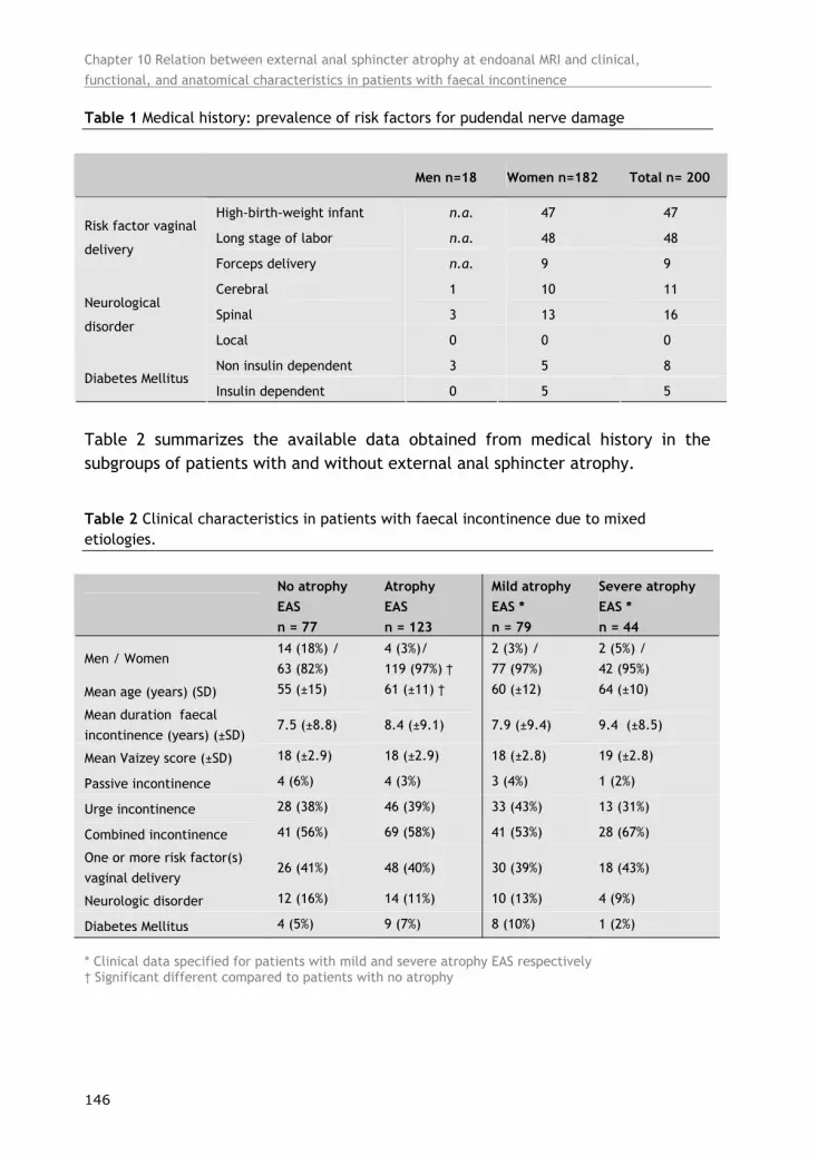
Chapter 10 Relation between external anal sphincter atrophy at endoanal MRI and clinical, functional, and anatomical characteristics in patients with faecal incontinence
146
Table 1 Medical history: prevalence of risk factors for pudendal nerve damage
Table 2 summarizes the available data obtained from medical history in the subgroups of patients with and without external anal sphincter atrophy.
Table 2 Clinical characteristics in patients with faecal incontinence due to mixed etiologies.
* Clinical data specified for patients with mild and severe atrophy EAS respectively † Significant different compared to patients with no atrophy
Men n=18 Women n=182 Total n= 200
High-birth-weight infant n.a. 47 47
Long stage of labor n.a. 48 48 Risk factor vaginal
delivery Forceps delivery n.a. 9 9
Cerebral 1 10 11
Spinal 3 13 16 Neurological
disorder Local 0 0 0
Non insulin dependent 3 5 8 Diabetes Mellitus
Insulin dependent 0 5 5
No atrophy EAS n = 77
Atrophy EAS n = 123
Mild atrophy EAS * n = 79
Severe atrophy EAS * n = 44
Men / Women 14 (18%) / 63 (82%)
4 (3%)/ 119 (97%) †
2 (3%) / 77 (97%)
2 (5%) / 42 (95%)
Mean age (years) (SD) 55 (±15) 61 (±11) † 60 (±12) 64 (±10)
Mean duration faecal incontinence (years) (±SD)
7.5 (±8.8) 8.4 (±9.1) 7.9 (±9.4) 9.4 (±8.5)
Mean Vaizey score (±SD) 18 (±2.9) 18 (±2.9) 18 (±2.8) 19 (±2.8)
Passive incontinence 4 (6%) 4 (3%) 3 (4%) 1 (2%)
Urge incontinence 28 (38%) 46 (39%) 33 (43%) 13 (31%)
Combined incontinence 41 (56%) 69 (58%) 41 (53%) 28 (67%)
One or more risk factor(s) vaginal delivery
26 (41%) 48 (40%) 30 (39%) 18 (43%)
Neurologic disorder 12 (16%) 14 (11%) 10 (13%) 4 (9%)
Diabetes Mellitus 4 (5%) 9 (7%) 8 (10%) 1 (2%)

Chapter 10
147
Table 2 shows also the data obtained from medical history specified for patients with mild and severe external anal sphincter atrophy, respectively. Patients with external anal sphincter atrophy were more often female (p<0.001) and significantly older (p=0.003), compared to patients without external anal sphincter atrophy.
No significant differences in sex (p=0.55) and age (p=0.07) were found between patients with mild and those with severe external anal sphincter atrophy. Patients with external anal sphincter atrophy were not significantly different from patients without external anal sphincter atrophy in terms of duration (p=0.47), nature (passive incontinence, urge incontinence, combined incontinence, respectively, p=0.48, p=0.97, p=0.81), and severity of faecal incontinence (p=0.31). Further, no significant differences in risk factors for pudendal nerve damage could be observed between patients with and without external anal sphincter atrophy (risk factors vaginal delivery, neurological disorders, diabetes mellitus, respectively, p=0.6, p=0.39, p=0.55). Functional characteristics The results of the anorectal functional tests in patients with and without external anal sphincter atrophy are demonstrated in Table 3. Table 3 shows also the findings of anorectal functional tests specified for patients with mild and severe external anal sphincter atrophy, respectively.
Due to technical failure or patient refusal, anorectal functional test results were not available in all patients. Pudendal nerve terminal motor latencies could be assessed at both sides in 166 patients, only at the right side in 9, and only at the left side in 11 patients. Pathological pudendal nerve terminal motor latencies at one side were found in 46 (28%) and at both sides in 41 (25%) patients. No significant differences in pathological pudendal nerve terminal motor latencies at one side (p=0.29) or at both sides (p=0.73) were demonstrated between patients with and those without external anal sphincter atrophy.
Anal manometry data were available in 193 patients. The mean resting pressure was 49 mmHg (SD± 23), mean maximal squeeze pressure 87 mmHg (SD ± 37) and mean squeeze increment pressure was 38 mmHg (SD ± 30). Mean maximal squeeze pressure (p=0.01) and squeeze increment pressure (p<0.001) were significant lower in patients with external anal sphincter atrophy compared to those patients without external anal sphincter atrophy. The maximal squeeze pressure (p=0.003) and squeeze increment pressure (p<0.001) were significantly lower in patients with severe compared to patients with mild external anal sphincter atrophy.

Chapter 10 Relation between external anal sphincter atrophy at endoanal MRI and clinical, functional, and anatomical characteristics in patients with faecal incontinence
148
Table 3 Functional and anatomical characteristics in patients with faecal incontinence due to mixed etiologies
* Clinical data specified for patients with mild and severe atrophy EAS respectively † Significant different compared to patients with no atrophy □ Significant different compared to patients with mild atrophy
Decrease of maximal squeeze pressure and squeeze increment pressure by increasing degree of external anal sphincter atrophy was not attenuated by sex, age, type of the anal manometry technique used, and coexistence of an external anal sphincter defect (Table 4).
Table 4 Squeeze function pressures adjusted for potential confounding variables
* Data represent the estimated marginal means adjusted for sex, age, type of the anal manometry technique used, and coexistence of an external anal sphincter defect
No atrophy EAS n = 77
Atrophy EAS * n = 123
Mild atrophy EAS * n = 79
Severe atrophy EAS * n = 44
Pathological PNTML one side
21 (32%) 25 (25%) 12 (18%) 13 (37%)
Pathological PNTML both
sides
17 (26%) 24 (24%) 13 (20%) 11 (31%)
Mean resting pressure
(mmHg) (±SD)
49 (±26) 50 (±21) 50 (±21) 49 (±20)
Mean maximal squeeze
pressure (mmHg) (±SD)
96 (±42) 81 (±33) † 87 (±35) 70 (±25) □
Mean squeeze increment
pressure (mmHg) (±SD)
47 (±32) 32 (±28) † 37 (±31) 21 (±17) □
Mean sensory threshold for
anal sensation (mAmp)(±SD)
8.2 (±7.2) 7.7 (±4.7) 6.8 (±3.8) 9.1 (±5.8)
Defect EAS
32 (42%) 56 (45%) 39 (49%) 17 (39%)
Mean thickness EAS right
(mm) (±SD)
3.6 (±1.8) 3.6 (±1.9) 3.7 (±1.9) 3.4 (±2)
Mean thickness EAS thickest
point (mm) (±SD)
4.8 (±1.9) 4.8 (±2) 5.2 (±2) 4.2 (±2)
No atrophy EAS Mild atrophy
EAS
Severe atrophy
EAS
P-value
Mean maximal squeeze pressure (mmHg) (±SE) *
94 (±4.1) 89 (±4) 72 (±5.4) 0.006
Mean Squeeze increment pressure (mmHg) (±SE) *
46 (±3.2) 38 (±3.2) 23 (±4.3) <0.001

Chapter 10
149
Anal sensitivity measurements were performed in 176 patients. The mean threshold for anal sensation was 7.9 mAmp (SD± 5.8). No significant difference in mean threshold for anal sensation was observed between patients with and those without external anal sphincter atrophy (p=0.53).
Anatomical characteristics In 88 of 200 (44%) patients an external anal sphincter defect was depicted at endoanal MRI. Mean external anal sphincter thickness at the right lateral side was 3.6 mm (SD± 1.9) and 4.8 mm (SD± 2) at the thickest point.
Table 3 summarizes the number of external anal sphincter defects and the external anal sphincter thickness measurements in the subgroups of patients with and without external anal sphincter atrophy. Table 3 shows also the number of external anal sphincter defects and the external anal sphincter thickness measurements specified for patients with mild and severe external anal sphincter atrophy, respectively. External anal sphincter defects did not occur more often in patients with external anal sphincter atrophy, compared to patients without external anal sphincter atrophy (p=0.58). No significant differences were seen in external anal sphincter thickness measurements at the right side (p=0.97) or at the thickest point of the external anal sphincter (p=0.88) between patients with and without external anal sphincter atrophy.
Discussion
This study demonstrated that external anal sphincter atrophy can be depicted at endoanal MR imaging in 62% of patients with moderate to severe faecal incontinence due to different etiologies. Patients with external anal sphincter atrophy were mainly female, older, and had a significantly lower maximal squeeze pressure and squeeze increment pressure compared to patients without atrophy. Further, maximal squeeze pressure and squeeze increment pressure were significantly lower in patients with severe external anal sphincter atrophy compared to patients with mild external anal sphincter atrophy. These differences were not attenuated by sex, age, type of anal manometry technique used, or the coexistence of an external anal sphincter defect. Patients with external anal sphincter atrophy could not be identified a priori by other clinical, functional or anatomical characteristics.
A number of potential limitations of this study should be taken into account. We included patients with a Vaizey score of at least 12, indicating moderate to severe faecal incontinence, and excluded some patients groups for this study.

Chapter 10 Relation between external anal sphincter atrophy at endoanal MRI and clinical, functional, and anatomical characteristics in patients with faecal incontinence
150
Consequently, the study population does not represent the full spectrum of faecal incontinence complaints and, therefore, the observed results cannot be unconditionally generalized to all patients with faecal incontinence.
The disproportion between female and male patients participating in this study is not due to a form of selection bias but is inherent to the etiology of faecal incontinence.36
We are aware of the fact that histology obtained during anal sphincter repair is the reference standard for demonstrating external anal sphincter atrophy. Nevertheless we did not opt for histology as the reference standard in this study, as not all patients were eligible for surgery. We used endoanal MRI to assess external anal sphincter atrophy since other techniques, such as electromyography and pudendal nerve terminal motor latency testing, are uncomfortable or might misinterpret the presence of external anal sphincter atrophy.18 Furthermore endoanal MRI showed its superiority over endoanal sonography for demonstrating atrophy of the external anal sphincter.17,27,29,32
Endoanal MRI was performed in two hospitals and scan parameters were optimized to the MR unit used. As a result there were some differences in the used T2 weighted fast spin echo sequences, with consequently slight differences in resolution, but this was not reflected in differences in identification of the external anal sphincter between both hospitals.
The radiologists based their diagnosis external anal sphincter atrophy on a visual subjective qualitative interpretation of thinning of the external anal sphincter muscle fibers and/or the diffuse replacement of external anal sphincter muscle by fat. In clinical practice no hard criteria exist for the visual diagnosis of external anal sphincter atrophy at MRI. Recent studies found that measurements of the area of remaining external anal sphincter and of the percentage of fat content of the external anal sphincter correlated with squeeze pressure and contributed to the diagnosis of external anal sphincter atrophy.17,27,28,31 External anal sphincter thickness measurements were not found to be correlated to the cross-sectional external anal sphincter area.28 In our study no significant differences in external anal sphincter thickness measurements between patients with and patients without atrophy could be demonstrated. The latter might be explained by the fact that the thickness of the external anal sphincter is not necessarily diminished in patients with atrophy, since the MRI measurements comprised, when fatty replacement had occurred within the external anal sphincter, not only external anal sphincter muscle but also fat mixed in the external anal sphincter.
Patients with and without external anal sphincter atrophy were compared on data obtained from medical history. Data concerning the possible risk factors for

Chapter 10
151
pudendal nerve damage, including the obstetric details, were collected based on each patient’s memory rather than based on patient’s records. This might have affected the reliability of the data.
The anorectal functional tests were performed in different hospitals and only for anal manometry different techniques were used. The observed decrease in maximal squeeze pressure and squeeze increment pressure by increasing degrees of atrophy were not affected by the anal manometry technique used.
A recent study found a relation between squeeze increment pressure and respectively external anal sphincter bulk and sphincter percentage fat content in patients with an intact anal sphincter complex.27 The authors reported a significant lower squeeze pressure in patients with severe external anal sphincter atrophy compared to patients with no external anal sphincter atrophy, but found no significant lower squeeze pressure in patients with mild compared to patients with no external anal sphincter atrophy. We found a lower squeeze increment pressure and a significant lower maximal squeeze pressure in patients with external anal sphincter atrophy. Maximal squeeze and squeeze increment pressure were lower in patients with severe compared to patients with mild external anal sphincter atrophy.
An increase in the proportion of connective tissue in the external anal sphincter and a progressive reduction in external anal sphincter thickness due to aging are described in the literature.37,38 Normal squeeze pressures are generally lower in women and older patients.18 Patients with external anal sphincter atrophy in our study were more often female and older but significant differences in squeeze function between patient with and patients without external anal sphincter atrophy could not be explained by age or sex. Presence of an external anal sphincter defect also influences squeeze function.34 The observed significant differences in squeeze function parameters were not influenced by the coexistence of an external anal sphincter defect.
Opinions about the value of pudendal nerve terminal motor latency testing for diagnosing external anal sphincter atrophy vary in the literature.18 Like others we did not find a significant difference in pathological pudendal latencies between patients with and those without external anal sphincter atrophy.28 This may be explained by the fact that pudendal nerve latency measures only the conduction time of the fastest nerve fibers, and latencies may be normal even in the presence of external anal sphincter atrophy.12 Like Williams and co-workers we did not observe a relation between anal sensitivity and presence of external anal sphincter atrophy.28
Measuring the cross-sectional area and fat percentage content to assess external anal sphincter atrophy requires great care and is most of all relatively

Chapter 10 Relation between external anal sphincter atrophy at endoanal MRI and clinical, functional, and anatomical characteristics in patients with faecal incontinence
152
time-consuming.27 Our study demonstrates the relation between external anal sphincter squeeze function parameters and the qualitative assessment of external anal sphincter atrophy by radiologists. The observed deterioration of squeeze increment pressure, representing voluntary squeeze function, and maximal squeeze pressure, indicating squeeze function of the total sphincter complex, by increasing degree of external anal sphincter atrophy were not explained by sex, age, type of the anal manometry technique used, or the coexistence of an external anal sphincter defect.
Previous studies reported that the size of an anal sphincter defect correlates with the severity of faecal incontinence,39,40 while another study was not able to demonstrate a relation between muscle injury and the severity of clinical symptoms41. An interesting finding of this study is the absent relation between the severity of faecal incontinence and the presence of external anal sphincter atrophy, not even when only patients without an external anal sphincter defect were studied (data not shown). This substantiates the complex and multifactorial pathogenesis of faecal incontinence. Additionally to structural and functional anal sphincter disorders, also reduced rectal capacity and compliance and reduced faecal consistency may contribute to the inability to maintain continence.12,42
The fact that patients with external anal sphincter atrophy do not benefit as well after anterior anal sphincter repair as patients without atrophy might be explained by the relation between atrophy and the voluntary and total squeeze function of the anal sphincter complex. Nevertheless, one might expect that patients with mild atrophy, despite the diminished sphincter function, may gain more benefit from anterior anal sphincter repair than patients with severe external anal sphincter atrophy, in whom sphincter dysfunction is much worse. As the utility of grading external anal sphincter atrophy in selecting patients for anterior anal sphincter repair has not yet been established, a better understanding of how grading external anal sphincter atrophy at endoanal MRI is related to the outcome of anterior anal sphincter repair is a topic for future research.
In conclusion, this study demonstrates that atrophy of the external anal sphincter at endoanal MRI can be observed in the 62% of patients with moderate to severe faecal incontinence, varying from mild to severe. As external anal sphincter atrophy had a great impact on squeeze function and squeeze function deteriorated by increasing degree of atrophy, further studies are needed to evaluate if grading atrophy is of clinical value in selecting patients for anterior anal sphincter repair.

Chapter 10
153
References
1. Thomas TM, Egan M, Walgrove A, Meade TW. The prevalence of faecal and double incontinence. Community Med 1984;6:216-220.
2. Deutekom M, Terra MP, Dobben AC, et al. Impact of faecal incontinence severity on health domains. Colorectal Dis 2005;7:263-269.
3. Rao SS. Pathophysiology of adult fecal incontinence. Gastroenterology 2004;126:S14-S22. 4. Abbasakoor F, Nelson M, Beynon J, Patel B, Carr ND. Anal endosonography in patients with anorectal symptoms
after haemorrhoidectomy. British Journal of Surgery 1998;8:1522-1524. 5. Kamm MA. Obstetric damage and faecal incontinence. Lancet 1994;344:730-733. 6. Snooks S, Henry MM, Swash M. Faecal incontinence after anal dilatation. Br J Surg 1984;71:617-618. 7. Speakman CT, Burnett SJ, Kamm MA, Bartram CI. Sphincter injury after anal dilatation demonstrated by anal
endosonography. Br J Surg 1991;78:1429-1430. 8. Sultan AH, Kamm MA, Hudson CN, Thomas JM, Bartram CI. Anal-sphincter disruption during vaginal delivery. N
Engl J Med 1993;329:1905-1911. 9. Sultan AH, Kamm MA, Hudson CN, Bartram CI. Third degree obstetric anal sphincter tears: risk factors and
outcome of primary repair. BMJ 1994;308:887-891. 10. Engel AF, Kamm MA. The acute effect of straining on pelvic floor neurological function. Int J Colorectal Dis
1994;9:8-12. 11. Parks AG, Swash M, Urich H. Sphincter denervation in anorectal incontinence and rectal prolapse. Gut
1977;18:656-665. 12. Madoff RD, Parker SC, Varma MG, Lowry AC. Faecal incontinence in adults. Lancet 2004;364:621-632. 13. Malouf AJ, Norton CS, Engel AF, Nicholls RJ, Kamm MA. Long-term results of overlapping anterior anal-
sphincter repair for obstetric trauma. Lancet 2000;355:260-265. 14. Gilliland R, Altomare DF, Moreira H Jr, Oliveira L, Gilliland JE, Wexner SD. Pudendal neuropathy is predictive
of failure following anterior overlapping sphincteroplasty. Dis Colon Rectum 1998;41:1516-1522. 15. Jacobs PP, Scheuer M, Kuijpers JH, Vingerhoets MH. Obstetric fecal incontinence. Role of pelvic floor
denervation and results of delayed sphincter repair. Dis Colon Rectum 1990; 33(6):494-497. 16. Londono-Schimmer EE, Garcia-Duperly R, Nicholls RJ, Ritchie JK, Hawley PR, Thomson JP. Overlapping anal
sphincter repair for faecal incontinence due to sphincter trauma: five year follow-up functional results. Int J Colorectal Dis 1994;9:110-113.
17. Briel JW, Stoker J, Rociu E, Lameris JS, Hop WCJ, Schouten WR. External anal sphincter atrophy on endoanal magnetic resonance imaging adversely affects continence after sphincteroplasty. British Journal of Surgery 1999;86:1322-1327.
18. Diamant NE, Kamm MA, Wald A, Whitehead WE. AGA technical review on anorectal testing techniques. Gastroenterology 1999;116:735-760.
19. Law PJ, Kamm MA, Bartram CI. Anal endosonography in the investigation of faecal incontinence. Br J Surg 1991;78:312-314.
20. Cuesta MA, Meijer S, Derksen EJ, Boutkan H, Meuwissen SG. Anal sphincter imaging in fecal incontinence using endosonography. Dis Colon Rectum 1992;35:59-63.
21. Nielsen MB, Hauge C, Pedersen JF, Christiansen J. Endosonographic evaluation of patients with anal incontinence: findings and influence on surgical management. AJR Am J Roentgenol 1993;160:771-775.
22. Deen KI, Kumar D, Williams JG, Olliff J, Keighley MR. Anal sphincter defects. Correlation between endoanal ultrasound and surgery. Ann Surg 1993;218:201-205.
23. Meyenberger C, Bertschinger P, Zala GF, Buchmann P. Anal sphincter defects in fecal incontinence: correlation between endosonography and surgery. Endoscopy 1996;28:217-224.
24. deSouza NM, Hall AS, Puni R, Gilderdale DJ, Young IR, Kmiot WA. High resolution magnetic resonance imaging of the anal sphincter using a dedicated endoanal coil. Comparison of magnetic resonance imaging with surgical findings. Dis Colon Rectum 1996;39:926-934.
25. deSouza NM, Puni R, Gilderdale DJ, Bydder GM. Magnetic resonance imaging of the anal sphincter using an internal coil. Magn Reson Q 1995;11:45-56.
26. Rociu E, Stoker J, Zwamborn AW, Lameris JS. Endoanal MR imaging of the anal sphincter in fecal incontinence. Radiographics 1999;19:S171-S177.

Chapter 10 Relation between external anal sphincter atrophy at endoanal MRI and clinical, functional, and anatomical characteristics in patients with faecal incontinence
154
27. Williams AB, Bartram CI, Modhwadia D, et al. Endocoil magnetic resonance imaging quantification of external anal sphincter atrophy. Br J Surg 2001;88:853-859.
28. Williams AB, Malouf AJ, Bartram CI, Halligan S, Kamm MA, Kmiot WA. Assessment of external anal sphincter morphology in idiopathic fecal incontinence with endocoil magnetic resonance imaging. Dig Dis Sci 2001;46:1466-1471.
29. Rociu E, Stoker J, Eijkemans MJ, Schouten WR, Lameris JS. Fecal incontinence: endoanal US versus endoanal MR imaging. Radiology 1999;212:453-458.
30. deSouza NM, Puni R, Zbar A, Gilderale DJ, Coutts GA, Krausz T. MR imaging of the anal sphincter in multiparous women using an endoanal coil: Correlation with in vitro anatomy and appearances in fecal incontinence. AJR Am J Roentgenol 1996;167:1465-1471.
31. Briel JW, Zimmerman DD, Stoker J, et al. Relationship between sphincter morphology on endoanal MRI and histopathological aspects of the external anal sphincter. Int J Colorectal Dis 2000;15:87-90.
32. Fletcher JG, Busse RF, Riederer SJ, et al. Magnetic resonance imaging of anatomic and dynamic defects of the pelvic floor in defecatory disorders. Am J Gastroenterol 2003;98:399-411.
33. Vaizey CJ, Carapeti E, Cahill JA, Kamm MA. Prospective comparison of faecal incontinence grading systems. Gut 1999;44:77-80.
34. Soffer EE, Hull T. Fecal incontinence: a practical approach to evaluation and treatment. Am J Gastroenterol 2000;95:1873-1880.
35. Kiff ES, Swash M. Slowed conduction in the pudendal nerves in idiopathic (neurogenic) faecal incontinence. Br J Surg 1984;71:614-616.
36. Henry MM. Pathogenesis and management of fecal incontinence in the adult. Gastroenterol Clin North Am 1987;16:35-45.
37. Haas PA, Fox TA Jr. Age-related changes and scar formations of perianal connective tissue. Dis Colon Rectum 1980;23:160-169.
38. Rociu E, Stoker J, Eijkemans MJC, Lameris JS. Normal anal sphincter anatomy and age- and sex-related variations at high-spatial-resolution endoanal MR imaging. Radiology 2000;217:395-401.
39. Damon H, Henry L, Barth X, Mion F. Fecal incontinence in females with a past history of vaginal delivery: significance of anal sphincter defects detected by ultrasound. Dis Colon Rectum 2002;45:1445-1450.
40. Nichols CM, Gill EJ, Nguyen T, Barber MD, Hurt WG. Anal sphincter injury in women with pelvic floor disorders. Obstet Gynecol 2004;104:690-696.
41. Voyvodic F, Rieger NA, Skinner S, et al. Endosonographic imaging of anal sphincter injury: does the size of the tear correlate with the degree of dysfunction? Dis Colon Rectum 2003;46:735-741.
42. Lamah M, Kumar D. Fecal incontinence. Dig Dis Sci 1999;44:2488-2499.

Chapter 11
General Discussion

Chapter 11 General discussion
156
Methodological issues
The major aim of the work summarized in the present thesis was to collect evidence for building an optimal testing and treatment path for patients with faecal incontinence. In order to reach this goal, various steps needed to be taken and choices had to be made.
There exists no universally accepted method to build an optimal testing and treatment path. We decided to first study impact and outcome measures of faecal incontinence. We then studied the effects of treatment. Our last step was the evaluation of a number of tests.
In this discussion we describe the pros and cons of various methods that we have used or could have used in these three topics: impact and outcome measures, treatment, and evaluation of diagnostic tests. Although all studies in the present thesis concern faecal incontinence, similar methodological problems or difficulties can be encountered in other diseases, disorders and conditions.
How to measure impact and outcome For all conditions one has to find instruments that precisely express their impact and measures that can accurately assess treatment outcome. Mortality and life-time expectancy belong to the traditional indicators. These indicators are said to be of lesser importance in economically developed countries with their aging population and associated increased incidence of disability and chronic diseases.1 Faecal incontinence is one of the conditions where these indicators are not suited. A number of other measures exist to quantify impact and outcome which leads to the question: which one should we use? As quantifying impact differs from quantifying outcome, the search for an optimal tool must be performed separately. Quantifying impact A first step in quantifying impact could consist of assessing the prevalence of a condition. In faecal incontinence it is known that only a small part of symptomatic patients discuss their faecal incontinence complaints with their physician.2 The prevalence could therefore easily be underestimated. A recent systematic literature review has estimated that between 11% and 15% of the population encounters some problems with maintaining continence.3
Assessing the prevalence is not enough to establish the impact of a certain disease of disorder; one should also evaluate how it affects the life of affected patients. This can be achieved by measuring quality of life. A distinction can be made between disease specific quality of life and generic measures. Both methods for measuring quality of life have their disadvantages and advantages.4

Chapter 11
157
Disease specific questionnaires may be more sensitive to change, while generic questionnaires enable us to make comparisons between the target population and other populations.
In chapter 2 the health status of patients with faecal incontinence was compared with that of the general population, using a generic questionnaire. We used the EQ-5D instrument, developed by the EuroQol Group.5 This is a questionnaire that addresses 5 domains: mobility, self-care, usual activities, pain/discomfort, anxiety/depression. These domains each comprise 3 levels: no problems, some/ moderate problems and extreme problems/unable to. Patients with faecal incontinence reported more problems in all domains, except for self-care, than members of a reference population of the same age. Furthermore, we observed a significant association between severity of incontinence and impact on the domains of usual activities, pain/discomfort and anxiety/depression. Patients with a high incontinence score had a higher probability of having problems in these domains.
Another method to quantify impact of a condition is by measuring its societal costs. Two types of cost-of-illness studies are distinguished: general and disease-specific cost-of-illness studies.6 In general cost-of-illness studies health expenditures are assigned to all diseases clustered in more or less homogeneous groups using a top-down method. Disease-specific cost-of-illness studies provide cost estimates of particular diseases and aim to show the economic impact of a particular disease.
In chapter 3 we studied the disease specific costs of faecal incontinence by estimating the associated societal costs. This means that all relevant costs, regardless of who bears the costs, are taken into account.7 These total costs consists out direct medical costs of health care resources used as well as the direct and indirect non-medical costs of out-of-pocket expenses and production losses.7-9
We used questionnaire data from patients already entered in the medical circuit to estimate these costs. Several problems exists with this type of design. It is likely that costs will be lower for subjects who do not seek medical help. A further difficulty exists with using questionnaire data to estimate the costs of a disease. Although it was emphasized in the questionnaire that patients were invited to report only those resources that were associated with their faecal incontinence complaints, it cannot be fully assured that all responses were valid. Underreporting or incorrect attribution cannot entirely be excluded.
For medical costs it is probably more valid to use data from hospital and pharmacy records. Obtaining valid data concerning out-of-pocket responses, indirect medical costs (e.g. travel costs), and production losses is more

Chapter 11 General discussion
158
problematic. At this point there exists no optimal method for estimating these costs. Though the use of questionnaire data seems justified in this case, some caution is necessary in interpreting the results. Investigating treatment effect For many disorders there exists not one accepted outcome measure. In faecal incontinence studies a marked diversity in outcome measures can be observed. One method to determine the effects of treatment is using severity measures. Various severity measures have been proposed, differing in specificity and length, although most scales include questions about the type and the frequency of the incontinent episodes.10-14 In this thesis we used the Vaizey incontinence score to determine the severity of incontinence.12 This score exists of quantitative measurements in terms of stool type and frequency10,12 and items concerning impact on daily life, pad use, and urge incontinence. Although scores for all separate items were derived from patient reports, clinicians decided on what kind of items needed to be incorporated in the scores. The results from specific procedures, such as tests to document the functional and anatomic characteristics of the rectum and anal sphincter complex, are also often used to assess outcome while other studies have relied on quality of life measures.
In chapter 4 we related various outcome measures to the subjective perception of patients. As faecal incontinence is a symptom it has been said15 that the subjective perception of the patient must be the foundation of any evaluation of incontinence. We measured subjective perception in an interview by telephone. Patients were asked to score their situation after treatment (compared to before treatment), on a scale of 1 (much worse), 2 (worse), 3 (no change), 4 (better), or 5 (much better). The validation of the subjective measure is problematic. An alternative and possibly more objective measure would have been the use of diary report to assess improvement. From diary reports we would be able to see whether or not actual incontinent events truly decrease, but compliance in the use of diaries can be low.16
In our study we observed a relation between patients’ perception and the Vaizey score. In the absence of a true gold standard to assess treatment outcome, it is not possible to state that this observed relation is truly caused by agreement between patients and clinicians. One mechanism that could have biased this relation is a ‘placebo’ effect. Patients may (want to) believe that they have improved, report their situation to be better and coinciding report fewer episodes of unwanted stool loss.

Chapter 11
159
Evaluation of treatment There exists a hierarchy in the amount of evidence that is provided by a study. Weak evidence on the effectiveness of treatment is provided by a case report or short series. Such studies are often performed when a new treatment has been developed. In this type of studies no comparisons with other treatments are made; they only deliver information on the feasibility and outcome of a particular treatment, which might be studied further in the future. A cohort study in a large group of patients is able to provide more evidence on treatment effect. In chapter 6 we described the results of a cohort study on the effects of pelvic floor rehabilitation in patients with faecal incontinence. A disadvantage of this type of study is that the outcome after treatment cannot be exclusively attributed to the treatment itself, as this type of study has no comparison group. An advantage is that in a large group of patients the treatment effect can be studied and that results can be compared in clinical subgroups.
A comparative trial includes two or more groups of patients, each receiving a different form of treatment. In a randomized trial, all baseline differences are random and differences in outcome can be more evaluated using statistical techniques, allowing a stronger attribution of post treatment differences to treatment and, hence, more evidence on treatment effect. The treatment in the control group can vary. In some studies patients in the control group receive no intervention, while in other studies patients receive the established treatment.
Even stronger evidence can be obtained by means of a systematic review of the literature. With a systematic review one tries to combine all evidence from individual studies (preferably randomized controlled trials) concerning treatment for a certain disease in order to investigate to obtain more precise and valid estimates of the treatment effect. In chapter 5 we presented the results of a systematic review in which we tried to identify all studies on the use of anal plugs for the containment of faecal incontinence. We encountered many difficulties in making this systematic review. At first we could only identify a single randomized controlled trial, while three other studies were randomized cross-over studies. There existed considerable diversity in outcome measures, with a high percentage of drop-outs in all four studies. Overall, the methodological quality was rather weak. From such a systematic review one can probably deriver only tentative conclusions. Evaluation of diagnostic tests A number of diagnostic tests are available for the assessment of patients with faecal incontinence. After a physical exam, physicians can use various additional tests to investigate the function of the anorectum (e.g. manometry, pudendal nerve latency testing, sensibility and capacity measurements) and the anatomy

Chapter 11 General discussion
160
(e.g. endoanal MRI, endoanal sonography and defecography). Performing all tests in all patients would burden patients and lead to considerable use of health care resources, and the benefits of full testing in all patients are unclear. This calls for a more selective approach, using a limited set of additional tests, maybe guided by results from medical history and physical exam. So far it is unclear what the optimal diagnostic work-up would be of patients with faecal incontinence.
Various methods exist to evaluate a test or combination of tests. The validity of a test is constrained by its reliability. If a test does not consistently measure a construct or domain then it cannot expect to have high validity coefficients. One way to assess this is by performing intraobserver and interobserver studies.17-19 By estimating the intraobserver reliability one evaluates whether a single observer is consistent in his reading, while interobserver reliability determines consistency over different observers.
Although reliability is a prerequisite for a good diagnostic test, it is not sufficient. The most common method to evaluate a diagnostic test is to determine its accuracy, for which sensitivity and specificity are the two most widely used test statistics. The usual study design for this type of study is shown in Figure 1.
Figure 1 Usual study design to assess accuracy
In the case of faecal incontinence this design would result in recruiting patients with an unknown disease status. Next these patients would receive the index test (the test for which we aim to determine the sensitivity and specificity) and then a reference standard. This approach has several practical impediments when used in patients with faecal incontinence. The most important problem is that
Series of patients
Reference standard
Index test
Blinded cross-classification

Chapter 11
161
the disease status is generally known. In the clinical patient interview it becomes apparent whether a patient has or does not have faecal incontinence.
In faecal incontinence, diagnostic tests are not used to determine disease status, but are used to investigate the underlying pathophysiology of faecal incontinence. Various factors are held responsible for maintaining continence including anatomically intact pelvic floor muscles, adequate afferent and efferent pelvic floor innervation, adequate rectoanal coordination, and stool consistency.20,21 Impairment in any of these factors in isolation or in combination could result in faecal incontinence.
The characteristics of a diagnostic test could then be determined by its ability to identify the underlying pathophysiology. The problem with this approach in faecal incontinence is the lack of a valid reference standard. The clinical presentation of faecal incontinence has been thought to be related with the underlying physiological or anatomical abnormality. Passive incontinence is believed to be associated with impairment of the internal anal sphincter, resulting in decreased resting pressures.22,23 Decreased squeeze pressures and defects of the external sphincter points to dysfunction of the external anal sphincter, which has been linked to urge incontinence.22,24,25 A decreased rectal reservoir function has also been connected to urge incontinence.25,26
In chapter 9 we related test information to the clinical presentation of faecal incontinence. On the basis of test results we could not differentiate between patients with urge and passive incontinence. Due to the absence of a proper reference standard we cannot draw any conclusions with this method about the characteristics of the investigated diagnostic tests.
In the study described in chapter 10 we tried to relate various tests with each other, in order to investigate the grading of moderate versus severe atrophy of the external anal sphincter by relating MRI findings with other sources of information. We observed differences in clinical and functional information between patients with and without atrophy. Furthermore some functional characteristics were significantly different in patients with severe compared to patients with mild atrophy. This finding offered support for the grading of atrophy in the future.
This approach can also be used to test for redundancy in testing. When two tests provide identical information it is not necessary to execute both. When, for instance, a defect of the external sphincter can be detected with a physical examination as well as with endoanal MRI, performing both tests may not be necessary. Another way to evaluate tests is to establish the association between a test or combinations of tests and clinical outcome, or the outcome of therapy. In chapter 6 we described the results of treatment with electrical stimulation

Chapter 11 General discussion
162
and pelvic floor muscle training with biofeedback in patients with faecal incontinence. It appeared that treatment leads to an improvement in faecal incontinence symptoms in the majority of patients, but that only few patients demonstrated a substantial improvement.
Our goal was to try to reduce the uncertainty about the expected treatment effect on the basis of test information. Various methods are known to relate test information to treatment effect. The first is by means of a decision analysis. Decision modelling is a widely used approach to determine an optimal diagnostic path. The problem with this approach is that when you study a considerable amount of tests, the number of possible strategies can easily explode. According to Glasziou27 the number of branches within that tree can be calculated with the following formula: Applying the formula the number of possible branches that needs to be evaluated in the present study would be 8,41E+13. The number of branches can be reduced by choosing a restricted set of strategies eligible for evaluation. Criteria for the restricted set can be derived from expert opinion on patient logistics, e.g. practicality of test sequences. In our study it would not be feasible to perform first one out of the four anorectal function tests, then guide the patient to the endoanal MRI, and then ask the patient for the second anorectal function test. Another argument could be the availability of test equipment in (Dutch) hospitals.
After trying to reduce the amount of possible strategies we still were left with a gargantuan number of strategies, too large to analyze. We therefore used multivariable regression analysis to assess the predictive value of tests and combinations of tests on response to treatment.
Prognostic regression models may predict outcomes of various types, such as continuous outcomes, categorical outcomes, or dichotomous outcome measures. Many prediction studies have used a dichotomous outcome measure, such as success versus no success. With this type of outcome measure, logistic regression can be used. The results from such analyses can be informative in clinical practice, as these results allow the calculation of the chances of treatment success. In our study we could not use a dichotomous outcome measure as the success definition is not straightforward.
∑=
n
l
lnl kP
0)(
n = number of tests k = number of outcomes per test Pn
l = number of permutations of n tests taken l at a time

Chapter 11
163
The ultimate wish for every patient is, without doubt, to be completely cured from their complaints. Changes exist in the degree of improvement that offers satisfaction for an individual patient. For instance an elderly woman could be satisfied when treatment provides her 20 extra minutes time to delay defecation, as that provides her the freedom to leave her home. On the other hand a young working woman who is not able to perform her job sufficiently because of faecal incontinence aims for a complete cure. We have shown in Chapter 4 that the Vaizey score has a clear relation with patient’s subjective feeling concerning relief, but an exact, overall success definition cannot be defined. We therefore chose to use multivariable linear regression analysis of the Vaizey score after treatment. Beyond test characteristics In order to determine an optimal diagnostic work-up, one should look beyond the test characteristics. A sub-optimal informative test with very little costs may in some instances be preferred over a perfectly informative test with high costs. Even so a test with low patient burden might be preferred above a test with high patient burden, such as an invasive or painful test. In chapter 8 we studied the patient burden of a series of tests in the assessment of faecal incontinence by means of a self-administered questionnaire. Patients were asked to rate the amount of anxiety, pain, embarrassment and discomfort on a scale of 1 to 5. We observed low values (between 1 and 2) for all tests on all items, and differences between tests were, although sometimes statistically significant, marginal. In the search for an optimal strategy of faecal incontinence the burden of testing for patients appears to play no major role. One could speculate about the position of patient burden in a diagnostic strategy in general. Undergoing a diagnostic test in general is most likely not the most pleasant way to spend one’s time, yet it seems that most patients are willing to undergo a test when they have been explained that this test must be done in order to gain information about their disease status or the optimal treatment path.
It remains important to stress that patient burden must be minimized. This burden is not only reflected in, for instance pain or discomfort caused by a certain test, but also in long waiting times before undergoing the test, or in experiencing insecurity about test results.
There exists no general rule for weighing test characteristics, costs and burden of tests. An informed choice must be made for every clinical example. An expensive, invasive and very accurate test will be preferred in diagnosing a rare and life threatening disease, while the same test will not be the first choice as screening tool. In such case we are more willing to accept a small information loss by using a less costly and preferably non invasive test first.
Chapter 11 General discussion
164
Implications of this thesis
Methods to evaluate diagnostic tests without reference standard A diagnostic test should only be used in practice when it delivers valuable information. This implies that all tests should be thoroughly investigated. In the absence of a reference standard tests cannot be evaluated using the familiar used diagnostic accuracy calculations. In this thesis we have relied on a series of methods to evaluate tests in the absence of a reference standard. These methods were. Reliability testing: When a test is not reliable, it will never be valuable Relating tests with other tests: When two tests deliver the same information or make the same diagnosis, it is not necessary to execute them both Relating tests with treatment outcome: Calculate the predicted value of a test or combination of tests on a certain treatment outcome. Study other characteristics of a diagnostic test: Investigate the burden and costs of a test. Treatment and diagnosis of faecal incontinence The work collected in this thesis has shown that the average Vaizey score decreases from 18 points before treatment to 15 points after pelvic floor rehabilitation treatment in patients with severe faecal incontinence. The Vaizey score improved from baseline in 60% of all patients, remained unchanged in 23% and deteriorated in 17%. Vaizey score improvement was slight in 47% and substantial (more than 50% improvement) in 13%. We have shown that tests are unable to predict the post treatment Vaizey score with acceptable accuracy.
The study population of this study does not represent the full spectrum of faecal incontinence complaints and therefore the observed results cannot be unconditionally generalized to all patients with faecal incontinence. Some patients groups were excluded for this study as in these patients a specific disorder was held responsible for the faecal incontinence complaints. These patients needed treatment for that disorder and would not a priori be eligible for pelvic floor rehabilitation treatment.
The clinical implications of our findings are not all straightforward. One could argue that the improvement after pelvic floor rehabilitation is so marginal that pelvic floor rehabilitation could be doubted. As the results have been collected in a cohort study only we cannot even claim that the observed changes were caused by the treatment. Non-specific factors, such as the attention from the physiotherapist, natural history, and patients’ feelings towards their condition (‘I am not the only one with this problem’) could all have led to some

Chapter 11
165
minor relief. Following this line of argumentation we would conclude that pelvic floor rehabilitation is not an effective treatment modality and that our efforts should go out to making faecal incontinence less unfamiliar, giving patients adequate attention and support. A further implication would then be that we should investigate the predictive values of the diagnostic tests for other treatments for faecal incontinence.
Another way to argue is that there is no need to asses a patient as thoroughly as we did, with this variety of tests, when that patient enters the medical circuit. When major pathology has been excluded we could offer treatment with pelvic floor rehabilitation to every patient with faecal incontinence without further testing. This conclusion can be based on the findings that, on average, the severity of incontinence after treatment will be lower than before treatment while additional tests can only marginally predict treatment outcome. In case of an unsatisfactory response to treatment one could then decide on further testing to identify patients most likely to benefit from the next treatment option.
With the first option we would withhold effective treatment from a number of patients. With the second option many patients would receive treatment without benefiting from it. When weighing these pros and cons, I would recommend that all consenting patients receive, without prior testing, adequate pelvic floor rehabilitation. Pelvic floor rehabilitation is simple, relatively inexpensive and mostly without unfavourable physical side-effects. These characteristics would make it unethical to withhold patients from pelvic floor rehabilitation. The negative side effects from receiving a non-successful treatment could be minimized by explaining to patients before they receive treatment that the chances of major success are low, or by stopping early in case of inadequate response, a strategy not yet evaluated.
We have shown in the present thesis that faecal incontinence has a large impact for those affected. Patients with incontinence report significantly more often problems with usual activities, pain/discomfort, mobility and anxiety/depression. An effective treatment would thus be desirable. Debate exists around the definition of ‘effective’ in terms of treatment for faecal incontinence and additional studies may improve our understanding of the criteria for treatment success in faecal incontinence trials. We have shown that a therapy with pelvic floor rehabilitation decreases the severity of incontinence in 60% of patients. Diagnostic tests were not able to identify patients most likely to benefit from treatment. The role of diagnostic tests in the assessment of faecal incontinence remains largely unclear. We suggest that future studies should further evaluate the available diagnostic tests

Chapter 11 General discussion
166
according to the four methods described in this thesis. One first option could be to relate diagnostic tests to success on other (surgical) treatment options.
Based on the results of the present study we suggest that an extensive diagnostic work-up is not necessary before commencing pelvic floor rehabilitation.
References 1. Macran S, Weatherly H, Kind P. Measuring population health: a comparison of three generic health status
measures. Med Care 2003;41:218-231. 2. Johanson JF, Lafferty J. Epidemiology of fecal incontinence: the silent affliction. American Journal of
Gastroenterology 1996;91:33-36. 3. Macmillan AK, Merrie AE, Marshall RJ, Parry BR. The prevalence of fecal incontinence in community-dwelling
adults: a systematic review of the literature. Dis Colon Rectum 2004;47:1341-1349. 4. Rockwood TH. Incontinence severity and QOL scales for fecal incontinence. Gastroenterology 2004;126
(suppl):s104-s113. 5. Williams A. Euroqol - A New Facility for the Measurement of Health-Related Quality-Of-Life. Health Policy
1990;16:199-208. 6. Polder JJ, Meerding WJ, Bonneux L, van der Maas PJ. A cross-national perspective on cost of illness A
comparison of studies from The Netherlands, Australia, Canada, Germany, United Kingdom, and Sweden. Eur J Health Econ 2005.
7. van Hout BA. Whom and how to treat: weighing the costs and effects. Scand J Gastroenterol Suppl 2003;3-10. 8. Drummond MF, O'Brien BJ, Stoddard GL, Torrance GW. Methods for economic evaluation of health care
programmes. Oxford: Oxford university press, 1997. 9. Gold DM, Halligan S, Kmiot WA, Bartram CI. Intraobserver and interobserver agreement in anal
endosonography. Br J Surg 1999;86:371-375. 10. Pescatori M, Anastasio G, Bottini C, Mentasti A. New grading and scoring for anal incontinence. Evaluation of
335 patients. Dis Colon Rectum 1992;35:482-487. 11. Rockwood TH, Church JM, Fleshman JW, Kane RL, Mavrantonis C, Thorson AG, Wexner SD, Bliss D, Lowry AC.
Patient and surgeon ranking of the severity of symptoms associated with fecal incontinence: the fecal incontinence severity index. Dis Colon Rectum 1999;42:1525-1532.
12. Vaizey CJ, Carapeti E, Cahill JA, Kamm MA. Prospective comparison of faecal incontinence grading systems. Gut 1999;44:77-80.
13. Hull TL, Floruta C, Piedmonte M. Preliminary results of an outcome tool used for evaluation of surgical treatment for fecal incontinence. Dis Colon Rectum 2001;44:799-805.
14. Shelton AA, Madoff RD. Defining anal incontinence: Establishing a uniform continence scale. Seminars in Colon & Rectal surgery 1997;8:54-60.
15. Baxter NN, Rothenberger DA, Lowry AC. Measuring fecal incontinence. Dis Colon Rectum 2003;46:1591-1605. 16. Stone AA, Shiffman S, Schwartz JE, Broderick JE, Hufford MR. Patient non-compliance with paper diaries. BMJ
2002;324:1193-1194. 17. Terra, M. P., Beets-Tan, R. G., Van der Hulst, V. P. M, Deutekom, M., Dijkgraaf, M. GW, Bossuyt, P. M. M.,
Dobben, A. C., Baeten, C. G., and Stoker, J. MR imaging in evaluating atrophy of the external anal sphincter in patients with fecal incontinence. American Journal of Roentgenology . 2005
18. Terra, M. P. and et al. Evaluating anal sphincter defects in patients with fecal incontinence: Endoanal MR imaging versus external phased array MR imaging. Radiology . 2005.
19. Dobben, A. C., Wiersma, T. G., Janssen L.W.M, de Vos R, Terra, M. P., Baeten, C., and Stoker, J. Prospective assessment of interobserver agreement for defecography in fecal incontinence. American Journal of Roentgenology . 2005.
20. Fernandez-Fraga X, Azpiroz F, Malagelada JR. Significance of pelvic floor muscles in anal incontinence. Gastroenterology 2002;123:1441-1450.
21. Soffer EE, Hull T. Fecal incontinence: a practical approach to evaluation and treatment. Am J Gastroenterol 2000;95:1873-1880.
22. Engel AF, Kamm MA, Bartram CI, Nicholls RJ. Relationship of symptoms in faecal incontinence to specific sphincter abnormalities. Int J Colorectal Dis 1995;10:152-155.
23. Vaizey CJ, Kamm MA, Bartram CI. Primary degeneration of the internal anal sphincter as a cause of passive faecal incontinence. Lancet 1997;349:612-615.
24. Gee AS, Durdey P. Urge incontinence of feces is a marker of severe external anal-sphincter dysfunction. British Journal of Surgery 1995;82:1179-1182.
25. Chan CL, Scott SM, Williams NS, Lunniss PJ. Rectal hypersensitivity worsens stool frequency, urgency, and lifestyle in patients with urge fecal incontinence. Dis Colon Rectum 2005;48:134-140.
26. Bharucha AE, Fletcher JG, Harper CM, Hough D, Daube JR, Stevens C, Seide B, Riederer SJ, Zinsmeister AR. Relationship between symptoms and disordered continence mechanisms in women with idiopathic faecal incontinence. Gut 2005;54:546-555.
27. Glasziou P. Automated development of clinical strategies using multistage decision analysis. Methods Inf Med 1986;25:207-214.

Chapter 12
Summary in Dutch

Summary in Dutch
168
Achtergrond van dit proefschrift Faecale incontinentie is een aandoening met een hoge prevalentie. Een recente systematische review heeft aangetoond dat tussen de 11% en 15% van alle mensen een vorm van faecale incontinentie heeft. De aandoening gaat vaak gepaard met grote schaamte, waardoor patiënten niet snel hulp vragen.
Wanneer een patiënt wel het medische circuit betreedt, bestaat er een grote variëteit aan mogelijke tests en behandelingen. Na anamnese en lichamelijk onderzoek kan de functie van het anorectale complex onderzocht worden met een aantal tests (manometry, pudendusmeting, capaciteitsmeting en sensibiliteitsmeting). Voorts kan de anatomie van het anorectale complex in kaart gebracht worden met een endoanale MRI, endoanale echo of een defecogram.
Wanneer conservatieve therapie, bestaande uit onder andere dieetadviezen en medicatie gebruik, gefaald heeft, is fysiotherapie van de bekkenbodem de meest gebruikte behandeling. Deze behandeling leidt echter niet bij alle patiënten met faecale incontinentie tot vermindering van de klachten.
Er bestaat nog veel onduidelijkheid over het nut van de verschillende tests in de work-up van patiënten met faecale incontinentie. Het doel van dit proefschrift was om de bruikbaarheid van een aantal van deze tests te evalueren. Verder wilden we de impact van faecale incontinentie en de effecten van een aantal behandelingen onderzoeken.
Al het onderzoek in dit proefschrift, met uitzondering van dat beschreven in hoofdstuk 5, maakt gebruik van gegevens die verzameld zijn in het kader van het FIT-onderzoek. In dit onderzoek zijn ruim 300 patiënten met faecale incontinentie geïncludeerd. Na een uitgebreide anamnese zijn alle patiënten onderworpen aan een standaard reeks tests. Hierna hebben de patiënten een behandeling fysiotherapie ontvangen die bestond uit 9 wekelijkse sessies. Na deze behandeling zijn de patiënten opnieuw onderzocht en is het effect van behandeling geëvalueerd. In hoofdstuk 2 hebben we gebruik gemaakt van baseline gegevens om de relatie te bestuderen tussen de mate van beperkingen op verschillende gezondheidsdomeinen en de ernst van incontinentie. Het bleek dat de proportie patiënten die problemen rapporteerden significant groter werd naarmate de ernst van incontinentie hoger was. Zesendertig procent van de mensen in de minst ernstige groep hadden problemen met dagelijkse activiteiten, maar dit percentage steeg tot 71% in de meest ernstige groep. Dezelfde trend werd vastgesteld in de domeinen pijn/ongemak (oplopend van 35% tot 60%), en angst/depressie (oplopend van 23% tot 49%). Er werden geen significante

Summary in Dutch
169
verbanden geobserveerd in de domeinen mobiliteit en zelfzorg. Verder zagen we dat patiënten met faecale incontinentie op alle gezondheidsgebieden vaker problemen rapporteerden, dan een referentiepopulatie, behalve voor zelfzorg. In hoofdstuk 3 hebben we per patiënt de gemiddelde totale kosten voor de gemeenschap berekend. Deze kosten kwamen uit op €2169 per patiënt per jaar. Meer dan de helft van alle kosten werd veroorzaakt door productieverliezen in betaald en niet-betaald werk. Kosten geassocieerd met medische consulten vormden 20% van de totale kosten. Slechts 10% van de kosten kwam op naam van hygiëne/beschermingsmateriaal. Incontinentiemedicatie was verantwoordelijk voor 5% van de totale kosten. De tocht naar het vinden van een juiste manier om behandeleffect te kwantificeren is beschreven in hoofdstuk 4. In dit hoofdstuk hebben we een aantal soort mogelijke uitkomstmaten beschreven. Dat waren lijsten voor het meten van de ziektespecifieke kwaliteit van leven, de gezondheidstoestand, de ernst van incontinentie (Vaizey score (0-24) en Wexner score (0-20)) en manometrie (rustdruk en incrementele knijpkracht). Het bleek dat er een hoge correlatie bestond tussen de veranderingen in de twee maten voor de ernst van incontinentie. Veranderingen in deze maten correleerden matig met veranderingen in incrementele knijpkracht. Veranderingen in gezondheidsstatus en rustdruk waren niet gecorreleerd met een van de andere maten.
Na behandeling hebben we in een telefonisch interview aan patiënten gevraagd op een schaal van 1 (veel slechter) tot 5 (veel beter) aan te geven in hoeverre de fysiotherapie hen geholpen had. Beide ernstscores waren na behandeling 1 punt lager voor patiënten die hun situatie als ‘(veel)slechter’ of ‘gelijk’ bestempelden. Patiënten die hun situatie als ‘beter’ beschouwden, hadden een verlaging van de ernstscore van 4 punten, en patiënten die aangaven er ‘veel beter’ aan toe te zijn, lieten een verlaging in ernstscore zien van 9 punten. De resultaten van een systematisch literatuuronderzoek naar de effectiviteit van het gebruik van een anale plug (tampon) zijn beschreven in hoofdstuk 5. In totaal voldeden vier onderzoeken aan de inclusiecriteria, waarvan één gerandomiseerde vergelijkend onderzoek en drie gerandomiseerde cross-over onderzoeken. Deze vier onderzoeken bestonden in totaal uit 136 deelnemers. Twee onderzoeken vergeleken het gebruik van een anale plug met geen plug. Eén onderzoek vergeleek twee verschillende maten van een zelfde soort plug en één onderzoek vergeleek twee verschillende soorten pluggen met elkaar. In alle
Summary in Dutch
170
onderzoeken was er een aanzienlijke drop-out: in totaal verlieten 48 patiënten (35%) het onderzoek voor dit was afgelopen. De verkregen resultaten kunnen dus onderhevig zijn aan bias. De resultaten lieten zien dat anale pluggen moeilijk te verdragen zijn, maar dat ‘pseudo-continentie’ bereikt kon worden bij patiënten die het gebruik van de plug wel konden verdragen.
In hoofdstuk 6 is het effect van een behandeling met fysiotherapie beschreven. De Vaizey score was hierbij de primaire uitkomstmaat. Het bleek dat de Vaizey score verbeterde ten opzichte van baseline bij 60% van de patiënten. De Vaizey score bleef onveranderd bij 23% en verslechterde bij 17% van alle patiënten. Voor de gehele groep was de gemiddelde Vaizey score verlaagd met 3.2 punten. Er was ook een verbetering waar te nemen in de maximale knijpkracht en in het maximaal tolereerbare volume. Doel van het onderzoek in hoofdstuk 7 was na te gaan of we op basis van anamnestische gegevens of testgegevens konden voorspellen wat de Vaizey score na behandeling zou zijn. We hebben lineaire regressie gebruikt om kandidaat predictoren te identificeren en om vervolgens een predictiemodel met meerdere voorspellers te bouwen. Het eerste model bestond enkel uit de gegevens uit de anamnese. Naast de Vaizey score waren er nog 6 andere elementen uit de anamnese significant geassocieerd met de Vaizey score na behandeling: duur van incontinentie, passieve incontinentie, dunne ontlasting, vaste onlasting, het gevoel van incomplete evacuatie en ruptuur gehecht op kraambed. Deze 7 elementen tezamen leverden een model met een R2 van 0.20 op.
De voorspellende waarde was significant maar marginaal verbeterd (R2: 0.23; p=0.02) door het toevoegen van de volgende testgegevens: perianaal littekenweefsel (lichamelijk onderzoek), maximale knijpkracht (anale manometrie) en atrofie van de interne anale sphincter (endoanale MRI). Het blijkt dus dat aanvullend onderzoek een bescheiden rol heeft in het voorspellen van succes van fysiotherapie in patiënten met faecale incontinentie. In hoofdstuk 8 staat beschreven wat patiënten zelf van de verschillende diagnostische tests vonden. Patiënten werd gevraagd om in een vragenlijst aan te geven, op een schaal van 1 (helemaal niet) tot 5 (heel erg), hoeveel angst, discomfort, pijn en schaamte zij ondervonden bij de verschillende tests (MRI, defecogram, anorectale functie combinatie).
Het bleek dat de MRI de laagste score had voor schaamte en discomfort (1.6) en defecogram de hoogste (1.9, and 2.0, respectievelijk). De gemiddelde pijnscore was het laagst voor de MRI (1.4) en hoogst voor de anorectale functie

Summary in Dutch
171
combinatie (1.7). Angst was het hoogste bij de MRI (1.6 versus 1.4). Voorts werd de MRI als minst belastend beschouwd door 69% van de patiënten. Uit dit onderzoek bleek dat, hoewel er verschillen tussen de tests bestonden, de absolute waarden laag waren voor alle tests. Bij het zoeken naar een optimale diagnostische strategie bij faecale incontinentie zal de belasting voor de patiënt geen grote rol spelen. De relatie tussen frequentie van passieve en urge incontinentie episodes met anatomische en functionele eigenschappen van het anorectale complex is onderzocht in hoofdstuk 9. Er werd geen verband gevonden tussen de frequentie van urge incontinentie enerzijds en manometrische data, sensitiviteitsmeting, of externe en interne sphincterdefecten anderzijds. Een significant zwak verband werd gezien tussen de frequentie van urge incontinentie en het maximaal tolereerbare volume. Rustdruk was geassocieerd met de frequentie van passieve incontinentie. We zagen verder geen relatie met capaciteitsmetingen, sensitiviteitsmeting, of externe en interne sphincterdefecten.
Al met al lijken de associaties tussen urge en passieve incontinentie met functionele en anatomische karakteristieken van het anorectale complex gecompliceerder te zijn dan eerder werd aangenomen. De klinische presentatie van faecale incontinentie kan dus niet dienen als een vervanging van verdere diagnostiek.
Hoofdstuk 10 bevat het verslag van een onderzoek naar de relatie tussen atrofie van de externe sphincter, gedetecteerd middels endoanale MRI, en klinische, functionele en anatomische karakteristieken van het anorectale complex. Patiënten met atrofie waren vaker vrouw en ouder dan patiënten zonder atrofie. Deze patiënten hadden een lagere maximale knijpkracht en een lagere incrementele knijpkracht. Voorts hadden patiënten met ernstige atrofie een lagere maximale (incrementele) knijpkracht dan patiënten met matige atrofie. Uit dit onderzoek blijkt dat atrofie van de externe sphincter in een meerderheid (60%) van de patiënten met faecale incontinentie kan worden gezien. Hoe ernstiger de atrofie des te ernstiger de functionele beperkingen.
Tenslotte worden in hoofdstuk 11 alle gevonden resultaten bediscussieerd en worden de mogelijke implicaties voor de praktijk beschreven.


List of publications

List of publications
174
Deutekom M, Beltman JGM, De Ruiter CJ, De Koning JJ, De Haan A. No acute effects of short-term creatine supplementation on muscle properties and sprint performance European Journal of Applied Physiology 2000;82(3):223-9 Beltman JGM, Deutekom M, De Ruiter CJ, De Koning JJ, De Haan A. Short-term oral creatine supplementation has no effect on muscle properties and sprint performance Geneeskunde en Sport 2000; 34(1):4-11
Broekhof F, Pluim B, Deutekom M, Van Zoest W, Kuipers H, Weber K, Ferrauti A. The effects of creatine supplementation on tennis performance and various health parameters
Geneeskunde en Sport 2003; 36(2): 31-41.
Glas AS, Roos D, Deutekom M, Zwinderman AH, Bossuyt PMM, Kurth KH. Tumor markers in the diagnosis of primary bladder cancer: A systematic review Journal of Urology 2003; 169(6): 1975-1982 Deutekom M, Dobben AC, Dijkgraaf MG Terra MP, Stoker J, Bossuyt PM. Costs of outpatients with fecal incontinence Scandinavian Journal of Gastroenterology 2005;40(5):552-8 Deutekom M, Dobben A. Plugs for containing faecal incontinence Cochrane Database of Systematic Reviews 2005;(3):CD005086 Deutekom M, Terra MP, Dobben AC, Dijkgraaf M, Baeten CG, Stoker J, Bossuyt P. Impact of faecal incontinence severity on health domains Colorectal Disease 2005; 7(3):263-9 Deutekom M, Terra MP, Dobben AC, Dijkgraaf MG, Felt-Bersma RJ, Stoker J, Bossuyt PM. Selecting an outcome measure for evaluating treatment in faecal incontinence Diseases of Colon and Rectum, in press

List of publications
175
Dobben AC, Terra MP, Deutekom M, Bossuyt PM, Felt-Bersma RJ, Stoker J, M.D. Diagnostic work up of faecal incontinence in daily clinical practice in the Netherlands Netherlands journal of Medicine, 2005 Jul;63(7):265-9 Deutekom M, Terra MP, Dijkgraaf MG, Dobben AC, Stoker J, Boeckxstaens GE, Bossuyt PM. Patients' perception of tests in the assessment of faecal incontinence British Journal of Radiology 2005; in press Terra MP, Beets-Tan RG, Van der Hulst VP, Deutekom M, Dijkgraaf MG, Bossuyt PM, Dobben AC, Baeten CG, Stoker J. MR imaging in evaluating atrophy of the external anal sphincter in patients with fecal incontinence American Journal of Roentgenology 2005; in press
Lim I, Van Wegen E, De Goede C, Deutekom M, Nieuwboer A, Willems A, Jones D, Rochester L, Kwakkel G. Effects of external rhythmical cueing on gait in patients with Parkinson’s disease: A systematic Review Clinical rehabilitation 2005; in press Terra MP, Dobben AC, Berghmans B, Deutekom M, Baeten CG, Janssen LW, Boeckxstaens GE, Engel AF, Felt-Bersma RJ, Slors JF, Gerhards MF, Bijnen AB, Everhardt E, Schouten WR, Bossuyt PM, Stoker J.
Electrical stimulation and pelvic floor muscle training with biofeedback in patients with faecal incontinence: a cohort study of 281 patients Revision Submitted Diseases of the Colon and Rectum Terra MP, Deutekom M, Beets-Tan RG, Engel AF, Janssen LW, Boeckxstaens GE, Dobben AC, Baeten CG, De Priester JA, Bossuyt PM, Stoker J. Relation between external anal sphincter atrophy at endoanal MRI and clinical, functional, and anatomical characteristics in patients with fecal incontinence Revision Submitted Diseases of the Colon and Rectum Deutekom M, Dobben AC, Terra M, Engel A, Stoker J, Bossuyt P, Boeckxstaens G. Clinical presentation of faecal incontinence and anorectal function, what is the relationship? Submitted

List of publications
176
Terra MP, Deutekom M, Dobben AC, Baeten CG, Janssen LW, Boeckxstaens GE, Engel AF, Felt-Bersma RJ, Slors JF, Gerhards MF, Bijnen AB, Everhardt E, Schouten WR, Bossuyt PM, Stoker J. Ability of tests to predict outcome of physiotherapy in patients with faecal incontinence Submitted
Dobben AC, Terra MP, Berghmans B, Deutekom M, Boeckxstaens GE, Janssen LW, Bossuyt PM, Stoker J. Functional changes after physiotherapy in fecal incontinence Submitted
Dobben AC, Terra MP, Deutekom M, Gerhards M, Bijnen AB, Felt-Bersma RJ, Janssen, LW, Bossuyt PM, Stoker J. The value of digital rectal examination and anal inspection in fecal incontinent patients Submitted
Westerterp M, Van Westreenen HL, Deutekom M, Stoker J, Fockens P, Plukker JT, Bossuyt PM, Van Lanschot JJ, Sloof GW. Burden of diagnostic tests in the preoperative assessment of esophageal cancer Submitted Pluim BM, Ferrauti A, Broekhof F, Deutekom M, Gotzmann A, Kuipers H, Weber K The effects of creatine supplementation on selected factors of tennis-specific training. Submitted Verhagen RA, Deutekom M, Van Dijk CN, Maas M, Dijkgraaf MG Effectiveness of diagnostic combination strategies for osteochondral lesions of the talus Submitted

Dankwoord

Dankwoord
178
Op het moment dat je denkt dat het moeilijkste achter de rug is, komt nog het schrijven van een dankwoord. Hoewel het hierbij niet aankomt op ingewikkelde statistische analyses en wetenschappelijke interpretaties, ben ik mij er wel van bewust dat dit één van de, zoniet het, meest gelezen onderdeel van mijn hele proefschrift zal zijn. Als eerste wil ik uiteraard mijn promotoren, Patrick Bossuyt en Jaap Stoker, bedanken. Beste Patrick, naast promotor ben je sinds ruim een jaar ook mijn ‘dagelijks’ begeleider. Ik ben blij dat jij mij, sinds het afgelopen jaar, intensiever bent gaan begeleiden. Ook al waren onze besprekingen kort en schaars, na iedere bespreking kon ik weer met frisse moed en nieuwe ideeën verder. Jouw analytisch vermogen is ongekend en ik ben trots dat jij mijn promotor bent. Ik heb heel veel van je geleerd en hopelijk kan ik in de toekomst nog veel van je leren.
Beste Jaap, zonder jou als projectleider van de FIT studie was dit boekje er nooit geweest. Je hebt mij enorm geholpen om mij bij te scholen op het gebied van de radiologie. Verder bedankt voor je inbreng bij alle artikelen.
De leden van de Promotie-commissie, Prof. dr. Y. van der Graaf, Prof. dr. D.J. Gouma, Prof. dr. B.A. van Hout, Dr. P. Fockens, Dr. W.G. van Gemert en Dr. T.G. Wiersma, wil ik bedanken voor het beoordelen van mijn proefschrift.
Maaike Terra en Annette Dobben ben ik enorm veel dank verschuldigd. Terwijl ik analyses voorbereidde en drafts schreef waren jullie druk bezig met het includeren van patiënten, het nabellen van fysiotherapeuten, het checken van afspraken, het monitoren, en artsen te vriend houden. In het begin hebben we alle drie wel eens moeite gehad met deze ongelijke taakverdeling, maar dit is inmiddels gelukkig helemaal voorbij.
Lieve Maaike, het samenwerken is de laatste maanden alleen maar leuker en productiever geworden. Ook jij bent er bijna! Ik hoop dat het mooie predictie stuk ten tijde van jouw promotie inmiddels ergens geaccepteerd is. Veel succes de komende maanden met je ‘atrofie-strijd’, het afronden van de laatste stukken en je opleiding. Ik vind het trouwens een ‘schande’ dat we na vier jaar samenwerken nog nooit samen een biertje hebben gedronken (wat niet is, kan nog komen).
Lieve Annette, jouw werklust en precisie is ongekend. Ik vind het erg knap dat je de kans heb gezien om naast de FIT-studie en je overige taken op de afdeling radiologie je Masters te halen. Geniet van je verlof straks en ik wens je alle geluk toe met je aankomende kindje. Na je verlof nog de laatste loodjes en

Dankwoord
179
dan heb je enorm veel bereikt de afgelopen 4 jaar, zowel op privé als op werkgebied.
Dit proefschrift was ook niet tot stand gekomen zonder de hulp de vele artsen die betrokken zijn bij het FIT-project. In het bijzonder wil ik Guy Boeckxstaens, Alexander Engel, Richelle Felt-Bersma en Professor Baeten bedanken voor het meedenken en meeschrijven aan mijn artikelen.
Voorts wil ik Dr. J.F.W. Slors, Dr. M.F. Gerhards, Dr. V.P.M. van der Hulst, Dr. W.F. van Tets, Prof. dr. A.B. Bijnen, Dr. E.J. Spillenaar-Bilgen, Dr. E. Everhardt, Dr. J.J.G.M. Gerritsen, Dr. A.B. Huisman, Dr. J.W. de Bruijne, Dr. J.B.V.M. Delemarre, Dr. M.N.J.M. Wasser, Dr. R.G.H. Beets-Tan, Dr. B. Berghmans, Dr. W.R. Schouten, Dr. E. van der Harst, Dr. P.P.L.O. Coene, Dr. A. Sikkink, Dr. L.W.M. Janssen, Dr. J.A. de Priester, Dr. T.D. Witkamp, Dr. C.H. van der Vaart, Dr. J. Oors en Dr. R.L. West bedanken voor het includeren van patiënten, het behandelen van patiënten, het meeschrijven aan artikelen, het nadenken over de opzet van de studie en/of over de interpretatie van de resultaten van de studie. Marjon de Jong-Putto, Jascha Mooy en Inge Vervoorn wil ik bedanken voor hun hulp bij de administratie, het monitoren en/of de data invoer. I am very grateful to all members of the Cochrane Incontinence Group in Aberdeen (especially to June Cody and Sheila Wallace) for their assistance with my literature review. Further I want to thank Christine Norton for her useful comments on the review protocol. I also want to thank authors and suppliers of anal plugs for their cooperation with completing my review. Ook de 71 deelnemende fysiotherapeuten wil ik bedanken, alsmede alle patiënten die deelgenomen hebben aan ons onderzoek. Verder dank aan iedereen in de verschillende medische centra die direct of indirect hebben meegewerkt aan de FIT-studie. Zonder Marcel Dijkgraaf en Corianne de Borgie was ik nooit komen te werken op de afdeling Klinische Epidemiologie en Biostatistiek. Jullie zijn degene die mij in het najaar 2001 hebben aangenomen. Beste Marcel, ik wil je verder bedanken voor de begeleiding de eerste twee jaren. Ik ben je dankbaar voor het vinden van mijn plek op de afdeling en in het FIT-onderzoek. Corianne, word snel beter!

Dankwoord
180
Het gezicht van de afdeling wordt met name gevormd door Petra Lampe en Gre de Vries. Petra, jouw aanstekelijke lach en altijd behulpzame aard is een enorme aanwinst. Gre, altijd in voor een praatje en zeer begaan met ieders wel en wee. Allebei enorm bedankt! Verder wil ik Koos Zwinderman graag bedanken. Vooral het laatste half jaar kwam ik geregeld bij je langs voor ‘kleine’ statistische vragen. Het is een enorme luxe om een toegankelijke professor in de Biostatistiek als buurman te hebben. Joke Korevaar, ik ken niemand die zo duidelijk kan uit leggen als jij. Ik heb jou ook dikwijls lastig gevallen met statistische vraagjes. Na jouw uitleg had ik niet alleen antwoord op mijn eigen vraag maar werden mij ook meteen een heleboel omliggende onderwerpen duidelijk.
Dan mijn lieve kamergenootjes Bart en Susan. Volgens mij zijn er geen drie andere onderzoekers op de afdeling met meer verschillende onderzoeks-onderwerpen dan wij. Ondanks deze verschillende onderwerpen vind ik dat we de gezelligste, en sinds kort ook de mooiste (met dank aan Gre), kamer hebben. Nadine, ook al ben je officieel, geen Kebber meer, dit voel ik niet zo. De ontelbare sportuurtjes samen zijn onmisbaar voor het afblazen van stoom en minstens zo belangrijk, het bijkletsen. Ook Marlies is een fervent deelnemer aan deze inspannende en ontspannende uurtjes, ik hoop dat je er weer snel bij bent. Verder wil ik mijn ex-kamer genoot Kimberly bedanken. We hebben een boel lief en leed met elkaar gedeeld. Ik wens je alle goeds in de toekomst. Ik wil alle andere ‘junioren’ verder bedanken voor alle gezellige lunches, leuke borrels, en inspirerende overleggen. Op volgorde van gang afstand, Rebecca, Anne, Mariska, Jeroen, Olga, Barbara, Helene, en Lotty, bedankt!!
Uit angst om iemand te vergeten ga ik mijn lieve vriendinnen en vrienden niet persoonlijk bedanken. Maar lieve vrienden, jullie weten vast allemaal wel dat ik ook jou bedoel. Maartje en Carlo staan op 13 oktober naast mij om mij te ondersteunen.
Lieve Maart, je was erbij op die andere stressvolle dag om mij het hoofd koel te laten houden, nu ben je er weer. Jij kent mij dan ook als geen ander en

Dankwoord
181
ik heb er dan ook al het vertrouwen in dat het je nu weer lukt. Jouw vriendschap is mij enorm waardevol.
Lieve Carlo, of het mij gelukt was om dit proefschrift af te ronden zonder jou als afdelingsgenoot en troost en steun in bange dagen, zal altijd een vraag blijven. Feit is dat mijn verhuizing naar ‘jouw’ kamer mij enorm veel goed heeft gedaan. We hebben een boel meegemaakt en ik hoop dat we nog lang samen biertjes en bitterballen mogen nuttigen, koffietjes drinken en fietstochtjes maken.
Jaap en Cathy, bedankt voor alle belangstelling de afgelopen twee jaren. Lieve pa en ma, Jan en Tineke, mijn dankbaarheid aan jullie kan ik niet onder woorden brengen. Het vertrouwen dat jullie in mij, en in mijn kunnen hebben gehad is altijd enorm groot geweest en in elk geval groter dan mijn eigen vertrouwen. Jullie hebben mij altijd positief gestimuleerd en benadrukt in mij zelf te geloven. Met het voltooien van dit boekje denk ik dat ik eindelijk kan zeggen ‘jullie hadden gelijk’. En dan als laatste Job, mijn lieve man. Jouw relativeringsvermogen heeft mij de laatste maanden enorm geholpen. Jij bent alles wat ik mij ooit heb kunnen wensen, met jou wil ik oud worden. Nog even en dan gaan we zonder stress, en zonder computers, van elkaar en van het reizen genieten.


Curriculum Vitae

Curriculum Vitae
184
Marije Baart de la Faille-Deutekom werd geboren in Amsterdam op 15 januari 1977. In 1994 behaalde ze aan het Barlaeus gymnasium te Amsterdam haar middelbare school diploma. Hierna heeft ze een jaar psychologie gestudeerd aan de Universiteit van Amsterdam. Hoewel dit jaar qua studie niet al te succesvol was, heeft ze in dit jaar het studentenleven (m.n. de roeiwereld) des te succesvoller ontdekt.
In 1995 begon ze haar studie bewegingswetenschappen aan de Vrije Universiteit te Amsterdam. Ten tijde van haar studie heeft Marije naast haar intensieve roeicarrière gewerkt als student-assistent. In deze functie was zij verantwoordelijk voor het verzamelen van wetenschappelijke literatuur ten behoeve van NOC*NSF. Na een succesvol afgeronde onderzoeksstage over de effecten van creatine suppletie op de spier eigenschappen en sprint prestatie, ontving ze in 1999 een beurs voor verder onderzoek in het buitenland (de Gerrit-Jan van Ingen Schenau beurs). Zij besloot deze beurs te gebruiken voor een onderzoek naar de effecten van hitte op het spiermetabolisme aan de Deakin University in Melbourne, Australië. In de zomer van 2001 studeerde ze af in de bewegingswetenschappen met ‘sport’ als specialisatie.
In december 2001 is zij begonnen als junior onderzoeker op de afdeling Klinische Epidemiologie en Biostatistiek van het AMC in Amsterdam. In het AMC werd het onderzoek, zoals beschreven in dit proefschrift, uitgevoerd. Na haar promotie zal ze, samen met Job, drie maanden rond reizen door Zuid-Amerika.


