Embed Size (px)
Citation preview
RESEARCH Open Access
Contamination of human DNA samples withmouse DNA can lead to false detection ofXMRV-like sequencesBrendan Oakes1,2, Albert K Tai1, Oya Cingöz3,4, Madeleine H Henefield1, Susan Levine5, John M Coffin3,4,Brigitte T Huber1*
Abstract
Background: In 2006, a novel gammaretrovirus, XMRV (xenotropic murine leukemia virus-related virus), wasdiscovered in some prostate tumors. A more recent study indicated that this infectious retrovirus can be detectedin 67% of patients suffering from chronic fatigue syndrome (CFS), but only very few healthy controls (4%).However, several groups have published to date that they could not identify XMRV RNA or DNA sequences inother cohorts of CFS patients, while another group detected murine leukemia virus (MLV)-like sequences in 87% ofsuch patients, but only 7% of healthy controls. Since there is a high degree of similarity between XMRV andabundant endogenous MLV proviruses, it is important to distinguish contaminating mouse sequences from trueinfections.
Results: DNA from the peripheral blood of 112 CFS patients and 36 healthy controls was tested for XMRV withtwo different PCR assays. A TaqMan qPCR assay specific for XMRV pol sequences was able to detect viral DNA from2 XMRV-infected cells (~ 10-12 pg DNA) in up to 5 μg of human genomic DNA, but yielded negative results in thetest of 600 ng genomic DNA from 100,000 peripheral blood cells of all samples tested. However, positive resultswere obtained with some of these samples, using a less specific nested PCR assay for a different XMRV sequence.DNA sequencing of the PCR products revealed a wide variety of virus-related sequences, some identical to thosefound in prostate cancer and CFS patients, others more closely related to known endogenous MLVs. However, allsamples that tested positive for XMRV and/or MLV DNA were also positive for the highly abundant intracisternalA-type particle (IAP) long terminal repeat and most were positive for murine mitochondrial cytochrome oxidasesequences. No contamination was observed in any of the negative control samples, containing those with no DNAtemplate, which were included in each assay.
Conclusions: Mouse cells contain upwards of 100 copies each of endogenous MLV DNA. Even much less than onecell’s worth of DNA can yield a detectable product using highly sensitive PCR technology. It is, therefore, vital thatcontamination by mouse DNA be monitored with adequately sensitive assays in all samples tested.
BackgroundXMRV (xenotropic murine leukemia virus-related virus)is a novel gammaretrovirus that was identified in 2006in 10% of prostate cancers [1]. Its functional significancewas implied by the recent observation that it is preva-lent mainly in more aggressive tumors [2]. In 2009, itwas reported that 67% of chronic fatigue syndrome
(CFS) patients had this infectious gammaretrovirus,while only a small fraction of healthy volunteers wasXMRV-positive [3]. These data were received withenthusiasm because they pointed to a possible infectiousetiology of CFS, a chronic disability that is clinically ill-defined. However, several research groups challengedthese conclusions almost immediately [4-11] becausethey could not detect the predicted PCR products orantibodies in cohorts of CFS or prostate cancer patients(reviewed in [12-15]).
* Correspondence: [email protected] of Pathology, Tufts University School of Medicine, 150 HarrisonAvenue, Boston, MA 02111, USAFull list of author information is available at the end of the article
Oakes et al. Retrovirology 2010, 7:109http://www.retrovirology.com/content/7/1/109
© 2010 Oakes et al; licensee BioMed Central Ltd. This is an Open Access article distributed under the terms of the Creative CommonsAttribution License (http://creativecommons.org/licenses/by/2.0), which permits unrestricted use, distribution, and reproduction inany medium, provided the original work is properly cited.
Recently, sequences related to other murine leukemiaviruses (MLVs) were reported in 80% of CFS patients ver-sus only a small percentage of healthy controls [16]. Thisfinding implicated different retroviruses specifically linkedto this patient population than the originally describedXMRV [3]. The similarity of such sequences to large num-bers of endogenous MLVs present in any mouse strain[17-19] complicates interpretation of detection of suchsequences in clinical studies since possible contaminationof the human samples with mouse DNA [14,20] has to berigorously ruled out to validate such results.Our laboratory has been involved in CFS research
since 2005 and has a substantial library of samplesstored from a cohort of patients and controls. Using anested PCR for XMRV, we detected one XMRV-likeand various MLV-like sequences, but also observed a100% correlation between samples that were positive forXMRV/MLV sequences and those positive for mouseDNA, while most samples negative for XMRV/MLVwere also negative for mouse DNA. These results implyfrequent laboratory contamination with minute andhighly variable quantities of mouse DNA.
ResultsStudy populationsWe analyzed a library of 111 stored DNA samples thathad been collected from the peripheral blood mononuc-lear cells (PBMC) of CFS patients in 2005 for an unre-lated project (see Methods section for description). Inaddition, we collected 37 blood samples (one CFS and36 healthy controls) in 2009-2010.
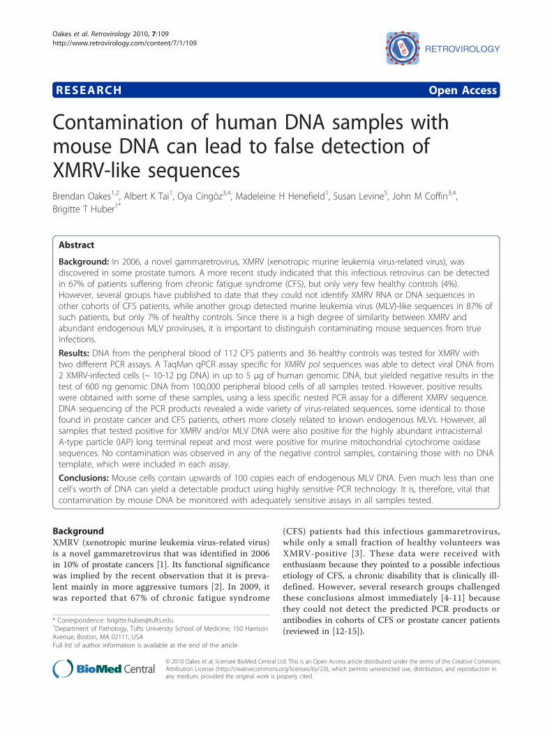
TaqMan qPCR specific for XMRV did not reveal positiveindividualsThe original XMRV results from patients with prostatecancer and CFS were obtained using a sensitive nestedPCR assay for XMRV [1,3] that also detects endogenousMLV sequences in murine genomic DNA. These datawere later extended, employing a qPCR assay specificfor a region in the XMRV pol gene not cross-reactivewith any sequence known to be present in mouse DNA[2, Singh, personal communication]. To test our cohortfor the presence of XMRV sequences, we analyzedPBMC DNA with this 2nd qPCR assay, using the pri-mers and probe as described in [2]. Titration of DNAfrom an XMRV-positive lymphoblastoid cell line, WPI-1282 (kindly provided by the Whittemore PetersonInstitute (WPI)), resulted in detection of XMRV downto 10-12 pg, equivalent to two cells, in the presence orabsence of 5 μg control DNA isolated from the humanLnCaP cell line (Figure 1). However, no positiveresponse (Ct > 60) was obtained with DNA from 112CFS patients and 36 healthy controls, when tested at600 ng to 5 μg per reaction (data not shown). These
data indicated that our samples were either XMRV-negative or had more divergent MLV sequences thanoriginally described [1,3]. In the latter case, the qPCRassay used, which is sensitive to small sequence differ-ences, would not have allowed detection.
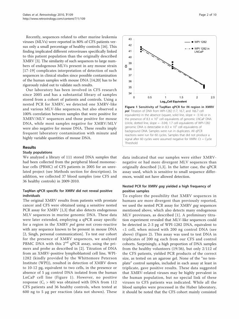
Nested PCR for XMRV gag yielded a high frequency ofpositive samplesTo explore the possibility that XMRV sequences inhumans are more divergent than previously reported,we used the nested PCR assay for XMRV gag sequencesmentioned above, which also detects many endogenousMLV proviruses, as described [1]. A preliminary titra-tion experiment revealed that MLV-like sequences couldbe detected in 2-3 pg of WPI-1282 DNA, equivalent to<1 cell, when mixed with 200 ng control DNA (seeabove) (Figure 2). This assay was used to test DNA intriplicates of 200 ng each from our CFS and controlcohorts. Surprisingly, a high proportion of DNA samplesfrom the healthy volunteers (19/36), but only 2/112 ofthe CFS patients, yielded PCR products of the correctsize, as tested on an agarose gel. None of the “no tem-plate” control samples, included in each assay at least intriplicate, gave positive results. These data suggestedthat XMRV-related viruses may be highly prevalent inthe human population, but no special link of theseviruses to CFS patients was indicated. While all theblood samples were processed in the Huber laboratory,it should be noted that the CFS cohort mainly consisted
34
35
36
37
38
39
40
41
42
43
0 0.5 1 1.5 2 2.5
Ct
Log10Cell Equivalent
WPI 1282
WPI 1282 in LNCaP
Figure 1 Sensitivity of TaqMan qPCR for IN region in XMRVpol. Titration of DNA from WPI-1282 (1.7, 16.7, and 166.7 cellequivalents) in the absence (square, solid line, slope = -3.14) or inthe presence of 8.3 × 10⁵ cell equivalents of genomic LNCaP DNA(circle, dotted line, slope = -3.04). 1.7 cell equivalents of WPI-1282genomic DNA is detectable in 8.3 × 10⁵ cell equivalents ofbackground DNA. Samples were run in duplicates. All qPCRreactions were run for 60 cycles. Samples that did not produce asignal after 60 cycles were assumed negative for XMRV. Ct = CycleThreshold
Oakes et al. Retrovirology 2010, 7:109http://www.retrovirology.com/content/7/1/109
Page 2 of 10
of banked samples collected and processed in 2005,whereas the healthy volunteers were recruited morerecently, between November of 2009 and May of 2010,and, as discussed later, were processed using a slightlydifferent protocol.
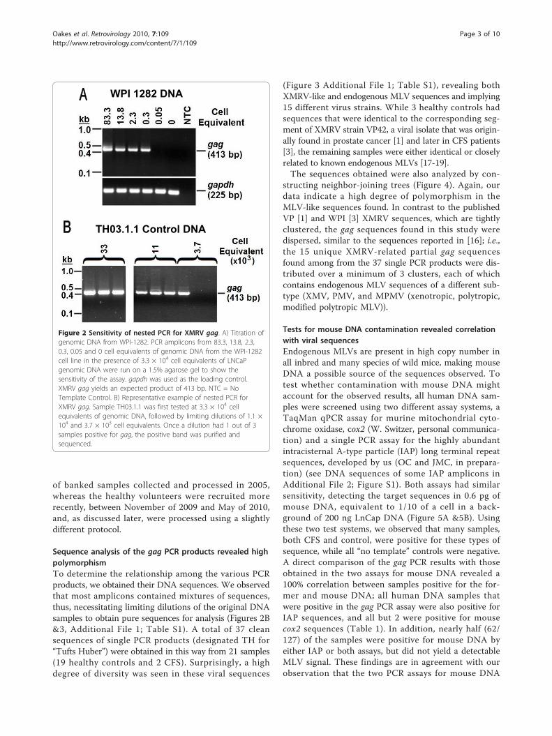
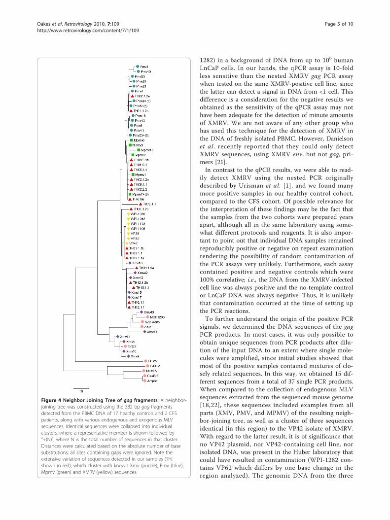
Sequence analysis of the gag PCR products revealed highpolymorphismTo determine the relationship among the various PCRproducts, we obtained their DNA sequences. We observedthat most amplicons contained mixtures of sequences,thus, necessitating limiting dilutions of the original DNAsamples to obtain pure sequences for analysis (Figures 2B&3, Additional File 1; Table S1). A total of 37 cleansequences of single PCR products (designated TH for“Tufts Huber”) were obtained in this way from 21 samples(19 healthy controls and 2 CFS). Surprisingly, a highdegree of diversity was seen in these viral sequences
(Figure 3 Additional File 1; Table S1), revealing bothXMRV-like and endogenous MLV sequences and implying15 different virus strains. While 3 healthy controls hadsequences that were identical to the corresponding seg-ment of XMRV strain VP42, a viral isolate that was origin-ally found in prostate cancer [1] and later in CFS patients[3], the remaining samples were either identical or closelyrelated to known endogenous MLVs [17-19].The sequences obtained were also analyzed by con-
structing neighbor-joining trees (Figure 4). Again, ourdata indicate a high degree of polymorphism in theMLV-like sequences found. In contrast to the publishedVP [1] and WPI [3] XMRV sequences, which are tightlyclustered, the gag sequences found in this study weredispersed, similar to the sequences reported in [16]; i.e.,the 15 unique XMRV-related partial gag sequencesfound among from the 37 single PCR products were dis-tributed over a minimum of 3 clusters, each of whichcontains endogenous MLV sequences of a different sub-type (XMV, PMV, and MPMV (xenotropic, polytropic,modified polytropic MLV)).
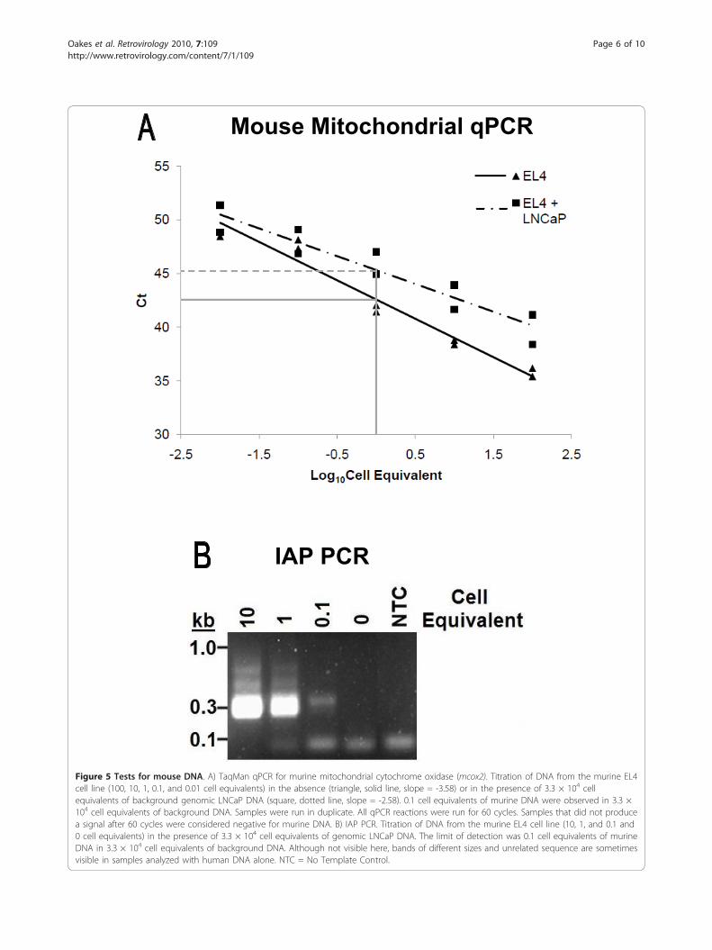
Tests for mouse DNA contamination revealed correlationwith viral sequencesEndogenous MLVs are present in high copy number inall inbred and many species of wild mice, making mouseDNA a possible source of the sequences observed. Totest whether contamination with mouse DNA mightaccount for the observed results, all human DNA sam-ples were screened using two different assay systems, aTaqMan qPCR assay for murine mitochondrial cyto-chrome oxidase, cox2 (W. Switzer, personal communica-tion) and a single PCR assay for the highly abundantintracisternal A-type particle (IAP) long terminal repeatsequences, developed by us (OC and JMC, in prepara-tion) (see DNA sequences of some IAP amplicons inAdditional File 2; Figure S1). Both assays had similarsensitivity, detecting the target sequences in 0.6 pg ofmouse DNA, equivalent to 1/10 of a cell in a back-ground of 200 ng LnCap DNA (Figure 5A &5B). Usingthese two test systems, we observed that many samples,both CFS and control, were positive for these types ofsequence, while all “no template” controls were negative.A direct comparison of the gag PCR results with thoseobtained in the two assays for mouse DNA revealed a100% correlation between samples positive for the for-mer and mouse DNA; all human DNA samples thatwere positive in the gag PCR assay were also positive forIAP sequences, and all but 2 were positive for mousecox2 sequences (Table 1). In addition, nearly half (62/127) of the samples were positive for mouse DNA byeither IAP or both assays, but did not yield a detectableMLV signal. These findings are in agreement with ourobservation that the two PCR assays for mouse DNA
WPI 1282 DNA
TH03.1.1 Control DNA
Figure 2 Sensitivity of nested PCR for XMRV gag. A) Titration ofgenomic DNA from WPI-1282. PCR amplicons from 83.3, 13.8, 2.3,0.3, 0.05 and 0 cell equivalents of genomic DNA from the WPI-1282cell line in the presence of 3.3 × 10⁴ cell equivalents of LNCaPgenomic DNA were run on a 1.5% agarose gel to show thesensitivity of the assay. gapdh was used as the loading control.XMRV gag yields an expected product of 413 bp. NTC = NoTemplate Control. B) Representative example of nested PCR forXMRV gag. Sample TH03.1.1 was first tested at 3.3 × 10⁴ cellequivalents of genomic DNA, followed by limiting dilutions of 1.1 ×10⁴ and 3.7 × 10³ cell equivalents. Once a dilution had 1 out of 3samples positive for gag, the positive band was purified andsequenced.
Oakes et al. Retrovirology 2010, 7:109http://www.retrovirology.com/content/7/1/109
Page 3 of 10
are at least 10-fold more sensitive than the XMRV gagPCR assay, when tested on genomic mouse DNA, andthat the IAP assay is more sensitive than the cox2 assayfor detection of mouse DNA. Overall, our data are con-sistent with the conclusion that the positive resultsobtained with the XMRV gag PCR assay are due to vari-able contamination of the human samples with mouseDNA, most likely in laboratory reagents.
DiscussionIn 2005, we initiated a study to examine the expressionlevel of an endogenous human betaretrovirus, HERV-
K18, in chronically ill CFS patients versus healthy con-trols. For this purpose, we accumulated a library ofDNA samples from CFS patients which has allowed usto investigate the possible association of XMRV withthis disease [3]. We initiated our studies on XMRVusing a TaqMan qPCR assay for a region in XMRV polthat is unique to XMRV and does not detect anysequences in genomic DNA from laboratory strains ofinbred mice [2]. None of the samples from either CFSpatients or healthy controls was positive in this assay,although we were able to detect a signal from twoXMRV-infected lymphoblastoid cells (cell line WPI-
Figure 3 Gag sequences from patient samples. Individual 382 bp sequences, free of double peaks and confirmed through forward andreverse sequencing, are compared in a Highlighter plot to the control WPI-1282 cell line sequence, VP62. The samples were coded to remainanonymous, with the first number being the patient number, the second number being the bleed number, the third number being the tube ofDNA, and the letter showing that we have multiple sequences in the same tube of DNA. Identical sequences were collapsed into individualclusters, those with more than two sequences are labeled TH+(N), where N is the total number of sequences in that cluster. CFS Type 1, 2 & 3are from Lo et al. [15]. Each vertical line shows a single nucleotide difference between the labeled sequence and the control VP62 sequence.
Oakes et al. Retrovirology 2010, 7:109http://www.retrovirology.com/content/7/1/109
Page 4 of 10
1282) in a background of DNA from up to 106 humanLnCaP cells. In our hands, the qPCR assay is 10-foldless sensitive than the nested XMRV gag PCR assaywhen tested on the same XMRV-positive cell line, sincethe latter can detect a signal in DNA from <1 cell. Thisdifference is a consideration for the negative results weobtained as the sensitivity of the qPCR assay may nothave been adequate for the detection of minute amountsof XMRV. We are not aware of any other group whohas used this technique for the detection of XMRV inthe DNA of freshly isolated PBMC. However, Danielsonet al. recently reported that they could only detectXMRV sequences, using XMRV env, but not gag, pri-mers [21].In contrast to the qPCR results, we were able to read-
ily detect XMRV using the nested PCR originallydescribed by Urisman et al. [1], and we found manymore positive samples in our healthy control cohort,compared to the CFS cohort. Of possible relevance forthe interpretation of these findings may be the fact thatthe samples from the two cohorts were prepared yearsapart, although all in the same laboratory using some-what different protocols and reagents. It is also impor-tant to point out that individual DNA samples remainedreproducibly positive or negative on repeat examinationrendering the possibility of random contamination ofthe PCR assays very unlikely. Furthermore, each assaycontained positive and negative controls which were100% correlative; i.e., the DNA from the XMRV-infectedcell line was always positive and the no-template controlor LnCaP DNA was always negative. Thus, it is unlikelythat contamination occurred at the time of setting upthe PCR reactions.To further understand the origin of the positive PCR
signals, we determined the DNA sequences of the gagPCR products. In most cases, it was only possible toobtain unique sequences from PCR products after dilu-tion of the input DNA to an extent where single mole-cules were amplified, since initial studies showed thatmost of the positive samples contained mixtures of clo-sely related sequences. In this way, we obtained 15 dif-ferent sequences from a total of 37 single PCR products.When compared to the collection of endogenous MLVsequences extracted from the sequenced mouse genome[18,22], these sequences included examples from allparts (XMV, PMV, and MPMV) of the resulting neigh-bor-joining tree, as well as a cluster of three sequencesidentical (in this region) to the VP42 isolate of XMRV.With regard to the latter result, it is of significance thatno VP42 plasmid, nor VP42-containing cell line, norisolated DNA, was present in the Huber laboratory thatcould have resulted in contamination (WPI-1282 con-tains VP62 which differs by one base change in theregion analyzed). The genomic DNA from the three
Figure 4 Neighbor Joining Tree of gag fragments. A neighbor-joining tree was constructed using the 382 bp gag fragmentsdetected from the PBMC DNA of 17 healthy controls and 2 CFSpatients, along with various endogenous and exogenous MLVsequences. Identical sequences were collapsed into individualclusters, where a representative member is shown followed by“+(N)”, where N is the total number of sequences in that cluster.Distances were calculated based on the absolute number of basesubstitutions; all sites containing gaps were ignored. Note theextensive variation of sequences detected in our samples (TH,shown in red), which cluster with known Xmv (purple), Pmv (blue),Mpmv (green) and XMRV (yellow) sequences.
Oakes et al. Retrovirology 2010, 7:109http://www.retrovirology.com/content/7/1/109
Page 5 of 10
Mouse Mitochondrial qPCR
IAP PCR
Figure 5 Tests for mouse DNA. A) TaqMan qPCR for murine mitochondrial cytochrome oxidase (mcox2). Titration of DNA from the murine EL4cell line (100, 10, 1, 0.1, and 0.01 cell equivalents) in the absence (triangle, solid line, slope = -3.58) or in the presence of 3.3 × 10⁴ cellequivalents of background genomic LNCaP DNA (square, dotted line, slope = -2.58). 0.1 cell equivalents of murine DNA were observed in 3.3 ×10⁴ cell equivalents of background DNA. Samples were run in duplicate. All qPCR reactions were run for 60 cycles. Samples that did not producea signal after 60 cycles were considered negative for murine DNA. B) IAP PCR. Titration of DNA from the murine EL4 cell line (10, 1, and 0.1 and0 cell equivalents) in the presence of 3.3 × 10⁴ cell equivalents of genomic LNCaP DNA. The limit of detection was 0.1 cell equivalents of murineDNA in 3.3 × 10⁴ cell equivalents of background DNA. Although not visible here, bands of different sizes and unrelated sequence are sometimesvisible in samples analyzed with human DNA alone. NTC = No Template Control.
Oakes et al. Retrovirology 2010, 7:109http://www.retrovirology.com/content/7/1/109
Page 6 of 10
healthy volunteers who had XMRV VP42 sequences alsocontained other MLV sequences. Thus, it is not possiblefor us to distinguish which one of the retrovirusesstemmed from mouse DNA contamination; i.e., it is for-mally possible that VP42 is an actual human retrovirus.It is also possible that it is an endogenous provirus, notpresent in the sequenced C57Bl/6 genome, but presentin the mouse species responsible for the sequencesobserved [19]. In the former case, the presence of VP42in DNA from healthy control samples, but not CFSpatients, would indicate that this virus is spread ran-domly through the human population, with no particu-lar link to CFS. Further analyses are required to clarifythis issue.The presence of mixtures of MLV sequences, all clo-
sely related to known endogenous MLVs [17-19], inmany of the DNA samples tested is not easily reconciledwith infection of human hosts with the correspondingviruses (reviewed in [14,20]). Two assays specific formurine DNA, for mitochondrial cox2 and IAPsequences, were used to test the possibility that theremight be trace amounts of mouse DNA contaminatingsome of the samples. Consistent with this idea, wefound that each DNA sample that was positive forXMRV/MLV also was positive for mouse DNA by theIAP assay, while >50% of XMRV/MLV-negative sampleswere positive for mouse DNA which is particularly strik-ing in the CFS group. Again, these results were con-firmed in repeat experiments and never deviated insubsequent analyses, suggesting that contamination hap-pened either during collection of blood, isolation ofPBMC, or during the preparation of the DNA from thePBMC. We interpret these data that possible contamina-tion with mouse DNA is ubiquitous, but the levelseemed to vary significantly from batch to batch of sam-ple preps, although all experimental procedures werecarried out in the same facility. In particular, althoughsamples collected at both times showed signs of con-tamination, the level of contamination in the normal
controls collected in 2009-2010 was noticeably greaterthan in the CFS samples from 2005. To date, we havenot been able to pinpoint a specific reagent or labora-tory vessel for being consistently positive for mouseDNA, but preliminary experiments implicate both fetalcalf serum (FCS) and phosphate buffered saline (PBS),although large variations in the surmised amount ofcontaminating mouse DNA were observed from bottleto bottle. All blood samples were collected in heparintubes rendering the anti-coagulant also a likely suspectfor mouse DNA contamination. However, a comparisonof parallel blood collections from the same healthy indi-vidual in heparin, Na-citrate and EDTA tubes did notsupport this hypothesis. In this particular set of samplesonly one DNA aliquot from Na-citrate-collected bloodwas positive for mouse DNA (results not shown).Currently there are highly discordant reports in the lit-
erature about the prevalence of XMRV in CFS and pros-tate cancer patients (reviewed in [12-15]). The originalpublication on CFS patients reported that almost 70% ofthese patients, but less than 5% of healthy individuals,harbor this virus [3], and that infectious virus and anti-viral antibodies could be detected in blood from thesepatients. Several reports have appeared in the literaturesince then contesting these findings [4-6,8,9], while arecent publication claimed that 80% of CFS patients, butnot healthy controls, contained endogenous MLV-likesequences, but were negative for mouse mitochondrialDNA [16]. The sequences from CFS patients identified inthis latter paper were distinct from the XMRV of the ori-ginal reports. A plausible explanation for these discrepantresults has not been put forward to date [13,14], but it isworth pointing out that the sequences identified in thelatter report were similar to the ones we found in thepresent study. Endogenous MLVs are abundant in alllaboratory mouse strains [17,18], as well as in wild Musspecies [19] and are carried by some human cell linesthat have been propagated in vivo in nude mice [20].Thus, extreme precautions have to be taken to exclude
Table 1 Correlation of MLV DNA sequence detection with mouse DNA contamination
CFS Patients Healthy Controls**
XMRV GAG cox IAP # of Samples (n = 112) Percent # of Samples (n = 36) Percent
+ + + 2* 1.8 17 47.2
- - - 53 47.3 12 33.3
+ - - 0 0 0 0
+ - + 0 0 2 5.6
+ + - 0 0 0 0
- + + 10 9.0 1 2.8
- - + 47 42.0 4 11.1
- + - 0 0 0 0
*One CFS sample from 2005 collection, and one CFS sample from 2010 collection. All the other CFS samples were collected in 2005.
**All collected in 2009-2010.
Oakes et al. Retrovirology 2010, 7:109http://www.retrovirology.com/content/7/1/109
Page 7 of 10
contamination with mouse DNA or DNA from any abun-dant MLV-producing cell line.
ConclusionsIn our study we have observed that 100% of human DNAsamples prepared in our laboratory that were positive forXMRV/MLV sequences were also positive for minutequantities of mouse DNA. Together with the similarity ofthe MLV sequences to multiple identified endogenousMLVs [17-19], this result provides a strong suspicion thatthe viral sequences detected in these samples were actuallyof murine origin. It is important to point out that negativecontrols included in each assay never yielded positiveresults, either for XMRV/MLV, or for mouse DNA,excluding the possibility that contamination with mouseDNA occurred at the bench during the final PCR assay,even though mouse derived cells and tissues are regularlyused in our laboratory. Of particular interest is the widevariety of sequences that we obtained, spanning bothXMRV and various MLV sequences. While most of theMLV-related sequences were identical to gag segments innonecotropic MLVs from inbred mice [17,18], some werefound to be unique; i.e., they have so far not been identi-fied in the sequenced mouse genome [22], but may be pre-sent in other laboratory strains or wild mice. Thus, ourdata are compatible with the conclusion that the detectionof MLV-related sequences in human genomic DNA sam-ples could be due to contamination with minute and vari-able quantities of mouse DNA, most likely contained invarious laboratory reagents.
MethodsSample collectionAll samples were collected according to the institutionalguidelines of Tufts University, after receiving informedconsent. The 36 healthy individuals (15 females and 21males) were recruited on a voluntary basis by the Huberlaboratory and were between 18 and 65 years of age. The112 CFS patients (89 females, 20 males and 3 unknown),recruited by Dr. Susan Levine, were between 18 and 65years of age and resided in the Northeastern UnitedStates. All patients were diagnosed for CFS according toCDC criteria [23], and the majority was completely dis-abled. The cohort comprised a combination of those withan abrupt and others with a gradual onset of symptoms.
Preparation of human blood samplesApproximately 30 ml of blood were drawn into threeheparinized tubes (Becton Dickinson) and shipped over-night (CFS patients) or processed immediately (healthycontrols). The blood collection tubes from each individualwere consolidated into one 50 ml tube and diluted withPBS, containing CaCl2 and MgCl2 (Sigma) at a 1:1 ratio.15 ml of Ficoll (GE Healthcare) was added to two new 50
ml tubes, and 25 ml of the diluted blood was gently layeredon top of the Ficoll, followed by a 30 min centrifugation ina Sorvall RT7plus rotor at 2000 rpm at room temperatureand collection of PBMCs from the interface. 10 ml ofplasma were also collected from each sample and stored at-80°C. The collected PBMCs were diluted with PBS (2005collection) or RPMI-1640 Medium (Sigma), supplementedwith 10% FCS (Gemini BioProducts), 100 U/ml penicillin(Sigma), 0.1 mg/ml streptomycin (Sigma), 2 mM L-gluta-mine (Sigma), and 1 mM sodium pyruvate (Sigma) (2010collection) (2009-2010 collection) (complete RPMI) at a1:1 ratio and then pelleted at 2000 rpm for 5 min. Thesupernatant was aspirated, and the pellet of PBMCs wasresuspended in 20 ml of PBS (2005 collection) or completeRPMI (2009-2010 collection). Cells were counted using alight microscope and a hemocytometer, aliquoted to 5 ×10⁶ cells per tube, spun down and resuspended in 350 μlof Buffer RLT Plus (Qiagen) (1% b-mercaptoethanol).Samples were stored in this lysis buffer at -80°C.
DNA isolation from PBMCsDNA was isolated using the procedures provided by the All-Prep DNA/RNA Mini Kit (Qiagen). Briefly, 350 μl of PBMClysate (RLT buffer, see above) (5 × 10⁶ cells) were placed onthe DNA spin column, which was centrifuged at 10,000rpm for 30 s in an Eppendorf 5417C Centrifuge. The col-umn was then transferred to a new collection tube. 500 μlAW1 Buffer (Qiagen) was added to the column, followed bya 15 s spin at 10,000 rpm. The flow-through was discarded,and the column was transferred to a new collection tube.500 μl of AW2 Buffer (Qiagen) was added to the column,followed by a 2 minute centrifugation at full speed. Theflow-through was discarded, and the column was transferredto a new 1.5 ml collection tube. 100 μl of Buffer EB (Qiagen)was added directly to the column, followed by 1 minuteincubation at room temperature. Finally, the column wascentrifuged at 10,000 rpm for 1 min to elute DNA. DNAconcentration was determined using 1 μl of sample on aThermo Scientific Nanodrop 2000 Spectrophotometer.
TaqMan qPCR assay for XMRV polPrimers and probe, as designed by Schlaberg et al. [2],were ordered from Applied Biosystems (see Table 2 forsequences). The reaction mix for the TaqMan qPCRscontained 1× Gene Expression Master Mix (AppliedBiosystems), 900 nM forward and reverse primers, 250nM probe, and 200 ng of DNA in a reaction volume of20 μl. The assay was validated with DNA from theWPI-1282 cell line containing VP62 XMRV (kindly sup-plied by J. Mikovits, WPI). The same DNA served aspositive control in each assay, which also included a no-template negative control. Thermocycler conditionswere 95°C for 10 minutes, followed by 60 cycles of 95°Cfor 15 s and then 60°C for 1 minute, using 96-well
Oakes et al. Retrovirology 2010, 7:109http://www.retrovirology.com/content/7/1/109
Page 8 of 10
Optical Reaction Plates (Applied Biosystems) on a 7300Real Time PCR System by Applied Biosystems. All reac-tions were performed in triplicate. Quality of DNA wasassessed using a TaqMan qPCR for the ribosomal 18 Sgene in the same reaction (Applied Biosystems).
Nested PCR assay for XMRV gagIdentical primers as originally described by Urisman et al.[1] and also employed by the Mikovits group [3] wereused. The reaction mix for all PCRs consisted of 1× Hot-Start-IT™FideliTaq™Master Mix, 200 nM forward andreverse primers, and 200 ng of sample DNA in a 50 μlreaction volume. The WPI-1282 lymphoblastoid cell linewas used as a positive control [3]. Thermocycler condi-tions for the first PCR were 2 minutes at 94°C, followed by30 cycles of 94°C for 30 s, 58°C for 30 s, and 72°C for 45 sand then finished off with 72°C for 7 minutes. Once thefirst PCR was complete, 2 μl of DNA from the first PCRwas used for the second PCR. The second PCR consistedof 1× HotStart-IT™FideliTaq™Master Mix, 200 nM forwardand reverse primers, and 200 ng of sample DNA in a 50 μlreaction volume. Thermocycler conditions for the secondPCR were 2 minutes at 94°C, followed by 30 cycles of 94°Cfor 30 s, 60°C for 30 s, and 72°C for 30 s and then finishedoff with 72°C for 7 minutes. Once the second PCR wascomplete, 15 μl of the samples were run on a 1.5% agarosegel for 1 h at 100 volts. Images of gels were taken using aVersaDoc Imaging System (Biorad). The expected frag-ment size of the second PCR is 413 bp [1].All positive samples from the second XMRV nested PCR
were isolated using a Qiaquick PCR Purification Kit (Qia-gen). DNA sequencing was performed by the Tufts Uni-versity Core Facility. Once sequenced, the traces were
monitored for double peaks, and sequences with doublepeaks were discarded. Samples that had mixed sequenceswere diluted, and the nested PCR was repeated. Onlyclean sequences with the forward sequence matching thereverse sequence were used for phylogenetic analysis.
TaqMan qPCR assay for mouse mitochondrial cox2Sequences for primers and probes were kindly suppliedby Dr. Switzer, CDC (Personal Communication) (seeTable 2). Primers and Probes were ordered fromApplied Biosystems. The reaction mix contained 1×Gene Expression Master Mix (Applied Biosystems),900 nM forward and reverse primers, 250 nM probe,and 200 ng of DNA in a reaction volume of 20 μl. DNAisolated from the murine EL4 cell line, diluted in 200 ngof human LNCaP DNA, was used as a positive control.Thermocycler conditions were 95°C for 9 minutes, fol-lowed by 60 cycles of 95°C for 30 s and 62°C for 30 s.96-well plates were used on a 7300 Real Time PCR Sys-tem by Applied Biosystems. All reactions were per-formed in duplicate or triplicate. Quality of DNA wasassessed using a TaqMan qPCR for the ribosomal 18 Sgene in the same reaction (Applied Biosystems).
PCR assay for Mouse IAP sequencesPrimers were designed by the Coffin Laboratory (OCand JMC, in preparation) and ordered from Invitrogen.The reaction mix for all PCRs consisted of 1× HotStart-IT™FideliTaq™Master Mix, 1 μM forward and reverseprimers, and 200 ng of sample DNA in a 50 μl reactionvolume. DNA isolated from the murine EL4 cell linewas diluted into 200 ng of human DNA (LNCaP) andused as a positive control. Thermocycler conditionswere 94°C for 2 minutes, followed by 45 cycles of 94°Cfor 30 s, 58°C for 30 s, and 72°C for 20 s and then fin-ished off with 72°C for 7 minutes. Samples were thenrun on a 1.5% agarose gel with sequence lengths varyingbetween 200 and 300 bp. Images of gels were takenusing a VersaDoc Imaging System (Biorad). IAP PCRproducts were cloned and sequenced and yielded theexpected results (see Additional File 2; Figure S1).
Additional material
Additional File 1: Supplementary Table 1 - List of identicalsequences grouped into clusters for analysis. Each cluster containsfragments that are identical in the corresponding 382 bp gag region.
Additional File 2: Supplemental Figure 1 - IAP sequences. IAPsequences amplified from the indicated control human DNA samplesusing the primers shown in Table II were cloned into a TOPO vector andsequenced. Four representative sequences are shown. Each sequencehad a 100% match in the sequenced mouse genome. Adenine (A) =Green, Cytosine (C) = Blue, Guanine (G) = Black, Thymine (T) = Red.
Table 2 Primers and probes used for TaqMan qPCRs,primary PCRs, and nested PCRs.
Primer Sequence
XMRV4552F 5’-CGA GAG GCA GCC ATG AAG G-3’
XMRV4673R 5’-CCC AGT TCC CGT AGT CTT TTG AG-3’
XMRV4572MGB 5’-6FAM-AGT TCT AGA AAC CTC TAC ACT C-MGBNFQ-3’
GAG-O-F 5’-CGC GTC TGA TTT GTT TTG TT-3’
GAG-O-R 5’-CCG CCT CTT CTT CAT TGT TC-3’
GAG-I-F 5’-TCT CGA GAT CAT GGG ACA GA-3’
GAG-I-R 5’-AGA GGG TAA GGG CAG GGT AA-3’
MCox2-F2 5’-TTC TAC CAG CTG TAA TCC TTA-3’
MCox2-R1 5’-GTT TTA GGT CGT TTG TTG GGA T-3’
MCox2-PR1 5’-FAM-CGT AGC TTC AGT ATC ATT GGT GCC CTA TGGT-MGBNFQ-3’
MCox2-P1 5’-FAM-TTG CTC TCC CCT CTC TAC GCA TTC TA-MGBNFQ-3’
IAP-Forward 5’-ATA ATC TGC GCA TGA GCC AAG G-3’
IAP-Reverse 5’-AGG AAG AAC ACC ACA GAC CAG A-3’
Oakes et al. Retrovirology 2010, 7:109http://www.retrovirology.com/content/7/1/109
Page 9 of 10
List of abbreviationsCFS: Chronic Fatigue Syndrome; FCS: fetal calf serum; IAP: intracisternal A-type particle; MLV: murine leukemia virus; MPLV: modified polytropic MLV;PBMC: peripheral blood mononuclear cells; PBS: phosphate buffered saline;PMV; polytropic MLV; WPI: Whittemore Peterson Institute; XMRV: xenotropicmurine leukemia virus-related virus; XMV: xenotropic MLV.
AcknowledgementsWe would like to thank Drs. WM Switzer (CDC) for communicating theunpublished information on the TaqMan qPCR for cox2 and JA Mikovits(WPI) for providing the WPI-1282 lymphoblastoid cell line. The work wassupported by a grant from the HHV6 Foundation of America to BH andgrant R37 CA 089441 to JMC. JMC was a Research Professor of the AmericanCancer Society with support from the FM Kirby Foundation.
Author details1Department of Pathology, Tufts University School of Medicine, 150 HarrisonAvenue, Boston, MA 02111, USA. 2Pharmacology Program, Tufts UniversitySchool of Medicine, 150 Harrison Avenue, Boston, MA 02111, USA.3Department of Molecular Biology and Microbiology, Tufts University Schoolof Medicine, 150 Harrison Avenue, Boston, MA 02111, USA. 4GeneticsProgram, Tufts University School of Medicine, 150 Harrison Avenue, Boston,MA 02111, USA. 5Private Practice, 115 East 72nd Street, New York, NY, USA.
Authors’ contributionsBTH, AKT and BO conceived and designed the study. AKT, BO and MHHcarried out the experiments. SL collected samples from the CFS patientcohort. AKT, BO, MHH, OC and JMC analyzed the data. BTH drafted themanuscript. All authors read and approved the final manuscript.
Competing interestsThe authors declare that they have no competing interests.
Received: 1 November 2010 Accepted: 20 December 2010Published: 20 December 2010
References1. Urisman A, Molinaro RJ, Fischer N, Plummer SJ, Casey G, Klein EA, Malathi K,
Magi-Galluzzi C, Tubbs RR, Ganem D, et al: Identification of a novelGammaretrovirus in prostate tumors of patients homozygous for R462QRNASEL variant. PLoS Pathog 2006, 2(3):e25.
2. Schlaberg R, Choe DJ, Brown KR, Thaker HM, Singh IR: XMRV is present inmalignant prostatic epithelium and is associated with prostate cancer,especially high-grade tumors. Proc Natl Acad Sci USA 2009, 106:16351-16356.
3. Lombardi VC, Ruscetti FW, Das Gupta J, Pfost MA, Hagen KS, Peterson DL,Ruscetti SK, Bagni RK, Petrow-Sadowski C, Gold B, et al: Detection of aninfectious retrovirus, XMRV, in blood cells of patients with chronicfatigue syndrome. Science 2009, 326:585-589.
4. Erlwein O, Kaye S, McClure MO, Weber J, Wills G, Collier D, Wessely S,Cleare A: Failure to detect the novel retrovirus XMRV in chronic fatiguesyndrome. PLoS One 2010, 5(1):e8519.
5. Groom HC, Boucherit VC, Makinson K, Randal E, Baptista S, Hagan S,Gow JW, Mattes FM, Breuer J, Kerr JR, et al: Absence of xenotropic murineleukaemia virus-related virus in UK patients with chronic fatiguesyndrome. Retrovirology 2010, 7:10.
6. van Kuppeveld FJM, de Yong AS, Lanke KH, Verhaegh GW, Meichers WJG,Swanink CMA, Bieljenberg G, Netea MG, Galama JMD, M. vdMJW:Prevalence of xenotropic murine leukaemia virus-related virus inpatients with chronic fatigue syndrome in the Netherlands: retrospectiveanalysis of samples from an established cohort. British Medical Journal2010, 340:c1018.
7. Hong P, Li J, Li Y: Failure to detect Xenotropic murine leukaemia virus-related virus in Chinese patients with chronic fatigue syndrome. Virol J2010, 7:224.
8. Switzer WM, Jia H, Hohn O, Zheng H, Tang S, Shankar A, Bannert N,Simmons G, Hendry RM, Falkenberg VR, et al: Absence of evidence ofxenotropic murine leukemia virus-related virus infection in persons withchronic fatigue syndrome and healthy controls in the United States.Retrovirology 2010, 7:57.
9. Henrich TX, Li JX, Felsenstein D, Kotton CX, Plenge RX, Pereyra F, Marty FX,Lin NX, Grazioso P, Crochiere DX, et al: Xenotropic Murine Leukemia Virus-
Related Virus Prevalence in Patients with Chronic Fatigue Syndrome orChronic Immunomodulatory Conditions. J Infect Dis 2010, 202(10):1478-81.
10. Barnes E, Flanagan P, Brown A, Robinson N, Brown H, McClure M,Oxenius A, Collier J, Weber J, HX GXfN, et al: Failure to Detect XenotropicMurine Leukemia Virus-Related Virus in Blood of Individuals at High Riskof Blood-Borne Viral Infections. J Infect Dis 2010, 202(10):1482-5.
11. Aloia AL, Sfanos KS, Isaacs WB, Zheng Q, Maldarelli F, De Marzo AM, Rein A:XMRV: A New Virus in Prostate Cancer? Cancer Res 2010, epub Oct 21.
12. Silverman RH, Nguyen C, Weight CJ, Klein EA: The human retrovirus XMRVin prostate cancer and chronic fatigue syndrome. Nat Rev Urol 2010,7:392-402.
13. Kaiser J: Virology. No meeting of minds on XMRV’s role in chronicfatigue, cancer. Science 2010, 329:1454.
14. Weiss RA: A cautionary tale of virus and disease. BMC Biol 2010, 8:124.15. Kearney M, Maldarelli F: Current Status of Xenotropic Murine Leukemia
Virus-Related Retrovirus in Chronic Fatigue Syndrome and ProstateCancer: Reach for a Scorecard, Not a Prescription Pad. J Infect Dis 2010,202(10):1463-6.
16. Lo SC, Pripuzova N, Li B, Komaroff AL, Hung GC, Wang R, Alter HJ:Detection of MLV-related virus gene sequences in blood of patientswith chronic fatigue syndrome and healthy blood donors. Proc Natl AcadSci USA 2010, 107(36):15874-15879.
17. Frankel WN, Stoye JP, Taylor BA, Coffin JM: A linkage map of endogenousmurine leukemia proviruses. Genetics 1990, 124:221-236.
18. Jern P, Stoye JP, Coffin JM: Role of APOBEC3 in genetic diversity amongendogenous murine leukemia viruses. PLoS Genet 2007, 3:2014-2022.
19. Tomonaga K, Coffin JM: Structures of endogenous nonecotropic murineleukemia virus (MLV) long terminal repeats in wild mice: implication forevolution of MLVs. J Virol 1999, 73:4327-4340.
20. Voisset C, Weiss RA, Griffiths DJ: Human RNA “rumor” viruses: the searchfor novel human retroviruses in chronic disease. Microbiol Mol Biol Rev2008, 72:157-196.
21. Danielson BP, Ayala GE, Kimata JT: Detection of xenotropic murineleukemia virus-related virus in normal and tumor tissue of patients fromthe southern United States with prostate cancer is dependent onspecific polymerase chain reaction conditions. J Infect Dis 2010,202:1470-1477.
22. Waterston RH, Lindblad-Toh K, Birney E, Rogers J, Abril JF, Agarwal P,Agarwala R, Ainscough R, Alexandersson M, An P, et al: Initial sequencingand comparative analysis of the mouse genome. Nature 2002,420:520-562.
23. Fukuda K, Straus SE, Hickie I, Sharpe MC, Dobbins JG, Komaroff A: Thechronic fatigue syndrome: a comprehensive approach to its definitionand study. International Chronic Fatigue Syndrome Study Group. AnnIntern Med 1994, 121:953-959.
doi:10.1186/1742-4690-7-109Cite this article as: Oakes et al.: Contamination of human DNA sampleswith mouse DNA can lead to false detection of XMRV-like sequences.Retrovirology 2010 7:109.
Submit your next manuscript to BioMed Centraland take full advantage of:
• Convenient online submission
• Thorough peer review
• No space constraints or color figure charges
• Immediate publication on acceptance
• Inclusion in PubMed, CAS, Scopus and Google Scholar
• Research which is freely available for redistribution
Submit your manuscript at www.biomedcentral.com/submit
Oakes et al. Retrovirology 2010, 7:109http://www.retrovirology.com/content/7/1/109
Page 10 of 10


