Embed Size (px)
Citation preview
Jones et al. BMC Evolutionary Biology (2017) 17:65 DOI 10.1186/s12862-017-0909-z
RESEARCH ARTICLE Open Access
Conservation of estrogen receptor functionin invertebrate reproduction
Brande L. Jones1*, Chris Walker2, Bahareh Azizi3, Laren Tolbert2, Loren Dean Williams2 and Terry W. Snell1Abstract
Background: Rotifers are microscopic aquatic invertebrates that reproduce both sexually and asexually. Thoughrotifers are phylogenetically distant from humans, and have specialized reproductive physiology, this work identifiesa surprising conservation in the control of reproduction between humans and rotifers through the estrogenreceptor. Until recently, steroid signaling has been observed in only a few invertebrate taxa and its role inregulating invertebrate reproduction has not been clearly demonstrated. Insights into the evolution of sexsignaling pathways can be gained by clarifying how receptors function in invertebrate reproduction.
Results: In this paper, we show that a ligand-activated estrogen-like receptor in rotifers binds human estradioland regulates reproductive output in females. In other invertebrates characterized thus far, ER ligand bindingdomains have occluded ligand-binding sites and the ERs are not ligand activated. We have used a suite ofcomputational, biochemical and biological techniques to determine that the rotifer ER binding site is notoccluded and can bind human estradiol.
Conclusions: Our results demonstrate that this mammalian hormone receptor plays a key role in reproductionof the ancient microinvertebrate Brachinous manjavacas. The presence and activity of the ER within the phylumRotifera indicates that the ER structure and function is highly conserved throughout animal evolution.
Keywords: Rotifera, Receptor, Estrogen receptor
BackgroundSignaling through steroid receptors regulates develop-ment, growth and reproduction in most vertebrateanimals [1–7]. Recently, concern has grown about sus-ceptibility of aquatic invertebrates to endocrine disrup-tion, which has been documented for vertebrates [8].Endocrine disruption is the process by which certainchemicals, called endocrine disrupting compounds,interfere with the endocrine system and disrupt develop-mental, reproductive, neurological, and immune pro-cesses. Endocrine disrupting compounds are a subclassof organic contaminants that have been detected inwastewater and surface waters throughout the world [9].Although steroid signaling is thought to be the chief
means by which most animals regulate reproduction,it has been confirmed in only a few invertebrate taxa[10] and its regulatory role has not been generally
* Correspondence: [email protected] of Biology, Georgia Institute of Technology, Atlanta, GA 30332-0230,USAFull list of author information is available at the end of the article
© The Author(s). 2017 Open Access This articInternational License (http://creativecommonsreproduction in any medium, provided you gthe Creative Commons license, and indicate if(http://creativecommons.org/publicdomain/ze
demonstrated [7, 11–13]. Steroid signaling may bepresent in a much more diverse group of animalsthan currently demonstrated. Genome and proteomeanalysis indicates that modern steroid receptors evolvedfrom an ancient receptor that arose more than 600 Maago, before the common ancestor of bilaterians divergedinto protostomes and deuterosomes [6].Rotifera is one of the largest microinvertebrate phyla
[14]. Although monogonont rotifers are capable ofboth asexual and sexual reproduction, the chemicalsignals regulating these are poorly understood. Sexualreproduction is triggered by a quorum sensing process [15],induced by secretion of a Mixis Inducing Protein (MIP)[16]. The similarity of MIP to a putative steroidogenesis-inducing protein in humans [17] suggests that steroid hor-mones may have a role in regulating sexual reproduction inB. manjavacas.The estrogen receptor (ER) is the most ancient of all
of the sex steroid receptors [2, 4, 5, 18]. However, thereare disparities and inconsistencies in the known phylo-genetic distribution of the ER in invertebrates and in the
le is distributed under the terms of the Creative Commons Attribution 4.0.org/licenses/by/4.0/), which permits unrestricted use, distribution, andive appropriate credit to the original author(s) and the source, provide a link tochanges were made. The Creative Commons Public Domain Dedication waiverro/1.0/) applies to the data made available in this article, unless otherwise stated.
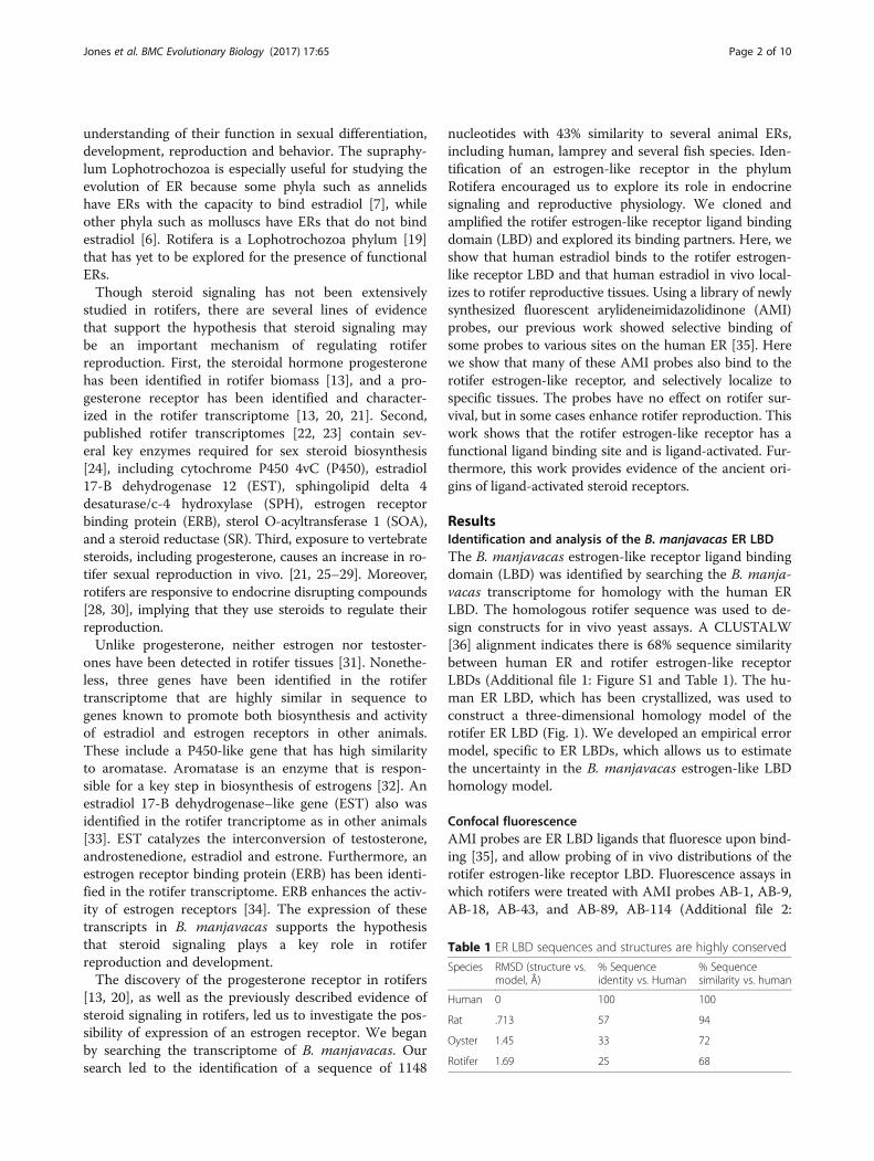
Table 1 ER LBD sequences and structures are highly conserved
Species RMSD (structure vs.model, Å)
% Sequenceidentity vs. Human
% Sequencesimilarity vs. human
Human 0 100 100
Rat .713 57 94
Oyster 1.45 33 72
Rotifer 1.69 25 68
Jones et al. BMC Evolutionary Biology (2017) 17:65 Page 2 of 10
understanding of their function in sexual differentiation,development, reproduction and behavior. The supraphy-lum Lophotrochozoa is especially useful for studying theevolution of ER because some phyla such as annelidshave ERs with the capacity to bind estradiol [7], whileother phyla such as molluscs have ERs that do not bindestradiol [6]. Rotifera is a Lophotrochozoa phylum [19]that has yet to be explored for the presence of functionalERs.Though steroid signaling has not been extensively
studied in rotifers, there are several lines of evidencethat support the hypothesis that steroid signaling maybe an important mechanism of regulating rotiferreproduction. First, the steroidal hormone progesteronehas been identified in rotifer biomass [13], and a pro-gesterone receptor has been identified and character-ized in the rotifer transcriptome [13, 20, 21]. Second,published rotifer transcriptomes [22, 23] contain sev-eral key enzymes required for sex steroid biosynthesis[24], including cytochrome P450 4vC (P450), estradiol17-B dehydrogenase 12 (EST), sphingolipid delta 4desaturase/c-4 hydroxylase (SPH), estrogen receptorbinding protein (ERB), sterol O-acyltransferase 1 (SOA),and a steroid reductase (SR). Third, exposure to vertebratesteroids, including progesterone, causes an increase in ro-tifer sexual reproduction in vivo. [21, 25–29]. Moreover,rotifers are responsive to endocrine disrupting compounds[28, 30], implying that they use steroids to regulate theirreproduction.Unlike progesterone, neither estrogen nor testoster-
ones have been detected in rotifer tissues [31]. Nonethe-less, three genes have been identified in the rotifertranscriptome that are highly similar in sequence togenes known to promote both biosynthesis and activityof estradiol and estrogen receptors in other animals.These include a P450-like gene that has high similarityto aromatase. Aromatase is an enzyme that is respon-sible for a key step in biosynthesis of estrogens [32]. Anestradiol 17-B dehydrogenase–like gene (EST) also wasidentified in the rotifer trancriptome as in other animals[33]. EST catalyzes the interconversion of testosterone,androstenedione, estradiol and estrone. Furthermore, anestrogen receptor binding protein (ERB) has been identi-fied in the rotifer transcriptome. ERB enhances the activ-ity of estrogen receptors [34]. The expression of thesetranscripts in B. manjavacas supports the hypothesisthat steroid signaling plays a key role in rotiferreproduction and development.The discovery of the progesterone receptor in rotifers
[13, 20], as well as the previously described evidence ofsteroid signaling in rotifers, led us to investigate the pos-sibility of expression of an estrogen receptor. We beganby searching the transcriptome of B. manjavacas. Oursearch led to the identification of a sequence of 1148
nucleotides with 43% similarity to several animal ERs,including human, lamprey and several fish species. Iden-tification of an estrogen-like receptor in the phylumRotifera encouraged us to explore its role in endocrinesignaling and reproductive physiology. We cloned andamplified the rotifer estrogen-like receptor ligand bindingdomain (LBD) and explored its binding partners. Here, weshow that human estradiol binds to the rotifer estrogen-like receptor LBD and that human estradiol in vivo local-izes to rotifer reproductive tissues. Using a library of newlysynthesized fluorescent arylideneimidazolidinone (AMI)probes, our previous work showed selective binding ofsome probes to various sites on the human ER [35]. Herewe show that many of these AMI probes also bind to therotifer estrogen-like receptor, and selectively localize tospecific tissues. The probes have no effect on rotifer sur-vival, but in some cases enhance rotifer reproduction. Thiswork shows that the rotifer estrogen-like receptor has afunctional ligand binding site and is ligand-activated. Fur-thermore, this work provides evidence of the ancient ori-gins of ligand-activated steroid receptors.
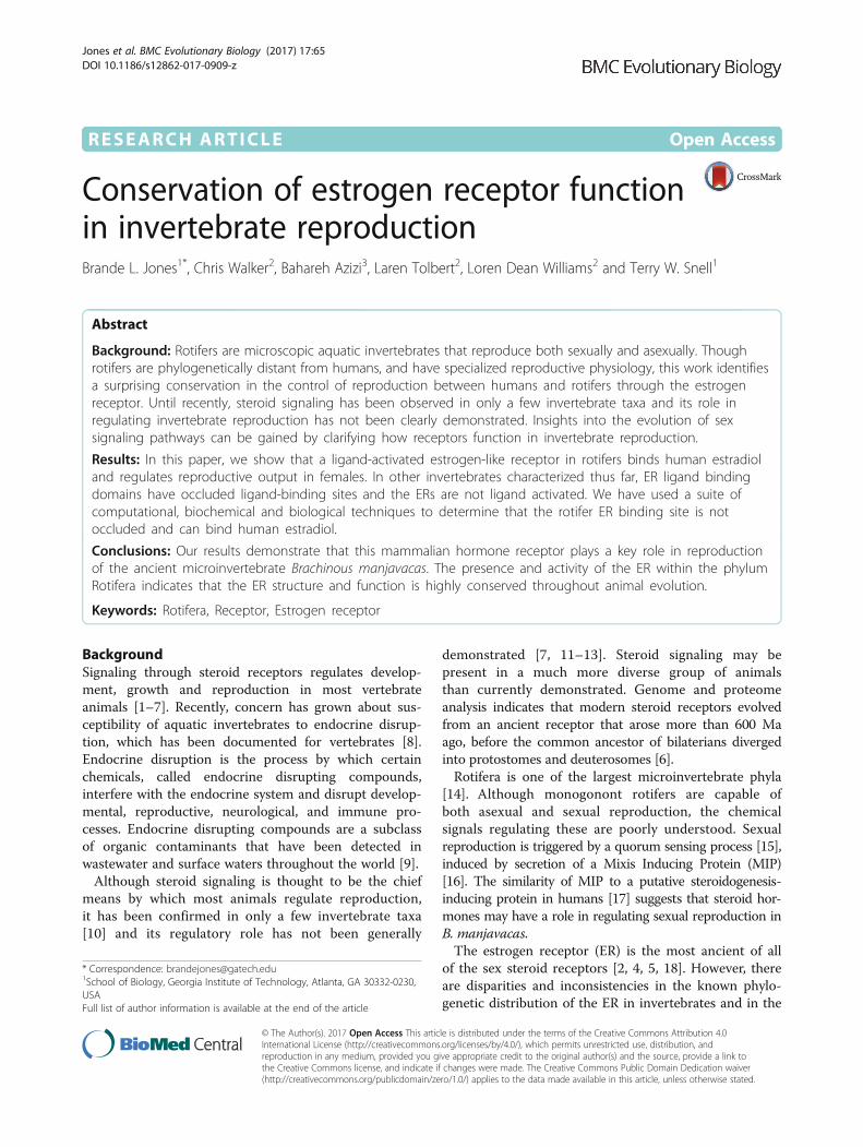
ResultsIdentification and analysis of the B. manjavacas ER LBDThe B. manjavacas estrogen-like receptor ligand bindingdomain (LBD) was identified by searching the B. manja-vacas transcriptome for homology with the human ERLBD. The homologous rotifer sequence was used to de-sign constructs for in vivo yeast assays. A CLUSTALW[36] alignment indicates there is 68% sequence similaritybetween human ER and rotifer estrogen-like receptorLBDs (Additional file 1: Figure S1 and Table 1). The hu-man ER LBD, which has been crystallized, was used toconstruct a three-dimensional homology model of therotifer ER LBD (Fig. 1). We developed an empirical errormodel, specific to ER LBDs, which allows us to estimatethe uncertainty in the B. manjavacas estrogen-like LBDhomology model.
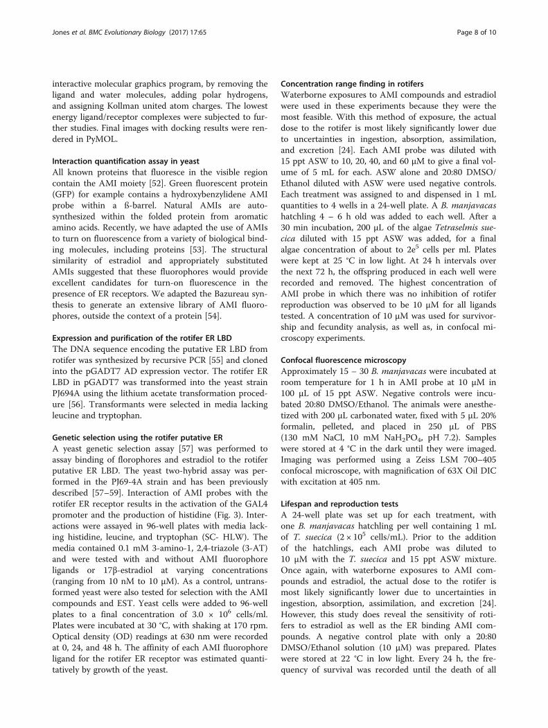
Confocal fluorescenceAMI probes are ER LBD ligands that fluoresce upon bind-ing [35], and allow probing of in vivo distributions of therotifer estrogen-like receptor LBD. Fluorescence assays inwhich rotifers were treated with AMI probes AB-1, AB-9,AB-18, AB-43, and AB-89, AB-114 (Additional file 2:
a b
Glu 9
Arg 50Phe 64
Ile 85
Cys 47
Fig. 1 A homology model of the 3D structure of the B. manjavacas estrogen-like receptor LBD (yellow). a Superimposition of the rotifer (model)and human (X-ray) ER LBDs (pink). The model suggests that the structure of the binding pocket is conserved between human and rotifer. b Closeup view of the rotifer ER LBD ligand binding site occupied by estradiol. The amino acids that form the ligand binding site are indicated. Hydrogenbonds are dashed black lines. van der Waals contacts are diffuse red lines. The 3D structure of the human ER LBD was used as a template toconstruct the 3D model of the rotifer estrogen-like receptor LBD
Jones et al. BMC Evolutionary Biology (2017) 17:65 Page 3 of 10
Figure S2) demonstrate localized binding in specifictissues, including ovaries and vitellarium (yolk gland)(Fig. 2). Auto-fluorescence and other confounding signalswere not observed in rotifers treated with estradiol alone.
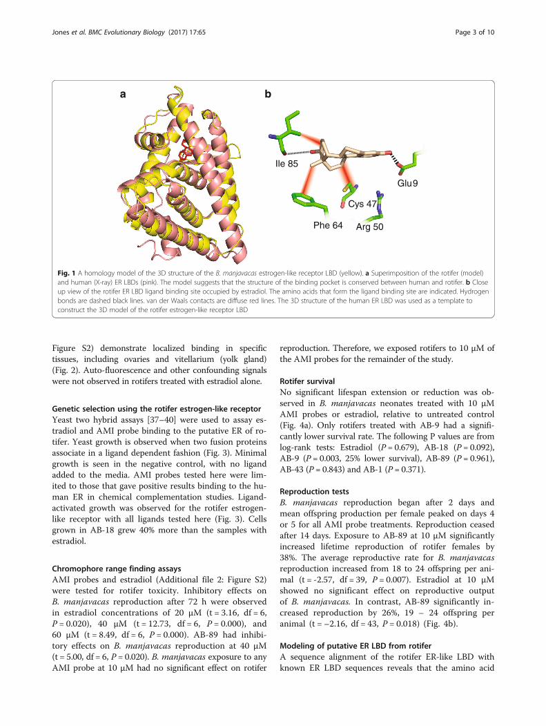
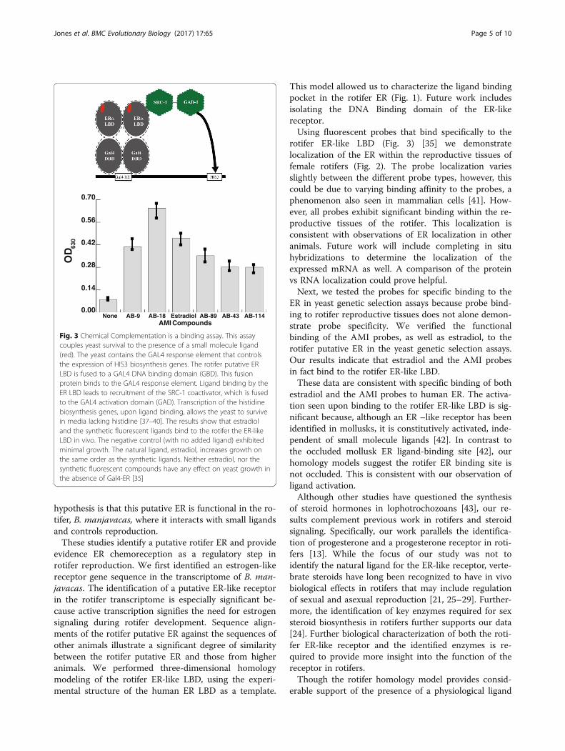
Genetic selection using the rotifer estrogen-like receptorYeast two hybrid assays [37–40] were used to assay es-tradiol and AMI probe binding to the putative ER of ro-tifer. Yeast growth is observed when two fusion proteinsassociate in a ligand dependent fashion (Fig. 3). Minimalgrowth is seen in the negative control, with no ligandadded to the media. AMI probes tested here were lim-ited to those that gave positive results binding to the hu-man ER in chemical complementation studies. Ligand-activated growth was observed for the rotifer estrogen-like receptor with all ligands tested here (Fig. 3). Cellsgrown in AB-18 grew 40% more than the samples withestradiol.
Chromophore range finding assaysAMI probes and estradiol (Additional file 2: Figure S2)were tested for rotifer toxicity. Inhibitory effects onB. manjavacas reproduction after 72 h were observedin estradiol concentrations of 20 μM (t = 3.16, df = 6,P = 0.020), 40 μM (t = 12.73, df = 6, P = 0.000), and60 μM (t = 8.49, df = 6, P = 0.000). AB-89 had inhibi-tory effects on B. manjavacas reproduction at 40 μM(t = 5.00, df = 6, P = 0.020). B. manjavacas exposure to anyAMI probe at 10 μM had no significant effect on rotifer
reproduction. Therefore, we exposed rotifers to 10 μM ofthe AMI probes for the remainder of the study.
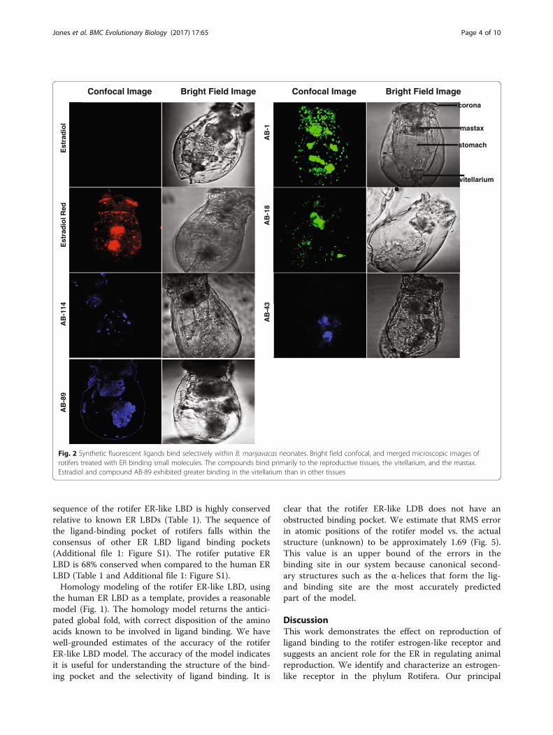
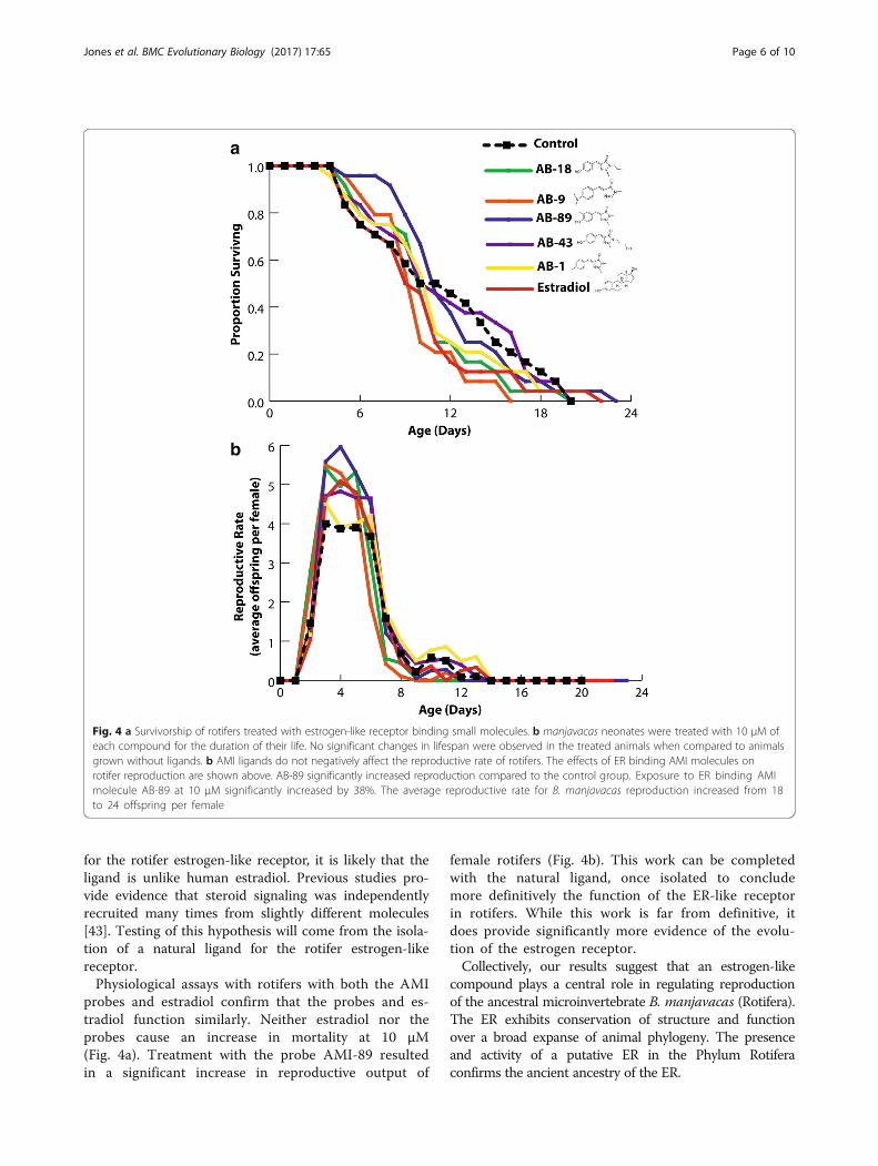
Rotifer survivalNo significant lifespan extension or reduction was ob-served in B. manjavacas neonates treated with 10 μMAMI probes or estradiol, relative to untreated control(Fig. 4a). Only rotifers treated with AB-9 had a signifi-cantly lower survival rate. The following P values are fromlog-rank tests: Estradiol (P = 0.679), AB-18 (P = 0.092),AB-9 (P = 0.003, 25% lower survival), AB-89 (P = 0.961),AB-43 (P = 0.843) and AB-1 (P = 0.371).
Reproduction testsB. manjavacas reproduction began after 2 days andmean offspring production per female peaked on days 4or 5 for all AMI probe treatments. Reproduction ceasedafter 14 days. Exposure to AB-89 at 10 μM significantlyincreased lifetime reproduction of rotifer females by38%. The average reproductive rate for B. manjavacasreproduction increased from 18 to 24 offspring per ani-mal (t = -2.57, df = 39, P = 0.007). Estradiol at 10 μMshowed no significant effect on reproductive outputof B. manjavacas. In contrast, AB-89 significantly in-creased reproduction by 26%, 19 – 24 offspring peranimal (t = –2.16, df = 43, P = 0.018) (Fig. 4b).
Modeling of putative ER LBD from rotiferA sequence alignment of the rotifer ER-like LBD withknown ER LBD sequences reveals that the amino acid
Confocal Image Bright Field Image Confocal Image Bright Field Image
Est
rad
iol
Est
rad
iol R
edA
B-1
14A
B-8
9
AB
-1A
B-1
8A
B-4
3
corona
mastax
vitellarium
stomach
Fig. 2 Synthetic fluorescent ligands bind selectively within B. manjavacas neonates. Bright field confocal, and merged microscopic images ofrotifers treated with ER binding small molecules. The compounds bind primarily to the reproductive tissues, the vitellarium, and the mastax.Estradiol and compound AB-89 exhibited greater binding in the vitellarium than in other tissues
Jones et al. BMC Evolutionary Biology (2017) 17:65 Page 4 of 10
sequence of the rotifer ER-like LBD is highly conservedrelative to known ER LBDs (Table 1). The sequence ofthe ligand-binding pocket of rotifers falls within theconsensus of other ER LBD ligand binding pockets(Additional file 1: Figure S1). The rotifer putative ERLBD is 68% conserved when compared to the human ERLBD (Table 1 and Additional file 1: Figure S1).Homology modeling of the rotifer ER-like LBD, using
the human ER LBD as a template, provides a reasonablemodel (Fig. 1). The homology model returns the antici-pated global fold, with correct disposition of the aminoacids known to be involved in ligand binding. We havewell-grounded estimates of the accuracy of the rotiferER-like LBD model. The accuracy of the model indicatesit is useful for understanding the structure of the bind-ing pocket and the selectivity of ligand binding. It is
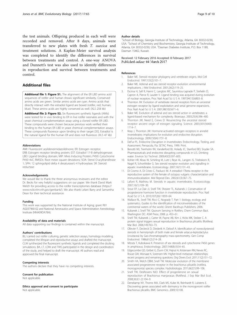
clear that the rotifer ER-like LDB does not have anobstructed binding pocket. We estimate that RMS errorin atomic positions of the rotifer model vs. the actualstructure (unknown) to be approximately 1.69 (Fig. 5).This value is an upper bound of the errors in thebinding site in our system because canonical second-ary structures such as the α-helices that form the lig-and binding site are the most accurately predictedpart of the model.
DiscussionThis work demonstrates the effect on reproduction ofligand binding to the rotifer estrogen-like receptor andsuggests an ancient role for the ER in regulating animalreproduction. We identify and characterize an estrogen-like receptor in the phylum Rotifera. Our principal
0.70
0.56
0.42
0.28
0.14
0.00
OD
630
None AB-9 AB-18 Estradiol AB-89 AB-43 AB-114AMI Compounds
Fig. 3 Chemical Complementation is a binding assay. This assaycouples yeast survival to the presence of a small molecule ligand(red). The yeast contains the GAL4 response element that controlsthe expression of HIS3 biosynthesis genes. The rotifer putative ERLBD is fused to a GAL4 DNA binding domain (GBD). This fusionprotein binds to the GAL4 response element. Ligand binding by theER LBD leads to recruitment of the SRC-1 coactivator, which is fusedto the GAL4 activation domain (GAD). Transcription of the histidinebiosynthesis genes, upon ligand binding, allows the yeast to survivein media lacking histidine [37–40]. The results show that estradioland the synthetic fluorescent ligands bind to the rotifer the ER-likeLBD in vivo. The negative control (with no added ligand) exhibitedminimal growth. The natural ligand, estradiol, increases growth onthe same order as the synthetic ligands. Neither estradiol, nor thesynthetic fluorescent compounds have any effect on yeast growth inthe absence of Gal4-ER [35]
Jones et al. BMC Evolutionary Biology (2017) 17:65 Page 5 of 10
hypothesis is that this putative ER is functional in the ro-tifer, B. manjavacas, where it interacts with small ligandsand controls reproduction.These studies identify a putative rotifer ER and provide
evidence ER chemoreception as a regulatory step inrotifer reproduction. We first identified an estrogen-likereceptor gene sequence in the transcriptome of B. man-javacas. The identification of a putative ER-like receptorin the rotifer transcriptome is especially significant be-cause active transcription signifies the need for estrogensignaling during rotifer development. Sequence align-ments of the rotifer putative ER against the sequences ofother animals illustrate a significant degree of similaritybetween the rotifer putative ER and those from higheranimals. We performed three-dimensional homologymodeling of the rotifer ER-like LBD, using the experi-mental structure of the human ER LBD as a template.
This model allowed us to characterize the ligand bindingpocket in the rotifer ER (Fig. 1). Future work includesisolating the DNA Binding domain of the ER-likereceptor.Using fluorescent probes that bind specifically to the
rotifer ER-like LBD (Fig. 3) [35] we demonstratelocalization of the ER within the reproductive tissues offemale rotifers (Fig. 2). The probe localization variesslightly between the different probe types, however, thiscould be due to varying binding affinity to the probes, aphenomenon also seen in mammalian cells [41]. How-ever, all probes exhibit significant binding within the re-productive tissues of the rotifer. This localization isconsistent with observations of ER localization in otheranimals. Future work will include completing in situhybridizations to determine the localization of theexpressed mRNA as well. A comparison of the proteinvs RNA localization could prove helpful.Next, we tested the probes for specific binding to the
ER in yeast genetic selection assays because probe bind-ing to rotifer reproductive tissues does not alone demon-strate probe specificity. We verified the functionalbinding of the AMI probes, as well as estradiol, to therotifer putative ER in the yeast genetic selection assays.Our results indicate that estradiol and the AMI probesin fact bind to the rotifer ER-like LBD.These data are consistent with specific binding of both
estradiol and the AMI probes to human ER. The activa-tion seen upon binding to the rotifer ER-like LBD is sig-nificant because, although an ER –like receptor has beenidentified in mollusks, it is constitutively activated, inde-pendent of small molecule ligands [42]. In contrast tothe occluded mollusk ER ligand-binding site [42], ourhomology models suggest the rotifer ER binding site isnot occluded. This is consistent with our observation ofligand activation.Although other studies have questioned the synthesis
of steroid hormones in lophotrochozoans [43], our re-sults complement previous work in rotifers and steroidsignaling. Specifically, our work parallels the identifica-tion of progesterone and a progesterone receptor in roti-fers [13]. While the focus of our study was not toidentify the natural ligand for the ER-like receptor, verte-brate steroids have long been recognized to have in vivobiological effects in rotifers that may include regulationof sexual and asexual reproduction [21, 25–29]. Further-more, the identification of key enzymes required for sexsteroid biosynthesis in rotifers further supports our data[24]. Further biological characterization of both the roti-fer ER-like receptor and the identified enzymes is re-quired to provide more insight into the function of thereceptor in rotifers.Though the rotifer homology model provides consid-
erable support of the presence of a physiological ligand
a
b
Fig. 4 a Survivorship of rotifers treated with estrogen-like receptor binding small molecules. b manjavacas neonates were treated with 10 μM ofeach compound for the duration of their life. No significant changes in lifespan were observed in the treated animals when compared to animalsgrown without ligands. b AMI ligands do not negatively affect the reproductive rate of rotifers. The effects of ER binding AMI molecules onrotifer reproduction are shown above. AB-89 significantly increased reproduction compared to the control group. Exposure to ER binding AMImolecule AB-89 at 10 μM significantly increased by 38%. The average reproductive rate for B. manjavacas reproduction increased from 18to 24 offspring per female
Jones et al. BMC Evolutionary Biology (2017) 17:65 Page 6 of 10
for the rotifer estrogen-like receptor, it is likely that theligand is unlike human estradiol. Previous studies pro-vide evidence that steroid signaling was independentlyrecruited many times from slightly different molecules[43]. Testing of this hypothesis will come from the isola-tion of a natural ligand for the rotifer estrogen-likereceptor.Physiological assays with rotifers with both the AMI
probes and estradiol confirm that the probes and es-tradiol function similarly. Neither estradiol nor theprobes cause an increase in mortality at 10 μM(Fig. 4a). Treatment with the probe AMI-89 resultedin a significant increase in reproductive output of
female rotifers (Fig. 4b). This work can be completedwith the natural ligand, once isolated to concludemore definitively the function of the ER-like receptorin rotifers. While this work is far from definitive, itdoes provide significantly more evidence of the evolu-tion of the estrogen receptor.Collectively, our results suggest that an estrogen-like
compound plays a central role in regulating reproductionof the ancestral microinvertebrate B. manjavacas (Rotifera).The ER exhibits conservation of structure and functionover a broad expanse of animal phylogeny. The presenceand activity of a putative ER in the Phylum Rotiferaconfirms the ancient ancestry of the ER.

1.6
1.4
1.2
1.0
.8
.6
.4
.2
0 100
95 90 85 80 75 70 65
1.8
RM
SD
% Sequence Similarity
human
rat
oyster
rotifer
Fig. 5 Error estimate for the rotifer ER-like LBD homology model. RMSD of atomic positions (Å) were calculated by superimposing the homologymodel for each organism against the real crystal structure. RMSD values vs. percentage sequence similarity are plotted. We can estimate the RMSDof the rotifer ER-like LBD model based on its sequence similarity to the human ER LBD. The estimated RMS error for the rotifer model is 1.7 Å. R2 = 0.95
Jones et al. BMC Evolutionary Biology (2017) 17:65 Page 7 of 10
ConclusionsHere we have identified and characterized an ER-like re-ceptor in the Phylum Rotifera. Our study provides aninitial synthesis of computational, chemical and bio-logical techniques to confirm the structure and functionof this receptor in B. manjavacas, an ancestral inverte-brate. Chemical cues have long been hypothesized tomediators for the switch from asexual to sexualreproduction in rotifer populations. This study providesevidence that microscopic invertebrates’ reproductivedevelopment may also be controlled by ligand activatedsignaling.
MethodsRotifer culturingB. manjavacas [44, 45] neonates were hatched from rest-ing eggs in 15 ppt artificial seawater (ASW, InstantOcean salts) under constant fluorescent illumination at25 °C. B. manjavacas was originally collected from AzovSea and was previously known as Brachionus plicatilis[16, 46]. B. manjavacas has been cultured continuouslyin the Snell laboratory since 1983 [13].
ER identification and homology modelingQuerying the B. manjavacas transcriptome database(https://www.ncbi.nlm.nih.gov/genbank/) using the hu-man ER and the BLASTX tool [47] returned a rotifer ERcDNA sequence. Alignments using its deduced aminoacid sequences were conducted with CLUSTALW [48].
The ER ligand binding domain (LDB) was modeled withthe SWISS-MODEL tool of the Swiss PDB Viewer [49]using human ER structure (PDB 1ERE) as a template.The efficacy of the SWISS-MODEL tool for modelingER structures was verified by modeling additional ERLBDs. We constructed models of a series of ER LBDSand compared the homology models with correspondingx-ray structures, which are known. Modeled ER LBDsfrom animals (oyster, rat, human and rotifer), usinghuman as the template, were superimposed on the cor-responding crystal structures. Root mean square devia-tions (RMSDs) of atomic positions were calculated todetermine relationships of sequence similarity (humantemplate compared to model) and degree of error in themodel in three dimensions (model compared to x-raystructure). The RMSDs were plotted against the aminoacid similarity. A trend line was used to predict theRMSD for the rotifer model versus the real (unknown)rotifer structure. In short we made an ER-specific errormodel, following a previous more general method [50]for assaying the quality of homology models.
In silico binding of AMI fluorophore ligand to the RotiferERStructures of AMI fluorophore probes were energy mini-mized with ChemBioDraw 3D Ultra (Cambridge Soft,USA). In silico docking of the probes to the rotifer ERLBD was carried out with Autodock Vina [51]. The ERwas prepared for docking via UCSF CHIMERA, an
Jones et al. BMC Evolutionary Biology (2017) 17:65 Page 8 of 10
interactive molecular graphics program, by removing theligand and water molecules, adding polar hydrogens,and assigning Kollman united atom charges. The lowestenergy ligand/receptor complexes were subjected to fur-ther studies. Final images with docking results were ren-dered in PyMOL.
Interaction quantification assay in yeastAll known proteins that fluoresce in the visible regioncontain the AMI moiety [52]. Green fluorescent protein(GFP) for example contains a hydroxybenzylidene AMIprobe within a ß-barrel. Natural AMIs are auto-synthesized within the folded protein from aromaticamino acids. Recently, we have adapted the use of AMIsto turn on fluorescence from a variety of biological bind-ing molecules, including proteins [53]. The structuralsimilarity of estradiol and appropriately substitutedAMIs suggested that these fluorophores would provideexcellent candidates for turn-on fluorescence in thepresence of ER receptors. We adapted the Bazureau syn-thesis to generate an extensive library of AMI fluoro-phores, outside the context of a protein [54].
Expression and purification of the rotifer ER LBDThe DNA sequence encoding the putative ER LBD fromrotifer was synthesized by recursive PCR [55] and clonedinto the pGADT7 AD expression vector. The rotifer ERLBD in pGADT7 was transformed into the yeast strainPJ694A using the lithium acetate transformation proced-ure [56]. Transformants were selected in media lackingleucine and tryptophan.
Genetic selection using the rotifer putative ERA yeast genetic selection assay [57] was performed toassay binding of florophores and estradiol to the rotiferputative ER LBD. The yeast two-hybrid assay was per-formed in the PJ69-4A strain and has been previouslydescribed [57–59]. Interaction of AMI probes with therotifer ER receptor results in the activation of the GAL4promoter and the production of histidine (Fig. 3). Inter-actions were assayed in 96-well plates with media lack-ing histidine, leucine, and tryptophan (SC- HLW). Themedia contained 0.1 mM 3-amino-1, 2,4-triazole (3-AT)and were tested with and without AMI fluorophoreligands or 17β-estradiol at varying concentrations(ranging from 10 nM to 10 μM). As a control, untrans-formed yeast were also tested for selection with the AMIcompounds and EST. Yeast cells were added to 96-wellplates to a final concentration of 3.0 × 10⁶ cells/ml.Plates were incubated at 30 °C, with shaking at 170 rpm.Optical density (OD) readings at 630 nm were recordedat 0, 24, and 48 h. The affinity of each AMI fluorophoreligand for the rotifer ER receptor was estimated quanti-tatively by growth of the yeast.
Concentration range finding in rotifersWaterborne exposures to AMI compounds and estradiolwere used in these experiments because they were themost feasible. With this method of exposure, the actualdose to the rotifer is most likely significantly lower dueto uncertainties in ingestion, absorption, assimilation,and excretion [24]. Each AMI probe was diluted with15 ppt ASW to 10, 20, 40, and 60 μM to give a final vol-ume of 5 mL for each. ASW alone and 20:80 DMSO/Ethanol diluted with ASW were used negative controls.Each treatment was assigned to and dispensed in 1 mLquantities to 4 wells in a 24-well plate. A B. manjavacashatchling 4 – 6 h old was added to each well. After a30 min incubation, 200 μL of the algae Tetraselmis sue-cica diluted with 15 ppt ASW was added, for a finalalgae concentration of about to 2e5 cells per ml. Plateswere kept at 25 °C in low light. At 24 h intervals overthe next 72 h, the offspring produced in each well wererecorded and removed. The highest concentration ofAMI probe in which there was no inhibition of rotiferreproduction was observed to be 10 μM for all ligandstested. A concentration of 10 μM was used for survivor-ship and fecundity analysis, as well as, in confocal mi-croscopy experiments.
Confocal fluorescence microscopyApproximately 15 – 30 B. manjavacas were incubated atroom temperature for 1 h in AMI probe at 10 μM in100 μL of 15 ppt ASW. Negative controls were incu-bated 20:80 DMSO/Ethanol. The animals were anesthe-tized with 200 μL carbonated water, fixed with 5 μL 20%formalin, pelleted, and placed in 250 μL of PBS(130 mM NaCl, 10 mM NaH2PO4, pH 7.2). Sampleswere stored at 4 °C in the dark until they were imaged.Imaging was performed using a Zeiss LSM 700–405confocal microscope, with magnification of 63X Oil DICwith excitation at 405 nm.
Lifespan and reproduction testsA 24-well plate was set up for each treatment, withone B. manjavacas hatchling per well containing 1 mLof T. suecica (2 × 10⁵ cells/mL). Prior to the additionof the hatchlings, each AMI probe was diluted to10 μM with the T. suecica and 15 ppt ASW mixture.Once again, with waterborne exposures to AMI com-pounds and estradiol, the actual dose to the rotifer ismost likely significantly lower due to uncertainties iningestion, absorption, assimilation, and excretion [24].However, this study does reveal the sensitivity of roti-fers to estradiol as well as the ER binding AMI com-pounds. A negative control plate with only a 20:80DMSO/Ethanol solution (10 μM) was prepared. Plateswere stored at 22 °C in low light. Every 24 h, the fre-quency of survival was recorded until the death of all
Jones et al. BMC Evolutionary Biology (2017) 17:65 Page 9 of 10
the test animals. Offspring produced in each well wererecorded and removed. After 8 days, animals weretransferred to new plates with fresh T. suecica andtreatment solutions. A Kaplan-Meier survival analysiswas completed to identify the differences in survivalbetween treatments and control. A one-way ANOVAand Dunnett’s test was also used to identify differencesin reproduction and survival between treatments andcontrol.
Additional files
Additional file 1: Figure S1. The alignment of the ER LBD amino acidsequences of rotifer and human shows significant similarity. Conservedamino acids are green. Similar amino acids are cyan. Amino acids thatdirectly interact with the estradiol ligand are boxed (rotifer, red; human;blue). These amino acids are highly conserved as well. (XLS 258 kb)
Additional file 2: Figure S2. Six fluorescent synthetic ligands (AMIs)were tested for in vivo binding to ER in live rotifer neonates and with theyeast chemical complementation assay using a cloned rotifer ER LBD.These compounds were chosen because previous work verified theirbinding to the human ER LBD in yeast chemical complementation assays.These compounds fluoresce upon binding to their target [35]. Estradiol isthe natural ligand for the human ER and does not fluoresce. (XLS 47 kb)
AbbreviationsAMI: Fluorescent arylideneimidazolidinone; ER: Estrogen receptor;ERB: Estrogen receptor binding protein; EST: Estradiol 17-B dehydrogenase;LBD: Ligand binding domain; MIP: Mixis Inducing Protein; P450: CytochromeP450 4vC; RMSDs: Root mean square deviations; SOA: Sterol O-acyltransferase1; SPH: 12 sphingolipid delta 4 desaturase/c-4 hydroxylase; SR: Steroidreductase
AcknowledgementsWe would like to thank the three anonymous reviewers and the editorDr. Becks for very helpful suggestions on our paper. We thank David MarkWelch for providing access to the rotifer transcriptomes database (https://www.ncbi.nlm.nih.gov/genbank/). We also thank Lelani Barry and SamanthaDean for their technical assistance.
FundingThis work was supported by the National Institute of Aging; grant R01AG037960-02 and National Aeronautics and Space Administration AstrobiologyInstitute (NNA09DA78A).
Availability of data and materialsAll data supporting our findings is contained within the manuscript.
Authors’ contributionsBLJ carried out rotifer culturing, genetic selection assays, homology modeling,completed the lifespan and reproductive assays and drafted the manuscript.CLW synthesized the fluorescent synthetic ligands and completed the dockingsimulations. BA, LT, LDW and TWS participated in the design and coordinationof the study, and helped to draft the manuscript. All authors read andapproved the final manuscript.
Competing interestsThe authors declare that they have no competing interests.
Consent for publicationNot applicable.
Ethics approval and consent to participateNot applicable.
Author details1School of Biology, Georgia Institute of Technology, Atlanta, GA 30332-0230,USA. 2School of Chemistry and Biochemistry, Georgia Institute of Technology,Atlanta, GA 30332-0230, USA. 3Dasman Diabetes Institute, P.O. Box 1180,Dasman 15462, Kuwait.
Received: 12 February 2016 Accepted: 8 February 2017
References1. Baker ME. Steroid receptor phylogeny and vertebrate origins. Mol Cell
Endocrinol. 1997;135(2):101–7.2. Baker ME. Adrenal and sex steroid receptor evolution: environmental
implications. J Mol Endocrinol. 2001;26(2):119–25.3. Escriva H, Safi R, Hanni C, Langlois MC, Saumitou-Laprade P, Stehelin D,
Capron A, Pierce R, Laudet V. Ligand binding was acquired during evolutionof nuclear receptors. Proc Natl Acad Sci U S A. 1997;94(13):6803–8.
4. Thornton JW. Evolution of vertebrate steroid receptors from an ancestralestrogen receptor by ligand exploitation and serial genome expansions.Proc Natl Acad Sci U S A. 2001;98(10):5671–6.
5. Baker ME. Evolution of adrenal and sex steroid action in vertebrates: aligand-based mechanism for complexity. Bioessays. 2003;25(4):396–400.
6. Thornton JW, Need E, Crews D. Resurrecting the ancestral steroidreceptor: ancient origin of estrogen signaling. Science. 2003;301(5640):1714–7.
7. Keay J, Thornton JW. Hormone-activated estrogen receptors in annelidinvertebrates: implications for evolution and endocrine disruption.Endocrinology. 2009;150(4):1731–8.
8. DeFur PL. Endocrine Disruption in Invertebrates: Endocrinology, Testing, andAssessment. Pensacola, Fla: SETAC Press, 1999. Print.
9. Benotti MJ, Trenholm RA, Vanderford BJ, Holady JC, Stanford BD, Snyder SA.Pharmaceuticals and endocrine disrupting compounds in U.S. Drinkingwater. Environ Sci Technol. 2009;43(3):597–603.
10. Kohler HR, Kloas W, Schirling M, Lutz I, Reye AL, Langen JS, Triebskorn R,Nagel R, Schonfelder G. Sex steroid receptor evolution and signalling inaquatic invertebrates. Ecotoxicology. 2007;16(1):131–43.
11. Di Cosmo A, Di Cristo C, Paolucci M. A estradiol-17beta receptor in thereproductive system of the female of octopus vulgaris: characterization andimmunolocalization. Mol Reprod Dev. 2002;61(3):367–75.
12. Lafont R, Mathieu M. Steroids in aquatic invertebrates. Ecotoxicology.2007;16(1):109–30.
13. Stout EP, La Clair JJ, Snell TW, Shearer TL, Kubanek J. Conservation ofprogesterone hormone function in invertebrate reproduction. Proc NatlAcad Sci U S A. 2010;107(26):11859–64.
14. Wallace RL, Snell TW, Ricci C, Nogrady T. Part 1: biology, ecology andsystematics. Guides to the identification of microinvertebrates of thecontinental waters of the world. Ghent: Backhuys Publishers; 2006.
15. Kubanek J, Snell TW. Quorum Sensing in Rotifers. Chem Commun Bact.Washington DC: ASM Press; 2008. p. 453–61.
16. Snell TW, Kubanek J, Carter W, Payne AB, Kim J, Hicks MK, Stelzer C. Aprotein signal triggers sexual reproduction in Brachionus plicatilis (Rotifera).Mar Biol. 2006;149:763–73.
17. Ollevier F, Declerck D, Diederik H, Deloof A. Identification of nonecdysteroidsteroids in hemolymph of both male and female astacus-leptodactylus(crustacea) by Gas-chromatography mass-spectrometry. Gen CompEndocrinol. 1986;61(2):214–28.
18. Mizuta T, Kubokawa K. Presence of sex steroids and cytochrome P450 genesin amphioxus. Endocrinology. 2007;148(8):3554–65.
19. Edgecombe GD, Giribet G, Dunn CW, Hejnol A, Kristensen RM, Neves RC,Rouse GW, Worsaae K, Sorensen MV. Higher-level metazoan relationships:recent progress and remaining questions. Org Divers Evol. 2011;11(2):151–72.
20. Smith HA, Welch DBM, Snell TW. Molecular evolution of the membraneassociated progesterone receptor in the brachionus plicatilis (rotifera,monogononta) species complex. Hydrobiologia. 2011;662(1):99–106.
21. Snell TW, DesRosiers NJD. Effect of progesterone on sexualreproduction of Brachionus manjavacas (Rotifera). J Exp Mar Biol Ecol.2008;363(1-2):104–9.
22. Denekamp NY, Thorne MA, Clark MS, Kube M, Reinhardt R, Lubzens E.Discovering genes associated with dormancy in the monogonont rotiferBrachionus plicatilis. BMC Genomics. 2009;10:108.
Jones et al. BMC Evolutionary Biology (2017) 17:65 Page 10 of 10
23. Suga K, Welch DM, Tanaka Y, Sakakura Y, Hagiwara A. Analysis of expressedsequence tags of the cyclically parthenogenetic rotifer Brachionus plicatilis.PLoS One. 2007;2(7):e671.
24. Snell TW. A review of the molecular mechanisms of monogonont rotiferreproduction. Hydrobiologia. 2011;662(1):89–97.
25. Gallardo WG, Hagiwara A, Snell TW. Effect of juvenile hormone andserotonin (5-HT) on mixis induction of the rotifer Brachionus plicatilis Muller.J Exp Mar Biol Ecol. 2000;252(1):97–107.
26. Gallardo WG, Hagiwara A, Tomita Y, Snell TW. Effect of growth hormoneand gamma-aminobutyric acid on Brachionus plicatilis (Rotifera)reproduction at low food or high ammonia levels. J Exp Mar Biol Ecol.1999;240(2):179–91.
27. Gallardo WG, Hagiwara A, Tomita Y, Soyano K, Snell TW. Effect of somevertebrate and invertebrate hormones on the population growth, micticfemale production, and body size of the marine rotifer Brachionus plicatilisMuller. Hydrobiologia. 1997;358:113–20.
28. Preston B, Snell T, Dingman B. Use of the freshwater rotifer Brachionuscalyciflorus in a screening assay for potential endocrine disruptors. EnvironToxicol Chem. 2000;19:2923–8.
29. Yang JX, Snell TW. Effects of progesterone, testosterone, and estrogen onsexual reproduction of the rotifer brachionus calyciflorus. Int Rev Hydrobiol.2010;95(6):441–9.
30. Snell TW, Joaquim-Justo C. Workshop on rotifers in ecotoxicology.Hydrobiologia. 2007;593:227–32.
31. Walker CL, Lukyanov KA, Yampolsky IV, Mishin AS, Bommarius AS, Duraj-ThatteAM, Azizi B, Tolbert LM, Solntsev KM. Fluorescence imaging using syntheticGFP chromophores. Curr Opin Chem Biol. 2015;(7):64–74.
32. Yague JG, Lavaque E, Carretero J, Azcoitia I, Garcia-Segura LM. Aromatase,the enzyme responsible for estrogen biosynthesis, is expressed by humanand rat glioblastomas. Neurosci Lett. 2004;368(3):279–84.
33. Brown KA, Sayasith K, Bouchard N, Lussier JG, Sirois J. Molecular cloning ofequine 17 beta-hydroxysteroid dehydrogenase type 1 and itsdownregulation during follicular luteinization in vivo. J Mol Endocrinol.2007;38(1-2):67–78.
34. Bu HF, Kashireddy P, Chang J, Zhu YT, Zhang ZY, Zheng W, Rao SM,Zhu YJ. ERBP, a novel estrogen receptor binding protein enhancingthe activity of estrogen receptor. Biochem Biophys Res Commun.2004;317(1):54–9.
35. Walker CL, Lukyanov KA, Yampolsky IV, Mishin AS, Bommarius AS,Duraj-Thatte AM, Azizi B, Tolbert LM, Solntsev KM. Fluorescenceimaging using synthetic GFP chromophores. Curr Opin Chem Biol.2015;27:64–74.
36. Sievers F, Higgins DG. Clustal omega, accurate alignment of very largenumbers of sequences. Mult Seq Alignment Methods. 2014;1079:105–16.
37. Schwimmer LJ, Rohatgi P, Azizi B, Seley KL, Doyle DF. Creation anddiscovery of ligand–receptor pairs for transcriptional control with smallmolecules. Proc Natl Acad Sci U S A. 2004;101(41):14707–12.
38. Azizi B, Chang EI, Doyle DF, Schwimmer LJ. Chemical complementation: ahigh throughput system for nuclear receptor drug discovery. Abstr Pap AmChem Soc. 2005;229:U186.
39. Azizi B, Doyle DF. Chemical complementation: a genetic selection system inyeast for drug discovery, protein and enzyme engineering. FASEB J.2006;20(4):A464.
40. Doyle DF, Schwimmer LJ, Azizi B, Rohatgi P. Chemical complementation:protein engineering through genetic selection. Abstr Pap Am Chem Soc.2005;229:U213.
41. Duraj-Thatte A. Fluorescent GFP chromophores as potential ligands forvarious nuclear receptors. PhD diss., Georgia Institute of Technology, 2012.
42. Bridgham JT, Keay J, Ortlund EA, Thornton JW. Vestigialization of an allostericswitch: genetic and structural mechanisms for the evolution of constitutiveactivity in a steroid hormone receptor. Plos Genet. 2014;10(1):e1004058.
43. Markov GV, Tavares R, Dauphin-Villemant C, Demeneix BA, Baker ME,Laudet V. Independent elaboration of steroid hormone signalingpathways in metazoans. Proc Natl Acad Sci. 2009;106(29):11913–8.
44. Fontaneto D, Herniou EA, Boschetti C, Caprioli M, Melone G, Ricci C,Barraclough TG. Independently evolving species in asexual bdelloid rotifers.PLoS Biol. 2007;5(4):e87.
45. Fontaneto D, Giordani I, Melone G, Serra M. Disentangling themorphological stasis in two rotifer species of the Brachionus plicatilisspecies complex. Hydrobiologia. 2007;583:297–307.
46. Rico-Martínez R, Snell TW. Comparative binding of antibody to a materecognition pheromone on female Brachionus plicatilis and Brachionusrotundiformis (Rotifera). Hydrobiologia. 1997;358(1):71–6.
47. Altschul SF, Gish W, Myers EW, Lipman DJ. Basic local alignment search tool.J Mol Biol. 1990;215:403–10.
48. Thompson JD, Higgins DG, Gibson TJ. Clustal W: improving sensiticity ofprogressive multiple sequence alignment through sequence weighting,position-specific gap penalties and weight matrix choice. Nucleic Acids Res.1994;22:4673–80.
49. Guex N, Peitsch MC. SWISS-MODEL and the Swiss-PdbViewer: anenvironment for comparative protein modeling. Electrophoresis.1997;18(15):2714–23.
50. Marti-Renom MA, Stuart AC, Fiser A, Sanchez R, Melo F, Sali A. Comparativeprotein structure modeling of genes and genomes. Annu Rev BiophysBiomol Struct. 2000;29:291–325.
51. Trott O, Olson AJ. Software news and update AutoDock Vina: improving thespeed and accuracy of docking with a New scoring function, efficientoptimization, and multithreading. J Comput Chem. 2010;31(2):455–61.
52. Tolbert LM, Baldridge A, Kowalik J, Solntsev KM. Collapse and recovery ofgreen fluorescent protein chromophore emission through topologicaleffects. Acc Chem Res. 2012;45(2):171–81.
53. Lee JS, Badridge A, Peng SH, Yang SQ, Kim YK, Tolbert LM, Chang YT.Fluorescence response profiling for small molecule sensors utilizing thegreen fluorescent protein chromophore and its derivatives. ACS Comb Sci.2011;13(1):32–8.
54. Lerestif JM, Perrocheau J, Tonnard F, Bazureau JP, Hamelin J. 1,3-dipolarcycloaddition of imidate ylides on imino-alcohols: synthesis of newimidazolones using solvent free conditions. Tetrahedron. 1995;51(24):6757–74.
55. Bowman JC, Azizi B, Lenz TK, Roy P, Williams LD. Preparation of longtemplates for RNA in vitro transcription by recursive PCR. Methods Mol Biol.2012;941:19–41.
56. Gietz RD, Woods RA. Transformation of yeast by lithium acetate/single-stranded carrier DNA/polyethylene glycol method. Methods Enzymol.2002;350:87–96.
57. Azizi B, Chang EI, Doyle DF. Chemical complementation: small-molecule-basedgenetic selection in yeast. Biochem Biophys Res Commun. 2003;306(3):774–80.
58. Schwimmer LJ, Rohatgi P, Azizi B, Seley KL, Doyle DF. Creation anddiscovery of ligand-receptor pairs for transcriptional control with smallmolecules. Proc Natl Acad Sci U S A. 2004;101(41):14707–12.
59. Shaffer HA, Rood MK, Kashlan B, Chang EI, Doyle DF, Azizi B. BAPJ69-4A: ayeast two-hybrid strain for both positive and negative genetic selection.J Microbiol Methods. 2012;91(1):22–9.
• We accept pre-submission inquiries
• Our selector tool helps you to find the most relevant journal
• We provide round the clock customer support
• Convenient online submission
• Thorough peer review
• Inclusion in PubMed and all major indexing services
• Maximum visibility for your research
Submit your manuscript atwww.biomedcentral.com/submit
Submit your next manuscript to BioMed Central and we will help you at every step:


