Embed Size (px)
Citation preview
The Spread of Tomato Yellow Leaf Curl Virus from theMiddle East to the WorldPierre Lefeuvre1,2, Darren P. Martin1,3, Gordon Harkins4, Philippe Lemey5, Alistair J. A. Gray6, Sandra
Meredith6, Francisco Lakay7, Aderito Monjane7, Jean-Michel Lett2, Arvind Varsani6,8*, Jahangir
Heydarnejad9
1 Institute of Infectious Diseases and Molecular Medicine, University of Cape Town, Observatory, Cape Town, South Africa, 2 CIRAD, UMR 53 PVBMT CIRAD-Universite de la
Reunion, Pole de Protection des Plantes, Ligne Paradis, Saint Pierre, La Reunion, France, 3 Centre for High-Performance Computing, Rosebank, Cape Town, South Africa,
4 South African National Bioinformatics Institute, University of the Western Cape, Cape Town, South Africa, 5 Department of Microbiology and Immunology, Katholieke
Universiteit Leuven, Leuven, Belgium, 6 Electron Microscope Unit, University of Cape Town, Rondebosch, Cape Town, South Africa, 7 Department of Molecular and Cell
Biology, University of Cape Town, Rondebosch, Cape Town, South Africa, 8 School of Biological Sciences, University of Canterbury, Christchurch, New Zealand,
9 Department of Plant Protection, College of Agriculture, Shahid Bahonar University of Kerman, Kerman, Iran
Abstract
The ongoing global spread of Tomato yellow leaf curl virus (TYLCV; Genus Begomovirus, Family Geminiviridae) represents aserious looming threat to tomato production in all temperate parts of the world. Whereas determining where and whenTYLCV movements have occurred could help curtail its spread and prevent future movements of related viruses,determining the consequences of past TYLCV movements could reveal the ecological and economic risks associated withsimilar viral invasions. Towards this end we applied Bayesian phylogeographic inference and recombination analyses toavailable TYLCV sequences (including those of 15 new Iranian full TYLCV genomes) and reconstructed a plausible history ofTYLCV’s diversification and movements throughout the world. In agreement with historical accounts, our results suggestthat the first TYLCVs most probably arose somewhere in the Middle East between the 1930s and 1950s (with 95% highestprobability density intervals 1905–1972) and that the global spread of TYLCV only began in the 1980s after the evolution ofthe TYLCV-Mld and -IL strains. Despite the global distribution of TYLCV we found no convincing evidence anywhere otherthan the Middle East and the Western Mediterranean of epidemiologically relevant TYLCV variants arising throughrecombination. Although the region around Iran is both the center of present day TYLCV diversity and the site of the mostintensive ongoing TYLCV evolution, the evidence indicates that the region is epidemiologically isolated, which suggests thatnovel TYLCV variants found there are probably not direct global threats. We instead identify the Mediterranean basin as themain launch-pad of global TYLCV movements.
Citation: Lefeuvre P, Martin DP, Harkins G, Lemey P, Gray AJA, et al. (2010) The Spread of Tomato Yellow Leaf Curl Virus from the Middle East to the World. PLoSPathog 6(10): e1001164. doi:10.1371/journal.ppat.1001164
Editor: Claude Fauquet, ILTAB/Donald Danforth Plant Science Center, United States of America
Received May 12, 2010; Accepted September 27, 2010; Published October 28, 2010
Copyright: � 2010 Lefeuvre et al. This is an open-access article distributed under the terms of the Creative Commons Attribution License, which permitsunrestricted use, distribution, and reproduction in any medium, provided the original author and source are credited.
Funding: Fundings from University of Kerman, University of Cape Town Block Grant and the Centre de recherche et de veille sanitaire sur les maladiesemergentes dans l’ocean Indien (CRVOI) through the EmerGe grant (NuPRAO/AIRD/CRVOI/08/03). DPM is supported by the Wellcome Trust. P. Lefeuvre is fundedby the Centre de recherche et de veille sanitaire sur les maladies emergentes dans l’ocean Indien (CRVOI) through the EmerGe grant. JML is supported by theConseil Regional de la Reunion. GW was supported by a grant from the South African National Research Foundation. P. Lemey was supported by a postdoctoralfellowship from the Fund for Scientific Research (FWO) Flanders. The funders had no role in study design, data collection and analysis, decision to publish, orpreparation of the manuscript.
Competing Interests: The authors have declared that no competing interests exist.
* E-mail: [email protected]
Introduction
Tomato yellow leaf curl disease (TYLCD) is one of the most
devastating emerging diseases of tomato in the warm and
temperate regions of the world. It is caused by a complex of at
least six virus species in the Begomovirus genus of the Family
Geminiviridae [1,2]. Tomato yellow leaf curl virus (TYLCV) is the
most widely distributed and best studied of these species and the
begomoviruses responsible for TYLCD are therefore collectively
referred to as TYLCV-like viruses. Besides TYLCV, the TYLCV-
like viruses include Tomato yellow leaf curl Sudan virus
(TYLCSDV), Tomato yellow leaf curl Axarquia virus (TYL-
CAxV), Tomato yellow leaf curl Malaga virus (TYLCMLV),
Tomato yellow leaf curl Sardinia virus (TYLCSV) and Tomato
yellow leaf curl Mali virus (TYLCMLV) [1,3].
Although TYLCV-like viruses were first described in the Jordan
Valley in Israel during the early 1960s, disease symptoms
resembling TYLCD (stunted tomato plants with downward leaf
curling, leaf discoloration and leaf deformation) had been observed
in the Jordan Valley since the late 1920s (cited in [4,5]). In Israel
during the early 1990s, two begomovirus strains associated with
TYLCD infections of different severities were cloned and named
Tomato yellow leaf curl virus–Israel (TYLCV-IL; TYLCV-
IL[IL:Reo:86]-X15656) and Tomato yellow leaf curl virus–Mild
(TYLCV-Mld; TYLCV-Mld[IL:93] – X76319) [5,6]. It was
subsequently determined that TYLCV-IL was a recombinant of
the TYLCV-Mld strain and another begomovirus species related to
Tomato leaf curl Karnataka virus (ToLCKV; [7]). TYLCV-IL
contains mostly TYLCV-Mld like sequences but the 59-portion of its
rep gene is very ToLCKV-like. Other subsequently characterised
PLoS Pathogens | www.plospathogens.org 1 October 2010 | Volume 6 | Issue 10 | e1001164
TYLCV strains such as the Gezira (e.g. TYLCV-Gez[SD:96]), Iran
(e.g. TYLCV-IR[IR:Ira:98]) and Oman (e.g. TYLCV-OM[O-
m:Alb:05]) also display evidence of having arisen through unique,
albeit similar, inter-species recombination events [8–10].
Of all the known TYLCV strains, TYLCV-IL and TYLCV-
Mld have the broadest geographical ranges stretching in the Old
world from Japan in the east [11] to Spain in the west [12] and the
Indian Ocean island of Reunion [13] and Australia [14] in the
south. Additionally, TYLCV-IL has apparently jumped at least
twice between the Old and New Worlds [15,16] and is currently
spreading into North and South America [17–19]. As the
international trafficking of crop varieties is relatively widespread,
it is perhaps not surprising that a virus like TYLCV-IL could
attain such a global distribution. Nevertheless, amongst the
geminiviruses, the TYLCV-IL geographical range is unusually
vast.
Given that the Mediterranean basin and the Middle East are
clearly centers of TYLCV diversity [20], it is probable that this is
where these viruses originate. The region has a climate that favors
tomato cultivation and collectively accounts for 30% of global
tomato production (FAOSTAT 2008). It is of some concern
therefore that recent reports have indicated a dramatic increase in
TYLCD incidence within the region [10,21–24]. In Iran
in particular where the climate has warmed and dried in recent
years there has apparently been a steady increase in the
incidence of whitefly transmitted geminivirus diseases in tomato
crops [9,25–29].
Considering the high degrees of TYLCV diversity in the Middle
East and the amount of inter-strain and inter-species recombina-
tion that has been detected between TYLCV and different Middle
Eastern begomovirus species [9,10], it is reasonable to suspect that
virus evolution within this region has had, and will probably
continue to have, a major impact on global TYLCD epidemiol-
ogy. We therefore isolated and sequenced 15 new Iranian TYLCV
isolates which were used along with publicly available sequences
both to identify where TYLCV originated, and to retrace the
virus’ movement patterns around the globe. Together with
detailed recombination analysis, we applied a newly developed
Bayesian phylogeography method to infer where and when major
events in the evolution of TYLCVs have occurred. In congruence
with previous assumptions, our analysis clearly indicates both that
the emergence and global spread of TYLCV have been extremely
rapid, and that the Middle East in general, and the region
surrounding Iran in particular, are probably the current and past
centers of ongoing TYLCV diversification.
Materials and Methods
Sampling and DNA extractionSamples from 27 tomato plants displaying typical TYLCD
symptoms (upward leaf curling, yellowing, distortion, and stunting)
were collected in the major tomato producing regions of Southern
Iran (Kerman, Hormozgan, Bushehr and Fars provinces) in 2006
and 2007 (Table S1). Total DNA was extracted from the fresh or
dried leaves using High Pure Viral Nucleic Acid Extraction Kit
(Roche, Germany) according to the method described by the
manufacturer.
Isolation, cloning and sequencing of full length genomesDNA-B and DNAb molecules that are commonly found within
begomovirus infections were tested for using the primer pairs
PBL1v2040/PCRc1 [30], and Beta01/Beta02 [31].
Viral genomes were amplified from total plant DNA extractions
using phi29DNA polymerase (TempliPhi, GE Healthcare, USA)
as previously described [32,33]. Amplified genomic concatemers
were digested with either XmnI or PstI to yield full length genomes
(,2.7 kb). The linearised fragments were either ligated to PstI
digested pGEM 3Zf+ (Promega Biotech) or blunt-end ligated to
the blunt cloning site of pJET1.2 (CloneJET PCR cloning kit,
Fermentas). Full genomes were commercially sequenced (Macro-
gen Inc., Korea) on both strands by primer walking. Sequences
were assembled and edited using DNAMAN (version 5.2.9; Lynnon
Biosoft) and MEGA 4 [34].
Phylogenetic and recombination analysesThe 15 new TYLCV genomes were aligned with all full-length
begomoviruses, DNA-A and DNA-A-like sequences available in
GenBank in July 2009 using POA v2 [35]. This alignment was
edited by eye in MEGA 4 [34] with ,595 poorly aligned
alignment columns within the intergenic region being removed
from all subsequent analyses (the resulting alignment is available
on request from the authors). Maximum likelihood phylogenetic
trees were constructed with PHYML [36] with model GTR+G4
(selected as the best-fitting model by RDP3; [37] and 1000 full
maximum likelihood (ML) bootstrap iterations. Degrees of
sequence identity shared by sequences were calculated using
MEGA 4 with pairwise deletion of gaps.
Detection of potential recombinant sequences, identification of
likely parental sequences, and localisation of possible recombina-
tion breakpoints was carried out on using the RDP [38],
GENECONV [39], BOOTSCAN [40], MAXIMUM CHI
SQUARE [41], CHIMAERA [37], SISCAN [42] and 3SEQ
[43] recombination detection methods as implemented in RDP3
[37]. The analysis was performed with default settings for the
different detection methods and a Bonferroni corrected P-value
cut-off of 0.05. Only events detected with two or more methods
coupled with significant phylogenetic support were considered
credible evidence of recombination. The breakpoint positions and
recombinant sequence(s) inferred for every detected potential
recombination event were manually checked and adjusted where
necessary using the extensive phylogenetic and recombination
signal analysis features available in RDP3.
Author Summary
Tomato yellow leaf curl virus (TYLCV) poses a seriousthreat to tomato production throughout the temperateregions of the world. Our analysis, using a suite ofbioinformatic tools applied to all publically availableTYLCV genome sequences, suggests that the virusprobably arose somewhere in the Middle East betweenthe 1930s and 1950s and that its global spread only beganin the 1980s after the emergence of two strains – TYLCV-Mld and -IL. In agreement with others, we also find thatthe highly invasive TYLCV-IL strain has jumped at leasttwice to the Americas – once from the Mediterraneanbasin in the early 1990s and once from Asia in the early2000s. Although our results corroborate historical accountsof TYLCV-like symptoms in tomato crops in the JordanValley in the late 1920s, they indicate that the regionaround Iran is both the current center of TYLCV diversityand is the site where the most intensive ongoing TYLCVevolution is taking place. However, our analysis indicatesthat this region is epidemiologically isolated suggestingthat novel TYLCV variants found there are probably notdirect global threats. Moreover, we identify the Mediter-ranean basin as the main launch-pad of global TYLCVmovements.
TYLCV Phylogeography
PLoS Pathogens | www.plospathogens.org 2 October 2010 | Volume 6 | Issue 10 | e1001164
Phylogeographic analysis and evolutionary rateestimation
The movement patterns of TYLCV over the past century were
reconstructed using a recently developed approach that, given a set
of sequences sampled from various discreet locations (such as
individual cities, countries or other geographical regions) over a few
decades, models changes in geographical location during the
evolution of the sequences [44]. This fully probabilistic approach,
implemented in the computer program, BEAST v1.5.3 [44], draws
on an explicit model describing how, during the evolution of the
sampled sequences since their last common ancestor, the unknown
geographical locations of ancestral sequences have changed
between the known locations of these sampled sequences. In a
process that is very similar to that used to infer ancestral nucleotide
sequences, the methodology employs continuous-time Markov
chain models of discrete state evolution (meaning that rather than
the individual GPS coordinates of each sequence being considered,
all the sequences from the same approximate region are assigned
the same region state) to determine the most probable geographical
locations of ancestral sequences. Besides inferring where amongst
the sampling locations ancestral sequences most likely resided, the
method additionally provides a statistically meaningful measure of
the over-all confidence that can be associated with movements
between any two of these locations. This is achieved by using a so-
called Bayesian stochastic search variable (BSSV) procedure [44]
which is associated with a Bayes factor [45,46] test that can be used
to identify the best supported movement routes between the various
geographical locations considered.
Following the results of Duffy and Holmes [47] we assumed a
constant population size tree prior and a log-normal relaxed
molecular clock for our TYLCV phylogeographic analyses.
Individual BEAST runs were performed with 200 million steps
in the Markov chain and sampling every 10,000 steps to produce a
posterior tree distribution containing 20,000 genealogies. Similar
results allowed us to combine log and tree files using LogCombiner
(available in BEAST package). The maximum clade credibility
tree (a point estimate of the tree with the highest cumulative
posterior probabilities in the posterior distribution of trees) was
annotated with geographical locations using the software TreeAn-
notator (available in BEAST package).
We used tools available from http://beast.bio.ed.ac.uk/
Google_Earth to produce a graphical animation in key markup
language (kml) file format of the spatio-temporal movement
dynamics of ancestral TYLCV sequences. These kml files,
available as Dataset S1 and Dataset S2, contain information on
routes and times of virus movements can be viewed using Google
Earth (available from http://earth.google.com).
Two temporally structured TYLCV datasets (sampling dates
spanning from 1988 to 2009) were analysed (see Table S1 for
details). Whereas the first, contained 82 full TYLCV genomes and
was called the FG dataset, the second contained 91 ,940 nt long
TYLCV sequences corresponding to genome positions 148–1090
in isolate TYLCV-IL[IL:Reo:86] (accession number X15656) and
was called the CP dataset. While the FG dataset contained
substantial evidence of inter-species recombination (particularly in
the sequences encoding the complementary sense genes), the CP
dataset was mostly free of detectable recombination and contained
absolutely no evidence of inter-species recombination. Therefore,
although it contained fewer phylogenetically informative sites,
analyses of the CP dataset were expected to be free of the
confounding effects that recombination in the FG dataset might
have on estimates of substitution rates and sequence divergence
times [48,49]. Using the sampling coordinates and a freely
available hierarchical clustering method (called ‘‘hclust’’) imple-
mented in R [50], we were able to optimally define groups of
sequences displaying definite geographical clustering. Longitude
and latitude coordinates at the centroids of each of the groups thus
defined, were used as the discrete sampling locations in our
phylogeographic analyses. The sequences in the FG and CP
datasets were respectively grouped into seven and nine of these
discreet sampling locations (see Table S1 for details). It is
important to stress that despite the fact that the dendrogram
constructed during the geographical clustering analysis superfi-
cially resembles a phylogenetic tree, the groupings depicted by the
dendrogram are based entirely on relative geographical proximity
and not on relative sequence similarity and as a result the
clustering methods could have in no way confounded our
subsequent phylogeographic analyses.
Whereas for the FG dataset similar numbers of sequences
(between 8–24) were sampled from the various locations considered
(the exceptions are Reunion and the Horn of Africa with only 2 and
1 samples respectively), there were quite significant sampling biases
in the CP dataset with substantially more sequences having been
sampled from Iran (,33%) relative to the other locations
considered. We used two separate tests to assess the consequences
of such sampling biases on our analyses. In the first test we
‘‘equalised’’ the sample sizes for all locations from which more than
eight sequences had been sampled by randomly sub-sampling eight
sequences from each of these. For each of ten smaller datasets thus
constructed from both of the FG and CP datasets (the FG-based
datasets contained 51 sequences and the CP-based datasets 42
sequences) we performed the same phylogeographic analyses as
those described above. In the second test, the analysis was also
carried out as above but the location states of the sequences were
randomized using an additional operator in the MCMC procedure
(BEAST can be set up to do this). The location state probabilities of
the root node determined during these analyses were compared
with those determined for the datasets analysed without the location
state randomization setting.
Dating and locating ancestral recombinantsBased on the dated maximum clade credibility (MCC) trees
constructed from the temporally structured FG and CP datasets
and the parental and recombinant sequences identified in our
recombination analyses we could determine the approximate dates
when recombination events occurred and pinpoint the geograph-
ical locations of the ancestral recombinants. For each detected
recombination event we first constructed a neighbour joining tree
based on the TYLCV derived sequences found within the
recombinant (using a Jukes Cantor nucleotide substitution model
in RDP3). The date ascribed to the corresponding node in the
dated MCC tree that marked the branching point of the
recombinant sequence(s) (in many cases there were multiple
sequences descended from a single ancestral recombinant) was
taken to be the earliest date when the recombination event could
have occurred (with the earlier bound of the associated 95%
highest probability density, or HPD, indicating the lowest credible
bound of this estimate). This ‘‘lower’’ node essentially represents
the most recent common ancestor of the recombinant(s) with a
non-recombinant. In cases where multiple sequences appeared to
bare traces of the same ancestral recombination event, the date
associated with the MCC tree node representing the last common
ancestor of the recombinant sequences was taken as being the
latest probable date when the recombination event might have
occurred (with the upper bound of the associated 95% HPD
indicating the upper credible bound of this estimate). This ‘‘upper’’
node represents the most recent common ancestor of the
recombinants. To determine the approximate geographical
TYLCV Phylogeography
PLoS Pathogens | www.plospathogens.org 3 October 2010 | Volume 6 | Issue 10 | e1001164
location of where recombination events might have occurred the
inferred geographical locations of sequences at these ‘‘lower’’ and
‘‘upper’’ nodes were assumed to bound the location where the
recombination event in question occurred. In cases where only a
single sequence carried evidence of a recombination event, the
latest date of the recombination event and the upper bound of the
95% HPD of this date were taken as the sampling date of the
sequence. In such cases the ‘‘upper’’ bound on the geographical
location where the recombination event may have occurred was
simply taken to be the sampling location of the recombinant.
Results/Discussion
Iran is a center of TYLCV diversityWe collected samples showing TYLCD symptoms in the
provinces of Kerman (Kahnooj, n = 4; Jiroft, n = 5 Orzuiyeh,
n = 1), Fars (Shiraz, n = 6; Lar, n = 1), Yazd (Taft, n = 1; Ashkezar,
n = 1), Hormozgan (Roodan, n = 4; Minab, n = 3) and Bushehr
(Borazjan, n = 1) and cloned and determined full-length DNA-A-
like sequence from 15 of these (Kahnooj, n = 2; Jiroft, n = 4;
Orzu’iyeh, n = 1; Shiraz, n = 3; Taft, n = 1; Roodan, n = 1; Minab,
n = 2; Borazjan = 1). No DNA-B or Beta molecules were detected
in any of the analysed samples. Phylogenetic analysis and pairwise
genome-wide similarity comparisons between these 15 new
sequences and those deposited in sequence databases (Figure 1
and Figure S1) indicated that five were TYLCV-IL isolates, five
were TYLCV-OM isolates, four were TYLCV-Ker isolates and
one was an isolate of a potentially new strain that we have
tentatively named TYLCV-Bou. TYLCV-Bou represents a new
strain based on the currently accepted geminivirus strain
demarcation criteria [3] in that it shares 92.5–94% identity with
TYLCV-Ker isolates (Figure S1). Different isolates from the
individual strain groupings displayed minimal evidence of
geographical clustering within Iran (see Figure S2).
It is noteworthy that five of the seven described TYLCV strains
are found in Iran. This is a greater number than have been found in
any other country (the next highest is two) - a fact which marks Iran
as probably being close to the global center of TYLCV diversity.
TYLCVs display complex inter- and intra-speciesrecombination patterns
As recombination is a major process influencing the evolution of
TYLCV and other begomoviruses we analysed 75 TYLCV full
length DNA-A-like sequences together with 658 DNA-A and
DNA-A-like sequences belonging to other begomoviruses for
evidence of (1) TYLCV sequence fragments being transferred into
the genomic backgrounds of other species (i.e. events with TYLCV
donors) and (2) the genomic fragments of other species being
transferred into mostly TYLCV-like genomic backgrounds (i.e.
events with TYLCV recipients).
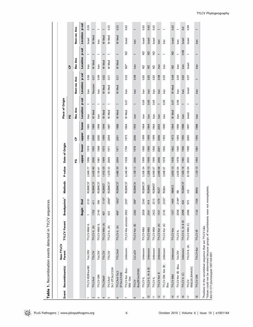
Of the 18 detected recombination events involving TYLCV
isolates, 16 were inter-species sequence exchanges (events 1 to 16
in Table 1 and Figure 1) and two were intra-species exchanges
(events 17 and 18 in Table 1 and Figure 1). Only four of the 16
inter-species recombination events involved TYLCVs as donors.
The recipient species in these four recombination events were
western Mediterranean TYLCSVs (events 2, 4 and 5 in Table 1
and Figure 1) and TYLCAxV (event 6 in Table 1) isolates. As has
been found previously, two of these events (2 and 4 in Table 1),
both involving TYLCSV as a recipient and TYLCV as a donor,
were pivotal in the creation of the TYLCAxV and TYLCMalV
species [51,52]. In fact, all three of the TYLCAxV isolates
examined (accession numbers AY227892, EU734831 and
EU734832), appear to be independently generated convergent
recombinants of TYLCSV and TYLCV-IL, highlighting the
possibility that, in the Western Mediterranean at least, such
recombinants have a high degree of fitness.
The remaining 12 inter-species recombination events involved
TYLCVs as recipients of ,1000 nucleotide fragments mostly
derived from the rep genes of either currently undescribed
begomovirus species, or species previously detected only in the
Middle East and/or India and Asia.
The fact that unique recombination events are detectable within
the rep sequences of every TYLCV isolate presents somewhat of a
problem when it comes to disentangling the evolutionary origins of
the various recombinationally derived fragments within this gene.
Specifically, without a provably non-recombinant TYLCV rep
gene in hand it is not possible to objectively judge the accuracy of
the parental sequence and recombinant designations given in
Table 1 and Figure 1. Put another way, it is possible, if not
probable, that some of the parental TYLCV sequences listed in
Table 1 are misidentified recombinant sequences and some of the
recombinant sequences are misidentified parental sequences.
In this regard, parental and recombinant sequence designations
for events 7, 9, and 11 listed in Table 1 were particularly difficult
to interpret. Evidence of these recombination events is found
within quite divergent TYLCV lineages implying that they either
(1) predate the divergence of these lineages or (2) that they are
more recent but that the recombinant fragments characterising the
events have been propagated by secondary intra-species recom-
bination between the various TYLCV lineages. For example, both
the fact that event 7 is found within the TYLCV-Ker, -Mld, -Gez,
and -Bou lineages and the evidence of it being overprinted by
subsequent recombination events such as 14 in the -Mld lineage, 8
in the –Gez lineage, and 12 in the –Ker(B) lineage, implies that it is
a reasonably old recombination event.
With events 9 and 11 on the other hand, it is plausible that a
secondary recombination event carrying a fragment baring traces of
both events has been transferred from a TYLCV-IL (A) variant into
the TYLCV-IL (C) variant (Figure 1). The young age of events 9
and 11 in some of the TYLCV-IL (A) isolates is also implied by how
closely some of these isolates resemble TYLCV-Mld (A) isolates
within the portion of their genomes upstream of the event 9 59-
breakpoint. For example, over a stretch of 1640 nucleotides the
TYLCV-Mld[ES:Alm:99] isolate, and the TYLCV-IL[ES:Alm:-
Pep:99] isolate, differ at only two nucleotide positions – implying a
very young age for the recombination event in rep that differentiates
them. However, over this 1640 nucleotide fragment these two
isolates are also much more closely related to one another than
either is to any other TYLCV-Mld or TYLCV-IL isolates. This
strongly suggests that after the original inter-species recombination
event(s) that resulted in the differentiation of TYLCV-IL from
TYLCV-Mld [7], the TYLCV-IL fragment containing traces of
events 9, 11 and 10 has, at least once, been transferred back into a
TYLCV-Mld isolate (in this case, one very closely resembling
TYLCV-Mld[ES:Alm:99]). In recombination analyses such as those
which we performed, the resulting recombinants would be virtually
indistinguishable from other TYLCV-IL isolates and no recombi-
nation would therefore be inferred.
The phylogenetic influences of such undetected cyclical
recombination events – where parental viruses are recombinants
and recombinants converge on parental viruses – are quite clearly
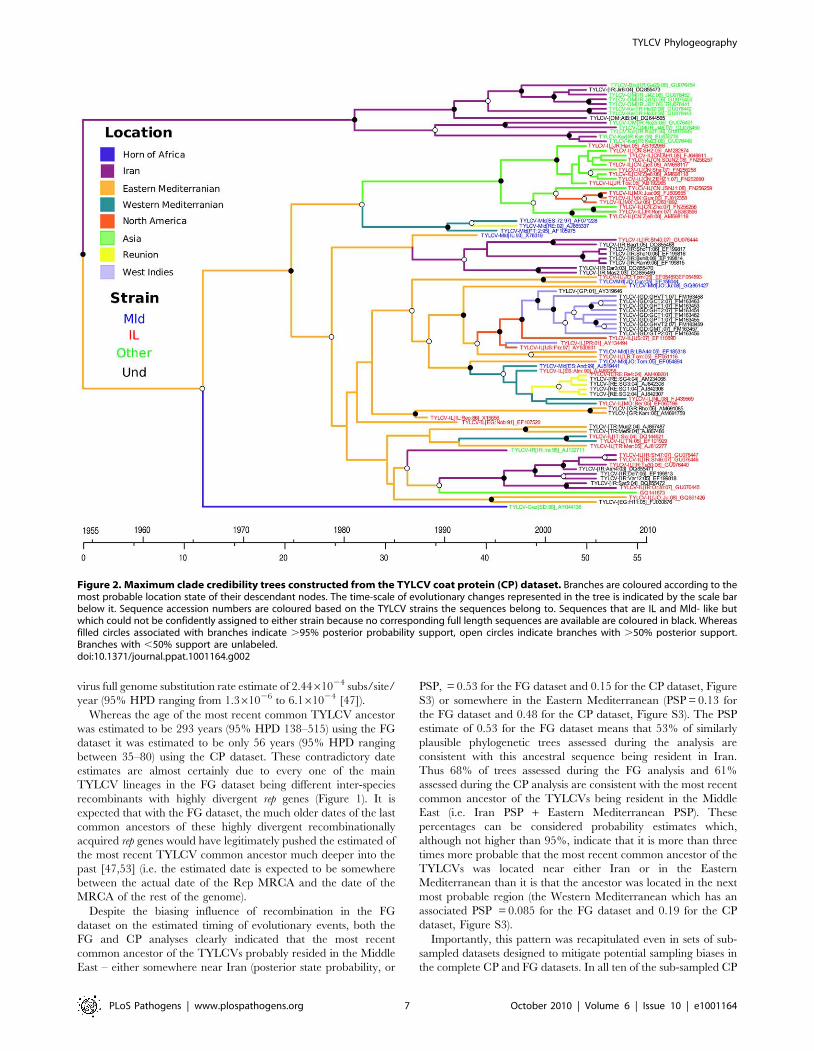
depicted in the MCC tree of TYLCV CP sequences presented in
Figure 2. In this tree where the names of IL and Mld isolates are
respectively coloured in red and blue, it is immediately obvious
that, from the perspective of their CP sequences at least, isolates
belonging to each of the strains are more closely related to isolates
of the other strain than they are to some isolates of their own
TYLCV Phylogeography
PLoS Pathogens | www.plospathogens.org 4 October 2010 | Volume 6 | Issue 10 | e1001164
strain. This makes it very difficult to phylogenetically determine
when recombination events such as those which generated
TYLCV-IL from TYLCV-Mld occurred.
TYLCV probably originated in the Middle East during thefirst half of the 20th century
Given that recombination is known to confound molecular clock
analyses [48,49] we assembled a mostly recombination-free
TYLCV coat protein gene dataset (called the CP dataset). We
analysed both this and the full genome (FG) TYLCV datasets with
BEAST to determine the time and place where TYLCV originated.
While the FG analysis indicated that the mean substitution rates
during TYLCV evolution was 4.561024 subs/site/year (95% HPD
ranging from (2.461024 to 6.861024), the CP analysis indicated a
rate of 7.961024 subs/site/year (95% HPD ranging from
4.961024 to 1.161023). These substitution rate estimates are
consistent with the previously published tomato infecting begomo-
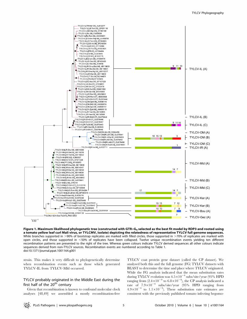
Figure 1. Maximum likelihood phylogenetic tree (constructed with GTR+G4 selected as the best fit model by RDP3 and rooted usinga tomato yellow leaf curl Mali virus, or TYLCMV, isolate) depicting the relatedness of representative TYLCV full genome sequences.While branches supported in .90% of bootstrap replicates are marked with filled circles, those supported in .70% of replicates are marked withopen circles, and those supported in ,50% of replicates have been collapsed. Twelve unique recombination events yielding ten differentrecombination patterns are presented to the right of the tree. Whereas green colours indicate TYLCV derived sequences all other colours indicatesequences derived from non-TYLCV sources. Recombination events are numbered according to Table 1.doi:10.1371/journal.ppat.1001164.g001
TYLCV Phylogeography
PLoS Pathogens | www.plospathogens.org 5 October 2010 | Volume 6 | Issue 10 | e1001164
Ta
ble
1.
Re
com
bin
atio
ne
ven
tsd
ete
cte
din
TY
LCV
seq
ue
nce
s.
Ev
en
tR
eco
mb
ina
nt(
s)N
on
-TY
LC
VP
are
nt
TY
LC
VP
are
nt
Bre
ak
po
ints
1M
eth
od
sP
-va
lue
Da
teo
fO
rig
inP
lace
of
Ori
gin
FG
CP
FG
CP
Re
cA
nc
No
n-r
ec
An
cR
ec
An
cN
on
-re
cA
nc
Be
gin
En
du
pp
er
low
er
up
pe
rlo
we
rL
oca
tio
np
-va
lL
oca
tio
np
-va
lL
oca
tio
np
-va
lL
oca
tio
np
-va
l
1T
YLC
V-I
R[I
R:Ir
a:9
8]
To
LCIR
VT
YLC
V-M
ld/-
IL1
21
31
RG
BM
CST
2.0
6E-
77
19
96
19
19
19
96
19
85
Iran
1Ir
an0
.96
Iran
1Is
rae
l0
.59
2T
YLC
AxV
TY
LCSV
TY
LCV
-IL
(A)
17
02
41
1R
GB
MC
ST3
.42
E-9
02
00
01
99
52
00
01
98
9W
Me
d1
Re
un
ion
0.5
1W
Me
d1
WM
ed
1
3T
YLC
V-O
MT
oLC
KV
TY
LCV
-Mld
/-IL
21
45
39
6R
GB
MC
ST5
.15
E-6
11
98
11
96
82
00
01
99
6Ir
an1
Iran
0.9
9Ir
an1
Iran
1
4T
YLC
Mal
VT
YLC
SVT
YLC
V-M
ld(A
)4
07
11
10
*R
GB
MST
1.9
1E-
59
19
99
19
75
19
99
19
94
WM
ed
1W
Me
d0
.82
WM
ed
1W
Me
d1
5T
YLC
AxV
-Si
c1-[
IT:S
ic2
/2:0
4T
YLC
SVT
YLC
V-I
L(A
)4
23
20
00
*R
GB
MC
ST1
.67
E-5
32
00
41
97
12
00
41
98
7W
Me
d1
WM
ed
0.5
1W
Me
d1
WM
ed
0.5
5
6T
YLC
AxV
-Sic
2-
[IT
:Sic
2/5
:04
]T
YLC
AxV
TY
LCV
-IL
(A)
40
6*
10
62
*R
GB
MC
ST3
.48
E-3
92
00
41
97
12
00
11
98
8W
Me
d1
WM
ed
0.5
1W
Me
d1
WM
ed
0.5
5
7T
YLC
V-K
er
-Bo
u-
Mld
-Ge
zU
nkn
ow
nT
YLC
V-l
ike
ance
sto
r2
00
22
59
7R
GB
MC
ST6
.34
E-4
41
93
81
79
41
97
31
96
4W
Me
d0
.47
Iran
0.9
2N
D*
ND
Isra
el
0.6
5
8T
YLC
V-
Bo
u[I
R:G
e2
9:0
6]
CLC
uG
VT
YLC
V-K
er
(B)
23
82
39
8*
RG
BM
CSt
1.1
0E-
15
20
06
19
78
20
06
19
93
Iran
1Ir
an0
.98
Iran
1Ir
an1
9T
YLC
V-I
LU
nkn
ow
nT
YLC
V-M
ld2
05
82
34
3R
GB
MC
ST5
.50
E-3
41
99
91
90
01
99
91
96
4Ir
an0
.69
Iran
0.8
5N
DN
DIs
rae
l0
.65
10
TY
LCV
-IL
(A&
B)
Un
kno
wn
TY
LCV
-Mld
25
41
41
4R
GB
MS
1.2
0E-
10
19
99
19
00
19
99
19
64
Iran
0.6
9Ir
an0
.85
ND
ND
Isra
el
0.6
5
11
TY
LCV
-IL
Un
kno
wn
TY
LCV
-Mld
24
22
25
13
RG
BM
ST6
.94
E-0
71
99
91
90
01
99
91
96
4Ir
an0
.69
Iran
0.8
5N
DN
DIs
rae
l0
.65
12
TY
LCV
-Ke
rB
Un
kno
wn
TY
LCV
-Ke
r(A
)1
53
11
57
9R
GST
2.0
4E-
08
20
06
20
01
20
06
20
03
Iran
1Ir
an0
.98
Iran
1Ir
an1
13
TY
LCV
-OM
-Ke
r(B
)-
Bo
uU
nkn
ow
nT
YLC
V-K
er
(A)
21
45
23
35
*R
GB
m3
.04
E-0
71
97
81
94
51
99
31
99
0Ir
an0
.99
Iran
0.9
5Ir
an1
Iran
1
14
TY
LCV
-Mld
Un
kno
wn
TY
LCV
-Ke
r1
35
71
46
9R
BM
CS
3.8
5E-
10
19
38
18
62
19
73
19
64
WM
ed
0.4
7W
Me
d0
.48
ND
ND
Isra
el
0.6
5
15
TY
LCV
-Ke
r(B
)-B
ou
To
LCK
VT
YLC
V-I
L2
05
82
14
4*
RB
2.6
3E-
04
19
78
19
45
19
93
19
90
Iran
0.9
9Ir
an0
.95
Iran
1Ir
an1
16
TY
LCV
-IL
(C)
Un
kno
wn
TY
LCV
-IL
(A&
B)
20
98
42
6R
GB
MC
ST7
.56
E-1
81
98
51
90
01
99
51
98
8Ir
an1
Iran
0.8
5Ir
an0
.98
Isra
el
0.6
17
TY
LCV
-M
ld[L
B:L
BA
44
:05
]T
YLC
V-I
L(A
)T
YLC
V-M
ld(C
)2
49
69
75
mS
8.7
2E-
04
20
05
19
90
20
05
19
97
Isra
el
1Is
rae
l0
.97
Isra
el
1Is
rae
l0
.99
18
TY
LCV
-OM
TY
LCV
-Ke
rT
YLC
V-I
R1
12
61
70
4B
MSt
1.1
2E-
10
19
93
19
81
19
95
19
88
Iran
1Ir
an1
Iran
1Ir
an1
1P
osi
tio
ns
inth
ere
com
bin
atse
qu
en
ceth
atar
eT
YLC
V-L
ike
.*V
alu
es
cou
ldn
ot
be
de
term
ine
db
eca
use
gro
up
so
fre
com
bin
ants
we
ren
ot
mo
no
ph
yle
tic.
do
i:10
.13
71
/jo
urn
al.p
pat
.10
01
16
4.t
00
1
TYLCV Phylogeography
PLoS Pathogens | www.plospathogens.org 6 October 2010 | Volume 6 | Issue 10 | e1001164
virus full genome substitution rate estimate of 2.4461024 subs/site/
year (95% HPD ranging from 1.361026 to 6.161024 [47]).
Whereas the age of the most recent common TYLCV ancestor
was estimated to be 293 years (95% HPD 138–515) using the FG
dataset it was estimated to be only 56 years (95% HPD ranging
between 35–80) using the CP dataset. These contradictory date
estimates are almost certainly due to every one of the main
TYLCV lineages in the FG dataset being different inter-species
recombinants with highly divergent rep genes (Figure 1). It is
expected that with the FG dataset, the much older dates of the last
common ancestors of these highly divergent recombinationally
acquired rep genes would have legitimately pushed the estimated of
the most recent TYLCV common ancestor much deeper into the
past [47,53] (i.e. the estimated date is expected to be somewhere
between the actual date of the Rep MRCA and the date of the
MRCA of the rest of the genome).
Despite the biasing influence of recombination in the FG
dataset on the estimated timing of evolutionary events, both the
FG and CP analyses clearly indicated that the most recent
common ancestor of the TYLCVs probably resided in the Middle
East – either somewhere near Iran (posterior state probability, or
PSP, = 0.53 for the FG dataset and 0.15 for the CP dataset, Figure
S3) or somewhere in the Eastern Mediterranean (PSP = 0.13 for
the FG dataset and 0.48 for the CP dataset, Figure S3). The PSP
estimate of 0.53 for the FG dataset means that 53% of similarly
plausible phylogenetic trees assessed during the analysis are
consistent with this ancestral sequence being resident in Iran.
Thus 68% of trees assessed during the FG analysis and 61%
assessed during the CP analysis are consistent with the most recent
common ancestor of the TYLCVs being resident in the Middle
East (i.e. Iran PSP + Eastern Mediterranean PSP). These
percentages can be considered probability estimates which,
although not higher than 95%, indicate that it is more than three
times more probable that the most recent common ancestor of the
TYLCVs was located near either Iran or in the Eastern
Mediterranean than it is that the ancestor was located in the next
most probable region (the Western Mediterranean which has an
associated PSP = 0.085 for the FG dataset and 0.19 for the CP
dataset, Figure S3).
Importantly, this pattern was recapitulated even in sets of sub-
sampled datasets designed to mitigate potential sampling biases in
the complete CP and FG datasets. In all ten of the sub-sampled CP
Figure 2. Maximum clade credibility trees constructed from the TYLCV coat protein (CP) dataset. Branches are coloured according to themost probable location state of their descendant nodes. The time-scale of evolutionary changes represented in the tree is indicated by the scale barbelow it. Sequence accession numbers are coloured based on the TYLCV strains the sequences belong to. Sequences that are IL and Mld- like butwhich could not be confidently assigned to either strain because no corresponding full length sequences are available are coloured in black. Whereasfilled circles associated with branches indicate .95% posterior probability support, open circles indicate branches with .50% posterior support.Branches with ,50% support are unlabeled.doi:10.1371/journal.ppat.1001164.g002
TYLCV Phylogeography
PLoS Pathogens | www.plospathogens.org 7 October 2010 | Volume 6 | Issue 10 | e1001164
and FG datasets the most probable location of the TYLCV
MRCA was either the region around Iran (CP and FG datasets
with respective mean PSPs = 0.26 and 0.25) or the Eastern
Mediterranean (CP and FG datasets with respective mean
PSPs = 0.4 and 0.12; Figure S3). Also, when we reran our
analyses with the full datasets in such a way that the location state
designations of all of the sequences were randomized throughout
the MCMC procedure, the maximum PSP achieved at the root
node for the most sampled location never exceeded 0.18 for the
FG dataset and 0.22 for the CP dataset – both much lower than
the maximum root node PSPs obtained without the location state
randomisation setting (which were 0.53 and 0.48 for the FG and
CP datasets respectively). Together these tests indicated that
sampling biases had not obviously influenced our identification of
the Middle East as the region where TYLCV most probably
originated.
The Mediterranean basin (and not Iran) is the source ofthe global TYLCV epidemic
To pinpoint the source of the TYLCV variants that are
spreading throughout the world, we retraced the movement
patterns of TYLCVs over the past 50 years. Figure 2 is a
phylogenetic depiction of TYLCV movement patterns (based on
the CP dataset MCC tree) in which the tree branches have been
coloured based on the most probable locations of their associated
virus lineages such that a colour change between two connected
nodes implies a probable migration event. In addition, a plausible
spatio-temporal animation of TYLCV movements since the time
of the most recent TYLCV common ancestor can be visualised by
opening in GoogleEarth (http://earth.google.com) the Dataset
S1.kml (FG dataset) and Dataset S2.kml (CP dataset). Figure 3
summarises the results presented in these files. It is important to
stress that in these analyses, we only considered the nine and seven
discreet locations respectively studied in the CP and FG datasets.
It must therefore be borne in mind that the locations indicated for
ancestral viruses and the movement patterns inferred from these
are simply the most plausible given the studied sampling locations
– i.e. that actual locations of ancestral sequences and movement
pathways may have included locations outside those that we have
considered.
Among the locations that we have considered, the FG and CP
datasets respectively indicate that the global dispersal of TYLCV
has involved at least 15 and 17 discrete migration events. As these
viral movements (or geographical location state transitions) were
inferred from node states of the FG and CP MCC trees, we only
summarise the realisations of a potentially rich history of location
state transitioning. The reason for this is that the geographical
location states mapped to the various tree nodes reflect the starting
and ending points of various movements – they do not recapture
the potentially long and winding routes taken during these
journeys.
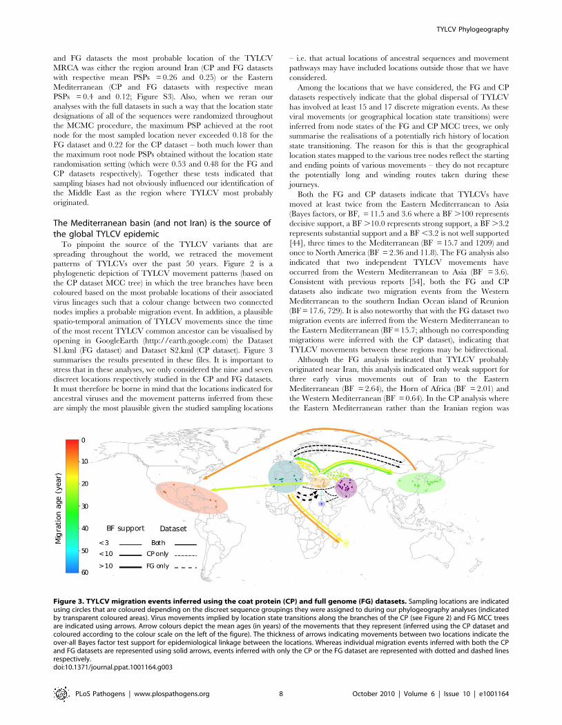
Both the FG and CP datasets indicate that TYLCVs have
moved at least twice from the Eastern Mediterranean to Asia
(Bayes factors, or BF, = 11.5 and 3.6 where a BF .100 represents
decisive support, a BF .10.0 represents strong support, a BF .3.2
represents substantial support and a BF ,3.2 is not well supported
[44], three times to the Mediterranean (BF = 15.7 and 1209) and
once to North America (BF = 2.36 and 11.8). The FG analysis also
indicated that two independent TYLCV movements have
occurred from the Western Mediterranean to Asia (BF = 3.6).
Consistent with previous reports [54], both the FG and CP
datasets also indicate two migration events from the Western
Mediterranean to the southern Indian Ocean island of Reunion
(BF = 17.6, 729). It is also noteworthy that with the FG dataset two
migration events are inferred from the Western Mediterranean to
the Eastern Mediterranean (BF = 15.7; although no corresponding
migrations were inferred with the CP dataset), indicating that
TYLCV movements between these regions may be bidirectional.
Although the FG analysis indicated that TYLCV probably
originated near Iran, this analysis indicated only weak support for
three early virus movements out of Iran to the Eastern
Mediterranean (BF = 2.64), the Horn of Africa (BF = 2.01) and
the Western Mediterranean (BF = 0.64). In the CP analysis where
the Eastern Mediterranean rather than the Iranian region was
Figure 3. TYLCV migration events inferred using the coat protein (CP) and full genome (FG) datasets. Sampling locations are indicatedusing circles that are coloured depending on the discreet sequence groupings they were assigned to during our phylogeography analyses (indicatedby transparent coloured areas). Virus movements implied by location state transitions along the branches of the CP (see Figure 2) and FG MCC treesare indicated using arrows. Arrow colours depict the mean ages (in years) of the movements that they represent (inferred using the CP dataset andcoloured according to the colour scale on the left of the figure). The thickness of arrows indicating movements between two locations indicate theover-all Bayes factor test support for epidemiological linkage between the locations. Whereas individual migration events inferred with both the CPand FG datasets are represented using solid arrows, events inferred with only the CP or the FG dataset are represented with dotted and dashed linesrespectively.doi:10.1371/journal.ppat.1001164.g003
TYLCV Phylogeography
PLoS Pathogens | www.plospathogens.org 8 October 2010 | Volume 6 | Issue 10 | e1001164
identified as the probable origin of TYLCV, three independent,
decisively supported (BF = 265), migration events from the
Eastern Mediterranean to Iran were inferred, possibly explaining
the broad degree of TYLCV diversity found in the latter region.
Finally, our analysis supports the hypotheses that TYLCV-IL has
been independently introduced to the New World, once from the
region around the Eastern Mediterranean (BF = 2.36 and 11.8 for
the FG and CP datasets respectively) and once from Asia (BF = 13.6
and 45.7 for the FG and CP datasets respectively; [16]).
Collectively these data indicate that although the region around
Iran is a center of TYLCV diversity and is possibly also the region
where the species originated, it has not been the direct source of
the TYLCV variants that are currently spreading worldwide. This
means that novel pathogenic TYLCV variants that arise in this
region will probably be less of a threat to global agriculture than
those arising closer to more internationally connected regions such
as the Mediterranean basin.
The geographical and temporal origins of TYLCVrecombinants
Our preliminary recombination analysis indicated that all of the
detectable recombinants that discernibly contained TYLCV-like
sequences had been sampled in the Mediterranean basin and the
Middle East. We suspected that within this region there might be
geographical recombination hotspots. By mapping the 18 detected
TYLCV recombination events to the FG and CP MCC trees
determined during our phylogeography analysis, it was possible for
us to approximate the locations where and the times when the
recombination events most likely occurred. For each recombina-
tion event in each tree this involved identification of the nodes
representing (1) the last common ancestor of the recombinants
(referred to as RecAnc in Table 1) and (2) the last TYLCV
ancestor not sharing evidence of the same recombination event
(referred to as Non-RecAnc in Table 1). The dates and locations of
the sequences at these two nodes in the MCC trees were assumed
to bound the date when, and the location where, the recombi-
nation event occurred.
Whereas it was possible to use this approach to infer dates and
locations for all 18 of the recombination events with the FG
dataset, groups of recombinant TYLCV-IL and –Mld sequences
sharing evidence of events 7, 9, 10, 11 and 14 were not
monophyletic in the CP tree (probably for reasons explained
above in the recombination analysis section; Figure 2), meaning
that locations and dates could not be properly inferred for these
recombination events using the CP dataset. Despite this, the CP
dataset yielded much tighter estimates of recombination dates than
the FG dataset, possibly due to its being free of the confounding
effects of the inter-species recombination events found in the latter.
The FG and CP datasets nevertheless indicated locations where
recombination events had occurred that were generally in good
agreement with one another (compare orange and blue bars in
Figure S4) and recombination date estimates that had broadly
overlapping 95% HPDs (compare orange and blue bars in Figure
S5). The exceptions were the five ‘‘problematic’’ events (7, 9, 10,
11 and 14) mentioned previously. For these the FG and CP
datasets yielded support for recombination events having occurred
in different locations. For example with events 9, 10 and 11 the FG
dataset indicated that the RecAnc and Non-RecAnc sequences
most probably resided near Iran, the CP dataset indicated that the
Non-RecAnc sequence most probably resided near the Eastern
Mediterranean (with the location of the RecAnc sequence
remaining undetermined for the CP dataset).
Nevertheless, the clear pattern emerging from these analyses
was that all 18 of the detected TYLCV recombination events
occurred either in the Western Mediterranean, the Eastern
Mediterranean or near Iran. Collectively these geographical
locations (representing 58% of the sequence) accounted for more
than 80% of the posterior probability distribution for every
ancestral sequence used to infer the locations of every recombi-
nation event. Based on dates inferred from the CP MCC tree,
these recombination events were also mostly all quite recent with
the oldest (events 7 and 14) having most probably occurred some
time after 1964 (Table 1 and Figure S5). If one discounts the
‘‘problematic’’ recombination events 7, 9, 10, 11 and 14, the
remaining thirteen events have all most probably occurred since
1985.
Nine of these thirteen events most probably occurred near Iran
or Israel with both the FG and CP analyses indicating that Iran
was the most probable site of eight of them (supported for all
events other than events 1 and 16 by the location state
probabilities of all the relevant RecAnc and Non-RecAnc
sequences in both the FG and CP datasets). Besides being the
most probable origin of TYLCV and the center of TYLCV
diversity, the Middle East in general, and Iran in particular, is
therefore also apparently the region where most of this virus’
evolutionary change through recombination has occurred.
In this regard it is interesting that recombination events 2, 4, 5
and 6, the only events that almost certainly occurred outside the
Middle East, are also the only four involving TYLCV sequences as
donors (i.e. such that a minority of the recombinant’s genome
consists of TYLCV-like sequences). Although this difference
between the character of TYLCV recombination events occurring
inside and outside the Middle East may be coincidental, it could
also be indicative of an important evolutionary trend associated
with the migration of viruses into environments different from
those in which they evolved.
The observed pattern is in fact what one might expect to occur
with recombining invasive virus species. For example, it is
expected that viruses residing in the locations where they evolved
would be well adapted to seasonal changes in the mix of host and
vector genotypes that typify their home environments. One might
expect both that these adaptations would provide them with a
‘‘home environment advantage’’ over invasive newcomers and
that the genetic underpinnings of these adaptations would be
distributed throughout their genomes. The invasive newcomers,
however, would not be invasive unless they had some specific,
especially adaptive genetic trait that provided them with their
invasive phenotype. When such indigenous and invasive viruses
recombine, the fittest of their offspring would probably be those
that incorporate the invader’s highly adaptive traits within an
indigenous genetic background. Unless TYLCV and TYLCSV
only replicate within genetically homogeneous cultivated tomato
species and are epidemiologically unaffected by local variations in
host species distributions across the Mediterranean and Middle
East, it is conceivable that both have an advantage in their
respective home environments. The net result may be that in the
Middle East when TYLCVs recombine with viruses originating in
India or Africa the TYLCVs are the better acceptors whereas in
the Western Mediterranean they are better as donors to
indigenous viruses like TYLCSV.
A plausible history of TYLCVTo retrace the global movements of TYLCV we considered
phylogeographic inferences made with both the FG and CP
datasets. However, given that the estimated calendar dates of
movement events differed between the FG and CP analyses and
the probable impact that inter-species recombination has had on
evolution rates estimated with the FG dataset, we used results
TYLCV Phylogeography
PLoS Pathogens | www.plospathogens.org 9 October 2010 | Volume 6 | Issue 10 | e1001164
obtained with the mostly recombination-free CP dataset to
estimate the timing of key events during the evolution and
dissemination of TYLCV (summarised in Figure 3). It is important
to reiterate here that both the age, location and migration route
estimates that follow are associated with degrees of uncertainty
and that the descriptive history we provide is simply the most
plausible given an admittedly sparse TYLCV sequence dataset
and imperfect analytical tools. Nevertheless, although the Bayesian
analyses underlying the description do not account for important
factors such as natural selection, they do provide us with 95%
HPD intervals that are an honest expression of the uncertainty
surrounding the various date, location and migration route
estimates that we infer.
At some point between 1937 and 1952 (95% HPD 1905–1972),
a virus arose somewhere within the Middle East which was the first
recognisable TYLCV. By ,1952 this ‘‘first’’ TYLCV lineage had
evolved into the most recent common ancestor of all known
contemporary TYLCVs. It is possible, although in no way certain,
that this virus was a recombinant that had inherited the majority
of its genome from an earlier TYLCV prototype but a large
portion of its rep gene and its origin of virion strand replication
from some other unknown (but possibly Asian) begomovirus
species (see event 7 in Figure 1, Table 1 and Figures S4 and S5). It
is also plausible that the immediate descendants of this virus were
responsible for the Middle Eastern TYLCD epidemics of the early
1960s [4,5].
There is good evidence from our analysis that during the 1960s
these viruses evolved within the Middle East to yield prototypical
versions of the TYLCV-Gez strain in the Eastern Mediterranean
(PSP = 0.65), by ,1964 (95% HPDs 1948–1978), the TYLCV-
Mld strain also in the Eastern Mediterranean (PSP = 0.90) by
,1973 (95% HPDs 1963–1982) and the TYLCV-Ker strain in the
region of Iran (PSP = 0.97) by ,1979 (95% HPDs 1964–1992).
Later, between 1993 and 2006 (95% HPD 1986–2006) and also
probably in Iran (PSP = 0.98–1.00), a recombination event
between a TYLCV-Ker variant and CLCuGV (event 8 in
Table 1 and Figure 1) created the first member of TYLCV-Bou,
the most recently evolved of the seven currently described TYLCV
strains.
Although both inter- and intra-species recombination events
involving early TYLCV variants probably persistently occurred
within the broader Middle East during these years, the first of
these that would come to largely differentiate the seven current
TYLCV strains probably occurred somewhere in this region
(PSP = 0.80–0.76) between 1964 and the mid 1970s (95% HPD
1948–1999). This event (or possibly a series of events), traces of
which are possibly evident in events 9, 10 and 11 in Table 1 and
Figure 1, yielded the founder of the IL strain. Similar
recombination events between either TYLCV-Mld or -IL (it is
unclear which) and TolCRV somewhere in the Middle east
(PSP = 0.97–1.0) between 1985 and 1996 (95% HPDs 1978–
1996; event 1 in Table 1 and Figure 1) and between TYLCV-Mld
or -IL and ToLCKV near Iran (PSP = 0.99–1.0) between 1996
and 2000 (95% HPDs 1991–2003; event 3 in Table 1 and Figure 1)
would respectively yield the first members of what are currently
known as the TYLCV-IR and -OM strains.
At some point between 1981 and 1989 (95% HPDs 1971–1993)
the world-wide dissemination of TYLCV began when a TYLCV-
IL virus (most likely from the Eastern Mediterranean), moved to,
and became established within, the Western Mediterranean. This
trip was later repeated at least once by a TYLCV-Mld virus
between 1990 and 2001 (95% HPDs 1982–2003). Although the
polarity of the movement is uncertain (the FG and CP datasets
conflict on this point), additional movements of IL viruses between
the Middle East and the Western Mediterranean also occurred
during this period. Viruses within the newly established Western
Mediterranean TYLCV-Mld and -IL populations were then
transported to Asia between 1989 and 1996 (95% HPDs 1983–
1996) and the Indian Ocean island of Reunion between 1991 and
2002 (95% HPDs 1987–2003).
At least two other long distance movements of IL viruses to Asia
also occurred from the Middle East between 1981 and 1999 (95%
HPD 1970–1999). Whereas the trans-Atlantic movement of a
Middle Eastern TYLCV-IL virus to the New World probably
happened between 1992 and 1994 (95% HPD 1988–1994) –
within two years of the first TYLCVs being sampled there [15] –
the trans-Pacific transport of an Asian TYLCV-IL virus (a
descendant of one of the lineages introduced from the Middle
East) to North America probably only occurred between 1999 and
2003 (95% HPD 1996–2004).
Concluding remarksWe have described how within thirty years of their Middle
Eastern origin, both TYLCV-Mld and the TYLCV-IL lineage
have ascended to the point where they are today ranked among
the greatest biotic threats to tomato production world-wide [55].
This emergence has been so swift that no precise estimates of
either their current or projected future economic impacts exist.
The epidemiological, evolutionary and ecological impacts of their
movements are probably even harder to predict although in this
regard patterns seen in the Western Mediterranean where they
have spent their greatest time outside the Middle East will possibly
prove informative [56–58]. For example, given the high
frequencies of inter-species TYLCV recombination events that
we have mapped to the Middle East, it is perhaps reasonable to
expect that, as has happened in the Western Mediterranean
[7,22,51], TYLCVs introduced to the Americas, the southern
Indian Ocean, and Asia will recombine with viruses indigenous to
these regions. While it is impossible to predict how evolutionarily
productive any such recombination events will be, the possibility
remains that TYLCV genetic material within the context of mostly
indigenous recombinant begomovirus genomes could shortly begin
showing up in Asia, the Indian Ocean islands and the Americas.
We envision that tracking the movements of the various TYLCV
invasion fronts and monitoring virus sequence data before the
fronts hit and in the years thereafter could prove very fruitful in
our endeavours to answer some key questions relating to the
economic, epidemiological, ecological and evolutionary impacts of
such plant virus invasions.
Supporting Information
Figure S1 Maximum likelihood phylogenetic tree (with GTR +G4 selected as the best fit model by RDP3) and pair-wise sequence
similarity matrix of 75 virus isolates representing the seven
different TYLCV strains (denoted by different colours on the tree
branches). The phylogeny is rooted using TYLCMLV. The
colours in the matrix represent the pair-wise similarities indicated
on the colour scale. Similarity scores beneath the accepted
begomovirus species demarcation cut-off, 89% are in a yellow
scale, scores in the strain range, between 89% to 93% are in a light
blue scale and scores in the variant range between 93% and 100%
are in a blue scale.
Found at: doi:10.1371/journal.ppat.1001164.s001 (1.93 MB TIF)
Figure S2 Sampling locations of Iranian TYLCV isolates. Small
circles at sample sites are coloured depending on whether (green)
or not (black) TYLCVs were cloned from samples collected at the
sites. Sites where TYLCVs were sampled in other studies are given
TYLCV Phylogeography
PLoS Pathogens | www.plospathogens.org 10 October 2010 | Volume 6 | Issue 10 | e1001164
in blue. Coloured areas represent the known geographical
distributions within Iran of the different TYLCV strains.
Found at: doi:10.1371/journal.ppat.1001164.s002 (0.59 MB TIF)
Figure S3 The posterior probability distribution indicating the
most probable geographical locations of the last common TYLCV
ancestor. Bars indicate Bayesian posterior probabilities that the
last common TYLCV ancestor resided in the various sampling
locations. Blue bars represent inferences of ancestral sequence
locations made using the full genome (FG) dataset and orange bars
represent those made using the coat protein (CP) dataset.
Found at: doi:10.1371/journal.ppat.1001164.s003 (0.11 MB TIF)
Figure S4 The approximate geographical origins of TYLCV
recombinants. Bars indicate Bayesian posterior probabilities that
sequences closely related to the ancestral recombinant sequence
(the last non-recombinant most recent common ancestor or the
recombinants and the most recent common recombinant ancestor
of the recombinant(s)) resided in the various sampling locations.
Blue bars represent inferences of ancestral sequence locations
made using the full genome dataset and orange bars represent
those made using the coat protein (CP) dataset. Whereas the
darker bars indicate the probability that the last non-recombinant
ancestor of the recombinant sequences was situated in the
specified locations, the lighter bars indicate the probability that
the last common ancestor of all sampled recombinants was located
in the regions. In cases where only one recombinant has been
sampled the probability associated with the location where the
recombinant was sampled is 1. Wherever it was not possible to
directly infer the location of the last non-recombinant ancestor (see
M&M for details on how locations were estimated) estimates are
marked with an asterisk.
Found at: doi:10.1371/journal.ppat.1001164.s004 (0.68 MB TIF)
Figure S5 Dating of TYLCV recombination events. The upper
and lower bounds of the coloured bars respectively indicate the
most probable range of dates when the various recombination
events might have occurred. The thinner error bars indicate the
upper and lower 95% HPD intervals of the date estimates. Refer
to the M&M to see how the dates were calculated.
Found at: doi:10.1371/journal.ppat.1001164.s005 (1.04 MB TIF)
Table S1 Dataset details.
Found at: doi:10.1371/journal.ppat.1001164.s006 (0.07 MB PDF)
Dataset S1 Google earth file with an animation of TYLCV
migration inferred using the full genome dataset.
Found at: doi:10.1371/journal.ppat.1001164.s007 (0.07 MB ZIP)
Dataset S2 Google earth file with an animation of TYLCV
migration inferred using the CP dataset.
Found at: doi:10.1371/journal.ppat.1001164.s008 (0.06 MB ZIP)
Author Contributions
Conceived and designed the experiments: P. Lefeuvre, D. Martin, A.
Varsani, J. Heydarnejad. Performed the experiments: A. Gray, S.
Meredith, F. Lakay, A. Varsani, J. Heydarnejad. Analyzed the data: P.
Lefeuvre, D. Martin, G. Harkins, P. Lemey, A. Gray, A. Monjane, J. Lett,
A. Varsani. Contributed reagents/materials/analysis tools: A. Varsani, J.
Heydarnejad. Wrote the paper: P. Lefeuvre, D. Martin, G. Harkins, P.
Lemey, A. Varsani, J. Heydarnejad.
References
1. Abhary M, Patil B, Fauquet C (2007) Molecular biodiversity, taxonomy, and
nomenclature of tomato yellow leaf curl-like viruses. In: Czosnek H, ed. Tomato
yellow leaf curl virus disease. pp 85–118.
2. Dıaz-Pendon JA, Nizares MC, Moriones E, Bejerano ER, Czosnek H, et al.
(2010) Tomato yellow leaf curl viruses: menage a trois between the virus
complex, the plant and the whitefly vector. Mol Plant Pathol 11: 441–450.
3. Fauquet CM, Briddon RW, Brown JK, Moriones E, Stanley J, et al. (2008)
Geminivirus strain demarcation and nomenclature. Arch Virol 153: 783–
821.
4. Pico B, Diez M, Nuez F (1996) Viral diseases causing the greatest economic
losses to the tomato crop. 2. The Tomato yellow leaf curl virus - A review. Sci
Horti 67: 151–196.
5. Antignus Y, Cohen S (1994) Complete nucleotide-sequence of an infectious
clone of a mild isolate of Tomato Yellow Leaf Curl Virus (TYLCV).
Phytopathology 84: 707–712.
6. Navot N, Pichersky E, Zeidan M, Zamir D, Czosnek H (1991) Tomato yellow
leaf curl virus: a whitefly-transmitted geminivirus with a single genomic
component. Virology 185: 151–161.
7. Navas-Castillo J, Sanchez-Campos S, Noris E, Louro D, Accotto GP, et al.
(2000) Natural recombination between Tomato yellow leaf curl virus-is and
Tomato leaf curl virus. J Gen Virol 81: 2797–2801.
8. Idris AM, Brown JK (2005) Evidence for interspecific-recombination for three
monopartite begomoviral genomes associated with the tomato leaf curl disease
from central Sudan. Arch Virol 150: 1003–1012.
9. Bananej K, Kheyr-Pour A, Salekdeh GH, Ahoonmanesh A (2004) Complete
nucleotide sequence of Iranian tomato yellow leaf curl virus isolate: further
evidence for natural recombination amongst begomoviruses. Arch Virol 149:
1435–1443.
10. Khan AJ, Idris AM, Al-Saady NA, Al-Mahruki MS, Al-Subhi AM, et al. (2008)
A divergent isolate of tomato yellow leaf curl virus from Oman with an
associated DNA beta satellite: an evolutionary link between Asian and the
Middle Eastern virus-satellite complexes. Virus Genes 36: 169–176.
11. Sugiyama K, Matsuno K, Doi M, Tatara A, Kato M, et al. (2008) TYLCV
detection in Bemisia tabaci (Gennadius) (Hemiptera: Aleyrodidae) B and Q
biotypes, and leaf curl symptom of tomato and other crops in winter greenhouses
in Shizuoka Pref., Japan. Appl Entomol Zool 43: 593–598.
12. Navas-Castillo J, Sanchez-Campos S, Diaz J, Saez-Alonso E, Moriones E (1999)
Tomato yellow leaf curl virus-Is causes a novel disease of common bean and
severe epidemics in tomato in Spain. Plant Disease 83: 29–32.
13. Peterschmitt M, Granier M, Mekdoud R, Dalmon A, Gambin O, et al. (1999)
First report of tomato yellow leaf curl virus in Reunion Island. Plant disease 83:
303–303.
14. Stonor J, Hart P, Gunther M, DeBarro P, Rezaian M (2003) Tomato leaf curl
geminivirus in Australia: occurrence, detection, sequence diversity and host
range. Plant Pathol 52: 379–388.
15. McGlashan D, Polston J, Bois D (1994) Tomato Yellow Leaf Curl Geminivirus
in Jamaica. Plant Disease 78: 1219–1219.
16. Duffy S, Holmes EC (2007) Multiple introductions of the Old World
begomovirus Tomato yellow leaf curl virus into the New World. Appl EnvironMicrobiol 73: 7114–7117.
17. Zambrano K, Carballo O, Geraud F, Chirinos D, Fernandez C, et al. (2007)
First report of Tomato yellow leaf curl virus in Venezuela. Plant Disease 91:768–768.
18. Polston J, McGovern R, Brown L (1999) Introduction of tomato yellow leaf curl
virus in Florida and implications for the spread of this and other geminiviruses oftomato. Plant Disease 83: 984–988.
19. Czosnek H, Laterrot H (1997) A worldwide survey of tomato yellow leaf curl
viruses. Arch Virol 142: 1391–1406.
20. Fauquet CM, Sawyer S, Idris AM, Brown JK (2005) Sequence analysis andclassification of apparent recombinant begomoviruses infecting tomato in the
nile and mediterranean basins. Phytopathology 95: 549–555.
21. Fazeli R, Heydarnejad J, Massumi H, Shaabanian M, Varsani A (2009) Geneticdiversity and distribution of tomato-infecting begomoviruses in Iran. Virus
Genes 38: 311–319.
22. Davino S, Napoli C, Dellacroce C, Miozzi L, Noris E, et al. (2009) Two newnatural begomovirus recombinants associated with the tomato yellow leaf curl
disease co-exist with parental viruses in tomato epidemics in Italy. Virus Res
143: 15–23.
23. Crescenzi A, Comes S, Napoli C, Fanigliulo A, Pacella R, et al. (2004) Severeoutbreaks of tomato yellow leaf curl Sardinia virus in Calabria, Southern Italy.
Commun Agric Appl Biol Sci 69: 575–580.
24. Garcıa-Andres S, Accotto GP, Navas-Castillo J, Moriones E (2007) Foundereffect, plant host, and recombination shape the emergent population of
begomoviruses that cause the tomato yellow leaf curl disease in theMediterranean basin. Virology 359: 302–312.
25. Bananej K, Vahdat A, Hosseini-Salekdeh G (2009) Begomoviruses Associated
with Yellow Leaf Curl Disease of Tomato in Iran. J Phytopathol 157: 243–247.
26. Bananej K, Ahoonmanesh A, Shahraeen N (1998) Occurrence and identifica-tion of Tomato yellow leaf curl virus from Khorasan province of Iran. Proc the
13th Iranian Plant Protec Karaj, Iran. 193 p.
27. Shahriary D, Bananej K (1998) Occurrence of Tomato yellow leaf curl virus(TYLCV) in tomato fields of Varamin. J Appl Entomol Phytopathol. pp 29–30.
28. Behjatnia SAA, Izadpanah KA, Dry I, Rezaian MA (2003) Molecular
characterization and taxonomic position of the Iranian isolate of Tomato leaf
curl virus. Iran J Plan Pathol 40: 77–94.
TYLCV Phylogeography
PLoS Pathogens | www.plospathogens.org 11 October 2010 | Volume 6 | Issue 10 | e1001164
29. Hajimorad M, KheyrPour A, Alavi V, Ahoonmanesh A, Bahar M, et al. (1996)
Identification of whitefly transmitted tomato yellow leaf curl geminivirus fromIran and a survey of its distribution with molecular probes. Plant Pathol 45:
418–425.
30. Rojas MR, Gilbertson RL, Russell DR, Maxwell DP (1993) Use of degenerateprimers in the polymerase chain reaction to detect whitefly-transmitted
geminiviruses. Plant disease 77: 340–347.31. Briddon RW, Bull SE, Mansoor S, Amin I, Markham PG (2002) Universal
primers for the PCR-mediated amplification of DNA beta: a molecule associated
with some monopartite begomoviruses. Mol Biotechnol 20: 315–318.32. Shepherd DN, Martin DP, Lefeuvre P, Monjane AL, Owor BE, et al. (2008) A
protocol for the rapid isolation of full geminivirus genomes from dried planttissue. J Virol Methods 149: 97–102.
33. Owor BE, Shepherd DN, Taylor NJ, Edema R, Monjane AL, et al. (2007)Successful application of FTA Classic Card technology and use of bacteriophage
phi29 DNA polymerase for large-scale field sampling and cloning of complete
maize streak virus genomes. J Virol Methods 140: 100–105.34. Tamura K, Dudley J, Nei M, Kumar S (2007) MEGA4: Molecular Evolutionary
Genetics Analysis (MEGA) software version 4.0. Mol Biol Evol 24: 1596–1599.35. Grasso C, Lee C (2004) Combining partial order alignment and progressive
multiple sequence alignment increases alignment speed and scalability to very
large alignment problems. Bioinformatics 20: 1546–1556.36. Guindon S, Gascuel O (2003) A simple, fast, and accurate algorithm to estimate
large phylogenies by maximum likelihood. Syst Biol 52: 696–704.37. Martin DP, Williamson C, Posada D (2005) RDP2: recombination detection and
analysis from sequence alignments. Bioinformatics 21: 260–262.38. Martin D, Rybicki E (2000) RDP: detection of recombination amongst aligned
sequences. Bioinformatics 16: 562–563.
39. Padidam M, Sawyer S, Fauquet CM (1999) Possible emergence of newgeminiviruses by frequent recombination. Virology 265: 218–225.
40. Martin DP, Posada D, Crandall K, Williamson C (2005) A modified bootscanalgorithm for automated identification of recombinant sequences and recom-
bination breakpoints. AIDS Res Hum Retro 21: 98–102.
41. Smith J (1992) Analysing the mosaic structure of genes. J Mol Evol 34: 126–129.42. Gibbs M, Armstrong J, Gibbs A (2000) Sister-Scanning: a Monte Carlo
procedure for assessing signals in recombinant sequences. Bioinformatics 16:573–582.
43. Boni MF, Posada D, Feldman MW (2007) An exact nonparametric method forinferring mosaic structure in sequence triplets. Genetics 176: 1035–1047.
44. Lemey P, Rambaut A, Drummond AJ, Suchard MA (2009) Bayesian
phylogeography finds its roots. PLoS Comput Biol 5: e1000520.
45. Kass R, Raftery A (1995) Bayes factors. J Am Stat Assoc 90: 773–795.
46. Suchard MA, Weiss RE, Sinsheimer JS (2001) Bayesian selection of continuous-
time Markov chain evolutionary models. Mol Biol Evol 18: 1001–1013.
47. Duffy S, Holmes EC (2008) Phylogenetic evidence for rapid rates of molecular
evolution in the single-stranded DNA begomovirus tomato yellow leaf curl virus.
J Virol 82: 957–965.
48. Schierup MH, Hein J (2000) Recombination and the molecular clock. Mol Biol
Evol 17: 1578–1579.
49. Posada D (2001) Unveiling the molecular clock in the presence of
recombination. Mol Biol Evol 18: 1976–1978.
50. Team RDC (2008) R: A Language and Environment for Statistical Computing.
Vienna, Austria: Available at: http://www.R-project.org.
51. Monci F, Sanchez-Campos S, Navas-Castillo J, Moriones E (2002) A natural
recombinant between the geminiviruses Tomato yellow leaf curl Sardinia virus
and Tomato yellow leaf curl virus exhibits a novel pathogenic phenotype and is
becoming prevalent in Spanish populations. Virology 303: 317–326.
52. Garcıa-Andres S, Monci F, Navas-Castillo J, Moriones E (2006) Begomovirus
genetic diversity in the native plant reservoir Solanum nigrum: Evidence for the
presence of a new virus species of recombinant nature. Virology 350: 433–442.
53. Awadalla P (2003) The evolutionary genomics of pathogen recombination. Nat
Rev Genet. Jan 4(1): 50–60.
54. Delatte H, Holota H, Naze F, Peterschmitt M, Reynaud B, et al. (2005) The
presence of both recombinant and nonrecombinant strains of Tomato yellow
leaf curl virus on tomato in Reunion Island. Plant Pathol 54: 262–262.
55. Hanssen IM, Lapidot M, Thomma BPHJ (2010) Emerging viral diseases of
tomato crops. Mol Plant Microbe Interact 23: 539–548.
56. Sanchez-Campos S, Dıaz JA, Monci F, Bejarano ER, Reina J, et al. (2002) High
Genetic Stability of the Begomovirus Tomato yellow leaf curl Sardinia virus in
Southern Spain Over an 8-Year Period. Phytopathology 92: 842–849.
57. Moriones E, Navas-Castillo J (2008) Rapid evolution of the population of
begomoviruses associated with the tomato yellow leaf curl disease after invasion
of a new ecological niche. Span J Agric Res 6: 147–159.
58. Sanchez-Campos S, Navas-Castillo J, Camero R, Soria C, Diaz J, et al. (1999)
Displacement of tomato yellow leaf curl virus (TYLCV)-Sr by TYLCV-Is in
tomato epidemics in spain. Phytopathology 89: 1038–1043.
TYLCV Phylogeography
PLoS Pathogens | www.plospathogens.org 12 October 2010 | Volume 6 | Issue 10 | e1001164


