Embed Size (px)
Citation preview
Immunity, Vol. 18, 665–674, May, 2003, Copyright 2003 by Cell Press
Competing Functions Encodedin the Allergy-Associated Fc�RI� Gene
on human Langerhans cells, dendritic cells, and mono-cytes (Bieber et al., 1992; Maurer et al., 1996, 1994;Wang et al., 1992). A correlation thus exists between
Emmanuel Donnadieu,1,3 Marie-Helene Jouvin,1
Shalini Rana,1 Miriam F. Moffatt,2
Ester H. Mockford,2 William O. Cookson,2
the tetrameric isoform and allergy mediator release, asand Jean-Pierre Kinet1,*well as between the trimeric isoform and antigen presen-1Department of Pathologytation. Whether this correlation is due to a differenceBeth Israel Deaconess Medical Center andin expression and signaling capacity between the twoHarvard Medical Schoolisoforms has not been investigated.Boston, Massachusetts 02215
The � subunit of the Fc�RI complex is a type I mem-2 Asthma Genetics Groupbrane protein comprised of two extracellular immuno-University of Oxfordglobulin (Ig)-like domains and a short intracellular tail.Wellcome Trust Centre for Human GeneticsThe � chain is heavily N-glycosylated. ApproximatelyRoosevelt Drive50% of the mature � chain molecular weight is derivedOxford OX3 7BNfrom glycosylation. This chain is responsible for bindingUnited KingdomIgE (Wurzburg and Jardetzky, 2002). The � chain spansthe membrane four times such that the amino and car-boxy termini are intracellular (Holowka and Metzger,Summary1982). An atypical ITAM is found near the carboxy termi-nus of �. This ITAM is anomalous in that it contains anAllergic reactions are triggered via crosslinking of theextra tyrosine and a shortened spacer region. The �high-affinity receptor for immunoglobulin E, Fc�RI. InITAM does not appear to engage in appreciable signalhumans, Fc�RI is expressed as a tetramer (���2) andtransduction in and of itself (Jouvin et al., 1994; Shiuea trimer (��2). The � subunit is an amplifier of Fc�RIet al., 1995; Wilson et al., 1995). The main signalingsurface expression and signaling. Here, we show thatsubunit of Fc�RI is the � chain dimer. The � subunit isas a consequence of alternative splicing, the Fc�RI�a type I membrane protein with a short extracellulargene encodes two proteins with opposing and com-portion and a long intracellular tail containing an ITAM.peting functions. One isoform is the full-length classi-It is homologous to the � chain of the T cell receptor.cal �, the other a novel truncated form, �T. In contrast
Comparison of the two forms of Fc�RI on human cellsto �, �T prevents Fc�RI surface expression by inhibitinghas revealed a dual amplifier role for the � chain. Cells� chain maturation. Moreover, �T competes with � toexpressing the tetrameric form, ���2, display increasedcontrol Fc�RI surface expression in vitro. We proposesignaling capacity and increased cell surface expressionthat the relative abundance of the products of the �of Fc�RI as compared to cells expressing the trimericgene may control the level of Fc�RI surface expressionform, ��2 (Dombrowicz et al., 1998; Donnadieu et al.,and thereby influence susceptibility to allergic dis-2000b; Lin et al., 1996; Saini et al., 2001). The mechanismeases.of the signaling amplification in the presence of � is dueto increased phosphorylation of � and recruitment of
Introduction syk after receptor aggregation. This enhancement ap-pears to be due to the association of signaling proteins
Fc�RI, the high-affinity cell surface receptor for the Fc such as Lyn with � under resting conditions and afterportion of IgE, is a multimeric complex expressed on receptor aggregation (Jouvin et al., 1994; Kihara andbasophils, mast cells, and antigen-presenting cells in Siraganian, 1994; Kimura et al., 1996; Vonakis et al.,humans (Kinet, 1999). On mast cells and basophils, 1997).Fc�RI aggregation by multivalent antigen causes cell With regard to the amplification of cell surface expres-activation, which proceeds along multiple signaling sion, it has been demonstrated that � increases thepathways that ultimately cause allergic mediator secre- intracellular processing of the � chain (Donnadieu et al.,tion and cytokine gene transcription (Turner and Kinet, 2000b). Newly synthesized � chain is core-glycosylated1999). On antigen-presenting cells, Fc�RI aggregation in the endoplasmic reticulum (ER). This glycosylation isleads to antigen internalization and processing, which required for proper folding of the � chain in the ER andresults in efficient presentation by MHC molecules for subsequent cell-surface expression (Letourneur et(Maurer et al., 1996). al., 1995). This immature � is then transferred to the
The Fc�RI complex is expressed exclusively as a tetra- Golgi where terminal glycosylation occurs, such that themer on rodent cells but exists as a tetramer or trimer core mannose-rich sugars are replaced with complex,on human cells. The tetrameric form is expressed on heterogeneous sugars. In ���2-expressing cells, the rel-mast cells and basophils and is composed of an � sub- ative amount of mature � is substantially higher than inunit, a � subunit, and a disulfide-linked � subunit dimer ��2 expressors for the same level of � chain synthesis.(���2). Complexes lacking the � subunit (��2) are found It would, therefore, appear that � chain acts as a chaper-
one for transporting the Fc�RI complex to the cellsurface.*Correspondence: [email protected]
Given the capacity of � to act as an amplifier for Fc�RI3Present address: Institut Cochin, CNRS UMR 8104 - INSERM U567,Paris 75014, France. signaling and expression, it follows that the � chain may
Immunity666
play a role in atopy. This hypothesis is supported by were used as controls for the � or the �T protectedfragments. The antisense � probe used in this analysisvarious genetic analyses (reviewed in Cookson, 2002).
Population studies have found a linkage between human extended from the end of exon 4 through exon 5 andup to the beginning of intron 5. Hybridization with classi-chromosomal region 11q13 and asthma and allergic dis-
eases (Collee et al., 1993; Cookson, 1999; Cookson et cal � message should protect 171 nucleotides (nt) of thelabeled probe, whereas hybridization with �T messageal., 1989; Dizier et al., 2000; Folster-Holst et al., 1998;
Hizawa et al., 1998; Moffatt and Cookson, 1999; Palmer should protect 212 nt. Protected fragments of the ex-pected length for � and �T were detected in ���2 andet al., 1998; Shirakawa et al., 1994a; van Herwerden et
al., 1995). The Fc�RI� gene is also found within 11q13. ��T�2 transfectants, respectively, but not in untrans-fected U937 cells, thus confirming the specificity of theMoreover, polymorphisms of the Fc�RI� gene have been
reported to be associated with asthma and allergic dis- probe as well as the sizes of the protected fragments(Figure 1C). RPA analysis on RNA derived from CBMCeases (Adra et al., 1999; Cookson, 1999; Green et al.,
1998; Hijazi et al., 1998; Hill and Cookson, 1996; Hill et revealed that both � transcripts were endogenously pro-duced and coexpressed. Mouse equivalents of � andal., 1995; Laprise et al., 2000; Nagata et al., 2001; Rigoli
et al., 2000; Shirakawa et al., 1994b). The mechanism �T were detected by RPA in the mouse basophilic cellline C1.MC/C57.1 (C57) with the corresponding mouseby which these polymorphisms may contribute to the
pathogenesis of these diseases remains unknown (Don- probe, indicating that �T synthesis is not limited to hu-mans (Figure 1C).nadieu et al., 2000b). In our search for additional poly-
morphisms in the � gene, we have identified an isoform As aforementioned, the protein corresponding to the�T message would be truncated due to the presence ofof �. We show here that this isoform represents an alter-
nate splice product of the � gene. The corresponding an in-frame stop codon within intron 5. Specifically, thefourth transmembrane region and C-terminal intracellu-protein is produced and reveals a function that is the
opposite of classical �. Whereas classical � is an ampli- lar tail of classical � would be deleted and replaced with16 novel amino acids encoded at the beginning of intronfier of Fc�RI function and expression, the variant protein
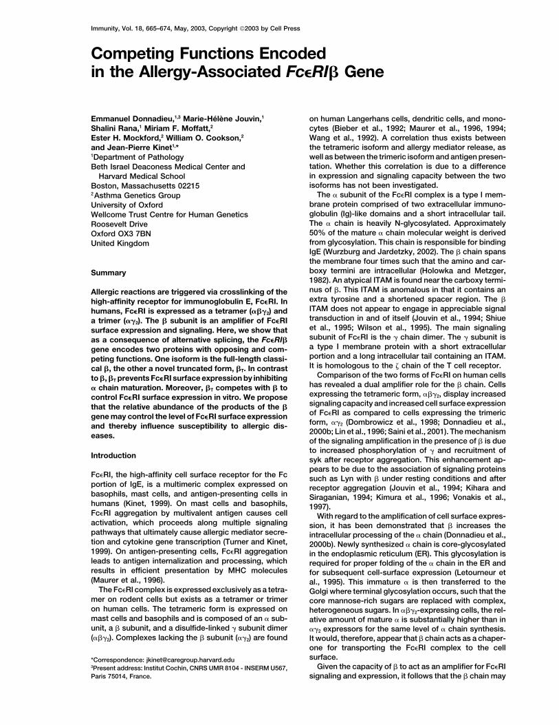
is a negative regulator of Fc�RI expression. 5 (Figure 1B). To demonstrate that the �T message isindeed translated into a protein, lysates from CBMCand from the human basophilic cell line KU812 wereResultsimmunoprecipitated and immunoblotted with an anti-human � antibody (Donnadieu et al., 2000b; Maurer etIdentification of an Alternative Spliceal., 1996). This antibody is directed against the � N termi-Product of the � Gene, �T
nus, which is common between the two transcripts. Ly-Given the strong candidacy of Fc�RI� as a risk factorsates from the ���2 and ��T�2 transfectants were pro-for allergic diseases, it is important to identify potentiallycessed in an identical fashion and used as controls. Asrelevant polymorphisms. A number of associated pointexpected, a band of 28 kDa corresponding to � wasmutations have already been described, yet the mecha-immunoprecipitated and immunoblotted from the lysatenism by which they increase susceptibility to atopy re-of the ���2 transfectants and KU812, but not from themains unknown (Donnadieu et al., 2000a). In the process��T�2 transfectants. A protein of the predicted size (21.4of screening for additional polymorphisms of the Fc�RI�kDa) for �T was present in KU812 and ��T�2, but not ingene in humans, we have identified a novel � chain���2 transfectants. Both the truncated protein and full-transcript. Basophils were semipurified from blood ob-length protein were also present in CBMC (Figure 1D).tained from individuals in the general population. RNAThese data are further evidence that � and �T are endog-was extracted, reverse-transcribed (RT), and amplifiedenously produced and coexpressed in a human baso-by polymerase chain reaction (PCR) using a forwardphilic cell line as well as in primary human mast cells.primer located within exon 2 and a reverse primer lo-
cated within exon 7 of the Fc�RI� gene (Kuster et al.,1992). Amplification produced the anticipated 700 bp �T Functions as a Negative Regulator
of Fc�RI Surface Expressionband corresponding to the fully spliced message as wellas an unexpected band of 1.1 kb. Both transcripts were Having established that �T protein is endogenously pro-
duced, the next step was to determine its function. Sincealso present in human cord blood-derived mast cells(CBMC), but not in the human monocytic cell line U937, � is an amplifier of Fc�RI expression and signaling, the
effect of �T on cell surface expression was tested (Dom-which does not express � (Figure 1A). Sequencing ofthe 1.1 kb band identified it to result from retention of browicz et al., 1998; Donnadieu et al., 2000b; Lin et
al., 1996). The transfection strategy previously used tothe 5th intron in mature � message (Figure 1B) (Kusteret al., 1992). The novel transcript, therefore, represents demonstrate the expression amplification function of �
was applied here (Donnadieu et al., 2000b; Lin et al.,an alternative splice product of the � gene. The pre-dicted translation product of the newly identified tran- 1996). A number of ��2, ���2, and ��T�2 stable transfec-
tants were established in U937 cells and in the mousescript would be truncated due to the presence of an inframe stop codon within the sequence contributed by fibroblastic cell line NIH 3T3. The entire panel of clones
was then compared for Fc�RI cell surface expression.the intron (Figure 1B). The transcript and resultant pro-tein will thus be referred to as �T. The cells were stained with IgE, and surface expression
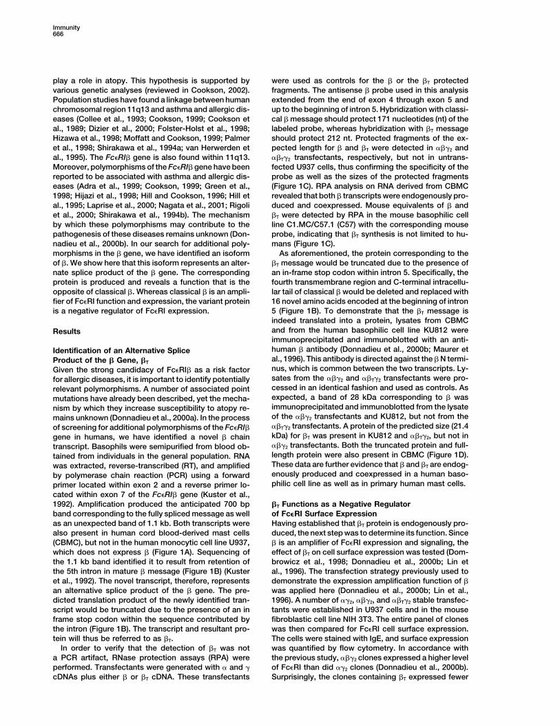
was quantified by flow cytometry. In accordance withIn order to verify that the detection of �T was nota PCR artifact, RNase protection assays (RPA) were the previous study, ���2 clones expressed a higher level
of Fc�RI than did ��2 clones (Donnadieu et al., 2000b).performed. Transfectants were generated with � and �cDNAs plus either � or �T cDNA. These transfectants Surprisingly, the clones containing �T expressed fewer
Competing Functions of the Fc�RI� Gene667
Figure 1. Detection of the �T Message and Protein in Mast Cells and Basophils
(A) Detection of �T message by RT-PCR in human basophils (basophils; 2 donors) and cord blood mast cells (CBMC; 2 donors), but not in anon-� expressing cell line (U937).(B) Schematic representation of the � gene (upper panel) and the � and �T messages (lower panels). Numbers represent individual introns. �
and �T use the same ATG located at nt 45 (exon 1). The stop codon in � is located at nt 776 (exon 7). The �T message is identical to � up tothe end of exon 5 (nt 581). In �T, intron 5 is retained between exon 5 and exon 6. As a result, �T is 402 nt longer. An in-frame stop codon ispresent after 48 nt in intron 5. Retention of intron 5 results in a shorter protein in which the fourth transmembrane domain and the C-terminaltail are replaced with a novel 16 amino acid peptide.(C) Detection of �T message by RNase protection assay in CBMC and the mouse basophil cell line C57. (Left panel) The expected fragmentof 212 nt for human �T is detected in CBMC and ��T�2 transfectants, but not in untransfected U937 or in ���2 transfectants. The expectedfragment of 171 nt for human � is detected in CBMC and ���2 transfectants, but not in untransfected U937 or in ��T�2 transfectants. Thelower band in the ��T�2 lane is a nonspecific degradation product. (Right panel) RNA from the mouse mast cell line Cl.MC/C57.1 (C57), butnot mouse liver RNA (CT), contains both protected mouse �T (266 nt) and � (163 nt) fragments. The full-length mouse probe is detected whenRNase treatment is omitted (RNase -).(D) Detection of the �T protein in CBMC (20 E6 cells/lane) and in the human basophil cell line KU812 (50 E6 cells/lane). Cells were treatedwith the proteasome inhibitor ALLN and lysed. � and �T were immunoprecipitated and Western blotted with an anti-� antibody directed againstthe common N-terminal tail. ���2 and ��T�2 transfectants (10 E6 cells/lane upper panel, 20 E6 cells/lane lower panel) are used as controls for� and �T, respectively.
surface receptors than the ���2 clones (p � 4.5 E-6) nadieu et al., 2000b). Considering that �T appears toand notably less than the ��2 clones as well (p � 1.5 inhibit receptor expression, it stood to reason that per-E-3) (Figure 2). Similar results were obtained with NIH haps �T interferes with � maturation. Newly synthesized3T3 stable transfectants (data not shown). The lower � chain is core-glycosylated in the ER. It migrates as aexpression levels in ��T�2 as compared to ��2 clones band of about 46 kDa on SDS PAGE. This immature �highlight the fact that �T does not simply lack the expres- is endoglycosidase H (Endo H) sensitive and results insion amplification function associated with �, but ac- a band of about 30 kDa on SDS PAGE following Endotively prevents receptor expression. Furthermore, KU812 H treatment. This is the expected molecular weight forcells were stably transfected with � and � cDNA encod- the core protein. Immature � is then transferred to theing constructs. Transient transfection with � and �T Golgi where terminal glycosylation occurs, such that thecDNA caused a 20% reduction in the Fc�RI MFI as com- core mannose-rich sugars are replaced with complex,pared to transfection with � cDNA alone (data not heterogeneous sugars that are Endo H resistant. Matureshown). This indicates that �T can act as a negative � appears as a smear around 66 kDa (Letourneur et al.,regulator in a human basophil cell line as well and can 1995). When ��2 and ���2 transfectants with the samecompete with the function of �. level of surface Fc�RI expression are compared, the
amount of immature � chain is much greater in the ��2
than in the ���2 clones. This indicates that � maturation�T Prevents � Maturationis considerably improved in the presence of � chain.The mechanism of the apparent inhibitory action of �T
Verily, the effect on � maturation may be the primaryon Fc�RI surface expression was investigated. Classicalmechanism by which � amplifies Fc�RI surface expres-� amplifies receptor expression by facilitating the intra-
cellular processing and maturation of the � chain (Don- sion (Donnadieu et al., 2000b). The effect of �T on �
Immunity668
Figure 2. �T Prevents Cell Surface Expression of Fc�RI
Fc�RI expression was assessed by flow cytometry after staining with IgE in ��T�2, ��2, and ���2 transfectants and is expressed as meanfluorescence intensity (MFI). The means �/� SD of MFI for all the clones obtained in each transfection type were calculated and comparedwith the Student’s unpaired t test (three independent sets of transfections).
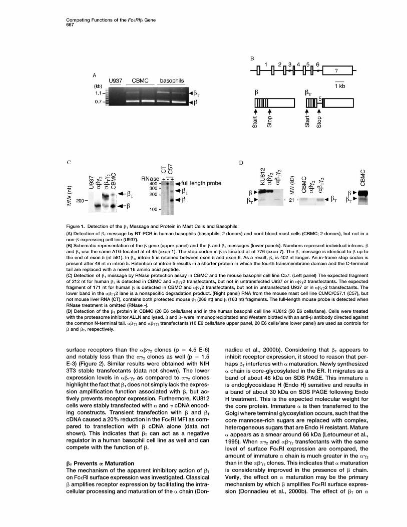
maturation was investigated. Three clones each of the protein. Pulse-chase analysis was employed to comparethe half-lives of � and �T. One ���2 clone and two ��T�2��2, ���2, and ��T�2 transfectants were selected for their
low, medium, and high levels of Fc�RI expression. The clones were labeled for 10 min and then transferred tounlabeled medium for a chase of 0 to 30 min. � and �T� chain was immunoprecipitated and blotted. As ex-
pected, � promotes the maturation of � chain, as there were immunoprecipitated from their respective lysatesand resolved by SDS-PAGE. Autoradiography followedwas less immature � in the ���2 clones relative to the
��2 clones with comparable amounts of mature � (Figure by quantification of the � and �T bands revealed thatwhereas � is stable over the 30 min chase, �T has the3A) (Donnadieu et al., 2000b). With regard to �T, imma-
ture � was substantially more abundant in the presence remarkably short half-life of approximately 10 min (Fig-ure 4A). The short half-life of �T may reflect rapid tar-of �T than in the absence of � for the same amount of
mature � (Figure 3A, compare lanes 8 and 4 and lanes geting to a degradation pathway, such as the protea-some complex (Ciechanover, 1998; Cresswell and9 and 5). The intensity of the mature � band was plotted
as a function of the intensity of the immature � band Hughes, 1997; Ward et al., 1995; Wiertz et al., 1996;Yang et al., 1998). The contribution of the proteasomefor each clone (Figure 3B). The ��T�2 clones required
much more immature � chain to express the same level to �T degradation was assessed by the utilization ofproteasome inhibitors, such as N-Acetyl-Leucyl-Leucyl-of mature � chain than did the ���2 clones and the ��2
clones. This indicates that � maturation is prevented Norleucinal (ALLN), lactacystin, and carboxybenzyl-leu-cyl-leucyl vinylsulfone ZL3VS (Wiertz et al., 1996). Pre-when �T is present and suggests that the inhibition of
� chain maturation ultimately reduces surface expres- treatment of �T expressing cells with these inhibitorssion of Fc�RI. significantly increased the visibility of �T following immu-
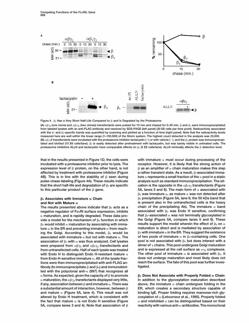
noprecipitation and immunoblot (Figure 4B). This sug-gests that the proteasome is one major mechanism by�T Has a Short Half-Life due to Rapid Targeting
for Degradation which �T is degraded. Other degradation pathways maybe involved as well, particularly since the specificity ofThe opposite effects of � and �T on Fc�RI expression
and � maturation led us to investigate the fate of the �T these inhibitors for the proteasome is not absolute. Note
Figure 3. �T Prevents Maturation of the � Chain
The � chain was immunoprecipitated from ��2, ���2, and ��T�2 transfectants (three clones of each type) with the anti-� monoclonal antibody15-1 that recognizes both the immature and mature forms of �. The immunoprecipitates were resolved by SDS-PAGE and immunoblottedwith the polyclonal anti-� antibody 997 that recognizes all forms of � (A) (15 E6 cells/lane). The intensity of the immature and mature � bandswas quantified by scanning. For each transfectant type, the amount of mature � was plotted as a function of the amount of immature � ineach clone (B).
Competing Functions of the Fc�RI� Gene669
Figure 4. �T Has a Very Short Half-Life Compared to � and Is Degraded by the Proteasome
(A) ���2 (one clone) and ��T�2 (two clones) transfectants were pulsed for 10 min and chased for 0–30 min. � and �T were immunoprecipitatedfrom labeled lysates with an anti-FLAG antibody and resolved by SDS-PAGE (left panel) (20 E6 cells per time point). Radioactivity associatedwith the �- and �T-specific bands was quantified by scanning and plotted as a function of time (right panel). Note that the radioactivity levelsmeasured here are well within the linear range (1–100,000) of the Storm system. The highest count detected in the analysis was 23,000.(B) ��T�2 transfectants were incubated with the proteasome inhibitor lactacystin (�) or with vehicle (�), and the �T protein was immunoprecipi-tated and blotted (15 E6 cells/lane). �T is easily detected after pretreatment with lactacystin, but was barely visible in untreated cells. Theproteasome inhibitors ALLN and lactacystin have comparable effects on �T (5 E6 cells/lane). ALLN minimally affects the � detection level.
that in the results presented in Figure 1D, the cells were with immature � must occur during processing of thereceptor. However, it is likely that the strong action ofincubated with a proteasome inhibitor prior to lysis. The
expression level of � protein, on the other hand, is not � as an amplifier of � chain maturation makes this stepa rather transient state. As a result, �-associated imma-affected by treatment with proteasome inhibitor (Figure
4B). This is in line with the stability of � seen during ture � represents a small fraction of the � pool in a staticanalysis such as standard immunoprecipitation. The sit-pulse-chase labeling (Figure 4A). These results indicate
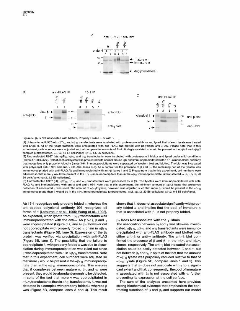
that the short half-life and degradation of �T are specific uation is the opposite in the ��T�2 transfectants (Figure5A, lanes 5 and 6). The main form of � associated withto this particular product of the � gene.�T was immature �, as mature � was not detected after�T precipitation (Figure 5A, lane 6; the 50 kDa band that�T Associates with Immature � Chainis present also in the untransfected cells is the heavyand Not with Mature �chain of the precipitating Ab). The immature � bandThe results presented above indicate that �T acts as aassociated with �T was Endo H sensitive, confirmingnegative regulator of Fc�RI surface expression, inhibitsthat �T-associated � was not terminally glycosylated in� maturation, and is rapidly degraded. These data pro-the Golgi (Figure 5A, compare lanes 5 and 6). Thesevide a model for the mechanism of �T function in whichresults support the model wherein the effect of �T on ��T would inhibit � maturation by associating with imma-maturation is direct and is mediated by association ofture � in the ER and preventing immature � from reach-�T with immature � in the ER. They suggest the existenceing the Golgi. According to this model, �T would beof two pools of immature � in �T-containing cells. Oneassociated with immature � but not with mature �. Thepool is not associated with �T but does interact with aassociation of �T with � was thus analyzed. Cell lysatesdimer of � chains. This pool undergoes Golgi maturationwere prepared from ���2 and ��T�2 transfectants andand is expressed at the cell surface as ��2 complexes.from untransfected cells. Half of each lysate was treatedThe other pool of immature � is associated with �T. Itwith Endo H to distinguish Endo H-resistant mature �does not undergo maturation and most likely does notfrom Endo H-sensitive immature �. All of the lysate frac-reach the surface. The fate of this pool was further inves-tions were then immunoprecipitated with anti-FLAG an-tigated.tibody (to immunoprecipitate � and �T) and immunoblot-
ted with the polyclonal anti-� (997) that recognizes all� forms. As expected, given the capacity of � to promote �T Does Not Associate with Properly Folded � Chain
In addition to the glycosylation maturation described� maturation, the ���2 transfectants displayed very little,if any, association between � and immature �. There was above, the immature � chain undergoes folding in the
ER, which creates a secondary structure capable ofa substantial amount of interaction, however, between �and mature � (Figure 5A, lane 4). This result was not binding IgE. Proper folding requires mannose-rich gly-
cosylation of � (Letourneur et al., 1995). Properly foldedaltered by Endo H treatment, which is consistent withthe fact that mature � is not Endo H sensitive (Figure � and misfolded � can be distinguished based on their
reactivity with various anti-� antibodies. The monoclonal5A, compare lanes 3 and 4). Note that association of �
Immunity670
Figure 5. �T Is Not Associated with Mature, Properly Folded � or with �
(A) Untransfected U937 (ut), ��T�2, and ���2 transfectants were incubated with proteasome inhibitor and lysed. Half of each lysate was treatedwith Endo H. All of the lysate fractions were precipitated with anti-FLAG and blotted with polyclonal anti-� 997. Please note that in thisexperiment, cells numbers were adjusted so that comparable amounts of Endo H deglycosylated � would be present in the ���2 and ��T�2samples (untransfected, ��T�2, 40 E6 cells/lane; ���2, 1.5 E6 cells/lane).(B) Untransfected U937 (ut), ��T�2, ���2, and ��2 transfectants were incubated with proteasome inhibitor and lysed under mild conditions(Triton X-100 0.25%). Half of each cell lysate was precleared with normal mouse IgG and immunoprecipitated with 15-1, a monoclonal antibodythat recognizes only properly folded � (lanes 3–6). Immunoprecipitates were separated by Western blot and blotted. The blot was incubatedwith polyclonal anti-� 961 and anti-� 934 Abs (lanes 3–6). As a control for the presence of � and �T, the remaining half of the lysates wasimmunoprecipitated with anti-FLAG Ab and immunoblotted with anti-� (lanes 1 and 2) Please note that in this experiment, cell numbers wereadjusted so that more � would be present in the ��T�2 immunoprecipitate than in the ���2 immunoprecipitate (untransfected, ��2, ��T�2, 20E6 cells/lane; ���2, 2.5 E6 cells/lane).(C) Untransfected U937 (ut), ��T�2, ���2, and ��2 transfectants were processed as in (B). The lysates were immunoprecipitated with anti-FLAG Ab and immunoblotted with anti-� and anti-� 934. Note that in this experiment, the minimum amount of ���2 lysate that preservesdetection of associated � was used. The amount of ��T�2 lysate, however, was adjusted such that more �T would be present in the ��T�2
immunoprecipitate than � would be in the ���2 immunoprecipitate (untransfected, ��2, ��T�2, 20 E6 cells/lane; ���2, 0.5 E6 cells/lane).
Ab 15-1 recognizes only properly folded �, whereas the shows that �T does not associate significantly with prop-erly folded � and implies that the pool of immature �anti-peptide polyclonal antibody 997 recognizes all
forms of � (Letourneur et al., 1995; Wang et al., 1992). that is associated with �T is not properly folded.As expected, when lysate from ���2 transfectants wasimmunoprecipitated with the anti-� Ab (15-1), � and � �T Does Not Associate with the � Chain
The association between �T and � was likewise investi-were coprecipitated (Figure 5B, lane 4). �T, however didnot coprecipitate with properly folded � chain in ��T�2 gated. ��T�2, ���2, and ��2 transfectants were immuno-
precipitated with anti-FLAG antibody and blotted withtransfectants (Figure 5B, lane 3). Expression of the �T
protein was verified via precipitation with anti-FLAG either anti-� or anti-� antibody. The anti-� blot con-firmed the presence of � and �T in the ���2 and ��T�2(Figure 5B, lane 1). The possibility that the failure to
coprecipitate �T with properly folded � was due to disso- clones, respectively. The anti-� blot indicated that asso-ciation could be easily detected between � and �, butciation during immunoprecipitation was ruled out since
� was coprecipitated with � in ��T�2 transfectants. Note not between �T and �, in spite of the fact that the amountof ���2 lysate was purposely reduced relative to that ofthat in this experiment, cell numbers were adjusted so
that more � would be present in the ��T�2 immunoprecip- ��T�2 lysate (Figure 5C, compare lanes 1 and 2). Thissuggests that �T does not associate with � to a signifi-itate than in the ���2 immunoprecipitate. This ensures
that if complexes between mature �, �T, and �2 were cant extent and that, consequently, the pool of immature� associated with �T is not associated with �, furtherpresent, they would be abundant enough to be detected.
In spite of the fact that more � was coprecipitated in preventing its expression at the cell surface.The sum of the analyses presented here provides��T�2 transfectants than in ���2 transfectants, �T was not
detected in a complex with properly folded � whereas � strong biochemical evidence that emphasizes the con-trasting functions of � and �T and supports our modelwas (Figure 5B, compare lanes 3 and 4). This result
Competing Functions of the Fc�RI� Gene671
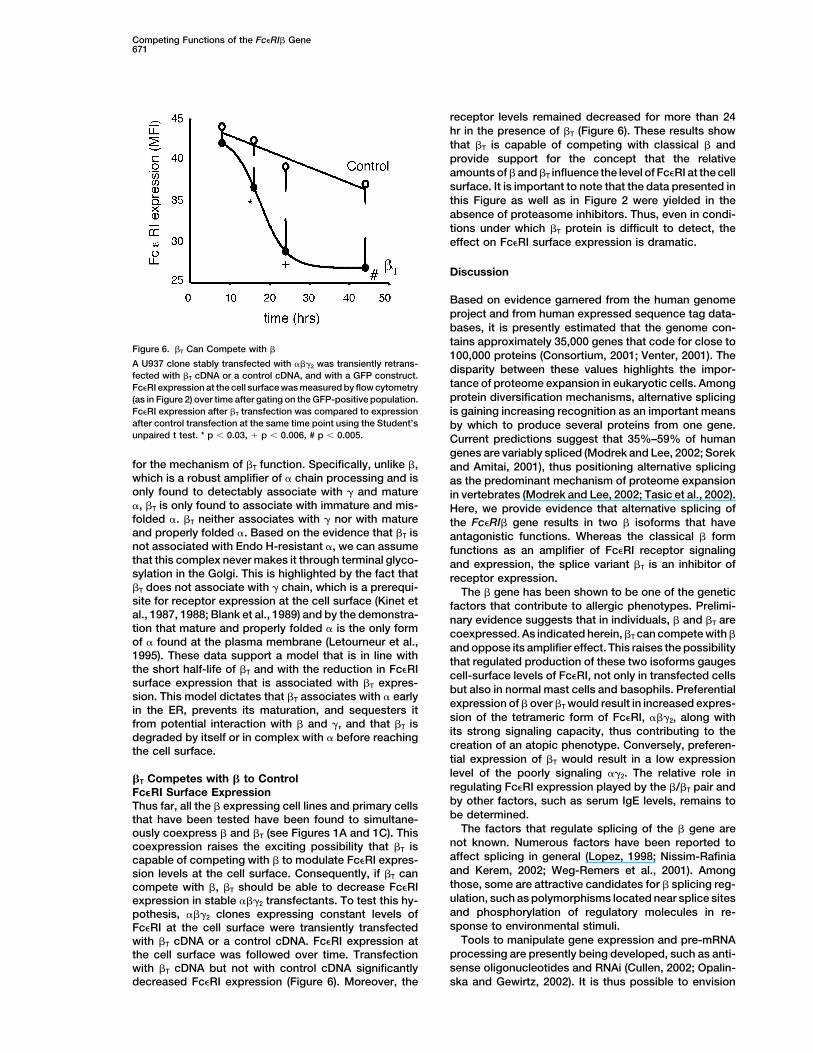
receptor levels remained decreased for more than 24hr in the presence of �T (Figure 6). These results showthat �T is capable of competing with classical � andprovide support for the concept that the relativeamounts of � and �T influence the level of Fc�RI at the cellsurface. It is important to note that the data presented inthis Figure as well as in Figure 2 were yielded in theabsence of proteasome inhibitors. Thus, even in condi-tions under which �T protein is difficult to detect, theeffect on Fc�RI surface expression is dramatic.
Discussion
Based on evidence garnered from the human genomeproject and from human expressed sequence tag data-bases, it is presently estimated that the genome con-tains approximately 35,000 genes that code for close to
Figure 6. �T Can Compete with �100,000 proteins (Consortium, 2001; Venter, 2001). The
A U937 clone stably transfected with ���2 was transiently retrans- disparity between these values highlights the impor-fected with �T cDNA or a control cDNA, and with a GFP construct.
tance of proteome expansion in eukaryotic cells. AmongFc�RI expression at the cell surface was measured by flow cytometryprotein diversification mechanisms, alternative splicing(as in Figure 2) over time after gating on the GFP-positive population.
Fc�RI expression after �T transfection was compared to expression is gaining increasing recognition as an important meansafter control transfection at the same time point using the Student’s by which to produce several proteins from one gene.unpaired t test. * p 0.03, � p 0.006, # p 0.005. Current predictions suggest that 35%–59% of human
genes are variably spliced (Modrek and Lee, 2002; Sorekfor the mechanism of �T function. Specifically, unlike �, and Amitai, 2001), thus positioning alternative splicingwhich is a robust amplifier of � chain processing and is as the predominant mechanism of proteome expansiononly found to detectably associate with � and mature in vertebrates (Modrek and Lee, 2002; Tasic et al., 2002).�, �T is only found to associate with immature and mis- Here, we provide evidence that alternative splicing offolded �. �T neither associates with � nor with mature the Fc�RI� gene results in two � isoforms that haveand properly folded �. Based on the evidence that �T is antagonistic functions. Whereas the classical � formnot associated with Endo H-resistant �, we can assume functions as an amplifier of Fc�RI receptor signalingthat this complex never makes it through terminal glyco- and expression, the splice variant �T is an inhibitor ofsylation in the Golgi. This is highlighted by the fact that receptor expression.�T does not associate with � chain, which is a prerequi- The � gene has been shown to be one of the geneticsite for receptor expression at the cell surface (Kinet et factors that contribute to allergic phenotypes. Prelimi-al., 1987, 1988; Blank et al., 1989) and by the demonstra- nary evidence suggests that in individuals, � and �T aretion that mature and properly folded � is the only form coexpressed. As indicated herein, �T can compete with �of � found at the plasma membrane (Letourneur et al., and oppose its amplifier effect. This raises the possibility1995). These data support a model that is in line with that regulated production of these two isoforms gaugesthe short half-life of �T and with the reduction in Fc�RI cell-surface levels of Fc�RI, not only in transfected cellssurface expression that is associated with �T expres-
but also in normal mast cells and basophils. Preferentialsion. This model dictates that �T associates with � early
expression of � over �T would result in increased expres-in the ER, prevents its maturation, and sequesters it
sion of the tetrameric form of Fc�RI, ���2, along withfrom potential interaction with � and �, and that �T isits strong signaling capacity, thus contributing to thedegraded by itself or in complex with � before reachingcreation of an atopic phenotype. Conversely, preferen-the cell surface.tial expression of �T would result in a low expressionlevel of the poorly signaling ��2. The relative role in�T Competes with � to Controlregulating Fc�RI expression played by the �/�T pair andFc�RI Surface Expressionby other factors, such as serum IgE levels, remains toThus far, all the � expressing cell lines and primary cellsbe determined.that have been tested have been found to simultane-
The factors that regulate splicing of the � gene areously coexpress � and �T (see Figures 1A and 1C). Thisnot known. Numerous factors have been reported tocoexpression raises the exciting possibility that �T isaffect splicing in general (Lopez, 1998; Nissim-Rafiniacapable of competing with � to modulate Fc�RI expres-and Kerem, 2002; Weg-Remers et al., 2001). Amongsion levels at the cell surface. Consequently, if �T canthose, some are attractive candidates for � splicing reg-compete with �, �T should be able to decrease Fc�RIulation, such as polymorphisms located near splice sitesexpression in stable ���2 transfectants. To test this hy-and phosphorylation of regulatory molecules in re-pothesis, ���2 clones expressing constant levels ofsponse to environmental stimuli.Fc�RI at the cell surface were transiently transfected
Tools to manipulate gene expression and pre-mRNAwith �T cDNA or a control cDNA. Fc�RI expression atprocessing are presently being developed, such as anti-the cell surface was followed over time. Transfectionsense oligonucleotides and RNAi (Cullen, 2002; Opalin-with �T cDNA but not with control cDNA significantly
decreased Fc�RI expression (Figure 6). Moreover, the ska and Gewirtz, 2002). It is thus possible to envision
Immunity672
selected in G418. Transient transfection with the �T construct, orthat the amount of �T relative to � may be deliberatelyan irrelevant construct, and a GFP construct in U937 and KU812altered. The production and delivery of oligonucleotides���2 clones was performed as described (Donnadieu et al., 2000b).that bind to the intron 5 splice sites would render them
undetectable to the spliceosome. The presumable result Flow Cytometric Analysis of Fc�RI Expression at the Cell Surfaceof such interference would be an increase in the relative Fc�RI expression in stable and transient transfectants was analyzedamount of �T and a consequent decrease in Fc�RI sur- after binding of biotinylated recombinant IgE (Schwarzbaum et al.,
1989) and streptavidin-phycoerythrin (Donnadieu et al., 2000b). Forface expression. In a therapeutic model, one would pre-transient transfection, Fc�RI expression was measured after gatingdict a concomitant decrease in susceptibility to allergicon GFP-positive cells.disorders. Given the impact of Fc�RI expression on the
pathophysiology of atopy, an understanding of the mo-Immunoprecipitation, Western Blotting, Proteasome Inhibitor
lecular mechanism by which � and �T are produced will Treatment, and Endo-H Treatmenthave profound implications on research in the area of Immunoprecipitation and Western blotting were performed as inallergic diseases. Donnadieu et al. (2000b), except in Figure 5 where cells were lysed
in Triton X-100 0.25%. The following antibodies (Ab) were used:anti-human Fc�RI� monoclonal Ab 15-1 (Wang et al., 1992), anti-Experimental Procedureshuman Fc�RI� rabbit polyclonal Ab 997 (Letourneur et al., 1995),anti-human � rabbit polyclonal Ab 961 (Maurer et al., 1996), andCellsanti-FLAG M2 monoclonal Ab (Sigma, St. Louis, MO). In Figures 1D,Semipurified basophils (2%–5% basophils) were obtained from4B, and 5, cells were treated with one of the following proteasomeblood donors by Percoll (Pharmacia) centrifugation (density 1.08)inhibitors, ALLN (Calbiochem, San Diego, CA) 250 M, lactacystin(Leonard et al., 1984). CBMC were prepared by growing cord blood(Calbiochem) 10 M, carboxybenzyl-leucyl-leucyl vinylsulfonemononuclear cells with 100 ng/ml recombinant human stem cell(ZL3VS) 20 M (Wiertz et al., 1996), or vehicle (DMSO) for 2 hr atfactor (Biosource, Camarillo, CA), 10 ng/ml recombinant human IL-637�C before lysis. In Figure 5A, immunoprecipitates were treated(Endogen, Woburn, MA), and 1 M prostaglandin E2 (Cayman Chem-with endo-�-N-acetylglucosamidase (Endo H) (New England Bio-ical, Ann Arbor, MI) for 10 weeks (Saito et al., 1995). The humanlabs) as previously described (Letourneur et al., 1995).basophilic cell line KU812, the human monocytoid cell line U937,
and the mouse fibroblastic cell line NIH 3T3, untransfected andtransfected, were maintained as described (Donnadieu et al., 2000b; Pulse-Chase Analysis
Cells (5 � 106/ml) were pulsed with 0.2 mCi/ml TRAN 35S-LABELLin et al., 1996).(ICN, Costa Mesa, CA) for 10 min at 37�C and chased for the indi-cated periods of time in cold medium. At each time point, 15 � 106RNA Preparationcells were lysed in 0.5% Triton X-100, 300 mM NaCl, and 50 mMPolyA RNA was isolated from CBMC using Fast-Tack 2.1 (Invitrogen,Tris (pH 7.5) in the presence of protease inhibitors (Complete, RocheCarlsbad, CA). Total RNA was extracted from all other cells usingMolecular Biochemicals, Indianapolis, IN). Lysates were preclearedRNAzol B (Tel-Test, Inc, Friendswood, TX).and then immunoprecipitated with anti-FLAG antibody as above.Proteins were separated on SDS 14% polyacrylamide gels, and theRT, PCR, and Cloning of PCR Productsdried gels were exposed to a PhosphorImager screen (MolecularReverse transcription was performed using Omniscript reverse tran-Dynamic, Sunnyvale, CA). Radiolabeled proteins detected by scan-scriptase (Qiagen, Valencia, CA) and oligodT. PCR was performedning of the exposed screen were quantified with the ImageQuantin 50 l final volume with 10 l RT reaction (equivalent to 0.1 gsoftware (Molecular Dynamics).RNA), 1.25 U Taq polymerase (Fisher Scientific, Pittsburgh, PA), 200
M dNTPs, 60 pmoles of forward (GTGCCTGCATTTGAAGTCTTG)Statisticsand reverse (TGGATCCTTGGCTGTGAATC) primers, 1.5 mM MgCl2,Means are expressed SD. Comparisons between groups were20 mM Tris (pH 8.3), and 25 mM KCl. Cycling was 94�C 45 s, 60�Cperformed with the Student’s unpaired t test.45 s, and 72�C 1 min for 25 cycles. PCR products were cloned by
TA cloning in the pCRII vector (Invitrogen).Acknowledgments
RNase Protection Assay (RPA)We thank Hidde Ploegh of the Harvard Medical School for the kindRPA was performed using the RPAIII kit (Ambion, Austin, TX) withgift of the proteasome inhibitor ZL3VS and for advice on the experi-an antisense probe specific for human or mouse � generated withments related to proteasome degradation.Maxiscript (Ambion). The human � probe covers the last 12 bases
of exon 4, the entirety of exon 5, and the first 41 bases of intron 5(Genbank accession number M89796) (Kuster et al., 1992). The Received: November 11, 2002
Revised: January 31, 2003mouse � probe covers the last 12 bases of exon 4, the entirety ofexon 5, and the first 103 bases of intron 5 (Genbank accession Accepted: February 26, 2003
Published: May 13, 2003numbers NM013516 and AB033617) (Ra et al., 1989). Ten micro-grams of total RNA was used for U937 and C57 samples, whereaspolyA RNA from 10E6 cells was used for CBMC samples. A FcR� Referencesprobe was used as positive control for U937 samples and CBMC.
Adra, C.N., Mao, X.Q., Kawada, H., Gao, P.S., Korzycka, B., Donate,Generation of the Classical � and �T Expression Constructs, J.L., Shaldon, S.R., Coull, P., Dubowitz, M., Enomoto, T., et al. (1999).and Transfections Chromosome 11q13 and atopic asthma. Clin. Genet. 55, 431–437.An untagged �T template was generated by PCR using � cDNA Bieber, T., de la Salle, H., Wollenberg, A., Hakimi, J., Chizzonite, R.,(Kuster et al., 1992) as template, a forward primer including the ATG, Ring, J., Hanau, D., and de la Salle, C. (1992). Human epidermaland a reverse primer including the first 51 bases of intron 5 up to Langerhans cells express the high affinity receptor for immunoglob-the in-frame stop codon (Kuster et al., 1992). This �T cDNA, as well ulin E (Fc�RI). J. Exp. Med. 175, 1285–1290.as � cDNA, were then tagged by addition of a FLAG sequence
Blank, U., Ra, C., Miller, L., White, K., Metzger, H., and Kinet, J.P.(DYKDDDDK) at the 5� end by PCR. The tagged � and �T inserts
(1989). Complete structure and expression in transfected cells ofwere ligated independently into the mammalian expression vector
high-affinity IgE receptor. Nature 337, 187–189.pBJ1neo (Lin et al., 1990). Insert sequences were verified by DNA
Ciechanover, A. (1998). The ubiquitin-proteasome pathway: on pro-sequencing. U937 and NIH3T3 cells were cotransfected by electro-tein death and cell life. EMBO J. 17, 7151–7160.poration with 10 g each of Fc�RI� and Fc�RI� and 2 g each of
either �, �T, or the empty vector pBJ1 neo. Stable clones were Collee, J.M., ten Kate, L.P., de Vries, H.G., Kliphuis, J.W., Bouman,
Competing Functions of the Fc�RI� Gene673
K., Scheffer, H., and Gerritsen, J. (1993). Allele sharing on chromo- Kinet, J.P. (1999). The high-affinity IgE receptor (Fc epsilon RI): fromphysiology to pathology. Annu. Rev. Immunol. 17, 931–972.some 11q13 in sibs with asthma and atopy. Lancet 342, 936.
Consortium, T.G.I.S. (2001). Initial Sequencing and Analysis of the Kinet, J.P., Metzger, H., Hakimi, J., and Kochan, J. (1987). A cDNAHuman Genome. Nature 409, 860–921. presumptively coding for the alpha subunit of the receptor with high
affinity for immunoglobulin E. Biochemistry 26, 4605–4610.Cookson, W.O. (1999). The alliance of genes and environment inasthma and allergy. Nature 402, 85–11. Kinet, J.P., Blank, U., Ra, C., White, K., Metzger, H., and Kochan,
J. (1988). Isolation and characterization of cDNAs coding for theCookson, W.O. (2002). Asthma genetics. Chest 121, 7S–13S.beta subunit of the high-affinity receptor for immunoglobulin E. Proc.Cookson, W.O., Sharp, P.A., Faux, J.A., and Hopkin, J.M. (1989).Natl. Acad. Sci. USA 85, 6483–6487.Linkage between immunoglobulin E responses underlying asthmaKuster, H., Zhang, L., Brini, A.T., MacGlashan, D.W., and Kinet, J.P.and rhinitis and chromosome 11q. Lancet 1, 1292–1295.(1992). The gene and cDNA for the human high affinity immunoglob-Cresswell, P., and Hughes, E.A. (1997). Protein degradation: the insulin E receptor � chain and expression of the complete humanand outs of the matter. Curr. Biol. 7, R552–R555.receptor. J. Biol. Chem. 267, 12782–12787.
Cullen, B.R. (2002). RNA interference: antiviral defense and geneticLaprise, C., Boulet, L.P., Morissette, J., Winstall, E., and Raymond, V.tool. Nat. Immunol. 3, 597–599.(2000). Evidence for association and linkage between atopy, airway
Dizier, M.H., Besse-Schmittler, C., Guilloud-Bataille, M., Annesi-hyper-responsiveness, and the � subunit Glu237Gly variant of the
Maesano, I., Boussaha, M., Bousquet, J., Charpin, D., Degioanni,high-affinity receptor for immunoglobulin E in the French-Canadian
A., Gormand, F., Grimfeld, A., et al. (2000). Genome screen forpopulation. Immunogenetics 51, 695–702.
asthma and related phenotypes in the French EGEA study. Am. J.Leonard, E.J., Roberts, R.L., and Skeel, A. (1984). Purification ofRespir. Crit. Care Med. 162, 1812–1818.human blood basophils by single step isopycnic banding on Percoll.Dombrowicz, D., Lin, S., Flamand, V., Brini, A.T., Koller, B.H., andJ. Leukoc. Biol. 35, 169–177.Kinet, J.P. (1998). Allergy-associated FcR � is a molecular amplifierLetourneur, O., Sechi, S., Willette-Brown, J., Robertson, M.W., andof IgE- and IgG- mediated in vivo responses. Immunity 8, 517–529.Kinet, J.P. (1995). Glycosylation of human truncated Fc�RI � chainDonnadieu, E., Cookson, W.O., Jouvin, M.,H., and Kinet, J.P. (2000a).is necessary for efficient folding in the endoplasmic reticulum. J.Allergy-associated polymorphisms of the Fc�RI � subunit do notBiol. Chem. 270, 8249–8256.impact its two amplification functions. J. Immunol. 165, 3917–3922.Lin, A.Y., Devaux, B., Green, A., Sagerstrom, C., Elliott, J.F., andDonnadieu, E., Jouvin, M.H., and Kinet, J.P. (2000b). A second ampli-Davis, M.M. (1990). Expression of T cell antigen receptor hetero-fier function for the allergy-associated Fc�RI � subunit. Immunitydimers in a lipid-linked form. Science 249, 677–679.12, 515–523.Lin, S., Cicala, C., Scharenberg, A.M., and Kinet, J.P. (1996). TheFolster-Holst, R., Moises, H.W., Yang, L., Fritsch, W., Weissenbach,Fc�RI � subunit functions as an amplifier of Fc�RI �-mediated cellJ., and Christophers, E. (1998). Linkage between atopy and the IgEactivation signals. Cell 85, 985–995.high-affinity receptor gene at 11q13 in atopic dermatitis families.
Hum. Genet. 102, 236–239. Lopez, A.J. (1998). Alternative splicing of pre-mRNA: developmentalconsequences and mechanisms of regulation. Annu. Rev. Genet.Green, S.L., Gaillard, M.C., Song, E., Dewar, J.B., and Halkas, A.32, 279–305.(1998). Polymorphisms of the � chain of the high-affinity immuno-
globulin E receptor (Fc�RI �) in South African black and white asth- Maurer, D., Fiebiger, E., Reininger, B., Wolff-Winiski, B., Jouvin,matic and nonasthmatic individuals. Am. J. Respir. Crit. Care Med. M.H., Kilgus, O., Kinet, J.P., and Stingl, G. (1994). Expression of158, 1487–1492. functional high affinity immunoglobulin E receptors (Fc�RI) on mono-
cytes of atopic individuals. J. Exp. Med. 179, 745–750.Hijazi, Z., Haider, M.Z., Khan, M.R., and Al-Dowaisan, A.A. (1998).High frequency of IgE receptor Fc epsilonRIbeta variant (Leu181/ Maurer, D., Fiebiger, E., Ebner, C., Reininger, B., Fischer, G.F., Wich-Leu183) in Kuwaiti Arabs and its association with asthma. Clin. las, S., Jouvin, M.-H., Schmitt-Egenolf, M., Kraft, D., Kinet, J.-P.,Genet. 53, 149–152. and Stingl, G. (1996). Peripheral blood dendritic cells express Fc�RI
as a complex composed of Fc�RI�- and Fc�RI�-chains and can useHill, M.R., and Cookson, W.O. (1996). A new variant of the � subunitof the high-affinity receptor for immunoglobulin E (Fc�RI� E237G): this receptor for IgE-mediated allergen presentation. J. Immunol.
157, 607–616.associations with measures of atopy and bronchial hyper-respon-siveness. Hum. Mol. Genet. 5, 959–962. Modrek, B., and Lee, C. (2002). A genomic view of alternative splic-Hill, M.R., James, A.L., Faux, J.A., Ryan, G., Hopkin, J.M., le Souef, ing. Nat. Genet. 30, 13–19.P., Musk, A.W., and Cookson, W.O. (1995). Fc�RI-� polymorphism Moffatt, M.F., and Cookson, W.O. (1999). Genetics of asthma andand risk of atopy in a general population sample. BMJ 311, 776–779. inflammation: the status. Curr. Opin. Immunol. 11, 606–609.Hizawa, N., Freidhoff, L.R., Ehrlich, E., Chiu, Y.F., Duffy, D.L., Schou, Nagata, H., Mutoh, H., Kumahara, K., Arimoto, Y., Tomemori, T.,C., Dunston, G.M., Beaty, T.H., Marsh, D.G., Barnes, K.C., and Sakurai, D., Arase, K., Ohno, K., Yamakoshi, T., Nakano, K., et al.Huang, S.K. (1998). Genetic influences of chromosomes 5q31-q33 (2001). Association between nasal allergy and a coding variant ofand 11q13 on specific IgE responsiveness to common inhaled aller- the Fc epsilon RI beta gene Glu237Gly in a Japanese population.gens among African American families. Collaborative Study on the Hum. Genet. 109, 262–266.Genetics of Asthma (CSGA). J. Allergy Clin. Immunol. 102, 449–453.
Nissim-Rafinia, M., and Kerem, B. (2002). Splicing regulation as aHolowka, D., and Metzger, H. (1982). Further characterization of
potential genetic modifier. Trends Genet. 18, 123–127.the beta-component of the receptor for immunoglobulin E. Mol.
Opalinska, J.B., and Gewirtz, A.M. (2002). Nucleic-acid therapeutics:Immunol. 19, 219–227.basic principles and recent applications. Nat. Rev. Drug Discov. 1,Jouvin, M.H., Adamczewski, M., Numerof, R., Letourneur, O., Valle,503–514.A., and Kinet, J.P. (1994). Differential control of the tyrosine kinasesPalmer, L.J., Daniels, S.E., Rye, P.J., Gibson, N.A., Tay, G.K., Cook-Lyn and Syk by the two signaling chains of the high affinity immuno-son, W.O., Goldblatt, J., Burton, P.R., and LeSouef, P.N. (1998).globulin E receptor. J. Biol. Chem. 269, 5918–5925.Linkage of chromosome 5q and 11q gene markers to asthma-asso-Kihara, H., and Siraganian, R.P. (1994). Src homology 2 domains ofciated quantitative traits in Australian children. Am. J. Respir. Crit.Syk and Lyn bind to tyrosine-phosphorylated subunits of the highCare Med. 158, 1825–1830.affinity IgE receptor. J. Biol. Chem. 269, 22427–22432.Ra, C., Jouvin, M.H., and Kinet, J.P. (1989). Complete structure ofKimura, T., Kihara, H., Bhattacharyya, S., Sakamoto, H., Appella, E.,the mouse mast cell receptor for IgE (Fc�RI) and surface expressionand Siraganian, R.P. (1996). Downstream signaling molecules bindof chimeric receptors (rat-mouse-human) on transfected cells. J.to different phosphorylated immunoreceptor tyrosine-based activa-Biol. Chem. 264, 15323–15327.tion motif (ITAM) peptides of the high affinity IgE receptor. J. Biol.
Chem. 271, 27962–27968. Rigoli, L., Salpietro, D.C., Lavalle, R., Cafiero, G., Zuccarello, D., and
Immunity674
Barberi, I. (2000). Allelic association of gene markers on chromo- processing, ubiquitination, and proteasome-dependent removalfrom ER membranes. J. Exp. Med. 187, 835–846.some 11q in Italian families with atopy. Acta Paediatr. 89, 1056–1061.
Saini, S.S., Richardson, J.J., Wofsy, C., Lavens-Phillips, S., Bochner,B.S., and Macglashan, D.W., Jr. (2001). Expression and modulationof Fc�RI� and Fc�RI� in human blood basophils. J. Allergy Clin.Immunol. 107, 832–841.
Saito, H., Ebisawa, M., Sakaguchi, N., Onda, T., Iikura, Y., Yanagida,M., Uzumaki, H., and Nakahata, T. (1995). Characterization of cord-blood-derived human mast cells cultured in the presence of Steelfactor and interleukin-6. Int. Arch. Allergy Immunol. 107, 63–65.
Schwarzbaum, S., Nissim, A., Alkalay, I., Ghozi, M.C., Schindler,D.G., Bergman, Y., and Eshhar, Z. (1989). Mapping of murine IgEepitopes involved in IgE-Fc� receptor interactions. Eur. J. Immunol.19, 1015–1023.
Shirakawa, T., Hashimoto, T., Furuyama, J., Takeshita, T., and Mori-moto, K. (1994a). Linkage between severe atopy and chromosome11q13 in Japanese families. Clin. Genet. 46, 228–232.
Shirakawa, T., Li, A., Dubowitz, M., Dekker, J.W., Shaw, A.E., Faux,J.A., Ra, C., Cookson, W.O., and Hopkin, J.M. (1994b). Associationbetween atopy and variants of the � subunit of the high- affinityimmunoglobulin E receptor. Nat. Genet. 7, 125–129.
Shiue, L., Zoller, M.J., and Brugge, J.S. (1995). Syk is activatedby phosphotyrosine-containing peptides representing the tyrosine-based activation motifs of the high affinity receptor for IgE. J. Biol.Chem. 270, 10498–10502.
Sorek, R., and Amitai, M. (2001). Piecing together the significanceof splicing. Nat. Biotechnol. 19, 196.
Tasic, B., Nabholz, C.E., Baldwin, K.K., Kim, Y., Rueckert, E.H.,Ribich, S.A., Cramer, P., Wu, Q., Axel, R., and Maniatis, T. (2002).Promoter choice determines splice site selection in protocadherin� and � pre-mRNA splicing. Mol. Cell 10, 21–33.
Turner, H., and Kinet, J.-P. (1999). Fc�RI signaling; thresholds andtuning in the generation of an allergic response. Nature 402,B24–B30.
van Herwerden, L., Harrap, S.B., Wong, Z.Y., Abramson, M.J., Kutin,J.J., Forbes, A.B., Raven, J., Lanigan, A., and Walters, E.H. (1995).Linkage of high-affinity IgE receptor gene with bronchial hyperreac-tivity, even in absence of atopy. Lancet 346, 1262–1265.
Venter, C. (2001). The sequence of the human genome. Science 291,1304–1351.
Vonakis, B.M., Chen, H., Haleem-Smith, H., and Metzger, H. (1997).The unique domain as the site on Lyn kinase for its constitutiveassociation with the high affinity receptor for IgE. J. Biol. Chem.272, 24072–24080.
Wang, B., Rieger, A., Kilgus, O., Ochiai, K., Maurer, D., Fodinger,D., Kinet, J.P., and Stingl, G. (1992). Epidermal Langerhans cellsfrom normal human skin bind monomeric IgE via Fc epsilon RI. J.Exp. Med. 175, 1353–1365.
Ward, C.L., Omura, S., and Kopito, R.R. (1995). Degradation of CFTRby the ubiquitin-proteasome pathway. Cell 83, 121–127.
Weg-Remers, S., Ponta, H., Herrlich, P., and Konig, H. (2001). Regu-lation of alternative pre-mRNA splicing by the ERK MAP-kinasepathway. EMBO J. 20, 4194–4203.
Wiertz, E.J., Tortorella, D., Bogyo, M., Yu, J., Mothes, W., Jones, T.R.,Rapoport, T.A., and Ploegh, H.L. (1996). Sec61-mediated transfer ofa membrane protein from the endoplasmic reticulum to the protea-some for destruction. Nature 384, 432–438.
Wilson, B.S., Kapp, N., Lee, R.J., Pfeiffer, J.R., Martinez, A.M., Platt,Y., Letourneur, F., and Oliver, J.M. (1995). Distinct functions of theFc epsilon R1 gamma and beta subunits in the control of Fc epsilonR1-mediated tyrosine kinase activation and signaling responses inRBL-2H3 mast cells. J. Biol. Chem. 270, 4013–4022.
Wurzburg, B.A., and Jardetzky, T.S. (2002). Structural insights intothe interactions between human IgE and its high affinity receptorFcepsilonRI. Mol. Immunol. 38, 1063–1072.
Yang, M., Omura, S., Bonifacino, J.S., and Weissman, A.M. (1998).Novel aspects of degradation of T cell receptor subunits from theendoplasmic reticulum (ER) in T cells: importance of oligosaccharide