Embed Size (px)
Citation preview

TOXICOLOGY AND APPLIED PHARMACOLOGY 112,4 l-50 ( 1992)
Comparative Study of the Acute Lung Toxicity of Pure Cobalt Powder and Cobalt-Tungsten Carbide Mixture in Rat
GERARDLASFARGUES,* DOMINIQUE LEON,* PAULMALDAGUE,~ANDROBERTLAUWERYS*~'
*Industrial Toxicology and Occupational Medicine Unit, YExperimental Pathology and Cytology Unit, School of Medicine, Catholic University of Louvain, 30.54. Clos Chapelle-aux-Champs, 1200 Brussels, Belgium
Received May 23, I99 I ; accepted September 16, 199 1
Comparative Study of the Acute Lung Toxicity of Pure Cobalt Powder and Cobalt-Tungsten Carbide Mixture in Rat. LAS- FARGUES,G., LEON, D., MALDAGUE, P., AND LAUWERYS,R. (1992). Toxicol. Appl. Pharmacol. 112,41-50.
Alveolitis progressing to lung fibrosis has been reported in workers exposed to cobalt containing dust (e.g., tungsten car- bide-cobalt mixture as produced by the hard metal industry) but rarely following exposure to pure cobalt dust (e.g., in cobalt- producing factories). We have previously demonstrated that tungsten carbide-cobalt mixture is more toxic toward rat al- veolar macrophages in vitro than pure cobalt metal powder. The present study was undertaken to compare in female rats the acute pulmonary response (lung weight, lung histology, cellular and biochemical analyses of bronchoalveolar lavage fluid, and mortality) following the intratracheal instillation of pure cobalt (Co) particles (median particle size, d50:4 pm), pure tungsten carbide (WC) particles (d50:2 pm), tungsten car- bide-cobalt (WC-Co) powder (d50:2 pm; cobalt 6.3%, tungsten 84%, carbon 5.4%) and crystalline silica (d50 < 5 pm) used as pneumotoxic reference material. WC alone (15.67 mg/lOO g body wt) behaves as an inert dust producing only a mild ac- cumulation of macrophages in the alveolar duct walls. Co alone (1 .O mg/lOO g) only causes a moderate inflammatory response. An identical amount of Co given as WC-Co mixture (16.67 mg/lOO g; corresponding to 1 .O mg Co/100 g) produces a severe alveolitis and fatal pulmonary edema. Cellular and biochemical characteristics of bronchoalveolar lavage fluid collected 24 hr after the intratracheal instillation of WC (1 .O mg/lOO g) or Co (0.06 mg/lOO g) are not significantly different from those of control animals instilled with sterile saline. On the contrary, bronchoalveolar lavage fluid changes following administration of the WC-Co mixture (1.0 mg/lOO g; corresponding to 0.06 mg Co/100 g) are very similar to those induced by crystalline silica (1.0 mg/lOO g). The amount of cobalt excreted in urine is significantly higher when the animals are exposed to WC- Co powder as compared to an equivalent amount of pure cobalt particles, suggesting an increased bioavailability of cobalt metal
’ To whom correspondence should be addressed.
when combined with tungsten carbide. This study demonstrates that the acute lung toxicity of tungsten carbide-cobalt mixture is much higher than that of each individual component and may explain why lung fibrosis is rarely if ever induced by ex- posure to pure cobalt dust. o 1992 Academic press, IIIC.
Hard metals are composed of tungsten carbide, cobalt, and small amounts of a few other metallic compounds; they are almost as hard as diamond and remarkably resistant to high temperatures; they are therefore used for multiple in- dustrial applications (NIOSH, 198 1). Occupational exposure to hard metal dust may cause different types of lung disease such as asthma, alveolitis, and interstitial fibrosis (Cullen, 1984). The responsibility of cobalt in the development of asthma is well established whereas its exact role in the oc- currence of alveolitis and lung fibrosis is still debated (Har- tung et al., 1982; Cullen, 1984; Demedts et al., 1984; Sprince et al., 1988; Demedts and Ceuppens, 1989). Fibrosis has mostly been observed in the hard metal and in the diamond polishing industries, where workers are usually exposed to particles containing not only cobalt but also various metallic compounds (Demedts et al., 1984; Riittner et al., 1987). In contrast, to our knowledge, only two cases have been reported in the cobalt producing industry where workers are usually exposed to pure cobalt dust (Kochetkova, 1960; Rein1 et al., 1979). This discrepancy cannot simply be explained by a difference in the exposure levels but rather suggests that the role of the so-called inert constituents and their possible in- teractions with cobalt particles must be considered in the pathogenesis of interstitial lesions. In this regard, it is inter- esting to note that recent studies on rat alveolar macrophages have demonstrated a higher cellular toxicity of a tungsten carbide-cobalt mixture compared to that of pure cobalt metal powders (Lison and Lauwerys, 1990). The ground tungsten carbide-cobalt mixture as prepared by the hard metal in- dustry was almost as toxic as crystalline silica, whereas pure tungsten carbide had no effect and pure cobalt metal powders only slightly impaired cell viability. The cobalt uptake by
41 0041-008X/92 $3.00 Copyright 0 1992 by Academic Press, Inc.
All rights of reproduction in any form reserved.

42 LASFARGUES ET AL.

43 ACUTE LUNG TOXICITY OF COBALT CONTAINING DUSTS
TABLE 1 Lung Weight of Rats 2 Days after Intratracheal Instillation of Saline (Control), Tungsten Carbide (WC),
Cobalt (Co), or Tungsten Carbide-Cobalt (WC-Co) Dust
WC co WC-CO Control (15.67 mg/lOO g) (1.00 mg/lOO g) (16.67 mg/lOO g)
Lung weight Absolute (mg) 1147 223 1248 +33 i415 161* 2210 + Ill** Relative (mg/g b.w.) 5.9 f 0.2 6.1 f 0.2 6.8 + 0.3* 11.0 ?z 0.3**
Note. Values are means f SE of IO or 8 animals. * Significantly different from the control group (p -e 0.05).
** Significantly different from the control, WC and Co groups (p < 0.0005).
macrophages was found to be increased in the presence of tungsten carbide, suggesting an enhanced bioavailability of cobalt when mixed with tungsten carbide.
To further assess the apparent different biological reactivity of cobalt when inhaled alone or in association with other particles, we have compared the acute pulmonary effects of a cobalt metal powder and of a tungsten carbide-cobalt mix- ture administered intratracheally to rats.
The mortality and the histological lung responses were assessed after intratracheal instillation of high doses of cobalt administered alone or in combination with tungsten carbide.
Cellular and biochemical parameters of bronchoalveolar lavage fluid (BALF) were measured 24 hr after instillation of nonletal doses of cobalt metal or tungsten carbide-cobalt mixture. Crystalline silica was used as a reference pneu- motoxic material.
In order to determine whether cobalt bioavailability is also influenced in vivo by the presence of tungsten carbide, the urinary excretion of cobalt was measured after intratracheal instillation of cobalt metal or tungsten carbide-cobalt mix- ture.
MATERIAL AND METHODS Animals. Female Sprague-Dawley rats, 2-3 months old, were used. The
animals were housed in an air-conditioned room (22 + 2°C relative humidity 50 f 10%) with a 12-hr light/dark cycle; they were fed on a conventional laboratory diet and had free access to tap water.
Reagents. Bovine serum albumin (BSA), NAD+ and the /3-N-acetyl-glu- cosaminidase (NAG) substrate, 4 methyhrmbelliferyl-&Dgalactoside, were obtained from Sigma Chemical Co. (St. Louis, MO). Antibody against rat albumin was from USB (Cleveland, OH). The plasmin chromogenic substrate S225 1 and plasminogen were purchased from Kabi Vitrum (Brussels, Bel- gium).
The following powders were used: (1) extra fine cobalt metal powder (99.87% purity; median particle size (d50), 4 pm; Fisher, 1.4 pm) called hereafter Co, produced by a cobalt refinery; (2) tungsten carbide-cobalt
mixture (WC-Co) as prepared by a hard metal producing factory before paraffin addition and sintering (cobalt 6.3%, tungsten 84%, carbon 5.4%; d50, 2 cm); and (3) tungsten carbide powder (WC, purity > 99%; cobalt content, 0.002%) obtained after chemical removal (HCl dissolution) of cobalt from the above mixture (d50, 2pm). Crystalline silica was the classical ref- erence powder DQ12 (d50 < 5 pm). The concentration of crystalline silica, Co, WC, and WC-Co in sterile 0.9% saline were adjusted so that each rat received the same volume (0. I ml/100 g body wt). The suspensions were sonicated and sterilized by autoclaving. All other reagents were of analytical grade (Merck, Darmstadt, Germany).
Treatments. The rats were anesthetized by intraperitoneal injection of sodium pentobarbital (45 mg/kg) and placed on a tilt restraint board. In- tratracheal instillation was performed by direct injection into the trachea after surgical opening of the neck.
In a first series of experiments, four groups of 10 animals each were instilled intratracheally with either sterile saline (control group) or suspensions of Co (1.0 mg/lOO g body wt) or WC-Co (16.67 mg/lOO g body wt; corre- sponding to 1.0 mg Co/100 g body wt) or WC (15.67 mg/lOO g body wt). Mortality was assessed during the 24 hr following instillation. On Day 2 following the instillation, the surviving animals were killed under pento- barbital anesthesia and the lungs were removed, weighed, and placed in Bouin’s solution. Paraffinembedded histologic sections from both lungs were stained with hematoxylin, eosin, and saffron for light microscopic exami- nation.
In a second series of experiments, groups of five animals were intratra- cheally instilled with either sterile saline (control group) or suspensions of Co (0.06 mg/ IO0 g body wt) or WC-Co ( 1 .O mg/ 100 g body wt; corresponding to 0.06 mg Co/100 g body wt) or WC (1.0 mg/lOO g body wt), or DQ12 (1.0 mg/lOO g body wt). Twenty-four hours later, bronchoalveolar lavage was performed in situ according to the technique described by Brain and Beck, 1985.
The lungs were lavaged five times with 10 ml of sterile 0.9% saline. The lavage fluids were centrifuged (300 g, I5 mitt, 4°C) and the cell pellets com- bined for total and differential cell count. The cell-free supematant of the first lavage fraction was used for the biochemical measurements.
In a third series of experiments, rats were intratracheally instilled with suspensions of Co (1 .O or 0.03 mg/lOO g body wt) or WC-Co (16.67 or 0.50 mg/lOO g body wt). They were placed in individual metabolism cages for 24 hr urine collection.
Biachemical and chemical analyses. Lactate dehydrogenase activity (LDH) was assayed spectrophotometrically by monitoring the reduction of
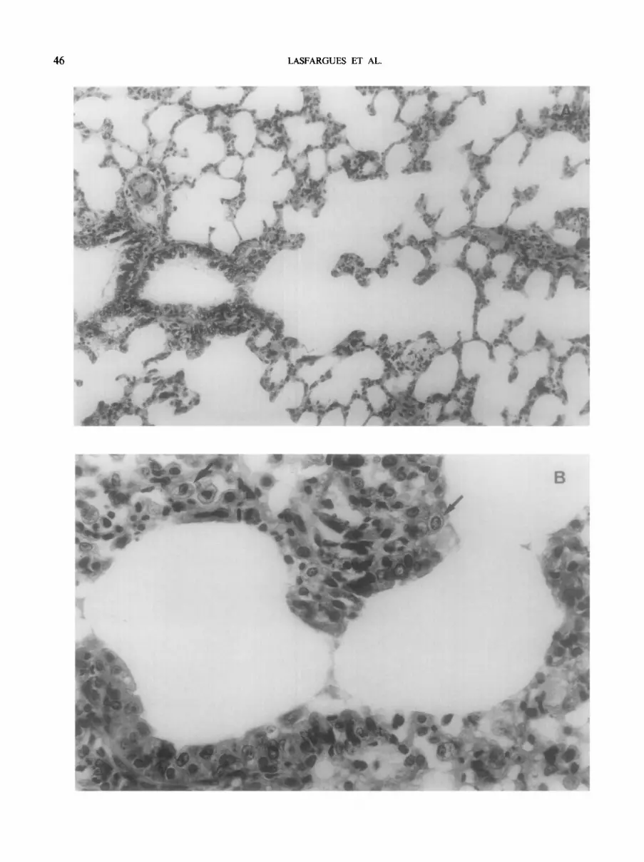
FIG. 1. Lung section 48 hr aher instillation oftungsten carbide (15.67 mg/lOO g) to adult female rat (hematoxylin-eosin-saffron). (A) 200 X magnification. (B) 500 X magnification. Normal parenchymal and bronchiolar structures.

44 LASFARGUES ET AL.

ACUTE LUNG TOXICITY OF
NAD+ at 340 nm in the presence of lactate (Lison and Lauwerys, 1990). NAG was assayed according to the automated method of Tucker et al. ( 1975). Total proteins (TP) were determined by the Coomassie blue staining method using the Bio-Rad kit according to the instructions of the manufacturer; BSA was used as standard. Rat serum albumin was determined by latex immunoassay, calibrated with a pool of normal sera (Bernard and Lauwerys, 1983). Plasminogen activator (PA) activity was assayed spectrophotomet- rically in the presence of S225 1 (0.3 mM) and plasminogen (0.165 CU/ml) in Tris (100 mM, pH 7.4). The acceleration of para-nitroaniline (pNA) pro- duction was monitored at 405 nm as described previously (Knoops et al., 1990). PA activity was expressed in pmol pNA produced/ml/min2. Lysozyme activity was assayed spectrophotometrically by monitoring at 540 nm the decrease in absorbance of a Micrococcus lysodeicticus suspension (250 pgl ml) in phosphate (66 mM)-NaCl (75 mM) buffer (pH 7.3).
The concentration of cobalt in urine was determined by flameless atomic absorption spectrometry.
Cell count. The cell pellets were resuspended in 5 ml sterile 0.9% NaCl containing 0.1% BSA and the total number of live cells was determined by the trypan blue exclusion method. The cells (2OO/rat) were differentiated on cytocentrifuge preparations fixed in methanol and stained with May-Griln- wald-Giemsa.
Srurisrics. All values except for PA activity and cobalturia are arithmetic means f SE. For body and lung weights and BALF parameters, differences between means were assessed using a one-way ANOVA, followed by pairwise comparisons with the Newman-Keuls test. When a Bartlett’s test revealed that the variances were significantly different, comparisons between means were made using the Kruskal-Wallis test.
Differences between mean cobalturias were evaluated using a Mann- Whitney test. Statistical significance was considered at p =S 0.05.
RESULTS
Within 24 hr following the intratracheal instillation of sa- line, cobalt as pure metal powder (1 .O mg Co/ 100 g) or WC- Co mixture (16.67 mg/lOO g) and pure WC (15.67 mg/lOO g), the mortality was 10, 20, 60, and lo%, respectively. Among the animals of the WC-Co group, gasping, cyanosis, and discharge of a pinkish and frothy fluid from the mouth and the nostrils, all signs characteristic of a massive pul- monary edema, were observed prior to death. These signs were not seen in the other groups.
The lung weights of the WC-treated rats were not signif- icantly different from those of the control rats. By comparison with the latter, significant increases in absolute and relative lung weights were observed in the Co and the WC-Co groups. However, the increase was significantly higher in the WC- Co group than in the Co group (Table 1).
The microscopic examination of the lungs of animals in- stilled with WC did not reveal marked changes; there was no edematous reaction, only a mild infiltration of macro- phages in the alveolar duct walls (Fig. 1). The intratracheal instillation of WC-Co induced an acute and diffuse inflam-
COBALT CONTAINING DUSTS 45
matory reaction with generalized oedematous alveolitis (Fig. 2). An accumulation of particle-laden macrophages and neutrophils was observed in the alveoli. Bronchiolar and al- veolar walls were infiltrated by enlarged and vacuolated macrophages with voluminous nuclei and prominent nu- cleoli. The cellular response was particularly pronounced at the bifurcation of alveolar ducts where particle deposits were important. In certain areas, bronchial and bronchiolar epi- thelia were abraded. Lymphoid hyperplasia was also observed in tracheal and bronchial lymph nodes.
Lung damages in animals instilled with Co were more localized and much less important than in the WC-Co-treated rats (Fig. 3). Scattered sites of exudative alveolitis were de- tected in both lungs and cellular proliferation was only found at the origin of alveolar ducts.
A bronchoalveolar lavage was performed 24 hr after the instillation of nonletal amounts of the different preparations; the results of the cellular and biochemical analyses performed on the collected fluid are summarized in Tables 2 and 3, respectively. At the doses tested, WC and Co treatments had no significant effect on the cell content and the various bio- chemical parameters. In contrast, the WC-Co mixture in- duced a significant increase of the macrophage and neutro- phi1 numbers, LDH activity, total protein, and albumin con- centration. These effects were rather similar to those observed after DQ 12 instillation. The activities of NAG and lysozyme were not significantly affected by the various treatments.
The amounts of cobalt excreted in urine within 24 hr fol- lowing the intratracheal instillation of pneumotoxic or nonpneumotoxic doses of cobalt administered as pure Co (1 .O and 0.03 mg/lOO g body wt) or WC-Co mixture (16.67 and 0.50 mg/ 100 g body wt) were significantly more impor- tant when cobalt was administered as WC-Co mixture than as pure Co alone (Table 4).
DISCUSSION
The objective of this study was to compare the acute lung response to cobalt metal powder alone or associated with tungsten carbide dust such as present in hard metal industry. The results clearly demonstrate that, on a cobalt content basis, the tungsten carbide-cobalt mixture is more toxic to the respiratory tract than cobalt alone.
For the mortality and histology studies, the doses of the various powders were selected on the basis of a few prelim- inary experiments designed to assess the dose of pure Co dust ( 1 .O mg/ 100 g) which slightly affected the animal sur- vival. In the final experiment, one control animal instilled
FIG. 2. Lung section 48 hr after instillation of tungsten carbide-cobalt mixture (16.67 mg/lOO g) (corresponding to 1 mg cobalt/l00 g) to adult female rat (hematoxylin-eosin-saffron). (A) 200 X magnification. exudative alveolitis. (B) 500 X magnification. About 50% of cells are macrophages, generally with voluminous nuclei and prominent nucleoli (indicated by arrows).
46 LASFARGUES ET AL.

ACUTE LUNG TOXICITY OF COBALT CONTAINING DUSTS 47
TABLE 2 Bronchoalveolar Lavage Fluid Cell Numbers 24 hr after Intratracheal Instillation of Saline (Control), Tungsten Carbide (WC),
Cobalt (Co), Tungsten Carbide-Cobalt (WC-Co), or Silica (DQ12) Dust in Rats
Groups” Total cells Macrophages Neutrophik Lymphocytes
Saline 2.20 + 0.26b 2.15 + 0.26 0.02 -t 0.01 0.01 2 0.01 WC (1.00 mg/lOO g) 3.00 I!I 0.22 2.19 + 0.20 0.18 It 0.03 0.03 2 0.01 Co (0.06 mg/lOO g) 2.90 + 0.25 2.15 f 0.20 0.12 f 0.10 0.02 + 0.01 WC-Co ( 1 .OO mg/ 100 g) 5.10 * 0.75**t 3.61 f 0.56*'t 1.37 + O.IO*'t 0.05 zk 0.02 DQ12 (1.00 mg/lOO g) 6.80 + 0.65**t 6.00 f 0.49**t 0.12 f 0.25*y 0.07 f 0.02*
’ Five animals in each group. b Arithmetic mean + SE, number of cells X 106. * Significantly different from the control group (p < 0.05). ’ Significantly different from the WC group (p < 0.05). t Significantly different from the Co group (p < 0.05).
with saline died; this exceptional event was clearly due to pentobarbital anesthesia.
Instillation of WC-Co (16.67 mg/lOO g corresponding to 1 .O mg Co/ 100 g) produces a massive pulmonary edema as evidenced by a marked increase in the lung weight, a gen- eralized alveolitis, and a high mortality.
Pure Co dust (1 .O mg/lOO g) is much less toxic; it only produces a small increase in lung weight. Sites of inflam- matory reaction can be detected in all lung lobes but they are very confined; macrophage accumulation is only ob- served at the origin of alveolar ducts, very close to the sites where the particles are deposited. The different histological changes in Co- and WC-Co-instilled animals cannot be ex- plained by a different distribution of the particles in the re- spiratory tract, since for both dusts the lesions are scattered throughout the lungs but are much less intense for pure Co particles. The greater acute toxicity of WC-Co as compared to Co is not due to the different amount of dust instilled ( 16.67 and 1 .O mg/ 100 g, respectively), since animals instilled with WC dust (15.67 mg/lOO g) only show very discrete morphological changes.
Analysis of BALF 24 hr after exposure is a sensitive means to characterize the acute inflammatory response of the lung; it can be used to rank intratracheally instilled mineral dusts or metallic compounds with regard to their acute lung toxicity (Beck et al., 1982, 1987; Henderson et al., 1979a,b; 1985; Lindenschmidt et al., 1990; Moores et al., 198 1; Morgan et al., 1980). Since previous studies had shown that BALF changes were induced by silica (our positive control) at a dose of 1 .O mg/lOO g (Lindenschmidt et al., 1990), the same dose was selected for the WC-Co mixture. The responses of the
various biochemical and cellular parameters measured in this study demonstrate that the acute pneumotoxicity of the WC- Co mixture ( 1 .O mg/ 100 g corresponding to 0.06 mg Co/100 g) is very close to that of crystalline silica ( 1 .OO mg/ 100 g). WC (1 .OO mg/ 100 g) or Co (0.06 mg Co/100 g) dusts alone do not induce any significant elevation in inflammatory cells in BALF as compared to saline. On the contrary, treatment with WC-Co and DQ 12 produce a statistically significant in- crease of the number of macrophages and neutrophils. It is generally accepted that macrophages play a key role in initi- ating the cascade of adverse reactions leading to lung paren- chymal damages induced by toxic particles (Bowden, 1987; Brain, 1988). These cells can release chemotactic factors that attract polymorphonuclear leucocytes and/or monocytes,and stimulate fibroblast proliferation (Bitterman et al., 1982; Martin et al., 1987). Macrophages and neutrophils can also release a number of mediators such as reactive oxygen species and proteolytic enzymes which can damage the lung paren- chyma (Davis, 1986).
The biochemical changes induced by the different dusts parallel the cellular changes and also underline the different lung toxicity of the Co and WC-Co dusts. The biochemical responses are quantitatively and qualitatively similar for WC- Co and DQl2, again reflecting their similar acute lung tox- icity. LDH activity, a cytosolic enzyme frequently used as a nonspecific indicator of cell injury (Beck et al., 1982; Hen- derson et al., 1979a,b; Moores et al., 1981), appears to be a more sensitive parameter than the lysosomal enzymes, NAG and lysozyme (Weissman et al., 1980).
In the WC-Co group, TP and albumin content in BALF reach almost 200 and 300% of the control values, respec-
FIG. 3. Lung section 48 hr after instillation of cobalt particles (1.00 mg/lOO g) to adult female rat (hematoxylin-eosin-saffron). (A) 200 X magnification. The lesion is localized at the origin of alveolar ducts: the number of cells in alveolar walls is increased. (B) 500 X magnification; increased number of macrophages (indicated by arrows).

48 LASFARGUES ET AL.
TABLE 3 Lactate Dehydrogenase (LDH), /3-N-Acetyl-Glucosaminidase (NAG), Plasminogen Activator (PA), and Lysozyme Activities,
Total Protein (TP), and Serum Albumin (ALB) Content of Bronchoalveolar Lavage Fluid 24 hr after Intratracheal Instillation of Saline (Control), Cobalt (Co), Tungsten Carbide (WC),
Tunstgen Carbide-Cobalt (WC-Co), or Silica (DQ12) Dust in Rats
Groups’ LDH (It-l/liter)
Saline 44.5 f I.36 WC (1.00 mg/lOO g) 50.8 + 2.6 Co (0.06 mg/lOO g) 36.6 f 6.9 WC-Co (1.00 mg/lOO g) 145.0 f 39.1**t DQ12 (1.00 mg/lOO g) 112.9 rt 16.0**t
NAG (IU/ liter)
0.97 + 0.12 1.05 + 0.14 1.24 f 0.26 1.74 f 0.37 1.62 f 0.35
PA (pmol/ ml/min’)
1.45 (0.50-2.40)’ 1.24 (0.50-l .97) 3.99 (0.50-7.47) 7.56 (0.50-14.61)
10.82 (0.50-21.14)
Lysozyme h/ml)
8.34 + 1.42 9.38 f 1.56
10.21 f 2.11 9.86 IL 2.08 9.61 + 0.95
TP h/ml)
446.4 2 79.1 438.0 f 37.9 445.0 f 56.0 860.2 2 162.5*“1 803.2 + 122.2*‘t
ALB (&ml)
79.8 + 6.6 105.8 + 13.1 110.0 f 21.0 229.7 + 71.1* 195.2 + 49.2*
u Five animals in each group. b Arithmetic mean k SE. ’ Median (ranges). * Significantly different from the control group (p < 0.05). ’ Significantly different from the WC group (p < 0.05). t Significantly different from the Co group (p i 0.05).
tively. The albumin increase is proportionally more impor- tant than that of TP, reflecting an increased vascular per- meability associated with the inflammatory reaction (Brain, 1988; Beck et al., 1982). In contrast, an equivalent dose of cobalt alone does not produce any significant changes in the biochemical parameters.
Since phagocytic cells play an important role in the early pathogenesis of pulmonary fibrosis, we also determined two of their secretory products, i.e., lyzozyme and PA. Lyzozyme is the major constitutive secretory product of macrophages (about 25% of extracellularly secreted proteins). This secre- tion proceeds in a continuous fashion and is unaffected by a variety of stimuli such as cytokines. PA, a serine protease that converts plasminogen to the active enzyme plasmin, promotes the degradation of various extracellular substrates. PA has been suggested to participate in a number of processes
including fibrinolysis, inflammation, and tissue remodeling. In contrast to lyzozyme, its production by macrophages is modulated by stimuli such as steroids and cytokines (Gordon, 1978). An increased production of PA by alveolar macro- phages has been reported in subjects with cryptogenic fibrosis (Robinson, 1988) or asbestos-induced alveolitis (Cantin et al., 1989). Alveolar epithelial cells represent also an impor- tant source of PA in the rat lung (Gross, 1990). Despite the significative increase in the number of macrophages and neutrophils in the WC-Co and DQ 12 groups, no modification of lyzozyme activity in BALF was observed. In contrast, the median value of PA activity in the different groups increases in the following order: WC < Co < WC-Co < DQ 12. These results suggest that PA activity of BALF may be an indicator of lung inflammation and tissue damage. However, in several animals of each group, PA activity of BALF is below the
TABLE 4 Urinary Excretion of Cobalt before and after the Intratracheal Instillation of Identical Amounts of Cobalt Given as Pure Cobalt
Particles (Co) or as Tungsten Carbide-Cobalt Mixture (WC-Co) in Rats
Treatment Cobalt
(w/l00 g) N
Cobalt in urine (r(g/24 hr)
Before treatment After treatment
co 0.03 0.03 5 0.58 (0.27-0.88)” 6.81 (1.52-12.10) 1.00 1.00 8 0.44 (0.22-0.65) 49.14 (7.91-90.36)
WC-CO 0.50 0.03 5 0.47 (0.20-0.74) 22.17 (9.3-35.04)* 16.67 1.00 8 0.38 (0.12-0.64) 371.07 (93.81-648.33)**
a Median (ranges); significantly different from the corresponding Co group. * p < 0.05.
** p < 0.005.

limit of detection; this is likely due to the fact that PA activity estimated from an in vivo hamster bioassay. Toxicol. Appl. Pharmacol.
is determined by the balance between enzyme and PA in- 87,222-234.
hibitors present in the BALF. Both can increase in the course Bitterman, P. B., Rennard, S. I., Hunninghake, G. W., and Crystal, R. G.
of an inflammatory process and it may be that in some an- (I 982). Human alveolar growth factor for fibroblasts. J. Clin. Invest. 70,
imals the inhibitory activity is predominant (Chapman et 806-822.
a!., 1990). Bowden, D. H. (1987). Macrophages, dust and pulmonary disease. Exp.
Whatever the dose level, cobalturia is significantly higher Lung Res. 12,89-107.
in the WC-Co-treated animals than in the corresponding Co Brain, J. D. (1988). Lung macrophages: How many are there? What do they
do? Am. Rev. Respir. Dis. 137, 507-510. groups. In animals treated with a high dose (16.67 mg/lOO Brain, J. D., and Beck, B. D. (1985). Bronchoalveolar lavage. In Handbook g) of WC-Co, increased lung vascular permeability might of Experimental Pharmacology, pp. 203-226. Springer-Verlag New York/ have partly contributed to the higher absorption of cobalt. Berlin.
In the low-dose group (0.50 mgf100 g), LDH activity (38.3 Cantin, A., Allard, C., and Begin, R. (1989). Increased alveolar plasminogen
+ 12.3 III/l), TP (336.0 + 81.3 bg/ml) and albumin (84.4 +- activator in early asbestosis. Am. Rev. Respir. Dis. 139,604-609.
25.5 pg/ml) contents of BALF are not altered, indicating the Chapman, H. A., Yang, X., Zenzius Sailor, L., and Sugarbaker, D. J. ( 1990).
absence of lung lesion. Even under these conditions, cobal- Developmental expression of plasminogen activator inhibitor type I by
turia is significantly higher than in the corresponding group alveolar macrophages. Possible role in lung injury. J. Immunol. 145,3398- 3405.
treated with cobalt alone (0.03 mg/ 100 g). These observations support our previous in vitro finding suggesting an increased
Cullen, M. R. (1984). Respiratory diseases from hard metal exposure: A continuing enigma. Chest. 86, 513-5 14.
bioavailability of cobalt dust when combined with tungsten Davis, G. S. (1986). Pathogenesis of silicosis: Current concept and hypotheses. carbide. Lung. 164, 139-I 54.
In conclusion, the present in vivo study confirms and ex- Demedts, M., and Ceuppens, J. L. (1989). Respiratory diseases from hard tends our previous in vitro findings on rat alveolar macro- metal or cobalt exposure-Solving the enigma. Chest. 95, 2.
phages regarding the different biological reactivity of cobalt Demedts, M., Gheysens, B., Nagels, J., Verbeken, E., Lauweryns, J., Van
alone or in association with tungsten carbide. The acute lung Den Eeckhout, A., Lahaye, D., and Gyselen, A. (1984). Cobalt lung in
toxicity of the tungsten carbide-cobalt mixture is much diamond polishers. Am. Rev. Respir. Dis. 130, 130-I 35.
higher than that of each individual component; it is close to Gordon, S. (1978). Regulation of enzyme secretion by mononuclear phago-
that of crystalline silica. cytes: Studies with macrophage plasminogen activator and lyzozyme. Fed. Proc. 37,2754-2758.
If the acute toxicity of these preparations is predictive for Gross, T. J., Simon, R. H., and Sitrin, R. G. (1990). Expression of urokinase- their long-term toxicity, this finding may have practical im- type plasminogen activator by rat pulmonary alveolar epithelial cells. Am.
plications for the prevention of cobalt-induced lung toxicity. J. Respir. Cell. Mol. Biol. 3, 449-456.
Acceptable exposure levels to the metal might have to vary Hartung, M., Schaller, K. H., and Brand, E. (1982). On the question of the
depending on whether exposure is to pure cobalt metal par- pathogenic importance of cobalt for hard metal fibrosis of the lung. Int.
ticles or to a combination of cobalt with other substances Arch. Occup. Environ. Health 50, 53-51.
such as tungsten carbide. Further studies are currently in Henderson, R. F., Rebar, A. H., Pickrell, J. A., and Newton, G. J. (1979a).
progress to compare the long-term lung toxicity of these Early damage indicators in the lungs. III. Biochemical and cytological
preparations and to assess the predictive value of the early response of the lung to inhaled metal salts. Toxicol. Appl. Pharmacol. 50, 123-126.
biological changes detected in BALF. Henderson, R. F., Rebar, A. H., and Denicola, D. B. (1979b). Early damage indicators in the lungs. IV. Biochemical and cytological response of the
ACKNOWLEDGMENTS lung to inhaled metal salts. Toxicol. Appl. Pharmacol. 51, 129- 135.
Henderson, R. F., Hobbs. C. H., Hahn, F. F., Benson, J. M., Pickrell, J. A.,
This work was supported by the Fondation Ernest Duffo (France) and and Silbaugh, S. A. (1985). A comparison of in vitro and in vivo toxicity
the Fonds de la Recherche Scientilique Medicale (Belgium). Dr. Lasfargues of mineral dusts. In In Vitro Effects of Mineral Dusts (E. G. Beck and J.
is a research fellow of the Brussels, Capital Region (Belgium). Bignon, Eds.), pp. 521-527. Springer-Verlag, Berlin. Knoops, B., Lison, D., Collette. C., Lauwerys, R., and Picard, J. J. (1990).
Plasminogen activator activity of normal and retinoic acid-treated post- REFERENCES implantation embryos. Biochem. Pharmacol. 39, 1545-1548.
Kochetkova, T. A. (1960). On the question of the effect of cobalt powders. Bernard, A., and Lauwerys, R. (1983). Continuous flow system for the au- Gig. Tr. Pr@ Zabol. 4, 34-38.
tomation of latex immunoassay by particle counting. Clin. Chem. 19, 1007-1011.
Lindenschmidt, R. C., Driscoll, K. E., Perkins, M. A., Higgins, J. M., Maurer, J. K., and Belfiore. K. A. (1990). The comparison of a fibrogenic and two
Beck, B. D., Brain, J. D., and Bohannon, D. E. (1982). An in vivo hamster nonfibrogenic dusts by bronchoalveolar lavage. Toxicol. Appl. Pharmacol. bioassay to assess the toxicity of particles for the lungs. Touicol. Appl. 102,268-28 1. Pharmacol. 66,9-29. Lison, D., and Lauwerys, R. (1990). In vitro cytotoxic effects of cobalt con-
Beck, B. D.. Feldman, H. A., Brain, J. D., Smith, T. J., Hallock, M., and taining dusts on mouse peritoneal and rat alveolar macrophages. Environ. Gerson, B. (1987). The pulmonary toxicity of talc and granite dust as Res. 52, 187-198.
ACUTE LUNG TOXICITY OF COBALT CONTAINING DUSTS 49

50 LASFARGUES ET AL.
Martin, T. R., Rangi, G., Merritt, T. L., and Henderson, W. R. (1987). The relative contribution of leukotriene B4 to the neutrophil chemotactic activity prcduced by the human alveolar macrophage. J. Clin. Invest. &I,1 114- 1124.
Morgan, A., Moores, S. R., Holmes, A., Evans, J. C., Evans, N. H., and Black, A. (1980). The effect of quartz, administered by intratracheal instillation. on the rat lung. 1. The cellular response. Environ. Res. 22, 1-12.
Moores, S. R., Black, A., Evans, J. C., Evans, N., Holmes, A., and Morgan, A. (198 1). The effect of quartz, administered by intratracheal instillation, on the rat lung. II. The short term biochemical response. Environ. Res. 24,275-285.
NIOSH Occupational Hazard Assessment. (198 1). Criteria for Controlling Occupational Exposure to Cobalt. DHHS (NIOSH) publication No. 82- 107. Superintendent of Documents, U.S. Government Printing Office, Washington.
Reinl, W., Schnellbacher, F., and Rahm, G. (1979). Lungenfibrosen und entzilndliche lungenerkrankungen nach einwirkung von cobaltkon- taktmasse. Zbl. Arbaitsmed. Arbeitsschutz Prophyl. 29, 3 18-325.
Robinson, B. W. S. (1988). Production of plasminogen activator by alveolar macrophages in normal subjects and patients with interstitial lung disease. Thorax 43,508-5 15.
Ruttner, J. R., Spycher, M. A., and Stolkin, I. (1987). Inorganic particulates in pneumoconiotic lungs of hard metal grinders. Br. J. Znd. Med. 44,657- 660.
Sprince, N. L., Oliver, C., Eisen, E. A.. Greene, R. E., and Chamberlin, R. I. (1988). Cobalt exposure and lung disease in tungsten carbide pro- duction. Am. Rev. Resp. Dis. 138, 1220.
Tucker, S. M., Boyd, P. J. R., Thompson, A. E., and Price, R. G. (1975). Automated assay of N-acetyl-&glucosaminidase in normal and patho- logical urine. Clin. Chim. Acta. 62, 333-339.
Weissman, G., Smolin, J. E., and Korchak, H. M. (1980). Release of in- flammatory mediators from stimulated neutrophils. N. Engl. J. Med. 303, 27-39.


