Embed Size (px)
Citation preview
Experimental Parasitology 128 (2011) 217–224
Contents lists available at ScienceDirect
Experimental Parasitology
journal homepage: www.elsevier .com/locate /yexpr
Cloning, sequencing and functional expression of cytosolic malatedehydrogenase from Taenia solium: Purification and characterizationof the recombinant enzyme
Gabriela Nava a, Juan P. Laclette b, Raúl Bobes b, Julio C. Carrero b, Horacio Reyes-Vivas c,Sergio Enriquez-Flores c, Guillermo Mendoza-Hernández d, Agustín Plancarte a,⇑a Departamento de Microbiología y Parasitología y, Universidad Nacional Autónoma de Mexico, Facultad de Medicina, D.F., Mexicob Departamento de Inmunología, Instituto de Investigaciones Biomédicas, Universidad Nacional Autónoma de México, D.F., Mexicoc Laboratorio de Bioquímica-Genética y División de Investigación, Instituto Nacional de Pediatría 04530 México, DF., Mexicod Departamento de Bioquímica, Facultad de Medicina, Universidad Nacional Autónoma de México, D.F., Mexico
a r t i c l e i n f o a b s t r a c t
Article history:Received 5 March 2011Accepted 19 March 2011Available online 31 March 2011
Keywords:Taenia soliumCysticercosisMalate dehydrogenaseMetabolismRecombinant enzymeCloning
0014-4894/$ - see front matter � 2011 Elsevier Inc. Adoi:10.1016/j.exppara.2011.03.008
⇑ Corresponding author. Fax: +52 55 5623 2384.E-mail address: [email protected] (A. Planca
We report herein the complete coding sequence of a Taenia solium cytosolic malate dehydrogenase(TscMDH). The cDNA fragment, identified from the T. solium genome project database, encodes a proteinof 332 amino acid residues with an estimated molecular weight of 36517 Da. For recombinant expression,the full length coding sequence was cloned into pET23a. After successful expression and enzyme purifi-cation, isoelectrofocusing gel electrophoresis allowed to confirm the calculated pI value at 8.1, as deducedfrom the amino acid sequence. The recombinant protein (r-TscMDH) showed MDH activity of 409 U/mgin the reduction of oxaloacetate, with neither lactate dehydrogenase activity nor NADPH selectivity. Opti-mum pH for enzyme activity was 7.6 for oxaloacetate reduction and 9.6 for malate oxidation. Kcat valuesfor oxaloacetate, malate, NAD, and NADH were 665, 47, 385, and 962 s�1, respectively. Additionally, apartial characterization of TsMDH gene structure after analysis of a 1.56 Kb genomic contig assemblyis also reported.
� 2011 Elsevier Inc. All rights reserved.
1. Introduction important products: NADH as reducing equivalent, and oxaloace-
Humans develop neurocysticercosis (NC) when Taenia soliumcysticerci are lodged in their central nervous system. NC is a heter-ogeneous neurological disease that can sometimes be the cause ofdeath (Sotelo and Marin, 1987). This neurologic disease is endemicto developing countries, although it has been introduced into thedeveloped world through infected migrants (Schantz et al., 1998).In spite of its importance as a global public health problem, re-search on this parasite has remained neglected and little is knownabout its energy metabolism. Adequate knowledge of how this par-asite generates and uses energy, could aid specific drug design forbetter treatment against T. solium.
A possible enzyme candidate is the NAD-dependent cytosolicmalate dehydrogenase (TscMDH, EC 1.1.1.37) (Banaszak andBradshaw, 1975; Williamson et al., 1973), a ubiquitous enzymethat exists in all eukaryotes (Gietl, 1992). The NAD-dependentdehydrogenases are one of the largest and best-studied familiesof nucleotide-binding proteins: over 100 different members havealready been identified (Minárik et al., 2002). cMDH is an enzymethat converts malate to oxaloacetate. This reaction results in two
ll rights reserved.
rte).
tate, as metabolic substrate. Together with aspartate aminotrans-ferase isoenzymes, cMDH is involved in the transfer of reducingequivalents from cytosol to mitochondria (aspartate–malate shut-tle), in the form of malate/oxaloacetate rather than as NAD/NADH.(Musrati et al.,1998). Additionally, cMDH, in combination with theNADP dependent malate dehydrogenase (decarboxylating EC1.1.1.39) has been proposed as a MDH-complex involved inregulating energy metabolism in helminthes (Fodge et al., 1972;Landsperger and Harris, 1976). Moreover, cMDH is important inother metabolic pathways such as the glyoxylate cycle (Davisand Goodman, 1992), glyoxylate degradation (Sakai et al., 2008),mixed acid fermentation (Amarneh and Vik, 2005) and gluconeo-genesis (Chakravarty et al., 2005). Cloning and functional recombi-nant expression of the TscMDH should allow for structure/functionstudies, eventually leading to the identification of drugs that couldselectively inhibit this parasite enzyme.
2. Material and methods
2.1. Materials
Plasmid vector pET23a was obtained from EMD Chemicals Inc.(Darmstadt, Germany). Escherichia coli strains BL21 was purchased

218 G. Nava et al. / Experimental Parasitology 128 (2011) 217–224
from Invitrogen Corporation (Carlsbad, CA, USA). Plasmid mini-preparation kit was obtained from Invitrogen Corporation, (Carl-bad, CA, USA). Bacto tryptone and Bacto yeast extract were fromBecton Dickinson and Company, (Texarkana, TX, USA). Sodiumdodecyl sulfate and agarose LE were from Bio-Rad, (Hercules CA,USA). TAQ DNA polymerase, was from GoTaq Flexi DNA Polymer-ase Promega Corporation, (Madison WI, USA). EcoR1 and Hind IIIrestriction enzymes were from Biolabs, (Ipswich, MA, USA). T4DNA ligase was purchased from Promega Corporation, (Madison,WI, USA). IPTG, L-malate, NAD and ampicillin were from Sigma–Al-drich (St. Louis MO, USA). Other chemicals were of at least reagentbiology grade.
2.2. Cloning and sequence analysis of the MDH gene
An N-terminal twenty amino acid sequence, previously ob-tained from purified native T. solium cMDH (Plancarte et al.,2009), was used to query for MDH in the T. solium genome projectdatabase (Aguilar-Díaz et al., 2006). The full length coding se-quence was available as a cDNA clone. Two primers were designedto amplify the full length coding sequence of TscMDH (Invitrogen).The forward primer 50_AGCGGAATTCATGCCTGGACCTCTTAG_30 in-cluded an EcoR1 restriction site and the reverse primer50_ATGGAAGCTTCTTGAAGGAGGAAAGAGC_30 included a Hind IIIrestriction side. PCR was performed as follows: an initial incuba-tion at 94 �C for 2 min denaturizing; 40 cycles of amplification at94 �C for 30 s, annealing at 55 �C for 90 s, and extension at 72 �Cfor 90 s; the final extension was at 72 �C for 10 min. The PCR prod-uct was run in 1% agarose gel electrophoresis with DNA markers assize standard, and visualized with ethidium bromide. PCR productwas sequenced by Sanger procedure (Sanger and Goulson, 1975)and analyzed by 310 Genetic Analyzer (Applied Biosystems). Thenucleotide sequence was identified as coding for TscMDH by usingBLAST (http://www. Ncbi.nlm.nih.gov/BLAST/) (Altschul et al.,1979).
2.3. Recombinant expression and purification of recombinant T. soliumcytoplasmic malate dehydrogenase (r-TscMDH)
The full coding region of the r-TscMDH obtained by PCR wascloned into EcoR1 and Hind III sites of the expression vectorpET23A (Novagen, Darmstadt, Germany) following standard meth-ods. After transformation into E. coli BL21 (DE3) pLysS, positiveclones were selected on ampicillin and induced for expression ofamino-terminal His-tagged r-TscMDH by incubation with 1 mMIPTG. Lysates from BL21 (DE3) pLysS cells induced cultures wereresolved in SDS–PAGE to confirm expression levels. Finally, His-tagged recombinant protein was purified from inclusion bodiesby affinity chromatography using HiTrap columns (Pharmacia Bio-tech, USA) and stored at �20 �C until use. Protein samples weresubjected to SDS–PAGE at room temperature in slab gels (15%acrylamide) (Laemmli, 1970), and the gels stained for protein withCoomassie blue or silver nitrate.
Proteins’ concentration was determined by the Lowry method(Lowry et al., 1951) with BSA as standard. Additionally, concentra-tion of the r-TscMDH was also determined by measuring its absor-bance at 280 nm and using the molar extinction coefficient value ofe = 1.213 (Stanley and von Hippel, 1989)
2.4. Isoelectric focusing and in situ enzyme reaction
The isoelectric focusing gel procedure of Beauchamp andFridovich (1971) was employed. A 0.1 lg sample of r-TscMDHwas mixed with 2 ll of sample buffer (glycerol 60%, ampholytepH 3.5–10, 0.66%) (Amersham Pharmacia Biotech AB) and ampho-lyte pH 5–7, 3.3% (Amersham Pharmacia Biotech AB). Immediately
after isoelectric focusing, the gel was incubated with 0.21 mMNADH, 0.2 mM oxaloacetate and 1 mM nitroblue tetrazolium(NBT) (Sigma–Aldrich). Enzyme activity was detected by reductionof NBT in presence of 3 mM phenazide methosulfate (PMS) by theaction of the NADH in the MDH forward reaction. The reaction wasstopped by addition of distilled water.
2.5. MDH activity and kinetic parameters calculation for r-TscMDH
r-TscMDH enzyme activity was studied in an Ultrospec 3100(Amersham) spectrophotometer, in 1 cm light path cells at roomtemperature, and following the procedure described elsewhere(Englard, 1969) with some modifications. Briefly, forward MDHactivity, with oxaloacetate as substrate, was measure in freshlyprepared solutions of 0.2 mM oxaloacetate and 0.21 mM b-NADHin Tris–HCl (pH 8.0), and 0.5 lg of r-TscMDH at a final volume of1 mL. Initial velocities were determined spectrophotometricallyby measuring NADH decrease at 340 nm. For reactions measuringreverse MDH activity, the procedure of Prichard and Schofield(1968) with some modifications was followed. The reaction buffercontained 50 mM glycine (pH 9.6), 3 mM L-malate and 0.52 mMNAD. Initial velocities were determined spectrophotometricallyby measuring NADH increase at 340 nm. In addition, assays werecarried out substituting D-malate for L-malate, and NAD for NADP.One MDH unit is defined as the amount of enzyme required to oxi-dize or reduce 1 lmol coenzyme min�1mg�1. Enzyme specificactivity was defined as enzyme units per milligram of protein.
2.6. Effect of pH and temperature on r-TscMDH stability
The pH-dependence on MDH activity of r-TscMDH was evalu-ated by measuring its forward velocity reaction in the pH range4.0–11, as described above. The pH-dependent stability of ther-TscMDH activity was determined by equilibrating aliquots ofthe recombinant protein (5 lg/ml) through dialysis against a0.1 M solution of the following buffers: acetate (pH 4.0–5.5), phos-phate (pH 6.0–8.0) and tris–HCl (pH 8.5–11.0) for 24 h. After incu-bation, the enzyme activity of each aliquot was measured at theoptimum pH. All pH assays were conducted at room temperature.
The effect of temperature on the oxaloacetate reduction reac-tion catalyzed by r-TscMDH was examined under standard assayconditions (Englard, 1969). The r-TscMDH was incubated at tem-peratures ranging from 5 �C to 60 �C for 10 min in Tris–HCl50 mM pH 8.0. Afterwards, the aliquots were allowed to reachroom temperature and enzymatic activity was determined at theoptimum pH, as described above.
2.7. Effect of NAD analogs on MDH activity
The r-TscMDH enzyme activity for five different pyridine nucle-otide analogues of b-NAD, altered in the pyridine moiety, weredetermined through initial velocity studies in the reverse reaction,as described above (Yoon and Anderson, 1988). Coenzyme analogconcentrations were varied from 0.2 to 0.8 mM for 3-acetyl pyri-dine adenine dinucleotide (APAD); nicotinamide mononucleotide(NM); nicotine acid adenine dinucleotide (NAAD); nicotinamideadenine dinucleotide phosphate (NADP), and a-nicotinamide ade-nine dinucleotide (NAD). Molar extinction coefficients at maximalabsorption wavelength were used to determine the reaction rates.
2.8. Inhibition study
Inhibition studies were performed by the procedure of Tahirand Mannervik (1985) in both directions of the r-TscMDHactivity. Praziquantel (PZQ) and albendazole (AB) (Sigma–AldrichChem.) were used in variable concentrations of 0.1–6 mM and
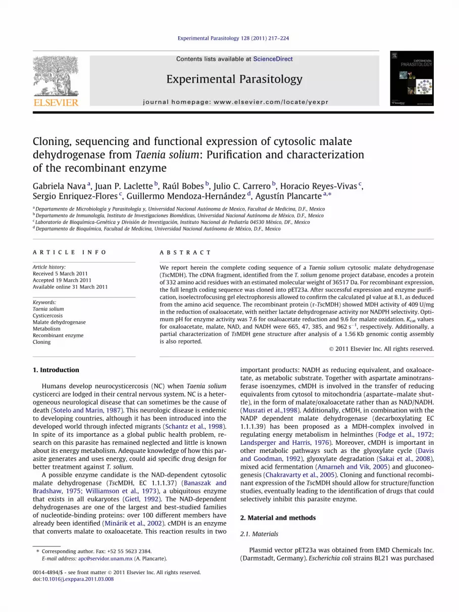
Fig. 1. (A). Alignment of the predicted amino acid sequence of the cloned r-TscMDH with other MDH sequences. Amino acids known to be involved in binding to NAD andmalate are respectively shown in grey and bold. AA involved in the binding of both substrates are indicated by asterisks. Eg. Echinococcus granulosus (Acc. no gi 6016537), Sc.Sus scrofa (Acc. no gi 5107783), Strongylocentrotus purpuratus (Acc. no gi 72124124), Mb. Monosiga brevicollis MX1 (Acc. no gi 167519943), Dd. Dictyostelium discoideum (Acc.no gi 66804051), Tt. Tetrahymena thermophila (Acc. no gi 146165401), Cs. Clonorchis sinensis (Acc. no gi 48686715), Ce. Caenorhabditis elegans (Acc. no gi 17561064), Xl.Xenopus laevis (Acc. no gi 38014771), Mc. Mytilus californianus (Acc. no gi 73656362). (B). Gene structure and protein synthesis sequence of TscMDH. The gene is composed oftwo exons and one intron. TATA-box, INF1, TFIID and SP1, as potential transcription factors are shown for 5́promotor region, and uga, uua, uag, and poly A tail as stop codonsin the 3́region.
G. Nava et al. / Experimental Parasitology 128 (2011) 217–224 219

Table 1Purification of r-TscMDH.
Step Total protein (mg) r-cMDHTs (mg) Purity (%) Yield (%)
Lysatea 33.15 9.95 30 100Eluate 6 6 95 60
a Purification was achieved from 2.5 g of wet obtained from 1 L of E. coli BL21harboring plasmid pET23a.
220 G. Nava et al. / Experimental Parasitology 128 (2011) 217–224
0.1–0.3 mM, respectively. Each drug was dissolved in each one ofthe solvents: ethanol, methanol, isopentyl alcohol and dimethylsulfoxide (DMSO) at 1% or less as final concentration in the assaybuffer. The r-TscMDH was incubated with these drugs for15–30 min or nonincubated before developed the MDH activity.
Fig. 2. SDS–PAGE, immunoblot and isoelectric focusing of r-TscMDH. (A) SDS–PAGEof r-TscMDH expressed in E. coli BL21. Lane 1: protein molecular weight markers,lane 2: lysate of bacteria with parent plasmid pET23a, lane 3: lysate of bacteria withrecombinant r-TscMDH induced with IPTG, lane 4: recombinant r-TscMDH purifiedby HiTrap IMAC metal affinity resin. (B) Immunoblot. SDS–PAGE was performed asin panel A and the r-TscMDH and T. solium crude extract were electroblotted ontoPVDF sheet. The recombinant protein was developed with a polyclonal serum raisedin rabbits against r-TscMDH in the purified recombinant protein, lane 5; and in theT. solium crude extract, lane 6. Lanes 7 and 8 are the same as lanes 5 and 6, butincubated with a normal rabbit serum. (C) Isoelectric focusing. Lane 8: isoelectricfocusing of r-TscMDH performed as described under Methods. After focusing the gelwas stained for MDH activity. Only one protein appears at pI � 8.5.
2.9. Circular dichroism spectra (CD) spectra
Spectra from spectropolarimeter (JASCO J-810) were recordedat 25 �C using a 0.1 cm quartz cell. CD measurements were ob-tained in continuous scanning mode at a speed of 100 nm/min, atfar-UV (260–190 nm). r-TscMDH samples (1 mg/ml) were previ-ously dialyzed against 25 mM sodium phosphate buffer and1 mM DTT (pH 7.4) and centrifuged to remove any precipitated.Three scans were recorder and averaged. Results were expressedas the mean residue molar ellipticity (h) which is defined as:
H ¼ hobsðMRWÞð100Þ=lc
where hobs is the observed mean residue ellipticity in degrees; c isthe concentration of protein in mg/ml; MRW is the mean residueweight (109.9) and l is the light path length in centimeters.
2.10. Thermal unfolding assay
Thermal denaturation curves were determined by monitoringthe change in the circular dichroism (CD) value at 222 nm usinga 0.1 cm quartz cell. The temperature was increased from 40 to65 �C at a rate of 1 �C/min. For experiments, r-TscMDH (1 mg/ml)was previously dialyzed against 25 mM sodium phosphate buffer(pH 7.4). From the data, the apparent fraction of denatured sub-units (fD) was calculated using the equation:
FD ¼ yN� y=yN� yD
where yN and yD are the ellipticity values of the native andunfolded fractions, respectively. Both parameters were linearextrapolations from the initial and terminal portions of the curveof y as a function of temperature.
3. Results
3.1. Cloning and characterization of r-TscMDH
A cDNA library from T. solium cysticerci was screened using aDNA sequence deduced from a 20 amino acid sequence previouslyobtained from purified native T. solium cMDH (Plancarte et al.,2009). To confirm that the isolated clones corresponded to T. soli-um cMDH (r-TscMDH), inserts of positive plaques were sequencedin both directions using an automated fluorescent genetic analyzer(ABI PRISM Applied Biosystems, CA, USA). The nucleotide anddeduced amino acid sequences of r-TscMDH have been depositedin GenBank accession no HQ207526.
The identified cDNA insert contained 1562 bp (Fig. 1A),encoding a protein of 332 amino acid residues, corresponding toa 36517 Da protein with an isoelectric point of 8.7. The gene iscomposed of two 498 pb exons, and one 76 pb intron. Bioinformat-
ics programs yielded the potential transcription factors as TATA-box, TFIID, SP1 and INF.1, situated in the UTR 50 region. In fact,INF.1 is in the 2-Cys peroxiredoxin promoter gene region from T.solium and T. crassiceps (Vaca-Paniagua et al., 2009). The uga,uua, uag and poly A tail stop codons were ubicated in the UTR 30
(Fig. 1 B).Alignment with other malate dehydrogenases showed the high-
est identity (90%) with cMDH of Equinococcus granulosus, a closerelative of T. solium, as well as moderate identity with MDH of pork(Sus scrofa), Tetrahymena thermophila and Strongylocentrotus purpu-ratus (58%, 57%, 52% identity, respectively). The conserved residuesArg-92, Arg-99, Asp-159, Arg-162, and His-187 (Fig. 1A), known toparticipate in the substrate binding and catalysis, are present in ther-TscMDH (Goward and Nicholls, 1994).
3.2. Recombinant expression and purification of r-TscMDH
Recombinant expression of r-TscMDH was carried out in BL21cells using pET-23a. The plasmid construct contained the fulllength coding sequence for r-TscMDH as a fusion protein with aHis-tag at the C-terminal end. Purified r-TscMDH protein was ob-tained by affinity chromatography through HiTrap IMAC Sepharosecolumns. As shown in Table 1, up to 60% of the recombinant pro-tein was recovered. In a typical preparation, about 33 mg of puri-fied r-TscMDH could be obtained from 1 L transformed cellculture. After concentrating the purified r-TscMDH with an Amiconsystem, its concentration reached up to 1.3 mg/ml. SDS–PAGE ofthe purified r-TscMDH (Fig. 2A) revealed a single band of around36 kDa. This value was in agreement with the deduced monomericmolecular mass of the C-terminal His6-tagged r-TscMDH(MW = 37340 Da). Also, r-TscMDH and T. solium cMDH in a crude

Fig. 3. (A) Lineweaver–Burk plot of initial velocities r-TscMDH activity (1/v) versus 1/[oxaloacetate] in the forward reaction. 3 (B). (1/v) versus 1/[malate] in the reversereaction. The intercepts in the ordinates are Vmax values, the intercepts in the abscissas are km values.
G. Nava et al. / Experimental Parasitology 128 (2011) 217–224 221
extract were recognized by polyclonal serum raised in rabbitsagainst r-TscMDH (Fig. 2B).
3.3. Isoelectric focusing and in situ enzyme reaction
When r-TscMDH was analyzed by isoelectric focusing gel(Fig. 2C) the estimated pI at 8.1, deduced from its amino acid se-quence, was confirmed by determination of MDH activity at theposition of the gel.
3.4. Kinetic characterization of r-TscMDH enzyme activity
The double reciprocal plots of initial velocities against severaloxaloacetate and NADH fixed concentrations, produced sets ofstraight intersecting lines (Fig. 3A). Catalytic constants for the re-verse reaction developed by r-TscMDH were obtained by the same
method (Fig. 3B). The km and Vmax values, as well as the Kcat andKcat/km ratios obtained for both reaction directions are shown in ta-ble II. The r-TscMDH showed specific MDH activity of 409 U mg�1,and no activity was detected when NADP(H) was used as a substi-tute for NAD(H), even at concentrations higher than 20 mM or at10-fold higher enzyme concentration. No enzyme LDH activitywas observed, even at an enzyme concentration that was 10 timeshigher than that used in the MDH activity assay, in absence ofD-malate.
3.5. Effect of pH and temperature on r-TscMDH stability
Enzyme activity, evaluated in the 4.0–11 pH range, showed thehighest specific activity at pH 8.0. Functional assays carried out atvarious pHs and temperatures, showed highest stability in the 7.0–8.5 pH range, and within a 5 �C to 60 �C temperature range. Above

Fig. 4. Circular dichroism spectra of r-TscMDH in the near-UV. The curve ischaracteristic of an a-helix (208–190 and 222 nm) and ß-sheet (215–218 nm)structure protein.
Fig. 5. Temperature-induced denaturation. The curve was determined by monitor-ing ellipticity at 222 nm in the near-UV circular dichroism spectra. At the 0.5 valuethe r–TscMDH was totally inactive.
222 G. Nava et al. / Experimental Parasitology 128 (2011) 217–224
40 �C, r-TscMDH activity gradually decreased and became unde-tected at 60 �C. In addition, this enzyme was stable at 20 �C and37 �C over a 30 min period.
3.6. Effect of NAD analogues on r-TscMDH enzyme activity
When r-TscMDH activity was evaluated in presence of fivedifferent co-substrates, only 3-acetyl pyridine adenine (APA)was reduced. However, enzyme activity for APA was lower(9.55 lmol min�1 mg�1) than that obtained for NAD 27.2 (lmolmin�1 mg�1).
Table 2KM, Kcat, Vmax values in the direct and reverse reactions catalyzed by r-cMDHTs.
Substrate Co-substrate (mM) KM (mM)
L-malate NAD 0.52 0.1
NAD L-malate 3 0.042
Oxaloacetate NADH 0.21 0.044NADH Oxaloacetate 0.2 0.167
Kinetic constants obtained as the mean value of three measurements.
3.7. Inhibition study
Praziquantel and albendazole did not show any inhibition to theactivities of the r-TscMDH.
3.8. Circular dichroism and denaturalization of r-TscMDH
The unheated r-TscMDH spectrum showed a strong positivemaximum between 190 and 208 nm, together with negative min-ima at 222 nm (Fig. 4). These results are in agreeing with a charac-teristic a-helix structure. The negative region between 215 and218 nm developed for the r-TscMDH also shown a classic b-sheetstructure signal.
3.9. Thermal unfolding assay
The CD of denaturalization of r-TscMDH by heat is showed inFig. 5. The apparent melting temperature (tm) of r-TscMDH calcu-lated from spectra of CD was 50.5 �C.
4. Discussion
Malate dehydrogenase activity was originally described in beefheart tissue (Siegel and Englard, 1961). Since then, abundant re-search on this enzyme from animals, plants and bacteria, has clar-ified its fundamental role in cellular metabolism (Rotmans, 1978;Unnasch, 1992; Goward and Nicholls, 1994; Trejo et al., 1996; Ohet al., 2002). In contrast, little attention has been directed toMDH from parasites, and few reports have described recombinantexpression, purification and functional assays of parasite MDHs(Agüero et al., 2004; Leroux et al., 2006).
Analysis of the amino acid sequence of r-TscMDH, deduced formits nucleotide coding sequence, showed the characteristic glycinemotifs GAXGXXG/A (Fig. 1A) close to the N-terminus, in the dinu-cleotide-binding domain known for MDHs that use NAD as a cofac-tor (Rossmann et al., 1974; Wierenga et al., 1986). We alsoidentified the active residues Arg 92, 99, 162; Asp 42, 159; andHis 187, which are important for substrate and coenzyme binding,and catalysis (Birktoft et al., 1982; Nishiyama et al., 1996). Uponformation of the enzyme-coenzyme substrate ternary complex, aconformational change brings an external loop closer to the ac-tive-site, hindering solvent interaction with substrate and catalyt-ically important residues. This highly conserved loop regionappears to be present in r-TscMDH in residues 87–104.
The His 195 and Asp 168 residues generally observed in typ-ical MDHs and lactate dehydrogenases were situated in positionsHis 187 and Asp 159 in our recombinant protein. These residuesare important because they form a proton relay system in theactive site and allow for the histidine imidazole ring to act bothas an acid and as an alkali (Rossmann et al., 1975; Birktoft andBanaszak, 1983). This pair could provide an explanation for therelatively stronger binding of NADH vs NAD in the case of cyto-plasmic malate dehydrogenase and lactate dehydrogenase. Resi-due Asp 42 found in r-TscMDH (Fig. 1A) is present in all NAD+
dependent MDHs, and is involved in the modulation of coen-zyme specificity (Birktoft and Banaszak, 1983). This residue has
Kcat s�1 Kcat/KM (s�1 M�1) Vmax lmol min�1 mg�1
47.4 4.74 � 105 77.03
385.6 1.88 � 106 79.12
665.34 1.51 � 107 450.3962.66 5.76 � 106 721.4

G. Nava et al. / Experimental Parasitology 128 (2011) 217–224 223
the coenzyme binding capacity and specificity towards hydrogenbonding with adenosine ribose hydroxyl groups (Wigley et al.,1992; Kelly et al., 1993).
In this report, recombinant r-TscMDH was expressed in bacte-rial cells and purified through affinity chromatography. Functionalassays demonstrated that r-TscMDH showed typical enzymaticactivity that could be inhibited by the substrate in the directionof reduction of oxaloacetate. This substrate inhibition at high con-centrations of oxaloacetate (>500 lM) is a common feature ofMDHs (van Kuijk and Stams, 1996; Hunter et al., 2000). Maxi-mum rates of oxaloacetate reduction by NADH proceeded 6 timesfaster than malate oxidation by NAD (Table 2). The more relevantparameter is Kcat/km, which reflects the efficiency of enzyme catal-ysis. Comparing Kcat/km for the non-nucleotide substrates, theNADH-dependent reduction of oxaloacetate was favored 32 timesover the oxidation of malate, suggesting that r-TscMDH catalyzedthe reduction of oxaloacetate more efficiently than the oxidationof malate. It is conceivable that malate enters the mitochondriato be dismutated into one of the two energy metabolite types,acetate or succinate as end products (Smith and McMannus,1989). In this way T. solium obtaining of 3.7 ATPs for glucose mol-ecule instead of two ATPs by homolactate fermentation. So, T. soli-um appears to acquire a physiological advantage by dismutatedmalate instead of homolactate fermentation (Tielens, 1994).
The CD spectra of r-TscMDH showed that the r-TscMDH was ob-tained in optimal tridimensional structure with its active sites inionized way. Additionally, the secondary structure of the r-TscMDHis in agreement with the crystal structure of Sus scrofa cMDH(Chapman et al., 1999) where the a/b secondary structure is a-he-lix majority. The (tm) is in accord with the results obtained with thenative c-TsMDH’s temperature stability assays.
Recent studies using structural analogues of nucleotide MDHsubstrates have demonstrated that some structural moietiesmay be essential for nucleotide binding (Sicsic et al., 1984; Kahnand Anderson, 1986). We show here that r-TscMDH reduced theNAD analogue 3-acetyl pyridine adenine (APA) less efficientlythan NAD. It is possible that any of the two hydrides on theacetyl group of the 3-acetylpyridine molecule mimics the amidegroup of the nicotinamide moiety on NAD, for binding carbon C4in L-malate. In contrast, nicotinic acid adenine dinucleotide(NAA) was not fit as substrate for r-TscMDH. NAA has a carboxylgroup in the pyridine ring instead of the amide group of NAD.Therefore, the amide group could form a hydrogen bond withthe C4 of L-malate becoming a better reducing agent than thecarboxyl group. Another NAD analogue, b-nicotinamide mononu-cleotide (NMN), which lacks adenine, was also inactive towardsr-TscMDH catalysis. These observations indicated the importanceof the adenine dinucleotide moiety in the specificity of the en-zyme for L-malate.
The NAD-dependent malate dehydrogenases play an antioxi-dant role through the action of oxaloacetate (Oh et al., 2002;Nath et al., 1995). Additionally, it has been reported that MDHgene expression is induced by oxygen, as a response to animbalanced generation of reactive oxygen species (Park et al.,1995). This induced response is modulated by the protein ArcA,which functions as an activator-repressor of MDH gene expres-sion. Thus, it is conceivable that TscMDH aids this parasite toovercome oxidative stress developed by the host immune re-sponse, and keeps it alive for long periods in asymptomatichosts’ tissue (Dixon and Lipscomb, 1961).
The knowledge of the complete gene structure as well ascharacterization of the gene expressed recombinant protein ofthe cytoplasmic malate dehydrogenase of T. solium are tools thatwill allow for a better understanding of the parasite’s biology,opening the possibility for manipulation of this enzyme, aimed atcontrolling its infection.
Acknowledgements
The present study was funded by grant from The UniversidadNacional Autónoma de México, DGAPA (PAPIIT-IN200510-3). Allexperiments complied with the current laws of the UniversidadNacional Autónoma de México. M in C. Gabriela Nava expressesher gratitude for the training received in the Posgraduate Programin Biological Sciences, UNAM. The authors are grateful for thesequencing work done by M. in C. Patricia de la Torre and we wishto thank Dr. Gerardo Medina for valuable English corrections.
References
Aguilar-Díaz, H., Bobes, R., Carrero, J.C., Camacho-Carranza, R., Cervantes, C.,Cevallos, M., Dávila, G., Rodríguez-Dorantes, M., Escobedo, G., Fernández, J.L.,Fragoso, G., Gaytán, P., Garciarubio, A., González, V.M., González, L., José, M.,Jiménez, L., Laclette, J.P., Landa, A., Sarralde, C., Morales-Montor, J., Morett, E.,Ostoa-Saloma, P., Sciutto, E., Santamaría, R., Soberón, X., de la Torre, P., Valés, V.,Yánez, J., 2006. The genome project of Taenia solium. Parasitology International55, S127–S130.
Agüero, F., Noe, G., Hellman, U., Repetto, Y., Zaha, A., Cazzulo, J.J., 2004. Purification,cloning and expression of the mitochondrial malate dehydrogenase (mMDH)from protoscolices of Echinococcus granulosus. Molecular and BiochemicalParasitology 137, 207–214.
Amarneh, B., Vik, S.B., 2005. Direct transfer of NADH from malate dehydrogenase tocomplex I in Escherichia coli. Cell Biochemistry Biophysics 42, 251–261.
Altschul, S.F., Maden, T.L., Schäffer, A.A., Zhang, J., Zhang, Z., Miller, W., Lipman, D.J.,1979. Gapped BLAST and PSI-BLAST: a new generation of protein databasesearch program. Nucleic Acids Research 25, 3389–3402.
Banaszak, L.J., Bradshaw, R.A., 1975. Malate dehydrogenase. In: Boyer, P.D. (Ed.), TheEnzymes, Third ed. Academic Press, New York, pp. 369–396.
Beauchamp, Ch., Fridovich, I., 1971. Superoxide dismutase: Improved assay and anassay applicable to acrylamide gels. Analytical Biochemistry 44, 276–287.
Birktoft, J.J., Fernly, R.T., Bradshow, R.A., Banaszak, L.J., 1982. Amino acid sequencehomology among the 2-hydroxyacid dehydrogenases: mitochondrial andcytoplasmic malate dehydrogenases from homologous systems with lactatedehydrogenases. Proceedings of the National Academic of Science. USA 79,6166–6170.
Birktoft, J.J., Banaszak, L.J., 1983. The presence of a histidine-aspartic acid pair in theactive site of 2-hydroxyacid dehydrogenases. Journal of Biological Chemistry258, 472–482.
Chakravarty, K., Cassuto, H., Resef, L., Hanson, R.W., 2005. Factors that control thetissue-specific transcription of the gene for phosphoenolpyruvatecarboxykinase-C. Critical Reviews of Biochemistry and Molecular Biology 40,129–154.
Chapman, A.D.M., Cortes, A., Dafforn, T.R., Clarke, A.R., Brady, R.L., 1999. Crystalstructure of ternary complex of porcine cytoplasmic malate. Journal ofMolecular Biology 285, 703–712.
Davis, W.L., Goodman, D.B., 1992. Evidence for the glyoxylate cycle in human liver.The Anatomical Record 234, 461–468.
Dixon, H.B.F., Lipscomb, F.M., 1961. Cysticercosis: an analysis and follow up 450cases. Privy. Council Medicine Research. Special Report 229, 1–58.
Englard, S., 1969. Extramitochondrial L-malate dehydrogenase of beef heart.Methods in Enzymology 13, 123–129.
Fodge, D.W., Gracy, R.W., Harris, B.G., 1972. Studies on enzymes from parasitichelminthes. Purification and physical properties of malic enzymes from muscletissue of Ascaris suum. Biochimica et Biophysica Acta 268, 271–284.
Gietl, Ch., 1992. Malate dehydrogenase isoenzymes: Cellular locations and role inthe flow of metabolites between the cytoplasm and organelles. Biochimica etBiophysica Acta 1100, 217–234.
Goward, Ch., Nicholls, D., 1994. Malate dehydrogenase: a model for structure,evolution, and catalysis. Protein Science 3, 1883–1888.
Hunter, G., Hellman, U., Cazzulo, J.J., Nowicki, C., 2000. Tetrameric and dimericmalate dehydrogenase isoenzymes in Trypanosoma cruzi epimastigotes.Molecular Biochemical Parasitology 105, 203–214.
Kahn, D.W., Anderson, B.M., 1986. Characterization of Haemophilus influenzaeNucleotide Pyrophosphatase: an enzyme of critical importance for growth ofthe organism. Journal of Biological Chemistry 261, 6016–6025.
Kelly, C.A., Nishiyama, M., Ohnishi, Y., Beppu, T., Birktoft, J.J., 1993. Determinants ofprotein thermostability observed in the 1.9 A crystal structure of malatedehydrogenase from the thermophilic bacterium Thermus flavus. Biochemistry32, 3913–3922.
Laemmli, U.K., 1970. Cleavage of structural proteins during the assembly of thehead of bacteriophage T4. Nature, London 227, 680–685.
Landsperger, W.J., Harris, B.G., 1976. NAD+-malic enzyme regulatory properties ofthe enzyme from Ascaris suum. Journal of Biological Chemistry 251, 3599–3602.
Leroux, A., Fleming-Canepa, X., Aranda, A., Maugeri, D., Cazzulo, J.J., Sanchez, M.A.,Nowicki, Ch., 2006. Functional characterization and subcellular localization ofthe three malate dehydrogenase isozymes in Leishmania spp. MolecularBiochemical Parasitology 149, 74–85.
Lowry, O., Rosenbrough, N., Lewis-Farr, A., Randall, R., 1951. Protein measurementwith the Folin phenol reagent. Journal of Biological Chemistry 193, 265–275.

224 G. Nava et al. / Experimental Parasitology 128 (2011) 217–224
Minárik, P., Tomaskova, N., Kollarova, M., Antalik, M., 2002. Malate dehydrogenasesstructure and function. Gene Physiology and Biophysical 21, 257–265.
Musrati, R.A., Kollarove, M., Mernik, N., Mikulasova, D., 1998. Malatedehydrogenase: distribution, function and properties. Gene Physiology andBiophysical 17, 193–210.
Nath, K.A., Ngo, E.O., Hebbel, R.P., 1995. Ketoacids scavenge H2O2 in vitro and in vivoand reduced menadione-induced DNA injury and cytotoxicity. AmericanJournal of Physiology 268, 227–236.
Nishiyama, M., Kinoshita, M., Kudo, H., Horinouchi, S., Tanokura, M., 1996.Enhancement of the turnover number of thermostable malate dehydrogenaseby deleting hydrogen bonds around the catalytic site. Biochemical andBiophysical Research Communication 225, 844–848.
Oh, T.J., Kim, I.G., Park, S.Y., Kim, K.Ch., Shim, H.W., 2002. NAD-dependent malatedehydrogenase protects against oxidative damage in Escherichia coli K-12through the action of oxaloacetate. Environmental Toxicology Pharmacology11, 9–14.
Plancarte, A., Nava, G., Mendoza-Hernandez, G., 2009. Purification, properties andkinetic studies of cytoplasmic malate dehydrogenase from Taenia soliumcysticerci. Parasitology Research 105, 175–183.
Park, S.J., Cotter, P.A., Gunsalus, R.P., 1995. Regulation of malate dehydrogenase(MDH) gene expression in Escherichia coli in response to oxygen carbon andheme availability. Journal of Bacteriology 177, 6652–6656.
Prichard, R.K., Schofield, P.J., 1968. A comparative study of the tricarboxylic acidcycle enzymes in Fasciola hepatica and rat liver. Compendium of Biochemistryand Physiology 25, 1005–1019.
Rossmann, M.G., Moras, D., Olsen, K.W., 1974. Chemical and biological evolution ofnucleotide-binding protein. Nature 250, 194–199.
Rossmann, M.G., Liljas, A., Branden, C.I., Banaszak, L.J., 1975. The Enzymes. In: Boyer,P.I. (Ed.), Third ed. Academic Press, New York, pp. 61–102. Vol. X1A.
Rotmans, J.P., 1978. Schistosoma mansoni] Purification and characterization ofmalate dehydrogenases. Experimental Parasitology 46, 31–48.
Schantz, P.M., Wilkins, P.P., Tsang, V.C., 1998. Immigrants, imaging andimmunoblots: the emergence of neurocysticercosis as a significant publichealth problem. In: Scheld, W., Craig, A., Hughes, J.M. (Eds.), Emerginginfections, Vol. 2. ASM Press, Washington, D.C, pp. 213–224.
Sakai, S., Inokuma, K., Nakashimada, Y., Nishio, N., 2008. Degradation of glyoxylateand glycolate with ATP synthesis by a thermophilic anaerobic bacterium,Moorella sp. Strain HUC22–1. Applied and Environmental Microbiology 74,1447–1452.
Sanger, F., Goulson, A.R., 1975. A rapid method for determining sequences in DNAby primed synthesis with DNA polymerase. Journal of Molecular Biology 94,441–448.
Stanley, C., von Hippel, G.P., 1989. Calculation of protein extinction coefficients fromamino acid sequence data. Analytical Biochemistry 182, 319–326.
Siegel, L., Englard, S., 1961. Beef-heart malic dehydrogenases: I. Properties of theenzyme purified from extracts of acetone-dried powders. Biochemical etBiophysical Acta 54, 67–76.
Sicsic, S., Durand, P., Langrene, S., le Goffrc, F., 1984. A new approach for usingcofactor dependent enzymes: example of alcohol dehydrogenase. Federation ofEuropean Biochemical Societies 176, 321–324.
Smith, J.D., McManus, D.P., 1989. The adult: carbohydrate metabolism. In: Smith,J.D., McMnus, D.P. (Eds.), The Physiology and Biochemistry of Cestodes.Cambridge, UK, pp. 77–113.
Sotelo, J., Marin, C., 1987. Hydrocephalus secondary to cysticercotic arachnoiditis. Along-term follow-up review of 92 cases. Journal of. Neurosurgery 66, 686–689.
Tahir, M.K., Mannervik, B., 1985. Inhibitors for distinction of three types of humanglutathione transferase. Federation of European Biochemical Societies 181,249–252.
Tielens, A.G.M., 1994. Energy generation in parasitic helminthes. Parasitology Today10, 346–352.
Trejo, F., Costa, M., Gelpi, J.L., Busquets, M., Clarke, A.R., Holbrook, J.J., Cortes, A.,1996. Cloning, sequencing and functional expression of a DNA encoding pigcytosolic malate dehydrogenase: purification and characterization of therecombinant enzyme. Gene 172, 303–308.
Unnasch, N.L., 1992. Purification and properties of Plasmodium falciparum malatedehydrogenase. Molecular Biochemical Parasitology 50, 17–26.
Van Kuijk, B.L., Stams, A.J.M., 1996. Purification and characterization of malatedehydrogenase from the syntrophic propionate-oxidizing bacterium strainMPOB. Federation of European Microbiological Societies Microbiology Letters144, 141–144.
Vaca–Paniagua, F., Parra–Unda, R., Landa, A., 2009. Characterization of one typical2_Cys Peroxiredoxin gene of Taenia solium and Taenia crassiceps. ParasitologyResearch 105, 781–787.
Wierenga, R.K., Terpstra, P., Hol, W.G.J., 1986. Prediction of the occurrence of theADP-binding bab-fold in proteins, using an amino acid sequence fingerprint.Journal of Molecular Biology 187, 101–107.
Wigley, D.B., Gamblin, S.J., Turkenburg, J.P., Dodson, E.J., Piontek, K., Muirhead, H.,Holbrook, J.J., 1992. Structure of a ternary complex of an allosteric lactatedehydrogenase from Bacillus stearothermophilis at 2.5 A resolution. Journal ofMolecular Biology 223, 317–335.
Williamson, J.R., Safer, B., LaNoue, K.F., Smith, C.M., Walajitys, E., 1973.Mitochondrial-cytosolic interactions in cardiac tissue: role of the malate-aspartate cycle in the removal of glycolytic NADH from the cytosol. In: Davis,D.D. (Ed.), Rate Control of Biological Processes. Cambridge University Press.,London, pp. 241–281.
Yoon, H., Anderson, B., 1988. Kinetic studies of Hemophilus influenzae malatedehydrogenase. Biochemical et Biophysical Acta 955, 10–18.


