Embed Size (px)
Citation preview
Clinically Translatable Cell Tracking and Quantificationby MRI in Cartilage Repair Using Superparamagnetic IronOxidesGerben M. van Buul1,2, Gyula Kotek1, Piotr A. Wielopolski1, Eric Farrell2,3, P. Koen Bos2, Harrie
Weinans2,4, Anja U. Grohnert5, Holger Jahr2, Jan A. N. Verhaar2, Gabriel P. Krestin1, Gerjo J. V. M. van
Osch2,3., Monique R. Bernsen1*.
1 Department of Radiology, Erasmus MC, Rotterdam, The Netherlands, 2 Department of Orthopaedics, Erasmus MC, Rotterdam, The Netherlands, 3 Department of
Otorhinolaryngology, Erasmus MC, Rotterdam, The Netherlands, 4 Department of Biomechanical Engineering, Delft University of Technology, Delft, The Netherlands,
5 Department of Internal Medicine, Erasmus MC, Rotterdam, The Netherlands
Abstract
Background: Articular cartilage has very limited intrinsic regenerative capacity, making cell-based therapy a temptingapproach for cartilage repair. Cell tracking can be a major step towards unraveling and improving the repair process ofthese therapies. We studied superparamagnetic iron oxides (SPIO) for labeling human bone marrow-derived mesenchymalstem cells (hBMSCs) regarding effectivity, cell viability, long term metabolic cell activity, chondrogenic differentiation andhBMSC secretion profile. We additionally examined the capacity of synovial cells to endocytose SPIO from dead, labeledcells, together with the use of magnetic resonance imaging (MRI) for intra-articular visualization and quantification of SPIOlabeled cells.
Methodology/Prinicipal Findings: Efficacy and various safety aspects of SPIO cell labeling were determined usingappropriate assays. Synovial SPIO re-uptake was investigated in vitro by co-labeling cells with SPIO and green fluorescentprotein (GFP). MRI experiments were performed on a clinical 3.0T MRI scanner. Two cell-based cartilage repair techniqueswere mimicked for evaluating MRI traceability of labeled cells: intra-articular cell injection and cell implantation in cartilagedefects. Cells were applied ex vivo or in vitro in an intra-articular environment and immediately scanned. SPIO labeling waseffective and did not impair any of the studied safety aspects, including hBMSC secretion profile. SPIO from dead, labeledcells could be taken up by synovial cells. Both injected and implanted SPIO-labeled cells could accurately be visualized byMRI in a clinically relevant sized joint model using clinically applied cell doses. Finally, we quantified the amount of labeledcells seeded in cartilage defects using MR-based relaxometry.
Conclusions: SPIO labeling appears to be safe without influencing cell behavior. SPIO labeled cells can be visualized in anintra-articular environment and quantified when seeded in cartilage defects.
Citation: van Buul GM, Kotek G, Wielopolski PA, Farrell E, Bos PK, et al. (2011) Clinically Translatable Cell Tracking and Quantification by MRI in Cartilage RepairUsing Superparamagnetic Iron Oxides. PLoS ONE 6(2): e17001. doi:10.1371/journal.pone.0017001
Editor: Marc Tjwa, University of Frankfurt - University Hospital Frankfurt, Germany
Received October 12, 2010; Accepted January 18, 2011; Published February 23, 2011
Copyright: � 2011 van Buul et al. This is an open-access article distributed under the terms of the Creative Commons Attribution License, which permitsunrestricted use, distribution, and reproduction in any medium, provided the original author and source are credited.
Funding: The authors gratefully acknowledge the support of the Smart Mix Program of the Netherlands Ministry of Economic Affairs and the NetherlandsMinistry of Education, Culture and Science (grant # SSM06004); and the ENCITE consortium of the European Community under the 7th framework programme(grant # HEALTH-F5-2008-201842). The funders had no role in study design, data collection and analysis, decision to publish, or preparation of the manuscript.
Competing Interests: The authors have declared that no competing interests exist.
* E-mail: [email protected]
. These authors contributed equally to this work.
Introduction
Articular cartilage provides low friction and allows for efficient
load bearing and distribution in synovial joints. Cartilage is
avascular, aneural and lacks lymphatic drainage. Consequently,
natural repair mechanisms are underprovided, giving cartilage a
very limited intrinsic regenerative capacity [1]. Current cell-based
cartilage repair techniques aim at restoration of the functional
properties of damaged cartilage. Human bone marrow-derived
mesenchymal stem cells (hBMSCs) and chondrocytes are two cell-
types currently used for these approaches [2,3,4,5]. hBMSCs can
be either implanted in focal defects, or injected intra-articularly to
repair more generalized cartilage lesions like osteoarthritis [4,5].
Chondrocytes are mainly being used for implantation in focal
cartilage defects, a procedure known as autologous chondrocyte
implantation (ACI) [2,3]. Despite the improvements made in cell-
based cartilage regeneration in the past decades, repaired cartilage
does not entirely resemble native cartilage and can not guarantee a
permanent solution at this moment [6].
To elucidate the relevant repair mechanisms and for optimiza-
tion of cell-based therapies, it is necessary to determine the fate of
implanted cells. Superparamagnetic iron oxides (SPIOs), such as
ferumoxides, have already been used clinically for cell labeling and
in vivo cell tracking in dermal oncology, neural regeneration and
PLoS ONE | www.plosone.org 1 February 2011 | Volume 6 | Issue 2 | e17001
pancreatic islet transplantation [7]. No negative effects of the
labeling procedure have been reported in these initial clinical
studies. Before implementing this approach to an orthopedic
application, any possible negative influence of SPIO labeling on
cell behavior must be ruled out. Besides repopulation of damaged
tissue through proliferation and differentiation of hBMSCs,
secretion of certain trophic factors by transplanted hBMSCs has
more recently been suggested as a possible mechanism by which
hBMSCs promote tissue regeneration [8]. These bioactive
molecules could provide a regenerative microenvironment to
initiate a self-regulated regenerative response. To our knowledge,
the effects of SPIO labeling on factors secreted by hBMSCs have
not been investigated before. Another point of interest is the
feasibility of tracking SPIO-labeled cells within the joint in relation
to other anatomical structures, using a clinical magnetic resonance
imaging (MRI) protocol, in a model of clinically relevant size. For
these purposes, it must be checked how long cells retain the SPIO
label and it should be validated if generated MRI signals arise
from originally labeled cells. Next to cell visualization, quantifi-
cation of labeled cells implanted in cartilage defects by MRI could
provide an exquisite opportunity to longitudinally map the role
and contribution of implanted cells to cartilage repair. The aim of
this study was to label hBMSCs using SPIOs and evaluate the
effects of iron incorporation on cell viability, long term metabolic
cell activity, differentiation and secretion profile. Furthermore we
examined the SPIO retention of labeled cells and the capacity of
synovial cells to endocytose SPIO from dead, labeled cells. Finally,
we studied the use of MRI for accurate intra-articular visualization
and quantification of SPIO labeled cells.
Results
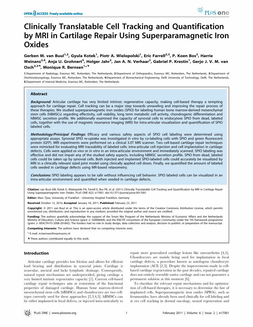
Labeling effectivity and cell behaviorhBMSCs were efficiently labeled with ferumoxides-protamine
sulfate complexes (Figure 1A). We observed a labeling efficiency
ranging from 41.2633.5% at an SPIO dose of 2.5 mg/cm2 to
94.567.8% at an SPIO dose of 25 mg/cm2 (Figure 1B) with
resulting average total iron loads (TILs) of cells ranging from
4.062.9 pg/cell to 19.566.1 pg/cell (Figure 1C). Both labeling
efficiency and TIL increased in relation to SPIO labeling dose up
to a dose of 10 mg/cm2. The higher SPIO dose of 25 mg/cm2 did
not significantly increase labeling efficiency (P = 0.42) or average
TIL (P = 0.50). hBMSC viability was not influenced by SPIO
labeling for dosages up to 25 mg/cm2 (Figure 1D). Additionally,
SPIO labeling did not inhibit cell metabolic activity compared to
unlabeled controls for at least seven days (Figure 1E). Based on
these results an SPIO dose of 10 mg/cm2, corresponding to a final
labeling concentration of 50 mg/ml SPIO, was considered optimal
and used in all further experiments.
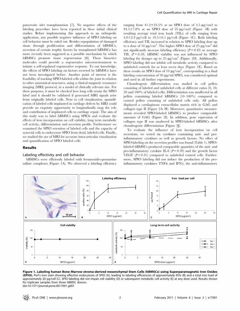
Chondrogenic differentiation was studied in cell pellets
consisting of labeled and unlabeled cells at different ratios (0, 10,
50 and 100% of labeled cells). Differentiation was unaffected in all
pellets containing labeled hBMSCs (10–100%) compared to
control pellets consisting of unlabeled cells only. All pellets
displayed a cartilaginous extracellular matrix rich in GAG and
collagen type II (Figure 2A–H). Moreover, quantitative measure-
ments revealed SPIO-labeled hBMSCs to produce comparable
amounts of GAG (Figure 2I). In addition, gene expression of
collagen type II was unaltered in SPIO-labeled hBMSCs after
chondrogenic differentiation (Figure 2J).
To evaluate the influence of iron incorporation on cell
secretions, we tested six cytokines containing anti- and pro-
inflammatory cytokines as well as growth factors. No effect of
SPIO-labeling on the secretion profiles was found (Table 1). SPIO-
labeled hBMSCs produced comparable quantities of the anti- and
pro-inflammatory cytokine IL-6 (P = 0.39) and the growth factor
VEGF (P = 0.31) compared to unlabeled control cells. Further-
more, SPIO labeling did not induce the production of the pro-
inflammatory cytokines TNFa and IFNc, the anti-inflammatory
Figure 1. Labeling human Bone Marrow stroma-derived mesenchymal Stem Cells (hBMSCs) using Superparamagnetic Iron Oxides(SPIO). Perl’s iron stain showing effective endocytosis of SPIO (A), leading to labeling efficiencies of approximately 95% (B) and a total iron load ofapproximately 20 pg/cell (C). SPIO labeling did not impair cell viability (D) or subsequent metabolic cell activity (E) at any dose used. Results shownfor triplicate samples from three hBMSC donors.doi:10.1371/journal.pone.0017001.g001
Cell Quantification by MRI in Cartilage Repair
PLoS ONE | www.plosone.org 2 February 2011 | Volume 6 | Issue 2 | e17001
cytokine IL-10 or the growth factor FGF2. These four factors were
not detectably secreted by control or SPIO-labeled hBMSCs.
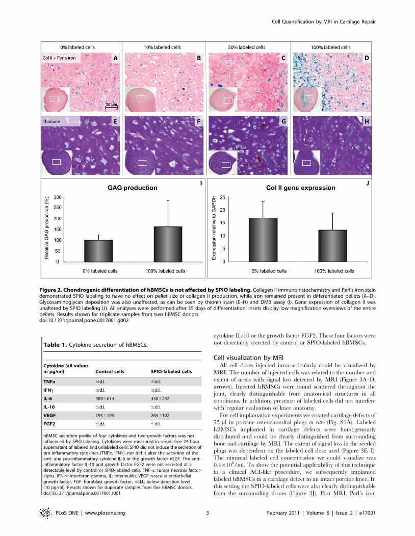
Cell visualization by MRIAll cell doses injected intra-articularly could be visualized by
MRI. The number of injected cells was related to the number and
extent of areas with signal loss detected by MRI (Figure 3A–D,
arrows). Injected hBMSCs were found scattered throughout the
joint, clearly distinguishable from anatomical structures in all
conditions. In addition, presence of labeled cells did not interfere
with regular evaluation of knee anatomy.
For cell implantation experiments we created cartilage defects of
75 ml in porcine osteochondral plugs in vitro (Fig. S1A). Labeled
hBMSCs implanted in cartilage defects were homogenously
distributed and could be clearly distinguished from surrounding
bone and cartilage by MRI. The extent of signal loss in the seeded
plugs was dependent on the labeled cell dose used (Figure 3E–I).
The minimal labeled cell concentration we could visualize was
0.46106/ml. To show the potential applicability of this technique
in a clinical ACI-like procedure, we subsequently implanted
labeled hBMSCs in a cartilage defect in an intact porcine knee. In
this setting the SPIO-labeled cells were also clearly distinguishable
from the surrounding tissues (Figure 3J). Post MRI, Perl’s iron
Figure 2. Chondrogenic differentiation of hBMSCs is not affected by SPIO labeling. Collagen II immunohistochemistry and Perl’s iron staindemonstrated SPIO labeling to have no effect on pellet size or collagen II production, while iron remained present in differentiated pellets (A–D).Glycosaminoglycan deposition was also unaffected, as can be seen by thionin stain (E–H) and DMB assay (I). Gene expression of collagen II wasunaltered by SPIO labeling (J). All analyses were performed after 35 days of differentiation. Insets display low magnification overviews of the entirepellets. Results shown for triplicate samples from two hBMSC donors.doi:10.1371/journal.pone.0017001.g002
Table 1. Cytokine secretion of hBMSCs.
Cytokine (all valuesin pg/ml) Control cells SPIO-labeled cells
TNFa #d.l. #d.l.
IFNc #d.l. #d.l.
IL-6 4896613 3306242
IL-10 #d.l. #d.l.
VEGF 1936103 2656102
FGF2 #d.l. #d.l.
hBMSC secretion profile of four cytokines and two growth factors was notinfluenced by SPIO labeling. Cytokines were measured in serum free 24 hoursupernatant of labeled and unlabeled cells. SPIO did not induce the secretion ofpro-inflammatory cytokines (TNFa, IFNc), nor did it alter the secretion of theanti- and pro-inflammatory cytokine IL-6 or the growth factor VEGF. The anti-inflammatory factor IL-10 and growth factor FGF2 were not secreted at adetectable level by control or SPIO-labeled cells. TNF-a: tumor necrosis factor-alpha, IFN-c: interferon-gamma, IL: interleukin, VEGF: vascular endothelialgrowth factor, FGF: fibroblast growth factor, #d.l.: below detection level(10 pg/ml). Results shown for duplicate samples from five hBMSC donors.doi:10.1371/journal.pone.0017001.t001
Cell Quantification by MRI in Cartilage Repair
PLoS ONE | www.plosone.org 3 February 2011 | Volume 6 | Issue 2 | e17001
stain confirmed SPIO positive cells to be present inside the defect
(Fig. S1B).
A point of interest is whether observed areas with signal loss are
generated by originally labeled cells, since SPIO remains in the
joint after labeled cells die. The joint cavity is lined by an inner
layer of synovial tissue that contains macrophages. We investigated
the capacity of synovial cells to endocytose SPIO from labeled
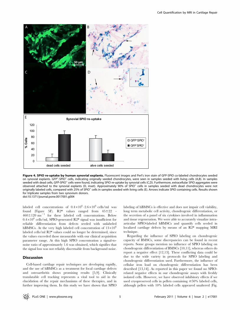
cells, by seeding dead or living GFP-SPIO co-labeled chondrocytes
on synovial tissue explants. In the synovium samples seeded with
living GFP-SPIO co-labeled cells, GFP+ cells were observed
immediately after seeding (Fig. S2A). Moreover, GFP+-SPIO+ cells
were found after the co-culture period of five days, suggesting
these cells to be originally labeled chondrocytes (Figure 4A and
4B). When seeded with dead GFP-SPIO co-labeled cells,
fluorescence microscopy confirmed the vast majority of cells to
have died directly following the freeze-thaw procedure (Fig. S2B).
After five days of co-culture, 88.4611.4% of the SPIO+ cells were
GFP̄ in these samples, indicative of iron re-uptake by synovial cells
(Figure 4C–E). Next to this, extracellular SPIO aggregates were
found attaching to the synovial membrane (Figure 4D inset). In
samples seeded with living chondrocytes, 25.5610.5% of SPIO
positive cells showed no GFP signal either. Whether this was due
to extinguishing GFP signal from originally labeled chondrocytes,
synovial re-uptake of SPIO from living cells, or caused by iron re-
uptake from seeded cells that died during the co-culture period,
could not be determined.
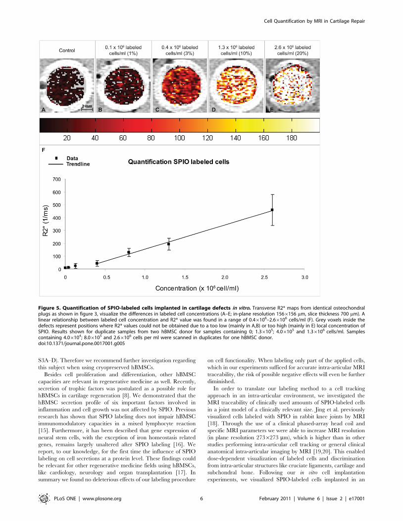
Cell quantification by MR-based relaxometryIn addition to visualization of seeded cells by MRI, we were able
to quantify the concentration of hBMSCs seeded in defects in vitro
by applying a R2* mapping technique, in which R2* = 1/T2*
(Figure 5A–E). A linear relationship between R2* values and
Figure 3. MRI visualization of SPIO-labeled hBMSCs in an intra-articular environment. Injected cells were visualized in a dose dependentmanner (A–D, in-plane resolution 2736273 mm; slice thickness 600 mm). Cells were found scattered throughout the joint (arrows) and could bediscerned from intra-articular structures. Labeled cells implanted in cartilage defects in vitro were homogeneously distributed and imaged in acomparable way; dose dependent and clearly distinguishable from structures like cartilage and bone (E–I; in-plane resolution 78678 mm; slicethickness 1500 mm). After implanting cells in a cartilage defect in an intact porcine knee, MRI showed the cells inside the defect (J; in-plane resolution2736273 mm; slice thickness 400 mm). In (A); b: bone; c: cartilage; c.l.: cruciate ligament. Intra-articular cells in porcine knees were imaged as a singlesample for one donor. hBMSCs implanted in cartilage defects in vitro were imaged in duplicate samples from two hBMSC donors.doi:10.1371/journal.pone.0017001.g003
Cell Quantification by MRI in Cartilage Repair
PLoS ONE | www.plosone.org 4 February 2011 | Volume 6 | Issue 2 | e17001
labeled cell concentrations of 0.46106–2.66106 cells/ml was
found (Figure 5F). R2* values ranged from 45622 -
4606120 ms21 for these labeled cell concentrations. Below
0.46106 cells/ml, SPIO-generated R2* signal was insufficient for
reliable differentiation from defects seeded with unlabeled
hBMSCs. At the very high labeled cell concentration of 136106
labeled cells/ml R2* values could no longer be determined, since
the values exceeded those measurable with our clinical acquisition
parameter range. At this high SPIO concentration a signal-to-
noise ratio of approximately 1.6 was obtained, which signifies that
the signal loss was not reliably discernable from background noise.
Discussion
Cell-based cartilage repair techniques are developing rapidly,
and the use of hBMSCs as a treatment for focal cartilage defects
and osteoarthritis shows promising results [5,9]. Clinically
translatable cell tracking represents a vital tool to aid in the
elucidation of the repair mechanisms of these therapies, and in
further improving them. In this study we have shown that SPIO
labeling of hBMSCs is effective and does not impair cell viability,
long term metabolic cell activity, chondrogenic differentiation, or
the secretion of a panel of six cytokines involved in inflammation
and tissue regeneration. We were able to accurately visualize intra-
articular SPIO-labeled hBMSCs and quantify cells seeded in
localized cartilage defects by means of an R2* mapping MRI
technique.
Regarding the influence of SPIO labeling on chondrogenic
capacity of BMSCs, some discrepancies can be found in recent
reports. Some groups mention no influence of SPIO labeling on
chondrogenic differentiation of BMSCs [10,11], whereas others do
report a negative effect [12,13]. These conflicting data could be
due to the wide variety in protocols for SPIO labeling and
chondrogenic differentiation used. Furthermore, the influence of
cellular iron load on chondrogenic differentiation has been
described [13,14]. As reported in this paper we found no SPIO-
related negative effects in our chondrogenic assays with freshly
isolated cells. However, we have observed inhibitory effects if we
used cryopreserved cells in pellets containing $50% labeled cells,
although pellets with 10% labeled cells appeared unaltered (Fig.
Figure 4. SPIO re-uptake by human synovial explants. Fluorescent images and Perl’s iron stain of GFP-SPIO co-labeled chondrocytes seededon synovial explants. GFP+-SPIO+ cells, indicating originally seeded chondrocytes, were seen in samples seeded with living cells (A,B). In samplesseeded with dead cells, GFP̄-SPIO+ cells were found, indicating SPIO re-uptake by synovial cells (C,D). Furthermore, extracellular SPIO aggregates wereobserved attached to the synovial explants (D, inset). Approximately 90% of SPIO+ cells in samples seeded with dead chondrocytes were notoriginally labeled cells, compared with 25% of SPIO+ cells in samples seeded with living cells (E). Arrows indicate SPIO containing cells. Results shownfor triplicate samples from two synovium donors.doi:10.1371/journal.pone.0017001.g004
Cell Quantification by MRI in Cartilage Repair
PLoS ONE | www.plosone.org 5 February 2011 | Volume 6 | Issue 2 | e17001
S3A–D). Therefore we recommend further investigation regarding
this subject when using cryopreserved hBMSCs.
Besides cell proliferation and differentiation, other hBMSC
capacities are relevant in regenerative medicine as well. Recently,
secretion of trophic factors was postulated as a possible role for
hBMSCs in cartilage regeneration [8]. We demonstrated that the
hBMSC secretion profile of six important factors involved in
inflammation and cell growth was not affected by SPIO. Previous
research has shown that SPIO labeling does not impair hBMSC
immunomodulatory capacities in a mixed lymphocyte reaction
[15]. Furthermore, it has been described that gene expression of
neural stem cells, with the exception of iron homeostasis related
genes, remains largely unaltered after SPIO labeling [16]. We
report, to our knowledge, for the first time the influence of SPIO
labeling on cell secretions at a protein level. These findings could
be relevant for other regenerative medicine fields using hBMSCs,
like cardiology, neurology and organ transplantation [17]. In
summary we found no deleterious effects of our labeling procedure
on cell functionality. When labeling only part of the applied cells,
which in our experiments sufficed for accurate intra-articular MRI
traceability, the risk of possible negative effects will even be further
diminished.
In order to translate our labeling method to a cell tracking
approach in an intra-articular environment, we investigated the
MRI traceability of clinically used amounts of SPIO-labeled cells
in a joint model of a clinically relevant size. Jing et al. previously
visualized cells labeled with SPIO in rabbit knee joints by MRI
[18]. Through the use of a clinical phased-array head coil and
specific MRI parameters we were able to increase MRI resolution
(in plane resolution 2736273 mm), which is higher than in other
studies performing intra-articular cell tracking or general clinical
anatomical intra-articular imaging by MRI [19,20]. This enabled
dose-dependent visualization of labeled cells and discrimination
from intra-articular structures like cruciate ligaments, cartilage and
subchondral bone. Following our in vitro cell implantation
experiments, we visualized SPIO-labeled cells implanted in an
Figure 5. Quantification of SPIO-labeled cells implanted in cartilage defects in vitro. Transverse R2* maps from identical osteochondralplugs as shown in figure 3, visualize the differences in labeled cell concentrations (A–E; in-plane resolution 1566156 mm, slice thickness 700 mm). Alinear relationship between labeled cell concentration and R2* value was found in a range of 0.46106–2.66106 cells/ml (F). Grey voxels inside thedefects represent positions where R2* values could not be obtained due to a too low (mainly in A,B) or too high (mainly in E) local concentration ofSPIO. Results shown for duplicate samples from two hBMSC donor for samples containing 0; 1.36105; 4.06105 and 1.36106 cells/ml. Samplescontaining 4.06104; 8.06105 and 2.66106 cells per ml were scanned in duplicates for one hBMSC donor.doi:10.1371/journal.pone.0017001.g005
Cell Quantification by MRI in Cartilage Repair
PLoS ONE | www.plosone.org 6 February 2011 | Volume 6 | Issue 2 | e17001
ACI-like procedure in an intact porcine knee. Air artifacts show up
as hypointensities on MRI, which interferes with the signal loss
generated by labeled cells. To avoid these artifacts, we were
obliged to perform this operation under water. This is impracti-
cable in a clinical setting indicating that immediately post-
operative this technique can be used to visualize cells that
remained in the defect, but that the technique is unsuitable to
discern labeled cells that might have leaked in the intra-articular
space. These leaked-out cells could be identified after intra-
articular air has resolved, which in our personal clinical experience
occurs within a day.
SPIO for cell-labeling has great clinical potential, but
unfortunately the label will dilute upon cell division. This could
limit the duration that cells can be accurately detected by MRI.
Previous results from our group and others show an approxi-
mate 75% decrease in SPIO positive cells after 7 days of
expansion in culture [21] and a total loss of iron after 5–8 cell
divisions in rapidly dividing HeLa cells [22]. On the other hand,
duration of traceability increases with slower proliferating cells,
and SPIO could be detected after 44 days in non-dividing
human MSCs [22]. Accordingly, our group previously reported
traceability in vivo of SPIO labeled human MSCs in a
subcutaneous mouse model for at least seven weeks post labeling
[10]. In additional experiments we found a marked decrease in
SPIO-generated signal voids from proliferating labeled hBMSCs
over a period of 14 days in vitro, representing 5–6 cell divisions
(data not shown). How long SPIO labeled cells can be detected
in an intra-articular environment remains to be studied in an in
vivo model.
Another point regarding accurate detection of labeled cells is the
fact that cell death will not result in the disappearance of the label.
SPIOs released from these cells could lead to misinterpretation of
MR images. Pawelczyk et al. have previously shown approxi-
mately 10–20% of local macrophages to become SPIO positive
after injection of SPIO-labeled BMSCs in a subcutaneous
inflammation mouse model [15]. Synovium contains the majority
of macrophages present in synovial joints. We observed the vast
majority of SPIO+ cells to be non-originally labeled after applying
dead, labeled cells on synovial explants. In the case of cell
implantation in a cartilage defect, SPIO re-uptake by host cells
might be of less consequence because there is no direct contact
between the cells seeded in the defect and the synovial membrane.
Nonetheless, before clinical application we recommend in vivo
animal studies using intra-articular cell tracking by MRI in
combination with histological confirmation of the localization of
originally labeled cells. This would enable the correct interpreta-
tion of obtained MR images.
Besides visualizing cells, we were able to quantify beforehand
known cell concentrations implanted in cartilage defects in a
clinically relevant cell-concentration range. To our knowledge, this
has not been reported before in a model comprising biological
tissue. Rad et al. were unsuccessful in translating their SPIO-
labeled cell quantification experiments to an ex vivo setting [23],
and Politi et al. did not validate their ex vivo quantification results
afterwards [24]. Since the original concentration of applied cells is
known in an ACI procedure, and seeded in a confined volume, it is
possible to quantify the total amount of labeled cells and follow
them in the same patient in subsequent MRI sessions. Since both
hBMSCs and chondrocytes can be labeled using SPIO [25,26],
these findings indicate the potential of SPIO labeling for tracking
cells in numerous orthopedic treatments and many other
regenerative medicine fields [27]. Cell visualization and especially
quantification using MRI can be a major step towards improve-
ment in all these applications.
In summary, we consider SPIO labeling to hold great potential
for clinically translatable cell tracking using MRI in cell-based
cartilage repair. In an experimental setting this would show the
distribution and diffusion of the applied cells, thereby elucidating
the regenerative mechanisms and providing opportunities to
improve current repair strategies. The use of MRI has the
advantage of simultaneous cell tracking and evaluation of cartilage
regeneration in one MRI session.
Materials and Methods
Ethics StatementAll human samples were obtained after approval by the
Erasmus MC medical ethical committee. hBMSCs were isolated
from heparinized femoral-shaft marrow aspirate of patients
undergoing total hip arthroplasty (after written informed consent;
protocol # MEC-2004-142). Articular cartilage and synovial
explants were obtained as redundant material from patients
undergoing total knee replacement surgery. All patients implicitly
consented to the use of these tissues for scientific research (protocol
# MEC-2004-322).
Cell isolations and cultureshBMSCs and human chondrocytes were isolated and cultured
using previously described procedures [28,29]. All isolated cells or
explants were cultured in DMEM containing 10% FCS, 50 mg/ml
gentamicin and 1.5 mg/ml fungizone (defined as standard
medium) unless stated otherwise. hBMSCs were cultured using
additional 1 ng/ml FGF2 and 161024 M vitamin C. All hBMSCs
and chondrocytes were used at passage 2–5 in labeling
experiments.
SPIO labelingSPIO labeling was performed using ferumoxides (EndoremH,
Guerbet S.A., Paris, France) complexed to protamine sulfate (LEO
Pharma N.V., Wilrijk, Belgium) as described earlier [25]. For
removal of extracellular iron, cells were washed with PBS
containing heparin 10 U/ml (LEO Pharma B.V., Breda, the
Netherlands). SPIO labeling mix was made at a constant
concentration of 100 mg/ml ferumoxides with 5 mg/ml protamine
to ensure identical particle formation [30]. Cells were labeled with
doses ranging from 2.5–25 mg/cm2 of ferumoxides.
Labeling efficiency, chondrogenic differentiation andfunctional assays
Histological iron detection was performed using a Perl’s iron
stain (Klinipath BVBA, Duiven, the Netherlands) according to the
manufacturer’s protocol. Labeling efficiency was determined in
triplicate samples for three donors by manually counting labeled
cells stained with Perl’s iron [25]. Total iron load was measured in
triplicate samples from three donors by inductively coupled plasma
- optical emission spectroscopy [25]. Cell viability was evaluated
by trypan blue exclusion. After labeling, cells were seeded in 96-
well plates at a density of 10,000 cells per well. AlamarBlueH(Invitrogen, Breda, the Netherlands) was used according to
manufacturer’s protocol for longitudinal measurement of meta-
bolic cell activity at day 0, 3 and 7.
Chondrogenic differentiation was performed in triplicate samples
on hBMSCs pellets from two donors according to previously
reported protocols [31]. Chondrogenically differentiated hBMSCs
were stained using a collagen II monoclonal antibody (II-II6B3;
Developmental Studies Hybridoma Bank, Iowa City, IA) and
thionine stain [31,32]. hBMSCs used for gene expression analysis
and glycosaminoglycan (GAG) measurements were chondrogeni-
Cell Quantification by MRI in Cartilage Repair
PLoS ONE | www.plosone.org 7 February 2011 | Volume 6 | Issue 2 | e17001
cally differentiated in alginate beads [33] using an otherwise
identical differentiation protocol. RNA extraction, cDNA synthesis,
real-time RT-PCR and collagen II primers for gene expression
analysis were described earlier [34]. GAG measurements were
performed using a dimethylmethylene blue (DMB) assay [35].
To investigate the influence of SPIO labeling on cytokines
secreted by hBMSCs, labeled and unlabeled cells from five donors
were cultured for 24 hours in serumfree standard medium
supplemented with ITS+1 (serum replacement; BD Bioscience,
Bedford, MA) in a 1:100 v/v ratio. Secretion of tumor necrosis
factor-alpha (TNF-a), interferon-gamma (IFN-c), interleukin (IL)-
6, IL-10, vascular endothelial growth factor (VEGF) and fibroblast
growth factor 2 (FGF2) were analyzed in duplicate samples using a
human Cytokine Bead Array flexsets (BD Bioscience, Breda, the
Netherlands) according to the manufacturer’s protocol. Flow
cytometric measurements were performed on a FACSCanto II
(BD, San Jose, CA, USA) and analyzed using FACSDiva software
version 6.1.2 (BD Biosciences).
Intra-articular cell injections and cell implantationsDue to the ex vivo nature of our MRI experiments, all cells were
fixed in 4% formalin prior to injection or implantation. SPIO-
labeled hBMSCs were injected ex vivo into porcine knees (Yorkshire
6 Landrace, 35 kilograms, age 4–5 months) in a dose range of
0.16106 to 56106 labeled cells (injected in 100 ml physiological
saline). For cell implantation, osteochondral plugs with a diameter
of 8 mm were harvested from porcine femoral condyles ex vivo.
Circular full-cartilage defects of 6 mm diameter (approximate
volume of 75 ml) were created in those plugs. Cells from two
donors were suspended in 1% Agar (Fluka BioChemika, Sigma-
Aldrich, Buchs, Switzerland) in saline before implantation. A total
of 16106 cells were seeded per defect in vitro (cell concentration of
136106 cells/ml), containing 0.3 – 100% labelled cells (i.e.
0.046106–136106 labeled cells/ml). Cell implantation was also
performed ex vivo in an intact porcine knee according to previously
described autologous chondrocyte implantation (ACI) procedures
[2]. The knee joint was opened and a circular cartilage defect was
created (diameter 6 mm; volume 75 ml). A periosteal flap was
sutured on top of the defect using PDS 6-0 sutures (Johnson &
Johnson, New Brunswick, New Jersey) and fibrin glue (TissucolHDuo 500, Baxter, Uden, the Netherlands) was applied at 90% of
the edges of the flap as a sealant. 16106 SPIO-labeled cells
suspended in 1% Agar, were seeded into the defect and fibrin glue
was used to seal the final 10% of the periostal flap margin. The
joint was closed in layers afterwards. All surgical procedures -
except the cell seeding and fibrin glue application - were
performed under water in order to prevent air artifacts on MRI.
Validation SPIO-generated signalTo validate if SPIO-generated signal was related to originally
labeled cells, or that SPIO particles were taken up by host cells,
human green fluorescent protein (GFP) transfected chondrocytes
were co-labeled with SPIO. Cells were lentivirally transduced and
constitutively expressed GFP under the control of a CMV
promoter (SHC003 vector, Sigma-Aldrich MissionH, shRNA
library). To investigate the fate of SPIO after cell death, we
considered the cell type used for labeling less relevant and chose
chondrocytes over hBMSCs due to their substantially higher
transduction efficiency. 256104 GFP+-SPIO+ cells were seeded in
duplicates on synovial explants from two human donors. Cells
were alive or killed (by repeated free-thaw cycles) before seeding.
Cells and synovial explants were co-cultured for five days.
Fluorescent images were made from frozen sections using an
Axiovert s100 microscope (Carl Zeiss B.V., Sliedrecht, the
Netherlands). Subsequently, samples were stained using Perl’s
iron stain and corresponding fields of view were evaluated by light
microscopy. GFP and SPIO containing cells were determined by
manually counting a minimum of 100 cells per sample.
Magnetic Resonance ImagingScanning was performed on a clinical 3.0T MRI scanner
(General Electric Healthcare, Milwaukee, Wisconsin). A 3D SPGR
sequence was used to scan labeled hBMSCs in vitro (TR 87.7 ms, TE
15.1 ms, FOV 17 mm, matrix 5126512, scanning time 11 min.
35 sec.) in combination with a custom made receive-only surface
coil with a diameter of 1 cm. Scanning of injected or implanted
hBMSCs in intact porcine knees was performed using a FIESTA-
3D sequence (TR 7.9 ms, TE 2.2 ms, FOV 140 mm, matrix
5126256, scanning time 28 min. 46 sec.) and a clinical phased-
array head coil. Images of hBMSCs implanted in defects in vitro were
obtained by a Spin Echo sequence (TR 1500 ms, TE 40 ms, FOV
40 mm, matrix 5126512, scanning time 14 min. 42 sec.) and a
custom made receive-only surface coil with a diameter of 3 cm.
R2*-mapping was performed in duplicate samples for two hBMSC
donors by a SPGR sequence (TR 45 ms, TE 4–120 ms, FOV
40 mm, matrix 2566256, scanning time 21 min. 21 sec.). R2*
values were calculated and validated by mono-exponential curve
fitting (MATLAB 7.0.1, MathWorks, MA). Voxels with an r2,0.95
were excluded from the measurements.
Statistical analysisStatistical differences between different treatment groups were
analysed by the paired Student t-test (normality test p.0.05). P-
values of #0.05 were considered statistically significant. All
numerical data are presented as mean 6 standard deviation.
Supporting Information
Figure S1 In vitro and ex vivo cell implantationprocedures. Osteochondral plugs with a diameter of 8 mm
were created from the femoral part of porcine knees (A). Ex vivo,
post-MRI Perl’s iron stain shows macroscopically the presence of
blue, iron containing cells in the defect (B). Dotted line in (A)
represents created defect. Intra-articular cell implantation in a
porcine knee was performed for one donor.
(TIF)
Figure S2 Cell seeding on synovial explants. Fluorescent
images directly after seeding GFP+-SPIO+ cells on synovial
explants showing abundant GFP signal in samples seeded with
living cells (A), while GFP signal is absent in samples seeded with
dead cells (B). This confirmed that the vast majority of killed cells
had not survived the multiple freeze-thaw procedures. Represen-
tative images are shown for triplicate samples from two synovium
donors.
(TIF)
Figure S3 Chondrogenic differentiation of cryopre-served SPIO-labeled hBMSCs. Perl’s iron stain (A–D) and
collagen II immunohistochemistry (A–C) of chondrogenically
differentiated cryopreserved hBMSCs. SPIO labeling of 10% of
the cells did not influence pellet size or collagen II deposition
compared to control cells (A and B). Using 50% of SPIO-labeled
cells did negatively influence these outcome measures (C). Pellets
consisting of 100% of labeled cells disintegrated within 7 days,
showing viable and iron containing cells on a cytospin (D). Results
shown for triplicate samples from two hBMSC donors.
(TIF)
Cell Quantification by MRI in Cartilage Repair
PLoS ONE | www.plosone.org 8 February 2011 | Volume 6 | Issue 2 | e17001
Acknowledgments
The authors are grateful to N. Kops, J.L.M. Koevoet, S.T. van Tiel and J.
van der Slegt for their technical assistance; to M. Hoogduijn for his
assistance in obtaining and analyzing the CBA data; to A. Uijtterdijk for
kindly providing porcine materials; to J.H. Waarsing for his support with
statistical analyses; and to U. Woroniecka and B. Wolterbeek for their
assistance with the ICP-OES measurements. The monoclonal antibody
developed by T. Linsenmayer was obtained from the Developmental
Studies Hybridoma Bank developed under the auspices of the NICHD and
maintained by The University of Iowa, Department of Biology, Iowa City,
IA.
Author Contributions
Conceived and designed the experiments: GMvB GK PAW EF PKB HW
JANV GPK GJVMvO MRB. Performed the experiments: GMvB GK
PAW PKB AUG HJ. Analyzed the data: GMvB GK PAW GJVMvO
MRB. Contributed reagents/materials/analysis tools: GMvB GK PAW
PKB HW GJVMvO MRB AUG HJ. Wrote the paper: GMvB GK PAW
EF PKB HW JANV GPK GJVMvO MRB.
References
1. Steinert AF, Ghivizzani SC, Rethwilm A, Tuan RS, Evans CH, et al. (2007)
Major biological obstacles for persistent cell-based regeneration of articularcartilage. Arthritis Res Ther 9: 213.
2. Brittberg M, Lindahl A, Nilsson A, Ohlsson C, Isaksson O, et al. (1994)Treatment of deep cartilage defects in the knee with autologous chondrocyte
transplantation. N Engl J Med 331: 889–895.
3. van Osch GJ, Brittberg M, Dennis JE, Bastiaansen-Jenniskens YM, Erben RG,et al. (2009) Cartilage repair: past and future–lessons for regenerative medicine.
J Cell Mol Med 13: 792–810.4. Centeno CJ, Schultz JR, Cheever M, Robinson B, Freeman M, et al. (2010)
Safety and complications reporting on the re-implantation of culture-expanded
mesenchymal stem cells using autologous platelet lysate technique. Curr StemCell Res Ther 5: 81–93.
5. Wakitani S, Nawata M, Tensho K, Okabe T, Machida H, et al. (2007) Repair ofarticular cartilage defects in the patello-femoral joint with autologous bone
marrow mesenchymal cell transplantation: three case reports involving nine
defects in five knees. J Tissue Eng Regen Med 1: 74–79.6. Saris DB, Vanlauwe J, Victor J, Haspl M, Bohnsack M, et al. (2008)
Characterized chondrocyte implantation results in better structural repair whentreating symptomatic cartilage defects of the knee in a randomized controlled
trial versus microfracture. Am J Sports Med 36: 235–246.7. Bulte JW (2009) In vivo MRI cell tracking: clinical studies. AJR Am J Roentgenol
193: 314–325.
8. Caplan AI (2007) Adult mesenchymal stem cells for tissue engineering versusregenerative medicine. J Cell Physiol 213: 341–347.
9. Centeno CJ, Busse D, Kisiday J, Keohan C, Freeman M, et al. (2008) Increasedknee cartilage volume in degenerative joint disease using percutaneously
implanted, autologous mesenchymal stem cells. Pain Physician 11: 343–353.
10. Farrell E, Wielopolski P, Pavljasevic P, van Tiel S, Jahr H, et al. (2008) Effects ofiron oxide incorporation for long term cell tracking on MSC differentiation in
vitro and in vivo. Biochem Biophys Res Commun 369: 1076–1081.11. Pawelczyk E, Arbab AS, Pandit S, Hu E, Frank JA (2006) Expression of
transferrin receptor and ferritin following ferumoxides-protamine sulfate labelingof cells: implications for cellular magnetic resonance imaging. NMR Biomed 19:
581–592.
12. Kostura L, Kraitchman DL, Mackay AM, Pittenger MF, Bulte JW (2004)Feridex labeling of mesenchymal stem cells inhibits chondrogenesis but not
adipogenesis or osteogenesis. NMR Biomed 17: 513–517.13. Henning TD, Sutton EJ, Kim A, Golovko D, Horvai A, et al. (2009) The
influence of ferucarbotran on the chondrogenesis of human mesenchymal stem
cells. Contrast Media Mol Imaging 4: 165–173.14. Bulte JW, Kraitchman DL, Mackay AM, Pittenger MF (2004) Chondrogenic
differentiation of mesenchymal stem cells is inhibited after magnetic labelingwith ferumoxides. Blood 104: 3410–3412; author reply 3412-3413.
15. Pawelczyk E, Jordan EK, Balakumaran A, Chaudhry A, Gormley N, et al.(2009) In vivo transfer of intracellular labels from locally implanted bone marrow
stromal cells to resident tissue macrophages. PLoS One 4: e6712.
16. Kedziorek DA, Muja N, Walczak P, Ruiz-Cabello J, Gilad AA, et al. (2010)Gene expression profiling reveals early cellular responses to intracellular
magnetic labeling with superparamagnetic iron oxide nanoparticles. MagnReson Med 63: 1031–1043.
17. Lee KD (2008) Applications of mesenchymal stem cells: an updated review.
Chang Gung Med J 31: 228–236.18. Jing XH, Yang L, Duan XJ, Xie B, Chen W, et al. (2008) In vivo MR imaging
tracking of magnetic iron oxide nanoparticle labeled, engineered, autologousbone marrow mesenchymal stem cells following intra-articular injection. Joint
Bone Spine 75: 432–438.
19. Kijowski R (2010) Clinical cartilage imaging of the knee and hip joints. AJR
Am J Roentgenol 195: 618–628.20. Saldanha KJ, Doan RP, Ainslie KM, Desai TA, Majumdar S (2010)
Micrometer-sized iron oxide particle labeling of mesenchymal stem cells formagnetic resonance imaging-based monitoring of cartilage tissue engineering.
Magn Reson Imaging.
21. van Tiel ST, Wielopolski PA, Houston GC, Krestin GP, Bernsen MR (2010)Variations in labeling protocol influence incorporation, distribution and
retention of iron oxide nanoparticles into human umbilical vein endothelialcells. Contrast Media Mol Imaging 5: 247–257.
22. Arbab AS, Bashaw LA, Miller BR, Jordan EK, Lewis BK, et al. (2003)
Characterization of biophysical and metabolic properties of cells labeled withsuperparamagnetic iron oxide nanoparticles and transfection agent for cellular
MR imaging. Radiology 229: 838–846.23. Rad AM, Arbab AS, Iskander AS, Jiang Q, Soltanian-Zadeh H (2007)
Quantification of superparamagnetic iron oxide (SPIO)-labeled cells using MRI.
J Magn Reson Imaging 26: 366–374.24. Politi LS, Bacigaluppi M, Brambilla E, Cadioli M, Falini A, et al. (2007)
Magnetic-resonance-based tracking and quantification of intravenously injectedneural stem cell accumulation in the brains of mice with experimental multiple
sclerosis. Stem Cells 25: 2583–2592.25. van Buul GM, Farrell E, Kops N, van Tiel ST, Bos PK, et al. (2009)
Ferumoxides-protamine sulfate is more effective than ferucarbotran for cell
labeling: implications for clinically applicable cell tracking using MRI. ContrastMedia Mol Imaging 4: 230–236.
26. Farrell E, Wielopolski P, Pavljasevic P, Kops N, Weinans H, et al. (2009) Celllabelling with superparamagnetic iron oxide has no effect on chondrocyte
behaviour. Osteoarthritis Cartilage 17: 961–967.
27. Chanda D, Kumar S, Ponnazhagan S (2010) Therapeutic potential of adultbone marrow-derived mesenchymal stem cells in diseases of the skeleton. J Cell
Biochem.28. Das RH, van Osch GJ, Kreukniet M, Oostra J, Weinans H, et al. (2010) Effects
of individual control of pH and hypoxia in chondrocyte culture. J Orthop Res28: 537–545.
29. de Mos M, Koevoet WJ, Jahr H, Verstegen MM, Heijboer MP, et al. (2007)
Intrinsic differentiation potential of adolescent human tendon tissue: an in-vitrocell differentiation study. BMC Musculoskelet Disord 8: 16.
30. Montet-Abou K, Montet X, Weissleder R, Josephson L (2005) Transfectionagent induced nanoparticle cell loading. Mol Imaging 4: 165–171.
31. Hellingman CA, Koevoet W, Kops N, Farrell E, Jahr H, et al. (2009) Fibroblast
Growth Factor Receptors in in-Vitro and in-Vivo Chondrogenesis: RelatingTissue Engineering Using Adult Mesenchymal Stem Cells to Embryonic
Development. Tissue Eng Part A.32. Bos PK, van Osch GJ, Frenz DA, Verhaar JA, Verwoerd-Verhoef HL (2001)
Growth factor expression in cartilage wound healing: temporal and spatialimmunolocalization in a rabbit auricular cartilage wound model. Osteoarthritis
Cartilage 9: 382–389.
33. Hauselmann HJ, Aydelotte MB, Schumacher BL, Kuettner KE, Gitelis SH,et al. (1992) Synthesis and turnover of proteoglycans by human and bovine adult
articular chondrocytes cultured in alginate beads. Matrix 12: 116–129.34. Uitterlinden EJ, Jahr H, Koevoet JL, Jenniskens YM, Bierma-Zeinstra SM, et al.
(2006) Glucosamine decreases expression of anabolic and catabolic genes in
human osteoarthritic cartilage explants. Osteoarthritis Cartilage 14: 250–257.35. Bastiaansen-Jenniskens YM, Koevoet W, Feijt C, Bos PK, Verhaar JA, et al.
(2009) Proteoglycan production is required in initial stages of new cartilagematrix formation but inhibits integrative cartilage repair. J Tissue Eng Regen
Med 3: 117–123.
Cell Quantification by MRI in Cartilage Repair
PLoS ONE | www.plosone.org 9 February 2011 | Volume 6 | Issue 2 | e17001


