Embed Size (px)
Citation preview
Pergamon NanaSbucturcdMataiak. Vol. 10. No. 6. pp. 1001-1011.1998
Ekevia Science Ltd
PII SO9659773(98)00133-O
6 1998 Acta Mdallurgica Inc. PriMed in the USA. All ri8ht.s r-cd
09659773/98 $19.00 + .OO
SUPERPARAMAGNETIC TRANSITION AND LOCAL DISORDER IN CuFezO4 NANOPARTICLES
G.F. Goya and H.R. Rechenberg
Instituto de Fisica, Universidade de Sb Paul0 C.P. 66318.05315970 / SP SzIo Paul0 - Brazil
(Accepted May 18,1998)
Abstract - We present X-ray dt@?action (XRD), Miissbauer spectroscopy (MS) and d.c. magnetization measurements performed on ball-milled CuFezOr samples. The average particle size cd> was found to decrease to the nanometer range after t=I5 min of milling. Room temperature Miissbauer data showed that thefraction ofparticles above the blocking temperature TB increases with milling time, and almost complete superparamagnetic samples are obtainedfor cd> = 7(2) nm. Magnetization measurements below TB suggest spin canting in milled samples. The values of saturation moment ps reveal that site populations are slightly dected by milling. Miissbauer resonant intensities are accounted for on the basis of local disorder of Fe=?’ environments, and the development of sample inhomogeneities of CunFo-.,& composition. 01998 Acta Metallurgica Inc.
INTRODUCTION
Macroscopic magnetic tunneling, magnetic relaxation, and spin canting are some of the phenomena recently found in nanosized mater@, which have become an intense research field (14). An increasing fraction of these nanosized materials am being obtained by mechanical grinding, and the resulting phases have shown rather peculiar structural and magnetic properties (5-8). Relaxation effects in mixed ferrites have been studied (9-11) and recently; spin canting in maghemite nanoparticles induced by structural disorder have been reported (12).
The copper spine1 structure has a cubic close-packed arrangement of the oxygen ions, with the Cu2+ and Fe3’ ions in two different kind of sites (13). These sites have tetrahedral and octahedral oxygen coordination (A- and B-sites respectively), so the resulting local symmetries of both sites am different. Additionally, there is an off-center displacement of the metallic ions, usually described in terms of a parameter u, which depends on the ionic radii of the transition metals involved. The local symmetry of A-site does not depend on the value of u, but B-sites are distorted by deviations of the ideal value u=3/8. The resulting ionic distribution in this kind of structure may be represented by
1001
1002 GF GOYA AND HR RECHENBERG
where &O and 6 =l stand for the inverse and normal cases, respectively. Although most spine1 ferrites are cubic, CuFe204 can have tetragonal unit cell symmetry if cooled slowly from high temperatures, with values of 6 between 0.1-0.2 (14-16). Attempts to explain the resulting ionic distribution from electrostatic (Madelung) energy calculations have led only to qualitative results (13). In &Fe204 spinel, the presence of Cu2+ with orbitally degenerate ground state lead to expect Jahn-Teller interaction to be operative, modifying the local symmetry of the sites.
We present in this work Mossbauer spectroscopy, d.c. magnetization and x-ray diffraction (XRD) data on tetragonal CuF~04 spine1 ferrite, ball milled using “low” energy (low rotational velocity) and short times (several minutes), to investigate the superparamagnetic (SPM) transition and the resulting ionic distribution after the grinding process.
EXPERIMENTAL
Samples of CuFaO4 spine1 were prepared by standard solid state reaction, mixing stoichiometric amounts of a-Fe203 and CuO (99.999%). The mixture was fired in air at 900 C for 72 h, with intermediate grinding in agate mortar. Samples were cooled slowly (2 K/min) to avoid quenching of the high-temperature cubic spine1 phase. Ball milling of the resulting &Fe204 was carried out in a planetary ball mill (Fristch Pulverisette 7) with hardened steel vials and balls. The ratio of masses was chosen as 10: 1, and the milling intensity set as 500 rpm. The process was interrupted at several intermediate times to take out small parts of the powder. Samples were labeled SO; S 15; S45; S 105; S200 and S300 accordingly to the total minutes of grinding time. XRD characterization was performed in a Philips PW-1140 diffractometer using Cu-Kcr radiation. Magnetization measurements were performed in a vibrating sample magnetometer at 300K and 4.2K, in fields up to 90 kOe. MUssbauer data was taken at 4.2, 78 and 296 K in transmission geometry, using a 57Co source of about 50 mCi in constant acceleration mode. A non-linear least-squares program was used to fit the spectra to Lorentzian line shapes. Isomer shifts are referred to o-Fe at 300K.
RESULTS AND DISCUSSION
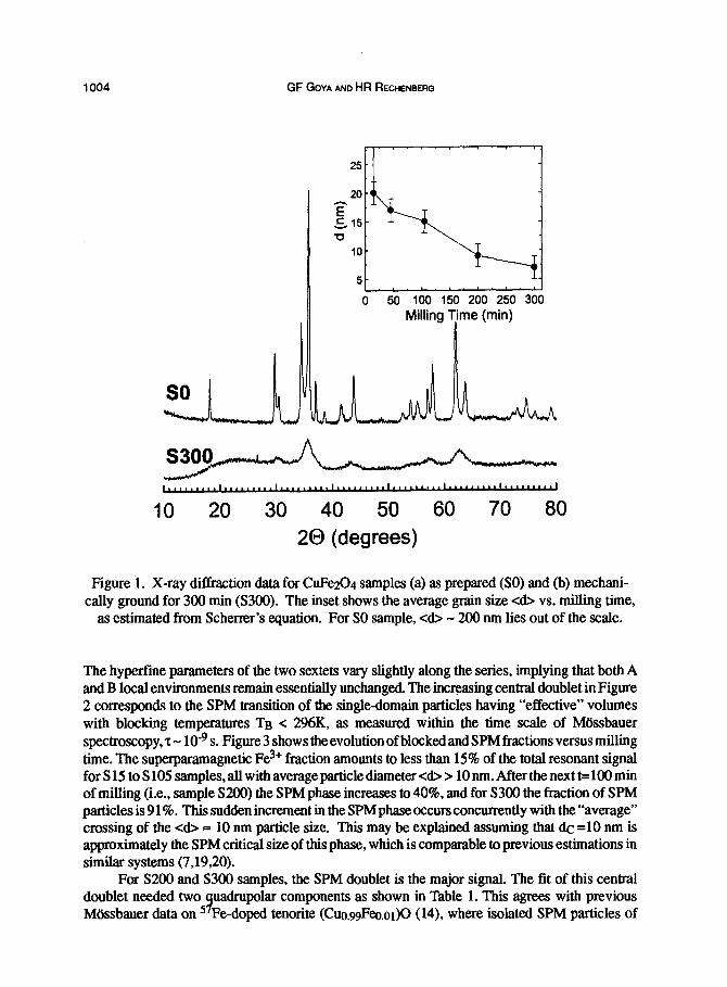
Sample SO, as prepared for the milling experiment, was found to be single phase CuFezO4 spine1 (tetragonal unit cell, space group D& - I 4l/amd), with lattice parameters au= 8.222(5) A and cc = 8.690(5) A. The XRD patterns of pristine and milled CuFe204 (Figure 1) show the broadening of the peaks with milling, although their positions do not change along the process. Average grain sizes cd> were estimated from Scherrer’s equation, subtracting instrumental broadening from the experimental linewidth The resulting values of <d> are shown in the inset of Figure 1. It is observed that particle size drops abruptly to c&=20(2) nm after the first 15 minutes of milling. Afterwards, cd> decreases monotonically to a minimum value <d>=7(2) nm.
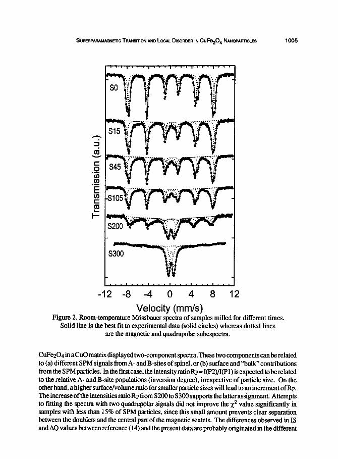
Mossbauer spectrum of pristine CuFe204 (sample SO), shown in Figure 2, is composed of two partially resolved magnetic sextets arising from Fe? in tetrahedral (A) and octahedral (B) sites, as previously reported (17,18). The spectraof milledsamples S 15 to S 105 couldbe fitted with two magnetic plus one quadrupolar signals (Table 1 and Figure 2).
!~JPERPARAMAGNETIC TRANSITION AND LOCAL DISORDER IN CuFe204 NA~WA~ICLES 1003
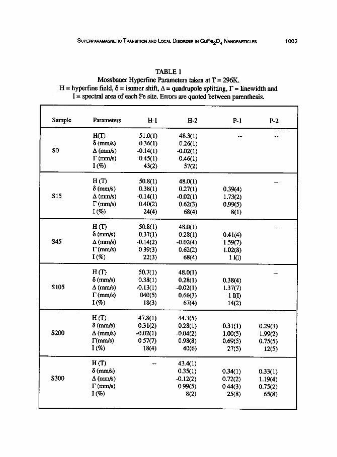
TABLE 1 Mossbauer Hyperfine Parameters taken at T = 296K.
H = hyperfine field, 6 = isomer shift, A = quadrupole splitting, r = linewidth and I == spectral area of each Fe site. Errors are quoted between parenthesis.
Sample Parameters H-l H-2 P-l P-2
W-0 51.0(l) 48.3(l) __ __
6 (mm/s) 0.36( 1) 0.26( 1) so A (ds) -0.14( 1) -0.02( 1)
r mm 0.45( 1) 0.46( 1) I@) 43(2) 57(2)
H 0’) 50.8( 1) 48.0( 1) __
6 (mm/s) 0.38( 1) 0.27( 1) 0.39(4) s15 A (mm/s) -0.14(l) -0.02( 1) 1.73(2)
r (mm/s) 0.40(2) 0.62(3) 0.99(5) I(%) 24(4) 68(4) 8(l)
s45
H(T) 6 bds) A bds) I- (mmls) 1 (a)
50.8( 1) 48.0(l) _- 0.37(l) 0.28( 1) 0.41(4)
-0.14(2) -0.02(4) 1.59(7) 0 39(3) 0.62(2) 1.02(8)
22~3) 68(4) 1 KU
s105
H(T) 6 (mm/s) A (mm/s) r (ms) I(%)
50.7( 1) 0.38( 1)
-0.13(l) 040(5)
18(3)
48.0( 1) 0.28( 1)
-0.02( 1) 0.66(3)
67(4)
__ 0.38(4) 1.37(7)
1 I(I) 14~2)
s200
H t-U 6 (Ws) A (ds) r(nmlls) I(%)
47.8( 1) 44.3(5) 0.31(2) 0.28( 1) 0.31(l)
-0.02( 1) -0.04(2) 1.00(5) 0 57(7) 0.98(8) 0.69(5)
18(4) 40(6) 27(5)
0.29(3) 1.99(2) 0.75(5)
12(5)
HO __ 43.4( 1)
6 (mns/s) 0.35( 1) 0.34( 1) s300 A (mnlls) -0.12(2) 0.72(2)
r (mm/s) 0 99(5) 0 44(3) I(%) 8(2) 25(8)
0.33( 1) 1.19(4) 0.75(2)
65(8)
1004 GF GOYA AND HR RECHENBERG
0 50 100 150 200 250 300
Milling Trne (min)
IO 20 30 40 50 60 70 80
20 (degrees)
Figure 1. X-ray diffraction data for CuFe204 samples (a) as prepared (SO) and (b) mechani- cally ground for 300 min (S300). The inset shows the average gram size <d> vs. milling time,
as estimated from Scherrer’s equation. For SO sample, cd> - 200 nm lies out of the scale.
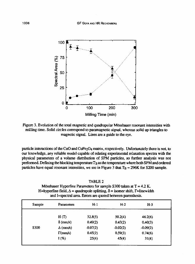
The hyperfine parameters of the two sextets vary slightly along the series, implying that both A and B local environments remain essentially unchanged. The increasing central doublet in Figure 2 corresponds to the SPM transition of the single-domain particles having “effective” volumes with blocking temperatures Tn < 296K, as measured within the time scale of MUssbauer spectroscopy, z - 10T9 s. Figure 3 shows the evolution of blocked and SPM fractions versus milling time. The superparamagnetic Fe3’ fraction amounts to less than 15% of the total resonant signal for S 15 to S 105 samples, all with average particle diameter <d> > 10 nm. After the next t=lOO mm of milling (Le., sample S200) the SPM phase increases to 40%, and for S300 the fraction of SPM particles is 9 1%. This sudden increment in the SPM phase occurs concurrently with the “average” crossing of the <d> 5 10 nm particle size. This may be explained assuming that d.c =lO nm is approximately the SPM critical size of this phase, which is comparable to previous estimations in similar systems (7,19,20).
For S2OO and S300 samples, the SPM doublet is the major signal. The fit of this central doublet needed two quadrupolar components as shown in Table 1. This agrees with previous Mtissbauer data on 57Fe-doped tenorite (Cuo.99Fec.or)O (14), where isolated SPM particles of
%JPEFIPARAMAGNETIC Tratusmm AND LOCAL HORDER IN CuFe20, ~~NOPAFITICLES 1005
-12 -8 -4 0 4 8 12
Velocity (mm/s) Figure 2. Room-temperature Mossbauer spectra of samples milled for different times.
Solid line is the best fit to experimental data (solid circles) whereas dotted lines are tbe magnetic and quadrupolar subespectra.
CuFe204 in aClu0 matrix displayed two-component spectra. These two components can be related to (a) different SPM signals from A- and B-sites of spinel, or(b) surface and “bulk”contributions from the SPMparticles. Inthef~tcase,theintensityratioRp=I(P2)/I(Pl)isexpectedtoberelated to the relative .A- and B-site populations (inversion degree), inespective of particle size. On the other hand, a h:igher surface/volume ratio for smaller particle sizes will lead to an increment of Rp. The increase of the intensities ratio Rp from S200 to S300 supports the latter assignment. Attempts to fitting the spectra with two quadrupolar signals did not improve the x2 value significantly in samples with less than 15% of SPM particles, since this small amount prevents clear separation between the doublets and the central part of the magnetic sextets. The differences observed in IS and AQ values between reference (14) and the present data are probably originated in the different
1006 GF GOYA AND HR RECHENBERG
100
0
B I-
$.-.* . . . ..__..____ ~.. I .
‘.., ,:’ ‘..,
‘.., ,:’
. . ‘...
‘.‘$ ~,,,,,,.. .,.,, ‘. ,.-.
I ‘.
,.’ ‘..
,:’ ‘. .,
: ;
.’ :
. .
: ;
: . .
‘.
,?--
_~.........“‘.~@”
b; I L
0 100 200
Milling Time (min)
300
Figure 3. Evolution of the total magnetic and quadrupolar Mossbauer resonant intensities with milling time. Solid circles correspond to paramagnetic signal, whereas solid up triangles to
magnetic signal. Lines are a guide to the eye.
particle interactions of the CuO and CuF~04 matrix, respectively. Unfortunately there is not, to our knowledge, any reliable model capable of relating experimental relaxation spectra with the physical parameters of a volume distribution of SPM particles, so further analysis was not performed. Defining the blocking temperatUreTB as the temperature whereboth SPMandordered particles have equal resonant intensities, we see in Figure 3 that TB = 296K for S2fKl sample.
TABLE 2 Mossbauer Hyperfine Parameters for sample S300 taken at T = 4.2 K.
H=hyperfine field, A = quadrupole splitting, 6 = isomer shift, F=linewidth and I=spectral area. Errors are quoted between parenthesis.
Sample Parameters H-l H-2 H-3
H CD 52.8(5) 50.2(4) 46.2(6)
6 (mm/s) 0.49(2) 0.43(2) 0.40(3)
s300 A (mm/s) -0.07(2) -0.02(2) -0.09(2)
bds) 0.45(2) 0.59(3) 0.74(6)
I(%) 25(4) 45(4) 31(4)
!&wwfwmNmc TwNstnoN AND LOCAL DISORDER IN CuFe,O, ~~~PARTICLES 1007
-9 -6 -3 0 3 6 9
Velocity (mm/s) Figure 4. Mossbauer spectra of (a) sample S200 and (b) sample S300 at T=78K.
(c) Sample S300 measured at 4.2 K. Dotted lines: magnetic subspectra. Solid line: best fit to experimental data.
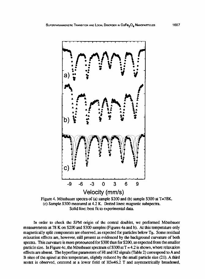
In order to check the SPM origin of the central doublet, we performed Mossbauer measurements at 78 K on S200 and S300 samples (Figures 4a and b). At this temperature only magnetically qplit components are observed, as expected for particles below TB. Some residual relaxation effelcts are, however, still present as evidenced by the background curvature of both spectra. This curvature is more pronounced for S300 than for S200, as expected from the smaller particle size. In Figure 4c, the Mossbauer spectrum of S300 at T = 4.2 is shown, where relaxation effects are absent. The hypefime parameters of Hl and H2 signals (Table 2) correspond to A and B sites of the spine1 at this temperature, slightly reduced by the small particle size (21). A third sextet is observed, centered at a lower field of H3=46.2 T and asymmetrically broadened,
i 008 GF GOYA AND HR RECHENBERG
1.0
-0.5
-1 .o
I. 9 .I. “I. - ‘I - - ‘I.. .I’.’ I *
,111 -2 -1 0 1 2
H.T“ (k0e.K”)
- 4.2 K 300K
I . . . I . . . . ., . t . . * a . . . I. a. I * -21 -14 -7 0 7 14 21
H.-T-’ (kOe.K-‘)
Figure 5. Magnetization curves vs. reduced field (H/T), taken at 300 and 4.2 K, of sample milled 300 minutes. The maximum field is H=90 lcOe. The inset shows the low-field region.
indicating a distribution of hyperfine fields. This may be due to the formation of the solid solution CuxFe3-x04 with an extended range of x, where the different Fe3+ environments result in the observed hyperfine field distribution (22). It has been reported that ball milling in a closed container transforms a-Fe203 into Fe304 (23). The formation of sample areas with Cu,Feg-x04 stoichiometries, after 300 minutes of milling, seems to be related to the beginning of this Fe reduction. The broadening of the XRD peaks in milled samples prevented the confirmation of CuxFeg-x04 phase, due to the overlap with the corresponding lines of CuF~04.
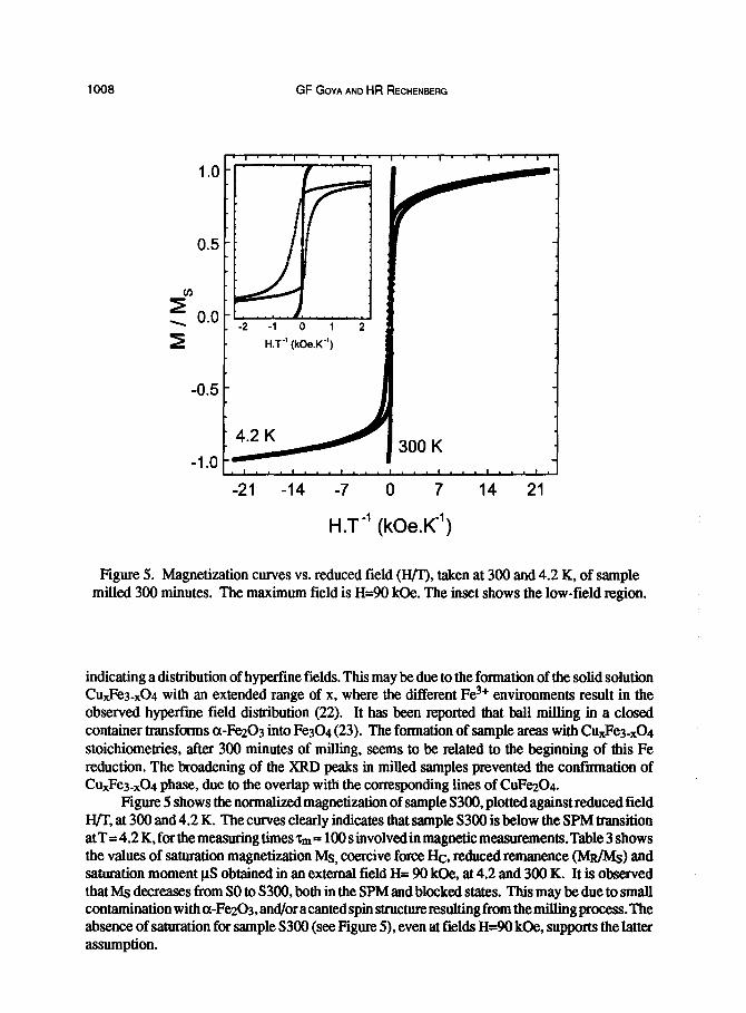
Figure 5 shows the normalized magnetization of sample S300, plotted against reduced field H/T, at 300 and 4.2 K. The curves clearly indicates that sample S300 is below the SPM transition atT=4.2K,forthemeasmingtimeszm= 100 s involved in magnetic measurements. Table 3 shows the values of saturation magnetization Ma, coercive force Hc, reduced remanence (M&v&) and saturation moment pS obtained in an external field H= 90 l&e, at 4.2 and 300 K. It is observed that Ms decreases from SO to S300, both in the SPM and blocked states. This may be due to small contamination with cr-Fe203, and/or acanted spin structure resulting from the milling process. The absence of saturation for sample S3OO (see Figure 5), even at fields H=W kOe, supports the latter assumption.
SUPERPARAMAQN~C TFUNSITION AND LOCAL DISORDER IN CuFe204 NAwmncm 1009
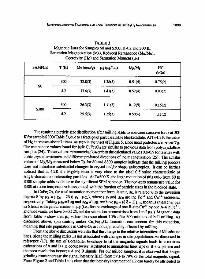
TABLE3 Magnetic Data for Samples SO and S300, at 4.2 and 300 K.
Saturation Magnetization (MS), Reduced Remanence (Mu/h@, Coercivity (Hc) and Saturation Moment (ps)
T(K) Ms (emu/g) PS &B/f.u.) MR/Ms
300 32.8(3) 1.38(3) OSl(5) 0.75(2)
4.2 33.4(3) 141(3) 0.52(4) 0.83(2)
300 26.3(3) 1.11(3) 0.12(5) 0.15(2)
4.2 29.5(3) 1.25(3) 0 5W4) 1.11(2)
The resulting particle size distribution after milling leads to non-zero coercive force at 300 K for sample S300 (Table 3), due to a fraction of particles in the blocked state. At T34.2 K the value of EIc increases about 7 times, as seen in the inset of Figure 5, since most particles are below Tu. The remanenc:e values found for bulk CuFe204 are similar to previous data from polycrystalline samples (24). These values are somewhat lower than the calculated values 0.8-0.9 for ferrites with cubic crystal structures and different preferred directions of the magnetization (25). The similar values of Mu/MS measured below TB for SO and S300 samples indicate that the milling process does not introduce substantial changes in crystal and/or shape anisotropies. It can be further noticed that at 4.2K the MR/IvIs ratio is very close to the ideal 0.5 value characteristic of single-domain noninteracting particles. At T=300 K, the large reduction of this ratio from SO to S300 samples adds evidence to the significant SPM behavior. The non-zero remanence value for S300 at room temperature is associated with the fraction of particle sizes in the blocked state.
In CuFe204, the total saturation moment per formula unit, p.8, is related with the inversion degree S by pts = pcu + 28 (CLFe - m), where pFe and ‘lo are the Fe? and Cu2+ moments, respectively. Taking j& =5 pu and p.c,, =l pu, we have p.q = (8 6 + 1) pu, and thus small changes in 6 leads to latrge increments in ps (i.e., for the exchange of one B-site Cu2+ by one A-site Fe3+, and vice versa, we have 6 =O. 125, and the saturation moment rises from 1 to 2 pu.) Magnetic data from Table 3 show that ps values decrease about 10% after 300 minutes of ball milling. As discussed above, spin canting and/or CuxFe3_x04 formation can account for this reduction, meaning that site populations in &Fe204 are not appreciably affected by milling.
From the above discussion we infer that the change in the relative intensities of Mossbauer lines, along the milling series, is not associated with changes in site populations. As discussed in reference (17), the use of Lorentzian lineshape to fit the magnetic signals leads to erroneous estimations of A and B site occupancies, attributed to anomalous lineshape of B-site pattern and the poor resolution between A and B signals. For our milled samples, it is observed that longer grinding times increase the signal intensity I(H2) from 57% to 79% of the total magnetic signal. From Figure 2 and Table 1 it is clear that the intensity increment of II2 can hardly be attributed to
1010 GF GOYA AND HR RECHENBERG
resolution effects. The ratio of A to B Mossbauer intensities, RH = I(Hl)/I(H2), is related to the inversion degree by RH = (l+S)/(l-8). The fit to the spectra using Lorentzian lineshapes gives values of RH ranging from 0.75 to 0.27, for SO and S105 samples respectively. These values are clearly incorrect, since a RB < 1 value has no physical meaning.
It is known that CuFe204 spine1 shows a marked Jahn-Teller effect, arising from the doubly degenerate Es-type ground state of the Cu2+ ion. These local distortions give rise to fluctuations of the superexchange interactions that broadens the resonant line. From Table 1 it can be seen that Fl < lY2 in all milled samples. Additionally, the increase of F2 with t indicates that A site is more affected by ionic disorder than B site. This can be understood assuming that, as a result of the milling process, a fraction of B sites are distorted to a cubic-like symmetry, thus contributing to the resonant line of the A site. These changes in local symmetry induced by structural disorder might be driven by similar mechanisms to those determining the cubic CuFaO4 phase when quenching from T>900°C, by freezing the high-temperature disordered phase. However, we cannot discard that an additional source of fluctuations in the hype&me field might be an incipient amount of inhomogeneities of composition CuxFes-x04 discussed above, which becomes detect- able in S300 below Tn.
In summary, we have studied the structural and magnetic properties of nanosized particles of CuFe204 spine1 above and below the superparamagnetic transition. We found that several minutes of mechanical grinding are enough toreduce the average grain size to the nanometer range. After 300 minutes of mechanical grinding, the fraction of SPM spine1 is almost 100%. The magnetic hyperfinefields,coerciveforceandreducedremanence measuredbe10wT~ showedthat, at this temperature, samples are in the blocked state. No evidence of new phases was found up to 200 minutes of milling, but for 300 minutes a new Fe3+ site is observed, assigned toFe3+ in CuxFes_ x04. Magnetic data below the blocking temperature indicated that milling does not modify the inversion degree substantially. The observed decrease in the magnetic moment with milling, and the absence of saturation at 90 kOe, suggest that spin canting might occur. The decrease ratio RH of Mossbauer intensities with milling is attributed to (a) distortions of the local environments of Fe sites by milling-induced disorder, and/or (b) the formation of sample inhomogeneities with composition CuxFes&4.
ACKNOWLEDGMENTS
One of us (G.F.G.) thanks financial support from FAPESP through a post-doctoral fellow- ship.
REFERENCES
1. Tejada, J. and Zhang, X., Journal of Magnetism and Magnetic Materials, 1995,140-144.1815. 2. Kodama, R.H., Seaman, C.L., Berkowitz, A.E. andMaple,M.B., Journal ofApplied Physics, 1994,
15,5639. 3. Balcells, Ll., Iglesias, 0. and Labarta, A., Physical Review B, 1997,55.8940. 4. Sui, Y., Xu, D.P., Zheng, EL. and Su, W.H., Journal of Applied Physics, 1996,80,719. 5. Koch, C.C., Nanostructured Materials, 1997,9, 13. 6. Ding, J., McCormick, P.G. and Street, R., Journal ofMagnetism andMagnetic Materials, 1997,
171,309.
SUPERPAFWAGNETIC TRANSITION AND LCCAL DEORDER IN CuFe,O, NANOPARTICLES 1011
7. 8.
9. 10.
Tronc, EL. and Jolivet, J.P., Materials Science Forum, 1997,235,659. Jiang, J.Z., Lin, R., Nielsen, K., Morup, S., Rickerby, D.G. and Clasen, R., Physical Review B, 1997,55,14830. Bathe, X., Garcfa de1 Mum, M. and Labarta, A., Physical Review B, 1997,55,6440. Srivastava, C.M., Shringi, S.N., Shrivatava, R.G. and Nanadikar, N., Physical Review B, 1975,14, 2032.
11. 12.
13.
14. 15. 16. 17. 18. 19. 20.
Daniels, J.M. and Rosencwaig, A., Canadian Journal ofPhysics, 1969,48,381. Morales, M.P., Serna, C. J., Bodker, F. and Morup, S., Journal of Physics: Condensed Matter, 1997, 9,546l. Krtlpica., S. and Novak, P., “Oxide Spinels” in Fertumagnetic Materials, ed. E.P. Wolfarth, North- Holland Publishing Co., 1982, Vol. 3, and references therein Goya, G.F., Journal of Materials Science Letters, 1997,7,563. Evans, B.J. and Hafner, S., Physics Letters, 1966,23,24. Prince, I!. and Treuting, R.G., Acta Crystallographica, 1956,9, 1025. Evans, Et.J. and Hafner, S.. Journal of Physical Chemistry of Solids, 1968,29, 1573. Janicki, J., Pietrzak, J., Pobreska, A. and Suwalski, J., Physica Status Solidi, 1982,72,95. Morup, S. and Tronc, E., Physical Review Letters, 1994,72,3278. Lehlooh, A.F., Mahmood, S. and Abu-Aljarayesh, I., Journal of Magnetism and Magnetic Materiab, 1994, 136, 143.
21. Greenwood, N.N. and Gibb, T.C., in Miissbauer Spectroscopy, Chapman & Hall, London, 1971. 22. Bhaduri, M., Journal of Chemical Physics, 1981,75,3674. 23. Jiang, J.Z., Zhou, Y.X., M$rup and Koch, C.B., Nanostructured Materials, 1996,7,401. 24. Berkowitz, A.E. and Schuele, W.J., Journal of Applied Physics, 1959.30, 134s. 25. Smit, J. and Wijn, H.P.J., Ferrites, Wiley, New York, 1959, and references therein.


