Embed Size (px)
Citation preview


Chenomx NMR Suite 9.0Copyright © 2001-2021 Chenomx Inc.

Table of ContentsNotices and Trademarks . . . . . . . . . . . . . . . . . . . . . . . . . . . . . . . . . . . . . . . . . . . . . . . . . . . . . . . . . . . . . . . . . . . . . . . . . . . . . . . . . . . . . vii1. Introduction . . . . . . . . . . . . . . . . . . . . . . . . . . . . . . . . . . . . . . . . . . . . . . . . . . . . . . . . . . . . . . . . . . . . . . . . . . . . . . . . . . . . . . . . . . . . . . . . . . . . 1
1.1. Installing Chenomx NMR Suite . . . . . . . . . . . . . . . . . . . . . . . . . . . . . . . . . . . . . . . . . . . . . . . . . . . . . . . . . . . . . . . . 1Windows (64-bit) . . . . . . . . . . . . . . . . . . . . . . . . . . . . . . . . . . . . . . . . . . . . . . . . . . . . . . . . . . . . . . . . . . . . . . . . . . . . . . . . 1Windows (32-bit) . . . . . . . . . . . . . . . . . . . . . . . . . . . . . . . . . . . . . . . . . . . . . . . . . . . . . . . . . . . . . . . . . . . . . . . . . . . . . . . . 1Mac OS X . . . . . . . . . . . . . . . . . . . . . . . . . . . . . . . . . . . . . . . . . . . . . . . . . . . . . . . . . . . . . . . . . . . . . . . . . . . . . . . . . . . . . . . . . . . . 1Linux (64-bit) . . . . . . . . . . . . . . . . . . . . . . . . . . . . . . . . . . . . . . . . . . . . . . . . . . . . . . . . . . . . . . . . . . . . . . . . . . . . . . . . . . . . . 1Linux (32-bit) . . . . . . . . . . . . . . . . . . . . . . . . . . . . . . . . . . . . . . . . . . . . . . . . . . . . . . . . . . . . . . . . . . . . . . . . . . . . . . . . . . . . . 1
1.2. Running Chenomx NMR Suite . . . . . . . . . . . . . . . . . . . . . . . . . . . . . . . . . . . . . . . . . . . . . . . . . . . . . . . . . . . . . . . . . . 21.3. Software Updates . . . . . . . . . . . . . . . . . . . . . . . . . . . . . . . . . . . . . . . . . . . . . . . . . . . . . . . . . . . . . . . . . . . . . . . . . . . . . . . . . . 21.4. Tutorial and Sample Files . . . . . . . . . . . . . . . . . . . . . . . . . . . . . . . . . . . . . . . . . . . . . . . . . . . . . . . . . . . . . . . . . . . . . . . 3
2. Handling Samples and Spectra . . . . . . . . . . . . . . . . . . . . . . . . . . . . . . . . . . . . . . . . . . . . . . . . . . . . . . . . . . . . . . . . . . . . . . . . . . . 42.1. Sample Preparation . . . . . . . . . . . . . . . . . . . . . . . . . . . . . . . . . . . . . . . . . . . . . . . . . . . . . . . . . . . . . . . . . . . . . . . . . . . . . . . 4
Preparing Filter Tubes for Sample Filtration . . . . . . . . . . . . . . . . . . . . . . . . . . . . . . . . . . . . . . . . . . . 4Filtering the Sample through Microcentrifuge Filter Tube . . . . . . . . . . . . . . . . . . . . . . . . 4Internal Standard Solution . . . . . . . . . . . . . . . . . . . . . . . . . . . . . . . . . . . . . . . . . . . . . . . . . . . . . . . . . . . . . . . . . . . 4
2.2. Spectral Data Acquisition . . . . . . . . . . . . . . . . . . . . . . . . . . . . . . . . . . . . . . . . . . . . . . . . . . . . . . . . . . . . . . . . . . . . . . . . 5Pulse Sequences . . . . . . . . . . . . . . . . . . . . . . . . . . . . . . . . . . . . . . . . . . . . . . . . . . . . . . . . . . . . . . . . . . . . . . . . . . . . . . . . . . 5Required NMR Parameters . . . . . . . . . . . . . . . . . . . . . . . . . . . . . . . . . . . . . . . . . . . . . . . . . . . . . . . . . . . . . . . . . . . 6
2.3. Spectral Data Processing . . . . . . . . . . . . . . . . . . . . . . . . . . . . . . . . . . . . . . . . . . . . . . . . . . . . . . . . . . . . . . . . . . . . . . . . 6Data Processed with Other Software . . . . . . . . . . . . . . . . . . . . . . . . . . . . . . . . . . . . . . . . . . . . . . . . . . . . . . 6
3. Common Tasks . . . . . . . . . . . . . . . . . . . . . . . . . . . . . . . . . . . . . . . . . . . . . . . . . . . . . . . . . . . . . . . . . . . . . . . . . . . . . . . . . . . . . . . . . . . . . . . . 83.1. Processing a Spectrum .. . . . . . . . . . . . . . . . . . . . . . . . . . . . . . . . . . . . . . . . . . . . . . . . . . . . . . . . . . . . . . . . . . . . . . . . . . 83.2. Profiling a Spectrum .. . . . . . . . . . . . . . . . . . . . . . . . . . . . . . . . . . . . . . . . . . . . . . . . . . . . . . . . . . . . . . . . . . . . . . . . . . . . . 83.3. Identifying Compounds . . . . . . . . . . . . . . . . . . . . . . . . . . . . . . . . . . . . . . . . . . . . . . . . . . . . . . . . . . . . . . . . . . . . . . . . . . 93.4. Profiling Overlapped Regions in a Spectrum .. . . . . . . . . . . . . . . . . . . . . . . . . . . . . . . . . . . . . . . . . . . 103.5. Determining Compound Concentrations . . . . . . . . . . . . . . . . . . . . . . . . . . . . . . . . . . . . . . . . . . . . . . . . . 11
4. The Basics . . . . . . . . . . . . . . . . . . . . . . . . . . . . . . . . . . . . . . . . . . . . . . . . . . . . . . . . . . . . . . . . . . . . . . . . . . . . . . . . . . . . . . . . . . . . . . . . . . . . . 134.1. Opening Files . . . . . . . . . . . . . . . . . . . . . . . . . . . . . . . . . . . . . . . . . . . . . . . . . . . . . . . . . . . . . . . . . . . . . . . . . . . . . . . . . . . . . . 134.2. Importing Spectra . . . . . . . . . . . . . . . . . . . . . . . . . . . . . . . . . . . . . . . . . . . . . . . . . . . . . . . . . . . . . . . . . . . . . . . . . . . . . . . . 134.3. Saving Files . . . . . . . . . . . . . . . . . . . . . . . . . . . . . . . . . . . . . . . . . . . . . . . . . . . . . . . . . . . . . . . . . . . . . . . . . . . . . . . . . . . . . . . . . 144.4. Closing Files . . . . . . . . . . . . . . . . . . . . . . . . . . . . . . . . . . . . . . . . . . . . . . . . . . . . . . . . . . . . . . . . . . . . . . . . . . . . . . . . . . . . . . . . 154.5. Exiting Modules . . . . . . . . . . . . . . . . . . . . . . . . . . . . . . . . . . . . . . . . . . . . . . . . . . . . . . . . . . . . . . . . . . . . . . . . . . . . . . . . . . . 154.6. Undo and Redo . . . . . . . . . . . . . . . . . . . . . . . . . . . . . . . . . . . . . . . . . . . . . . . . . . . . . . . . . . . . . . . . . . . . . . . . . . . . . . . . . . . . 164.7. Change Columns . . . . . . . . . . . . . . . . . . . . . . . . . . . . . . . . . . . . . . . . . . . . . . . . . . . . . . . . . . . . . . . . . . . . . . . . . . . . . . . . . . 164.8. Sidebar . . . . . . . . . . . . . . . . . . . . . . . . . . . . . . . . . . . . . . . . . . . . . . . . . . . . . . . . . . . . . . . . . . . . . . . . . . . . . . . . . . . . . . . . . . . . . . . 16
Legend . . . . . . . . . . . . . . . . . . . . . . . . . . . . . . . . . . . . . . . . . . . . . . . . . . . . . . . . . . . . . . . . . . . . . . . . . . . . . . . . . . . . . . . . . . . . . 17Files . . . . . . . . . . . . . . . . . . . . . . . . . . . . . . . . . . . . . . . . . . . . . . . . . . . . . . . . . . . . . . . . . . . . . . . . . . . . . . . . . . . . . . . . . . . . . . . . . 18Reference Cards . . . . . . . . . . . . . . . . . . . . . . . . . . . . . . . . . . . . . . . . . . . . . . . . . . . . . . . . . . . . . . . . . . . . . . . . . . . . . . . . 19Spectrum Details . . . . . . . . . . . . . . . . . . . . . . . . . . . . . . . . . . . . . . . . . . . . . . . . . . . . . . . . . . . . . . . . . . . . . . . . . . . . . . . 19Processing History . . . . . . . . . . . . . . . . . . . . . . . . . . . . . . . . . . . . . . . . . . . . . . . . . . . . . . . . . . . . . . . . . . . . . . . . . . . . . 20Compound Sets . . . . . . . . . . . . . . . . . . . . . . . . . . . . . . . . . . . . . . . . . . . . . . . . . . . . . . . . . . . . . . . . . . . . . . . . . . . . . . . . . 20Simulation . . . . . . . . . . . . . . . . . . . . . . . . . . . . . . . . . . . . . . . . . . . . . . . . . . . . . . . . . . . . . . . . . . . . . . . . . . . . . . . . . . . . . . . . 20
4.9. Spectrum Overlays . . . . . . . . . . . . . . . . . . . . . . . . . . . . . . . . . . . . . . . . . . . . . . . . . . . . . . . . . . . . . . . . . . . . . . . . . . . . . . . 204.10. Spectrum Graph Tools . . . . . . . . . . . . . . . . . . . . . . . . . . . . . . . . . . . . . . . . . . . . . . . . . . . . . . . . . . . . . . . . . . . . . . . . 22
Show Entire Spectrum .. . . . . . . . . . . . . . . . . . . . . . . . . . . . . . . . . . . . . . . . . . . . . . . . . . . . . . . . . . . . . . . . . . . . . . . 22Set Zoom .. . . . . . . . . . . . . . . . . . . . . . . . . . . . . . . . . . . . . . . . . . . . . . . . . . . . . . . . . . . . . . . . . . . . . . . . . . . . . . . . . . . . . . . . . 22Undo/Redo Zoom .. . . . . . . . . . . . . . . . . . . . . . . . . . . . . . . . . . . . . . . . . . . . . . . . . . . . . . . . . . . . . . . . . . . . . . . . . . . . . 23Auto Zoom .. . . . . . . . . . . . . . . . . . . . . . . . . . . . . . . . . . . . . . . . . . . . . . . . . . . . . . . . . . . . . . . . . . . . . . . . . . . . . . . . . . . . . . . 23Increase/Decrease Vertical Zoom .. . . . . . . . . . . . . . . . . . . . . . . . . . . . . . . . . . . . . . . . . . . . . . . . . . . . . . . 23Increase/Decrease Horizontal Zoom .. . . . . . . . . . . . . . . . . . . . . . . . . . . . . . . . . . . . . . . . . . . . . . . . . . . 23
iii

Spectrum Graph . . . . . . . . . . . . . . . . . . . . . . . . . . . . . . . . . . . . . . . . . . . . . . . . . . . . . . . . . . . . . . . . . . . . . . . . . . . . . . . . 23Spectrum Thumbnail . . . . . . . . . . . . . . . . . . . . . . . . . . . . . . . . . . . . . . . . . . . . . . . . . . . . . . . . . . . . . . . . . . . . . . . . . 24Select Region . . . . . . . . . . . . . . . . . . . . . . . . . . . . . . . . . . . . . . . . . . . . . . . . . . . . . . . . . . . . . . . . . . . . . . . . . . . . . . . . . . . . 24
4.11. Export Spectrum Image . . . . . . . . . . . . . . . . . . . . . . . . . . . . . . . . . . . . . . . . . . . . . . . . . . . . . . . . . . . . . . . . . . . . . . 254.12. Copy Spectrum Image to Clipboard . . . . . . . . . . . . . . . . . . . . . . . . . . . . . . . . . . . . . . . . . . . . . . . . . . . . . . 264.13. Display Options . . . . . . . . . . . . . . . . . . . . . . . . . . . . . . . . . . . . . . . . . . . . . . . . . . . . . . . . . . . . . . . . . . . . . . . . . . . . . . . . . 264.14. Preferences . . . . . . . . . . . . . . . . . . . . . . . . . . . . . . . . . . . . . . . . . . . . . . . . . . . . . . . . . . . . . . . . . . . . . . . . . . . . . . . . . . . . . . . 27
5. Processor . . . . . . . . . . . . . . . . . . . . . . . . . . . . . . . . . . . . . . . . . . . . . . . . . . . . . . . . . . . . . . . . . . . . . . . . . . . . . . . . . . . . . . . . . . . . . . . . . . . . . . 305.1. Overview . . . . . . . . . . . . . . . . . . . . . . . . . . . . . . . . . . . . . . . . . . . . . . . . . . . . . . . . . . . . . . . . . . . . . . . . . . . . . . . . . . . . . . . . . . . . 305.2. Processing Tools . . . . . . . . . . . . . . . . . . . . . . . . . . . . . . . . . . . . . . . . . . . . . . . . . . . . . . . . . . . . . . . . . . . . . . . . . . . . . . . . . . 31
Line Broadening . . . . . . . . . . . . . . . . . . . . . . . . . . . . . . . . . . . . . . . . . . . . . . . . . . . . . . . . . . . . . . . . . . . . . . . . . . . . . . . . 31Phase Correction . . . . . . . . . . . . . . . . . . . . . . . . . . . . . . . . . . . . . . . . . . . . . . . . . . . . . . . . . . . . . . . . . . . . . . . . . . . . . . . 32Baseline Correction . . . . . . . . . . . . . . . . . . . . . . . . . . . . . . . . . . . . . . . . . . . . . . . . . . . . . . . . . . . . . . . . . . . . . . . . . . . 33Shim Correction . . . . . . . . . . . . . . . . . . . . . . . . . . . . . . . . . . . . . . . . . . . . . . . . . . . . . . . . . . . . . . . . . . . . . . . . . . . . . . . . 34Region Deletion . . . . . . . . . . . . . . . . . . . . . . . . . . . . . . . . . . . . . . . . . . . . . . . . . . . . . . . . . . . . . . . . . . . . . . . . . . . . . . . . . 35Reverse Spectrum .. . . . . . . . . . . . . . . . . . . . . . . . . . . . . . . . . . . . . . . . . . . . . . . . . . . . . . . . . . . . . . . . . . . . . . . . . . . . 36Batch Process . . . . . . . . . . . . . . . . . . . . . . . . . . . . . . . . . . . . . . . . . . . . . . . . . . . . . . . . . . . . . . . . . . . . . . . . . . . . . . . . . . . . 37
5.3. Calibration Tools . . . . . . . . . . . . . . . . . . . . . . . . . . . . . . . . . . . . . . . . . . . . . . . . . . . . . . . . . . . . . . . . . . . . . . . . . . . . . . . . . 39Calibrate CSI . . . . . . . . . . . . . . . . . . . . . . . . . . . . . . . . . . . . . . . . . . . . . . . . . . . . . . . . . . . . . . . . . . . . . . . . . . . . . . . . . . . . . 39Define Custom CSI . . . . . . . . . . . . . . . . . . . . . . . . . . . . . . . . . . . . . . . . . . . . . . . . . . . . . . . . . . . . . . . . . . . . . . . . . . . . . 40Calibrate pH . . . . . . . . . . . . . . . . . . . . . . . . . . . . . . . . . . . . . . . . . . . . . . . . . . . . . . . . . . . . . . . . . . . . . . . . . . . . . . . . . . . . . . 41Calibration Tool . . . . . . . . . . . . . . . . . . . . . . . . . . . . . . . . . . . . . . . . . . . . . . . . . . . . . . . . . . . . . . . . . . . . . . . . . . . . . . . . 42
5.4. Information Tools . . . . . . . . . . . . . . . . . . . . . . . . . . . . . . . . . . . . . . . . . . . . . . . . . . . . . . . . . . . . . . . . . . . . . . . . . . . . . . . . 43Spectrum Details (Sidebar) . . . . . . . . . . . . . . . . . . . . . . . . . . . . . . . . . . . . . . . . . . . . . . . . . . . . . . . . . . . . . . . . . 43Processing History (Sidebar) . . . . . . . . . . . . . . . . . . . . . . . . . . . . . . . . . . . . . . . . . . . . . . . . . . . . . . . . . . . . . . 43
5.5. Importing and Exporting Data . . . . . . . . . . . . . . . . . . . . . . . . . . . . . . . . . . . . . . . . . . . . . . . . . . . . . . . . . . . . . . . 44Batch Import . . . . . . . . . . . . . . . . . . . . . . . . . . . . . . . . . . . . . . . . . . . . . . . . . . . . . . . . . . . . . . . . . . . . . . . . . . . . . . . . . . . . . 44Send to Profiler . . . . . . . . . . . . . . . . . . . . . . . . . . . . . . . . . . . . . . . . . . . . . . . . . . . . . . . . . . . . . . . . . . . . . . . . . . . . . . . . . 45Export as JCAMP-DX . . . . . . . . . . . . . . . . . . . . . . . . . . . . . . . . . . . . . . . . . . . . . . . . . . . . . . . . . . . . . . . . . . . . . . . . . . 45
6. Profiler . . . . . . . . . . . . . . . . . . . . . . . . . . . . . . . . . . . . . . . . . . . . . . . . . . . . . . . . . . . . . . . . . . . . . . . . . . . . . . . . . . . . . . . . . . . . . . . . . . . . . . . . . 466.1. Overview . . . . . . . . . . . . . . . . . . . . . . . . . . . . . . . . . . . . . . . . . . . . . . . . . . . . . . . . . . . . . . . . . . . . . . . . . . . . . . . . . . . . . . . . . . . . 466.2. Profiling Tools . . . . . . . . . . . . . . . . . . . . . . . . . . . . . . . . . . . . . . . . . . . . . . . . . . . . . . . . . . . . . . . . . . . . . . . . . . . . . . . . . . . . . 47
Compound Table . . . . . . . . . . . . . . . . . . . . . . . . . . . . . . . . . . . . . . . . . . . . . . . . . . . . . . . . . . . . . . . . . . . . . . . . . . . . . . . 47Custom Colors . . . . . . . . . . . . . . . . . . . . . . . . . . . . . . . . . . . . . . . . . . . . . . . . . . . . . . . . . . . . . . . . . . . . . . . . . . . . . . . . . . . 49Pinned Compounds . . . . . . . . . . . . . . . . . . . . . . . . . . . . . . . . . . . . . . . . . . . . . . . . . . . . . . . . . . . . . . . . . . . . . . . . . . . . 49Starred Compounds . . . . . . . . . . . . . . . . . . . . . . . . . . . . . . . . . . . . . . . . . . . . . . . . . . . . . . . . . . . . . . . . . . . . . . . . . . . 50Remember Selection . . . . . . . . . . . . . . . . . . . . . . . . . . . . . . . . . . . . . . . . . . . . . . . . . . . . . . . . . . . . . . . . . . . . . . . . . . 51Quick Searches . . . . . . . . . . . . . . . . . . . . . . . . . . . . . . . . . . . . . . . . . . . . . . . . . . . . . . . . . . . . . . . . . . . . . . . . . . . . . . . . . . 52Concentrations and Compound Fits . . . . . . . . . . . . . . . . . . . . . . . . . . . . . . . . . . . . . . . . . . . . . . . . . . . . . 53Fit Automatically . . . . . . . . . . . . . . . . . . . . . . . . . . . . . . . . . . . . . . . . . . . . . . . . . . . . . . . . . . . . . . . . . . . . . . . . . . . . . . . 56Concentration Fitting and Optimization Tool . . . . . . . . . . . . . . . . . . . . . . . . . . . . . . . . . . . . . . . . 56Enforce Transform Windows . . . . . . . . . . . . . . . . . . . . . . . . . . . . . . . . . . . . . . . . . . . . . . . . . . . . . . . . . . . . . . 59Scale Concentrations . . . . . . . . . . . . . . . . . . . . . . . . . . . . . . . . . . . . . . . . . . . . . . . . . . . . . . . . . . . . . . . . . . . . . . . . . . 60Batch Edit . . . . . . . . . . . . . . . . . . . . . . . . . . . . . . . . . . . . . . . . . . . . . . . . . . . . . . . . . . . . . . . . . . . . . . . . . . . . . . . . . . . . . . . . . 61pH Tool . . . . . . . . . . . . . . . . . . . . . . . . . . . . . . . . . . . . . . . . . . . . . . . . . . . . . . . . . . . . . . . . . . . . . . . . . . . . . . . . . . . . . . . . . . . . 62Compound Snapper . . . . . . . . . . . . . . . . . . . . . . . . . . . . . . . . . . . . . . . . . . . . . . . . . . . . . . . . . . . . . . . . . . . . . . . . . . . 62Batch Fit . . . . . . . . . . . . . . . . . . . . . . . . . . . . . . . . . . . . . . . . . . . . . . . . . . . . . . . . . . . . . . . . . . . . . . . . . . . . . . . . . . . . . . . . . . . 63
6.3. Cluster Navigator . . . . . . . . . . . . . . . . . . . . . . . . . . . . . . . . . . . . . . . . . . . . . . . . . . . . . . . . . . . . . . . . . . . . . . . . . . . . . . . . . 636.4. Compound Set Tools . . . . . . . . . . . . . . . . . . . . . . . . . . . . . . . . . . . . . . . . . . . . . . . . . . . . . . . . . . . . . . . . . . . . . . . . . . . . 64
Compound Sets (Sidebar) . . . . . . . . . . . . . . . . . . . . . . . . . . . . . . . . . . . . . . . . . . . . . . . . . . . . . . . . . . . . . . . . . . . 646.5. Importing and Exporting Data . . . . . . . . . . . . . . . . . . . . . . . . . . . . . . . . . . . . . . . . . . . . . . . . . . . . . . . . . . . . . . . 65
Batch Export . . . . . . . . . . . . . . . . . . . . . . . . . . . . . . . . . . . . . . . . . . . . . . . . . . . . . . . . . . . . . . . . . . . . . . . . . . . . . . . . . . . . . 65Export Compound Table . . . . . . . . . . . . . . . . . . . . . . . . . . . . . . . . . . . . . . . . . . . . . . . . . . . . . . . . . . . . . . . . . . . . . 67
iv

Export Profiled Compounds . . . . . . . . . . . . . . . . . . . . . . . . . . . . . . . . . . . . . . . . . . . . . . . . . . . . . . . . . . . . . . . . 67Import Profile . . . . . . . . . . . . . . . . . . . . . . . . . . . . . . . . . . . . . . . . . . . . . . . . . . . . . . . . . . . . . . . . . . . . . . . . . . . . . . . . . . . 68Send to Processor . . . . . . . . . . . . . . . . . . . . . . . . . . . . . . . . . . . . . . . . . . . . . . . . . . . . . . . . . . . . . . . . . . . . . . . . . . . . . . 70
6.6. Spectral Binning . . . . . . . . . . . . . . . . . . . . . . . . . . . . . . . . . . . . . . . . . . . . . . . . . . . . . . . . . . . . . . . . . . . . . . . . . . . . . . . . . . 706.7. Information Tools . . . . . . . . . . . . . . . . . . . . . . . . . . . . . . . . . . . . . . . . . . . . . . . . . . . . . . . . . . . . . . . . . . . . . . . . . . . . . . . . 71
Legend (Sidebar) . . . . . . . . . . . . . . . . . . . . . . . . . . . . . . . . . . . . . . . . . . . . . . . . . . . . . . . . . . . . . . . . . . . . . . . . . . . . . . . 71Reference Card Panel (Sidebar) . . . . . . . . . . . . . . . . . . . . . . . . . . . . . . . . . . . . . . . . . . . . . . . . . . . . . . . . . . . 72Spectrum Details (Sidebar) . . . . . . . . . . . . . . . . . . . . . . . . . . . . . . . . . . . . . . . . . . . . . . . . . . . . . . . . . . . . . . . . . 73Concentration Units . . . . . . . . . . . . . . . . . . . . . . . . . . . . . . . . . . . . . . . . . . . . . . . . . . . . . . . . . . . . . . . . . . . . . . . . . . . 73
7. Spin Simulator . . . . . . . . . . . . . . . . . . . . . . . . . . . . . . . . . . . . . . . . . . . . . . . . . . . . . . . . . . . . . . . . . . . . . . . . . . . . . . . . . . . . . . . . . . . . . . . 747.1. Overview . . . . . . . . . . . . . . . . . . . . . . . . . . . . . . . . . . . . . . . . . . . . . . . . . . . . . . . . . . . . . . . . . . . . . . . . . . . . . . . . . . . . . . . . . . . . 747.2. Simulating Tools . . . . . . . . . . . . . . . . . . . . . . . . . . . . . . . . . . . . . . . . . . . . . . . . . . . . . . . . . . . . . . . . . . . . . . . . . . . . . . . . . . 75
New Simulation . . . . . . . . . . . . . . . . . . . . . . . . . . . . . . . . . . . . . . . . . . . . . . . . . . . . . . . . . . . . . . . . . . . . . . . . . . . . . . . . . 75Spin Systems . . . . . . . . . . . . . . . . . . . . . . . . . . . . . . . . . . . . . . . . . . . . . . . . . . . . . . . . . . . . . . . . . . . . . . . . . . . . . . . . . . . . . 75Spin Definitions . . . . . . . . . . . . . . . . . . . . . . . . . . . . . . . . . . . . . . . . . . . . . . . . . . . . . . . . . . . . . . . . . . . . . . . . . . . . . . . . . 76Spin Navigator . . . . . . . . . . . . . . . . . . . . . . . . . . . . . . . . . . . . . . . . . . . . . . . . . . . . . . . . . . . . . . . . . . . . . . . . . . . . . . . . . . 78J-Modifiers . . . . . . . . . . . . . . . . . . . . . . . . . . . . . . . . . . . . . . . . . . . . . . . . . . . . . . . . . . . . . . . . . . . . . . . . . . . . . . . . . . . . . . . . 78
7.3. Information Tools . . . . . . . . . . . . . . . . . . . . . . . . . . . . . . . . . . . . . . . . . . . . . . . . . . . . . . . . . . . . . . . . . . . . . . . . . . . . . . . . 80Spin Definition (Sidebar) . . . . . . . . . . . . . . . . . . . . . . . . . . . . . . . . . . . . . . . . . . . . . . . . . . . . . . . . . . . . . . . . . . . . 80Simulation Details . . . . . . . . . . . . . . . . . . . . . . . . . . . . . . . . . . . . . . . . . . . . . . . . . . . . . . . . . . . . . . . . . . . . . . . . . . . . . 81
8. Compound Builder . . . . . . . . . . . . . . . . . . . . . . . . . . . . . . . . . . . . . . . . . . . . . . . . . . . . . . . . . . . . . . . . . . . . . . . . . . . . . . . . . . . . . . . . . 828.1. Overview . . . . . . . . . . . . . . . . . . . . . . . . . . . . . . . . . . . . . . . . . . . . . . . . . . . . . . . . . . . . . . . . . . . . . . . . . . . . . . . . . . . . . . . . . . . . 828.2. Building Tools . . . . . . . . . . . . . . . . . . . . . . . . . . . . . . . . . . . . . . . . . . . . . . . . . . . . . . . . . . . . . . . . . . . . . . . . . . . . . . . . . . . . . 83
New Compound . . . . . . . . . . . . . . . . . . . . . . . . . . . . . . . . . . . . . . . . . . . . . . . . . . . . . . . . . . . . . . . . . . . . . . . . . . . . . . . . . 83Import Simulation . . . . . . . . . . . . . . . . . . . . . . . . . . . . . . . . . . . . . . . . . . . . . . . . . . . . . . . . . . . . . . . . . . . . . . . . . . . . . 83Adding Peaks . . . . . . . . . . . . . . . . . . . . . . . . . . . . . . . . . . . . . . . . . . . . . . . . . . . . . . . . . . . . . . . . . . . . . . . . . . . . . . . . . . . . 84Selecting Peaks and Clusters . . . . . . . . . . . . . . . . . . . . . . . . . . . . . . . . . . . . . . . . . . . . . . . . . . . . . . . . . . . . . . . 85Delete Selected Peaks . . . . . . . . . . . . . . . . . . . . . . . . . . . . . . . . . . . . . . . . . . . . . . . . . . . . . . . . . . . . . . . . . . . . . . . . . 86Adjusting Peaks . . . . . . . . . . . . . . . . . . . . . . . . . . . . . . . . . . . . . . . . . . . . . . . . . . . . . . . . . . . . . . . . . . . . . . . . . . . . . . . . . 86Grouping Peaks as a Cluster . . . . . . . . . . . . . . . . . . . . . . . . . . . . . . . . . . . . . . . . . . . . . . . . . . . . . . . . . . . . . . . . 87Optimize Selected Peak Shapes . . . . . . . . . . . . . . . . . . . . . . . . . . . . . . . . . . . . . . . . . . . . . . . . . . . . . . . . . . . 88Generate Cluster for Region . . . . . . . . . . . . . . . . . . . . . . . . . . . . . . . . . . . . . . . . . . . . . . . . . . . . . . . . . . . . . . . . 88Transform Windows . . . . . . . . . . . . . . . . . . . . . . . . . . . . . . . . . . . . . . . . . . . . . . . . . . . . . . . . . . . . . . . . . . . . . . . . . . 89pH Sensitivities . . . . . . . . . . . . . . . . . . . . . . . . . . . . . . . . . . . . . . . . . . . . . . . . . . . . . . . . . . . . . . . . . . . . . . . . . . . . . . . . . 91Manage Cluster IDs . . . . . . . . . . . . . . . . . . . . . . . . . . . . . . . . . . . . . . . . . . . . . . . . . . . . . . . . . . . . . . . . . . . . . . . . . . . . 92Cluster Navigator . . . . . . . . . . . . . . . . . . . . . . . . . . . . . . . . . . . . . . . . . . . . . . . . . . . . . . . . . . . . . . . . . . . . . . . . . . . . . . 93
8.3. Information Tools . . . . . . . . . . . . . . . . . . . . . . . . . . . . . . . . . . . . . . . . . . . . . . . . . . . . . . . . . . . . . . . . . . . . . . . . . . . . . . . . 94Reference Card Panel (Sidebar) . . . . . . . . . . . . . . . . . . . . . . . . . . . . . . . . . . . . . . . . . . . . . . . . . . . . . . . . . . . 94Compound Details . . . . . . . . . . . . . . . . . . . . . . . . . . . . . . . . . . . . . . . . . . . . . . . . . . . . . . . . . . . . . . . . . . . . . . . . . . . . . 94Information Panel . . . . . . . . . . . . . . . . . . . . . . . . . . . . . . . . . . . . . . . . . . . . . . . . . . . . . . . . . . . . . . . . . . . . . . . . . . . . . . 95
8.4. Compound Fit Style . . . . . . . . . . . . . . . . . . . . . . . . . . . . . . . . . . . . . . . . . . . . . . . . . . . . . . . . . . . . . . . . . . . . . . . . . . . . . . 969. Library Manager . . . . . . . . . . . . . . . . . . . . . . . . . . . . . . . . . . . . . . . . . . . . . . . . . . . . . . . . . . . . . . . . . . . . . . . . . . . . . . . . . . . . . . . . . . . . 98
9.1. Overview . . . . . . . . . . . . . . . . . . . . . . . . . . . . . . . . . . . . . . . . . . . . . . . . . . . . . . . . . . . . . . . . . . . . . . . . . . . . . . . . . . . . . . . . . . . . 989.2. Library Tools . . . . . . . . . . . . . . . . . . . . . . . . . . . . . . . . . . . . . . . . . . . . . . . . . . . . . . . . . . . . . . . . . . . . . . . . . . . . . . . . . . . . . . 98
Compound Table . . . . . . . . . . . . . . . . . . . . . . . . . . . . . . . . . . . . . . . . . . . . . . . . . . . . . . . . . . . . . . . . . . . . . . . . . . . . . . . 99Quick Searches . . . . . . . . . . . . . . . . . . . . . . . . . . . . . . . . . . . . . . . . . . . . . . . . . . . . . . . . . . . . . . . . . . . . . . . . . . . . . . . . . . 99Add Compounds . . . . . . . . . . . . . . . . . . . . . . . . . . . . . . . . . . . . . . . . . . . . . . . . . . . . . . . . . . . . . . . . . . . . . . . . . . . . . . 100Export Compounds . . . . . . . . . . . . . . . . . . . . . . . . . . . . . . . . . . . . . . . . . . . . . . . . . . . . . . . . . . . . . . . . . . . . . . . . . . 101Send to Compound Builder . . . . . . . . . . . . . . . . . . . . . . . . . . . . . . . . . . . . . . . . . . . . . . . . . . . . . . . . . . . . . . . 101Remove Compounds . . . . . . . . . . . . . . . . . . . . . . . . . . . . . . . . . . . . . . . . . . . . . . . . . . . . . . . . . . . . . . . . . . . . . . . . 102Update Compounds . . . . . . . . . . . . . . . . . . . . . . . . . . . . . . . . . . . . . . . . . . . . . . . . . . . . . . . . . . . . . . . . . . . . . . . . . . 103
9.3. Compound Set Tools . . . . . . . . . . . . . . . . . . . . . . . . . . . . . . . . . . . . . . . . . . . . . . . . . . . . . . . . . . . . . . . . . . . . . . . . . . 103Compound Sets (Sidebar) . . . . . . . . . . . . . . . . . . . . . . . . . . . . . . . . . . . . . . . . . . . . . . . . . . . . . . . . . . . . . . . . . 104
v

Automatic Compound Sets . . . . . . . . . . . . . . . . . . . . . . . . . . . . . . . . . . . . . . . . . . . . . . . . . . . . . . . . . . . . . . . . 104Compound Sets . . . . . . . . . . . . . . . . . . . . . . . . . . . . . . . . . . . . . . . . . . . . . . . . . . . . . . . . . . . . . . . . . . . . . . . . . . . . . . . 105Smart Compound Sets . . . . . . . . . . . . . . . . . . . . . . . . . . . . . . . . . . . . . . . . . . . . . . . . . . . . . . . . . . . . . . . . . . . . . . 106Rename Compound Sets . . . . . . . . . . . . . . . . . . . . . . . . . . . . . . . . . . . . . . . . . . . . . . . . . . . . . . . . . . . . . . . . . . . 107Remove Compound Sets . . . . . . . . . . . . . . . . . . . . . . . . . . . . . . . . . . . . . . . . . . . . . . . . . . . . . . . . . . . . . . . . . . . 108
9.4. Reference Card Editor . . . . . . . . . . . . . . . . . . . . . . . . . . . . . . . . . . . . . . . . . . . . . . . . . . . . . . . . . . . . . . . . . . . . . . . . 108Required Details . . . . . . . . . . . . . . . . . . . . . . . . . . . . . . . . . . . . . . . . . . . . . . . . . . . . . . . . . . . . . . . . . . . . . . . . . . . . . . 108Optional Details . . . . . . . . . . . . . . . . . . . . . . . . . . . . . . . . . . . . . . . . . . . . . . . . . . . . . . . . . . . . . . . . . . . . . . . . . . . . . . . 109
9.5. Information Tools . . . . . . . . . . . . . . . . . . . . . . . . . . . . . . . . . . . . . . . . . . . . . . . . . . . . . . . . . . . . . . . . . . . . . . . . . . . . . . 109Reference Card Panel (Sidebar) . . . . . . . . . . . . . . . . . . . . . . . . . . . . . . . . . . . . . . . . . . . . . . . . . . . . . . . . . 109
vi

Notices and TrademarksDeveloped by Chenomx Inc.
© 2001-2021 Chenomx Inc. No copying or reproduction allowed without express writtenpermission.
Technical SupportIf you have any questions or concerns, please contact Chenomx Technical Support at:
<[email protected]>E-mail:+1 780 432 0033Phone:+1 780 432 3388Fax:
When you contact ChenomxTechnical Support, youmay be asked to provide an applicationlog. The application log contains data that can help us track down issues that youmay haveexperienced while using Chenomx NMR Suite.
Help > Save Application Log...Menu Location
Icon
To save an application log file
1. Open the Save Application Log dialog.
2. Locate the folder in which you would like to save the application log file.
3. In the File Name field, type a name for the application log file. If you do not specifya different name, the file is saved as application-YYYY-MM-DD.log.
4. Click Save.
Tips and Tricks
• In the unlikely event that Chenomx NMR Suite encounters an unrecoverable errorand crashes, you will be asked to save an application log the next time you run thesoftware. We strongly recommend that you do so, and send the resulting file toChenomx Technical Support. The information contained in the log file will help usto improve future versions of Chenomx NMR Suite.
Chenomx is located at:
Suite 435010230 Jasper AvenueEdmonton, AlbertaCanada T5J 4P6
Disclaimer of Warranties and LiabilitiesThe software described in this document is furnished under a License Agreement. Thesoftware may be used or copied only in accordance with the terms of such agreement. Nopart of this publication may be reproduced, stored in a retrieval system, or transmitted inany form or any means electronic or mechanical including photocopying and recording,except as permitted in the License Agreement.
The information contained in this document is subject to change without notice. ChenomxInc. assumes no responsibility for any errors, omissions, or inaccuracies whatsoever.Chenomx Inc. shall not be held liable for any damages, including special or consequentialdamages, arising out of the use of the information contained in this document and thesoftwaredescribedherein, even if Chenomx Inc. hasbeenadvised inadvanceof thepossibility
vii

of such damages. Chenomx Inc. makes nowarrantieswith regard to thismaterial including,but not limited to, the implied warranties of merchantability and fitness for a particularpurpose.
TrademarksChenomx NMR Suite is a trademark of Chenomx Inc.
Microsoft, Windows, Windows XP, Windows Vista, Windows 7 andWindows 8 are eitherregistered trademarks or trademarks of Microsoft Corporation in the United States and/orother countries.
Mac, Mac OS, and OS X are trademarks of Apple Computer, Inc., registered in the U.S. andother countries.
Linux is a registered trademark of Linus Torvalds.
Other brand and product names are trademarks or registered trademarks of theirrepresentative holders and should be noted as such.
viii

Chapter 1. Introduction
Welcome to Chenomx NMR Suite! Before you begin analyzing spectra with Chenomx NMRSuite, you need to install and activate the software.
1.1 Installing Chenomx NMR SuiteInstall Chenomx NMR Suite by following the directions for your operating system:
Windows (64-bit)1. Run the installer ChenomxNmrSuite-9.0-windows-setup-x86_64.exe.
2. Follow the instructions displayed on screen.
3. You have now installed Chenomx NMR Suite!
Windows (32-bit)1. Run the installer ChenomxNmrSuite-9.0-windows-setup-x86.exe.
2. Follow the instructions displayed on screen.
3. You have now installed Chenomx NMR Suite!
Mac OS X1. Open the DMG file ChenomxNmrSuite-9.0-osx.dmg.
2. Drag and drop the Chenomx NMR Suite.app into your Applications folder.
3. You have now installed Chenomx NMR Suite!
Linux (64-bit)1. Copy the installer script ChenomxNmrSuite-9.0-linux-x86_64.sh to your
desired installation folder.
2. Run ChenomxNmrSuite-9.0-linux-x86_64.sh.
3. A new folder named ChenomxNmrSuite is created; you can run Chenomx NMRSuite by running theChenomxNmrSuite.sh script located in this folder or bydoubleclicking on the provided launcher files.
4. You have now installed Chenomx NMR Suite!
Linux (32-bit)1. Copy the installer script ChenomxNmrSuite-9.0-linux-x86.sh to your desired
installation folder.
2. Run ChenomxNmrSuite-9.0-linux-x86.sh.
3. A new folder named ChenomxNmrSuite is created; you can run Chenomx NMRSuite by running theChenomxNmrSuite.sh script located in this folder or bydoubleclicking on the provided launcher files.
1Installing Chenomx NMR Suite

4. You have now installed Chenomx NMR Suite!
1.2 Running Chenomx NMR SuiteThe first timeyou runChenomxNMRSuite software, youwill see adialog askingyouwhetheryou wish to evaluate or activate the software. If you have not purchased a license for thesoftware, click on Evaluate to continue using the software without activating it.
While evaluating the software, you can open and view files, but you will not be able to saveyourwork inProfiler or CompoundBuilder. Also, certain features such as “Spectral Binning”and “Batch Fit” will be unavailable.
If you have purchased a license, the following steps will guide you through the activationprocess:
1. Open anymodule of Chenomx NMR Suite. A dialog will appear asking whether youwish to evaluate or activate the software. Click on Activate to begin the activationprocess. The Chenomx License Activation dialog will appear.
2. Send the activationkey listed in thedialog toChenomxat <[email protected]>, or call us at +1 780 432 0033 if you do not have access to email. Your activationkey is unique to your computer, so you cannot use it to activate the software onother computers. If you are installing Chenomx NMR Suite on multiple computers,you'll need provide Chenomx with activation keys for each one.
3. When you receive a license file (.cnxlic) from Chenomx, save the it to any folderon your computer.
4. Open any module. A dialog will appear again asking whether you wish to evaluateor activate the software. Click on Activate to return to the Chenomx LicenseActivation dialog.
5. Click the Install License File... button.
6. Browse to the folder where you saved the license file, select the file, and click theOpen button. If youwould like to allow all users of your computer to use ChenomxNMRSuite, check theboxmarkedLicenseextends tootheruserson this computer(you may need to run the software with administrative privileges to select thisoption).
7. Once you click on the Activate button the software will become activated and themodule you initally tried to open will appear.
You can view your activation details at any time by clicking Software Activation... in theHelpmenu in any module.
1.3 Software UpdatesChenomx NMR Suite will periodically check for updates and notify you if any are available.There are two kinds of updates: MINOR and MAJOR.
1. MINOR updates include mainly bug fixes, and do not require you to obtain a newsoftware license. When you see a minor update notification, click on Download ItNow to get an installer for that update. Close all openChenomxNMRSuitewindowsbefore running this installer. If you want to dismiss the update notification untillater, click on Remind Me Later, or click on Skip this Update if you prefer not tobe reminded about this update again.
2Running Chenomx NMR Suite

2. MAJORupdatesprovidenew features and compounds. If you install aMAJORupdate,you will need to obtain a new license from Chenomx before you can use it.
Update checks happen automatically, and update notifications can appear at any timewhilethe software is running. You can disable this function from the software's Preferencesdialog by clearing the Check for Updates Automatically option in the General tab.
You can also use theCheckNowbutton to force the software to check for available updates,and immediately display a notification if any are found.
1.4 Tutorial and Sample FilesChenomx NMR Suite includes a simple tutorial designed to introduce you to the software.This tutorial is available as a PDF document, and it comes with a small collection of samplefiles that you'll use as you work through each section of the document.
Help > TutorialMenu Location
Icon
To get started with the tutorial
1. Open any module in Chenomx NMR Suite.
2. In the Helpmenu, click Tutorial...
3. You will be prompted to choose where your personal copy of the tutorial files willbe saved. Select a location and click OK.
4. The tutorial PDF document and the sample files will be copied into the chosendirectory. Open the PDF document to begin.
Note: if you are unable to open the PDFdocument, youmay need to install a free PDF readersuch as Adobe Reader® before proceeding.
You can also view sample files directly, without going through the tutorial.
Help > Open Sample FileMenu Location
Icon
To open a sample file
1. Open either Processor, Profiler, Compound Builder, or Spin Simulator.
2. In the Helpmenu, click Open Sample File
3Tutorial and Sample Files

Chapter 2. Handling Samples and Spectra
Accurate analysis of your NMR spectra with Chenomx NMR Suite requires preparing goodsamples and acquiring high-quality spectra.
2.1 Sample PreparationPreparing your samples properly helps you get the best possible results fromyour analyseswith Chenomx NMR Suite.
Preparing Filter Tubes for Sample FiltrationThe “Amicon Ultra-0.5 Centrifugal FilterTube” contains trace amounts of Glycerol. If thismaterial interfereswith analysis, rinse the devicewithMilli-Qwater before use. Centrifugethe filter tube for 5minutes at 13,000 rpmor 13,793 rcf. Remove thewater from the filtratereceiver (bottom half of the microcentrifuge filter tube) and discard. Repeat steps 2-4 fourtimes to ensure removal of the glycerol preservative.
Caution: Do not allow the membrane in filter devices to dry out once wet. If you are notusing the device immediately after rinsing, leave fluid on the membrane until the device isused.
Filtering the Sample through Microcentrifuge Filter TubeMost type of samples like biofluids, tissue culture extracts and cell lysates (except urine)must be purified of macromolecular components by using filter tubes. Pipette the sampleinto the sample reservoir (upper part) of the prewashed microcentrifuge filter tube.
• The reservoir holds amaximumof 500 μL. Because of this limitation,most sampleswill require two filters for efficient collection of filtered samples. Centrifuge thesample for 30-60 minutes at 13,000 rpm or 13,793 rcf.
• Some samples may need more time in the centrifuge. Centrifuge until you get themaximum amount of sample possible accumulated in the filtrate receiver. Recordthe time in your laboratory notebook.
• The final filtrate should ideally be clear of any coloration. In most cases, colorationis the result of a filter breakdown, in which case the filtrate should be re-filteredthrough a new filter.
Transfer the filtered sample into a labeled Eppendorf tube using a pipette.
Internal Standard SolutionYour samplesmust contain an internal standard for accurate quantification using ChenomxNMR Suite. The internal standard is used as an internal reference to calibrate theconcentration and chemical shifts of the analytes.
The internal standard solution recommended for use with Chenomx NMR Suite isprepared by the following procedure:
1. To the desired volume of 99.9% D2O, add 5 mM (DSS-d6)3-(Trimethylsilyl)-1-propanesulfonic acid-d6 sodium salt solution as a chemical
4Sample Preparation

shape indicator (CSI) and0.1%w/v sodiumazide (NaN3) to inhibit bacterial growth.D2O is used to help with the lock.
Remember that DSS is hygroscopic in the solid form. For accurate concentrationsfromaChenomxNMRSuite analysis, youmustdetermine the finalDSS concentrationaccurately.
2. For optional pHmeasurement in Processor, add one ormore of 100mM imidazole,100 mM creatinine and 20 mM DFTMP.
3. Adjust the resulting solution to about pH 6.5.
Add the internal standard as a1 in10 (10%v/v) spike to eachof your samples. For example,for a 700 μL NMR analysis preparation, add 70 μL of Chenomx ISTD to 630 μL of acquiredsample or an ISTD volume equivalent to 10% of the total sample volume to each of filteredsamples. Vortex the sample for 30 seconds for uniformmixing. Transfer the samplecontaining Chenomx ISTD into a NMR tube. It is recommended to add 185 μL and 560 μLinto 3mm and 5 mm NMR tubes, respectively.
Tips and Tricks
• To adjust the pH of the sample solutions, you can use a buffered internal standardsolution.
• You can purchase the internal standard solution directly from Chenomx.
• Instead of DSS, you may use 3-trimethylsilylpropionate (TSP or TMSP) or formateas a CSI. However, we do not recommend TSP or formate, as their chemical shiftsare sensitive to pH.
• For accurate sample analysis with Chenomx NMR Suite, the pH of your samplesshould be between 4 and 9, and ideally close to 7 for manual analysis.
• For using “Spectral Binning” feature, it is recommended to adjust the pH of all ofthe samples at a specific number to have an accurate analysis
• Spectra that are poorly collectedor collected fromsamples that are poorly preparedmay be difficult or even impossible to convert, process, and analyze accurately.
• If you choose not to add any pH indicators to your samples, you still need to supplya measured pH value for proper analysis with Chenomx NMR Suite (see “CalibratepH” ). More accurate pH values for your samples allow you to make better use ofpH-sensitive signatures to profile spectra of the samples.
2.2 Spectral Data AcquisitionFor the best results from your analysis with Chenomx NMR Suite, you must acquirehigh-quality spectra. Pay particular attention to proper shimming, phase correction andbaseline correction to produce spectra that you can accurately analyze and interpret usingChenomx NMR Suite.
Pulse SequencesUsing Chenomx NMR Suite, you can quantitatively analyze spectra produced by NOESYpulse sequences with tmix of 100 ms. You can use other pulse sequences for identificationpurposes, but absolute quantificationbasedon alternate pulse sequencesmaybeunreliable.
5Spectral Data Acquisition

Required NMR ParametersFor the best results from Chenomx NMR Suite when analyzing your data, use the followingparameters for NMR data collection:
• Temperature = 25 °C
• Acquisition time = 4 s
• Relaxation delay = 1 s
• Spectral width = 12 ppm
Tips and Tricks
• Chenomx NMR Suite can automatically detect DSS or TSP signals in most spectra.However, automatic detection relies on the water peak being centered in thespectrum. For best results, ensure that the water peak is centered in your spectra.
2.3 Spectral Data ProcessingChenomx NMR Suite can import spectral data from a number of common sources. Theseinclude:
• Agilent fid files and phasefiles
• Bruker fid and 1r files
• JEOL .jdf files
• JCAMP-DX .jdx, .jcm and .dx files
• NMRPipe .fid and .ft2 files
• Mestrelab .mnova files (uncompressed format only)
Before you analyze a spectrumwith Chenomx NMR Suite, you must properly process yourrawspectral datausingeither theProcessormoduleoroneof thealternate softwarepackagesdescribed in the next section. Note that the following steps can be partially or completelyautomated when using the Processor module.
• Zero-fill your data, using the smallest power of 2 that is at least twice as large asthe number of points acquired in the original spectrum (e.g., for 16000 points inthe original spectrum, use zero-filling of 32K points).
To do this in Processor, select Automatic in the import dialog under zero-filling.
• Donot apply any transformations like sinebell orGaussian functions to your spectra.
• For thebest results, youmust properlyphase andbaseline correct all of your spectra.
• For the best results, your spectra should have CSI linewidths of no more than 1.5Hz.Use anappropriate combinationof shimcorrectionand linebroadening to adjustthe linewidths.
Data Processed with Other SoftwareIf you prefer to process your spectra using a third-party software package, you need toensure that the Fourier-transformed (frequencydomain) data fromyourprocessed spectrais available for Chenomx NMR Suite to import. The processed data from the followingsoftware packages are supported:
6Spectral Data Processing

• VNMR and VNMRj (Agilent phasefile files)
• XWIN-NMR (Bruker 1r files)
• Delta (JEOL .jdf files)
• NMRPipe (.ft2 files)
• ACD/Labs software package. Export to the JCAMP-DX format (.jdx files).
• Mestrelab .mnova files (uncompressed format only)
Spectra processed by most of these software packages requires no additional preparationbefore they can be imported. However, if you have spectra that were processed in olderversions of VNMRor VNMRJ and youwould like to retain your processingwhen you importthem into Chenomx NMR Suite, you will need to copy each spectrum's phasefile into its fidfolder.
To manually copy an Agilent phasefile into the fid folder
1. Open the spectrum in VNMR and process the spectrum.
2. Change experiments or quit VNMR,makingnote of the experimentnumber inwhichyou have opened the spectrum (e.g., exp2).
3. In a Solaris terminal window, copy the datdir folder into the mydata.fid folderwhere mydata.fid is your Agilent spectrum. mydata.fid is the folder that youneed to access to open the spectrum using Chenomx NMR Suite.
Use the following commands:
cd ~/vnmrsys/data/mydata.fidcp -r ~/vnmrsys/expn/datdir .
where expn is the experiment number noted previously, and mydata.fid is thefid folder youwant to accesswithChenomxNMRSuite. In this example,expnwouldbe exp2.
Note that it is possible to automate this process using VNMR if this is something you expectto dooften. Consult yourVNMRdocumentation for further informationon automating tasks.
7Spectral Data Processing

Chapter 3. Common Tasks
Chenomx NMR Suite can help you to performmany useful tasks. You can find overviews ofsome common tasks in this chapter. Detailed descriptions of each functionmentioned hereappear in later chapters.
3.1 Processing a SpectrumBefore you analyze or otherwiseuse a spectrum inChenomxNMRSuite, youneed to processit.
To process a spectrum in Processor
1. Open the spectrum (see “Opening Files” and “Importing Spectra” ). If necessary,reverse the spectrumplot in the frequency domain. Reversing is only necessary forsome Bruker spectra (see “Reverse Spectrum” ).
2. (Optional) Apply line broadening to improve the signal-to-noise ratio (see “LineBroadening” ).
3. Adjust the phase correction (see “Phase Correction” ) as needed.
4. Apply baseline correction (see “Baseline Correction” ) as needed.
5. (Optional) Apply shim correction to correct lineshape distortions (see “ShimCorrection” ).
6. (Optional) Delete the solvent peak from the spectrum (see “Region Deletion” ).
Region deletion may not be necessary or desirable for all samples.
7. Define the CSI parameters (see “Calibrate CSI” ).
8. Determine the sample pH using imidazole, creatinine or DFTMP (see “CalibratepH” ).
9. Save the spectrum (see “Saving Files” ), or jump to Profiler (see Chapter 6, Profiler) to continue with your analysis.
3.2 Profiling a SpectrumProfiling a spectrum (i.e "targeted profiling") can be a subtle and potentially complex task.You can choose the compounds that you profile in a particular spectrum to suit a variety oftimeframes, research goals, or other requirements. However, one of the most importantaspects of your approach toprofiling a spectrumshouldbemaintaining a consistent profilingstrategy, as this gives you more consistent and more easily-interpreted results.
For more details on specific profiling techniques and how to apply them to your spectra,please see “Tutorial and Sample Files” .
To profile a spectrum using targeted profiling
1. Process the spectrum (see “Processing a Spectrum” ).
2. (Optional) Create a compound set containing the compounds that you would liketo analyze (see “Compound Set Tools” ).
3. Open the spectrum in Profiler (see “Opening Files” ).
8Processing a Spectrum

4. (Optional) Select a compoundset containing compoundsappropriate for the sample.If you created a compound set above, select the compound set that you created.Otherwise, a default set containing compounds fromtheChenomxReferenceLibraryis selected for you.
5. Identify andprofileprominent signals in the spectrum(see “IdentifyingCompounds”).
Somecompounds in the spectrumareveryeasy topickout, includingCSI compoundslike DSS or TSP and pH indicators like imidazole, creatinine or DFTMP. Dependingon the type of sample that you are analyzing, there may be other compounds thatalso stand out.
6. Systematically review the remaining signals in the spectrum; identify and profileas needed.
You canuse informationandexternal links fromtheReferencePanel (see “ReferenceCard Panel (Sidebar)” ) to help establish the presence of particular compounds inthe samples that you are analyzing.
3.3 Identifying CompoundsThe first step in profiling a sample is to identify a compound in that sample. To do this, youneed to match the compound signatures from the software with patterns in your samplespectrum. There are several ways that you can do this.
To identify specific compounds
1. Select at least one compound set in the Compound Sets view (see “Compound Sets(Sidebar)” ).
2. Locate a compound in theCompoundTable that youbelieve is present in the sampleand select it (see “Compound Table” ).
3. For each cluster, compare the shapeof thepreview line (light blue) and the spectrumline (black) and decide whether it is reasonable that the preview line could becontributing to the spectrum line in the displayed region.
4. If the shape appears reasonable, click the preview line, and adjust the resultingshape by determining appropriate heights and cluster locations for the compound(see “Concentrations and Compound Fits” .
5. If the shape does not appear reasonable, continue by selecting another compoundin the Compound Table.
To find possible compounds contributing to a specific peak
1. Select at least one compound set in the Compound Sets view (see “Compound Sets(Sidebar)” ).
2. Right-click on the peak, and click Search for Compounds Near x.xx ppm.
3. In the Compound Table, select one of the listed compounds to display the previewline and Cluster Navigator for the compound (see “Cluster Navigator” ).
4. Compare the shape of the preview line (light blue) and the spectrum line (black)anddecidewhetherornot it is reasonable that thepreview line couldbe contributingto the spectrum line in the displayed region.
9Identifying Compounds

5. If the shape appears reasonable in the region near the cluster that you areinvestigating, use the Cluster Navigator to verify that the other clusters for thecompound also appear to match reasonably well (see “Cluster Navigator” ).
6. If all clusters appear reasonably well-matched, apply an automatic fit, then adjustthe fit for the compound as needed (see “Concentrations and Compound Fits” and“Fit Automatically” ).
7. If the shape does not appear reasonable, continue evaluating the rest of thecompounds in the list.
8. When you have finished with the filtered list, clear the frequency filter to continueyour analysis with the full compound list (see “Quick Searches” ).
9. If none of the compounds in the list of potential candidates provides a reasonableshape near the peak that you are investigating, consider the possibility that thecompound responsible for the peaks is not in your compound library, or not in yourcurrently selected compound set.
To identify all compounds contributing to a particular region
1. Select at least one compound set in the Compound Set Panel (see “Compound Sets(Sidebar)” ).
2. Select a region in the Spectrum Graph (see “Select Region” ).
3. Search the Compound Table for compounds in the selected region (see “QuickSearches” ).
4. In the Compound Table, select one of the listed compounds.
5. Compare the shape of the preview line (light blue) and the spectrum line (black)anddecidewhetherornot it is reasonable that thepreview line couldbe contributingto the spectrum line in the displayed region.
6. If the shape appears reasonable in the region near the peak that you areinvestigating, use the Cluster Navigator to verify that the other clusters for thecompound also appear to match reasonably well (see “Cluster Navigator” ).
7. If all clusters appear reasonably well-matched, apply an automatic fit, then adjustthe fit for the compound as needed (see “Concentrations and Compound Fits” and“Fit Automatically” ).
8. If the shape does not appear reasonable, continue evaluating the remainder of thecompounds in the list.
9. When you have finished with the filtered list, clear the frequency filter to continueyour analysis with the full compound list (see “Quick Searches” ).
3.4 Profiling Overlapped Regions in a SpectrumFor all but the simplest spectra, you will need to deal with compounds whose signaturesoverlap. Approaches to profiling these overlapped regions are as varied as are the peopleprofiling them, but the general guidelines belowmayhelp you to develop your own strategy.As with yourmore general approach to profiling spectra, a consistent strategy for profilingoverlapped regions will produce more consistent results.
For more details on this and other profiling strategies, see “Tutorial and Sample Files” .
10Profiling Overlapped Regions in a Spectrum

To profile compounds in an overlapped region of the spectrum
1. Identify likely compounds contributing to an overlapped region (see “IdentifyingCompounds” ).
2. Evaluate each compound for the presence of additional, non-overlapped peakselsewhere in the spectrum (see “Cluster Navigator” ).
a. If they are present:
i. Profile the non-overlapped peaks to help establish a concentration rangefor the compound (see “Concentrations and Compound Fits” ).
ii. Look for clusters of peaks in the overlapped region with some clearlyvisible peaks. Such clusters occurmostly at the edges of the overlap region,but remember to look throughout the region, as some peaks stand outnicely between the peaks of another cluster.
iii. Use these partially-exposed peaks to establish the optimal frequency forthe cluster.
b. If they are not present:
i. Look for clusters of peaks in the overlapped region with some clearlyvisible peaks.
ii. Use these partially-exposed peaks to establish the optimal frequency forthe cluster.
3. Once you have approximately profiled most of the groups in the overlap region,fine-tune the heights and frequencies of each cluster in the region to obtain anoptimal shape. You can use the subtraction line to guide your modifications, as awell-matched clusterwill leave a subtraction line that resembles a normal spectrumwith the effects of the cluster removed.
4. In overlap regions, profile multi-peak clusters before single-peak clusters. Also, ifurea is present, you may want to profile it last, to minimize its interference withother compounds in the spectrum.
3.5 Determining Compound ConcentrationsOnce you have identified compounds present in a sample, you need to determine theirconcentrations based on the data in the spectrum. If youhave automatically fit a compound,you may need to refine the suggested concentration.
For more details on techniques for determining optimal profiles for compounds inexperimental spectra, see “Tutorial and Sample Files” .
Note that it is not possible to determine (or adjust) the concentrations of compounds thatmakeuseof the region-based fit style. For those compounds, youmustperformyouranalysisusing compound Areas instead. See “Compound Fit Style” for more details.
To determine the concentration of a compound
1. If the subtraction line (green) is not currently visible, turn it on (see “Legend” ).
2. Set a concentration for the compound so that you can see the clusters clearly (see“Concentrations and Compound Fits” ).
3. Use the Cluster Navigator to locate a cluster that you can use to approximate thecompound concentration (see “Cluster Navigator” ).
11Determining Compound Concentrations

When choosing a cluster with which to establish a compound's concentration, usea cluster that overlaps other clusters as little as possible.
4. Click and drag the height control for the compound and observe the subtractionline. Adjust the height of the compound until the subtraction line under the clusterapproximates a normal spectrum. You may need to move the cluster from side toside somewhat to properly adjust the subtraction line (see “Tutorial and SampleFiles” ).
5. If any of the clusters for the current compound overlap with compounds that youhave already profiled, you may need to readjust those compounds.
6. Compound concentrations are shown in the Compound Table (see “CompoundTable” ).
12Determining Compound Concentrations

Chapter 4. The Basics
Chenomx NMR Suite includes many advanced features that help you process and analyzespectra, create custom compound signatures, and manage compound signatures andcompound sets in your library. Underlying all of these advanced features is a set of morebasic ones that are fundamental to the proper use of ChenomxNMRSuite. Thebasics includefile operations, navigation, display options and preferences that directly affect yourday-to-day use of Chenomx NMR Suite.
4.1 Opening FilesYou can open Chenomx NMR Suite spectra files (.cnx) in Processor and Profiler, and youcan use them as overlays in Processor, Profiler, Compound Builder and Spin Simulator. YoucanopenChenomxcompound files (.xcpd) inCompoundBuilder, andyoucanopenChenomxspin simulation files (.xss) in Spin Simulator, or import them in Compound Builder.
File > OpenMenu Location
Icon
How do I open a file?
1. Open a module.
2. Open the file selection dialog.
3. Locate the folder containing the spectrum.
4. Select the file and click Open.
5. If the file you are opening requires importing, select import options as necessary.
File > Open NextMenu Location
Icon
File > Open PreviousMenu Location
Icon
Tips and Tricks
• Whenyouopen the next or previous file, the zoomsettings, selected compound andselected compound sets, where applicable, are preserved.
• You can also open files using the Files Sidebar Panel (see “Files” ). This is especiallyuseful for skimming through sets of spectra.
• If youwant to open aAgilent spectrumnamedExample.fid, the rawdata is storedin the file named Example.fid\fid, while the processed data is stored in the filenamed Example.fid\datdir\phasefile.
• If you want to open a Bruker spectrum named Example\1, the raw data is storedin the file named Example\1\fid, while the processed data is stored in the filenamed Example\1\pdata\1\1r.
4.2 Importing SpectraWhen you import a spectrum from a supported format (see “Spectral Data Processing” )in Processor or Profiler, you can choose from a number of import options.
13Opening Files

Table 4.1. Import NMR Data Options
Indicate whether a Chemical Shape Indicator (CSI)indicator is present in the sample. Specify the CSI used,
Sample contains a ChemicalShape Indicator (CSI)
andenter its concentration inmillimolesper litre (mM).You can choose DSS, TSP, Formate, or a CustomCSI (see“Define Custom CSI” ).
Indicate whether any pH indicators are present in thesample. If they are, Processor will attempt to
Sample contains one or morepH Indicators
automatically detect thepHof the sample, subject to thespecified uncertainty, using signals from imidazole,creatinine and DFTMP. You must have at least one ofthese pH indicators present in the sample to attemptautomatic pH detection. If the pH is not detected, it willbe set to 7.00. You can change the pHmanually after thespectrum is imported.
Automatically apply phase adjustments to yourspectrum.When importing Agilent raw data (fid), you
Phase Correction
are given two options: Auto appliesautomatically-determined phase angles, while Importfrom VNMR imports the phase angles found in theVNMR procpar file.
Automatically apply baseline correction to yourspectrum. Linear (Drift) applies a simple two-point
Automatic Baseline Correction
linear baseline correction, while Spline applies a cubicspline baseline correction based onautomatically-determined breakpoints.
Automatically apply shim correction to your spectrum.You can set a target linewidth in the Linewidth (Hz)field.
Automatic Shim Correction
Reverseyourspectrumhorizontally.Onlyavailablewhenyou import Bruker raw spectrum files (fid).
ReverseSpectrum (Brukeronly)
Apply an exponential FID weighting (line broadening)function to your data. Enter a positive value in Hz.
Line Broadening (Hz)
Automatically remove the water region from thespectrum during the import process.
Delete Water Region
Enable zero-filling. Automatic zero-fills to the nearestpower of two that is at least twice as large as thenumber
Zero Fill To (FIDs only)
of points acquired in your spectrum. This is normallythe correct choice. You canalso choosea specific numberof data points, if youprefer. This option is only availablewhen you import raw spectrum data (e.g. fid files).
Restore all import settings for this type of importedspectrum to their default values.
Defaults
4.3 Saving FilesSaving a file ensures that any changes you have made are preserved for future reference.When you are working on a complex task, save often.
14Saving Files

File > SaveMenu Location
Icon
File > Save As...Menu Location
Icon
How do I save a file?
1. Click Save.
2. If you have not previously specified a location and filename, do so now.
How do I save a file with a different file name?
1. Click Save As.
2. Locate the folder in which you would like to save the spectrum.
3. In the File Name field, type the new name of the spectrum file.
4. Click Save.
Tips and Tricks
• Saving compound files in Compound Builder or simulation files in Spin Simulatorpreservesonly themathematical peakdescriptionscreatedduring the fittingprocess.CompoundBuilder andSpin Simulatordonot retain spectrumoverlays in the savedfiles.
• In the event that Chenomx NMR Suite stops running unexpectedly, (due to, forexample, a power outage or system crash) the next time you run the software youmay be given the option to restore your unsaved data. Restoring your data is notalwayspossible, however, so it is still highly recommended that you saveyourworkfrequently.
4.4 Closing FilesWhen you finish working with a file, you can close it and open a new one without exitingthe program.
File > CloseMenu Location
Icon
How do I close a file?
1. Click Close.
2. If you have made changes to the file and they are not saved, choose whether youwould like to save the changes.
3. If necessary, choose a location and filename for the file.
Tips and Tricks
• You cannot close a file that has unsaved changeswithout having the option of savingthe changes. If there arenounsaved changes, the file is simply closedwithout furtherinteraction.
4.5 Exiting ModulesWhen you are finished working with a module, you can exit the module.
File > ExitMenu Location
Icon
How do I exit a module?
1. Click Exit.
2. If you have made changes to the currently open file and they are not saved, choosewhether you would like to save the changes.
3. If necessary, choose a location and filename for the file.
15Closing Files

Tips and Tricks
• If you try to exit amodulewith an open file that contains unsaved changes, youwillbe given the option of saving the changes. If there are no unsaved changes, themodule exits without any further interaction.
4.6 Undo and RedoYou can undo recent changes to the currently open file, and redo recently undone changes.
Edit > UndoMenu Location
Icon
Edit > RedoMenu Location
Icon
How do I undo a recent change?
1. Click Undo.
How do I redo a recently undone change?
1. Click Redo.
Tips and Tricks
• You can only redo changes that you have just undone. If you undo a change, thenmake other changes, you can no longer redo the change that was undone.
4.7 Change ColumnsIn modules that have Compound Tables (Profiler and Library Manager), you can specifywhich of the available columns are visible in the Compound Table.
View > Change Columns...Menu Location
Icon
How do I change the visible columns in the Compound Table?
1. Open the Change Columns dialog.
2. Select the checkboxes for the columns youwould like to display, and clear the checkboxes for the columns you would like to hide.
3. Click OK.
Tips and Tricks
• You can quickly display or hide a particular column by right-clicking in the headerrow of the Compound Table and clicking the column that you would like to displayor hide.
4.8 Sidebar
View > Show Left SidebarMenu Location
Icons
View > Show Right SidebarMenu Location
Icons
TheSidebars are dedicated areas of the screenused todisplay frequently-used information.You can open Sidebars on both the left and right side of the screen, each holding differentinformation. Several views are available in each Sidebar, depending on themodule currentlyin use and the contents of the other Sidebar. Available views include Legend (all modulesexcept LibraryManager), Files (allmodules except LibraryManager), Reference Card Panel(Profiler, Compound Builder and Library Manager), Compound Sets (Profiler and LibraryManager), Simulation (Spin Simulator) and Processing History (Processor).
Tips and Tricks
• You canopen and close the left and right sidebars independently, to display asmuchor as little extra information as you like.
• Use the drop-down menu at the top of a Sidebar to choose which view you wouldlike available in that Sidebar.
16Undo and Redo

• A Sidebar must be opened before you can access any particular view in it.
• If you set the contents of one Sidebar to be the same as the other Sidebar, thecontents of the Sidebars will simply swap sides.
Related Topics
• “Information Tools” , in Chapter 5: Processor
• “Information Tools” , in Chapter 6: Profiler
• “Information Tools” , in Chapter 7: Spin Simulator
• “Information Tools” in Chapter 8: Compound Builder
LegendTheLegend is an interactive display of the various lines currently appearing in the SpectrumGraph. You can use the Legend to toggle the display of each line, change their colors, or evenremove overlays or individual compound lines from the display completely.
View > Sidebars > Left Sidebar >Legend
Menu Location
Icon
How do I toggle the display of a line?
1. Switch to the Legend view in the Sidebar.
2. Select the check box next to a line to display it, or clear the check box to remove itfrom the Spectrum Graph.
View > Sidebars > Right Sidebar> Legend
Menu Location
Icon
How do I change a line color using the Legend?
1. Click the colored box beside an entry in the Legend.
2. Click one of the preconfigured colors in the color chooser, or clickMore... to choosefrom a larger selection of colors.
How do I remove an overlay using the Legend?
1. Click the X beside an overlay entry in the Legend.
2. Or clickRemoveAllOverlays in the dropdownmenu for theOverlay Lines section.
Howdo I automatically set the colors for all the current overlays or pinned compounds?
1. Click Set All to Different Colors or Set All to Default Color ( ) in the dropdownmenu for the Overlay Lines or Compound Lines section.
Tips and Tricks
• Items in the legend are organized into sections. Many of these sections have adropdownmenu in the top right corner. If you click on this dropdown, you can seeadditional actions that can be performed on all the items in that section.
• The topmost sectionof theLegendcontains thedefault lines that are alwaysavailablein the current module.
• You can change the colors of the default lines in amodule using the preferences forthat module.
• Each of the default lines for amodule can be toggled on or off temporarily using theLegend. To change thedefault state of these lines, use thePreferences for themodulethat you would like to change.
17Sidebar

• Changing the color of a compound line using the Legend in Profiler is equivalent tosetting a custom color using the Compound Table. The custom color is saved withthe spectrum, and will be used when the spectrum is reopened.
Related Topics
• “Spectrum Overlays”
• “Preferences”
• “Compound Table”
• “Custom Colors”
FilesThe Files view is an interactive display of other files appearing in the same folder as yourcurrently open file.
View > Sidebars > Left Sidebar >Files
Menu Location
Icon
View > Sidebars > Right Sidebar> Files
Menu Location
Icon
How do I change the displayed folder?
1. Open the Files view in one of the Sidebars.
2. Click the browse button (...).
3. Navigate to the folder you would like to display, then click Select.
How do I load a new file using the Files view?
• Click Next or Previous to open a file adjacent to the currently open file.
• Double click one of the files in the Files view.
Tips and Tricks
• You can expand a directory from within the Files view by double-clicking on it.
• There is a dropdownmenu next to theNext and Previous buttons that allows youto control which aspects of the current display will be preserved whenever youclick on either button. For example, you can choosewhether tomaintain the currentzoom level in the newly opened file, or the current selected compounds, or thecurrent spectrum overlays.
• You can open a file in a newwindow by right-clicking on the entry in the Files viewand selecting Open in NewWindow....
• You can still use the Next and Previous buttons when the Files view is not open.Simply press Ctrl-Shift-R or Ctrl-Shift-W to open the next or previous file,respectively, or click the appropriate option in the File menu.
Related Topics
• “Spectrum Overlays”
• “Preferences”
• “Compound Table”
• “Custom Colors”
18Sidebar

Reference Cards
View > Sidebars > Left Sidebar >Reference Card
Menu Location
Icon
View > Sidebars > Right Sidebar> Reference Card
Menu Location
Icon
The Reference Card Panel Sidebar view displays information about specific compounds.The displayed information includes a structure image, molecular formula and weight,alternate names and links to external reference sites. You can use this information to verifythe identity of the compound that you are fitting and better understand the signature andbehaviour of the compound in your sample.
The compound represented in the Reference Card Panel in Profiler and Library Manager isthe compound that you currently have selected in the Compound Table. In CompoundBuilder, it is the compound matching the Chenomx Compound ID that you have specifiedin the Compound Details dialog.
The structure image for all Chenomx reference compounds have been annotated to showthe atoms that contribute to each compound's NMR signature. In Profiler and CompoundBuilder, you can click on an atom in the structure image, and the clusters associated withit will be be selected. Likewise, when you select a cluster, the associated atoms on thestructure will become highlighted.
How do I copy information from the Reference Card Panel?
1. Text can be copied by selecting the text in the reference card panel.
2. Copy the text to the clipboard by pressing Ctrl-V or Command-V.
Tips and Tricks
• Some sections in the Reference Card Panel are collapsed by default, includingAlternateNames andAlternate CASRegistry. To expand them, click the button nextto the section name. Click the button again to collapse the section.
• Each of the External Database References are links to the indicated database. Clicka link to open the associated database entry in your system's web browser.
Related Topics
• “Preferences”
• “Profiling Tools”
• “Building Tools”
• “Compound Details”
Spectrum Details
View > Sidebars > Left Sidebar >Spectrum Details
Menu Location
Icon
View > Sidebars > Right Sidebar> Spectrum Details
Menu Location
Icon
The Spectrum Details Sidebar view displays information about your currently loadedspectrum. This information appears in three sections.
The Calibration section shows a summary of the calibration parameters for the currentspectrum. This includes the CSI that you selected, its concentration, and the spectrum's pH.These values cannot be edited directly in the sidebar, but you can change them by usingProcessor's calibration tools.
TheAcquisition section displays details about how your spectrumwas originally acquiredand imported, including the spectrum's spectral (sweep) width, magnet frequency, pulsesequence, etc. These details are permanent and cannot be edited.
The Comments section is used for additional user comments about the spectrum. If anynotes were imported from the original spectrum source, they'll appear in this section. Youcan replace or expand this text however you wish.
19Sidebar

Tips and Tricks
• You can hide any section in the Spectrum Details view by clicking on the section'stitle bar. Click on the title again to restore the hidden section.
• You can only change the details in the Calibration section by using Processor's CSIand pH calibration tools.
• You must save the spectrum to retain any changes that you have made to theComments section.
Related Topics
• Chapter 2, Handling Samples and Spectra
• “Importing Spectra”
• “Calibrate CSI”
• “Calibrate pH”
Processing HistoryThe Processing History view in Processor tracks processing effects that you add to aspectrum, recordingdetails about each effect and letting you edit or reverse themasneeded.
Related Topics
• “Processing History (Sidebar)”
Compound SetsThe Compound Sets view in Profiler and Library Manager lets you modify the contents ofthe Compound Table to include compounds from any combination of default compoundsets and custom compound sets that you have created in Library Manager.
Related Topics
• “Compound Sets (Sidebar)”
• “Compound Set Tools”
SimulationTheSimulationview in Spin Simulator lets you select, viewandedit elements of the currentlyopen spin simulation, including spin systems, spin definitions and J-modifiers.
Related Topics
• “Spin Definition (Sidebar)”
4.9 Spectrum OverlaysYou can overlay Chenomx spectra (.cnx) in anymodule with a Spectrum View. Overlayinga spectrum inCompoundBuilder or Spin Simulator can aid the compound signature creationprocess, while overlays in Profiler and Processor can provide useful visual references toother spectra in a dataset.
20Spectrum Overlays

Tools > Add Spectrum OverlaysMenu Locations
Tools > Set Spectrum Overlay
Icon
How do I overlay a spectrum in Compound Builder and Spin Simulator?
1. Click Set Spectrum Overlay.
2. Locate the folder containing the spectra that you would like to overlay, and selectthe spectra.
3. Click OK.
How do I overlay multiple spectra in Profiler and Processor?
1. ClickTools>AddSpectrumOverlaysordrag-and-drop files or directoriesdirectlyinto the Legend panel.
2. Choose whether to Add Files or Add Folder
3. Locate the files or single folder containing the spectra that youwould like to overlay.
4. Select whether to color all overlays as a default grey color, or whether toautomatically assign different colors to each overlay using the radio buttons.
5. Click OK.
How do I remove a single spectrum overlay?
1. In the Legend view of the Sidebar, click the X beside the overlay that youwould liketo remove.
Tools > Remove SpectrumOverlays
Menu LocationsTools > Remove Spectrum
Overlay
Icon
How do I remove all spectrum overlays in Profiler and Processor?
1. Click Tools > Remove Spectrum Overlays.
How do I automatically set the overlay colors?
1. Click the Automatic Coloring ( ) icon in the Legend Sidebar panel.
This will assign a different color to each spectrum overlay. Clicking it again will setall overlays back to the default color.
Tools > Stack SpectrumOverlays...
Menu Locations
Icon
How do I stack overlaid spectra in Profiler or Processor?
1. Click the Stack Spectrum Overlays...menu item in the Tools menu.
This will open a dialog for you to input the height of all overlays.
2. Or you canuse the stackoverlaywidget on the left hand sideof the graph tomanuallyadjust the stack height.
Tips and Tricks
• You candrag cnx files (evendirectories) fromyourdesktopor theFilespanel directlyinto the Legend panel to automatically overlay their respective spectra.
• You can double click on a listed overlay line to load the corresponding file.
• You can also remove all spectrum overlays by clicking on the X beside the OverlayLines section in the Legend.
• You can only overlay spectra in the Chenomx NMR Suite format (.cnx). If thespectrum you would like to use is in a different, supported format, you need toimport and save it in Processor before using it as an overlay.
• In Compound Builder and Spin Simulator, you can only have one spectrum overlayat a time. In Profiler and Processor, you can have up to fifty spectrum overlays.
21Spectrum Overlays

• In Compound Builder, if you overlay a spectrum whose frequency does not matchthe magnet frequency specified in the Compound Details, you are notified of themismatch, and given the chance to update the Compound Details to match thespectrum overlay.
• Profiler and Compound Builder also give the possibility to ovelay the sum orsubtraction lines fromdifferentChenomxspectra. These functionalities are availablefrom the Tools menu.
• In Compound Builder, if you overlay a spectrum whose frequency does not matchthe magnet frequency specified in the Compound Details, you are notified of themismatch, and given the chance to update the Compound Details to match thespectrum overlay.
Related Topics
• “Sidebar”
• Chapter 5, Processor
• “Compound Details”
4.10 Spectrum Graph ToolsThe Spectrum Graph displays your NMR spectrum as a graph of intensity vs. frequency. Inaddition to the simple zoom and scroll that you may be familiar with in other software,Chenomx NMR Suite includes a number of specialized tools for manipulating your view ofyour spectrum.
View > Zoom > Show EntireSpectrum
Menu Location
Icon
Show Entire SpectrumShow Entire Spectrum lets you quickly expand the current view, setting both the verticaland horizontal scales to their largest possible values.
Tips and Tricks
• Show Entire Spectrum is not available when the full spectrum is already visible.
• Show Entire Spectrum is applied by default when you first open any spectrum,unless you have set specific zoom settings in the Preferences.
Related Topics
• “Preferences”
Set Zoom
View > Zoom > Set ZoomMenu Location
Icon
You can set the Spectrum Graph to display specific frequency (horizontal) and height(vertical) ranges. Displaying specific ranges can be useful for preparing illustrations orfocusing on a particular area of the spectrum.
How do I zoom to specific frequency and height ranges?
1. Open the Set Zoom dialog.
2. Enter the horizontal range (X-Axis) in ppm that you would like to display, or selectEntire Axis to zoom out to the full spectrum width.
3. Enter the vertical range (Y-Axis) in standard intensity (si) that you would like todisplay, or select Entire Axis to zoom out to the full spectrum height.
22Spectrum Graph Tools

4. Click OK.
Tips and Tricks
• When you open the Set Zoom dialog, the values that appear initially correspond toyour current view. If you change the values in only one dimension, your currentsettings in the other dimension remain intact.
Undo/Redo Zoom
View > Zoom > Undo ZoomMenu Location
Icon
View > Zoom > Redo ZoomMenu Location
Icon
Just as Undo and Redo let you recall recent actions, Undo Zoom and Redo Zoom let yourecall recent views of the spectrum.
Tips and Tricks
• Redo Zoom is not available until you have used Undo Zoom at least once.
Auto Zoom
View > Zoom > Auto ZoomMenu Location
Icon
Auto Zoom lets you quickly adjust the vertical scale of the current view so that the tallestpeak in the currently displayed frequency range fits nicely in the viewing pane.
Tips and Tricks
• You can apply Auto Zoom to the Spectrum Graph at any time by clicking anywhereon the spectrum with the middle mouse button.
Increase/Decrease Vertical Zoom
View > Zoom > Increase VerticalZoom
Menu Location
Icon
View> Zoom>DecreaseVerticalZoom
Menu Location
Icon
Increase/Decrease Vertical Zoom lets you incrementally adjust the vertical scale of thecurrent view, without changing the displayed frequency range.
Tips and Tricks
• Decrease Vertical Zoom is not available when the full intensity range is visible.
Increase/Decrease Horizontal Zoom
View > Zoom > IncreaseHorizontal Zoom
Menu Location
Icon
View > Zoom > DecreaseHorizontal Zoom
Menu Location
Icon
Increase/Decrease Horizontal Zoom lets you incrementally adjust the horizontal scale ofthe current view, without changing the displayed intensity range.
Tips and Tricks
• Decrease Horizontal Zoom is not available when the full frequency range is visible.
Spectrum GraphProcessor offers a number of useful ways to change the current view by interacting directlywith the Spectrum Graph.
• Click and drag directly on the spectrum. A red zoom box appears to show you theregion you are about to zoom in on.
• Use the horizontal and vertical scroll bars appearing at the bottom and right sidesof the Spectrum Graph.
• Middle-click anywhere in the Spectrum Graph to apply an Auto Zoom.
• Select a region using the “Select Region” tool, and double-click in the region, orright-click in the region and selectExpandRegion. The region expandshorizontallyto fill the Spectrum Graph.
23Spectrum Graph Tools

• Select a region using the “Select Region” tool, and middle-click in the region, orright-click in the region and select Auto Zoom Region. The region expandshorizontally to fill the Spectrum Graph, and an Auto Zoom is applied.
• Place themouse cursor anywhere in the SpectrumGraph, and use themouse scrollwheel to adjust the Vertical Zoom.
• Place themouse cursor anywhere in the SpectrumGraph, then hold down the Shiftkey and use the scroll wheel on your mouse to scroll horizontally.
• Place themouse cursor anywhere in theSpectrumGraph, press andhold theControlkey (Command for Mac OS X) and use the mouse scroll wheel to adjust theHorizontal Zoom.
• Press + (plus) or - (minus) to adjust the Vertical Zoom.
• Press * (asterisk) or / (slash) to adjust the Horizontal Zoom.
Spectrum ThumbnailThe Spectrum Thumbnail appears in the bottom right corner of modules with a SpectrumView, and displays a miniature version of the Spectrum Graph. You can interact with theSpectrum Thumbnail to change what's shown in the Spectrum Graph.
View > Show SpectrumThumbnail
Menu Location
How do I toggle the display of the Spectrum Thumbnail?
1. Click Show Spectrum Thumbnail.
How do I use the Spectrum Thumbnail?
• Click and drag in the Spectrum Thumbnail. A red zoom box appears to show youthe region you are about to zoom inon, and remains to indicate the region currentlydisplayed in the Spectrum Graph.
• Click anddrag inside the zoombox tomove it. The SpectrumGraphupdates to showthe new region.
• Click anddrag one of the solid borders of the zoombox tomove that border, or clickand drag one of the corners to move the size and shape of the displayed region.
• Click or double-click anywhere in the Spectrum Thumbnail outside the zoom boxto show the entire spectrum in the Spectrum Graph. The results are the same asusing the “Show Entire Spectrum” zoom tool.
Select RegionYou canuse the Select Region tool as an alternate zoom function, to specify areaswithwhichto filter the Compound Table, and to obtain information about the selected region itself,including the region's width and the area under various lines within the region.
View > Zoom > Select RegionMenu Location
Icons
How do I select a region?
1. Activate Select Region.
2. Click and drag across a region of the Spectrum Graph to select the region.
Tips and Tricks
• You can also activate Select Region by double-clicking anywhere in the SpectrumView.
24Spectrum Graph Tools

• Double-click in the selected region to expand the region horizontally to fill theSpectrum Graph.
• Middle-click in the selected region to expand the region horizontally to fill theSpectrum Graph and apply an Auto Zoom.
• Additional information about the selected region appears in the top right corner ofthe Spectrum Graph. The information displayed always includes the region widthin Hz and ppm. Depending on the module, the display can also include the areasunder the compound line, overlay line, simulation line, spectrum line or sum linewithin the selected region, as well as the percentage of the selected area fit by thesum line (Profiler only).
• In Profiler, you can use the selected region to filter the contents of the CompoundTable via Quick Searches. Right-click on the selected region, then click Filter forCompounds in Selected Region.
• The selected region remains in the Spectrum Graph until you click on an emptyregion of the Spectrum Graph, select a new region using the Select Region tool, orexpand the current selected region to fill the Spectrum Graph.
Related Topics
• “Auto Zoom”
• “Compound Table”
• “Quick Searches”
4.11 Export Spectrum ImageWhen you prepare publications or presentations based on your work in Chenomx NMRSuite, you may need to include images illustrating the fitting process or particular regionsof the spectrum. You can export spectrum images from anymodule with a SpectrumGraphinto a variety of image formats.
File > Export > Spectrum Image...Menu Location
Icon
How do I export an image of the current spectrum?
1. Start the Export Spectrum Image wizard.
2. Choose a file format. You can choose a from a variety of vector or raster imageformats, including portable network graphics (.png), graphics interchange format(.gif), scalable vector graphics (.svg), or portable document format (.pdf).
3. Choose a folder and filename for the exported image.
Tips and Tricks
• Choose Do not shade area under lines to ensure that none of the peak shadingeffects are retained in the exported image. Disabling shading can be useful whenyou export an image of an area containing many clusters, as shading can create avery heavy look.
• If you are exporting the graph image for display on another computer (in an emailor webpage, for example), you should use a raster format like .png or .gif for thebest results.
• If you are exporting the graph image to be embedded as a small figure in a paperdocument or on a large wall poster, you should use a vector format like .svg.
25Export Spectrum Image

Related Topics
• “Spectrum Graph Tools”
• “Display Options”
4.12 Copy Spectrum Image to Clipboard
Edit > Copy Spectrum Image toClipboard
Menu Location
Icon
You can use Copy Spectrum Image to Clipboard to quickly make an image of the currentspectrum available to paste directly into another application, without creating anintermediary file.
Tips and Tricks
• The resolution and size of the spectrum image copied to the clipboard is the sameas the Spectrum Graph on your screen. If adjusting combinations of your modulewindow size and/or screen resolution do not let you produce images of a highenough quality, consider using Export Spectrum Image.
Related Topics
• “Spectrum Graph Tools”
• “Export Spectrum Image”
• “Display Options”
4.13 Display OptionsYou can customize howmodules in Chenomx NMR Suite display information by togglingthe display of graph elements in the Spectrum Graph, or by changing the units used fordisplay. You can find all of the following options in the Viewmenu.
Table 4.2. View Menu Options
Changes the units that compound concentrations arerepresented in. Available units are mM, μM and mg/dL.
Concentration Units
Changes the units represented on the horizontal scale inthe Spectrum Graph. Available units are ppm and Hz.
X Scale
Changes the units representedon the vertical scale in theSpectrum Graph. Available units are si (standardizedintensity) and ni (normalized intensity).
Y Scale
Switches the Sidebar to the Compound Sets view.Available in Profiler and Library Manager.
Sidebar > Compound Sets
Switches the Sidebar to the Legend view. Available inProcessor, Profiler, Spin Simulator and CompoundBuilder.
Sidebar > Legend
Switches the Sidebar to the Processing History view.Available in Processor.
Sidebar > Processing History
Switches the Sidebar to the Reference Card Panel view.Available in Profiler, Compound Builder and LibraryManager.
Sidebar > Reference Card
Switches the Sidebar to the Simulation view. Availablein Spin Simulator.
Sidebar > Simulation
Toggles the display of the Left Sidebar.Show Left Sidebar
26Copy Spectrum Image to Clipboard

Toggles the display of the Right Sidebar.Show Right Sidebar
Toggles thedisplayof the spectrumthumbnail inmoduleswith a Spectrum View.
Show Spectrum Thumbnail
Toggles the display of the cluster navigator in Profilerand Compound Builder.
Show Cluster Navigator
Toggles the display of the spin navigator in SpinSimulator.
Show Spin Navigator
Toggles thedisplayof the J-Value editor in Spin Simulator.Show J-Value Editor
Toggles the display of the grid lines on the SpectraGraph.Show Grid Lines
Toggles the display of the stack overlay slider inProcessor and Profiler.
Show Stack Slider
Specify which of the available columns are visible inmodules with a Compound Table.
Change Columns
• You can also change the colors ofmanyelements in the SpectrumGraphby adjustingsettings in the Preferences dialog.
Related Topics
• “Preferences”
4.14 PreferencesIn Preferences, you can specify default folders, default settings for the Spectrum Graph,colors of various graph elements and some module-specific settings.
File > PreferencesMenu Location
Icon
How do I change preferences?
1. Open the Preferences module.
2. Select the module for which you would like to change preferences.
3. Make your desired changes.
4. Click OK.
How do I turn off animated zooming?
1. Switch to the General module in Preferences.
2. Clear the check box labeled Enable Zoom Animation.
How do I change colors?
1. Click the swatch box corresponding to the color you would like to change.
2. Click a new color on the color palette.
3. Click OK to accept the selected color.
How do I set the initial zoom?
1. Select a module tab.
2. Select the Initial Zoom check box.
3. In the Initial Zoom fields, enter the default values (in ppm) for the range of thespectrum to display.
27Preferences

How do I specify the default file folder?
1. Select a module tab.
2. Click the Always use this folder button.
3. Click the default folder browse button (...).
4. Locate the desired folder.
5. Click Select to select the specified folder.
Tips and Tricks
• When you click on a color swatch to change a color, the simple color chooser thatappears initially lets you choose froma selection of 15 common,well-distinguishedcolors. Colors in this selection that are already used in the current module'spreferences are indicatedwith a faintwhite checkmark,while the currently selectedcolor is indicated with a strong white checkmark.
• Avoid light line colors with a light (or white) background, as they are difficult tosee on screen or in a printout.
• The low and high frequencies for the initial zoommust be at least 1 ppm apart.
• The Advanced Preferences button allows you to specific additional preferencesand special preference overrides. Most users will not use this feature.
Related Topics
• “Spectrum Graph Tools”
Table 4.3. Preferences Overview
General Preferences
Lets you check for software updates, and togglewhetherthe software will check for updates automatically.
Application Updates
Lets you change the color of the zoombox, and toggle thezoominganimationsusedwhenyouchangezoomsettingsin the Spectrum Graph.
Zooming Preferences
Turns on initial welcome dialogs for all modules, in caseyou want to see them again after choosing Don't showthis message again.
Reset Welcome Dialogs
Graph Settings
Toggles the default display of the spectrum line in theSpectrum Graph in Processor and Profiler, and lets youset its default color.
Show Spectrum Line
Toggles the default display of the sum line in theSpectrum Graph in Profiler, and lets you set its defaultcolor.
Show Sum Line
Toggles the default display of the compound line in theSpectrum Graph in Compound Builder, and lets you setits default color.
Show Compound Line
Toggles the default display of the simulation line in theSpectrum Graph in Spin Simulator, and lets you set itsdefault color.
Show Simulation Line
28Preferences

Toggles the default display of the subtraction line in theSpectrumGraph ineverymoduleexceptLibraryManager,and lets you set its default color.
Show Subtraction Line
Changes the default color of the selection line in theSpectrumGraph in Profiler, Compound Builder and SpinSimulator.
Selection Line
Changes the default color of spectrum overlay lines inthe Spectrum Graph in Profiler, Processor, CompoundBuilder and Spin Simulator.
Overlay Lines
Changes the default color of the CSI line in the CalibrateCSI Spectrum Graph in Processor.
CSI Line
Changes the default color of the processing tools inProcessor, including thebaseline correctionbreakpoints,shim correction peak, adjustment triangles, etc.
Processing Tools
Sets the frequency range of the Spectrum Graph thatinitially appears when you open a file.
Initial Zoom (ppm)
File Preferences
Sets the number of files displayed when you click File,then Open Recent.
Display Recent Files
Sets the default folder for saving and opening files.Options are to use the last used folder, or always use thespecified folder.
Default File Folder
Library Manager
Sets the folder where you can save Custom ReferenceCards. The softwarewill load these cards to usewith your
Custom Reference CardsLocation
custom compounds, or override default cards forcompounds.
29Preferences

Chapter 5. Processor
The Processor module lets you import spectra into the Chenomx NMR Suite format (.cnx)and then perform standard processing and calibration operations. Spectral data can beimported frommany third-party vendors and softwarepackages, includingAgilent, Bruker,JEOL, JCAMP-DX, Mnova, and NMRPipe.
For a hands-on introduction to Processor, see “Tutorial and Sample Files” .
5.1 Overview
❶ Sidebar View Dropdown Menu. Lets you change the currently visible Sidebar view.❷ Processing History. Displays a list of every processing layer that you have applied to
the current spectrum, and lets you remove any processing layer that you have appliedusing the Processor module. Also displays information relating to the selectedprocessing layer.
❸ Processor Dashboard. Contains quick-access buttons for the most commonly usedprocessing features and calibration tools.
❹ Spectrum Graph. Displays the spectrum as well as guidelines used during pHdetermination, CSI adjustments and other processing steps.
❺ Spectrum Thumbnail. Displays a thumbnail image of the current spectrum. You canalso use the thumbnail to navigate the spectrum at various zoom settings.
❻ Status Bar. Displays the current cursor position in the units specified by the View > XScale andView>YScale settings. Also displays information about recent user actions.
30Overview

5.2 Processing ToolsProcessor applies each processing step as a layer, altering the shape of your spectrum, butleaving the original spectral data untouched. The Processing History records each layeralongwith its associated parameters, like phase angles, baseline breakpoints and linewidthadjustments. You can review the layers, edit them, and even remove them if necessary,restoring your spectrum to an earlier state.
When you import a processed spectrum, all processing that you have performed in othersoftware appears in the ProcessingHistory as a layer called External Processing; the detailsof the External Processing layer indicate the format from which the processed data wasimported. The External Processing layer is not reversible, and cannot be removed. If youneed toworkwith the spectrum as it appeared before processing in other software, importthe raw data (fid, .jdf, .fid) directly.
Line BroadeningLine broadening is a mathematical operation that multiplies your fid by an exponentialfunction before the fid is Fourier transformed. Line broadening effectively increases thelinewidth in your spectrum while averaging out instrument noise.
You may not need to apply line broadening to every spectrum. If you choose to use linebroadening, apply it after phase correction and baseline correction.
While importing a spectrum, you may choose to have Processor automatically apply linebroadening of 0 to 1000 Hz. After importing a spectrum, you can apply line broadeningmanually. In either case, a Line Broadening layer appears in the Processing History; youcan remove the layer if necessary.
Processing > Line Broadening...Menu Location
Icon
How do I apply line broadening to my spectrum?
1. Switch to Line Broadening mode.
2. Enter a value between 0 and 1000 Hz.
3. Click the Accept button.
Tips and Tricks
• If you intend to apply shimcorrection to your spectrum, you canadd linebroadeningat the same time. See “Shim Correction” for details.
• To apply line broadening to more than one spectrum at a time, use the “BatchProcess” tool.
Related Topics
• “Importing Spectra”
• “Phase Correction”
• “Baseline Correction”
• “Shim Correction”
• “Processing History (Sidebar)”
• “Batch Import”
• “Batch Process”
31Processing Tools

Phase CorrectionPhase correction lets you correctphase shifts thatmayhaveoccurredduringdata acquisition.You can recognize phase shifts in a spectrum as an asymmetric baseline on either side ofpeaks and peak clusters in the spectrum. In some cases, youmay seewhole peaks inverted,pointing down from the baseline instead of up, while extreme phase shifts may also add aperiodic oscillation or "rolling" to the baseline.
Most spectra require some degree of phase correction. Phase should be adjusted beforeapplying baseline correction or shim correction.
While importing a spectrum, youmay choose to have Processor automatically apply phasecorrection.After importing a spectrum, you canapply automatic ormanual phase correction.In either case, a Phase Correction layer appears in the Processing History; you can removethe layer if necessary.
Processing > Phase Correction...Menu Location
Icon
How do I apply phase correction to my spectrum?
1. Switch to Phase Correction mode.
2. Automatic: Click the Auto button to have Processor automatically calculate phaseangles. Review the spectrum after automatic phase correction to ensure that theresults are acceptable. If necessary, you can use the manual controls to adjust thephase angles before accepting them.
3. Manual: Click and drag the Zero-order Phase and First-order Phase sliders, ortype angles in the boxes beside each slider. Use the Fine Tune checkbox to adjustthe spectrummore gradually while moving the sliders.
4. Click theAcceptbutton to apply your changes, or click theCancelbutton to discardthem.
Tips and Tricks
• Properly phase all of your spectra, as goodphase correction is essential to accuratelyanalyzing your data using Chenomx NMR Suite. Your spectrum is properly phasedwhen its baseline is a smooth curve (or line) across the entirewidth of the spectrum.
• A good strategy for manual phasing is to pick a peak on the far right side of yourspectrum, adjust the Zero-order phase until the baseline on either side of the peaklines up, then pick a peak on the far left side and adjust the First-order phase in thesame way. Repeat this process until the entire spectrum is well-phased.
• Only phase your spectrum once. If you do not like the results of phase correction,undo it or remove it using the Processing History, then try again.
• Always phase your spectrum before applying baseline correction or any otherprocessing layers.
• Select one of the phase angle controls, and use the arrow keys to make fineadjustments to the phase angle. The arrow keys adjust in increments of 0.15°normally, and in 0.01° increments when the Fine Tune checkbox is selected.
• In spectra of aqueous samples, the water peakmay be distorted relative to the restof the spectrum; do not use thewater peak to determine phase correction. Instead,try to obtain smooth baseline curves to either side of the water peak.
• Duringaphase correctionsession, youcanclear your current adjustmentsby clickingthe Reset button. After you have accepted your changes, you can clear previousadjustments through the Processing History panel.
32Processing Tools

• To apply phase correction to more than one spectrum at a time, use the “BatchProcess” tool.
Related Topics
• “Importing Spectra”
• “Baseline Correction”
• “Shim Correction”
• “Processing History (Sidebar)”
• “Batch Import”
• “Batch Process”
Baseline CorrectionBaseline correction lets you remove distortions in the baseline of the spectrum. You canrecognize distorted baselines as a net curvature or slant of the regions of your spectrumthat contain no signal ("noise" regions). Common forms of baseline distortions include"smiles" (outer regionsof the spectrumturnedup), "frowns" (outer regionsof the spectrumturned down) and "wings" (left and right edges of the spectrum turned up or down, withthemiddle region flat). If you notice a periodic oscillation or "rolling" of the baseline, checkthe phase correction of your spectrum before proceeding with baseline correction.
Most spectra require some degree of baseline correction, particularly if you need accuratequantification in Profiler. Apply baseline correction after phase correction, but before shimcorrection.
While importing a spectrum, youmay choose to have Processor automatically apply eithersimple linear baseline correction (drift correction) or a cubic spline baseline correctionbased on automatically-determinedbreakpoints. After importing a spectrum, you can applyautomatic ormanual baseline correction. In either case, a BaselineCorrection layer appearsin the Processing History panel; you can remove this layer if necessary.
For more direct control over the baseline correction applied to your spectrum, you canspecify breakpoints manually.
Processing > BaselineCorrection...
Menu Location
Icon
How do I apply baseline correction to my spectrum?
1. Switch to Baseline Correction mode.
2. Select the typeof baseline tobeperformed fromtheAutomaticBaseLineCorrectionpull-down menu. Four different types of baseline are available:
3. Linear (Drift): Simple drift correction by passing a baseline through the edges ofthe spectrum.
Or
Chenomx Spline: Cubic-spline-based baseline adjustment.
Or
Whitaker Spline:Whitaker filter followed by cubic interpolation of equally-spacedpoints.
Or
BXR Spline:BXRalgorithm followedby cubic interpolation of equally-spacedpoints.
33Processing Tools

4. You can control the number of automatically generatedbreakpoints, and in the caseof theWhitaker anxBXRsplines, the smoothnessof theproducedbaselineby clickingon the engineer wheel icon next to the pull-down menu.
5. Manual: Click on the spectrum to set breakpoints for a cubic spline fit. Click anddrag existing breakpoints (including the endpoints) to move them. Hold downControl or Command to place breakpoints anywhere in the spectrum, instead ofdirectly on the spectrum line itself. Hold down Shiftwhile clicking on an existingbreakpoint to delete it. Hold down Shiftwhile delimiting a box inside the spectrumto delete multiple breakpoints at once.
6. Click theAcceptbutton to apply your changes, or click theCancelbutton to discardthem.
Tips and Tricks
• Only apply baseline correction after you are satisfied with the phase correction inyour spectrum.
• Even if you intend to manually adjust baseline correction, you may find it helpfulto use the Auto Linear function first. Auto Linear lets Processor first locate the"natural" endpoints of the spectrum, giving you a useful starting point for anymorecomplex baseline fit.
• When setting breakpoints, be aware of the amount of curvature in the area you aretrying to fit. You need to set more breakpoints to fit regions with very strongcurvature, such as the areas near the water peak, than you would for a relativelyflat region.
• To apply baseline correction to more than one spectrum at a time, use the “BatchProcess” tool.
Related Topics
• “Importing Spectra”
• “Phase Correction”
• “Shim Correction”
• “Processing History (Sidebar)”
• “Batch Import”
• “Batch Process”
Shim CorrectionShim correction is a method of reconstructing an ideal spectrum by removing lineshapedistortions based on the shape of a reference peak. To successfully apply shim correctionto a spectrum, you need to find a single, isolated peak, well-separated from the signals inthe rest of the spectrum. If you have added DSS or TSP to your sample, the methyl signalfrom either of these is an ideal choice.
Shim correction is a linear process, using direct and inverse Fourier transforms, soquantitative relationships among compounds in the spectrumremain intact. Applying shimcorrection to a spectrum assumes that all lineshapes in the spectrum are affected bysystematic distortions in the same way.
34Processing Tools

Many spectra can benefit from the use of shim correction, but it is not required. It isparticularly useful in reducing the effects of varying shimming techniques, or when youneed accurate quantification from regions with strong overlap of signals.
You need to apply shim correctionmanually after you have imported your spectrum. Afteryouhave applied shimcorrection, a ShimCorrection layer appears in the ProcessingHistorypanel; you can remove this layer if necessary.
Processing > Shim Correction...Menu Location
Icon
How do I apply shim correction to my spectrum?
1. Switch to Shim Correction mode.
2. Zoom in to the highlighted peak in the Spectrum Graph. If it is not properly fit tothe peak that you are targeting for shim correction, adjust its fit as necessary.
3. If you are using DSS or TSP as your target peak, select the Use DSS/TSP Satellitescheck box. Otherwise, clear the check box.
4. Enter the desired target linewidth.
5. Click theAcceptbutton to apply your changes, or click theCancelbutton to discardthem.
Tips and Tricks
• Only apply shim correction after you are satisfied with the phase correction andbaseline correction in your spectrum.
• The target linewidth text box initially contains thenative linewidthof your spectrum,based on your CSI definition.
• Choosing a target linewidth larger than the native linewidth in your spectrum isequivalent to applying line broadening. Choosing a value smaller than the nativelinewidth may provide some resolution enhancement, at the expense of a reducedsignal-to-noise ratio and possible spectral artifacts.
• To apply shim correction to more than one spectrum at a time, use the “BatchProcess” tool.
Related Topics
• “Importing Spectra”
• “Line Broadening”
• “Phase Correction”
• “Baseline Correction”
• “Processing History (Sidebar)”
• “Batch Import”
• “Batch Process”
Region Deletion
Processing > Region Deletion...Menu Location
Icon
In many spectra of biofluids or cell extracts, the solvent peak (typically water) may be verylarge. In some cases, it is so large that seeing any other peaks while completely zoomed outis difficult. Region deletion is a simple processing layer that masks data points within aspecified frequency range. It is designed to prevent the height of the solvent peak fromdominating the vertical range of the spectrum display.
35Processing Tools

Using regiondeletion to remove the solvent peak is optional. Ensure that nopeaks of interestoccur close to the solvent peak before you apply region deletion, as such peaks may bemasked by the region deletion layer.
While importing a spectrum from an aqueous sample, you may choose to have Processorautomatically apply region deletion to the water peak. A Region Deletion layer appears inthe Processing History panel; you can remove this layer if necessary.
How do I apply region deletion to my spectrum?
1. Switch to Region Deletion mode.
2. Enter the desired frequency range.
Or
Interactwith the highlighted region in the graph to define the frequency range. Youcan drag the region by clicking inside of it, and expand or contract it by clicking oneither edge of the region.
3. Click theAcceptbutton to apply your changes, or click theCancelbutton to discardthem.
Tips and Tricks
• Region deletion works best if it is applied after phase correction.
• Regiondeletion is not onlyuseful for removing the solventpeak fromyour spectrum;it can be used to remove other peaks and clusters as well.
• To apply region deletion to more than one spectrum at a time, use the “BatchProcess” tool.
Related Topics
• “Importing Spectra”
• “Reverse Spectrum”
• “Calibrate CSI”
• “Processing History (Sidebar)”
• “Batch Import”
• “Batch Process”
Reverse Spectrum
Processing > Reverse SpectrumMenu Location
Icon
Reverse spectrum reverses the order of the data points in your spectrum in the frequencydomain, effectively flipping the spectrum horizontally.
You only need to use reverse spectrum with certain types of spectra from Brukerspectrometers. If youhave trouble locating theCSI peak (whenyouhave aCSI in the sample),or evenmaking any sense of your spectrum after importing it, youmay need to reverse thespectrum.
While importing a spectrum, you may choose to have Processor reverse the spectrumautomatically. After importing a spectrum, you can reverse the spectrummanually. In eithercase, a Reverse Spectrum layer appears in the Processing History panel; you can removethis layer if necessary.
36Processing Tools

Tips and Tricks
• If you notice that a spectrum from a particular spectrometer must be reversed, itis likely that all spectra from that instrument, especially those acquired with thesame parameters, need to be reversed.
• If it is necessary, apply Reverse Spectrum first, as other processing layers may bedifficult to apply correctly when the spectrum is backwards.
• To apply Reverse Spectrum to more than one spectrum at a time, use the “BatchProcess” tool.
Related Topics
• “Importing Spectra”
• “Calibrate CSI”
• “Processing History (Sidebar)”
• “Batch Import”
• “Batch Process”
Batch Process
Tools > Batch ProcessMenu Location
Icon
The Batch Process tool allows you to add, remove, and change calibration and processingeffects on multiple .cnx files at the same time.
How do I add/remove/change processing effects for multiple spectra at once?
1. Click on Batch Process in the Tools menu.
2. Use theAdd Files orAdd Folder buttons asmany times as desired to select one ormore .cnx files. These are the files that you'll be modifying. ClickNext to move onto the next page.
3. Choose to which calibrations and processing effects you would like to adjust bymoving them from the list on the left to the list on the right. You can do this withthe arrow buttons or simply by dragging and dropping themwith the mouse. ClickNext to continue.
4. Youwill nowsee apageof options for each calibrationor effect that youhave chosen.Adjust the options as you like, clicking Next after each.
5. On the final page, you'll see a summary of the changes that you are about to make.Once you are satisfied with them, click Finish and your changes will be applied.
How do I change the pH settings for multiple spectra at once?
The pH information of several samples/filenames can be read from an excel workbook andbatch applied to a series of spectra.
1. Select BatchProcess from theToolsmenu. After choosing a set of files, select ChangepH as a step to perform in the interface:
37Processing Tools

2. In the next interface that will appear, choose “From a custom list of file names andpH values”:
The provided excel workbook will be scanned for sample file names and pHinformation. The programwill scan each tab/sheet of the provided excelworkbookdocument (.xlsx) until it finds one that has a single row containing one of themandatory valid column headers for BOTH the file names and their associated pHvalues.
The valid column header identifiers for the file names are: “name”, “sample”, or“filename”. The listed file names do not need the “.cnx” extension. If the file namesare numerical in nature, and listed without their default “.cnx” extension, forinstance: 10, 20,30, the cells must explicitly be formatted as Text inside Excel, notnumbers.
The column containing pH information must be labeled “pH”, and nothing else.
Optionally, it is also possible to specify equidistant pH uncertainty ranges (+/-),with one of the following valid column labels: “∆pH”, “∆ pH”, “+/- pH”, “± pH”, or“pH uncertainty”. If this column is not provided, only the raw pH value of the listedsamples will be changed to their listed values.
Once a row with all valid column headers have been found, the rest of the excelsheet will be read from the following row on and the information (filenames andpH values) will be extracted from their corresponding columns.
Tips and Tricks
• The effects and calibrations that you apply with this tool are identical to the onesyou can apply manually in Processor. You can read more detailed descriptions ofeach of them in other sections of this document.
• You can check to see if your changes were applied correctly by opening your anyof your newly batch processed spectra in Processor and inspecting its ProcessingHistory.
38Processing Tools

Related Topics
• “Processing History (Sidebar)”
• “Line Broadening”
• “Phase Correction”
• “Baseline Correction”
• “Shim Correction”
• “Region Deletion”
• “Reverse Spectrum”
• “Calibrate CSI”
• “Calibrate pH”
5.3 Calibration ToolsOnce you've finished processing your spectrum, you should calibrate it so that it can beanalyzed in Profiler or used to create compound signatures and simulations in CompoundBuilder and Spin Simulator.
Calibrate CSIThe chemical shape indicator (CSI) settings form the basis for all fits performed in Profilerand Compound Builder. The accuracy of these settings also have a direct impact on theaccuracy of profiled spectra (.cnx), exporteddata (.txt), compound files (.xcpd) and spinsimulations (.xss). Calibrate CSI mode lets you view, define and change these settings forthe current spectrum.
When you import a spectrum, Processor attempts to automatically locate the CSI, anddetermine itswidth andheight. You can change these automatically determinedparametersusing Calibrate CSI mode if necessary.
Tools > Calibrate CSI...Menu Location
Icon
How do I modify the CSI settings?
1. Switch to Calibrate CSI mode.
2. Type the concentration of the CSI in your sample in the Concentration (mM) textentry box under Calibration Peak.
3. Select the CSI present in your sample using the drop-down box under ChemicalShape Indicator (CSI). Chenomx NMR Suite includes CSI definitions for DSS, TSPand formate. A custom CSI can also be defined.
4. Automatic: Click the Calibrate Automatically button.
5. Manual:
• Click and drag the calibration peak to match it with the spectrum line (black).
• Adjust the width by dragging the width control (red triangles to either side ofthe peak) or typing a number directly into theWidth (Hz) text entry box.
• Use the up and down arrow keys to adjust peak height, and the left and rightarrow keys to adjust peak position.
39Calibration Tools

How do I handle samples that have no CSI?
1. Switch to Calibrate CSI mode.
2. Using the drop-down box under Chemical Shape Indicator (CSI), select None.
3. Click and drag the horizontal axis to set a chemical shift reference.
4. Set an appropriate line width for the spectrum by typing a value in Hz underCalibration Peak.
Tips and Tricks
• Calibrate the CSI only after you have finished applying all necessary processinglayers. If you change a spectrum's processing, you will need to recalibrate its CSIto ensure accurate quantification and generally good fits.
• Processor can only automatically calibrate the CSI if its signal has a chemical shiftless than 0 or greater than 10. This includes DSS, TSP, and some Custom CSIs, butexcludes formate.
• If Processor cannot calibrate the CSI automaticallywhen you use DSS or TSP, checkthe phase correction and baseline correction of your spectrum and try again. IfProcessor still cannot calibrate the CSI, you need to use the manual tools to locateit instead.
• If you have selected DSS as the CSI, then the indicated center position always reads0 ppm. If you use TSP or formate instead, the center position varies based on thecurrent pH value for the spectrum.
• If youwould like to adjust the height orwidth of the calibration peakwhile keepingits total area constant, check the Preserve Peak Area box.
• When you select None as the CSI, the red calibration peak in the Spectrum Graphdisappears. You can only adjust the chemical shift and width settings as describedabove.
• Tomake adjustments to theCSI inmore thanone spectrumat a time, use the “BatchProcess” tool.
Related Topics
• Chapter 2, Handling Samples and Spectra
• “Importing Spectra”
• “Processing Tools”
• “Calibrate pH”
• “Spectrum Details (Sidebar)”
• “Batch Process”
Define Custom CSIIf youhave spectra thatwere acquiredwith aCSI other than the three supportedbyChenomxNMR Suite (DSS, TSP or formate), you can define a custom CSI for use with Chenomx NMRSuite.
40Calibration Tools

Tools > Define Custom CSI...Menu Location
Icon
How do I define the custom CSI?
1. Open a spectrum of a sample that contains both your custom CSI and aChenomx-supported CSI (DSS, TSP or formate). This spectrum should already becalibrated using its Chenomx-supported CSI.
2. Open the Define Custom CSI dialog.
3. Adjust the custom CSI peak (red) to match the appropriate peak in the calibratedspectrum.
4. Enter a name for your custom CSI.
5. Click Accept.
Tips and Tricks
• You may only have one custom CSI defined at a time.
• The spectrum that you use to define the custom CSI should also contain aChenomx-supported CSI like DSS, TSP or formate. This reference spectrummustbe calibrated using the Chenomx CSI before you use it to define the custom CSI.
• Once you have defined the custom CSI, you may not delete it. You can only edit itsparameters.
Calibrate pHThe signal position and shape of compounds in aqueous solution can be very sensitive topH. The accuracy of the pH that you set for a spectrum will have an impact on the qualityand accuracy of fits that you perform on that spectrum in Profiler, Compound Builder orSpin Simulator. Calibrate pHmode lets you calculate or set the pH for the current spectrum.
If your sample contains any combination of creatinine, imidazole or DFTMP, Processormaybe able to calculate the spectrum pH. If your sample does not contain these compounds, orif you prefer to use an externally measured pH, you may enter a pH value manually.
While importing a spectrum, Processor attempts to calculate the pH of the sampleautomatically using the frequencies of the imidazole, creatinine and DFTMPpeaks. You canrefine or change this calculated pH in Calibrate pH mode. If Processor cannot calculate thepH of your sample, you need to set it manually.
Whether you use automatic or manual methods to set the spectrum pH, you can also set anuncertainty for thepHvalue. ThepHuncertaintyhelps to determine the size of the transformwindows for pH-sensitive compound signatures in Profiler.
The predicted frequencies of the imidazole, creatinine and DFTMP peaks, based on the pHvalue that you currently have set, appear in the SpectrumGraph as a pair of blue (imidazole)and purple lines (histidine) at low frequencies (high ppm value)s, a pair of greenlines(creatinine) atmid frequency, and a single orange line (DFTMP) at high frequency. Youcanuse these linesduringmanual pHdeterminationbyaligning themwith the correspondingpeaks for creatinine, imidazole, histidine and DFTMP, when these compounds are present.
Tools > Calibrate pH...Menu Location
Icon
How do I set the spectrum pH?
1. Switch to Calibrate pH mode.
2. Set the desired pH using any one of the following methods:
• Click the Find Automatically button.
41Calibration Tools

• Click anddrag the pH slider bar to set the determined frequencies of imidazole,creatinine and DFTMP as close as possible to the actual frequencies of thepeaks in the spectrum (if known). The resulting pH appears in the text boxbeside the slider bar.
• Select the pH slider control, and use the arrow keys to make fine adjustmentsto the pH (with the Fine Tune checkbox selected).
• Type the desired numeric pH into the text entry box to the right of the sliderbar.
Tips and Tricks
• Setting an accurate spectrum pH helps Profiler choose more accurate startingpositions and transform windows for clusters.
• Once you have made coarse adjustments to the pH, you can zoom in to particularindicator lines tomake finer adjustments. Select theFineTune check box to let youmake more detailed adjustments over a smaller pH range.
• When aligning the displayed lines to peaks in your spectrum, it is not necessary tohave the lines match the peak positions precisely. A common situation is to arriveat two pH values separated by 0.01, one of which places the indicator lines on oneside of the peak, while the other places them on the other side of the peak. In suchcases, you may use either value.
• If you have selected TSP or formate as the CSI, then changing the pH changes theposition of the horizontal axis, since the frequencies of the TSP and formate peaksare pH-sensitive.
• Tomake adjustments to the pH inmore than one spectrumat a time, use the “BatchProcess” tool.
Related Topics
• Chapter 2, Handling Samples and Spectra
• “Importing Spectra”
• “Calibrate CSI”
• “Spectrum Details (Sidebar)”
• “Concentrations and Compound Fits”
• “Batch Process”
Calibration ToolIt is possible to calibrate the frequency axis manually by right-clicking anywhere in thespectrum and selecting “Set new frequency at this location” in the contextual menu thatappears. This will bring up an interface will to specify a resonance frequency at the clickedlocation.
42Calibration Tools
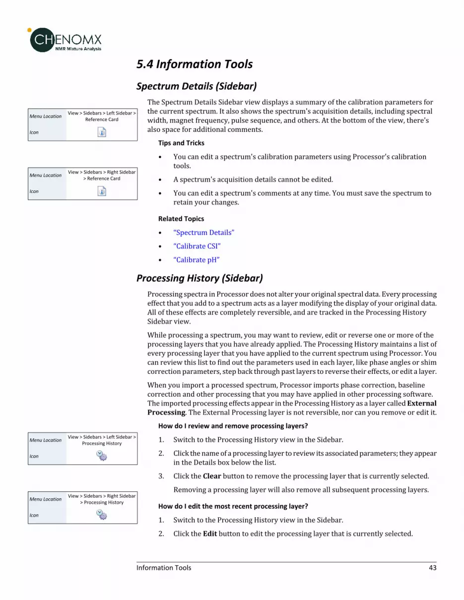
5.4 Information Tools
Spectrum Details (Sidebar)
View > Sidebars > Left Sidebar >Reference Card
Menu Location
Icon
View > Sidebars > Right Sidebar> Reference Card
Menu Location
Icon
The Spectrum Details Sidebar view displays a summary of the calibration parameters forthe current spectrum. It also shows the spectrum's acquisition details, including spectralwidth, magnet frequency, pulse sequence, and others. At the bottom of the view, there'salso space for additional comments.
Tips and Tricks
• You can edit a spectrum's calibration parameters using Processor's calibrationtools.
• A spectrum's acquisition details cannot be edited.
• You can edit a spectrum's comments at any time. You must save the spectrum toretain your changes.
Related Topics
• “Spectrum Details”
• “Calibrate CSI”
• “Calibrate pH”
Processing History (Sidebar)Processing spectra in Processor does not alter your original spectral data. Every processingeffect that you add to a spectrum acts as a layermodifying the display of your original data.All of these effects are completely reversible, and are tracked in the Processing HistorySidebar view.
While processing a spectrum, you may want to review, edit or reverse one or more of theprocessing layers that you have already applied. The Processing History maintains a list ofevery processing layer that you have applied to the current spectrum using Processor. Youcan review this list to find out the parameters used in each layer, like phase angles or shimcorrection parameters, step back through past layers to reverse their effects, or edit a layer.
When you import a processed spectrum, Processor imports phase correction, baselinecorrection and other processing that you may have applied in other processing software.The imported processing effects appear in the ProcessingHistory as a layer calledExternalProcessing. The External Processing layer is not reversible, nor can you remove or edit it.
View > Sidebars > Left Sidebar >Processing History
Menu Location
Icon
View > Sidebars > Right Sidebar> Processing History
Menu Location
Icon
How do I review and remove processing layers?
1. Switch to the Processing History view in the Sidebar.
2. Click thenameof aprocessing layer to review its associatedparameters; they appearin the Details box below the list.
3. Click the Clear button to remove the processing layer that is currently selected.
Removing a processing layer will also remove all subsequent processing layers.
How do I edit the most recent processing layer?
1. Switch to the Processing History view in the Sidebar.
2. Click the Edit button to edit the processing layer that is currently selected.
43Information Tools

3. When you have finished your changes, click the Accept button.
Editing aprocessing layerwill cause all subsequentprocessing layers tobe removed.
Tips and Tricks
• The Processing History only tracks processing effects, including phase correction,baseline correction, shim correction, water deletion and line broadening. Changesto spectrummetadata, such as the spectrum details or CSI and pH settings, do notappear in this panel.
• If you have just accidentally removed or edited a layer in the Processing History,you can use the Undo button to restore it.
Related Topics
• Chapter 2, Handling Samples and Spectra
• “Importing Spectra”
• “Legend”
• “Processing Tools”
• “Batch Import”
5.5 Importing and Exporting DataProcessor can help you to efficiently convert raw spectral data fromvarious sources, importthem into Chenomx spectra files (.cnx) and begin working with them.
Batch ImportFor largerprojects, youmaywant to import groupsof spectra intoChenomxspectra (.cnx),processing them with a consistent set of parameters to ensure consistent analysis results.
Tools > Batch Import...Menu Location
Icon
How do I batch import spectra?
1. Start the Batch Import wizard.
2. Choose the source spectra to import. The source spectra must be in one of thesupported file formats ( “Spectral Data Processing” )
3. If the source spectra include multiple file types, select the type that you would liketo import.
4. Choose the CSI and pH calibration options.
5. Choose the processing layers that you want to apply.
6. Specify a target folder for the imported spectra.
Tips and Tricks
• If the source folder containsmore than one type of spectrum file, you need to selectone type to import in the current session, since different import optionsmay applyto each available file type. You can do additional sessions to import other file types.
• The import settings available through the Batch Import wizard are the same asthose available when you import individual spectra ( “Importing Spectra” ).
• Storing the imported spectra next to the source filesmeans that eachnewspectrumfile appears in the same folder as the spectrum file from which it was created. You
44Importing and Exporting Data

may find itmoreuseful to haveProcessorplace the imported spectra into an entirelydifferent folder.
Related Topics
• “Importing Spectra”
• “Processing Tools”
• “Processing History (Sidebar)”
Send to Profiler
File > Send to ProfilerMenu Location
Icon
Whenyou finish processing a spectrum inProcessor, your next task is often to start profilingthe spectrum in Profiler. Send to Profiler closes the current spectrum in Processor, andre-opens it in Profiler.
Tips and Tricks
• Any changes that you have made to the spectrum in Processor are retained whenyou send the spectrum to Profiler, and are visible if you bring the spectrum backinto Processor. However, you still need to save the spectrum, either in Processoror Profiler, to permanently save those changes to the spectrum file.
Related Topics
• Chapter 6, Profiler
Export as JCAMP-DX
File > Export > JCAMP-DX...Menu Location
Icon
Processor has the ability to export your spectrum into the JCAMP-DX file format, which canbe imported into most NMR spectral processing software packages.
The effect of processing layers that you have applied to your spectrum will be retained inthe exported JCAMP-DX file, but other analysis data such as compound fits and concentrationwill not.
45Importing and Exporting Data

Chapter 6. Profiler
The Profiler module helps you to identify and quantify compounds in a sample spectrum,as well as apply spectral analysis techniques like spectral binning. You can fit any availablecompound to the current spectrum, and narrow down the compounds you're interested inby using compound sets and filters.
For a hands-on introduction to Profiler, see “Tutorial and Sample Files” .
6.1 Overview
❶ Sidebar View Dropdown Menu. Lets you change the currently visible Sidebar view.❷ Legend. A sidebar view letting you customize the lines appearing in the SpectrumView.❸ Cluster Navigator. Lets you navigate among the clusters of the currently selected
compound. You can enable or disable the Cluster Navigator using the Viewmenu.❹ Compound Table. Displays compounds, their calculated concentrations, and their
profiling status. You can access signatures of these compounds in the SpectrumGraphby selecting them. As well, you can view additional compound information in theReference Panel by selecting a compound in the Compound Table and switching thesidebar view to the Reference Card Panel. You can change the default concentrationunits displayed in the Compound Table using the Viewmenu.
❺ Spectrum Graph. Displays the spectrum as well as the sum line and subtraction line.You can modify the display by focusing in on a particular area or displaying andmodifying compound signatures.
❻ Quick Search. Lets you filter the Compound Table to focus on a particular compoundor group of compounds.
❼ Spectrum Thumbnail. Displays a thumbnail image of the current spectrum. You canalso use the thumbnail to navigate the Spectrum Graph at various zoom settings.
46Overview

❽ Status Bar. Displays the current cursor position in the units specified by the View > XScale andView>YScale settings. Also displays information about recent user actions.
6.2 Profiling ToolsProfiler provides you with a variety of tools to help you extract compound data from yourspectra. The data extraction process, called profiling, includes selecting compounds fromyour Library, setting concentrations, determining peak locations, and finding and usingextended information about compounds that you would like to profile.
Compound TableThe Compound Table summarizes the results of your profiling work with the currentspectrum. It contains entries for compounds that are available to profile in your spectrum,as well as those that you have already profiled, along with their maximum and profiledconcentrations. You can modify the display of the Compound Table to show as much or aslittle information as you like, to better suit your preferred approach to profiling.
The currently selected compound in the Compound Table is the active compound in theSpectrum Graph, where you can adjust its shape, position, and concentration.
You can filter the current contents of the Compound Table using Quick Searches forcompound names, comments, compound IDs, regions of the spectrum, or specific locationsin the spectrum.
Using theCompoundTable, youcanset customdisplay colors for compounds, pin compoundsso that they are visible even when they are not selected and toggle stars on compounds togenerally mark them for future reference.
You can select more than one compound from the Compound Table by holding down Ctrl(Cmd in Mac OS X) and clicking additional compounds. If you would like to select a rangeof compounds, click on the first compound and then hold down Ctrl-Shift (Cmd-Shift inMac OS X) and click on the last compound. This form of multiple selection will allow you toapply colors, pins, or stars to multiple compounds simultaneously.
There are many columns available for display in the Compound Table. You can choose todisplay any number of the available columns, with a minimum of one visible at any time,and you can drag the column headers to position the columns in any order that you like.You can sort the Compound Table on any column by clicking its column header. Click theheader again to reverse the sort order.
Tips and Tricks
• You must select at least one compound set in the Compound Sets view to see anycompounds in the Compound Table.
• When you have nothing selected in the Compound Table, pressing the up or downarrow keys will select the first compound in the Compound Table.
• After typing in the Quick Search box, press the down arrow key to navigate to theCompound Table.
• When you select a compound without a concentration in the Compound Table, apreview line for the compound appears in the Spectrum Graph, scaled vertically tosuit your current zoom level. You can click on the preview line to begin profilingthat compound; its concentrationwill be set tomatch the height of the preview line.
• When you select a compound with an icon in the Status column, the status area tothe left of the Quick Search box shows a detailed status message. You can also see
47Profiling Tools

the detailed status message in the tooltip that appers when you hover the mousecursor over the status icon.
• Whenyouhover themouse cursor over someparts of theCompoundTable, tooltipsappear containing more details. For example, hovering over the optional matchedclusters column for a compound shows you which clusters are matched.
• You can set compound concentrations directly in the Compound Table, bydouble-clicking on a compound's concentration cell and typing in a value.
• You can performmany common compound operations through the context menu,available by right-clicking on a compound's row in the table. Operations accessiblethrough the context menu include scaling concentrations, automatically fitting thecompound to the spectrum, and clearing fits, custom colors, stars or pins.
• You can copy the contents of a cell to the Clipboard by right-clicking on the cell andselecting Copy [cell name] to Clipboard. You can then paste the contents as textto any other program.
• Compounds that you have modified from their default state (including fits, customcolors, pins and stars) will appear in the Profiled Compounds set, and appear inbold in the Compound Table.
• PressDeletewithoneormore compounds selected in theCompoundTable to clearall fits, custom colors, pins and stars for the selected compounds.
• Tomakemore space for theCompoundTable, you canhide the SpectrumThumbnailby clicking View > Show Spectrum Thumbnail.
Related Topics
• “Change Columns”
• “Sidebar”
• “Preferences”
• “Custom Colors”
• “Pinned Compounds”
• “Starred Compounds”
• “Remember Selection”
• “Quick Searches”
• “Concentrations and Compound Fits”
• “Fit Automatically”
• “Compound Set Tools”
• “Batch Export”
• “Export Profiled Compounds”
• “Export Compound Table”
• “Information Tools”
• “Concentration Units”
48Profiling Tools

Custom ColorsWhen working with multiple compounds, it can become difficult to distinguish whichcompounds are contributing which parts to an overlapped region. You can set specificcompounds to appear in colors of your choice whenever they are visible in the SpectrumView.
Compound > Assign Color...Menu Location
Icon
How do I set a custom color for one or more compounds?
1. Select one or more compounds in the Compound Table.
2. In the Compound menu, click Assign Color... and select your desired color.Alternatively, if yourCompoundTable has aColor Columnopen, click in theCustomColor column ( ) to access the color chooser.
3. Click one of the preconfigured colors in the color chooser, or clickMore... to choosefrom a larger selection of colors.
Compound > Clear > ColorMenu Location
Icon
How do I clear a custom color from one or more compounds?
1. Select one or more compounds in the Compound Table.
2. In the Compound menu, click Clear > Color, or click in the Custom Color column( ) and click Clear Assigned Color.
Tips and Tricks
• You can also set or clear custom colors for compounds visible in the Legend, byclicking on the colored box beside the compound in the Legend.
• Setting a custom color for a compound adds it to the Profiled Compounds set, evenif you have not fit the compound.
• Custom colors are stored with the spectrum when you save it as a .cnx file. Thecolors you select are still associated with the compounds when you next open thefile, until you clear them. If you want to set the color of a specific compound in agroup of .cnx files, use the Batch Edit feature.
• You can export custom colors to a file using Batch Export.
Related Topics
• “Legend”
• “Preferences”
• “Compound Table”
• “Batch Export”
• “Batch Edit”
• “Pinned Compounds”
• “Starred Compounds”
• “Legend (Sidebar)”
Pinned CompoundsIt is often useful to display several different compounds at the same time while profiling,especially when you are working in heavily overlapped area. Pinning a compound ensuresthat it remains visible regardless of which compound you have selected in the Compound
49Profiling Tools

Table. In combination with custom colors and exporting spectrum graphics, pinning canalso help you to prepare snapshots of your profile that are suitable for publication.
Compound > Pin to GraphMenu Location
Icon
How do I pin one or more compounds?
• Click the pin for the compound in the Pinned column ( ) of the Compound Table.
Or
Click the pin next to the compound's line in the Legend ( ).
Or
Select oneormore compounds in theCompoundTable and, in theCompoundmenu,click Pin to Graph.
Compound > Clear > PinMenu Location
Icon
How do I clear a pinned compound?
1. Select the compound in the Compound Table.
2. In the Compound menu, click Clear > Pin, clear the check box for the compoundin the Pinned column, click the red pin next to the compound's line in the Legend,or click the X beside the compound's entry in the Legend.
Tips and Tricks
• Pinning a compound adds it to the Profiled Compounds set, even if you have not fitthe compound.
• Pin settings are stored with the spectrum when you save it as a .cnx file. Thecompound that you pin remains pinnedwhen you next open the file, until you clearthe pins. If you want to pin a specific compound in a group of .cnx files, use theBatch Edit feature.
• You can export the 'pinned' status of all compounds to a file using Batch Export.
Related Topics
• “Legend”
• “Compound Table”
• “Custom Colors”
• “Batch Export”
• “Batch Edit”
• “Starred Compounds”
• “Legend (Sidebar)”
Starred CompoundsStarring compounds lets youmark them for future reference. For example, you can use thisfunction to label compounds of interest for later analysis. Stars are saved in the spectrumfile, and remain visible when you next open the file, or until you clear them.
Compound > Add StarMenu Location
Icon
How do I star one or more compounds?
1. Click the grey star for the compound in the Starred column ( ) of the CompoundTable.
Or
50Profiling Tools

Select one or more compounds and, in the Compound menu, click Add Star.
Compound > Clear > StarMenu Location
Icon
How do I clear one or more starred compounds?
1. Select one or more compounds in the Compound Table.
2. In the Compoundmenu, clickClear> Star, or click the yellow star for the compoundin the Starred column ( ).
Tips and Tricks
• Starring a compound adds it to the Profiled Compounds set, even if you have notfit the compound.
• Stars are stored with the spectrumwhen you save it as a .cnx file. The compoundthat you star remains starredwhen you next open the file, until you clear the stars.If you want to star a specific compound in a group of .cnx files, use the Batch Editfeature.
• You can export the 'starred' status of all compounds to a file using Batch Export.
Related Topics
• “Compound Table”
• “Custom Colors”
• “Pinned Compounds”
• “Remember Selection”
• “Concentrations and Compound Fits”
• “Fit Automatically”
• “Scale Concentrations”
• “Batch Export”
• “Batch Edit”
Remember SelectionProfiler can rememberwhich compounds are selected in the current spectrum, and re-applythis selection to compounds inother spectra. This is usefulwhenworkingwithother featuresin the Compound menu across a set of spectra, such as “Fit Automatically”.
Tools > Remember SelectionMenu Location
Icon
How do I select a set of compounds from my current spectrum to work with in otherspectra?
1. Select one or more compounds.
2. In the Edit menu, click Remember Selection.
Tools > Restore SelectionMenu Location
Icon
How do I re-apply selected compounds to a new spectrum?
1. Open the spectrum that you want to select your compounds in.
2. In the Edit menu, click Restore Selection.
Tips and Tricks
• You can use this feature to quickly select a number of known compounds in theCompound Table to be fit automatically.
51Profiling Tools

Related Topics
• “Compound Table”
• “Custom Colors”
• “Pinned Compounds”
• “Starred Compounds”
• “Concentrations and Compound Fits”
• “Fit Automatically”
• “Scale Concentrations”
Quick SearchesWhenyouareprofiling a spectrum, youmayhave several compound sets active. Thenumberof compounds appearing in the Compound Table can make it difficult to locate a particularcompound to profile. Quick Searches let you temporarily limit the number of compoundsappearing in the Compound Table based on search criteria. The search criteria can includetext searches based on compound name, location, compound IDs, compound comments orall of these. You can also search by frequency, listing all compounds appearing at a specificfrequency, or within a frequency range.
How do I limit the compounds visible in the Compound Table?
1. Locate the search box at the top right of the Compound Table or press Ctrl-L toautomatically focus the search field.
Click the binoculars icon at the left edge of the search box to choose the type ofsearch to use (Compound Name, Compound Comment, Compound ID, Location orAll). If you do not specify a search type, the default is All.
2. Type your search terms in the Quick Search box. The contents of the CompoundTable update to show all matches to your current entry.
3. Click the X at the right edge of the search box to cancel the search and restore thefull list of compounds. Clearing the search field also resets the search type to All.
Howdo I search for compounds near a single ormultiple frequencies using the SpectrumView?
1. Right-click on the spectrum, and click Search for Compounds Near x.xx ppm toinitiate a single location search, or Add x.xx ppm to search locations to appendthe current location to the existing search location(s).
2. Click the X at the right edge of the search box to cancel the search and restore thefull list of compounds.
Howdo I search for compounds in a single ormultiple frequency ranges using a selectedregion?
1. Use the Select Region tool to select a region of the spectrum.
2. Right-click on the selected region, and click Search for Compounds in thisRegionor Add Region to search locations.
3. Click the X at the right edge of the search box to cancel the search and restore thefull list of compounds.
52Profiling Tools

Search For Compounds is an alternate method for setting up a Quick Search based on afrequency or range that you type, rather than based on clicked locations in the SpectrumView.
Tools > Search For Compounds...Menu Location
Icon
How do I filter the compounds in the Compound Table based on a specific frequencyor range?
1. Open the Search for Compounds dialog.
2. Enter the frequencyor range that youwant to use to search theCompoundTable.Allindividual queries must start with @, and the list must be comma separated. Forinstance, @1.20 to 1.25, @7.21 to 7.23 would search for compounds having peaksappearing in both the 1.20 to 1.25, and 7.21 to 7.23 ppm range.
3. Click OK.
Tips and Tricks
• Use Quick Searches to temporarily limit the number of compounds displayed. Usecompound sets created in Library Manager to define a more permanent workingset of compounds for profiling a particular type of spectra.
• You can reset the Quick Search box and clear all filters at any time by pressing Esc.
• After editing the Quick Search box, press the down arrow to navigate to theCompound Table.
• You can click repeatedly anywhere under the sum line (red) in the SpectrumGraphto cycle through the compounds contributing to the sum line at the frequency thatyou clicked.
• When you right-click anywhere under the sum line (red) in the Spectrum Graph,the resulting context menu shows a list of compounds contributing to the sum lineat the frequency that you clicked. Select a compound from the list to make it theactive compound.
• You can perform frequency and frequency range searches directly from the QuickSearch box. For example, [email protected] to find compounds appearing at 2.89 ppm,or [email protected] to 7.0 to find compounds appearing between 3.0 and 7.0 ppm.
Related Topics
• “Spectrum Graph Tools”
• “Compound Table”
• “Compound Set Tools”
Concentrations and Compound FitsWhen you match the shape and position of a compound signature in the software to theshape of your spectrum, you are said to be 'fitting' or 'profiling' that compound. Compoundswith a peak-based fit style, such as those found in the Chenomx Reference Library, are fitby adjusting the compound height and by making adjustments (sometimes called'transforms') to the locations of their clusters. It is also possible to create your owncompounds that use a region-based fit style, which automatically fit all of the spectrumareainwhatever region they are located in. Formore informationabout fit styles, see “CompoundFit Style” .
When using peak-based compounds to profile a spectrum, you measure compoundconcentrations by determining the height of each compound signature that best matches
53Profiling Tools

the peak heights in your spectrum. When you adjust the height of a compound, the heightsof all peaks and clusters corresponding to the compound scale proportionately.
The frequencies (i.e. positions) of the clusters in a compound signature may need to beadjusted during fitting, as well. These frequencies may be different from those in yourspectrum due to slight mismatches in solution pH, ion concentrations, and other matrixeffects. These potential errors are captured by a region around each cluster known as its'transformwindow'. Profiler displays the transformwindow for each cluster as two triangleson the spectrum baseline when the cluster is selected. If the pH value for your spectrumhas been calibrated correctly, you should be able to move each cluster to obtain a good fitwithout needing to move outside of the limits of the cluster's transform window.
How do I fit a compound and obtain a concentration?
1. Select a peak-based compound in the Compound Table (for example, choose anycompound found in the standard Chenomx Reference Library). If you do not seeany compounds in the Compound Table, select additional compound sets.
2. Click and drag the cluster to adjust the fit. You can also click and drag the bluetriangle on the vertical axis of the graph to changeonly the compoundconcentration,or drag a blue triangle on the horizontal axis to change only a cluster's frequency.
3. The concentration of the compoundwill appear in the Concentration column in theCompound Table.
How do I fit a region-based compound?
1. Select a region-based compound in the Compound Table. If you do not see anycompounds in the Compound Table, select additional compound sets. Note thatthere are no region-based compounds present in the default Chenomx ReferenceLibrary--you will likely need to create your own and add it to your CompoundLibrary.
2. Click and drag the cluster to adjust the fit. Notice how changing the height of thecompound has no effect on which part of the spectrum is fit.
3. Concentrations cannot be calculated for region-based compounds. Instead, theotherwise unfit spectrumareas near each clusterwill be summedup and displayedin the Area column of the Compound Table. This value is proportional to the trueconcentration of the compound.
How do I clear the fit for one or more compounds?
1. Select one or more fit compounds in the Compound Table. If you do not see any fitcompounds in the Compound Table, make sure that you have selected the ProfiledCompounds set. If you still do not see any fit compounds, then the current spectrumhas no compounds that have been profiled.
2. In the Compound menu, click Clear > Fit, or right-click on the compound in theSpectrum Graph or the Compound Table and click Clear > Fit.
Tips and Tricks
• You can have Profiler suggest a fit for the currently selected compound by clickingCompound > Fit Automatically, or by pressing the space bar.
• You can double-click on a peak-based compound's concentration in the CompoundTable and type a value directly to set the concentration. Type a number from 0 to5000 mM (or equivalent concentrations in μM or mg/dL), or type "--" to set theconcentration to unknown.
54Profiling Tools

• Whenyouhave selected a compound in theCompoundTable, you can click its shapein the Spectrum Graph, then use the arrow keys to adjust the compound'sconcentration or the cluster's position. Hold down Shift to allow finer adjustments.
• If youdonotwant to accidentallymove any cluster outside of its transformwindow,you can make Profiler strictly enforce all cluster transform windows by clickingEdit > Enforce TransformWindows.
• You can view the size of the transform that has been applied to any cluster byhovering the mouse cursor over it. The transform size appears in the top rightcornerof the SpectrumGraph, andvisually as a thickblue line connecting the startingposition of the selected cluster and its current position.
• When you move a cluster outside its transform window, its transform adjustmenttriangle turns red. TheTransform column in theCompoundTablewill also indicatewhether the compound has any clusters that have been moved beyond theirtransform windows
• To clear all of the fits for the current spectrum, clickEdit > Select All and then clickCompound > Clear > Fit.
• Toclear fits only for selectedcompounds, select the compoundsandclickCompound> Clear > Fit.Multiple compounds can be selected by holding down Shift or Ctrl(Cmd in Mac OS X).
• All compound concentrations and areas start out as unknown (--).
• If a compound's concentration is unknown (--) and you drag it up and then backdown to the baseline, the concentration is set to 0.00 mM. To return theconcentration to unknown, you must clear it.
• You can export a file describing the transform status of all compounds by clickingTools > Batch Export.
Related Topics
• “New Compound”
• “Compound Fit Style”
• “Reference Cards”
• “Spectrum Graph Tools”
• “Compound Table”
• “Starred Compounds”
• “Quick Searches”
• “TransformWindows”
• “Fit Automatically”
• “Scale Concentrations”
• “Compound Set Tools”
• “Batch Export”
• “Export Profiled Compounds”
• “Export Compound Table”
• “Reference Card Panel (Sidebar)”
55Profiling Tools

• “Concentration Units”
Fit AutomaticallyProfiling a spectrummanually may involve a lot of effort in setting compound clusterpositions and compound concentrations. You can have Profiler fit cluster postions usingthe “Compound Snapper” tool or compound concentrations using the “ConcentrationFitting and Optimization Tool” for either the currently selected compound, a selection ofcompounds, or all compounds in the compound table.
Compound > Fit AutomaticallyMenu Location
Icon
How do I fit one or more clusters automatically?
1. Select one or more specific clusters from the selected compound in the Compoundtable, or select one or more compounds in the Compound Table.
2. Right-click in the Compound Table and choose Snap Selection, or choose SnapCompound from the Compound menu.
How do I fit the concentration of one or more compounds automatically?
1. In the Compound menu, select Fit Concentrations
Tips and Tricks
• More reliable results from properly processed spectra. This includes well-definedCSI parameters, properly corrected baseline and accurate spectrum pH values.
• You can automatically fit compounds inmultiple spectra at once by using Batch Fitfrom the Tools menu.
• Use the Remember Selections feature to the mark compounds that can be reliablyautofit in your project. You can then restore the selection when working onsubsequent spectra, allowing you to quickly autofit those compounds.
Related Topics
• “Calibrate CSI”
• “Calibrate pH”
• “Compound Table”
• “Remember Selection”
• “Concentrations and Compound Fits”
• “Batch Fit”
Concentration Fitting and Optimization ToolOncepeak clusters havebeenpositioned, compound concentrations canbe fit andoptimizedusing constrained penalized linear least squares, according to:
Where...
1. A is themodelmatrixmadeup theunscaled individual signaturesof the compoundsbeing fit. The current locations of the peak clusters are taken into account whenthe Amatrix is calculated. The optimization process will not modify the location ofthe peak clusters.
56Profiling Tools

2. x is a vector made up all of the compound concentrations to be optimized. Eachcomponent of the vector, xi, is bounded between li (lower bound) and ui (upperbound). Non-negative restraints are imposed (li ≥ 0) since concentrations cannotbe negative.
3. b is the spectral subtraction line, i.e. the experimental spectrumminus thecontribution of all compounds part of the current profile that are not part of theoptimization process. In other words, if a compound has been fit and his not partof the fitting process, its contribution is subtracted from the experimental spectrumbefore the process starts.
4. W is a weight vector, which is used to modulate the relative importance of eachspectral point. Optionally, the algorithm can increase the weight of the points thatare overfit though an iterative penalization process.
The Concentration Fitting tool can be invoked either from the Tools menu, or from thecontextual menus shown by right-clicking or control-clicking on selected compounds inProfiler’s compound table, or on a selected spectral area.
Concentration Fitting can be performed without any graphical interface inputs, using theShift+Space bar keyboard shortcut, in which case it uses the default options and valuesstores in Profiler's preferences. Alternatively, temporary settings can be entered via agraphical interface using only the space bar keyboard shortcut.
The interface is composed of threemain sections: Regions Selection, Compounds Selection,and Fitting options. The role of each section is describedbelow. The applicationPreferencespanel can be used to control and set all the default values appearing in the interface.
57Profiling Tools

Regions Selection: Comma-separated list of spectral regions (in ppm units) to be includedor excluded in the fit. Each component of the lists must include a lower and upper boundin ppm, separated by a dash. For instance, “0.8-2.3, 3.0-4.2” is a valid list.
The IncludedRegions input field displays the limits of the currently selected spectral region(as selected with the Select Region Tool), or of the selected clusters for an individualcompound. If no region is currently selected, the Included Regions input field defaults tothe entire spectrum.
The Excluded Regions input field will automatically include the region associated with thewater peak. It is recommended that any peaks around the water region be excluded due toattenuation caused by water saturation. Similarly, the region associated with urea (5.4-6.0ppm) should also be excluded if urea is present.
Excluded regions have prevalence over any included regions, i.e. any spectral point fallingwithin any of the listed excluded regions will automatically be ignored during the fit.
58Profiling Tools

Compound Selections: User can optimize either all of the members of the current profile,all of the currently selected compounds in the compound table, or all of the currently listedcompounds in the compound table.
Note that compounds that donot have a singlepeak clusterwithin theparticipating spectralregions will not be considered for the fit. Their concentrations will not be changed.
Fitting options: This group of options control the behavior of the optimization engine.
Resolution (bin size): The spectrumcanbe fit using either its full representation, or a binnedrepresentation of it. This parameter controls the number of spectral points per bin. Set thisparameter to 1 for full resolution. However, it is highly recommended to use a binnedrepresentation if thepeak cluster positionshavenot beenoptimized. Fitting at full resolutionwill require more memory and CPU time.
Overfittingpenalty factor: The amount of overfitting canbe limitedby increasing theweightsof the spectral pointswhere overfitting occurs. Thiswill effectively increase the importanceof the spectral points where this occurs relative to the other ones, adding a penalty to payfor predicting points over the spectrum line. For a given spectral point, the square of thedifference between the experimentally observed spectrum intensity and thepredicted/calculatedonewill bemultipliedby the factor specified in this field if thedifferenceis negative. The weights are adjusted iteratively and up to 5 cycles of optimizations areperformed if the specified penalty factor is >1.0.
Upper constraints: Non-negative constrained are used during the optimization process sothat concentration values remain ≥ 0. Two different settings are offered to control theirupper bounds. They can be set to be unlimited via the Unconstrained option, or limitedaccording to their predeterminedmaximal potential concentration. Themaximal potentialconcentration of a given compound is the highest concentration for which the compoundsignature would not exceed the experimental spectrum. Because of possible discrepanciesbetween the compound library signatures and the experimental spectrum, mainly due topH conditions, matrix effects, etc, the potential concentrations determined by Profiler maynot be optimal and should only be regarded as guidelines. To give potential concentrationsmore wiggle room, potential concentrations can be pre-multiplied by a specified constant.The derived set of scaled potential concentration valueswill then be used as upper bounds.
It very important to note that the baseline is not modeled from the residuals at any stage.It is therefore crucial that the baseline be properly corrected prior to concentration fitting.
Related Topics
• “Calibrate CSI”
• “Calibrate pH”
• “Compound Table”
• “Remember Selection”
• “Concentrations and Compound Fits”
• “Batch Fit”
Enforce TransformWindowsTransformwindowsappear alongside compound signatures to guideprofilingby restrictingthe frequency range that you can adjust each cluster. All Chenomx Reference Compoundshave had transform windows set to allow the flexibility to fit most common biofluids. Youcan choose to have the transform windows enforced, preventing you frommoving any
59Profiling Tools

cluster outside its transform window, or not enforced, allowing you to move any clusterbeyond its transform window.
Edit > Enforce TransformWindows
Menu Location
How do I enforce transform windows?
1. On the Edit menu, check Enforce TransformWindows.
How do I allow clusters to move outside their transform windows?
1. On the Edit menu, clear Enforce TransformWindows.
Tips and Tricks
• Clusters that are outside their transformwindowappear in theTransforms columnof the Compound Table as OutsideWindows. Note that you may need to turn onthe Transforms column in the Compound Table.
• When you move a cluster outside its transform window, the adjustment triangleon the horizontal axis of the graph turns red. Also, when you hover over the clusterof its triangle, the visual indicator for the adjustment is red, as is the text in the topright corner of the Spectrum Graph describing the extent of the adjustment.
• Transformwindows for pH sensitive compound signatures (including those foundin the Chenomx Reference Library) are determined in part based on the pHuncertainty for the current spectrum. If you find that you frequentlyneed tooverridethe transformwindows for a spectrum, user Processor to check the accuracy of itspH calibration, and consider increasing its pH uncertainty value.
Related Topics
• “Calibrate pH”
• “Compound Table”
• “Cluster Navigator”
• “TransformWindows”
Scale ConcentrationsScale Concentrations lets you modify the concentrations of profiled compounds in thecurrent spectrum by a common multiplication factor. You can use this to compensate fordilution effects, errors in CSI concentration or other systematic influences on compoundconcentrations.
Compound > ScaleConcentration...
Menu Location
Icon
How do I scale the concentration of one or more compounds by a common factor?
1. Select oneormore compounds in theCompoundTable, and in theCompoundmenu,click Scale Concentrations...
2. Enter the multiplication factor you would like to use. To increase concentrations,use a factor greater than one. To decrease them, use a factor less than one.
3. Click OK.
How do I scale the concentrations of all compounds by a common factor?
1. In the Edit menu, click Select All, and then in the Compound menu, click ScaleConcentrations...
2. Enter the multiplication factor you would like to use. To increase concentrations,use a factor greater than one. To decrease them, use a factor less than one.
60Profiling Tools

3. Click OK.
Tips and Tricks
• You cannot enter amultiplication factor thatwould result in a concentrationhigherthan 5000 mM for any compound.
• If you set the multiplication factor to zero, all of the profiled compounds are set to0 mM.
• Scale Concentrationsdoes not affect compoundswith unknown concentrations (--).
• If youwant to scale concentrations for a specific compound or for all of the profiledcompounds in a group of .cnx files, use the Batch Edit feature.
Related Topics
• “Starred Compounds”
• “Concentrations and Compound Fits”
• “Batch Edit”
• “Compound Set Tools”
Batch EditBatchEdit lets youchooseagroupofChenomxspectra (.cnx), and thenperformanautomatedprofiling operationononeormore compounds in eachof the chosen spectra. You can chooseto add stars, pins, or colors to compounds, scale compound concentrations, or clear profileddata. After selecting an option youwill get to choose the compounds that youwant to affect.Once completed, you can open the .cnx spectra in Profiler to review the changes that weremade.
Tools > Batch Edit...Menu Location
Icon
How do I star/pin/color a set of compounds in multiple spectra at once?
1. Select Batch Edit from the Tools menu.
2. Select the spectra that you want the compounds to be edited in.
3. Choose to star, pin, color, scale the concentration of, or clear the profiled data ofthe compounds of interest.
4. Choose a Chenomx library or a compound set that contains the compounds ofinterest.
5. You will be given a list of all the compounds in the library or compound set thatyou selected in the previous step. Select the compounds that you want to adjustand click the right-hand arrow.
6. Click finish to add the changes to the spectra selected.
Related Topics
• “Custom Colors”
• “Pinned Compounds”
• “Starred Compounds”
• “Scale Concentrations”
61Profiling Tools

pH ToolProfiler can propose the pH of the sample by analysing the position of the fit peaks clustersthat are pH sensitive and report an optimized pH value thatwould best explain the locationof each.
The pH analysis is carried on based on 3 different selection modes:
1. the cluster(s) that are specifically selected for a given compound
2. the set of compounds selected in the compound table
3. if no clusters or compounds are selected, then it analyzes the entire list of profiledcompounds.
Compounds with no concentration and clusters with 0 net intensity or without pH-definedchemical shifts are ignored.
The results of the analysis of each clusters/compounds are presented in a scrollable list,with compounds in the first column, cluster ID in the second, best pH value in the third, thevalid compound pH range in the fourth, and the pH sensitivity of the cluster in the lastcolumn. The sensitivity is reported as the change in ppm around ± 0.1 unit of pH aroundthe global optimum pH that is proposed.
Only the clusterswith a sensitivity of ≥ 0.001 ppmare used in the calculations and reportedin the list.
The reported optimized pH is the value that minimizes the weighted amount of transformsthat would need to be applied to the listed clusters to explain their current position. Thelisted sensitivity values are used as weights.
It is possible to change the pH of the spectrum directly in the interface. Upon pH change,the clusters transforms are automatically recalculated to preserve the initial positions ofthe clusters.
Compound SnapperThis feature allows for automatic positioning of peak clusters on the spectrum. It willessentially positionpeak clusterswhere their shapebestmatch the experimental spectrum,independently of intensity.
Compound Snapper will not attempt to set or modify the concentration of compounds. Itonly focuses on optimizing peak cluster locations, individually or in group, depending onthe user’s selected cluster(s) or compound(s) when the feature is called. This functionalityautomatically positions peak clusters on the spectrum at the location that best matchestheir global shape. If twoormore clusters overlapwithin their allowed transformwindow,all possible combinations are analysed.
As such, this feature is powerful, and will find the proper cluster combination, assophisticated as it may be, that matches, according to shape, a relevant spectral region.
A snapping operation can either be performed on the original spectral line, or on thesubtraction line, i.e. by considering the “left-over” signal from everything that has been fitalready. It can also be done on a compound selected in the compound table and whoseconcentration has not been set already.
To perform a snapping operation, first select one or several clusters from an individualcompound, a single or several compounds at once from the compound table, right-click inyour selection to bring a contextual menu, or choose Snap Compound from the CompoundMenu (main toolbar).
62Profiling Tools

To snap all clusters resonating in a particular spectral region at once, first select the regionwith the Select Region tool (main button tool bar), right-click and select “Find and SnapPeak Clusters in this Region”.
Batch FitBatch Fit lets you choose a group of processed and calibrated spectra (.cnx), and thenautomatically fit one or more compounds in each of the chosen spectra. After selectingspectra you will get to choose the compounds that you want to have fit. Once completed,you can open the .cnx spectra in Profiler to review the changes that were made.
Tools > Batch Fit...Menu Location
Icon
How do I automatically fit a set of compounds in multiple spectra at once?
1. Select Batch Fit from the Tools menu.
2. Select the spectra that you want the compounds to be profiled in.
3. Choose a spectrum, a Chenomx library, or a compound set that contains thecompounds of interest.
4. You will be given a list of all the compounds in the spectrum, Chenomx library, orcompound set that you selected in the previous step. All of the compounds areselected to be fit automatically. Select any compounds that you do not want to fitautomatically and click the left-hand arrow to remove them from the list.
5. Click finish to fit the chosen compounds in the spectra. The changes made will besaved in the spectra that were selected to be fit automatically.
6. Review the spectra to ensure that the compoundswere correctly profiled, andmakeany adjustments that are needed.
Tips and Tricks
• Use this feature to profile compounds that can be reliably autofit at once.
• When choosing a profiled spectrum for the compounds of interest, check the "usetransforms from this spectrum as a starting point" box for a more precise fit. Thistakes into account any clusters that you have transformedmanually in the profiledspectrum, and starts the auto fit in these transformed regions. By unchecking thisbox profiler will fit automatically with the modeled cluster positions from the pHSensitive Chenomx Reference Compounds.
• Although Profiler will often arrive at acceptable values, always review the resultsof an automatic fit before moving on to other compounds.
Related Topics
• “Fit Automatically”
6.3 Cluster NavigatorThe Cluster Navigator lets you move quickly among the clusters of the currently displayedcompound. It appears in the top left corner of the Spectrum Graph as an ordered list of theapproximate positions of all the clusters, in ppm. When you click on a cluster position, theviewzooms toprovide anoptimal viewof that cluster. You can also use theClusterNavigatorto move sequentially through the list of clusters in either direction.
63Cluster Navigator

View > Show Cluster NavigatorMenu Location
How do I display the clusters of a compound in the Spectrum Graph?
1. Make sure that the Cluster Navigator is visible (it is visible by default), and selecta compound in the Compound Table.
2. Click a cluster position to zoom directly to that cluster. Click the left or right arrowto move sequentially through the list of clusters.
Tips and Tricks
• Use the Q and E keys to display the next or previous cluster.
• Clusters that appear green in the Cluster Navigator are 'matched', meaning theyclosely match the spectrum line as determined by comparing the sum line (red)and the spectrum line (black) near each cluster.
• You can click on a cluster and drag along the Cluster Navigator to display multipleclusters in the Spectrum Graph.
Related Topics
• “Spectrum Graph Tools”
• “Compound Table”
• “Concentrations and Compound Fits”
• “Enforce TransformWindows”
• “Compound Set Tools”
6.4 Compound Set ToolsCompound sets are groups containing compounds found in your Library. You can usecompound sets that you have created in LibraryManager tomake the task of profiling yourspectra easier, by placing compounds that interest you the most at your fingertips.
Compound Sets (Sidebar)The Compound Sets view lets you modify the contents of the Compound Table to includeany combinationof theCompoundSets that youhave created inLibraryManager. Thepanelcontains a list of every compound set that is currently defined. Contents of the checkedcompound sets appear in the Compound Table.
View > Sidebars > Left Sidebar >Compound Sets
Menu Location
Icon
View > Sidebars > Right Sidebar> Compound Sets
Menu Location
Icon
How do I select multiple compound sets?
1. Click the checkboxnext to theCompoundSets that youwant to add to theCompoundTable.
How do I select a single compound set?
1. Click on the name (as opposed to the checkbox) of the Compound Set that youwantto select. All other Compound Sets will be deselected.
Tips and Tricks
• Reference Compound Sets, which contain any Chenomx Reference Compounds inyour library, appear automatically in the Compound Sets view. If your librarycontains any non-Chenomx compounds, including ones that you created yourself,they will appear in a separate compound set called Custom Compounds.
64Compound Set Tools

• When you filter the Compound Table by frequency or range, only compounds fromall the currently active compound sets are considered, since these are the onlycompounds in the table. You can use this, for example, by creating a compound setcontaining only compounds that appear in urine samples. Then, when this urinecompound set is the only one active, any quick searches that you performwill onlyreturn compounds that appear in urine samples as potential hits.
• A compound set called Profiled Compounds always appears at the top of theCompoundSets view inProfiler. TheProfiledCompounds set includesall compoundsin the current spectrum that have modified fits, custom colors, pins or stars.
• Profiled Compounds also contains subsets that appear as you start profilingcompounds. These sets are automatically updated as you work, and allow you toquickly select profiled compounds that meet specific critia. For example, you canselect to view only the compounds that have a defined fit, or only the starredcompounds.
• The Profiled Compounds set updates continuously, so if you clear a compound, itimmediately disappears from Profiled Compounds. In this case, you can still selectthe compound from its original compound set.
• If you change the contents of a compound set in Library Manager, your changesimmediately appear in Profiler, including modified selection rules for SmartCompound Sets, manual changes to Compound Sets by adding or removingcompounds, and created or deleted compound sets.
Related Topics
• “Profiling Tools”
• “Automatic Compound Sets”
• “Compound Sets”
• “Smart Compound Sets”
6.5 Importing and Exporting DataYou can export data from your profiled spectra for further analysis, bin the spectra foranalysis by more traditional techniques, or create images of your spectra for use inpublications and presentations.
Batch ExportWhen you have finished profiling a spectrum or set of spectra, you often need to get theresulting data into a spreadsheet or statistical analysis package. BatchExport lets you createa text file containing all of the relevant details from profiled spectra, in formats that youcan easily open in software like Microsoft Excel or Umetrics SIMCA-P.
Tools > Batch Export...Menu Location
Icon
How do I export data from one or more spectra?
1. Start the Batch Export wizard.
2. Select the type of data thatwould you like to export. You can export concentrationsin various units, other profiling results like profiled areas, matched clusters andtransform summaries, or annotations like stars, pins and custom colors.
3. Choose the source spectra to export, and specify a target folder and filename forthe exported text file. You can specify the currently open spectrum, other individualspectrum files or one or more folders containing spectrum files as the source.
65Importing and Exporting Data

4. Choose a save location and file format. You can export as comma-separated text(.csv), as tab-separated text (.tsv), or as an excel spreadsheet (.xlsx).
5. Choose the compounds forwhichyouwould like to export data. You canuse customcompound sets that you have created in Library Manager to filter the output tospecific compounds, or you can export all profiled compounds in all of the spectra.
6. Choose additional data that youwould like to include in the exported file. Additionaldata can include spectrum pH values, Chenomx compound IDs for the exportedcompounds, or InChI or SMILES strings for the exported compounds.
7. Choose the default layout (files in rows and compounds in columns) or transposedlayout (compounds in rows, files in columns) for the output file, depending on howyou intend to use the file.
Concentration Confidence Intervals
• Whenexporting concentrations, the softwarewill also export the statistical standarddeviations on concentrations as a second tab in the excel workbook. Concentrationconfidence intervals (± values) at 90%, 95%, and 99% can be calculated bymultiplying the reported standarddeviationsby1.645, 1.96, and2.576, respectively.
• For each compound, the standarddeviationof each cluster is calculated individually,by calculating a ‘local’ sum of difference of squares in the spectral region currentlyoccupied by each cluster. The minimum value found for all clusters is reported. Assuch, the reported standard deviation should be considered “best-case scenario’.
• Failure to properly correct the spectral baselinewill result is larger sumof squares,and therefore larger concentration errors.
• Confidence intervals canbeused to assist in thedeterminationof themeaningfulnessof reported fit concentrations, and in the decision to include or reject compoundsin statistical models.
Tips and Tricks
• Youmust export Areas rather than Concentrationswhenworkingwith compoundsthat have a region-based Fit Style (see “Compound Details” )
• To filter the exported compounds without modifying the source spectra, create aCompound Set or Smart Compound Set in Library Manager, and only exportcompounds in that Compound Set.
• Use the default layout to export files that you intend to openwith statistical analysispackages. Use the transposed layout to export files that you intend to open in aspreadsheet program, or if you prefer the transposed layout.
• If you intend to open the exported file in Microsoft Excel 2003 or earlier, limit thenumber of compounds to less than 253 for theDefault layout, or the number of filesto less than 253 for the Transposed layout, since these versions of Excel do notsupport large numbers of columns.
Related Topics
• “Profiling Tools”
• “Compound Set Tools”
• “Concentration Units”
• “Export Profiled Compounds”
66Importing and Exporting Data

• “Export Compound Table”
• “Compound Fit Style”
Export Compound TableWhen collaborating with other people on projects, you may want to quickly compareCompound Tables while profiling. Export Compound Table creates a copy of the currentcompound table in a format which can be easily imported into any modern spreadsheetprogram.
File > Export > CompoundTable...
Menu Location
Icon
How do I export my current compound table from this spectrum?
1. Click File > Export > Compound Table.
2. Choose a save location and file format. You can export as comma-separated text(.csv), as tab-separated text (.tsv), or as an excel spreadsheet (.xlsx).
3. Click Save.
Tips and Tricks
• If there are blank cells, there will be corresponding blank values in the exporteddata (this can happen if the table contains compounds that are missing from yourcompound library or if you export columns that can have empty cells, like "author","pulse sequence", or "compound comment")
• The "Status" column will never be exported, even if it is visible in the table.
• The Starred and Pinned columns will export as "Yes" or "No" for each cell, and theColor columnwill export as a hex RGB value, such as 0000FF for each cell. Cells thathave no color assignedwill all export as the "default" color for that cell, i.e. the colorthat the cells compound appears when displayed on the graph.
• You canadd/removecolumns, reorder columns, or clickon theheaderof any columnto change the table sort order. The columns you chose to appear in your CompoundTable will be exported, and the order of the rows and columns in the exported filewill be in the same order as your Compound Table.
Related Topics
• “Profiling Tools”
• “Compound Set Tools”
• “Concentration Units”
Export Profiled CompoundsWhen you collaborate with other people on a project, you may have different compoundsin your library than your collaborator has in theirs. Exporting profiled compounds createsa compound pack (.pack) containing the set of actual compound signatures that you usedto profile the project, including compounds that have been annotated with stars, pins, orcolors. You can exchange compound packs with other users of Chenomx NMR Suite, andimport new packs via Library Manager. This is one way to be certain that you and yourcollaborator are always profiling spectra for that project using exactly the same collectionof compound signatures.
67Importing and Exporting Data

File > Export > ProfiledCompounds...
Menu Location
Icon
How do I export a compound pack from the current spectrum?
1. Open the Export Profiled Compounds dialog.
2. Enter a name for the exported compound pack. The name you enter is used to labelthe associated Compound Set when the compound pack is imported into anotherlibrary via Library Manager.
3. Click OK.
4. In the resulting file chooser, locate the folder in which you would like to save thecompound pack.
5. In the File Name field, type the newname for the compound pack file. The specifiedname can be different from the name of the compound pack itself, but you mayprefer to keep them the same.
6. Click Save.
Tips and Tricks
• If youwant to export all of the compounds in a compound set, regardless ofwhetherthey have been fit in a particular spectrum, use Export as Compound Pack inLibrary Manager.
Related Topics
• “Profiling Tools”
• “Compound Set Tools”
• “Add Compounds”
• “Export Compounds”
• “Export Compound Table”
• “Compound Set Tools”
Import ProfileSpectral profiling can be accelerated by importing, partially or entirely, profiled data ofspectra that have already been fit. This is particularly efficient when dealing with largeseries of similar spectra.
File > Import Profile...Menu Location
Icon
How do I import a profile from another spectrum?
There are 4 ways profiled data can be imported:
1. By choosing Import Profile from the File menu.
2. By selecting Batch Import Profile from the Batch Operations in the Tools menu.
3. By dragging and dropping a cnx file directly in the compound table. The file canalso be chosen from Files panel that can be displayed in the left or right verticalsidebar in Profiler
4. By selecting and dragging compounds from the compound table of one openedinstance of Profiler to another one.
Except for the last option, the changes are automatically applied. Any dragged compoundsfromanotherprofilewill automaticallyoverwritewhatmayexist. Theoperation is cancellableby selecting Undo Import Profile from the Edit menu.
68Importing and Exporting Data

For the other first 3 options, the following interface will be presented to allow for moreoptions:
The right listboxwill automatically bepopulatedwith all of the compounds that are availablefrom the source cnx spectrum, and automatically chosen as the default set of compoundsto be imported. Compounds that should not be imported can be selected and drag to the“Will not be imported” listbox on the left side of the interface. All compounds chosen forimport will merge with the existing profile, and the profile of pre-existing compounds willbe automatically replaced by those imported, unless the “Skip compounds that are alreadyprofiled” option is selected (see below).
Two additional options are available to control the behaviour of the import operation:
1. Completely overwrite the existing profile: This option will first completely wipethe current existing profile before the selected compounds are imported.
2. Skip compounds that are already profile: If selected, this option will ignore theimport of compounds that are pre-existing
Tips and Tricks
• When you import a profile, fully or partially, the imported data completely replaceswhat currently exists. That applies to peak cluster transforms, compoundconcentrations, and compound annotations (colors, etc).
Related Topics
• “Profiling Tools”
69Importing and Exporting Data

Send to Processor
File > Send to ProcessorMenu Location
Icon
As youworkwith a spectrum in Profiler, youmay notice that the phase correction, baselinecorrectionor other aspects of the spectrum's lineshapes are not properly adjusted. Similarly,you may import a spectrum directly into Profiler, only to realize that it requires phasecorrection, baseline correction, or additional pH or CSI calibration. Send to Processorcloses the current spectrum in Profiler, and opens it in Processor, retaining any changesyou have made in Profiler.
Tips and Tricks
• Anychanges that youhavemade inProfiler are retainedwhenyousend the spectrumto Processor, and will be visible if you bring the spectrum back into Profiler.However, you still need to save the spectrum, either in Processor or Profiler, topermanently save those changes to the spectrum file.
Related Topics
• Chapter 5, Processor
6.6 Spectral BinningStatistical approaches to analyzing large numbers of spectra often involve data reductionmethods like spectral binning to reduce the number of variables that must be considered.Spectral binningdivides spectra into a series of regions, or bins; subsequent analysis involvesthe integrated area of these bins instead of the raw spectral data. Profiler lets you performspectral binning on collections of spectra, exporting the results to an excel document (.xlsx)and to tab-delimited text (.tsv) that is easily read by spreadsheet programs and statisticalanalysis software packages.
Tools > Spectral Binning...Menu Location
Icon
How do I bin spectra?
1. Start the Spectral Binning wizard.
2. Choose the source spectra (.cnx) for binning, and specify a target folder for theresulting binned data. You can specify a series of spectrum files or one or morefolders containing spectrum files as the source.
If you would like to repeat a binning session that you have previously saved, youcan specify a binning configuration file (BinningConfiguration.xml) to import.
A binning configuration file is automatically created each time you calculate binningresults.
3. Choose a binning method to apply to the specified spectra. You can use a definedbin size in ppm or a defined number of bins. You can also use bins based on thetransform windows of the compounds in a Compound Set ("targeted binning").
If you would like to reuse a predefined set of bins, you can specify a bin definitionfile (BinDefinitions.tsv) to load.
4. Choose regions of the spectrum to exclude from binning, if desired. You can specifyany regions of the spectrum using start and end values in ppm.
5. Choose a binning target. You can bin spectra based on the sum line (standardbinning) or the subtraction line (residual binning).
6. Choose a normalization method. You can normalize the bins relative to the totalarea under each spectrum line or to units of standardized area (sa). Note that 1 sais the area under a theoretical DSS methyl peak at 0.50 mM in each spectrum.
70Spectral Binning
Tips and Tricks
• Binning can take a long time, especially on older computers or particularly largedatasets. If you have never tried binning before, try a small dataset of five or tenspectra first, before moving on to larger datasets. The time needed for the smallerdataset will help you to more accurately estimate the time needed to complete abinning session.
• If you choose one or more folders as the source, binning includes spectra bothdirectly in the specified folders and in subfolders of the specified folders.
• Binning based on number of bins divides the spectrum into the specified numberof bins after excluding the regions that you have defined, if any.
• To do residual binning, each of the source spectra must have been analyzed inProfiler and must contain at least one compound with a defined concentration.
• Excluding regions from a binning dataset can help you to remove the influence ofinterfering or confounding signals, such as solvent peaks.
• Normalization based on total area gives the area of a particular bin as a fraction ofthe total area of all bins, except area in any of the excluded regions that you mayhave defined.
• Spectral binning is subject to several numerical limits:
• Each binning session can include a maximum of 5000 spectra.
• The range that youdefinewhenusingnumberof bins or size of bins as abinningmethod must be at least 1 ppm wide, and can be as large as 1998 ppm (-999to 999 ppm).
• You can specify a maximum of 100000 (one hundred thousand) bins or aminimum of 1.
• You can specify a minimum bin width of 0.00001 ppm and a maximum binwidth equal to thewidth of the range that you have chosen. For example, if youare binning across a range of 2 to 10 ppm, your maximum bin width is 8 ppm.
Related Topics
• “Profiling Tools”
• “Compound Set Tools”
6.7 Information ToolsWhile profiling spectra, you can view acquisition and sample information connected to thecurrent spectrum, and change the concentration units that appear in the Compound Tableand any exported profiles. The Sidebar lets you change the lines displayed in the SpectrumGraph, select compound sets to use in fitting spectra, and view extended information aboutthe compounds in your Library
Legend (Sidebar)TheLegend is an interactive display of the various lines currently appearing in the SpectrumGraph. In Profiler, you can use the Legend to pin or unpin compound lines to the spectrum,change their colors, or make a pinned compound active for fitting.
71Information Tools

View > Sidebars > Left Sidebar >Legend
Menu Location
Icon
View > Sidebars > Right Sidebar> Legend
Menu Location
Icon
How do I pin a compound using the Legend?
1. Switch to the Legend view in the Sidebar.
2. Select a compound in the Compound Table.
3. In the Legend view, click the gray pin icon ( ) appearing to the right of thecompound's line.
How do I unpin a compound using the Legend?
1. Switch to the Legend view in the Sidebar.
2. Locate a pinned compound in the Legend view.
3. Click the red pin appearing to the right of the pinned compound's name.
How do I temporarily hide a pinned compound?
1. Switch to the Legend view in the Sidebar.
2. Locate a pinned compound in the Legend view.
3. Uncheck the box appearing to the left of the pinned compound's name.
Tips and Tricks
• Youcanadjust thedisplayof compound lines through theLegendview in theSidebar,including setting custom colors, pinning compounds and selecting a compound foruse in profiling.
Related Topics
• “Legend”
• “Compound Table”
• “Custom Colors”
• “Pinned Compounds”
Reference Card Panel (Sidebar)
View > Sidebars > Left Sidebar >Reference Card
Menu Location
Icon
View > Sidebars > Right Sidebar> Reference Card
Menu Location
Icon
The Reference Card Panel Sidebar view displays information specific to the compound thatyouhave selected in theCompoundTable. This includesa structure image,molecular formulaandweight, alternate names, InChI and SMILES strings, extended descriptive text and linksto external reference sites. You canuse this information toverify the identityof the compoundthat you are fitting and better understand the signature and behaviour of the compound inyour spectra.
Related Topics
• “Reference Cards”
• “Preferences”
• “Profiling Tools”
72Information Tools

Spectrum Details (Sidebar)
View > Sidebars > Left Sidebar >Reference Card
Menu Location
Icon
View > Sidebars > Right Sidebar> Reference Card
Menu Location
Icon
The SpectrumDetails Sidebar view shows a summary of the calibration parameters for thecurrent spectrum. It also displays the spectrum's acquisition details, including spectralwidth, magnet frequency, pulse sequence, and others. At the bottom of the view, there'salso space for additional comments.
Tips and Tricks
• You can edit a spectrum's calibration parameters by opening it in Processor andusing the available calibration tools.
• A spectrum's acquisition details cannot be edited.
• You can edit a spectrum's comments at any time. You must save the spectrum toretain your changes.
Related Topics
• “Spectrum Details”
• “Calibrate CSI”
• “Calibrate pH”
Concentration Units
View > Concentration UnitsMenu Location
You can change the units of concentration used in the Compound Table. Available unitsinclude millimoles per litre (mM), micromoles per litre (μM) and milligrams per decilitre(mg/dL).
Tips and Tricks
• The maximum allowed concentration for any compound is 5000 mM (5 M). Whenyou select mg/dL as the concentration unit, the maximum value varies percompound, since the units incorporate the molecular weight of the compound.However, the maximum value is always equivalent to 5000 mM.
• Concentrations inmg/dLare only available for compoundswhosemolecularweighthas been specified in the compound's Reference Card , and the accuracy of suchconcentrations depends on the accuracy of the molecular weight provided. Themolecular weight has already been correctly specified for all compounds in thedefault Chenomx Reference Library.
Related Topics
• “Profiling Tools”
• “Batch Export”
• “Compound Details”
• “Send to Compound Builder”
73Information Tools

Chapter 7. Spin Simulator
Spin Simulator is a powerful tool for creating simulations of NMR spectra, based onuser-defined spin systems and reference spectra. You can use these simulations as startingpoints for creatingyourowncompoundsignatures, especially if youneed to create signaturesat multiple magnet frequencies, or you want to create robust signatures with a strongtheoretical backing.
To use Spin Simulator effectively, you'll need a basic understanding of NMR theory, and youshould know how to interpret NMR spectra, especially J-coupling. If necessary, Chenomxexperts can also provide compound fitting services andhelpdevelop specialized compoundlibraries for you.
For a hands-on introduction to Spin Simulator, see “Tutorial and Sample Files” .
7.1 Overview
❶ Sidebar View Dropdown Menu. Lets you change the currently visible Sidebar view.❷ Spin Definition. Sidebar view that displays the currently selection Spin Definition.❸ J-Value Editor. Lets you vary the J-value for the currently selected J-modifier.❹ J-Modifier List. Lets you add, select, edit or remove J-modifiers associated with the
currently selected spin definition.❺ SpinDefinition List and SimulationDetails. Facilitates selection of spin definitions and
spin systems. Also displays information about the current simulation, including thesimulation frequency and numbers of spin systems, transitions, and J-modifiersrepresented in the simulation.
❻ Spin Navigator. Lets you navigate among the spin definitions of the currently opensimulation. You can enable or disable the Spin Navigator using the Viewmenu.
74Overview

❼ Spectrum Graph. Displays the spectrum overlay as well as the simulation line and thecurrently selected spin definitions.
❽ Spectrum Thumbnail. Displays a thumbnail image of the current spectrum. You canalso use the thumbnail to navigate the Spectrum View at various zoom settings.
❾ Status Bar. Displays the current cursor position in the units specified by the View > XScale andView>YScale settings. Also displays information about recent user actions.
7.2 Simulating ToolsSpin Simulator provides you with a variety of tools to help you create accurate spinsimulations by adding,manipulating and removing spin definitions and coupling constants,grouping spindefinitions into spin systems, andviewing summarydetails about your currentsimulation.
Spin simulation is a computationally intensiveprocess, and takesmore timeas the simulationyou are creating becomes more complex. Generally, the amount of time required torecalculate a simulation increasesmore or less linearlywith the number of spin definitions(protons) in the simulation, and exponentiallywith the number of distinct J-couplings. SpinSimulatormonitors theoverall complexityof your simulationas youwork, and if youattemptto take an action thatwould result in unusable simulation times, the action is cancelled, andyou see a warning dialog indicating that the resulting simulation would be too complex.
New Simulation
File > New Simulation...Menu Location
Icon
NewSimulation creates a new spin simulationwith the default simulation frequency of 800MHz.
Tips and Tricks
• The new simulation does not have a spectrum overlay by default. Add a spectrumoverlay before you continue editing the simulation.
• You can overlay only one spectrum at a time in Spin Simulator.
• You need to save your simulation to an .xss file retain the changes that you havemade.
• Spectrum overlays are not retained in the saved files.
Related Topics
• “Spectrum Overlays”
• “Simulation Details”
Spin SystemsIf you need tomodel distinct structures in a single simulation, such as different enantiomersin solution, you may want to use spin systems to organize them. A spin system in SpinSimulator is a set of spin definitions that you have defined as only interacting with eachother. These spin systems can have their own abundance in solution.
Simulation > Add New SpinSystem...
Menu Location
Icon
How do I add a new spin system?
1. Click Add New Spin System.
2. If this is the first spin system you have added, a new dropdown list appears abovethe SpinDefinition list, showing the available spin systems. Additional spin systemsalso appear in this dropdown list.
75Simulating Tools

How do I select a different spin system?
1. In the Spin Definition list, select the spin system to which you would like to switchfrom the drop-down list.
Alternately, click on a spin definition belonging to the other spin system in theSpectrum Graph.
Simulation >Delete Current SpinSystem...
Menu Location
Icon
How do I delete a spin system?
1. Select a spin system from the drop-down list above the spin definition list.
2. Click Delete Current Spin System.
Tips and Tricks
• Deleting a spin system deletes all spin definitions and J-modifiers associated withthe spin system as well.
Related Topics
• “Spin Definitions”
• “Spin Navigator”
• “Spin Definition (Sidebar)”
• “Simulation Details”
Spin DefinitionsSpin Simulator creates computer models of the interactions among the members of a spinsystem. Within Spin Simulator, you may consider a member of a spin system as equivalentto a single proton, or a group of protons that are both chemical shift equivalent andmagnetic-equivalent. Spin definitons describe the members of spin systems, and are thebasic building blocks of the models created by Spin Simulator.
A spin definition consists of three basic pieces of information: The number of protonsrepresented by the spin definition, the center of the spin definition in ppm (equivalent toa chemical shift in an NMR spectrum), and a width in sw (analogous to a linewidth athalf-height in an NMR spectrum).
You can organize spin definitions into spin systems, as described elsewhere in this chapter(see “Spin Systems” ).
Simulation > Add SpinDefinitions
Menu Location
Icon
How do I add a new spin definition?
1. Click the Add Spin Definitions button on the toolbar, or from the menu underSimulation > Add Spin Definitions.
2. Click on the screen the ppm location where you want the new spin definition. Asingle proton spin definition will be added to the simulation. You can change thenumber of protons and add J-Modifiers later.
3. Click on the Stop Adding Spin Definitions button on the toolbar (or press ESC) toleave spin definition addition mode.
How do I edit a spin definition?
1. Select the spin definition by clicking on it in the Spectrum Graph, or by selecting itin the Spin Definition list in the Simulation Details panel.
2. Do any of the following:
76Simulating Tools

• Click and drag the blue triangle on the horizontal axis to change the center ofthe spin definition.
• Click and drag the blue triangle on the vertical axis to change the height of thesimulation.
• Click and drag the spin definition directly in the Spectrum View to change itscenter and the simulation height simultaneously.
• Click anddrag the blue triangles on either edge of the spin definition (at aroundhalf the height of the tallest peak) to change the width of the spin definition.
• Use the W and S keys or the up and down arrow keys to adjust the height ofthe whole simulation up and down.
• Use the A and D keys or the left and right arrow keys to adjust the position ofthe selected spin definitions left and right.
• Use the Q and E keys to display the next or previous spin definition.
Simulation > Reassign SelectedSpin Definitions...
Menu Location
Icon
How do I reassign a spin definition from one spin system to another?
1. Select a spin definition in the SpectrumGraphor by selecting it in the SpinDefinitionlist in the information panel.
2. Click Reassign Selected Spin Definitions....
3. Select either a target spin system, or select a new spin system.
4. All spin definitions coupled to the current selected spin definitions will also bereassigned. Uncheck the check box if you want to uncouple the selected spindefinitions and move only the selected spin definitions instead.
Simulation > Delete SelectedSpin Definition
Menu Location
Icon
How do I delete a spin definition?
1. Select a spin definition in the SpectrumGraphor by selecting it in the SpinDefinitionlist in the information panel.
2. Click Delete Selected Spin Definition.
Tips and Tricks
• A spin definition can describe nomore than ten protons. However, given that a spindefinition typically describes chemically and magnetically equivalent protons, avalue greater than three (a methyl group) is often not necessary.
• A spin system can contain any number of protons and spin definitions, as long asthe total number of transitions is less than 2000. If you need to create a simulationrequiring more than 2000 transitions, you will need to use multiple spin systems.
The total number of transitions in the current simulation appears in the simulationdetails (see “Simulation Details” ).
• Height adjustments always affect the whole spin system, not just the currentlyselected spin definition. If you need to adjust the heights more than is possible inSpin Simulator, finish the simulation, save it and import it in Compound Builder,then make your adjustments there.
• Deleting a spin definition also deletes any J-modifiers that youhave associatedwiththat spin definition.
77Simulating Tools

Related Topics
• “Spin Systems”
• “Spin Navigator”
• “J-Modifiers”
• “Spin Definition (Sidebar)”
• “Simulation Details”
• “Import Simulation”
Spin NavigatorThe Spin Navigator lets you quicklymove among the spin definitions that you have createdin the current simulation. It appears in the top left corner of the Spectrum Graph as anordered list of the approximate positions of all the spin definitions of the simulation, in ppm.When you click on a spin definition's position, the view zooms to provide an optimal viewof the spin definition. You can also use the Spin Navigator tomove sequentially through thelist of spin definitions in either direction.
View > Show Spin NavigatorMenu Location
How do I display the spin definitions of a simulation in the Spectrum Graph?
1. Make sure that the Spin Navigator is visible.
2. Click a spin definition's position to zoom directly to that spin definition. Click theleft or right arrow to move sequentially through the list of spin definitions.
Tips and Tricks
• Click on a spin definition anddrag along the SpinNavigator to include adjacent spindefinitions in the Spectrum Graph.
• Use the Q and E keys to display the next or previous cluster.
Related Topics
• “Spin Systems”
• “Spin Definitions”
• “Spin Definition (Sidebar)”
• “Simulation Details”
J-ModifiersWhen you create a new spin definition, it appears on the SpectrumView as a single, unsplitpeak. J-modifiers let you split a spin definition into more complex shapes, to fit the shapeof real spectra. J-modifiers can be uncoupled, affecting only one spin definition, or coupled,linking the spin definition to another one via its coupling constant. Coupled J-modifiersresult inmore accurate simulations, as Spin Simulator canuse them to calculatehigher-ordercoupling effects, accounting for tenting and other complex behaviour.
Compound > Add NewJ-Modifier...
Menu Location
Icon
How do I add an uncoupled J-modifier to a spin definition?
1. Select the spin definition to which you would like to add a J-modifier.
2. Open the Add New J-Modifier... dialog.
3. Enter a J-value for the new J-modifier, in Hz.
78Simulating Tools

4. Select Uncoupled Split, and specify a split count for the new J-modifier.
5. Click OK.
How do I add a coupled J-modifier to a spin definition?
1. Select one of the two spin definitions that you would like to link with a coupledJ-modifier.
2. Open the Add New J-Modifier... dialog.
3. Enter a J-value for the new J-modifier, in Hz.
4. Select Coupled, and select the spin definition to which you would like to link.
5. Click OK.
Compound > Edit SelectedJ-Modifier...
Menu Location
Icon
How do I edit an existing J-modifier?
1. Select a spin definition.
2. Select a J-modifier that you would like to edit.
3. Open the Edit Selected J-Modifier... dialog.
4. Make your desired changes.
5. Click OK.
How do I couple two spin definitions using splits that I have already defined?
1. Select one of the two spin definitions that you would like to link, and select theuncoupled split that will link it to the other spin definition.
2. Open the Edit Selected J-Modifier... dialog.
3. Select Coupled, and select the spin definition to which you would like to link.
4. Select the uncoupled split from the second spin definition that you would like tolink to.
5. Click OK.
Compound > Delete SelectedJ-Modifier
Menu Location
Icon
How do I delete a J-modifier?
1. Select a spin definition.
2. Select a J-modifier that you would like to delete.
3. Click Delete Selected J-Modifier.
Compound > UncoupleMenu Location
Icon
How do I uncouple a J-modifier?
1. Select a spin definition.
2. Select a J-modifier that you would like to uncouple.
3. Click Uncouple.
79Simulating Tools

Compound > Ungroup SelectedSplits
Menu Location
Icon
Compound > Group SelectedSplits
Menu Location
Icon
How do I ungroup J-modifiers?
1. Select a spin definition.
2. Select an uncoupled split with a split count greater than 1.
3. Click Ungroup Selected Splits..
How do I group several J-modifiers?
1. Select a spin definition.
2. Select several uncoupled splits that you would like to combine. Hold down Ctrl orCmd to multi-select J-modifiers.
3. Click Group Selected Splits..
Tips and Tricks
• J-Modifiers can only link two spin definitions that are in the same spin system.
• When coupling two spin definitions, uncoupled splits are only shown if the degreeof splitting in the second spin definition makes theoretical sense according to thenumber of protons in the first spin definition.
• When you delete a coupled J-modifier, you will delete it from both spin definitionstowhich it applies. If you onlywant to delete the J-modifier fromone spin definition,uncouple it first.
• Uncoupling a coupled J-modifierwill result in an uncoupled split on each of the twopreviously coupled spindefinitions, each split having the same J-value as theoriginalcoupled J-value.
• Most operations involving J-modifiers are available by right-clicking on a J-modifierin the Simulation view.
Related Topics
• “Spin Systems”
• “Spin Definitions”
• “Spin Definition (Sidebar)”
7.3 Information ToolsWhile creating spin simulations, you can view and update spin systems, spin definitionsand J-modifiers associated with the current compound, and view overall details on thecurrent simulation.
Spin Definition (Sidebar)The Spin Definition Sidebar view shows the currently selected spin definitionwhich allowsyou to edit the properties, and create, edit or remove J-modifiers.
80Information Tools

View > Sidebars > Left Sidebar >Spin Definition
Menu Location
Icon
View > Sidebars > Right Sidebar> Spin Definition
Menu Location
Icon
How do I edit a spin definition and J-modifiers using the Spin Definition view?
1. Select a spin definition by clicking it in the SpectrumGraph, or by selecting it in theSpin Definition list.
2. Click in any of the text fields and enter a new value to edit the number of protons,center or width of the spin definition.
3. Edit J-modifiers by doing any of the following:
• Click Add... to add a new J-modifier to the current spin definition.
• Click Edit... to edit the selected J-modifier in the current spin definition.
• Click Delete to delete the selected J-modifier in the current spin definition.
Tips and Tricks
• Double-click on a J-modifier to change its coupling constant and coupling status,including which spin definition the current one is coupled to, if any.
Related Topics
• “Spin Systems”
• “Spin Definitions”
• “J-Modifiers”
Simulation DetailsThe bottom center pane in the Spin Simulator window displays the Spin Definition list andsimulation details, i.e. information about the current simulation. Simulation details includethe simulation frequency, numbersof spin systemsand spindefinitions (spins), andnumbersof transitions present in the current simulation.
Tips and Tricks
• When you overlay a spectrum, the simulation frequency is set to match that of thespectrum overlay.
• Whenyoudonot have a spectrumoverlay, you can change the simulation frequencyin the simulation details to explore the effects of spectrometer frequency on yoursimulated compound.
Related Topics
• “Spectrum Overlays”
• “Spin Systems”
• “Spin Definitions”
81Information Tools

Chapter 8. Compound Builder
TheCompoundBuildermodule lets you create compound signatures so that you can identifyand quantify your own custom compounds. You can add your newly created compoundsignatures (.xcpd) to your compound library by using Library Manager. Once they are inyour library, these custom compouds can be used to profile spectra in Profiler.
For a hands-on introduction to Compound Builder, see “Tutorial and Sample Files” .
8.1 Overview
❶ Sidebar View Dropdown Menu. Lets you change the currently visible Sidebar view.❷ Reference Card Panel. Displays additional information for the compound indicated by
the Chenomx Compound ID in the Compound Details, including structure, molecularweight, CAS-RN and so on.
❸ Cluster and Peak Lists. Let you edit, select or remove clusters or peaks in the currentlyopen compound. Also lets you define new clusters based on the currently selectedpeaks or clusters.
❹ Cluster Navigator. Lets you navigate among the clusters of the currently opencompound. You can enable or disable the Cluster Navigator using the Viewmenu.
❺ Spectrum Graph. Displays the spectrum as well as the sum line and the currentlyselected cluster(s) or peak(s).
❻ Information Panel. Displays information relating to the currently selected cluster(s)and peak(s).
❼ SpectrumThumbnail. Displays a thumbnail imageof the SpectrumView.The thumbnailcan also be used to navigate the spectrum at various zoom settings.
❽ Status Bar. Displays the current cursor position in the units specified by the View > XScale andView>YScale settings. Also displays information about recent user actions.
82Overview

8.2 Building ToolsCompound Builder includes a variety of tools to help you build and manipulate customsignatures for your compounds. Compound signatures are comprised of clusters,which arein turn comprised of groups of peaks. Compound Builder provides a variety of tools forcreating and manipulating these signatures.
New Compound
File > New Compound...Menu Location
Icon
The New Compound feature creates a new compound file that contains the size, position,and shape of the clusters and peaks that make up a single compound's NMR signature. Thenew compound starts with default “Compound Details” , including a magnet frequency of800 MHz and a sample concentration of 5 mM.
Tips and Tricks
• When you open or create a compound, it does not have a spectrum overlay bydefault. Adda spectrumoverlaybeforeyoubeginbuilding the compound's signature.
• You can overlay one spectrum at a time in Compound Builder.
• You need to save the new compound (as an .xcpd file) to retain changes that youhave made.
• Spectrum overlays are not retained when a compound is saved.
• If the compound that you've created is not always going to be a good fit when it isused to profile real NMR signatures, you might want to switch your compound'sFit Style from from peak-based to region-based (see “Compound Fit Style” ).
Related Topics
• “Spectrum Overlays”
• “Compound Fit Style”
• “Compound Details”
Import SimulationImporting lets youuse an existing spin simulation (.xss) or legacy compoundalbum(.cca)to guide the construction of a new compound signature.
File > Import Simulation...Menu Location
Icon
How do I import a spin simulation or compound album?
1. Open the Import Compound dialog.
2. Locate the folder containing the simulation or album file that you would like toimport.
3. Select the simulation or album file and click Open.
4. Choose a calibration method.
If you are calibrating to a spectrum file (.cnx), click the Browse button (...) andlocate the spectrum file you would like to use.
The spectrum that you choose is overlaid in the Spectrum View.
5. Click OK.
83Building Tools

Tips and Tricks
• You should calibrate a signature to a real spectrum for accurate results when usingthe signature for an analysis. This will ensure that your simulation is run at thesame magnet frequency as the calibration spectrum when it is imported.
• Calibrating to theDefault Environment insteadof a spectrumwill run the simulationat 800 MHz while importing it. It will have a pH of 7.00, and CSI settingscorresponding to DSS at 0.500 mM (height of 1 si, width of 1 sw).
• The magnet frequency and spectrum pH for the current spectrum overlay appearin the status bar at the bottom of the Compound Builder window. If there is nospectrum overlay, the displayed values correspond to the default environment.
Related Topics
• “Spectrum Overlays”
• Chapter 7, Spin Simulator
• “Compound Details”
Adding PeaksAdd Peaks lets you add new peaks to the current compound signature by clicking on thegraph. New peaks appear in the peaks list in a new cluster.
Compound > Add PeaksMenu Location
Icons
How do I add a peak to the current signature?
1. Switch to Add Peaks mode. The mouse cursor changes to a large crosshair.
2. Click somewhereon the spectrumto addanewpeak. Thepoint that you clickdefinesboth the position and the height of the new peak.
3. Repeat as necessary. When finished, click Compound > Stop Adding Peaks.
Tips and Tricks
• All new peaks are automatically added to their own new cluster (i.e. grouping). Toadd a peak to the currently selected cluster, hold down the Ctrl or Cmd key whileclicking to add the new peak.
• The new peak appears in the peak list in numerical order with the currentlydisplayed peaks, not necessarily at the beginning or end of the list.
• Although the position that you click on the spectrum sets the position and heightof the new peak, you are free to change both after you have added the peak.
• You can also right-click anywhere in the Spectrum Graph that does not alreadycontain a peak and selectAddPeakat x.xxppm to add a newpeak to a newcluster.
Related Topics
• “Selecting Peaks and Clusters”
• “Delete Selected Peaks”
• “Adjusting Peaks”
• “Grouping Peaks as a Cluster”
• “Optimize Selected Peak Shapes”
• “Generate Cluster for Region”
84Building Tools

• “TransformWindows”
• “Cluster Navigator”
• “Information Panel”
Selecting Peaks and ClustersMany actions that you perform in Compound Builder require you to first select a peak orcluster to act on. There are several ways to select peaks, unassociated groups of peaks, orclusters.
You can select peaks or clusters by clicking on them in the graph, or in the InformationPanel.
Select by ClusterMenu Location
How do I select a cluster?
1. Make sure selectionby cluster is turned on, then click on the cluster in the SpectrumGraph.
OR
Click on the cluster's position in the Information Panel.
OR
Double click on the cluster.
How do I select a peak?
1. Make sure selectionby cluster is turned off, then click on the cluster in the SpectrumGraph.
OR
Click on the peak's position in the Information Panel.
How do I select multiple peaks or clusters?
1. Turn selection by cluster on or off, as needed.
2. Hold down the Ctrl or Cmd key, then click several peaks or clusters, either in theSpectrum Graph, the Information Panel, or both. You can hold down Shift insteadto select a range of peaks or clusters.
Tips and Tricks
• When you have a cluster currently selected, only the peaks associated with thatcluster are available to select via the Information Panel.
• When you have no clusters currently selected, no peaks are visible or available toselect via the Information Panel.
• If the peak that you would like to select is not visible in the Information Panel, youmay need to select a different cluster.
Related Topics
• “Adding Peaks”
• “Delete Selected Peaks”
• “Adjusting Peaks”
• “Grouping Peaks as a Cluster”
85Building Tools

• “Optimize Selected Peak Shapes”
• “Generate Cluster for Region”
• “TransformWindows”
• “Cluster Navigator”
• “Information Panel”
Delete Selected PeaksAs you build a compound signature, you may need to delete one or more peaks from thesignature to refine or simplify the signature.
Compound > Delete SelectedPeaks
Menu Location
Icon
How do I delete peaks?
1. Select the peaks or clusters that you would like to delete.
2. Click Delete Selected Peaks.
Tips and Tricks
• You can deletemultiple peaks or clusters by selecting themvia the SpectrumGraphor the Information Panel.
• You can also right-click on a selection in the Spectrum Graph or Information Paneland click Delete Selected Peaks.
Related Topics
• “Adding Peaks”
• “Selecting Peaks and Clusters”
• “Information Panel”
Adjusting PeaksInmost cases, the initial properties of a peak do not accuratelymatch the spectrumoverlay.You need tomodify the height or frequency (center position) of one ormore peaks to arriveat a signature that fits the spectrum overlay shape.
Compound > Edit SelectedPeaks...
Menu Location
Icon
How do I set peak properties to specific values?
1. Select the peaks or clusters that you would like to adjust.
2. Open the Edit Selected Peaks dialog.
3. Enter the desired center (position in ppm), amplitude (height in si) and width (insw) to apply to all selected peaks or clusters.
4. Click OK.
How do I adjust peak properties?
1. Select the peaks or clusters that you would like to adjust.
2. Do any of the following:
• Click and drag the selected peaks or clusters in the Spectrum Graph to adjusttheir positions andheights. Use the blue triangles on thehorizontal and verticalaxes for fine control over position and height, respectively.
86Building Tools

• Click and drag the blue triangles on either side of the selected peaks andclusters, at approximately half-height, to adjust their widths.
• Use the W and S keys or the up and down arrow keys to adjust the height ofthe selection up and down.
• Use the A and D keys or the left and right arrow keys to adjust the position ofthe selection left and right.
• Use the Q and E keys to display the next or previous cluster.
Tips and Tricks
• When you enter specific amplitudes and widths in the Edit Selected Peaks dialog,all of the selected peaks are set to the specified values, regardless of which clustersthey are associated with.
• Double-click on a peak or cluster in the Information Panel to open the Edit SelectedPeaks dialog for that peak or cluster.
• Right-click on a selection in the Spectrum Graph or Information Panel and selectEdit Selected Peaks to edit properties for the selection.
• Whenyouadjust theproperties for a singlepeak, you canusemathematical functionsof 'ph' instead of constant values to set the center, height andwidth of the peak. Toinsert an function expression instead of a single value, right-click in the text boxthat you would like to change, then click Expression....
• To view or edit an existing expression, click the expression icon ( ) in the text boxthat you would like to change.
Related Topics
• “Adding Peaks”
• “Selecting Peaks and Clusters”
• “Grouping Peaks as a Cluster”
• “Optimize Selected Peak Shapes”
• “Generate Cluster for Region”
• “TransformWindows”
• “Cluster Navigator”
• “Information Panel”
Grouping Peaks as a ClusterDuring thebuildingprocess, andespeciallywhenyouarebuildingmore complex compounds,you may need to group peaks from one or more separate clusters into a single cluster oftheir own. Groupingpeaks into clusters ensures that all of the selectedpeaksmove togetherwhen you use the compound signature for profiling, and that they obey the same transformwindow settings.
Compound > Group SelectedPeaks as Cluster
Menu Location
Icon
How do I group peaks as a cluster?
1. Select the peaks and clusters that you would like to group as a new cluster.
2. Click Group Selected Peaks as Cluster.
87Building Tools

Tips and Tricks
• Right-click on a selection in the Spectrum Graph or Information Panel and selectGroup Selected Peaks as Cluster to group the selected peaks directly.
• After grouping peaks into a new cluster, you may want to use the Edit SelectedPeaks dialog to set them all to a common width.
Related Topics
• “Adding Peaks”
• “Selecting Peaks and Clusters”
• “Adjusting Peaks”
• “TransformWindows”
• “Cluster Navigator”
• “Information Panel”
Optimize Selected Peak ShapesAdding peaks to a signature and adjusting them to approximatelymodel a particular regionof the spectrum is fairly simple. However, the task of fine-tuning those peaks to accuratelymatch the lineshape in that region can be tedious. Once you have approximated the shapeof a region of the spectrum, you can use Compound Builder's optimization algorithms torapidly obtain a more refined match to that region.
Tools > Optimize Selected PeakShapes
Menu Location
Icon
How do I optimize the shape of peaks in my compound signature?
1. Select the peaks and clusters that you would like to optimize.
2. Click Optimize Selected Peak Shapes.
Tips and Tricks
• You must select fewer than 50 peaks to optimize.
• Optimization will generally give better results if your starting shape is reasonablyclose to the actual shape of the spectrum. In particular, it is important to have theright number of peaks. Simulating the compound in Spin Simulator can often giveyou a very good starting shape.
Related Topics
• “Adding Peaks”
• “Selecting Peaks and Clusters”
• “Adjusting Peaks”
• “Generate Cluster for Region”
Generate Cluster for RegionYou may wish to build a compound signature that will let you verify that the compound ispresent in a spectrum, without necessarily providing the full range of flexibility that achemically accurate signature affords. You can quickly create these limited-use signaturesusing automatically generated clusters. Generated clusters lack the chemical accuracy ofsignatures that are backed by spin simulations, but provide a quick model of the lineshapeof a compound under specific conditions.
88Building Tools

Tools > Generate Cluster forRegion
Menu Location
Icon
How do I generate a cluster to match a region of the spectrum?
1. Select the region of the spectrum that you would like to match with a generatedcluster.
2. Click Generate Cluster for Region.
Tips and Tricks
• Unlike Spin Simulated clusters, generated clusters haveno formal theoretical basis.They simply contain a number of Lorentzian peaks that approximate the overallshape of part of the overlaid source spectrum. These generated peaks will not letyou accurately account for significant changes in the spectrum based on magnetfrequency, pH or other systematic variations.
• You should only expect a compoundwith generated clusters to fit accurately if youare profiling spectra that have been acquired under conditions very similar to thecompound's source spectrum. This restriction makes compounds with generatedclusters ideal candidates for the region-based fit style (see “Compound Fit Style” .)
• Generated clusters are alreadyoptimized.Using theOptimize SelectedPeak Shapesfeature on them will not give any better results.
• When you generate a cluster, Compound Builder will only use up to 100 peaks tomatch the selected region. If the region you are trying tomatch cannot be accuratelymodeled using 100 peaks, try selecting a smaller region, adding more peaksmanually, or simulating the compound in Spin Simulator and importing thesimulation.
Related Topics
• “Select Region”
• “Import Simulation”
• “Adding Peaks”
• “Selecting Peaks and Clusters”
• “Adjusting Peaks”
• “Optimize Selected Peak Shapes”
TransformWindowsYou can limit the amount that you can move individual clusters in a compound signaturein Profiler using transform windows. Establishing transform windows for your owncompounds improves the quality of profiles that you performwith your custom compoundsignatures.
If you set transform windows manually, ensure that they are wide enough to account forthe degree of variability in cluster position that you expect to see in your samples. Forexample, if you routinely analyze spectra with widely varying pH, you will need largertransform windows than if your sample spectra are always close to the same pH.
The most common influences on the position of a cluster in a compound are pH and ionconcentrations. To manually set accurate transform windows for a compound, consideracquiring spectra of the pure compound at the outer limits of the conditions you expect toencounter when profiling the compounds in experimental mixtures. This makes it easierto determinemaximum extent of these effects on the cluster positions in your compounds.
89Building Tools

In the absence of specific information about the behavior of a cluster, you can set a defaulttransformwindow for the cluster. Thedefault transformwindow is 0.04ppmwide, centeredon the current cluster center. For a cluster appearing at 1.000 ppm, the default transformwindow sets lower and upper limits of 0.98 and 1.02 ppm, respectively.
Compound>TransformWindow> Edit for Selected Cluster...
Menu Location
Icon
How do I set the transform window for a cluster?
1. Select the cluster for which you would like to set a transform window.
2. Open the Edit TransformWindow dialog.
3. Select Limit transforms to, and enter the new lower and upper limits for theselected cluster.
4. Click OK.
How do I change the transform window for a cluster?
1. Select the cluster whose transform window you would like to change.
2. Open the Edit TransformWindow dialog.
3. Enter the new lower and upper limits for the selected cluster.
4. Click OK.
Compound>TransformWindow> Set Default on Selected
ClustersMenu Location
Icon
Compound>TransformWindow> Set Default on All Clusters
Menu Location
Icon
How do I set the default transform window for a cluster?
1. Select the cluster for which you would like to set the transform window.
2. On the Compound menu, select TransformWindow > Set Default on SelectedCluster.
How do I set the default transform window for all clusters?
1. On theCompoundmenu, selectTransformWindow>SetDefault onAll Clusters.
Compound>TransformWindow> Clear for Selected Clusters
Menu Location
Icon
Compound>TransformWindow> Clear for All Clusters
Menu Location
Icon
How do I clear the transform window for a cluster?
1. Select the cluster for which you would like to clear the transform window.
2. On theCompoundmenu, selectTransformWindow>Clear for SelectedClusters.
How do I clear the transform windows for all clusters?
1. On the Compound menu, select TransformWindow > Clear for All Clusters.
Tools > pH Sensitivities...Menu Location
Icon
How do I set pH sensitive transform windows?
1. On the Tools menu, click pH Sensitivities....
2. Click Load Data Points....
3. Select the signatures that you would like to use to describe pH sensitive centersand transform windows for the compound.
4. Click Set Data Points.
5. Click Optimize Lines.
6. Review the clusters in the compound. Click Accept to apply the automaticallygenerated expressions for cluster centers and transform windows to yourcompound..
90Building Tools

Tips and Tricks
• You should always apply transform windows to any compound that you create.Compoundswithout transformwindowswill always appear in theCompoundTablein Profiler when performing a Quick Search by region.
• Manyof theoperationsdescribedabove for single clusters (settingdefault transformwindows, clearing transform, etc.) can also be performed on multiple selectedclusters at once.
• A transform window applies to the center of a cluster, not the positions of itsoutermost peaks. Consider only cluster centers when you manually set transformwindows for your compounds.
• After you have set a transform window for a cluster, if you try to move the clusterbeyond either limit of the transformwindow, the limit moves to accommodate thenew position of the cluster. If the transform window was an expression, adjustingit in this manner will replace the expression with a constant.
• You may need to acquire multiple spectra to determine appropriate transformwindows for a compound. Remember that some compounds, and some clusterswithin a compound, aremore sensitive than others to pH changes and othermatrixeffects.
• When you create pH sensitive compounds, expressions (functions of 'pH') for thetransform windows are calculated automatically, based on the set of spectra thatyou select. You can view or edit this expressions by clicking on the expression icon( ) in the text box for the transform.
Related Topics
• “Adding Peaks”
• “Selecting Peaks and Clusters”
• “Adjusting Peaks”
• “Grouping Peaks as a Cluster”
• “Cluster Navigator”
pH SensitivitiesYou can make compound signatures that you create in Compound Builder sensitive to thepHof the spectrum that you are profiling.Whenused in Profiler, the clusters of pH sensitivecompounds will automatically adapt their centers and transform windows based on theprofiled spectrum's pH, ensuring that they start closer to their correct position andsimplifying compound identification in complex regions of the spectrum.
Tools > pH Sensitivities...Menu Location
Icon
How do I make a compound signature pH sensitive?
1. Make sure that you have a set of compound signatures (.xcpd files) available touseas data points when calculating pH curves. You will create these signatures usingCompound Builder. They must satisfy the following criteria:
• All of the compound signaturesmust have the same number of clusters, in thesame order.
• Each signature must have its valid pH set to the pH value from the signature'soriginating sample, via theCompoundDetails dialog. Use a single value insteadof a range.
91Building Tools

2. Select a signature to use as a 'master' signature. This signature will become the pHsensitive compound signature, and therefore should be verywell fit to its associatedspectrum file (.cnx). The remaining signaturesmust haveaccurate cluster locations,but otherwise do not need to be perfectly fit to their associated spectra.
3. Open the master signature file (.xcpd) in Compound Builder, and overlay itsassociated spectrum file (.cnx).
4. On the Tools menu, click pH Sensitivities....
5. Click Load Data Points... and choose the your compound signatures.
6. Click Optimize Lines.
7. Review the resulting pH curves for each cluster by clicking the various tabs at thetop of the pH Sensitivities dialog.
8. Click Accept to apply the automatically generated cluster center and transformwindow expressions to the clusters in your master signature, and set the valid pHof the master signature to the range defined by the pH values of your individualdata points.
Tips and Tricks
• Clusters that appear green in the Cluster Navigator are pH sensitive, meaning thattheir heights, positions, and widths all change with spectrum pH. If a compoundsignatures's cluster centers and transformswere setwith thepHSensitivities dialog,all of its clusters should be green.
• If youmanually adjust any cluster after applying pH sensitivities to the compound,someor all of its automatically generatedpHsensitivity expressionswill be replacedwith constant values. These clusters are no longer entirely pH sensitive, and willnot appear green in the Cluster Navigator.
• If the clusters in your set of signatures appear in different orders in some of thefiles, use Manage Cluster IDs to rearrange the cluster IDs in the signatures so thatthey match.
• If the signatures you use as data points are at a different spectrometer frequencythan yourmaster signature, youmay need to apply an offset to ensure accurate pHcurves. Click Lock to Current Clusters in the pH Sensitivities dialog to calculatethe offset before reviewing and accepting the pH curves.
Related Topics
• “Adjusting Peaks”
• “TransformWindows”
• “Manage Cluster IDs”
• “Compound Details”
Manage Cluster IDsClusters have ID numbers that are normally not visible to the user. But if you create a newversionsof an existing compoundormultiple compoundsignatures for the samecompound(for example, if you are going to make a pH sensitive compound) then it is important tomake sure that the cluster ID for each cluster is the same in every compound signature.
92Building Tools

Tools > Manage Cluster IDs...Menu Location
Icon
How do I change the cluster IDs in a signature?
1. On the Tools menu, clickManage Cluster IDs....
2. Click and drag the clusters in the list to change their order.
Or
Select a cluster in the list and use the green up and down arrows to change theposition of the cluster in the list.
3. Click OK.
Tips and Tricks
• Cluster IDs are used by the pH sensitivities dialog to determine which clustersshould be considered as corresponding to the same signal across all of your selecteddata points. Make sure that the clusters appear in the proper order in each of yourdata points before applying pH sensitivities.
• A cluster that appears upfield fromanother cluster at one pHmay appear downfieldfrom the same cluster at a different pH. Consider overlaying all of your data pointspectra (.cnx) together in Processor or Profiler to more easily see the behavior ofclusters across the full pH range.
• If you have already used a compound to profile spectra, replacing that compoundin your library with a new version that has different cluster IDs will make yourexisting fits of that compound invalid.
Related Topics
• “pH Sensitivities”
Cluster NavigatorThe Cluster Navigator lets you quickly move among the clusters that you have created inthe current compound signature. It appears in the top left corner of the Spectrum Graph asan ordered list of the approximate positions of all the clusters of the compound, in ppm.When you click on a cluster position in the navigator, the SpectrumGraph zooms to providean optimal view of the cluster. You can also use the Cluster Navigator to move sequentiallythrough the list of clusters in either direction.
View > Show Cluster NavigatorMenu Location
How do I display the clusters of a compound in the Spectrum View?
1. Make sure that the Cluster Navigator is visible.
2. Click a cluster position to zoom directly to that cluster. Click the left or right arrowto move sequentially through the list of clusters.
Tips and Tricks
• Clusters that appear green in the Cluster Navigator are pH sensitive, meaning thattheir heights, positions, and widths all change with spectrum pH.
• Click on a cluster and drag along the ClusterNavigator to zoom tomultiple adjacentclusters in the Spectrum Graph.
Related Topics
• “Adding Peaks”
• “Selecting Peaks and Clusters”
93Building Tools

• “Adjusting Peaks”
• “Grouping Peaks as a Cluster”
• “TransformWindows”
• “pH Sensitivities”
8.3 Information ToolsWhile building compound signatures, you can access extended information about commoncompounds, view and update details associated with the current compound, and moreprecisely manage peaks and clusters in complex compounds.
Reference Card Panel (Sidebar)
View > Sidebars > Left Sidebar >Reference Panel
Menu Location
Icon
View > Sidebars > Right Sidebar> Reference Panel
Menu Location
Icon
The Reference Card Panel Sidebar view displays information specific to the compoundmatching the Chenomx Compound ID that you have specified in the CompoundDetails. Thedisplayed information includes a structure image, molecular formula andweight, alternatenamesand links to external reference sites. You canuse this information tobetterunderstandthe structure and behaviour of a compound as you build its signature.
Related Topics
• “Reference Cards”
• “Preferences”
• “Building Tools”
• “Compound Details”
Compound Details
File > Compound Details...Menu Location
Icon
Using Compound Details, you can associate information with compound signatures thatyoubuild. The information that youenter lets youuse the signaturewith appropriate spectra,display correct information in the Reference Card Panel, calculate accurate concentrations,and manage the contents of your compound library effectively.
Table 8.1. Compound Details
The name of this compound, as you would like it to appearin the Compound Table in Profiler
Compound Name
The author of this compound, as you would like it to appearin the Compound Table in Profiler
Compound Author
The frequencyofNMRspectra, inmegahertz (MHz), that thiscompound is intended to be used with when profiling.
Magnet Frequency (MHz)
Specify whether this compound should use a peak-based orregion-based fit stylewhen it is profiled in Profiler. Formoredetails, see “Compound Fit Style” .
Fit Style
The concentrationof this compound in the sample spectrumthat you used as a reference (overlay) when building this
Sample Concentration(mM)
compound signature. Inmillimoles per litre (mM). This valueis only used for compounds with the peak-based fit style.
Chenomx compound ID associated with this compoundChenomx Compound ID
94Information Tools

The pulse sequence used to acquire the sample spectrumthat you used as a reference (overlay) when building thiscompound signature
Pulse Sequence
The pH value or range at which you expect this compoundsignature to be valid
Valid pH
Thedate and timeatwhich this compoundwas lastmodified(and saved).
Last Modified
The Universally Unique Identifier used to uniquely identifythis compound signature in the compound library. Newer
UUID
and older versions of this compound should maintain thesame UUID, but different compounds should always havedistinct UUIDs.
Any text commentsassociatedwith this compoundsignature;you can use this to record any additional information thatyou consider relevant
Comment
Tips and Tricks
• For accurate quantification results, consider verifying the sample concentrationthat you enter using multiple independent techniques.
• Entering the correct Chenomx Compound ID ensures that information appropriateto the compound appears in the Reference Panel. If your new compound has an IDthat isn't recognizedby the software, you canuse “ReferenceCardEditor” to createyour own reference card.
• Make sure that any new signature that you create has its own distinct UUID. TheUUID is used by Chenomx NMR Suite to identify different versions of the samecompound, so only different versions of the same compound should share the sameUUID.
Related Topics
• “Reference Cards”
• “Building Tools”
• “Reference Card Panel (Sidebar)”
• Chapter 9, Library Manager
• “Reference Card Editor”
Information PanelThe Information Panel is always visible at the bottom of the Compound Builder window. Itcontains current informationabout the compoundsignature as youworkon it. It lists clustersand peaks that you have created, describes the elements that you have currently selectedand the environment onwhich your compound is based, and lets you access the edit dialogfor individual clusters or peaks.
Tips and Tricks
• You can selectmultiple peaks and clusters in the lists in the InformationPanel. Holddown the Ctrl or Cmd keys while clicking on the desired peaks and clusters. UseShift to select a range of peaks or clusters.
95Information Tools

• If you do not have a spectrum overlay, the Information Panel indicates that yourcompound signature is in the default environment. When you overlay a spectrum,the environment changes to match the overlay.
• The Information Panel displays awarning if your spectrumoverlay does notmatchthe information that you provided in the Compound Details. You need to updatethe Compound Details to match the overlay or choose a new overlay spectrum toremove this warning.
Related Topics
• “Spectrum Overlays”
• “Building Tools”
• “Compound Details”
8.4 Compound Fit StyleWhen creating a compound, you can adjust its fit style by using the “Compound Details”dialog. By default all compounds use the peak-based fit style, which allows you to fit theexact areas and lineshapes of individual compounds while you are profiling an NMRspectrum.Thesepeak-based compounds canbeused toquickly andeasily deconvolve (breakdown) overlapping regions in your spectrum and generate accurate profiled concentrationvalues.
Sometimes, however, youmay prefer to assign the region-based fit style to a newly createdcompound. Region-based compounds will automatically fit themselves to your spectrumas you are profiling, ignoring the shape of their own individual peaks and instead fitting theentire region around each of their clusters. This fit style is ideal for compounds whoseindividual peak shapes are not modelled accurately enough to provide a good fit.
Using the region-based fit style allows you to create new compounds very quickly andwithoutworrying toomuchabouthowtheyare affectedby changes topH,magnet frequency,and other acquisition variables. But it has disadvantages, too.
For example, region-based compounds cannot overlapwith other region-based compounds.Also, they are not associated with a sample concentration, so Profiler will never report aconcentration value for them. Instead, Profiler calculates the area of a region-basedcompoundby summingup the areas of your spectrumwithin eachof the compound's clusterregions. This total area is proportional to the real underlying concentrationof the compound,so it can still be used for various kinds of analysis.
Region-based compounds can also used for simple compound identification.
How do change the fit style of a compound?
1. Click Compound Details.
2. Choose region-based fits or peak-based fits on the General tab.
Tips and Tricks
• You don't have to worry too much about the exact shape of each cluster in yourregion-based compounds. Just make sure that the peaks are far enough apart tocapture each cluster's entire area.
• The “Generate Cluster for Region” and “Optimize Selected Peak Shapes” featuresprovideaquickandeasyway togenerate and fit cluster shapes for your region-basedcompounds.
96Compound Fit Style

Related Topics
• “Compound Details”
• “Generate Cluster for Region”
• “Optimize Selected Peak Shapes”
97Compound Fit Style

Chapter 9. Library Manager
The Library Manager module lets you create and modify compound sets based on thecompounds in your library, and it also allows you to add compounds to your library byimporting compound packs and .xcpd files that you have createdwith Compound Builder.
For a hands-on introduction to Library Manager, see “Tutorial and Sample Files” .
9.1 Overview
❶ Sidebar View Dropdown Menu. Lets you change the currently visible Sidebar view.❷ Compound Sets. Sidebar view letting you control which compound set appears in the
Compound Table.❸ CompoundTable.Displays informationabout compoundsand their status in the library.
You can send the signature of a compound to CompoundBuilder for editing by clickingFile > Send to Compound Builder or by double-clicking on the compound.
❹ Quick Search. Lets you filter the Compound Table to focus on a particular compoundor group of compounds.
❺ Status Bar. Displays information about recent user actions.
9.2 Library ToolsLibraryManager lets you browse andmanage the contents of your compound library. Yourcompound library contains the compounds that are available for you to use when profilinga spectrum.
98Overview

Compound TableThe Compound Table in Library Manager displays compounds in your library that are partof the currently selected Compound Set. When you first open Library Manager, the AllCompounds set is selected, so the Compound Table displays all of the compounds in yourlibrary. When you select a different Compound Set, the Compound Table changes to showthe contents of that set.
You can use the Compound Table to help you build new Compound Sets via drag-and-dropand context menus. You can also use text-based Quick Searches to temporarily filter thecompounds that are currently visible in the Compound Table.
There are many columns available for display in the Compound Table. You can choose todisplay any number of them, with a minimum of one visible at any time, and you can dragthe column headers to position the columns in any order that you like. You can sort theCompound Table on any column by clicking its column header. Click the header again toreverse the sort order.
Tips and Tricks
• You can add compounds to a Compound Set by dragging them from the CompoundTable onto the name of the Compound Set, or by right-clicking on the compoundand selecting Add to New Compound Set or Add to. You cannot add compoundsto Smart Compound Sets or to Reference Compound Sets in this manner.
• You can viewa compound in the CompoundTable inmore detail by double-clickingon it or by right-clicking on its entry and selecting Send to Compound Builder.
Related Topics
• “Change Columns”
• “Quick Searches”
• “Send to Compound Builder”
• “Remove Compounds”
• “Compound Set Tools”
Quick SearchesThe number of compounds appearing in the Compound Table canmake it difficult to locatea particular compound. The Quick Search box helps with this by letting you enter searchtext to temporarily limit the number of compounds appearing in the Compound Table. Thesearch text can by based on compound name, compound IDs, or comments.
How do I limit the compounds visible in the Compound Table?
1. Click the binoculars icon at the left edge of the search box to choose the type ofsearch to use (Compound Name, Compound Comment, Chenomx Compound ID orAll). If you do not select something here, the default search type is All.
2. Type your search terms in the Quick Search box at the top left of the CompoundTable. The contents of the Compound Table updates to show only compoundsmatching your current search terms.
3. Click the X at the right edge of the search box or press escape to cancel the searchand restore the full list of compounds. Clearing a search also resets the search typeto All.
99Library Tools
Tips and Tricks
• Use Quick Searches to temporarily limit the number of compounds displayed. Usecompound sets to define amore permanentworking set of compounds for profilinga particular spectrum.
Related Topics
• “Compound Table”
• “Remove Compounds”
• “Compound Set Tools”
Add CompoundsYou can add compounds to your compound library from a variety of sources, includingcompound files, compoundpacks, oldChenomx library files andZIP (compressed) archives.Add Compounds lets you add compounds from all of these sources to your library, whichin turn makes the compound signatures available for use in Profiler.
Library > Add Compounds...Menu Location
Icon
How do I add Chenomx Reference Compounds to my library?
1. Open the Add Compounds wizard.
2. Select Chenomx Reference Compounds, then click Next.
3. Select the frequencies for which you would like to add Chenomx ReferenceCompounds.
4. Click Finish to add the selected compounds.
How do I add other compounds to my library?
1. Open the Add Compounds wizard.
2. Select Importable Compounds, then click the Browse button (...).
3. Select any combination of compound files (.xcpd), compound packs (.pack), oldChenomx library files (.cnxlib), ZIP files (.zip) or folders containing any of thesefile types, then click Choose Files.
4. Click Next.
5. If you would like the compounds added to a new Compound Set, select the TrackImported Compounds checkbox and choose a name.
6. Click Finish to add the selected compounds.
Tips and Tricks
• When you add compounds, the icon for any compound sets that were created ormodified during the addition process will 'pulse' for a couple seconds to indicatethat they were affected by the import.
Related Topics
• “Compound Table”
• “Compound Set Tools”
100Library Tools

Export CompoundsWhen you collaborate with other people on a project, you may have different compoundsin your library than your collaborator has in theirs. Exporting compounds allows you toexchange compound signatures with other users of Chenomx NMR Suite. You can then becertain that you and your collaborator are always fitting spectra for that project using thesame set of compound signatures.
You can export compounds as a single compound pack file (.pack), or as collections ofindividual signature files (.xcpd).
Compounds > Export SelectedCompounds to > Compound
Pack (.pack)...Menu Location
Icon
How do I export compounds to a compound pack?
1. Select the compounds or compound set that you would like to export.
2. Open the Export Compound Pack dialog.
3. Enter a name for the exported compound pack. The entered name is used to labelthe associated compound set when the compound pack is added to another libraryvia Library Manager.
4. Click OK.
5. In the resulting file chooser, locate the folder in which you would like to save thecompound pack.
6. In the File Name field, type the newname for the compound pack file. The specifiedname can be different from the name of the compound pack itself, but you mayprefer to keep them the same.
7. Click Save.
Compounds > Export SelectedCompounds to>CompoundFiles
(.xcpd)...Menu Location
Icon
How do I export compounds to individual compound files?
1. Select the compounds or compound set that you would like to export.
2. On theCompoundsmenu, clickExport SelectedCompounds to>CompoundFiles(.xcpd)....
3. In the resulting file chooser, select the folder in which you would like to save thecompound files.
4. Click Select.
Tips and Tricks
• If you only want to export compounds that were actually profiled in a particularspectrum, load the spectrum in Profiler and use Export Profiled Compounds.
Related Topics
• “Add Compounds”
• “Compound Set Tools”
• “Export Profiled Compounds”
Send to Compound BuilderLibraryManager can let you view the details and lineshape of any compound in your libraryby sending a copy of that compound to Compound Builder.
101Library Tools

File > Send to CompoundBuilderMenu Location
Icon
How do I send a compound to Compound Builder?
1. Select a compound in the Compound Table.
2. Click Send to Compound Builder.
OR
Double-click on the compound in the Compound Table.
Tips and Tricks
• Changes that youmake to compounds that you have sent to CompoundBuilderwillNOT be automatically reflected in your compound library. If you want changes toappear in your library, youneed to save themtoan.xcpd file inCompoundBuilder,and then re-add that compound file to your library with Library Manager.
• You can send multiple compounds to Compound Builder by selecting them in theCompoundTable, right-clickingonyour selection, and selectingSend toCompoundBuilder.
Related Topics
• Chapter 8, Compound Builder
• “Compound Table”
• “Quick Searches”
• “Compound Set Tools”
Remove CompoundsIf you are certain that you no longer want a compound to be accessible in Chenomx NMRSuite, you can remove it from your library.
Library > Selected Compounds >Remove
Menu Location
Icon
Library > Remove Compounds >[Compound Set]
Menu Location
RemoveCompounds Icon
How do I remove compounds from my library?
1. Select the compound that you would like to remove in the Compund Table.
2. Click Selected Compounds > Remove.
How do I remove all compounds from a compound set?
1. ClickRemoveCompounds> [CompoundSet]. Thiswill also remove theCompoundSet itself.
Tips and Tricks
• When you have removed compounds from the library, you cannot recover themthrough Undo. If you still have the original compound files (.xcpd) or compoundpacks (.pack) available, you can add them again.
If you have removed Chenomx Reference Compounds from your library, you caneasily recover them by running the Update Compounds or or the Add Compoundsfeature.
Related Topics
• “Compound Table”
• “Quick Searches”
• “Compound Set Tools”
102Library Tools

• “Add Compounds”
• “Update Compounds”
Update CompoundsWhenever you run anewmajor version ofNMRSuite for the first time, youwill be promptedto update any Chenomx Reference Compounds that you have installed. If you choose not toupdate, your reference compounds will remain unchanged. In that case, you may wish toperform this update manually.
Library > Update Compounds...Menu Location
Icon
How do I manually update my reference compounds?
1. You can choose to update to the latest version of the Chenomx Reference Libraryat any time. Click Update Compounds.
2. Click Yes.
Tips and Tricks
• You can run Update Compounds at any time to restore your Chenomx ReferenceCompounds to the latest version.This canbeuseful for restoringmissing compounds.
• Update Compounds will only update your library with the latest referencecompounds that are available for your version of Chenomx NMR Suite. Newerversions of Chenomx NMR Suite will contain additional reference compounds thatcannot be obtained by running this feature.
• It is recommended that youuse the sameversionof theChenomxReferenceLibrarywhen profiling spectra that belong to the same study.
• If required, previous versionsof theChenomxReferenceLibrary canbedownloadedfrom our website at http://www.chenomx.com/compounds . A version history isalso available.
Related Topics
• “Automatic Compound Sets”
9.3 Compound Set ToolsCompound Sets are groupings of compounds from your compound library that you can useinProfiler. You can create themeitherby selecting specific compoundsorbydefining specificrules (Smart Compound Sets). Library Manager includes a number of tools to help youmanage your compound sets.
103Compound Set Tools

Compound Sets (Sidebar)
View > Sidebars > Left Sidebar >Compound Sets
Menu Location
Icon
View > Sidebars > Right Sidebar> Compound Sets
Menu Location
Icon
The Compound Sets view in the Sidebar contains a list of the currently defined compoundsets and lets you interact with them by displaying their contents or editing them.
How do I display the contents of a compound set?
1. Open the Compound Sets view in a Sidebar.
2. Select the set in the Compound Sets view. The contents of the set appear in theCompound Table.
Tips and Tricks
• You can select and drag compounds onto a blank part of the Compound Sets viewto create a new compound set.
• You can drag compounds onto a Compound Set in the Compound Sets view to addthem to the set. You cannot drag compounds onto Smart Compound Sets.
• You can rename compound sets directly in the Compound Sets view. Click on thenameof a selected set, type the newname, andpressEnter to accept the newname.
• Several operations involving compound sets are available by right-clickingon themin the Compound Sets view.
Related Topics
• “Library Tools”
• “Export Compounds”
• “Compound Sets”
• “Smart Compound Sets”
• “Rename Compound Sets”
• “Remove Compound Sets”
Automatic Compound SetsSome compound sets are created and removedautomatically byChenomxNMRSuite. Thesesets cannot be edited or deleted, and their contents are automatically updated wheneveryou add or remove compounds from your library.
For example, a compound setwill be created for each availablemagnet frequencywhen anyof the Chenomx Reference Compounds are added to your compound library. The names ofthese new compound sets will include their associated magnet frequency, as well as theversion numbers of the Chenomx Reference Library that their compounds belong to.
The Custom Compounds set is another type of automatic compound set. It contains all ofthe custom compounds in your library (recall that custom compounds are compounds thatyou createdwithCompoundBuilder.) This compoundsetwill appear automatically the firsttime you add a custom compound to your library.
Tips and Tricks
• Aversionhistoryof changes to the reference compounds in theChenomxReferenceLibrary is available on our website at http://www.chenomx.com/compounds .
104Compound Set Tools

Related Topics
• “Add Compounds”
• “Remove Compounds”
• “Update Compounds”
Compound SetsYou may need to create a compound set that does not change as you add new compoundsto your library. For example, analysis of an extended dataset may require profiling a fixedset of compounds to help ensure a consistent analysis, or you may want to profile only aspecific group compounds. By using a Compound Set, you can include arbitrary compoundson a per-compound basis, hand-picking the members of the set to suit your needs.
File > New Compound Set...Menu Location
Icon
How do I create an new, empty Compound Set?
1. Click New Compound Set.
2. Enter a name for the new Compound Set.
3. Start adding compounds from your library or other compound sets.
How do I create a new Compound Set containing specific compounds?
1. Select a compound or group of compounds in the Compound Table.
2. Click New Compound Set.
3. Enter a name for the new Compound Set.
4. Alternately, drag the selected compounds onto an empty space in the CompoundSets view.
5. Alternatively, right-click on a selected compound or group of compounds in theCompound Table, and click Add to New Compound Set.
File > New Compound Set fromProfile...
Menu Location
Icon
How do I create a new Compound Set containing all the profiled compounds in a .cnxfile?
1. Click New Compound Set from Profile....
2. Use theAdd Files orAdd Folder buttons asmany times as desired to select one ormore .cnx files.
3. Alternately, drag and drop one or more .cnx files or folders onto an empty spacein the Compound Sets view.
4. Enter a name for thenewCompoundSet, or keep thename that is already suggested.
5. ClickOK to create a new Compound Set, which will contain all the compounds thathave been profiled in any of your selected .cnx files.
Library > Selected Compounds >Add to [Compound Set]
Menu Location
Icon
How do I add compounds to an existing Compound Set?
1. Select a compound or group of compounds in the Compound Table.
2. Click Add to [Compound Set] to add the compounds to an existing set.
3. Alternately, drag the selected compounds onto the name of the Compound Set inthe Compound Sets view.
105Compound Set Tools

Library > Selected Compounds >Remove
Menu Location
Icon
How do I remove compounds from a Compound Set?
1. Select the Compound Set from which you would like to remove compounds in theCompound Sets view.
2. Select the compounds that you would like to remove in the Compound Table.
3. Click Remove.
Tips and Tricks
• When the contents of any compound set change, usually because you've added orremovedcompounds fromyour library, the compoundset's icon 'pulses' for a coupleseconds.
• When you add new compounds to your library, you can choose to track thosecompounds by creating a new Compound Set containing only the newly addedcompounds.
Related Topics
• “Library Tools”
• “Export Compounds”
• “Compound Sets (Sidebar)”
• “Smart Compound Sets”
• “Rename Compound Sets”
• “Remove Compound Sets”
Smart Compound SetsIn LibraryManager, you can create Smart CompoundSets basedon rules that choose specificcompounds fromyour library. Smart CompoundSets automaticallyupdate to reflect changesin your library. For example, if you add new compounds to your library, they immediatelyappear in any Smart Compound Sets whose rules they match.
Simple rules might be "all compounds that I can use to profile 800 MHz spectra" or "allcompounds with a region-based fit style." However, you can also use Smart Compound Setsto select "all compounds useful for profiling 600MHz spectra, with a valid pH range from5.54to 8.39, with a molecular weight less than 300 g/mol and whose name starts with the letter'a'." You can be as specific or as general as you like. You can alsomerge results fromexistingCompound Sets, selecting "compounds that are in Set A, but not in Set D."
Smart Compound Sets can help you focus on just the compounds that you are interested in,without having toworry about updating the sets as you addnewcompounds to your libraryor update existing compounds.
File > New Smart CompoundSet...
Menu Location
Icon
How do I create a new Smart Compound Set?
1. Open the New Smart Compound Set dialog.
2. Enter a name for the new Smart Compound Set.
3. Enter the desired magnet frequency for the Smart Compound Set.
4. Use the drop-downmenus to define any additional search rules that youwould liketo apply to the Smart Compound Set. Use the + buttons to add new rules, and the -buttons to remove existing rules.
106Compound Set Tools

5. Click OK.
File > Edit Smart CompoundSet...
Menu Location
Icon
How do I edit a Smart Compound Set?
1. Select a Smart Compound Set in the Compound Sets view.
2. Click Edit Smart Compound Set.
3. Make any desired changes to the name of the set, magnet frequency restriction, orfilters.
4. Click OK.
Tips and Tricks
• You cannot manually add compounds to a Smart Compound Set. Only compoundsin your library that match the rules that you define in the Smart Compound Setappear in the set. Use Edit Smart Compound Set to modify the compoundsappearing in a Smart Compound Set.
• Smart Compound Sets update automatically as the contents of your library change.When you add a new compound to your library, it appears in any Smart CompoundSetwhose rules itmatches, even if you created the Smart Compound Set before youadded the new compound.
• If the contents of any Smart CompoundSet changes, usually because youhave addedor removed compounds from your library, the set's icon in the Sidebar 'pulses' fora couple seconds.
Related Topics
• “Library Tools”
• “Compound Sets (Sidebar)”
• “Compound Sets”
• “Rename Compound Sets”
• “Remove Compound Sets”
Rename Compound SetsYou can rename Compound Sets or Smart Compound Sets after you have created them.
File > Rename Compound Set...Menu Location
Icon
How do I rename a compound set?
1. Select a compound set in the Compound Sets view.
2. Click Rename Compound Set.
3. Enter a new name for the set.
4. Press Enter.
Tips and Tricks
• Click on the name of a selected compound set, or right-click on the name of acompound set and select Rename to rename the set.
• You can rename a Smart Compound Set via the Edit Smart Compound Set dialog.
107Compound Set Tools

Related Topics
• “Compound Sets (Sidebar)”
• “Compound Sets”
• “Smart Compound Sets”
Remove Compound SetsYou can remove compound sets when you no longer need them.
File > Remove Compound Set...Menu Location
Icon
Library > Remove Compounds >[Compound Set]
Menu Location
Icon
How do I remove a compound set?
1. Select a compound set in the Compound Sets view.
2. Click Remove Compound Set.
3. Youwill be given the option to permanently remove all compounds in this set fromyour library.
Tips and Tricks
• Right-click on the name of a compound set and select Remove to remove the set.
• You can also click Remove Compounds > [Compound Set]. This will remove allcompounds in this set from your library AND remove the chosen compound set.
Related Topics
• “Compound Sets (Sidebar)”
• “Compound Sets”
• “Smart Compound Sets”
9.4 Reference Card Editor
Tools > Reference Card Editor...Menu Location
Icon
When you are working with a compound, Chenomx NMR Suite will often display a relevantreference card in the “Reference Cards” . You can use the Reference Card Editor to createyour own custom reference cards,which can be applied to your customcompounds or evenused to override the default Chenomx reference cards.
Custom reference cards are saved as .xmc files on your file system and can be opened andsaved by Reference Card Editor.
A new folder will be created in on your computer for storing and accessing your customreference cards. This folder is yourCustomReferenceCardsLocation, and it canbe configuredin “Preferences” .
Any custom reference card that you save in the Custom Reference Cards Location will beautomatically loaded by Chenomx NMR Suite.
Required DetailsThere are four required details that need to be filled out before a reference card can besaved.
Required Details
• ChenomxCompound IDneeds tobe the sameas theChenomxCompound IDassignedto your custom compound in Compound Builder. Note that this is not a required
108Reference Card Editor

field when creating a new compound in Compound Builder, so youmay need to goback to each custom compound and assign it a Chenomx Compound ID beforecontinuing. You should always assign ChenomxCompound IDs that are higher than100000.
• Common Name is the name of the compound that you wish to see as the header inyour custom reference card.
• Formula is the molecular formula for your custom compound. This field will takeatoms and number of atoms that make up the custom compound. (ie. C2O2H4)
• Weight is the molecular weight of the custom compound in Daltons. Note this isused in Profiler to calculate mass concentration units.
Optional DetailsThere are additional optional details that can be added to your custom reference card.
Optional Details
• IUPAC Name is the IUPAC (www.iupac.org) name for the custom compound.
• CAS Registry is the unique CAS registry number for your custom compound.
• InChI String is the IUPAC International Chemical Identifier for your customcompound.
• SMILES String is the Simplified Molecular Input Line Entry System string for yourcustom compound.
• Structure Image File is the file that shows the chemical structure of your customcompound. This file can either be a JPEG, PNG, GIF or SVG image type.
9.5 Information ToolsWhile organizing compound sets, you can access extended information about compoundsin your library.
Reference Card Panel (Sidebar)
View > Sidebars > Left Sidebar >Reference Panel
Menu Location
Icon
View > Sidebars > Right Sidebar> Reference Panel
Menu Location
Icon
The Reference Card Panel Sidebar view displays information specific to the compound thatyou have selected in the Compound Table (if available). This includes a structure image,molecular formula and weight, alternate names, InChI and SMILES strings, extendeddescriptive text and links to external reference sites.
Related Topics
• “Reference Cards”
• “Reference Card Editor”
• “Preferences”
109Information Tools


