Embed Size (px)
Citation preview

www.landesbioscience.com mAbs 563
mAbs 1:6, 563-571; November/December, 2009; © 2009 Landes Bioscience
RepoRt RepoRt
*Correspondence to: Xiaoying (Nancy) Chen; Email: [email protected]: 07/28/09; Accepted: 09/12/09Previously published online: www.landesbioscience.com/journals/mabs/article/10058
Introduction
Recombinant monoclonal antibodies (mAbs) represent one of the fastest growing protein therapeutic areas.1,2 Within this product class, antibody-drug conjugates (ADCs) are attracting significant recent attention.3-13 ADCs may improve the therapeutic index of cytotoxic drugs by using the interaction of the antibody with tumor-specific antigens to deliver chemotherapy directly to can-cer cells, thereby reducing toxicity to normal tissue. THIOMABs are antibodies with an engineered and unpaired cysteine residue on each heavy chain in a non-CDR region.13 These can be used as intermediates to generate ADCs. Unlike conventional antibody conjugation strategies, where drug attachment is either through exposed lysine residues5,6 or through inter-chain disulfides7-9 forming a heterogeneous mixture of ADCs with different drug-to-antibody ratios (DARs), the THIOMAB platform provides one specific site on each heavy chain (Fab region) for drug attach-ment. Therefore, conjugation of a THIOMAB generates a more homogenous ADC product with a primary DAR close to two. The resulting homogeneity of these ADCs leads to a simplified drug profile and improved therapeutic index.13
The presence of unpaired cysteine residues in a protein, however, may lead to heterogeneity due to their reactivity.14-19
Charge-based analysis of antibodies with engineered cysteines
From multiple peaks to a single main peakXiaoying (Nancy) Chen,* Mary Nguyen, Fred Jacobson and Jun ouyang
Protein Analytical Chemistry; Genentech Inc.; 1 DNA Way; South San Francisco, CA USA
Key words: monoclonal antibody, THIOMAB, cation-exchange chromatography, cysteine/cystine redox pair, cysteinylation, glutathionylation, imaged capillary isoelectric focusing, bioanalyzer, unpaired cysteine
Abbreviations: CEX, cation-exchange chromatography; cIEF, capillary isoelectric focusing; mAb, monoclonal antibody; ADC, antibody-drug conjugate; Cys, cysteine; Ctn, cystine; LC/MS, liquid chromatography/mass spectrometry
THIOMABs produced in Chinese hamster ovary (CHO) cells have shown some intrinsic complexity where the thiol of the engineered cysteine (Fig. 1) was found to be blocked through a disulfide bond by a cysteine (Cys), a glutathione (GSH) or an extra mAb light chain introduced during the cell culture process. While the purification process can remove the light chain-adduct successfully, the cysteinylated and glutathionylated molecules still exist as the predominant species in the isolated THIOMAB.
Although these species are not a concern for the final ADC, as de-cysteinylation and de-glutathionylation occur during the first step of the drug conjugation process,13 they present a signifi-cant challenge in the development of charge-based assays, such as CEX-HPLC and imaged cIEF for analysis of the antibody intermediate. The cysteinylation and glutathionylation alter the surface charge distribution of THIOMABs and change the typi-cally observed charge heterogeneity profile of an antibody from one main peak to multiple peaks. To observe the true charge vari-ants that are associated with the antibody polypeptide chain, and to simplify the charge heterogeneity profile for batch-to-batch purity comparison and stability studies, we needed to develop a method that either completely removes the modifications from the engineered cysteines or homogeneously modifies them prior to charge-based analysis.
tHIoMABs are antibodies with an engineered unpaired cysteine residue on each heavy chain that can be used as inter-mediates to generate antibody-drug conjugates. Multiple charge variant peaks were observed during cation-exchange chromatography (CeX) and imaged capillary isoelectric focusing (cIeF) analysis of several different tHIoMABs. this charge heterogeneity was due to cysteinylation and/or glutathionylation at the engineered and unpaired cysteines through disulfide bonds formed during the cell culture process. Cysteine treatment followed by analysis using CEX, LC/MS and electrophoresis demonstrates that cysteine is a mild reductant that can remove glutathione and cysteine bound to the engineered cysteines without disrupting the inter- or intra-chain disulfide bonds of antibodies. We further demonstrated that using a cysteine/cystine redox pair (rather than cysteine alone) can not only effectively remove glutathione at the engineered cysteines, but also generate homogeneously cysteinylated species, which resulted in one main peak in both CeX-HpLC and imaged cIeF assays for antibodies with engineered and unpaired cysteines.

564 mAbs Volume 1 Issue 6
as compared to the conventional mAb analyzed under identical conditions (Fig. 2B). Without further characterization, profiles with multiple main peaks are difficult to interpret and the com-plexity of the profiles from CEX-HPLC and imaged cIEF analy-sis limits the application of both methods for quality control and product characterization.
Since the engineered cysteine residues do not interfere with the protein’s structural and functional properties, and are acces-sible to undergo thiol reactions for drug conjugation,11 it should be possible to chemically modify them using a controlled redox reaction. Our goal was to modify the THIOMAB to either com-pletely remove the “capping” species or to homogeneously intro-duce “capping” species without disrupting inter- and intra-chain disulfide bonds. This would allow us to observe and quantify the true charge variants that are associated with the antibody poly-peptides. Therefore, cysteine, a mild reductant, was evaluated for eliminating the chemical heterogeneity at the engineered and unpaired cysteine.
Time course study of Cys treatment and peak identification. An initial study suggested that glutathione removal could be achieved using 5 mM Cys treatment at 37°C. As shown in Figure 3A, after Cys treatment for 24 hours, the first two main peaks, +2GSH and +1Cys+1GSH in CEX-HPLC of the THIOMAB disappear, while a cluster of peaks (labeled p1, p2 and p3 in all figures) appear. A more detailed time course study as described in the Materials and Methods Section revealed changes to the rela-tive intensities of the peaks in the cluster: peak p1 increased while peaks p2 and p3 decreased with time. Peak p1, eluting with a reten-tion time equivalent to that of the +2Cys form in the untreated THIOMAB, became the dominant peak after 24 hours (Fig. 3A). Banks et al.21 also observed a similar CEX-HPLC chromatogram shift from multiple peaks to a more basic homogenous peak after cysteine treatment of a mAb that contained unpaired cysteine residues (MAB007). To identify the peaks in the CEX-HPLC profile of Cys-treated THIOMAB, fractions were collected, con-centrated, deglycosylated and analyzed using LC/MS. As seen in Figure 4, the deconvoluted mass spectra of the collected peak fractions (p1-p3) indicate that peaks in the cluster are isoforms of cysteinylated species: peak p1 is fully cysteinylated (+2Cys), peak p2 is half cysteinylated (+1Cys, although there is significant amount of the +2Cys), and peak p3 contains predominantly the non-cysteinylated, +0Cys form. LC/MS analysis of the fractions eluted immediately before and after the isoforms of cysteinylated species are consistent with what is typically observed with recom-binant mAbs, shown as acidic peaks and basic peaks (contain-ing C-terminal lysine) in cysteinylated forms (data not shown). Due to the excellent resolution of this CEX-HPLC method, we observed that during Cys treatment, THIOMAB first underwent reductive de-glutathionylation and de-cysteinylation, resulting in a non-cysteinylated (+0Cys) species. With increased time, this was followed by an oxidative cysteinylation reaction yielding cysteinylated (+1Cys and +2Cys) species.
Imaged cIEF analysis (Fig. 3B) of the cysteine treatment time course samples also gave rise to a cluster of peaks (labeled p1, p2 and p3) changing intensity with the same pattern observed by CEX-HPLC (Fig. 3A). Imaged cIEF analysis of the fractions
In this report, we show that cysteine is a mild reductant that can remove GSH or Cys on the engineered cysteine while keep-ing the inter- and intra-chain disulfides intact. We further dem-onstrate that using a cysteine/cystine redox pair (rather than Cys alone) can not only remove glutathione at the engineered cysteines, but also effectively generate homogeneously cysteiny-lated species, which significantly simplifies the charge distribu-tion profile of THIOMABs.
Results and Discussion
Analysis of the purified bulk drug substance of the THIOMAB by CEX-HPLC resulted in three main peaks compared to one, seen in the conventional mAb analyzed under identical conditions (Fig. 2A). The disparity between these two chromatograms is not expected to be due to differences in pI values since the theoretical pI values of the THIOMAB and the conventional mAb are iden-tical (pI = 9.1) based on amino acid sequence. Additionally, the relative intensities of the three main THIOMAB peaks on CEX-HPLC can vary from lot to lot. Therefore a mass spectrometry study (data not shown) was conducted and results from this study indicated that the multiple peaks observed by CEX-HPLC were due to cysteinylation and/or glutathionylation at the engineered cysteine in each heavy chain, shown as +2GSH, +1GSH+1Cys, +2Cys species. Since GSH is more acidic than Cys, the glutathio-nylated species elute earlier than the cysteinylated species (Fig. 2A), as if these were the acidic variants of the cysteinylated spe-cies. These main peaks obscure the true charge variants due to the side chain modifications, commonly observed as acidic and basic regions in a conventional mAb (Fig. 2A). A small number of researchers have identified cysteinylation of unpaired cysteine residues of mAbs. Gadgil et al.20 previously observed that approx-imately 60% of their CHO-derived MAB007 material was cysteinylated at an unpaired cysteine in the heavy chain CDR3 variable region. Banks et al.21 later reported 89% cysteinylation of MAB007 used in their experiments; it is interesting that glu-tathionylation at the unpaired cysteine was not observed. In our case, the THIOMAB was found to be both cysteinylated and glutathionylated at the engineered cysteines to greater or lesser extents depending on the lot. Similar to CEX-HPLC, imaged cIEF analysis of purified bulk drug substance of the THIOMAB also resulted in multiple main peaks instead of one main peak,
Figure 1. Location of tHIoMAB engineered cysteine residues.

www.landesbioscience.com mAbs 565
Figure 2. Charge heterogeneity profile overlay of conventional mAb (without unpaired cysteine residues, line a in each panel) vs. THIOMAB (with engineered and unpaired cysteine residues, line b in each panel). (A) CeX-HpLC chromatograms. the insert is the mass spectrum of tHIoMAB. (B) Imaged cIeF electropherograms.
Figure 3. Overlay of charge heterogeneity profiles of the THIOMAB treated with 5 mM Cys at 37°C for different durations, and the untreated sample. peaks labeled as p1, p2 and p3 are fully cysteinylated (+2Cys), half cysteinylated (+1Cys), and non-cysteinylated (+0Cys) tHIoMAB, respec-tively. (A) CeX-HpLC chromatograms. (B) Imaged cIeF electropherograms.

566 mAbs Volume 1 Issue 6
the untreated THIOMAB. Finally, the integrity of intra-chain disulfide bonds in the THIOMAB was confirmed by analyzing the mass spectra of Fab fragments generated by papain digestion in the presence of 5 mM Cys at 37°C. As shown in Figure 6A, the THIOMAB lot used for the time course study was initially highly glutathionylated, but GSH is removed in less than 2 hours of Cys treatment (Fig. 6B). During Cys treatment of up to 24 hours (Fig. 6B and C), only two masses, 48,313 Da and 48,432 Da are observed, corresponding to Fab+0Cys (theoretical mass: 48,313) and Fab+1Cys (theoretical mass: 48,432). This demon-strated the integrity of the Fab structure during Cys treatment, and further indicated that Cys treatment is a mild approach to reducing chemical heterogeneity at the engineered and unpaired cysteine of the THIOMAB, while keeping the integrity of inter- and intra-chain disulfide bonds.
Sample treatment with Cys/Ctn redox pair. As mentioned previously, when the THIOMAB was treated with 5 mM Cys at 37°C, GSH was effectively removed at the engineered cysteine in less than two hours, as identified by Fab-LC/MS analysis (Fig. 6B). However, the subsequent cysteinylation reaction proceeded slowly, as indicated by a slow decrease in Fab+0Cys species
collected from CEX-HPLC suggested that the peaks in the clus-ter were also isoforms of cysteinylated species (data not shown).
As a reducing agent, Cys treatment may affect the integrity of inter-chain disulfide bonds of mAb, leading to the dissocia-tion of light chain and heavy chain under denaturing condi-tions. A series of experiments was conducted to evaluate the impact of Cys treatment on the THIOMAB. First, the conven-tional mAb was treated with 20 mM Cys at 37°C for 24 hours. The CEX-HPLC profiles are identical pre- and post-treatment (data not shown). This suggested that Cys treatment was mild, at least with no obvious impact on CEX-HPLC profile of the conventional mAb. Second, Bioanalyzer analysis of the time course samples indicated that there was no significant impact on inter-chain disulfide bonds induced by Cys treatment. As a control, the THIOMAB was also treated without Cys at 37°C for 24 hours. As displayed in Figure 5, the profile overlay of the size standards and the time course samples shows no appreciable light chain, heavy chain, or half antibody from the THIOMAB treated with 5 mM Cys at 37°C for up to 24 hours, and the time course samples have no significant change in the amount of frag-ments (>15 kDa) or aggregates, as compared to the control and
Figure 4. Identification of cysteinylated isoforms (peak p1, p2 and p3) of Cys-treated THIOMAB using CEX-HPLC fraction collection and LC/MS analysis. (A) overlay of CeX-HpLC chromatograms of the Cys-treated tHIoMAB for fraction collection and the collected fractions (Fractions p1-p3). (B) Deconvoluted mass spectra of Fractions p1-p3.

www.landesbioscience.com mAbs 567
Figure 5. Overlay of microchip electropherograms of the THIOMAB treated with 5 mM Cys at 37°C for different durations and the untreated tHIoMAB.
Figure 6. Deconvoluted mass spectra of the THIOMAB treated with papain and 5 mM Cys at 37°C for different durations. (A) Starting material-intact tHIoMAB. (B) 2 hours. (C) 24 hours.

568 mAbs Volume 1 Issue 6
(Fig. 6B and C), as well as by the presence of a significant amount of Fab+0Cys species even after 24 hours of Cys treatment (Fig. 6C). The low rate of cysteinylation was likely due to the lack of a sufficient level of oxidant in the reaction mixture to increase the redox potential to favor cysteinylation. To achieve homogenously cysteinylated species in a short time, sample treatment with a Cys/Ctn redox pair was evaluated.
The ratio and concentration of Cys/Ctn redox pair was opti-mized and the final reaction condition is described in Section Materials and Methods. Figure 7 shows the ion-exchange profile overlaying the THIOMAB treated with different ratios of Cys:Ctn at 37°C for two hours. At a 1:4 ratio of Cys:Ctn (1 mM cysteine: 4 mM cystine), the various isoforms were efficiently converted to the fully-cysteinylated species (+2Cys). This is shown as a single main peak in CEX-HPLC profile, similar to that of the conven-tional mAb (Fig. 8). A small shoulder on the tailing side of the main peak was observed that could be related to a low content of the +1Cys species. Since cysteinylation involves disulfide formation through a reversible equilibrium (see the reaction scheme below),22 the reaction may not reach 100% of the +2Cys form with this redox reagent.
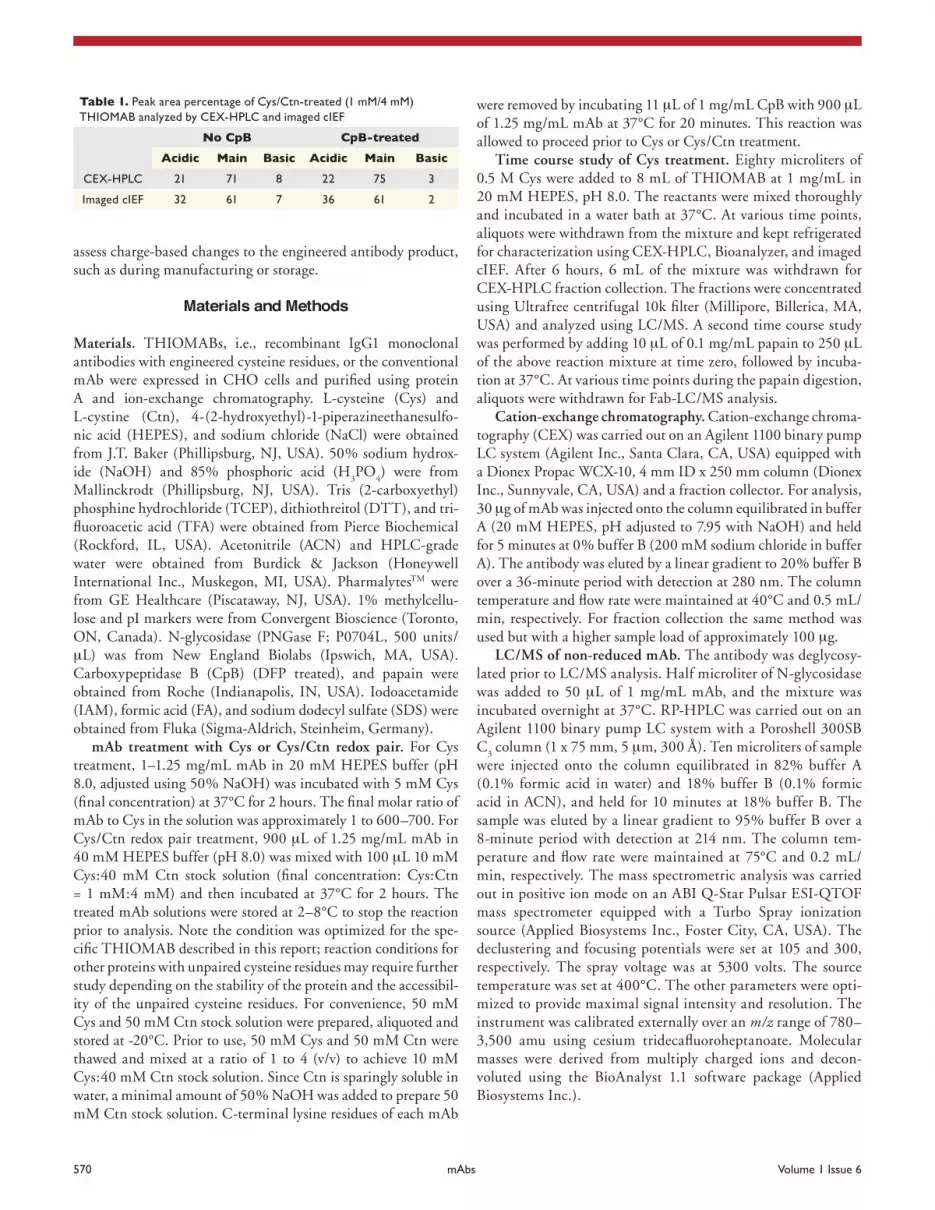
treated, Cys/Ctn-treated and a co-mix of the two are overlaid. Based on imaged cIEF peak identification as described previ-ously, the small peak immediately after the highest peak of the Cys/Ctn-treated in imaged cIEF was considered to be the half-cysteinylated species (+1Cys). As a result, those two peaks were integrated as the main peak in imaged cIEF. Also owing to the high resolution of imaged cIEF, a subtle front-shoulder of the main peak in CEX-HPLC (Fig. 8) was resolved better in imaged cIEF (Fig. 9), contributing partially to the higher acidic peak percentage obtained by imaged cIEF as compared to CEX-HPLC (Table 1). Due to its higher resolution and shorter run time, the imaged cIEF method is the preferred method for analysis of THIOMAB.
Conclusions
Our characterization work indicates that cysteine can be used as a mild reductant to decrease the heterogeneity at the engi-neered cysteines of monoclonal antibodies without disrupting their inter- and intra-chain disulfide bonds. We further dem-onstrated that using a cysteine/cystine redox pair (rather than cysteine alone) can not only effectively remove glutathionylation at the engineered cysteines, but also generate homogeneously cysteinylated species. We have thus developed an effective sample preparation method using a cysteine/cystine redox pair to obtain homogenously cysteinylated antibodies with unpaired cysteines. By obtaining one main peak in both cation-exchange chroma-tography and imaged cIEF assays, these methods can be used to
Figure 7. Overlay of CEX-HPLC chromatograms of the THIOMAB treated with Cys/Ctn redox pair at 37°C for 2 hours at different ratios (Cys:Ctn) (A) 1 mM:4 mM, (B) 2 mM:3 mM, (C) 3 mM:2 mM, (D) 4 mM:1 mM, (e) 5 mM:0 mM.
In imaged cIEF, a method with higher resolution than CEX-HPLC, the half-cysteinylated (+1Cys) species was resolved as a small peak (~6%) (Fig. 9), and its identity was confirmed by a co-mix experiment. As shown in Figure 9, the profiles of the Cys-

www.landesbioscience.com mAbs 569
Figure 8. CEX-HPLC chromatograms of Cys/Ctn-treated (1 mM Cys:4 mM Ctn, 37°C, 2 hours) THIOMAB and untreated conventional mAb.
Figure 9. Identification of half-cysteinylated peak in imaged cIEF electropherogram of THIOMAB treated with Cys/Ctn redox pair. (A) Cys-treated (5 mM Cys, 37°C, 5 hours), (B) a mixture of (A and C) at 1:1 (v/v), (C) Cys/Ctn-treated (1 mM Cys:4 mM Ctn, 37°C, 2 hours).
570 mAbs Volume 1 Issue 6
were removed by incubating 11 μL of 1 mg/mL CpB with 900 μL of 1.25 mg/mL mAb at 37°C for 20 minutes. This reaction was allowed to proceed prior to Cys or Cys/Ctn treatment.
Time course study of Cys treatment. Eighty microliters of 0.5 M Cys were added to 8 mL of THIOMAB at 1 mg/mL in 20 mM HEPES, pH 8.0. The reactants were mixed thoroughly and incubated in a water bath at 37°C. At various time points, aliquots were withdrawn from the mixture and kept refrigerated for characterization using CEX-HPLC, Bioanalyzer, and imaged cIEF. After 6 hours, 6 mL of the mixture was withdrawn for CEX-HPLC fraction collection. The fractions were concentrated using Ultrafree centrifugal 10k filter (Millipore, Billerica, MA, USA) and analyzed using LC/MS. A second time course study was performed by adding 10 μL of 0.1 mg/mL papain to 250 μL of the above reaction mixture at time zero, followed by incuba-tion at 37°C. At various time points during the papain digestion, aliquots were withdrawn for Fab-LC/MS analysis.
Cation-exchange chromatography. Cation-exchange chroma-tography (CEX) was carried out on an Agilent 1100 binary pump LC system (Agilent Inc., Santa Clara, CA, USA) equipped with a Dionex Propac WCX-10, 4 mm ID x 250 mm column (Dionex Inc., Sunnyvale, CA, USA) and a fraction collector. For analysis, 30 μg of mAb was injected onto the column equilibrated in buffer A (20 mM HEPES, pH adjusted to 7.95 with NaOH) and held for 5 minutes at 0% buffer B (200 mM sodium chloride in buffer A). The antibody was eluted by a linear gradient to 20% buffer B over a 36-minute period with detection at 280 nm. The column temperature and flow rate were maintained at 40°C and 0.5 mL/min, respectively. For fraction collection the same method was used but with a higher sample load of approximately 100 μg.
LC/MS of non-reduced mAb. The antibody was deglycosy-lated prior to LC/MS analysis. Half microliter of N-glycosidase was added to 50 μL of 1 mg/mL mAb, and the mixture was incubated overnight at 37°C. RP-HPLC was carried out on an Agilent 1100 binary pump LC system with a Poroshell 300SB C
3 column (1 x 75 mm, 5 μm, 300 Å). Ten microliters of sample
were injected onto the column equilibrated in 82% buffer A (0.1% formic acid in water) and 18% buffer B (0.1% formic acid in ACN), and held for 10 minutes at 18% buffer B. The sample was eluted by a linear gradient to 95% buffer B over a 8-minute period with detection at 214 nm. The column tem-perature and flow rate were maintained at 75°C and 0.2 mL/min, respectively. The mass spectrometric analysis was carried out in positive ion mode on an ABI Q-Star Pulsar ESI-QTOF mass spectrometer equipped with a Turbo Spray ionization source (Applied Biosystems Inc., Foster City, CA, USA). The declustering and focusing potentials were set at 105 and 300, respectively. The spray voltage was at 5300 volts. The source temperature was set at 400°C. The other parameters were opti-mized to provide maximal signal intensity and resolution. The instrument was calibrated externally over an m/z range of 780–3,500 amu using cesium tridecafluoroheptanoate. Molecular masses were derived from multiply charged ions and decon-voluted using the BioAnalyst 1.1 software package (Applied Biosystems Inc.).
assess charge-based changes to the engineered antibody product, such as during manufacturing or storage.
Materials and Methods
Materials. THIOMABs, i.e., recombinant IgG1 monoclonal antibodies with engineered cysteine residues, or the conventional mAb were expressed in CHO cells and purified using protein A and ion-exchange chromatography. L-cysteine (Cys) and L-cystine (Ctn), 4-(2-hydroxyethyl)-1-piperazineethanesulfo-nic acid (HEPES), and sodium chloride (NaCl) were obtained from J.T. Baker (Phillipsburg, NJ, USA). 50% sodium hydrox-ide (NaOH) and 85% phosphoric acid (H
3PO
4) were from
Mallinckrodt (Phillipsburg, NJ, USA). Tris (2-carboxyethyl) phosphine hydrochloride (TCEP), dithiothreitol (DTT), and tri-fluoroacetic acid (TFA) were obtained from Pierce Biochemical (Rockford, IL, USA). Acetonitrile (ACN) and HPLC-grade water were obtained from Burdick & Jackson (Honeywell International Inc., Muskegon, MI, USA). PharmalytesTM were from GE Healthcare (Piscataway, NJ, USA). 1% methylcellu-lose and pI markers were from Convergent Bioscience (Toronto, ON, Canada). N-glycosidase (PNGase F; P0704L, 500 units/μL) was from New England Biolabs (Ipswich, MA, USA). Carboxypeptidase B (CpB) (DFP treated), and papain were obtained from Roche (Indianapolis, IN, USA). Iodoacetamide (IAM), formic acid (FA), and sodium dodecyl sulfate (SDS) were obtained from Fluka (Sigma-Aldrich, Steinheim, Germany).
mAb treatment with Cys or Cys/Ctn redox pair. For Cys treatment, 1–1.25 mg/mL mAb in 20 mM HEPES buffer (pH 8.0, adjusted using 50% NaOH) was incubated with 5 mM Cys (final concentration) at 37°C for 2 hours. The final molar ratio of mAb to Cys in the solution was approximately 1 to 600–700. For Cys/Ctn redox pair treatment, 900 μL of 1.25 mg/mL mAb in 40 mM HEPES buffer (pH 8.0) was mixed with 100 μL 10 mM Cys:40 mM Ctn stock solution (final concentration: Cys:Ctn = 1 mM:4 mM) and then incubated at 37°C for 2 hours. The treated mAb solutions were stored at 2–8°C to stop the reaction prior to analysis. Note the condition was optimized for the spe-cific THIOMAB described in this report; reaction conditions for other proteins with unpaired cysteine residues may require further study depending on the stability of the protein and the accessibil-ity of the unpaired cysteine residues. For convenience, 50 mM Cys and 50 mM Ctn stock solution were prepared, aliquoted and stored at -20°C. Prior to use, 50 mM Cys and 50 mM Ctn were thawed and mixed at a ratio of 1 to 4 (v/v) to achieve 10 mM Cys:40 mM Ctn stock solution. Since Ctn is sparingly soluble in water, a minimal amount of 50% NaOH was added to prepare 50 mM Ctn stock solution. C-terminal lysine residues of each mAb
Table 1. peak area percentage of Cys/Ctn-treated (1 mM/4 mM) tHIoMAB analyzed by CeX-HpLC and imaged cIeF
No CpB CpB-treated
Acidic Main Basic Acidic Main Basic
CeX-HpLC 21 71 8 22 75 3
Imaged cIeF 32 61 7 36 61 2

www.landesbioscience.com mAbs 571
with a PrinCE Microinjector autosampler (Prince Technologies, the Netherlands). The ampholyte solution consisted of a 15%/85% (v/v) mixture of PharmalyteTM solution pH 3–10 and pH 8–10.5, respectively, with 0.2% (v/v) of each of the pI markers 7.4 and 9.77. The ampholyte solution also contained 0.35% (v/v) methylcellulose (MC). Samples at 1.25 mg/mL were mixed with the ampholyte solution at a 1:4 (v/v) ratio. Separation was carried out on a fluorocarbon-coated fused-sil-ica capillary (5 cm long, 100 um I.D.) cartridge (Convergent Bioscience, Toronto, Canada). The catholyte was 100 mM NaOH in 0.1% methylcellulose and the anolyte was 80 mM H
3PO
4 in 0.1% methylcellulose. Samples were introduced from
the autosampler (set at 8°C) and transferred to the cartridge for about 150 seconds by pressure. Period I pre-focusing was con-ducted at 1,500 V for 1 minute followed by period II focusing at 3,000 V for 10 minutes. The focused image at 280 nm was captured by a charge-coupled device (CCD) camera. Data were converted by iCE
280 Analyzer and analyzed using EZChrom
software version 6.8 (Scientific Software International Inc., Lincolnwood, IL, USA).
Acknowledgements
We would like to thank Moira Kelly and Rod Keck for initial CEX-HPLC peak characterization of THIOMAB, Louisetta Basa for assistance with the mass spectrometer settings, Oscar Salas-Solano and colleagues for the capillary electrophoresis methods, and Reed Harris for a literature reference.
Fab-LC/MS. The Fab fragment was generated from the time course study of papain digestion. RP-HPLC was carried out on an Agilent 1100 binary pump LC system equipped with a Poroshell 300SB C
3 column, 1 x 75 mm, 5 μm, 300 Å (Agilent Inc., Santa
Clara, CA, USA). Five microliters of each sample were injected onto the column equilibrated in 82% buffer A (0.1% formic acid, 0.025% TFA in water) and 18% buffer B (0.1% formic acid, 0.025% TFA in ACN) and held for 10 minutes at 18% buffer B. Sample was eluted by a linear gradient to 50% buf-fer B over a 16-minute period with detection at 214 nm. The column temperature and flow rate were maintained at 75°C and 0.2 mL/min. The mass spectrometric analysis was carried as described previously, with the exception that the declustering and focusing potentials were set at 45 and 300, respectively.
Microchip electrophoresis on bioanalyzer. Analysis of low molecular weight species was accomplished by CE-SDS analy-sis using an Agilent 2100 Bioanalyzer (Agilent Inc., Santa Clara, CA, USA). Twenty four microliters of IAM-SDS solution (50 mM IAM, 0.5% SDS) and 2 μL of sample buffer (Agilent Protein 230 Kit) were added to 4 μL of each sample from the time course study followed by incubation at 70°C for 5 minutes. Then, 60 μL of water was added to each sample and the sample was loaded on a protein chip of Agilent 2100 Bioanalyzer (Agilent, Waldbronn, Germany) for analysis. The results were analyzed using Agilent 2100 Expert software.
Imaged cIEF. Imaged cIEF was carried out on an iCE280
Analyzer (Convergent Bioscience, Toronto, Canada) equipped
References1. Reichert JM, Rosensweig CJ, Faden LB, Dewitz MC.
Monoclonal antibody successes in the clinic. Nat Biotechnol 2005; 23:1073-8.
2. Clark M. Antibody humanization: a case of the “Emperor’s new clothes”? Immunol Today 2000; 21:397-402.
3. Hsieh FY, Tengstrand E, Li LY, Huang YN, Milton MN, Silverman L, et al. Toxicological Protein Biomarker Analysis—An Investigative One-week Single Dose Intravenous Infusion Toxicity and Toxicokinetic Study in Cynomolgus Monkeys using an Antibody-cytotoxic Conjugate against Ovarian Cancer. Pharm Res 2007; 25:1309-17.
4. Senter PD. Potent antibody drug conjugates for cancer therapy. Curr Opin Chem Biol 2009; 13:1-10.
5. Doronina SO, Toki BE, Torgov MY, Mendelsohn BA, Cerveny CG, Chace DF, et al. Development of potent monoclonal antibody auristatin conjugates for cancer therapy. Nat Biotechnol 2003; 21:778-84.
6. Hamblett KJ, Senter PD, Chace DF, Sun MM, Lenox J, Cerveny CG, et al. Effects of drug loading on the antitumor activity of a monoclonal antibody drug conjugate. Clin Cancer Res 2004; 10:7063-70.
7. Lu SX, Takach EJ, Solomon M, Zhu Q, Law SJ, Hsieh FY. Mass spectral analyses of labile DOTA-NHS and heterogeneity determination of DOTA or DM1 conju-gated anti-PSMA antibody for prostate cancer therapy. J Pharm Sci 2005; 94:788-97.
8. Henry MD, Wen S, Silva MD, Chandra S, Milton M, Worland PJ. A prostate-specific membrane antigen-targeted monoclonal antibody-chemotherapeutic con-jugate designed for the treatment of prostate cancer. Cancer Res 2004; 64:7995-8001.
9. Doronina SO, Mendelsohn BA, Bovee TD, Cerveny CG, Alley SC, Meyer DL, et al. Enhanced activity of monomethylauristatin F through monoclonal antibody delivery: effects of linker technology on efficacy and toxicity. Bioconjugate Chem 2006; 17:114-24.
10. Polson AG, Yu S-F, Elkins K, Zheng B, Clark S, Ingle GS, et al. Antibody-drug conjugates targeted to CD79 for the treatment of non-Hodgkin lymphoma. Blood 2007; 110:616-23.
11. Junutula JR, Bhakta S, Raab H, Ervin KE, Eigenbrot C, Vandlen R, et al. Rapid identification of reactive cysteine residues for site-specific labeling of antibody-Fabs. J Immunological Methods 2008; 332:41-52.
12. Stimmel JB, Merrill BM, Kuyper LF, Moxham CP, Hutchins JT, Fling ME, et al. Site-specific Conjugation on Serine→Cysteine Variant Monoclonal Antibodies. J Biol Chem 2000; 275:30445-50.
13. Junutula JR, Raab H, Clark S, Bhakta S, Leipold DD, Weir S, et al. Site-specific conjugation of a cytotoxic drug to an antibody improves the therapeutic index. Nat Biotechnol 2008; 26:925-32.
14. Mawatari S, Murakami K. Different types of glutathio-nylation of hemoglobin can exist in intact erythrocytes. Arch Biochem Biophys 2004; 421:108-14.
15. Landino LM, Moynihan KL, Todd JV, Kennett KL. Modulation of the redox state of tubulin by the glutathione/glutaredoxin reductase system. Biochem Biophys Res Commun 2004; 314:555-60.
16. Dormann P, Borchers T, Korf U, Hojrup P, Roepstorff P, Spener F. Amino acid exchange and covalent modifi-cation by cysteine and glutathione explain isoforms of fatty acid-binding protein occurring in bovine liver. J Biol Chem 1993; 268:16286-92.
17. Davis DA, Dorsey K, Wingfield PT, Stahl SJ, Kaufman J, Fales HM, et al. Fales and R.L. Levine, Regulation of HIV-1 protease activity through cysteine modification. Biochemistry 1996; 35:2482-8.
18. Lim A, Wally J, Walsh MT, Skinner M, Costello CE. Identification and location of a cysteinyl posttransla-tional modification in an amyloidogenic κ
1 light chain
protein by electrospray ionization and matrix-assisted laser desorption/ionization mass spectrometry. Anal Biochem 2001; 295:45-56.
19. Bondarenko PV, Farwig ZN, McNeal CJ, Macfarlane RD. MALDI- and ESI-MS of the HDL apolipopro-teins: new isoforms of apoA-I, II. J Mass Spectrom 2002; 219:671-80.
20. Gadgil HS, Bondarenko PV, Pipes GD, Dillon TM, Banks D, Abel J, et al. Identification of cysteinylation of a free cysteine in the Fab region of a recombinant monoclonal IgG1 antibody using Lys-C limited pro-teolysis coupled with LC/MS analysis. Anal Biochem 2006; 355:165-74.
21. Banks DD, Gadgil HS, Pipes GD, Bondarenko PV, Hobbs V, Scavezze JL, et al. Removal of cysteinylation from an unpaired sulfhydryl in the variable region of a recombinant monoclonal IgG1 antibody improves homogeneity, stability, and biological activity. J Pharm Sci 2008; 97:775-90.
22. Gilbert HF. Thiol/disulfide exchange equilibria and disulfidebond stability. Methods Enzymol 1995; 251:8-28.


