Embed Size (px)
Citation preview
Characterizing Traditionally-defined Periodontal Disease in HIV+Adults
Lance Vernon1,*, Catherine Demko2, Christopher Whalen3, Michael Lederman4, ZahraToossi5, Mianda Wu6, Yiping Han7, and Aaron Weinberg1
1Department of Biological Sciences, Case Western Reserve University, School of DentalMedicine, Cleveland, Ohio, USA2Department of Community Dentistry, Case Western Reserve University, Cleveland, Ohio, USA3Department of Epidemiology and Biostatistics, University of Georgia, College of Public Health,Athens, Georgia, USA4Center for AIDS Research, Case Western Reserve University/University Hospitals of Cleveland,Cleveland, Ohio, USA5Department of Medicine, Division of Infectious Diseases, Case Western Reserve University,Cleveland, Ohio, USA6Department of Medicine, Division of Infectious Diseases, Case Western Reserve University/University Hospitals of Cleveland, Cleveland, Ohio, USA7Departments of Periodontics and Pathology, Case Western Reserve University, School of DentalMedicine, Cleveland, Ohio, USA
IntroductionPeriodontal disease (PD) has been associated with human immunodeficiency virus type one(HIV-1) over the past several decades (1,2). Initial descriptions of oral health in HIV+patients focused on extreme forms of PD (3,4), followed later by more conventionaldefinitions (1,5,6), making the comparison of study results difficult. Without appropriate andconsistent characterization of PD in at-risk populations, such as those with HIV-1 infection,the level and severity of traditionally-defined PD may be underestimated, which could leadto inadequate prevention and treatment as well as a diminished sense of concern regardingperiodontal health.
Persons with HIV-1 infection are especially vulnerable to comorbidities; their immunesystems are compromised, and in the era of highly active antiretroviral therapy (HAART),some metabolic factors (i.e. lipids, glucose and insulin levels) are altered (7). Utilization ofdental care services may be restricted due to real or perceived stigma, confidentialityconcerns, or economic and social factors even if access to a dentist is available. Substanceabuse, psycho-social stressors, depression and denial may result in the neglect of healthpromoting behaviors and lead to lapses in medication adherence, which can further limitimmune reconstitution and the efficacy of remaining HIV treatment options. In sum, personswith HIV have an increased risk for adverse oral and general health outcomes.
*Corresponding Author: Dr. Lance Vernon, Department of Biological Sciences, Case Western Reserve University, School of DentalMedicine, 10900 Euclid Ave, Cleveland, Ohio 44106-4905, USA, Tel: 216-368-0712, Fax: 216-368-0145, [email protected].
NIH Public AccessAuthor ManuscriptCommunity Dent Oral Epidemiol. Author manuscript; available in PMC 2010 October 1.
Published in final edited form as:Community Dent Oral Epidemiol. 2009 October ; 37(5): 427–437. doi:10.1111/j.1600-0528.2009.00485.x.
NIH
-PA Author Manuscript
NIH
-PA Author Manuscript
NIH
-PA Author Manuscript
There have been inconsistent reports of the levels of PD in HIV+ persons (1,2,5,6,8–16),including both high (5,16,17), and low (8,9,13) levels of PD. There are a number of possiblereasons for these inconsistencies. Different definitions were used to characterize PD (2) andmethodologies used to collect clinical periodontal measurements also varied (e.g. partialmouth vs. full mouth probing and the number of sites probed per tooth). Many studiesreporting low levels of periodontal disease have used partial mouth periodontal datacollection methodologies (6,8,9,13), but partial mouth analyses can underestimate theprevalence of PD (18). Further, the prevalence of periodontal disease may vary by patientcharacteristics in the cohort (1,2). Such patient characteristics may include immune factors(importantly, the distribution of the cohort’s CD4+ T-cell counts) (5,6,16), metabolic factors(19), utilization of dental care services (20) and oral health behaviors (21). These patientcharacteristics are not always well described in the literature, nor are all of thesecharacteristics routinely included in reports of PD in HIV+ cohorts.
To provide a more comprehensive view of PD in the HAART era, we clearly characterizedour HIV+ cohort according to select immune and metabolic markers, the presence ofsubgingival bacterial pathogens, dental care utilization and oral health behaviors. Weexamined the level of PD in our cohort using two consensus definitions of PD (18,22), aswell as by its two distinct clinical measures (periodontal probing depth and recession) andone summary measure (clinical attachment level), as has been suggested by authorsinvestigating the perio-systemic connection (23).
MethodsSubject Selection
Adult subjects were recruited from three outpatient HIV medical clinics in Cleveland, Ohio:University Hospitals of Cleveland (UH), The Cleveland Clinic Foundation (CCF) andMetroHealth Medical Center (Metro). The study was approved for patient recruitment byparticipating institutions. Most participants were self-referred. All subjects signed a UHIRB-approved informed consent document and an authorization form to use and discloseProtected Health Information (PHI) for research purposes. Exclusion criteria includedevidence of cardiovascular disease, a history of Type I or II diabetes mellitus, fewer than 20teeth, uncontrolled systemic illnesses, diagnosis or treatment of cancer in the past five years,pregnancy, and need for antibiotic prophylaxis prior to dental care as per the currentAmerican Dental Association (ADA) and other guidelines (24,25). Inclusion criteria weremedication-compliant adult (age 18 and over) subjects on HAART or about to start HAARTwithin 2 months of their baseline visit. We accessed pre-study clinical data from the primaryHIV-clinic’s longitudinal electronic database or from retrospective chart reviews. Importantvariables from these sources included: CD4+ T-cell count and plasma HIV RNA levels(Roche Amplicor) nearest to and before the baseline visit, complete antiretroviralmedication history, nadir CD4 T-cell count and risk factors for HIV-1 infection. Studysubjects were seen from May, 2005 through January, 2008.
Periodontal Disease MeasurementsThe periodontal probing depth (PPD)—the height of the free gingival margin to the mostapical location of the periodontal pocket—was determined at six sites per tooth (mesio-facial, mid facial, disto-facial, disto-lingual, mid lingual, and mesio-lingual) using aUniversity of North Carolina (UNC) #15 probe. The distance between the height of the freegingival margin to the cemento-enamel junction (CEJ) was termed recession (REC), andwas also measured at the same six sites per tooth, with values apical to the CEJ beingpositive and values coronal to the CEJ being negative. PPD plus REC yielded the clinicalattachment level (CAL), where a positive number indicated loss of attachment. PPD and
Vernon et al. Page 2
Community Dent Oral Epidemiol. Author manuscript; available in PMC 2010 October 1.
NIH
-PA Author Manuscript
NIH
-PA Author Manuscript
NIH
-PA Author Manuscript

REC values were rounded up to the next whole millimeter value. A viable tooth was definedas having at least one-half of a remaining clinical crown (i.e. at least three contiguous sitesin which PPD and REC were measurable). Full-mouth periodontal probing was performedby one dentist (LV), whose measurements were previously calibrated to a trained andexperienced periodontist. The percent agreement from ongoing intra-rater reliability forperiodontal probing, +/− 1 mm, was 98% with an intra-class correlation coefficient of 0.88.The periodontal data collection methodology conformed to expert guidelines (26)Supragingival plaque, debris, blood or saliva was removed prior to performing precise full-mouth periodontal measurements, which typically required over 60 minutes in order toensure accurate measurements.
Definitions of Periodontal DiseaseWe used three different definitions of PD to characterize our cohort. The first definition ofPD was proposed by a working group with representatives from both the Centers for DiseaseControl and Prevention (CDC) and the American Academy of Periodontology (AAP). Theydefined severe periodontitis as ≥2 interproximal sites with CAL ≥6 mm (not on same tooth)and ≥1 interproximal site with PPD ≥5 mm; moderate periodontitis as ≥2 interproximalsites with CAL ≥4 mm (not on same tooth) or ≥2 interproximal sites with PPD ≥5 mm (noton same tooth); and no or mild periodontitis as neither “moderate” nor “severe” periodontitis(18). The second definition of PD was proposed by Tonetti and Claffey for the Group CConsensus Report of the 5th European Workshop in Periodontology (EWP), 2005. This two-level definition included the presence of proximal attachment loss of ≥3 mm at ≥2 non-adjacent teeth and the presence of proximal attachment loss of ≥5 mm at ≥30% of teethpresent (22); we adapted this definition into three categories (see legend on Figure two). Tominimize possible misclassification due to categorizing data, we defined and analyzed thePD data as a continuous variable. Thus, we evaluated PD by its component parts, PPD, RECand the summary measure, CAL, as previously described (23,27). These components mayrepresent different processes contributing to periodontal disease; while PPD more closelyreflects acute PD, REC represents the cumulative results of long-term destruction ofperiodontal tissue and bone (23,28). Each component of periodontal disease may be viewedas a potential independent contributor to longitudinal systemic outcomes (23,27) and mayhelp illuminate how HIV-1 infection and/or systemic factors influence or interact with theprogression of PD. Guided by expert reports (18,22), we selected the following threeseparate thresholds to define periodontal disease: PPD ≥5 mm, REC >0 mm and CAL≥4mm. For each component we calculated the percent of teeth with at least one site pertooth that met the threshold and analyzed these outcome measures as continuous variables.
Demographic/Behavioral DataOne researcher/dentist (LV) administered questionnaires to obtain demographic, medicaland oral behavioral data were completed on each study subject. Patients reported the numberof times per week that they brushed and flossed. Smoking was recorded as ever havingsmoked more than 100 cigarettes, a calculation of pack per day years (ppdyrs) of cigarettesmoking, and the total number of years of smoking. Drug use was coded as yes or no, andincluded past or present use of illegal substances, including marijuana (more thanexperimental use), cocaine (any type) and other “street drugs”.
Definition of HAARTHAART was defined as a treatment regimen that included at least three differentantiretroviral drugs from at least two different classes, i.e., nucleoside/nucleotide analogues(NUC), protease inhibitors (PI), non-nucleotide reverse transcriptase inhibitors (NNRTI) andentry or fusion inhibitors (EI). Most subjects were taking a backbone of two NUCs witheither a PI or an NNRTI.
Vernon et al. Page 3
Community Dent Oral Epidemiol. Author manuscript; available in PMC 2010 October 1.
NIH
-PA Author Manuscript
NIH
-PA Author Manuscript
NIH
-PA Author Manuscript

Metabolic MeasurementsAll metabolic measurements, including a lipid panel (i.e. HDL-cholesterol, LDL cholesterol,VLDL cholesterol and triglycerides), insulin, glucose and high sensitivity C-reactive protein(hs-CRP), were determined using standard laboratory serum assays after at least 12 hours offasting. For complete information regarding these assays, see Appendix A. Homeostasismodel assessment of insulin resistance (HOMA-IR) was calculated as fasting insulin (µIU/ml) × glucose (mg/dL)/405 (29). Patients were considered to be insulin resistant whenHOMA-IR was ≥2.6 (30).
Collection of Dental Plaque for Periodontal PathogensUsing separate sterile 13/14 Gracey curettes for each tooth, subgingival dental plaque wascollected from the most apical portion of the accessible probing depth and alongside the rootof the tooth at two different interproximal sites (usually the distolingual and distobuccal) ofthe first molar in each quadrant, and transferred into a microcentrifuge tube containing 0.5ml Tri-reagent (Molecular Research Center Inc., Cincinnati, Ohio). For each patient, eightsites were harvested from molar teeth and pooled into one plaque sample. If the first molartooth was missing, then the next most distal tooth was accessed. If all molar teeth weremissing, then the most distal tooth in that quadrant was sampled, as previously described(31). After temporary storage on ice, microcentrifuge tubes were transferred to a −70°Cfreezer for storage until accessed for analysis.
Isolation and quantification of DNADNA was extracted from each plaque sample according to the manufacturer’s instructions aspreviously described by Toossi et al., 2005 (32). Bacterial DNA was assessed by real-timePCR (Taqman 7700, Applied Biosystems, Foster City, CA). Oral bacteria examinedincluded P. gingivalis (Pg), T. forsythia (Tf), and T. denticola (Td). In addition, DNAencoding total bacterial 23s ribosomal RNA was assessed in each sample. Taqman primerand probes for Pg, Td, and bacterial 23s ribosomal RNA were designed using PrimerExpress software (Applied Biosystems) Primers and probe for Tf were as previouslyreported by Morillo et al., 2004 (33). For details on probes and primers, see Appendix B.Bacterial 23 S ribosomal DNA products were detected by Sybr green (Applied Biosystems).Quantities of each bacterial DNA were determined by using a dilution series ofoligonucleotide sequences (Invitrogen, Carlsbad, CA) synthesized for the target DNA ineach assay. The concentration and purity of plaque DNA was determined byspectrophotometery (BECKMAN DU640). DNA copies for each pathogen were calculatedby correction to the total DNA in each plaque sample and expressed as copies of bacterialDNA per microgram of DNA.
Data AnalysisData analyses were conducted using SPSS v. 13 (SPSS Corporation, Chicago, Illinois,USA). Bivariate relationships between continuous variables were examined by Pearson andSpearman correlations, as appropriate. Logarithmic transformation was used to normalizethe distribution of pathogen DNAs. For dichotomous independent variables, differences inperiodontal measures between the groups were tested using t-tests. HOMA-IR wasdichotomized into values < and ≥ 2.6. CD4+ T-cell counts were dichotomized into <200cells/mm3 and ≥ 200 cells/mm3; smoking and flossing were categorized as ever (yes) ornever (no). The number of dental visits in the past 5 years was averaged and dichotomized totwo categories, those with at least one dental visit per year versus those with less than onevisit per year. We set a significance level of p<.05 for all statistical tests. Backward stepwisemultiple regression (p=.10 to enter and p=.05 to leave) was performed to identify factorsassociated with the periodontal outcome measure, clinical attachment loss (CAL). Predictor
Vernon et al. Page 4
Community Dent Oral Epidemiol. Author manuscript; available in PMC 2010 October 1.
NIH
-PA Author Manuscript
NIH
-PA Author Manuscript
NIH
-PA Author Manuscript
variables significant at p<.05 in the bivariate analysis with CAL that were entered into themodel included flossing, annual dental visits, levels of Pg. DNA, total plaque DNA,smoking status, CD4+ T-cell counts, LDL-cholesterol, BMI, HOMA-IR and insulin.Although not significant in the backward strategy, sex, race, primary insurance and viralload were retained in the final model for adjustment.
ResultsA total of 112 subjects completed all baseline measures and were included in the cross-sectional analysis (Table 1). Patients in this cohort were predominantly male (74%),African-American (64%), had more than a high school education (59%), received Medicaid(56%), and had a median age of 42 years (range 21–57). Among the 78 subjects (69%) whoself-reported cigarette smoking, the mean ppdyrs was 11.0 (+/− 10.6), with a median of 8.8ppdyrs. Drug use history was reported by 61% of study participants. Oral hygiene behaviorswere poor and less than half averaged an annual dental visit over the past 5 years. Patientshad an average of 26 teeth (range, 20–32). Of the 123 ineligible volunteers, 49 people (40%)were excluded because they had fewer than 20 teeth (Figure 1).
Figure 2 compares the distribution of our cohort by different case definitions of periodontaldisease (18,22), demonstrating that over 90% of persons in this cohort had traditionally-defined periodontal disease that was moderate or severe. Note in Figure 2 that the CDC/AAP 2007 definition classifies more subjects as having severe periodontal disease, whereasthe EWP/Tonetti 2007 definition classifies a greater proportion of subjects as havingmoderate periodontal disease. We modeled the three periodontal disease measures ascontinuous variables. By our definitions of periodontal disease, subjects had an average 38%(±24%) of their teeth with at least one site of PD ≥5mm, 55% (±31%) of their teeth with atleast one site of REC >0mm, and 50% (±32%) of their teeth with at least one site of CAL≥4mm.
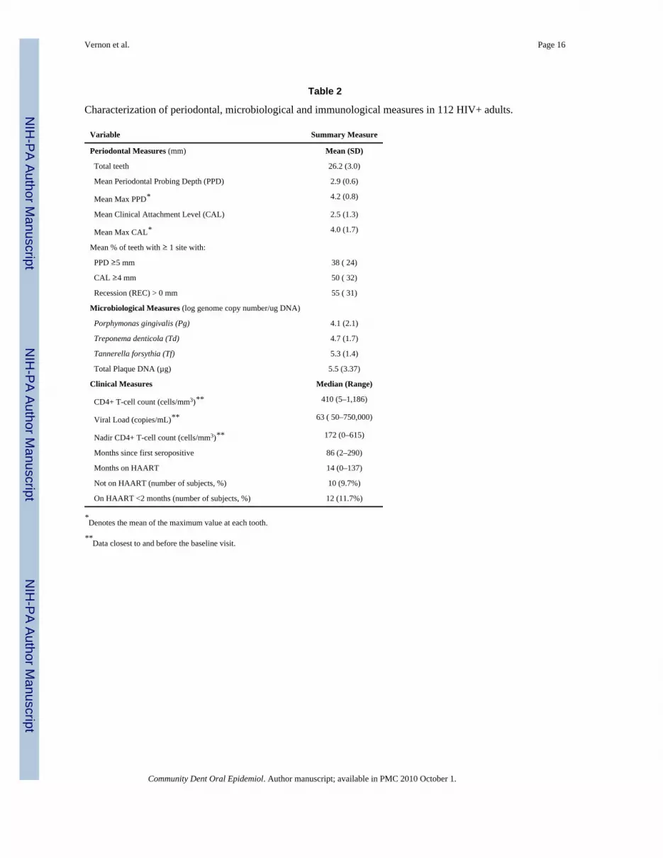
Study participants had a wide range of immune competence (see Table 2). Participants had amedian CD4 + T-cell count of 410 cells/mm3; 19 patients (17%) had CD4 + T-cells counts <200 cells/mm3. A total of 53% of subjects had previously experienced a nadir CD4+ t-cellcount <200 cells/mm3. Viremia was generally well controlled. Forty-eight subjects (43%)were classified as having an undetectable viral load, and the median value was 86 copies/mL. The median time since first HIV seropositive test result was 7.2 years; the most recentdiagnosis was 2 months before the baseline visit, while the most remote diagnosis was 24years before the baseline visit. At the baseline visit, 90% of subjects were on HAART andthe median time on HAART was 14 months, with a range from 0 to 137 months.
We examined the association of PPD, REC and CAL with gender, flossing, dental visits andsmoking status (data not shown). Males had greater levels of REC (p<.05), but no otherdifferences in periodontal measures by gender were evident. Patients who reported flossingand annual dental visits over the past 5 years had significantly lower levels of PPD and CAL(p<.01). Periodontal measures were worse for smokers compared to non-smokers but thisassociation did not reach statistical significance in the bivariate analysis. No significantdifferences in the periodontal measures were observed by categories of race, primaryinsurance source, educational attainment or duration of time on HAART.
Metabolic measures (Table 3) were mostly within the normal range, except for HDLcholesterol in which 56% of subjects had values below the normal range (<40 mg/dL).Using the standard BMI thresholds, 26% of subjects were overweight and 27% were obese.Triglyceride levels were elevated (>130 mg/dL) in 28% of subjects and hs-CRP waselevated (≥3.0 mg/L) in 35% of subjects.
Vernon et al. Page 5
Community Dent Oral Epidemiol. Author manuscript; available in PMC 2010 October 1.
NIH
-PA Author Manuscript
NIH
-PA Author Manuscript
NIH
-PA Author Manuscript

The independent variables were not uniformly associated with the outcome variables, PPD,REC and CAL (Table 4). While all microbiological variables were associated with PPD,only the level of Pg DNA was associated with PPD, REC and CAL. Most immune andmetabolic variables (i.e. CD4+ T cell count, insulin, LDL-cholesterol and HOMAIR) thatwere associated with REC were also associated with CAL. CD4+ T-cell count and durationof time on HAART were not correlated with PPD in this cohort. Only one variable, BMI,was correlated with CAL (Spearman’s rho= −.238, p<.05), but not with PPD or REC.Glucose levels and hs-CRP levels were not associated with any periodontal measures in thiscross-sectional analysis. An unexpected finding was the correlation of Td levels in a positivedirection with PPD (Spearman’s rho= .372, p<.001) and in a negative direction with REC(Spearman’s rho= −.229), resulting in no association with CAL.
From the backward stepwise multiple regression analysis, eight variables were identified assignificant predictors of CAL (CD4+ T-cell count, annual dental visits, smoking, levels ofPg, total plaque DNA, LDL-cholesterol, HOMA-IR and age), while adjusting for gender,race, insurance and viral load. This full model explains slightly more than half of thevariability in CAL (F=9.559, adjusted R2=.509, p<.001). Coefficients for the independentvariables from the regression model are shown in Table 5. In the second column, B-coefficients (B) show the effect on CAL (the percent of teeth) for one unit of change in theindependent factor or factor level, when all other variables are held constant. For example,holding all other variables constant, patients with CD4+ T-cell counts less than 200 cells/mm3 are expected to have an additional 25.2% of their teeth with at least one site of CAL≥4mm, as compared to patients with CD4+ T-cell counts greater than 200 cells/mm3.Smokers and patients without annual dental visits in the past five years are predicted to havehigher CAL values (by 11.8% and 15.7% respectively) compared to non-smokers andpatients who obtained annual dental care. Older age, increased levels of Pg DNA andincreased total subgingival plaque DNA also predicted greater periodontal disease in thiscohort. Unexpected findings were the association of higher levels of serum LDL-cholesteroland higher levels of HOMA-IR with lower levels of CAL. No other metabolic measureremained significant in the multivariable model.
The standardized beta-coefficients (β) from the multivariable regression model representedthe effect of the independent factor on the periodontal disease outcome measurement (CAL)in standardized units, to permit comparison of the relative predictive significance of eachfactor. CD4+ T cell counts had the strongest effect (β=.306) on periodontal disease followedclosely by the level of Pg (β=.290). Age (β=.268) and annual dental visits over the past fiveyears (β=.240) exerted similar effects. CD4+ T-cell counts <200 cells/mm3 hadapproximately twice the effect on CAL compared to smoking (standardized β coefficient .306 versus .164). This same model was run using REC as the continuous outcome variable,yielding an adjusted R2 of .475, F=8.5, p< .001 (data not shown).
DiscussionEach subject had on average 38% (±24%) of their teeth with at least one site of PD ≥5mm,55% (±31%) of their teeth with at least one site of REC >0mm, and 50% (±32%) of theirteeth with at least one site of CAL ≥4mm. Factors associated with high levels of periodontaldisease in this cohort, as measured by CAL, were immunosupression (i.e. CD4+ T-cellcounts < 200 cells/mm3), the level of Pg DNA and total DNA in subgingival plaque,substandard utilization of dental care, cigarette smoking and older age. In this cohort,lowered CD4+ T cell counts (<200 cells/mm3) had approximately twice the effect onperiodontal disease as did cigarette smoking, a known, strong risk factor for PD (22,34).Having an annual dental visit remained an independent predictor for lower levels of PD.Cohort characteristics were precisely described.
Vernon et al. Page 6
Community Dent Oral Epidemiol. Author manuscript; available in PMC 2010 October 1.
NIH
-PA Author Manuscript
NIH
-PA Author Manuscript
NIH
-PA Author Manuscript

To characterize a patient’s level of periodontal disease, we used three outcome measures(PPD, REC and CAL) as continuous variables representing the percent of teeth which met orexceeded the measure’s cut point (Table 2, Table 4 and Table 5). Modeling periodontaldisease as a continuous variable allows us to examine the entire spectrum of periodontaldisease and minimizes the chance of misclassification based on categorical thresholds (i.e.mild, moderate and severe).
We examined the correlation of independent variables with the dependent outcomevariables: PPD, REC and CAL. In the bivariate analysis (Table 4), the independent variablescorrelated differently across the three outcome measures of periodontal disease. Forexample, only the level of Pg DNA demonstrated a comparable and significant effect onPPD, REC and CAL. However, a limited set of immune (CD4+ T cell count) and metabolic(insulin, LDL-cholesterol and HOMA-IR) host factors appeared to correlate better with thechronic or historic processes (23) (REC) than with the more acute component (23) (PPD) ofperiodontal disease. These observations support the previous premise by Beck et al, 2002that PPD and REC capture independent processes (23). Further, these same immune andmetabolic factors that were significantly correlated with REC were also significantlycorrelated with CAL, suggesting that HIV and/or immune reconstitution (i.e. while onHAART) influences PD through a systemic mechanism associated with REC. Thisobservation is supported by previous cross-sectional findings (5,35).
Our findings concerning the impact of CD4+ T-cell count on periodontal disease are inagreement with several previous studies (1,5,10,15,16). Our data most closely agree with areport by Robinson et al, 1996, which found, prior to the widespread use of HAART, thatdecreased CD4+ T-cell count predicted the presence, extent and severity of CAL, but notPPD (5). In terms of immune status, their cohort was very similar to ours; over half (54%) oftheir subjects met the 1993 case definition for AIDS (5) and 53% of our subjects had aCD4+ T-cell count <200 cells/mm3. McKaig et al, 1998, whose cohort had a similar racialcomposition to ours, also found severe and extensive periodontal disease in their cohort (16).They found that the extent of recession was significantly greater (p<.01) in persons withCD4+ T-cell count <200 cells/mm3 than in two groups with higher CD4+ T-cell counts (16).In their study, 58% of their cohort had experienced an AIDS-defining event, and 48% had abaseline CD4+ T-cell count <200 cells/ mm3 (i.e. some subjects had experienced immunereconstitution on combination antiviral medication) (16).
Our results, concerning the relationship between PD and CD4 + T-cell count, differ fromother studies. For example, Tomar et al., 1995, did not find a cross-sectional associationbetween PD and CD4+ T-cell count using multiple logistic regression; however, they used<300 cells/mm3 as their cut point for CD4+ T-cell counts and their cohort was characterizedusing Walter Reed staging classification system (instead of CD4+ T-cell count and viralload), making comparison to our study difficult (14). As well, Gonclaves (36) examined 64HIV+ Brazilian persons and found no statistically significant associations between CD4+ T-cell count and periodontal measures; however, they excluded persons with severeperiodontal conditions and all subjects were taking trimethoprim and sulfamethoxazole(Bactrim®) prophylaxis to prevent opportunistic infections, a regimen which could limit thelevel and severity of PD (10,12,37). Further, 34% of their study subjects had a baselineCD4+ T count <200 cells/ mm3 and nadir CD4 + T-cell counts were not reported. The aboveanalysis highlights some of the complexities of comparing previous HIV-relatedepidemiological research, and underscores the importance of thorough reporting of cohortcharacteristics, especially in those with HIV-1 infection during the era of HAART.
Older studies (8,9,13) as well as more recent studies (6,36,38) have reported lower levels ofperiodontal disease in HIV+ patients, compared to the level of PD in our cohort. Reasons for
Vernon et al. Page 7
Community Dent Oral Epidemiol. Author manuscript; available in PMC 2010 October 1.
NIH
-PA Author Manuscript
NIH
-PA Author Manuscript
NIH
-PA Author Manuscript
this may be due to: 1) how authors defined periodontal disease, 2) the methodology used tocollect periodontal data, 3) the distribution of patient characteristics within cohorts(especially immune and medication-related), and 4) the limited number of post-HAART erainvestigations on this topic. Some studies that found low levels of periodontal disease(6,8,9,13,38) used partial mouth examination techniques that are known to potentiallyunderestimate PD (18) and have not, to our knowledge, been validated in an HIV-1 infectedcohort.
As previously reported (39–41), the levels of Pg DNA in our study were highly correlatedwith periodontal disease. We found that traditional periodontal “red” complex (42) bacteria,Pg, Td and Tf, were highly correlated with PPD. While levels of Pg DNA were correlatedwith all individual measures of periodontal disease (PPD, REC and CAL), Td wasnegatively correlated with REC, perhaps suggesting that it is displaced by other species inchronic periodontal disease. This negative association of Td with REC may agree in partwith findings by Aas et al, 2007. They propose that there is a shift in the biological ecologyaway from some of the traditional red complex species (i.e. Pg, Td and Tf) (43). However,our data suggest that Pg played a role in chronic periodontal disease, as measured by RECand CAL (23).
While HAART greatly reduces the prevalence of HIV-related oral manifestations (44), thisreport demonstrated that traditionally defined periodontal disease is clearly still present andrepresents a potential challenge to the long-term oral health of persons with HIV-1 infection.CD4+ T-cell count and duration of time on HAART were not correlated with PPD,suggesting that these immune factors have less impact on acute periodontal disease (23).Since increases in LDL cholesterol can occur while taking HAART (45), and insulinresistance can increase due to PI use (46), the association of higher levels of serum LDLcholesterol and higher levels of HOMA-IR with lower levels of CAL could indicate aprotective effect of HAART in this cross-sectional analysis. Analysis of our longitudinaldata will better address this finding.
Limitations of this study include the cross-sectional design, from which only associationscan be made, not causal inferences. We could have slightly overestimated the extent andseverity of periodontal disease by rounding up to the next whole millimeter in ourperiodontal probing measurements. Also, our participants were self-referred, which mayhave resulted in a convenient sample with poorer periodontal health and/or those interestedin obtaining dental care. However, of the 123 ineligible subjects, 48 subjects were excludedbecause they had fewer than 20 teeth, suggesting that caries and/or periodontal disease havealready had a significant effect on many HIV+ persons from this geographic location.
The strength of this study is in its detailed and extensive characterization of periodontaldisease and associated factors. The previously applied CDC definition is more sensitive todetect localized areas of severe periodontal disease, while the EWP definition detects a moregeneralized loss of attachment (i.e. ≥30% of teeth present with CAL ≥5 mm). To minimizethe potential misclassification from using categorical threshold cut points (i.e. mild,moderate and severe), we separated and modeled the three periodontal disease measures ascontinuous variables. This methodology allowed us to explore the link of HIV to PD morecomprehensively. Our adjusted multivariable model accounted for slightly over 50%(adjusted R2=.509) of the variability in CAL, which suggests that we identified many of themajor determinants of CAL in this cohort.
Periodontal disease remains a significant complication of HIV infection and AIDS. Earlierand more effective referral to dental care with ongoing monitoring of compliance is clearlyindicated for those with a CD4 + T-cell count <200 cells/mm3, those who do not see the
Vernon et al. Page 8
Community Dent Oral Epidemiol. Author manuscript; available in PMC 2010 October 1.
NIH
-PA Author Manuscript
NIH
-PA Author Manuscript
NIH
-PA Author Manuscript
dentist regularly, and those who smoke or who are older. Earlier intervention with HAARTcould help limit exposure to immunosuprression (i.e. CD4+ T-cell Count < 200 cells/mm3)and reduce the morbidity of PD in this setting. As persons are living longer with HIV-1infection, neglecting the importance of oral health may adversely affect such individuals’general health and quality of life (47). Ongoing dental care is an important component ofcomprehensive care, especially for those with HIV-1 infection. A greater emphasis ontreatment and prevention of periodontal disease is indicated for persons in this cohort and inother populations with similar risk factors.
AcknowledgmentsWe thank all our subjects who volunteered their time, the physicians, nurses and staff at the following researchunits as well as Drs. Wendy Armstrong, Robert Asaad, Nabil Bissada, Barabara Gripshover, Robert Kalayjian,Benigno Rodriquez, Stephen Wotman and Mr. Allan Chiunda. Supported by NIDCR, Grant #1 K23DE15746-01A1, The Center for AIDS Research (CFAR), AI36219, The Dahms Clinical Research Unit (CRU) ofthe CTSC, UL1 RR024989, NIH, M01 RR000080, The General Clinical Research Center (GCRC), and Oral HIV/AIDS Research Alliance (OHARA), BRS-ACURE-Q0600136. The authors report that they do not have anyconflict of interest.
References1. Barr C, Lopez MR, Rua-Dobles A. Periodontal changes by HIV serostatus in a cohort of
homosexual and bisexual men. J Clin Periodontol. 1992 Nov; 19(10):794–801. [PubMed: 1452807]2. Holmstrup P, Westergaard J. Periodontal diseases in HIV-infected patients. J Clin Periodontol. 1994
Apr; 21(4):270–280. [PubMed: 8195444]3. Greenspan JS, Barr CE, Sciubba JJ, Winkler JR. The U.S.A. Oral AIDS Collaborative Group. Oral
manifestations of HIV infection. Definitions, diagnostic criteria, and principles of therapy. OralSurg Oral Med Oral Pathol. 1992 Feb; 73(2):142–144. [PubMed: 1532234]
4. Winkler JR, Murray PA. Periodontal disease. A potential intraoral expression of AIDS may berapidly progressive periodontitis. CDA J. 1987 Jan; 15(1):20–24. [PubMed: 3467851]
5. Robinson PG, Sheiham A, Challacombe SJ, Zakrzewska JM. The periodontal health of homosexualmen with HIV infection: a controlled study. Oral Dis. 1996 Mar; 2(1):45–52. [PubMed: 8957937]
6. Alves M, Mulligan R, Passaro D, Gawell S, Navazesh M, Phelan J, et al. Longitudinal evaluation ofloss of attachment in HIV-infected women compared to HIV-uninfected women. J Periodontol.2006 May; 77(5):773–779. [PubMed: 16671868]
7. Lekakis J, Tsiodras S, Ikonomidis I, Palios J, Poulakou G, Rallidis L, et al. HIV-positive patientstreated with protease inhibitors have vascular changes resembling those observed in atheroscleroticcardiovascular disease. Clin Sci (Lond). 2008 Sep; 115(6):189–196. [PubMed: 18251713]
8. Drinkard CR, Decher L, Little JW, Rhame FS, Balfour HH Jr, Rhodus NL, et al. Periodontal statusof individuals in early stages of human immunodeficiency virus infection. Community Dent OralEpidemiol. 1991 Oct; 19(5):281–285. [PubMed: 1742994]
9. Friedman RB, Gunsolley J, Gentry A, Dinius A, Kaplowitz L, Settle J. Periodontal status of HIV-seropositive and AIDS patients. J Periodontol. 1991 Oct; 62(10):623–627. [PubMed: 1770422]
10. Glick M, Muzyka BC, Salkin LM, Lurie D. Necrotizing ulcerative periodontitis: a marker forimmune deterioration and a predictor for the diagnosis of AIDS. J Periodontol. 1994 May; 65(5):393–397. [PubMed: 7913962]
11. Robinson P. Periodontal diseases and HIV infection. A review of the literature. J Clin Periodontol.1992 Oct; 19(9 Pt 1):609–614. [PubMed: 1430287]
12. Robinson PG, Sheiham A, Challacombe SJ, Zakrzewska JM. Periodontal health and HIV infection.Oral Dis. 1997 May.3 Suppl 1:S149–S152. [PubMed: 9456679]
13. Scheutz F, Matee MI, Andsager L, Holm AM, Moshi J, Kagoma C, et al. Is there an associationbetween periodontal condition and HIV infection? J Clin Periodontol. 1997 Aug; 24(8):580–587.[PubMed: 9266346]
14. Tomar SL, Swango PA, Kleinman DV, Burt BA. Loss of periodontal attachment in HIV-seropositive military personnel. J Periodontol. 1995 Jun; 66(6):421–428. [PubMed: 7562330]
Vernon et al. Page 9
Community Dent Oral Epidemiol. Author manuscript; available in PMC 2010 October 1.
NIH
-PA Author Manuscript
NIH
-PA Author Manuscript
NIH
-PA Author Manuscript
15. Yeung SC, Stewart GJ, Cooper DA, Sindhusake D. Progression of periodontal disease in HIVseropositive patients. J Periodontol. 1993 Jul; 64(7):651–657. [PubMed: 8366414]
16. McKaig RG, Thomas JC, Patton LL, Strauss RP, Slade GD, Beck JD. Prevalence of HIV-associated periodontitis and chronic periodontitis in a southeastern US study group. J PublicHealth Dent. 1998 Fall;58(4):294–300. [PubMed: 10390712]
17. Ndiaye CF, Critchlow CW, Leggott PJ, Kiviat NB, Ndoye I, Robertson PB, et al. Periodontal statusof HIV-1 and HIV-2 seropositive and HIV seronegative female commercial sex workers inSenegal. J Periodontol. 1997 Sep; 68(9):827–831. [PubMed: 9379325]
18. Page RC, Eke PI. Case definitions for use in population-based surveillance of periodontitis. JPeriodontol. 2007 Jul; 78(7 Suppl):1387–1399. [PubMed: 17608611]
19. McComsey GA, O'Riordan M, Hazen SL, El-Bejjani D, Bhatt S, Brennan ML, et al. Increasedcarotid intima media thickness and cardiac biomarkers in HIV infected children. AIDS. 2007 May11; 21(8):921–927. [PubMed: 17457085]
20. Shiboski CH, Cohen M, Weber K, Shansky A, Malvin K, Greenblatt RM. Factors associated withuse of dental services among HIV-infected and high-risk uninfected women. J Am Dent Assoc.2005 Sep; 136(9):1242–1255. [PubMed: 16196229]
21. Desvarieux M, Demmer RT, Rundek T, Boden-Albala B, Jacobs DR Jr, Papapanou PN, et al.Relationship between periodontal disease, tooth loss, and carotid artery plaque: the Oral Infectionsand Vascular Disease Epidemiology Study (INVEST). Stroke. 2003 Sep; 34(9):2120–2125.[PubMed: 12893951]
22. Tonetti MS, Claffey N. Advances in the progression of periodontitis and proposal of definitions ofa periodontitis case and disease progression for use in risk factor research. Group C consensusreport of the 5th European Workshop in Periodontology. J Clin Periodontol. 2005; 32 Suppl6:210–213. [PubMed: 16128839]
23. Beck JD, Offenbacher S. Relationships among clinical measures of periodontal disease and theirassociations with systemic markers. Ann Periodontol. 2002 Dec; 7(1):79–89. [PubMed: 16013220]
24. Seymour RA, Whitworth JM, Martin M. Antibiotic prophylaxis for patients with joint prostheses -still a dilemma for dental practitioners. Br Dent J. 2003 Jun 28; 194(12):649–653. [PubMed:12830173]
25. Wilson W, Taubert KA, Gewitz M, Lockhart PB, Baddour LM, Levison M, et al. Prevention ofinfective endocarditis: guidelines from the American Heart Association: a guideline from theAmerican Heart Association Rheumatic Fever, Endocarditis, and Kawasaki Disease Committee,Council on Cardiovascular Disease in the Young, and the Council on Clinical Cardiology, Councilon Cardiovascular Surgery and Anesthesia, and the Quality of Care and Outcomes ResearchInterdisciplinary Working Group. Circulation. 2007 Oct 9; 116(15):1736–1754. [PubMed:17446442]
26. Lenton, PA. Examiner Guide to Measuring Periodontal Parameters and Indices. Minneapolis:Minnesota: University of Minnesota; 1997.
27. Beck JD, Eke P, Heiss G, Madianos P, Couper D, Lin D, et al. Periodontal disease and coronaryheart disease: a reappraisal of the exposure. Circulation. 2005 Jul 5; 112(1):19–24. [PubMed:15983248]
28. Beck JD, Offenbacher S. Systemic effects of periodontitis: epidemiology of periodontal diseaseand cardiovascular disease. J Periodontol. 2005 Nov; 76(11 Suppl):2089–2100. [PubMed:16277581]
29. Matthews DR, Hosker JP, Rudenski AS, Naylor BA, Treacher DF, Turner RC. Homeostasis modelassessment: insulin resistance and beta-cell function from fasting plasma glucose and insulinconcentrations in man. Diabetologia. 1985 Jul; 28(7):412–419. [PubMed: 3899825]
30. McAuley KA, Williams SM, Mann JI, Walker RJ, Lewis-Barned NJ, Temple LA, et al.Diagnosing insulin resistance in the general population. Diabetes Care. 2001 Mar; 24(3):460–464.[PubMed: 11289468]
31. Fleiss JL, Park MH, Chilton NW, Alman JE, Feldman RS, Chauncey HH. Representativeness ofthe "Ramfjord teeth" for epidemiologic studies of gingivitis and periodontitis. Community DentOral Epidemiol. 1987 Aug; 15(4):221–224. [PubMed: 3476248]
Vernon et al. Page 10
Community Dent Oral Epidemiol. Author manuscript; available in PMC 2010 October 1.
NIH
-PA Author Manuscript
NIH
-PA Author Manuscript
NIH
-PA Author Manuscript

32. Toossi Z, Mayanja-Kizza H, Baseke J, Peters P, Wu M, Abraha A, et al. Inhibition of humanimmunodeficiency virus-1 (HIV-1) by beta-chemokine analogues in mononuclear cells fromHIV-1-infected patients with active tuberculosis. Clin Exp Immunol. 2005 Nov; 142(2):327–332.[PubMed: 16232220]
33. Morillo JM, Lau L, Sanz M, Herrera D, Martin C, Silva A. Quantitative real-time polymerasechain reaction based on single copy gene sequence for detection of periodontal pathogens. J ClinPeriodontol. 2004 Dec; 31(12):1054–1060. [PubMed: 15560805]
34. Ryder MI. The influence of smoking on host responses in periodontal infections. Periodontol 2000.2007; 43:267–277. [PubMed: 17214844]
35. Patton LL, Shugars DC. Immunologic and viral markers of HIV-1 disease progression:implications for dentistry. J Am Dent Assoc. 1999 Sep; 130(9):1313–1322. [PubMed: 10492538]
36. Goncalves Lde S, Ferreira SM, Silva A Jr, Villoria GE, Costinha LH, Colombo AP. Association ofT CD4 lymphocyte levels and chronic periodontitis in HIV-infected brazilian patients undergoinghighly active anti-retroviral therapy: clinical results. J Periodontol. 2005 Jun; 76(6):915–922.[PubMed: 15948685]
37. Kucers, A.; Bennet, NM. The Use of Antibiotics. London: William Heinemann; 1987.38. Mulligan R, Phelan JA, Brunelle J, Redford M, Pogoda JM, Nelson E, et al. Baseline
characteristics of participants in the oral health component of the Women's Interagency HIVStudy. Community Dent Oral Epidemiol. 2004 Apr; 32(2):86–98. [PubMed: 15061857]
39. Cross DL, Smith GL. Comparison of periodontal disease in HIV seropositive subjects and controls(II). Microbiology, immunology and predictors of disease progression. J Clin Periodontol. 1995Jul; 22(7):569–577. [PubMed: 7560241]
40. Murray PA, Grassi M, Winkler JR. The microbiology of HIV-associated periodontal lesions. J ClinPeriodontol. 1989 Nov; 16(10):636–642. [PubMed: 2693496]
41. Zambon JJ, Reynolds HS, Genco RJ. Studies of the subgingival microflora in patients withacquired immunodeficiency syndrome. J Periodontol. 1990 Nov; 61(11):699–704. [PubMed:2123926]
42. Socransky SS, Haffajee AD, Cugini MA, Smith C, Kent RL Jr. Microbial complexes insubgingival plaque. J Clin Periodontol. 1998 Feb; 25(2):134–144. [PubMed: 9495612]
43. Aas JA, Barbuto SM, Alpagot T, Olsen I, Dewhirst FE, Paster BJ. Subgingival plaque microbiotain HIV positive patients. J Clin Periodontol. 2007 Mar; 34(3):189–195. [PubMed: 17309593]
44. Patton LL, McKaig R, Strauss R, Rogers D, Eron JJ Jr. Changing prevalence of oral manifestationsof human immuno-deficiency virus in the era of protease inhibitor therapy. Oral Surg Oral MedOral Pathol Oral Radiol Endod. 2000 Mar; 89(3):299–304. [PubMed: 10710453]
45. Riddler SA, Li X, Chu H, Kingsley LA, Dobs A, Evans R, et al. Longitudinal changes in serumlipids among HIV-infected men on highly active antiretroviral therapy. HIV Med. 2007 Jul; 8(5):280–287. [PubMed: 17561873]
46. Grinspoon SK. Metabolic syndrome and cardiovascular disease in patients with humanimmunodeficiency virus. Am J Med. 2005 Apr.118 Suppl 2:23S–28S. [PubMed: 15903292]
47. Oral health in America: a report of the Surgeon General. J Calif Dent Assoc. 2000 Sep; 28(9):685–695. [PubMed: 11324049]
Appendix AMetabolic measures:
Lipids were determined by a clinical chemistry system using photometric absorbance(Dimension RXL, Siemens Health Care Diagnostics, Incorporated, Deerfield, Illinois,USA.). Insulin was measured by radioimmunoassay, (Coat-A-Count, Siemens Health CareDiagnostics, Incorporated, Deerfield, Illinois, USA.), glucose was measured by immobilizedenzyme biosensor with an enzymatic method using glucose oxidase (YSI 2300 STAT Plus,YSI Incorporated, Yellow Springs, Ohio, USA.), and high sensitivity C-Reactive Proteinwas measured by an enzyme-labeled immunometric sandwich assay (IMMULITE 2500,Siemens Health Care Diagnostics, Incorporated, Deerfield, Illinois, USA).
Vernon et al. Page 11
Community Dent Oral Epidemiol. Author manuscript; available in PMC 2010 October 1.
NIH
-PA Author Manuscript
NIH
-PA Author Manuscript
NIH
-PA Author Manuscript

Appendix BProbes and Primers for select periodontal bacteria:
Pg Probe: 5’-TTGCCCATTCTTTCCCGTTCTCTTGC-3’
Forward primer: 5’-TCTCGGAGAAAGGTACGCCTAT-3’
Reverse primer: 5’-TCATCGCACGTGTTTCAGAAA-3’
Td Probe: 5’-CCGGATTTGATCCTGCTGCAACATCT-3’
Forward primer: 5’-GGAAAGGCCGGTGTTCATG-3’
Reverse primer: 5’-CAATCCCATACCTAAATACGGCTTA-3’
23 S ribosome:
Forward primer: 5’-AGCCCCAGTAAACGGCG-3’
Reverse primer: 5’-AATTTCGCTACCTTAGGACCGTTA-3’
Tf Probe: 5’-VIC-CCGCGACGT-GAAATGGTATTCCTC-3’
Forward primer 5’-TCCCAAAGACGCG-GATATCA-3’
Reverse primer 5’-ACGGTCGCGATGT-CATTGT-3’
Vernon et al. Page 12
Community Dent Oral Epidemiol. Author manuscript; available in PMC 2010 October 1.
NIH
-PA Author Manuscript
NIH
-PA Author Manuscript
NIH
-PA Author Manuscript

Figure 1.Subject recruitment flow diagram.* Reasons included: Could not be reached by phone, subject lost interest, schedulingconflicts.
Vernon et al. Page 13
Community Dent Oral Epidemiol. Author manuscript; available in PMC 2010 October 1.
NIH
-PA Author Manuscript
NIH
-PA Author Manuscript
NIH
-PA Author Manuscript
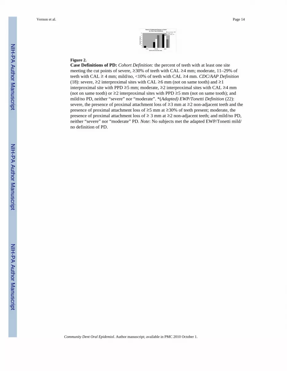
Figure 2.Case Definitions of PD: Cohort Definition: the percent of teeth with at least one sitemeeting the cut points of severe, ≥30% of teeth with CAL ≥4 mm; moderate, 11–29% ofteeth with CAL ≥ 4 mm; mild/no, <10% of teeth with CAL ≥4 mm. CDC/AAP Definition(18): severe, ≥2 interproximal sites with CAL ≥6 mm (not on same tooth) and ≥1interproximal site with PPD ≥5 mm; moderate, ≥2 interproximal sites with CAL ≥4 mm(not on same tooth) or ≥2 interproximal sites with PPD ≥5 mm (not on same tooth); andmild/no PD, neither “severe” nor “moderate”. *(Adapted) EWP/Tonetti Definition (22):severe, the presence of proximal attachment loss of ≥3 mm at ≥2 non-adjacent teeth and thepresence of proximal attachment loss of ≥5 mm at ≥30% of teeth present; moderate, thepresence of proximal attachment loss of ≥ 3 mm at ≥2 non-adjacent teeth; and mild/no PD,neither “severe” nor “moderate” PD. Note: No subjects met the adapted EWP/Tonetti mild/no definition of PD.
Vernon et al. Page 14
Community Dent Oral Epidemiol. Author manuscript; available in PMC 2010 October 1.
NIH
-PA Author Manuscript
NIH
-PA Author Manuscript
NIH
-PA Author Manuscript

NIH
-PA Author Manuscript
NIH
-PA Author Manuscript
NIH
-PA Author Manuscript
Vernon et al. Page 15
Table 1
Cohort characteristics including demographics and oral health behaviors of 112 HIV+ adults.
Variable N (%)
Demographics 112 (100)
Age (median, range in yrs) 42 (21–57)
Gender
Male 83 (74)
Female 29 (26)
Race
Black 72 (64)
White 33 (30)
Other 7 (6)
Education
<HS Graduate/GED 19 (17)
HS Graduate/GED 27 (24)
>HS Graduate/GED 65 (59)
Primary Insurance
Medicaid: 62 (56)
Ryan White: 25 (22)
Private: 25 (22)
Oral Health Behaviors
Brushing
Twice daily 64 (57)
Less than twice daily 48(43)
Flossing
Yes 44 (39)
No 68 (61)
Dental visits in past 5 yrs
< 5 visits 58 (52)
≥5 visits 54 (48)
Smoking status
Ever 77 (69)
Never 35 (31)
Pack Per DayYears (median, range) 3 (0–56)
Demographics: High School (HS), General Educational Development (GED).
Community Dent Oral Epidemiol. Author manuscript; available in PMC 2010 October 1.
NIH
-PA Author Manuscript
NIH
-PA Author Manuscript
NIH
-PA Author Manuscript
Vernon et al. Page 16
Table 2
Characterization of periodontal, microbiological and immunological measures in 112 HIV+ adults.
Variable Summary Measure
Periodontal Measures (mm) Mean (SD)
Total teeth 26.2 (3.0)
Mean Periodontal Probing Depth (PPD) 2.9 (0.6)
Mean Max PPD* 4.2 (0.8)
Mean Clinical Attachment Level (CAL) 2.5 (1.3)
Mean Max CAL* 4.0 (1.7)
Mean % of teeth with ≥ 1 site with:
PPD ≥5 mm 38 ( 24)
CAL ≥4 mm 50 ( 32)
Recession (REC) > 0 mm 55 ( 31)
Microbiological Measures (log genome copy number/ug DNA)
Porphymonas gingivalis (Pg) 4.1 (2.1)
Treponema denticola (Td) 4.7 (1.7)
Tannerella forsythia (Tf) 5.3 (1.4)
Total Plaque DNA (µg) 5.5 (3.37)
Clinical Measures Median (Range)
CD4+ T-cell count (cells/mm3)** 410 (5–1,186)
Viral Load (copies/mL)** 63 ( 50–750,000)
Nadir CD4+ T-cell count (cells/mm3)** 172 (0–615)
Months since first seropositive 86 (2–290)
Months on HAART 14 (0–137)
Not on HAART (number of subjects, %) 10 (9.7%)
On HAART <2 months (number of subjects, %) 12 (11.7%)
*Denotes the mean of the maximum value at each tooth.
**Data closest to and before the baseline visit.
Community Dent Oral Epidemiol. Author manuscript; available in PMC 2010 October 1.

NIH
-PA Author Manuscript
NIH
-PA Author Manuscript
NIH
-PA Author Manuscript
Vernon et al. Page 17
Table 3
Metabolic characteristics of 112 HIV+ subjects
Metabolic Measures Median (Range) Normal Range N (%) in Normal Range
Lipids
Cholesterol (mg/dL) 172 (93–261) (80–200) 89 (80.9)
HDL Cholesterol (mg/dL) 37 (18–97) >40 48 (43.6)
LDL (mg/dL) 106 (27–181) <130 83 (76.9)
VLDL (mg/dL) 22 (7–74) 0–40 95 (88.0)
Triglycerides (mg/dL) 111 (34–652) <150 79 (71.8)
BMI (kg/m2) 26 (16–64)
Underweight <18.5 2 (1.8)
Normal 18.5–24.9 49 (44.5)
Overweight 25–29.9 29 (26.4)
Obese >30 30 (27.3)
Hs-CRP (mg/L) 1.8 (0.1–64.4) <3.0 71 (65.1)
Insulin (µIU/ml) 4 (0–42)
Glucose (mg/dL) 91 (73–123) 70–110 105 (94.6)
HOMA-IR 0.88 (0–9.32) ≤2.6 99 (89.2)
Metabolic Measures: Body mass index (BMI), high-sensitivity C-reactive protein (hs-CRP), triglycerides (TRIG), high density lipoproteins(HDL), low density lipoproteins (LDL), very low density lipoproteins (VLDL), homeostasis model assessment-insulin resistance (HOMA-IR).Note: Normal range values for insulin are most appropriately interpreted within the context of HOMA-IR.
Community Dent Oral Epidemiol. Author manuscript; available in PMC 2010 October 1.

NIH
-PA Author Manuscript
NIH
-PA Author Manuscript
NIH
-PA Author Manuscript
Vernon et al. Page 18
Table 4
Spearman’s Correlation Coefficient (rho) for percent of teeth exceeding the threshold of periodontal probingdepth, clinical attachment level and recession with independent predictors.
Variable Periodontal ProbingDepth (PPD)
Recession(REC)
Clinical AttachmentLevel (CAL)
Age −.040 .398** .211*
Microbiological Measures
Tf .420** −.023 .179
Td .372** −.229* −.005
Pg .330** .265** .374**
Total Plaque DNA .237* .184 .197*
Immunological Measures
CD4 (cells/mm3) −.168 −.306** −.424**
Viral Load (copies/mL) −.003 .036 .143
Months on HAART .001 .202* .074
Months sero positive −.045 .130 .071
Metabolic Measures
BMI −.093 −.172 −.238*
Insulin −.120 −.250** −.283**
Glucose −.006 −.087 −.141
hs-CRP .013 .112 .061
TRIG −.196* .028 −.156
HDL .086 −.115 .009
LDL −.012 −.221* −.280**
VLDL −.203* .035 −.141
HOMA-IR −.151 −.203* −.246**
*Significance: p<.05
**p<.001
Community Dent Oral Epidemiol. Author manuscript; available in PMC 2010 October 1.

NIH
-PA Author Manuscript
NIH
-PA Author Manuscript
NIH
-PA Author Manuscript
Vernon et al. Page 19
Table 5
Final prediction model for the percent of teeth with CAL ≥4mm.
Factor B StandardError
Standardizedβ
P-value
Constant .257 .186 .172
CD4+ T-cell count (<200) .252 .067 .306 <.001
P. gingivalis (log) .047 .012 .290 <.001
Age (yrs) .010 .003 .268 .001
< 1 Annual Dental Visit .157 .053 .242 .004
Total Plaque DNA .019 .007 .207 .006
Ever Smoker .118 .053 .164 .029
LDL −.002 .001 −.202 .011
HOMA-IR(elevated) −.118 .075 −.189 .014
Analysis of variance for final model: F= 9.559; P<.001; R2=0.569; adjusted R2=.509. Model adjusted for: gender, race, primary insurance andbaseline viral load. Variables: Total copy number of subgingival plaque DNA (Total Plaque DNA); HOMA-IR ≥2.6 (HOMA-IR elevated).
Community Dent Oral Epidemiol. Author manuscript; available in PMC 2010 October 1.


![[Biocompatibility studies of periodontal dressings]](https://img.dokumen.tips/doc/110x75/6354ce5b60cfc5aa0404e054/biocompatibility-studies-of-periodontal-dressings.jpg)


