Embed Size (px)
Citation preview

Veterinary Parasitology 95 (2001) 143–154
Characterization of aSarcocystis neuronaisolate from a Missouri horse with equine
protozoal myeloencephalitis
A.E. Marsha,∗, P.J. Johnsonb, J. Ramos-Varac, G.C. Johnsonca Department of Veterinary Pathobiology, College of Veterinary Medicine, University of Missouri,
Connaway Hall, 1600 East Rollins Dr., Columbia, MO 65211, USAb Department of Veterinary Medicine and Surgery, College of Veterinary Medicine,
University of Missouri, Columbia, MO 65211, USAc Veterinary Medical Diagnostic Laboratory, College of Veterinary Medicine,
University of Missouri, Columbia, MO 65211, USA
Abstract
Little information is available about antigenic variation ofSarcocystis neuronaisolated fromhorses with equine protozoal myeloencephalitis, nor is there much information available on thespecific antibody pattern toS. neuronaantigens of horses from different geographic regions whereS. neuronaisolates have been obtained. This communication reports on the characterization ofa newS. neuronaisolate, SN-MU1. The isolate was obtained from a 3-year old Thoroughbredthat had asymmetrical neurological signs and localized skeletal muscle atrophy. ThisS. neuronaisolate is similar to otherS. neuronaisolates by molecular analysis of the internal transcribed spacer(ITS-1) region and a random-amplified polymorphic DNA marker, but is phenotypically distinctfrom the otherS. neuronaisolates examined. Evaluation of the antibodies from the affected horseand immunohistochemical results suggested that antigenic variation ofS. neuronacan result invariable antibody–antigen reactivity observed in theS. neuronaimmunoblot test. © 2001 ElsevierScience B.V. All rights reserved.
Keywords:Equine protozoal myeloencephalitis; Apicomplexa;Sarcocystis neurona; Immunohistochemistry
1. Introduction
Equine protozoal myeloencephalitis (EPM) is caused by a protozoal infection of thecentral nervous system (Rooney et al., 1970; Cusick et al., 1974; Dubey et al., 1991a; Marshet al., 1996a) and is the most commonly diagnosed neurologic disease of horses in North
∗ Corresponding author. Tel.:+1-573-884-2673; fax:+1-573-884-5414.E-mail address:[email protected] (A.E. Marsh).
0304-4017/01/$ – see front matter © 2001 Elsevier Science B.V. All rights reserved.PII: S0304-4017(00)00386-1

144 A.E. Marsh et al. / Veterinary Parasitology 95 (2001) 143–154
America (MacKay, 1997). Approximately 50% of the horse population in the United Statesis seropositive toSarcocystis neurona(Bentz et al., 1997; Blythe et al., 1997; Saville et al.,1997). However, a minority of all seropositive horses develop clinical signs. Detailed clinicalexamination and cerebrospinal fluid (CSF) antibody evaluation are often used to diagnoseEPM (MacKay, 1997). Histopathologic evaluation of the brain stem and spinal cord forlesions compatible with non-suppurative meningoencephalomyelitis is used diagnosticallysince organisms are difficult to detect by routine staining methods (Summers et al., 1995).Immunohistochemical staining aids in the detection of organisms in tissues (Granstromet al., 1991). However, there have been equine cases in which protozoal organisms havenot stained withS. neuronapolyclonal antibodies (Hamir et al., 1997) and marine mammalcases in whichS. neurona-like organisms have been detected by immunohistochemistryusing anti-S. neuronaantibodies (Lapointe et al., 1998). It would appear highly unlikelythat these marine mammals became infected from the same sporocyst source as the horse(Lapointe et al., 1998).
There is a paucity of information regarding antigenic variation ofS. neuronaisolatedfrom horses with EPM, or the antibody response pattern toS. neuronaantigens of horsesfrom different geographic regions. This communication reports the characterization of anew S. neuronaisolate, SN-MU1 and its comparison to four previously reported isolates(Davis et al., 1991; Marsh et al., 1996b, 1997a). We show that all isolates are similar bymolecular analysis of the internal transcribed spacer (ITS-1) region (Marsh et al., 1999)and a random-amplified polymorphic DNA marker (Tanhauser et al., 1999) but that thisisolate is phenotypically distinct from the otherS. neuronaisolates examined. Evaluationof antibodies from the affected horse suggested that antigenic variation ofS. neuronacanaccount for the result in variable antibody reactivity to parasite antigens in theS. neuronaimmunoblot test.
2. Materials and methods
2.1. Case history
A 3-year old Thoroughbred colt, raised and trained in Missouri, was presented to theUniversity of Missouri’s Veterinary Medical Teaching Hospital (VMTH) for diagnosis ofabnormal action in the pelvic limbs of several weeks’ duration. Although lameness affectedboth pelvic limbs, the left limb appeared to be more prominently affected than the right.Lameness in both pelvic limbs was exacerbated by flexion. A complete neurological ex-amination did not identify any neurological abnormalities. The horse had been regularlydewormed and vaccinated against tetanus, influenza, togaviral encephalomyelitis, rhinop-neumonitis, strangles, and Potomac Horse Fever. Medical problems had not been recognizedin any of the other horses on the same premises and under similar management conditions.
Further diagnostic tests included the use of bone scintigraphy and arthroscopic exam-ination. Lameness was attributed to bilateral osteochondritis dessicans (OCD) affectingthe lateral trochlear ridge of both stifle joints. Lesions associated with OCD were treatedarthroscopically. Following completion of the arthroscopic procedures, CSF was collectedfrom the cerebellomedullary cistern.
A.E. Marsh et al. / Veterinary Parasitology 95 (2001) 143–154 145
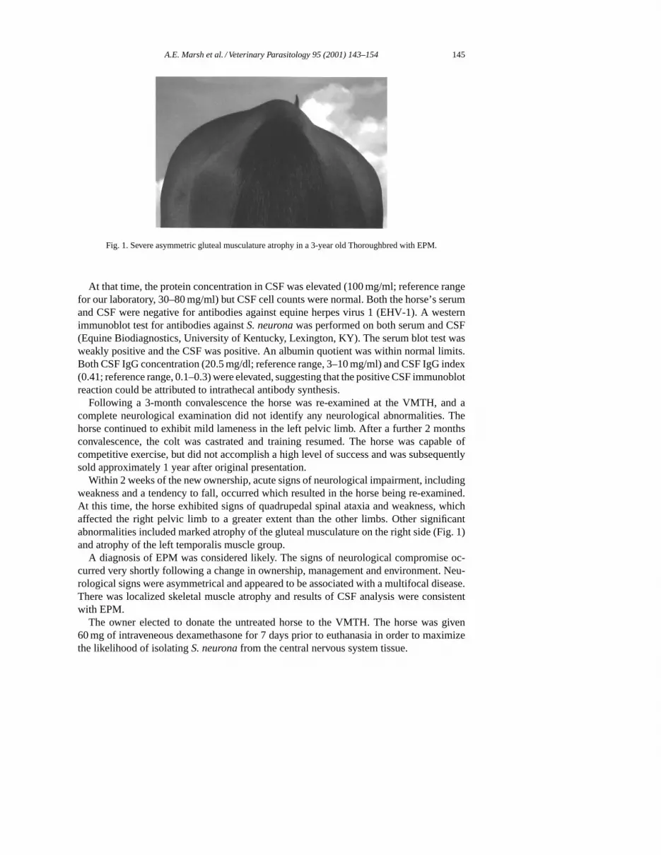
Fig. 1. Severe asymmetric gluteal musculature atrophy in a 3-year old Thoroughbred with EPM.
At that time, the protein concentration in CSF was elevated (100 mg/ml; reference rangefor our laboratory, 30–80 mg/ml) but CSF cell counts were normal. Both the horse’s serumand CSF were negative for antibodies against equine herpes virus 1 (EHV-1). A westernimmunoblot test for antibodies againstS. neuronawas performed on both serum and CSF(Equine Biodiagnostics, University of Kentucky, Lexington, KY). The serum blot test wasweakly positive and the CSF was positive. An albumin quotient was within normal limits.Both CSF IgG concentration (20.5 mg/dl; reference range, 3–10 mg/ml) and CSF IgG index(0.41; reference range, 0.1–0.3) were elevated, suggesting that the positive CSF immunoblotreaction could be attributed to intrathecal antibody synthesis.
Following a 3-month convalescence the horse was re-examined at the VMTH, and acomplete neurological examination did not identify any neurological abnormalities. Thehorse continued to exhibit mild lameness in the left pelvic limb. After a further 2 monthsconvalescence, the colt was castrated and training resumed. The horse was capable ofcompetitive exercise, but did not accomplish a high level of success and was subsequentlysold approximately 1 year after original presentation.
Within 2 weeks of the new ownership, acute signs of neurological impairment, includingweakness and a tendency to fall, occurred which resulted in the horse being re-examined.At this time, the horse exhibited signs of quadrupedal spinal ataxia and weakness, whichaffected the right pelvic limb to a greater extent than the other limbs. Other significantabnormalities included marked atrophy of the gluteal musculature on the right side (Fig. 1)and atrophy of the left temporalis muscle group.
A diagnosis of EPM was considered likely. The signs of neurological compromise oc-curred very shortly following a change in ownership, management and environment. Neu-rological signs were asymmetrical and appeared to be associated with a multifocal disease.There was localized skeletal muscle atrophy and results of CSF analysis were consistentwith EPM.
The owner elected to donate the untreated horse to the VMTH. The horse was given60 mg of intraveneous dexamethasone for 7 days prior to euthanasia in order to maximizethe likelihood of isolatingS. neuronafrom the central nervous system tissue.

146 A.E. Marsh et al. / Veterinary Parasitology 95 (2001) 143–154
2.2. Parasite isolation
Immediately following euthanasia, the thoracolumbar area of the spinal cord was removedintact and placed in sterile phosphate buffered saline with 1000 U penicillin/ml and 1000mgof streptomycin/ml (antibiotic saline). The cord was transected into 1.0 cm segments undera laminar flow hood using sterile instruments. A small focal lesion was grossly apparent inone section. An approximately 0.5 cm section containing this focus (gross lesion) was placedinto neutral buffered 10% formalin for histopathological processing and examination. Theremaining affected tissue and its margins were separated from the remaining cord, placedinto sterile antibiotic saline and washed once. Tissue was homogenized and the tissuemixture was applied for 1.5 h to individual flasks containing confluent monolayers cells ofequine dermal (ATCC CCL-57), bovine turbinate (ATCC CRL-1390), or deer testis (Marshet al., 1996a). The tissue mixture was removed and replaced with RPMI-1640 as previouslydescribed (Davis et al., 1991) except 10% heat inactivated fetal bovine serum (FBS) wasused in place of horse serum (HS) and 20mg/ml gentamicin was included in the bovineturbinate and deer testis monolayer cultures. The cultures were maintained as previouslydescribed with FBS rather than HS, and amphotericin was not included in the media (Daviset al., 1991).
2.3. Histopathology and immunohistochemistry
A complete necropsy was performed. Samples from multiple tissues were examined andimmersed in neutral buffered 10% formalin, routinely processed, and stained using hema-toxylin and eosin (H&E) for light microscopic examination. Immunohistochemical exam-ination was performed on selected sections of brain and spinal cord to detect and identifyparasites, using polyclonal antisera raised againstS. neurona(Marsh et al., 1996b) andS. fal-catula(Marsh et al., 1997b). A labeled streptavidin-biotin (LSAB) detection method (Dako)was used with diaminobenzidine (DAB) as the chromogen. Antibody incubations were doneat room temperature with 30 min incubations (Ramos-Vara and Beissenherz, 2000). Neg-ative controls were tissue sections containingToxoplasma gondiior Neospora caninumor N. hughesiorganisms and substitution of the primary antibodies with non-immune orpre-immune rabbit sera. Positive controls were tissues sections that containedS. neurona(experimental infection of nude mice) (Marsh et al., 1997a) orS. falcatula(experimentalinfection of budgerigars) (Marsh et al., 1997a).
2.4. Parasites
Viable merozoites from fiveS. neuronaisolates (SN-UCD 1–3, SN 2, and SN-MU1),and threeS. falcatula(SF-MU1, SF-UCD 1, and SF-Florida 1) were harvested from simi-larly infected and maintained monolayers. TheS. falcatulaisolate (SF-MU1) was obtainedusing similar procedures as previously described for SF-UCD 1 and SF-Florida 1 (Marshet al., 1997a). Briefly, sporocyts harvested from a Missouri opossum (Didelphis virginiana)were inoculated into a laboratory budgerigar (Melopsittacus undulatus) which subsequentlydied at 13 days post-inoculation and the in vitro isolate was established from the infected
A.E. Marsh et al. / Veterinary Parasitology 95 (2001) 143–154 147
lung tissue. The merozoites from theS. neuronaandS. falcatulaisolates were enumerated,washed twice by pelleting and resuspending in PBS to remove the media from the mero-zoites. Uninfected cultures were treated in the same way to serve as a negative control insubsequent protein and DNA analysis studies. The resulting pellets were stored at−20◦Cuntil used for protein and DNA analysis.
2.5. Tachyzoite protein profile analyses
Thawed tachyzoite pellets and control uninfected monolayer cells were lysed in samplebuffer. Samples corresponding to the same number of parasites or host monolayer cells wereseparated by sodium dodecyl sulfate–polyacrylamide gel electrophoresis (SDS–PAGE)(Laemmli, 1970; Gallagher and Smith, 1995). Prestained SDS–PAGE broad ranged molec-ular size standards (BioRad) were included with each gel. After electrophoretic separation,antigens were transferred to nitrocellulose membranes (BioRad), blocked with Tris-bufferedsaline (TBS) containing 5% (w/v) non-fat dry milk and 0.1% Tween 20, and then incu-bated with the primary antibody (equine sera, cerebrospinal fluid or polyclonal rabbit sera)overnight at 4◦C or 2 h at room temperature. Goat anti-horse IgG (H+ L) (Kirkegaardand Perry) or donkey anti-rabbit Ig F(ab′)2 fragment labeled with horseradish peroxidase(Amersham Life Science) was used as the secondary antibody. After washing, the blots wereprocessed for color development using diaminobenzidine plus nickel chloride (DAB–Ni)substrate.
2.6. Molecular characterization
Genomic DNA was isolated as previously described, the ITS 1 region was amplifiedand sequenced as previously described (Marsh et al., 1999). Final DNA sequence con-struction, alignment, and comparisons were facilitated using the Genetics Computer Groupprograms. The SN-MU1 ITS 1 DNA sequence is listed in GenBank (accession AF204230).Random-amplified polymorphic DNA (RAPD) primers JNB25/JD 396 were used to gen-erate amplified DNA products from theS. neuronaisolates and theS. falcatulaisolate(Tanhauser et al., 1999). The amplified DNA products were subsequently treated with ei-ther Hind III or Hinf I restriction enzymes and analyzed by gel separation. The amplifiedDNA products were also sequenced (SN-MU1 accession AF205937; SF-MU1 accessionAF205938) for DNA alignment with published sequence data (Tanhauser et al., 1999).
3. Results
At post-inoculation day 18, merozoites of theS. neuronaisolate, SN-MU1, were firstdetected in the equine dermal monolayer. No parasites were detected in either the bovineturbinate or deer testis monolayers; however, subpassage of this isolate was performed onall three monolayers.
The histopathologic examination of the central nervous system was unremarkable exceptfor the thoracolumbar site where the gross lesion was detected. There were mild to moderateswollen axon sheaths that were most frequently observed in the ventral funiculi. Small
148 A.E. Marsh et al. / Veterinary Parasitology 95 (2001) 143–154
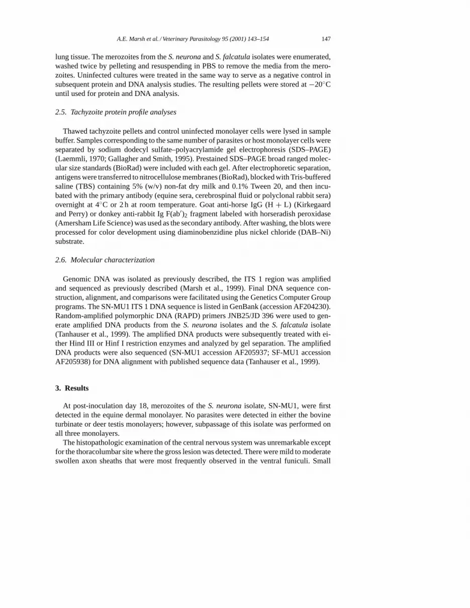
Fig. 2. Spinal cord from affected horse: (a) small clusters of lymphocytes (arrow) and lymphocytic perivascularcuffing is evident (arrowhead); hematoxylin and eosin stained section, 120×; (b) S. falcatulaantiserum demon-strates infrequent but intense staining of some cells, suggesting possible intracellular organisms (arrow). LSABimmunohistochemical method. Hematoxylin counterstain, 610×.
clusters of lymphocytes could be seen in the adjacent gray matter and around vessels inthe white matter (Fig. 2a). No sarcocysts were observed. Individual parasites or schizontscould not be seen using conventional H&E staining. Immunohistochemistry using theS.neuronarabbit antiserum did not react to tissue sections taken sequentially from the sametissue block. TheS. falcatulaantibodies revealed intense staining of some cells possiblycontaining zoites (Fig. 2b), but typical distinct individual or budding merozoites could notbe identified.
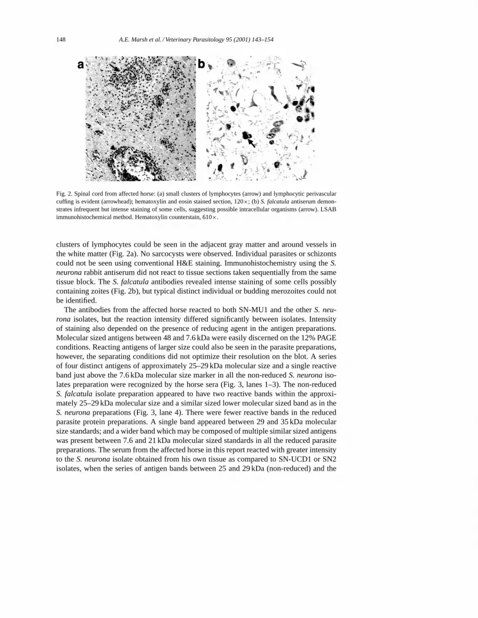
The antibodies from the affected horse reacted to both SN-MU1 and the otherS. neu-rona isolates, but the reaction intensity differed significantly between isolates. Intensityof staining also depended on the presence of reducing agent in the antigen preparations.Molecular sized antigens between 48 and 7.6 kDa were easily discerned on the 12% PAGEconditions. Reacting antigens of larger size could also be seen in the parasite preparations,however, the separating conditions did not optimize their resolution on the blot. A seriesof four distinct antigens of approximately 25–29 kDa molecular size and a single reactiveband just above the 7.6 kDa molecular size marker in all the non-reducedS. neuronaiso-lates preparation were recognized by the horse sera (Fig. 3, lanes 1–3). The non-reducedS. falcatulaisolate preparation appeared to have two reactive bands within the approxi-mately 25–29 kDa molecular size and a similar sized lower molecular sized band as in theS. neuronapreparations (Fig. 3, lane 4). There were fewer reactive bands in the reducedparasite protein preparations. A single band appeared between 29 and 35 kDa molecularsize standards; and a wider band which may be composed of multiple similar sized antigenswas present between 7.6 and 21 kDa molecular sized standards in all the reduced parasitepreparations. The serum from the affected horse in this report reacted with greater intensityto theS. neuronaisolate obtained from his own tissue as compared to SN-UCD1 or SN2isolates, when the series of antigen bands between 25 and 29 kDa (non-reduced) and the
A.E. Marsh et al. / Veterinary Parasitology 95 (2001) 143–154 149
Fig. 3. Immunostaining of Western blots from SDS–PAGE separated merozoite proteins (non-reduced, lanes 1–5,and reduced, lanes 6–9) probed with sera diluted 1:200 from the affected Missouri horse (top panel) and a Californiahorse infected withS. neuronaSN-UCD2 (bottom panel). Lane 1, SN-MU1; lane 2, SN-UCD1; lane 3, SN2; lane4, SF-MU1; lane 5, bovine turbinate monolayer; lane 6, SN-MU1; lane 7, SN-UCD1; lane 8, SN2; lane 9, SF-MU1.
single antigen band between 29 and 35 kDa are compared (Fig. 3, top panel). Antibodiesfrom a confirmedS. neuronainfected horse from California (Marsh et al., 1996a, 1997a)reacted with greater intensity to the SN-UCD1 and SN2 isolates than the SN-MU1 isolatewhen the non-reduced and reduced preparations are compared (Fig. 3, bottom panel). Over-all, the non-reduced antigens had greater reactivity than reduced antigens when tested withantibodies against the homologous or heterogeneousS. neuronaisolates, andS. falcatulaantigens can be recognized byS. neuronainfected horses.
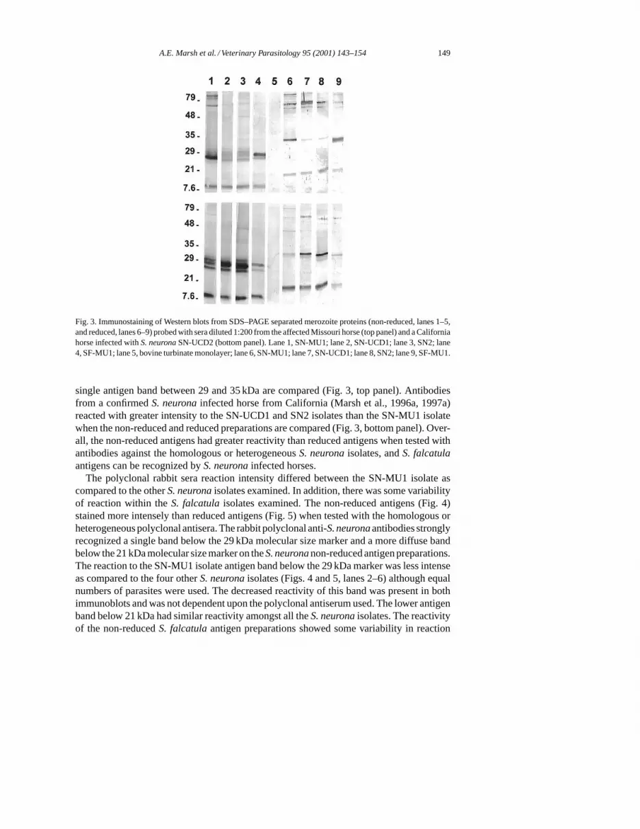
The polyclonal rabbit sera reaction intensity differed between the SN-MU1 isolate ascompared to the otherS. neuronaisolates examined. In addition, there was some variabilityof reaction within theS. falcatulaisolates examined. The non-reduced antigens (Fig. 4)stained more intensely than reduced antigens (Fig. 5) when tested with the homologous orheterogeneous polyclonal antisera. The rabbit polyclonal anti-S. neuronaantibodies stronglyrecognized a single band below the 29 kDa molecular size marker and a more diffuse bandbelow the 21 kDa molecular size marker on theS. neuronanon-reduced antigen preparations.The reaction to the SN-MU1 isolate antigen band below the 29 kDa marker was less intenseas compared to the four otherS. neuronaisolates (Figs. 4 and 5, lanes 2–6) although equalnumbers of parasites were used. The decreased reactivity of this band was present in bothimmunoblots and was not dependent upon the polyclonal antiserum used. The lower antigenband below 21 kDa had similar reactivity amongst all theS. neuronaisolates. The reactivityof the non-reducedS. falcatulaantigen preparations showed some variability in reaction

150 A.E. Marsh et al. / Veterinary Parasitology 95 (2001) 143–154
Fig. 4. Immunostaining of Western blots from SDS–PAGE separated merozoite proteins (non-reduced) probedwith rabbit anti-S. neurona(top panel) or anti-S. falcatula(bottom panel) polyclonal sera diluted 1:500. Lane 1,control host cell monolayer; lane 2, SN-MU1; lane 3, SN-UCD1; lane 4, SN-UCD2; lane 5, SN-UCD3; lane 6,SN2; lane 7, SF-MU1; lane 8, SF-UCD1; lane 9, SF-Florida.
intensity as well as the molecular size of the antigens recognized (Fig. 4, lanes 7–9). TheS. neuronaantiserum reacted more strongly with the non-reducedS. neuronapreparations ascompared to the non-reducedS. falcatulapreparations; whereas, theS. falcatulaantiserumreacted with almost equal intensity against all the isolates except SN-MU1 and SF-Florida(Fig. 4, bottom panel). Both rabbit antisera reacted to the reducedS. neuronaantigenpreparations similar to the reaction seen with the California equine antibodies (SN-UCD2), asingle band just below the 35 kDa and another just below the 21 kDa molecular sized marker
Fig. 5. Immunostaining of Western blots from SDS–PAGE separated merozoite proteins (reduced) probed withrabbit anti-S. neurona(top panel) or anti-S. falcatula(bottom panel) polyclonal sera diluted 1:500. Lane 1, controlhost cell monolayer; lane 2, SN-MU1; lane 3, SN-UCD1; lane 4, SN-UCD2; lane 5, SN-UCD3; lane 6, SN2; lane7, SF-MU1; lane 8, SF-UCD1; lane 9, SF-Florida.
A.E. Marsh et al. / Veterinary Parasitology 95 (2001) 143–154 151
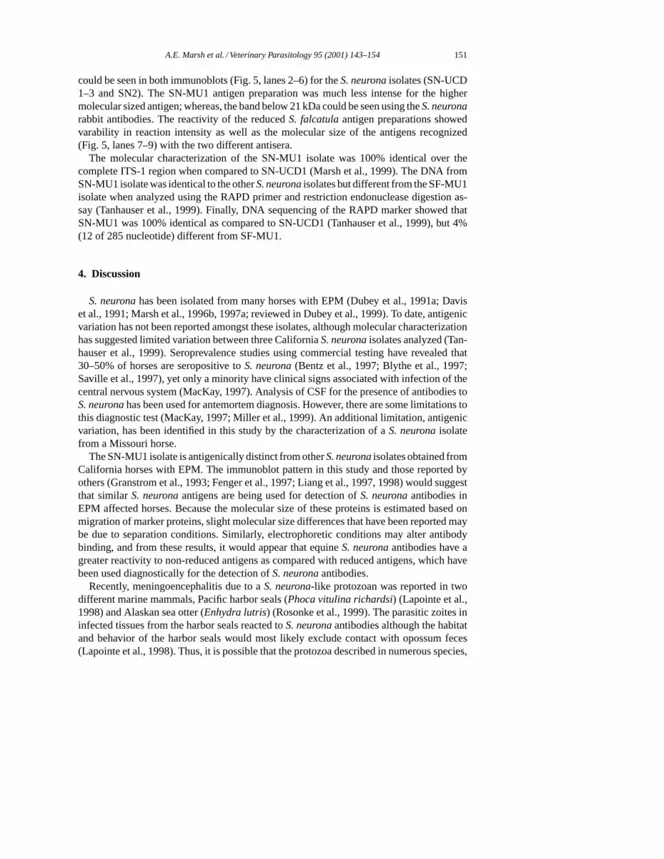
could be seen in both immunoblots (Fig. 5, lanes 2–6) for theS. neuronaisolates (SN-UCD1–3 and SN2). The SN-MU1 antigen preparation was much less intense for the highermolecular sized antigen; whereas, the band below 21 kDa could be seen using theS. neuronarabbit antibodies. The reactivity of the reducedS. falcatulaantigen preparations showedvarability in reaction intensity as well as the molecular size of the antigens recognized(Fig. 5, lanes 7–9) with the two different antisera.
The molecular characterization of the SN-MU1 isolate was 100% identical over thecomplete ITS-1 region when compared to SN-UCD1 (Marsh et al., 1999). The DNA fromSN-MU1 isolate was identical to the otherS. neuronaisolates but different from the SF-MU1isolate when analyzed using the RAPD primer and restriction endonuclease digestion as-say (Tanhauser et al., 1999). Finally, DNA sequencing of the RAPD marker showed thatSN-MU1 was 100% identical as compared to SN-UCD1 (Tanhauser et al., 1999), but 4%(12 of 285 nucleotide) different from SF-MU1.
4. Discussion
S. neuronahas been isolated from many horses with EPM (Dubey et al., 1991a; Daviset al., 1991; Marsh et al., 1996b, 1997a; reviewed in Dubey et al., 1999). To date, antigenicvariation has not been reported amongst these isolates, although molecular characterizationhas suggested limited variation between three CaliforniaS. neuronaisolates analyzed (Tan-hauser et al., 1999). Seroprevalence studies using commercial testing have revealed that30–50% of horses are seropositive toS. neurona(Bentz et al., 1997; Blythe et al., 1997;Saville et al., 1997), yet only a minority have clinical signs associated with infection of thecentral nervous system (MacKay, 1997). Analysis of CSF for the presence of antibodies toS. neuronahas been used for antemortem diagnosis. However, there are some limitations tothis diagnostic test (MacKay, 1997; Miller et al., 1999). An additional limitation, antigenicvariation, has been identified in this study by the characterization of aS. neuronaisolatefrom a Missouri horse.
The SN-MU1 isolate is antigenically distinct from otherS. neuronaisolates obtained fromCalifornia horses with EPM. The immunoblot pattern in this study and those reported byothers (Granstrom et al., 1993; Fenger et al., 1997; Liang et al., 1997, 1998) would suggestthat similarS. neuronaantigens are being used for detection ofS. neuronaantibodies inEPM affected horses. Because the molecular size of these proteins is estimated based onmigration of marker proteins, slight molecular size differences that have been reported maybe due to separation conditions. Similarly, electrophoretic conditions may alter antibodybinding, and from these results, it would appear that equineS. neuronaantibodies have agreater reactivity to non-reduced antigens as compared with reduced antigens, which havebeen used diagnostically for the detection ofS. neuronaantibodies.
Recently, meningoencephalitis due to aS. neurona-like protozoan was reported in twodifferent marine mammals, Pacific harbor seals (Phoca vitulina richardsi) (Lapointe et al.,1998) and Alaskan sea otter (Enhydra lutris) (Rosonke et al., 1999). The parasitic zoites ininfected tissues from the harbor seals reacted toS. neuronaantibodies although the habitatand behavior of the harbor seals would most likely exclude contact with opossum feces(Lapointe et al., 1998). Thus, it is possible that the protozoa described in numerous species,
152 A.E. Marsh et al. / Veterinary Parasitology 95 (2001) 143–154
including raccoons (Dubey et al., 1990), cat (Dubey et al., 1994), birds (Jacobson et al.,1984; Dubey et al., 1991b), rhesus monkey (Klumpp et al., 1994), chickens (Mutalib et al.,1995), sheep (Dubey et al., 1989; O’Toole et al., 1993), skunk (Dubey et al., 1996) andmink (Dubey and Hedstrom, 1993) asS. neurona-like, are actually different species withvarying life cycles. Further, the question should be raised how much variation exists betweendifferentS. neuronaisolates that infect horses. This report and a recently published studyby Hamir et al. (1997) suggests thatS. neuronamay vary antigenically.
The affected horse in this report had microscopic lesions consistent with EPM (Summerset al., 1995). Unfortunately, the immunohistochemistry was not definitive since distinctparasite forms were not discernible using theS. falcatulapolyclonal antibodies, and noreaction was seen using theS. neuronapolyclonal antibodies. It is not surprising that theS. falcatularabbit antiserum showed reactivity since this antiserum will react with bothS.neuronaandS. falcatulamerozoites in experimentally infected tissues (B.C. Barr, personalcommunication). In contrast,S. neuronarabbit antiserum used in the present study will notreact with merozoites, tissue cysts or sporozoites ofS. falcatula(Marsh et al., 1997a). Theimmunohistochemistry results were atypical in that the rabbit antibodies raised againstS.neurona(SN-UCD1) did not react on the tissue sections; whereas, reactivity was detectedusingS. falcatulapolyclonal antibodies, which will react to bothS. falcatulaandS. neuronaparasites in control tissues. These results suggested that differences in staining could be dueto the specific antisera used, antigenic variation or lack of intact or numerous parasites in thespecific sections. Experimentally,S. neurona(SN-MU1) infected mice could provide thenecessary tissue material for evaluating the immunohistochemistry results observed withthe two different antisera. The molecular analysis in this report supported the presence ofa S. neuronawhich was antigenically distinguishable from other known strains. Antigenicvariation has been reported for multiple isolates ofToxoplasma gondii(De la Cruz et al.,1989; Ware and Kasper, 1987); therefore, it would not be unusual for antigenic variationto be detected within another closely related parasite species such asS. neurona. Froma diagnostic or vaccine development perspective it would be important to consider hostresponses to antigenic variations when evaluating potential diagnostic tests or immunizationagainst parasite proteins.
Acknowledgements
This project was supported by the Southern California Equine Research Foundation withfunds provided by the Dolly Green Research Foundation and the American Quarter HorseAssociation. The SN2 isolate was kindly provided by Dr. J.P. Dubey. We thank Dr. B.C.Barr for consultation and Marilyn Beissenherz for assistance with immunohistochemistry,and Dr. C.A. Carson for his critical review of the manuscript.
References
Bentz, B.G., Granstrom, D.E., Stamper, S., 1997. Seroprevalence of antibodies toSarcocystis neuronain horsesresiding in a county of southeastern Pennsylvania. J. Am. Vet. Med. Assoc. 210, 517–518.
Blythe, L.L., Granstrom, D.E., Hansen, D.E., Walker, L.L., Bartlett, J., Stamper, S., 1997. Seroprevalence ofantibodies toSarcocystis neuronain horses residing in Oregon. J. Am. Vet. Med. Assoc. 210, 525–527.

A.E. Marsh et al. / Veterinary Parasitology 95 (2001) 143–154 153
Cusick, P.K., Sells, D.M., Hamilton, D.P., Jardenbrook, H.J., 1974. Toxoplasmosis in two horses. J. Am. Vet. Med.Assoc. 164, 77–80.
Davis, S.W., Speer, C.A., Dubey, J.P., 1991. In vitro cultivation ofSarcocystis neuronafrom the spinal cord of ahorse with equine protozoal myelitis. J. Parasitol. 77, 789–792.
De la Cruz, A.A., Dreesen, D.W., Evans, D.L., 1989. Western blot analysis and LD50 determinations ofToxoplasmagondii isolates. Vet. Immunol. Immunopathol. 23, 355–364.
Dubey, J.P., Hedstrom, O.R., 1993. Meningoencephalitis in mink associated with aSarcocystis neurona-likeorganism. J. Vet. Diagn. Invest. 5, 467–471.
Dubey, J.P., Speer, C.A., Munday, B.L., Lipscomb, T.P., 1989. Ovine sporozoan encephalomyelitis linked toSarcocystisinfection. Vet. Parasitol. 34, 159–163.
Dubey, J.P., Hamir, A.N., Hanlon, C.A., Topper, M.J., Rupprecht, C.E., 1990. Fatal necrotizing encephalitis in aracoon associated with aSarcocystis-like protozoan. J. Vet. Diagn. Invest. 2, 345–347.
Dubey, J.P., Davis, S.W., Speer, C.A., Bowman, D.D., de Lahunta, A., Granstrom, D.E., Topper, M.J., Hamir,A.N., Cummings, J.F., Suter, M.M., 1991a.Sarcocystis neuronan. sp. (Protozoa: Apicomplexa), the etiologicagent of equine protozoal myeloencephalitis. J. Parasitol. 77, 212–218.
Dubey, J.P., Porter, S.L., Hattel, A.L., Kradel, D.C., Topper, M.J., Johnson, L., 1991b.Sarcocystis-associatedclinical encephalitis in a golden eagle (Aquila chrysaetos). J. Zool. Wildl. Med. 22, 233–236.
Dubey, J.P., Higgins, R.J., Barr, B.C., Spangler, W.L., Kollins, B., Jorgensen, L.S., 1994.Sarcocystis-associatedmeningoencephalomyelitis in a cat. J. Vet. Diagn. Invest. 6, 118–120.
Dubey, J.P., Hamir, A.N., Niezgoda, M., Rupprecht, C.E., 1996. ASarcocystis neurona-like organism associatedwith encephalitis in a striped skunk (Mephitis mephitis). J. Parasitol. 82, 172–174.
Dubey, J.P., Mattson, D.E., Speer, C.A., Baker, R.J., Mulrooney, D.M., Tornquist, S.J., Hamir, A.N., Gerros, T.C.,1999. Characterization of aSarcocystis neuronaisolate (SN6) from a naturally infected horse from Oregon. J.Eukaryot. Microbiol. 46, 500–506.
Fenger, C.K., Granstrom, D.E., Gajadhar, A.A., Williams, N.M., McCrillis, S.A., Stamper, S., Langemeier, J.L.,Dubey, J.P., 1997. Experimental induction of equine protozoal myeloencephalitis in horses usingSarcocystissp. sporocysts from the opossum (Didelphis virginiana). Vet. Parasitol. 68, 199–213.
Gallagher, S.R., Smith, J.A., 1995. Electrophoretic separation of proteins, Vol. 2. In: Coligan, J., Kruisbeek,A., Margulies, D., Shevach, E., Strober, W. (Eds.), Current Protocols in Immunology. Wiley, New York,pp. 8.4.1–8.4.21.
Granstrom, D.E., Giles, R.C., Tuttle, P.A., Williams, N.M., Poonacha, K.B., Petrites-Murphy, M.B., Tramonin,R.R.S.T.W., Hong, C.B., Rexabek, G.B., Lyons, E.T., Drudge, J.H., 1991. Immunohistochemical diagnosis ofprotozoan parasites in lesions of equine protozoal myeloencephalitis. J. Vet. Diagn. Invest. 3, 75–77.
Granstrom, D.E., Dubey, J.P., Davis, S.W., Fayer, R., Fox, J.C., Poonacha, K.B., Giles, R.C., Comer, P.F., 1993.Equine protozoal myeloencephalitis: antigen analysis of culturedSarcocystis neuronamerozoites. J. Vet. Diagn.Invest. 5, 88–90.
Hamir, A.N., Gerros, T.C., Dubey, J.P., 1997. Pyogranulomatous encephalitis associated with an unidentifiedSarcocystis neurona-like organism in a horse. J. Vet. Diagn. Invest. 9, 331–333.
Jacobson, E.R., Gardiner, C.H., Nicholson, A., Page, C.D., 1984.Sarcocystisencephalitis in a cockatiel. J. Am.Vet. Med. Assoc. 185, 904–906.
Klumpp, S.A., Anderson, D.C., McClure, H.M., Dubey, J.P., 1994. Encephalomyelitis due to aSarcocystisneurona-like protozoan in a rhesus monkey (Macaca mulatta) infected with simian immunodeficienay virus.Am. J. Trop. Med. Hyg. 51, 332–338.
Laemmli, E.K., 1970. Cleavage of structural proteins during the assembly of the head of bacteriophage T4. Nature227, 680–685.
Lapointe, J.M., Duignan, P.J., Marsh, A.E., Gulland, F.M., Barr, B.C., Naydan, D.K., King, D.P., Farman, C.A.,Burek, K.A., Lowenstine, J.L., 1998. Meningoencephalitis due to aSarcocystis neurona-like protozoan inPacific Harbor seals (Phoca vitulina richardsi). J. Parasitol. 84, 1184–1189.
Liang, F.T., Granstrom, D.E., Timoney, J.F., Shi, Y.F., 1997. Micropreparative high resolution purification ofproteins by a combination of sodium dodecyl sulfate–polyacrylamide gel electrophoresis, isoelectric focusing,and membrane blotting. Anal. Biochem. 250, 61–65.
Liang, F.T., Granstrom, D.E., Zhao, X.M., Timoney, J.F., 1998. Evidence that surface proteins Sn14 and Sn16 ofSarcocystis neuronamerozoites are involved in infection and immunity. Infect. Immun. 66, 1834–1838.

154 A.E. Marsh et al. / Veterinary Parasitology 95 (2001) 143–154
MacKay, R.J., 1997. Equine protozoal myeloencephalitis. In: Veterinary Clinics of North America Equine Practice.Saunders, Philadelphia, PA, pp. 79–96.
Marsh, A.E., Barr, B.C., Madigan, J., Lakritz, J., Nordhausen, R., Conrad, P.A., 1996a. Neosporosis as a cause ofequine protozoal myeloencephalitis. J. Am. Vet. Med. Assoc. 209, 1907–1913.
Marsh, A.E., Barr, B.C., Madigan, J., Lakritz, J., Conrad, P.A., 1996b. Sequence analysis and polymerase chainreaction amplification of small subunit ribosomal DNA fromSarcocystis neurona. Am. J. Vet. Res. 57, 975–981.
Marsh, A.E., Barr, B.C., Lakritz, J., Nordhausen, R., Madigan, J.E., Conrad, P.A., 1997a. Experimental infectionof nude mice as a model forSarcocystis neurona-associated encephalitis. Parasitol. Res. 83, 706–711.
Marsh, A.E., Barr, B.C., Tell, L., Koski, M., Greiner, E., Dame, J., Conrad, P.A., 1997b. In vitro cultivationand experimental inoculation ofSarcocystis falcatulaandSarcocystis neuronamerozoites into budgerigars(Melopsittacus undulatus). J. Parasitol. 83, 1189–1192.
Marsh, A.E., Barr, B., Tell, L., Bowman, D.D., Conrad, P.A., Ketcherside, C., Green, T., 1999. Comparison of theinternal transcribed spacer, ITS-1, fromSarcocystis falcatulaisolates andSarcocystis neurona. J. Parasitol. 85,750–757.
Miller, M.M., Sweeney, C.R., Russel, G.E., Sheetz, R.M., Morrow, J.K., 1999. Effects of blood contaminationof cerebrospinal fluid on Western blot analysis for detection of antibodies againstSarcocystis neuronaand onalbumin quotient and immunoglobulin G index in horses. J. Am. Vet. Med. Assoc. 215, 67–71.
Mutalib, A., Keirs, R., Maslin, W.T.M., Dubey, J.P., 1995.Sarcocystis-associated encephalitis in chickens. AvianDis. 39, 436–440.
O’Toole, A., Jeffrey, M., Challoner, D., Maybey, R., Welch, V., 1993. Ovine myeloencephalitis–leukomyelomalaciaassociated with aSarcocystis-like protozoan. J. Vet. Diagn. Invest. 5, 212–225.
Ramos-Vara, J., Beissenherz, M.E., 2000. Antigen retrieval methods to optimize diagnostic immunohistochemistry:Experience with 63 markers. J. Vet. Diagn. Invest. 12, 307–311.
Rooney, J.R., Prickett, M., Delandy, F.M., Crow, M.W., 1970. Focal myelitis and encephalitis in horses. CornellVet. 74, 494–501.
Rosonke, B.J., Brown, S.R., Tornquist, S.J., Synder, S.P., Garner, M.M., Blythe, L.L., 1999. Encephalomyelitisassociated with aSarcocystis neurona-like organism in a sea otter. J. Am. Vet. Med. Assoc. 215, 1839–1842.
Saville, W.J., Reed, S.M., Granstrom, D.E., Hinchcliff, K.W., Kohn, C.W., Wittum, T.E., Stamper, S., 1997.Seroprevalence of antibodies toSarcocystis neuronain horses residing in Ohio. J. Am. Vet. Med. Assoc. 210,519–524.
Summers, B.A., Cummings, J.F., de Lahunta, A., 1995. Inflammatory diseases of the central nervous system. In:Veterinary Neuropathology. Mosby, St. Louis, pp. 95–188.
Tanhauser, S.M., Yowell, C.A., Culter, T.J., Greiner, E.C., MacKay, R.J., Dame, J.B., 1999. Multiple DNA markersdifferentiateSarcocystis neuronaandSarcocystis falcatula. J. Parasitol. 85, 221–228.
Ware, P.L., Kasper, L.H., 1987. Strain-specific antigens ofToxoplasma gondii. Infect. Immun. 55, 778–783.


