Embed Size (px)
Citation preview
Characterization of a Nonclassical Class I MHC Gene in aReptile, the Galapagos Marine Iguana (Amblyrhynchuscristatus)Scott Glaberman1*, Louis Du Pasquier2, Adalgisa Caccone1
1 Department of Ecology and Evolutionary Biology and the Yale Institute for Biospheric Studies, Yale University, New Haven, Connecticut, United States of America,
2 Institute of Zoology and Evolutionary Biology, University of Basel, Basel, Switzerland
Abstract
Squamates are a diverse order of vertebrates, representing more than 7,000 species. Yet, descriptions of full-length majorhistocompatibility complex (MHC) genes in this group are nearly absent from the literature, while the number of MHCstudies continues to rise in other vertebrate taxa. The lack of basic information about MHC organization in squamatesinhibits investigation into the relationship between MHC polymorphism and disease, and leaves a large taxonomic gap inour understanding of amniote MHC evolution. Here, we use both cDNA and genomic sequence data to characterize a class IMHC gene (Amcr-UA) from the Galapagos marine iguana, a member of the squamate subfamily Iguaninae. Amcr-UA appearsto be functional since it is expressed in the blood and contains many of the conserved peptide-binding residues that arefound in classical class I genes of other vertebrates. In addition, comparison of Amcr-UA to homologous sequences fromother iguanine species shows that the antigen-binding portion of this gene is under purifying selection, rather thanbalancing selection, and therefore may have a conserved function. A striking feature of Amcr-UA is that both the cDNA andgenomic sequences lack the transmembrane and cytoplasmic domains that are necessary to anchor the class I receptormolecule into the cell membrane, suggesting that the product of this gene is secreted and consequently not involved inclassical class I antigen-presentation. The truncated and conserved character of Amcr-UA lead us to define it as a nonclassicalgene that is related to the few available squamate class I sequences. However, phylogenetic analysis placed Amcr-UA in abasal position relative to other published classical MHC genes from squamates, suggesting that this gene diverged near thebeginning of squamate diversification.
Citation: Glaberman S, Du Pasquier L, Caccone A (2008) Characterization of a Nonclassical Class I MHC Gene in a Reptile, the Galapagos Marine Iguana(Amblyrhynchus cristatus). PLoS ONE 3(8): e2859. doi:10.1371/journal.pone.0002859
Editor: Robert DeSalle, American Museum of Natural History, United States of America
Received January 18, 2008; Accepted June 24, 2008; Published August 6, 2008
Copyright: � 2008 Glaberman et al. This is an open-access article distributed under the terms of the Creative Commons Attribution License, which permitsunrestricted use, distribution, and reproduction in any medium, provided the original author and source are credited.
Funding: Funding for this project was provided by the Yale Institute for Biospheric Studies (YIBS). Work was also supported by pilot study funds from the YIBSCenter for Field Ecology. The funders had no role in study design, data collection and analysis, decision to publish, or preparation of the manuscript.
Competing Interests: The authors have declared that no competing interests exist.
* E-mail: [email protected]
Introduction
Class I major histocompatibility complex (MHC) molecules are
well known for their pivotal role in the recognition of altered self
cells (e.g. virus-infected cells) by T cytotoxic (TC) cells. In this
process, short peptide fragments derived from pathogens within the
host cell are bound to class I receptor structures and transported to
the cell surface. Here, the paired receptor/antigen complex is
recognized by CD8+ TC cells, initiating a sequence of events that
ultimately leads to the lysis of the infected host cell [1,2].
Since class I antigen presentation is essential for the cell-
mediated clearance of intracellular pathogens, it is not surprising
that these molecules are expressed on most somatic cells and in the
majority of host tissue types [3]. An additional characteristic of the
genes encoding class I receptors is a high level of polymorphism,
which enables the host to recognize and bind a wide array of
foreign peptides [4]. However, as the number of descriptions of
vertebrate MHC loci has increased, it has become clear that many
class I genes do not possess the features described above.
The classification of vertebrate class I loci has been divided into
two general categories: classical (or class Ia) and nonclassical (or
class Ib). Classical genes are those found within the MHC region,
possessing high polymorphism, strong and wide expression, and
are involved in the presentation of endogenous antigens to TC
cells. Classical loci have been well characterized in humans (HLA-
A, -B, and -C) and mice (H-2K, D, and L) but are also well
described in some other mammals and in fish, and to a lesser to
degree in amphibians, birds, and non-avian reptiles.
Nonclassical genes can be located inside or outside of the MHC
and typically possess little or no polymorphism and weak
expression that is often limited to specific tissue types. Class Ib
molecules are known to perform a diverse array of functions which
include the recognition of antigenic lipids (human and mouse
CD1) [5], the binding and transportation of classical class I
molecules within the host cell (human HLA-E and mouse Qa-1b)
[6], and the targeting of evolutionarily conserved protein epitopes
of pathogens (mouse H2-M3) [7]. There are even nonclassical loci
whose functions are not related to immunity. For example, the
human HFE molecule is known to play an important role in iron
metabolism [6].
Phylogenetic studies depict an extremely complex evolutionary
history of class I genes, where both classical and nonclassical loci
exhibit little orthology across mammals as well as among
vertebrates in general [8]. Since particular species, or closely
PLoS ONE | www.plosone.org 1 August 2008 | Volume 3 | Issue 8 | e2859
related taxa, often possess exclusive sets of paralogous genes, it
appears that class I lineages have undergone repeated, indepen-
dent expansion and diversification events over the course of
vertebrate evolution. This pattern has been shown to be congruent
with a birth-and-death model of evolution, where loci are
frequently duplicated and lost, even over short timescales [9].
However, concerted evolution is also thought to contribute to the
close relationship of class I genes within species by the
homogenization of even divergent lineages through inter-locus
gene conversion [10,11]. Although the function of most nonclas-
sical genes is not well understood, their presence within these
independently expanded class I clades is a testament to their
importance in vertebrate immunity.
Since class I evolution is known to be highly erratic, large
taxonomic gaps in MHC characterization prevent the examina-
tion of the events and processes which have led to class I
diversification. Therefore, a comparative phylogenetic approach
may serve as an important tool for understanding class I history.
This was recently demonstrated in the gray short-tailed opossum
(Monodelphis domestica) where 11 class I loci were shown to have
diverged after the split of marsupials from other mammals,
yielding insight into the timing and breadth of class I differenti-
ation within this lineage [12].
Among vertebrates, non-avian reptiles are the most poorly
represented taxon in terms of MHC data. For example, the order
Squamata, which includes lizards and snakes, contains over 7,000
species that have been independently evolving for over 200 million
years [13–15]. Yet, only a single study characterizing full-length
class I genes has been published in the last 15 years [16].
Squamates, together with the only two extant species in the order
Sphenodontia, make up the superorder Lepidosauria, which is the
other main lineage of amniotes in addition to archosaurs (birds
and crocodilians) and mammals. Given the vast differences in the
organization of mammalian and avian MHC regions [11,17], the
description of squamate class I genes can therefore provide a
firmer phylogenetic basis upon which to reconstruct the
characteristics of the ancestral amniote MHC, an important
launching point for understanding how the mode of MHC
evolution differs among major vertebrate lineages.
In this study, we use both cDNA and genomic sequence data to
characterize a class I gene from the Galapagos marine iguana
(Amblyrhynchus cristatus), a member of the squamate subfamily
Iguaninae. We also present partial fragments of a similar gene
from other iguanine species. The marine iguana sequence
possesses several hallmarks of a nonclassical locus and can serve
as the basis for more detailed functional studies of class I genes in
this group. We compare this sequence to the few others available
from squamates, including those from a recent study of class Ia
genes from marine iguanas and two other iguanines, the
Galapagos land iguana (Conolophus subcristatus) and the common
green iguana (Iguana iguana) [16]. The information provided here
will hopefully aid in the collection of additional class I data from
squamates and fill in the vast phylogenetic gap that is missing in
our understanding of amniote MHC evolution.
Materials and Methods
RNA isolation and cDNA synthesisTotal RNA was isolated from the blood of a single marine
iguana from the island of Santa Cruz, Galapagos, using the Tri
Reagent BD kit (Molecular Research Center, Cincinnati, OH,
USA) and the protocol provided. Complementary DNA (cDNA)
was synthesized using the SuperScript III RT enzyme and
reagents provided in the GeneRacer kit (Invitrogen, Carlsbad,
CA, USA). The complete kit protocol was followed for generating
first-strand cDNA pools with intact 59 ends except that both
random hexamers and the oligo dT primer provided in the kit
were used in a single reverse transcription reaction to obtain full-
length cDNA fragments.
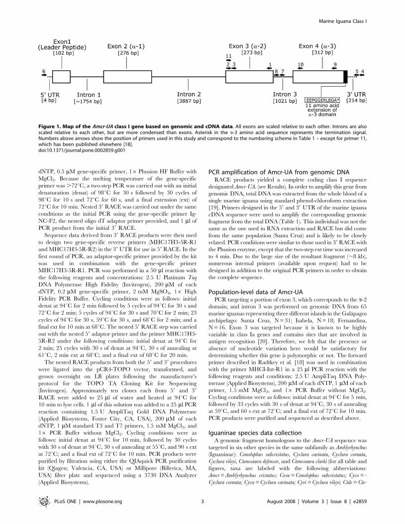
59 and 39 RACE of marine iguana cDNAIn order to design gene-specific primers for 39 RACE, a small
fragment (201 base pairs [bp]) in the class I a-2 domain was
amplified by PCR using a forward primer described in Radtkey et
al. [18] and a degenerate primer (MHCI-R3) designed from an
alignment of a range of vertebrate class I sequences (see Table 1
for primer list and sequences; see Figure 1 for primer locations).
Sequences derived from the small, amplified fragment were used
to design two forward-facing gene-specific primers for use in 39
RACE (IgNC-F1 and IgNC-F2).
For the 39 RACE, nested PCR was performed on marine
iguana first-strand cDNA. The first round of PCR utilized the
oligo dT adaptor-specific primer provided in the GeneRacer kit
and the primer Ig-NC-F1. The PCR was performed with the
following reagents and concentrations: 1U Phusion High Fidelity
DNA Polymerase (Finnzymes, Espoo, Finland), 200 mM of each
Table 1. Gene-specific primers used in this study.
Primer Name Sequence (59 to 39) Target Region
(1) MHCI-R3 ASGTAYYTCBBCAGCCACT Initial exon 3 genomic amplification
(2) IgNC-F1 TGYGAGCTGAGGAAAGATGGGAGCATAG 39 RACE
(3) IgNC-F2 TTACCARTGTGCTTATGATGGGAGGGAC 39 RACE (Nested)
(4) MHC17H3-5R-R1 TTCATGCAGTCCAAAGGCAGCAG 59 RACE; Genomic fragment
(5) MHC17H3-5R-R2 TCTTCTGCCTTGCTTCTGTCAAATATGGAG 59 RACE (Nested)
(6) MHC1-Gr2-F1 ACCGAGAGGGTTGAGCTGGAGAG Genomic fragment
(7) MHCI-Int-R1 TGAGGCTGASAGAGTGTAATCCTCCC Iguaninae exon3/intron3 fragment
(8) MHC1-Int2-F2 AGRTGTRTGTAATATATCATCCAGG Intron3/exon4 fragment (All Iguaninae)
(9) MHC1-R4 CAGRCYGGYRTGMTCCACRCGSCAC Intron3/exon4 fragment (Amcr; Cosu)
(10) MHC1-R6 TCTCCTTGGGGTAGAAGCCRTC Intron3/exon4 fragment (Cyco; Ctde; Cyca; Ctcl; Cyri)
Numbers before primer refer to location in the gene as indicated by arrows in Figure 1.Footnote: See Materials and methods for Iguaninae taxa abbreviations.doi:10.1371/journal.pone.0002859.t001
Marine Iguana Class I
PLoS ONE | www.plosone.org 2 August 2008 | Volume 3 | Issue 8 | e2859
dNTP, 0.5 mM gene-specific primer, 16Phusion HF Buffer with
MgCl2. Because the melting temperature of the gene-specific
primer was .72uC, a two-step PCR was carried out with an initial
denaturation (denat) of 98uC for 30 s followed by 30 cycles of
98uC for 10 s and 72uC for 60 s, and a final extension (ext) of
72uC for 10 min. Nested 39 RACE was carried out under the same
conditions as the initial PCR using the gene-specific primer Ig-
NC-F2, the nested oligo dT adaptor primer provided, and 1 ml of
PCR product from the initial 39 RACE.
Sequence data derived from 39 RACE products were then used
to design two gene-specific reverse primers (MHC17H3-5R-R1
and MHC17H3-5R-R2) in the 39 UTR for use in 59 RACE. In the
first round of PCR, an adaptor-specific primer provided by the kit
was used in combination with the gene-specific primer
MHC17H3-5R-R1. PCR was performed in a 50 ml reaction with
the following reagents and concentrations: 2.5 U Platinum Taq
DNA Polymerase High Fidelity (Invitrogen), 200 mM of each
dNTP, 0.2 mM gene-specific primer, 2 mM MgSO4, 16 High
Fidelity PCR Buffer. Cycling conditions were as follows: initial
denat at 94uC for 2 min followed by 5 cycles of 94uC for 30 s and
72uC for 2 min; 5 cycles of 94uC for 30 s and 70uC for 2 min; 23
cycles of 94uC for 30 s, 59uC for 30 s, and 68uC for 2 min; and a
final ext for 10 min at 68uC. The nested 59 RACE step was carried
out with the nested 59 adaptor primer and the primer MHC17H3-
5R-R2 under the following conditions: initial denat at 94uC for
2 min; 25 cycles with 30 s of denat at 94uC, 30 s of annealing at
61uC, 2 min ext at 68uC; and a final ext of 68uC for 20 min.
The nested RACE products from both the 59 and 39 procedures
were ligated into the pCR4-TOPO vector, transformed, and
grown overnight on LB plates following the manufacturer’s
protocol for the TOPO TA Cloning Kit for Sequencing
(Invitrogen). Approximately ten clones each from 59 and 39
RACE were added to 25 ml of water and heated at 94uC for
10 min to lyse cells. 1 ml of this solution was added to a 25 ml PCR
reaction containing 1.5 U AmpliTaq Gold DNA Polymerase
(Applied Biosystems, Foster City, CA, USA), 200 mM of each
dNTP, 1 mM standard T3 and T7 primers, 1.5 mM MgCl2, and
16 PCR Buffer without MgCl2. Cycling conditions were as
follows: initial denat at 94uC for 10 min, followed by 30 cycles
with 30 s of denat at 94uC, 30 s of annealing at 55uC, and 90 s ext
at 72uC; and a final ext of 72uC for 10 min. PCR products were
purified by filtration using either the QIAquick PCR purification
kit (Qiagen; Valencia, CA, USA) or Millipore (Billerica, MA,
USA) filter plate and sequenced using a 3730 DNA Analyzer
(Applied Biosystems).
PCR amplification of Amcr-UA from genomic DNARACE products yielded a complete coding class I sequence
designated Amcr-UA. (see Results). In order to amplify this gene from
genomic DNA, total DNA was extracted from the whole blood of a
single marine iguana using standard phenol-chloroform extraction
[19]. Primers designed in the 59 and 39 UTR of the marine iguana
cDNA sequence were used to amplify the corresponding genomic
fragment from the total DNA (Table 1). This individual was not the
same as the one used in RNA extraction and RACE but did come
from the same population (Santa Cruz) and is likely to be closely
related. PCR conditions were similar to those used in 39 RACE with
the Phusion enzyme, except that the two-step ext time was increased
to 4 min. Due to the large size of the resultant fragment (,8 kb),
numerous internal primers (available upon request) had to be
designed in addition to the original PCR primers in order to obtain
the complete sequence.
Population-level data of Amcr-UAPCR targeting a portion of exon 3, which corresponds to the a-2
domain, and intron 3 was performed on genomic DNA from 65
marine iguanas representing three different islands in the Galapagos
archipelago: Santa Cruz, N = 31; Isabela, N = 18; Fernandina,
N = 16. Exon 3 was targeted because it is known to be highly
variable in class Ia genes and contains sites that are involved in
antigen recognition [20]. Therefore, we felt that the presence or
absence of nucleotide variation here would be satisfactory for
determining whether this gene is polymorphic or not. The forward
primer described in Radtkey et al. [18] was used in combination
with the primer MHCI-Int-R1 in a 25 ml PCR reaction with the
following reagents and conditions: 2.5 U AmpliTaq DNA Poly-
merase (Applied Biosystems), 200 mM of each dNTP, 1 mM of each
primer, 1.5 mM MgCl2, and 16 PCR Buffer without MgCl2.
Cycling conditions were as follows: initial denat at 94uC for 5 min,
followed by 33 cycles with 30 s of denat at 94uC, 30 s of annealing
at 59uC, and 60 s ext at 72uC; and a final ext of 72uC for 10 min.
PCR products were purified and sequenced as described above.
Iguaninae species data collectionA genomic fragment homologous to the Amcr-UA sequence was
targeted in six other species in the same subfamily as Amblyrhynchus
(Iguaninae): Conolophus subcristatus, Cyclura carinata, Cyclura cornuta,
Cyclura rileyi, Ctenosaura defensor, and Ctenosaura clarki (for all table and
figures, taxa are labeled with the following abbreviations:
Amcr = Amblyrhynchus cristatus; Cosu = Conolophus subcristatus; Cyco = -
Cyclura cornuta; Cyca = Cyclura carinata; Cyri = Cyclura rileyi; Ctde = Cte-
Figure 1. Map of the Amcr-UA class I gene based on genomic and cDNA data. All exons are scaled relative to each other. Introns are alsoscaled relative to each other, but are more condensed than exons. Asterisk in the a-3 amino acid sequence represents the termination signal.Numbers above arrows show the position of primers used in this study and correspond to the numbering scheme in Table 1 – except for primer 11,which has been published elsewhere [18].doi:10.1371/journal.pone.0002859.g001
Marine Iguana Class I
PLoS ONE | www.plosone.org 3 August 2008 | Volume 3 | Issue 8 | e2859
nosaura defensor; Ctcl = Ctenosaura clarki). This fragment corresponds
to the majority of exon 3, the entire intron 3, and a small portion
of exon 4 in the Amcr-UA sequence, and was amplified in two
separate but overlapping fragments. The first fragment, which
spans exon 3 and the first part of intron 3, is identical to the one
amplified in the population sample of Amblyrhynchus, and was
generated using the same PCR protocol described above. The
second fragment covers the second part of intron 3 and the very
beginning of exon 4. The forward primer used to amplify this
fragment was the same for all taxa (MHC1-Int-F2), but a different
reverse primer (MHC1-R4) was used to amplify Amblyrhynchus and
Conolophus specimens than for the other species (MHC1-R6). The
PCR reagent concentrations for both primer pairs were the same
as for the exon 3/intron 3 fragment, but the cycling conditions for
the MHC1-Int-F2/MHC1-R4 primer combination were as
follows: initial denat at 94uC for 5 min, followed by 35 cycles
with 40 s of denat at 94uC, 40 s of annealing at 60uC, and 2 min
30 s ext at 72uC; and a final ext of 72uC for 15 min. The PCR
cycling conditions for the MHC1-Int-F2/MHC1-R6 primer
combination were 94uC for 5 min for initial denat, followed by
35 cycles with 45 s of denat at 94uC, 45 s of annealing at 55uC,
and 60 s ext at 72uC; and a final ext of 72uC for 10 min.
Data analysisSequences from the Amcr-UA genomic fragment were aligned in
the program SEQUENCHER 4.2.2 (Gene Codes Corporation,
Ann Arbor, MI, USA). Comparison of the genomic and cDNA
sequences was used to identify the exon/intron structure of Amcr-
UA.
The program MUSCLE v3.6 [21] was used to produce full-
length amino acid and exon 4 (a-3 domain) nucleotide alignments
for Amcr-UA and the following vertebrate class I sequences from
GenBank: Galapagos marine iguana (A. cristatus), Amcr-UB*01
EU604308, Amcr-UB*02 EU604309, Amcr-UB*03 EU604310,
Amcr-UB*0401 EU604311, Amcr-UB*0402 EU604312; Galapagos
land iguana (C. subcristatus), Cosu-UB*0101 EU604313, Cosu-
UB*0102 EU604314, Cosu-UB*02 EU604315, Cosu-UB*03
EU604316; Green iguana (Iguana iguana), Igig-UB*0101
EU604317, Igig-UB*0102 EU604318, Igig-UB*02 EU604319;
Ameiva lizard, LC5 M81095, LC25 M91097; Northern water
snake (Nerodia sipedon), SC1 M81099; Chinese soft-shelled turtle
(Pelodiscus sinensis), AB185243; Chicken (Gallus gallus), B-F10
X12780; Mallard (Anas platyrhynchos), Du2 AB115242; Great reed
warbler (Acrocephalus arundinaceus), cN3 AJ005503; Axolotl (Ambys-
toma mexicanum), Amme-3 U83137; African clawed frog (Xenopus
laevis), UAA-1f L20733; Mouse (Mus musculus), H2K L36312, H2-D1
NM_010380, H2-Q1 NM_010390, H2-Q10 NM_010391; Walla-
by (Macropus rufogriseus), Maru-UB*01 L04952; Platypus (Ornithor-
hynchus anatinus), Oran2-1 AY112715; Possum (Trichosurus vulpecula),
Trvu-UB AF359509; Rainbow trout (Oncorhynchus mykiss), Onmy-
UBA AF287487; Zebrafish (Danio rerio), Dare-UBA NM131471;
Human, HLA-B7 U29057, HLA-Cw D50852. This set of sequences
is similar to the one used in a study of tuatara class I genes by
Miller et al. [22] as well as the recent study of iguanine class Ia loci
[16], and was chosen for consistency. For the protein data, percent
identity between Amcr-UA and other vertebrate class I genes was
derived from p-distance values calculated separately for each
structural domain in the program MEGA 4.0 [23]. Conserved
vertebrate class I amino acid positions were identified following
Kaufman et al. [24].
The exon 4 alignment provided the basis for Bayesian,
maximum likelihood (ML), and neighbor-joining (NJ) phylogenetic
reconstruction. The program MRMODELTEST v2 [25], which is
based on code from the MODELTEST software [26], was used to
compare the fit of different nucleotide substitution models to the
vertebrate dataset. The general time reversible model (GTR) with
additional parameters for gamma distribution and fraction of
invariable sites provided the best fit to the data according to both
the hierarchical likelihood ratio test and the Akaike information
criterion. This model was implemented in a Bayesian framework
using the program MRBAYES [27] as well as in ML
reconstruction using the TREEFINDER software [28]. For
Bayesian analysis, the default software settings were used, and
the search was run for 2,000,000 generations with the first 10% of
parameter samples discarded as burn-in. In the ML analysis, 1,000
bootstrap replicates were run to assess support for specific nodes.
NJ search and bootstrap analysis were carried out in PAUP using
the GTR substitution model with 500 replicates.
Sequence fragments spanning the majority of exon 3 and all of
intron 3 in the seven iguanine species were aligned in MEGA, and
p-distance values were calculated separately for the exon and
intron.
Sequences generated in this study were deposited in GenBank
under the following accession numbers.: EU839663 (full-length
Amcr-UA cDNA); EU839664 (Amcr-UA genomic fragment);
EU839665-EU839670 (Iguaninae exon 3/intron 3).
Results
Characterization of cDNA sequences from Amblyrhynchuscristatus
Multiple clones were sequenced from PCR products generated
by both 59and 39 RACE of marine iguana cDNA. For 39 RACE,
resulting clones carried two unique class I-like sequences. One of
these became the subjected of another study [16], while the other
was the focus of this paper, and was used to design specific 59
RACE primers. The sequence of this 39 RACE clone was identical
in the area of overlap with the single class I fragment obtained
from 59 RACE. When aligned, these sequences comprised a
1,294 bp fragment which spanned the complete coding sequence
(CDS) as well as the 59 and 39 UTRs of a single class I sequence
type (Figure 1). This gene was labeled Amcr-UA based on
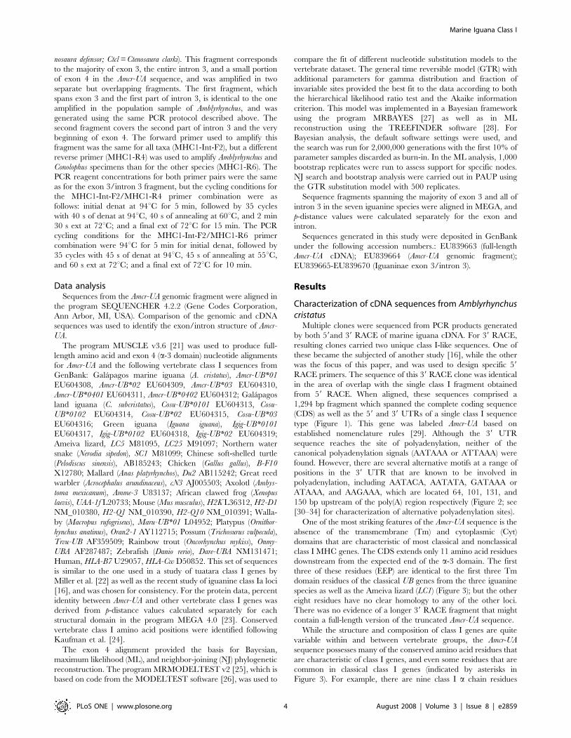
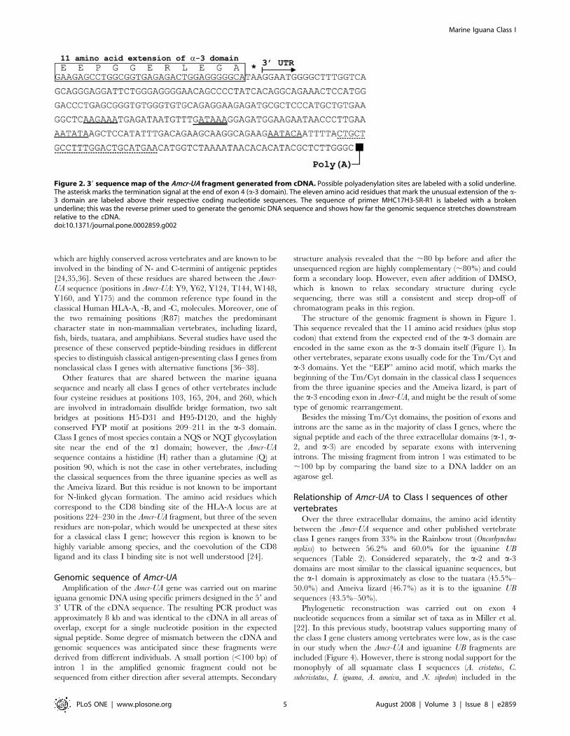
established nomenclature rules [29]. Although the 39 UTR
sequence reaches the site of polyadenylation, neither of the
canonical polyadenylation signals (AATAAA or ATTAAA) were
found. However, there are several alternative motifs at a range of
positions in the 39 UTR that are known to be involved in
polyadenylation, including AATACA, AATATA, GATAAA or
ATAAA, and AAGAAA, which are located 64, 101, 131, and
150 bp upstream of the poly(A) region respectively (Figure 2; see
[30–34] for characterization of alternative polyadenylation sites).
One of the most striking features of the Amcr-UA sequence is the
absence of the transmembrane (Tm) and cytoplasmic (Cyt)
domains that are characteristic of most classical and nonclassical
class I MHC genes. The CDS extends only 11 amino acid residues
downstream from the expected end of the a-3 domain. The first
three of these residues (EEP) are identical to the first three Tm
domain residues of the classical UB genes from the three iguanine
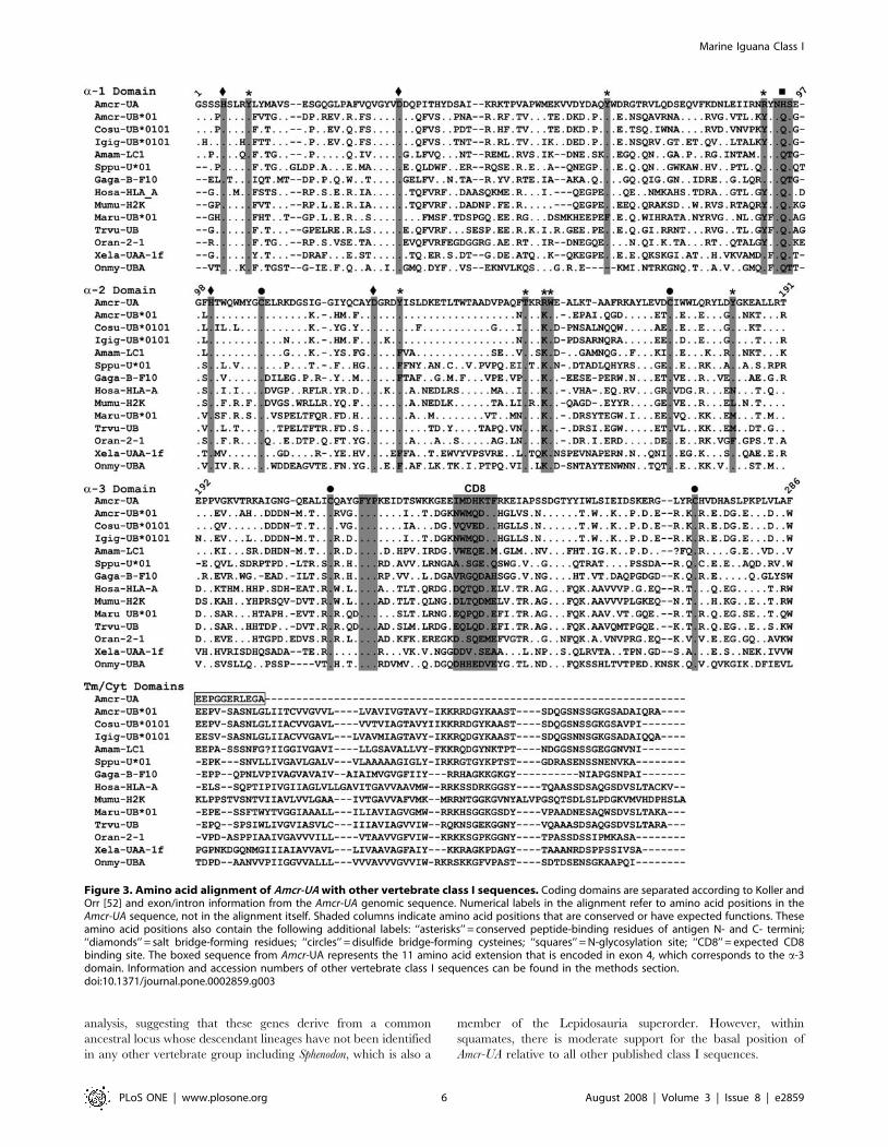
species as well as the Ameiva lizard (LC1) (Figure 3); but the other
eight residues have no clear homology to any of the other loci.
There was no evidence of a longer 39 RACE fragment that might
contain a full-length version of the truncated Amcr-UA sequence.
While the structure and composition of class I genes are quite
variable within and between vertebrate groups, the Amcr-UA
sequence possesses many of the conserved amino acid residues that
are characteristic of class I genes, and even some residues that are
common in classical class I genes (indicated by asterisks in
Figure 3). For example, there are nine class I a chain residues
Marine Iguana Class I
PLoS ONE | www.plosone.org 4 August 2008 | Volume 3 | Issue 8 | e2859
which are highly conserved across vertebrates and are known to be
involved in the binding of N- and C-termini of antigenic peptides
[24,35,36]. Seven of these residues are shared between the Amcr-
UA sequence (positions in Amcr-UA: Y9, Y62, Y124, T144, W148,
Y160, and Y175) and the common reference type found in the
classical Human HLA-A, -B, and -C, molecules. Moreover, one of
the two remaining positions (R87) matches the predominant
character state in non-mammalian vertebrates, including lizard,
fish, birds, tuatara, and amphibians. Several studies have used the
presence of these conserved peptide-binding residues in different
species to distinguish classical antigen-presenting class I genes from
nonclassical class I genes with alternative functions [36–38].
Other features that are shared between the marine iguana
sequence and nearly all class I genes of other vertebrates include
four cysteine residues at positions 103, 165, 204, and 260, which
are involved in intradomain disulfide bridge formation, two salt
bridges at positions H5-D31 and H95-D120, and the highly
conserved FYP motif at positions 209–211 in the a-3 domain.
Class I genes of most species contain a NQS or NQT glycosylation
site near the end of the a1 domain; however, the Amcr-UA
sequence contains a histidine (H) rather than a glutamine (Q) at
position 90, which is not the case in other vertebrates, including
the classical sequences from the three iguanine species as well as
the Ameiva lizard. But this residue is not known to be important
for N-linked glycan formation. The amino acid residues which
correspond to the CD8 binding site of the HLA-A locus are at
positions 224–230 in the Amcr-UA fragment, but three of the seven
residues are non-polar, which would be unexpected at these sites
for a classical class I gene; however this region is known to be
highly variable among species, and the coevolution of the CD8
ligand and its class I binding site is not well understood [24].
Genomic sequence of Amcr-UAAmplification of the Amcr-UA gene was carried out on marine
iguana genomic DNA using specific primers designed in the 59 and
39 UTR of the cDNA sequence. The resulting PCR product was
approximately 8 kb and was identical to the cDNA in all areas of
overlap, except for a single nucleotide position in the expected
signal peptide. Some degree of mismatch between the cDNA and
genomic sequences was anticipated since these fragments were
derived from different individuals. A small portion (,100 bp) of
intron 1 in the amplified genomic fragment could not be
sequenced from either direction after several attempts. Secondary
structure analysis revealed that the ,80 bp before and after the
unsequenced region are highly complementary (,80%) and could
form a secondary loop. However, even after addition of DMSO,
which is known to relax secondary structure during cycle
sequencing, there was still a consistent and steep drop-off of
chromatogram peaks in this region.
The structure of the genomic fragment is shown in Figure 1.
This sequence revealed that the 11 amino acid residues (plus stop
codon) that extend from the expected end of the a-3 domain are
encoded in the same exon as the a-3 domain itself (Figure 1). In
other vertebrates, separate exons usually code for the Tm/Cyt and
a-3 domains. Yet the ‘‘EEP’’ amino acid motif, which marks the
beginning of the Tm/Cyt domain in the classical class I sequences
from the three iguanine species and the Ameiva lizard, is part of
the a-3 encoding exon in Amcr-UA, and might be the result of some
type of genomic rearrangement.
Besides the missing Tm/Cyt domains, the position of exons and
introns are the same as in the majority of class I genes, where the
signal peptide and each of the three extracellular domains (a-1, a-
2, and a-3) are encoded by separate exons with intervening
introns. The missing fragment from intron 1 was estimated to be
,100 bp by comparing the band size to a DNA ladder on an
agarose gel.
Relationship of Amcr-UA to Class I sequences of othervertebrates
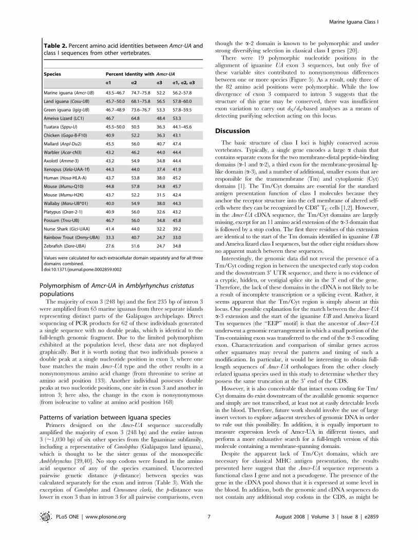
Over the three extracellular domains, the amino acid identity
between the Amcr-UA sequence and other published vertebrate
class I genes ranges from 33% in the Rainbow trout (Oncorhynchus
mykiss) to between 56.2% and 60.0% for the iguanine UB
sequences (Table 2). Considered separately, the a-2 and a-3
domains are most similar to the classical iguanine sequences, but
the a-1 domain is approximately as close to the tuatara (45.5%–
50.0%) and Ameiva lizard (46.7%) as it is to the iguanine UB
sequences (43.5%–50%).
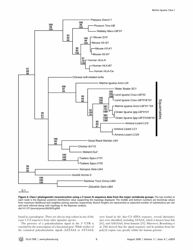
Phylogenetic reconstruction was carried out on exon 4
nucleotide sequences from a similar set of taxa as in Miller et al.
[22]. In this previous study, bootstrap values supporting many of
the class I gene clusters among vertebrates were low, as is the case
in our study when the Amcr-UA and iguanine UB fragments are
included (Figure 4). However, there is strong nodal support for the
monophyly of all squamate class I sequences (A. cristatus, C.
subcristatus, I. iguana, A. ameiva, and N. sipedon) included in the
Figure 2. 39 sequence map of the Amcr-UA fragment generated from cDNA. Possible polyadenylation sites are labeled with a solid underline.The asterisk marks the termination signal at the end of exon 4 (a-3 domain). The eleven amino acid residues that mark the unusual extension of the a-3 domain are labeled above their respective coding nucleotide sequences. The sequence of primer MHC17H3-5R-R1 is labeled with a brokenunderline; this was the reverse primer used to generate the genomic DNA sequence and shows how far the genomic sequence stretches downstreamrelative to the cDNA.doi:10.1371/journal.pone.0002859.g002
Marine Iguana Class I
PLoS ONE | www.plosone.org 5 August 2008 | Volume 3 | Issue 8 | e2859
analysis, suggesting that these genes derive from a common
ancestral locus whose descendant lineages have not been identified
in any other vertebrate group including Sphenodon, which is also a
member of the Lepidosauria superorder. However, within
squamates, there is moderate support for the basal position of
Amcr-UA relative to all other published class I sequences.
Figure 3. Amino acid alignment of Amcr-UA with other vertebrate class I sequences. Coding domains are separated according to Koller andOrr [52] and exon/intron information from the Amcr-UA genomic sequence. Numerical labels in the alignment refer to amino acid positions in theAmcr-UA sequence, not in the alignment itself. Shaded columns indicate amino acid positions that are conserved or have expected functions. Theseamino acid positions also contain the following additional labels: ‘‘asterisks’’ = conserved peptide-binding residues of antigen N- and C- termini;‘‘diamonds’’ = salt bridge-forming residues; ‘‘circles’’ = disulfide bridge-forming cysteines; ‘‘squares’’ = N-glycosylation site; ‘‘CD8’’ = expected CD8binding site. The boxed sequence from Amcr-UA represents the 11 amino acid extension that is encoded in exon 4, which corresponds to the a-3domain. Information and accession numbers of other vertebrate class I sequences can be found in the methods section.doi:10.1371/journal.pone.0002859.g003
Marine Iguana Class I
PLoS ONE | www.plosone.org 6 August 2008 | Volume 3 | Issue 8 | e2859
Polymorphism of Amcr-UA in Amblyrhynchus cristatuspopulations
The majority of exon 3 (248 bp) and the first 235 bp of intron 3
were amplified from 65 marine iguanas from three separate islands
representing distinct parts of the Galapagos archipelago. Direct
sequencing of PCR products for 62 of these individuals generated
a single sequence with no double peaks, which is identical to the
full-length genomic fragment. Due to the limited polymorphism
exhibited at the population level, these data are not displayed
graphically. But it is worth noting that two individuals possess a
double peak at a single nucleotide position in exon 3, where one
base matches the main Amcr-UA type and the other results in a
nonsynonymous amino acid change (from threonine to serine at
amino acid position 133). Another individual possesses double
peaks at two nucleotide positions, one site in exon 3 and another in
intron 3; here also, the change in the exon is nonsynonymous
(from isoleucine to valine at amino acid position 168)
Patterns of variation between Iguana speciesPrimers designed on the Amcr-UA sequence successfully
amplified the majority of exon 3 (248 bp) and the entire intron
3 (,1,030 bp) of six other species from the Iguaninae subfamily,
including a representative of Conolophus (Galapagos land iguana),
which is thought to be the sister genus of the monospecific
Amblyhrynchus [39,40]. No stop codons were found in the amino
acid sequence of any of the species examined. Uncorrected
pairwise genetic distance (p-distance) between species was
calculated separately for the exon and intron (Table 3). With the
exception of Conolophus and Ctenosaura clarki, the p-distance was
lower in exon 3 than in intron 3 for all pairwise comparisons, even
though the a-2 domain is known to be polymorphic and under
strong diversifying selection in classical class I genes [20].
There were 19 polymorphic nucleotide positions in the
alignment of iguanine UA exon 3 sequences, but only five of
these variable sites contributed to nonsynonymous differences
between one or more species (Figure 5). As a result, only three of
the 82 amino acid positions were polymorphic. While the low
divergence of exon 3 compared to intron 3 suggests that the
structure of this gene may be conserved, there was insufficient
exon variation to carry out dN/dS-based analyses as a means of
detecting purifying selection acting on this locus.
Discussion
The basic structure of class I loci is highly conserved across
vertebrates. Typically, a single gene encodes a large a chain that
contains separate exons for the two membrane-distal peptide-binding
domains (a-1 and a-2), a third exon for the membrane-proximal Ig-
like domain (a-3), and a number of additional, smaller exons that are
responsible for the transmembrane (Tm) and cytoplasmic (Cyt)
domains [1]. The Tm/Cyt domains are essential for the standard
antigen presentation function of class I molecules because they
anchor the receptor structure into the cell membrane of altered self-
cells where they can be recognized by CD8+ TC cells [1,2]. However,
in the Amcr-UA cDNA sequence, the Tm/Cyt domains are largely
missing, except for an 11 amino acid extension of the a-3 domain that
is followed by a stop codon. The first three residues of this extension
are identical to the start of the Tm domain identified in iguanine UB
and Ameiva lizard class I sequences, but the other eight residues show
no apparent match between these sequences.
Interestingly, the genomic data did not reveal the presence of a
Tm/Cyt coding region in between the unexpected early stop codon
and the downstream 39 UTR sequence, and there is no evidence of
a cryptic, hidden, or vestigial splice site in the 39 end of the gene.
Therefore, the lack of these domains in the cDNA is not likely to be
a result of incomplete transcription or a splicing event. Rather, it
seems apparent that the Tm/Cyt region is simply absent at this
locus. One possible explanation for the match between the Amcr-UA
a-3 extension and the start of the iguanine UB and Ameiva lizard
Tm sequences (the ‘‘EEP’’ motif) is that the ancestor of Amcr-UA
underwent a genomic rearrangement in which a small portion of the
Tm-containing exon was transferred to the end of the a-3 encoding
exon. Characterization and comparison of similar genes across
other squamates may reveal the pattern and timing of such a
modification. In particular, it would be interesting to obtain full-
length sequences of Amcr-UA orthologues from the other closely
related iguana species used in this study to determine whether they
possess the same truncation at the 39 end of the CDS.
However, it is also conceivable that intact exons coding for Tm/
Cyt domains do exist downstream of the available genomic sequence
and simply are not transcribed, at least not at easily detectable levels
in the blood. Therefore, future work should involve the use of large
insert vectors to explore adjacent stretches of genomic DNA in order
to rule out this possibility. In addition, it is equally important to
measure expression levels of Amcr-UA in different tissues, and
perform a more exhaustive search for a full-length version of this
molecule containing a membrane-spanning domain.
Despite the apparent lack of Tm/Cyt domains, which are
necessary for classical MHC antigen presentation, the results
presented here suggest that the Amcr-UA sequence represents a
functional class I gene and not a pseudogene. The presence of the
gene in the cDNA pool shows that it is expressed at some level in
the blood. In addition, both the genomic and cDNA sequences do
not contain any additional stop codons in the CDS, as might be
Table 2. Percent amino acid identities between Amcr-UA andclass I sequences from other vertebrates.
Species Percent Identity with Amcr-UA
a1 a2 a3 a1, a2, a3
Marine iguana (Amcr-UB) 43.5–46.7 74.7–75.8 52.2 56.2–57.8
Land iguana (Cosu-UB) 45.7–50.0 68.1–75.8 56.5 57.8–60.0
Green iguana (Igig-UB) 46.7–48.9 73.6–76.7 53.3 57.8–59.5
Ameiva Lizard (LC1) 46.7 64.8 48.4 53.3
Tuatara (Sppu-U) 45.5–50.0 50.5 36.3 44.1–45.6
Chicken (Gaga-B-F10) 40.9 52.2 36.3 43.1
Mallard (Anpl-Du2) 45.5 56.0 40.7 47.4
Warbler (Acar-cN3) 43.2 46.2 44.0 44.4
Axolotl (Amme-3) 43.2 54.9 34.8 44.4
Xenopus (Xela-UAA-1f) 44.3 44.0 37.4 41.9
Human (Hosa-HLA-A) 43.7 53.8 38.0 45.2
Mouse (Mumu-Q10) 44.8 57.8 34.8 45.7
Mouse (Mumu-H2K) 43.7 52.2 31.5 42.4
Wallaby (Maru-UB*01) 40.0 54.9 38.0 44.3
Platypus (Oran-2-1) 40.9 56.0 32.6 43.2
Possum (Trvu-UB) 46.7 56.0 34.8 45.8
Nurse Shark (Gici-UAA) 41.4 44.0 32.2 39.2
Rainbow Trout (Onmy-UBA) 33.3 40.7 24.7 33.0
Zebrafish (Dare-UBA) 27.6 51.6 24.7 34.8
Values were calculated for each extracellular domain separately and for all threedomains combined.doi:10.1371/journal.pone.0002859.t002
Marine Iguana Class I
PLoS ONE | www.plosone.org 7 August 2008 | Volume 3 | Issue 8 | e2859
found in a pseudogene. There are also no stop codons in any of the
exon 3 UA sequences from other iguanine species.
The presence of a polyadenylation signal in the 39 UTR is
essential for the transcription of a functional gene. While neither of
the canonical polyadenylation signals (AATAAA or ATTAAA)
were found in the Amcr-UA cDNA sequence, several alternative
sites were identified, including ATAAA, which is known from fish
[41], and AAGAAA, from humans [31]. Moreover, Beaudoing et
al. [30] showed that the signal sequence and its position from the
poly(A) region vary greatly within the human genome.
Figure 4. Class I phylogenetic reconstruction using a-3 (exon 4) sequence data from the major vertebrate groups. The top number ateach node is the Bayesian posterior distribution value supporting the topology displayed. The middle and bottom numbers are bootstrap valuesfrom maximum likelihood and neighbor joining searches respectively. Branch lengths are represented as expected number of substitutions per siteand were inferred along with topology in the Bayesian analysis.doi:10.1371/journal.pone.0002859.g004
Marine Iguana Class I
PLoS ONE | www.plosone.org 8 August 2008 | Volume 3 | Issue 8 | e2859

The higher divergence in intron 3 compared to exon 3 among
the iguanine UA sequences suggests that this gene is conserved and
under some degree of purifying selection, or at least is not evolving
neutrally as would be expected for a nonfunctional pseudogene.
However, other physiological and molecular data, including
expression at the protein level, must be collected in order to
confirm the functionality of Amcr-UA. But for the purpose of
discussion, we will proceed with the assumption that this gene is
functional in order to explore its characteristics and relationship
with other vertebrate class I genes.
Despite the absence of a Tm/Cyt region, the Amcr-UA sequence
shares many of the conserved peptide-binding residues that are
suggestive of classical class I function (Figure 3). However, the
comparison of UA exon 3 sequences between Iguaninae taxa
shows that there is little adaptive divergence in the antigen-binding
region between species, suggesting that this gene is under purifying
selection, rather than balancing-selection, and has a conserved
function. Thus, it is possible that the product of this gene is
involved in antigen binding, but not in the classical sense.
Although Kaufman et al. [24] interpreted the presence of
certain conserved residues at peptide-binding sites as preliminary
evidence of classical function, many of these amino acid character
states are maintained in nonclassical genes such as the human
HLA-E, -F, and -G loci, as well as the mouse H2-M3 gene. None
of these loci differ from the classical mammalian type by more
than three out of the nine residues [36]. Thus, it is not at all
unprecedented that Amcr-UA displays several nonclassical features
while still showing signs of peptide-binding ability.
Since a lack of polymorphism is an important criterion for
describing nonclassical loci, sequence data was collected from exon
3 and intron 3 of Amcr-UA for three marine iguana populations.
The near absence of exon 3 variability in these samples seems to
support the pattern of purifying selection indicating a conserved
function. However, the simultaneous lack of diversity in the
adjacent intron indicates either strong linkage to the conserved
exon or perhaps that an insufficient amount of time has passed for
a large number of substitutions to accumulate in the intron. The
latter pattern would not be surprising since mitochondrial DNA
Table 3. Uncorrected pairwise distances (p-distance) between Amcr-UA and similar sequences from other iguanine species.
1 2 3 4 5 6 7
1 Ctde 0.04435 0.03226 0.02823 0.03226 0.04435 0.04839
2 Ctcl 0.04546 0.03629 0.03226 0.03629 0.06048 0.05645
3 Cyco 0.04789 0.05722 0.00403 0.00806 0.03226 0.02823
4 Cyca 0.04693 0.05822 0.01975 0.00403 0.02823 0.02419
5 Cyri 0.04791 0.05430 0.02073 0.00988 0.03226 0.02823
6 Cosu 0.04671 0.05694 0.03846 0.03552 0.03850 0.01210
7 Amcr-UA 0.05366 0.06577 0.04924 0.04633 0.05120 0.02650
Distances were calculated separately for exon 3 (above the diagonal) and intron 3 (below the diagonal). See methods for Iguaninae taxa abbreviations. Pairwise valuesbetween Ctcl and Cosu are italicized because they deviate from the otherwise typical pattern of higher divergence in intron 3 versus exon 3.doi:10.1371/journal.pone.0002859.t003
Figure 5. (a) Nucleotide and (b) amino-acid alignments of iguanine exon 3 (a-2 domain) sequences. The lower-case letters and shadedboxes represent specific codon positions that show variation in amino acid residues between species. Species abbreviations are described in theMaterials and methods section.doi:10.1371/journal.pone.0002859.g005
Marine Iguana Class I
PLoS ONE | www.plosone.org 9 August 2008 | Volume 3 | Issue 8 | e2859
evidence suggests that marine iguana populations are not
genetically diverse and may have recently expanded in the
Galapagos archipelago [42]. Therefore, the unclear evolutionary
history of this species makes it difficult to attribute the dearth of
polymorphism in Amcr-UA to its nonclassical function.
While rare, several truncated class I molecules are known to lack
Tm/Cyt regions in humans and mice. For example, the mouse
Q10 gene contains a 13 bp deletion in the exon encoding the Tm
domain, causing a frame shift and the introduction of a premature
termination signal downstream in the same exon. In addition, the
remaining transcribed portion of the Tm domain has numerous
polar amino acid residues that would likely prevent its insertion
into the cell membrane. Therefore, it seems clear that the Q10
molecule is not involved in classical antigen presentation. It also
possesses several other nonclassical features, including a lack of
polymorphism and expression that is almost exclusive to mouse
liver cells [43–45]. Several studies have shown that Q10 is likely
secreted and can bind a wide array of non-self peptides in a similar
manner to classical molecules, but its function is still not well
understood [44,46]. While there is no phylogenetic similarity
between Amcr-UA and the mouse Q10, the characteristics of the
latter gene demonstrate that truncated, soluble, secreted class I
molecules can exist which lack genetic variation and maintain
protein binding capabilities.
Numerous other classical and nonclassical class I molecules are
known to exist in soluble form for secretion. For example, the
human HLA-G locus possesses all of the coding features of a
membrane-bound class I receptor, but is sometimes subjected to
alternative transcription where the Tm/Cyt domains are deleted.
This molecule is expressed specifically in placental tissue and is
secreted during pregnancy [47]. The role of this gene is also not
well understood, but is thought to be involved in inducing
apoptosis in activated maternal CD8+ T cells [48]. Classical
human class I genes (HLA-A, -B, and -C) are also known to exist in
soluble forms and to play a role in cell death of activated T cells
[49]. Nevertheless, a unique feature of the Amcr-UA sequence
which distinguishes it from the Q10 and HLA-G loci, as well as
other soluble class I molecules, is that there does not appear be any
sign of a Tm/Cyt coding sequence in the genomic DNA,
regardless of whether it is transcribed or not; but again, additional
support for this conclusion must come from further collection of
sequence data in the 39 region of the gene.
Some truncated class Ib molecules, such as those in the mouse
Qa-2 family, are not necessarily secreted, but are rather linked to
the cell membrane through a glycosylphosphatidylinositol (GPI)
anchor that is added to the carboxyl terminus of the protein during
posttranslational modification [50,51]. Therefore, the apparent
lack of a membrane-spanning domain in Amcr-UA doesn’t rule out
its expression on the cell surface or its involvement in the primary
T cell response.
Overall, the Amcr-UA CDS showed the highest similarity with
iguanine UB and Ameiva lizard sequences. In addition, phyloge-
netic reconstruction supported the monophyly of all available
squamate class I sequences. The basal position of Amcr-UA relative
to all other squamate class I sequences suggests that this gene
diverged very early in the evolution of this reptilian order. The
most recent common ancestor of iguanines and the two groups
represented by the Ameiva lizard (Family: Teiidae) and northern
water snake (Suborder: Serpentes) is estimated to have existed
between 179–206 million years ago [14], providing a minimum
time for the split of Amcr-UA from class Ia genes in squamates.
Outside of the squamate grouping, the tree topology is not well
supported, suggesting that class I sequences are highly divergent
among vertebrate groups.
In summary, the Amcr-UA sequence possesses several charac-
teristics of a functional, non-classical class I gene with a conserved
protein structure. Additional work must be conducted to
understand whether it is expressed at the level of the protein
and what its position is in the genome relative to published
classical loci [16]. While Amcr-UA is most closely related to the
other published squamate sequences, it does not cluster with class
Ia sequences from the same species, suggesting that it has long
been on a separate evolutionary trajectory. Further characteriza-
tion of UA-like sequences from other squamates will reveal
whether this gene is part of a lineage that has maintained a non-
classical function over the course of squamate evolution.
Acknowledgments
Maria Moreno and Stephen Dellaporta (Yale University) provided helpful
advice for carrying out RACE protocol. Larry Buckley (Rochester Institute
of Technology) contributed genomic DNA from four of the iguanine
species used in this study (C. defensor, C. clarki, C. rileyi, and C. carinata). The
C. cornuta specimen was provided by the Peabody Museum of Natural
History with the help of Greg Watkins-Colwell. The Charles Darwin
Research Station (Santa Cruz, Galapagos, Ecuador) and the Galapagos
National Park provided logistical support for the collection and export of
marine iguana blood and RNA. Cruz Marquez helped with marine iguana
blood sampling. Special thanks to Ylenia Chiari and Hilary Miller for
helpful comments on the manuscript.
Author Contributions
Conceived and designed the experiments: SG. Performed the experiments:
SG. Analyzed the data: SG LdP. Contributed reagents/materials/analysis
tools: AC. Wrote the paper: SG.
References
1. Klein J (1986) Natural history of the major histocompatibility complex. New
York: Wiley. xv, 775 p.
2. Ploegh H, Watts C (1998) Antigen recognition. Curr Opin Immunol 10: 57–58.
3. Bjorkman PJ, Parham P (1990) Structure, function, and diversity of class I majorhistocompatibility complex molecules. Annu Rev Biochem 59: 253–288.
4. Potts WK, Wakeland EK (1990) Evolution of diversity at the majorhistocompatibility complex. Trends Ecol Evol 5: 181–187.
5. Brigl M, Brenner MB (2004) CD1: Antigen presentation and T cell function.
Annual Rev Immunol 22: 817–890.
6. Braud VM, Allan DS, McMichael AJ (1999) Functions of nonclassical MHC andnon-MHC-encoded class I molecules. Curr Opin Immunol 11: 100–108.
7. Hansen TH, Huang S, Arnold PL, Fremont DH (2007) Patterns of nonclassical
MHC antigen presentation. Nat Immunol 8: 563–568.
8. Hughes AL, Nei M (1989) Evolution of the major histocompatibility complex:
independent origin of nonclassical class I genes in different groups of mammals.
Mol Biol Evol 6: 559–579.
9. Nei M, Gu X, Sitnikova T (1997) Evolution by the birth-and-death process in
multigene families of the vertebrate immune system. Proc Natl Acad Sci USA
94: 7799–7806.
10. Rada C, Lorenzi R, Powis SJ, Vandenbogaerde J, Parham P, et al. (1990)
Concerted evolution of class I genes in the major histocompatibility complex of
murine rodents. Proc Natl Acad Sci USA 87: 2167–2171.
11. Hess CM, Edwards SV (2002) The evolution of the major histocompatibility
complex in birds. Bioscience 52: 423–431.
12. Belov K, Deakin JE, Papenfuss AT, Baker ML, Melman SD, et al. (2006)
Reconstructing an ancestral mammalian immune supercomplex from a
marsupial major histocompatibility complex. PLoS Biol 4: 317–328.
13. Rest JS, Ast JC, Austin CC, Waddell PJ, Tibbetts EA, et al. (2003) Molecular
systematics of primary reptilian lineages and the tuatara mitochondrial genome.
Mol Phylogenet Evol 29: 289–297.
14. Vidal N, Hedges SB (2005) The phylogeny of squamate reptiles (lizards, snakes,
and amphisbaenians) inferred from nine nuclear protein-coding genes.
C R Biologies 328: 1000–1008.
15. Vitt LJ, Pianka ER, Cooper WE, Schwenk K (2003) History and the global
ecology of squamate reptiles. Am Nat 162: 44–60.
16. Glaberman S, Caccone A (2008) Species-specific evolution of class I MHC genes
in iguanas (Order: Squamata; Subfamily: Iguaninae). Immunogenetics 60:
371–382.
Marine Iguana Class I
PLoS ONE | www.plosone.org 10 August 2008 | Volume 3 | Issue 8 | e2859
17. Flajnik M (2004) Comparative genomics of the MHC. Tissue Antigens 64:
328–328.18. Radtkey RR, Becker B, Miller RD, Riblet R, Case TJ (1996) Variation and
evolution of class I Mhc in sexual and parthenogenetic geckos. Proc R Soc
London Ser B 263: 1023–1032.19. Sambrook J, Russell DW (2001) Molecular cloning : a laboratory manual. Cold
Spring Harbor, N.Y.: Cold Spring Harbor Laboratory Press.20. Hughes AL, Yeager M (1998) Natural selection at major histocompatibility
complex loci of vertebrates. Annu Rev Genet 32: 415–435.
21. Edgar RC (2004) MUSCLE: multiple sequence alignment with high accuracyand high throughput. Nucleic Acids Res 32: 1792–1797.
22. Miller HC, Belov K, Daugherty CH (2006) Proceedings of the SMBE tri-national young investigators’ workshop 2005. MHC class I genes in the tuatara
(Sphenodon spp.): evolution of the MHC in an ancient reptilian order. Mol BiolEvol 23: 949–956.
23. Tamura K, Dudley J, Nei M, Kumar S (2007) MEGA4: Molecular evolutionary
genetics analysis (MEGA) software version 4.0. Mol Biol Evol 24: 1596–1599.24. Kaufman J, Salomonsen J, Flajnik M (1994) Evolutionary conservation of MHC
class I and class II molecules–different yet the same. Semin Immunol 6:411–424.
25. Nylander JAA (2004) MrModeltest v2. Evolutionary Biology Centre, Uppsala
University: Program distributed by the author.26. Posada D, Crandall KA (1998) MODELTEST: testing the model of DNA
substitution. Bioinformatics 14: 817–818.27. Ronquist F, Huelsenbeck JP (2003) MrBayes 3: Bayesian phylogenetic inference
under mixed models. Bioinformatics 19: 1572–1574.28. Jobb G, von Haeseler A, Strimmer K (2004) TREEFINDER: a powerful
graphical analysis environment for molecular phylogenetics. BMC Evol Biol 4:
18.29. Klein J, Bontrop RE, Dawkins RL, Erlich HA, Gyllensten UB, et al. (1990)
Nomenclature for the major histocompatibility complexes of different species - aproposal. Immunogenetics 31: 217–219.
30. Beaudoing E, Freier S, Wyatt JR, Claverie JM, Gautheret D (2000) Patterns of
variant polyadenylation signal usage in human genes. Genome Res 10:1001–1010.
31. Plant MH, Laneuville O (1999) Characterization of a novel transcript ofprostaglandin endoperoxide H synthase 1 with a tissue-specific profile of
expression. Biochem J 344: 677–685.32. Pauws E, van Kampen AH, van de Graaf SA, de Vijlder JJ, Ris-Stalpers C
(2001) Heterogeneity in polyadenylation cleavage sites in mammalian mRNA
sequences: implications for SAGE analysis. Nucleic Acids Res 29: 1690–1694.33. Tian B, Hu J, Zhang HB, Lutz CS (2005) A large-scale analysis of mRNA
polyadenylation of human and mouse genes. Nucleic Acids Res 33: 201–212.34. Adhikary G, Gupta S, Sil P, Saad Y, Sen S (2005) Characterization and
functional significance of myotrophin: A gene with multiple transcripts. Gene
353: 31–40.35. Madden DR (1995) The three-dimensional structure of peptide-MHC
complexes. Annu Rev Immunol 13: 587–622.
36. Shum BP, Rajalingam R, Magor KE, Azumi K, Carr WH, et al. (1999) A
divergent non-classical class I gene conserved in salmonids. Immunogenetics 49:479–490.
37. Grimholt U, Hordvik I, Fosse VM, Olsaker I, Endresen C, et al. (1993)
Molecular cloning of major histocompatibility complex class I cDNAs fromAtlantic salmon (Salmo salar). Immunogenetics 37: 469–473.
38. Timon M, Elgar G, Habu S, Okumura K, Beverley PC (1998) Molecularcloning of major histocompatibility complex class I cDNAs from the pufferfish
Fugu rubripes. Immunogenetics 47: 170–173.
39. Rassmann K (1997) Evolutionary age of the Galapagos iguanas predates the ageof the present Galapagos Islands. Mol Phylogenet and Evol 7: 158–172.
40. Wiens JJ, Hollingsworth BD (2000) War of the iguanas: Conflicting molecularand morphological phylogenies and long-branch attraction in iguanid lizards.
Syst Biol 49: 143–159.41. Kwon JY, Prat F, Randall C, Tyler CR (2001) Molecular characterization of
putative yolk processing enzymes and their expression during oogenesis and
embryogenesis in rainbow trout (Oncorhynchus mykiss). Biol Reprod 65:1701–1709.
42. Rassmann K, Tautz D, Trillmich F, Gliddon C (1997) The microevolution ofthe Galapagos marine iguana Amblyrhynchus cristatus assessed by nuclear and
mitochondrial genetic analyses. Mol Ecol 6: 437–452.
43. Cosman D, Kress M, Khoury G, Jay G (1982) Tissue-specific expression of anunusual H-2 (class I)-related gene. Proc Natl Acad Sci USA 79: 4947–4951.
44. Kress M, Cosman D, Khoury G, Jay G (1983) Secretion of a transplantation-related antigen. Cell 34: 189–196.
45. Mellor AL, Weiss EH, Kress M, Jay G, Flavell RA (1984) A nonpolymorphicclass I gene in the murine major histocompatibility complex. Cell 36: 139–144.
46. Zappacosta F, Tabaczewski P, Parker KC, Coligan JE, Stroynowski I (2000) The
murine liver-specific nonclassical MHC class I molecule Q10 binds a classicalpeptide repertoire. J Immun 164: 1906–1915.
47. Rebmann V, Pfeiffer K, Passler M, Ferrone S, Maier S, et al. (1999) Detection ofsoluble HLA-G molecules in plasma and amniotic fluid. Tissue Antigens 53:
14–22.
48. Fournel S, Aguerre-Girr M, Huc X, Lenfant F, Alam A, et al. (2000) SolubleHLA-G1 triggers CD95/CD95 ligand-mediated apoptosis in activated CD8(+)
cells by interacting with CD8. J Immunol 164: 6100–6104.49. Contini P, Ghio M, Poggi A, Filaci G, Indiveri F, et al. (2003) Soluble HLA-A,-
B,-C and -G molecules induce apoptosis in T and NKCD8(+) cells and inhibitcytotoxic T cell activity through CD8 ligation. Eur J Immunol 33: 125–134.
50. Stiernberg J, Low MG, Flaherty L, Kincade PW (1987) Removal of Lymphocyte
Surface Molecules with Phosphatidylinositol-Specific Phospholipase-C - Effectson Mitogen Responses and Evidence That Thb and Certain Qa-Antigens Are
Membrane-Anchored Via Phosphatidylinositol. J Immunol 138: 3877–3884.51. Stroynowski I, Tabaczewski P (1996) Multiple products of class Ib Qa-2 genes
which ones are functional? Res Immunol 147: 290–301.
52. Koller BH, Orr HT (1985) Cloning and complete sequence of an HLA-A2 gene:analysis of two HLA-A alleles at the nucleotide level. J Immunol 134:
2727–2733.
Marine Iguana Class I
PLoS ONE | www.plosone.org 11 August 2008 | Volume 3 | Issue 8 | e2859


