Embed Size (px)
Citation preview

1
CO
NG
ENIT
AL
AN
OM
ALI
ES A
ND
PE
DIA
TRIC
PLA
STIC
SU
RGER
Y
INTRODUCTIONOrthognathic surgery is the term used to describe surgi-cal movement of the tooth-bearing segments of the jaws. Patients with dentofacial deformities that cannot be treated with orthodontics alone are candidates for jaw surgery. These malocclusions are typically associated with skeletal discrep-ancies secondary to congenital anomalies, congenital syn-dromes, nonsyndromic dentofacial abnormalities, or trauma. Approximately 2.5% of the American population has occlusal discrepancies severe enough to require surgical correction.1 Regardless of the etiology, patient examination and treatment planning principles are the same. The goal of orthognathic surgery is to establish ideal dental occlusion with the jaws in a position that optimizes facial aesthetics.
BASIC DENTAL TERMINOLOGYApertognathia—Vertical separation of the maxillary and
mandibular anterior teeth, frequently described as ante-rior open bite.
Articulator—A mechanical device that simulates the tem-poromandibular joints and jaws to which the maxillary and mandibular casts may be attached.
Buccal—Pertaining to or adjacent to the cheek.Cast—A plaster replica of the teeth and surrounding
tissues.Centric occlusion—The relation of opposing occlusal sur-
faces that provides the maximum planned contact or intercuspation.
Centric relation—The relationship of the mandible to the maxilla when the condyles are in their most posterosu-perior unstrained positions in the glenoid fossa.
Cephalometric radiograph—A radiograph of the head made with precise reproducible relationships between X-ray source, subject, and film.
Class I occlusion—The mesiobuccal cusp of the first per-manent maxillary molar occludes in the buccal groove of the permanent mandibular first molar.
Class II malocclusion—The mesiobuccal cusp of the first permanent maxillary molar occludes mesial to the buc-cal groove of the permanent mandibular first molar.
Class III malocclusion—The mesiobuccal cusp of the first permanent maxillary molar occludes distal to the buccal groove of the permanent mandibular first molar.
Distal—Away from the median sagittal plane of the face and following the curvature of the arch
Labial—Pertaining to the lip, especially in reference to the surface of a tooth.
Lingual—Pertaining to the tongue, especially in reference to the surface of a tooth.
Mesial—Situated toward the midline of the dental arch.Overjet—Degree to which the upper incisors extend beyond
the lower incisors labially. Normal overjet is 2 mm.Overlap—The amount maxillary incisal edges vertically
overlap the mandibular incisor edges with the mouth closed. Normal incisal overlap is 2 mm.
Palatal—Pertaining to the palate, especially in reference to the surface of a tooth.
Proclination—Anterior angulation of the anterior teeth.Prognathic—A forward position of the mandible in rela-
tion to the cranial base.
AQ1 Retroclination—Posterior angulation of the anterior teeth.Retrognathic—The condition of a mandible that is poste-
riorly positioned in relation to the cranial base.
ESTABLISHING THE DIAGNOSISHistory
An accurate understanding of the chief complaint is essential in developing a treatment plan. Systemic diseases, such as juvenile rheumatoid arthritis, diabetes, and scleroderma, may affect treatment planning. With jaw asymmetries, a history of hyperplasia or hypoplasia secondary to a syndrome, traumatic accident, or neoplasm will affect treatment considerations. Each patient should be questioned regarding symptoms of temporomandibular joint (TMJ) disease or myofascial pain. Motivation and realistic expectations are extremely important to ensure an optimal outcome. It is important for the patient to have a clear understanding of the procedure, the recovery, and the anticipated result prior to surgery. Orthognathic sur-gery is a major undertaking, frequently lasting 2 to 3 years, and the patient must be appropriately motivated to undergo all necessary perioperative orthodontic treatment and reha-bilitation to achieve the desired result. When it comes to jaw surgery, the patient is a particularly important member of the orthognathic surgery team.
Physical ExaminationGood oral hygiene and periodontal health is necessary prior to orthodontics and surgery. The occlusal classification is deter-mined, and the degree of incisor overlap and overjet is quanti-fied. Often, the clinician can get a sense of the degree of dental compensation (see below) on physical examination, but radio-graphic analysis is necessary to quantify dental compensation. The surgeon should assess the transverse dimension of the maxilla. If a crossbite is present, models are obtained to dif-ferentiate a relative crossbite from a true crossbite. An abso-lute crossbite is due to maxillary constriction and will require either orthopedic (orthodontic appliance) or surgical expan-sion to correct (see below). If the mandibular third molars are present, they will require extraction 6 months before perform-ing a sagittal split osteotomy. Any missing teeth and periapical pathology are noted. Any signs or symptoms of TMJ dysfunc-tion are evaluated before proceeding with surgery.
The frontal facial evaluation consists of assessing the verti-cal facial thirds: trichion to glabella, glabella to subnasale, and subnasale to menton. Each of these facial thirds should be about equal. If the lower two-thirds of the face are short, they can be increased by inferiorly repositioning the maxilla, which will result in increasing the distance from the glabella to pogonion. In contrast, a long lower face may benefit from a maxillary impaction, which would have the opposite effect. The most important factor in determining the ideal vertical height of the maxilla is the amount of incisor showing while the patient’s lips are in repose. A man should show at least 2 to 3 mm while as much as 4 to 5 mm is considered attractive in a woman. If the patient shows the correct degree of incisor in repose but shows excessive gingiva in full smile, the max-illa must not be impacted. It is more important to show the correct degree of incisor in repose than it is to be concerned about excess gingiva in full smile. The surgeon certainly would not want to bury the incisors in repose just to reduce
CHAPTER 25 ■ ORTHOGNATHIC SURGERYSTEPHEN B. BAKER
09559_ch25_p001-014.indd 109559_ch25_p001-014.indd 1 12/11/12 1:56 PM12/11/12 1:56 PM

2 Part III: Congenital Anomalies and Pediatric Plastic Surgery
the degree of gingiva in a full smile. If lip incompetence or mentalis strain is present, it is usually an indicator of vertical maxillary excess.
The sagittal facial fifths are also evaluated. The inter-canthal distance should be about the same as the distance between the medial and lateral canthus of each eye. If the lateral fifths are deficient, augmentation can be performed with bone grafts or implants. The inferior orbital rims, malar eminence, and piriform areas are evaluated for the degree of projection. If these regions appear deficient, maxil-lary advancement is indicated; if they are excessively promi-nent, the maxilla may benefit from posterior repositioning. The alar base width should also be assessed prior to surgery since orthognathic surgery may alter the width (Chapter 48). Asymmetries of the maxilla and mandible should be docu-mented on physical examination, and the degree of deviation from the facial midline noted. The soft tissue envelope of the upper face is evaluated for descent of the malar fat pads, the severity of the nasolabial creases, and folds. Skeletal move-ments of the maxilla will affect these areas. It is important for the surgeon to realize that skeletal expansion (anterior or inferior repositioning of the jaws) will attenuate the creases and folds, while skeletal contraction (posterior or superior movements of the jaws) will accentuate these problems.2 The surgeon certainly does not want to give the patient a prematurely aged appearance as a result of the procedure. However, as will be discussed later, the surgeon can take advantage of skeletal expansion to reduce some of these soft tissue creases giving the patient a youthful appearance and reducing the signs of aging. In evaluating the chin, the clini-cian assesses the labiomental angle (Chapter 51). An acute angle indicates a short or prominent chin, while effacement of the crease suggests excessive vertical length or insufficient anterior projection.
The profile evaluation focuses on the projection of the fore-head, the malar region, the upper and lower jaws, the nose, the chin, and the neck. The assessment can be verified through cephalometric analysis, but an experienced clinician can usu-ally determine whether the deformity is due to the maxilla, mandible, or both. Frequently, the optimal aesthetic result is achieved by ignoring the cephalometric norms and treatment planning the jaw movements based on the patient’s facial form as determined by the surgeon’s physical examination. If the jaws appear to be aligned but the chin projection is either pronounced or deficient, a genioplasty may be all that is nec-essary (Chapter 51). The proper position of the nose relates to the upper lip, which is supported by the maxillary incisors, and the chin. Because both of these structures may be altered by orthognathic surgery, it is important to predict how the dimensions of the nose will fit into the new facial proportions (Chapter 48). A rhinoplasty may be necessary to maintain proper facial proportions. The soft tissues of the neck should also be assessed. The patient with submental laxity will not benefit aesthetically from posterior positioning of the mandi-ble. Mandibular advancement, however, will improve the lax-ity and the cervicomental angle. In a patient with prominent submental fat in whom mandibular advancement is contrain-dicated, suction-assisted lipectomy is helpful in removing the adipose deposits (Chapter 65). Redundant skin will require direct excision.
The maxillary and mandibular dental midlines are assessed to determine if they are congruent with each other and the true facial midline. Any deviations are noted and quantified. The presence of mandibular third molars is noted because if a sagittal split osteotomy is planned, it is recommended that they be removed 6 months before surgery so the screw fixa-tion has adequate bone. The presence and degree of dental compensation is also recorded. The term “dental compensa-tion” is used to describe the tendency of teeth to tilt in a direc-tion that minimizes the dental malocclusion. For example, in a patient with an overbite (class II malocclusion), the upper
AQ2
incisors will retrocline while the lower incisors will procline. The opposite will occur in a patient who has dental compen-sation for an underbite (class III malocclusion). Thus, dental compensation will mask the true degree of skeletal discrep-ancy. Precise analysis of the dental compensation is done on the lateral cephalometric radiograph.
If the patient desires surgical correction of the defor-mity, presurgical orthodontics will decompensate the occlu-sion, thereby reversing the compensation that has occurred. Decompensation has the effect of exaggerating the malocclu-sion but allows the surgeon to maximize skeletal movements. If the patient is ambivalent or not interested in surgery, mild cases of malocclusion may be treated by further dental com-pensation. Compensation will camouflage the deformity and restore proper overjet and overlap. The dental movements for decompensation and compensation are in opposite direc-tions, making a decision regarding surgery and communica-tion between surgeon, orthodontist, and patient extremely important.3
Cephalometric Analysis and Dental ModelsA lateral cephalometric radiograph is performed under repro-ducible conditions so that serial images can be compared. This film is usually taken in the orthodontist’s office utilizing a cephalostat, an apparatus specifically designed for this pur-pose, and head frame to maintain consistent head position. It is important to be certain that the surgeon can visualize bony as well as soft tissue features on the image to facilitate trac-ing all the landmarks. A piece of transparent acetate tracing paper is secured with tape over the radiograph and the follow-ing landmarks are traced: sella, inferior orbital rim, nasion, frontal bone, nasal bones, maxilla, maxillary first molar and central incisor, external auditory meatus, the condylar head and mandible, and the mandibular first molar and incisor. The soft tissue of the forehead, nose, lips, and chin is also traced. Once the normal structures are traced, several planes and angles are determined (Figure 25.1).
The maxillary plane is represented by a line drawn between the anterior nasal spine (ANS) and posterior nasal spine (PNS). The occlusal plane is drawn between the occlusal surfaces of the teeth. The mandibular plane is drawn between menton and gonion, and the Frankfort horizontal plane is delineated between the superior portion of the external auditory meatus (porion) and the inferior orbital rim (orbitale). Analysis of these planes aids in establishing an accurate diagnosis. A steep mandibular plane is usually associated with a class II maloc-clusion, anterior open bite, and a short mandible. A shallow mandibular plane is associated with a deep bite, class III mal-occlusion, and a long mandible.
The SNA and SNB are the two most important angles in determining the positions of the maxilla and mandible rela-tive to each other as well as to the cranial base. These angles are determined by drawing lines from sella to nasion to point A or point B, respectively. By forming an angle with the sella and nasion, this position is related to the cranial base. The maxilla will be considered first. Point “A” represents the anteroposterior position of the maxilla. If the SNA angle is excessive, the maxilla exhibits abnormal anterior position relative to the cranium. If SNA is less than normal, the max-illa is posteriorly positioned relative to the cranial base. The same principle applies to the mandible; the only difference is that point “B” is used to relate mandibular position to the cranial base. The importance of the cranial base as a reference is that it allows the clinician to determine if one or both jaws contribute to the deformity. For example, a patient’s class III malocclusion (underbite) could develop from several differ-ent etiologies: a retrognathic maxilla and normal mandible, a normal maxilla and a prognathic mandible, a retrogna-thic mandible and a more severely retrognathic maxilla, or a prognathic maxilla and a more severely prognathic mandible.
09559_ch25_p001-014.indd 209559_ch25_p001-014.indd 2 12/11/12 1:56 PM12/11/12 1:56 PM

Chapter 25: Orthognathic Surgery 3
CO
NG
ENIT
AL
AN
OM
ALI
ES A
ND
PE
DIA
TRIC
PLA
STIC
SU
RGER
Y
surrounding the facial skeleton is the most crucial factor in determining the aesthetic success of orthognathic procedures, and the jaws should be positioned so that they provide opti-mal soft tissue support.
Historically, skeletal movements that expanded the soft tissue of the face were less stable, so posterior and superior movements were preferred. Although more stable, these movements resulted in contraction of the facial skeleton with the associated redundancy of soft tissue characteristic of pre-mature aging. Since the introduction of rigid fixation, oste-otomies that result in skeletal expansion have been achieved with predictability. It is important that the surgeon develop a treatment plan that will expand or maintain the preoperative volume of the face.2 If a superior or posterior (contraction) movement of one of the jaws is planned, an attempt should be made to neutralize the skeletal contraction with an advance-ment or inferior movement of the other jaw or the chin. It is important to avoid a net contraction of the facial skeleton as this may result in a prematurely aged appearance.
As skeletal expansion is increased, soft tissue laxity is reduced and facial creases are softened. These effects increase the definition of the face, creating a more attractive appear-ance. It has been shown that skeletal expansion is aesthetically pleasing even if facial disproportion is necessary to achieve the expansion.4 Fashion models often exhibit slight degrees of facial disproportion and are considered beautiful. The aesthetic benefits the patient receives by expanding the facial envelope frequently justify the small degree of disproportion necessary to achieve them. Even in young adolescent patients that do not show signs of aging, one must not ignore these principles. A successful surgeon will incorporate these principles into the treatment plan of every patient so that as the patient ages, the signs of aging will be minimized and a youthful appearance will be maintained as long as possible (Figure 25.2).
An example demonstrating these principles is an adult woman who presents for surgery with a slightly prognathic mandible, submental laxity, jowl descent, and deep nasola-bial creases. If a treatment plan were developed based solely on cephalometric values, a mandibular setback would be the appropriate procedure. This procedure would do nothing for the upper face and would accentuate the submental laxity and jowl descent. In contrast, if one were to accept the slight prognathism and advance the maxilla to a class I relationship, the nasolabial crease and jowl descent would be improved, and the submental laxity would not be made worse. Thus, moving the normal maxilla into a class I occlusion with a prognathic mandible establishes a normal occlusion and achieves excellent aesthetics despite being slightly dispropor-tionate (Figure 25.3).
A class I occlusion can be achieved with the jaws in a vari-ety of different positions. The goal in treatment planning is to use the data from the patient’s examination to predict the location of the jaws that will optimize the soft tissue features of the face. Reducing the emphasis on normal values and increasing the awareness of the soft tissue effects of skeletal movements, a skeletal “disproportion” may be chosen inten-tionally, leading to a more favorable result. However, it is the discretion of the surgeon to incorporate these principles into a treatment plan pleasing to the patient and understood by the patient.4
BASIC APPROACHES TO COMMONLY ENCOUNTERED
PROBLEMSThe basic treatment principles in commonly encountered den-tofacial deformities will be presented. The surgeon must rec-ognize there are multiple solutions to a single problem; this is where the proper application of the previously discussed prin-ciples is crucial to achieving the best aesthetic result.
All of these conditions yield a class III malocclusion, yet each requires a different treatment approach. The surgeon can delineate the true etiology of the deformity by relating the maxilla and mandible independently to a stable reference point, the cranial base.
Plaster dental casts are obtained during the treatment plan-ning process. Casts are useful because they allow the surgeon to evaluate the occlusion when the casts are articulated into the proper position. Analysis of the new occlusion gives the clinician an idea of how intensive the presurgical orthodon-tic treatment plan will be. Casts also allow the clinician to distinguish between absolute and relative transverse maxillary deficiencies. Absolute transverse maxillary deficiency presents as a posterior crossbite with the jaws in a class I relationship. A relative maxillary transverse deficiency is commonly seen in a patient with a class III malocclusion. As the maxilla is advanced or the mandible retruded, the crossbite is elimi-nated. Articulation of the casts into a class I occlusion allows the surgeon to easily distinguish between relative and absolute maxillary constriction.
DEVELOPING A TREATMENT PLANOnce the data are obtained, the surgeon can determine which abnormalities the patient exhibits and the extent to which these features deviate from the norm. However, the treat-ment plan is the application of this data to give the patient the best aesthetic result while establishing a class I occlusion. The goal is not to “treat the numbers” in an attempt to “normal-ize” every patient. The appearance of the soft tissue envelope
FIGURE 25.1. Standard lateral cephalometric tracing with land-marks. S, sella—the center of the pituitary fossa; N, nasion—the most anterior point of the nasofrontal suture in the midsagittal plane; Ar, articulare—the intersection of basisphenoid and the posterior border of the condyle; A, subspinale—the deepest point of the anterior bor-der of the maxilla between the anterior nasal spine and prosthion, usually around the level of the apex of the maxillary central incisor; Pog, pogonion—the most anterior point of the contour of the chin; B, supramentale—the deepest point between infradentale and pogo-nion at the level of apices of the mandibular incisors; ANS, anterior nasal spine—the most anterior point of the nasal floor; Me, menton—the lowest point of the contour of the mandibular symphysis; GN, gnathion—the midpoint between Pog and ME created by bisecting the facial line (N-Pog) and the mandibular plane (Go-Me); PNS, posterior nasal spine—the most posterior point on the contour of the hard palate; MP, mandibular plane—a plane constructed from menton (Me) and gonion (Go); NF, nasal floor—a plane constructed from PNS to ANS; Go, gonion—located by bisecting the posterior ramal plane and the mandibular plane angles: It is the midpoint of the curvature connecting the ramus and the mandibular body. (From Wolfe AA, Berkowitz S. Plastic Surgery of the Facial Skeleton. Boston, MA: Little, Brown and Co.; 1989:57, with permission.)
09559_ch25_p001-014.indd 309559_ch25_p001-014.indd 3 12/11/12 1:56 PM12/11/12 1:56 PM

4 Part III: Congenital Anomalies and Pediatric Plastic Surgery
A B
C
D
E
F
FIGURE 25.2. Sagittal split osteotomy and alloplastic mid-face augmentation. This patient exhibited a left unilateral crossbite, a prominent chin, and malar deficiency (she is miss-ing a central incisor). She did not desire two-jaw surgery and her malocclusion was treated with bilateral sagittal split oste-otomy. Her midface deficiency was corrected with prosthetic malar and piriform augmentation. A. Frontal examination preoperation. B. Profile preoperation. C. Occlusion preopera-tion. D. Frontal postoperation. Note the attenuated nasolabial creases from malar implants. E. Profile postoperation. Noted improved malar projection achieved and alloplastic augmenta-tion. F. Postoperation occlusion.
09559_ch25_p001-014.indd 409559_ch25_p001-014.indd 4 12/11/12 1:56 PM12/11/12 1:56 PM
Chapter 25: Orthognathic Surgery 5
CO
NG
ENIT
AL
AN
OM
ALI
ES A
ND
PE
DIA
TRIC
PLA
STIC
SU
RGER
Y
different treatments to achieve aesthetic goals. If some poste-rior positioning of the mandible is necessary, one may advance the maxilla to counteract the skeletal contraction produced from the mandible. Additionally, the patient may benefit from a genioplasty that can counteract any skeletal contraction that occurs from a mandibular setback. As in the class II patient, a minor malocclusion with minimal dental compensation may be corrected with orthodontic treatment alone. In contrast, a minor malocclusion with dental compensation may become a significant malocclusion after dental decompensation, making the patient a good surgical candidate.
Maxillary ConstrictionMany patients can exhibit a maxilla that is narrow. Maxillary constriction may occur as an isolated finding or as one of multiple abnormalities. Up to about 15 years of age, the orthodontist can expand the maxilla nonsurgically with a palatal expander. If orthopedic expansion cannot be done, a surgically assisted rapid palatal expansion (SARPE) can be performed.5 If the maxilla requires movement in other dimen-sions, a two-piece Le Fort I osteotomy can be performed to widen the maxilla and simultaneously move the maxilla in the desired direction (Figure 25.5).
Skeletal Class II OcclusionA skeletal class II malocclusion is almost always due to man-dibular retrognathia and is almost always best treated by man-dibular advancement (Figure 25.4). The mandible is small, and forward positioning is an expansile movement that enhances the facial form. If the maxilla is also slightly deficient or in a normal position, one may consider a bimaxillary advancement to further enhance facial soft tissue definition. If the malocclu-sion is minimal and there is little pre-existing dental compen-sation, one may choose to have the orthodontist intentionally compensate the dentition to correct the occlusion and avoid surgery. In contrast, if the malocclusion appears minimal but there is dental compensation, from previous orthodontic treat-ment, the skeletal discrepancy will be more significant after the orthodontist decompensates the dentition, allowing surgi-cal correction.
Skeletal Class III MalocclusionA prominent lower jaw may be treated by advancing the max-illa, posteriorly positioning the mandible, or by combining these procedures. It is important to consider the contributions of the mandible and the chin separately as each may require
A B C
D E F
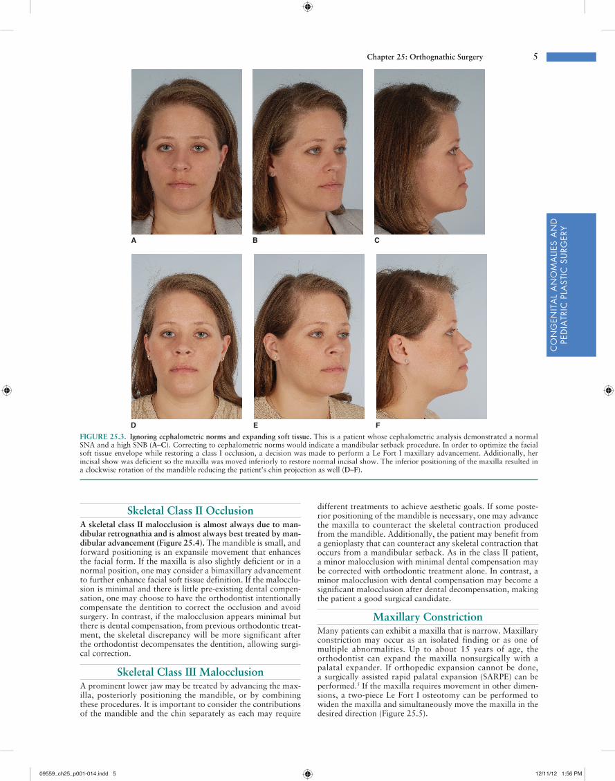
FIGURE 25.3. Ignoring cephalometric norms and expanding soft tissue. This is a patient whose cephalometric analysis demonstrated a normal SNA and a high SNB (A–C). Correcting to cephalometric norms would indicate a mandibular setback procedure. In order to optimize the facial soft tissue envelope while restoring a class I occlusion, a decision was made to perform a Le Fort I maxillary advancement. Additionally, her incisal show was deficient so the maxilla was moved inferiorly to restore normal incisal show. The inferior positioning of the maxilla resulted in a clockwise rotation of the mandible reducing the patient’s chin projection as well (D–F).
09559_ch25_p001-014.indd 509559_ch25_p001-014.indd 5 12/11/12 1:56 PM12/11/12 1:56 PM

6 Part III: Congenital Anomalies and Pediatric Plastic Surgery
Short Lower FaceA short lower face is marked by insufficient incisor show and/or a short distance between subnasale and pogonion. Treatment is aimed at establishing a proper degree of incisor show. The facial skeleton should be expanded to the degree that provides optimal soft tissue aesthetics. As the maxilla is inferiorly posi-tioned, clockwise mandibular rotation will occur, leading to posterior positioning of the chin. The surgeon needs to assess the new chin position on the cephalometric tracing to deter-mine if an advancement genioplasty is now necessary to coun-ter the effects of mandibular clockwise rotation.
Facial AsymmetryFacial asymmetry may occur from asymmetric growth of the mandible due to hemifacial microsomia (Chapter 24), pediatric trauma, radiation, neoplasms, or other etiologies. Correcting facial asymmetry typically requires a maxillary osteotomy to level the occlusal plane of the maxilla and center the maxillary dental midline so that it is congruent with the facial midline. A mandibular osteotomy is then performed to bring the mandibular dentition into a class I occlusion with the maxillary dentition. The chin is assessed to determine if it will be in the midline after the mandibular osteotomy. If
ApertognathiaAn anterior open bite is caused by a premature contact of the posterior molars. The recommended treatment is a posterior impaction of the maxilla with or without coun-terclockwise rotation of the occlusal plane. (By convention, clockwise and counterclockwise movements are defined by the direction the jaw on jaws move when viewed from the right lateral view.) By reducing the vertical height of the posterior maxilla, the mandible can come into occlusion with the remaining mandibular teeth. Posterior maxillary impaction does not necessarily result in incisor impaction; the posterior maxilla is simply rotated upward using the incisal tip as the axis of rotation. Therefore, incisor show should not be affected. If a change in incisor show is also desired, the posterior impaction is done and then the whole maxilla can be inferiorly positioned or impacted to its new position (Figure 25.6).
Vertical Maxillary ExcessVertical maxillary excess is typically associated with lip incom-petence, mentalis strain (chin dimpling), and an excessive degree of gingival show. This condition is also known as long face syndrome. The treatment approach is to impact the max-illa to achieve the proper incisal show with the lips in repose. Impaction results in skeletal contraction, so the surgeon must consider anterior positioning of the jaws could be tolerated to neutralize the associated adverse soft tissue effects. As the maxilla is impacted, the mandible rotates counterclockwise to maintain occlusion. This rotation results in anterior positioning of the chin and is called mandibular autorotation (Figures 25.3 and 25.7). The opposite occurs if the maxilla is moved in an inferior direction. In this case, the chin point rotates in a clock-wise direction, which results in posterior positioning of the chin point. It is important to note these effects on the cephalometric tracing during treatment planning because a genioplasty may be required to reestablish proper chin position.
AQ3
FIGURE 25.4. Technique for sagittal split osteotomy. The sagittal splitting procedure is the most versatile ramus osteotomy, as it can be used for both mandibular advancement (A) and mandibular setback (B). The procedure is performed through an intraoral approach and requires special instrumentation to be performed with ease. Three or four screws are placed percutaneously to effect rigid fixation. It is vital that the mandibular condyles be properly seated at the end of any mandibular osteotomy. If they are not, when intermaxillary fixation is removed they go back into the glenoid fossae with a resultant anterior open bite.
A B
C
D
E D
FIGURE 25.5. Versatility of the Le Fort I osteotomy. The Le Fort I osteotomy sections the maxilla transversely at a level between the roots of the teeth (note that the root of the cuspid may extend as high as the piriform rim) and the infraorbital foramen. After the lower portion of the maxilla is mobilized, movement in a number of direc-tions is possible. A. Lengthening of the maxilla with an interpositional bone graft (note the use of miniplates for fixation). B. Shortening of the maxilla after resection of bone above the osteotomy line. C. Advancement of the maxilla. D. Segmentalization of the maxilla after downfracture and extraction of teeth. E. Setback of maxilla.
09559_ch25_p001-014.indd 609559_ch25_p001-014.indd 6 12/11/12 1:56 PM12/11/12 1:56 PM

Chapter 25: Orthognathic Surgery 7
CO
NG
ENIT
AL
AN
OM
ALI
ES A
ND
PE
DIA
TRIC
PLA
STIC
SU
RGER
Y
tissue profile. They also allow the surgeon to determine the distances the bones will be moved. Different tracing methods are used for isolated maxillary, isolated mandibular, or two-jaw surgeries. All cephalometric tracings begin by securing a clear piece of acetate tracing paper over the cephalometric radiograph. The anatomy and aforementioned cephalometric points are then marked on the tracing paper (Figure 25.6A).
Mandibular Surgery. When isolated mandibular surgery is indicated, a second piece of acetate is used to trace only the mandible and the soft tissue of the chin and lower lip. This second tracing of only the mandible is then placed into the
the chin is asymmetric or deficient, a genioplasty is performed to move it into a normal position based on the new maxil-lomandibular relationship. Occasionally, a mandibular angle bone graft and/or autologous fat grafting may be necessary to optimize skeletal and soft tissue facial symmetry (Figure 25.8)
PREPARING FOR SURGERYPre-Op Cephalometric Tracing
Cephalometric tracings give the surgeon an idea of how skel-etal movements will affect one another as well as the soft
A B
C
FIGURE 25.6. A. This patient exhibits an anterior open bite, but has good incisal show on repose. The normal landmarks on the cepha-logram are recorded. B. The maxilla is then traced and is put into ideal position. Because incisal show is good, the anterior maxilla is not lowered and the occlusion is achieved with a posterior impaction. C. The mandible is then rotated into its new position using another piece of acetate that includes the mandible. The condyle is the center of rotation when moving the mandible. Because the mandible cannot be advanced without creating a malocclusion, the chin is moved for-ward to advance pogonion.
AQ4
09559_ch25_p001-014.indd 709559_ch25_p001-014.indd 7 12/11/12 1:56 PM12/11/12 1:56 PM

8 Part III: Congenital Anomalies and Pediatric Plastic Surgery
FIGURE 25.7. Autorotation of mandible. A counterclockwise mandibu-lar rotation will produce an increase in the anterior position of the chin. Clockwise rotation will posteriorly position the chin. The final anticipated projection of the chin needs to be cephalometrically assessed to determine if it will require surgery to move it into a more aesthetic position.
desired occlusion against the maxilla on the first tracing. The differences in distance between the point B and the first molar cusps when comparing the new and original positions of the mandible denote the distance that the mandible will be anteri-orly or posteriorly positioned. The new soft tissue profile can also be estimated from the tracing.
Maxillary Surgery. In isolated maxillary surgery, a second piece of acetate with only the maxilla, first molar, upper inci-sors, and lip is placed into the desired position. The incisal edges of the incisors should be placed at least 3 to 4 mm below the lower margin of the upper lip. The anterior positioning of the maxilla will be determined by placing the maxillary teeth against the mandibular teeth in a class I occlusion (Figure 25.6B).
An anterior open bite will require posterior maxillary impaction until the mandibular teeth contact the maxillary teeth in normal occlusion. The appropriate degree of poste-rior maxillary impaction is verified by using a tracing of the mandible and rotating it counterclockwise with the condyle remaining in the original position (Figure 25.6C). The proper position of the maxilla is determined by establishing a class I occlusion with the incisal edges 3 to 4 mm below the upper lip. The movement required in each portion of the maxilla can be determined by measuring the distance between landmarks of the new maxillary position and the original position. Useful landmarks for the maxilla are the incisal edge, the mesial cusp of the first molar, ANS, and PNS. The anteroposterior posi-tion of the maxilla will be determined by soft tissue aesthetics
A B C
D E F
FIGURE 25.8. Facial asymmetry after subcondylar fracture as a child. Preoperative images demonstrate facial asymmetry (A), microgenia (B), and an occlusal cant (C). To restore facial symmetry, the patient had a leveling Le Fort I osteotomy, bilateral sagittal split osteotomy, center-ing and advancement genioplasty, left mandibular angle bone graft, and autogenous fat grafting to the left cheek (D–F). These procedures restore facial symmetry and correct the occlusal cant.
09559_ch25_p001-014.indd 809559_ch25_p001-014.indd 8 12/11/12 1:56 PM12/11/12 1:56 PM

Chapter 25: Orthognathic Surgery 9
CO
NG
ENIT
AL
AN
OM
ALI
ES A
ND
PE
DIA
TRIC
PLA
STIC
SU
RGER
Y
maxilla is in its new position, the cast is secured with sticky wax or plaster to the mounting ring (Figure 25.11). Now it can be placed on the articulator. At this point, the surgeon has a mounting of the postoperative maxilla related to the pre-operative position of the mandible. An acrylic splint is made at this point. This splint is called the intermediate splint and will be used in the operating room to index the new position of the maxilla to the preoperative position of the mandible. A second mounting with the casts in the occlusion desired by the orthodontist is used to make a final splint that will represent the new position of the mandible to the maxilla. This is fabri-cated in a manner similar to the splint for isolated mandibular surgery (Figure 25.12). If the occlusion is good, intercuspal position can be used to position the mandible without a splint.
COMPUTER-ASSISTED SURGICAL SIMULATION
Recent advances in 3D computer-aided topography as well as computer-aided design/manufacturing (CAD/CAM) have led to an emergence of several computer-assisted surgical simulation (CASS) software programs with a wide range of applications. Recently, CASS technology has been adapted for orthognathic surgery to assist in cephalometric analysis, vir-tual model surgery, and splint fabrication.
CASS incorporates 3D computed tomography (CT) imag-ing and virtual surgery with CAD/CAM splint fabrication for patients undergoing orthognathic surgery. The patients have their treatment plan developed by clinical examination
and the occlusion. If soft tissue aesthetics require moving the maxilla into a position that precludes a class I occlusion, the mandible will have to be moved with the maxilla.
Two-Jaw Surgery. In two-jaw surgery, the maxilla is posi-tioned on the cephalometric radiograph as above, and then the new position of the mandible is placed using separate pieces of acetate overlying the original cephalometric tracing. By measuring the difference between the new landmarks and the original landmarks, the surgeon determines the distance he/she is moving the maxilla and/or mandible in each dimen-sion.6 Predictive computer imaging may be useful at this point. The patient should be informed that the computer-generated image is not a guaranteed result, only a guide to the surgeon’s goals. It is useful to make sure the patient and the surgeon are in agreement about the goals prior to surgery. The next step is to simulate these movements on plaster casts of the jaws.
Traditional Model SurgeryModel surgery starts by obtaining accurate casts of the patient’s occlusion. If the surgeon does not have a dental laboratory, the orthodontist is usually helpful in obtaining the casts. It is important to obtain accurate casts as the success of the techni-cal portion of orthognathic surgery correlates directly to the accuracy of the model surgery and splint fabrication.
Isolated Mandibular Surgery. It should be noted that if isolated mandibular surgery is being performed, the casts can be hand articulated into the desired occlusion. The Galetti articulator is a useful tool that allows securing of the casts with a screw mount. A universal joint allows the casts to be set to the desired relationship. Surgical splints can then be made from the articulator. If the maximum intercuspal position is the desired postoperative occlusion, a splint is not necessary. The surgeon can osteotomize the mandible and secure it into its new position using the maximum intercuspal position as the guide to the new position. The surgeon should always ver-ify the desired postoperative occlusion with the orthodontist prior to surgery.
Isolated Maxillary and Two-Jaw Surgery. A face bow is a device that is used to accurately relate the maxillary model to the cranium on an articulator. If a maxillary osteotomy is being performed, one set of models should be mounted on an articulator using the face bow (Figure 25.9). Two other sets of models are used in treatment planning. Next, an Erickson model block is used to measure the current position of the maxillary central incisors, cuspids, and the mesiobuccal cusp of the first molar.7 The face bow–mounted maxillary cast is placed on the model block. The maxillary model is then mea-sured to the tenth of a millimeter vertically, anteroposteriorly, and end-on (Figure 25.10). By having numerical records in three dimensions, the surgeon can reproduce the maxillary cast’s exact location as well as determine a new location. Reference lines are circumferentially inscribed every 5 mm around the maxillary cast mounting. The distances the max-illa will move in an anteroposterior, lateral, and vertical direc-tion have been determined from the previous cephalometric examination and the resulting proposed changes in the maxil-lary position. These numbers are added or subtracted from the current values in the x-, y-, and z-axes measured on the model block to determine the new three-dimensional (3D) position of the maxillary cast. The occlusal portion of the maxillary cast is removed from its base using a saw. As much plaster is removed from the cast as is necessary to accommodate the new position of the maxilla. Soft wax is inserted in the gap between the base of the cast and the occlusal portion. The wax allows slight manipulations of the occlusal portion of the cast while providing some stability. The tooth cusps are measured in three dimensions until they match the desired postoperative position of the maxilla. Once the model block verifies that the
FIGURE 25.9. A semiadjustable articulator with mounted casts enables assessment of occlusal cants, malocclusion, and skeletal relationships.
09559_ch25_p001-014.indd 909559_ch25_p001-014.indd 9 12/11/12 1:56 PM12/11/12 1:56 PM

10 Part III: Congenital Anomalies and Pediatric Plastic Surgery
surgical splints. The splints, as well as 3D treatment planning images, are mailed to the surgeon overnight.8
OVERVIEW OF PRE-OP TREATMENTThe patient has multiple visits at the surgeon’s office and has undergone a thorough discussion of the surgical options. Good oral health has been achieved. Any TMJ problems have been addressed and appropriate clearance has been obtained. The patient has had preoperative orthodontics to level, align, and decompensate the occlusion. Based on the physical and radiographic examinations, a treatment plan has been developed that will achieve a class I occlusion and optimize the facial soft tissue aesthetics. The preoperative examination will determine the distances the jaws will have to move to achieve the desired result, and model surgery or CASS has been performed to develop surgical splints that will intraoperatively position the jaws into the position deter-mined by the cephalometric tracing. If a segmental osteot-omy is planned, the orthodontist will have diverged the root apices on either side of the proposed osteotomy to minimize the chance of damage to tooth roots. The surgeon verifies that the splints fit and that good surgical lugs have been applied to the arch wire. Soldered lugs work the best. If the lugs break in surgery, the proper application of the splint can be compromised, making an ideal result much more difficult to obtain. In two-jaw surgery, autodonation of packed red blood cells is useful.
SURGICAL PROCEDURESPertinent Anatomy
The important structures in the mandible that may be injured in the mandibular osteotomy are the inferior alveolar nerve, its terminal branch called the mental nerve, and the teeth apices. The third division of the trigeminal nerve enters the mandibular foramen to become the inferior alveolar nerve. It runs within the mandible below the tooth roots and exits at the level of the first to the second premolar through the men-tal foramen to become the mental nerve. The nerve is most medial to the outer cortex in the region of external oblique ridge. This is where the vertical portion of the BSSO is made because it affords the largest margin of error.
The maxilla is associated with the descending palatine artery, the infraorbital nerve, the tooth roots, and the inter-nal maxillary artery. The internal maxillary artery runs about 25 mm above the pterygomaxillary junction, and the descend-ing palatal artery descends in the posteromedial maxillary sinus. The infraorbital nerve exits the infraorbital foramen below the infraorbital rim along the midpupillary line. The maxillary tooth roots extend within the maxilla in a superior direction. The canine has the longest root and is usually visible through the maxillary cortical bone.
General PrinciplesSeveral principles have broad application to jaw surgery. Blood loss can be substantial in maxillofacial surgery. Standard
AQ5and evaluation of dental casts received from the orthodontist. After the plan is established, the patient has a bite registra-tion performed that is attached to a bite jig with radiopaque markers. Before the CT is performed, natural head position is recorded by entering Euler angles that record the pitch, yaw, and roll, and thus, give a reference for natural head position once the 3D CT has been obtained. The CT scan and a set of dental casts marked with the desired occlusal relationship are then sent to the CASS company (Figure 25.13).
Once the materials have been received, the surgeon com-municates with the CASS company by phone, and both parties access the 3D CT scan using gotomeeting.com. The surgeon discusses the planned osteotomies with the consultant and can visualize the skeletal movements on the computer while the consultant moves the jaws into their desired position. Once the surgeon has confirmed the planned osteotomies, the new data can be used to fabricate the intermediate and final
FIGURE 25.10. The preoperative maxillary position is recorded in three dimensions shown with the Erickson model block. The land-marks are the incisal edge, the canine cusp, and the mesiobuccal first molar cusp. The movements in an anteroposterior, transverse, and vertical direction were determined by the physical and radiographic examinations. These measurements are added to or subtracted from the preoperative measurements.
FIGURE 25.11. The cast is divided with enough plaster removed to allow the desired manipulation. The maxilla is moved into its new position and secured with sticky wax. This cast is then reattached to the articulator and the intermediate splint is made. The final splint can be made on a Galetti articulator as shown in Figure 27.10.
09559_ch25_p001-014.indd 1009559_ch25_p001-014.indd 10 12/11/12 1:56 PM12/11/12 1:56 PM

Chapter 25: Orthognathic Surgery 11
CO
NG
ENIT
AL
AN
OM
ALI
ES A
ND
PE
DIA
TRIC
PLA
STIC
SU
RGER
YFIGURE 25.12. A: The Galetti articulator enables articulation of models for mandibular surgery. A. This photograph demonstrates the preop-erative occlusion. B. This photograph shows the desired occlusion by the orthodontist. The anterior occlusion has no open bite and the midlines are congruent. The posterior open bite is easily closed with postoperative orthodontics; in this case, a splint was used to preserve the posterior open bite intra- and postoperatively. The splint minimizes the risk of the posterior bite closing, which would compromise the anterior occlusion.
A B
FIGURE 25.13. Computer-assisted surgical simulation is a new tech-nique that obviates the need for traditional model surgery and splint fabrication. A bite registration is taken (A) and then attached to a gyroscope connected to a computer (B) that allows the computer to record the head position in three dimensions (C). The CT scan and the 3D bite registration data are sent to the CASS company. The 3D CT data are then used to simulate the postoperative result and record the anatomic landmarks of the new jaw position (D). The surgical plan is determined by the surgeon on a conference call with the CASS team. Once the postoperative result is confirmed, the splints are fabricated with CAD/CAM technology and mailed to the surgeon the next day.
ANS left
A left
U6R U6L
UK9R UK9L
L6R L6L
BISU1
ISL1
ANS right
A right
A B
C
D
09559_ch25_p001-014.indd 1109559_ch25_p001-014.indd 11 12/11/12 1:56 PM12/11/12 1:56 PM

12 Part III: Congenital Anomalies and Pediatric Plastic Surgery
just posterior to the last maxillary molar and drops through the maxillary tuberosity. The cut should be made at least 5 mm above the apices of the teeth. This distance is determined from the panorex radiograph. If cuts are complete, the max-illa should be able to be downfractured with manual pressure. An alternative is to use the Rowe disimpaction forceps. These fit into the piriform aperture and on the palate to provide increased leverage for the downfracture. Pressure should be applied in a slow, steady controlled fashion, not in a series of quick random movements. If the maxilla is not mobilized with relative ease, the cuts are likely not complete and should be reevaluated. Once the downfracture is complete, a bone hook can be used by the assistant to hold maxilla down while any remaining bony interferences are removed. The descending palatine arteries will be seen near the posteromedial maxillary sinus. These can be clipped prophylactically without compro-mising the blood supply to the maxilla. The splint is then used to place the maxilla in its proper position. Intermaxillary fixa-tion is then applied with 26 gauge wires around the surgical lugs. The amount the maxilla will be impacted or elongated has been determined in the treatment plan. This distance is added or subtracted from the medial canthal–incisor dis-tance to determine the new vertical position of the maxilla. Four 2 mm plates, usually L-shaped, can be used to secure the maxilla. The MMF is released and occlusion verified prior to closure. If the alar base is wide, an alar cinch can be per-formed to normalize the width. This is performed by placing a suture around the transverse nasalis muscle and setting the alar base to the desired width. Lip shortening may also result from closure. A V-Y closure at the central incisor can help alleviate this effect.
In patients that require increased cheek projection, a high Le Fort I osteotomy can be performed. This differs in that the transverse osteotomy is made as high as the infraorbital nerve will allow. If further cheek projection is necessary, bone grafts can be added. In the case of inferior or anterior posi-tioning, gaps between the segments greater than 3 mm should be grafted with either autogenous bone, cadaveric bone, or block hydroxyapatite. Finally, if simultaneous expansion of the maxilla is necessary, the maxilla can be split into two or more pieces to allow simultaneous expansion (see Figures 25.5 and 25.13).
Surgically Assisted Rapid Palatal ExpansionCorrection of transverse maxillary constriction can be made in adolescence with nonsurgical orthodontic appliances. As the sutures begin to close during late adolescence, relapse increases. A multi-piece Le Fort osteotomy can be performed to provide simultaneous maxillary expansion, but the degree of relapse is high. In the young adult, the preferred procedure is the SARPE. The orthodontist places a palatal expander prior to the procedure. A Le Fort I osteotomy is performed to completely mobilize the maxilla from the upper face. A small osteotome is used to make a thin cut between the roots of the central incisors. The midline split is completed to the PNS. Separation is verified by activating the device. The maxilla is widened until the gingiva blanches and is then relaxed sev-eral turns to avoid ischemia.5 The SARPE offers the best sta-bility for maxillary expansion in the young adult and older patient.10,11 Transverse deficiencies of the mandible can be cor-rected with a similar technique, which will be discussed in the chapter on distraction osteogenesis (Chapter 24).
Bilateral Sagittal Split OsteotomyThe endotracheal tube placement and epinephrine injection are the same as for the Le Fort osteotomy. The cut is made with electrocautery about 1 cm from the lateral aspect of the molars and extends from midramus to the region of the second molar. If insufficient tissue is left on the dental side of the inci-sion, closure is more difficult. A periosteal elevator is used to
AQ6
techniques of head elevation, hypotensive anesthesia, blood donation, and administration of erythropoietin are useful adjuncts. Before the incisions are made, an antimicrobial rinse is helpful to minimize the intraoral bacterial count. A topical steroid is applied to the lips to reduce pain and swelling asso-ciated with prolonged retraction. Intravenous steroids may also be useful to reduce postoperative edema.
The occlusion desired postoperatively may not be the same as maximum intercuspal position. The splint is useful in main-taining the occlusion in the desired location when it does not correspond to the maximal intercuspal position. It is easy for the orthodontist to close a posterior open bite, but it is very difficult to close an anterior open bite with orthodontics alone. At the end of the case, it is important to have the ante-rior teeth and the canines in a class I relationship without an open bite.
After surgery, it is useful to use guiding elastics to control the bite. Class II elastics are placed in a vector to correct a class II relationship (maxillary lug is anterior to the mandib-ular lug). Class III elastics are applied to correct a class III discrepancy. With rigid fixation, the elastics will not correct malpositioned jaws. They serve only to help the patient adapt to their new occlusion. Minor malocclusions can be corrected with postoperative orthodontics or occlusal equilibration.
Certain skeletal movements are inherently more stable than others. Stable movements include mandibular advancement and superior positioning of the maxilla. Movements with intermediate stability include maxillary impaction combined with mandibular advancement, maxillary advancement com-bined with mandibular setback, and correction of mandibular asymmetry. The unstable movements include posterior posi-tioning of the mandible and inferior positioning of the max-illa. The least stable movement is transverse expansion of the maxilla. Long-term relapse with rigid fixation has not been demonstrated to be clearly superior to nonrigid fixation in single-jaw surgery. However, in two-jaw surgery, rigid fixa-tion results in less relapse.9 The judgment of the surgeon will dictate the extent to which the facial skeleton can be expanded without resulting in unacceptable relapse.
Le Fort I OsteotomyThe first step in any facial osteotomy is satisfactorily securing the nasal endotracheal tube; the author’s preference is a nasal RAE tube. The vertical position of the maxilla is recorded by measuring the distance between the medial canthus and the incisal edge of the maxillary incisor. The maxillary vestibule is injected with epinephrine prior to patient preparation. An incision is made with electrocautery 5 mm above the mucogin-gival junction from first molar to first molar. A periosteal ele-vator is then used to expose the maxilla around the piriform rim and infraorbital nerve. Rapid exposure of the maxilla is achieved by elevating in the “hot lanes” of facial dissection: just lateral to the piriform aperture and along the zygomatic buttress. These areas allow rapid subperiosteal elevation but avoid the infraorbital nerve. Once the “hot lanes” have been elevated, the nerve is easily identified and the remainder of the maxilla can be safely exposed. Obwegeser toe-in retractors are held by the assistant at the head of the operating table. The tis-sue is released from the ANS. As the dissection extends later-ally, it is important to remain subperiosteal to avoid exposure of the buccal fat pad. A Woodson elevator is used to initi-ate reflection of the nasal mucosa, and a periosteal elevator is used to complete the dissection of the nasal floor and lateral nasal wall. A double-balled osteotome is used to separate the septum from the maxilla and a uniballed osteotome is used to release the lateral nasal wall. The surgeon can insert a finger on the posterior palate to help feel when the cut is complete. A periosteal elevator is used to protect the nasal mucosa and then a reciprocating saw is used to make a transverse osteot-omy from the piriform aperture laterally until the cut descends
09559_ch25_p001-014.indd 1209559_ch25_p001-014.indd 12 12/11/12 1:56 PM12/11/12 1:56 PM
Chapter 25: Orthognathic Surgery 13
CO
NG
ENIT
AL
AN
OM
ALI
ES A
ND
PE
DIA
TRIC
PLA
STIC
SU
RGER
Y
must be made posterior to the mandibular foramen. The anti-lingula is an elevation on the lateral mandible that serves as a landmark because it indicates the location of the mandibular foramen. After both sides are complete, the distal segment is moved into occlusion making sure that the proximal segments remain lateral to the distal segments posteriorly. Because rigid fixation is difficult to apply, a single wire or no fixa-tion is used, and the patient remains in intermaxillary fixation for 6 weeks. This osteotomy can be done from an extraoral approach, but this incision results in a scar on the neck.
Two-Jaw SurgeryMoving the maxilla and the mandible in one procedure requires osteotomizing both jaws and precisely securing them into the position determined by the treatment plan. If proper treatment planning, model surgery, and splint fabrication are performed, each jaw should be able to be placed into its desired position with precision. The mandibular bony cuts are made first, but the actual splitting of the bones is not per-formed. The maxillary osteotomy is made and the maxilla is mobilized and placed into its new position using the interme-diate splint. The splint is used to wire the teeth into tempo-rary intermaxillary fixation. The intermediate splint indexes the new position of the maxilla to the preoperative position of the mandible. With the condyles gently seated, the maxil-lomandibular complex is rotated so that the maxillary inci-sal edge is at the correct vertical height. The maxilla is plated into position, and the intermaxillary fixation is released. The mandibular osteotomies are completed and the distal segment of the mandible is placed into the desired ultimate occlusion using the final splint. If the teeth are in good occlusion with-out the splint, the final splint may not be necessary to estab-lish the desired occlusal relationship. Wire loops secure the occlusal relationship and the rigid fixation of the mandible is completed as previously described.
COMPLICATIONSImproper positioning of the jaws is manifested by poor occlusion or an obvious unaesthetic result. If the complica-tion results from improper condyle position during fixation or improper indexing of the splint, fixation must be removed and reapplied. It is wise to verify splint fit prior to surgery. Meticulous treatment planning prior to surgery minimizes splint-related problems.
Measures to reduce the chance of a bad sagittal split should always be employed. Removal of mandibular third molars 6 months prior to the osteotomy allows time for the sockets to heal, which decreases the chance of a bad split. If the segments do not appear to be easily separating, the surgeon should verify that the osteotomies are complete. Excessive force increases the chance of an uncontrolled mandibular split. If a bad split occurs, the segments can be plated to reestablish normal anatomy, and the proximal and distal segments can then be secured into the desired position with rigid fixation.
Bleeding may occur from any area but most commonly from the descending palatine artery in the maxilla. This can be stopped with packing or by placing a hemoclip on the artery. Bone wax is useful for bleeding bony edges.
Nerve damage is rare but may occur. The nerves associ-ated with these procedures are the infraorbital, the inferior alveolar, and the mental nerves. If a transection is witnessed, coaptation with 7-0 suture is recommended if possible. The patient should be informed that there is about a 25% chance of some paresthesia immediately after surgery but permanent changes are seen only in 1% to 2% of patients.
Nonunion or malunion is rare after surgery. If a mal-union occurs, re-ostomy may be necessary. A nonunion would require secondary bone grafting to establish osseous continuity.
expose the lateral mandible and the anterior coronoid process in a subperiosteal plane. As the coronoid process is exposed, placement of a notched coronoid retractor may facilitate the dissection. After the top of the coronoid process is exposed, a curved Kocher with a chain can be snapped in place and the chain secured to the drapes. To optimize blood supply, sub-periosteal dissection is limited to those areas required to com-plete the osteotomy. A J-stripper is used to release the inferior border of the mandible from the attachments of the pterygo-masseteric sling. The external oblique ridge and inferior bor-der of the mandible should be exposed. The medial aspect of the ramus is also dissected subperiosteally. The mandibular nerve should be identified. A Seldin elevator is then inserted medial to the ramus and above the nerve. The superior edge of the elevator is then rotated medially exposing the medial ramus and protecting the nerve. A reciprocating saw is used to make a cut on the medial ramus that is parallel to the occlusal plane and extends through about two-thirds to the posterior ramus. The cut extends from medial to lateral until the saw is in the cancellous portion of the ramus, which is about half the width of the ramus. Mandibular body retractors are then placed and the osteotomy is continued from the midramus down along the external oblique ridge gently curving to the inferior border of the mandible. The cuts are verified with an osteotome, and then large osteotomes are inserted and rotated to gently separate the segments. The tooth-bearing segment is referred to as the distal segment and the condylar portion as the proximal segment. The inferior alveolar nerve should be identified and found in the distal segment. If part of the nerve is located within the proximal segment, it should gen-tly be released with a small curette or tapped out by placing an osteotome against the inner aspect of the cortical bone of the proximal segment so that the nerve is released as a unit with the enveloping cancellous bone. After both osteotomies are complete, the distal segment is placed into occlusion and secured by tightening 26 gauge wire loops around the surgical lugs. If a surgical splint is necessary to establish the required occlusion, it is placed between the teeth prior to intermaxil-lary wiring. The proximal segment is then gently rotated to ensure it is seated within the glenoid fossa. When the condyle is comfortably seated within the fossa, it is rotated to align the inferior borders of the two segments and then secured into position with a clamp. Three lag screws will be placed at the superior border of the overlapping segments. To ensure that the transbuccal trocar will be in the proper place, a hemo-stat is placed at the proposed screw location and pointed out toward the cheek. A small stab incision is made in the skin, and the trocar is placed through the tissue bluntly until the tip enters the oral incision. The trocar is then exchanged for a drill guide, and the 2.0 and 1.5 mm drills are used in the lag sequence to make three holes through the overlapping por-tion of the proximal and distal segments. The screw lengths are measured and the screws inserted. The contralateral side is then done in a similar fashion. The intermaxillary fixation is then released, and the mandible is gently opened and closed. The teeth should meet in a class I occlusion. If a malocclu-sion is noted, the most likely etiology is that one or both con-dyles were not seated properly during application of fixation. The screws should be removed and replaced until the correct occlusion is established. The wounds are irrigated and closed with interrupted 3-0 chromic sutures.
Intraoral Vertical Ramus OsteotomyA second technique for correcting mandibular prognathism or asymmetry is the intraoral vertical ramus osteotomy. The inci-sion is the same as the sagittal split osteotomy. A subperiosteal dissection is performed from the lateral ramus and a Merrill-LeVasseur retractor is used to hold this tissue laterally. An oscillating saw is then used to make a vertical cut from the sigmoid notch to the inferior border of the mandible. The cut
09559_ch25_p001-014.indd 1309559_ch25_p001-014.indd 13 12/11/12 1:56 PM12/11/12 1:56 PM

14 Part III: Congenital Anomalies and Pediatric Plastic Surgery
Treatment of Dentofacial Deformity. St. Louis, MO: Mosby; 2003:213-223.
7. Erickson K. An Instructional Manual for the Model Platform and Model Block. Tonawanda, NY: Great Lakes Orthodontics, Ltd.; 1990.
8. Goldstein J, Baker S. Outcomes in computer-assisted surgical simulation for orthognathic surgery. J Craniofacial Surg. In press.
9. Proffit WR, Turvey TA, Phillips C. Orthognathic surgery: a hierarchy of stability. Int J Adult Orthod Orthogn Surg. 1996;11:191-204.
10. Stromberg C, Holm J. Surgically assisted rapid maxillary expansion in adults: a retrospective long-term follow-up study. J Craniomaxillofac Surg. 1995;23:222.
11. Silverstein K, Quinn PD. Surgically assisted rapid palatal expansion for management of transverse maxillary deficiency. J Oral Maxillofac Surg. 1997;55:725.
AQ8
References 1. Morris AL. Ackerman JL, Flesch R, et al. Handicapping Orthodontic
Conditions. Washington, DC: National Academy of Sciences; 1975. 2. Rosen HM. Facial skeletal expansion: treatment strategies and rationale.
Plast Reconstr Surg. 1992;89:798. 3. Tompach PC, Wheeler JJ, Fridrich KL. Orthodontic considerations in
orthognathic surgery. Int J Orthod Orthognath Surg. 1995;10:97. 4. Rosen HM. Aesthetics in facial skeletal surgery. Perspect Plast Surg. 1993;6:1. 5. Betts NJ, Vanarsdall RL, Barber HD, Higgins-Barber K, Fonseca RJ.
Diagnosis and treatment of transverse maxillary deficiency. Int J Orthod Orthognath Surg. 1995;10:75.
6. Proffit WR, Sarver DM. Treatment planning: optimizing benefit to the patient. In: Proffit, WR, White RP, Sarver DM, eds. Contemporary
AQ7
09559_ch25_p001-014.indd 1409559_ch25_p001-014.indd 14 12/11/12 1:56 PM12/11/12 1:56 PM

Query Num Page Num Remarks
AQ1 1 AU: Please check the hierarchy of head levels.
AQ2 2 AU: Please check the sentence “Frequently, the optimal aesthetic result is ….” for completeness.
AQ3 6 AU: Please check the sentence “Impaction results in skeletal contraction …” for clarity.
AQ4 7 AU: Please provide title to Figures 25.6, 25.9, 25.10, 25.11, and 25.12.
AQ5 10 AU: Please define “BSSO” if applicable.
AQ6 12 AU: Please define MMF.
AQ7 14 AU: Please check the edits made to the reference [1].
AQ8 14 AU: Please update the year, volume number, and page range for reference [8].
09559_ch25_p001-014.indd 1509559_ch25_p001-014.indd 15 12/11/12 1:56 PM12/11/12 1:56 PM


