Embed Size (px)
Citation preview
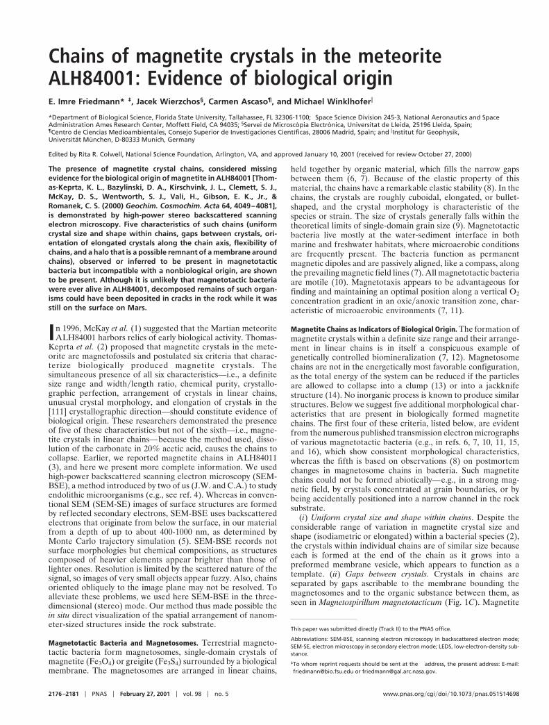
Chains of magnetite crystals in the meteoriteALH84001: Evidence of biological originE. Imre Friedmann*†‡, Jacek Wierzchos§, Carmen Ascaso¶, and Michael Winklhoferi
*Department of Biological Science, Florida State University, Tallahassee, FL 32306-1100; †Space Science Division 245-3, National Aeronautics and SpaceAdministration Ames Research Center, Moffett Field, CA 94035; §Servei de Microscopia Electronica, Universitat de Lleida, 25196 Lleida, Spain;¶Centro de Ciencias Medioambientales, Consejo Superior de Investigaciones Cientıficas, 28006 Madrid, Spain; and iInstitut fur Geophysik,Universitat Munchen, D-80333 Munich, Germany
Edited by Rita R. Colwell, National Science Foundation, Arlington, VA, and approved January 10, 2001 (received for review October 27, 2000)
The presence of magnetite crystal chains, considered missingevidence for the biological origin of magnetite in ALH84001 [Thom-as-Keprta, K. L., Bazylinski, D. A., Kirschvink, J. L., Clemett, S. J.,McKay, D. S., Wentworth, S. J., Vali, H., Gibson, E. K., Jr., &Romanek, C. S. (2000) Geochim. Cosmochim. Acta 64, 4049–4081],is demonstrated by high-power stereo backscattered scanningelectron microscopy. Five characteristics of such chains (uniformcrystal size and shape within chains, gaps between crystals, ori-entation of elongated crystals along the chain axis, flexibility ofchains, and a halo that is a possible remnant of a membrane aroundchains), observed or inferred to be present in magnetotacticbacteria but incompatible with a nonbiological origin, are shownto be present. Although it is unlikely that magnetotactic bacteriawere ever alive in ALH84001, decomposed remains of such organ-isms could have been deposited in cracks in the rock while it wasstill on the surface on Mars.
In 1996, McKay et al. (1) suggested that the Martian meteoriteALH84001 harbors relics of early biological activity. Thomas-
Keprta et al. (2) proposed that magnetite crystals in the mete-orite are magnetofossils and postulated six criteria that charac-terize biologically produced magnetite crystals. Thesimultaneous presence of all six characteristics—i.e., a definitesize range and widthylength ratio, chemical purity, crystallo-graphic perfection, arrangement of crystals in linear chains,unusual crystal morphology, and elongation of crystals in the[111] crystallographic direction—should constitute evidence ofbiological origin. These researchers demonstrated the presenceof five of these characteristics but not of the sixth—i.e., magne-tite crystals in linear chains—because the method used, disso-lution of the carbonate in 20% acetic acid, causes the chains tocollapse. Earlier, we reported magnetite chains in ALH84011(3), and here we present more complete information. We usedhigh-power backscattered scanning electron microscopy (SEM-BSE), a method introduced by two of us (J.W. and C.A.) to studyendolithic microorganisms (e.g., see ref. 4). Whereas in conven-tional SEM (SEM-SE) images of surface structures are formedby reflected secondary electrons, SEM-BSE uses backscatteredelectrons that originate from below the surface, in our materialfrom a depth of up to about 400-1000 nm, as determined byMonte Carlo trajectory simulation (5). SEM-BSE records notsurface morphologies but chemical compositions, as structurescomposed of heavier elements appear brighter than those oflighter ones. Resolution is limited by the scattered nature of thesignal, so images of very small objects appear fuzzy. Also, chainsoriented obliquely to the image plane may not be resolved. Toalleviate these problems, we used here SEM-BSE in the three-dimensional (stereo) mode. Our method thus made possible thein situ direct visualization of the spatial arrangement of nanom-eter-sized structures inside the rock substrate.
Magnetotactic Bacteria and Magnetosomes. Terrestrial magneto-tactic bacteria form magnetosomes, single-domain crystals ofmagnetite (Fe3O4) or greigite (Fe3S4) surrounded by a biologicalmembrane. The magnetosomes are arranged in linear chains,
held together by organic material, which fills the narrow gapsbetween them (6, 7). Because of the elastic property of thismaterial, the chains have a remarkable elastic stability (8). In thechains, the crystals are roughly cuboidal, elongated, or bullet-shaped, and the crystal morphology is characteristic of thespecies or strain. The size of crystals generally falls within thetheoretical limits of single-domain grain size (9). Magnetotacticbacteria live mostly at the water-sediment interface in bothmarine and freshwater habitats, where microaerobic conditionsare frequently present. The bacteria function as permanentmagnetic dipoles and are passively aligned, like a compass, alongthe prevailing magnetic field lines (7). All magnetotactic bacteriaare motile (10). Magnetotaxis appears to be advantageous forfinding and maintaining an optimal position along a vertical O2concentration gradient in an oxicyanoxic transition zone, char-acteristic of microaerobic environments (7, 11).
Magnetite Chains as Indicators of Biological Origin. The formation ofmagnetite crystals within a definite size range and their arrange-ment in linear chains is in itself a conspicuous example ofgenetically controlled biomineralization (7, 12). Magnetosomechains are not in the energetically most favorable configuration,as the total energy of the system can be reduced if the particlesare allowed to collapse into a clump (13) or into a jackknifestructure (14). No inorganic process is known to produce similarstructures. Below we suggest five additional morphological char-acteristics that are present in biologically formed magnetitechains. The first four of these criteria, listed below, are evidentfrom the numerous published transmission electron micrographsof various magnetotactic bacteria (e.g., in refs. 6, 7, 10, 11, 15,and 16), which show consistent morphological characteristics,whereas the fifth is based on observations (8) on postmortemchanges in magnetosome chains in bacteria. Such magnetitechains could not be formed abiotically—e.g., in a strong mag-netic field, by crystals concentrated at grain boundaries, or bybeing accidentally positioned into a narrow channel in the rocksubstrate.
(i) Uniform crystal size and shape within chains. Despite theconsiderable range of variation in magnetite crystal size andshape (isodiametric or elongated) within a bacterial species (2),the crystals within individual chains are of similar size becauseeach is formed at the end of the chain as it grows into apreformed membrane vesicle, which appears to function as atemplate. (ii) Gaps between crystals. Crystals in chains areseparated by gaps ascribable to the membrane bounding themagnetosomes and to the organic substance between them, asseen in Magnetospirillum magnetotacticum (Fig. 1C). Magnetite
This paper was submitted directly (Track II) to the PNAS office.
Abbreviations: SEM-BSE, scanning electron microscopy in backscattered electron mode;SEM-SE, electron microscopy in secondary electron mode; LEDS, low-electron-density sub-stance.
‡To whom reprint requests should be sent at the † address, the present address: E-mail:[email protected] or [email protected].
2176–2181 u PNAS u February 27, 2001 u vol. 98 u no. 5 www.pnas.orgycgiydoiy10.1073ypnas.051514698

Fig. 1. (A and B) SEM-SE stereo micrograph of area in Fig. 4A, showing surface features. (C) SEM-BSE image of magnetosome chain in freeze-dried culture ofMagnetospirillum magnetotacticum. (D and E) SEM-BSE stereo micrograph of A and B. (F) Outlines of areas shown in G and H and J and K. (I and L) Chains visiblein G and H and J and K, respectively, are highlighted. px, orthopyroxene; ca, carbonate.
Friedmann et al. PNAS u February 27, 2001 u vol. 98 u no. 5 u 2177
MIC
ROBI
OLO
GY
SPEC
IAL
FEA
TURE
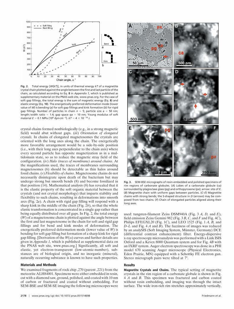
crystal chains formed nonbiologically (e.g., in a strong magneticfield) would abut without gaps. (iii) Orientation of elongatedcrystals. In chains of elongated magnetosomes the crystals areoriented with the long axes along the chain. The energeticallymore favorable arrangement would be a side-by-side position(i.e., with their long axes perpendicular to the chain axis) whereevery second particle has opposite magnetization as in a mul-tidomain state, so as to reduce the magnetic stray field of theconfiguration. (iv) Halo (traces of membrane) around chains. Atthe magnifications used, the traces of membranes surroundingmagnetosomes (6) should be detectable as thin halos aroundfossil chains. (v) Flexibility of chains. Magnetosome chains do notnecessarily disintegrate upon death of the bacterium but mayundergo strong but smooth bends (8) and become fossilized inthat position (14). Mathematical analysis (8) has revealed that itis the elastic property of the soft organic material between thecrystals (and not crystal morphology) that imparts stability andflexibility to such chains, resulting in deformations into smootharcs (Fig. 2a). A chain with rigid gap filling will respond with asharp kink in the middle of the chain (Fig. 2b), so that the wholeelastic transformation is concentrated in a single gap rather thanbeing equally distributed over all gaps. In Fig. 2, the total energy(W) of a magnetosome chain is plotted against the angle betweenthe first and last magnetosome in the chain for soft and rigid gapfillings and for bend and kink modes of deformation. Theenergetically preferred deformation mode (lower value of W) isbending for soft gap filling but formation of a sharp kink for rigidgap filling. [Derivation of the W(a) curves and further details aregiven in Appendix 1, which is published as supplemental data onthe PNAS web site, www.pnas.org.] Significantly, all soft andelastic, yet electron-transparent (low-atomic-number), sub-stances are of biological origin, and no inorganic (mineral),naturally occurring substance is known to have such properties.
Materials and MethodsWe examined fragments of rock chip ,270 (parent ,221) from themeteorite ALH84001. Specimens were either embedded in resin,cut with a diamond saw, ground, polished, and coated with 10 nmof carbon or fractured and coated without embedding. ForSEM-BSE and SEM-SE imaging the following microscopes were
used: tungsten-filament Zeiss DSM940A (Fig. 3 A, D, and E),field emission Zeiss Gemini 982 (Fig. 3 B, C, and F and Fig. 4C),Philips EFEGXL20 (Fig. 1C), and LEO 1525 (Fig. 1 A, B, andD–L and Fig. 4 A and B). The fuzziness of images was reducedby an analySIS (Soft Imaging System, Munster, Germany) DCE(differential contrast enhancement) filter. Energy-dispersivex-ray spectroscopy microanalysis was performed with a Link ISISOxford and a Kevex 8000 Quantum system and for Fig. 4B withan IXRF system. Auger electron spectroscopy was done in a PHImodel 670 scanning Auger microscope (Physical Electronics,Eden Prairie, MN) equipped with a Schottky FE electron gun.Stereo micrograph pairs were tilted at 7°.
ResultsMagnetite Crystals and Chains. The typical setting of magnetitecrystals in the rim region of a carbonate globule is shown in Fig.4 A and B. This specimen was fractured and carbon coatedwithout resin embedding, and imaging was through the intactsurface. The wide iron-rich rim stretches approximately vertically,
Fig. 2. Total energy [W(kT)], in units of thermal energy kT of a magnetitecrystal chain plotted against the angle between the first and last particle of thechain, as calculated according to Eq. 8 in Appendix 1, which is published assupplementary material on the PNAS web site, www.pnas.org. For the case ofsoft gap fillings, the total energy is the sum of magnetic energy (Eq. 8) andelastic energy (Eq. 10). The energetically preferred deformation mode (lowervalue of W) is bending (a) for soft gap fillings and kink formation (b) for rigidgap fillings. Number of particles in chain n 5 5; particle size a 5 50 nm;lengthywidth ratio 5 1.4; gap space qa 5 10 nm; Young modulus of softmaterial E 5 0.1 MPa (106 dynzcm22). kT 5 4 3 10214 J.
Fig. 3. SEM-BSE micrographs of resin-embedded and polished specimens ofrim regions of carbonate globules. (A) Lobes of a carbonate globule (ca)surrounded by plagioclase glass (pg) and orthopyroxene (px); arrow: site of E.(B) Magnetite chain with uniform gaps between particles. (C–E) Magnetitechains with strong bends; the S-shaped structure in D (arrows) may be com-posed from two chains. (F) Chain of elongated particles aligned along theirlong axes.
2178 u www.pnas.orgycgiydoiy10.1073ypnas.051514698 Friedmann et al.

Fig. 4. (A–C) SEM-BSE micrographs of magnetite crystals in the rim region of carbonate globules in ALH84001. (A) Imaged through the intact surface of a freshlyfractured specimen, chain of large elongated crystals (arrow), further chains of smaller crystals and other details in Fig. 1. (B) Energy-dispersive x-ray spectrometryanalyses of elemental composition showing position of Fe-rich rim (detailed data are presented in Appendix 2, which is published as supplemental data on thePNAS web site, www.pnas.org). (C) Similar site in a resin-embedded, sectioned, and polished specimen, one chain marked by arrow, also site of Auger electronspectroscopy analyses. (D) Results of analyses of areas outlined in C: squares, magnetite crystal chains present; triangles, absent; ca, carbonate; px, orthopyroxene.
Friedmann et al. PNAS u February 27, 2001 u vol. 98 u no. 5 u 2179
MIC
ROBI
OLO
GY
SPEC
IAL
FEA
TURE
evident as a zone of small granules overlying the substrate,orthopyroxene between green and blue lines and carbonatebetween blue and purple (Fig. 4B). Elemental analysis (below)indicates that the granules are rich in Fe and O, with only tracesof S. Previous studies (1, 2) showed that these Fe- and O-richrims are composed mainly of magnetite crystals, with a fewgrains possibly of iron sulfides, embedded in carbonate matrix.The stereo pair in Fig. 1 A and B shows the surface of the areain SEM-SE, whereas Fig. 1 D and E, in SEM-BSE, shows theunderlying structures up to ca. 1 mm deep. Comparison of theseimages reveals that the entire area is covered by a low-electron-density (i.e., low-atomic-number) substance (LEDS) of un-known (perhaps organic?) chemical composition, with a gran-ular surface visible in SEM-SE but transparent in the BSE mode.Fig. 1C is an SEM-BSE image of a magnetosome chain in afreeze-dried preparation of Magnetospirillum magnetotacticum,shown for comparison. The areas indicated in Fig. 1F are shownin BSE stereo pairs in Fig. 1 G and H and Fig. 1 J and K. In thesestereo images the magnetite crystals embedded in transparentLEDS appear as though floating, whereas others are embeddedin the large carbonate crystals. In the large crystal conglomer-ation in the upper left corner of Fig. 4A, the individual crystalsare not resolved, because of superposition of the BSE signals. Inother, less compacted clusters, individual crystals are distin-guishable, and among these, many appear to form chains. In Fig.4A a conspicuous chain of 7 or 8 elongated crystals arrangedalong their long axes is indicated by an arrow. In the stereo imagepairs in Fig. 1 G and H and Fig. 1 J and K, several chains ofvarious lengths, clarity, and state of imaging can be distin-guished, as indicated in the auxiliary Fig. 1 I and L. TheSEM-BSE micrographs in Fig. 3 A–F and Fig. 4C are ofresin-embedded specimens. In the sectioned specimen shown inFig. 3A the small carbonate structures (probably lobes of acarbonate globule) are embedded in plagioclase glass and or-thopyroxene. The arrow indicates the site of the chain shown inFig. 3E. Fig. 3B is a chain of six crystals clearly separated by gaps.Fig. 3 C and D show crystal chains with strong bends; in Fig. 3Dthe apparent S-shaped structure (arrows) may be composed oftwo chains. The bent chain of about 15 large isodiametric crystalsin Fig. 3E and the chain of 6 small elongated crystals arrangedin the direction of their long axes in Fig. 3F illustrate the widerange of crystal forms and sizes in ALH84001 (note differentscales). Significantly, the size and shape of crystals within eachchain are uniform. Many or perhaps most magnetite crystals,both single and in chains, seem to be surrounded, at least in part,by LEDS, which appears in SEM-BSE as a dark area. Becausesuch areas are evident both in unprocessed (i.e., freshly frac-tured, Fig. 4A) and in resin-embedded (Figs. 3 B–F and 4C)specimens, they are not voids but filled with LEDS. A chain inFig. 1 G and H (marked by an arrow in Fig. 1 I) is surrounded bya dark halo, evident against the white background of carbonate
in which the chain is embedded. Similar halos, if present aroundother chains, may not be visible against the dark LEDS back-ground. Fig. 4C is a resin-embedded specimen with numerouschains and chain fragments. One chain is marked by an arrow,others are inside squares. The chain in the rightmost square isshown in Fig. 3B.
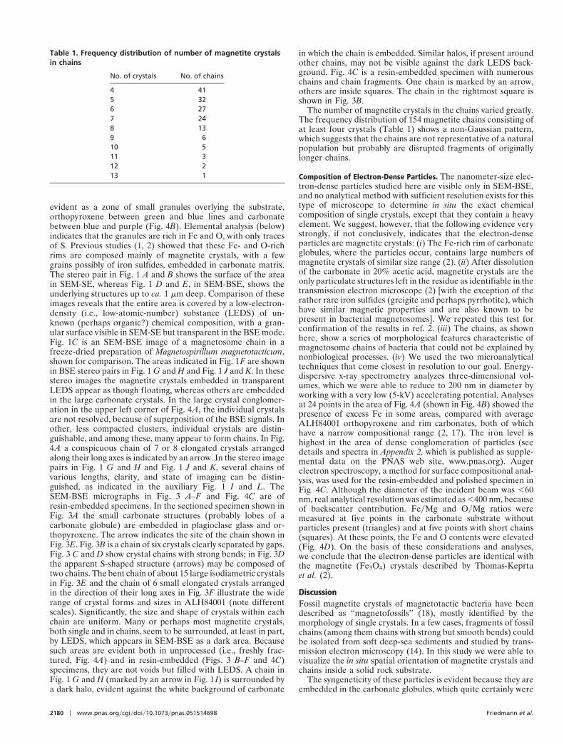
The number of magnetite crystals in the chains varied greatly.The frequency distribution of 154 magnetite chains consisting ofat least four crystals (Table 1) shows a non-Gaussian pattern,which suggests that the chains are not representative of a naturalpopulation but probably are disrupted fragments of originallylonger chains.
Composition of Electron-Dense Particles. The nanometer-size elec-tron-dense particles studied here are visible only in SEM-BSE,and no analytical method with sufficient resolution exists for thistype of microscope to determine in situ the exact chemicalcomposition of single crystals, except that they contain a heavyelement. We suggest, however, that the following evidence verystrongly, if not conclusively, indicates that the electron-denseparticles are magnetite crystals: (i) The Fe-rich rim of carbonateglobules, where the particles occur, contains large numbers ofmagnetite crystals of similar size range (2). (ii) After dissolutionof the carbonate in 20% acetic acid, magnetite crystals are theonly particulate structures left in the residue as identifiable in thetransmission electron microscope (2) [with the exception of therather rare iron sulfides (greigite and perhaps pyrrhotite), whichhave similar magnetic properties and are also known to bepresent in bacterial magnetosomes]. We repeated this test forconfirmation of the results in ref. 2. (iii) The chains, as shownhere, show a series of morphological features characteristic ofmagnetosome chains of bacteria that could not be explained bynonbiological processes. (iv) We used the two microanalyticaltechniques that come closest in resolution to our goal. Energy-dispersive x-ray spectrometry analyzes three-dimensional vol-umes, which we were able to reduce to 200 nm in diameter byworking with a very low (5-kV) accelerating potential. Analysesat 24 points in the area of Fig. 4A (shown in Fig. 4B) showed thepresence of excess Fe in some areas, compared with averageALH84001 orthopyroxene and rim carbonates, both of whichhave a narrow compositional range (2, 17). The iron level ishighest in the area of dense conglomeration of particles (seedetails and spectra in Appendix 2, which is published as supple-mental data on the PNAS web site, www.pnas.org). Augerelectron spectroscopy, a method for surface compositional anal-ysis, was used for the resin-embedded and polished specimen inFig. 4C. Although the diameter of the incident beam was ,60nm, real analytical resolution was estimated as ,400 nm, becauseof backscatter contribution. FeyMg and OyMg ratios weremeasured at five points in the carbonate substrate withoutparticles present (triangles) and at five points with short chains(squares). At these points, the Fe and O contents were elevated(Fig. 4D). On the basis of these considerations and analyses,we conclude that the electron-dense particles are identical withthe magnetite (Fe3O4) crystals described by Thomas-Keprtaet al. (2).
DiscussionFossil magnetite crystals of magnetotactic bacteria have beendescribed as ‘‘magnetofossils’’ (18), mostly identified by themorphology of single crystals. In a few cases, fragments of fossilchains (among them chains with strong but smooth bends) couldbe isolated from soft deep-sea sediments and studied by trans-mission electron microscopy (14). In this study we were able tovisualize the in situ spatial orientation of magnetite crystals andchains inside a solid rock substrate.
The syngeneticity of these particles is evident because they areembedded in the carbonate globules, which quite certainly were
Table 1. Frequency distribution of number of magnetite crystalsin chains
No. of crystals No. of chains
4 415 326 277 248 139 610 511 312 213 1
2180 u www.pnas.orgycgiydoiy10.1073ypnas.051514698 Friedmann et al.
formed on Mars (1, 19, 20). Furthermore, Fig. 3A shows lobes ofa carbonate globule containing magnetite chains (Fig. 3E)embedded in plagioclase glass and orthopyroxene.
The magnetite crystals in ALH84001 form chains similar tothose in modern magnetotactic bacteria. This informationshould satisfy the last of the six criteria postulated (2) as evidenceof biological origin. We also describe five additional criteriacharacteristic of biologically produced magnetite chains, whichcould not be present in abiotically formed chains of magnetitecrystals (no such chains have ever been observed in nature). Wesuggest that these criteria have now been satisfied: Uniformcrystal size and shape within chains is seen in Fig. 1 G and H, Fig.1 J and K, and Fig. 4A (in the latter the crystals in the chainmarked by an arrow are elongated and larger in diameter thanthose in the numerous chains of smaller and approximatelyisodiametric crystals), as well as in Fig. 3 B–F, which show chainsof different sizes. Fig. 3 E and F (note different scales) show thedifference between the largest, approximately isodiametric, andthe smallest, elongated, crystals. No abiological process is knownthat would result in such sorting of crystals from a mixed pool ofsizes and shapes. Gaps between crystals are best seen in Fig. 3B,although they are evident in all sufficiently resolved chains asdark lines between crystals. Orientation of elongated crystals alongthe chain axis is evident in Figs. 3F and 4A (arrow). Halo aroundchains, the possible remnant of a membrane, is visible in Fig. 1G–I. Flexibility of chains, a character ascribable to the elasticproperty of the organic substance between but independentfrom the shape of the crystals, is evident in many of the smallchains in Fig. 1 G–I, Fig. 1 J–L, and Fig. 3 B–F. We conclude thatthe chains of electron-opaque particles in ALH84001 are mag-netofossils, as no other consistent explanation would account forthese findings.
The ecological implications of the assumption that the mag-netite chains in ALH84001 are of biological origin must be
examined: Is it likely, on the basis of our knowledge of terrestrialmagnetotactic bacteria, that such microorganisms were presentin cracks of ALH84001? Numerous endolithic microorganismsexist in both terrestrial (21) and aquatic (22) habitats, yet it isvery unlikely that magnetotactic bacteria were ever alive inALH84001. All known endolithic microorganisms are sessile,attached to the surface; none is motile, understandable becauseswimming would be impossible in the microscopic spaces inrocks. Magnetotactic bacteria, with magnetosomes to serve asguides in the direction of swimming, are all motile. Furthermore,the non-Gaussian frequency distribution of the number ofcrystals in chains (Table 1) is incompatible with the variability ofchain length in a living population. We suggest the followingscenario: First, decomposed remains of dead magnetobacteriasuspended in a carbonate-rich fluid penetrated fissures ofALH84001, already crushed by previous asteroid impact, per-haps after the second impact event (I2) postulated (23). Second,evaporation of the liquid led to the deposition of pancake-shapedcarbonate globules, and magnetite crystals and chain fragmentswere deposited in the periphery of the carbonate discs, perhapsthrough the mechanism for ring deposition of particles dispersedin liquid drops (24).
Thanks are due to D. A. Bazylinski, J. L. Kirschvink, and J. W. Schopffor critical reading of the manuscript; to H. Jaksch and J. Greiser (LEOGmbH, Oberkochen, Germany) for help and cooperation in takingmicrographs in Figs. 1 A, B, D–L, and 4A; to S. Douglass (Jet PropulsionLaboratory, Pasadena, CA) for Fig. 1C; to J. F. Almagro (Acerinox,Algeciras, Spain) for Figs. 3 B and C and 4C; to R. Bargallo, L. E.Bertani, L. Calvo-Barrio, A. Martinez, F. Pinto, and K. Riddle forsupport and technical help; and to A. B. Thistle for critical editing. Thiswork was supported by National Aeronautics and Space AdministrationGrants NAG5y4921 and -9845 to E.I.F., Direccio General de Recerca deGeneralitat de Catalunya Grant ACES98-15y1 to J.W., and Ministeriode Educacion y Cultura Grant APC1998-0045 to C.A. and J.W.
1. McKay, D. S., Gibson, E. K., Jr., Thomas-Keprta, K. L., Vali, H., Romanek,C. S., Clemett, S. J., Chillier, X. D. F., Maechling, C. R. & Zare, R. N. (1996)Science 273, 924–930.
2. Thomas-Keprta, K. L., Bazylinski, D. A., Kirschvink, J. L., Clemett, S. J.,McKay, D. S., Wentworth, S. J., Vali, H., Gibson, E. K., Jr., & Romanek, C. S.(2000) Geochim. Cosmochim. Acta 64, 4049–4081.
3. Friedmann, E. I., Wierzchos, J. & Ascaso, C. (1998) in Workshop on the Issueof Martian Meteorites (Lunar Planetary Institute, Houston, TX), Contribution956, pp. 14–16.
4. Ascaso, C., Wierzchos, J. & de los Rios, A. (1995) Bot. Acta 108, 474–481.5. Joy, D. C. (1991) Scanning Microsc. 5, 329–337.6. Gorby, Y. A., Beveridge, T. J. & Blakemore, R. P. (1988) J. Bacteriol. 170,
834–871.7. Bazylinski, D. A. & Moskowitz, B. M. (1997) Rev. Mineral. 35, 181–223.8. Shcherbakov, V. P., Winklhofer, M., Hanzlik, M. & Petersen, N. (1997) Eur.
Biophys. J. 26, 319–326.9. Butler, R. F. & Banerjee, S. K. (1975) J. Geophys. Res. 80, 4049–4058.
10. Blakemore, R. P., Blakemore, N. A., Bazylinski, D. A. & Moench, T. T. (1989)in Bergey’s Manual of Systematic Bacteriology, eds. Staley, J. T., Bryant, M. P.,Pfennig, N. & Holt, J. G. (Williams & Wilkins, Baltimore), Vol. 3, pp.1882–1889.
11. Frankel, R. B. & Bazylinski, D. A. (1994) Hyperfine Interact. 90, 135–142.12. Lowenstam, H. A. (1981) Science 211, 1126–1131.
13. Kirschvink, J. L. (1982) Earth Planet. Sci. Lett. 59, 388–392.14. von Dobeneck, T., Petersen, N. & Vali, H. (1987) Geowissenschaften in unserer
Zeit 5, 27–35.15. Mann, S. & Frankel, R. B. (1989) in Biomineralization: Chemical and Biochem-
ical Perspectives, eds. Mann, S., Webb., J. & Williams, R. J. P. (VCH, NewYork), pp. 389–426.
16. Bazylinski, D. A., Garratt-Reed, A. J. & Frankel, R. B. (1994) Microsc. Res.Tech. 27, 389–401.
17. Mittlefehldt, D. W. (1994) Meteoritics 29, 214–221.18. Chang, S.-B. R. & Kirschvink, L. M. (1989) Annu. Rev. Earth Planet. Sci. 17,
169–195.19. Romanek, C. S., Grady, M. M., Wright, I. P., Mittlefehldt, D. W., Socki, R. A.,
Pillinger, C. T. & Gibson, E. K., Jr. (1994) Nature (London) 372, 655–657.20. Borg, L. E., Connelly, J. N., Nyquist, L. E., Shih, C.-Y., Wiesmann, H. & Reese,
Y. (1999) Science 286, 90–93.21. Friedmann, E. I. & Ocampo-Friedmann, R. (1984) in Current Perspectives in
Microbial Ecology, eds. Klug, M. J. & Reddy, C. A. (Am. Soc. Microbiol.,Washington, DC), pp. 177–185.
22. Golubic, S., Perkins, R. D. & Lukas, K. J. (1975) in The Study of Trace Fossils,ed. Frey, R.W. (Springer, New York), pp. 177–185.
23. Treiman, A. H. (1998) Meteor. Planet. Sci. 33, 753–764.24. Deegan, R. D., Bakajin, O., Dupont, T. F., Huber, G., Nagel, S. R. & Whitten,
T. A. (1997) Nature (London) 389, 827–829.
Friedmann et al. PNAS u February 27, 2001 u vol. 98 u no. 5 u 2181
MIC
ROBI
OLO
GY
SPEC
IAL
FEA
TURE