Embed Size (px)
Citation preview

Downloaded from www.microbiologyresearch.org by
IP: 54.90.45.205
On: Thu, 28 Jan 2016 10:12:06
Journal of General Microbiology (1977), 102, 365-374. Printed in Great Britain
Cell Division and Photosynthetic Apparatus Construction in the Cell Envelope Deficient ‘Phofll’ Mutant
of Rhodopseudomonas sphaeroides
By JACQUES P R A D E L A N D J E N N Y C L E M E N T - M E T R A L Laboratoire de Photosyntht?se, CNRS, G$-sur- Yvette 91 190, France and
Laboratoire de Biochimie Vkgktale, Universite‘ Aix, Marseille I 3288, France
(Received I 8 April I 977)
365
~
The ‘ Phofil ’ mutant of Rhodopseudornonas sphaeroides showed a disturbed adaptation with respect to a change from aerobic to photosynthetic conditions. The mutation was a pleio- tropic one : firstly, the cells formed multinucleate filaments with occasional septa, and secondly, they integrated only a fraction of the synthetized pigments. The phenotypes appeared in photosynthetic conditions during a period of unbalanced growth (unadapted state) followed by balanced exponential growth where the phenotypes were corrected (adapted state). Ultraviolet light irradiation or exposure to low temperature, but not partial or total depigmentation, caused adapted cells to return to the unadapted state. Except for those associated with pigments, no appreciable defect in the polypeptide composition of the cell envelope was detected by SDS-polyacrylamide gel electrophoresis. However, the protein/phospholipid ratio of the envelope fraction was approximately 50 % higher, in the mutant than in the parent. These findings, and others described in the text, suggested the existence of defects in the cell wall and in the inner membrane.
INTRODUCTION
Most Rhodospirillaceae grow photosynthetically as well as chemotrophically (Oelze & Drews, 1972; Cohen-Bazire, Sistrom & Stanier, 1957). The development of the photo- synthetic apparatus located on a membrane system is controlled by light, and, more im- portantly, by the degree of aerobiosis. Under low aeration conditions in the dark, with no light-dependent energy available, the bacteria contain a photosynthetic intracytoplasmic membrane. Thus, when aerobically-grown cells are made anaerobic in the light or semi- aerobic in the dark, differentiation of the membrane occurs during an adaptation phase (Saunders & Jones, 1974) simultaneously with the construction of the photosynthetic apparatus.
Recent reports have provided new information on this adaptation process, particularly on the multi-step assembly of photopigments in the developing membrane (Oelze & Pahlke, 1976; Niederman, Mallon & Langan, 1976). Several aspects of this process, however, are poorly understood, especially those concerning the initial stages of the induction of the photosynthetic membrane system (Peters & Cellarius, 1972 ; Niederman et al., 1976). A convenient approach to the study of this problem was by the use of mutants defective in the adaptation process. Such mutants were mainly photosynthetically incompetent strains; only two, which retained the ability to synthetize bacteriochlorophyll at a decreased rate, have been studied (Uspenskaya & Lyskova, 1972; Drews, 1974). The ‘Phofil’ mutant isolated in this laboratory (Pradel & CICment-MCtral, I 976) was also aberrant in its adapta- tion to photosynthetic growth. Its physiological properties and the nature of the biochemical defects are reported here.

Downloaded from www.microbiologyresearch.org by
IP: 54.90.45.205
On: Thu, 28 Jan 2016 10:12:06
J. P R A D E L A N D J . C L ~ M E N T - M ~ T R A L
M E T H O D S
Orgnnisnis atid cirttiire conditions. Wild-type Rhodopseudomonas splioeroides (strain Y) and the derived ‘Phofil’ mutant were used. A synthetic medium equally suitable for aerobic and photosynthetic growth (Jolchine RC Reiss-Husson, 1974) was used and solidified, when necessary, with 1.5 :b (W:V) agar (Difco). Unless indicated otherwise, the bacteria were grown at 30 “C, with magnetic stirring, in glass bottles (2 1 and IOO ml). Photosynthetic cultures were bubbled with nitrogen and illuminated with incandescent bulbs (5000 Ix). Aerobic cultures were grown either in the light and sparged with air or in the dark sparged with oxygen. Semi-aerobiosis was obtained by gassing with 02/Ny (5 :95 , v,v) or by incubating in Erlenmeyer flasks on a low-speed shaker. The cell density was estimated by measuring or by determining the number of viable cells.
Microscopy. A Zeiss microscope with phase-contrast illumination was used. Bacterial nucleoids were visualized using a low-sensitivity film and a long exposure time.
Ultraviolet light irradintion. Philips lamps (TUV) placed in a glove-box were used. The evposure was determined with a Latarjet ultraviolet light dosimeter (Latarjet, Morenne & Berger, 1953).
Cell envelope isohtion andpicrification. The method of Takemoto & Lascelles (1973) was employed with the following modifications. To remove the ribosomes the membrane fraction was suspended in 30 ?{ (w. v) CsCl and centrifuged at 350000 g for 60 min. The ribosome-free envelope fraction was then collected, washed and layered on a discontinuous density gradient of Ficoll.
The method of Laemmli (1970) as modified by Clayton & Haselkorn (1972) was used for sodium dodecyl sulphate (SDS)-polyacrylamide gel electrophoresis. The stained gels were scanned at 550 nm on a Safas 3000 D spectrophotometer equipped with a linear transport device.
Anulyticul procedures. Bacteriochlorophyll was determined in the acetone/methanol (7 : 2, v/v) extract using c7i0 = 76 x 1o-O 1 mol-’ cm-l (Clayton, 1963). 4-Vinylprotochlorophyllide was determined as des- cribed previously (Pradel & CICment-MCtral, 1976). Lipids were extracted with ch1oroform:methanol (2 : I ,
v/v) by the method of Bligh and Dyer as modified by Kates (1972). Total phosphorus was determined by the method of Bartlett (1959). Protein was estimated according to Lowry e ta / . (1951) with bovine serum albumin as standard.
R E S U L T S
Physiological observations
Isolation and phenotype. The mutant studied was isolated (Pradel & Clement-Mitral, 1976) as a green colony of normal size after aerobic growth; under photosynthetic growth conditions, the mutant colonies were markedly smaller than those of the wild type. The relatively slow growth of the mutant in photosynthetic conditions was used to test its genetic stability in terms of reversion frequency. Mutant cells previously bleached by aerobic growth in the light, were plated and grown photosynthetically. The revertant cells appeared as purple points on a greenish background. When 6 x 10’ mutant cells were plated, 30 revertants were obtained. The reliability of this method was confirmed in a reconstruction test by counting the purple points on Petri dishes plated with a mixture of 102 wild-type and 106 mutant cells.
The green colour was due to overproduction and excretion of a bacteriochlorophyll precursor complex. Microscopic examination revealed that in photosynthetic conditions the bacteria formed filaments of variable lengths with occasional septa (Fig. I a), and with general swelling and local bulging (Fig. I b).
Growth characteristics. Growth of the mutant strain was followed under extreme condi- tions: either in the dark at high partial pressure of oxygen or anaerobically in the light (the cells were pre-cultivated semi-aerobically). In aerobic conditions the lag phase was very short. In photosynthetic conditions it was very long: the doubling time, initially about I 2 h, decreased progressively to I 00 min during exponential growth. We shall hereafter describe the mutant cells during the long lag phase as ‘unadapted’ (to photosynthesis), in contrast to ‘adapted’ cells which grow exponentially under anaerobic conditions in the light.
Growth and pigmentation. (i) Adapted mutant. After adaptation, the mutant strain had corrected both phenotypes; septation and cell division occurred at a normal rate, and all the pigmenl synthetized was integrated into the photosynthetic apparatus. Growth and pig-

Downloaded from www.microbiologyresearch.org by
IP: 54.90.45.205
On: Thu, 28 Jan 2016 10:12:06
Cell envelope mutant of R. sphaeroides 367
Fig. I . Phase-contrast micrographs showing (a) nuclear bodies in filaments of R. sphueroides ' Phofil' mutant grown in photosynthetic conditions, (b) structural anomalies. Bar marker repre- sents 25 pm.
Fig. 2. Colonies from adapted cells of R. sphueroides 'Phofil' mutant grown semi-aerobically at 24 "C. The enlargements of colonies u and b are at 8 x magnification.
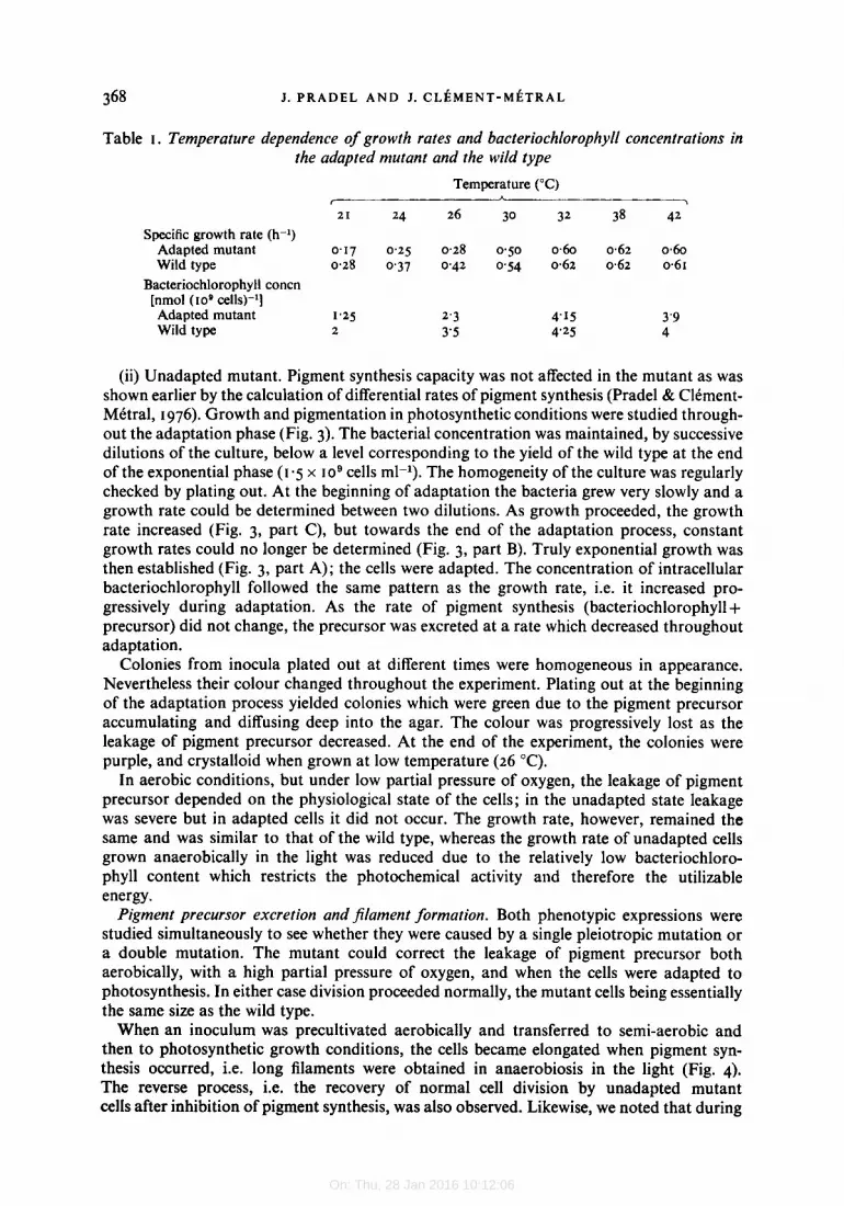
mentation under photosynthetic conditions were temperature dependent. The mutant and parent strain had similar growth rates and cellular bacteriochlorophyll concentrations at temperatures above 30 "C (Table I), but the effect of low temperature was more pronounced in the mutant. After aerobic growth on solid medium at temperatures above 30 "C, the colonies of both strains were identical. At lower temperatures, however, the mutant colon- ies were unusually crystalloid (Fig. 2) and were composed of two types of cell, some identi- cal to wild-type cells and others forming short filaments.
24 M I C I02
Downloaded from www.microbiologyresearch.org by
IP: 54.90.45.205
On: Thu, 28 Jan 2016 10:12:06
368 J. P R A D E L A N D J. C L ~ ~ M E N T - M ~ T R A L
Table I . Temperature dependence of growth rates and bacteriochlorophyll concentrations in the adapted mutant and the wild type
Temperature ("C)
21 24 26 30 32 38 42
A I >
Specific growth rate (h-l) Adapted mutant 0.17 0.25 0.28 0.50 0.60 0.62 0.60 Wild type 0.28 0.37 0.42 0.54 0.62 0.62 0.61
Bacteriochlorophyll concn [nmol ( I O O cells)-1] Adapted mutant 1.25 2.3 4.15 3'9 Wild type 2 3'5 4'25 4
(ii) Unadapted mutant. Pigment synthesis capacity was not affected in the mutant as was shown earlier by the calculation of differential rates of pigment synthesis (Pradel & ClCment- MCtral, I 976). Growth and pigmentation in photosynthetic conditions were studied through- out the adaptation phase (Fig. 3). The bacterial concentration was maintained, by successive dilutions of the culture, below a level corresponding to the yield of the wild type at the end of the exponential phase ( I -5 x 10⁹ cells ml-l). The homogeneity of the culture was regularly checked by plating out. At the beginning of adaptation the bacteria grew very slowly and a growth rate could be determined between two dilutions. As growth proceeded, the growth rate increased (Fig. 3, part C), but towards the end of the adaptation process, constant growth rates could no longer be determined (Fig. 3, part B). Truly exponential growth was then established (Fig. 3, part A); the cells were adapted. The concentration of intracellular bacteriochlorophyll followed the same pattern as the growth rate, i.e. it increased pro- gressively during adaptation. As the rate of pigment synthesis (bacteriochlorophyll+ precursor) did not change, the precursor was excreted at a rate which decreased throughout adaptation.
Colonies from inocula plated out at different times were homogeneous in appearance. Nevertheless their colour changed throughout the experiment. Plating out at the beginning of the adaptation process yielded colonies which were green due to the pigment precursor accumulating and diffusing deep into the agar. The colour was progressively lost as the leakage of pigment precursor decreased. At the end of the experiment, the colonies were purple, and crystalloid when grown at low temperature (26 "C).
In aerobic conditions, but under low partial pressure of oxygen, the leakage of pigment precursor depended on the physiological state of the cells; in the unadapted state leakage was severe but in adapted cells it did not occur. The growth rate, however, remained the same and was similar to that of the wild type, whereas the growth rate of unadapted cells grown anaerobically in the light was reduced due to the relatively low bacteriochloro- phyll content which restricts the photochemical activity and therefore the utilizable energy.
Pigment precursor excretion and $lament formation. Both phenotypic expressions were studied simultaneously to see whether they were caused by a single pleiotropic mutation or a double mutation. The mutant could correct the leakage of pigment precursor both aerobically, with a high partial pressure of oxygen, and when the cells were adapted to photosynthesis. In either case division proceeded normally, the mutant cells being essentially the same size as the wild type.
When an inoculum was precultivated aerobically and transferred to semi-aerobic and then to photosynthetic growth conditions, the cells became elongated when pigment syn- thesis occurred, i.e. long filaments were obtained in anaerobiosis in the light (Fig. 4). The reverse process, i.e. the recovery of normal cell division by unadapted mutant cells after inhibition of pigment synthesis, was also observed. Likewise, we noted that during

Downloaded from www.microbiologyresearch.org by
IP: 54.90.45.205
On: Thu, 28 Jan 2016 10:12:06
Cell envelope mutant of R. sphaeroides 369
I I A
1 I 1 1 5 10 '0
Number of generations
Fig. 3. Growth of R. spkaeroidvs 'Phofil' mutant in photosynthetic conditions at 30 "C showing decrease of the doubling time with the number of generations. Arrows indicate the successive dilutions with fresh medium. A, Truly exponential growth; B and C, unadapted cells in lag phase. The initial dashed line illustrates the difficulty in defining, in phototrophy, a limit for the unadapted state. In such a state, mutant cells would be photosynthetically incompetent since all the pigments are excreted as pigment precursor.
Fig. 4. Modifications to the shape of unadapted mutant cells induced by limiting the oxygen pressure. Cells growing aerobically with oxygen bubbling through the culture ((I) were transferred to semi-aerobiosis [Na/02 (95 : 5 , v, v)] for two (b) and four (c) generations and then to anaerobiosis with light for three generations (d). Bar marker represents 10 pm.
the adaptation process (Fig. 3), the filament length was directly proportional to the rate of pigment precursor excretion.
Thus the two phenotypic expressions appeared to be the consequence of the same muta- tional event. Moreover, the reversion rate of 5 x 10⁻⁷ indicated that the mutation was a single pleiotropic one.
24-2
Downloaded from www.microbiologyresearch.org by
IP: 54.90.45.205
On: Thu, 28 Jan 2016 10:12:06
370 J. P R A D E L A N D J. C L ~ M E N T - M ~ T R A L
Mutational defects
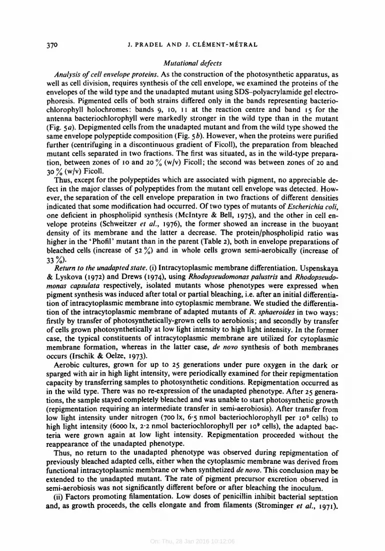
Analysis of cell envelope proteins. As the construction of the photosynthetic apparatus, as well as cell division, requires synthesis of the cell envelope, we examined the proteins of the envelopes of the wild type and the unadapted mutant using SDS-polyacrylamide gel electro- phoresis. Pigmented cells of both strains differed only in the bands representing bacterio- chlorophyll holochromes: bands 9, 10, I I at the reaction centre and band 15 for the antenna bacteriochlorophyll were markedly stronger in the wild type than in the mutant (Fig. 5a). Depigmented cells from the unadapted mutant and from the wild type showed the same envelope polypeptide composition (Fig. 5 b). However, when the proteins were purified further (centrifuging in a discontinuous gradient of Ficoll), the preparation from bleached mutant cells separated in two fractions. The first was situated, as in the wild-type prepara- tion, between zones of 10 and 20 % (w/v) Ficoll; the second was between zones of 20 and 30 % (w/v) Ficoll.
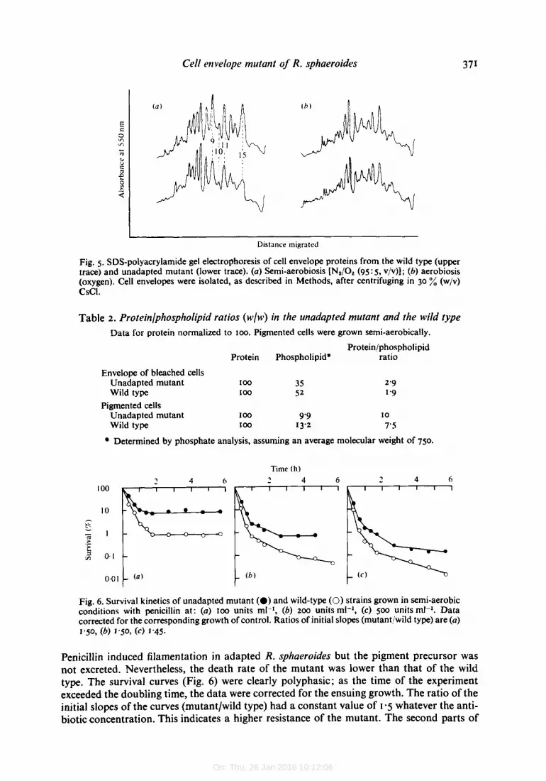
Thus, except for the polypeptides which are associated with pigment, no appreciable de- fect in the major classes of polypeptides from the mutant cell envelope was detected. How- ever, the separation of the cell envelope preparation in two fractions of different densities indicated that some modification had occurred. Of two types of mutants of Escherichia coli, one deficient in phospholipid synthesis (McIntyre & Bell, I975), and the other in cell en- velope proteins (Schweitzer et al., 1976), the former showed an increase in the buoyant density of its membrane and the latter a decrease. The protein/phospholipid ratio was higher in the ‘Phofil’ mutant than in the parent (Table 2), both in envelope preparations of bleached cells (increase of 52 %) and in whole cells grown semi-aerobically (increase of 33 %)*
Return to the unadapted state. (i) Intracytoplasmic membrane differentiation. Uspenskaya & Lyskova ( I 972) and Drews ( I 974 , using Rhodopseudomonas palustris and Rhodopseudo- monas capsulata respectively, isolated mutants whose phenotypes were expressed when pigment synthesis was induced after total or partial bleaching, i.e. after an initial differentia- tion of intracytoplasmic membrane into cytoplasmic membrane. We studied the differentia- tion of the intracytoplasmic membrane of adapted mutants of R. sphaeroides in two ways: firstly by transfer of photosynthetically-grown cells to aerobiosis ; and secondly by transfer of cells grown photosynthetically at low light intensity to high light intensity. In the former case, the typical constituents of intracytoplasmic membrane are utilized for cytoplasmic membrane formation, whereas in the latter case, de novo synthesis of both membranes occurs (Irschik & Oelze, 1973).
Aerobic cultures, grown for up to 25 generations under pure oxygen in the dark or sparged with air in high light intensity, were periodically examined for their repigmentation capacity by transferring samples to photosynthetic conditions. Repigmentation occurred as in the wild type. There was no re-expression of the unadapted phenotype. After 25 genera- tions, the sample stayed completely bleached and was unable to start photosynthetic growth (repigmentation requiring an intermediate transfer in semi-aerobiosis). After transfer from low light intensity under nitrogen (700 lx, 6-5 nmol bacteriochlorophyll per 109 cells) to high light intensity (6000 lx, 2-2 nmol bacteriochlorophyll per 109 cells), the adapted bac- teria were grown again at low light intensity. Repigmentation proceeded without the reappearance of the unadapted phenotype.
Thus, no return to the unadapted phenotype was observed during repigmentation of previously bleached adapted cells, either when the cytoplasmic membrane was derived from functional intracytoplasmic membrane or when synthetized de novo. This conclusion may extended to the unadapted mutant. The rate of pigment precursor excretion observed in semi-aerobiosis was not significantly different before or after bleaching the inoculum.
(ii) Factors promoting filamentation. LOW doses of penicillin inhibit bacterial septation and, as growth proceeds, the cells elongate and from filaments (Strominger et a/., 1971).
Downloaded from www.microbiologyresearch.org by
IP: 54.90.45.205
On: Thu, 28 Jan 2016 10:12:06
Cell envelope mutant of' R. sphaeroides 371
Distance migrated
Fig. 5 . SDS-polyacrylamide gel electrophoresis of cell envelope proteins from the wild type (upper trace) and unadapted mutant (lower trace). (a) Semi-aerobiosis [N,/O, (95: 5 , v/v)]; (b) aerobiosis (oxygen). Cell envelopes were isolated, as described in Methods, after centrifuging in 30 % (w/v) CSCI.
Table 2 . Proteinlphospholipid ratios (wlw) in the unadapted mutant and the wild type Data for protein normalized to 100. Pigmented cells were grown semi-aerobically.
Protein/phospholipid Protein Phospholipid* ratio
Envelope of bleached cells Unadapted mutant I 0 0 35 Wild type I 0 0 52
Unadapted mutant 100 9'9 Wild type 100 I 3.2
Pigmented cells
2'9 1'9
I 0
7'5
Determined by phosphate analysis, assuming an average molecular weight of 750.
2 4 6
t
Time (11) 2 4 6
Fig. 6. Survival kinetics of unadapted mutant (0) and wild-type (0) strains grown in semi-aerobic conditions with penicillin at: (a) 100 units ml-l, (b) 200 units ml-l, (c) 500 units m1-l. Data corrected for the corresponding growth of control. Ratios of initial slopes (mutant/wild type) are (a) 1-50, (b) 1'50, (C) 1-45.
Penicillin induced filamentation in adapted R. sphaeroides but the pigment precursor was not excreted. Nevertheless, the death rate of the mutant was lower than that of the wild type. The survival curves (Fig. 6) were clearly polyphasic; as the time of the experiment exceeded the doubling time, the data were corrected for the ensuing growth. The ratio of the initial slopes of the curves (mutantlwild type) had a constant value of 1.5 whatever the anti- biotic concentration. This indicates a higher resistance of the mutant. The second parts of
Downloaded from www.microbiologyresearch.org by
IP: 54.90.45.205
On: Thu, 28 Jan 2016 10:12:06
372 J. P R A D E L A N D J. C L ~ ~ M E N T - M ~ T R A L
the curves showed increasing slopes up to infinity (Fig. 6a, 6) ; the surviving cells then grew like the control.
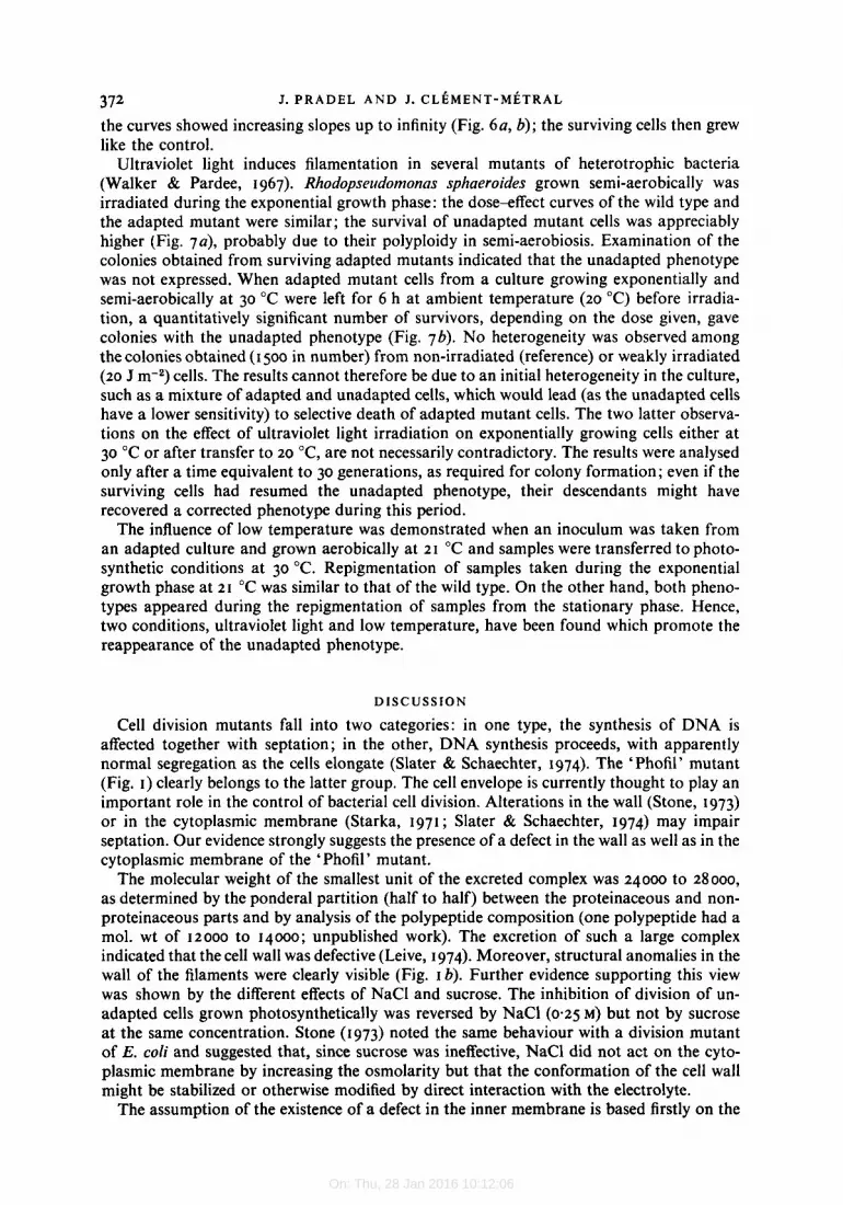
Ultraviolet light induces filamentation in several mutants of heterotrophic bacteria (Walker & Pardee, I 967). Rhodopseudomoncis sphaeroides grown semi-aerobically was irradiated during the exponential growth phase: the dose-effect curves of the wild type and the adapted mutant were similar; the survival of unadapted mutant cells was appreciably higher (Fig. 7a), probably due to their polyploidy in semi-aerobiosis. Examination of the colonies obtained from surviving adapted mutants indicated that the unadapted phenotype was not expressed. When adapted mutant cells from a culture growing exponentially and semi-aerobically at 30 "C were left for 6 h at ambient temperature (20 "C) before irradia- tion, a quantitatively significant number of survivors, depending on the dose given, gave colonies with the unadapted phenotype (Fig. 7b). No heterogeneity was observed among the colonies obtained (I 500 in number) from non-irradiated (reference) or weakly irradiated (20 J m-2) cells. The results cannot therefore be due to an initial heterogeneity in the culture, such as a mixture of adapted and unadapted cells, which would lead (as the unadapted cells have a lower sensitivity) to selective death of adapted mutant cells. The two latter observa- tions on the effect of ultraviolet light irradiation on exponentially growing cells either at 30 "C or after transfer to 20 "C, are not necessarily contradictory. The results were analysed only after a time equivalent to 30 generations, as required for colony formation; even if the surviving cells had resumed the unadapted phenotype, their descendants might have recovered a corrected phenotype during this period.
The influence of low temperature was demonstrated when an inoculum was taken from an adapted culture and grown aerobically at 21 "C and samples were transferred to photo- synthetic conditions at 30 "C. Repigmentation of samples taken during the exponential growth phase at 21 "C was similar to that of the wild type. On the other hand, both pheno- types appeared during the repigmentation of samples from the stationary phase. Hence, two conditions, ultraviolet light and low temperature, have been found which promote the reappearance of the unadapted phenotype.
DISCUSSION
Cell division mutants fall into two categories: in one type, the synthesis of DNA is affected together with septation; in the other, DNA synthesis proceeds, with apparently normal segregation as the cells elongate (Slater & Schaechter, 1974). The 'Phofil' mutant (Fig. I ) clearly belongs to the latter group. The cell envelope is currently thought to play an important role in the control of bacterial cell division. Alterations in the wall (Stone, 1973) or in the cytoplasmic membrane (Starka, 1971; Slater & Schaechter, 1974) may impair septation. Our evidence strongly suggests the presence of a defect in the wall as well as in the cytoplasmic membrane of the 'Phofil' mutant.
The molecular weight of the smallest unit of the excreted complex was 24000 to 28000, as determined by the ponderal partition (half to half) between the proteinaceous and non- proteinaceous parts and by analysis of the polypeptide composition (one polypeptide had a mol. wt of 12000 to 14000; unpublished work). The excretion of such a large complex indicated that the cell wall was defective (Leive, 1974). Moreover, structural anomalies in the wall of the filaments were clearly visible (Fig. I b). Further evidence supporting this view was shown by the different effects of NaCl and sucrose. The inhibition of division of un- adapted cells grown photosynthetically was reversed by NaCl (0.25 M) but not by sucrose at the same concentration. Stone (1973) noted the same behaviour with a division mutant of E. coli and suggested that, since sucrose was ineffective, NaCl did not act on the cyto- plasmic membrane by increasing the osmolarity but that the conformation of the cell wall might be stabilized or otherwise modified by direct interaction with the electrolyte.
The assumption of the existence of a defect in the inner membrane is based firstly on the

Downloaded from www.microbiologyresearch.org by
IP: 54.90.45.205
On: Thu, 28 Jan 2016 10:12:06
Cell envelope mutant of R. sphaeroides 373
Dose ( J m-’) Dose (J rn-3 100
Fig. 7. Effect of ultraviolet light irradiation on the viability of (a) unadapted mutant (0) and wild- type (m) cells (survivals of wild type and adapted mutant cells were identical), and (b) adapted cells left in suspension for 6 h at 20 “C prior to irradiation (0); frequency of colonies with unadapted phenotype among survivors (0) calculated after examining 500 to 1000 colonies.
relative inability of this membrane to incorporate the pigment complexes synthetized, partic- ularly the last precursor F,, bacteriochlorophyll(ide) complex (Pradel & Clement-MCtral, 1976), and secondly on the higher tolerance of the mutant towards penicillin. Such a toler- ance cannot be attributed to a decreased ability of the antibiotic to enter the cell, as the wall shows a defective barrier function, but rather to a restriction of the rate a t which penicillin can reach its target. Recent work of Spratt & Pardee (1975) indicates that all the penicillin- binding proteins are in the cytoplasmic membrane and that one is an enzyme involved in the terminal stages of peptidoglycan metabolism. Thus, an alteration of the cytoplasmic mem- brane could affect the function of this enzyme leading simultaneously to higher resistance to penicillin and to a deficient peptidoglycan layer of the cell wall. The protein/phospholipid ratio and the qualitative analysis of polypeptides by gel electrophoresis suggest that the membrane of the mutant is defective in phospholipids rather than in polypeptides.
As the unadapted phenotype is not expressed after bleaching adapted cells, the mutation is not in the photosynthetic apparatus itself. The positive actions of low temperature and ultraviolet light irradiation suggest instead that the disturbed adaptation process corres- ponds to a period of unbalanced growth (Normark & Wolf-Watz, 1974). During this un- balanced growth the different layers of the cell envelope are defective in several functions: diffusion barrier, structural integrity, cell division and photosynthetic apparatus construc- tion.
During the disturbed adaptation of the ‘Phofil’ mutant, the inner membrane differen- tiates only to a certain extent, the degree of differentiation being representative of the degree of adaptation of the inoculum. The reverse process, i.e. loss of pigmentation by transfer from anaerobic to aerobic conditions, induces no or little modification of this degree of adaptation. Thus, bleached mutant cultures can be obtained with different potentials for cytoplasmic membrane differentiation. This property, which permits the study of the assembly of photopigments in various, and particularly low, levels of membrane differentia- tion, will be examined further.
We thank F. Espardellier for the constructive criticism of the manuscript and C. Astier for her valuable assistance during survival tests with penicillin.

Downloaded from www.microbiologyresearch.org by
IP: 54.90.45.205
On: Thu, 28 Jan 2016 10:12:06
374 J . P R A D E L A N D J. C L E M E N T - M E T R A L
REFERENCES
BARLETT, G. R. (1959). Phosphorus assay in column chromatography. Journal of Biological Chemistry 234,466-468.
CLAYTON, R. K. (1963). Absorption spectra of photosynthetic bacteria and their chlorophylls. In Bacterial Photosynthesis, pp. 498-500. Edited by H. Gest, A. San Pietro and L. P. Vernon. Yellow Springs, Ohio: Antioch Press.
CLAYTON, R. K. & HASELKORN, R. (1972). Protein components of bacterial photosynthetic mem- branes. Journal of Moleculur Biology 68, 95- 105.
COHEN-BAZIRE, G., SISTROM, W. R. & STANIER, R. Y. (1957). Kinetic studies of pigment synthesis by non-sulfur purple bacteria. Journal of Cellular and Comparative Physiology 49, 25-68.
DREWS, G. (1974). Composition of a protochloro- phyll-protopheophytin complex, excreted by mutant strains of Rhodopseudomonas capsulata, in comparison with the photosynthetic apparatus. Archives of Microbiology 100, 397-407.
IRSCHIK, H. & OELZE, J. (1973). Membrane differ- entiation in phototrophically growing Rhodo- spirillum rubrum during transition from low to high light intensity. Biochimica et biophysica acta 330, 80-89.
JOLCHINE, G. & REISS-HUWN, F. (1974). Compara- tives studies on two reaction center preparations from Rhodopseudomonas spheroides. FEBS Let- ters 40, 5-8.
KATES, M. (1972). Lipid extraction procedures. In Techniques in Lipodology, pp. 347-353. Edited by T. S. Work and E. Work. Amsterdam: North Holland Publishing Company.
LAEMMLI, U. K. (1970). Cleavage of structural pro- teins during the assembly of the head of bacterio- phage TI. Nature, London 227, 680-685.
LATARJET, R., MORENNE, P. & BERGER, R. (1953). Un appareil simple pour le dosage des rayonne- ments ultra-violets 6mis par les lampes germicides. Annales de I'Institut Pasteur 85, 174-178.
LEIVE, L. (1974). The barrier function of the gram- negative envelope. Annals of the New York Academy of Sciences 235, 109-129.
LOWRY, 0. H., ROSERROUGH, N. J., FARR, A. L. & RANDALL, R. J. (195 I). Protein measurement with the Folin phenol reagent. Journal of Bio- logical Chemistry 193, 265-275.
MCINTYRE, T. M. & BELL, R. M. (1975). Mutant of Escherichia coli defective in membrane phos- pholipid synthesis. Effect of cessation of net phospholipid synthesis on cytoplasmic and outer membrane. Journal of Biological Chemistry 250, 9 0 5 3 9 5 9 .
NIEDERMAN, R. A., MALLON, D. E. & LANGAN, J. J. (1976). Membrane of Rhodopseudomonas sphe- roides. IV. Assembly of chromatophores in low- aeration cell suspension. Biochimica et biophysica
NORMARK, S. & WOLF-WATZ, H. (1974). Cell division and permeability of unbalanced envelope mu tan ts of Escherichin coli. Annales de Micro- biologie (Institiit Puterir) 125B, 2 I 1-226.
OELZE, J. & DREWS, G. (1972). Membranes of photosynthetic bacteria. Biochimica et biophysica
OELZE, J. & PAtiLKE, W. (1976). The early formation of the photosynthetic apparatus in Rhodospirillum ritbriim. Archives of' Microbiology 108, 28 I -285.
PETERS, G . A. & CELLARIUS, R. A. (1972). Photo- synthetic membrane development in Rhodo- pseudomonos spheroides. I I. Correlation of pig- ment incorporation with morphological aspects of thylakoid formation. Bioenergetics 3, 345-359.
PRADEL, J. & CL~MENT-M~TRAL, J. (1976). A 4-vinyl- protochlorophyllide complex accumulated by ' Phofil ' mutant of Rhodopseridomonas spheroides. An authentic intermediate in the development of the photosjnthetic apparatus. Biocltimica et biophysica acta 430, 253-264.
SAUNDERS, V. A. & JONES, 0. T. G. (1974). Adapta- t ion in Rhodopseildomonas spheroides. FEBS Letters 44, 169-172.
SCHWEITZER, M., SCHWARZ, H., %"TAG, 1. & HENNING, U. (1976). Mutational change of membrane architecture. Mutants of Escherichia coli K I ~ missing major proteins of the outer cell envelope membrane. Biochimica et biophysica
SLATER, M. & SCHAECHTER, M. (1974). Control of cell division in bacteria. &cteriological Reviews
SPRATT, B. G. & PARDEE, A. B. (1975). Penicillin- binding proteins and cell shape in Escherichia coli. Nature, London 254, 5 I 6 5 I 7.
STARKA, J. (1971). Cell envelope proteins of dividing and non dividing cells of Escherichin coli. FEBS Letters 16, 223-225.
STONE, A. B. (1973). Regulation of cell division in a temperature-sensitive division mutant of Escheri- chia coli. Journal of Bacteriology 116, 74 1-750.
STROMINGER, J. L., BLUMBERG, P. H., SUGINAKA, H. & UMBREir, J. (1971). How penicillin kills bac- teria : progress and problems. Proceedings of the Royul Society B179, 267-276.
between bacteriochlorophyll and membrane protein synthesis in Rhodopseudomonas spheroides. Proceedings of the National Academy of Sciences of the United States of America 70, 799-803.
USPENSKAYA, V. E. & LYSKOVA, G. V. (1972). Synthesis of pigments by the mutant of Rhodo- pseudomonas palustris under different growth conditions. Mikrobiologiya 6, I 038- 1 0 4 .
WALKER, J. R. & PARDEE, A. B. (1967). Conditional mutations involving septum formation in Es-
aclli265, 209-239.
acts 44% 474-49 I -
38, 199-221.
TAKEMOTO, J . & LASCELLES, J. (1973). Coupling
actu 440~4~9-447 . cherichia coli. Journal of Bacteriology 93, 107-1 14.


